a textbook of modern toxicology - ban 1080 poison
TRANSCRIPT

A TEXTBOOK OF MODERNTOXICOLOGY
THIRD EDITION
Edited by
Ernest HodgsonDepartment of Environmentaland Biochemical ToxicologyNorth Carolina State University
A JOHN WILEY & SONS, INC., PUBLICATION


A TEXTBOOK OF MODERNTOXICOLOGY
THIRD EDITION


A TEXTBOOK OF MODERNTOXICOLOGY
THIRD EDITION
Edited by
Ernest HodgsonDepartment of Environmentaland Biochemical ToxicologyNorth Carolina State University
A JOHN WILEY & SONS, INC., PUBLICATION
Copyright 2004 by John Wiley & Sons, Inc. All rights reserved.
Published by John Wiley & Sons, Inc., Hoboken, New Jersey.Published simultaneously in Canada.
No part of this publication may be reproduced, stored in a retrieval system, or transmitted in anyform or by any means, electronic, mechanical, photocopying, recording, scanning, or otherwise,except as permitted under Section 107 or 108 of the 1976 United States Copyright Act, withouteither the prior written permission of the Publisher, or authorization through payment of theappropriate per-copy fee to the Copyright Clearance Center, Inc., 222 Rosewood Drive, Danvers,MA 01923, 978-750-8400, fax 978-646-8600, or on the web at www.copyright.com. Requests to thePublisher for permission should be addressed to the Permissions Department, John Wiley & Sons,Inc., 111 River Street, Hoboken, NJ 07030, (201) 748-6011, fax (201) 748-6008.
Limit of Liability/Disclaimer of Warranty: While the publisher and author have used their bestefforts in preparing this book, they make no representations or warranties with respect to theaccuracy or completeness of the contents of this book and specifically disclaim any impliedwarranties of merchantability or fitness for a particular purpose. No warranty may be created orextended by sales representatives or written sales materials. The advice and strategies containedherein may not be suitable for your situation. You should consult with a professional whereappropriate. Neither the publisher nor author shall be liable for any loss of profit or any othercommercial damages, including but not limited to special, incidental, consequential, or otherdamages.
For general information on our other products and services please contact our Customer CareDepartment within the U.S. at 877-762-2974, outside the U.S. at 317-572-3993 or fax 317-572-4002.
Wiley also publishes its books in a variety of electronic formats. Some content that appears in print,however, may not be available in electronic format.
Library of Congress Cataloging-in-Publication Data:
Hodgson, Ernest, 1932–A textbook of modern toxicology / Ernest Hodgson.—3rd ed.
p. cm.Includes bibliographical references and index.
ISBN 0-471-26508-X1. Toxicology. I. Title.
RA1211.H62 2004615.9—dc22 2003017524
Printed in the United States of America.
10 9 8 7 6 5 4 3 2 1
CONTENTS
Preface xix
Contributors xxi
I Introduction 1
1 Introduction to Toxicology 3Ernest Hodgson
1.1 Definition and Scope, Relationship to Other Sciences, and History 31.1.1 Definition and Scope 31.1.2 Relationship to Other Sciences 81.1.3 A Brief History of Toxicology 8
1.2 Dose-Response Relationships 101.3 Sources of Toxic Compounds 10
1.3.1 Exposure Classes 111.3.2 Use Classes 11
1.4 Movement of Toxicants in the Environment 11Suggested Reading 12
2 Introduction to Biochemical and Molecular Methods in Toxicology 13Ernest Hodgson, Gerald A. LeBlanc, Sharon A. Meyer, and Robert C. Smart
2.1 Introduction 132.2 Cell Culture Techniques 13
2.2.1 Suspension Cell Culture 142.2.2 Monolayer Cell Culture 142.2.3 Indicators of Toxicity in Cultured Cells 14
2.3 Molecular Techniques 162.3.1 Molecular Cloning 172.3.2 cDNA and Genomic Libraries 172.3.3 Northern and Southern Blot Analyses 182.3.4 Polymerase Chain Reaction (PCR) 182.3.5 Evaluation of Gene Expression, Regulation, and Function 19
2.4 Immunochemical Techniques 19Suggested Reading 22
3 Toxicant Analysis and Quality Assurance Principles 23Ross B. Leidy
3.1 Introduction 233.2 General Policies Related to Analytical Laboratories 23
v
vi CONTENTS
3.2.1 Standard Operating Procedures (SOPs) 243.2.2 QA/QC Manuals 243.2.3 Procedural Manuals 243.2.4 Analytical Methods Files 253.2.5 Laboratory Information Management
System (LIMS) 253.3 Analytical Measurement System 26
3.3.1 Analytical Instrument Calibration 263.3.2 Quantitation Approaches and Techniques 26
3.4 Quality Assurance (QA) Procedures 273.5 Quality Control (QC) Procedures 273.6 Summary 28
Suggested Reading 28
II Classes of Toxicants 31
4 Exposure Classes, Toxicants in Air, Water, Soil, Domestic andOccupational Settings 33W. Gregory Cope
4.1 Air Pollutants 334.1.1 History 334.1.2 Types of Air Pollutants 344.1.3 Sources of Air Pollutants 354.1.4 Examples of Air Pollutants 364.1.5 Environmental Effects 38
4.2 Water and Soil Pollutants 404.2.1 Sources of Water and Soil Pollutants 404.2.2 Examples of Pollutants 41
4.3 Occupational Toxicants 444.3.1 Regulation of Exposure Levels 444.3.2 Routes of Exposure 454.3.3 Examples of Industrial Toxicants 46Suggested Reading 48
5 Classes of Toxicants: Use Classes 49W. Gregory Cope, Ross B. Leidy, and Ernest Hodgson
5.1 Introduction 495.2 Metals 49
5.2.1 History 495.2.2 Common Toxic Mechanisms and Sites of Action 505.2.3 Lead 515.2.4 Mercury 525.2.5 Cadmium 525.2.6 Chromium 535.2.7 Arsenic 535.2.8 Treatment of Metal Poisoning 54
CONTENTS vii
5.3 Agricultural Chemicals (Pesticides) 545.3.1 Introduction 545.3.2 Definitions and Terms 555.3.3 Organochlorine Insecticides 575.3.4 Organophosphorus Insecticides 585.3.5 Carbamate Insecticides 605.3.6 Botanical Insecticides 605.3.7 Pyrethroid Insecticides 615.3.8 New Insecticide Classes 615.3.9 Herbicides 625.3.10 Fungicides 635.3.11 Rodenticides 635.3.12 Fumigants 645.3.13 Conclusions 64
5.4 Food Additives and Contaminants 645.5 Toxins 65
5.5.1 History 655.5.2 Microbial Toxins 665.5.3 Mycotoxins 665.5.4 Algal Toxins 675.5.5 Plant Toxins 685.5.6 Animal Toxins 68
5.6 Solvents 705.7 Therapeutic Drugs 705.8 Drugs of Abuse 715.9 Combustion Products 715.10 Cosmetics 71
Suggested Reading 73
III Toxicant Processing In vivo 75
6 Absorption and Distribution of Toxicants 77Ronald E. Baynes and Ernest Hodgson
6.1 Introduction 776.2 Cell Membranes 786.3 Mechanisms of Transport 80
6.3.1 Passive Diffusion 806.3.2 Carrier-Mediated Membrane Transport 83
6.4 Physicochemical Properties Relevantto Diffusion 856.4.1 Ionization 866.4.2 Partition Coefficients 87
6.5 Routes of Absorption 886.5.1 Extent of Absorption 886.5.2 Gastrointestinal Absorption 896.5.3 Dermal Absorption 916.5.4 Respiratory Penetration 94
viii CONTENTS
6.6 Toxicant Distribution 976.6.1 Physicochemical Properties and Protein Binding 976.6.2 Volume of Distribution (Vd) 103
6.7 Toxicokinetics 105Suggested Reading 109
7 Metabolism of Toxicants 111Randy L. Rose and Ernest Hodgson
7.1 Introduction 1117.2 Phase I Reactions 112
7.2.1 The Endoplasmic Reticulum, Microsomal Preparation, andMonooxygenations 112
7.2.2 The Cytochrome P450-Dependent Monooxygenase System 1137.2.3 The Flavin-Containing Monooxygenase (FMO) 1287.2.4 Nonmicrosomal Oxidations 1307.2.5 Cooxidation by Cyclooxygenases 1327.2.6 Reduction Reactions 1337.2.7 Hydrolysis 1357.2.8 Epoxide Hydration 1357.2.9 DDT Dehydrochlorinase 136
7.3 Phase II Reactions 1377.3.1 Glucuronide Conjugation 1387.3.2 Glucoside Conjugation 1397.3.3 Sulfate Conjugation 1397.3.4 Methyltransferases 1417.3.5 Glutathione S-Transferases (GSTs) and Mercapturic Acid
Formation 1437.3.6 Cysteine Conjugate β-Lyase 1457.3.7 Acylation 1457.3.8 Phosphate Conjugation 148Suggested Reading 148
8 Reactive Metabolites 149Randy L. Rose and Patricia E. Levi
8.1 Introduction 1498.2 Activation Enzymes 1508.3 Nature and Stability of Reactive Metabolites 151
8.3.1 Ultra-short-lived Metabolites 1518.3.2 Short-lived Metabolites 1528.3.3 Longer-lived Metabolites 152
8.4 Fate of Reactive Metabolites 1538.4.1 Binding to Cellular Macromolecules 1538.4.2 Lipid Peroxidation 1538.4.3 Trapping and Removal: Role of Glutathione 153
8.5 Factors Affecting Toxicity of Reactive Metabolites 1548.5.1 Levels of Activating Enzymes 154
CONTENTS ix
8.5.2 Levels of Conjugating Enzymes 1548.5.3 Levels of Cofactors or Conjugating Chemicals 154
8.6 Examples of Activating Reactions 1548.6.1 Parathion 1558.6.2 Vinyl Chloride 1558.6.3 Methanol 1558.6.4 Aflatoxin B1 1568.6.5 Carbon Tetrachloride 1568.6.6 Acetylaminofluorene 1578.6.7 Benzo(a)pyrene 1588.6.8 Acetaminophen 1588.6.9 Cycasin 159
8.7 Future Developments 160Suggested Reading 161
9 Chemical and Physiological Influences on XenobioticMetabolism 163Randy L. Rose and Ernest Hodgson
9.1 Introduction 1639.2 Nutritional Effects 163
9.2.1 Protein 1639.2.2 Carbohydrates 1649.2.3 Lipids 1649.2.4 Micronutrients 1649.2.5 Starvation and Dehydration 1659.2.6 Nutritional Requirements in Xenobiotic Metabolism 165
9.3 Physiological Effects 1669.3.1 Development 1669.3.2 Gender Differences 1689.3.3 Hormones 1699.3.4 Pregnancy 1719.3.5 Disease 1719.3.6 Diurnal Rhythms 172
9.4 Comparative and Genetic Effects 1729.4.1 Variations Among Taxonomic Groups 1739.4.2 Selectivity 1819.4.3 Genetic Differences 181
9.5 Chemical Effects 1849.5.1 Inhibition 1859.5.2 Induction 1909.5.3 Biphasic Effects: Inhibition and Induction 199
9.6 Environmental Effects 1999.7 General Summary and Conclusions 201
Suggested Reading 201
10 Elimination of Toxicants 203Gerald A. LeBlanc
10.1 Introduction 203
x CONTENTS
10.2 Transport 20510.3 Renal Elimination 20510.4 Hepatic Elimination 207
10.4.1 Entero-hepatic Circulation 20810.4.2 Active Transporters of the Bile
Canaliculus 20910.5 Respiratory Elimination 21010.6 Conclusion 210
Suggested Reading 211
IV Toxic Action 213
11 Acute Toxicity 215Gerald A. LeBlanc
11.1 Introduction 21511.2 Acute Exposure and Effect 21511.3 Dose-response Relationships 21711.4 Nonconventional Dose-response Relationships 21911.5 Mechanisms of Acute Toxicity 220
11.5.1 Narcosis 22011.5.2 Acetylcholinesterase Inhibition 22011.5.3 Ion Channel Modulators 22211.5.4 Inhibitors of Cellular Respiration 223Suggested Reading 224
12 Chemical Carcinogenesis 225Robert C. Smart
12.1 General Aspects of Cancer 22512.2 Human Cancer 228
12.2.1 Causes, Incidence, and Mortality Rates of Human Cancer 22812.2.2 Known Human Carcinogens 23112.2.3 Classification of Human Carcinogens 233
12.3 Classes of Agents Associated with Carcinogenesis 23612.3.1 DNA-Damaging Agents 23712.3.2 Epigenetic Agents 239
12.4 General Aspects of Chemical Carcinogenesis 24012.5 Initiation-Promotion Model for Chemical Carcinogenesis 24112.6 Metabolic Activation of Chemical Carcinogens and DNA Adduct
Formation 24312.7 Oncogenes 245
12.7.1 Mutational Activation of Proto-oncogenes 24512.7.2 Ras Oncogene 246
12.8 Tumor Suppressor Genes 24712.8.1 Inactivation of Tumor Suppressor Genes 24712.8.2 p53 Tumor Suppressor Gene 247
12.9 General Aspects of Mutagenicity 248
CONTENTS xi
12.10 Usefulness and Limitations of Mutagenicity Assays for theIdentification of Carcinogens 249Suggested Reading 250
13 Teratogenesis 251Stacy Branch
13.1 Introduction 25113.2 Principles of Teratology 25113.3 Mammalian Embryology Overview 25213.4 Critical Periods 25513.5 Historical Teratogens 256
13.5.1 Thalidomide 25613.5.2 Accutane (Isotetrinoin) 25613.5.3 Diethylstilbestrol (DES) 25613.5.4 Alcohol 25713.5.5 “Non Chemical” Teratogens 257
13.6 Testing Protocols 25713.6.1 FDA Guidelines for Reproduction Studies for Safety
Evaluation of Drugs for Human Use 25813.6.2 International Conference of Harmonization (ICH) of
Technical Requirements for Registration ofPharmaceuticals for Human Use (ICH)—US FDA, 1994 258
13.6.3 Alternative Test Methods 25913.7 Conclusions 259
Suggested Reading 259
V Organ Toxicity 261
14 Hepatotoxicity 263Ernest Hodgson and Patricia E. Levi
14.1 Introduction 26314.1.1 Liver Structure 26314.1.2 Liver Function 263
14.2 Susceptibility of the Liver 26414.3 Types of Liver Injury 264
14.3.1 Fatty Liver 26414.3.2 Necrosis 26614.3.3 Apoptosis 26614.3.4 Cholestasis 26614.3.5 Cirrhosis 26614.3.6 Hepatitis 26714.3.7 Oxidative Stress 26714.3.8 Carcinogenesis 267
14.4 Mechanisms of Hepatotoxicity 26814.5 Examples of Hepatotoxicants 269
14.5.1 Carbon Tetrachloride 26914.5.2 Ethanol 270
xii CONTENTS
14.5.3 Bromobenzene 27014.5.4 Acetaminophen 271
14.6 Metabolic Activation of Hepatotoxicants 272Suggested Reading 272
15 Nephrotoxicity 273Ernest Hodgson and Patricia E. Levi
15.1 Introduction 27315.1.1 Structure of the Renal System 27315.1.2 Function of the Renal System 273
15.2 Susceptibility of the Renal System 27415.3 Examples of Nephrotoxicants 275
15.3.1 Metals 27515.3.2 Aminoglycosides 27615.3.3 Amphotericin B 27615.3.4 Chloroform 27715.3.5 Hexachlorobutadiene 27715.3.6 Tetrafluoroethylene 278Suggested Reading 278
16 Toxicology of the Nervous System 279Bonita L. Blake
16.1 Introduction 27916.2 The Nervous system 279
16.2.1 The Neuron 28016.2.2 Neurotransmitters and their Receptors 28216.2.3 Glial Cells 28316.2.4 The Blood-Brain Barrier 28416.2.5 The Energy-Dependent Nervous System 285
16.3 Toxicant Effects on the Nervous System 28616.3.1 Structural Effects of Toxicants on Neurons 28716.3.2 Effects of Toxicants on Other Cells 28916.3.3 Toxicant-Mediated Alterations in Synaptic Function 290
16.4 Neurotoxicity Testing 29316.4.1 In vivo Tests of Human Exposure 29316.4.2 In vivo Tests of Animal Exposure 29516.4.3 In vitro Neurochemical and Histopathological End Points 296
16.5 Summary 297Suggested Reading 297
17 Endocrine System 299Gerald A. LeBlanc
17.1 Introduction 29917.2 Endocrine System 299
17.2.1 Nuclear Receptors 30217.2.2 Membrane-Bound Steroid Hormone Receptors 304
CONTENTS xiii
17.3 Endocrine Disruption 30617.3.1 Hormone Receptor Agonists 30617.3.2 Hormone Receptor Antagonists 30817.3.3 Organizational versus Activational Effects of Endocrine
Toxicants 30917.3.4 Inhibitors of Hormone Synthesis 31017.3.5 Inducers of Hormone Clearance 31017.3.6 Hormone Displacement from
Binding Proteins 31117.4 Incidents of Endocrine Toxicity 311
17.4.1 Organizational Toxicity 31117.4.2 Activational Toxicity 31217.4.3 Hypothyroidism 313
17.5 Conclusion 314Suggested Reading 315
18 Respiratory Toxicity 317Ernest Hodgson, Patricia E. Levi, and James C. Bonner
18.1 Introduction 31718.1.1 Anatomy 31718.1.2 Cell Types 31718.1.3 Function 317
18.2 Susceptibility of the Respiratory System 32018.2.1 Nasal 32018.2.2 Lung 320
18.3 Types of Toxic Response 32018.3.1 Irritation 32018.3.2 Cell Necrosis 32118.3.3 Fibrosis 32118.3.4 Emphysema 32118.3.5 Allergic Responses 32118.3.6 Cancer 32118.3.7 Mediators of Toxic Responses 322
18.4 Examples of Lung Toxicants RequiringActivation 32218.4.1 Introduction 32218.4.2 Monocrotaline 32218.4.3 Ipomeanol 32318.4.4 Paraquat 324
18.5 Defense Mechanisms 324Suggested Reading 325
19 Immunotoxicity 327MaryJane K. Selgrade
19.1 Introduction 32719.2 The Immune System 327
xiv CONTENTS
19.3 Immune Suppression 33019.4 Classification of Immune-Mediated Injury (Hypersensitivity) 33519.5 Effects of Chemicals on Allergic Disease 336
19.5.1 Allergic Contact Dermatitis 33719.5.2 Respiratory Allergens 33819.5.3 Adjuvants 340
19.6 Emerging Issues: Food Allergies, Autoimmunity, and theDeveloping Immune System 341Suggested Reading 342
20 Reproductive System 343Stacy Branch
20.1 Introduction 34320.2 Male Reproductive Physiology 34320.3 Mechanisms and Targets of Male Reproductive Toxicants 344
20.3.1 General Mechanisms 34420.3.2 Effects on Germ Cells 34520.3.3 Effects on Spermatogenesis and Sperm Quality 34520.3.4 Effects on Sexual Behavior 34520.3.5 Effects on Endocrine Function 345
20.4 Female Reproductive Physiology 34620.5 Mechanisms and Targets of Female Reproductive Toxicants 347
20.5.1 Tranquilizers, Narcotics, and Social Drugs 34720.5.2 Endocrine Disruptors (EDs) 34820.5.3 Effects on Germ Cells 34820.5.4 Effects on the Ovaries and Uterus 34820.5.5 Effects on Sexual Behavior 348Suggested Reading 349
VI Applied Toxicology 351
21 Toxicity Testing 353Helen Cunny and Ernest Hodgson
21.1 Introduction 35321.2 Experimental Administration of Toxicants 355
21.2.1 Introduction 35521.2.2 Routes of Administration 356
21.3 Chemical and Physical Properties 35821.4 Exposure and Environmental Fate 35821.5 In vivo Tests 358
21.5.1 Acute and Subchronic Toxicity Tests 35921.5.2 Chronic Tests 37021.5.3 Reproductive Toxicity and Teratogenicity 37121.5.4 Special Tests 378
21.6 In vitro and Other Short-Term Tests 38521.6.1 Introduction 38521.6.2 Prokaryote Mutagenicity 385
CONTENTS xv
21.6.3 Eukaryote Mutagenicity 38721.6.4 DNA Damage and Repair 38921.6.5 Chromosome Aberrations 39021.6.6 Mammalian Cell Transformation 39221.6.7 General Considerations and Testing Sequences 393
21.7 Ecological Effects 39321.7.1 Laboratory Tests 39421.7.2 Simulated Field Tests 39421.7.3 Field Tests 395
21.8 Risk Analysis 39521.9 The Future of Toxicity Testing 395
Suggested Reading 396
22 Forensic and Clinical Toxicology 399Stacy Branch
22.1 Introduction 39922.2 Foundations of Forensic Toxicology 39922.3 Courtroom Testimony 40022.4 Investigation of Toxicity-Related Death/Injury 400
22.4.1 Documentation Practices 40122.4.2 Considerations for Forensic Toxicological Analysis 40122.4.3 Drug Concentrations and Distribution 402
22.5 Laboratory Analyses 40322.5.1 Colorimetric Screening Tests 40322.5.2 Thermal Desorption 40322.5.3 Thin-Layer Chromatography (TLC) 40322.5.4 Gas Chromatography (GC) 40422.5.5 High-Performance Liquid Chromatography (HPLC) 40422.5.6 Enzymatic Immunoassay 404
22.6 Analytical Schemes for Toxicant Detection 40422.7 Clinical Toxicology 405
22.7.1 History Taking 40622.7.2 Basic Operating Rules in the Treatment of Toxicosis 40622.7.3 Approaches to Selected Toxicoses 407Suggested Reading 409
23 Prevention of Toxicity 411Ernest Hodgson
23.1 Introduction 41123.2 Legislation and Regulation 411
23.2.1 Federal Government 41223.2.2 State Governments 41623.2.3 Legislation and Regulation in Other Countries 416
23.3 Prevention in Different Environments 41723.3.1 Home 41723.3.2 Workplace 41823.3.3 Pollution of Air, Water, and Land 419
xvi CONTENTS
23.4 Education 420Suggested Reading 421
24 Human Health Risk Assessment 423Ronald E. Baynes
24.1 Introduction 42324.2 Risk Assessment Methods 424
24.2.1 Hazard Identification 42424.2.2 Exposure Assessment 42524.2.3 Dose Response and Risk Characterization 426
24.3 Noncancer Risk Assessment 42724.3.1 Default Uncertainty and Modifying Factors 42824.3.2 Derivation of Developmental Toxicant RfD 42924.3.3 Determination of RfD and RfC of Naphthalene with the
NOAEL Approach 43024.3.4 Benchmark Dose Approach 43024.3.5 Determination of BMD and BMDL for ETU 43124.3.6 Quantifying Risk for Noncarcinogenic Effects: Hazard
Quotient 43224.3.7 Chemical Mixtures 432
24.4 Cancer Risk Assessment 43324.5 PBPK Modeling 436
Suggested Reading 437
VII Environmental Toxicology 439
25 Analytical Methods in Toxicology 441Ross B. Leidy
25.1 Introduction 44125.2 Chemical and Physical Methods 442
25.2.1 Sampling 44225.2.2 Experimental Studies 44625.2.3 Forensic Studies 44625.2.4 Sample Preparation 44725.2.5 Separation and Identification 44825.2.6 Spectroscopy 45525.2.7 Other Analytical Methods 460Suggested Reading 461
26 Basics of Environmental Toxicology 463Gerald A. LeBlanc
26.1 Introduction 46326.2 Environmental Persistence 464
26.2.1 Abiotic Degradation 465
CONTENTS xvii
26.2.2 Biotic Degradation 46526.2.3 Nondegradative Elimination Processes 466
26.3 Bioaccumulation 46726.3.1 Factors That Influence Bioaccumulation 469
26.4 Toxicity 47026.4.1 Acute Toxicity 47026.4.2 Mechanisms of Acute Toxicity 47126.4.3 Chronic Toxicity 47226.4.4 Species-Specific Chronic Toxicity 47326.4.5 Abiotic and Biotic Interactions 474
26.5 Conclusion 477Suggested Reading 477
27 Transport and Fate of Toxicants in the Environment 479Damian Shea
27.1 Introduction 47927.2 Sources of Toxicants to the Environment 48027.3 Transport Processes 483
27.3.1 Advection 48327.3.2 Diffusion 485
27.4 Equilibrium Partitioning 48727.4.1 Air–Water Partitioning 48727.4.2 Octanol–Water Partitioning 48827.4.3 Lipid–Water Partitioning 48827.4.4 Particle–Water Partitioning 489
27.5 Transformation Processes 49027.5.1 Reversible Reactions 49027.5.2 Irreversible Reactions 493
27.6 Environmental Fate Models 497Suggested Reading 498
28 Environmental Risk Assessment 501Damian Shea
28.1 Introduction 50128.2 Formulating the Problem 503
28.2.1 Selecting Assessment End Points 50328.2.2 Developing Conceptual Models 50628.2.3 Selecting Measures 506
28.3 Analyzing Exposure and Effects Information 50728.3.1 Characterizing Exposure 50828.3.2 Characterizing Ecological Effects 510
28.4 Characterizing Risk 51228.4.1 Estimating Risk 51228.4.2 Describing Risk 512
xviii CONTENTS
28.5 Managing Risk 516Suggested Reading 517
VIII Summary 519
29 Future Considerations for Environmental and Human Health 521Ernest Hodgson
29.1 Introduction 52129.2 Risk Management 52229.3 Risk Assessment 52329.4 Hazard and Exposure Assessment 52329.5 In vivo Toxicity 52329.6 In vitro Toxicity 52429.7 Biochemical and Molecular Toxicology 52429.8 Development of Selective Toxicants 524
Glossary 525
Index 543
PREFACE
There are some excellent general reference works in toxicology, including Casarettand Doull’s Toxicology, 6th, edition, edited by Klaassen; a 13-volume ComprehensiveToxicology, edited by Sipes, Gandolfi, and McQueen; as well as many specializedmonographs on particular topics. However, the scarcity of textbooks designed forteacher and student to use in the classroom setting that impelled us to produce thefirst and second editions of this work is still apparent. With the retirement of Dr. Levi,a mainstay of the first two editions, and the continuing expansion of the subject matter,it seemed appropriate to invite others to contribute their expertise to the third edition.All of the authors are, or have been, involved in teaching a course in general toxicol-ogy at North Carolina State University and thus have insights into the actual teachingprocess as well as the subject matter of their areas of specialization.
At North Carolina State University, we continue to teach a course in general toxi-cology that is open to graduate students and undergraduate upperclassmen. In addition,in collaboration with Toxicology Communications, Inc., of Raleigh, North Carolina,we present an accelerated short course at the same level. Our experience leads us tobelieve that this text is suitable, in the junior or senior year, for undergraduate studentswith some background in chemistry, biochemistry, and animal physiology. For grad-uate students it is intended to lay the foundation for subsequent specialized coursesin toxicology, such as those in biochemical and molecular toxicology, environmentaltoxicology, chemical carcinogenesis, and risk assessment.
We share the view that an introductory text must present all of the necessary funda-mental information to fulfill this purpose, but in as uncomplicated a manner as possible.To enhance readability, references have been omitted from the text, although furtherreading is recommended at the end of each chapter.
Clearly, the amount of material, and the detail with which some of it is presented,is more than is needed for the average general toxicology course. This, however, willpermit each instructor to select and emphasize those areas that they feel need particularemphasis. The obvious biochemical bias of some chapters is not accidental, rather itis based on the philosophy that progress in toxicology continues to depend on furtherunderstanding of the fundamental basis of toxic action at the cellular and molecularlevels. The depth of coverage of each topic represents that chapter author’s judgmentof the amount of material appropriate to the beginning level as compared to thatappropriate to a more advanced course.
Thanks to all of the authors and to the students and faculty of the Departmentof Environmental and Molecular Toxicology at North Carolina State University andto Carolyn McNeill for much word processing. Particular thanks to Bob Esposito ofJohn Wiley and Sons, not least for his patience with missed deadlines and subse-quent excuses.
ERNEST HODGSON
Raleigh, North Carolina
xix

CONTRIBUTORS
Baynes, Ronald E., Cutaneous Pharmacology and Toxicology Center, College ofVeterinary Medicine, North Carolina State University, Raleigh, NC
Blake, Bonita L., Department of Pharmacology and Neuroscience Center, Universityof North Carolina at Chapel Hill, Chapel Hill, NC
Bonner, James C., National Institute of Environmental Health Sciences, ResearchTriangle Park, NC
Branch, Stacy, Department of Environmental and Molecular Toxicology, North Car-olina State University, Raleigh, NC
Cope, W. Gregory, Department of Environmental and Molecular Toxicology, NorthCarolina State University, Raleigh, NC
Cunny, Helen, Bayer Crop Science, Research Triangle Park, NC
Hodgson, Ernest, Department of Environmental and Molecular Toxicology, NorthCarolina State University, Raleigh, NC
LeBlanc, Gerald A., Department of Environmental and Molecular Toxicology, NorthCarolina State University, Raleigh, NC
Leidy, Ross B., Department of Environmental and Molecular Toxicology, North Car-olina State University, Raleigh, NC
Levi, Patricia E., Department of Environmental and Molecular Toxicology, NorthCarolina State University, Raleigh, NC
Meyer, Sharon A., Department of Toxicology, University of Louisiana, Monroe, LA
Rose, Randy L., Department of Environmental and Molecular Toxicology, NorthCarolina State University, Raleigh, NC
Selgrade, MaryJane K., United States Environmental Protection Agency, ResearchTriangle Park, NC
Shea, Damian, Department of Environmental and Molecular Toxicology, North Car-olina State University, Raleigh, NC
Smart, Robert C., Department of Environmental and Molecular Toxicology, NorthCarolina State University, Raleigh, NC
xxi


PART I
INTRODUCTION

CHAPTER 1
Introduction to Toxicology
ERNEST HODGSON
1.1 DEFINITION AND SCOPE, RELATIONSHIP TO OTHER SCIENCES,AND HISTORY
1.1.1 Definition and Scope
Toxicology can be defined as that branch of science that deals with poisons, and apoison can be defined as any substance that causes a harmful effect when administered,either by accident or design, to a living organism. By convention, toxicology alsoincludes the study of harmful effects caused by physical phenomena, such as radiationof various kinds and noise. In practice, however, many complications exist beyondthese simple definitions, both in bringing more precise meaning to what constitutesa poison and to the measurement of toxic effects. Broader definitions of toxicology,such as “the study of the detection, occurrence, properties, effects, and regulation oftoxic substances,” although more descriptive, do not resolve the difficulties. Toxicityitself can rarely, if ever, be defined as a single molecular event but is, rather, a cascadeof events starting with exposure, proceeding through distribution and metabolism, andending with interaction with cellular macromolecules (usually DNA or protein) andthe expression of a toxic end point. This sequence may be mitigated by excretion andrepair. It is to the complications, and to the science behind them and their resolution,that this textbook is dedicated, particularly to the how and why certain substancescause disruptions in biologic systems that result in toxic effects. Taken together, thesedifficulties and their resolution circumscribe the perimeter of the science of toxicology.
The study of toxicology serves society in many ways, not only to protect humansand the environment from the deleterious effects of toxicants but also to facilitate thedevelopment of more selective toxicants such as anticancer and other clinical drugsand pesticides.
Poison is a quantitative concept, almost any substance being harmful at some dosesbut, at the same time, being without harmful effect at some lower dose. Betweenthese two limits there is a range of possible effects, from subtle long-term chronictoxicity to immediate lethality. Vinyl chloride may be taken as an example. It is apotent hepatotoxicant at high doses, a carcinogen with a long latent period at lower
A Textbook of Modern Toxicology, Third Edition, edited by Ernest HodgsonISBN 0-471-26508-X Copyright 2004 John Wiley & Sons, Inc.
3
4 INTRODUCTION TO TOXICOLOGY
doses, and apparently without effect at very low doses. Clinical drugs are even morepoignant examples because, although therapeutic and highly beneficial at some doses,they are not without deleterious side effects and may be lethal at higher doses. Aspirin(acetylsalicylic acid), for example, is a relatively safe drug at recommended doses andis taken by millions of people worldwide. At the same time, chronic use can causedeleterious effects on the gastric mucosa, and it is fatal at a dose of about 0.2 to0.5 g/kg. Approximately 15% of reported accidental deaths from poisoning in childrenresult from ingestion of salicylates, particularly aspirin.
The importance of dose is well illustrated by metals that are essential in the dietbut are toxic at higher doses. Thus iron, copper, magnesium, cobalt, manganese, andzinc can be present in the diet at too low a level (deficiency), at an appropriate level(maintenance), or at too high a level (toxic). The question of dose-response relationshipsis fundamental to toxicology (see Section 1.2).
The definition of a poison, or toxicant, also involves a qualitative biological aspectbecause a compound, toxic to one species or genetic strain, may be relatively harmlessto another. For example, carbon tetrachloride, a potent hepatotoxicant in many species,is relatively harmless to the chicken. Certain strains of rabbit can eat Belladonna withimpunity while others cannot. Compounds may be toxic under some circumstancesbut not others or, perhaps, toxic in combination with another compound but nontoxicalone. The methylenedioxyphenyl insecticide synergists, such as piperonyl butoxide,are of low toxicity to both insects and mammals when administered alone but are, byvirtue of their ability to inhibit xenobiotic-metabolizing enzymes, capable of causingdramatic increases in the toxicity of other compounds.
The measurement of toxicity is also complex. Toxicity may be acute or chronic,and may vary from one organ to another as well as with age, genetics, gender, diet,physiological condition, or the health status of the organism. As opposed to experi-mental animals, which are highly inbred, genetic variation is a most important factorin human toxicity since the human population is highly outbred and shows extensivegenetic variation. Even the simplest measure of toxicity, the LD50 (the dose requiredto kill 50% of a population under stated conditions) is highly dependent on the extentto which the above variables are controlled. LD50 values, as a result, vary markedlyfrom one laboratory to another.
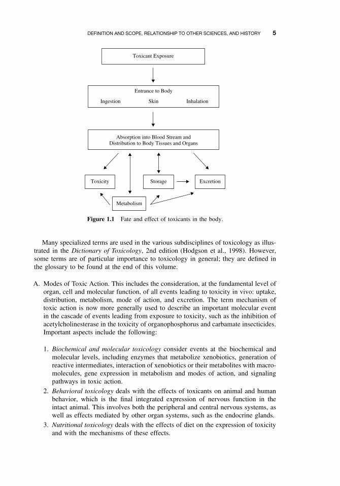
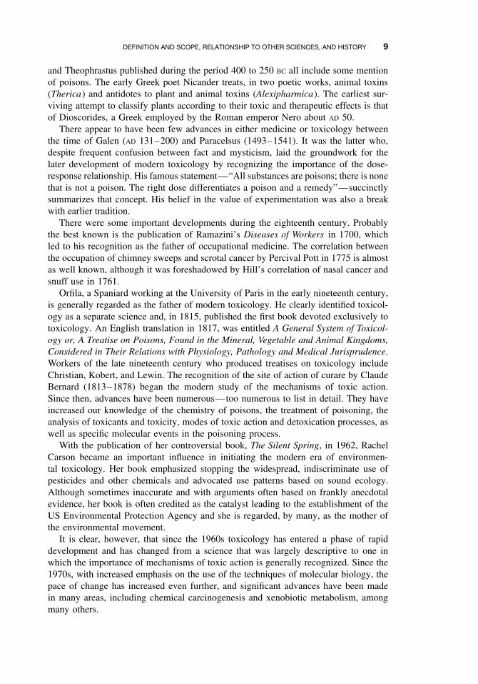
Exposure of humans and other organisms to toxicants may result from many activ-ities: intentional ingestion, occupational exposure, environmental exposure, as well asaccidental and intentional (suicidal or homicidal) poisoning. The toxicity of a par-ticular compound may vary with the portal of entry into the body, whether throughthe alimentary canal, the lungs, or the skin. Experimental methods of administrationsuch as injection may also give highly variable results; thus the toxicity from intra-venous (IV), intraperitoneal (IP), intramuscular (IM), or subcutaneous (SC) injectionof a given compound may be quite different. Toxicity may vary as much as tenfoldwith the route of administration. Following exposure there are multiple possible routesof metabolism, both detoxifying and activating, and multiple possible toxic endpoints(Figure 1.1).
Attempts to define the scope of toxicology, including that which follows, musttake into account that the various subdisciplines are not mutually exclusive and arefrequently interdependent. Due to overlapping of mechanisms as well as use and chem-ical classes of toxicants, clear division into subjects of equal extent or importance isnot possible.
DEFINITION AND SCOPE, RELATIONSHIP TO OTHER SCIENCES, AND HISTORY 5
Toxicant Exposure
Entrance to Body
Skin InhalationIngestion
Absorption into Blood Stream andDistribution to Body Tissues and Organs
Toxicity Storage
Metabolism
Excretion
Figure 1.1 Fate and effect of toxicants in the body.
Many specialized terms are used in the various subdisciplines of toxicology as illus-trated in the Dictionary of Toxicology, 2nd edition (Hodgson et al., 1998). However,some terms are of particular importance to toxicology in general; they are defined inthe glossary to be found at the end of this volume.
A. Modes of Toxic Action. This includes the consideration, at the fundamental level oforgan, cell and molecular function, of all events leading to toxicity in vivo: uptake,distribution, metabolism, mode of action, and excretion. The term mechanism oftoxic action is now more generally used to describe an important molecular eventin the cascade of events leading from exposure to toxicity, such as the inhibition ofacetylcholinesterase in the toxicity of organophosphorus and carbamate insecticides.Important aspects include the following:
1. Biochemical and molecular toxicology consider events at the biochemical andmolecular levels, including enzymes that metabolize xenobiotics, generation ofreactive intermediates, interaction of xenobiotics or their metabolites with macro-molecules, gene expression in metabolism and modes of action, and signalingpathways in toxic action.
2. Behavioral toxicology deals with the effects of toxicants on animal and humanbehavior, which is the final integrated expression of nervous function in theintact animal. This involves both the peripheral and central nervous systems, aswell as effects mediated by other organ systems, such as the endocrine glands.
3. Nutritional toxicology deals with the effects of diet on the expression of toxicityand with the mechanisms of these effects.
6 INTRODUCTION TO TOXICOLOGY
4. Carcinogenesis includes the chemical, biochemical, and molecular events thatlead to the large number of effects on cell growth collectively known as cancer.
5. Teratogenesis includes the chemical, biochemical, and molecular events that leadto deleterious effects on development.
6. Mutagenesis is concerned with toxic effects on the genetic material and theinheritance of these effects.
7. Organ toxicity considers effects at the level of organ function (neurotoxicity,hepatotoxicity, nephrotoxicity, etc.).
B. Measurement of Toxicants and Toxicity. These important aspects deal primarilywith analytical chemistry, bioassay, and applied mathematics; they are designedto provide the methodology to answer certain critically important questions. Is thesubstance likely to be toxic? What is its chemical identify? How much of it ispresent? How can we assay its toxic effect, and what is the minimum level atwhich this toxic effect can be detected? A number of important fields are included:
1. Analytical toxicology is a branch of analytical chemistry concerned with theidentification and assay of toxic chemicals and their metabolites in biologicaland environmental materials.
2. Toxicity testing involves the use of living systems to estimate toxic effects. Itcovers the gamut from short-term tests for genotoxicity such as the Ames testand cell culture techniques to the use of intact animals for a variety of testsfrom acute toxicity to lifetime chronic toxicity. Although the term “bioassay”is used properly only to describe the use of a living organism to quantitate theamount of a particular toxicant present, it is frequently used to describe any invivo toxicity test.
3. Toxicologic pathology is the branch of pathology that deals with the effectsof toxic agents manifested as changes in subcellular, cellular, tissue, or organmorphology.
4. Structure-activity studies are concerned with the relationship between the chem-ical and physical properties of a chemical and toxicity and, particularly, the useof such relationships as predictors of toxicity.
5. Biomathematics and statistics relate to many areas of toxicology. They dealwith data analysis, the determination of significance, and the formulation of riskestimates and predictive models.
6. Epidemiology as it applies to toxicology, is of great importance as it deals withthe relationship between chemical exposure and human disease in actual popu-lations rather than in experimental settings.
C. Applied Toxicology. This includes the various aspects of toxicology as they applyin the field or the development of new methodology or new selective toxicants forearly application in the field setting.
1. Clinical toxicology is the diagnosis and treatment of human poisoning.2. Veterinary toxicology is the diagnosis and treatment of poisoning in animals other
than humans, particularly livestock and companion animals, but not excludingferal species. Other important concerns of veterinary toxicology are the possible
DEFINITION AND SCOPE, RELATIONSHIP TO OTHER SCIENCES, AND HISTORY 7
transmission of toxins to the human population in meat, fish, milk, and otherfoodstuffs and the care and ethical treatment of experimental animals.
3. Forensic toxicology concerns the medicolegal aspects, including detection ofpoisons in clinical and other samples.
4. Environmental toxicology is concerned with the movement of toxicants and theirmetabolites and degradation products in the environment and in food chains andwith the effect of such contaminants on individuals and, especially, populations.Because of the large number of industrial chemicals and possibilities for expo-sure, as well as the mosaic of overlapping laws that govern such exposure, thisarea of applied toxicology is well developed.
5. Industrial toxicology is a specific area of environmental toxicology that dealswith the work environment and constitutes a significant part of industrial hygiene.
D. Chemical Use Classes. This includes the toxicology aspects of the development ofnew chemicals for commercial use. In some of these use classes, toxicity, at least tosome organisms, is a desirable trait; in others, it is an undesirable side effect. Useclasses are not composed entirely of synthetic chemicals; many natural productsare isolated and used for commercial and other purposes and must be subjected tothe same toxicity testing as that required for synthetic chemicals. Examples of suchnatural products include the insecticide, pyrethrin, the clinical drug, digitalis, andthe drug of abuse, cocaine.
1. Agricultural chemicals include many compounds, such as insecticides, herbi-cides, fungicides, and rodenticides, in which toxicity to the target organismis a desired quality whereas toxicity to “nontarget species” is to be avoided.Development of such selectively toxic chemicals is one of the applied roles ofcomparative toxicology.
2. Clinical drugs are properly the province of pharmaceutical chemistry and phar-macology. However, toxic side effects and testing for them clearly fall withinthe science of toxicology.
3. Drugs of abuse are chemicals taken for psychological or other effects and maycause dependence and toxicity. Many of these are illegal, but some are of clinicalsignificance when used correctly.
4. Food additives are of concern to toxicologists only when they are toxic or beingtested for possible toxicity.
5. Industrial chemicals are so numerous that testing them for toxicity or controllingexposure to those known to be toxic is a large area of toxicological activity.
6. Naturally occurring substances include many phytotoxins, mycotoxins, and min-erals, all occurring in the environment. The recently expanded and now extensiveuse of herbal remedies and dietary supplements has become a cause of concernfor toxicologists and regulators. Not only is their efficacy frequently dubious,but their potential toxicity is largely unknown.
7. Combustion products are not properly a use class but are a large and importantclass of toxicants, generated primarily from fuels and other industrial chemicals.
E. Regulatory Toxicology These aspects, concerned with the formulation of laws,and regulations authorized by laws, are intended to minimize the effect of toxicchemicals on human health and the environment.
8 INTRODUCTION TO TOXICOLOGY
1. Legal aspects are the formulation of laws and regulations and their enforce-ment. In the United States, enforcement falls under such government agenciesas the Environmental Protection Agency (EPA), the Food and Drug Adminis-tration (FDA), and the Occupational Safety and Health Administration (OSHA).Similar government agencies exist in many other countries.
2. Risk assessment is the definition of risks, potential risks, and the risk-benefitequations necessary for the regulation of toxic substances. Risk assessment islogically followed by risk communication and risk management.
1.1.2 Relationship to Other Sciences
Toxicology is highly eclectic science and human activity drawing from, and contribut-ing to, a broad spectrum of other sciences and human activities. At one end of thespectrum are those sciences that contribute their methods and philosophical concepts toserve the needs of toxicologists, either in research or in the application of toxicology tohuman affairs. At the other end of the spectrum are those sciences to which toxicologycontributes.
In the first group chemistry, biochemistry, pathology, physiology, epidemiology,immunology, ecology, and biomathematics have long been important while molecu-lar biology has, in the last two or three decades, contributed to dramatic advancesin toxicology.
In the group of sciences to which toxicology contributes significantly are suchaspects of medicine as forensic medicine, clinical toxicology, pharmacy and pharmacol-ogy, public health, and industrial hygiene. Toxicology also contributes in an importantway to veterinary medicine, and to such aspects of agriculture as the development andsafe use of agricultural chemicals. The contributions of toxicology to environmentalstudies has become increasingly important in recent years.
Clearly, toxicology is preeminently an applied science, dedicated to the enhancementof the quality of life and the protection of the environment. It is also much more.Frequently the perturbation of normal life processes by toxic chemicals enables usto learn more about the life processes themselves. The use of dinitrophenol and otheruncoupling agents to study oxidative phosphorylation and the use of α-amanitin to studyRNA polymerases are but two of many examples. The field of toxicology has expandedenormously in recent decades, both in numbers of toxicologists and in accumulatedknowledge. This expansion has brought a change from a primarily descriptive scienceto one which utilizes an extensive range of methodology to study the mechanismsinvolved in toxic events.
1.1.3 A Brief History of Toxicology
Much of the early history of toxicology has been lost and in much that has survivedtoxicology is of almost incidental importance in manuscripts dealing primarily withmedicine. Some, however, deal more specifically with toxic action or with the useof poisons for judicial execution, suicide or political assassination. Regardless of thepaucity of the early record, and given the need for people to avoid toxic animals andplants, toxicology must rank as one of the oldest practical sciences.
The Egyptian papyrus, Ebers, dating from about 1500 BC, must rank as the earliestsurviving pharmacopeia, and the surviving medical works of Hippocrates, Aristotle,
DEFINITION AND SCOPE, RELATIONSHIP TO OTHER SCIENCES, AND HISTORY 9
and Theophrastus published during the period 400 to 250 BC all include some mentionof poisons. The early Greek poet Nicander treats, in two poetic works, animal toxins(Therica) and antidotes to plant and animal toxins (Alexipharmica). The earliest sur-viving attempt to classify plants according to their toxic and therapeutic effects is thatof Dioscorides, a Greek employed by the Roman emperor Nero about AD 50.
There appear to have been few advances in either medicine or toxicology betweenthe time of Galen (AD 131–200) and Paracelsus (1493–1541). It was the latter who,despite frequent confusion between fact and mysticism, laid the groundwork for thelater development of modern toxicology by recognizing the importance of the dose-response relationship. His famous statement—“All substances are poisons; there is nonethat is not a poison. The right dose differentiates a poison and a remedy”—succinctlysummarizes that concept. His belief in the value of experimentation was also a breakwith earlier tradition.
There were some important developments during the eighteenth century. Probablythe best known is the publication of Ramazini’s Diseases of Workers in 1700, whichled to his recognition as the father of occupational medicine. The correlation betweenthe occupation of chimney sweeps and scrotal cancer by Percival Pott in 1775 is almostas well known, although it was foreshadowed by Hill’s correlation of nasal cancer andsnuff use in 1761.
Orfila, a Spaniard working at the University of Paris in the early nineteenth century,is generally regarded as the father of modern toxicology. He clearly identified toxicol-ogy as a separate science and, in 1815, published the first book devoted exclusively totoxicology. An English translation in 1817, was entitled A General System of Toxicol-ogy or, A Treatise on Poisons, Found in the Mineral, Vegetable and Animal Kingdoms,Considered in Their Relations with Physiology, Pathology and Medical Jurisprudence.Workers of the late nineteenth century who produced treatises on toxicology includeChristian, Kobert, and Lewin. The recognition of the site of action of curare by ClaudeBernard (1813–1878) began the modern study of the mechanisms of toxic action.Since then, advances have been numerous—too numerous to list in detail. They haveincreased our knowledge of the chemistry of poisons, the treatment of poisoning, theanalysis of toxicants and toxicity, modes of toxic action and detoxication processes, aswell as specific molecular events in the poisoning process.
With the publication of her controversial book, The Silent Spring, in 1962, RachelCarson became an important influence in initiating the modern era of environmen-tal toxicology. Her book emphasized stopping the widespread, indiscriminate use ofpesticides and other chemicals and advocated use patterns based on sound ecology.Although sometimes inaccurate and with arguments often based on frankly anecdotalevidence, her book is often credited as the catalyst leading to the establishment of theUS Environmental Protection Agency and she is regarded, by many, as the mother ofthe environmental movement.
It is clear, however, that since the 1960s toxicology has entered a phase of rapiddevelopment and has changed from a science that was largely descriptive to one inwhich the importance of mechanisms of toxic action is generally recognized. Since the1970s, with increased emphasis on the use of the techniques of molecular biology, thepace of change has increased even further, and significant advances have been madein many areas, including chemical carcinogenesis and xenobiotic metabolism, amongmany others.
10 INTRODUCTION TO TOXICOLOGY
1.2 DOSE-RESPONSE RELATIONSHIPS
As mentioned previously, toxicity is a relative event that depends not only on thetoxic properties of the chemical and the dose administered but also on individual andinterspecific variation in the metabolic processing of the chemical. The first recog-nition of the relationship between the dose of a compound and the response elicitedhas been attributed to Paracelsus (see Section 1.1.3). It is noteworthy that his state-ment includes not only that all substances can be toxic at some dose but that “theright dose differentiates a poison from a remedy,” a concept that is the basis forpharmaceutical therapy.
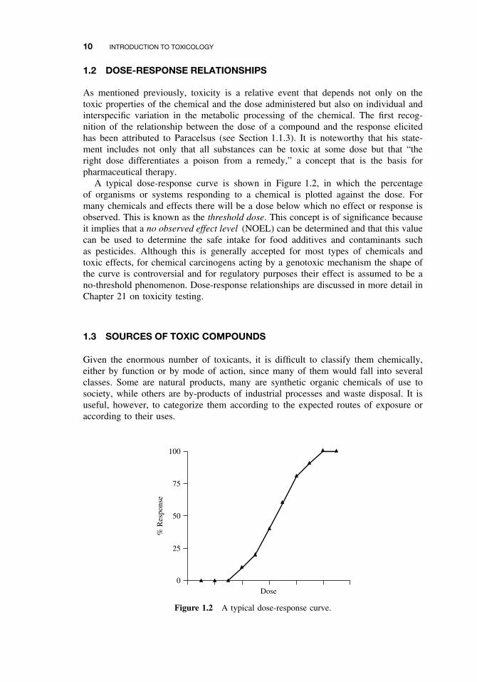
A typical dose-response curve is shown in Figure 1.2, in which the percentageof organisms or systems responding to a chemical is plotted against the dose. Formany chemicals and effects there will be a dose below which no effect or response isobserved. This is known as the threshold dose. This concept is of significance becauseit implies that a no observed effect level (NOEL) can be determined and that this valuecan be used to determine the safe intake for food additives and contaminants suchas pesticides. Although this is generally accepted for most types of chemicals andtoxic effects, for chemical carcinogens acting by a genotoxic mechanism the shape ofthe curve is controversial and for regulatory purposes their effect is assumed to be ano-threshold phenomenon. Dose-response relationships are discussed in more detail inChapter 21 on toxicity testing.
1.3 SOURCES OF TOXIC COMPOUNDS
Given the enormous number of toxicants, it is difficult to classify them chemically,either by function or by mode of action, since many of them would fall into severalclasses. Some are natural products, many are synthetic organic chemicals of use tosociety, while others are by-products of industrial processes and waste disposal. It isuseful, however, to categorize them according to the expected routes of exposure oraccording to their uses.
100
75
50
25
0
% R
espo
nse
Dose
Figure 1.2 A typical dose-response curve.
MOVEMENT OF TOXICANTS IN THE ENVIRONMENT 11
1.3.1 Exposure Classes
Exposure classes include toxicants in food, air, water, and soil as well as toxicantscharacteristic of domestic and occupational Settings. Toxicant exposure classes aredescribed in detail in Chapter 4.
1.3.2 Use Classes
Use classes include drugs of abuse, therapeutic drugs, agricultural chemicals, food addi-tives and contaminants, metals, solvents, combustion products, cosmetics, and toxins.Some of these, such as combustion products, are the products of use processes ratherthan being use classes. All of these groups of chemicals are discussed in detail inChapter 5.
1.4 MOVEMENT OF TOXICANTS IN THE ENVIRONMENT
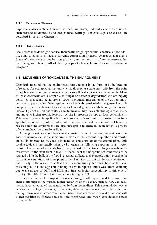
Chemicals released into the environment rarely remain in the form, or at the location,of release. For example, agricultural chemicals used as sprays may drift from the pointof application as air contaminants or enter runoff water as water contaminants. Manyof these chemicals are susceptible to fungal or bacterial degradation and are rapidlydetoxified, frequently being broken down to products that can enter the carbon, nitro-gen, and oxygen cycles. Other agricultural chemicals, particularly halogenated organiccompounds, are recalcitrant to a greater or lesser degree to metabolism by microorgan-isms and persist in soil and water as contaminants; they may enter biologic food chainsand move to higher trophic levels or persist in processed crops as food contaminants.This same scenario is applicable to any toxicant released into the environment for aspecific use or as a result of industrial processes, combustion, and so on. Chemicalsreleased into the environment are also susceptible to chemical degradation, a processoften stimulated by ultraviolet light.
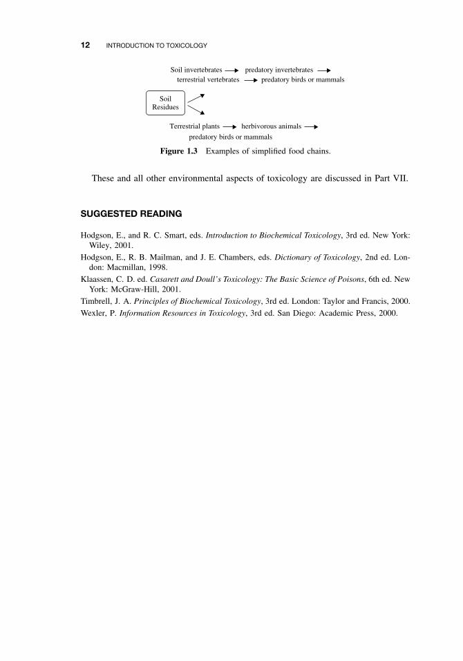
Although most transport between inanimate phases of the environment results inwider dissemination, at the same time dilution of the toxicant in question and transferamong living creatures may result in increased concentration or bioaccumulation. Lipidsoluble toxicants are readily taken up by organisms following exposure in air, water,or soil. Unless rapidly metabolized, they persist in the tissues long enough to betransferred to the next trophic level. At each level the lipophilic toxicant tends to beretained while the bulk of the food is digested, utilized, and excreted, thus increasing thetoxicant concentration. At some point in the chain, the toxicant can become deleterious,particularly if the organism at that level is more susceptible than those at the levelpreceding it. Thus the eggshell thinning in certain raptorial birds was almost certainlydue to the uptake of DDT and DDE and their particular susceptibility to this type oftoxicity. Simplified food chains are shown in Figure 1.3.
It is clear that such transport can occur through both aquatic and terrestrial foodchains, although in the former, higher members of the chains, such as fish, can accu-mulate large amounts of toxicants directly from the medium. This accumulation occursbecause of the large area of gill filaments, their intimate contact with the water andthe high flow rate of water over them. Given these characteristics and a toxicant witha high partition coefficient between lipid membranes and water, considerable uptakeis inevitable.
12 INTRODUCTION TO TOXICOLOGY
Soil invertebratesterrestrial vertebrates
Terrestrial plants herbivorous animals
predatory birds or mammalspredatory invertebrates
predatory birds or mammals
SoilResidues
Figure 1.3 Examples of simplified food chains.
These and all other environmental aspects of toxicology are discussed in Part VII.
SUGGESTED READING
Hodgson, E., and R. C. Smart, eds. Introduction to Biochemical Toxicology, 3rd ed. New York:Wiley, 2001.
Hodgson, E., R. B. Mailman, and J. E. Chambers, eds. Dictionary of Toxicology, 2nd ed. Lon-don: Macmillan, 1998.
Klaassen, C. D. ed. Casarett and Doull’s Toxicology: The Basic Science of Poisons, 6th ed. NewYork: McGraw-Hill, 2001.
Timbrell, J. A. Principles of Biochemical Toxicology, 3rd ed. London: Taylor and Francis, 2000.
Wexler, P. Information Resources in Toxicology, 3rd ed. San Diego: Academic Press, 2000.
CHAPTER 2
Introduction to Biochemicaland Molecular Methods in Toxicology
ERNEST HODGSON, GERALD A. LEBLANC, SHARON A. MEYER,and ROBERT C. SMART
2.1 INTRODUCTION
This chapter is not designed to summarize biochemical methods long used in toxicol-ogy such as colorimetric and radiometric methods for the investigation of xenobioticmetabolism, either in vivo or in vitro, but rather to give a brief summary of the methodsof molecular and cellular biology that have become, more recently, of critical impor-tance in toxicological research. The chapter owes much to Chapters 2 through 4 of thethird edition of Introduction to Biochemical Toxicology (see Suggested Reading), andthe reader is referred to these chapters for additional information.
2.2 CELL CULTURE TECHNIQUES
While scientists have had the ability to culture many unicellular organisms for sometime, recent advances in the culture of cells from multicellular organisms have played apivotal role in recent advances in toxicology. Cells can be isolated and either maintainedin a viable state for enough time to conduct informative experiments or, in somecases, propagated in culture. The advantages of cultured cells are that they can provideliving systems for the investigation of toxicity that are simplified relative to the intactorganism and they can be used as replacements for whole animal toxicity testing if thetoxic end point can be validated. Human cells play an important role in the extrapolationof toxic effects, discovered in experimental animals, to humans. Cultured cells, fromhumans or other mammals, are utilized in many of the molecular methods mentionedbelow. There are, however, limitations in the use of cellular methods. It has not beenpossible to culture many cell types, and of those that have been cultured, the lossof differentiated cell function is a common problem. Extrapolation of findings to theintact animal is often problematical and the use of undefined media constituents suchas serum, often essential for cell viability, may have unwanted or undefined effects oncell function and toxicant bioavailability.
A Textbook of Modern Toxicology, Third Edition, edited by Ernest HodgsonISBN 0-471-26508-X Copyright 2004 John Wiley & Sons, Inc.
13
14 INTRODUCTION TO BIOCHEMICAL AND MOLECULAR METHODS IN TOXICOLOGY
Studies have been carried out on cells isolated from tissues and maintained insuspension culture or on cells that have formed monolayers.
2.2.1 Suspension Cell Culture
Circulating blood cells or cells easily obtained by lavage such as peritoneal and alveolarmacrophages can normally survive in suspension culture when provided with a suitablenutrient medium. Cells from organized solid organs or tissues must be separated fromthe tissue and, if possible, separated into cell types, before being suspended in sucha medium.
Cell association within organs depends on protein complex formation, which in turnis Ca2+ dependent. Consequently dissociation media generally contain a proteolyticenzyme and the Ca2+ chelator EDTA. There are a number of methods available to sep-arate cell types from the mixture of dispersed cells, the commonest being centrifugationwithout a density gradient, wherein cells are separated by size, or centrifugation througha density gradient wherein cells are separated on the basis of their buoyant density.
Cells in suspension may be maintained for a limited period of time in definedmedia or for longer periods in nutrient, but less well-defined, media. In either casethese cultures are often used for studies of xenobiotic metabolism.
2.2.2 Monolayer Cell Culture
Proliferation of most cells in culture requires attachment to a substrate and occurs untillimited by cell-to-cell contact, resulting in the formation of a cellular monolayer. Thesubstrate provided for attachment is usually polystyrene modified to carry a charge.The medium for continued maintenance and growth contains salts and glucose, usuallywith a bicarbonate buffer. Because of the bicarbonate buffering system these culturesare maintained in a 5–10% CO2 atmosphere in a temperature and humidity controlledincubator. Many cells require serum for optimal growth, inducing considerable variabil-ity into the experimental system. Since the factors provided by serum are numerousand complex, defined serum substitutes are not always successful. The factors pro-vided by serum include proteins such as growth factors, insulin and transferrin (toprovide available iron), small organic molecules such as ethanolamine, and pyruvateand inorganic ions, such as selenium.
2.2.3 Indicators of Toxicity in Cultured Cells
Routine observation of cultured is usually carried out by phase contrast microscopy,utilizing the inverted phase contrast microscope. More recently, more detailed obser-vations have become possible utilizing fluorescent tags and inverted fluorescent micro-scopes. Fluorescent tags currently in use permit the assessment of oxidant status andmitochondrial function as well as the intracellular concentration of sulfhydryl groups,Ca2+, H+, Na+, and K+.
Toxicity to cultured cells may be the result either of inadequacies in the culture orthe toxicity effects of the chemical being investigated. Short-term toxicity is usually
Tabl
e2.
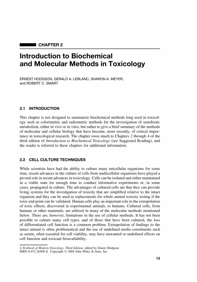
1E
xam
ples
ofA
pplic
atio
nof
Cel
lL
ines
Ret
aini
ngD
iffe
rent
iate
dP
rope
rtie
sin
the
Stud
yof
Toxi
cE
ffec
ts∗
Cel
lL
ine
Sour
ceD
iffe
rent
iate
dC
ell
Type
Toxi
cant
Mea
sure
dE
ndPo
int
N1E
-115
Mou
sene
urob
last
oma
Cho
liner
gic
neur
onL
ead
Pyre
thro
idin
sect
icid
eB
lock
age
ofvo
ltage
-dep
ende
ntC
a2+ch
anne
lsPr
olon
ged
open
time
for
volta
ge-d
epen
dent
Na+
chan
nels
PC12
Rat
pheo
chro
moc
ytom
a(a
dren
alm
edul
lary
tum
or)
Adr
ener
gic
neur
onT
ricr
esyl
phos
phat
e(o
rgan
opho
spha
te)
Inhi
bitio
nof
neur
ofila
men
tas
sem
bly
and
axon
algr
owth
SK-N
-S11
Hum
anne
urob
last
oma
Neu
ron
N2O
(ane
sthe
tic)
Dep
ress
edch
olin
ergi
cC
a2+si
gnal
ing
Hep
a-1
Mou
sehe
pato
ma
Hep
atoc
yte
2,3,
7,8-
tera
chlo
ro-d
iben
zodi
oxan
e(T
CC
D)
Indu
ctio
nof
CY
P1A
1an
d1B
1
H11
4E
Rat
hepa
tom
aH
epat
ocyt
ePo
lych
lori
nate
dbi
phen
yls
(PC
Bs)
Indu
ctio
nof
CY
P1A
1H
epG
2H
uman
hepa
tobl
asto
ma
Hep
atoc
yte
Cyc
loph
osph
amid
e(a
ntin
eopl
astic
)C
ytoc
hrom
eP4
50-d
epen
dent
geno
toxi
city
3T3-
L1
Mou
seem
bryo
fibro
blas
tsA
dipo
cyte
sT
CD
DIn
hibi
tion
ofgl
ucos
etr
ansp
ort
and
lipop
rote
inli
pase
Y1
Mou
sead
reno
cort
ical
tum
orA
dren
ocor
tical
cell
Met
hyl
sulf
one
met
abol
ites
ofD
DT
and
PCB
sIn
hibi
tion
ofco
rtic
oste
rone
synt
hesi
sby
com
peti
tive
inhi
biti
onof
cyto
chro
me
P450
LL
C-P
K1
Pig
kidn
eyR
enal
tubu
leep
ithel
ial
cell
Cad
miu
mC
ytot
oxic
ity,
apop
tosi
sM
DC
KD
ogki
dney
Ren
altu
bule
epith
elia
lce
llO
rgan
icm
ercu
ryco
mpo
unds
Cyt
otox
icity
,tr
anse
pith
elia
lle
akin
ess
Sour
ce:
E.
Hod
gson
and
R.
C.
Smar
t,ed
s.,
An
Intr
oduc
tion
toM
oder
nTo
xico
logy
,3r
ded
.N
ewY
ork:
Wile
y,20
01.
15
16 INTRODUCTION TO BIOCHEMICAL AND MOLECULAR METHODS IN TOXICOLOGY
DYE INFLUX MEMBRANE LEAKAGE
Trypan BluePropidium lodide
Cytoplasmic LDH, Cr2−
Plasma Membrane
NeutralRed
Lysosome
LysosomesNeutral Red
Uptake
↓
↓
↓↓
↓
BLEB
MitochondriaMTT ReductionRhodamine 123
Retention
EndoplasmicReticulumProtein
Synthesis
Synthesis
NucleusDNA, RNA
H+
ATP/ADP
NucleusC
ortical Microfilam
ents
Mitochondria
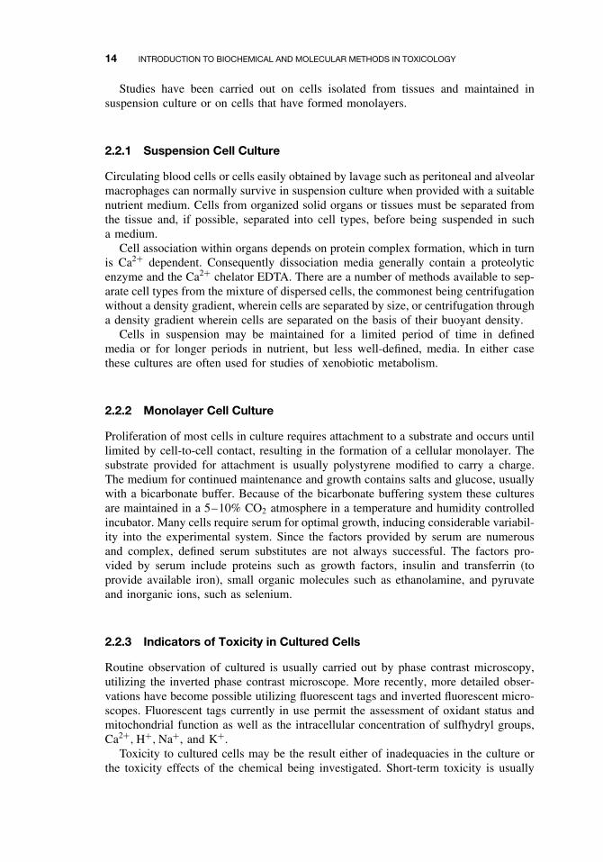
Figure 2.1 Idealized diagram of a cell to illustrate parameters often used to measure cytotoxi-city and the corresponding affected subcellular organelle. (From An Introduction to BiochemicalToxicology, 3rd ed., E. Hodgson and R. C. Smart, eds., Wiley, 2001.)
evaluated by examination of end points that indicate effects on cellular organelles suchas leakage of cell constituents into the medium, uptake of dyes into the cell and theformation of surface “blebs.” This is illustrated in Figure 2.1.
Longer term assessments of cell toxicity are highly dependent on the relevant toxicend point. They may include measurement of growth competence, apoptosis, and/ornecrosis, incorporation of radioactive precursors into essential cellular constituents suchas RNA, DNA, and protein and specialized cellular functions. Some examples of theuse of cultured cell lines in the study of toxicity effects are shown in Table 2.1.
2.3 MOLECULAR TECHNIQUES
Recombinant DNA techniques, including molecular cloning, have provided recent dra-matic advances in many areas of both fundamental and applied biology, toxicologynot excepted. Responses to toxicants may involve changes in gene expression and thenew microarray techniques enable the simultaneous examination of the level of expres-sion of many genes. The completion of the Human Genome Project will permit toxiceffects in humans to be investigated and will facilitate extrapolation from experimentalanimals. The human genome will also provide the essential genetic background infor-mation for studies of polymorphisms in xenobiotic-metabolizing and other enzymes.Such polymorphisms have already been shown to be very important in individualsensitivity to clinical drugs and in the definition of populations and/or individuals atincreased risk from particular toxicants. Chemically induced mutations, particularly inoncogenes and tumor-suppressor genes are important in chemical carcinogenesis. The
MOLECULAR TECHNIQUES 17
Promoter region containingproximal and distal elementsand enhancers
EXON EXON EXONINTRON INTRON
5′
5'
3′DNA sense strand
cap sitesplicejunctions
splicejunctions
translation initiation codon
polyadenylation signal
translation termination codon
TRANSCRIPTION
PROCESSING
TRANSLATION
Amino terminusProtein
mRNA
hnRNA
Met..........Phe.....Stop Carboxy terminus
..ATG.. GT
..AUG.. GU
..AUG... ..UUU....UAA...AAUAA AAAAAAAA 3'
AG...UUU... GU AG ..UAA....AAUAA AAAAAAA
AG..TTT... ..TAA.....AATAAGT AG
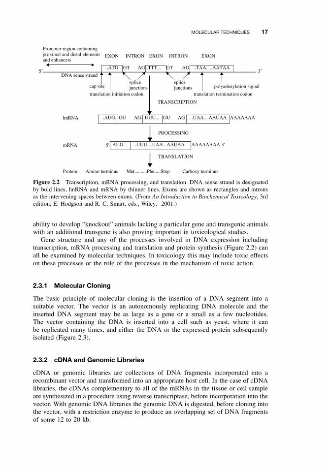
Figure 2.2 Transcription, mRNA processing, and translation. DNA sense strand is designatedby bold lines, hnRNA and mRNA by thinner lines. Exons are shown as rectangles and intronsas the intervening spaces between exons. (From An Introduction to Biochemical Toxicology, 3rdedition, E. Hodgson and R. C. Smart, eds., Wiley, 2001.)
ability to develop “knockout” animals lacking a particular gene and transgenic animalswith an additional transgene is also proving important in toxicological studies.
Gene structure and any of the processes involved in DNA expression includingtranscription, mRNA processing and translation and protein synthesis (Figure 2.2) canall be examined by molecular techniques. In toxicology this may include toxic effectson these processes or the role of the processes in the mechanism of toxic action.
2.3.1 Molecular Cloning
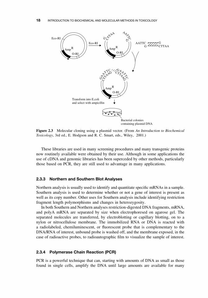
The basic principle of molecular cloning is the insertion of a DNA segment into asuitable vector. The vector is an autonomously replicating DNA molecule and theinserted DNA segment may be as large as a gene or a small as a few nucleotides.The vector containing the DNA is inserted into a cell such as yeast, where it canbe replicated many times, and either the DNA or the expressed protein subsequentlyisolated (Figure 2.3).
2.3.2 cDNA and Genomic Libraries
cDNA or genomic libraries are collections of DNA fragments incorporated into arecombinant vector and transformed into an appropriate host cell. In the case of cDNAlibraries, the cDNAs complementary to all of the mRNAs in the tissue or cell sampleare synthesized in a procedure using reverse transcriptase, before incorporation into thevector. With genomic DNA libraries the genomic DNA is digested, before cloning intothe vector, with a restriction enzyme to produce an overlapping set of DNA fragmentsof some 12 to 20 kb.
18 INTRODUCTION TO BIOCHEMICAL AND MOLECULAR METHODS IN TOXICOLOGY
CTT
AA AATTCGG
Eco-RI
Eco-RI
CTT
AAG
GA
A
TTCCTTAA
G
GAATTC
AATTCG
GCTTAA
Transform into E.coliand select with ampicillin
Bacterial coloniescontaining plasmid DNA
AmpO-RI O-RI
O-RI
R Amp R
Amp R
Figure 2.3 Molecular cloning using a plasmid vector. (From An Introduction to BiochemicalToxicology, 3rd ed., E. Hodgson and R. C. Smart, eds., Wiley, 2001.)
These libraries are used in many screening procedures and many transgenic proteinsnow routinely available were obtained by their use. Although in some applications theuse of cDNA and genomic libraries has been superceded by other methods, particularlythose based on PCR, they are still used to advantage in many applications.
2.3.3 Northern and Southern Blot Analyses
Northern analysis is usually used to identify and quantitate specific mRNAs in a sample.Southern analysis is used to determine whether or not a gene of interest is present aswell as its copy number. Other uses for Southern analysis include identifying restrictionfragment length polymorphisms and changes in heterozygosity.
In both Southern and Northern analyses restriction-digested DNA fragments, mRNA,and polyA mRNA are separated by size when electrophoresed on agarose gel. Theseparated molecules are transferred, by electroblotting or capillary blotting, on to anylon or nitrocellulose membrane. The immobilized RNA or DNA is reacted witha radiolabeled, chemiluminescent, or fluorescent probe that is complementary to theDNA/RNA of interest, unbound probe is washed off, and the membrane exposed, in thecase of radioactive probes, to radioautographic film to visualize the sample of interest.
2.3.4 Polymerase Chain Reaction (PCR)
PCR is a powerful technique that can, starting with amounts of DNA as small as thosefound in single cells, amplify the DNA until large amounts are available for many
IMMUNOCHEMICAL TECHNIQUES 19
different kinds of research. Twenty to 40 cycles of can provide up to 105 times theoriginal DNA sample.
It is necessary to know as much of the sequence of the DNA of interest as possi-ble in order to construct appropriate primers. These primers are complementary to thesequence at each end of the DNA sequence to be amplified. The DNA is incubated ina thermal cycler with thermostable DNA polymerase, all four dNTP, and the primers.The incubation temperature is raised to separate the DNA strands, lowered to permitannealing of the primers to the complementary regions of the DNA and then raised topermit the polymerase to synthesize DNA. This cycle is then repeated up to 40 times.The PCR technique has been used for many types of toxicological investigation includ-ing; uncovering polymorphisms in xenobiotic-metabolizing enzymes, isolating genesfrom cDNA and genomic libraries and for mutational analysis, to name only a few.
2.3.5 Evaluation of Gene Expression, Regulation, and Function
The methods used for the evaluation of regulation of gene expression are too numerousto be described in detail here. They include Northern analysis to determine levels ofa particular mRNA, nuclear run on to determine whether an increase in mRNA is dueto an increase in the rate of transcription, and promoter deletion analysis to identifyspecific elements in the promoter region responsible for the control of expression. Ofmuch current interest is the use of microarrays that permit the study of the expressionof hundreds to thousands of genes at the same time. Reverse transcriptase–polymerasechain reaction and RNase protection assay techniques are used to amplify and quantitatemRNAs, while the electrophoretic mobility shift assay is used to measure binding ofa transcription factor to its specific DNA consensus sequence.
Gene function in cultured cells can be investigated by expression of the gene productin a suitable expression system or, in vivo, by the creation of transgenic mice, eitherknockout mice in which the gene in question has been functionally deleted or miceinto which a transgene has been introduced.
A general, but more detailed and specific, account of these methods may be foundin Smart (2001; see Suggested Reading).
2.4 IMMUNOCHEMICAL TECHNIQUES
Most of the recently developed methods for the detection, characterization, and quan-titation of proteins are immunoassays based on the fact that proteins are antigens,compounds that can be recognized by an antibody. It is also true that by combiningsmall molecules (haptens) with a larger carrier molecule such as a protein, these meth-ods can be extended to small molecules of interest since antibodies can be producedthat recognize epitopes (specific sites on the antigen recognized by the antibody) thatinclude the hapten.
The antibodies used may be polyclonal or monoclonal, each with characteristicsfitting them for use in particular immunochemical methods. Injection of a mammalwith a foreign protein (immunogen) gives rise to an immune reaction that includesthe generation of antibodies from B lymphocytes. Each B lymphocyte gives rise toonly a single antibody type that recognizes a single epitope on the antigen. However,
20 INTRODUCTION TO BIOCHEMICAL AND MOLECULAR METHODS IN TOXICOLOGY
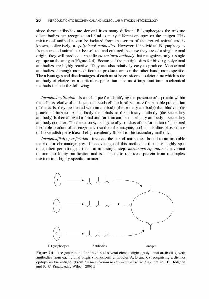
since these antibodies are derived from many different B lymphocytes the mixtureof antibodies can recognize and bind to many different epitopes on the antigen. Thismixture of antibodies can be isolated from the serum of the treated animal and isknown, collectively, as polyclonal antibodies. However, if individual B lymphocytesfrom a treated animal can be isolated and cultured, because they are of a single clonalorigin, they will produce a specific monoclonal antibody that recognizes only a singleepitope on the antigen (Figure 2.4). Because of the multiple sites for binding polyclonalantibodies are highly reactive. They are also relatively easy to produce. Monoclonalantibodies, although more difficult to produce, are, on the other hand, more specific.The advantages and disadvantages of each must be considered to determine which is theantibody of choice for a particular application. The most important immunochemicalmethods include the following:
Immunolocalization is a technique for identifying the presence of a protein withinthe cell, its relative abundance and its subcellular localization. After suitable preparationof the cells, they are treated with an antibody (the primary antibody) that binds to theprotein of interest. An antibody that binds to the primary antibody (the secondaryantibody) is then allowed to bind and form an antigen—primary antibody—secondaryantibody complex. The detection system generally consists of the formation of a coloredinsoluble product of an enzymatic reaction, the enzyme, such as alkaline phosphataseor horseradish peroxidase, being covalently linked to the secondary antibody.
Immunoaffinity purification involves the use of antibodies, bound to an insolublematrix, for chromatography. The advantage of this method is that it is highly spe-cific, often permitting purification in a single step. Immunoprecipitation is a variantof immunoaffinity purification and is a means to remove a protein from a complexmixture in a highly specific manner.
B Lymphocytes Antibodies Antigen
A
B
C
Figure 2.4 The generation of antibodies of several clonal origins (polyclonal antibodies) withantibodies from each clonal origin (monoclonal antibodies A, B and C) recognizing a distinctepitope on the antigen. (From An Introduction to Biochemical Toxicology, 3rd ed., E. Hodgsonand R. C. Smart, eds., Wiley, 2001.)
IMMUNOCHEMICAL TECHNIQUES 21
C T
C T
Gel Membrane
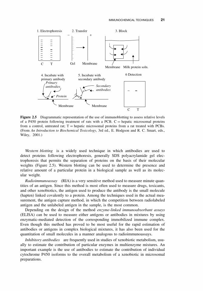
1. Electrophoresis 2. Transfer 3. Block
Membrane Milk protein soln.
6 Detection5. Incubate withsecondary antibody
4. Incubate withprimary antibody
Secondaryantibodies
Primaryantibodies
Protein
Membrane Membrane
+−
Figure 2.5 Diagrammatic representation of the use of immunoblotting to assess relative levelsof a P450 protein following treatment of rats with a PCB. C = hepatic microsomal proteinsfrom a control, untreated rat; T = hepatic microsomal proteins from a rat treated with PCBs.(From An Introduction to Biochemical Toxicology, 3rd ed., E. Hodgson and R. C. Smart, eds.,Wiley, 2001.)
Western blotting is a widely used technique in which antibodies are used todetect proteins following electrophoresis, generally SDS polyacrylamide gel elec-trophoresis that permits the separation of proteins on the basis of their molecularweights (Figure 2.5). Western blotting can be used to determine the presence andrelative amount of a particular protein in a biological sample as well as its molec-ular weight.
Radioimmunoassay (RIA) is a very sensitive method used to measure minute quan-tities of an antigen. Since this method is most often used to measure drugs, toxicants,and other xenobiotics, the antigen used to produce the antibody is the small molecule(hapten) linked covalently to a protein. Among the techniques used in the actual mea-surement, the antigen capture method, in which the competition between radiolabeledantigen and the unlabeled antigen in the sample, is the most common.
Depending on the design of the method enzyme-linked immunoabsorbant assays(ELISA) can be used to measure either antigens or antibodies in mixtures by usingenzymatic-mediated detection of the corresponding immobilized immune complex.Even though this method has proved to be most useful for the rapid estimation ofantibodies or antigens in complex biological mixtures, it has also been used for thequantitation of small molecules in a manner analogous to radioimmunoassays.
Inhibitory antibodies are frequently used in studies of xenobiotic metabolism, usu-ally to estimate the contribution of particular enzymes in multienzyme mixtures. Animportant example is the use of antibodies to estimate the contribution of individualcytochrome P450 isoforms to the overall metabolism of a xenobiotic in microsomalpreparations.

22 INTRODUCTION TO BIOCHEMICAL AND MOLECULAR METHODS IN TOXICOLOGY
SUGGESTED READING
Hodgson, E., and R. C. Smart, eds. Introduction to Biochemical Toxicology, 3rd ed. New York:Wiley, 2001. Relevant chapters include:
Chapter 2. Smart, R. C. Overview of molecular techniques in toxicology: genes/transgenes.
Chapter 3. LeBlanc, G. A. Immunochemical techniques in toxicology.
Chapter 4. Meyer, S. A. Overview of cellular techniques in toxicology.
CHAPTER 3
Toxicant Analysis and QualityAssurance Principles
ROSS B. LEIDY
3.1 INTRODUCTION
Today’s analytical chemist is armed with an amazing number of tools for the deter-mination and quantitation of potentially toxic compounds at concentrations thought tobe impossible to detect 10 years ago. Those involved in the analyses of samples con-taining, for example, carcinogens in tissue samples, pesticide residues from soils, andpolycyclic aromatic hydrocarbons (PAHs) from air, must have a solid understanding ofall procedural aspects related to the samples that they analyze to ensure that the datagenerated are accurate and complete. For example, the ability to determine residues oftoxicants in environmental matrices (e.g., food, soil, and water) is crucial to supportefforts that are designed to protect both the environment and human health. Toxicolo-gists who are involved in interpreting or reviewing data from studies conducted in thepharmaceutical or agrochemical industries must be familiar with the associated qualityassurance and quality control aspects to ensure that the completed data package con-tains all of the information required by federal and state regulations. To provide therequired data, certain approaches to chemical analysis must be followed. This chapteris designed to provide a brief overview of these approaches, including the importanceof each step and how a failure to properly consider and follow accepted practicescan result in flawed enforcement or regulatory decisions. A more detailed descrip-tion of sampling, extraction and cleanup procedures, including the primary analyticalinstruments used to quantitate concentrations, will be discussed in Chapter 25.
3.2 GENERAL POLICIES RELATED TO ANALYTICAL LABORATORIES
Every analytical laboratory, governmental, private, or university, has a standard set ofprocedures that provide both general and specific information to laboratory members.These fall into certain categories, including the laboratory’s standard operating proce-dures (SOPs), quality assurance/quality control manuals (QA/QC manuals), proceduralmanuals, analytical method files, and laboratory information management systems
A Textbook of Modern Toxicology, Third Edition, edited by Ernest HodgsonISBN 0-471-26508-X Copyright 2004 John Wiley & Sons, Inc.
23
24 TOXICANT ANALYSIS AND QUALITY ASSURANCE PRINCIPLES
(LIMSs). These documents/computer programs are designed to standardize all of theaspects of analyses so that a logical, planned, series of events can be followed. Poli-cies generally are reviewed yearly, and the input provided by the laboratory teamenhances the efficiency and proficiency of the members to generate accurate results ina timely manner.
3.2.1 Standard Operating Procedures (SOPs)
Over the years members of the laboratory staff have developed routine ways of doingthings such as the proper washing of glassware or how many fortifications are done perunit of samples (e.g., 1/10 samples). This set of documents, gathered into the standardoperating procedures (SOPs) manual, provides details for both new and experiencedchemists on procedures that are performed routinely by the laboratory members. Thekey word associated with SOPs is routine, because they deal with laboratory proceduresthat are ancillary to the actual analyses of samples. A laboratory should have SOPs forsample handling, storage, maintenance, replacement of laboratory chemicals, solventsand standards, and the use of laboratory equipment such as pH meters. However,as equipment such as pH meters are replaced with newer models and new systems(e.g., high-speed blenders) are introduced, set procedures must be developed for each.Thus, any SOP is a “living document” that is routinely updated (usually yearly) by thelaboratory director with input by all laboratory members.
3.2.2 QA/QC Manuals
Sets of instructions that detail the procedures designed to reduce errors occurringduring analytical procedures and ensure accurate quantitations are found in the qualityassurance (QA) and quality control (QC) manuals. Quality assurance procedures areused by the laboratory to detect and correct problems in analytical processes. Asnewer methods and instrumentation are added to the laboratory, older procedures mustbe modified or changed completely. Quality control procedures are used to maintain ameasurement system (i.e., a gas chromatograph) in a statistically satisfactory state toensure the production of accurate and reliable data.
3.2.3 Procedural Manuals
Beginning chemists must be taught how to use the equipment that they will work withon a daily basis. Questions such as “How does one set the voltage on an electron capturedetector?” will be answered initially by the more experienced chemists. However, laterthey might not be available, so where does one turn if unfamiliar equipment is beingused? Many laboratories have procedural manuals that provide detailed information onusing the various pieces of equipment that are found in a modern analytical labora-tory. They provide step-by-step instructions on using these items and are a valuablereference guide to their proper use. In addition, placing the manufacturer’s proceduralmanual with the laboratory-generated instruction sheet should provide detailed infor-mation on troubleshooting, a parts list, telephone numbers, and Internet sites. Like theother manuals listed above, these must be updated on a routine schedule, and as newequipment is procured to replace existing items, new instructions must be added andold information removed.
GENERAL POLICIES RELATED TO ANALYTICAL LABORATORIES 25
3.2.4 Analytical Methods Files
All laboratories maintain files containing analytical methods for sample matrices thoughtto contain specific toxicants that are analyzed routinely. If the US Environmental Pro-tection Agency (USEPA) or manufacturer’s methods are used and have been modifiedby the laboratory, the modifications can be highlighted followed by a brief discussion asto why the modifications were made. The objective is to provide insight into the logicof why the modification was made. Files can be arranged by sample matrix as a generalcategory, and subdivided by compound type (e.g., organophosphate), then further sub-divided by specific compound (e.g., diazinon). If kept up to date, these files provide alarge amount of information at all levels of experience.
3.2.5 Laboratory Information Management System (LIMS)
As computers and software became more sophisticated, it was only a matter of timebefore attention was turned to laboratory management. Through large databases, appli-cations were designed to manage and store information associated with a laboratory,and to include many unique features. These laboratory information management sys-tems (LIMS) have found widespread use in regulatory laboratories. A typical LIMScontains the following functions:
1. A sample tracking module, using a unique computer-generated identificationnumber, that allows sample tracking from the time the sample enters the lab-oratory until it is analyzed.
2. A sample scheduling module that automatically logs in the sample, prints bar-coded labels and assigns analyses for routine projects.
3. A personnel and equipment module that maintains employee training records,tracks instrument calibration, repairs, costs, and so on.
4. A data entry module that allows the chemist to enter results into the LIMS, assignQC runs, and prepare reports.
5. A QA/QC module that allows generation of control charts and produces graphsthat detail trends.
6. An electronic data transfer module that automatically transfers data generatedfrom the electronics of the analytical instrument into the LIMS, thus reducingtranscription errors.
7. A chemical and reagent inventory module that manages the purchase and use oflaboratory supplies, keeps track of lot number purchases, shelf lives, and costs.
8. A maintenance module that allows laboratory managers to manage the database,monitor studies, methods, priorities of projects, and so on.
LIMS have become more sophisticated and recently have been combined withtwo newly developed systems, the chemical information management system (CIMS),which searches for, manipulates, and stores chemical structures, reactions, and associ-ated data, and the analytical information management system (AIMS), which acts as acentral repository for instrumental data (e.g., chromatograms).
26 TOXICANT ANALYSIS AND QUALITY ASSURANCE PRINCIPLES
3.3 ANALYTICAL MEASUREMENT SYSTEM
Toxicants are generally found at low concentrations (e.g., ppm or ppb) regardless ofthe sample matrix being evaluated. These concentrations are based on the measurementof a response from some instrument to the compound(s) of interest from an extractof the sample matrix. Thus it is necessary to have a system capable of measuring thecompound of interest, and in order to ensure the reliability of the data, the analyticalprocess (instrument and analytical method) must be monitored closely.
This measurement process involves much more than injecting some amount of theextracted sample and comparing its response to that of a standard of known concentra-tion. Analytical standards must be prepared, weighed, and diluted carefully to ensurethat the concentrations reported reflect those found in the sample analyzed. In addi-tion the analytical instrument used must be calibrated properly to ensure accuracy.Essentially this involves two processes: calibration of the detector against the com-pound of interest in order to eliminate or minimize any deviation (bias) of responsein one direction or another from that expected from previous experience or expectedresults. Second, calibration of the total analytical system using statistical approachesto minimize bias in determining the presence or absence of the analyte being sought.
3.3.1 Analytical Instrument Calibration
In setting up instrument parameters, consider what is involved in determining residuelevels of an analyte. The data produced are only as good as the extract derived fromthe original sample. If the analyte is distributed uniformly over the area sampled, theconcentrations found will be equal, regardless of where the sample is taken. Alongthese same lines, the analytical procedure will result in uniform residue values ifall procedures and instrument parameters remain the same. Based on experience, weknow that this distribution of residue over an area will vary as will the analyticalprocedures and instrument parameters. If we increase the number of samples collectedand analyzed, the differences observed will tend to get smaller, resulting in a meanor average value that locates the center of the distribution. Ideally this distribution iscalled a normal distribution or Gaussian distribution, and looks like a bell (the classic“bell-shaped curve”) when the parameters being measured are plotted on a graph (i.e.,frequency vs. concentration). Second, the difference in individual measurement, calledthe standard deviation (σ ), defines the variation found in individual measurements.Equations and tables have been developed to determine the significance of suspecteddeviations and are used to confirm the presence of a suspected problem. If an infinitenumber of samples from the area are collected and analyzed, the variation in 95% ofthe samples will cover the true population percentage.
3.3.2 Quantitation Approaches and Techniques
Quantitation is an extremely important part of the analysis to the residue chemist.Residue analysis involves the removal of the compound of interest from some samplematrix. Accurate results come from being thoroughly familiar with the proceduresinvolved in order to establish and maintain appropriate tolerances. The most funda-mental decision made is whether the analyte is present or absent, particularly when its
QUALITY CONTROL (QC) PROCEDURES 27
concentration is at or close to its detection limit. Since the measurements are deriva-tions of a known relationship between the analyte concentration and the magnitude ofthe signal made by the instrument, there is additional signal (noise) generated fromthe presence of co-extractives, instrument noise, and the like. The analyst uses this“contaminated” signal to decide whether the analyte is present or absent, and selectsone of these choices. The decision process is subject to two types of errors: the analyteis present when actually it is not, and the analyte is absent when actually it is present.The terminology for these decision processes are commonly called “false positives”and “false negatives,” respectively.
3.4 QUALITY ASSURANCE (QA) PROCEDURES
Over the last 20 years the reliability of data produced by analytical laboratories hasincreased dramatically. Strict requirements have ensured that the data were producedunder defined standards of quality with a stated level of confidence. The routineday-to-day activities (e.g., matrix fortifications) to control, assess, and ensure the qual-ity of generated data are the quality controls associated with analytical processes.The management of the system that ensures that these processes are in place andfunctional is the quality assurance portion of the laboratory program to produce reli-able data.
Quality assurance (QA) is an essential part of analytical protocols. Each laboratoryis required to detect and correct problems in analytical processes and to reduce errors toagreed-upon limits. To produce data that have acceptable quality, all laboratory mem-bers must follow established guidelines and protocols. Some of the essential elementsthat must be included in a QA program are as follows:
1. Laboratory practices (e.g., glass washing protocols) must be developed, reviewed,and updated with the staff’s participation on a scheduled basis and followedstrictly by all laboratory members;
2. Operating procedures (e.g., SOPs monitoring freezer temperatures daily) must bestandardized, documented, and supplied to each member of the laboratory staffand updated on a set schedule;
3. Monitoring programs (e.g., surface water monitoring of supplies furnishing publicdrinking water) must be carefully designed.
4. Maintenance of equipment and instruments must be documented in LIMS orappropriate maintenance books kept with the equipment.
5. Expiration dates of analytical standards, chemicals, and solvents must be observedand replacements made prior to their expiration date.
6. Good laboratory practices (GLPs) must be implemented as needed.7. Audits must be performed on a scheduled basis to verify that all aspects of the
QA program are operating sufficiently.
3.5 QUALITY CONTROL (QC) PROCEDURES
Quality control (QC) concerns procedures that maintain a measurement system in astate of statistical control. This does not mean that statistics control the analytical
28 TOXICANT ANALYSIS AND QUALITY ASSURANCE PRINCIPLES
procedures but that statistical evidence is used to ensure that the procedure is workingunder the conditions set by protocol. The accuracy of an analytical method dependson statistical control being conducted prior to determining any other parameter. Howwell the basic method will work with the sample matrix being evaluated will dependon the way the QC samples are examined. A comprehensive QC analytical procedurewould include the following:
1. Replicated environmental samples to test the precision of the sampling or ana-lytical procedures.
2. Replicated analyses conducted on the same sample multiple times in order todetermine analytical precision.
3. Trip blanks to determine if contaminants are introduced the processes of collect-ing, shipping, or storing of samples.
4. Matrix-fortified laboratory blanks consisting of solvent and reagent blanks todetermine levels of bias due to matrix effects or analytical method problems.
5. Sample blanks (a sample matrix that does not contain the toxicant, although thisis sometimes difficult to obtain) to ensure no extraneous or interfering peaks; thepeaks indicate where a problem might exist in the method used.
6. Fortified field blanks to determine the effects that the matrix might have onanalyte recovery.
3.6 SUMMARY
Planning the essential elements of an analytical protocol is critical to ensuring thatthe data generated answer what is requested, provide information affecting regulatoryaction, or enable some decision affecting environmental or human welfare. Essentialdecision criteria must be included in the protocol that describes the analytical processin detail, including the objective(s) of the study, the QA/QC requirements, the sampleplan, methods of analysis, calculations, documentation, and data reporting. Meaningfuldata can only be generated with the proper method of analysis.
SUGGESTED READING
Statistical Principles
Middlebrooks, E. J. Statistical Calculations “How to Solve Statistical Problems.” Ann Arbor,MI: Ann Arbor Science Publishers, 1977. (No theory but a lot of practical exercises)
Snedecor, G. W., and W. G. Cochran. 1996 and Numerous editions. Statistical Methods. Ames:Iowa State University Press. (One of the classical textbooks on theory and use of statistics)
Steele, R. G. D., and J. H. Torre. 1992 and Numerous editions. Principles and Procedures ofStatistics. New York: McGraw-Hill. (Another classical textbook)
Youden, W. J., and E. H. Steiner. 1975. Statistical Manual of the AOAC. Arlington, VA: AOAC,1975. (A presentation of statistical techniques used in collaborative testing of analyticalmethods)
SUGGESTED READING 29
Environmental Analyses
Currie, L. A. Detection in Analytical Chemistry: Importance, Theory and Practice. AmericanChemical Society (ACS) Symposium Series 361. Washington, DC: ACS, 1988. (Containsboth theory and considerable information on detection principles)
Keith, L. H. Environmental Sampling and Analysis. Chelsea, MI: Lewis Publishers, 1991.
Quality Assurance
Garfield, F. M. Quality Assurance Principles for Analytical Laboratories. Arlington, VA: AOAC,1984. (An excellent volume containing all phases of QA, including forms, equipment, andaudits)
Taylor, J. K. Quality Assurance of Chemical Measurements. Chelsea, MI: Lewis Publishers,1987. (Excellent summary of principles and extensive bibliography)
Analytical Methods
1. University Libraries (many scientific journals contain pesticide residue studies and can besearched through databases available on computer)a. Journals include: Journal of the Association of Official Analytical Chemists (JAOAC);
Journal of Agricultural and Food Chemistry (J Agric. Food Chem.); EnvironmentalMonitoring and Assessment (Environ. Monitoring Assess.)
b. Databases include: AGRICOLA, MEDLINE, TOXLINE
2. Official methods, Association of Official Analytical Chemists (AOAC)
3. State and federal agencies and laboratories (e.g., the US EPA repository at Ft. Meade,MD, has many)
4. Web Sites
5. US EPA Methods of Analysis
6. American Society of Testing and Materials (ASTM)
7. Chemical Manufacturers (contact local technical representatives for assistance)


PART II
CLASSES OF TOXICANTS

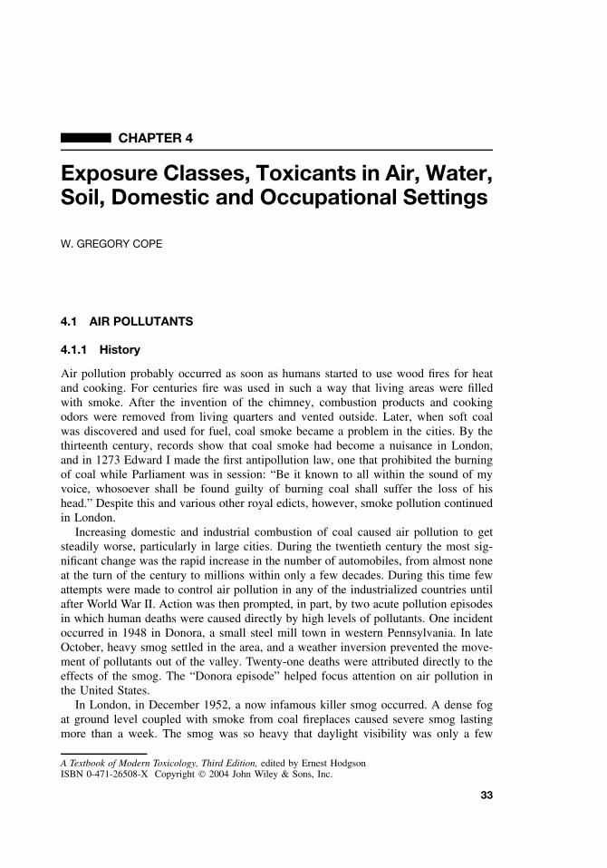
CHAPTER 4
Exposure Classes, Toxicants in Air, Water,Soil, Domestic and Occupational Settings
W. GREGORY COPE
4.1 AIR POLLUTANTS
4.1.1 History
Air pollution probably occurred as soon as humans started to use wood fires for heatand cooking. For centuries fire was used in such a way that living areas were filledwith smoke. After the invention of the chimney, combustion products and cookingodors were removed from living quarters and vented outside. Later, when soft coalwas discovered and used for fuel, coal smoke became a problem in the cities. By thethirteenth century, records show that coal smoke had become a nuisance in London,and in 1273 Edward I made the first antipollution law, one that prohibited the burningof coal while Parliament was in session: “Be it known to all within the sound of myvoice, whosoever shall be found guilty of burning coal shall suffer the loss of hishead.” Despite this and various other royal edicts, however, smoke pollution continuedin London.
Increasing domestic and industrial combustion of coal caused air pollution to getsteadily worse, particularly in large cities. During the twentieth century the most sig-nificant change was the rapid increase in the number of automobiles, from almost noneat the turn of the century to millions within only a few decades. During this time fewattempts were made to control air pollution in any of the industrialized countries untilafter World War II. Action was then prompted, in part, by two acute pollution episodesin which human deaths were caused directly by high levels of pollutants. One incidentoccurred in 1948 in Donora, a small steel mill town in western Pennsylvania. In lateOctober, heavy smog settled in the area, and a weather inversion prevented the move-ment of pollutants out of the valley. Twenty-one deaths were attributed directly to theeffects of the smog. The “Donora episode” helped focus attention on air pollution inthe United States.
In London, in December 1952, a now infamous killer smog occurred. A dense fogat ground level coupled with smoke from coal fireplaces caused severe smog lastingmore than a week. The smog was so heavy that daylight visibility was only a few
A Textbook of Modern Toxicology, Third Edition, edited by Ernest HodgsonISBN 0-471-26508-X Copyright 2004 John Wiley & Sons, Inc.
33
34 EXPOSURE CLASSES, TOXICANTS IN AIR, WATER, SOIL, DOMESTIC AND OCCUPATIONAL SETTINGS
meters, and bus conductors had to walk in front of the buses to guide the driversthrough the streets. Two days after the smog began, the death rate began to climb,and between December 5 and December 9, there were an estimated 4000 deaths abovethe normal daily count. The chief causes of death were bronchitis, pneumonia, andassociated respiratory complaints. This disaster resulted in the passage in Britain ofthe Clean Air Act in 1956.
In the United States the smog problem began to occur in large cities across thecountry, becoming especially severe in Los Angeles. In 1955 federal air pollutionlegislation was enacted, providing federal support for air pollution research, training,and technical assistance. Responsibility for the administration of the federal programnow lies with the US Environmental Protection Agency (EPA). Technological interestsince the mid-1950s has centered on automobile air pollution, pollution by oxides ofsulfur and nitrogen, and the control of these emissions. Attention is also being directedtoward the problems that may be caused by a possible greenhouse effect resulting fromincreased concentrations of carbon dioxide (CO2) in the atmosphere, possible depletionof the stratospheric ozone layer, long-range transport of pollution, and acid deposition.
4.1.2 Types of Air Pollutants
What is clean air? Unpolluted air is a concept of what the air would be if humansand their works were not on earth, and if the air were not polluted by natural pointsources such as volcanoes and forest fires. The true composition of “unpolluted” airis unknown because humans have been polluting the air for thousands of years. Inaddition there are many natural pollutants such as terpenes from plants, smoke fromforest fires, and fumes and smoke from volcanoes. Table 4.1 lists the components that,in the absence of such pollution, are thought to constitute clean air.
Gaseous Pollutants. These substances are gases at normal temperature and pressureas well as vapors evaporated from substances that are liquid or solid. Among pollutantsof greatest concern are carbon monoxide (CO), hydrocarbons, hydrogen sulfide (H2S)
Table 4.1 Gaseous Components of Normal Dry Air
Compound Percent by Volume Concentration (ppm)
Nitrogen 78.09 780,900Oxygen 20.94 209,400Argon 0.93 9300Carbon dioxide 0.0325 325Neon 0.0018 18Helium 0.0005 5.2Methane 0.0001 1.1Krypton 0.0001 1.0Nitrous oxide 0.5Hydrogen 0.5Xenon 0.008Nitrogen dioxide 0.02Ozone 0.01–0.04
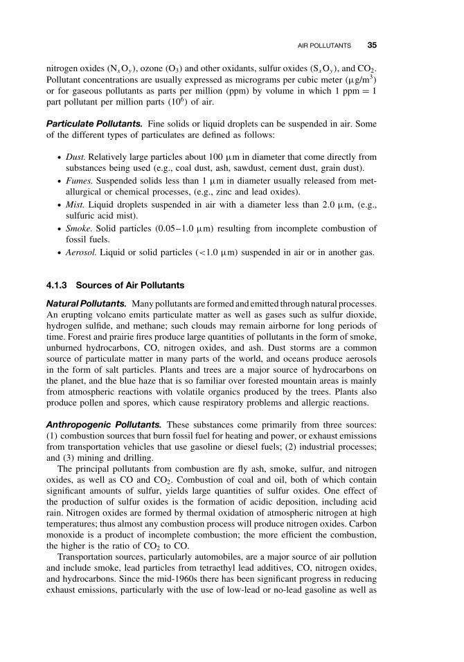
AIR POLLUTANTS 35
nitrogen oxides (NxOy), ozone (O3) and other oxidants, sulfur oxides (SxOy), and CO2.Pollutant concentrations are usually expressed as micrograms per cubic meter (µg/m3)
or for gaseous pollutants as parts per million (ppm) by volume in which 1 ppm = 1part pollutant per million parts (106) of air.
Particulate Pollutants. Fine solids or liquid droplets can be suspended in air. Someof the different types of particulates are defined as follows:
ž Dust. Relatively large particles about 100 µm in diameter that come directly fromsubstances being used (e.g., coal dust, ash, sawdust, cement dust, grain dust).
ž Fumes. Suspended solids less than 1 µm in diameter usually released from met-allurgical or chemical processes, (e.g., zinc and lead oxides).
ž Mist. Liquid droplets suspended in air with a diameter less than 2.0 µm, (e.g.,sulfuric acid mist).
ž Smoke. Solid particles (0.05–1.0 µm) resulting from incomplete combustion offossil fuels.
ž Aerosol. Liquid or solid particles (<1.0 µm) suspended in air or in another gas.
4.1.3 Sources of Air Pollutants
Natural Pollutants. Many pollutants are formed and emitted through natural processes.An erupting volcano emits particulate matter as well as gases such as sulfur dioxide,hydrogen sulfide, and methane; such clouds may remain airborne for long periods oftime. Forest and prairie fires produce large quantities of pollutants in the form of smoke,unburned hydrocarbons, CO, nitrogen oxides, and ash. Dust storms are a commonsource of particulate matter in many parts of the world, and oceans produce aerosolsin the form of salt particles. Plants and trees are a major source of hydrocarbons onthe planet, and the blue haze that is so familiar over forested mountain areas is mainlyfrom atmospheric reactions with volatile organics produced by the trees. Plants alsoproduce pollen and spores, which cause respiratory problems and allergic reactions.
Anthropogenic Pollutants. These substances come primarily from three sources:(1) combustion sources that burn fossil fuel for heating and power, or exhaust emissionsfrom transportation vehicles that use gasoline or diesel fuels; (2) industrial processes;and (3) mining and drilling.
The principal pollutants from combustion are fly ash, smoke, sulfur, and nitrogenoxides, as well as CO and CO2. Combustion of coal and oil, both of which containsignificant amounts of sulfur, yields large quantities of sulfur oxides. One effect ofthe production of sulfur oxides is the formation of acidic deposition, including acidrain. Nitrogen oxides are formed by thermal oxidation of atmospheric nitrogen at hightemperatures; thus almost any combustion process will produce nitrogen oxides. Carbonmonoxide is a product of incomplete combustion; the more efficient the combustion,the higher is the ratio of CO2 to CO.
Transportation sources, particularly automobiles, are a major source of air pollutionand include smoke, lead particles from tetraethyl lead additives, CO, nitrogen oxides,and hydrocarbons. Since the mid-1960s there has been significant progress in reducingexhaust emissions, particularly with the use of low-lead or no-lead gasoline as well as
36 EXPOSURE CLASSES, TOXICANTS IN AIR, WATER, SOIL, DOMESTIC AND OCCUPATIONAL SETTINGS
the use of oxygenated fuels—for example, fuels containing ethanol or MTBE (methylt-butyl ether).
Industries may emit various pollutants relating to their manufacturing processes—acids (sulfuric, acetic, nitric, and phosphoric), solvents and resins, gases (chlorine andammonia), and metals (copper, lead, and zinc).
Indoor Pollutants. In general, the term “indoor air pollution” refers to home andnonfactory public buildings such as office buildings and hospitals. Pollution can comefrom heating and cooking, pesticides, tobacco smoking, radon, gases, and microbesfrom people and animals.
Although indoor air pollution has increased in developed nations because of tighterbuilding construction and the use of building materials that may give off gaseouschemicals, indoor air pollution is a particular problem in developing countries. Wood,crop residues, animal dung, and other forms of biomass are used extensively for cookingand heating—often in poorly ventilated rooms. For women and children, in particular,this leads to high exposures of air pollutants such as CO and polycyclic aromatichydrocarbons.
4.1.4 Examples of Air Pollutants
Most of the information on the effects of air pollution on humans comes from acutepollution episodes such as the ones in Donora and London. Illnesses may result fromchemical irritation of the respiratory tract, with certain sensitive subpopulations beingmore affected: (1) very young children, whose respiratory and circulatory systems arepoorly developed, (2) the elderly, whose cardiorespiratory systems function poorly,and (3) people with cardiorespiratory diseases such as asthma, emphysema, and heartdisease. Heavy smokers are also affected more adversely by air pollutants. In mostcases the health problems are attributed to the combined action of particulates andsulfur dioxides (SO2); no one pollutant appears to be responsible. Table 4.2 summarizessome of the major air pollutants and their sources and effects.
Carbon Monoxide. Carbon monoxide combines readily with hemoglobin (Hb) toform carboxyhemoglobin (COHb), thus preventing the transfer of oxygen to tissues.The affinity of hemoglobin for CO is approximately 210 times its affinity for oxy-gen. A blood concentration of 5% COHb, equivalent to equilibration at approximately45 ppm CO, is associated with cardiovascular effects. Concentrations of 100 ppm cancause headaches, dizziness, nausea, and breathing difficulties. An acute concentrationof 1000 ppm is invariably fatal. Carbon monoxide levels during acute traffic congestionhave been known to be as high as 400 ppm; in addition, people who smoke elevatetheir total body burden of CO as compared with nonsmokers. The effects of low con-centrations of CO over a long period are not known, but it is possible that heart andrespiratory disorders are exacerbated.
Sulfur Oxides. Sulfur dioxide is a common component of polluted air that resultsprimarily from the industrial combustion of coal, with soft coal containing the highestlevels of sulfur. The sulfur oxides tend to adhere to air particles and enter the innerrespiratory tract, where they are not effectively removed. In the respiratory tract, SO2
combines readily with water to form sulfurous acid, resulting in irritation of mucous
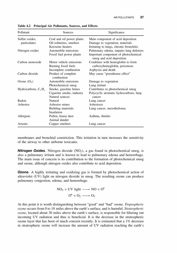
AIR POLLUTANTS 37
Table 4.2 Principal Air Pollutants, Sources, and Effects
Pollutant Sources Significance
Sulfur oxides,particulates
Coal and oil power plantsOil refineries, smeltersKerosene heaters
Main component of acid depositionDamage to vegetation, materialsIrritating to lungs, chronic bronchitis
Nitrogen oxides Automobile emissions Pulmonary edema, impairs lung defensesFossil fuel power plants Important component of photochemical
smog and acid depositionCarbon monoxide Motor vehicle emissions
Burning fossil fuelsIncomplete combustion
Combines with hemoglobin to formcarboxyhemoglobin, poisonous
Asphyxia and deathCarbon dioxide Product of complete
combustionMay cause “greenhouse effect”
Ozone (O3) Automobile emissions Damage to vegetationPhotochemical smog Lung irritant
Hydrocarbons, CxHy Smoke, gasoline fumes Contributes to photochemical smogCigarette smoke, industryNatural sources
Polycyclic aromatic hydrocarbons, lungcancer
Radon Natural Lung cancerAsbestos Asbestos mines Asbestosis
Building materials Lung cancer, mesotheliomaInsulation
Allergens Pollen, house dust Asthma, rhinitisAnimal dander
Arsenic Copper smelters Lung cancer
membranes and bronchial constriction. This irritation in turn increases the sensitivityof the airway to other airborne toxicants.
Nitrogen Oxides. Nitrogen dioxide (NO2), a gas found in photochemical smog, isalso a pulmonary irritant and is known to lead to pulmonary edema and hemorrhage.The main issue of concern is its contribution to the formation of photochemical smogand ozone, although nitrogen oxides also contribute to acid deposition.
Ozone. A highly irritating and oxidizing gas is formed by photochemical action ofultraviolet (UV) light on nitrogen dioxide in smog. The resulting ozone can producepulmonary congestion, edema, and hemorrhage.
NO2 + UV light −−−→ NO + Ož
Ož + O2 −−−→ O3
At this point it is worth distinguishing between “good” and “bad” ozone. Troposphericozone occurs from 0 to 10 miles above the earth’s surface, and is harmful. Stratosphericozone, located about 30 miles above the earth’s surface, is responsible for filtering outincoming UV radiation and thus is beneficial. It is the decrease in the stratosphericozone layer that has been of much concern recently. It is estimated that a 1% decreasein stratospheric ozone will increase the amount of UV radiation reaching the earth’s
38 EXPOSURE CLASSES, TOXICANTS IN AIR, WATER, SOIL, DOMESTIC AND OCCUPATIONAL SETTINGS
surface by 2% and cause a 10% increase in skin cancer. Major contributors to damageto stratospheric ozone are thought to be the chlorofluorocarbons (CFCs). Chlorine isremoved from the CFC compounds in the upper atmosphere by reaction with UVlight and is then able to destroy the stratospheric ozone through self-perpetuating freeradical reactions.
Cl + O3 −−−→ ClO + O2
ClO + O −−−→ Cl + O2
Before being inactivated by nitrogen dioxide or methane, each chlorine atom candestroy up to 10,000 molecules of ozone. Use of CFC compounds is now being phasedout by international agreements.
Hydrocarbons (HCs) or Volatile Organic Compounds (VOCs). These are derivedprimarily from two sources: approximately 50% are derived from trees as a result ofthe respiration process (biogenic); the other 45% to 50% comes from the combustionof fuel and from vapor from gasoline. Many gasoline pumps now have VOC recoverydevices to reduce pollution.
Lead. One of the most familiar of the particulates in air pollutants is lead, withyoung children and fetuses being the most susceptible. Lead can impair renal function,interfere with the development of red blood cells, and impair the nervous system,leading to mental retardation and even blindness. The two most common routes ofexposure to lead are inhalation and ingestion. It is estimated that approximately 20%of the total body burden of lead comes from inhalation.
Solid Particles. Dust and fibers from coal, clay, glass, asbestos, and minerals can leadto scarring or fibrosis of the lung lining. Pneumoconiosis, a condition common amongcoal miners that breathe coal dust, silicosis caused by breathing silica-containing dusts,and asbestosis from asbestos fibers are all well-known industrial pollution diseases.
4.1.5 Environmental Effects
Vegetation. Pollutants may visibly injure vegetation by bleaching, other colorchanges, and necrosis, or by more subtle changes such as alterations in growth orreproduction. Table 4.3 lists some of the more common visual effects of air pollutantson vegetation. Air pollution can also result in measurable effects on forest ecosystems,such as reduction in forest growth, change in forest species, and increased susceptibilityto forest pests. High-dose exposure to pollutants, which is associated with point sourceemissions such as smelters, frequently results in complete destruction of trees andshrubs in the surrounding area.
Domestic Animals. Although domestic animals can be affected directly by air pollu-tants, the main concern is chronic poisoning as a result of ingestion of forage that hasbeen contaminated by airborne pollutants. Pollutants important in this connection are
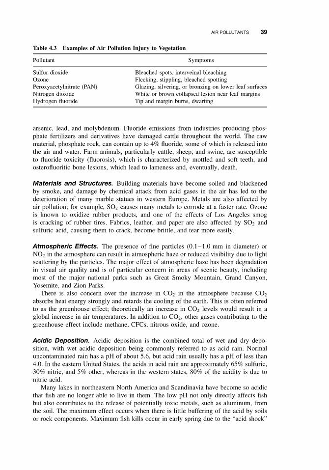
AIR POLLUTANTS 39
Table 4.3 Examples of Air Pollution Injury to Vegetation
Pollutant Symptoms
Sulfur dioxide Bleached spots, interveinal bleachingOzone Flecking, stippling, bleached spottingPeroxyacetylnitrate (PAN) Glazing, silvering, or bronzing on lower leaf surfacesNitrogen dioxide White or brown collapsed lesion near leaf marginsHydrogen fluoride Tip and margin burns, dwarfing
arsenic, lead, and molybdenum. Fluoride emissions from industries producing phos-phate fertilizers and derivatives have damaged cattle throughout the world. The rawmaterial, phosphate rock, can contain up to 4% fluoride, some of which is released intothe air and water. Farm animals, particularly cattle, sheep, and swine, are susceptibleto fluoride toxicity (fluorosis), which is characterized by mottled and soft teeth, andosterofluoritic bone lesions, which lead to lameness and, eventually, death.
Materials and Structures. Building materials have become soiled and blackenedby smoke, and damage by chemical attack from acid gases in the air has led to thedeterioration of many marble statues in western Europe. Metals are also affected byair pollution; for example, SO2 causes many metals to corrode at a faster rate. Ozoneis known to oxidize rubber products, and one of the effects of Los Angeles smogis cracking of rubber tires. Fabrics, leather, and paper are also affected by SO2 andsulfuric acid, causing them to crack, become brittle, and tear more easily.
Atmospheric Effects. The presence of fine particles (0.1–1.0 mm in diameter) orNO2 in the atmosphere can result in atmospheric haze or reduced visibility due to lightscattering by the particles. The major effect of atmospheric haze has been degradationin visual air quality and is of particular concern in areas of scenic beauty, includingmost of the major national parks such as Great Smoky Mountain, Grand Canyon,Yosemite, and Zion Parks.
There is also concern over the increase in CO2 in the atmosphere because CO2
absorbs heat energy strongly and retards the cooling of the earth. This is often referredto as the greenhouse effect; theoretically an increase in CO2 levels would result in aglobal increase in air temperatures. In addition to CO2, other gases contributing to thegreenhouse effect include methane, CFCs, nitrous oxide, and ozone.
Acidic Deposition. Acidic deposition is the combined total of wet and dry depo-sition, with wet acidic deposition being commonly referred to as acid rain. Normaluncontaminated rain has a pH of about 5.6, but acid rain usually has a pH of less than4.0. In the eastern United States, the acids in acid rain are approximately 65% sulfuric,30% nitric, and 5% other, whereas in the western states, 80% of the acidity is due tonitric acid.
Many lakes in northeastern North America and Scandinavia have become so acidicthat fish are no longer able to live in them. The low pH not only directly affects fishbut also contributes to the release of potentially toxic metals, such as aluminum, fromthe soil. The maximum effect occurs when there is little buffering of the acid by soilsor rock components. Maximum fish kills occur in early spring due to the “acid shock”
40 EXPOSURE CLASSES, TOXICANTS IN AIR, WATER, SOIL, DOMESTIC AND OCCUPATIONAL SETTINGS
from the melting of winter snows. Much of the acidity in rain may be neutralized bydissolving minerals in the soil such as aluminum, calcium, magnesium, sodium, andpotassium, which are leached from the soil into surface waters. The ability of the soilto neutralize or buffer the acid rain is very dependent on the alkalinity of the soil. Muchof the area in eastern Canada and the northeastern United States is covered by thinsoils with low acid neutralizing capacity. In such areas the lakes are more susceptibleto the effects of acid deposition leading to a low pH and high levels of aluminum, acombination toxic to many species of fish.
A second area of concern is that of reduced tree growth in forests. The leaching ofnutrients from the soil by acid deposition may cause a reduction in future growth ratesor changes in the type of trees to those able to survive in the altered environment. Inaddition to the change in soil composition, there are the direct effects on the trees fromsulfur and nitrogen oxides as well as ozone.
4.2 WATER AND SOIL POLLUTANTS
With three-quarters of the earth’s surface covered by water and much of the remaindercovered by soil, it is not surprising that water and soil serve as the ultimate sinks formost anthropogenic chemicals. Until recently the primary concern with water pollutionwas that of health effects due to pathogens, and in fact this is still the case in mostdeveloping countries. In the United States and other developed countries, however,treatment methods have largely eliminated bacterial disease organisms from the watersupply, and attention has been turned to chemical contaminants.
4.2.1 Sources of Water and Soil Pollutants
Surface water can be contaminated by point or nonpoint sources. An effluent pipefrom an industrial plant or a sewage-treatment plant is an example of a point source;a field from which pesticides and fertilizers are carried by rainwater into a river isan example of a nonpoint source. Industrial wastes probably constitute the greatestsingle pollution problem in soil and water. These contaminants include organic wastessuch as solvents, inorganic wastes, such as chromium and many unknown chemicals.Contamination of soil and water results when by-product chemicals are not properlydisposed of or conserved. In addition industrial accidents may lead to severe localcontamination. For a more in-depth discussion of sources and movements of waterpollutants, see Chapter 27.
Domestic and municipal wastes, both from sewage and from disposal of chemicals,are another major source of chemical pollutants. At the turn of the twentieth century,municipal wastes received no treatment and were discharged directly into rivers oroceans. Even today, many older treatment plants do not provide sufficient treatment,especially plants in which both storm water and sewage are combined. In addition toorganic matter, pesticides, fertilizers, detergents, and metals are significant pollutantsdischarged from urban areas.
Contamination of soil and water also results from the use of pesticides and fertilizers.Persistent pesticides applied directly to the soil have the potential to move from the soilinto the water and thus enter the food chain from both soil and water. In a similar way
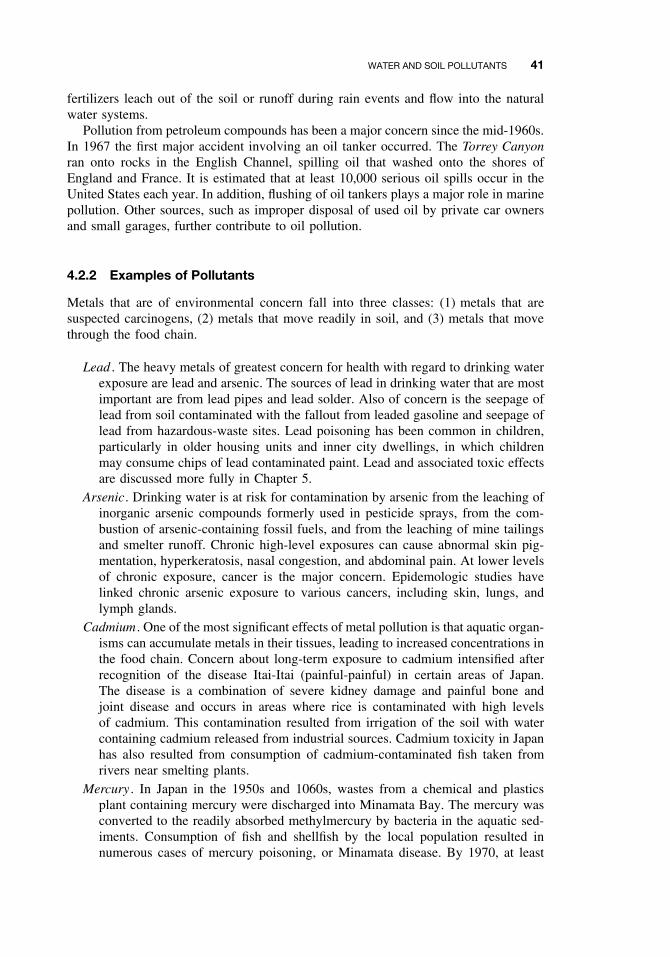
WATER AND SOIL POLLUTANTS 41
fertilizers leach out of the soil or runoff during rain events and flow into the naturalwater systems.
Pollution from petroleum compounds has been a major concern since the mid-1960s.In 1967 the first major accident involving an oil tanker occurred. The Torrey Canyonran onto rocks in the English Channel, spilling oil that washed onto the shores ofEngland and France. It is estimated that at least 10,000 serious oil spills occur in theUnited States each year. In addition, flushing of oil tankers plays a major role in marinepollution. Other sources, such as improper disposal of used oil by private car ownersand small garages, further contribute to oil pollution.
4.2.2 Examples of Pollutants
Metals that are of environmental concern fall into three classes: (1) metals that aresuspected carcinogens, (2) metals that move readily in soil, and (3) metals that movethrough the food chain.
Lead . The heavy metals of greatest concern for health with regard to drinking waterexposure are lead and arsenic. The sources of lead in drinking water that are mostimportant are from lead pipes and lead solder. Also of concern is the seepage oflead from soil contaminated with the fallout from leaded gasoline and seepage oflead from hazardous-waste sites. Lead poisoning has been common in children,particularly in older housing units and inner city dwellings, in which childrenmay consume chips of lead contaminated paint. Lead and associated toxic effectsare discussed more fully in Chapter 5.
Arsenic. Drinking water is at risk for contamination by arsenic from the leaching ofinorganic arsenic compounds formerly used in pesticide sprays, from the com-bustion of arsenic-containing fossil fuels, and from the leaching of mine tailingsand smelter runoff. Chronic high-level exposures can cause abnormal skin pig-mentation, hyperkeratosis, nasal congestion, and abdominal pain. At lower levelsof chronic exposure, cancer is the major concern. Epidemologic studies havelinked chronic arsenic exposure to various cancers, including skin, lungs, andlymph glands.
Cadmium . One of the most significant effects of metal pollution is that aquatic organ-isms can accumulate metals in their tissues, leading to increased concentrations inthe food chain. Concern about long-term exposure to cadmium intensified afterrecognition of the disease Itai-Itai (painful-painful) in certain areas of Japan.The disease is a combination of severe kidney damage and painful bone andjoint disease and occurs in areas where rice is contaminated with high levelsof cadmium. This contamination resulted from irrigation of the soil with watercontaining cadmium released from industrial sources. Cadmium toxicity in Japanhas also resulted from consumption of cadmium-contaminated fish taken fromrivers near smelting plants.
Mercury . In Japan in the 1950s and 1060s, wastes from a chemical and plasticsplant containing mercury were discharged into Minamata Bay. The mercury wasconverted to the readily absorbed methylmercury by bacteria in the aquatic sed-iments. Consumption of fish and shellfish by the local population resulted innumerous cases of mercury poisoning, or Minamata disease. By 1970, at least
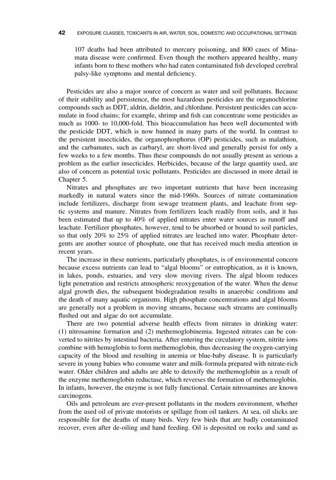
42 EXPOSURE CLASSES, TOXICANTS IN AIR, WATER, SOIL, DOMESTIC AND OCCUPATIONAL SETTINGS
107 deaths had been attributed to mercury poisoning, and 800 cases of Mina-mata disease were confirmed. Even though the mothers appeared healthy, manyinfants born to these mothers who had eaten contaminated fish developed cerebralpalsy-like symptoms and mental deficiency.
Pesticides are also a major source of concern as water and soil pollutants. Becauseof their stability and persistence, the most hazardous pesticides are the organochlorinecompounds such as DDT, aldrin, dieldrin, and chlordane. Persistent pesticides can accu-mulate in food chains; for example, shrimp and fish can concentrate some pesticides asmuch as 1000- to 10,000-fold. This bioaccumulation has been well documented withthe pesticide DDT, which is now banned in many parts of the world. In contrast tothe persistent insecticides, the organophosphorus (OP) pesticides, such as malathion,and the carbamates, such as carbaryl, are short-lived and generally persist for only afew weeks to a few months. Thus these compounds do not usually present as serious aproblem as the earlier insecticides. Herbicides, because of the large quantity used, arealso of concern as potential toxic pollutants. Pesticides are discussed in more detail inChapter 5.
Nitrates and phosphates are two important nutrients that have been increasingmarkedly in natural waters since the mid-1960s. Sources of nitrate contaminationinclude fertilizers, discharge from sewage treatment plants, and leachate from sep-tic systems and manure. Nitrates from fertilizers leach readily from soils, and it hasbeen estimated that up to 40% of applied nitrates enter water sources as runoff andleachate. Fertilizer phosphates, however, tend to be absorbed or bound to soil particles,so that only 20% to 25% of applied nitrates are leached into water. Phosphate deter-gents are another source of phosphate, one that has received much media attention inrecent years.
The increase in these nutrients, particularly phosphates, is of environmental concernbecause excess nutrients can lead to “algal blooms” or eutrophication, as it is known,in lakes, ponds, estuaries, and very slow moving rivers. The algal bloom reduceslight penetration and restricts atmospheric reoxygenation of the water. When the densealgal growth dies, the subsequent biodegradation results in anaerobic conditions andthe death of many aquatic organisms. High phosphate concentrations and algal bloomsare generally not a problem in moving streams, because such streams are continuallyflushed out and algae do not accumulate.
There are two potential adverse health effects from nitrates in drinking water:(1) nitrosamine formation and (2) methemoglobinemia. Ingested nitrates can be con-verted to nitrites by intestinal bacteria. After entering the circulatory system, nitrite ionscombine with hemoglobin to form methemoglobin, thus decreasing the oxygen-carryingcapacity of the blood and resulting in anemia or blue-baby disease. It is particularlysevere in young babies who consume water and milk-formula prepared with nitrate-richwater. Older children and adults are able to detoxify the methemoglobin as a result ofthe enzyme methemoglobin reductase, which reverses the formation of methemoglobin.In infants, however, the enzyme is not fully functional. Certain nitrosamines are knowncarcinogens.
Oils and petroleum are ever-present pollutants in the modern environment, whetherfrom the used oil of private motorists or spillage from oil tankers. At sea, oil slicks areresponsible for the deaths of many birds. Very few birds that are badly contaminatedrecover, even after de-oiling and hand feeding. Oil is deposited on rocks and sand as
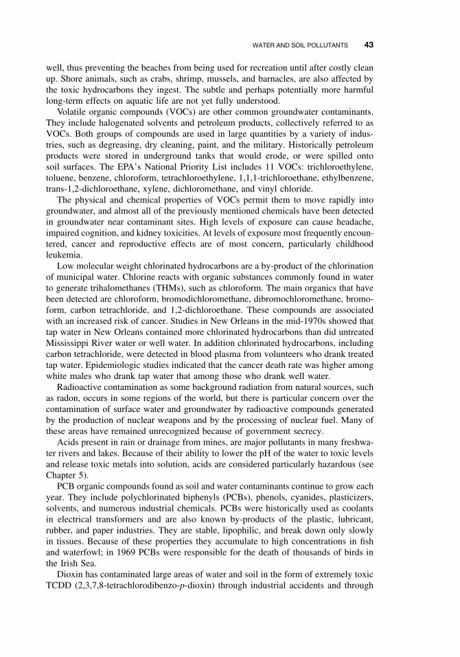
WATER AND SOIL POLLUTANTS 43
well, thus preventing the beaches from being used for recreation until after costly cleanup. Shore animals, such as crabs, shrimp, mussels, and barnacles, are also affected bythe toxic hydrocarbons they ingest. The subtle and perhaps potentially more harmfullong-term effects on aquatic life are not yet fully understood.
Volatile organic compounds (VOCs) are other common groundwater contaminants.They include halogenated solvents and petroleum products, collectively referred to asVOCs. Both groups of compounds are used in large quantities by a variety of indus-tries, such as degreasing, dry cleaning, paint, and the military. Historically petroleumproducts were stored in underground tanks that would erode, or were spilled ontosoil surfaces. The EPA’s National Priority List includes 11 VOCs: trichloroethylene,toluene, benzene, chloroform, tetrachloroethylene, 1,1,1-trichloroethane, ethylbenzene,trans-1,2-dichloroethane, xylene, dichloromethane, and vinyl chloride.
The physical and chemical properties of VOCs permit them to move rapidly intogroundwater, and almost all of the previously mentioned chemicals have been detectedin groundwater near contaminant sites. High levels of exposure can cause headache,impaired cognition, and kidney toxicities. At levels of exposure most frequently encoun-tered, cancer and reproductive effects are of most concern, particularly childhoodleukemia.
Low molecular weight chlorinated hydrocarbons are a by-product of the chlorinationof municipal water. Chlorine reacts with organic substances commonly found in waterto generate trihalomethanes (THMs), such as chloroform. The main organics that havebeen detected are chloroform, bromodichloromethane, dibromochloromethane, bromo-form, carbon tetrachloride, and 1,2-dichloroethane. These compounds are associatedwith an increased risk of cancer. Studies in New Orleans in the mid-1970s showed thattap water in New Orleans contained more chlorinated hydrocarbons than did untreatedMississippi River water or well water. In addition chlorinated hydrocarbons, includingcarbon tetrachloride, were detected in blood plasma from volunteers who drank treatedtap water. Epidemiologic studies indicated that the cancer death rate was higher amongwhite males who drank tap water that among those who drank well water.
Radioactive contamination as some background radiation from natural sources, suchas radon, occurs in some regions of the world, but there is particular concern over thecontamination of surface water and groundwater by radioactive compounds generatedby the production of nuclear weapons and by the processing of nuclear fuel. Many ofthese areas have remained unrecognized because of government secrecy.
Acids present in rain or drainage from mines, are major pollutants in many freshwa-ter rivers and lakes. Because of their ability to lower the pH of the water to toxic levelsand release toxic metals into solution, acids are considered particularly hazardous (seeChapter 5).
PCB organic compounds found as soil and water contaminants continue to grow eachyear. They include polychlorinated biphenyls (PCBs), phenols, cyanides, plasticizers,solvents, and numerous industrial chemicals. PCBs were historically used as coolantsin electrical transformers and are also known by-products of the plastic, lubricant,rubber, and paper industries. They are stable, lipophilic, and break down only slowlyin tissues. Because of these properties they accumulate to high concentrations in fishand waterfowl; in 1969 PCBs were responsible for the death of thousands of birds inthe Irish Sea.
Dioxin has contaminated large areas of water and soil in the form of extremely toxicTCDD (2,3,7,8-tetrachlorodibenzo-p-dioxin) through industrial accidents and through
44 EXPOSURE CLASSES, TOXICANTS IN AIR, WATER, SOIL, DOMESTIC AND OCCUPATIONAL SETTINGS
widespread use of the herbicide 2,4,5-T. Small amounts of TCDD were contained as acontaminant in herbicide manufacturing. The US Army used this herbicide, known asAgent Orange, extensively as a defoliant in Vietnam. TCDD is one of the most toxicsynthetic substances known for laboratory animals: LD50 for male rats, 0.022 mg/kg;LD50 for female rats, 0.045 mg/kg; LD50 for female guinea pigs (the most sensitivespecies tested), 0.0006 mg/kg. In addition it is fetotoxic to pregnant rats at a dose ofonly 1/400 of the LD50, and has been shown to cause birth defects at levels of 1 to3 ng/kg. TCDD is a proven carcinogen in both mice and rats, with the liver beingthe primary target. Although TCDD does not appear to be particularly acutely toxicto humans, chronic low-level exposure is suspected of contributing to reproductiveabnormalities and carcinogenicity.
4.3 OCCUPATIONAL TOXICANTS
Assessment of hazards in the workplace is a concern of occupational/industrial toxi-cology and has a history that dates back to ancient civilizations. The Greek historianStrabo, who lived in the first century AD, gave a graphic description of the arsenicmines in Pantus: “The air in mines is both deadly and hard to endure on account ofthe grievous odor of the ore, so that the workmen are doomed to a quick death.” Withthe coming of the industrial revolution in the nineteenth century, industrial diseasesincreased, and new ones, such as chronic mercurialism caused by exposure to mercuricnitrate used in “felting” animal furs, were identified. Hat makers, who were especiallyat risk, frequently developed characteristic tremors known as “hatters’ shakes,” and theexpression “mad as a hatter” was coined. In recent years concern has developed overthe carcinogenic potential of many workplace chemicals.
4.3.1 Regulation of Exposure Levels
The goal of occupational toxicology is to ensure work practices that do not entailany unnecessary health risks. To do this, it is necessary to define suitable permissiblelevels of exposure to industrial chemicals, using the results of animal studies andepidemiological studies. These levels can be expressed by the following terms forallowable concentrations.
Threshold limit values (TLVs) refer to airborne concentrations of substances and rep-resent conditions under which it is believed that nearly all workers may be repeatedlyexposed day after day without adverse effect. Because of wide variation in individualsusceptibility, a small percentage of workers may experience discomfort from somesubstances at or below the threshold limit; a smaller percentage may be affected moreseriously by aggravation of a preexisting condition or by development of an occu-pational illness. Threshold limits are based on the best available information fromindustrial experience, from experimental human and animal studies, and when possi-ble, from a combination of the three. The basis on which the values are establishedmay differ from substance to substance; protection against impairment of health maybe a guiding factor for some, whereas reasonable freedom from irritation, narcosis,nuisance, or other forms of stress may form the basis for others. Three categories ofTLVs follow:
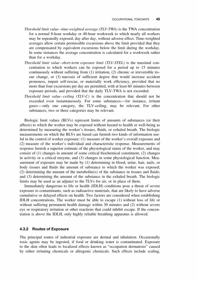
OCCUPATIONAL TOXICANTS 45
Threshold limit value–time-weighted average (TLV-TWA) is the TWA concentrationfor a normal 8-hour workday or 40-hour workweek to which nearly all workersmay be repeatedly exposed, day after day, without adverse effect. Time-weightedaverages allow certain permissible excursions above the limit provided that theyare compensated by equivalent excursions below the limit during the workday.In some instances the average concentration is calculated for a workweek ratherthan for a workday.
Threshold limit value–short-term exposure limit (TLV-STEL) is the maximal con-centration to which workers can be exposed for a period up to 15 minutescontinuously without suffering from (1) irritation, (2) chronic or irreversible tis-sue change, or (3) narcosis of sufficient degree that would increase accidentproneness, impair self-rescue, or materially work efficiency, provided that nomore than four excursions per day are permitted, with at least 60 minutes betweenexposure periods, and provided that the daily TLV-TWA is not exceeded.
Threshold limit value–ceiling (TLV-C) is the concentration that should not beexceeded even instantaneously. For some substances—for instance, irritantgases—only one category, the TLV-ceiling, may be relevant. For othersubstances, two or three categories may be relevant.
Biologic limit values (BLVs) represent limits of amounts of substances (or theiraffects) to which the worker may be exposed without hazard to health or well-being asdetermined by measuring the worker’s tissues, fluids, or exhaled breath. The biologicmeasurements on which the BLVs are based can furnish two kinds of information use-ful in the control of worker exposure: (1) measure of the worker’s overall exposure and(2) measure of the worker’s individual and characteristic response. Measurements ofresponse furnish a superior estimate of the physiological status of the worker, and mayconsist of (1) changes in amount of some critical biochemical constituent, (2) changesin activity or a critical enzyme, and (3) changes in some physiological function. Mea-surement of exposure may be made by (1) determining in blood, urine, hair, nails, orbody tissues and fluids the amount of substance to which the worker was exposed;(2) determining the amount of the metabolite(s) of the substance in tissues and fluids;and (3) determining the amount of the substance in the exhaled breath. The biologiclimits may be used as an adjunct to the TLVs for air, or in place of them.
Immediately dangerous to life or health (IDLH) conditions pose a threat of severeexposure to contaminants, such as radioactive materials, that are likely to have adversecumulative or delayed effects on health. Two factors are considered when establishingIDLH concentrations. The worker must be able to escape (1) without loss of life orwithout suffering permanent health damage within 30 minutes and (2) without severeeye or respiratory irritation or other reactions that could inhibit escape. If the concen-tration is above the IDLH, only highly reliable breathing apparatus is allowed.
4.3.2 Routes of Exposure
The principal routes of industrial exposure are dermal and inhalation. Occasionallytoxic agents may be ingested, if food or drinking water is contaminated. Exposureto the skin often leads to localized effects known as “occupation dermatosis” causedby either irritating chemicals or allergenic chemicals. Such effects include scaling,
46 EXPOSURE CLASSES, TOXICANTS IN AIR, WATER, SOIL, DOMESTIC AND OCCUPATIONAL SETTINGS
eczema, acne, pigmentation changes, ulcers, and neoplasia. Some chemicals may alsopass through the skin; these include aromatic amines such as aniline and solvents suchas carbon tetrachloride and benzene.
Toxic or potentially toxic agents may be inhaled into the respiratory tract where theymay cause localized effects such as irritation (e.g., ammonia, chlorine gas), inflamma-tion, necrosis, and cancer. Chemicals may also be absorbed by the lungs into thecirculatory system, thereby leading to systemic toxicity (e.g., CO, lead).
4.3.3 Examples of Industrial Toxicants
Carcinogen exposure is largely due to lifestyle, such as cigarette smoking, but occu-pation is an important source of exposure to carcinogens. Table 4.4 lists some occu-pational chemical hazards and the cancers associated with them.
Cadmium is a cumulative toxicant with a biologic half-life of up to 30 years inhumans. More than 70% of the cadmium in the blood is bound to red blood cells;accumulation occurs mainly in the kidney and the liver, where cadmium is boundto metallothionein. In humans the critical target organ after long-term exposure tocadmium is the kidney, with the first detectable symptom of kidney toxicity being anincreased excretion of specific proteins.
Chromium toxicity results from compounds of hexavalent chromium that can bereadily absorbed by the lung and gastrointestinal (GI) tract and to a lesser extent bythe skin. Occupational exposure to chromium (Cr6+) causes dermatitis, ulcers on thehands and arms, perforation of the nasal septum (probably caused by chromic acid),inflammation of the larynx and liver, and bronchitis. Chromate is a carcinogen causingbronchogenic carcinoma; the risk to chromate plant workers for lung cancer is 20times greater than that for the general population. Compounds of trivalent chromium
Table 4.4 Some Occupational Hazards and Associated Cancers
Agent Tumor Sites Occupation
Asbestos Lung, pleura, peritoneum Miners, manufacturers, usersArsenic Skin, lung, liver Miners and smelters, oil refinery,
pesticide workersBenzene Hemopoietic tissue Process workers, textile workersCadmium Lung, kidney, prostate Battery workers, smeltersChloroethers Lung Chemical plant workers, process workersChromium Lung, nasal cavity,
sinusesProcess and production workers, pigment
workersMustard gas Bronchi, lung, larynx Production workersNaphthylamines Bladder Dyestuff makers and workers,
Chemical workers, printersNickel Lung, nasal sinuses Smelters and process workersPolycyclic aromatic
hydrocarbonsRespiratory system,
bladderFurnace, foundry, shale, and gas
workers; chimney sweepsRadon, radium,
uraniumSkin, lung, bone tissue,
bone marrowMedical and industrial chemists, miners
UV radiation Skin Outdoor exposureX rays Bone marrow, skin Medical and industrial workers
OCCUPATIONAL TOXICANTS 47
are poorly absorbed. Chromium is not a cumulative chemical, and once absorbed, it israpidly excreted into the urine.
Lead is a ubiquitous toxicant in the environment, and consequently the normal bodyconcentration of lead is dependent on environmental exposure conditions. Approxi-mately 50% of lead deposited in the lung is absorbed, whereas usually less than 10%of ingested lead passes into the circulation. Lead is not a major occupational problemtoday, but environmental pollution is still widespread. Lead interferes in the biosyn-thesis of porphyrins and heme, and several screening tests for lead poisoning makeuse of this interaction by monitoring either inhibition of the enzyme δ-aminolevulinicacid dehydratase (ALAD) or appearance in the urine of aminolevulinic acid (ALA)and coproporphorin (UCP). The metabolism of inorganic lead is closely related tothat of calcium, and excess lead can be deposited in the bone where it remains foryears. Inorganic lead poisoning can produce fatigue, sleep disturbances, anemia, colic,and neuritis. Severe exposure, mainly of children who have ingested lead, may causeencephalopathy, mental retardation, and occasionally, impaired vision.
Organic lead has an affinity for brain tissue; mild poisoning may cause insomnia,restlessness, and GI symptoms, whereas severe poisoning results in delirium, halluci-nations, convulsions, coma, and even death.
Mercury is widely used in scientific and electrical apparatus, with the largest indus-trial use of mercury being in the chlorine-alkali industry for electrolytic produc-tion of chlorine and sodium hydroxide. Worldwide, this industry has been a majorsource of mercury contaminations. Most mercury poisoning, however, has been dueto methylmercury, particularly as a result of eating contaminated fish. Inorganic andorganic mercury differ in their routes of entry and absorption. Inhalation is the prin-cipal route of uptake of metallic mercury in industry, with approximately 80% of themercury inhaled as vapor being absorbed; metallic mercury is less readily absorbed bythe GI route. The principal sites of deposition are the kidney and brain after exposureto inorganic mercury salts. Organic mercury compounds are readily absorbed by allroutes. Industrial mercurialism produces features such as inflammation of the mouth,muscular tremors (hatters’ shakes), psychic irritation, and a nephritic syndrome charac-terized by proteinuria. Overall, however, occupational mercurialism is not a significantproblem today.
Benzene was used extensively in the rubber industry as a solvent for rubber latexin the latter half of the nineteenth century. The volatility of benzene, which made it soattractive to the industry, also caused high atmospheric levels of the solvent. Benzene-based rubber cements were used in the canning industry and in the shoe manufacturingindustry. Although cases of benzene poisoning had been reported as early as 1897and additional reports and warnings were issued in the 1920s, the excellent solventproperties of benzene resulted in its continued extensive use. In the 1930s cases ofbenzene toxicity occurred in the printing industry in which benzene was used as anink solvent. Today benzene use exceeds 11 billion gallons per year.
Benzene affects the hematopoietic tissue in the bone marrow and also appears to bean immunosuppressant. There is a gradual decrease in white blood cells, red blood cells,and platelets, and any combination of these signs may be seen. Continued exposure tobenzene results in severe bone marrow damage and aplastic anemia. Benzene exposurehas also been associated with leukemia.
Asbestos and other fibers of naturally occurring silicates will separate into flexiblefibers. Asbestos is the general name for this group of fibers. Chrysotile is the most
48 EXPOSURE CLASSES, TOXICANTS IN AIR, WATER, SOIL, DOMESTIC AND OCCUPATIONAL SETTINGS
important commercially and represents about 90% of the total used. Use of asbestoshas been extensive, especially in roofing and insulation, asbestos cements, brake lin-ings, electrical appliances, and coating materials. Asbestosis, a respiratory disease, ischaracterized by fibrosis, calcification, and lung cancer. In humans, not only is there along latency period between exposure and development of tumors but other factors alsoinfluence the development of lung cancer. Cigarette smoking, for example, enhancestumor formation. Recent studies have shown that stomach and bowel cancers occurin excess in workers (e.g., insulation workers) exposed to asbestos. Other fibers havebeen shown to cause a similar disease spectrum, for instance, zeolite fibers.
SUGGESTED READING
Air Pollutants
Costa, D. L. Air pollution. In Casarett and Doull’s Toxicology: The Basic Science of Poisons,6th ed., C. D. Klaassen, ed. New York: McGraw-Hill, 2001, pp. 979–1012.
Holgate, S. T., J. M. Samet, H. Koren, and R. Maynard, eds. Air Pollution and Health. SanDiego: Academic Press, 1999.
Water and Soil Pollutants
Abel, P. D., ed. Water Pollution Biology. London: Taylor and Francis, 1996.
Hoffman, D. J., B. A. Rattner, G. A. Burton, and J. Cairns, eds. Handbook of Ecotoxicology,2nd ed. Boca Raton: Lewis, 2002.
Larson, S. J., P. D. Capel, and M. S. Majewski, eds. Pesticides in Surface Waters. Chelsea, MI:Ann Arbor Press, 1998.
Occupational Toxicants
Thorne, P. S. Occupational toxicology. In Casarett and Doull’s Toxicology: The Basic Scienceof Poisons, 6th ed., C. D. Klaassen, ed. New York: McGraw-Hill, 2001, pp. 1123–1140.
Doull, J. Recommended limits for occupational exposure to chemicals. In Casarett and Doull’sToxicology: The Basic Science of Poisons, 6th ed., C. D. Klaassen, ed. New York: McGraw-Hill, 2001, pp. 1155–1176.
CHAPTER 5
Classes of Toxicants: Use Classes
W. GREGORY COPE, ROSS B. LEIDY, and ERNEST HODGSON
5.1 INTRODUCTION
As discussed in Chapter 1, use classes include not only chemicals currently in use butalso the toxicological aspects of the development of new chemicals for commercialuse, chemicals produced as by-products of industrial processes, and chemicals result-ing from the use and/or disposal of chemicals. Because any use class may includechemicals from several different chemical classes, this classification is not sufficientfor mechanistic considerations. It is, however, essential for an understanding of thescope of toxicology and, in particular, is essential for many applied branches of toxi-cology such as exposure assessment, industrial hygiene, public health toxicology andregulatory toxicology.
5.2 METALS
5.2.1 History
Although most metals occur in nature in rocks, ores, soil, water, and air, levels areusually low and widely dispersed. In terms of human exposure and toxicological sig-nificance, it is anthropogenic activities that are most important because they increasethe levels of metals at the site of human activities.
Metals have been used throughout much of human history to make utensils, machin-ery, and so on, and mining and smelting supplied metals for these uses. These activitiesincreased environmental levels of metals. More recently metals have found a num-ber of uses in industry, agriculture, and medicine. These activities have increasedexposure not only to metal-related occupational workers but also to consumers of thevarious products.
Despite the wide range of metal toxicity and toxic properties, there are a number oftoxicological features that are common to many metals. Some of the more importantaspects are discussed briefly in the following sections. For a metal to exert its toxicity,it must cross the membrane and enter the cell. If the metal is in a lipid soluble formsuch as methylmercury, it readily penetrates the membrane; when bound to proteins
A Textbook of Modern Toxicology, Third Edition, edited by Ernest HodgsonISBN 0-471-26508-X Copyright 2004 John Wiley & Sons, Inc.
49
50 CLASSES OF TOXICANTS: USE CLASSES
such as cadmium-metallothionein, the metal is taken into the cell by endocytosis; othermetals (e.g., lead) may be absorbed by passive diffusion. The toxic effects of metalsusually involve interaction between the free metal and the cellular target. These targetstend to be specific biochemical processes and/or cellular and subcellular membranes.
5.2.2 Common Toxic Mechanisms and Sites of Action
Enzyme Inhibition/Activation. A major site of toxic action for metals is interactionwith enzymes, resulting in either enzyme inhibition or activation. Two mechanismsare of particular importance: inhibition may occur as a result of interaction betweenthe metal and sulfhydryl (SH) groups on the enzyme, or the metal may displace anessential metal cofactor of the enzyme. For example, lead may displace zinc in thezinc-dependent enzyme δ-aminolevulinic acid dehydratase (ALAD), thereby inhibitingthe synthesis of heme, an important component of hemoglobin and heme-containingenzymes, such as cytochromes.
Subcellular Organelles. Toxic metals may disrupt the structure and function of anumber of organelles. For example, enzymes associated with the endoplasmic reticulummay be inhibited, metals may be accumulated in the lysosomes, respiratory enzymesin the mitochondria may be inhibited, and metal inclusion bodies may be formed inthe nucleus.
Carcinogenicity. A number of metals have been shown to be carcinogenic in humansor animals. Arsenic, certain chromium compounds, and nickel are known human car-cinogens; beryllium, cadmium, and cisplatin are probable human carcinogens. Thecarcinogenic action, in some cases, is thought to result from the interaction of themetallic ions with DNA (see Chapter 11 for a detailed discussion of carcinogenesis).
Kidney. Because the kidney is the main excretory organ of the body, it is a commontarget organ for metal toxicity. Cadmium and mercury, in particular, are potent nephro-toxicants and are discussed more fully in the following sections and in Chapter 15.
Nervous System. The nervous system is also a common target of toxic metals;particularly, organic metal compounds (see Chapter 16). For example, methylmercury,because it is lipid soluble, readily crosses the blood-brain barrier and enters the nervoussystem. By contrast, inorganic mercury compounds, which are more water soluble, areless likely to enter the nervous system and are primarily nephrotoxicants. Likewiseorganic lead compounds are mainly neurotoxicants, whereas the first site of inorganiclead is enzyme inhibition (e.g., enzymes involved in heme synthesis).
Endocrine and Reproductive Effects. Because the male and female reproductiveorgans are under complex neuroendocrine and hormonal control, any toxicant that altersany of these processes can affect the reproductive system (see Chapters 17 and 20).In addition metals can act directly on the sex organs. Cadmium is known to producetesticular injury after acute exposure, and lead accumulation in the testes is associatedwith testicular degeneration, inhibition of spermatogenesis, and Leydig-cell atrophy.
Respiratory System. Occupational exposure to metals in the form of metal dustmakes the respiratory system a likely target. Acute exposure may cause irritations and
METALS 51
inflammation of the respiratory tract, whereas chronic exposure may result in fibrosis(aluminum) or carcinogenesis (arsenic, chromium, nickel). Respiratory toxicants arediscussed more fully in Chapter 18.
Metal-Binding Proteins. The toxicity of many metals such as cadmium, lead, andmercury depends on their transport and intracellular bioavailability. This availabil-ity is regulated to a degree by high-affinity binding to certain cytosolic proteins.Such ligands usually possess numerous SH binding sites that can outcompete otherintracellular proteins and thus mediate intracellular metal bioavailability and toxicity.These intracellular “sinks” are capable of partially sequestering toxic metals away fromsensitive organelles or proteins until their binding capacity is exceeded by the doseof the metal. Metallothionein (MT) is a low molecular weight metal-binding protein(approximately 7000 Da) that is particularly important in regulating the intracellularbioavailability of cadmium, copper, mercury, silver, and zinc. For example, in vivoexposure to cadmium results in the transport of cadmium in the blood by various highmolecular weight proteins and uptake by the liver, followed by hepatic induction ofMT. Subsequently cadmium can be found in the circulatory system bound to MT asthe cadmium-metallothionein complex (CdMT).
5.2.3 Lead
Because of the long-term and widespread use of lead, it is one of the most ubiquitousof the toxic metals. Exposure may be through air, water, or food sources. In the UnitedStates the major industrial uses, such as in fuel additives and lead pigments in paints,have been phased out, but other uses, such as in batteries, have not been reduced.Other sources of lead include lead from pipes and glazed ceramic food containers.
Inorganic lead may be absorbed through the GI tract, the respiratory system, andthe skin. Ingested inorganic lead is absorbed more efficiently from the GI tract ofchildren than that of adults, readily crosses the placenta, and in children penetratesthe blood-brain barrier. Initially, lead is distributed in the blood, liver, and kidney;after prolonged exposure, as much as 95% of the body burden of lead is found inbone tissue.
The main targets of lead toxicity are the hematopoietic system and the nervoussystem. Several of the enzymes involved in the synthesis of heme are sensitive toinhibition by lead, the two most susceptible enzymes being ALAD and heme syn-thetase (HS). Although clinical anemia occurs only after moderate exposure to lead,biochemical effects can be observed at lower levels. For this reason inhibition of ALADor appearance in the urine of ALA can be used as an indication of lead exposure.
The nervous system is another important target tissue for lead toxicity, especially ininfants and young children in whom the nervous system is still developing (Chapter 16).Even at low levels of exposure, children may show hyperactivity, decreased attentionspan, mental deficiencies, and impaired vision. At higher levels, encephalopathy mayoccur in both children and adults. Lead damages the arterioles and capillaries, resultingin cerebral edema and neuronal degeneration. Clinically this damage manifests itselfas ataxia, stupor, coma, and convulsions.
Another system affected by lead is the reproductive system (Chapter 20). Leadexposure can cause male and female reproductive toxicity, miscarriages, and degener-ate offspring.
52 CLASSES OF TOXICANTS: USE CLASSES
5.2.4 Mercury
Mercury exists in the environment in three main chemical forms: elemental mercury(Hg0), inorganic mercurous (Hg+) and mercuric (Hg2+) salts, and organic methylmer-cury (CH3Hg) and dimethylmercury (CH3HgCH3) compounds. Elemental mercury,in the form of mercury vapor, is almost completely absorbed by the respiratory sys-tem, whereas ingested elemental mercury is not readily absorbed and is relativelyharmless. Once absorbed, elemental mercury can cross the blood-brain barrier intothe nervous system. Most exposure to elemental mercury tends to be from occupa-tional sources.
Of more concern from environmental contamination is exposure to organic mer-cury compounds. Inorganic mercury may be converted to organic mercury throughthe action of sulfate-reducing bacteria, to produce methylmercury, a highly toxic formreadily absorbed across membranes. Several large episodes of mercury poisoning haveresulted from consuming seed grain treated with mercury fungicides or from eatingfish contaminated with methylmercury. In Japan in the 1950s and 1960s wastes froma chemical and plastics plant containing mercury were drained into Minamata Bay.The mercury was converted to the readily absorbed methylmercury by bacteria in theaquatic sediments. Consumption of fish and shellfish by the local population resulted innumerous cases of mercury poisoning or Minamata disease. By 1970 at least 107 deathshad been attributed to mercury poisoning, and 800 cases of Minamata disease wereconfirmed. Even though the mothers appeared healthy, many infants born to motherswho had eaten contaminated fish developed cerebral palsy-like symptoms and mentaldeficiency. Organic mercury primarily affects the nervous system, with the fetal brainbeing more sensitive to the toxic effects of mercury than adults.
Inorganic mercury salts, however, are primarily nephrotoxicants, with the site ofaction being the proximal tubular cells. Mercury binds to SH groups of membraneproteins, affecting the integrity of the membrane and resulting in aliguria, anuria,and uremia.
5.2.5 Cadmium
Cadmium occurs in nature primarily in association with lead and zinc ores and isreleased near mines and smelters processing these ores. Industrially cadmium is usedas a pigment in paints and plastics, in electroplating, and in making alloys and alkalistorage batteries (e.g., nickel-cadmium batteries). Environmental exposure to cadmiumis mainly from contamination of groundwater from smelting and industrial uses aswell as the use of sewage sludge as a food-crop fertilizer. Grains, cereal products, andleafy vegetables usually constitute the main source of cadmium in food. Reference hasalready been made to the disease Itai-Itai resulting from consumption of cadmium-contaminated rice in Japan (see Chapter 4, Section 4.2.2).
Acute effects of exposure to cadmium result primarily from local irritation. Afteringestion, the main effects are nausea, vomiting, and abdominal pain. Inhalation expo-sure may result in pulmonary edema and chemical pneumonitis.
Chronic effects are of particular concern because cadmium is very slowly excretedfrom the body, with a half-life of about 30 years. Thus low levels of exposure canresult in considerable accumulation of cadmium. The main organ of damage followinglong-term exposure is the kidney, with the proximal tubules being the primary site of
METALS 53
action. Cadmium is present in the circulatory system bound primarily to the metal-binding protein, metallothionein, produced in the liver. Following glomerular filtrationin the kidney, CdMT is re-absorbed efficiently by the proximal tubule cells, whereit accumulates within the lysosomes. Subsequent degradation of the CdMT complexreleases Cd+2, which inhibits lysosomal function, resulting in cell injury.
5.2.6 Chromium
Because chromium occurs in ores, environmental levels are increased by mining, smelt-ing, and industrial uses. Chromium is used in making stainless steel, various alloys, andpigments. The levels of this metal are generally very low in air, water, and food, andthe major source of human exposure is occupational. Chromium occurs in a numberof oxidation states from Cr+2 to Cr+6, but only the trivalent (Cr+3) and hexavalent(Cr+6) forms are of biological significance. Although the trivalent compound is themost common form found in nature, the hexavalent form is of greater industrial impor-tance. In addition hexavalent chromium, which is not water soluble, is more readilyabsorbed across cell membranes than is trivalent chromium. In vivo the hexavalentform is reduced to the trivalent form, which can complex with intracellular macro-molecules, resulting in toxicity. Chromium is a known human carcinogen and induceslung cancers among exposed workers. The mechanism of chromium (Cr+6) carcino-genicity in the lung is believed to be its reduction to Cr+3 and generation of reactiveintermediates, leading to bronchogenic carcinoma.
5.2.7 Arsenic
In general, the levels of arsenic in air and water are low, and the major source of humanexposure is food. In certain parts of Taiwan and South America, however, the watercontains high levels of this metalloid, and the inhabitants often suffer from dermalhyperkeratosis and hyperpigmentation. Higher levels of exposure result in a moreserious condition; gangrene of the lower extremities or “blackfoot disease.” Cancer ofthe skin also occurs in these areas.
Approximately 80% of arsenic compounds are used in pesticides. Other uses includeglassware, paints, and pigments. Arsine gas is used in the semiconductor industry.Arsenic compounds occur in three forms: (1) pentavalent, As+5, organic or arsenatecompounds (e.g., alkyl arsenates); (2) trivalent, As+3, inorganic or arsenate compounds(e.g., sodium arsenate, arsenic trioxide); and (3) arsine gas, AsH3, a colorless gasformed by the action of acids on arsenic. The most toxic form is arsine gas witha TLV-TWA of 0.05 ppm. Microorganisms in the environment convert arsenic todimethylarsenate, which can accumulate in fish and shellfish, providing a source forhuman exposure. Arsenic compounds can also be present as contaminants in well water.Arsenite (As+3) compounds are lipid soluble and can be absorbed following ingestion,inhalation, or skin contact. Within 24 hours of absorption, arsenic distributes over thebody, where it binds to SH groups of tissue proteins. Only a small amount crosses theblood-brain barrier. Arsenic may also replace phosphorus in bone tissue and be storedfor years.
After acute poisoning, severe GI gastrointestinal symptoms occur within 30 minutesto 2 hours. These include vomiting, watery and bloody diarrhea, severe abdominal pain,
54 CLASSES OF TOXICANTS: USE CLASSES
Table 5.1 Examples of Chelating Drugs Used to Treat Metal Toxicity
British antilewisite (BAL[2,3–dimercaptopropanol]), dimercaprolDMPS (2,3-dimercapto-1-propanesulfonic acid)DMSA (meso-2,3-dimercaptosuccinic acid)EDTA (ethylenediaminetetraacetic acid, calcium salt)DTPA (diethylenetriaminepentaacetic acid, calcium salt)DTC (dithiocarbamate)Penicillamine (β-β-dimethylcysteine), hydrolytic product of penicillin
and burning esophageal pain. Vasodilatation, myocardial depression, cerebral edema,and distal peripheral neuropathy may also follow. Later stages of poisoning includejaundice and renal failure. Death usually results from circulatory failure within 24 hoursto 4 days.
Chronic exposure results in nonspecific symptoms such as diarrhea, abdominal pain,hyperpigmentation, and hyperkeratosis. A symmetrical sensory neuropathy often fol-lows. Late changes include gangrene of the extremities, anemia, and cancer of the skin,lung, and nasal tissue.
5.2.8 Treatment of Metal Poisoning
Treatment of metal exposure to prevent or reverse toxicity is done with chelating agentsor antagonists. Chelation is the formation of a metal ion complex, in which the metalion is associated with an electron donor ligand. Metals may react with O-, S-, andN-containing ligands (e.g., –OH, –COOH, –S–S–, and –NH2). Chelating agents needto be able to reach sites of storage, form nontoxic complexes, not readily bind essentialmetals (e.g., calcium, zinc), and be easily excreted.
One of the first clinically useful chelating drugs was British antilewisite (BAL[2,3-dimercaptopropanol]), which was developed during World War II as an antag-onist to arsenical war gases. BAL is a dithiol compound with two sulfur atoms onadjacent carbon atoms that compete with critical binding sites involved in arsenic tox-icity. Although BAL will bind a number of toxic metals, it is also a potentially toxicdrug with multiple side effects. In response to BAL’s toxicity, several analogues havenow been developed. Table 5.1 lists some of the more common chelating drugs intherapeutic use.
5.3 AGRICULTURAL CHEMICALS (PESTICIDES)
5.3.1 Introduction
Chemicals have been used to kill or control pests for centuries. The Chinese usedarsenic to control insects, the early Romans used common salt to control weeds andsulfur to control insects. In the 1800s pyrethrin (i.e., compounds present in the flowersof the chrysanthemum, Pyrethrum cineraefolium) was found to have insecticidal prop-erties. The roots of certain Derris plant species, (D. elliptica and Lonchocarpus spp.)were used by the Chinese and by South American natives as a fish poison. The activeingredient, rotenone, was isolated in 1895 and used for insect control. Another material
AGRICULTURAL CHEMICALS (PESTICIDES) 55
developed for insect control in the 1800s was Paris Green, a mixture of copper andarsenic salts. Fungi were controlled with Bordeaux Mixture, a combination of lime andcopper sulfate.
However, it was not until the 1900s that the compounds we identify today as havingpesticidal properties came into being. Petroleum oils, distilled from crude mineral oilswere introduced in the 1920s to control scale insects and red spider mites. The 1940ssaw the introduction of the chlorinated hydrocarbon insecticides such as DDT and thephenoxy acid herbicides such as 2,4-D). Natural compounds such as Red Squill, derivedfrom the bulbs of red squill, Urginea (Scilla) maritima, were effective in controllingrodents. Triazine herbicides, such as atrazine, introduced in the late 1950s, dominatedthe world herbicide market for years. Synthetic pyrethrins or pyrethroid insecticides(e.g., resmethrin) became and continue to be widely used insecticides due to their lowtoxicity, enhanced persistence compared to the pyrethrins and low application rates.New families of fungicides, herbicides, and insecticides continue to be introducedinto world markets as older compounds lose their popularity due to pest resistance oradverse health effects.
Pesticides are unusual among environmental pollutants in that they are used delib-erately for the purpose of killing some form of life. Ideally pesticides should be highlyselective, destroying target organisms while leaving nontarget organisms unharmed. Inreality, most pesticides are not so selective. In considering the use of pesticides, thebenefits must be weighed against the risk to human health and environmental qual-ity. Among the benefits of pesticides are control of vector-borne diseases, increasedagricultural productivity, and control of urban pests. A major risk is environmentalcontamination, especially translocation within the environment where pesticides mightenter both food chains and natural water systems. Factors to be considered in thisregard are persistence in the environment and potential for bioaccumulation.
5.3.2 Definitions and Terms
The term “agricultural chemicals” has largely been replaced by the term “pesticides,”defined as economic poisons, regulated by federal and state laws, that are used tocontrol, kill, or repel pests. Depending on what a compound is designed to do, pes-ticides have been subclassified into a number of categories (Table 5.2). The primaryclasses of pesticides in use today are fumigants, fungicides, herbicides, and insecticideswith total US production of 1.2 billion pounds (1997: US Environmental ProtectionAgency’s latest figures) and production of some 665 million pounds of wood preser-vatives. Table 5.3 describes the relative use of different classes of pesticides in theUnited States.
Generally, it takes some five to seven years to bring a pesticide to market once itspesticidal properties have been verified. Many tests must be conducted to determinesuch things as the compound’s synthesis, its chemical and physical properties, andits efficacy. In addition, in order for registration for use by the US EPA, numeroustoxicity tests are undertaken including those for acute toxicity, those for chronic effectssuch as reproductive anomalies, carcinogenesis, and neurological effects and those forenvironmental effects.
The mandated pesticide label contains a number of specified items, including theconcentration and/or percentage of both the active (A.I.) and inert ingredients; propermixing of the formulation with water to obtain the application rate of A.I., what the A.I.
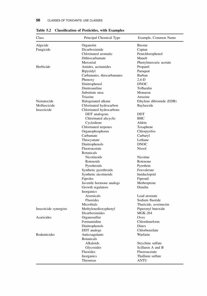
56 CLASSES OF TOXICANTS: USE CLASSES
Table 5.2 Classification of Pesticides, with Examples
Class Principal Chemical Type Example, Common Name
Algicide Organotin BrestarFungicide Dicarboximide Captan
Chlorinated aromatic PentchlorophenolDithiocarbamate ManebMercurial Phenylmercuric acetate
Herbicide Amides, acetamides PropanilBipyridyl ParaquatCarbamates, thiocarbamates BarbanPhenoxy 2,4-DDinitrophenol DNOCDinitroaniline TrifluralinSubstitute urea MonuronTriazine Atrazine
Nematocide Halogenated alkane Ethylene dibromide (EDB)Molluscicide Chlorinated hydrocarbon BayluscideInsecticide Chlorinated hydrocarbons
DDT analogous DDTChlorinated alicyclic BHCCyclodiene Aldrin
Chlorinated terpenes ToxapheneOrganophosphorus ChlorpyrifosCarbamate CarbarylThiocyanate LethaneDinitrophenols DNOCFluoroacetate NissolBotanicals
Nicotinoids NicotineRotenoids RotenonePyrethroids Pyrethrin
Synthetic pyrethroids FenvalerateSynthetic nicotinoids ImidaclopridFiproles FipronilJuvenile hormone analogs MethropreneGrowth regulators DimilinInorganics
Arsenicals Lead arsenateFluorides Sodium fluoride
Microbials Thuricide, avermectinInsecticide synergists Methylenedioxyphenyl Piperonyl butoxide
Dicarboximides MGK-264Acaricides Organosulfur Ovex
Formamidine ChlordimeformDinitrophenols DinexDDT analogs Chlorbenzilate
Rodenticides Anticoagulants WarfarinBotanicals
Alkaloids Strychine sulfateGlycosides Scillaren A and B
Fluorides FluoroacetateInorganics Thallium sulfateThioureas ANTU
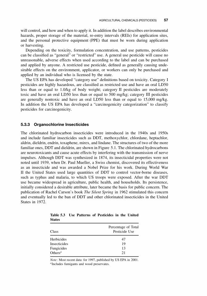
AGRICULTURAL CHEMICALS (PESTICIDES) 57
will control, and how and when to apply it. In addition the label describes environmentalhazards, proper storage of the material, re-entry intervals (REIs) for application sites,and the personal protective equipment (PPE) that must be worn during applicationor harvesting.
Depending on the toxicity, formulation concentration, and use patterns, pesticidescan be classified as “general” or “restricted” use. A general use pesticide will cause nounreasonable, adverse effects when used according to the label and can be purchasedand applied by anyone. A restricted use pesticide, defined as generally causing unde-sirable effects on the environment, applicator, or workers can only be purchased andapplied by an individual who is licensed by the state.
The US EPA has developed “category use” definitions based on toxicity. Category Ipesticides are highly hazardous, are classified as restricted use and have an oral LD50less than or equal to 1.0/kg of body weight; category II pesticides are moderatelytoxic and have an oral LD50 less than or equal to 500 mg/kg; category III pesticidesare generally nontoxic and have an oral LD50 less than or equal to 15,000 mg/kg.In addition the US EPA has developed a “carcinogenicity categorization” to classifypesticides for carcinogenicity.
5.3.3 Organochlorine Insecticides
The chlorinated hydrocarbon insecticides were introduced in the 1940s and 1950sand include familiar insecticides such as DDT, methoxychlor, chlordane, heptachlor,aldrin, dieldrin, endrin, toxaphene, mirex, and lindane. The structures of two of the morefamiliar ones, DDT and dieldrin, are shown in Figure 5.1. The chlorinated hydrocarbonsare neurotoxicants and cause acute effects by interfering with the transmission of nerveimpulses. Although DDT was synthesized in 1874, its insecticidal properties were notnoted until 1939, when Dr. Paul Mueller, a Swiss chemist, discovered its effectivenessas an insecticide and was awarded a Nobel Prize for his work. During World WarII the United States used large quantities of DDT to control vector-borne diseases,such as typhus and malaria, to which US troops were exposed. After the war DDTuse became widespread in agriculture, public health, and households. Its persistence,initially considered a desirable attribute, later became the basis for public concern. Thepublication of Rachel Carson’s book The Silent Spring in 1962 stimulated this concernand eventually led to the ban of DDT and other chlorinated insecticides in the UnitedStates in 1972.
Table 5.3 Use Patterns of Pesticides in the UnitedStates
ClassPercentage of Total
Pesticide Use
Herbicides 47Insecticides 19Fungicides 13Othersa 21
Note: Most recent data: for 1997, published by US EPA in 2001.aIncludes fumigants and wood preservates.
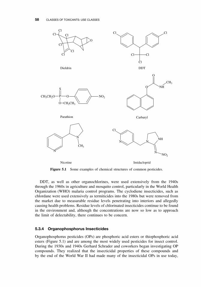
58 CLASSES OF TOXICANTS: USE CLASSES
ClCl
Cl
ClCl
Cl
O
Cl Cl
Cl Cl
Cl
O
O
NH
CH3
N
N
CH3
N NNH
NNO2
Cl
Dieldrin DDT
O NO2CH3CH2O
O
P
S
CH2CH3
Parathion Carbaryl
Nicotine Imidacloprid
Figure 5.1 Some examples of chemical structures of common pesticides.
DDT, as well as other organochlorines, were used extensively from the 1940sthrough the 1960s in agriculture and mosquito control, particularly in the World HealthOrganization (WHO) malaria control programs. The cyclodiene insecticides, such aschlordane were used extensively as termiticides into the 1980s but were removed fromthe market due to measurable residue levels penetrating into interiors and allegedlycausing health problems. Residue levels of chlorinated insecticides continue to be foundin the environment and, although the concentrations are now so low as to approachthe limit of delectability, there continues to be concern.
5.3.4 Organophosphorus Insecticides
Organophosphorus pesticides (OPs) are phosphoric acid esters or thiophosphoric acidesters (Figure 5.1) and are among the most widely used pesticides for insect control.During the 1930s and 1940s Gerhard Schrader and coworkers began investigating OPcompounds. They realized that the insecticidal properties of these compounds andby the end of the World War II had made many of the insecticidal OPs in use today,
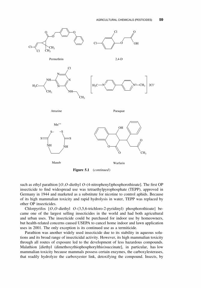
AGRICULTURAL CHEMICALS (PESTICIDES) 59
ClCl CH3
CH3
OO
O Cl
Cl O
O
OH
CH3
H3C
NH
N
N
N
NH
CH3
Cl
N+ N+ CH3H3C 2Cl−
O
OH
O CH3
O
NHNH
SS
S− −S
Maneb Warfarin
Paraquat
2,4-DPermethrin
Atrazine
Mn++
Figure 5.1 (continued )
such as ethyl parathion [O,O-diethyl O-(4-nitrophenyl)phosphorothioate]. The first OPinsecticide to find widespread use was tetraethylpyrophosphate (TEPP), approved inGermany in 1944 and marketed as a substitute for nicotine to control aphids. Becauseof its high mammalian toxicity and rapid hydrolysis in water, TEPP was replaced byother OP insecticides.
Chlorpyrifos [O,O-diethyl O-(3,5,6-trichloro-2-pyridinyl) phosphorothioate] be-came one of the largest selling insecticides in the world and had both agriculturaland urban uses. The insecticide could be purchased for indoor use by homeowners,but health-related concerns caused USEPA to cancel home indoor and lawn applicationuses in 2001. The only exception is its continued use as a termiticide.
Parathion was another widely used insecticide due to its stability in aqueous solu-tions and its broad range of insecticidal activity. However, its high mammalian toxicitythrough all routes of exposure led to the development of less hazardous compounds.Malathion [diethyl (dimethoxythiophosphorylthio)succinate], in particular, has lowmammalian toxicity because mammals possess certain enzymes, the carboxylesterases,that readily hydrolyze the carboxyester link, detoxifying the compound. Insects, by
60 CLASSES OF TOXICANTS: USE CLASSES
contrast, do not readily hydrolyze this ester, and the result is its selective insectici-dal action.
OPs are toxic because of their inhibition of the enzyme acetylcholinesterase. Thisenzyme inhibition results in the accumulation of acetylcholine in nerve tissue and effec-tor organs, with the principal site of action being the peripheral nervous system (PNS)(see Chapter 16). In addition to acute effects, some OP compounds have been asso-ciated with delayed neurotoxicity, known as organophosphorus-induced delayed neu-ropathy (OPIDN). The characteristic clinical sign is bilateral paralysis of the distalmuscles, predominantly of the lower extremities, occurring some 7 to 10 days fol-lowing ingestion (see Chapter 16). Not all OP compounds cause delayed neuropathy.Among the pesticides associated with OPIDN are leptophos, mipafox, EPN, DEF, andtrichlorfon. Testing is now required for OP substances prior to their use as insecticides.
The OP and carbamate insecticides are relatively nonpersistent in the environment.They are applied to the crop or directly to the soil as systemic insecticides, and theygenerally persist from only a few hours to several months. Thus these compounds,in contrast to the organochlorine insecticides, do not represent a serious problem ascontaminants of soil and water and rarely enter the human food chain. Being esters, thecompounds are susceptible to hydrolysis, and their breakdown products are generallynontoxic. Direct contamination of food by concentrated compounds has been the causeof poisoning episodes in several countries.
5.3.5 Carbamate Insecticides
The carbamate insecticides are esters of N -methyl (or occasionally N ,N -dimethyl) car-bamic acid (H2NCOOH). The toxicity of the compound varies according to the phenolor alcohol group. One of the most widely used carbamate insecticides is carbaryl (1-napthyl methylcarbamate), a broad spectrum insecticide (Figure 5.1). It is used widelyin agriculture, including home gardens where it generally is applied as a dust. Carbarylis not considered to be a persistent compound, because it is readily hydrolyzed. Basedon its formulation, it carries a toxicity classification of II or III with an oral LD50 of250 mg/kg (rat) and a dermal LC50 of >2000 mg/kg.
An example of an extremely toxic carbamate is aldicarb [2-methyl-2-(methylthio)propionaldehyde]. Both oral and dermal routes are the primary portals of entry, andit has an oral LD50 of 1.0 mg/kg (rat)and a dermal LD50 of 20 mg/kg (rabbit). Forthis reason it is recommended for application to soils on crops such as cotton, cit-rus, and sweet potatoes. This compound moves readily through soil profiles and hascontaminated groundwater supplies.
Like the OP insecticides, the mode of action of the carbamates is acetylcholinesteraseinhibition with the important difference that the inhibition is more rapidly reversed thanwith OP compounds.
5.3.6 Botanical Insecticides
Extracts from plants have been used for centuries to control insects. Nicotine [(S)-3-(1-methyl-2-pyrrolidyl)pyridine] (Figure 5.1) is an alkaloid occurring in a number ofplants and was first used as an insecticide in 1763. Nicotine is quite toxic orally aswell as dermally. The acute oral LD50 of nicotine sulfate for rats is 83 mg/kg and
AGRICULTURAL CHEMICALS (PESTICIDES) 61
the dermal LD50 is 285 mg/kg. Symptoms of acute nicotine poisoning occur rapidly,and death may occur with a few minutes. In serious poisoning cases death resultsfrom respiratory failure due to paralysis of respiratory muscles. In therapy attention isfocused primarily on support of respiration.
Pyrethrin is an extract from several types of chrysanthemum, and is one of theoldest insecticides used by humans. There are six esters and acids associated withthis botanical insecticide. Pyrethrin is applied at low doses and is considered to benonpersistent.
Mammalian toxicity to pyrethrins is quite low, apparently due to its rapid break-down by liver microsomal enzymes and esterases. The acute LD50 to rats is about1500 mg/kg. The most frequent reaction to pyrethrins is contact dermatitis and allergicrespiratory reactions, probably as a result of other constituents in the formulation. Syn-thetic mimics of pyrethrins, known as the pyrethroids, were developed to overcomethe lack of persistence.
5.3.7 Pyrethroid Insecticides
As stated, pyrethrins are not persistent, which led pesticide chemists to develop com-pounds of similar structure having insecticidal activity but being more persistent.This class of insecticides, known as pyrethroids, have greater insecticidal activityand are more photostable than pyrethrins. There are two broad classes of pyrethroidsdepending on whether the structure contains a cyclopropane ring [e.g., cyperme-thrin {(±)-α-cyano-3-phenoxybenzyl (±)-cis,trans-3-(2,2-dichlorovinyl 2,2-dimethylcyclopropanecarboxylate)}] or whether this ring is absent in the molecule [e.g., fenvale-rate{(RS )-α-cyano-3-phenoxybenzyl(RS )-2-(4-chlorophenyl)-3-methylbutyrate}]. Theyare generally applied at low doses (e.g., 30 g/Ha) and have low mammalian toxicities[e.g., cypermethrin, oral (aqueous suspension) LD50 of 4,123 mg/kg (rat) and dermalLD50 of >2000 mg/kg (rabbit)]. Pyrethroids are used in both agricultural and urbansettings (e.g., termiticide; Figure 5.1).
Pyrethrins affect nerve membranes by modifying the sodium and potassium chan-nels, resulting in depolarization of the membranes. Formulations of these insecticidesfrequently contain the insecticide synergist piperonyl butoxide [5-{2-(2-butoxyethoxy)ethoxymethyl}-6-propyl-1,3-benzodioxole], which acts to increase the efficacy of theinsecticide by inhibiting the cytochrome P450 enzymes responsible for the breakdownof the insecticide.
5.3.8 New Insecticide Classes
There are new classes of insecticides that are applied at low dosages and are extremelyeffective but are relatively nontoxic to humans. One such class is the fiproles, and one ofthese receiving major attention is fipronil [(5-amino-1-(2,6-dichloro-4-(trifluoromethyl)phenyl)-4-((1,R,S)-(trifluoromethyl)su-1-H -pyrasole-3-carbonitrile)]. Although it isused on corn, it is becoming a popular termiticide because of its low applicationrate (ca. 0.01%) and long-term effectiveness. Another class of insecticides, thechloronicotinoids, is represented by imidacloprid [1-(6-chloro-3-pyridin-3-ylmethyl)-N -nitroimidazolidin-2-ylidenamine] (Figure 5.1), which also is applied at low doserates to soil and effectively controls a number of insect species, including termites.
62 CLASSES OF TOXICANTS: USE CLASSES
5.3.9 Herbicides
Herbicides control weeds and are the most widely used class of pesticides. The latestUS EPA data show that some 578 million pounds of herbicides were used in theUnited States in 1997 and accounts for some 47% of pesticides used. This class ofpesticide can be applied to crops using many strategies to eliminate or reduce weedpopulations. These include preplant incorporation, pre- and postemergent applications.New families of herbicides continue to be developed, and are applied at low doses, arerelatively nonphytotoxic to beneficial plants and are environmentally friendly. Some ofthe newer families such as the imidazolinones inhibit the action of acetohydroxyacidsynthase that produces branched-chain amino acids in plants. Because this enzyme isproduced only in plants, these herbicides have low toxicities to mammals, fish, insects,and birds.
The potential for environmental contamination continues to come from families ofherbicides that have been used for years. The chlorophenoxy herbicides such as 2,4-D (2,4-dichlorophenoxy acetic acid) and 2,4,5-T (2,4,5-trichlorophenoxy-acetic acid)(Figure 5.1) are systemic acting compounds to control broadleaf plants and have beenin use since the 1940s. The oral toxicities of these compounds are low.
A mixture of 2,4-D and 2,4,5-T , known as Agent Orange, was used by the US mil-itary as a defoliant during the Vietnam conflict, and much controversy has arisen overclaims by military personnel of long-term health effects. The chemical of major toxico-logical concern was identified as a contaminant, TCDD (2,3,7,8-tetrachlorodibenzo-p-dioxin), that was formed during the manufacturing process. TCDD is one of the mosttoxic synthetic substances known in laboratory animals. The LD50 for male rats is0.022 mg/kg, and for female guinea pigs (the most sensitive species tested) the LD50is 0.0006 mg/kg. In addition it is toxic to developing embryos in pregnant rats at adose of only 1/400 of the LD50, and has been shown to cause birth defects at levelsof 1 to 3 ng/kg of body weight. TCDD is a proven carcinogen in both mice and rats,with the liver being the primary target. This chemical has also been shown to alter theimmune system and enhance susceptibility in exposed animals.
Another family of herbicides, the triazines, continues to cause concern to environ-mentalists and toxicologists because of the contamination of surface and groundwatersupplies that become public drinking water. The herbicide, atrazine [6-chloro-N -ethyl-N -(1-methylethyl)-1,2,5-triazine-2,4-diamine (Figure 5.1) is used primarily on cornand has an MCL of 3.0 µg/L. This herbicide has been found in surface and ground-waters worldwide with widely varying concentrations (e.g., 1 to >130 µg/L). It,along with two other triazines, cyanazine [2-{{4-chloro-6-(ethylamino)-1,3,5-triazin-2-yl}amino}-2-methylpropanenitrile] and simazine (6-chloro-N ,N -diethyl-1,3,5-triazine-2,4-diamine) (MCL of 4.0 µg/L). The uses of cyanazine were canceled in 2001 andno further use was permitted after 2002. Although relatively nontoxic [e.g., atrazine,oral LD50 of 3,100 mg/kg (rat)], the major concern with these types of compoundsis their carcinogenic effects, and US EPA considers these three triazines as possiblehuman carcinogens (category C).
A member of the bipyridylium family of herbicides is the compound paraquat (1,1-dimethyl-4,4-bipyridinium ion as the chloride salt) (Figure 5.1). It is a very water-soluble contact herbicide that is active against a broad range of plants and is usedas a defoliant on many crops. The compound binds tightly to soil particles following
AGRICULTURAL CHEMICALS (PESTICIDES) 63
application and becomes inactivated. However, this compound is classified as a class Itoxicant with an oral LD50 of 150 mg/kg (rat). Most poisoning cases, which are oftenfatal, are due to accidental or deliberate ingestion of paraquat. Toxicity results fromlung injury resulting from both the preferential uptake of paraquat by the lungs andthe redox cycling mechanism.
5.3.10 Fungicides
Every year fungi cause crop losses in the United States amounting to millions of dollars.In addition recent studies have shown that toxins and other airborne organic compoundsreleased from fungi inhabiting the interior of dwellings probably are responsible fora number of adverse health effects. Compounds produced to combat these losses andadverse health effects are called fungicides, and a number of these families have beenaround for years.
The fungicide, chlorothalonil (tetrachloroisophthalonitrile), is a broad-spectrum fun-gicide which is used widely in urban environments. It is relatively cheap and controlssome 140 species of organisms. As a result of the popularity of this compound, itis found routinely in surface waters entering public drinking water supplies. In theformulation that can be purchased by the general public, it is relatively nontoxic.
One family of fungicides that is of concern are the dithiocarbamates, sulfur deriva-tives of dithiocarbamic acid and include the metallic dimethyldithiocarbamates. Thelatter group includes mancozeb (a coordination product of zinc ion and manganese ethy-lene bisdithiocarbamate), maneb (manganese ethylenebisdithiocarbamate)(Figure 5.1),and zineb (zinc ethylenebisdithiocarbamate). All are effective fungicides and are usedon a variety of crops including grapes, sugar beets, and ornamental plants. Althoughrelatively nontoxic, they do hydrolyze producing known carcinogens such as ethylth-iourea (ETU).
5.3.11 Rodenticides
This class of compounds is used to control rodents that cause yearly losses of 20% to30% in grain and other food storage facilities. These pests harbor diseases in the formof fleas that carry bacteria and other organisms. A number of the rodenticides havebeen used for years and include warfarin [3-(α-acetonylbenzyl)-4-hydroxycoumarin](Figure 5.1), an anticoagulant. This is a potent toxicant with an oral LD50 of 3.0 mg/kg(rat). As the rats navigate through narrow passages, they bruise themselves, developingsmall hemorrhages. Anticoagulants prevent the blood from clotting, and the animalsbleed to death in about a week. Humans who are exposed to this class of compoundsare given vitamin K, and if the poisoning is severe, blood transfusions as a treat-ment. Other rodenticides poison the animal and many times are applied along withan attractant such as peanut butter to overcome bait shyness. Fluoroacetamide is afast acting poison with an oral LD50 (rat) of 15 mg/kg. This material is suppliedas bait pellets or grains. ANTU (α -naphthylthiourea), strychnine, and thallium saltsare other fast acting poisons, and have been on the market for many years. Most ofthe rodenticides are classified as restricted use and are applied only by licensed pest
64 CLASSES OF TOXICANTS: USE CLASSES
control operators. Human poisonings associated with rodenticides usually result fromaccidental or suicidal ingestion of the compounds.
5.3.12 Fumigants
Fumigants are extremely toxic gases used to protect stored products, especially grains,and to kill soil nematodes. These materials are applied to storage warehouses, freightcars, and houses infested with insects such as powder post beetles. They present aspecial hazard due to inhalation exposure and rapid diffusion into pulmonary blood;thus extreme care must be taken when handling and applying this class of pesticides. Allfumigants are classified as restricted use compounds and require licensed applicatorsto handle them.
One of the most effective fumigants is methyl bromide. It essentially sterilizes soilwhen applied under a ground covering, because it kills insects, nematodes, and weedseed but also is used to fumigate warehouses. Overexposure to this compound causesrespiratory distress, cardiac arrest, and central nervous effects. The inhalation LC50 is0.06 mg/L (15 min) of air (rat) and 7900 ppm (1.5 h) (human). Methyl bromide hasbeen classified as an ozone depleter under the Clean Air Act and is due to be phasedout of use by 2005.
Chloropicrin (trichloronitromethane) is another soil/space fumigant that has beenused for many years. It has an inhalation LC50 of 150 ppm (15 min). Thus it is highlytoxic by inhalation, can injure the heart, and cause severe eye damage.
5.3.13 Conclusions
This section has covered only a few of the pesticides available today on the UnitedStates and world markets. An understanding of the basic chemical processes affectedby pesticides has led to the discovery and production of new families of chemicals.Today’s modern pesticide is generally safe to use if the directions on the label arefollowed. Advances in instrumentation and an understanding of how adverse healtheffects are produced have resulted in the production of many environmentally friendlybut effective pesticides.
5.4 FOOD ADDITIVES AND CONTAMINANTS
Chemicals are added to food for a number of reasons: as preservatives with antibacte-rial, antifungal, or antioxidant properties; to change physical characteristics, particularlyfor processing; to change taste; to change color; and to change odor. In general, foodadditives have proved to be safe and without chronic toxicity. Many were introducedwhen toxicity testing was relatively unsophisticated, however, and some of these havebeen subsequently shown to be toxic. Table 5.4 gives examples of different types oforganic food additives. Inorganics, the most important of which are nitrate and nitrite,are discussed later. Certainly hundreds, and possibly thousands, of food additives are inuse worldwide, many with inadequate testing. The question of synergistic interactionsbetween these compounds has not been explored adequately. Not all toxicants in foodare synthetic; many examples of naturally occurring toxicants in the human diet areknown, including carcinogens and mutagens.
TOXINS 65
Table 5.4 Examples of Organic Chemicals Used as Food Additives
Function Class Example
Preservatives Antioxidants ButylatedhydroxyanisoleAscorbic acid
Fungistatic agents Methyl p-benzoic acidPropionatesBactericides Sodium nitrite
Processing aids Anticaking agents Calcium silicateSodium aluminosilicate
Emulsifiers Propylene glycolMonoglycerides
Chelating agents EDTASodium tartrate
Stabilizers Gum ghattiSodium alginate
Humectants Propylene glycolGlycerol
Flavor and tastemodification Synthetic sweeteners Saccharin
MannitolAspartame
Synthetic flavors PiperonalVanillin
Color modification Synthetic dyes Tartrazine (FD&C yellow5)Sunset Yellow
Nutritional supplements Vitamins ThiaminVitamin D3
Amino acids AlanineAspartic acid
Inorganics Manganese sulfateZinc sulfate
5.5 TOXINS
5.5.1 History
A discussion of toxins first necessitates the understanding and distinction between thetoxicological terms toxicant and toxin. A toxicant is any chemical, of natural or syn-thetic origin, capable of causing a deleterious effect on a living organism. A toxin isa toxicant that is produced by a living organism and is not used as a synonym fortoxicant—all toxins are toxicants, but not all toxicants are toxins. Toxins, whetherproduced by animals, plants, insects, or microbes are generally metabolic productsthat have evolved as defense mechanisms for the purpose of repelling or killingpredators or pathogens. The action of natural toxins has long been recognized andunderstood throughout human history. For example, ancient civilizations used naturaltoxins for both medicinal (therapeutic) and criminal purposes. Even today, we con-tinue to discover and understand the toxicity of natural products, some for beneficialpharmaceutical or therapeutic purposes whose safety and efficacy are tested, and somefor other less laudable purposes like biological or chemical warfare. Toxins may be
66 CLASSES OF TOXICANTS: USE CLASSES
classified in various ways depending on interest and need, such as by target organtoxicity or mode of action, but are commonly classified according to source.
5.5.2 Microbial Toxins
The term “microbial toxin” is usually reserved by microbiologists for toxic substancesproduced by microorganisms that are of high molecular weight and have antigenicproperties; toxic compounds produced by bacteria that do not fit these criteria arereferred to simply as poisons. Many of the former are proteins or mucoproteins andmay have a variety of enzymatic properties. They include some of the most toxicsubstances known, such as tetanus toxin, botulinus toxin, and diphtheria toxin. Bacterialtoxins may be extremely toxic to mammals and may affect a variety of organ systems,including the nervous system and the cardiovascular system. A detailed account oftheir chemical nature and mode of action is beyond the scope of this volume.
The range of poisonous chemicals produced by bacteria is also large. Again, suchcompounds may also be used for beneficial purposes, for example, the insecticidalproperties of Bacillus thuringiensis, due to a toxin, have been utilized in agriculturefor some time.
5.5.3 Mycotoxins
The range of chemical structures and biologic activity among the broad class of fungalmetabolites is large and cannot be summarized briefly. Mycotoxins do not constitute aseparate chemical category, and they lack common molecular features.
Mycotoxins of most interest are those found in human food or in the feed of domesticanimals. They include the ergot alkaloids produced by Claviceps sp., aflatoxins andrelated compounds produced by Aspergillus sp., and the tricothecenes produced byseveral genera of fungi imperfecti, primarily Fusarium sp.
The ergot alkaloids are known to affect the nervous system and to be vasocon-strictors. Historically they have been implicated in epidemics of both gangrenous andconvulsive ergotism (St. Anthony’s fire), although such epidemics no longer occur inhumans due to increased knowledge of the cause and to more varied modern diets. Out-breaks of ergotism in livestock do still occur frequently, however. These compoundshave also been used as abortifacients. The ergot alkaloids are derivatives of ergotine,the most active being, more specifically, amides of lysergic acid.
Aflatoxins are products of species of the genus Aspergillus, particularly A flavus,a common fungus found as a contaminant of grain, maize, peanuts, and so on. Firstimplicated in poultry diseases such as Turkey-X disease, they were subsequently shownto cause cancer in experimental animals and, from epidemiological studies, in humans.Aflatoxin B1, the most toxic of the aflatoxins, must be activated enzymatically to exertits carcinogenic effect.
Tricothecenes are a large class of sesquiterpenoid fungal metabolites produced par-ticularly by members of the genera Fusarium and Tricoderma. They are frequentlyacutely toxic, displaying bactericidal, fungicidal, and insecticidal activity, as wellas causing various clinical symptoms in mammals, including diarrhea, anorexia, andataxia. They have been implicated in natural intoxications in both humans and animals,such as Abakabi disease in Japan and Stachybotryotoxicosis in the former USSR, and
TOXINS 67
are the center of a continuing controversy concerning their possible use as chemicalwarfare agents.
Mycotoxins may also be used for beneficial purposes. The mycotoxin avermectin iscurrently generating considerable interest both as an insecticide and for the control ofnematode parasites of domestic animals.
5.5.4 Algal Toxins
Algal toxins are broadly defined to represent the array chemicals derived from manyspecies of cyanobacteria (blue-green bacteria), dinoflagellates, and diatoms. The toxinsproduced by these freshwater and marine organisms often accumulate in fish and shell-fish inhabiting the surrounding waters, causing both human and animal poisonings, aswell as overt fish kills. Unlike many of the microbial toxins, algal toxins are gener-ally heat stable and, therefore, not altered by cooking methods, which increases thelikelihood of human exposures and toxicity. Many of the more common algal toxinsresponsible for human poisonings worldwide are summarized herein.
Amnesic Shellfish Poisoning (ASP) was first identified in 1987 from Prince EdwardIsland, Canada after four people died from eating contaminated mussels. It iscaused by domoic acid produced by several species of Pseudonitzschia diatoms.The main contamination problems include mussels, clams, and crabs of the PacificNorthwest of the United States and Canada.
Paralytic Shellfish Poisoning (PSP) was first determined to be a problem in 1942after three people and many seabirds died from eating shellfish on the west coastof the United States, near the Columbia River. It is caused by the saxitoxin family(saxitoxin + 18 related compounds) produced by several species of Alexandriumdinoflagellates. The main contamination problems include mussels, clams, crabs,and fish of the Pacific Northwest and Northeast Atlantic.
Neurotoxic Shellfish Poisoning (NSP) is caused by a red-tide producer that was firstidentified in 1880 from Florida, with earlier historical references. It causes sick-ness in humans lasting several days. NSP is not fatal to humans; however, it isknown to kill fish, invertebrates, seabirds, and marine mammals (e.g., manatees).It is caused by the brevetoxin family (brevetoxin + 10 related compounds pro-duced by the dinoflagellate Karenia brevis a.k.a. Gymnodinium breve. The maincontamination problems include oysters, clams, and other filter feeders of theGulf of Mexico and southeast Atlantic, including North Carolina.
Diarrheic Shellfish Poisoning (DSP). Human poisonings were first identified in the1960s. It causes sickness in humans lasting several days but is not fatal. Itis caused by chemicals of the okadaic acid family (okadaic acid + 4 relatedcompounds) produced by several species of Dinophysis dinoflagellates. The maincontamination problems include mussels, clams, and other bivalves of the coldand warm temperate areas of the Atlantic and Pacific Oceans, mainly in Japanand Europe. Only two cases of DSP have been documented in North America.
Ciguatera Fish Poisoning (CFP) was first identified in 1511, CFP is a tropical-subtropical seafood poisoning that affects up to 50,000 people each year and isthe most often reported foodborne disease of a chemical origin in the UnitedStates. Caused by consumption of reef fishes (e.g., grouper, snapper), sickness in
68 CLASSES OF TOXICANTS: USE CLASSES
humans lasts several days to weeks, but the human fatality rate is low. It iscaused by the ciguatoxin family (ciguatoxin + 3 or more related compounds) andproduced by several species of dinoflagellates including Gambierdiscus, Proro-centrum, and Ostreopsis. The main contamination problems include herbivoroustropical reef fish worldwide.
Cyanobacterial (Blue-Green Bacteria) Toxins . Cyanobacterial poisonings were firstrecognized in the late 1800s. Human poisonings are rare; however, kills oflivestock, other mammals, birds, fish, and aquatic invertebrates are common.It is caused by a variety of biotoxins and cytotoxins, including anatoxin,microcystin, and nodularin produced by several species of cyanobacteria,including Anabaena, Aphanizomenon, Nodularia, Oscillatoria, and Microcystis.The main contamination problems include all eutrophic freshwater rivers, lakes,and streams.
Ambush Predator (Pfiesteria piscicida and Toxic Pfiesteria Complex) Toxins . Mem-bers belonging to this group of organisms were first identified in 1991 fromestuaries in North Carolina. They were believed to produce a toxin that has beenimplicated in several large fish kills and is suspect in causing adverse humanhealth effects. However, the toxin or toxins are not yet identified and toxicitytests are not universally conclusive. Produced by several dinoflagellate speciesincluding, Pfiesteria piscicida, Pfiesteria shumwayae, and perhaps several otherunidentified, un-named dinoflagellates belonging to the potentially toxic Pfies-teria complex. Main problems include major fish kills in North Carolina andMaryland and potential human health problems. The range may extend fromthe Gulf of Mexico to the Atlantic estuarine waters, including Florida, NorthCarolina, Maryland, and Delaware, and possibly outward to Europe.
5.5.5 Plant Toxins
The large array of toxic chemicals produced by plants (phytotoxins), usually referred toas secondary plant compounds, are often held to have evolved as defense mechanismsagainst herbivorous animals, particularly insects and mammals. These compounds maybe repellent but not particularly toxic, or they may be acutely toxic to a wide rangeof organisms. They include sulfur compounds, lipids, phenols, alkaloids, glycosides,and many other types of chemicals. Many of the common drugs of abuse such ascocaine, caffeine, nicotine, morphine, and the cannabinoids are plant toxins. Manychemicals that have been shown to be toxic are constituents of plants that form partof the human diet. For example, the carcinogen safrole and related compounds arefound in black pepper. Solanine and chaconine, which are cholinesterase inhibitors andpossible teratogens, are found in potatoes, and quinines and phenols are widespreadin food. Livestock poisoning by plants is still an important veterinary problem insome areas.
5.5.6 Animal Toxins
Some species from practically all phyla of animals produce toxins for either offen-sive or defensive purposes. Some are passively venomous, often following inadvertentingestion, whereas others are actively venomous, injecting poisons through specially
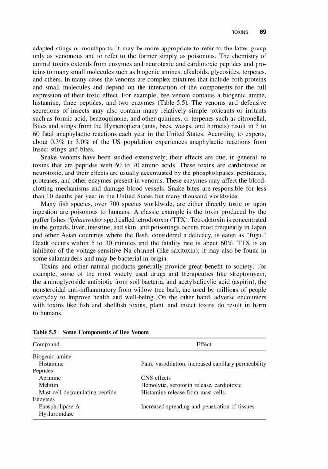
TOXINS 69
adapted stings or mouthparts. It may be more appropriate to refer to the latter grouponly as venomous and to refer to the former simply as poisonous. The chemistry ofanimal toxins extends from enzymes and neurotoxic and cardiotoxic peptides and pro-teins to many small molecules such as biogenic amines, alkaloids, glycosides, terpenes,and others. In many cases the venoms are complex mixtures that include both proteinsand small molecules and depend on the interaction of the components for the fullexpression of their toxic effect. For example, bee venom contains a biogenic amine,histamine, three peptides, and two enzymes (Table 5.5). The venoms and defensivesecretions of insects may also contain many relatively simple toxicants or irritantssuch as formic acid, benzoquinone, and other quinines, or terpenes such as citronellal.Bites and stings from the Hymenoptera (ants, bees, wasps, and hornets) result in 5 to60 fatal anaphylactic reactions each year in the United States. According to experts,about 0.3% to 3.0% of the US population experiences anaphylactic reactions frominsect stings and bites.
Snake venoms have been studied extensively; their effects are due, in general, totoxins that are peptides with 60 to 70 amino acids. These toxins are cardiotoxic orneurotoxic, and their effects are usually accentuated by the phospholipases, peptidases,proteases, and other enzymes present in venoms. These enzymes may affect the blood-clotting mechanisms and damage blood vessels. Snake bites are responsible for lessthan 10 deaths per year in the United States but many thousand worldwide.
Many fish species, over 700 species worldwide, are either directly toxic or uponingestion are poisonous to humans. A classic example is the toxin produced by thepuffer fishes (Sphaeroides spp.) called tetrodotoxin (TTX). Tetrodotoxin is concentratedin the gonads, liver, intestine, and skin, and poisonings occurs most frequently in Japanand other Asian countries where the flesh, considered a delicacy, is eaten as “fugu.”Death occurs within 5 to 30 minutes and the fatality rate is about 60%. TTX is aninhibitor of the voltage-sensitive Na channel (like saxitoxin); it may also be found insome salamanders and may be bacterial in origin.
Toxins and other natural products generally provide great benefit to society. Forexample, some of the most widely used drugs and therapeutics like streptomycin,the aminoglycoside antibiotic from soil bacteria, and acetylsalicylic acid (aspirin), thenonsteroidal anti-inflammatory from willow tree bark, are used by millions of peopleeveryday to improve health and well-being. On the other hand, adverse encounterswith toxins like fish and shellfish toxins, plant, and insect toxins do result in harmto humans.
Table 5.5 Some Components of Bee Venom
Compound Effect
Biogenic amineHistamine Pain, vasodilation, increased capillary permeability
PeptidesApamine CNS effectsMelittin Hemolytic, serotonin release, cardiotoxicMast cell degranulating peptide Histamine release from mast cells
EnzymesPhospholipase A Increased spreading and penetration of tissuesHyaluronidase
70 CLASSES OF TOXICANTS: USE CLASSES
5.6 SOLVENTS
Although solvents are more a feature of the workplace they are also found in thehome. In addition to cutaneous effects, such as defatting and local irritation, many havesystemic toxic effects, including effects on the nervous system or, as with benzene,on the blood-forming elements. Commercial solvents are frequently complex mixturesand may include nitrogen- or sulfur-containing organics—gasoline and other oil-basedproducts are examples of this. The common solvents fall into the following classes:
Aliphatic Hydrocarbons , such as hexane. These may be straight or branched-chaincompounds and are often present in mixtures.
Halogenated Aliphatic Hydrocarbons . The best-known examples are methylenedichloride, chloroform, and carbon tetrachloride, although chlorinated ethylenesare also widely used.
Aliphatic Alcohols . Common examples are methanol and ethanol.Glycols and Glycol Ethers . Ethylene and propylene glycols, for example, in
antifreeze give rise to considerable exposure of the general public. Glycol ethers,such as methyl cellosolve, are also widely used.
Aromatic Hydrocarbons . Benzene is probably the one of greatest concern, but others,such as toluene, are also used.
5.7 THERAPEUTIC DRUGS
Although the study of the therapeutic properties of chemicals fall within the provinceof pharmacology, essentially all therapeutic drugs can be toxic, producing deleteri-ous effects at some dose. The danger to the individual depends on several factors,including the nature of the toxic response, the dose necessary to produce the toxicresponse, and the relationship between the therapeutic dose and the toxic dose. Drugtoxicity is affected by all of factors that affect the toxicity of other xenobiotics,including individual (genetic) variation, diet, age, and the presence of other exoge-nous chemicals.
Even when the risk of toxic side effects from a particular drug has been evaluated, itmust be weighed against the expected benefits. The use of a very dangerous drug withonly a narrow tolerance between the therapeutic and toxic doses may still be justifiedif it is the sole treatment for an otherwise fatal disease. However, a relatively safe drugmay be inappropriate if safer compounds are available or if the condition being treatedis trivial.
The three principal classes of cytotoxic agents used in the treatment of cancerall contain carcinogens, for example, Melphalen, a nitrogen mustard, adriamycin, anantitumor antibiotic, and methotrexate, an antimetabolite. Diethylstilbestrol (DES), adrug formerly widely used, has been associated with cancer of the cervix and vaginain the offspring of treated women.
Other toxic effects of drugs can be associated with almost every organ system. Thestiffness of the joints accompanied by damage to the optic nerve (SMON—subacutemyelo-optic neuropathy) that was common in Japan in the 1960s was apparently atoxic side effect of chloroquinol (Enterovioform), an antidiarrhea drug. Teratogenosis
COSMETICS 71
can also be caused by drugs, with thalidomide being the most alarming example.Skin effects (dermatitis) are common side effects of drugs, an example being topicallyapplied corticosteroids.
A number of toxic effects on the blood have been documented, including agran-ulocytosis caused by chlorpromazine, hemolytic anemia caused by methyldopa, andmegaloblastic anemia caused by methotrexate. Toxic effects on the eye have beennoted and range from retinotoxicity caused by thioridazine to glaucoma caused bysystemic corticosteroids.
5.8 DRUGS OF ABUSE
All drugs are toxic at some dose. Drugs of abuse, however, either have no medici-nal function or are taken at dose levels higher than would be required for therapy.Although some drugs of abuse may affect only higher nervous functions—mood,reaction time, and coordination—many produce physical dependence and have seriousphysical effects, with fatal overdoses being a frequent occurrence.
The drugs of abuse include central nervous system depressants such as ethanol,methaqualone (Quaalude), and secobarbital; central nervous system stimulants, suchas cocaine, methamphetamine (speed), caffeine, and nicotine; opioids, such as heroinand mependine (demerol); and hallucinogens such as lysergic acid diethylamide (LSD),phencyclidine (PCP), and tetrahydrocannabinol, the most active principal of marijuana.A further complication of toxicological significance is that many drugs of abuse aresynthesized in illegal and poorly equipped laboratories with little or no quality control.The resultant products are therefore often contaminated with compounds of unknown,but conceivably dangerous, toxicity. The structures of some of these chemicals areshown in Figure 5.2.
5.9 COMBUSTION PRODUCTS
While many air pollutants (see Chapter 4) are the products of natural or anthropo-morphic combustion, some of the most important from the point of view of humanhealth are polycyclic aromatic hydrocarbons. Although also found in natural productssuch as coal and crude oil, they are generally associated with incomplete combustionof organic materials and are found in smoke from wood, coal, oil, and tobacco, forexample, as well as in broiled foods. Because some of them are carcinogens, they havebeen studied intensively from the point of view of metabolic activation, interactionswith DNA, and other aspects of chemical carcinogenesis. Some are heterocyclic, con-taining nitrogen in at least one of the rings. Some representative structures of the moststudied polycyclic aromatic hydrocarbons are shown in Figure 5.3.
5.10 COSMETICS
The most common deleterious effects of modern cosmetics are occasional allergicreactions and contact dermatitis. The highly toxic and/or carcinogenic azo or aromatic
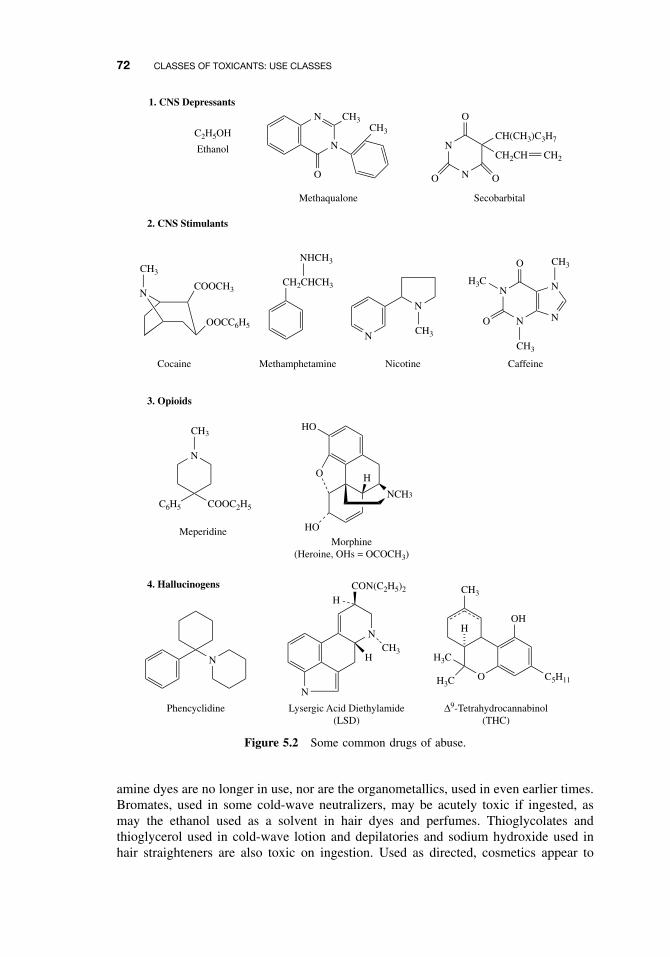
72 CLASSES OF TOXICANTS: USE CLASSES
1. CNS Depressants
C2H5OH
Ethanol N
N CH3CH3
O
Methaqualone
N
N
O
O O
CH2CH
CH(CH3)C3H7
CH2
Secobarbital
CH2CHCH3
NHCH3
2. CNS Stimulants
N
N
N
O
O
N
NN
CH3
H3C
CH3
CaffeineNicotineMethamphetamineCocaine
OOCC6H5
COOCH3N
CH3
CH3
3. Opioids
N
CH3
C6H5 COOC2H5
Meperidine
HO
O
HO
NCH3
H
Morphine(Heroine, OHs = OCOCH3)
4. Hallucinogens
N
Phencyclidine
N
NCH3
HCON(C2H5)2
H
Lysergic Acid Diethylamide(LSD)
O
CH3
OH
C5H11H3C
H3C
H
∆9-Tetrahydrocannabinol(THC)
Figure 5.2 Some common drugs of abuse.
amine dyes are no longer in use, nor are the organometallics, used in even earlier times.Bromates, used in some cold-wave neutralizers, may be acutely toxic if ingested, asmay the ethanol used as a solvent in hair dyes and perfumes. Thioglycolates andthioglycerol used in cold-wave lotion and depilatories and sodium hydroxide used inhair straighteners are also toxic on ingestion. Used as directed, cosmetics appear to
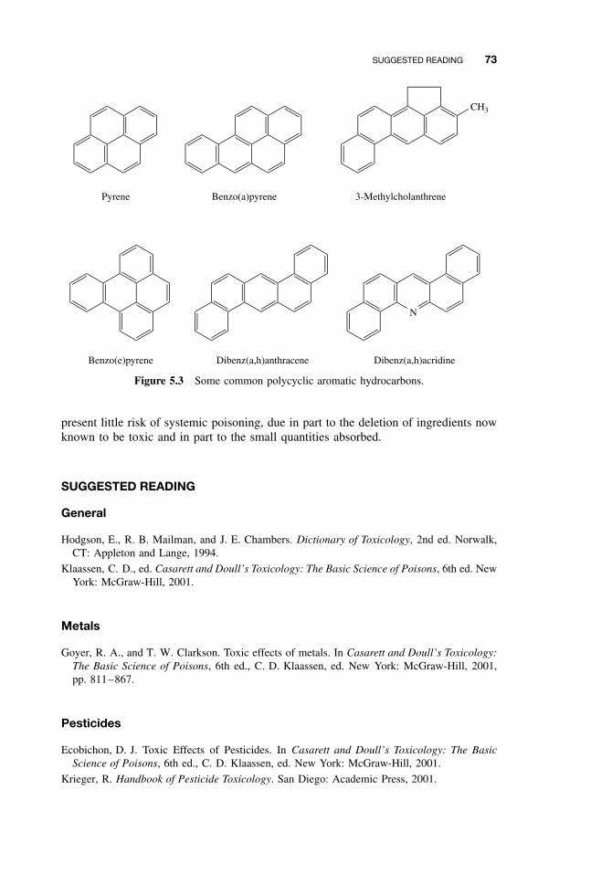
SUGGESTED READING 73
Pyrene Benzo(a)pyrene 3-Methylcholanthrene
N
Benzo(e)pyrene Dibenz(a,h)anthracene Dibenz(a,h)acridine
CH3
Figure 5.3 Some common polycyclic aromatic hydrocarbons.
present little risk of systemic poisoning, due in part to the deletion of ingredients nowknown to be toxic and in part to the small quantities absorbed.
SUGGESTED READING
General
Hodgson, E., R. B. Mailman, and J. E. Chambers. Dictionary of Toxicology, 2nd ed. Norwalk,CT: Appleton and Lange, 1994.
Klaassen, C. D., ed. Casarett and Doull’s Toxicology: The Basic Science of Poisons, 6th ed. NewYork: McGraw-Hill, 2001.
Metals
Goyer, R. A., and T. W. Clarkson. Toxic effects of metals. In Casarett and Doull’s Toxicology:The Basic Science of Poisons, 6th ed., C. D. Klaassen, ed. New York: McGraw-Hill, 2001,pp. 811–867.
Pesticides
Ecobichon, D. J. Toxic Effects of Pesticides. In Casarett and Doull’s Toxicology: The BasicScience of Poisons, 6th ed., C. D. Klaassen, ed. New York: McGraw-Hill, 2001.
Krieger, R. Handbook of Pesticide Toxicology. San Diego: Academic Press, 2001.
74 CLASSES OF TOXICANTS: USE CLASSES
Toxins
Burkholder, J. M., and H. B. Glasgow Pfiesteria piscicida and other Pfiesteria-like dinoflag-ellates: Behavior, impacts, and environmental controls. Limnol. Oceanogr. 42: 1052–1075,1997.
Falconer, I., ed. Algal Toxins in Seafood and Drinking Water. New York: Academic Press, 1993.
Kotsonis, F. N., G. A. Burdock, and W. G. Flamm. Food toxicology. In Casarett and Doull’sToxicology: The Basic Science of Poisons, 6th ed., C. D. Klaassen, ed. New York: McGraw-Hill, 2001, pp. 1049–1088.
Norton, S. Toxic effects of plants. In Casarett and Doull’s Toxicology: The Basic Science ofPoisons, 6th ed., C. D. Klaassen, ed. New York: McGraw-Hill, 2001, pp. 965–976.
Russell, F. E. Toxic effects of terrestrial animal venoms and poisons. In Casarett and Doull’sToxicology: The Basic Science of Poisons, 6th ed., C. D. Klaassen, ed. New York: McGraw-Hill, 2001, pp. 945–964.
Solvents
Bruckner, J. V., and D. A. Warren. Toxic effects of solvents and vapors. In Casarett and Doull’sToxicology: The Basic Science of Poisons, 6th ed., C. D. Klaassen, ed. New York: McGraw-Hill, 2001, pp. 945–964.
Therapeutic Drugs
Hardman, J. G., L. E. Limbird, P. B. Molinoff, R. W. Ruddon, and A. G. Gilman, eds. Good-man and Gilman’s The Pharmacological Basis of Therapeutics, 9th ed. New York: McGraw-Hill, 1996.

PART III
TOXICANT PROCESSING IN VIVO

CHAPTER 6
Absorption and Distribution of Toxicants
RONALD E. BAYNES and ERNEST HODGSON
6.1 INTRODUCTION
As illustrated in the previous chapter, the human body can be exposed to a varietyof toxicants that may be present in various environmental media such as air, soil,water, or food. However, just simply being exposed to these hazardous chemicalsdoes not necessarily translate into a toxicological response. The mammalian body hasseveral inherent defense mechanisms and membrane barriers that tend to prevent theentry or absorption and distribution of these toxicants once an exposure event hasoccurred. However, if the toxicant is readily absorbed into the body, there are stillother anatomical and physiological barriers that may prevent distribution to the targettissue to elicit a toxic response. As the toxicological response is often related to theexposed dose, interactions between the toxicant and the body’s barriers and defensemechanisms will have an effect on toxicant movement in the body, and ultimatelymodulate the rate and extent of toxicant absorption and distribution to the target tissue.
The skin represents the largest organ in the human body, and one of its primaryfunctions can be seen as a physical barrier to absorption of toxicants. The other majorroutes of toxicant entry into the body are through the respiratory and gastrointestinaltract, which can be seen to offer less resistance to toxicant absorption than the skin.In general, the respiratory tract offers the most rapid route of entry, and the dermalthe least rapid. One reason for this major difference is primarily because membranethickness, which is really the physical distance between the external environment (skinsurface, air in the lung, or lumen of the gut) and the blood capillaries, varies acrossthese portals of entry. The overall entry depends on both the amount present and thesaturability of the transport processes involved.
Liver metabolism will have the most significant effect on toxicant bioavailabilityfollowing gastrointestinal absorption, but microbial activity and various enzymes inthe gastrointestinal tract and the skin can play a significant role in oral and dermalabsorption, respectively. Physicochemical characteristics of the toxicant such as thechemical form can be a useful indicator of whether the toxicant will be absorbed anddistributed in the body. In this regard toxicant molecular weight, ionization (pKa), andoctanol/water partition coefficient (log P ) are useful indexes of predicting chemical
A Textbook of Modern Toxicology, Third Edition, edited by Ernest HodgsonISBN 0-471-26508-X Copyright 2004 John Wiley & Sons, Inc.
77
78 ABSORPTION AND DISTRIBUTION OF TOXICANTS
transport from an environmental media across biological membranes to the bloodstream. The reader should also be aware that for those toxicants that are readily ion-ized, the pH gradient across membranes can determine the extent of toxicant transportand accumulation in tissues.
Once the toxicant has been absorbed, the toxicant molecules can move around thebody in two ways: (1) by bulk flow transfer (i.e., in the blood stream) and (2) by dif-fusional transfer (i.e., molecule-by-molecule over short distances). Disposition is theterm often used to describe the simultaneous effects of distribution and elimination pro-cesses subsequent to absorption. The cardiovascular system provides distribution of alltoxicants, regardless of their chemical nature, to various organs and tissues with variouslevels of affinities for toxicants. It should be remembered that organ mass and bloodperfusion can vary, which can account for differential distribution of toxicants. Toxi-cant disposition can also be influenced by plasma protein binding in the blood stream.The nature of this toxicant-protein interaction is dependent on the chemical nature ofthe toxicant, the presence of other toxicants or drugs in the blood stream, as well asplasma protein levels. However, what distinguishes one toxicant pharmacokineticallyfrom another is its diffusional characteristics. That is, its ability to cross nonaqueousdiffusional barriers (e.g., cell membranes) from an aqueous compartment. This usuallyinvolves movement across several compartments separated by lipid membranes. It istherefore important to understand the mechanisms by which drugs cross membranes andthe physicochemical properties of molecules and membranes that influence the move-ment of drugs from the environment to the body via either oral, inhalation, or dermalroutes. These factors also influence movement from one compartment to another withinthe body during distribution as well as metabolism, and excretion.
We can quantitate this movement or transport from one compartment to anotherusing mathematical models to describe transport rates. This in fact is what we do inpharmacokinetic analysis and modeling. Pharmaco- or toxicokinetics is therefore thequantitation of the time course of toxicants in the body during the various processes ofabsorption, distribution, and elimination or clearance (metabolism and/or excretion) ofthe toxicant. Stated differently, this is a study of how the body “handles” the toxicant asit is reflected in the plasma concentration at various time points. The two most importantpharmacokinetic parameters that describe the disposition of a chemical are volume ofdistribution and systemic (body) clearance. Pharmaco- and toxicodynamics is the studyof the biochemical and physiological effects of drugs and toxicants and determinestheir mechanism of action. Physiologically based pharmaco- or toxicokinetic modelsare used to integrate this information and to predict disposition of toxicants for a givenexposure scenario. These concepts will be introduced at the end of this chapter.
6.2 CELL MEMBRANES
During absorption, distribution, and elimination processes the toxicant will encountervarious cell membranes before interacting with the target tissue. Each step of theseprocess involves translocation of the chemical across various membrane barriers, fromthe skin or mucosa through the capillary membranes, and through the cellular andorganelle membranes (Figure 6.1). These membrane barriers vary from the relativelythick areas of the skin to the relatively thin lung membranes. In all cases, however,the membranes of tissue, cell, and cell organelle are relatively similar.
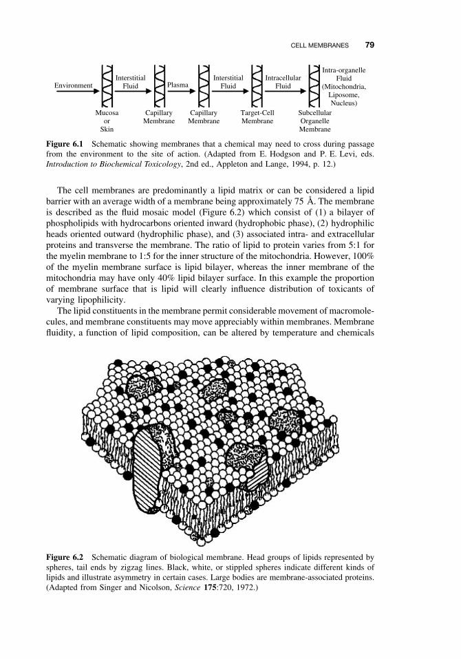
CELL MEMBRANES 79
EnvironmentInterstitial
FluidInterstitial
FluidIntracellular
FluidPlasma
Mucosaor
Skin
CapillaryMembrane
CapillaryMembrane
Target-CellMembrane
SubcellularOrganelleMembrane
Intra-organelleFluid
(Mitochondria,Liposome,Nucleus)
Figure 6.1 Schematic showing membranes that a chemical may need to cross during passagefrom the environment to the site of action. (Adapted from E. Hodgson and P. E. Levi, eds.Introduction to Biochemical Toxicology, 2nd ed., Appleton and Lange, 1994, p. 12.)
The cell membranes are predominantly a lipid matrix or can be considered a lipidbarrier with an average width of a membrane being approximately 75 A. The membraneis described as the fluid mosaic model (Figure 6.2) which consist of (1) a bilayer ofphospholipids with hydrocarbons oriented inward (hydrophobic phase), (2) hydrophilicheads oriented outward (hydrophilic phase), and (3) associated intra- and extracellularproteins and transverse the membrane. The ratio of lipid to protein varies from 5:1 forthe myelin membrane to 1:5 for the inner structure of the mitochondria. However, 100%of the myelin membrane surface is lipid bilayer, whereas the inner membrane of themitochondria may have only 40% lipid bilayer surface. In this example the proportionof membrane surface that is lipid will clearly influence distribution of toxicants ofvarying lipophilicity.
The lipid constituents in the membrane permit considerable movement of macromole-cules, and membrane constituents may move appreciably within membranes. Membranefluidity, a function of lipid composition, can be altered by temperature and chemicals
Figure 6.2 Schematic diagram of biological membrane. Head groups of lipids represented byspheres, tail ends by zigzag lines. Black, white, or stippled spheres indicate different kinds oflipids and illustrate asymmetry in certain cases. Large bodies are membrane-associated proteins.(Adapted from Singer and Nicolson, Science 175:720, 1972.)
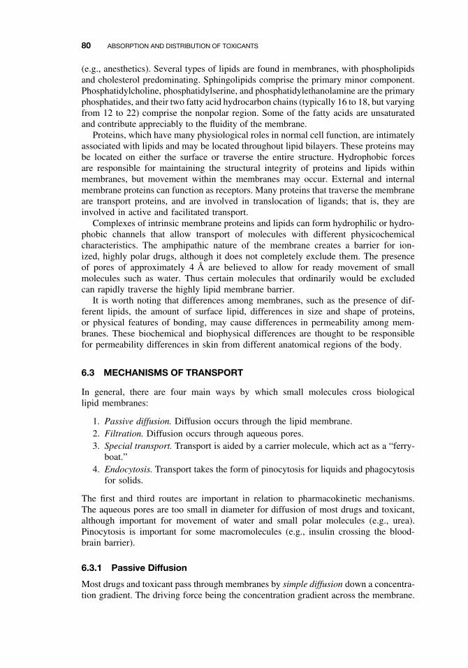
80 ABSORPTION AND DISTRIBUTION OF TOXICANTS
(e.g., anesthetics). Several types of lipids are found in membranes, with phospholipidsand cholesterol predominating. Sphingolipids comprise the primary minor component.Phosphatidylcholine, phosphatidylserine, and phosphatidylethanolamine are the primaryphosphatides, and their two fatty acid hydrocarbon chains (typically 16 to 18, but varyingfrom 12 to 22) comprise the nonpolar region. Some of the fatty acids are unsaturatedand contribute appreciably to the fluidity of the membrane.
Proteins, which have many physiological roles in normal cell function, are intimatelyassociated with lipids and may be located throughout lipid bilayers. These proteins maybe located on either the surface or traverse the entire structure. Hydrophobic forcesare responsible for maintaining the structural integrity of proteins and lipids withinmembranes, but movement within the membranes may occur. External and internalmembrane proteins can function as receptors. Many proteins that traverse the membraneare transport proteins, and are involved in translocation of ligands; that is, they areinvolved in active and facilitated transport.
Complexes of intrinsic membrane proteins and lipids can form hydrophilic or hydro-phobic channels that allow transport of molecules with different physicochemicalcharacteristics. The amphipathic nature of the membrane creates a barrier for ion-ized, highly polar drugs, although it does not completely exclude them. The presenceof pores of approximately 4 A are believed to allow for ready movement of smallmolecules such as water. Thus certain molecules that ordinarily would be excludedcan rapidly traverse the highly lipid membrane barrier.
It is worth noting that differences among membranes, such as the presence of dif-ferent lipids, the amount of surface lipid, differences in size and shape of proteins,or physical features of bonding, may cause differences in permeability among mem-branes. These biochemical and biophysical differences are thought to be responsiblefor permeability differences in skin from different anatomical regions of the body.
6.3 MECHANISMS OF TRANSPORT
In general, there are four main ways by which small molecules cross biologicallipid membranes:
1. Passive diffusion. Diffusion occurs through the lipid membrane.2. Filtration. Diffusion occurs through aqueous pores.3. Special transport. Transport is aided by a carrier molecule, which act as a “ferry-
boat.”4. Endocytosis. Transport takes the form of pinocytosis for liquids and phagocytosis
for solids.
The first and third routes are important in relation to pharmacokinetic mechanisms.The aqueous pores are too small in diameter for diffusion of most drugs and toxicant,although important for movement of water and small polar molecules (e.g., urea).Pinocytosis is important for some macromolecules (e.g., insulin crossing the blood-brain barrier).
6.3.1 Passive Diffusion
Most drugs and toxicant pass through membranes by simple diffusion down a concentra-tion gradient. The driving force being the concentration gradient across the membrane.
MECHANISMS OF TRANSPORT 81
This diffusion process can continue until equilibrium, although in reality there isalways movement but the net flux is zero. Eventually the concentration of unionizedor unbound (free) toxicant is the same on either side of the membrane. In other words,there is no competition of molecules and there is generally a lack of saturation. Sol-ubility in the lipid bilayer is important, and the greater the partition coefficient, thehigher is the concentration in the membrane, and the greater is the rate of diffusionacross the membrane. For ionized toxicants the steady state concentration is dependenton the differences in pH across the membrane. Most membranes are relatively perme-able to water either by diffusion or by flow that results from hydrostatic or osmoticdifferences across the membrane, and bulk flow of water can also carry with it smalland water soluble molecules by this mechanism. These substances generally have amolecular weight of less than 200. Although inorganic ions are small and will readilydiffuse across some membranes, their hydrated ionic radius is relatively large. In suchcases active transport is required (see below). Specific ion fluxes are also controlledby specific channels that are important in nerves, muscles, and signal transduction.
We can now quantitate the rate at which a toxicant can be transported by passivediffusion, and this can be described by Fick’s law of diffusion as follows:
Rate of diffusion = D × Sa × Pc
d(CH − CL),
where D is the diffusion coefficient, Sa is the surface area of the membrane, Pc
is the partition coefficient, d is the membrane thickness, and CH and CL are theconcentrations at both sides of the membrane (high and low, respectively). The firstpart of this equation (DPc/d) represents the permeability coefficient of the drug. Thepermeability expresses the ease of penetration of a chemical and has units of velocity,distance/time (cm/h).
The diffusion coefficient or diffusivity of the toxicant, D, is primarily dependenton solubility of the toxicant in the membrane and its molecular weight and molecularconformation. Depending on the membrane, there is a functional molecular size and/orweight cutoff that prevents very large molecules from being passively absorbed acrossany membrane. One would expect small molecular weight molecules to diffuse morerapidly than larger molecular weight toxicants. Therefore the magnitude of a toxicant’sdiffusion coefficient really reflects the ease with which it is able to diffuse through themembrane. The reader should also be aware that as a toxicant crosses from the donoror aqueous medium and through the membrane medium, there are really two diffusionenvironments and thus two diffusion coefficients to consider. Another important factorthat can influence the diffusion coefficient is membrane viscosity. This physicochemicalcharacteristic should remain constant in biological systems but can be modified in skinmembranes exposed to various pharmaceutical or pesticide formulations. Formulationadditives or excipients may enter the membrane barrier and reversibly or irreversiblychange viscosity and thus diffusion coefficient of the drug or pesticide in the barriermembranes of the skin.
The partition coefficient, which will be described in more detail later in this chapter,is the relative solubility of the compound in lipid and water, and the compound’ssolubility really reflects the ability of the toxicant to move from a relatively aqueousenvironment across a lipid membrane. It is this factor that is often manipulated inpesticide and drug formulations to create a vehicle. Membrane permeability is thereforestrongly correlated to the lipid solubility of the toxicant in the membrane as well as
82 ABSORPTION AND DISTRIBUTION OF TOXICANTS
the aqueous environment surrounding the membrane. Please be aware that there areinstances where partition coefficient or lipid solubility of the toxicant may be very large,and there may be a tendency for the drug to sequester in the membrane. Membranesurface area and membrane thickness can also vary across different organs in the body,but one does not expect these two factors in Fick’s equation to vary considerably. Thefinal component of Fick’s equation is the concentration gradient (CH − CL) acrossthe membrane, which is the driving force for diffusion, and as will be demonstratedbelow in our discussion on first-order kinetics, is the most important factor dictatingthe rate of transport across most biological membranes.
First-Order Kinetics. When the rate of a process is dependent on a rate constant anda concentration gradient, a linear or first-order kinetic process will be operative. Thereader should be aware that there are numerous deviations from the first-order processwhen chemical transport in vivo is analyzed, and this can be deemed an approximationsince, in many barriers, penetration is slow and a long period of time is required toachieve steady state.
The rate of movement of a toxicant across a membrane may be expressed as thechange in amount of toxicant, A, (dA) or toxicant concentration, C, (dC) per unit oftime (dt), which equals dA/dt. Calculus can be used to express instantaneous rates oververy small time intervals (dt). Thus rate processes may then be generally expressed as
dAdt
= KAn
where dA/dt is the rate of chemical (X) movement (e.g. absorption, distribution, elim-ination), K is the rate constant of the process, and n is the kinetic order of the transportprocess (e.g., absorption). The n either equals 1 (first order) or 0 (zero order). Thusthe first-order rate equation is written as
dAdt
= KA1 = KA,
and the zero-order rate equation as
dAdt
= KA0 = K.
We know from Fick’s law that the rate of diffusion (now expressed as dA/dt) is
dAdt
= DžSažPc(A1 − A2)
d.
Once a toxicant crosses a membrane, it is rapidly removed from the “receiving side”(compartment B in Figure 6.3) either by uptake into the blood stream or eliminationfrom the organism. Thus it is A1 that is the primary driving force, and if we replacethis with A in all equations, then
dAdt
=(
DžSažPc
d
)A.
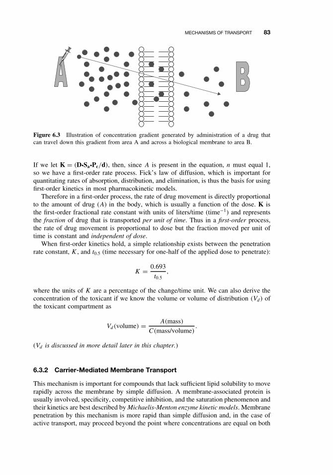
MECHANISMS OF TRANSPORT 83
Figure 6.3 Illustration of concentration gradient generated by administration of a drug thatcan travel down this gradient from area A and across a biological membrane to area B.
If we let K = (DžSažPc/d), then, since A is present in the equation, n must equal 1,so we have a first-order rate process. Fick’s law of diffusion, which is important forquantitating rates of absorption, distribution, and elimination, is thus the basis for usingfirst-order kinetics in most pharmacokinetic models.
Therefore in a first-order process, the rate of drug movement is directly proportionalto the amount of drug (A) in the body, which is usually a function of the dose. K isthe first-order fractional rate constant with units of liters/time (time−1) and representsthe fraction of drug that is transported per unit of time. Thus in a first-order process,the rate of drug movement is proportional to dose but the fraction moved per unit oftime is constant and independent of dose.
When first-order kinetics hold, a simple relationship exists between the penetrationrate constant, K , and t0.5 (time necessary for one-half of the applied dose to penetrate):
K = 0.693
t0.5,
where the units of K are a percentage of the change/time unit. We can also derive theconcentration of the toxicant if we know the volume or volume of distribution (Vd) ofthe toxicant compartment as
Vd(volume) = A(mass)
C(mass/volume).
(Vd is discussed in more detail later in this chapter.)
6.3.2 Carrier-Mediated Membrane Transport
This mechanism is important for compounds that lack sufficient lipid solubility to moverapidly across the membrane by simple diffusion. A membrane-associated protein isusually involved, specificity, competitive inhibition, and the saturation phenomenon andtheir kinetics are best described by Michaelis-Menton enzyme kinetic models. Membranepenetration by this mechanism is more rapid than simple diffusion and, in the case ofactive transport, may proceed beyond the point where concentrations are equal on both
84 ABSORPTION AND DISTRIBUTION OF TOXICANTS
sides of the membrane. Generally, there are two types of specialized carrier-mediatedtransport processes:
Passive facilitated diffusion involves movement down a concentration gradientwithout an input of energy. This mechanism, which may be highly selective for spe-cific conformational structures, is necessary for transport of endogenous compoundswhose rate of transport by simple diffusion would otherwise be too slow. The classicalexample of facilitated diffusion is transport of glucose into red blood cells.
Active transport requires energy, and transport is against a concentration. Main-tenance against this gradient requires energy. It is often coupled to energy-producingenzymes (e.g., ATPase) or to the transport of other molecules (e.g., Na+, Cl−, H+)that generate energy as they cross the membranes. Carrier-mediated drug transport canoccur in only a few sites in the body, and the main sites are
ž BBB, neuronal membranes, choroid plexusž Renal tubular cellsž Hepatocytes, biliary tract
There are instances in which toxicants have chemical or structural similarities toendogenous chemicals that rely on these special transport mechanisms for normal phys-iological uptake and can thus utilize the same system for membrane transport. Usefulexamples of drugs known to be transported by this mechanism include levodopa, whichis used in treating Parkinson’s disease, and fluorouracil, a cytotoxic drug. Levodopais taken up by the carrier that normally transports phenylalanine, and fluorouracil istransported by the system that carries the natural pyrimidines, thymine, and uracil. Ironis absorbed by a specific carrier in the mucosal cells of the jejunum, and calcium by avitamin D-dependent carrier system. Lead may be more quickly moved by a transportsystem that is normally involved in the uptake of calcium.
For carrier-mediated transport, the rate of movement across a membrane will nowbe constant, since flux is dependent on the capacity of the membrane carriers and notthe mass of the chemical to be transported. These processes are described by zero-orderkinetic rate equations of the form:
dXdt
= KX0 = K0.
K0 is now the zero-order rate constant and is expressed in terms of mass/time. Inan active carrier-mediated transport process following zero-order kinetics, the rate ofdrug transport is always equal to K once the system is fully loaded or saturated. Atsubsaturation levels, the rate is initially first order as the carriers become loaded withthe toxicant, but at concentrations normally encountered in pharmacokinetics, the ratebecomes constant. Thus, as dose increases, the rate of transport does not increase inproportion to dose as it does with the fractional rate constant seen in first-order process.This is illustrated in the Table 6.1 where it is assumed that the first-order rate constantis 0.1 (10% per minute) and the zero-order rate is 10 mg/min.
In the case of first order, these amounts will subsequently diminish (10% of 900is 90, etc.). In the case of zero order, the amount transported does not vary with time(constant rate of transport).
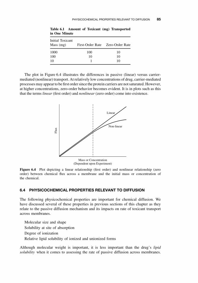
PHYSICOCHEMICAL PROPERTIES RELEVANT TO DIFFUSION 85
Table 6.1 Amount of Toxicant (mg) Transportedin One Minute
Initial ToxicantMass (mg) First-Order Rate Zero-Order Rate
1000 100 10100 10 1010 1 10
The plot in Figure 6.4 illustrates the differences in passive (linear) versus carrier-mediated (nonlinear) transport. At relatively low concentrations of drug, carrier-mediatedprocesses may appear to be first order since the protein carriers are not saturated. However,at higher concentrations, zero-order behavior becomes evident. It is in plots such as thisthat the terms linear (first order) and nonlinear (zero order) come into existence.
Linear
Non-linear
Mass or Concentration(Dependent upon Experiment)
Flux
Figure 6.4 Plot depicting a linear relationship (first order) and nonlinear relationship (zeroorder) between chemical flux across a membrane and the initial mass or concentration ofthe chemical.
6.4 PHYSICOCHEMICAL PROPERTIES RELEVANT TO DIFFUSION
The following physicochemical properties are important for chemical diffusion. Wehave discussed several of these properties in previous sections of this chapter as theyrelate to the passive diffusion mechanism and its impacts on rate of toxicant transportacross membranes.
Molecular size and shapeSolubility at site of absorptionDegree of ionizationRelative lipid solubility of ionized and unionized forms
Although molecular weight is important, it is less important than the drug’s lipidsolubility when it comes to assessing the rate of passive diffusion across membranes.
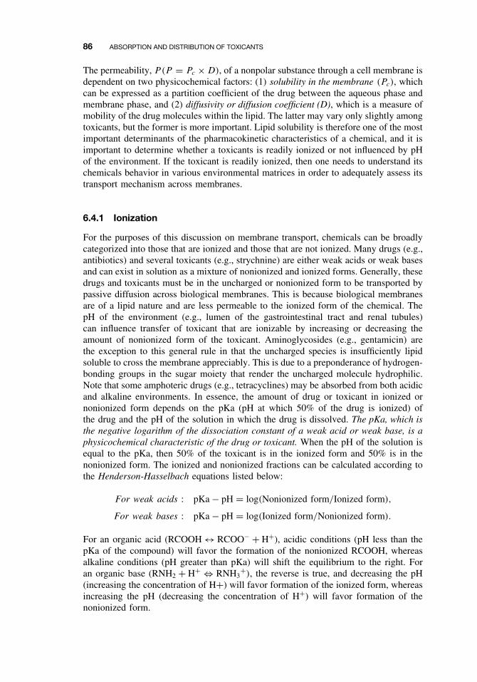
86 ABSORPTION AND DISTRIBUTION OF TOXICANTS
The permeability, P(P = Pc × D), of a nonpolar substance through a cell membrane isdependent on two physicochemical factors: (1) solubility in the membrane (Pc), whichcan be expressed as a partition coefficient of the drug between the aqueous phase andmembrane phase, and (2) diffusivity or diffusion coefficient (D), which is a measure ofmobility of the drug molecules within the lipid. The latter may vary only slightly amongtoxicants, but the former is more important. Lipid solubility is therefore one of the mostimportant determinants of the pharmacokinetic characteristics of a chemical, and it isimportant to determine whether a toxicants is readily ionized or not influenced by pHof the environment. If the toxicant is readily ionized, then one needs to understand itschemicals behavior in various environmental matrices in order to adequately assess itstransport mechanism across membranes.
6.4.1 Ionization
For the purposes of this discussion on membrane transport, chemicals can be broadlycategorized into those that are ionized and those that are not ionized. Many drugs (e.g.,antibiotics) and several toxicants (e.g., strychnine) are either weak acids or weak basesand can exist in solution as a mixture of nonionized and ionized forms. Generally, thesedrugs and toxicants must be in the uncharged or nonionized form to be transported bypassive diffusion across biological membranes. This is because biological membranesare of a lipid nature and are less permeable to the ionized form of the chemical. ThepH of the environment (e.g., lumen of the gastrointestinal tract and renal tubules)can influence transfer of toxicant that are ionizable by increasing or decreasing theamount of nonionized form of the toxicant. Aminoglycosides (e.g., gentamicin) arethe exception to this general rule in that the uncharged species is insufficiently lipidsoluble to cross the membrane appreciably. This is due to a preponderance of hydrogen-bonding groups in the sugar moiety that render the uncharged molecule hydrophilic.Note that some amphoteric drugs (e.g., tetracyclines) may be absorbed from both acidicand alkaline environments. In essence, the amount of drug or toxicant in ionized ornonionized form depends on the pKa (pH at which 50% of the drug is ionized) ofthe drug and the pH of the solution in which the drug is dissolved. The pKa, which isthe negative logarithm of the dissociation constant of a weak acid or weak base, is aphysicochemical characteristic of the drug or toxicant. When the pH of the solution isequal to the pKa, then 50% of the toxicant is in the ionized form and 50% is in thenonionized form. The ionized and nonionized fractions can be calculated according tothe Henderson-Hasselbach equations listed below:
For weak acids : pKa − pH = log(Nonionized form/Ionized form),
For weak bases : pKa − pH = log(Ionized form/Nonionized form).
For an organic acid (RCOOH ↔ RCOO− + H+), acidic conditions (pH less than thepKa of the compound) will favor the formation of the nonionized RCOOH, whereasalkaline conditions (pH greater than pKa) will shift the equilibrium to the right. Foran organic base (RNH2 + H+ ⇔ RNH3
+), the reverse is true, and decreasing the pH(increasing the concentration of H+) will favor formation of the ionized form, whereasincreasing the pH (decreasing the concentration of H+) will favor formation of thenonionized form.
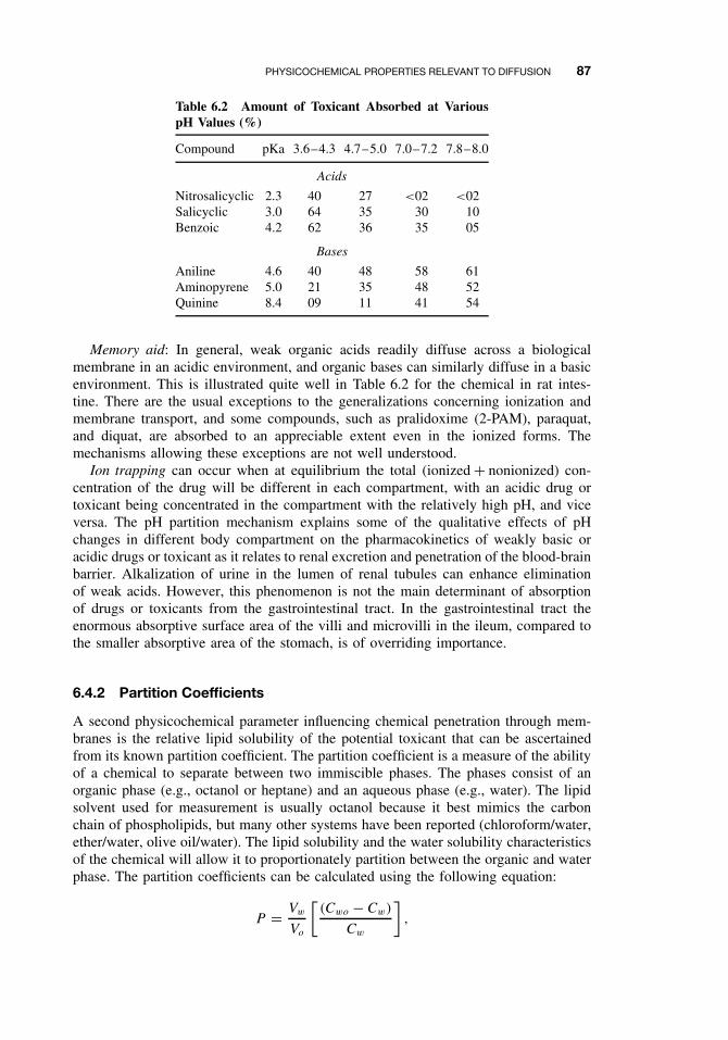
PHYSICOCHEMICAL PROPERTIES RELEVANT TO DIFFUSION 87
Table 6.2 Amount of Toxicant Absorbed at VariouspH Values (%)
Compound pKa 3.6–4.3 4.7–5.0 7.0–7.2 7.8–8.0
Acids
Nitrosalicyclic 2.3 40 27 <02 <02Salicyclic 3.0 64 35 30 10Benzoic 4.2 62 36 35 05
Bases
Aniline 4.6 40 48 58 61Aminopyrene 5.0 21 35 48 52Quinine 8.4 09 11 41 54
Memory aid: In general, weak organic acids readily diffuse across a biologicalmembrane in an acidic environment, and organic bases can similarly diffuse in a basicenvironment. This is illustrated quite well in Table 6.2 for the chemical in rat intes-tine. There are the usual exceptions to the generalizations concerning ionization andmembrane transport, and some compounds, such as pralidoxime (2-PAM), paraquat,and diquat, are absorbed to an appreciable extent even in the ionized forms. Themechanisms allowing these exceptions are not well understood.
Ion trapping can occur when at equilibrium the total (ionized + nonionized) con-centration of the drug will be different in each compartment, with an acidic drug ortoxicant being concentrated in the compartment with the relatively high pH, and viceversa. The pH partition mechanism explains some of the qualitative effects of pHchanges in different body compartment on the pharmacokinetics of weakly basic oracidic drugs or toxicant as it relates to renal excretion and penetration of the blood-brainbarrier. Alkalization of urine in the lumen of renal tubules can enhance eliminationof weak acids. However, this phenomenon is not the main determinant of absorptionof drugs or toxicants from the gastrointestinal tract. In the gastrointestinal tract theenormous absorptive surface area of the villi and microvilli in the ileum, compared tothe smaller absorptive area of the stomach, is of overriding importance.
6.4.2 Partition Coefficients
A second physicochemical parameter influencing chemical penetration through mem-branes is the relative lipid solubility of the potential toxicant that can be ascertainedfrom its known partition coefficient. The partition coefficient is a measure of the abilityof a chemical to separate between two immiscible phases. The phases consist of anorganic phase (e.g., octanol or heptane) and an aqueous phase (e.g., water). The lipidsolvent used for measurement is usually octanol because it best mimics the carbonchain of phospholipids, but many other systems have been reported (chloroform/water,ether/water, olive oil/water). The lipid solubility and the water solubility characteristicsof the chemical will allow it to proportionately partition between the organic and waterphase. The partition coefficients can be calculated using the following equation:
P = Vw
Vo
[(Cwo − Cw)
Cw
],
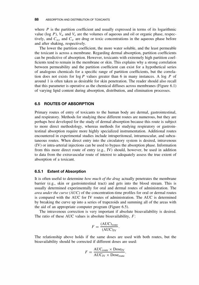
88 ABSORPTION AND DISTRIBUTION OF TOXICANTS
where P is the partition coefficient and usually expressed in terms of its logarithmicvalue (log P ), Vw and Vo are the volumes of aqueous and oil or organic phase, respec-tively, and Cwo and Cw are drug or toxic concentrations in the aqueous phase beforeand after shaking, respectively.
The lower the partition coefficient, the more water soluble, and the least permeablethe toxicant is across a membrane. Regarding dermal absorption, partition coefficientscan be predictive of absorption. However, toxicants with extremely high partition coef-ficients tend to remain in the membrane or skin. This explains why a strong correlationbetween permeability and the partition coefficient can exist for a hypothetical seriesof analogous chemicals for a specific range of partition coefficients, but the correla-tion does not exists for log P values greater than 6 in many instances. A log P ofaround 1 is often taken as desirable for skin penetration. The reader should also recallthat this parameter is operative as the chemical diffuses across membranes (Figure 6.1)of varying lipid content during absorption, distribution, and elimination processes.
6.5 ROUTES OF ABSORPTION
Primary routes of entry of toxicants to the human body are dermal, gastrointestinal,and respiratory. Methods for studying these different routes are numerous, but they areperhaps best developed for the study of dermal absorption because this route is subjectto more direct methodology, whereas methods for studying respiratory or gastroin-testinal absorption require more highly specialized instrumentation. Additional routesencountered in experimental studies include intraperitoneal, intramuscular, and subcu-taneous routes. When direct entry into the circulatory system is desired, intravenous(IV) or intra-arterial injections can be used to bypass the absorption phase. Informationfrom this more direct route of entry (e.g., IV) should, however, be used in additionto data from the extravascular route of interest to adequately assess the true extent ofabsorption of a toxicant.
6.5.1 Extent of Absorption
It is often useful to determine how much of the drug actually penetrates the membranebarrier (e.g., skin or gastrointestinal tract) and gets into the blood stream. This isusually determined experimentally for oral and dermal routes of administration. Thearea under the curve (AUC) of the concentration-time profiles for oral or dermal routesis compared with the AUC for IV routes of administration. The AUC is determinedby breaking the curve up into a series of trapezoids and summing all of the areas withthe aid of an appropriate computer program (Figure 6.5).
The intravenous correction is very important if absolute bioavailability is desired.The ratio of these AUC values is absolute bioavailability, F :
F = (AUC)route
(AUC)IV.
The relationship above holds if the same doses are used with both routes, but thebioavailability should be corrected if different doses are used:
F = AUCroute × DoseIV
AUCIV × Doseroute.
ROUTES OF ABSORPTION 89
T1
C6
C1
C2
C5
C3
C4
T2 T3 T4 T5 T6
Time
Con
cent
ratio
n
Slope
Figure 6.5 Plasma concentration time profile for oral exposure to a toxicant and depiction ofAUCs determined by summation of trapezoids at several time periods.
Another technique is to monitor drug or toxicant excretion rather than blood con-centrations, especially when blood or plasma concentrations are very low. Using thesame equations, the AUC is now replaced by chemical concentrations in urine, feces,and expired air. Some chemicals are primarily excreted by the kidney and urine dataalone may be necessary. The rate and extent of absorption are clearly important fortherapeutic and toxicological considerations. For example, different formulations of thesame pesticide can change the absorption rate in skin or gastrointestinal tract, and notbioavailability, but can result in blood concentrations near the toxic dose. Also differentformulations can result in similar absorption rates but different bioavailability.
6.5.2 Gastrointestinal Absorption
The gastrointestinal tract (GIT) is a hollow tube (Figure 6.6a) lined by a layer ofcolumnar cells, and usually protected by mucous, which offers minimal resistance totoxicant penetration. The distance from the outer membrane to the vasculature is about40 µm, from which point further transport can easily occur. However, the cornifiedepithelium of the esophagus prevents absorption from this region of the GIT. Most ofthe absorption will therefore occur in the intestine (pH = 6), and to some extent in thestomach (pH = 1–3). Buccal and rectal absorption can occur in special circumstances.Note that secretions from the lachrymal duct, salivary gland, and nasal passages canenter the GIT via the buccal cavity. Therefore, following IV administration, a toxicantcan enter the GIT if the drug is in these secretions.
The intestine can compensate the 2.5 log units difference between it and the stom-ach by the increased surface area in the small intestines. The presence of microvilli(Figure 6.6b) in the intestine is an increase of 600-fold in surface area compared to ahollow tube of comparable length. Note that there is no absorption, except for water,in the large intestine.
Most of the absorption in the GIT is by passive diffusion, except for nutrients;glucose, amino acids, and drugs that look like these substances are taken up by
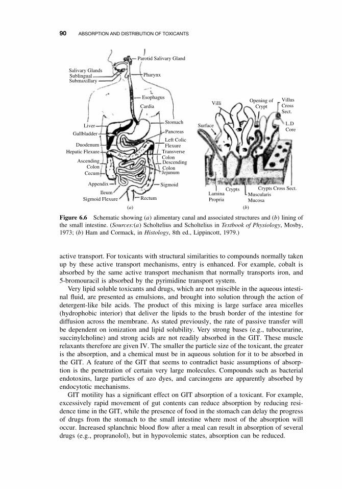
90 ABSORPTION AND DISTRIBUTION OF TOXICANTS
Parotid Salivary Gland
Pharynx
Esophagus
Cardia
Stomach
Pancreas
Left ColicFlexure
TransverseColonDescendingColonJejunum
Sigmoid
RectumSigmoid FlexureIleum
Appendix
Cecum
AscendingColon
Hepatic FlexureDuodenum
Gallbladder
Liver
SubmaxillarySublingualSalivary Glands
(a) (b)
Villi
Surface L.DCore
VillusCrossSect.
Opening ofCrypt
Crypts Crypts Cross Sect.LaminaPropria
MuscularisMucosa
Figure 6.6 Schematic showing (a) alimentary canal and associated structures and (b) lining ofthe small intestine. (Sources:(a) Scholtelius and Scholtelius in Textbook of Physiology, Mosby,1973; (b) Ham and Cormack, in Histology, 8th ed., Lippincott, 1979.)
active transport. For toxicants with structural similarities to compounds normally takenup by these active transport mechanisms, entry is enhanced. For example, cobalt isabsorbed by the same active transport mechanism that normally transports iron, and5-bromouracil is absorbed by the pyrimidine transport system.
Very lipid soluble toxicants and drugs, which are not miscible in the aqueous intesti-nal fluid, are presented as emulsions, and brought into solution through the action ofdetergent-like bile acids. The product of this mixing is large surface area micelles(hydrophobic interior) that deliver the lipids to the brush border of the intestine fordiffusion across the membrane. As stated previously, the rate of passive transfer willbe dependent on ionization and lipid solubility. Very strong bases (e.g., tubocurarine,succinylcholine) and strong acids are not readily absorbed in the GIT. These musclerelaxants therefore are given IV. The smaller the particle size of the toxicant, the greateris the absorption, and a chemical must be in aqueous solution for it to be absorbed inthe GIT. A feature of the GIT that seems to contradict basic assumptions of absorp-tion is the penetration of certain very large molecules. Compounds such as bacterialendotoxins, large particles of azo dyes, and carcinogens are apparently absorbed byendocytotic mechanisms.
GIT motility has a significant effect on GIT absorption of a toxicant. For example,excessively rapid movement of gut contents can reduce absorption by reducing resi-dence time in the GIT, while the presence of food in the stomach can delay the progressof drugs from the stomach to the small intestine where most of the absorption willoccur. Increased splanchnic blood flow after a meal can result in absorption of severaldrugs (e.g., propranolol), but in hypovolemic states, absorption can be reduced.
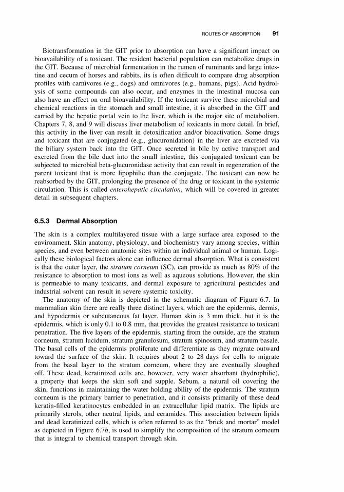
ROUTES OF ABSORPTION 91
Biotransformation in the GIT prior to absorption can have a significant impact onbioavailability of a toxicant. The resident bacterial population can metabolize drugs inthe GIT. Because of microbial fermentation in the rumen of ruminants and large intes-tine and cecum of horses and rabbits, its is often difficult to compare drug absorptionprofiles with carnivores (e.g., dogs) and omnivores (e.g., humans, pigs). Acid hydrol-ysis of some compounds can also occur, and enzymes in the intestinal mucosa canalso have an effect on oral bioavailability. If the toxicant survive these microbial andchemical reactions in the stomach and small intestine, it is absorbed in the GIT andcarried by the hepatic portal vein to the liver, which is the major site of metabolism.Chapters 7, 8, and 9 will discuss liver metabolism of toxicants in more detail. In brief,this activity in the liver can result in detoxification and/or bioactivation. Some drugsand toxicant that are conjugated (e.g., glucuronidation) in the liver are excreted viathe biliary system back into the GIT. Once secreted in bile by active transport andexcreted from the bile duct into the small intestine, this conjugated toxicant can besubjected to microbial beta-glucuronidase activity that can result in regeneration of theparent toxicant that is more lipophilic than the conjugate. The toxicant can now bereabsorbed by the GIT, prolonging the presence of the drug or toxicant in the systemiccirculation. This is called enterohepatic circulation, which will be covered in greaterdetail in subsequent chapters.
6.5.3 Dermal Absorption
The skin is a complex multilayered tissue with a large surface area exposed to theenvironment. Skin anatomy, physiology, and biochemistry vary among species, withinspecies, and even between anatomic sites within an individual animal or human. Logi-cally these biological factors alone can influence dermal absorption. What is consistentis that the outer layer, the stratum corneum (SC), can provide as much as 80% of theresistance to absorption to most ions as well as aqueous solutions. However, the skinis permeable to many toxicants, and dermal exposure to agricultural pesticides andindustrial solvent can result in severe systemic toxicity.
The anatomy of the skin is depicted in the schematic diagram of Figure 6.7. Inmammalian skin there are really three distinct layers, which are the epidermis, dermis,and hypodermis or subcutaneous fat layer. Human skin is 3 mm thick, but it is theepidermis, which is only 0.1 to 0.8 mm, that provides the greatest resistance to toxicantpenetration. The five layers of the epidermis, starting from the outside, are the stratumcorneum, stratum lucidum, stratum granulosum, stratum spinosum, and stratum basale.The basal cells of the epidermis proliferate and differentiate as they migrate outwardtoward the surface of the skin. It requires about 2 to 28 days for cells to migratefrom the basal layer to the stratum corneum, where they are eventually sloughedoff. These dead, keratinized cells are, however, very water absorbant (hydrophilic),a property that keeps the skin soft and supple. Sebum, a natural oil covering theskin, functions in maintaining the water-holding ability of the epidermis. The stratumcorneum is the primary barrier to penetration, and it consists primarily of these deadkeratin-filled keratinocytes embedded in an extracellular lipid matrix. The lipids areprimarily sterols, other neutral lipids, and ceramides. This association between lipidsand dead keratinized cells, which is often referred to as the “brick and mortar” modelas depicted in Figure 6.7b, is used to simplify the composition of the stratum corneumthat is integral to chemical transport through skin.
92 ABSORPTION AND DISTRIBUTION OF TOXICANTS
(b)(a)
AB
C D
Protein RichCorneocytes
IntercellularLipids
Figure 6.7 (a) Schematic diagram of the microstructure of mammalian skin and potentialpathways for absorption by (A) intercellular, (B) transcellular, (C) transfollicular, or (D) sweatpore routes. (b) Brick-and-mortar” model of the stratum corneum depicting intercellular pathway(i.e., route A) between keratinocytes through the lipid domain of the stratum corneum.
A number of appendages are associated with the skin, including hair follicles, seba-ceous glands, eccrine and apocrine sweat glands, and nails. Recently it was foundthat removal of the stratum corneum does not allow complete absorption; thus it isapparent that some role, although of lesser importance, is played by other parts of theskin. The dermis and subcutaneous areas of the skin are less important in influenc-ing penetration, and once a toxicant has penetrated the epidermis, the other layers aretraversed rather easily. The dermis is highly vascular, a characteristic that providesmaximal opportunity for further transport once molecules have gained entry throughthe epidermis or through skin appendages. Most of the systemic absorption occurs atthe capillary loops located at the epidermis-dermis junction. The blood supply of thedermis is under neural and humoral influences whose temperature-regulating functionscould thus affect penetration and distribution of toxicants. Vasoactive drugs or envi-ronmental temperature can also influence absorption by altering blood flow to thesecapillaries. The subcutaneous layer of the skin is highly lipid in nature and serves as ashock absorber, an insulator, and a reserve depot of energy. The pH of the skin variesbetween 4 and 7 and is markedly affected by hydration.
Cutaneous biotransformation is mostly associated with the stratum basale layerwhere there can be phase I and phase II metabolism. However, the skin is not veryefficient, compared to the liver. The epidermal layer accounts for the major portion ofbiochemical transformations in skin, although the total skin activity is low (2–6% thatof the liver). Where activity is based on epidermis alone, that layer is as active as theliver or, in the case of certain toxicants, several times more active. For some chemicals,metabolism can influence absorption, and transdermal delivery systems of drugs utilizethis activity. For example prodrug such as lipid esters are applied topically, and cuta-neous esterases liberate the free drug. These basal cells and extracellular esterases havebeen shown to be involved in detoxification of several pesticides and bioactivation ofcarcinogens such as benzo(a)pyrene. For rapidly penetrating substances, metabolismby the skin is not presently considered to be of major significance, but skin may havean important first-pass metabolic function, especially for compounds that are absorbedslowly.
ROUTES OF ABSORPTION 93
The intercellular pathway is now accepted as the major pathway for absorption.Recall that the rate of penetration is often correlated with the partition coefficient. Infact this is a very tortuous pathway, and the h (skin thickness) in Fick’s first lawof diffusion is really 10× the measured distance. By placing a solvent (e.g., ether,acetone) on the surface or tape stripping the surface, the stratum corneum (SC) isremoved, and absorption can be significantly increased by removing this outer barrier.This may not be the case for very lipophilic chemical. This is because the viableepidermis and dermis are regarded as aqueous layers compared to the SC. Note thatthe more lipophilic the drug, the more likely it will form a depot in the SC and beslowly absorbed over time and thus have a prolonged half-life.
The transcellular pathway has been discredited as a major pathway, although somepolar substances can penetrate the outer surface of the protein filaments of hydratedstratum corneum. The transfollicular pathway is really an invagination of the epidermisinto the dermis, and the chemical still has to penetrate the epidermis to be absorbedinto the blood stream. This is also a regarded as minor route. Sweat pores are notlined with the stratum corneum layer, but the holes are small, and this route is stillconsidered a minor route for chemical absorption. In general, the epidermal surface is100 to 1000 times the surface area of skin appendages, and it is likely that only verysmall and/or polar molecules penetrate the skin via these appendages.
Variations in areas of the body cause appreciable differences in penetration of tox-icants. The rate of penetration is in the following order:
Scrotal > Forehead > Axilla >= Scalp > Back = Abdomen > Palm and plantar.
The palmar and plantar regions are highly cornified and are 100 to 400 times thickerthan other regions of the body. Note that there are differences in blood flow and to alesser extent, hair density, that may influence absorption of more polar toxicants.
Formulation additives used in topical drug or pesticide formulations can alter thestratum corneum barrier. Surfactants are least likely to be absorbed, but they canalter the lipid pathway by fluidization and delipidization of lipids, and proteins withinthe keratinocytes can become denatured. This is mostly likely associated with for-mulations containing anionic surfactants than non-ionic surfactants. Similar effectscan be observed with solvents. Solvents can partition into the intercellular lipids,thereby changing membrane lipophilicity and barrier properties in the following order:ether/acetone > DMSO > ethanol > water. Higher alcohols and oils do not damagethe skin, but they can act as a depot for lipophilic drugs on the skin surface. Thepresence of water in several of these formulations can hydrate the skin. Skin occlu-sion with fabric or transdermal patches, creams, and ointments can increase epidermalhydration, which can increase permeability.
The reader should be aware of the animal model being used to estimate dermalabsorption of toxicants in humans. For many toxicants, direct extrapolation from arodent species to human is not feasible. This is because of differences in skin thickness,hair density, lipid composition, and blood flow. Human skin is the least permeablecompared to skin from rats, mice, and rabbits. Pig skin is, however, more analogousto human skin anatomically and physiologically, and pig skin is usually predictive ofdermal absorption of most drugs and pesticides in human skin. Human skin is thebest model, followed by skin from pigs, primates, and hairless guinea pigs, and thenrats, mice, and rabbits. In preliminary testing of a transdermal drug, if the drug does
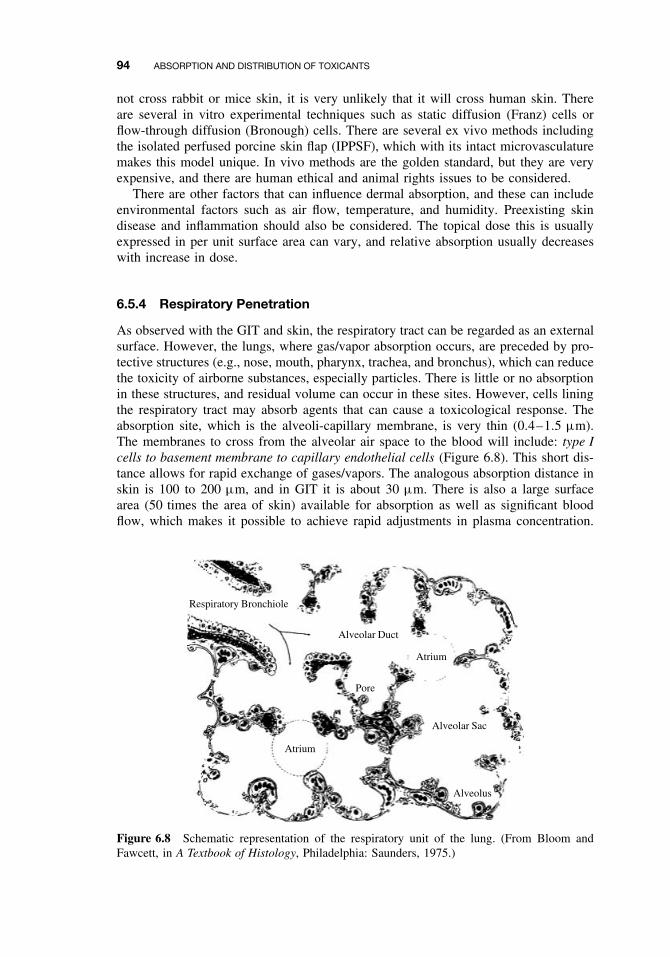
94 ABSORPTION AND DISTRIBUTION OF TOXICANTS
not cross rabbit or mice skin, it is very unlikely that it will cross human skin. Thereare several in vitro experimental techniques such as static diffusion (Franz) cells orflow-through diffusion (Bronough) cells. There are several ex vivo methods includingthe isolated perfused porcine skin flap (IPPSF), which with its intact microvasculaturemakes this model unique. In vivo methods are the golden standard, but they are veryexpensive, and there are human ethical and animal rights issues to be considered.
There are other factors that can influence dermal absorption, and these can includeenvironmental factors such as air flow, temperature, and humidity. Preexisting skindisease and inflammation should also be considered. The topical dose this is usuallyexpressed in per unit surface area can vary, and relative absorption usually decreaseswith increase in dose.
6.5.4 Respiratory Penetration
As observed with the GIT and skin, the respiratory tract can be regarded as an externalsurface. However, the lungs, where gas/vapor absorption occurs, are preceded by pro-tective structures (e.g., nose, mouth, pharynx, trachea, and bronchus), which can reducethe toxicity of airborne substances, especially particles. There is little or no absorptionin these structures, and residual volume can occur in these sites. However, cells liningthe respiratory tract may absorb agents that can cause a toxicological response. Theabsorption site, which is the alveoli-capillary membrane, is very thin (0.4–1.5 µm).The membranes to cross from the alveolar air space to the blood will include: type Icells to basement membrane to capillary endothelial cells (Figure 6.8). This short dis-tance allows for rapid exchange of gases/vapors. The analogous absorption distance inskin is 100 to 200 µm, and in GIT it is about 30 µm. There is also a large surfacearea (50 times the area of skin) available for absorption as well as significant bloodflow, which makes it possible to achieve rapid adjustments in plasma concentration.
Respiratory Bronchiole
Alveolar Duct
Alveolar Sac
Alveolus
Atrium
Atrium
Pore
Figure 6.8 Schematic representation of the respiratory unit of the lung. (From Bloom andFawcett, in A Textbook of Histology, Philadelphia: Saunders, 1975.)
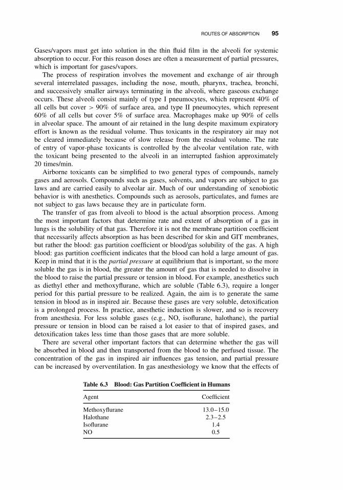
ROUTES OF ABSORPTION 95
Gases/vapors must get into solution in the thin fluid film in the alveoli for systemicabsorption to occur. For this reason doses are often a measurement of partial pressures,which is important for gases/vapors.
The process of respiration involves the movement and exchange of air throughseveral interrelated passages, including the nose, mouth, pharynx, trachea, bronchi,and successively smaller airways terminating in the alveoli, where gaseous exchangeoccurs. These alveoli consist mainly of type I pneumocytes, which represent 40% ofall cells but cover > 90% of surface area, and type II pneumocytes, which represent60% of all cells but cover 5% of surface area. Macrophages make up 90% of cellsin alveolar space. The amount of air retained in the lung despite maximum expiratoryeffort is known as the residual volume. Thus toxicants in the respiratory air may notbe cleared immediately because of slow release from the residual volume. The rateof entry of vapor-phase toxicants is controlled by the alveolar ventilation rate, withthe toxicant being presented to the alveoli in an interrupted fashion approximately20 times/min.
Airborne toxicants can be simplified to two general types of compounds, namelygases and aerosols. Compounds such as gases, solvents, and vapors are subject to gaslaws and are carried easily to alveolar air. Much of our understanding of xenobioticbehavior is with anesthetics. Compounds such as aerosols, particulates, and fumes arenot subject to gas laws because they are in particulate form.
The transfer of gas from alveoli to blood is the actual absorption process. Amongthe most important factors that determine rate and extent of absorption of a gas inlungs is the solubility of that gas. Therefore it is not the membrane partition coefficientthat necessarily affects absorption as has been described for skin and GIT membranes,but rather the blood: gas partition coefficient or blood/gas solubility of the gas. A highblood: gas partition coefficient indicates that the blood can hold a large amount of gas.Keep in mind that it is the partial pressure at equilibrium that is important, so the moresoluble the gas is in blood, the greater the amount of gas that is needed to dissolve inthe blood to raise the partial pressure or tension in blood. For example, anesthetics suchas diethyl ether and methoxyflurane, which are soluble (Table 6.3), require a longerperiod for this partial pressure to be realized. Again, the aim is to generate the sametension in blood as in inspired air. Because these gases are very soluble, detoxificationis a prolonged process. In practice, anesthetic induction is slower, and so is recoveryfrom anesthesia. For less soluble gases (e.g., NO, isoflurane, halothane), the partialpressure or tension in blood can be raised a lot easier to that of inspired gases, anddetoxification takes less time than those gases that are more soluble.
There are several other important factors that can determine whether the gas willbe absorbed in blood and then transported from the blood to the perfused tissue. Theconcentration of the gas in inspired air influences gas tension, and partial pressurecan be increased by overventilation. In gas anesthesiology we know that the effects of
Table 6.3 Blood: Gas Partition Coefficient in Humans
Agent Coefficient
Methoxyflurane 13.0–15.0Halothane 2.3–2.5Isoflurane 1.4NO 0.5
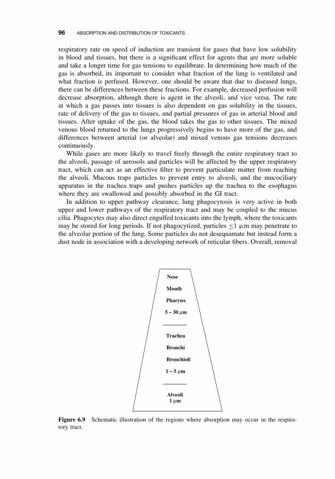
96 ABSORPTION AND DISTRIBUTION OF TOXICANTS
respiratory rate on speed of induction are transient for gases that have low solubilityin blood and tissues, but there is a significant effect for agents that are more solubleand take a longer time for gas tensions to equilibrate. In determining how much of thegas is absorbed, its important to consider what fraction of the lung is ventilated andwhat fraction is perfused. However, one should be aware that due to diseased lungs,there can be differences between these fractions. For example, decreased perfusion willdecrease absorption, although there is agent in the alveoli, and vice versa. The rateat which a gas passes into tissues is also dependent on gas solubility in the tissues,rate of delivery of the gas to tissues, and partial pressures of gas in arterial blood andtissues. After uptake of the gas, the blood takes the gas to other tissues. The mixedvenous blood returned to the lungs progressively begins to have more of the gas, anddifferences between arterial (or alveolar) and mixed venous gas tensions decreasescontinuously.
While gases are more likely to travel freely through the entire respiratory tract tothe alveoli, passage of aerosols and particles will be affected by the upper respiratorytract, which can act as an effective filter to prevent particulate matter from reachingthe alveoli. Mucous traps particles to prevent entry to alveoli, and the mucociliaryapparatus in the trachea traps and pushes particles up the trachea to the esophaguswhere they are swallowed and possibly absorbed in the GI tract.
In addition to upper pathway clearance, lung phagocytosis is very active in bothupper and lower pathways of the respiratory tract and may be coupled to the mucuscilia. Phagocytes may also direct engulfed toxicants into the lymph, where the toxicantsmay be stored for long periods. If not phagocytized, particles ≤1 µm may penetrate tothe alveolar portion of the lung. Some particles do not desequamate but instead form adust node in association with a developing network of reticular fibers. Overall, removal
Nose
Mouth
Pharynx
5 – 30 µm
Trachea
Bronchi
Bronchioli
1 – 5 µm
Alveoli1 µm
Figure 6.9 Schematic illustration of the regions where absorption may occur in the respira-tory tract.
TOXICANT DISTRIBUTION 97
of alveolar particles is markedly slower than that achieved by the directed upper pul-monary mechanisms. This defense mechanism is not important for vapors/gases. Theefficiency of the system is illustrated by the fact that on average, only 100 g of coal dustis found postmortem in the lungs of coal miners, although they inhale approximately6000 g during their lifetime.
The deposition site of particles in the respiratory tract is primarily dependent on theaerodynamic behavior of the particles. The particle size, density, shape, hygroscopicity,breathing pattern, and lung airway structure are also important factors influencing thedeposition site and efficiency. The aerodynamic-equivalent diameter (for particle >
0.5 µm) and diffusion-equivalent diameter (< 0.5 µm) are defined as the diameter ofa unit density sphere having the same settling velocity (aerodynamic-equivalent) or thesame diffusion rate (diffusion-equivalent) as the irregularly shaped particle of interest.Deposition occurs by five possible mechanisms: electrostatic precipitation, interception,impaction, sedimentation, impaction, and diffusion. The latter three are most important.Only particle sizes less than 10 to 20 µm that get pass the nasopharyngeal regions andreach the alveoli are of medical concern. As particle size decreases below 0.5 µm, theaerosol begins to behave like a gas (Figure 6.9). For these particles, diffusion becomesthe primary mechanism of deposition in the respiratory tract before it finally reachesthe alveoli.
6.6 TOXICANT DISTRIBUTION
6.6.1 Physicochemical Properties and Protein Binding
Absorption of toxicants into the blood needs to be high enough so that it will havea significant effect at the site of action in other areas of the body. The distributionprocess that takes the absorbed drug to other tissues is dependent on various physio-logical factors and physicochemical properties of the drug. This process is therefore areversible movement of the toxicant between blood and tissues or between extracellularand intracellular compartments. There are, however, several complicating factors thatcan influence the distribution of a toxicant. For example, perfusion of tissues is animportant physiological process, as some organs are better perfused (e.g., heart, brain)than others (e.g., fat). There can also be significant protein binding that affects deliv-ery of drug to tissues. To further complicate the issue, elimination processes such asexcretion and biotransformation (discussed at a later time) is occurring simultaneouslyto remove the toxicant from the blood as well as the target site.
There are several physiochemical properties of the toxicant that can influence itsdistribution. These include lipid solubility, pKa, and molecular weight, all of whichwere described earlier in this chapter (Section 6.4) and will not be described here. Formany toxicants, distribution from the blood to tissues is by simple diffusion down aconcentration gradient, and the absorption principles described earlier also apply here.The concentration gradient will be influenced by the partition coefficient or ratherthe ratio of toxicant concentrations in blood and tissue. Tissue mass and blood flowwill also have a significant effect on distribution. For example, a large muscle masscan result in increased distribution to muscle, while limited blood flow to fat or bonetissue can limit distribution. The ratio of blood flow to tissue mass is also a usefulindicator of how well the tissue is perfused. The well perfused tissues include liver,
98 ABSORPTION AND DISTRIBUTION OF TOXICANTS
kidney, and brain, and the low perfused tissues include fat and bone where there isslow elimination from these tissues. Initial distribution to well-perfused tissues (e.g.,heart, brain) occurs within the first few minutes, while delivery of drug to other tissues(e.g., fat, skin) is slower.
If the affinity for the target tissue is high, then the chemical will accumulate orform a depot. The advantage here is that if this is a drug, there is no need to loadup the central compartment to get to the active site. However, if the reservoir for thedrug has a large capacity and fills rapidly, it so alters the distribution of the drug thatlarger quantities of the drug are required initially to provide a therapeutic effectiveconcentration at the target organ. If this is a toxicant, this may be an advantageousfeature as toxicant levels at the target site will be reduced. In general, lipid-insolubletoxicants stay mainly in the plasma and interstitial fluids, while lipid-soluble toxicantsreach all compartments, and may accumulate in fat. There are numerous examples ofcellular reservoirs for toxicants and drugs to distribute. Tetracycline antibiotics have ahigh affinity for calcium-rich tissues in the body. The bone can become a reservoir forthe slow release of chemicals such as lead, and effects may be chronic or there may beacute toxicity if the toxicant is suddenly released or mobilized from these depots. Theantimalaria drug quinacrine accumulates due to reversible intracellular binding, andthe concentration in the liver can be several thousand times that of plasma. Anotherantimalaria drug, chloroquine, has a high affinity for melanin, and this drug can betaken up by tissues such as the retina, which is rich in melanin granules, and can causeretinitis with a drug overdose. Lipophilic pesticides and toxicants (e.g., PCBs) andlipid soluble gases can be expected to accumulate in high concentration in fat tissue.
There are unique anatomical barriers that can limit distribution of toxicants. Aclassical example of such a unique barrier is the blood-brain barrier (BBB), whichcan limit the distribution of toxicants into the CNS and cerebrospinal fluid. There arethree main processes or structures that keep drug or toxicant concentrations low inthis region: (1) The BBB, which consist of capillary endothelial tight junctions andglial cells, surrounds the precapillaries, reduces filtration, and requires that the toxicantcross several membranes in order to get to the CSF. (Note that endothelial cells inother organs can have intercellular pores and pinocytotic vesicles.) (2) Active transportsystems in the choroid plexus allow for transport of organic acids and bases from theCSF into blood. (3) The continuous process of CSF production in the ventricles andvenous drainage continuously dilutes toxicant or drug concentrations. Disease processessuch as meningitis can disrupt this barrier and can allow for penetration of antibiotics(e.g., aminoglycosides) that would not otherwise readily cross this barrier in a healthyindividual. Other tissue/blood barriers include prostate/blood, testicles/blood, and globeof eye/blood, but inflammation or infection can increase permeability of these barriers.Toxicants can cross the placenta primarily by simple diffusion, and this is most easilyaccomplished if the toxicants are lipid-soluble (i.e., nonionized weak acids or bases).The view that the placenta is a barrier to drugs and toxicants is inaccurate. The fetusis, at least to some extent, exposed to essentially all drugs even if those with low lipidsolubility are taken by the mother.
As was indicated earlier, the circulatory system and components in the blood streamare primarily responsible for the transport of toxicants to target tissues or reservoirs.Erythrocytes and lymph can play important roles in the transport of toxicants, butcompared to plasma proteins, their role in toxicant distribution is relatively minor formost toxicants. Plasma protein binding can affect distribution because only the unbound
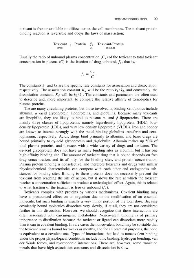
TOXICANT DISTRIBUTION 99
toxicant is free or available to diffuse across the cell membranes. The toxicant-proteinbinding reaction is reversible and obeys the laws of mass action:
Toxicant(free)
+ Protein k1↔k2
Toxicant-Protein(bound)
Usually the ratio of unbound plasma concentration (Cu) of the toxicant to total toxicantconcentration in plasma (C) is the fraction of drug unbound, fu , that is,
fu = Cu
C.
The constants k1 and k2 are the specific rate constants for association and dissociation,respectively. The association constant Ka will be the ratio k1/k2, and conversely, thedissociation constant, Kd will be k2/k1. The constants and parameters are often usedto describe and, more important, to compare the relative affinity of xenobiotics forplasma proteins.
The are many circulating proteins, but those involved in binding xenobiotics includealbumin, α1-acid glycoprotein, lipoproteins, and globulins. Because many toxicantsare lipophilic, they are likely to bind to plasma α- and β-lipoproteins. There aremainly three classes of lipoproteins, namely high-density lipoprotein (HDL), low-density lipoprotein (LDL), and very low density lipoprotein (VLDL). Iron and copperare known to interact strongly with the metal-binding globulins transferin and ceru-loplasmin, respectively. Acidic drugs bind primarily to albumin, and basic drugs arebound primarily to α1-acid glycoprotein and β-globulin. Albumin makes up 50% oftotal plasma proteins, and it reacts with a wide variety of drugs and toxicants. Theα1-acid glycoprotein does not have as many binding sites as albumin, but it has onehigh-affinity binding site. The amount of toxicant drug that is bound depends on freedrug concentration, and its affinity for the binding sites, and protein concentration.Plasma protein binding is nonselective, and therefore toxicants and drugs with similarphysicochemical characteristics can compete with each other and endogenous sub-stances for binding sites. Binding to these proteins does not necessarily prevent thetoxicant from reaching the site of action, but it slows the rate at which the toxicantreaches a concentration sufficient to produce a toxicological effect. Again, this is relatedto what fraction of the toxicant is free or unbound (fu).
Toxicants complex with proteins by various mechanisms. Covalent binding mayhave a pronounced effect on an organism due to the modification of an essentialmolecule, but such binding is usually a very minor portion of the total dose. Becausecovalently bound molecules dissociate very slowly, if at all, they are not consideredfurther in this discussion. However, we should recognize that these interactions areoften associated with carcinogenic metabolites. Noncovalent binding is of primaryimportance to distribution because the toxicant or ligand can dissociate more readilythan it can in covalent binding. In rare cases the noncovalent bond may be so stable thatthe toxicant remains bound for weeks or months, and for all practical purposes, the bondis equivalent to a covalent one. Types of interactions that lead to noncovalent bindingunder the proper physiological conditions include ionic binding, hydrogen bonding, vander Waals forces, and hydrophobic interactions. There are, however, some transitionmetals that have high association constants and dissociation is slow.
100 ABSORPTION AND DISTRIBUTION OF TOXICANTS
We know more about ligand-protein interactions today because of the numerousprotein binding studies performed with drugs. The major difference between drugsand most toxicants is the frequent ionizability and high water solubility of drugs ascompared with the non-ionizability and high lipid solubility of many toxicants. Thusexperience with drugs forms an important background, but one that may not alwaysbe relevant to other potentially toxic compounds.
Variation in chemical and physical features can affect binding to plasma constituents.Table 6.4 shows the results of binding studies with a group of insecticides with greatlydiffering water and lipid solubilities. The affinity for albumin and lipoproteins isinversely related to water solubility, although the relation may be imperfect. Chlo-rinated hydrocarbons bind strongly to albumin but even more strongly to lipoproteins.Strongly lipophilic organophosphates bind to both protein groups, whereas more water-soluble compounds bind primarily to albumin. The most water-soluble compoundsappear to be transported primarily in the aqueous phase. Chlordecone (Kepone) haspartitioning characteristics that cause it to bind in the liver, whereas DDE, the metabo-lite of DDT, partitions into fatty depots. Thus the toxicological implications for thesetwo compounds may be quite different.
Although highly specific (high-affinity, low-capacity) binding is more common withdrugs, examples of specific binding for toxicants seem less common. It seems probablethat low-affinity, high-capacity binding describes most cases of toxicant binding. Thenumber of binding sites can only be estimated, often with considerable error, becauseof the nonspecific nature of the interaction. The number of ligand or toxicant moleculesbound per protein molecule, and the maximum number of binding sites, n, define thedefinitive capacity of the protein. Another consideration is the binding affinity Kbinding
(or 1/Kdiss). If the protein has only one binding site for the toxicant, a single value,Kbinding, describes the strength of the interaction. Usually more than one binding site ispresent, each site having its intrinsic binding constant, k1, k2, . . . , kn. Rarely does onefind a case where k1 = k2 = . . . = kn, where a single value would describe the affinity
Table 6.4 Relative Distribution of Insecticides intoAlbumin and Lipoproteins
Percent Distribution ofBound Insecticide
Insecticide Percent Bound Albumin LOL HDL
DDT 99.9 35 35 30Deildrin 99.9 12 50 38Lindane 98.0 37 38 25Parathion 98.7 67 21 12Diazinon 96.6 55 31 14Carbaryl 97.4 99 <1 <1Carbofuran 73.6 97 1 2Aldicarb 30.0 94 2 4Nicotine 25.0 94 2 4
Source: Adapted from B. P. Maliwal and F. E. Guthrie, ChemBiol Interact 35:177–188, 1981.Note: LOL, low-density lipoprotein; HOL, high-density lipo-protein.
TOXICANT DISTRIBUTION 101
constant at all sites. This is especially true when hydrophobic binding and van derWaals forces contribute to nonspecific, low-affinity binding. Obviously the chemicalnature of the binding site is of critical importance in determining binding. The three-dimensional molecular structure of the binding site, the environment of the protein, thegeneral location in the overall protein molecule, and allosteric effects are all factorsthat influence binding. Studies with toxicants, and even more extensive studies withdrugs, have provided an adequate elucidation of these factors. Binding appears to betoo complex a phenomenon to be accurately described by any one set of equations.
There are many methods for analyzing binding, but equilibrium dialysis is the mostextensively used. Again, the focus of these studies is to determine the percentage oftoxicant bound, the number of binding sites (n), and the affinity constant (Ka). Theexamples presented here are greatly simplified to avoid the undue confusion engenderedby a very complex subject.
Toxicant-protein complexes that utilize relatively weak bonds (energies of the orderof hydrogen bonds or less) readily associate and dissociate at physiological tempera-tures, and the law of mass action applies to the thermodynamic equilibrium:
Kbinding = [T P ]
[T ][P ]= 1
Kdiss,
where Kbinding is the equilibrium constant for association, [TP] is the molar concen-tration of toxicant-protein complex, [T ] is the molar concentration of free toxicant,and [P ] is the molar concentration of free protein. This equation does not describe thebinding site(s) or the binding affinity. To incorporate these parameters and estimate theextent of binding, double-reciprocal plots of 1/[TP] versus 1/[T ] may be used to testthe specificity of binding. The 1/[TP] term can also be interpreted as moles of albuminper moles of toxicant. The slope of the straight line equals 1/nKa and the intercept ofthis line with the x-axis equals −Ka . Regression lines passing through the origin implyinfinite binding, and the validity of calculating an affinity constant under these circum-stances is questionable. Figure 6.10 illustrates one such case with four pesticides, andthe insert illustrates the low-affinity, “unsaturable” nature of binding in this example.
The two classes of toxicant-protein interactions encountered may be defined as(1) specific, high affinity, low capacity, and (2) nonspecific, low affinity, high capacity.The term high affinity implies an affinity constant (Kbinding) of the order of 108 M−1,whereas low affinity implies concentrations of 104 M−1. Nonspecific, low-affinity bind-ing is probably most characteristic of nonpolar compounds, although most cases arenot as extreme as that shown in Figure 6.10.
An alternative and well-accepted treatment for binding studies is the Scatchardequation especially in situations of high-affinity binding:
ν = nk[T ]
1 + k[T ],
which is simplified for graphic estimates to
ν
[T ]= k(n − ν),
where ν is the moles of ligand (toxicant) bound per mole of protein, [T ] is the con-centration of free toxicant, k is the intrinsic affinity constant, and n is the number ofsites exhibiting such affinity. When ν[T ] is plotted against ν, a straight line is obtained
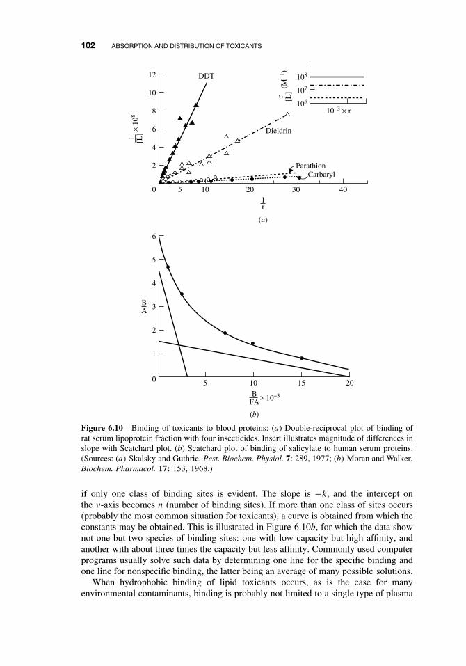
102 ABSORPTION AND DISTRIBUTION OF TOXICANTS
12
10
8
6
4
2
0
DDT
Dieldrin
ParathionCarbaryl
403020105
10−3 × r
108
107
106
1r
BA
6
5
4
3
2
1
0 5 201510
1 [L]
× 10
8
r [L]
(M−1
)
10−3BFA
×
(a)
(b)
Figure 6.10 Binding of toxicants to blood proteins: (a) Double-reciprocal plot of binding ofrat serum lipoprotein fraction with four insecticides. Insert illustrates magnitude of differences inslope with Scatchard plot. (b) Scatchard plot of binding of salicylate to human serum proteins.(Sources: (a) Skalsky and Guthrie, Pest. Biochem. Physiol. 7: 289, 1977; (b) Moran and Walker,Biochem. Pharmacol. 17: 153, 1968.)
if only one class of binding sites is evident. The slope is −k, and the intercept onthe ν-axis becomes n (number of binding sites). If more than one class of sites occurs(probably the most common situation for toxicants), a curve is obtained from which theconstants may be obtained. This is illustrated in Figure 6.10b, for which the data shownot one but two species of binding sites: one with low capacity but high affinity, andanother with about three times the capacity but less affinity. Commonly used computerprograms usually solve such data by determining one line for the specific binding andone line for nonspecific binding, the latter being an average of many possible solutions.
When hydrophobic binding of lipid toxicants occurs, as is the case for manyenvironmental contaminants, binding is probably not limited to a single type of plasma
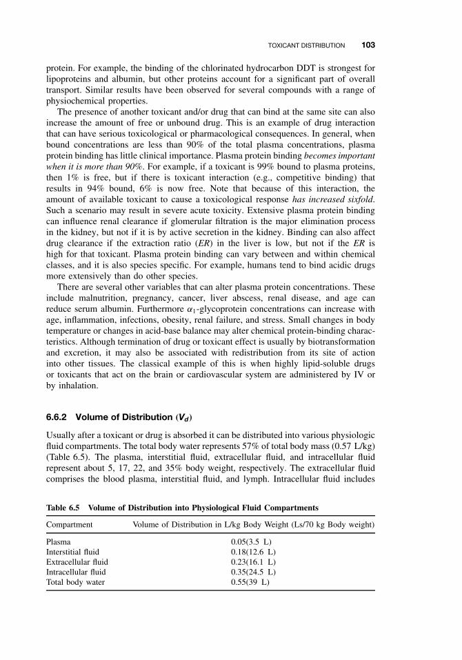
TOXICANT DISTRIBUTION 103
protein. For example, the binding of the chlorinated hydrocarbon DDT is strongest forlipoproteins and albumin, but other proteins account for a significant part of overalltransport. Similar results have been observed for several compounds with a range ofphysiochemical properties.
The presence of another toxicant and/or drug that can bind at the same site can alsoincrease the amount of free or unbound drug. This is an example of drug interactionthat can have serious toxicological or pharmacological consequences. In general, whenbound concentrations are less than 90% of the total plasma concentrations, plasmaprotein binding has little clinical importance. Plasma protein binding becomes importantwhen it is more than 90%. For example, if a toxicant is 99% bound to plasma proteins,then 1% is free, but if there is toxicant interaction (e.g., competitive binding) thatresults in 94% bound, 6% is now free. Note that because of this interaction, theamount of available toxicant to cause a toxicological response has increased sixfold.Such a scenario may result in severe acute toxicity. Extensive plasma protein bindingcan influence renal clearance if glomerular filtration is the major elimination processin the kidney, but not if it is by active secretion in the kidney. Binding can also affectdrug clearance if the extraction ratio (ER) in the liver is low, but not if the ER ishigh for that toxicant. Plasma protein binding can vary between and within chemicalclasses, and it is also species specific. For example, humans tend to bind acidic drugsmore extensively than do other species.
There are several other variables that can alter plasma protein concentrations. Theseinclude malnutrition, pregnancy, cancer, liver abscess, renal disease, and age canreduce serum albumin. Furthermore α1-glycoprotein concentrations can increase withage, inflammation, infections, obesity, renal failure, and stress. Small changes in bodytemperature or changes in acid-base balance may alter chemical protein-binding charac-teristics. Although termination of drug or toxicant effect is usually by biotransformationand excretion, it may also be associated with redistribution from its site of actioninto other tissues. The classical example of this is when highly lipid-soluble drugsor toxicants that act on the brain or cardiovascular system are administered by IV orby inhalation.
6.6.2 Volume of Distribution (Vd)
Usually after a toxicant or drug is absorbed it can be distributed into various physiologicfluid compartments. The total body water represents 57% of total body mass (0.57 L/kg)(Table 6.5). The plasma, interstitial fluid, extracellular fluid, and intracellular fluidrepresent about 5, 17, 22, and 35% body weight, respectively. The extracellular fluidcomprises the blood plasma, interstitial fluid, and lymph. Intracellular fluid includes
Table 6.5 Volume of Distribution into Physiological Fluid Compartments
Compartment Volume of Distribution in L/kg Body Weight (Ls/70 kg Body weight)
Plasma 0.05(3.5 L)Interstitial fluid 0.18(12.6 L)Extracellular fluid 0.23(16.1 L)Intracellular fluid 0.35(24.5 L)Total body water 0.55(39 L)
104 ABSORPTION AND DISTRIBUTION OF TOXICANTS
the sum of fluid contents of all cells in the body. There is also transcellular fluid thatrepresents 2% body weight, and this includes cerebrospinal, intraocular, peritoneal,pleural, and synovial fluids, and digestive secretions. Fat is about 20% body weight,while the GIT contents in monogastrics make up 1% body weight, and in ruminants itcan constitute 15% body weight.
Its sometimes useful to quantitate how well a drug or toxicant is distributed into thesevarious fluid compartments, and in this context the apparent volume of distributioncan be a useful parameter. The apparent volume of distribution, Vd , is defined as thevolume of fluid required to contain the total amount, A, of drug in the body at thesame concentration as that present in plasma, Cp,
Vd = A
Cp
.
In general, the Vd for a drug is to some extent descriptive of its distribution pat-tern in the body. For example, drugs or toxicants with relatively small Vd values maybe confined to the plasma as diffusion across the capillary wall is limited. There areother toxicants that have a slightly larger Vd (e.g., 0.23 L/kg), and these toxicantsmay be distributed in the extracellular compartment. This includes many polar com-pounds (e.g., tubocurarine, gentamicin, Vd = 0.2–0.4 L/kg). These toxicants cannotreadily enter cells because of their low lipid solubility. If the Vd for some of thesetoxicants is in excess of the theoretical value, this may be due to limited degree ofpenetration into cells or from the extravascular compartment. Finally there are manytoxicants distributed throughout the body water (Vd ≥ 0.55 L/kg) that may have Vd
values much greater than that for total body water. This distribution is achieved byrelatively lipid-soluble toxicants and drugs that readily cross cell membranes (e.g.,ethanol, diazepam; Vd = 1 to 2 L/kg). Binding of the toxicant anywhere outside of theplasma compartment, as well as partitioning into body fat, can increase Vd beyond theabsolute value for total body water. In general, toxicants with a large Vd can even reachthe brain, fetus, and other transcellular compartments. In general, toxicants with largeVd are a consequence of extensive tissue binding. The reader should be aware that weare talking about tissue binding, and not plasma protein binding where distribution islimited to plasma for obvious reasons.
The fraction of toxicant located in plasma is dependent on whether a toxicant bindsto both plasma and tissue components. Plasma binding can be measured directly, butnot tissue binding. It can, however, be inferred from the following relationship:
Amount in body = Amount in plasma + Amount outside plasma
Vd × C = Vp × C + VT W × CT W
where Vd is the apparent volume of distribution, Vp the volume of plasma, VT W theapparent volume of tissue, and CT W the tissue concentration. If the preceding equationis divided by C, it now becomes
Vd = Vp + VT W × CT W
C
Recall that fu = Cu/C occurs with plasma, and also that the fraction unbound intissues is fuT = CuT /CT W .
TOXICOKINETICS 105
Assuming at equilibrium that unbound concentration in tissue and plasma are equal,then we let the ratio of fu/fuT replace CT W/C and determine the volume of distributionas follows:
Vd = Vp + VT W ×(
fu
fuT
).
It is possible to predict what happens to Vd when fu or fuT changes as a resultof physiological or disease processes in the body that change plasma and/or tissueprotein concentrations. For example, Vd can increase with increased unbound toxicantin plasma or with a decrease in unbound toxicant tissue concentrations. The precedingequation explains why: because of both plasma and tissue binding, some Vd valuesrarely correspond to a real volume such as plasma volume, extracellular space, or totalbody water. Finally interspecies differences in Vd values can be due to differences inbody composition of body fat and protein, organ size, and blood flow as alluded toearlier in this section. The reader should also be aware that in addition to Vd , thereare volumes of distribution that can be obtained from pharmacokinetic analysis of agiven data set. These include the volume of distribution at steady state (Vd,ss), volumeof the central compartment (Vc), and the volume of distribution that is operative overthe elimination phase (Vd,area). The reader is advised to consult other relevant textsfor a more detailed description of these parameters and when it is appropriate to usethese parameters.
6.7 TOXICOKINETICS
The explanation of the pharmacokinetics or toxicokinetics involved in absorption, dis-tribution, and elimination processes is a highly specialized branch of toxicology, andis beyond the scope of this chapter. However, here we introduce a few basic conceptsthat are related to the several transport rate processes that we described earlier in thischapter. Toxicokinetics is an extension of pharmacokinetics in that these studies areconducted at higher doses than pharmacokinetic studies and the principles of pharma-cokinetics are applied to xenobiotics. In addition these studies are essential to provideinformation on the fate of the xenobiotic following exposure by a define route. Thisinformation is essential if one is to adequately interpret the dose-response relationshipin the risk assessment process. In recent years these toxicokinetic data from laboratoryanimals have started to be utilized in physiologically based pharmacokinetic (PBPK)models to help extrapolations to low-dose exposures in humans. The ultimate aim inall of these analyses is to provide an estimate of tissue concentrations at the target siteassociated with the toxicity.
Immediately on entering the body, a chemical begins changing location, concentra-tion, or chemical identity. It may be transported independently by several componentsof the circulatory system, absorbed by various tissues, or stored; the chemical mayeffect an action, be detoxified, or be activated; the parent compound or its metabo-lite(s) may react with body constituents, be stored, or be eliminated—to name someof the more important actions. Each of these processes may be described by rate con-stants similar to those described earlier in our discussion of first-order rate processesthat are associated with toxicant absorption, distribution, and elimination and occur
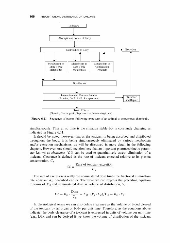
106 ABSORPTION AND DISTRIBUTION OF TOXICANTS
Metabolism toMore ToxicMetabolites
Metabolism toLess ToxicMetabolites
Metabolism toConjugation
Products
Interaction with Macromolecules(Proteins, DNA, RNA, Receptors,etc)
Exposure
Excretion
Toxic Effects(Genetic, Carcinogenic, Reproductive, Immunologic, etc)
Turnoverand Repair
Distribution
Distribution to Body
Absorption at Portals of Entry
Figure 6.11 Sequence of events following exposure of an animal to exogenous chemicals.
simultaneously. Thus at no time is the situation stable but is constantly changing asindicated in Figure 6.11.
It should be noted, however, that as the toxicant is being absorbed and distributedthroughout the body, it is being simultaneously eliminated by various metabolismand/or excretion mechanisms, as will be discussed in more detail in the followingchapters. However, one should mention here that an important pharmacokinetic param-eter known as clearance (C�) can be used to quantitatively assess elimination of atoxicant. Clearance is defined as the rate of toxicant excreted relative to its plasmaconcentration, Cp:
C� = Rate of toxicant excretion
Cp
.
The rate of excretion is really the administered dose times the fractional eliminationrate constant Kel described earlier. Therefore we can express the preceding equationin terms of Kel and administered dose as volume of distribution, Vd :
C� = Kel · Dose
Cp
= Kel · (Vd · Cp)/Cp = Kel · Vd.
In physiological terms we can also define clearance as the volume of blood clearedof the toxicant by an organ or body per unit time. Therefore, as the equations aboveindicate, the body clearance of a toxicant is expressed in units of volume per unit time(e.g., L/h), and can be derived if we know the volume of distribution of the toxicant
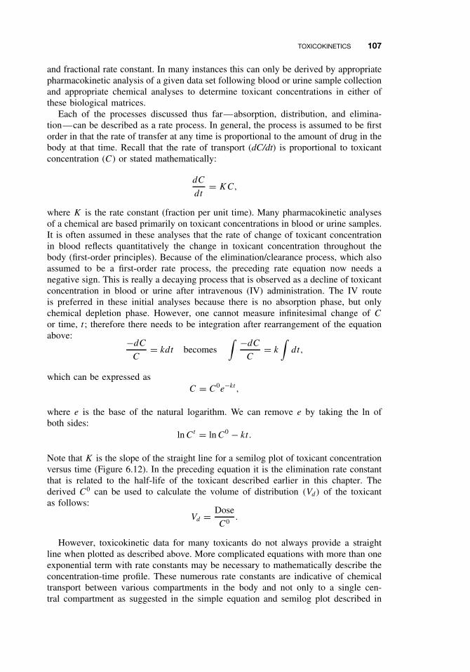
TOXICOKINETICS 107
and fractional rate constant. In many instances this can only be derived by appropriatepharmacokinetic analysis of a given data set following blood or urine sample collectionand appropriate chemical analyses to determine toxicant concentrations in either ofthese biological matrices.
Each of the processes discussed thus far—absorption, distribution, and elimina-tion—can be described as a rate process. In general, the process is assumed to be firstorder in that the rate of transfer at any time is proportional to the amount of drug in thebody at that time. Recall that the rate of transport (dC/dt) is proportional to toxicantconcentration (C) or stated mathematically:
dC
dt= KC,
where K is the rate constant (fraction per unit time). Many pharmacokinetic analysesof a chemical are based primarily on toxicant concentrations in blood or urine samples.It is often assumed in these analyses that the rate of change of toxicant concentrationin blood reflects quantitatively the change in toxicant concentration throughout thebody (first-order principles). Because of the elimination/clearance process, which alsoassumed to be a first-order rate process, the preceding rate equation now needs anegative sign. This is really a decaying process that is observed as a decline of toxicantconcentration in blood or urine after intravenous (IV) administration. The IV routeis preferred in these initial analyses because there is no absorption phase, but onlychemical depletion phase. However, one cannot measure infinitesimal change of C
or time, t ; therefore there needs to be integration after rearrangement of the equationabove: −dC
C= kdt becomes
∫ −dC
C= k
∫dt,
which can be expressed asC = C0e−kt ,
where e is the base of the natural logarithm. We can remove e by taking the ln ofboth sides:
ln Ct = ln C0 − kt.
Note that K is the slope of the straight line for a semilog plot of toxicant concentrationversus time (Figure 6.12). In the preceding equation it is the elimination rate constantthat is related to the half-life of the toxicant described earlier in this chapter. Thederived C0 can be used to calculate the volume of distribution (Vd) of the toxicantas follows:
Vd = Dose
C0.
However, toxicokinetic data for many toxicants do not always provide a straightline when plotted as described above. More complicated equations with more than oneexponential term with rate constants may be necessary to mathematically describe theconcentration-time profile. These numerous rate constants are indicative of chemicaltransport between various compartments in the body and not only to a single cen-tral compartment as suggested in the simple equation and semilog plot described in
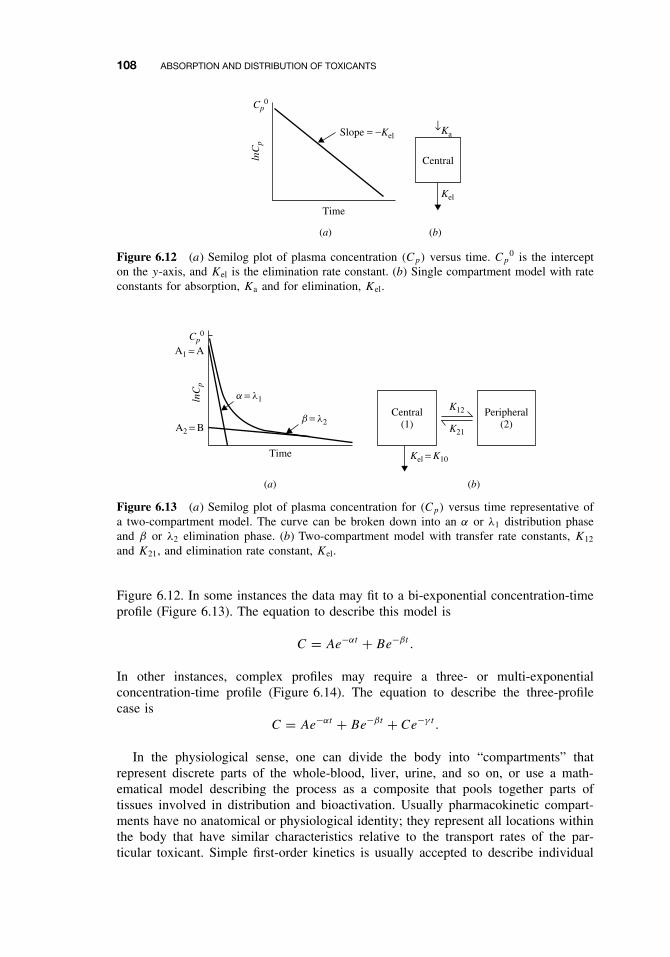
108 ABSORPTION AND DISTRIBUTION OF TOXICANTS
Ka
Kel
(a) (b)
Central
Time
lnC
p
Slope = −Kel
Cp0
↓
Figure 6.12 (a) Semilog plot of plasma concentration (Cp) versus time. Cp0 is the intercept
on the y-axis, and Kel is the elimination rate constant. (b) Single compartment model with rateconstants for absorption, Ka and for elimination, Kel.
(a) (b)
Central(1)
Peripheral(2)
Time
lnC
p
Cp0
K12
K21
Kel = K10
b = l2A2 = B
A1 = A
a = l1
Figure 6.13 (a) Semilog plot of plasma concentration for (Cp) versus time representative ofa two-compartment model. The curve can be broken down into an α or λ1 distribution phaseand β or λ2 elimination phase. (b) Two-compartment model with transfer rate constants, K12
and K21, and elimination rate constant, Kel.
Figure 6.12. In some instances the data may fit to a bi-exponential concentration-timeprofile (Figure 6.13). The equation to describe this model is
C = Ae−αt + Be−βt .
In other instances, complex profiles may require a three- or multi-exponentialconcentration-time profile (Figure 6.14). The equation to describe the three-profilecase is
C = Ae−αt + Be−βt + Ce−γ t .
In the physiological sense, one can divide the body into “compartments” thatrepresent discrete parts of the whole-blood, liver, urine, and so on, or use a math-ematical model describing the process as a composite that pools together parts oftissues involved in distribution and bioactivation. Usually pharmacokinetic compart-ments have no anatomical or physiological identity; they represent all locations withinthe body that have similar characteristics relative to the transport rates of the par-ticular toxicant. Simple first-order kinetics is usually accepted to describe individual
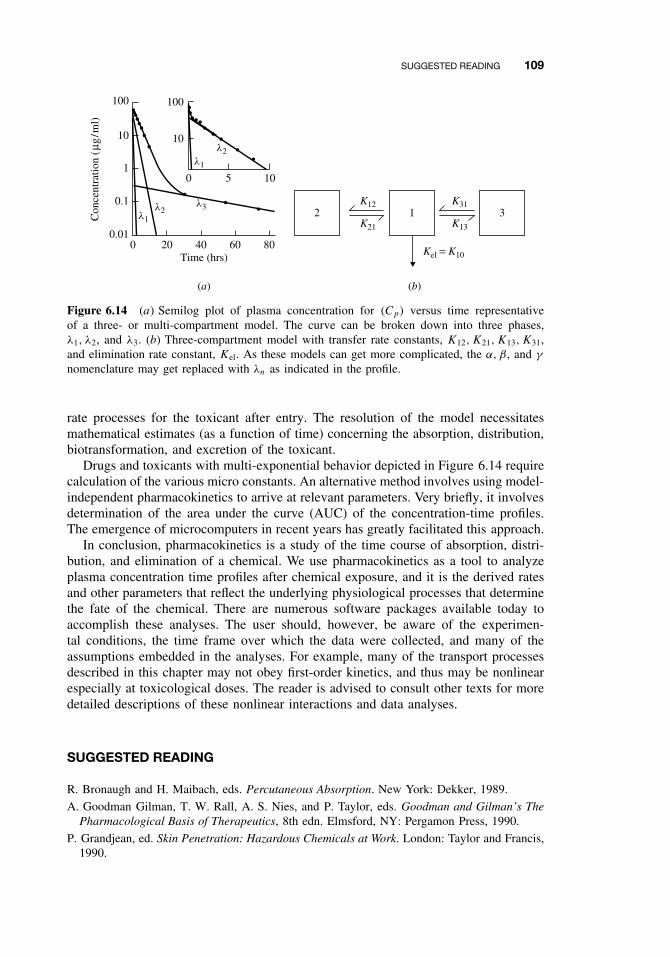
SUGGESTED READING 109
(a) (b)
Time (hrs)
K12
K21
K31
K13
Kel = K10
l1
l2
l1l2
l3
0
0 5 10
10
100
20 40 60 80
100
10
1
0.1
0.01
Con
cent
ratio
n (µ
g/m
l)
2 1 3
Figure 6.14 (a) Semilog plot of plasma concentration for (Cp) versus time representativeof a three- or multi-compartment model. The curve can be broken down into three phases,λ1, λ2, and λ3. (b) Three-compartment model with transfer rate constants, K12,K21,K13, K31,and elimination rate constant, Kel. As these models can get more complicated, the α, β, and γ
nomenclature may get replaced with λn as indicated in the profile.
rate processes for the toxicant after entry. The resolution of the model necessitatesmathematical estimates (as a function of time) concerning the absorption, distribution,biotransformation, and excretion of the toxicant.
Drugs and toxicants with multi-exponential behavior depicted in Figure 6.14 requirecalculation of the various micro constants. An alternative method involves using model-independent pharmacokinetics to arrive at relevant parameters. Very briefly, it involvesdetermination of the area under the curve (AUC) of the concentration-time profiles.The emergence of microcomputers in recent years has greatly facilitated this approach.
In conclusion, pharmacokinetics is a study of the time course of absorption, distri-bution, and elimination of a chemical. We use pharmacokinetics as a tool to analyzeplasma concentration time profiles after chemical exposure, and it is the derived ratesand other parameters that reflect the underlying physiological processes that determinethe fate of the chemical. There are numerous software packages available today toaccomplish these analyses. The user should, however, be aware of the experimen-tal conditions, the time frame over which the data were collected, and many of theassumptions embedded in the analyses. For example, many of the transport processesdescribed in this chapter may not obey first-order kinetics, and thus may be nonlinearespecially at toxicological doses. The reader is advised to consult other texts for moredetailed descriptions of these nonlinear interactions and data analyses.
SUGGESTED READING
R. Bronaugh and H. Maibach, eds. Percutaneous Absorption. New York: Dekker, 1989.
A. Goodman Gilman, T. W. Rall, A. S. Nies, and P. Taylor, eds. Goodman and Gilman’s ThePharmacological Basis of Therapeutics, 8th edn. Elmsford, NY: Pergamon Press, 1990.
P. Grandjean, ed. Skin Penetration: Hazardous Chemicals at Work. London: Taylor and Francis,1990.

110 ABSORPTION AND DISTRIBUTION OF TOXICANTS
R. Krieger, ed. Handbook of Pesticide Toxicology, 2nd edn. San Diego: Academic Press, 2001.
M. Rowland and T. N. Tozer, eds. Clinical Pharmacokinetics. Concepts and Applications, 3rdedn. Philadelphia: Lea and Febiger, 1995.
L. Shargel and A. B. C. Yu, eds. Applied Biopharmaceutics and Pharmacokinetics, 4th edn.Norwalk, CT: Appleton and Lange, 1999.
CHAPTER 7
Metabolism of Toxicants
RANDY L. ROSE and ERNEST HODGSON
7.1 INTRODUCTION
One of the most important determinants of xenobiotic persistence in the body andsubsequent toxicity to the organism is the extent to which they can be metabo-lized and excreted. Several families of metabolic enzymes, often with wide arrays ofsubstrate specificity, are involved in xenobiotic metabolism. Some of the more impor-tant families of enzymes involved in xenobiotic metabolism include the cytochromeP450 monooxygenases (CYPs), flavin-containing monooxygenases (FMOs), alcoholand aldehyde dehydrogenases, amine oxidases, cyclooxygenases, reductases, hydro-lases, and a variety of conjugating enzymes such as glucuronidases, sulfotransferases,methyltransferases, glutathione transferases, and acetyl transferases.
Most xenobiotic metabolism occurs in the liver, an organ devoted to the synthesis ofmany important biologically functional proteins and thus with the capacity to mediatechemical transformations of xenobiotics. Most xenobiotics that enter the body arelipophilic, a property that enables them to bind to lipid membranes and be transportedby lipoproteins in the blood. After entrance into the liver, as well as in other organs,xenobiotics may undergo one or two phases of metabolism. In phase I a polar reactivegroup is introduced into the molecule rendering it a suitable substrate for phase IIenzymes. Enzymes typically involved in phase I metabolism include the CYPs, FMOs,and hydrolases, as will be discussed later. Following the addition of a polar group,conjugating enzymes typically add much more bulky substituents, such as sugars,sulfates, or amino acids that result in a substantially increased water solubility of thexenobiotic, making it easily excreted. Although this process is generally a detoxicationsequence, reactive intermediates may be formed that are much more toxic than theparent compound. It is, however, usually a sequence that increases water solubilityand hence decreases the biological half life (t0.5) of the xenobiotic in vivo.
Phase I monooxygenations are more likely to form reactive intermediates thanphase II metabolism because the products are usually potent electrophiles capable ofreacting with nucleophilic substituents on macromolecules, unless detoxified by somesubsequent reaction. In the following discussion, examples of both detoxication andintoxication reactions are given, although greater emphasis on activation products isprovided in Chapter 8.
A Textbook of Modern Toxicology, Third Edition, edited by Ernest HodgsonISBN 0-471-26508-X Copyright 2004 John Wiley & Sons, Inc.
111
112 METABOLISM OF TOXICANTS
7.2 PHASE I REACTIONS
Phase I reactions include microsomal monooxygenations, cytosolic and mitochondrialoxidations, co-oxidations in the prostaglandin synthetase reaction, reductions, hydrol-yses, and epoxide hydration. All of these reactions, with the exception of reductions,introduce polar groups to the molecule that, in most cases, can be conjugated duringphase II metabolism. The major phase I reactions are summarized in Table 7.1.
7.2.1 The Endoplasmic Reticulum, Microsomal Preparation,and Monooxygenations
Monooxygenation of xenobiotics are catalyzed either by the cytochrome P450 (CYP)-dependent monooxygenase system or by flavin-containing monooxygenases (FMO).
Table 7.1 Summary of Some Important Oxidative and Reductive Reactions of Xenobiotics
Enzymes and Reactions Examples
Cytochrome P450Epoxidation/hydroxylation Aldrin, benzo(a)pyrene, aflatoxin, bromobenzeneN-, O-, S-Dealkylation Ethylmorphine, atrazine, p-nitroanisole,
methylmercaptanN-, S-, P -Oxidation Thiobenzamide, chlorpromazine,
2-acetylaminofluoreneDesulfuration Parathion, carbon disulfideDehalogenation Carbon tetrachloride, chloroformNitro reduction NitrobenzeneAzo reduction O-Aminoazotoluene
Flavin-containing monooxygenaseN-, S-, P -Oxidation Nicotine, imiprimine, thiourea, methimazoleDesulfuration Fonofos
Prostaglandin synthetase cooxidationDehydrogenation Acetaminophen, benzidine, epinephrineN-Dealkylation Benzphetamine, dimethylanilineEpoxidation/hydroxylation Benzo(a)pyrene, 2-aminofluorene, phenylbutazoneOxidation FANFT, ANFT, bilirubin
Molybdenum hydroxylasesOxidation Purines, pteridine, methotrexate, 6-deoxycyclovirReductions Aromatic nitrocompounds, azo dyes, nitrosoamines
Alcohol dehydrogenaseOxidation Methanol, ethanol, glycols, glycol ethersReduction Aldehydes and ketones
Aldehyde dehydrogenaseOxidation Aldehydes resulting from alcohol and glycol
oxidations
Esterases and amidasesHydrolysis Parathion, paraoxon, dimethoate
Epoxide hydrolaseHydrolysis Benzo(a)pyrene epoxide, styrene oxide
PHASE I REACTIONS 113
Both are located in the endoplasmic reticulum of the cell and have been studied inmany tissues and organisms. This is particularly true of CYPs, probably the moststudied of all enzymes.
Microsomes are derived from the endoplasmic reticulum as a result of tissuehomogenization and are isolated by centrifugation of the postmitochondrial supernatantfraction, described below. The endoplasmic reticulum is an anastomosing networkof lipoprotein membranes extending from the plasma membrane to the nucleusand mitochrondria, whereas the microsomal fraction derived from it consists ofmembranous vesicles contaminated with free ribosomes, glycogen granules, andfragments of other subcellular structures such as mitochondria and Golgi apparatus.The endoplasmic reticulum, and consequently the microsomes derived from it, consistsof two types, rough and smooth, the former having an outer membrane studded withribosomes, which the latter characteristically lack. Although both rough and smoothmicrosomes have all of the components of the CYP-dependent monooxygenase system,the specific activity of the smooth type is usually higher.
The preparation of microsomal fractions, S9, and cytosolic fractions from tissuehomogenates involves the use of two to three centrifugation steps. Following tis-sue extraction, careful mincing, and rinses of tissue for blood removal, the tissuesare typically homogenized in buffer and centrifuged at 10,000 × g for 20 minutes.The resulting supernatant, often referred to as the S9 fraction, can be used in stud-ies where both microsomal and cytosolic enzymes are desired. More often, however,the S9 fraction is centrifuged at 100,000 × g for 60 minutes to yield a microsomalpellet and a cytosolic supernatant. The pellet is typically resuspended in a volume ofbuffer, which will give 20 to 50 mg protein/ml and stored at −20 to −70◦C. Often,the microsomal pellet is resuspended a second time and resedimented at 100,000 × gfor 60 minutes to further remove contaminating hemoglobin and other proteins. Asdescribed above, enzymes within the microsomal fraction (or microsomes) includeCYPs, FMOs, cyclooxygenases, and other membrane-bound enzymes, including nec-essary coenzymes such as NADPH cytochrome P450 reductase for CYP. Enzymesfound in the cytosolic fraction (derived from the supernatant of the first 100,000 × gspin) include hydrolases and most of the conjugating enzymes such as glutathionetransferases, glucuronidases, sulfotransferases, methyl transferases, and acetylases. Itis important to note that some cytosolic enzymes can also be found in microsomalfractions, although the opposite is not generally the case.
Monooxygenations, previously known as mixed-function oxidations, are those oxi-dations in which one atom of a molecule of oxygen is incorporated into the substratewhile the other is reduced to water. Because the electrons involved in the reductionof CYPs or FMOs are derived from NADPH, the overall reaction can be written asfollows (where RH is the substrate):
RH + O2 + NADPH + H+ −−−→ NADP+ + ROH + H2O.
7.2.2 The Cytochrome P450-Dependent Monooxygenase System
The CYPs, the carbon monoxide-binding pigments of microsomes, are heme proteinsof the b cytochrome type. Originally described as a single protein, there are nowknown to be more than 2000 CYPs widely distributed throughout animals, plants, andmicroorganisms. A system of nomenclature utilizing the prefix CYP has been devised

114 METABOLISM OF TOXICANTS
for the genes and cDNAs corresponding to the different forms (as discussed later in thissection), although P450 is still appropriate as a prefix for the protein products. Unlikemost cytochromes, the name CYP is derived not from the absorption maximum of thereduced form in the visible region but from the unique wavelength of the absorptionmaximum of the carbon monoxide derivative of the reduced form, namely 450 nm.
The role of CYP as the terminal oxidase in monooxygenase reactions is supportedby considerable evidence. The initial proof was derived from the demonstration ofthe concomitant light reversibility of the CO complex of CYP and the inhibition,by CO, of the C-21 hydroxylation of 17 α-hydroxy-progesterone by adrenal glandmicrosomes. This was followed by a number of indirect, but nevertheless convincing,proofs involving the effects on both CYP and monooxygenase activity of CO, inducingagents, and spectra resulting from ligand binding and the loss of activity on degrada-tion of CYP to cytochrome P420. Direct proof was subsequently provided by thedemonstration that monooxygenase systems, reconstituted from apparently homoge-nous purified CYP, NADPH-CYP reductase, and phosphatidylchloline, can catalyzemany monooxygenase reactions.
CYPs, like other hemoproteins, have characteristic absorptions in the visible region.The addition of many organic, and some inorganic, ligands results in perturbationsof this spectrum. Although the detection and measurement of these spectra requires ahigh-resolution spectrophotometer, these perturbations, measured as optical differencespectra, have been of tremendous use in the characterization of CYPs, particularly inthe decades preceding the molecular cloning and expression of specific CYP isoforms.
The most important difference spectra of oxidized CYP are type I, with an absorptionmaximum at 385 to 390 nm. Type I ligands are found in many different chemicalclasses and include drugs, environmental contaminants, pesticides, and so on. Theyappear to be generally unsuitable, on chemical grounds, as ligands for the heme ironand are believed to bind to a hydrophobic site in the protein that is close enough tothe heme to allow both spectral perturbation and interaction with the activated oxygen.Although most type I ligands are substrates, it has not been possible to demonstrate aquantitative relationship between KS (concentration required for half-maximal spectraldevelopment) and KM (Michaelis constant). Type II ligands, however, interact directlywith the heme iron of CYP, and are associated with organic compounds having nitrogenatoms with sp2 or sp3 nonbonded electrons that are sterically accessible. Such ligandsare frequently inhibitors of CYP activity.
The two most important difference spectra of reduced CYP are the well-known COspectrum, with its maximum at or about 450 nm, and the type III spectrum, with twopH-dependent peaks at approximately 430 and 455 nm. The CO spectrum forms thebasis for the quantitative estimation of CYP. The best-known type III ligands for CYPare ethyl isocyanide and compounds such as the methylenedioxyphenyl synergists andSKF 525A, the last two forming stable type III complexes that appear to be related tothe mechanism by which they inhibit monooxygenations.
In the catalytic cycle of CYP, reducing equivalents are transferred from NADPHto CYP by a flavoprotein enzyme known as NADPH-cytochrome P450 reductase. Theevidence that this enzyme is involved in CYP monooxygenations was originally derivedfrom the observation that cytochrome c, which can function as an artificial electronacceptor for the enzyme, is an inhibitor of such oxidations. This reductase is an essentialcomponent in CYP-catalyzed enzyme systems reconstituted from purified components.Moreover antibodies prepared from purified reductase are inhibitors of microsomal
PHASE I REACTIONS 115
monooxygenase reactions. The reductase is a flavoprotein of approximately 80,000daltons that contain 2 mole each of flavin mononucleotide (FMN) and flavinadeninedinucleotide (FAD) per mole of enzyme. The only other component necessary foractivity in the reconstituted system is a phospholipid, phosphatidylchloline. This is notinvolved directly in electron transfer but appears to be involved in the coupling of thereductase to the cytochrome and in the binding of the substrate to the cytochrome.
The mechanism of CYP function has not been established unequivocally; however,the generally recognized steps are shown in Figure 7.1. The initial step consists of thebinding of substrate to oxidize CYP followed by a one electron reduction catalyzed byNADPH-cytochrome P450 reductase to form a reduced cytochrome-substrate complex.This complex can interact with CO to form the CO-complex, which gives rise to thewell-known difference spectrum with a peak at 450 nm and also inhibits monooxyge-nase activity. The next several steps are less well understood. They involve an initialinteraction with molecular oxygen to form a ternary oxygenated complex. This ternarycomplex accepts a second electron, resulting in the further formation of one or moreless understood complexes. One of these, however, is probably the equivalent of theperoxide anion derivative of the substrate-bound hemoprotein. Under some conditionsthis complex may break down to yield hydrogen peroxide and the oxidized cytochromesubstrate complex. Normally, however, one atom of molecular oxygen is transferred tothe substrate and the other is reduced to water, followed by dismutation reactions lead-ing to the formation of the oxygenated product, water, and the oxidized cytochrome.
The possibility that the second electron is derived from NADH through cytochromeb5 has been the subject of argument for some time and has yet to be completelyresolved. Cytochrome b5 is a widely distributed microsomal heme protein thatis involved in metabolic reactions such as fatty acid desaturation that involveendogenous substrates. It is clear, however, that cytochrome b5 is not essential for all
ROH R
Lipid OOH Lipid
NADPHNADPH
Cyt. P450Reductase
NADPHCyt. P450Reductase
NADHCyt. b5
Reductase
XOOH
2H+
e−
e−
e−
e−
e−
e−
H2O
H2O2
O2−
•
O2−
•
O2
Cyt-Fe+3
Cyt-Fe+3R Cyt-Fe+3R
Cyt. b5
Cyt-Fe+2R[CO]
hv
O
Cyt-Fe+3R
CO
Cyt-Fe+3RO2−
•
Cyt-Fe+2R
O2
Cyt-Fe+1R
O2
Cyt-Fe+2Ror
NADH
NADPH
Figure 7.1 Generalized scheme showing the sequence of events for P450 monooxygenations.
116 METABOLISM OF TOXICANTS
CYP-dependent monooxygenations because many occur in systems reconstituted fromNADPH, O2, phosphatidylchloline, and highly purified CYP and NADPH-cytochromeP450 reductase. Nevertheless, there is good evidence that many catalytic activities byisoforms including CYP3A4, CYP3A5, and CYP2E1 are stimulated by cytochromeb5. In some cases apocytochrome b5 (devoid of heme) has also been found to bestimulatory, suggesting that an alternate role of cytochrome b5 may be the result ofconformational changes in the CYP/NADPH cytochrome P450 reductase systems. Thuscytochrome b5 may facilitate oxidative activity in the intact endoplasmic reticulum.The isolation of forms of CYP that bind avidly to cytochrome b5 also tends to supportthis idea.
Distribution of Cytochrome P450. In vertebrates the liver is the richest sourceof CYP and is most active in the monooxygenation of xenobiotics. CYP and othercomponents of the CYP-dependent monooxygenase system are also in the skin, nasalmucosa, lung, and gastrointestinal tract, presumably reflecting the evolution of defensemechanisms at portals of entry. In addition to these organs, CYP has been demonstratedin the kidney, adrenal cortex and medulla, placenta, testes, ovaries, fetal and embryonicliver, corpus luteum, aorta, blood platelets, and the nervous system. In humans, CYPhas been demonstrated in the fetal and adult liver, the placenta, kidney, testes, fetaland adult adrenal gland, skin, blood platelets, and lymphocytes.
Although CYPs are found in many tissues, the function of the particular subset ofisoforms in organ, tissue, or cell type does not appear to be the same in all cases. Inthe liver, CYPs oxidize a large number of xenobiotics as well as some endogenoussteroids and bile pigments. The CYPs of the lung also appear to be concerned primar-ily with xenobiotic oxidations, although the range of substrates is more limited thanthat of the liver. The skin and small intestine also carry out xenobiotic oxidations,but their activities have been less well characterized. In normal pregnant females, theplacental microsomes display little or no ability to oxidize foreign compounds, appear-ing to function as a steroid hormone metabolizing system. On induction of the CYPenzymes, such as occurs in pregnant women who smoke, CYP-catalyzed aryl hydrocar-bon hydroxylase activity is readily apparent. The CYPs of the kidney are active in theω-oxidation of fatty acids, such as lauric acid, but are relatively inactive in xenobioticoxidation. Mitochondrial CYPs, such as those of the placenta and adrenal cortex, areactive in the oxidation of steroid hormones rather than xenobiotics.
Distribution of CYPs within the cell has been studied primarily in the mammalianliver, where it is present in greatest quantity in the smooth endoplasmic reticulum andin smaller but appreciable amounts in the rough endoplasmic reticulum. The nuclearmembrane has also been reported to contain CYP and to have detectable aryl hydro-carbon hydroxylase activity, an observation that may be of considerable importance instudies of the metabolic activation of carcinogens.
Multiplicity of Cytochrome P450, Purification, and Reconstitution ofCytochrome P450 Activity. Even before appreciable purification of CYP had beenaccomplished, it was apparent from indirect evidence that mammalian liver cellscontained more than one CYP enzyme. Subsequent direct evidence on the multiplicityof CYPs included the separation and purification of CYP isozymes, distinguished fromeach other by chromatographic behavior, immunologic specificity, and/or substratespecificity after reconstitution and separation of distinct polypeptides by sodium
PHASE I REACTIONS 117
dodecyl sulfate polyacrylamide gel electrophoresis (SDS-PAGE), which could thenbe related to distinct CYPs present in the original microsomes.
Purification of CYP and its usual constituent isoforms was, for many years, anelusive goal; one, however, that has been largely resolved. The problem of instability onsolubilization was resolved by the use of glycerol and dithiothreitol as protectants, andthe problem of reaggregation by maintaining a low concentration of a suitable detergent,such as Emulgen 911 (Kao-Atlas, Tokyo), throughout the procedure. Multiple CYPisoforms, as discussed previously, may be separated from each other and purified asseparate entities, although individual isoforms are now routinely cloned and expressedas single entities. The lengthy processes of column purification of CYPs have nowbeen largely superceded by the cloning and expression of transgenic isoforms in avariety of expression systems.
Systems reconstituted from purified CYP, NADPH-cytochrome P450 reductase andphosphatidylchloline will, in the presence of NADPH and O2, oxidize xenobioticssuch as benzphetamine, often at rates comparable to microsomes. Although systemsreconstituted from this minimal number of components are enzymatically active, othermicrosomal components, such as cytochrome b5, may facilitate activity either in vivoor in vitro or may even be essential for the oxidation of certain substrates.
One important finding from purification studies as well as cloning and expressing ofindividual isoforms is that the lack of substrate specificity of microsomes for monooxy-genase activity is not an artifact caused by the presence of several specific cytochromes.Rather, it appears that many of the cytochromes isolated are still relatively nonspecific.The relative activity toward different substrates does nevertheless vary greatly fromone CYP isoform to another even when both are relatively nonspecific. This lack ofspecificity is illustrated in Table 7.2, using human isoforms as examples.
Classification and Evolution of Cytochrome P450. The techniques of molecularbiology have been applied extensively to the study of CYP. More than 1925 genes havebeen characterized as of 2002, and the nucleotide and derived amino acid sequencescompared. In some cases the location of the gene on a particular chromosome has beendetermined and the mechanism of gene expression investigated.
A system of nomenclature proposed in 1987 has since been updated several times,most recently in 1996. The accepted guidelines from nomenclature designate cyto-chrome P450 genes as CYP (or cyp in the case of mouse genes). The CYP designationis followed by an Arabic numeral to denote the gene family, followed by a letterdesignating the subfamily. The individual isoform is then identified using a secondArabic numeral following the subfamily designation. Polymorphic isoforms of genesare indicated by an asterisk followed by an arabic numeral. If there are no subfamiliesor if there is only a single gene within the family or subfamily, the letter and/or thesecond numeral may be omitted (e.g., CYP17). The name of the gene is italicized,whereas the protein (enzyme) is not.
In general, enzymes within a gene family share more than 40% amino acid sequenceidentity. Protein sequences within subfamilies have greater than 55% similarity in thecase of mammalian genes, or 46% in the case of nonmammalian genes. So far, genesin the same subfamily have been found to lie on the same chromosome within thesame gene cluster and are nonsegregating, suggesting a common origin through geneduplication events. Sequences showing less than 3% divergence are arbitrarily desig-nated allelic variants unless other evidence exists to the contrary. Known sequences fit
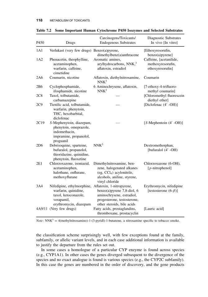
118 METABOLISM OF TOXICANTS
Table 7.2 Some Important Human Cytochrome P450 Isozymes and Selected Substrates
Carcinogens/Toxicants/ Diagnostic SubstratesP450 Drugs Endogenous Substrates In vivo [In vitro]
1A1 Verlukast (very few drugs) Benzo(a)pyrene,dimethylbenz(a)anthracene
[Ethoxyresorufin,benzo(a)pyrene]
1A2 Phenacetin, theophylline,acetaminophen,warfarin, caffeine,cimetidine
Aromatic amines,arylhydrocarbons, NNK,3
aflatoxin, estradiol
Caffeine, [acetanilide,methoxyresorufin,ethoxyresorufin]
2A6 Coumarin, nicotine Aflatoxin, diethylnitrosamine,NNK3
Coumarin
2B6 Cyclophosphamide,ifosphamide, nicotine
6 Aminochrysene, aflatoxin,NNK3
[7-ethoxy-4-trifluoro-methyl coumarin]
2C8 Taxol, tolbutamide,carbamazepine
— [Chloromethyl fluoresceindiethyl ether]
2C9 Tienilic acid, tolbutamide,warfarin, phenytoin,THC, hexobarbital,diclofenac
— [Diclofenac (4′ -OH)]
2C19 S-Mephenytoin, diazepam,phenytoin, omeprazole,indomethacin,impramine, propanolol,proguanil
— [S-Mephentoin (4′ -OH)]
2D6 Debrisoquine, sparteine,bufuralol, propanolol,thioridazine, quinidine,phenytoin, fluoxetine
NNK3 Dextromethorphan,[bufuralol (4′ -OH)
2E1 Chlorzoxazone, isoniazid,acetaminophen,halothane, enflurane,methoxyflurane
Dimethylnitrosamine, ben-zene, halogenated alkanes(eg, CCl4) acylonitrile,alcohols, aniline, styrene,vinyl chloride
Chlorzoxazone (6-OH),[p-nitrophenol]
3A4 Nifedipine, ethylmorphine,warfarin, quinidine,taxol, ketoconazole,verapamil,erythromycin, diazepam
Aflatoxin, 1-nitropyrene,benzo(a)pyrene 7,8-diol, 6aminochrysene, estradiol,progesterone, testosterone,other steroids, bile acids
Erythromycin, nifedipine[testosterone (6-β)]
4A9/11 (Very few drugs) Fatty acids, prostaglandins,thromboxane, prostacyclin
[Lauric acid]
Note: NNK3 = 4(methylnitrosamino)-1-(3-pyridl)-1-butanone, a nitrosamine specific to tobacco smoke.
the classification scheme surprisingly well, with few exceptions found at the family,subfamily, or allelic variant levels, and in each case additional information is availableto justify the departure from the rules set out.
In some cases a homologue of a particular CYP enzyme is found across species(e.g., CYP1A1). In other cases the genes diverged subsequent to the divergence of thespecies and no exact analogue is found is various species (e.g., the CYP2C subfamily).In this case the genes are numbered in the order of discovery, and the gene products
PHASE I REACTIONS 119
from a particular subfamily may even have differing substrate specificity in differentspecies (e.g., rodent vs. human). Relationships between different CYP families andsubfamilies are related to the rate and extent of CYP evolution.
Figure 7.2 demonstrates some of the evolutionary relationships between CYP genesbetween some of the earliest vertebrates and humans. This dendogram compares CYPgenes from the puffer fish (fugu) and 8 other fish species with human CYPs (including3 pseudogenes). The unweighted pair group method arithmetic averaging (UPGMA)phylogenetic tree demonstrates the presence of five CYP clans (clusters of CYPs thatare consistently grouped together) and delineates the 18 known human CYPs. This dataset demonstrates that the defining characteristics of vertebrate CYPs have not changedmuch in 420 million years. Of these 18 human CYPs, only 1 family was missing infugu (CYP39), indicating that the mammalian diversity of CYPs likely predates thetetrapod-ray finned fish divergence. The fish genome also has new CYP1C, 3B, and7C subfamilies that are not seen in mammals.
The gene products, the CYP isoforms, may still be designated P450 followed bythe same numbering system used for the genes, or the CYP designation may be used,for example, P4501A1 or CYP1A1.
As of May 16, 2002, a total of 1925 CYP sequences have been “named” with sev-eral others still awaiting classification. Of these, 977 are animal sequences, 607 fromplants, 190 from lower eukaryotes and 151 are bacterial sequences. These sequencesfall into more than 265 CYP families, 18 of which belong to mammals. Humans have40 sequenced CYP genes. As the list of CYPs is continually expanding, progress in thisarea can be readily accessed via the internet at the Web site of the P450 Gene Super-family Nomenclature Committee (http://drnelson. utmem.edu/nelsonhomepage.html) orat another excellent Web site (http://www.icgeb.trieste.it/p450).
Cytochrome P450 Families with Xenobiotic Metabolizing Potential. Althoughmammals are known to have 18 CYP families, only three families are primarily respon-sible for most xenobiotic metabolism. These families (families 1–3) are considered tobe more recently derived from the “ancestral” CYP families. The remaining fami-lies are less promiscuous in their metabolizing abilities and are often responsible forspecific metabolic steps. For example, members of the CYP4 family are responsiblefor the end-chain hydroxylation of long-chain fatty acids. The remaining mammalianCYP families are involved in biosynthesis of steroid hormones. In fact some of thenomenclature for some of these families is actually derived from the various positionsin the steroid nucleus where the metabolism takes place. For example, CYP7 medi-ates hydroxylation of cholesterol at the 7α-position, while CYP17 and 21 catalyze the17α and 21-hydroxylations of progesterone, respectively. CYP19 is responsible for thearomatization of androgens to estrogen by the initial step of hydroxylation at the 19-position. Many of the CYPs responsible for steroidogenesis are found in the adrenalcortex, while those involved in xenobiotic metabolism are found predominantly in tis-sues that are more likely to be involved in exposure such as liver, kidneys, lungs, andolfactory tissues.
To simplify discussion of important CYP family members, the following discussionconcentrates upon human CYP family members. However, since there is a great dealof homology among family members, many of the points of discussion are generallyapplicable to CYP families belonging to several species.
The CYP1 family contains three known human members, CYP1A1, CYP1A2, andCYP1B1. CYP1A1 and CYP1A2 are found in all classes of the animal kingdom.
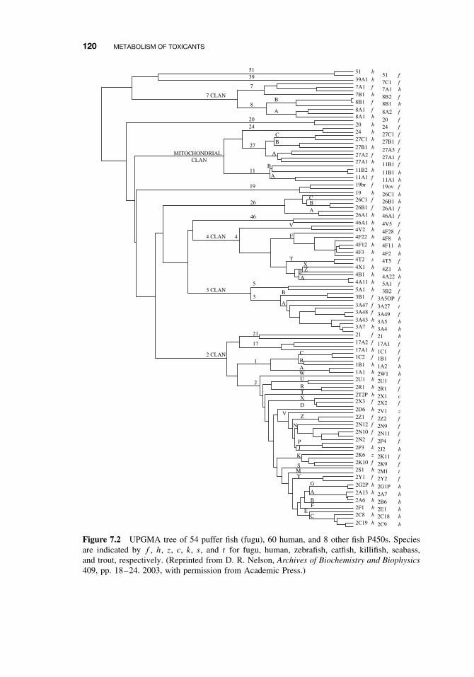
120 METABOLISM OF TOXICANTS
51 51 h
39A1 h7A1 f7B1 h8B1 f
8A1 f8A1 h
20 h24 h
51 f7C1 f7A1 h8B2 f8B1 h
8A2 f
20 f24 f27C1 f
27C1 h 27B1 f27B1 h 27A3 f27A2 f 27A1 f27A1 h 11B1 f11B2 h 11B1 h11A1 f 11A1 h19br f
26C1 f
26B1 f26A1 h46A1 h
19ov f19 h 26C1 h
26B1 h26A1 f46A1 f
4V5 f4V2 h4F22 h
4F12 h4F3 h
4F28 f4F8 h4F11 h
4F2 h4T2 s4X1 h4B1 h4A11 h5A1 h
4T5 f
4Z1 h4A22 h5A1 f3B2 f
3B1 f
3A47 f3A48 f
3A43 h3A7 h
3A5OP f
3A27 t
3A49 f3A5 h3A4 h
21 f 21 h17A2 f 17A1 f17A1 h 1C1 f1C2 f 1B1 f1B1 h 1A2 h1A1 h 2W1 h2U1 h 2U1 f2R1 h 2R1 f2T2P h 2X1 c2X3 f 2X2 f2D6 h 2V1 z2Z1 f 2Z2 f2N12 f 2N9 f2N10 f 2N11 f2N2 f 2P4 f2P3 k 2J2 h2K6 z 2K11 f2K10 f 2K9 f2S1 h 2M1 t2Y1 f 2Y2 f2G2P h 2G1P h2A13 h 2A7 h2A6 h 2B6 h2F1 h 2E1 h2C8 h 2C18 h2C19 h 2C9 h
39
2024
27
11
19
26
46
7
8B
A
A
A
A
V
F
TXZ
A
A
B
BC
AWURTXD
V Z
N
PJ
K
S
G
A
BF
EC
YM
B
BC
B
B
C
7 CLAN
4 CLAN
3 CLAN
2 CLAN
CLANMITOCHONDRIAL
4
5
21
17
1
2
3
Figure 7.2 UPGMA tree of 54 puffer fish (fugu), 60 human, and 8 other fish P450s. Speciesare indicated by f , h, z, c, k, s, and t for fugu, human, zebrafish, catfish, killifish, seabass,and trout, respectively. (Reprinted from D. R. Nelson, Archives of Biochemistry and Biophysics409, pp. 18–24. 2003, with permission from Academic Press.)
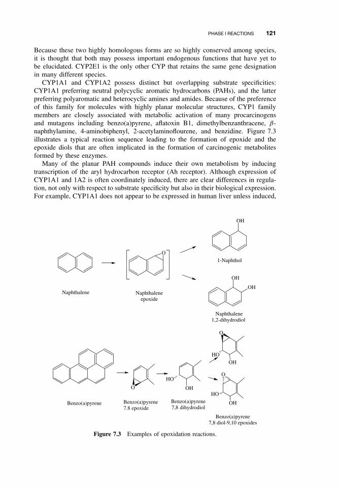
PHASE I REACTIONS 121
Because these two highly homologous forms are so highly conserved among species,it is thought that both may possess important endogenous functions that have yet tobe elucidated. CYP2E1 is the only other CYP that retains the same gene designationin many different species.
CYP1A1 and CYP1A2 possess distinct but overlapping substrate specificities:CYP1A1 preferring neutral polycyclic aromatic hydrocarbons (PAHs), and the latterpreferring polyaromatic and heterocyclic amines and amides. Because of the preferenceof this family for molecules with highly planar molecular structures, CYP1 familymembers are closely associated with metabolic activation of many procarcinogensand mutagens including benzo(a)pyrene, aflatoxin B1, dimethylbenzanthracene, β-naphthylamine, 4-aminobiphenyl, 2-acetylaminoflourene, and benzidine. Figure 7.3illustrates a typical reaction sequence leading to the formation of epoxide and theepoxide diols that are often implicated in the formation of carcinogenic metabolitesformed by these enzymes.
Many of the planar PAH compounds induce their own metabolism by inducingtranscription of the aryl hydrocarbon receptor (Ah receptor). Although expression ofCYP1A1 and 1A2 is often coordinately induced, there are clear differences in regula-tion, not only with respect to substrate specificity but also in their biological expression.For example, CYP1A1 does not appear to be expressed in human liver unless induced,
O
OH
OH
OH
1-Naphthol
Naphthalene1,2-dihydrodiol
Naphthalene Naphthaleneepoxide
O
OH
HO
O
OH
HO
OHO
OH
Benzo(a)pyrene Benzo(a)pyrene7.8 epoxide
Benzo(a)pyrene7,8 dihydrodiol
Benzo(a)pyrene7,8 diol-9,10 epoxides
Figure 7.3 Examples of epoxidation reactions.
122 METABOLISM OF TOXICANTS
whereas CYP1A2 is endogenously expressed in the liver. CYP1A1, however, is presentin many extrahepatic tissues including the lung, where there is a possible associa-tion between CYP-mediated activation of benzo(a)pyrene and other related chemicalspresent in cigarette smoke and lung cancer in humans.
The CYP2 family consists of 10 subfamilies, five of which are present in mammalianliver. Some of the more important isoforms found in humans within this family areCYP2A6, -2B6, -2C8, -2C9, -2C19, -2D6, and -2E1. The enzyme CYP2A6 is expressedprimarily in liver tissue, where it represents 1–10% of total CYP content. CYP2A6 isresponsible for the 7-hydroxylation of the naturally occurring plant compound coumarinand its activity is often phenotyped by monitoring this particular metabolic path-way. Other drugs metabolized by CYP2A6 include nicotine, 2-acetylaminofluorene,methoxyflurane, halothane, valproic acid, and disulfiram. Precarcinogens likely acti-vated by 2A6 include aflatoxin B1, 1,3 butadiene, 2,6-dichlorobenzonitrile, and anumber of nitrosamines. Because CYP2A6 is responsible for up to 80% of the humanmetabolism of nicotine, a number of studies have been conducted to determine whetherindividuals with 2A6 polymorphisms have reduced risk of lung cancers. Althoughtheoretically individuals lacking 2A6 would be expected to smoke less and be lesslikely to activate carcinogens found in tobacco smoke, studies have not conclusivelydemonstrated any clear associations between 2A6 polymorphisms and risk of lungcancer.
Like CYP2A6, the human isoform CYP2B6 has recently gained greater recogni-tion for its role in metabolism of many clinical drugs. Some common pharmaceuti-cal substrates for CYP2B6 include cyclophosphamide, nevirapine, S-mephobarbitol,artemisinin, bupropion, propofol, ifosfamide, ketamine, selegiline, and methadone.CYP2B6 has also been demonstrated to have a role in the activation of the organophos-phate, chlorpyrifos, and in the degradation of the commonly used insecticide repellant,diethyl toluamide (DEET). Historically it was thought that CYP2B6 is found in asmall proportion of livers (<25%), but more recent data using antibodies preparedfrom human proteins have demonstrated that most liver samples have detectable levelsof 2B6, though greater than 20-fold differences in levels of protein have been observed.
In contrast with CYP2A6 and CYP2B6, members of the CYP2C family consti-tute a fairly large percentage of CYP in human liver (ca. 20%) and are responsiblefor the metabolism of several drugs. All four members of the subfamily in humansexhibit genetic polymorphisms, many of which have important clinical consequences inaffected individuals. Genetic polymorphisms in CYP2C19 were shown to be respon-sible for one of the first described polymorphic effects, that involving mephenytoinmetabolism. This polymorphism significantly reduces the metabolism of mepheny-toin, resulting in the classification of those individuals possessing this trait as poormetabolizers (PM). Among Caucasians, PMs represent only 3–5% of the populations,while in Asian and Polynesian populations 12–23% and 38–79% of the populationsare represented, respectively. At least seven different mutations in this allele havebeen described, some of which negatively affect catalytic activity while others preventexpression of the protein. Other important drugs affected by these CYP2C19 polymor-phisms include the anti-ulcer drug omeprazole, other important proton pump inhibitors,barbiturates, certain tricyclic antidepressants such as imipramine, and the antimalarialdrug proguanil. Other important members of the CYP2C family in humans includeCYP2C8, -2C9, and -2C18. Substrates metabolized exclusively by CYP2C8 includeretinol, retinoic acid, taxol, and arachidonic acid. CYP2C9, the principal CYP2C in
PHASE I REACTIONS 123
human liver, metabolizes several important drugs including the diabetic agent tolbu-tamide, the anticonvulsant phenytoin, the anticoagulant warfarin and a number ofanti-inflammatory drugs including ibuprofen, diclofenac, and others. Both CYP2C9and -2C8, which are responsible for metabolism of the anticancer drug paclitaxel, havebeen demonstrated to be polymorphic.
CYP2E1 is the only member of the CYP2E family in most mammals with theexception of rabbits. Substrates for this family tend to be of small molecular weightand include ethanol, carbon tetrachloride, benzene, and acetaminophen. In contrastto many other inducible CYP families, CYP2E1 is regulated by a combination ofincreased transcription levels and increased message and protein stabilization.
Undoubtedly the largest amount of CYP in human liver is that of the CYP3 family.CYP3A4 is the most abundant CYP in the human liver, accounting for nearly 30%of the total amount, and is known to metabolize many important drugs includingcyclosporine A, nifedipine, rapamycin, ethinyl estradiol, quinidine, digitoxin, lido-caine, erythromycin, midazolam, triazolam, lovastatin, and tamoxifen. Other importantoxidations ascribed to the CYP3 family include many steroid hormones, macrolideantibiotics, alkaloids, benzodiazepines, dihydropyridines, warfarin, polycyclic hydro-carbon-derived dihydrodiols, and aflatoxin B1. Many chemicals are also capable ofinducing this family including phenobarbital, rifampicin, and dexamethasone. Becauseof potential difficulties arising from CYP induction, drugs metabolized by this familymust be closely examined for the possibility of harmful drug-drug interactions.
Cytochrome P450 Reactions. Although microsomal monooxygenase reactions arebasically similar in the role played by molecular oxygen and in the supply of electrons,the many CYP isoforms can attack a large variety of xenobiotic substrates, with bothsubstrates and products falling into many different chemical classes. In the followingsections enzyme activities are therefore classified on the basis of the overall chemicalreaction catalyzed; one should bear in mind, however, that not only do these classesoften overlap, but often a substrate may also undergo more than one reaction. SeeTable 7.1 for a listing of important oxidation and reduction reactions of CYPs.
Epoxidation and Aromatic Hydroxylation. Epoxidation is an extremely importantmicrosomal reaction because not only can stable and environmentally persistent epox-ides be formed (see aliphatic epoxidations, below), but highly reactive intermediatesof aromatic hydroxylations, such as arene oxides, can also be produced. These highlyreactive intermediates are known to be involved in chemical carcinogenesis as well aschemically induced cellular and tissue necrosis.
The oxidation of naphthalene was one of the earliest examples of an epoxide asan intermediate in aromatic hydroxylation. As shown in Figure 7.3, the epoxide canrearrange nonenzymatically to yield predominantly 1-naphthol, or interact with theenzyme epoxide hydrolase to yield the dihydrodiol, or interact with glutathione S-transferase to yield the glutathione conjugate, which is ultimately metabolized to amercapturic acid. These reactions are also of importance in the metabolism of otherxenobiotics that contain an aromatic nucleus, such as the insecticide carbaryl and thecarcinogen benzo(a)pyrene.
The ultimate carcinogens arising from the metabolic activation of benzo(a)pyrene arestereoisomers of benzo(a)pyrene 7,8-diol-9,10-epoxide (Figure 7.3). These metabolitesarise by prior formation of the 7,8 epoxide, which gives rise to the 7,8-dihydrodiol
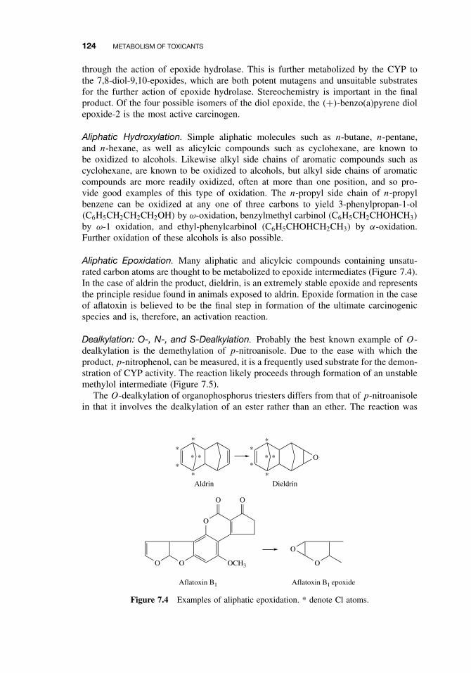
124 METABOLISM OF TOXICANTS
through the action of epoxide hydrolase. This is further metabolized by the CYP tothe 7,8-diol-9,10-epoxides, which are both potent mutagens and unsuitable substratesfor the further action of epoxide hydrolase. Stereochemistry is important in the finalproduct. Of the four possible isomers of the diol epoxide, the (+)-benzo(a)pyrene diolepoxide-2 is the most active carcinogen.
Aliphatic Hydroxylation. Simple aliphatic molecules such as n-butane, n-pentane,and n-hexane, as well as alicylcic compounds such as cyclohexane, are known tobe oxidized to alcohols. Likewise alkyl side chains of aromatic compounds such ascyclohexane, are known to be oxidized to alcohols, but alkyl side chains of aromaticcompounds are more readily oxidized, often at more than one position, and so pro-vide good examples of this type of oxidation. The n-propyl side chain of n-propylbenzene can be oxidized at any one of three carbons to yield 3-phenylpropan-1-ol(C6H5CH2CH2CH2OH) by ω-oxidation, benzylmethyl carbinol (C6H5CH2CHOHCH3)
by ω-1 oxidation, and ethyl-phenylcarbinol (C6H5CHOHCH2CH3) by α-oxidation.Further oxidation of these alcohols is also possible.
Aliphatic Epoxidation. Many aliphatic and alicylcic compounds containing unsatu-rated carbon atoms are thought to be metabolized to epoxide intermediates (Figure 7.4).In the case of aldrin the product, dieldrin, is an extremely stable epoxide and representsthe principle residue found in animals exposed to aldrin. Epoxide formation in the caseof aflatoxin is believed to be the final step in formation of the ultimate carcinogenicspecies and is, therefore, an activation reaction.
Dealkylation: O-, N-, and S-Dealkylation. Probably the best known example of O-dealkylation is the demethylation of p-nitroanisole. Due to the ease with which theproduct, p-nitrophenol, can be measured, it is a frequently used substrate for the demon-stration of CYP activity. The reaction likely proceeds through formation of an unstablemethylol intermediate (Figure 7.5).
The O-dealkylation of organophosphorus triesters differs from that of p-nitroanisolein that it involves the dealkylation of an ester rather than an ether. The reaction was
O
O O
O
Aldrin Dieldrin
O O
OCH3 O
O
Aflatoxin B1 Aflatoxin B1 epoxide
*
*
** ***
*
**
**
Figure 7.4 Examples of aliphatic epoxidation. * denote Cl atoms.
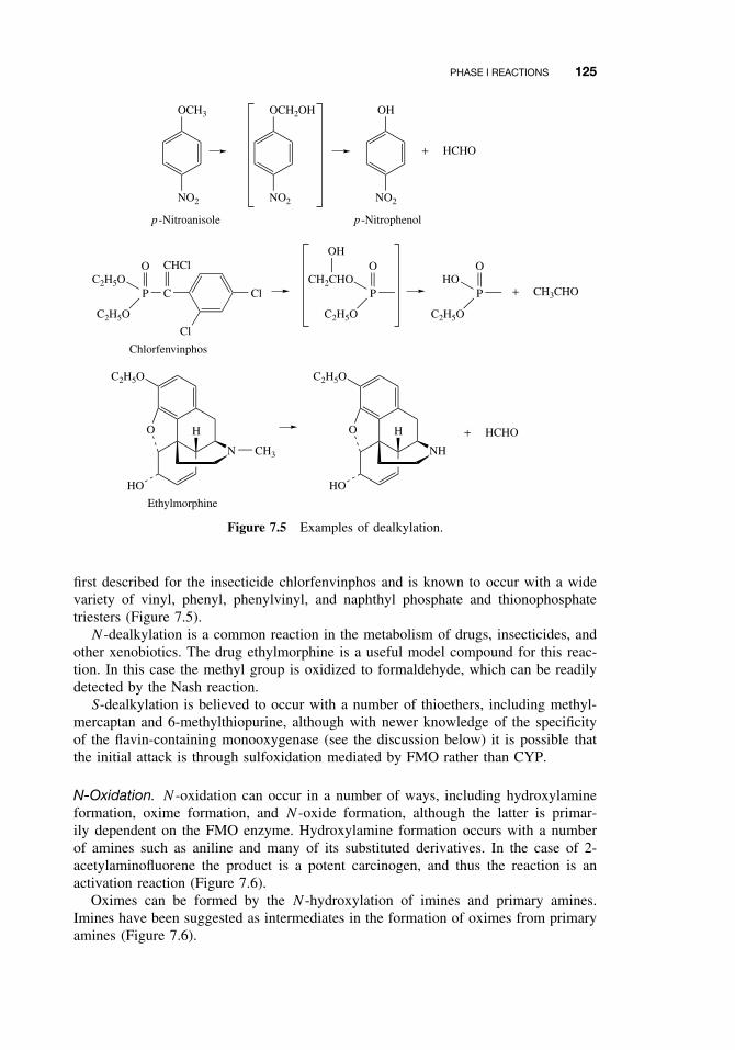
PHASE I REACTIONS 125
OCH3
NO2
OCH2OH
NO2
OH
NO2
+ HCHO
p -Nitroanisole p -Nitrophenol
Cl
Cl
CP
CHClOC2H5O
C2H5O
P
OCH2CHO
C2H5O
P
OHO
C2H5O
OH
+ CH3CHO
C2H5O
O
HO
H
N CH3
C2H5O
O
HO
H
NH
+ HCHO
Chlorfenvinphos
Ethylmorphine
Figure 7.5 Examples of dealkylation.
first described for the insecticide chlorfenvinphos and is known to occur with a widevariety of vinyl, phenyl, phenylvinyl, and naphthyl phosphate and thionophosphatetriesters (Figure 7.5).
N -dealkylation is a common reaction in the metabolism of drugs, insecticides, andother xenobiotics. The drug ethylmorphine is a useful model compound for this reac-tion. In this case the methyl group is oxidized to formaldehyde, which can be readilydetected by the Nash reaction.
S-dealkylation is believed to occur with a number of thioethers, including methyl-mercaptan and 6-methylthiopurine, although with newer knowledge of the specificityof the flavin-containing monooxygenase (see the discussion below) it is possible thatthe initial attack is through sulfoxidation mediated by FMO rather than CYP.
N-Oxidation. N -oxidation can occur in a number of ways, including hydroxylamineformation, oxime formation, and N -oxide formation, although the latter is primar-ily dependent on the FMO enzyme. Hydroxylamine formation occurs with a numberof amines such as aniline and many of its substituted derivatives. In the case of 2-acetylaminofluorene the product is a potent carcinogen, and thus the reaction is anactivation reaction (Figure 7.6).
Oximes can be formed by the N -hydroxylation of imines and primary amines.Imines have been suggested as intermediates in the formation of oximes from primaryamines (Figure 7.6).
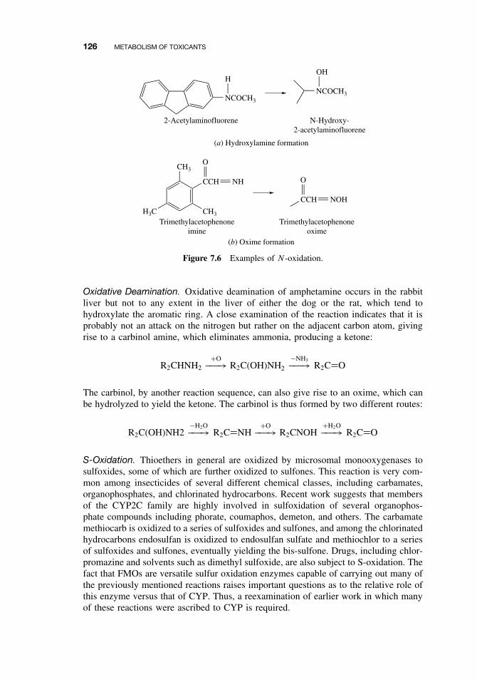
126 METABOLISM OF TOXICANTS
NCOCH3
H
(a) Hydroxylamine formation
2-Acetylaminofluorene
NCOCH3
OH
N-Hydroxy-2-acetylaminofluorene
H3C
CH3
CCH
CH3
NH
O
CCH NOH
O
(b) Oxime formation
Trimethylacetophenoneimine
Trimethylacetophenoneoxime
Figure 7.6 Examples of N-oxidation.
Oxidative Deamination. Oxidative deamination of amphetamine occurs in the rabbitliver but not to any extent in the liver of either the dog or the rat, which tend tohydroxylate the aromatic ring. A close examination of the reaction indicates that it isprobably not an attack on the nitrogen but rather on the adjacent carbon atom, givingrise to a carbinol amine, which eliminates ammonia, producing a ketone:
R2CHNH2+O−−−→ R2C(OH)NH2
−NH3−−−→ R2C=O
The carbinol, by another reaction sequence, can also give rise to an oxime, which canbe hydrolyzed to yield the ketone. The carbinol is thus formed by two different routes:
R2C(OH)NH2−H2O−−−→ R2C=NH
+O−−−→ R2CNOH+H2O−−−→ R2C=O
S-Oxidation. Thioethers in general are oxidized by microsomal monooxygenases tosulfoxides, some of which are further oxidized to sulfones. This reaction is very com-mon among insecticides of several different chemical classes, including carbamates,organophosphates, and chlorinated hydrocarbons. Recent work suggests that membersof the CYP2C family are highly involved in sulfoxidation of several organophos-phate compounds including phorate, coumaphos, demeton, and others. The carbamatemethiocarb is oxidized to a series of sulfoxides and sulfones, and among the chlorinatedhydrocarbons endosulfan is oxidized to endosulfan sulfate and methiochlor to a seriesof sulfoxides and sulfones, eventually yielding the bis-sulfone. Drugs, including chlor-promazine and solvents such as dimethyl sulfoxide, are also subject to S-oxidation. Thefact that FMOs are versatile sulfur oxidation enzymes capable of carrying out many ofthe previously mentioned reactions raises important questions as to the relative role ofthis enzyme versus that of CYP. Thus, a reexamination of earlier work in which manyof these reactions were ascribed to CYP is required.
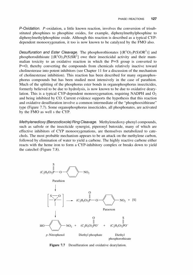
PHASE I REACTIONS 127
P-Oxidation. P -oxidation, a little known reaction, involves the conversion of trisub-stituted phosphines to phosphine oxides, for example, diphenylmethylphosphine todiphenylmethylphosphine oxide. Although this reaction is described as a typical CYP-dependent monooxygenation, it too is now known to be catalyzed by the FMO also.
Desulfuration and Ester Cleavage. The phosphorothionates [(R1O)2P(S)OR2)] andphosphorodithioate [(R1O)2P(S)SR2] owe their insecticidal activity and their mam-malian toxicity to an oxidative reaction in which the P=S group is converted toP=O, thereby converting the compounds from chemicals relatively inactive towardcholinesterase into potent inhibitors (see Chapter 11 for a discussion of the mechanismof cholinesterase inhibition). This reaction has been described for many organophos-phorus compounds but has been studied most intensively in the case of parathion.Much of the splitting of the phosphorus ester bonds in organophosphorus insecticides,formerly believed to be due to hydrolysis, is now known to be due to oxidative deary-lation. This is a typical CYP-dependent monooxygenation, requiring NADPH and O2
and being inhibited by CO. Current evidence supports the hypothesis that this reactionand oxidative desulfuration involve a common intermediate of the “phosphooxithirane”type (Figure 7.7). Some organophosphorus insecticides, all phosphonates, are activatedby the FMO as well s the CYP.
Methylenedioxy (Benzodioxole) Ring Cleavage. Methylenedioxy-phenyl compounds,such as safrole or the insecticide synergist, piperonyl butoxide, many of which areeffective inhibitors of CYP monooxygenations, are themselves metabolized to cate-chols. The most probable mechanism appears to be an attack on the methylene carbon,followed by elimination of water to yield a carbene. The highly reactive carbene eitherreacts with the heme iron to form a CYP-inhibitory complex or breaks down to yieldthe catechol (Figure 7.8).
NO2O(C2H5O)2P
S
Parathion
POS
NO2O(C2H5O)2P
O
+ [S]
NO2HO + (C2H5O)2PO−
O
+ (C2H5O)2PO−
S
Paraoxon
p -Nitrophenol Diethyl phosphate Diethylphosphorothioate
Figure 7.7 Desulfuration and oxidative dearylation.
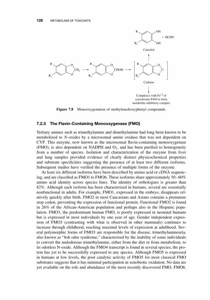
128 METABOLISM OF TOXICANTS
R
R1
OH
OH
+ HCHO
Catechol
R
R1O
CH2
O R
R1O
CHOH
O R
R1O
C:
O
Carbene
Complexes with Fe+2 ofcytochrome P450 to form
metabolite inhibitory complex
Figure 7.8 Monooxygenation of methylenedioxyphenyl compounds.
7.2.3 The Flavin-Containing Monooxygenase (FMO)
Tertiary amines such as trimethylamine and dimethylamine had long been known to bemetabolized to N -oxides by a microsomal amine oxidase that was not dependent onCYP. This enzyme, now known as the microsomal flavin-containing monooxygenase(FMO), is also dependent on NADPH and O2, and has been purified to homogeneityfrom a number of species. Isolation and characterization of the enzyme from liverand lung samples provided evidence of clearly distinct physicochemical propertiesand substrate specificities suggesting the presence of at least two different isoforms.Subsequent studies have verified the presence of multiple forms of the enzyme.
At least six different isoforms have been described by amino acid or cDNA sequenc-ing, and are classified as FMO1 to FMO6. These isoforms share approximately 50–60%amino acid identity across species lines. The identity of orthologues is greater than82%. Although each isoform has been characterized in humans, several are essentiallynonfunctional in adults. For example, FMO1, expressed in the embryo, disappears rel-atively quickly after birth. FMO2 in most Caucasians and Asians contains a prematurestop codon, preventing the expression of functional protein. Functional FMO2 is foundin 26% of the African-American population and perhaps also in the Hispanic popu-lation. FMO3, the predominant human FMO, is poorly expressed in neonatal humansbut is expressed in most individuals by one year of age. Gender independent expres-sion of FMO3 (contrasting with what is observed in other mammals) continues toincrease through childhood, reaching maximal levels of expression at adulthood. Sev-eral polymorphic forms of FMO3 are responsible for the disease, trimethylamineuria,also known as “fish odor syndrome,” characterized by the inability of some individualsto convert the malodorous trimethylamine, either from the diet or from metabolism, toits odorless N-oxide. Although the FMO4 transcript is found in several species, the pro-tein has yet to be successfully expressed in any species. Although FMO5 is expressedin humans at low levels, the poor catalytic activity of FMO5 for most classical FMOsubstrates suggests that it has minimal participation in xenobiotic oxidation. No data areyet available on the role and abundance of the most recently discovered FMO, FMO6.
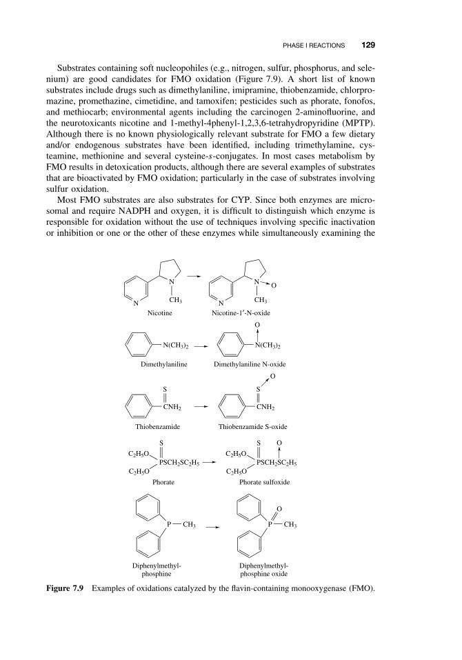
PHASE I REACTIONS 129
Substrates containing soft nucleopohiles (e.g., nitrogen, sulfur, phosphorus, and sele-nium) are good candidates for FMO oxidation (Figure 7.9). A short list of knownsubstrates include drugs such as dimethylaniline, imipramine, thiobenzamide, chlorpro-mazine, promethazine, cimetidine, and tamoxifen; pesticides such as phorate, fonofos,and methiocarb; environmental agents including the carcinogen 2-aminofluorine, andthe neurotoxicants nicotine and 1-methyl-4phenyl-1,2,3,6-tetrahydropyridine (MPTP).Although there is no known physiologically relevant substrate for FMO a few dietaryand/or endogenous substrates have been identified, including trimethylamine, cys-teamine, methionine and several cysteine-s-conjugates. In most cases metabolism byFMO results in detoxication products, although there are several examples of substratesthat are bioactivated by FMO oxidation; particularly in the case of substrates involvingsulfur oxidation.
Most FMO substrates are also substrates for CYP. Since both enzymes are micro-somal and require NADPH and oxygen, it is difficult to distinguish which enzyme isresponsible for oxidation without the use of techniques involving specific inactivationor inhibition or one or the other of these enzymes while simultaneously examining the
N
N
CH3 N
N
CH3
O
Nicotine Nicotine-1′-N-oxide
N(CH3)2 N(CH3)2
O
Dimethylaniline Dimethylaniline N-oxide
CNH2
Thiobenzamide
S
CNH2
Thiobenzamide S-oxide
S
O
PSCH2SC2H5
C2H5O
C2H5O
S
PSCH2SC2H5
C2H5O
C2H5O
S O
Phorate Phorate sulfoxide
P CH3 P CH3
O
Diphenylmethyl-phosphine
Diphenylmethyl-phosphine oxide
Figure 7.9 Examples of oxidations catalyzed by the flavin-containing monooxygenase (FMO).
130 METABOLISM OF TOXICANTS
metabolic contribution of the other. Since FMOs are generally heat labile, heating themicrosomal preparation to 50◦C for one minute inactivates the FMOs while havingminimal effects of CYPs. Alternatively, the contribution of FMO can be assessed byuse of a general CYP inhibitor such as N -benzylimidazole or by an inhibitory antibodyto NADPH cytochrome P450 reductase, a necessary CYP coenzyme. Typically resultsof these tests are sought in combination so that the best estimates of CYP and FMOcontribution can be obtained.
Toxicologically it is of interest that the FMO enzyme is responsible for the oxida-tion of nicotine to nicotine 1′-N-oxide, whereas the oxidation of nicotine to cotinineis catalyzed by two enzymes acting in sequence: CYP followed by a soluble aldehydedehydrogenase. Thus nicotine is metabolized by two different routes, the relative con-tributions of which may vary with both the extrinsic and intrinsic factors outlined inChapter 9.
7.2.4 Nonmicrosomal Oxidations
In addition to the microsomal monooxygenases, other enzymes are involved in theoxidation of xenobiotics. These enzymes are located in the mitochondria or in thesoluble cytoplasm of the cell.
Alcohol Dehydrogenase. Alcohol dehydrogenases catalyze the conversion of alco-hols to aldehydes or ketones:
RCH2OH + NAD+ −−−→ RCHO + NADH + H+
This reaction should not be confused with the monooxygenation of ethanol by CYPthat occurs in the microsomes. The alcohol dehydrogenase reaction is reversible, withthe carbonyl compounds being reduced to alcohols.
This enzyme is found in the soluble fraction of the liver, kidney, and lung and isprobably the most important enzyme involved in the metabolism of foreign alcohols.Alcohol dehydrogenase is a dimer whose subunits can occur in several forms undergenetic control, thus giving rise to a large number of variants of the enzyme. Inmammals, six classes of enzymes have been described. Alcohol dehydrogenase canuse either NAD or NADP as a coenzyme, but the reaction proceeds at a much slowerrate with NADP. In the intact organism the reaction proceeds in the direction of alcoholconsumption, because aldehydes are further oxidized to acids. Because aldehydes aretoxic and are not readily excreted because of their lipophilicity, alcohol oxidationmay be considered an activation reaction, the further oxidation to an acid being adetoxication step.
Primary alcohols are oxidized to aldehydes, n-butanol being the substrate oxidizedat the highest rate. Although secondary alcohols are oxidized to ketones, the rate isless than for primary alcohols, and tertiary alcohols are not readily oxidized. Alcoholdehydrogenase is inhibited by a number of heterocyclic compounds such as pyrazole,imidazole, and their derivatives.
Aldehyde Dehydrogense. Aldehydes are generated from a variety of endogenousand exogenous substrates. Endogenous aldehydes may be formed during metabolism ofamino acids, carbohydrates, lipids, biogenic amines, vitamins, and steroids. Metabolism
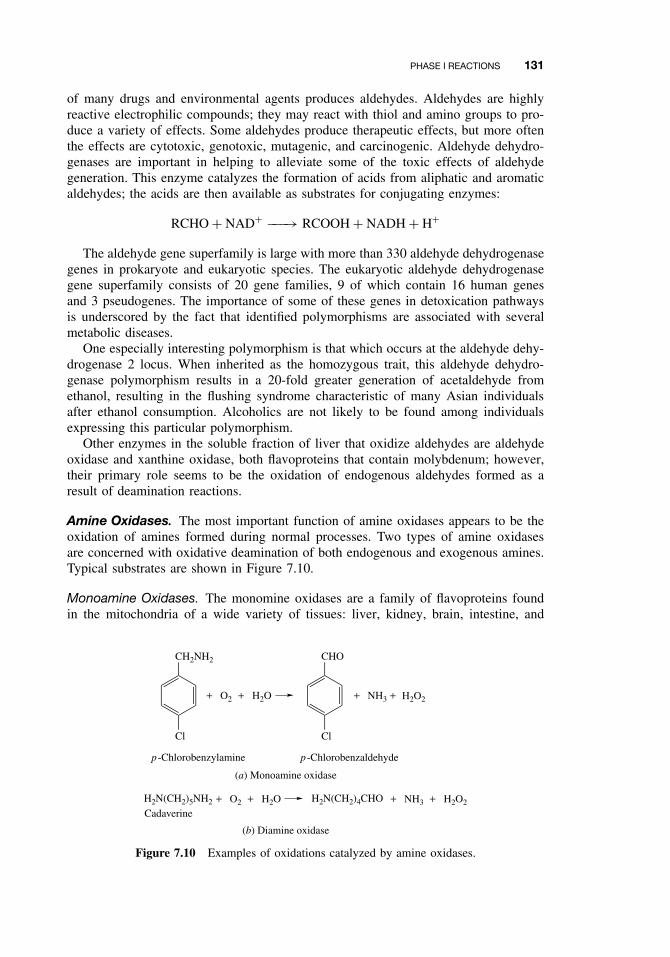
PHASE I REACTIONS 131
of many drugs and environmental agents produces aldehydes. Aldehydes are highlyreactive electrophilic compounds; they may react with thiol and amino groups to pro-duce a variety of effects. Some aldehydes produce therapeutic effects, but more oftenthe effects are cytotoxic, genotoxic, mutagenic, and carcinogenic. Aldehyde dehydro-genases are important in helping to alleviate some of the toxic effects of aldehydegeneration. This enzyme catalyzes the formation of acids from aliphatic and aromaticaldehydes; the acids are then available as substrates for conjugating enzymes:
RCHO + NAD+ −−−→ RCOOH + NADH + H+
The aldehyde gene superfamily is large with more than 330 aldehyde dehydrogenasegenes in prokaryote and eukaryotic species. The eukaryotic aldehyde dehydrogenasegene superfamily consists of 20 gene families, 9 of which contain 16 human genesand 3 pseudogenes. The importance of some of these genes in detoxication pathwaysis underscored by the fact that identified polymorphisms are associated with severalmetabolic diseases.
One especially interesting polymorphism is that which occurs at the aldehyde dehy-drogenase 2 locus. When inherited as the homozygous trait, this aldehyde dehydro-genase polymorphism results in a 20-fold greater generation of acetaldehyde fromethanol, resulting in the flushing syndrome characteristic of many Asian individualsafter ethanol consumption. Alcoholics are not likely to be found among individualsexpressing this particular polymorphism.
Other enzymes in the soluble fraction of liver that oxidize aldehydes are aldehydeoxidase and xanthine oxidase, both flavoproteins that contain molybdenum; however,their primary role seems to be the oxidation of endogenous aldehydes formed as aresult of deamination reactions.
Amine Oxidases. The most important function of amine oxidases appears to be theoxidation of amines formed during normal processes. Two types of amine oxidasesare concerned with oxidative deamination of both endogenous and exogenous amines.Typical substrates are shown in Figure 7.10.
Monoamine Oxidases. The monomine oxidases are a family of flavoproteins foundin the mitochondria of a wide variety of tissues: liver, kidney, brain, intestine, and
CH2NH2
Cl
+ O2 + H2O
CHO
Cl
+ NH3 + H2O2
p -Chlorobenzylamine p -Chlorobenzaldehyde
(a) Monoamine oxidase
H2N(CH2)5NH2 + O2 + H2O H2N(CH2)4CHO + NH3 + H2O2
Cadaverine
(b) Diamine oxidase
Figure 7.10 Examples of oxidations catalyzed by amine oxidases.
132 METABOLISM OF TOXICANTS
blood platelets. They are a group of similar enzymes with overlapping specificities andinhibition. Although the enzyme in the central nervous system is concerned primarilywith neurotransmitter turnover, that in the liver will deaminate primary, secondary, andtertiary aliphatic amines, reaction rates with the primary amines being faster. Electron-withdrawing substitutions on an aromatic ring increase the reaction rate, whereascompounds with a methyl group on the α-carbon such as amphetamine and ephedrineare not metabolized.
Diamine Oxidases. Diamine oxidases are enzymes that also oxidize amines to alde-hydes. The preferred substates are aliphatic diamines in which the chain length is four(putrescine) or five (cadaverine) carbon atoms. Diamines with carbon chains longerthan nine will not serve as substrates but can be oxidized by monoamine oxidases.Secondary and tertiary amines are not metabolized. Diamine oxidases are typicallysoluble pyridoxal phosphate-containing proteins that also contain copper. They havebeen found in a number of tissues, including liver, intestine, kidney, and placenta.
7.2.5 Cooxidation by Cyclooxygenases
During the biosynthesis of prostaglandins, a polyunsaturated fatty acid, such as arachi-donic acid, is first oxygenated to yield a hydroperoxy endoperoxide, prostaglandin G2.This is then further metabolized to prostaglandin H2, both reactions being catalyzedby the same enzyme, cyclooxygenase (COX), also known as prostaglandin synthase(Figure 7.11). This enzyme is located in the microsomal membrane and is found ingreatest levels in respiratory tissues such as the lung. It is also common in the kid-ney and seminal vesicle. It is a glycoprotein with a subunit molecular mass of about70,000 daltons, containing one heme per subunit. During the second step of the previoussequence (peroxidase), many xenobiotics can be cooxidized, and investigations of themechanism have shown that the reactions are hydroperoxide-dependent reactions cat-alyzed by a peroxidase that uses prostaglandin G as a substrate. In at least some of thesecases, the identity of this peroxidase has been established as a prostaglandin synthase.Many of the reactions are similar or identical to those catalyzed by other peroxidasesand also by microsomal monooxygenases; they include both detoxication and activa-tion reactions. This mechanism is important in xenobiotic metabolism, particularly intissues that are low in CYP and/or the FMO but high in prostaglandin synthase.
The cyclooxygenase (COX) enzyme is known to exist as two distinct isoforms.COX-1 is a constitutively expressed housekeeping enzyme found in nearly all tissuesand mediates physiological responses. COX-2 is an inducible form expressed primarilyby cells involved in the inflammatory response. Several tissues low in CYP expressionare rich in COX, which is believed to have significance in the carcinogenic effects ofaromatic amines in these organs.
During cooxidation, some substrates are activated to become more toxic than theywere originally. In some cases substrate oxidation results in the production of freeradicals, which may initiate lipid peroxidation or bind to cellular proteins or DNA.Another activation pathway involves the formation of a peroxyl radical from subsequentmetabolism of prostaglandin G2. This reactive molecule can epoxidize many substatesincluding polycyclic aromatic hydrocarbons, generally resulting in increasing toxicityof the respective substrates.
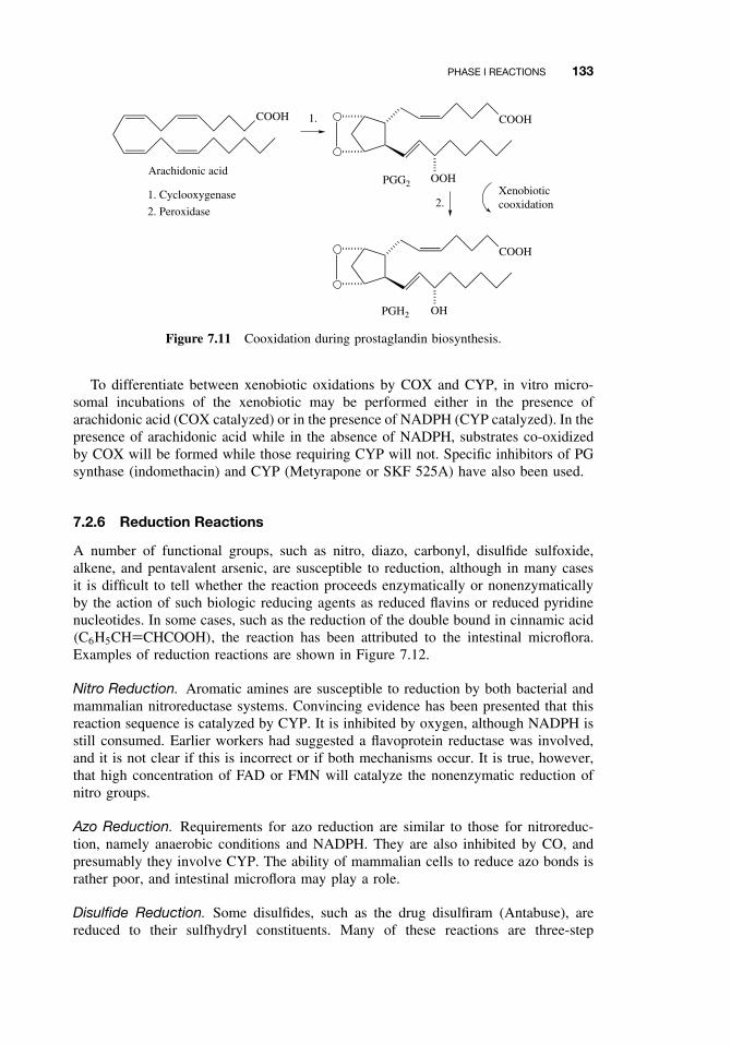
PHASE I REACTIONS 133
COOH
Arachidonic acid
1. Cyclooxygenase
2. Peroxidase
COOH
OOH
COOH
OH
PGG2
PGH2
Xenobioticcooxidation
1.
2.
Figure 7.11 Cooxidation during prostaglandin biosynthesis.
To differentiate between xenobiotic oxidations by COX and CYP, in vitro micro-somal incubations of the xenobiotic may be performed either in the presence ofarachidonic acid (COX catalyzed) or in the presence of NADPH (CYP catalyzed). In thepresence of arachidonic acid while in the absence of NADPH, substrates co-oxidizedby COX will be formed while those requiring CYP will not. Specific inhibitors of PGsynthase (indomethacin) and CYP (Metyrapone or SKF 525A) have also been used.
7.2.6 Reduction Reactions
A number of functional groups, such as nitro, diazo, carbonyl, disulfide sulfoxide,alkene, and pentavalent arsenic, are susceptible to reduction, although in many casesit is difficult to tell whether the reaction proceeds enzymatically or nonenzymaticallyby the action of such biologic reducing agents as reduced flavins or reduced pyridinenucleotides. In some cases, such as the reduction of the double bound in cinnamic acid(C6H5CH=CHCOOH), the reaction has been attributed to the intestinal microflora.Examples of reduction reactions are shown in Figure 7.12.
Nitro Reduction. Aromatic amines are susceptible to reduction by both bacterial andmammalian nitroreductase systems. Convincing evidence has been presented that thisreaction sequence is catalyzed by CYP. It is inhibited by oxygen, although NADPH isstill consumed. Earlier workers had suggested a flavoprotein reductase was involved,and it is not clear if this is incorrect or if both mechanisms occur. It is true, however,that high concentration of FAD or FMN will catalyze the nonenzymatic reduction ofnitro groups.
Azo Reduction. Requirements for azo reduction are similar to those for nitroreduc-tion, namely anaerobic conditions and NADPH. They are also inhibited by CO, andpresumably they involve CYP. The ability of mammalian cells to reduce azo bonds israther poor, and intestinal microflora may play a role.
Disulfide Reduction. Some disulfides, such as the drug disulfiram (Antabuse), arereduced to their sulfhydryl constituents. Many of these reactions are three-step
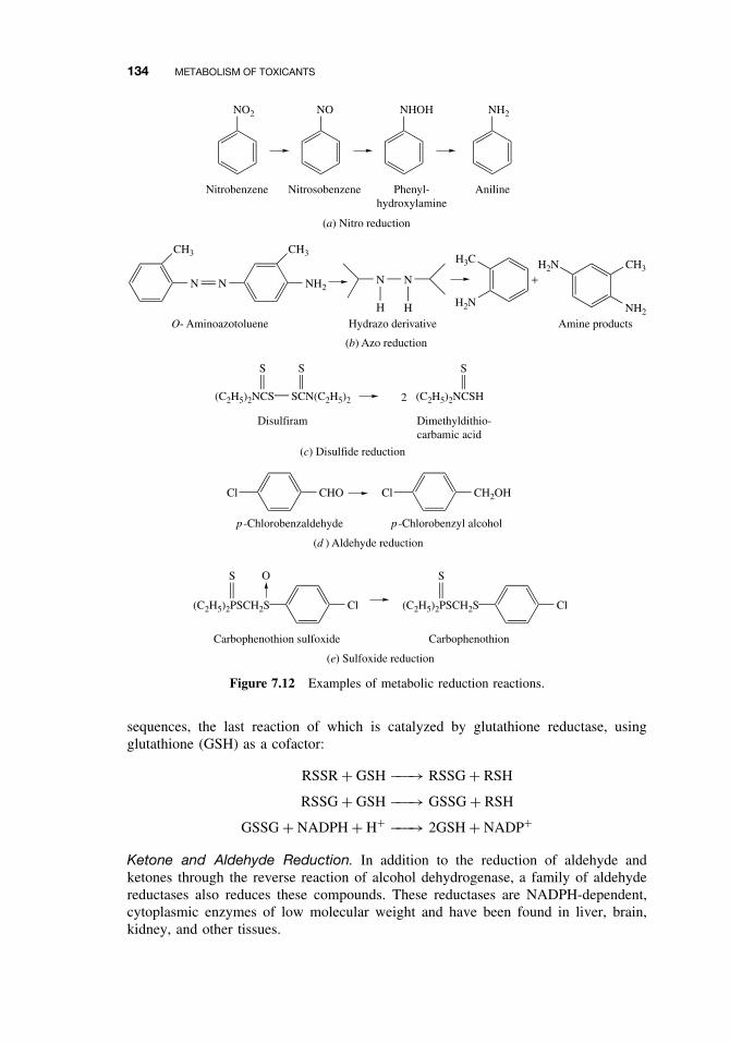
134 METABOLISM OF TOXICANTS
NO2 NO NHOH NH2
Nitrobenzene Nitrosobenzene Phenyl-hydroxylamine
Aniline
(a) Nitro reduction
CH3
N N
CH3
NH2
(b) Azo reduction
N N
H HO- Aminoazotoluene Hydrazo derivative
H3C
H2N
H2N+
CH3
NH2
Amine products
(C2H5)2NCS
S
SCN(C2H5)2
S
Disulfiram
(C2H5)2NCSH
S
Dimethyldithio-carbamic acid
2
(c) Disulfide reduction
CHOCl
(d ) Aldehyde reduction
p -Chlorobenzaldehyde
CH2OHCl
p -Chlorobenzyl alcohol
(e) Sulfoxide reduction
(C2H5)2PSCH2S
S O
Cl (C2H5)2PSCH2S
S
Cl
Carbophenothion sulfoxide Carbophenothion
Figure 7.12 Examples of metabolic reduction reactions.
sequences, the last reaction of which is catalyzed by glutathione reductase, usingglutathione (GSH) as a cofactor:
RSSR + GSH −−−→ RSSG + RSH
RSSG + GSH −−−→ GSSG + RSH
GSSG + NADPH + H+ −−−→ 2GSH + NADP+
Ketone and Aldehyde Reduction. In addition to the reduction of aldehyde andketones through the reverse reaction of alcohol dehydrogenase, a family of aldehydereductases also reduces these compounds. These reductases are NADPH-dependent,cytoplasmic enzymes of low molecular weight and have been found in liver, brain,kidney, and other tissues.
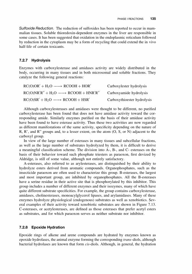
PHASE I REACTIONS 135
Sulfoxide Reduction. The reduction of sulfoxides has been reported to occur in mam-malian tissues. Soluble thioredoxin-dependent enzymes in the liver are responsible insome cases. It has been suggested that oxidation in the endoplasmic reticulum followedby reduction in the cytoplasm may be a form of recycling that could extend the in vivohalf-life of certain toxicants.
7.2.7 Hydrolysis
Enzymes with carboxylesterase and amidases activity are widely distributed in thebody, occurring in many tissues and in both microsomal and soluble fractions. Theycatalyze the following general reactions:
RC(O)OR′ + H2O −−−→ RCOOH + HOR′ Carboxylester hydrolysis
RC(O)NR′R′′ + H2O −−−→ RCOOH + HNR′R′′ Carboxyamide hydrolysis
RC(O)SR′ + H2O −−−→ RCOOH + HSR′ Carboxythioester hydrolysis
Although carboxylesterases and amidases were thought to be different, no purifiedcarboxylesterase has been found that does not have amidase activity toward the cor-responding amide. Similarly enzymes purified on the basis of their amidase activityhave been found to have esterase activity. Thus these two activities are now regardedas different manifestations of the same activity, specificity depending on the nature ofR, R’, and R” groups and, to a lesser extent, on the atom (O, S, or N) adjacent to thecarboxyl group.
In view of the large number of esterases in many tissues and subcellular fractions,as well as the large number of substrates hydrolyzed by them, it is difficult to derivea meaningful classification scheme. The division into A-, B-, and C- esterases on thebasis of their behavior toward such phosphate triesters as paraoxon, first devized byAldridge, is still of some value, although not entirely satisfactory.
A-esterases, also referred to as arylesterases, are distinguished by their ability tohydrolyze esters derived from aromatic compounds. Organophosphates, such as theinsecticide paraoxon are often used to characterize this group. B-esterases, the largestand most important group, are inhibited by organophosphates. All the B-esteraseshave a serine residue in their active site that is phosphorylated by this inhibitor. Thisgroup includes a number of different enzymes and their isozymes, many of which havequite different substrate specificities. For example, the group contains carboxylesterase,amidases, cholinesterases, monoacylglycerol lipases, and arylamidases. Many of theseenzymes hydrolyze physiological (endogenous) substrates as well as xenobiotics. Sev-eral examples of their activity toward xenobiotic substrates are shown in Figure 7.13.C-esterases, or acetylesterases, are defined as those esterases that prefer acetyl estersas substrates, and for which paraoxon serves as neither substrate nor inhibitor.
7.2.8 Epoxide Hydration
Epoxide rings of alkene and arene compounds are hydrated by enzymes known asepoxide hydrolases, the animal enzyme forming the corresponding trans-diols, althoughbacterial hydrolases are known that form cis-diols. Although, in general, the hydration
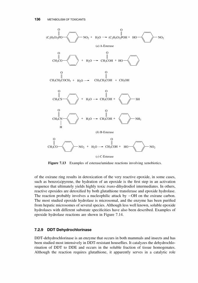
136 METABOLISM OF TOXICANTS
NO2(C2H5O)2PO + H2O (C2H5O)2POH
O O
+ NO2HO
(a) A-Esterase
+ H2O HOCH3CO
O
CH3COH
O
+
CH3CH2COCH3
O
+ H2O CH3CH2COH
O
+ CH3OH
CH3CS
O
+ H2O CH3COH
O
+ SH
CH3CN + H2O CH3COH
O
+ NH2
O
H
(b) B-Esterase
CH3CO + H2O CH3COH
O
+ NO2
O
NO2 HO
(c) C-Esterase
Figure 7.13 Examples of esterase/amidase reactions involving xenobiotics.
of the oxirane ring results in detoxication of the very reactive epoxide, in some cases,such as benzo(a)pyrene, the hydration of an epoxide is the first step in an activationsequence that ultimately yields highly toxic trans-dihydrodiol intermediates. In others,reactive epoxides are detoxified by both glutathione transferase and epoxide hydrolase.The reaction probably involves a nucleophilic attack by −OH on the oxirane carbon.The most studied epoxide hydrolase is microsomal, and the enzyme has been purifiedfrom hepatic microsomes of several species. Although less well known, soluble epoxidehydrolases with different substrate specificities have also been described. Examples ofepoxide hydrolase reactions are shown in Figure 7.14.
7.2.9 DDT Dehydrochlorinase
DDT-dehydrochlorinase is an enzyme that occurs in both mammals and insects and hasbeen studied most intensively in DDT-resistant houseflies. It catalyzes the dehydrochlo-rination of DDT to DDE and occurs in the soluble fraction of tissue homogenates.Although the reaction requires glutathione, it apparently serves in a catalytic role
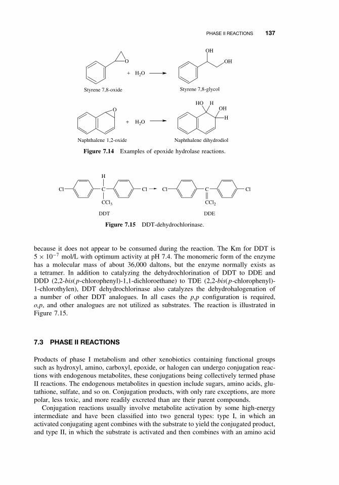
PHASE II REACTIONS 137
O
O
+ H2O
OH
OH
H
HHOOH
+ H2O
Styrene 7,8-oxide Styrene 7,8-glycol
Naphthalene 1,2-oxide Naphthalene dihydrodiol
Figure 7.14 Examples of epoxide hydrolase reactions.
Cl C
H
CCl3
Cl Cl C
CCl2
Cl
DDT DDE
Figure 7.15 DDT-dehydrochlorinase.
because it does not appear to be consumed during the reaction. The Km for DDT is5 × 10−7 mol/L with optimum activity at pH 7.4. The monomeric form of the enzymehas a molecular mass of about 36,000 daltons, but the enzyme normally exists asa tetramer. In addition to catalyzing the dehydrochlorination of DDT to DDE andDDD (2,2-bis(p-chlorophenyl)-1,1-dichloroethane) to TDE (2,2-bis(p-chlorophenyl)-1-chlorothylen), DDT dehydrochlorinase also catalyzes the dehydrohalogenation ofa number of other DDT analogues. In all cases the p,p configuration is required,o,p, and other analogues are not utilized as substrates. The reaction is illustrated inFigure 7.15.
7.3 PHASE II REACTIONS
Products of phase I metabolism and other xenobiotics containing functional groupssuch as hydroxyl, amino, carboxyl, epoxide, or halogen can undergo conjugation reac-tions with endogenous metabolites, these conjugations being collectively termed phaseII reactions. The endogenous metabolites in question include sugars, amino acids, glu-tathione, sulfate, and so on. Conjugation products, with only rare exceptions, are morepolar, less toxic, and more readily excreted than are their parent compounds.
Conjugation reactions usually involve metabolite activation by some high-energyintermediate and have been classified into two general types: type I, in which anactivated conjugating agent combines with the substrate to yield the conjugated product,and type II, in which the substrate is activated and then combines with an amino acid
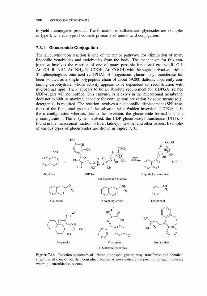
138 METABOLISM OF TOXICANTS
to yield a conjugated product. The formation of sulfates and glycosides are examplesof type I, whereas type II consists primarily of amino acid conjugation.
7.3.1 Glucuronide Conjugation
The glucuronidation reaction is one of the major pathways for elimination of manylipophilic xenobiotics and endobiotics from the body. The mechanism for this con-jugation involves the reaction of one of many possible functional groups (R–OH,Ar–OH, R–NH2, Ar–NH2, R–COOH, Ar–COOH) with the sugar derivative, uridine5′-diphosphoglucuronic acid (UDPGA). Homogeneous glucuronosyl transferase hasbeen isolated as a single polypeptide chain of about 59,000 daltons, apparently con-taining carbohydrate, whose activity appears to be dependent on reconstitution withmicrosomal lipid. There appears to be an absolute requirement for UDPGA: relatedUDP-sugars will not suffice. This enzyme, as it exists in the microsomal membrane,does not exhibit its maximal capacity for conjugation; activation by some means (e.g.,detergents), is required. The reaction involves a nucleophilic displacement (SN2 reac-tion) of the functional group of the substrate with Walden inversion. UDPGA is inthe α-configuration whereas, due to the inversion, the glucuronide formed is in theβ-configuration. The enzyme involved, the UDP glucuronosyl transferase (UGT), isfound in the microsomal fraction of liver, kidney, intestine, and other tissues. Examplesof various types of glucuronides are shown in Figure 7.16.
OH
+
UDPGA
O
OCOOH
OH
HO
OH
1-Naphthol Naphthol glucuronide
(b) Substrate Examples
O NH2SH
ONH
CH3
H3C
OH
N
H3C
H3C
N
Coumarin 2-Naphthylamine Thiophenol
Propanolol Imipramine
(a) Reaction Sequence
Cl
NHN
HO O
Oxazapam
OP
O
HOO
PO
OH
O
OCOOH
OH
HO
OH
N
NH
O
HO
OH
HO
HO
+ UDPUGT
Figure 7.16 Reaction sequences of uridine diphospho glucuronosyl transferase and chemicalstructures of compounds that form glucuronides. Arrows indicate the position on each moleculewhere glucuronidation occurs.
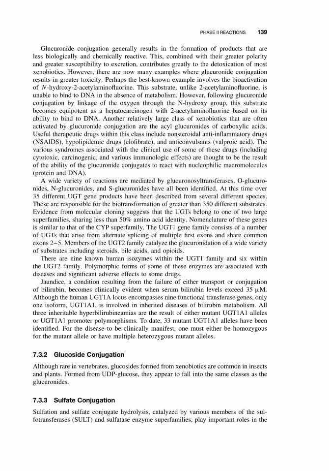
PHASE II REACTIONS 139
Glucuronide conjugation generally results in the formation of products that areless biologically and chemically reactive. This, combined with their greater polarityand greater susceptibility to excretion, contributes greatly to the detoxication of mostxenobiotics. However, there are now many examples where glucuronide conjugationresults in greater toxicity. Perhaps the best-known example involves the bioactivationof N -hydroxy-2-acetylaminofluorine. This substrate, unlike 2-acetylaminofluorine, isunable to bind to DNA in the absence of metabolism. However, following glucuronideconjugation by linkage of the oxygen through the N-hydroxy group, this substratebecomes equipotent as a hepatocarcinogen with 2-acetylaminofluorine based on itsability to bind to DNA. Another relatively large class of xenobiotics that are oftenactivated by glucuronide conjugation are the acyl glucuronides of carboxylic acids.Useful therapeutic drugs within this class include nonsteroidal anti-inflammatory drugs(NSAIDS), hypolipidemic drugs (clofibrate), and anticonvulsants (valproic acid). Thevarious syndromes associated with the clinical use of some of these drugs (includingcytotoxic, carcinogenic, and various immunologic effects) are thought to be the resultof the ability of the glucuronide conjugates to react with nucleophilic macromolecules(protein and DNA).
A wide variety of reactions are mediated by glucuronosyltransferases, O-glucuro-nides, N-glucuronides, and S-glucuronides have all been identified. At this time over35 different UGT gene products have been described from several different species.These are responsible for the biotransformation of greater than 350 different substrates.Evidence from molecular cloning suggests that the UGTs belong to one of two largesuperfamilies, sharing less than 50% amino acid identity. Nomenclature of these genesis similar to that of the CYP superfamily. The UGT1 gene family consists of a numberof UGTs that arise from alternate splicing of multiple first exons and share commonexons 2–5. Members of the UGT2 family catalyze the glucuronidation of a wide varietyof substrates including steroids, bile acids, and opioids.
There are nine known human isozymes within the UGT1 family and six withinthe UGT2 family. Polymorphic forms of some of these enzymes are associated withdiseases and significant adverse effects to some drugs.
Jaundice, a condition resulting from the failure of either transport or conjugationof bilirubin, becomes clinically evident when serum bilirubin levels exceed 35 µM.Although the human UGT1A locus encompasses nine functional transferase genes, onlyone isoform, UGT1A1, is involved in inherited diseases of bilirubin metabolism. Allthree inheritable hyperbilirubineamias are the result of either mutant UGT1A1 allelesor UGT1A1 promoter polymorphisms. To date, 33 mutant UGT1A1 alleles have beenidentified. For the disease to be clinically manifest, one must either be homozygousfor the mutant allele or have multiple heterozygous mutant alleles.
7.3.2 Glucoside Conjugation
Although rare in vertebrates, glucosides formed from xenobiotics are common in insectsand plants. Formed from UDP-glucose, they appear to fall into the same classes as theglucuronides.
7.3.3 Sulfate Conjugation
Sulfation and sulfate conjugate hydrolysis, catalyzed by various members of the sul-fotransferases (SULT) and sulfatase enzyme superfamilies, play important roles in the
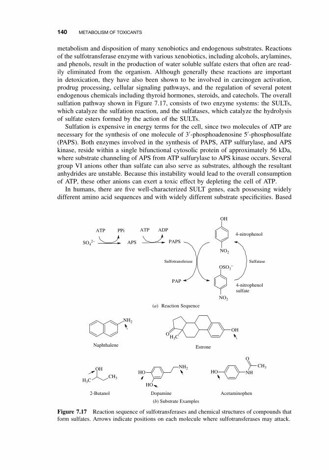
140 METABOLISM OF TOXICANTS
metabolism and disposition of many xenobiotics and endogenous substrates. Reactionsof the sulfotransferase enzyme with various xenobiotics, including alcohols, arylamines,and phenols, result in the production of water soluble sulfate esters that often are read-ily eliminated from the organism. Although generally these reactions are importantin detoxication, they have also been shown to be involved in carcinogen activation,prodrug processing, cellular signaling pathways, and the regulation of several potentendogenous chemicals including thyroid hormones, steroids, and catechols. The overallsulfation pathway shown in Figure 7.17, consists of two enzyme systems: the SULTs,which catalyze the sulfation reaction, and the sulfatases, which catalyze the hydrolysisof sulfate esters formed by the action of the SULTs.
Sulfation is expensive in energy terms for the cell, since two molecules of ATP arenecessary for the synthesis of one molecule of 3′-phosphoadenosine 5′-phosphosulfate(PAPS). Both enzymes involved in the synthesis of PAPS, ATP sulfurylase, and APSkinase, reside within a single bifunctional cytosolic protein of approximately 56 kDa,where substrate channeling of APS from ATP sulfurylase to APS kinase occurs. Severalgroup VI anions other than sulfate can also serve as substrates, although the resultantanhydrides are unstable. Because this instability would lead to the overall consumptionof ATP, these other anions can exert a toxic effect by depleting the cell of ATP.
In humans, there are five well-characterized SULT genes, each possessing widelydifferent amino acid sequences and with widely different substrate specificities. Based
ATP PPi ATP ADP
APS PAPS
NO2
OH
OSO3−
NO2
PAP
SO42−
NH2
OH3C
H3C
OH
CH3
OH NH2
HO
HO NHCH3
O
HO
Naphthalene Estrone
2-Butanol Dopamine Acetaminophen
(b) Substrate Examples
4-nitrophenol
SulfataseSulfotransferase
4-nitrophenolsulfate
(a) Reaction Sequence
Figure 7.17 Reaction sequence of sulfotransferases and chemical structures of compounds thatform sulfates. Arrows indicate positions on each molecule where sulfotransferases may attack.
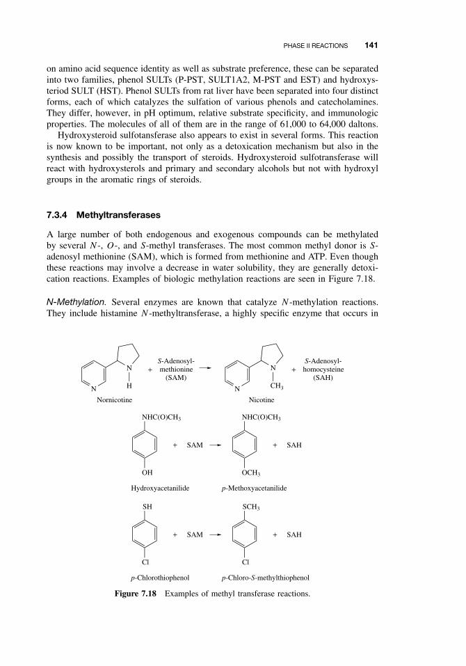
PHASE II REACTIONS 141
on amino acid sequence identity as well as substrate preference, these can be separatedinto two families, phenol SULTs (P-PST, SULT1A2, M-PST and EST) and hydroxys-teriod SULT (HST). Phenol SULTs from rat liver have been separated into four distinctforms, each of which catalyzes the sulfation of various phenols and catecholamines.They differ, however, in pH optimum, relative substrate specificity, and immunologicproperties. The molecules of all of them are in the range of 61,000 to 64,000 daltons.
Hydroxysteroid sulfotansferase also appears to exist in several forms. This reactionis now known to be important, not only as a detoxication mechanism but also in thesynthesis and possibly the transport of steroids. Hydroxysteroid sulfotransferase willreact with hydroxysterols and primary and secondary alcohols but not with hydroxylgroups in the aromatic rings of steroids.
7.3.4 Methyltransferases
A large number of both endogenous and exogenous compounds can be methylatedby several N -, O-, and S-methyl transferases. The most common methyl donor is S-adenosyl methionine (SAM), which is formed from methionine and ATP. Even thoughthese reactions may involve a decrease in water solubility, they are generally detoxi-cation reactions. Examples of biologic methylation reactions are seen in Figure 7.18.
N-Methylation. Several enzymes are known that catalyze N -methylation reactions.They include histamine N -methyltransferase, a highly specific enzyme that occurs in
N
+
Nornicotine
S-Adenosyl-methionine
(SAM)
N
+
Nicotine
S-Adenosyl-homocysteine
(SAH)
NHC(O)CH3
OH
+ SAM
NHC(O)CH3
OCH3
+ SAH
Hydroxyacetanilide p-Methoxyacetanilide
SH
Cl
+ SAM
SCH3
Cl
+ SAH
p-Chlorothiophenol p-Chloro-S-methylthiophenol
N
H
N
CH3
Figure 7.18 Examples of methyl transferase reactions.
142 METABOLISM OF TOXICANTS
the soluble fraction of the cell, phenylethanolamine N -methyltransferase, which cat-alyzes the methylation of noradrenaline to adrenaline as well as the methylation of otherphenylethanolamine derivatives. A third N -methyltransferase is the indoethylamine N -methyltansferase, or nonspecific N -methyltransferase. This enzyme occurs in varioustissues. It methylates endogenous compounds such as serotonin and tryptamine andexogenous compounds such as nornicotine and norcodeine. The relationship betweenthis enzyme and phenylethanolamine N -methyltransferase is not yet clear.
O-Methylation. Catechol O-methyltransferase occurs in the soluble fraction of severaltissues and has been purified from rat liver. The purified form has a molecular weight23,000 daltons, requires S-adenosylmethionine and Mg+, and catalyzes the methylationof epinephrine, norepinephrine, and other catechol derivatives. There is evidence thatthis enzyme exists in multiple forms.
A microsomal O-methyltransferase that methylates a number of alkyl-, methoxy-,and halophenols has been described from rabbit liver and lungs. These methylations areinhibited by SKF-525, N -ethyl-maleimide and p-chloromercuribenzoate. A hydroxyin-dole O-methyltransferase, which methylates N -acetyl-serotonin to melatonin and, toa lesser extent, other 5-hydroxyindoles and 5,6-dihydroxyindoles, has been describedfrom the pineal gland of mammals, birds, reptiles, amphibians, and fish.
S-Methylation. Thiol groups of some foreign compounds are also methylated, thereaction being catalyzed by the enzyme, thiol S-methyltransferase. This enzyme ismicrosomal and, as with most methyl transferases, utilizes S-adenosylmethionine. Ithas been purified from rat liver and is a monomer of about 28,000 daltons. A widevariety of substrates are methylated, including thiacetanilide, mercaptoethanol, anddiphenylsulfide. This enzyme may also be important in the detoxication of hydrogensulfide, which is methylated in two steps, first to the highly toxic methanethiol andthen to dimethylsulfide.
Methylthiolation, or the transfer of a methylthio (CH3S–) group to a foreign com-pound may occur through the action of another recently discovered enzyme, cysteineconjugate β-lyase. This enzyme acts on cysteine conjugates of foreign compoundsas follows:
RSCH2CH(NH2)COOH −−−→ RSH + NH3 + CH3C(O)COOH
The thiol group can then be methylated to yield the methylthio derivative of theoriginal xenobiotic.
Biomethylation of Elements. The biomethylation of elements is carried out principallyby microorganisms and is important in environmental toxicology, particularly in thecase of heavy metals, because the methylated compounds are absorbed through themembranes of the gut, the blood-brain barrier, and the placenta more readily thanare the inorganic forms. For example, inorganic mercury can be methylated first tomonomethylmercury and subsequently, to dimethylmercury:
Hg2+ −−−→ CH3HG+ −−−→ (CH3)2 Hg
The enzymes involved are reported to use either S-adenosylmethionine or vitaminB12 derivatives as methyl donors, and in addition to mercury, the metals, lead, tin,
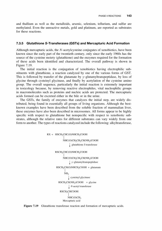
PHASE II REACTIONS 143
and thallium as well as the metalloids, arsenic, selenium, tellurium, and sulfur aremethylated. Even the unreactive metals, gold and platinum, are reported as substratesfor these reactions.
7.3.5 Glutathione S-Transferases (GSTs) and Mercapturic Acid Formation
Although mercapturic acids, the N -acetylcysteine conjugates of xenobiotics, have beenknown since the early part of the twentieth century, only since the early 1960s has thesource of the cysteine moiety (glutathione) and the enzymes required for the formationof these acids been identified and characterized. The overall pathway is shown inFigure 7.19.
The initial reaction is the conjugation of xenobiotics having electrophilic sub-stituents with glutathione, a reaction catalyzed by one of the various forms of GST.This is followed by transfer of the glutamate by γ -glutamyltranspeptidase, by loss ofglycine through cysteinyl glycinase, and finally by acetylation of the cysteine aminogroup. The overall sequence, particularly the initial reaction is extremely importantin toxicology because, by removing reactive electrophiles, vital nucleophilic groupsin macromolecules such as proteins and nucleic acids are protected. The mercapturicacids formed can be excreted either in the bile or in the urine.
The GSTs, the family of enzymes that catalyzes the initial step, are widely dis-tributed, being found in essentially all groups of living organisms. Although the best-known examples have been described from the soluble fraction of mammalian liver,these enzymes have also been described in microsomes. All forms appear to be highlyspecific with respect to glutathione but nonspecific with respect to xenobiotic sub-strates, although the relative rates for different substrates can vary widely from oneform to another. The types of reactions catalyzed include the following: alkyltransferase,
+RX
+ glutamate
HSCH2CHC(O)NHCH2COOH
NHC(O)CH2CH2CH(NH2)COOH
glutathione S-transferase
RSCH2CHC(O)NHCH2COOH
NHC(O)CH2CH2CH(NH2)COOH
g -glutamyltranspeptidase
RSCH2CH(O)NHCH2COOH
NH2
RSCH2CHCOOH
NHC(O)CH3
cysteinyl glycinase
+ glycineRSCH2CH(NH2)COOH
N-acetyl transferase
Mercapturic acid
Figure 7.19 Glutathione transferase reaction and formation of mercapturic acids.
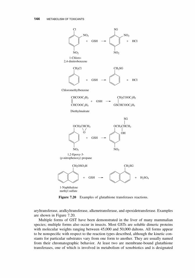
144 METABOLISM OF TOXICANTS
Cl
NO2
+ GSH
1-Chloro-2,4-dinitrobenzene
NO2
SG
NO2
+ HCI
NO2
CH2Cl
+ GSH
CH2SG
+ HCI
Chloromethylbenzene
CHCOOC2H5
CHCOOC2H5+ GSH
GSCHCOOC2H5
CH2COOC2H5
Diethylmaleate
OCH2CHCH2
NO2
+ GSH
OCH2CHCH2
NO2
O OH
SG
1,2-Epoxy-3-(p-nitrophenoxy) propane
CH2OSO3H
+ GSH
CH2SG
+ H2SO4
1-Naphthalenemethyl sulfate
Figure 7.20 Examples of glutathione transferases reactions.
aryltransferase, aralkyltransferase, alkenetransferase, and epoxidetransferase. Examplesare shown in Figure 7.20.
Multiple forms of GST have been demonstrated in the liver of many mammalianspecies; multiple forms also occur in insects. Most GSTs are soluble dimeric proteinswith molecular weights ranging between 45,000 and 50,000 daltons. All forms appearto be nonspecific with respect to the reaction types described, although the kinetic con-stants for particular substrates vary from one form to another. They are usually namedfrom their chromatographic behavior. At least two are membrane-bound glutathionetransferases, one of which is involved in metabolism of xenobiotics and is designated
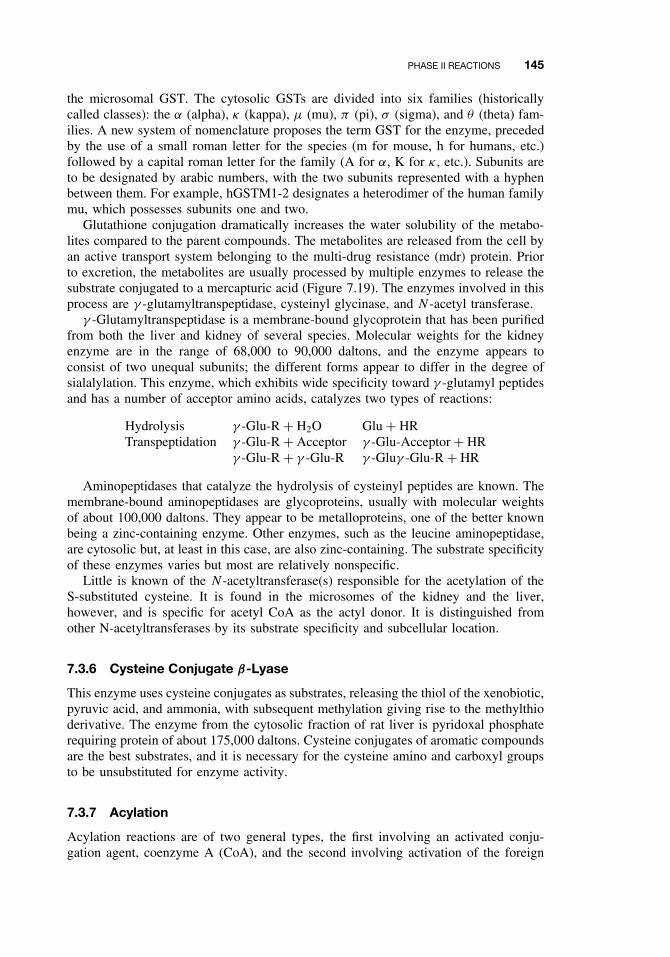
PHASE II REACTIONS 145
the microsomal GST. The cytosolic GSTs are divided into six families (historicallycalled classes): the α (alpha), κ (kappa), µ (mu), π (pi), σ (sigma), and θ (theta) fam-ilies. A new system of nomenclature proposes the term GST for the enzyme, precededby the use of a small roman letter for the species (m for mouse, h for humans, etc.)followed by a capital roman letter for the family (A for α, K for κ , etc.). Subunits areto be designated by arabic numbers, with the two subunits represented with a hyphenbetween them. For example, hGSTM1-2 designates a heterodimer of the human familymu, which possesses subunits one and two.
Glutathione conjugation dramatically increases the water solubility of the metabo-lites compared to the parent compounds. The metabolites are released from the cell byan active transport system belonging to the multi-drug resistance (mdr) protein. Priorto excretion, the metabolites are usually processed by multiple enzymes to release thesubstrate conjugated to a mercapturic acid (Figure 7.19). The enzymes involved in thisprocess are γ -glutamyltranspeptidase, cysteinyl glycinase, and N -acetyl transferase.
γ -Glutamyltranspeptidase is a membrane-bound glycoprotein that has been purifiedfrom both the liver and kidney of several species. Molecular weights for the kidneyenzyme are in the range of 68,000 to 90,000 daltons, and the enzyme appears toconsist of two unequal subunits; the different forms appear to differ in the degree ofsialalylation. This enzyme, which exhibits wide specificity toward γ -glutamyl peptidesand has a number of acceptor amino acids, catalyzes two types of reactions:
Hydrolysis γ -Glu-R + H2O Glu + HRTranspeptidation γ -Glu-R + Acceptor γ -Glu-Acceptor + HR
γ -Glu-R + γ -Glu-R γ -Gluγ -Glu-R + HR
Aminopeptidases that catalyze the hydrolysis of cysteinyl peptides are known. Themembrane-bound aminopeptidases are glycoproteins, usually with molecular weightsof about 100,000 daltons. They appear to be metalloproteins, one of the better knownbeing a zinc-containing enzyme. Other enzymes, such as the leucine aminopeptidase,are cytosolic but, at least in this case, are also zinc-containing. The substrate specificityof these enzymes varies but most are relatively nonspecific.
Little is known of the N -acetyltransferase(s) responsible for the acetylation of theS-substituted cysteine. It is found in the microsomes of the kidney and the liver,however, and is specific for acetyl CoA as the actyl donor. It is distinguished fromother N-acetyltransferases by its substrate specificity and subcellular location.
7.3.6 Cysteine Conjugate β-Lyase
This enzyme uses cysteine conjugates as substrates, releasing the thiol of the xenobiotic,pyruvic acid, and ammonia, with subsequent methylation giving rise to the methylthioderivative. The enzyme from the cytosolic fraction of rat liver is pyridoxal phosphaterequiring protein of about 175,000 daltons. Cysteine conjugates of aromatic compoundsare the best substrates, and it is necessary for the cysteine amino and carboxyl groupsto be unsubstituted for enzyme activity.
7.3.7 Acylation
Acylation reactions are of two general types, the first involving an activated conju-gation agent, coenzyme A (CoA), and the second involving activation of the foreign
146 METABOLISM OF TOXICANTS
(a) Acetylation
H2N NH2
Benzidine
+ CoA CH3CHN NH2
O
COOH
+ ATP + CoASH
C(O)SCoA
+ PPi + AMP
(b) Amino acid conjugation
C(O)SCoA
+
C(O)NHCH2COOH
+
Benzoic acid
NH2CH2COOH
Glycine Hippuric acid
CoASH
Figure 7.21 Examples of acylation reactions.
compounds and subsequent acylation of an amino acid. This type of conjugation iscommonly undergone by exogenous carboxylic acids and amides, and although theproducts are often less water soluble than the parent compound, they are usually lesstoxic. Examples of acylation reactions are shown in Figure 7.21.
Acetylation. Acetylated derivatives of foreign exogenous amines are acetylated byN -acetyl transferase, the acetyl donor being CoA. This enzyme is cytosolic, has beenpurified from rat liver, and is known to occur in several other organs. Evidence exists forthe existence of multiple forms of this enzyme. Although endogenous amino, hydroxy,and thiol compounds are acetylated in vivo, the acetylation of exogenous hydroxy andthiol groups is presently unknown.
Acetylation of foreign compounds is influenced by both development and genetics.Newborn mammals generally have a low level of the transferase, whereas due tothe different genes involved, fast and slow acetylators have been identified in bothrabbit and human populations. Slow acetylators are more susceptible to the effects ofcompounds detoxified by acetylation.
N,O-Acyltransferase. The N -acyltransferase enzyme is believed to be involved inthe carcinogenicity of arylamines. These compounds are first N -oxidized, and then, inspecies capable of their N -acetylation, acetylated to arylhydroxamic acids. The effectof N ,O-transacetylation is shown in Figure 7.22. The N -acyl group of the hydroxamicacid is first removed and is then transferred, either to an amine to yield a stableamide or to the oxygen of the hydroxylamine to yield a reactive N -acyloxyarylamine.These compounds are highly reactive in the formation of adducts with both proteinsand nucleic acids, and N ,O-acyltransferase, added to the medium in the Ames test,increases the mutagenicity of compounds such as N -hydroxy-2-acetylaminofluorine.
PHASE II REACTIONS 147
ArNCCH3
O
OH
Acyl-
transferaseArNH
OH
+ CH3C-enzyme
O
Ar′NH2Ar′NHCCH3
O
[ ArNHOCCH3]
O
[ ArNH+]
reaction withcellularnucleophiles
Figure 7.22 N-, O-Acyltransferase reactions of arylhydroxamic acid. Ar = aryl group.
Despite its great instability this enzyme has been purified from the cytosolic fractionof the rat liver.
Amino Acid Conjugation. In the second type of acylation reaction, exogenous car-boxylic acids are activated to form S-CoA derivative in a reaction involving ATP andCoA. These CoA derivatives then acylate the amino group of a variety of amino acids.Glycine and glutamate appear to be the most common acceptor of amino acids inmammals; in other organisms, other amino acids are involved. These include ornithinein reptiles and birds and taurine in fish.
The activating enzyme occurs in the mitochondria and belongs to a class of enzymesknown as the ATP-dependent acid: CoA ligases (AMP) but has also been known asacyl CoA synthetase and acid-activating enzyme. It appears to be identical to theintermediate chain length fatty acyl-CoA-synthetase.
Two acyl-CoA: amino acid N -acyltransferases have been purified from liver mito-chondria of cattle, Rhesus monkeys, and humans. One is a benzoyltransferase CoAthat utilizes benzyl-CoA, isovaleryl-CoA, and tiglyl-CoA, but not phenylacetyl CoA,malonyl-CoA, or indolacetyl-CoA. The other is a phenylacetyl transferase that uti-lizes phenylacetyl-CoA and indolacetyl-CoA but is inactive toward benzoyl-CoA.Neither is specific for glycine, as had been supposed from studies using less definedsystems; both also utilize asparagine and glutamine, although at lesser rates thanglycine.
Bile acids are also conjugated by a similar sequence of reactions involving a micro-somal bile acid: CoA ligase and a soluble bile acid N -acyl-transferase. The latter hasbeen extensively purified, and differences in acceptor amino acids, of which taurine isthe most common, have been related to the evolutionary history of the species.
Deacetylation. Deacetylation occurs in a number of species, but there is a large dif-ference between species, strains, and individuals in the extent to which the reactionoccurs. Because acetylation and deacetylation are catalyzed by different enzymes, thelevels of which vary independently in different species, the importance of deacetylationas a xenobiotic metabolizing mechanism also varies between species. This can be seenin a comparison of the rabbit and the dog. The rabbit, which has high acetyltransferaseactivity and low deacetylase, excretes significant amounts of acetylated amines. Thedog, in which the opposite situation obtains, does not.
A typical substrate for the aromatic deacetylases of the liver and kidney is acetani-lide, which is deacylated to yield aniline.
148 METABOLISM OF TOXICANTS
7.3.8 Phosphate Conjugation
Phosphorylation of xenobiotics is not a widely distributed conjugation reaction, insectsbeing the only major group of animals in which it is found. The enzyme from the gutof cockroaches utilizes ATP, requires Mg+, and is active in the phosphorylation of1-naphthol and p-nitrophenol.
SUGGESTED READING
Benedetti, M. S. Biotransformation of xenobiotics by amine oxidases. Fundam. Clinic. Pharma-col. 15: 75–84, 2001.
Coughtrie, M. W. H., S. Sharp, K. Maxwell, and N. P. Innes. Biology and function of the rever-sible sulfation pathway catalyzed by human sulfotransferases and sulfatases. Chemico-Biol.Inter. 109: 3–27, 1998.
Duffel, M. W., A. D. Marshall, P. McPhie, V. Sharma, and W. B. Jakoby. Enzymatic aspectsof the phenol (aryl) sulfotransferases. Drug Metab. Rev. 33: 369–395, 2001.
Hayes, J. D., and D. J. Pulford. The glutathione S-transferase supergene family: Regulation ofGST and the contribution of the isoenzymes to cancer chemoprotection and drug resistance.Crit. Rev. Biochem. Mol. Biol. 30: 445–600, 1995.
Hodgson, E., and J. A. Goldstein. Metabolism of toxicants: Phase I reactions and pharmacoge-netics. In Introduction to Biochemical Toxicology, 3rd ed., E. Hodgson and R. C. Smart, eds.New York: Wiley, 2001, pp. 67–113.
Koukouritaki, S. B., P. Simpson, C. K. Yeung, A. E. Rettie, and R. N. Hines. Human hepaticflavin-containing monooxygenases 1 (FMO1) and 3 (FMO3) developmental expression. Pedi-atric Res. 51: 236–243, 2002.
Lawton, M. P., J. R. Cashman, T. Cresteil, C. T. Dolphin, A. A. Elfarra, R. N. Hines, E. Hodg-son, T. Kimura, J. Ozols, I. R. Phillips, R. M. Philpot, L. L. Poulsen, A. E. Rettie, E. A.Shephard, D. E. Williams and D. M. Ziegler. A nomenclature for the mammalian flavin-containing monooxygenase gene family based on amino acid sequence identities. Arch. Bio-chem. Biophys. 308: 254–257, 1994.
LeBlanc, G. A., and W. A. Dauterman. Conjugation and elimination of toxicants: In Introduc-tion to Biochemical Toxicology, 3rd edn., E. Hodgson and R. C. Smart, eds. New York: Wiley,2001, pp. 115–135.
Nelson, D. R., L. Koymans, T. Kamataki, J. J. Stegeman, R. Feyereisen, D. J. Waxman, M. R.Waterman, O. Gotoh, M. J. Coon, R. W. Estabrook, I. C. Gunsalus, and D. W. Nebert. P450superfamily: Update on new sequences, gene mapping, accession numbers and nomenclature.Pharmacogenetics 6: 1–42, 1996.
Oesch, F, and M. Arand. Xenobiotic metabolism. In Toxicology, H. Marquardt, S. G. Shafer,R. McClellan, and F. Welsch, eds. New York: Academic Press, 1999, pp. 83–109.
Ritter, J. K. Roles of glucuronidation and UDP-glucuronosyltransferases in xenobiotic bioacti-vation reactions. Chemico-Biol. Inter. 129: 171–193, 2001.
Tukey, R. H., and C. P. Strassburg. Human UDP-glucuronosyltransferases: Metabolism, expres-sion, and disease. Ann. Rev. Pharmacol. Toxicol. 40: 581–616, 2000.
Vasiliou, V., A. Pappa, and D. R. Petersen. Role of aldehyde dehydrogenases in endogenousand xenobiotic metabolism. Chemico-Biol. Inter. 129: 1–19, 2000.
CHAPTER 8
Reactive Metabolites
RANDY L. ROSE and PATRICIA E. LEVI
8.1 INTRODUCTION
Between uptake from the environment and excretion from the body, many exoge-nous compounds (xenobiotics) undergo metabolism to highly reactive intermediates.These metabolites may interact with cellular constituents in numerous ways, such asbinding covalently to macromolecules and/or stimulating lipid peroxidation. This bio-transformation of relatively inert chemicals to highly reactive intermediary metabolitesis commonly referred to as metabolic activation or bioactivation, and it is known to bethe initial event in many chemically induced toxicities. Some toxicants are direct actingand require no activation, whereas other chemicals may be activated nonenzymatically.The focus of this chapter, however, is on toxicants requiring metabolic activation andto those processes involved in activation.
In the 1940s and 1950s the pioneering studies of James and Elizabeth Millerprovided early evidence for in vivo conversion of chemical carcinogens to reactivemetabolites. They found that reactive metabolites of the aminoazo dye N ,N -dimethyl-4-aminoazobenzene (DAB), a hepatocarcinogen in rats, would bind covalently to pro-teins and nucleic acids. The term, metabolic activation, was coined by the Millersto describe this process. Moreover they demonstrated that covalent binding of thesechemicals was an essential part of the carcinogenic process.
The overall scheme of metabolism for potentially toxic xenobiotics is outlinedin Figure 8.1. As illustrated by this diagram, xenobiotic metabolism can producenot only nontoxic metabolites, which are more polar and readily excreted (detoxi-cation), but also highly reactive metabolites, which can interact with vital intracellularmacromolecules, resulting in toxicity. In addition reactive metabolites can be detox-ified—for example, by interaction with glutathione. In general, reactive metabolitesare electrophiles (molecules containing positive centers). These electrophiles in turncan react with cellular nucleophiles (molecules containing negative centers), suchas glutathione, proteins, and nucleic acids. Other reactive metabolites may be freeradicals or act as radical generators that interact with oxygen to produce reactiveoxygen species that are capable of causing damage to membranes, DNA, and othermacromolecules.
A Textbook of Modern Toxicology, Third Edition, edited by Ernest HodgsonISBN 0-471-26508-X Copyright 2004 John Wiley & Sons, Inc.
149
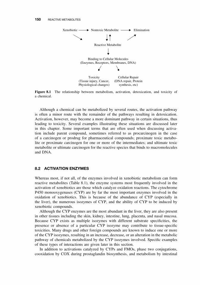
150 REACTIVE METABOLITES
Xenobiotic Nontoxic Metabolite
Reactive Metabolite
Binding to Cellular Molecules(Enzymes, Receptors, Membranes, DNA)
Elimination
Toxicity(Tissue injury, Cancer,Physiological changes)
Cellular Repair(DNA repair, Protein
synthesis, etc)
Figure 8.1 The relationship between metabolism, activation, detoxication, and toxicity ofa chemical.
Although a chemical can be metabolized by several routes, the activation pathwayis often a minor route with the remainder of the pathways resulting in detoxication.Activation, however, may become a more dominant pathway in certain situations, thusleading to toxicity. Several examples illustrating these situations are discussed laterin this chapter. Some important terms that are often used when discussing activa-tion include parent compound, sometimes referred to as procarcinogen in the caseof a carcinogen or prodrug for pharmaceutical compounds; proximate toxic metabo-lite or proximate carcinogen for one or more of the intermediates; and ultimate toxicmetabolite or ultimate carcinogen for the reactive species that binds to macromoleculesand DNA.
8.2 ACTIVATION ENZYMES
Whereas most, if not all, of the enzymes involved in xenobiotic metabolism can formreactive metabolites (Table 8.1), the enzyme systems most frequently involved in theactivation of xenobiotics are those which catalyze oxidation reactions. The cytochromeP450 monooxygenases (CYP) are by far the most important enzymes involved in theoxidation of xenobiotics. This is because of the abundance of CYP (especially inthe liver), the numerous isozymes of CYP, and the ability of CYP to be induced byxenobiotic compounds.
Although the CYP enzymes are the most abundant in the liver, they are also presentin other tissues including the skin, kidney, intestine, lung, placenta, and nasal mucosa.Because CYP exists as multiple isozymes with different substrate specificities, thepresence or absence of a particular CYP isozyme may contribute to tissue-specifictoxicities. Many drugs and other foreign compounds are known to induce one or moreof the CYP isozymes, resulting in an increase, decrease, or an alteration in the metabolicpathway of chemicals metabolized by the CYP isozymes involved. Specific examplesof these types of interactions are given later in this section.
In addition to activations catalyzed by CYPs and FMOs, phase two conjugations,cooxidation by COX during prostaglandin biosynthesis, and metabolism by intestinal
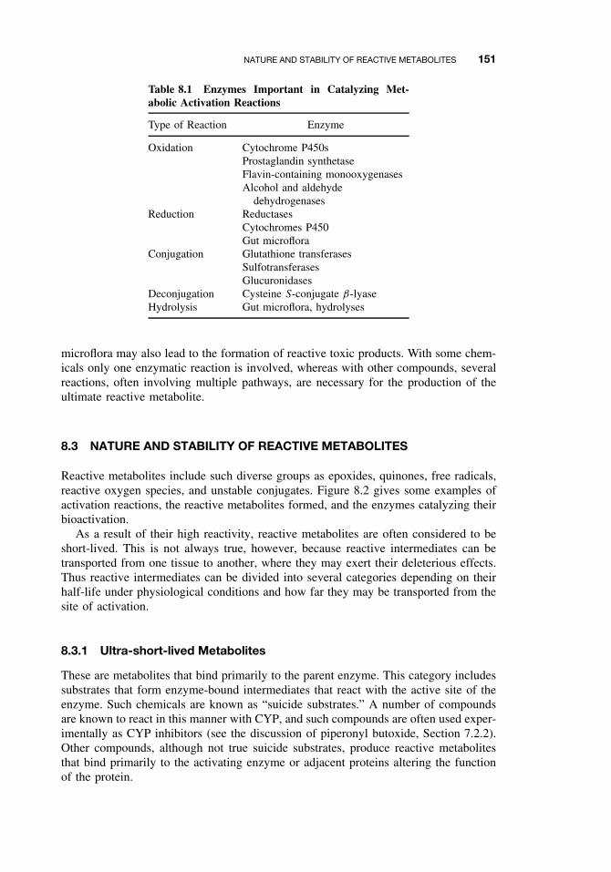
NATURE AND STABILITY OF REACTIVE METABOLITES 151
Table 8.1 Enzymes Important in Catalyzing Met-abolic Activation Reactions
Type of Reaction Enzyme
Oxidation Cytochrome P450sProstaglandin synthetaseFlavin-containing monooxygenasesAlcohol and aldehyde
dehydrogenasesReduction Reductases
Cytochromes P450Gut microflora
Conjugation Glutathione transferasesSulfotransferasesGlucuronidases
Deconjugation Cysteine S-conjugate β-lyaseHydrolysis Gut microflora, hydrolyses
microflora may also lead to the formation of reactive toxic products. With some chem-icals only one enzymatic reaction is involved, whereas with other compounds, severalreactions, often involving multiple pathways, are necessary for the production of theultimate reactive metabolite.
8.3 NATURE AND STABILITY OF REACTIVE METABOLITES
Reactive metabolites include such diverse groups as epoxides, quinones, free radicals,reactive oxygen species, and unstable conjugates. Figure 8.2 gives some examples ofactivation reactions, the reactive metabolites formed, and the enzymes catalyzing theirbioactivation.
As a result of their high reactivity, reactive metabolites are often considered to beshort-lived. This is not always true, however, because reactive intermediates can betransported from one tissue to another, where they may exert their deleterious effects.Thus reactive intermediates can be divided into several categories depending on theirhalf-life under physiological conditions and how far they may be transported from thesite of activation.
8.3.1 Ultra-short-lived Metabolites
These are metabolites that bind primarily to the parent enzyme. This category includessubstrates that form enzyme-bound intermediates that react with the active site of theenzyme. Such chemicals are known as “suicide substrates.” A number of compoundsare known to react in this manner with CYP, and such compounds are often used exper-imentally as CYP inhibitors (see the discussion of piperonyl butoxide, Section 7.2.2).Other compounds, although not true suicide substrates, produce reactive metabolitesthat bind primarily to the activating enzyme or adjacent proteins altering the functionof the protein.
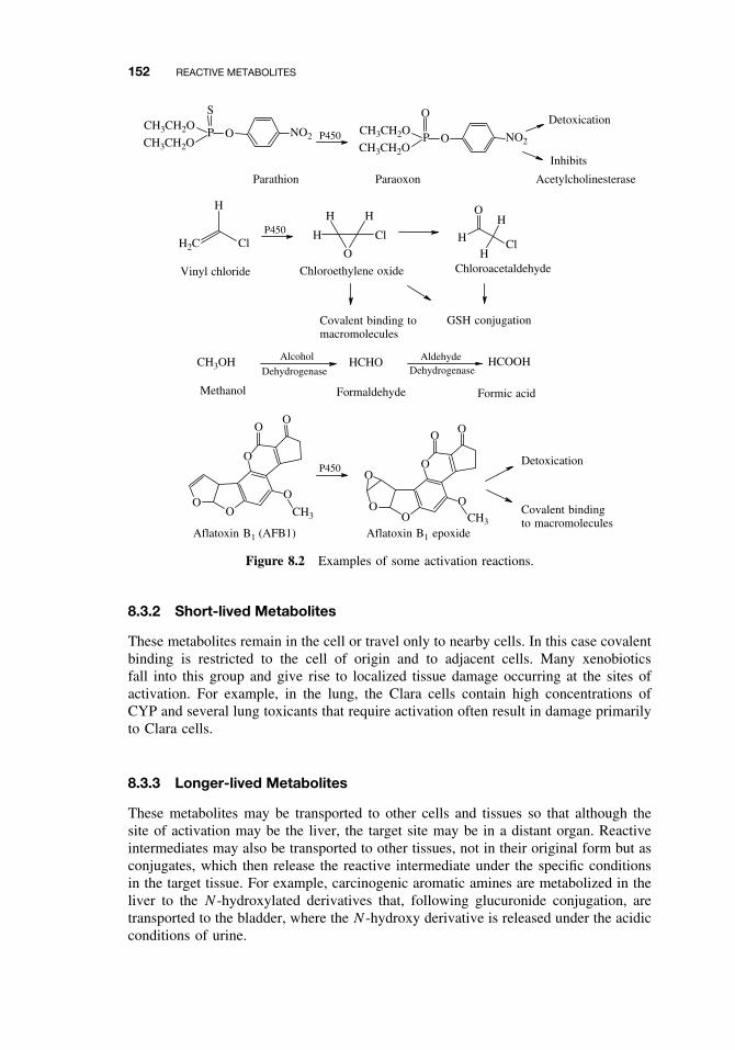
152 REACTIVE METABOLITES
O NO2P
SCH3CH2O
CH3CH2O OPCH3CH2O
CH3CH2OP450
P450
P450
Parathion Paraoxon
H2C Cl
H
Cl
O
H
H
H
Cl
OH
HH
O
Vinyl chloride Chloroethylene oxide Chloroacetaldehyde
GSH conjugationCovalent binding tomacromolecules
CH3OH HCHO HCOOH
Methanol Formaldehyde Formic acid
Alcohol Aldehyde
Dehydrogenase Dehydrogenase
OO
O
O
CH3
OO
OO
O
OCH3
OO
O
Aflatoxin B1 (AFB1) Aflatoxin B1 epoxide
Detoxication
Covalent bindingto macromolecules
NO2
Detoxication
Inhibits
Acetylcholinesterase
Figure 8.2 Examples of some activation reactions.
8.3.2 Short-lived Metabolites
These metabolites remain in the cell or travel only to nearby cells. In this case covalentbinding is restricted to the cell of origin and to adjacent cells. Many xenobioticsfall into this group and give rise to localized tissue damage occurring at the sites ofactivation. For example, in the lung, the Clara cells contain high concentrations ofCYP and several lung toxicants that require activation often result in damage primarilyto Clara cells.
8.3.3 Longer-lived Metabolites
These metabolites may be transported to other cells and tissues so that although thesite of activation may be the liver, the target site may be in a distant organ. Reactiveintermediates may also be transported to other tissues, not in their original form but asconjugates, which then release the reactive intermediate under the specific conditionsin the target tissue. For example, carcinogenic aromatic amines are metabolized in theliver to the N -hydroxylated derivatives that, following glucuronide conjugation, aretransported to the bladder, where the N -hydroxy derivative is released under the acidicconditions of urine.
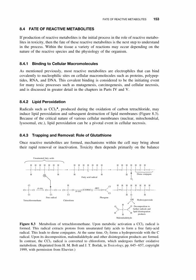
FATE OF REACTIVE METABOLITES 153
8.4 FATE OF REACTIVE METABOLITES
If production of reactive metabolites is the initial process in the role of reactive metabo-lites in toxicity, then the fate of these reactive metabolites is the next step to understandin the process. Within the tissue a variety of reactions may occur depending on thenature of the reactive species and the physiology of the organism.
8.4.1 Binding to Cellular Macromolecules
As mentioned previously, most reactive metabolites are electrophiles that can bindcovalently to nucleophilic sites on cellular macromolecules such as proteins, polypep-tides, RNA, and DNA. This covalent binding is considered to be the initiating eventfor many toxic processes such as mutagenesis, carcinogenesis, and cellular necrosis,and is discussed in greater detail in the chapters in Parts IV and V.
8.4.2 Lipid Peroxidation
Radicals such as CCl3ž, produced during the oxidation of carbon tetrachloride, mayinduce lipid peroxidation and subsequent destruction of lipid membranes (Figure 8.3).Because of the critical nature of various cellular membranes (nuclear, mitochondrial,lysosomal, etc.), lipid peroxidation can be a pivotal event in cellular necrosis.
8.4.3 Trapping and Removal: Role of Glutathione
Once reactive metabolites are formed, mechanisms within the cell may bring abouttheir rapid removal or inactivation. Toxicity then depends primarily on the balance
C C
H
H
C C
H
H
C C C
H
H
H H H H
C C
H
H
C C
H
C C C
H
H
H H H H
•C C
H
C C
H
C C CC
HH H
H
H H
•
Cl C Cl
Cl
Cl
(P-450)Cl C Cl
Cl
•Cl C Cl
H
Cl
(P-450)[C(OH)Cl3]
−HClO
O2
C
Cl
Cl
C C C
H
HHH
O O
C C C
HH
H
C C
H
C C
H H H H
C
H
O
OH
H
... ... ... ...
... ...
......
Fatty acid radical
Tetrachloromethane Chloroform Hydroxyperoxide
Malondialdehyde
Decomposition tofurther radicals andlipid disintegration
products
Free radical Phosgene
Diene conjugate
Unsaturated fatty acids
Figure 8.3 Metabolism of tetrachloromethane. Upon metabolic activation a CCl3 radical isformed. This radical extracts protons from unsaturated fatty acids to form a free fatty-acidradical. This leads to diene conjugates. At the same time, O2 forms a hydroperoxide with the Cradical. Upon its decomposition, malondialdehyde and other disintegration products are formed.In contrast, the CCl3 radical is converted to chloroform, which undergoes further oxidativemetabolism. (Reprinted from H. M. Bolt and J. T. Borlak, in Toxicology, pp. 645–657, copyright1999, with permission from Elsevier.)
154 REACTIVE METABOLITES
between the rate of metabolite formation and the rate of removal. With some com-pounds, reduced glutathione plays an important protective role by trapping electrophilicmetabolites and preventing their binding to hepatic proteins and enzymes. Althoughconjugation reactions occasionally result in bioactivation of a compound, the acetyl-,glutathione-, glucuronyl-, or sulfotransferases usually result in the formation of anontoxic, water-soluble metabolite that is easily excreted. Thus availability of theconjugating chemical is an important factor in determining the fate of the reactiveintermediates.
8.5 FACTORS AFFECTING TOXICITY OF REACTIVE METABOLITES
A number of factors can influence the balance between the rate of formation of reac-tive metabolites and the rate of removal, thereby affecting toxicity. The major factorsdiscussed in this chapter are summarized in the following subsections. A more in-depth discussion of other factors affecting metabolism and toxicity are presented inChapter 9.
8.5.1 Levels of Activating Enzymes
Specific isozymes of CYPs are often important in determining metabolic activationof a foreign compound. As mentioned previously, many xenobiotics induce specificforms of cytochrome P450. Frequently the CYP forms induced are those involvedin the metabolism of the inducing agent. Thus a carcinogen or other toxicant hasthe potential for inducing its own activation. In addition there are species and gen-der differences in enzyme levels as well as specific differences in the expression ofparticular isozymes.
8.5.2 Levels of Conjugating Enzymes
Levels of conjugating enzymes, such as glutathione transferases, are also known to beinfluenced by gender and species differences as well as by drugs and other environ-mental factors. All of these factors will in turn affect the detoxication process.
8.5.3 Levels of Cofactors or Conjugating Chemicals
Treatment of animals with N -acetylcysteine, a precursor of glutathione, protects ani-mals against acetaminophen-induced hepatic necrosis, possibly by reducing covalentbinding to tissue macromolecules. However, depletion of glutathione potentiates cova-lent binding and hepatotoxicity.
8.6 EXAMPLES OF ACTIVATING REACTIONS
The following examples have been selected to illustrate the various concepts of acti-vation and detoxication discussed in the previous sections.
EXAMPLES OF ACTIVATING REACTIONS 155
8.6.1 Parathion
Parathion is one of several organophosphorus insecticides that has had great economicimportance worldwide for several decades. Organophosphate toxicity is the result ofexcessive stimulation of cholinergic nerves, which is dependent on their ability toinhibit acetylcholinesterases. Interestingly the parent organophosphates are relativelypoor inhibitors of acetylcholinesterases, requiring metabolic conversion of a P=S bondto a P=O bond for acetylcholinesterase inhibition (Figure 8.2; see Chapters 11 and 16for a discussion of the mechanism of acetylcholinesterase inhibition). In vitro studies ofrat and human liver have demonstrated that CYP is inactivated by the electrophilic sul-fur atom released during oxidation of parathion to paraoxon. Some have shown that thespecific isoforms responsible for the metabolic activation of parathion are destroyedin the process. For example, preincubations of NADPH-supplemented human livermicrosomes with parathion resulted in the inhibition of some isoform-specific metabo-lites including testosterone (CYP3A4), tolbutamide (CYP2C9), and 7-ethylresorufin(CYP1A2) but not aniline (CYP2E1). These losses of metabolic activity were also asso-ciated with the loss of CYP content as measured by the CO-difference spectra. Theseresults suggest that parathion acts as a suicide substrate, in that its metabolism resultsin the destruction of the particular isoforms involved in its metabolism. This becomesparticularly important because the principal CYP involved in parathion metabolism isCYP3A4, which is the dominant CYP in humans; accounting for between 30–50%of the total liver CYP. Because of this enzyme’s importance in drug metabolism,the strong potential for inhibition by organophosphate compounds may have seriousconsequences in individuals undergoing drug therapy.
8.6.2 Vinyl Chloride
A second example of a suicide inhibitor is vinyl chloride. The first step in the bio-transformation of vinyl chloride involves the CYP-mediated oxidation of the doublebond leading to the formation of an epoxide, or oxirane, which is highly reactive andcan easily bind to proteins and nucleic acids. Following activation by CYP, reactivemetabolites such as those formed by vinyl chloride bind covalently to the pyrrole nitro-gens present in the heme moiety, resulting in destruction of the heme and loss of CYPactivity. The interaction of the oxirane structure with nucleic acids results in mutationsand cancer. The first indications that vinyl chloride was a human carcinogen involvedindividuals who cleaned reactor vessels in polymerization plants who were exposed tohigh concentrations of vinyl chloride and developed angiosarcomas of the liver as aresult of their exposure (Figure 8.2).
8.6.3 Methanol
Ingestion of methanol, particularly during the prohibition era, resulted in significantillness and mortality. Where epidemics of methanol poisoning have been reported,one-third of the exposed population recovered with no ill effects, one-third have severevisual loss or blindness, and one-third have died. Methanol itself is not responsible forthe toxic effects but is rapidly metabolized in humans by alcohol dehydrogenase toformaldehyde, which is subsequently metabolized by aldehyde dehydrogenase to form
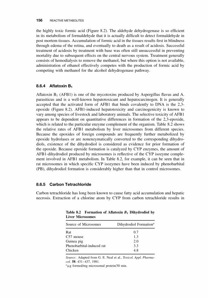
156 REACTIVE METABOLITES
the highly toxic formic acid (Figure 8.2). The aldehyde dehydrogenase is so efficientin its metabolism of formaldehyde that it is actually difficult to detect formaldehyde inpost mortem tissues. Accumulation of formic acid in the tissues results first in blindnessthrough edema of the retina, and eventually to death as a result of acidosis. Successfultreatment of acidosis by treatment with base was often still unsuccessful in preventingmortality due to subsequent effects on the central nervous system. Treatment generallyconsists of hemodialysis to remove the methanol, but where this option is not available,administration of ethanol effectively competes with the production of formic acid bycompeting with methanol for the alcohol dehydrogenase pathway.
8.6.4 Aflatoxin B1
Aflatoxin B1 (AFB1) is one of the mycotoxins produced by Aspergillus flavus and A.parasiticus and is a well-known hepatotoxicant and hepatocarcinogen. It is generallyaccepted that the activated form of AFB1 that binds covalently to DNA is the 2,3-epoxide (Figure 8.2). AFB1-induced hepatotoxicity and carcinogenicity is known tovary among species of livestock and laboratory animals. The selective toxicity of AFB1appears to be dependent on quantitative differences in formation of the 2,3-epoxide,which is related to the particular enzyme complement of the organism. Table 8.2 showsthe relative rates of AFB1 metabolism by liver microsomes from different species.Because the epoxides of foreign compounds are frequently further metabolized byepoxide hydrolases or are nonenzymatically converted to the corresponding dihydro-diols, existence of the dihydrodiol is considered as evidence for prior formation ofthe epoxide. Because epoxide formation is catalyzed by CYP enzymes, the amount ofAFB1-dihydrodiol produced by microsomes is reflective of the CYP isozyme comple-ment involved in AFB1 metabolism. In Table 8.2, for example, it can be seen that inrat microsomes in which specific CYP isozymes have been induced by phenobarbital(PB), dihydrodiol formation is considerably higher than that in control microsomes.
8.6.5 Carbon Tetrachloride
Carbon tetrachloride has long been known to cause fatty acid accumulation and hepaticnecrosis. Extraction of a chlorine atom by CYP from carbon tetrachloride results in
Table 8.2 Formation of Aflatoxin B1 Dihydrodiol byLiver Microsomes
Source of Microsomes Dihydrodiol Formationa
Rat 0.7C57 mouse 1.3Guinea pig 2.0Phenobarbital-induced rat 3.3Chicken 4.8
Source: Adapted from G. E. Neal et al., Toxicol. Appl. Pharma-col. 58: 431–437, 1981.aµg formed/mg microsomal protein/30 min.
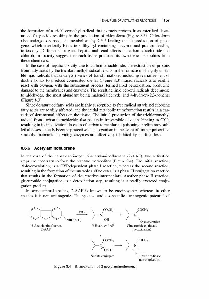
EXAMPLES OF ACTIVATING REACTIONS 157
the formation of a trichloromethyl radical that extracts protons from esterified desat-urated fatty acids resulting in the production of chloroform (Figure 8.3). Chloroformalso undergoes subsequent metabolism by CYP leading to the production of phos-gene, which covalently binds to sulfhydryl containing enzymes and proteins leadingto toxicity. Differences between hepatic and renal effects of carbon tetrachloride andchloroform toxicity suggest that each tissue produces its own toxic metabolites fromthese chemicals.
In the case of hepatic toxicity due to carbon tetrachloride, the extraction of protonsfrom fatty acids by the trichloromethyl radical results in the formation of highly unsta-ble lipid radicals that undergo a series of transformations, including rearrangement ofdouble bonds to produce conjugated dienes (Figure 8.3). Lipid radicals also readilyreact with oxygen, with the subsequent process, termed lipid peroxidation, producingdamage to the membranes and enzymes. The resulting lipid peroxyl radicals decomposeto aldehydes, the most abundant being malondialdehyde and 4-hydroxy-2,3-nonenal(Figure 8.3).
Since desaturated fatty acids are highly susceptible to free radical attack, neighboringfatty acids are readily affected, and the initial metabolic transformation results in a cas-cade of detrimental effects on the tissue. The initial production of the trichloromethylradical from carbon tetrachloride also results in irreversible covalent binding to CYP,resulting in its inactivation. In cases of carbon tetrachloride poisoning, preliminary sub-lethal doses actually become protective to an organism in the event of further poisoning,since the metabolic activating enzymes are effectively inhibited by the first dose.
8.6.6 Acetylaminofluorene
In the case of the hepatocarcinogen, 2-acetylaminofluorene (2-AAF), two activationsteps are necessary to form the reactive metabolites (Figure 8.4). The initial reaction,N -hydroxylation, is a CYP-dependent phase I reaction, whereas the second reaction,resulting in the formation of the unstable sulfate ester, is a phase II conjugation reactionthat results in the formation of the reactive intermediate. Another phase II reaction,glucuronide conjugation, is a detoxication step, resulting in a readily excreted conju-gation product.
In some animal species, 2-AAF is known to be carcinogenic, whereas in otherspecies it is noncarcinogenic. The species- and sex-specific carcinogenic potential of
NHCOCH3
P450N
OH
COCH3
N
O -glucuronide
COCH3
2-Acetylaminofluorene2-AAF
N-Hydroxy AAF Glucuronide conjugate(detoxication)
NOSO3
−
COCH3
N
COCH3
Binding to tissuemacromolecules
++
Sulfate conjugate
Figure 8.4 Bioactivation of 2-acetylaminofluorene.
158 REACTIVE METABOLITES
2-AAF is correlated with the ability of the organism to sequentially produce the N -hydroxylated metabolite followed by the sulfate ester. Therefore in an animal suchas the guinea pig, which does not produce the N -hydroxylated metabolite, 2-AAF isnot carcinogenic. In contrast, both male and female rats produce the N -hydroxylatedmetabolite, but only male rats have high rates of tumor formation. This is becausemale rats have up to 10-fold greater expression of sulfotransferase 1C1 than femalerats, which has been implicated in the sulfate conjugation of 2-AAF resulting in higherproduction of the carcinogenic metabolite.
8.6.7 Benzo(a)pyrene
The polycyclic aromatic hydrocarbons are a group of chemicals consisting of two ormore condensed aromatic rings that are formed primarily from incomplete combus-tion of organic materials including wood, coal, mineral oil, motor vehicle exhaust, andcigarette smoke. Early studies of cancer in the 1920s involving the fractionation ofcoal tar identified the carcinogenic potency of pure polycyclic aromatic hydrocarbons,including dibenz(a,h)anthracene and benzo(a)pyrene. Although several hundred differ-ent polycyclic aromatic hydrocarbons are known, environmental monitoring usuallyonly detects a few compounds, one of the most important of which is benzo(a)pyrene.Benzo(a)pyrene is also one of the most prevalent polycyclic aromatic hydrocarbonsfound in cigarette smoke.
Extensive studies of metabolism of benzo(a)pyrene have identified at least 15 phaseI metabolites. The majority of these are the result of CYP1A1 and epoxide hydrolasereactions. Many of these metabolites are further metabolized by phase II enzymes toproduce numerous different metabolites. Studies examining the carcinogenicity of thiscompound have identified the 7,8-oxide and 7,8-dihydrodiol as proximate carcinogensand the 7,8-diol-9,10 epoxide as a strong mutagen and ultimate carcinogen. Because ofthe stereoselective metabolizing abilities of CYP isoforms, the reactive 7,8-diol-9,10-epoxide can appear as four different isomers. (Figure 8.5). Interestingly only one ofthese isomers(+)-benzo(a)pyrene 7,8-diol-9,10 epoxide-2 has significant carcinogenicpotential. Comparative studies with several other polycyclic aromatic hydrocarbonshave demonstrated that only those substances that are epoxidized in the bay region ofthe ring system possess carcinogenic properties.
8.6.8 Acetaminophen
A good example of the importance of tissue availability of the conjugating chemical isfound with acetaminophen. At normal therapeutic doses, acetaminophen is safe, but canbe hepatotoxic at high doses. The major portion of acetaminophen is conjugated witheither sulfate or glucuronic acid to form water-soluble, readily excreted metabolitesand only small amounts of the reactive intermediate, believed to be quinoneimine, areformed by the CYP enzymes (Figure 8.6).
When therapeutic doses of acetaminophen are ingested, the small amount of reactiveintermediate forms is efficiently deactivated by conjugation with glutathione. Whenlarge doses are ingested, however, the sulfate and glucuronide cofactors (PAPS andUDPGA) become depleted, resulting in more of the acetaminophen being metabolizedto the reactive intermediate.
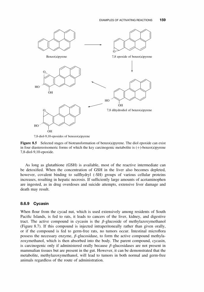
EXAMPLES OF ACTIVATING REACTIONS 159
O
Benzo(a)pyrene 7,8 epoxide of benzo(a)pyrene
7,8 dihydrodiol of benzo(a)pyrene
HO
O
OH
O
HO
OH
OH
HO
7,8-diol-9,10-epoxides of benozo(a)pyrene
Figure 8.5 Selected stages of biotransformation of benzo(a)pyrene. The diol epoxide can existin four diastereoisomeric forms of which the key carcinogenic metabolite is (+)-benzo(a)pyrene7,8-diol-9,10-epoxide.
As long as glutathione (GSH) is available, most of the reactive intermediate canbe detoxified. When the concentration of GSH in the liver also becomes depleted,however, covalent binding to sulfhydryl (-SH) groups of various cellular proteinsincreases, resulting in hepatic necrosis. If sufficiently large amounts of acetaminophenare ingested, as in drug overdoses and suicide attempts, extensive liver damage anddeath may result.
8.6.9 Cycasin
When flour from the cycad nut, which is used extensively among residents of SouthPacific Islands, is fed to rats, it leads to cancers of the liver, kidney, and digestivetract. The active compound in cycasin is the β-glucoside of methylazoxymethanol(Figure 8.7). If this compound is injected intraperitoneally rather than given orally,or if the compound is fed to germ-free rats, no tumors occur. Intestinal microflorapossess the necessary enzyme, β-glucosidase, to form the active compound methyla-zoxymethanol, which is then absorbed into the body. The parent compound, cycasin,is carcinogenic only if administered orally because β-glucosidases are not present inmammalian tissues but are present in the gut. However, it can be demonstrated that themetabolite, methylazoxymethanol, will lead to tumors in both normal and germ-freeanimals regardless of the route of administration.
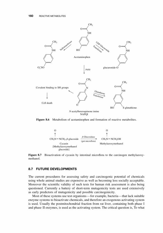
160 REACTIVE METABOLITES
O3−SO
NH
CH3
O
NH
CH3
O
HO
S-glutathione
Covalent binding to SH groups
Cell death
Acetaminophen
Sulfotra
nsferas
e
Transferase
UDP Glucuronide NH
CH3
O
glucuronide-O
N-acetylbenzoquinone imineNAPQI
N
CH3
O
O
NH
CH3
O
HO
P450
GlutathioneTransferase
Figure 8.6 Metabolism of acetaminophen and formation of reactive metabolites.
O
Cycasin[Methylazoxymethanol
glucoside]
b-Glucosidase
Methylazoxymethanol(gut microflora)
O
CH3N NCH2-b-glucoside CH3N NCH2OH
Figure 8.7 Bioactivation of cycasin by intestinal microflora to the carcinogen methylazoxy-methanol.
8.7 FUTURE DEVELOPMENTS
The current procedures for assessing safety and carcinogenic potential of chemicalsusing whole animal studies are expensive as well as becoming less socially acceptable.Moreover the scientific validity of such tests for human risk assessment is also beingquestioned. Currently a battery of short-term mutagenicity tests are used extensivelyas early predictors of mutagenicity and possible carcinogenicity.
Most of these systems use test organisms—for example, bacteria—that lack suitableenzyme systems to bioactivate chemicals, and therefore an exogenous activating systemis used. Usually the postmitochondrial fraction from rat liver, containing both phase Iand phase II enzymes, is used as the activating system. The critical question is, To what
SUGGESTED READING 161
extent does this rat system represent the true in vivo situation, especially in humans?If not this system, then what is the better alternative? As some of the examples inthis chapter illustrate, a chemical that is toxic or carcinogenic in one species or gendermay be inactive in another, and this phenomenon is often related to the complementof enzymes, either activation or detoxication, expressed in the exposed organism.
Another factor to consider is the ability of many foreign compounds to selectivelyinduce the CYP enzymes involved in their metabolism, especially if this inductionresults in the activation of the compound. With molecular techniques now available,considerable progress is being made in defining the enzyme and isozyme comple-ments of human and laboratory species and understanding their mechanisms of control.Another area of active research is the use of in vitro expression systems to study theoxidation of foreign chemicals (e.g., bacteria containing genes for specific humanCYP isozymes).
In summary, in studies of chemical toxicity, pathways and rates of metabolism aswell as effects resulting from toxicokinetic factors and receptor affinities are criticalin the choice of the animal species and experimental design. Therefore it is importantthat the animal species chosen as a model for humans in safety evaluations metabolizethe test chemical by the same routes as humans and, furthermore, that quantitative dif-ferences are considered in the interpretation of animal toxicity data. Risk assessmentmethods involving the extrapolation of toxic or carcinogenic potential of a chemicalfrom one species to another must consider the metabolic and toxicokinetic character-istics of both species.
SUGGESTED READING
Anders, M. W., W. Dekant, and S. Vamvakas, Glutathione-dependent toxicity. Xenobiotics 22:1135–1145, 1992.
Coughtrie, M. W. H., S. Sharp, K. Maxwell, and N. P. Innes. Biology and function of thereversible sulfation pathway catalysed by human sulfotransferases and sulfatases. Chemico-Biol. Interact. 109: 3–27, 1998.
Gonzalez, F. J., and H. V. Gelboin. Role of human cytochromes P450 in the metabolic activationof chemical carcinogens and toxins. Drug Metabol. Rev. 26: 165–183, 1994.
Guengerich, F. P. Bioactivation and detoxication of toxic and carcinogenic chemicals. DrugMetabol. Disp. 21: 1–6, 1993.
Guengerich, F. P. Metabolic activation of carcinogens. Pharmac. Ther. 54: 17–61, 1992.
Levi, P. E., and E. Hodgson. Reactive metabolites and toxicity. In Introduction to BiochemicalToxicology, 3rd ed., E. Hodgson and R. C. Smart, eds. New York: Wiley, 2001, pp. 199–220.
Omiecinski, C. J., R. P. Remmel, and V. P. Hosagrahara. Concise review of the cytochromeP450s and their roles in toxicology. Toxicol. Sci. 48: 151–156, 1999.
Rinaldi, R., E. Eliasson, S. Swedmark, and R. Morganstern. Reactive intermediates and thedynamics of glutathione transferases. Drug Metabol. Disp. 30: 1053–1058, 2002.
Ritter, J. K. Roles of glucuronidation and UDP-glucuronosyltransferases in xenobiotic bioacti-vation reactions. Chemico-Biol. Interact. 129: 171–193, 2000.
Vasiliou, V., A. Pappa, and D. R. Petersen. Role of aldehyde dehydrogenases in endogenousand xenobiotic metabolism. Chemico-Biol. Interact. 129: 1–19, 2000.

CHAPTER 9
Chemical and Physiological Influences onXenobiotic Metabolism
RANDY L. ROSE and ERNEST HODGSON
9.1 INTRODUCTION
The metabolism of toxicants and their overall toxicity can be modified by many factorsboth extrinsic and intrinsic to the normal functioning of the organism. It is entirelypossible that many changes in toxicity are due to changes in metabolism, because mostsequences of events that lead to overt toxicity involve activation and/or detoxication ofthe parent compound. In many cases the chain of cause and effect is not clear, due to thedifficulty of relating single events measured in vitro to the complex and interrelatedeffects that occur in vivo. This relationship between in vitro and in vivo studies isimportant and is discussed in connection with enzymatic inhibition and induction (seeSection 9.5). It is important to note that the chemical, nutritional, physiological, andother effects noted herein have been described primarily from experiments carriedout on experimental animals. These studies indicate that similar effects may occur inhumans or other animals, but not that they must occur or that they occur at the samemagnitude in all species if they occur at all.
9.2 NUTRITIONAL EFFECTS
Many nutritional effects on xenobiotic metabolism have been noted, but the informationis scattered and often appears contradictory. This is one of the most important ofseveral neglected areas of toxicology. This section is concerned only with the effectsof nutritional constituents of the diet; the effects of other xenobiotics in the diet arediscussed under chemical effects (see Section 9.5).
9.2.1 Protein
Low-protein diets generally decrease monooxygenase activity in rat liver microsomes,and gender and substrate differences may be seen in the effect. For example, aminopy-rine N -demethylation, hexobarbital hydroxylation, and aniline hydroxylation are all
A Textbook of Modern Toxicology, Third Edition, edited by Ernest HodgsonISBN 0-471-26508-X Copyright 2004 John Wiley & Sons, Inc.
163
164 CHEMICAL AND PHYSIOLOGICAL INFLUENCES ON XENOBIOTIC METABOLISM
decreased, but the effect on the first two is greater in males than in females. In thethird case, aniline hydroxylation, the reduction in males is equal to that in females.Tissue differences may also be seen. These changes are presumably related to thereductions in the levels of cytochrome P450 and NADPH-cytochrome P450 reductasethat are also noted. One might speculate that the gender and other variations are dueto differential effects on P450 isozymes. Even though enzyme levels are reduced bylow-protein diets, they can still be induced to some extent by compounds such asphenobarbital. Such changes may also be reflected in changes in toxicity. Changes inthe level of azoreductase activity in rat liver brought about by a low-protein diet arereflected in an increased severity in the carcinogenic effect of dimethylaminoazoben-zene. The liver carcinogen dimethylnitrosamine, which must be activated metabolically,is almost without effect in protein-deficient rats.
Strychnine, which is detoxified by microsomal monooxygenase action, is more toxicto animals on low-protein diets, whereas octamethylpyrophosphoramide, carbon tetra-chloride, and heptachlor, which are activated by monooxygenases, are less toxic. PhaseII reactions may also be affected by dietary protein levels. Chloramphenicol glu-curonidation is reduced in protein-deficient guinea pigs, although no effect is seenon sulfotransferase activity in protein-deficient rats.
9.2.2 Carbohydrates
High dietary carbohydrate levels in the rat tend to have much the same effect as lowdietary protein, decreasing such activities as aminopyrine N -demethylase, pentobarbitalhydroxylation, and p-nitrobenzoic acid reduction along with a concomitant decreasein the enzymes of the cytochrome P450 monooxygenase system. Because rats tend toregulate total caloric intake, this may actually reflect low-protein intake.
In humans it has been demonstrated that increasing the ratio of protein to carbohy-drate in the diet stimulates oxidation of antipyrine and theophylline, while changing theratio of fat to carbohydrate had no effect. In related studies, humans fed charcoal-broiledbeef (food high in polycyclic hydrocarbon content) for several days had significantlyenhanced activities of CYPs 1A1 and 1A2, resulting in enhanced metabolism ofphenacetin, theophylline, and antipyrine. Studies of this nature indicate that there issignificant interindividual variability in these observed responses.
9.2.3 Lipids
Dietary deficiencies in linoleic or in other unsaturated fats generally bring about areduction in P450 and related monooxygenase activities in the rat. The increase ineffectiveness of breast and colon carcinogens brought about in animals on high fatdiets, however, appears to be related to events during the promotion phase rather thanthe activation of the causative chemical.
Lipids also appear to be necessary for the effect of inducers, such as phenobarbital,to be fully expressed.
9.2.4 Micronutrients
Vitamin deficiencies, in general, bring about a reduction in monooxygenase activity,although exceptions can be noted. Riboflavin deficiency causes an increase in P450 and
NUTRITIONAL EFFECTS 165
aniline hydroxylation, although at the same time it causes a decrease in P450 reductaseand benzo(a)pyrene hydroxylation. Ascorbic acid deficiency in the guinea pig not onlycauses a decrease in P450 and monooxygenase activity but also causes a reduction inmicrosomal hydrolysis of procaine. Deficiencies in vitamins A and E cause a decreasein monooxygenase activity, whereas thiamine deficiency causes an increase. The effectof these vitamins on different P450 isozymes has not been investigated. Changes inmineral nutrition have also been observed to affect monooxygenase activity. In theimmature rat, calcium or magnesium deficiency causes a decrease, whereas, quiteunexpectedly, iron deficiency causes an increase. This increase is not accompaniedby a concomitant increase in P450, however. An excess of dietary cobalt, cadmium,manganese, and lead all cause an increase in hepatic glutathione levels and a decreasein P450 content.
9.2.5 Starvation and Dehydration
Although in some animals starvation appears to have effects similar to those of proteindeficiency, this is not necessarily the case. For example, in the mouse, monooxy-genation is decreased but reduction of p-nitrobenzoic acid is unaffected. In male rats,hexobarbital and pentobarbital hydroxylation as well as aminopyrine N -demethylationare decreased, but aniline hydroxylation is increased. All of these activities are stim-ulated in the female. Water deprivation in gerbils causes an increase in P450 anda concomitant increase in hexobarbital metabolism, which is reflected in a shortersleeping time.
9.2.6 Nutritional Requirements in Xenobiotic Metabolism
Because xenobiotic metabolism involves many enzymes with different cofactor require-ments, prosthetic groups, or endogenous cosubstrates, it is apparent that many differentnutrients are involved in their function and maintenance. Determination of the effectsof deficiencies, however, is more complex because reductions in activity of any par-ticular enzyme will be effective only if it affects a change in a rate-limiting step in aprocess. In the case of multiple deficiencies, the nature of the rate-limiting step maychange with time
Phase I Reactions. Nutrients involved in the maintenance of the cytochrome P450monooxygenase system are shown in Figure 9.1. The B complex vitamins niacin andriboflavin are both involved, the former in the formation of NADPH and the latter inthe formation of FAD and FMN. Essential amino acids are, of course, required forthe synthesis of all of the proteins involved. The heme of the cytochrome requiresiron, an essential inorganic nutrient. Other nutrients required in heme synthesis includepantothenic acid, needed for the synthesis of the coenzyme A used in the formationof acetyl Co-A, pyridoxine, a cofactor in heme synthesis and copper, required in theferroxidase system that converts ferrous to ferric iron prior to its incorporation intoheme. Although it is clear that dietary deficiencies could reduce the ability of the P450system to metabolize xenobiotics, it is not clear how this effect will be manifested invivo unless there is an understanding of the rate-limiting factors involved, which is aconsiderable task in such a complex of interrelated reactions. Similar considerations
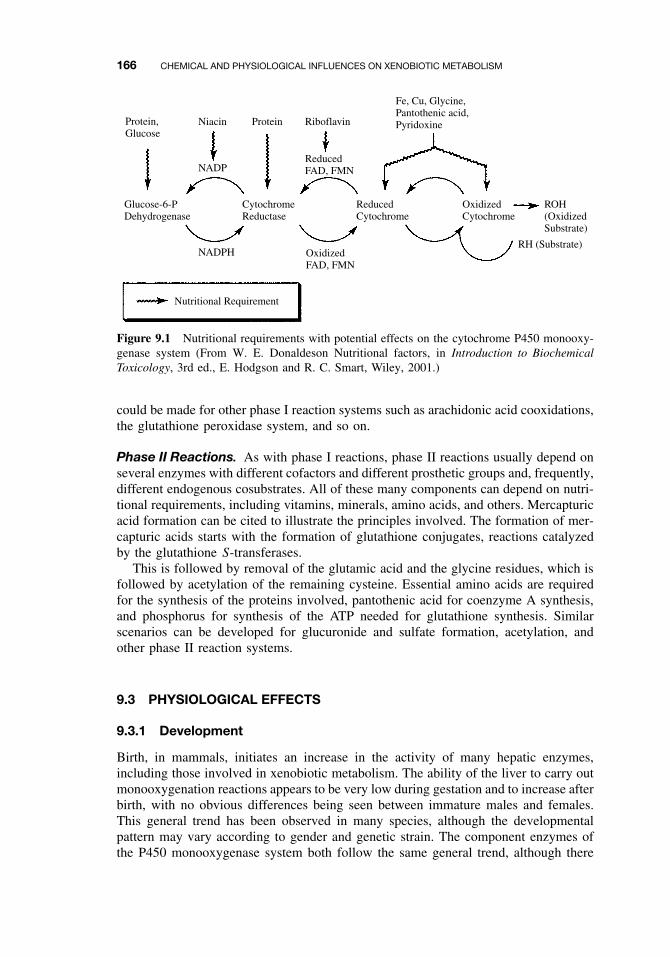
166 CHEMICAL AND PHYSIOLOGICAL INFLUENCES ON XENOBIOTIC METABOLISM
Protein,Glucose
Niacin
NADP
NADPH
Protein Riboflavin
ReducedFAD, FMN
OxidizedFAD, FMN
Fe, Cu, Glycine,Pantothenic acid,Pyridoxine
Glucose-6-PDehydrogenase
CytochromeReductase
ReducedCytochrome
OxidizedCytochrome
ROH(OxidizedSubstrate)
RH (Substrate)
Nutritional Requirement
Figure 9.1 Nutritional requirements with potential effects on the cytochrome P450 monooxy-genase system (From W. E. Donaldeson Nutritional factors, in Introduction to BiochemicalToxicology, 3rd ed., E. Hodgson and R. C. Smart, Wiley, 2001.)
could be made for other phase I reaction systems such as arachidonic acid cooxidations,the glutathione peroxidase system, and so on.
Phase II Reactions. As with phase I reactions, phase II reactions usually depend onseveral enzymes with different cofactors and different prosthetic groups and, frequently,different endogenous cosubstrates. All of these many components can depend on nutri-tional requirements, including vitamins, minerals, amino acids, and others. Mercapturicacid formation can be cited to illustrate the principles involved. The formation of mer-capturic acids starts with the formation of glutathione conjugates, reactions catalyzedby the glutathione S-transferases.
This is followed by removal of the glutamic acid and the glycine residues, which isfollowed by acetylation of the remaining cysteine. Essential amino acids are requiredfor the synthesis of the proteins involved, pantothenic acid for coenzyme A synthesis,and phosphorus for synthesis of the ATP needed for glutathione synthesis. Similarscenarios can be developed for glucuronide and sulfate formation, acetylation, andother phase II reaction systems.
9.3 PHYSIOLOGICAL EFFECTS
9.3.1 Development
Birth, in mammals, initiates an increase in the activity of many hepatic enzymes,including those involved in xenobiotic metabolism. The ability of the liver to carry outmonooxygenation reactions appears to be very low during gestation and to increase afterbirth, with no obvious differences being seen between immature males and females.This general trend has been observed in many species, although the developmentalpattern may vary according to gender and genetic strain. The component enzymes ofthe P450 monooxygenase system both follow the same general trend, although there
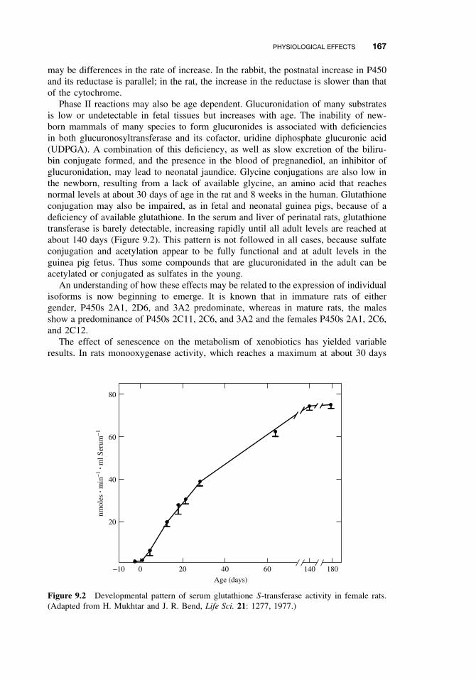
PHYSIOLOGICAL EFFECTS 167
may be differences in the rate of increase. In the rabbit, the postnatal increase in P450and its reductase is parallel; in the rat, the increase in the reductase is slower than thatof the cytochrome.
Phase II reactions may also be age dependent. Glucuronidation of many substratesis low or undetectable in fetal tissues but increases with age. The inability of new-born mammals of many species to form glucuronides is associated with deficienciesin both glucuronosyltransferase and its cofactor, uridine diphosphate glucuronic acid(UDPGA). A combination of this deficiency, as well as slow excretion of the biliru-bin conjugate formed, and the presence in the blood of pregnanediol, an inhibitor ofglucuronidation, may lead to neonatal jaundice. Glycine conjugations are also low inthe newborn, resulting from a lack of available glycine, an amino acid that reachesnormal levels at about 30 days of age in the rat and 8 weeks in the human. Glutathioneconjugation may also be impaired, as in fetal and neonatal guinea pigs, because of adeficiency of available glutathione. In the serum and liver of perinatal rats, glutathionetransferase is barely detectable, increasing rapidly until all adult levels are reached atabout 140 days (Figure 9.2). This pattern is not followed in all cases, because sulfateconjugation and acetylation appear to be fully functional and at adult levels in theguinea pig fetus. Thus some compounds that are glucuronidated in the adult can beacetylated or conjugated as sulfates in the young.
An understanding of how these effects may be related to the expression of individualisoforms is now beginning to emerge. It is known that in immature rats of eithergender, P450s 2A1, 2D6, and 3A2 predominate, whereas in mature rats, the malesshow a predominance of P450s 2C11, 2C6, and 3A2 and the females P450s 2A1, 2C6,and 2C12.
The effect of senescence on the metabolism of xenobiotics has yielded variableresults. In rats monooxygenase activity, which reaches a maximum at about 30 days
−10 0
20
40
60
80
20 40 60 140 180
Age (days)
nmol
es ·
min
−1 ·
ml S
erum
−1
Figure 9.2 Developmental pattern of serum glutathione S-transferase activity in female rats.(Adapted from H. Mukhtar and J. R. Bend, Life Sci. 21: 1277, 1977.)
168 CHEMICAL AND PHYSIOLOGICAL INFLUENCES ON XENOBIOTIC METABOLISM
of age, begins to decline some 250 days later, a decrease that may be associated withreduced levels of sex hormones. Glucuronidation also decreases in old animals, whereasmonoamine oxidase activity increases. These changes in the monooxygenase activitiesare often reflected by changes in drug efficacy or overall toxicity.
In humans, age-related impairment of enzyme activity is highly controversial. Age-related declines in activity were not detected with respect to the activity of CYP2Cand CYP3A isoforms among 54 liver samples from donors ranging in age from 9to 89 years. Studies involving an erythromycin breath test in humans also suggestedthat there were no age-related declines associated with CYP3A4 activity. However,a study of CYP content and antipyrine clearance in liver biopsies obtained from 226closely matched subjects indicated that subjects older than 70 had significantly lessactivity and clearance than younger subjects. Likewise, in older subjects, clearanceof the drug omeprazole, a CYP2C19 substrate, was nearly half the rates observed inyounger subjects.
9.3.2 Gender Differences
Metabolism of xenobiotics may vary with the gender of the organism. Gender dif-ferences become apparent at puberty and are usually maintained throughout adultlife. Adult male rats metabolize many compounds at rates higher than females, forexample, hexobarbital hydroxylation, aminopyrine N -demethylation, glucuronidationof o-aminophenol, and glutathione conjugation of aryl substrates; however, with othersubstrates, such as aniline and zoxazolamine, no gender differences are seen. In otherspecies, including humans, the gender difference in xenobiotic metabolism is less pro-nounced. The differences in microsomal monooxygenase activity between males andfemales have been shown to be under the control of sex hormones, at least in somespecies. Some enzyme activities are decreased by castration in the male and admin-istration of androgens to castrated males increases the activity of these sex-dependentenzyme activities without affecting the independent ones. Procaine hydrolysis is fasterin male than female rats, and this compound is less toxic to the male. Gender dif-ferences in enzyme activity may also vary from tissue to tissue. Hepatic microsomesfrom adult male guinea pigs are less active in the conjugation of p-nitrophenol thanare those from females, but no such gender difference is seen in the microsomes fromlung, kidney, and small intestines.
Many differences in overall toxicity between males and females of various speciesare known (Table 9.1). Although it is not always known whether metabolism is theonly or even the most important factor, such differences may be due to gender-relateddifferences in metabolism. Hexobarbital is metabolized faster by male rats; thus femalerats have longer sleeping times. Parathion is activated to the cholinesterase inhibitorparaoxon more rapidly in female than in male rats, and thus is more toxic to females.Presumably many of the gender-related differences, as with the developmental dif-ferences, are related to quantitative or qualitative differences in the isozymes of thexenobiotic-metabolizing enzymes that exist in multiple forms, but this aspect has notbeen investigated extensively.
In the rat, sexually dimorphic P450s appear to arise by programming, or imprint-ing, that occurs in neonatal development. This imprinting is brought about by a surgeof testosterone that occurs in the male, but not the female, neonate and appears toimprint the developing hypothalamus so that in later development the growth hormone
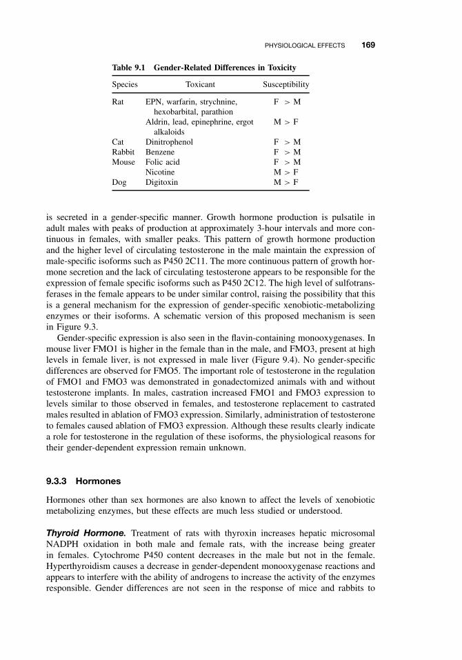
PHYSIOLOGICAL EFFECTS 169
Table 9.1 Gender-Related Differences in Toxicity
Species Toxicant Susceptibility
Rat EPN, warfarin, strychnine,hexobarbital, parathion
F > M
Aldrin, lead, epinephrine, ergotalkaloids
M > F
Cat Dinitrophenol F > MRabbit Benzene F > MMouse Folic acid F > M
Nicotine M > FDog Digitoxin M > F
is secreted in a gender-specific manner. Growth hormone production is pulsatile inadult males with peaks of production at approximately 3-hour intervals and more con-tinuous in females, with smaller peaks. This pattern of growth hormone productionand the higher level of circulating testosterone in the male maintain the expression ofmale-specific isoforms such as P450 2C11. The more continuous pattern of growth hor-mone secretion and the lack of circulating testosterone appears to be responsible for theexpression of female specific isoforms such as P450 2C12. The high level of sulfotrans-ferases in the female appears to be under similar control, raising the possibility that thisis a general mechanism for the expression of gender-specific xenobiotic-metabolizingenzymes or their isoforms. A schematic version of this proposed mechanism is seenin Figure 9.3.
Gender-specific expression is also seen in the flavin-containing monooxygenases. Inmouse liver FMO1 is higher in the female than in the male, and FMO3, present at highlevels in female liver, is not expressed in male liver (Figure 9.4). No gender-specificdifferences are observed for FMO5. The important role of testosterone in the regulationof FMO1 and FMO3 was demonstrated in gonadectomized animals with and withouttestosterone implants. In males, castration increased FMO1 and FMO3 expression tolevels similar to those observed in females, and testosterone replacement to castratedmales resulted in ablation of FMO3 expression. Similarly, administration of testosteroneto females caused ablation of FMO3 expression. Although these results clearly indicatea role for testosterone in the regulation of these isoforms, the physiological reasons fortheir gender-dependent expression remain unknown.
9.3.3 Hormones
Hormones other than sex hormones are also known to affect the levels of xenobioticmetabolizing enzymes, but these effects are much less studied or understood.
Thyroid Hormone. Treatment of rats with thyroxin increases hepatic microsomalNADPH oxidation in both male and female rats, with the increase being greaterin females. Cytochrome P450 content decreases in the male but not in the female.Hyperthyroidism causes a decrease in gender-dependent monooxygenase reactions andappears to interfere with the ability of androgens to increase the activity of the enzymesresponsible. Gender differences are not seen in the response of mice and rabbits to
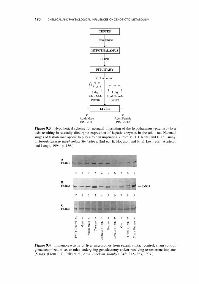
170 CHEMICAL AND PHYSIOLOGICAL INFLUENCES ON XENOBIOTIC METABOLISM
TESTES
HYPOTHALAMUS
Testosterone
PITUITARY
GHRH
LIVER
GH Secretion
Adult MaleP450 2C11
Adult MalePattern
1 day 1 dayAdult Female
Pattern
Adult FemaleP450 2C12
Figure 9.3 Hypothetical scheme for neonatal imprinting of the hypothalamus–pituitary–liveraxis resulting in sexually dimorphic expression of hepatic enzymes in the adult rat. Neonatalsurges of testosterone appear to play a role in imprinting. (From M. J. J. Ronis and H. C. Cunny,in Introduction to Biochemical Toxicology, 2nd ed. E. Hodgson and P. E. Levi, eds., Appletonand Lange, 1994, p. 136.)
C 1 2 3 4 5 6 7 8 9
C 1 2 3 4 5 6 7 8 9
C
FMO
Con
trol
1
Mal
e
2
Sham
Mal
e
3
Cas
trat
e
4
Cas
trat
e +
Test
.
5
Fem
ale
6
Fem
ale
+ Te
st.
7
Ove
x
8
Ove
x +
Test
.
9
Sham
Fem
ale
--- FMO3
AFMO1
BFMO3
CFMO5
Figure 9.4 Immunoreactivity of liver microsomes from sexually intact control, sham control,gonadectomized mice, or mice undergoing gonadectomy and/or receiving testosterone implants(5 mg). (From J. G. Falls et al., Arch. Biochem. Biophys. 342: 212–223, 1997.)
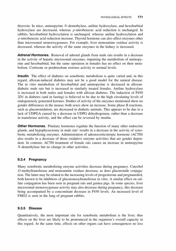
PHYSIOLOGICAL EFFECTS 171
thyroxin. In mice, aminopyrine N -demethylase, aniline hydroxylase, and hexobarbitalhydroxylase are decreased, whereas p-nitrobenzoic acid reduction is unchanged. Inrabbits, hexobarbital hydroxylation is unchanged, whereas aniline hydroxylation andp-nitrobenzoic acid reduction increase. Thyroid hormone can also affect enzymes otherthan microsomal monooxygenases. For example, liver monoamine oxidase activity isdecreased, whereas the activity of the same enzymes in the kidney is increased.
Adrenal Hormones. Removal of adrenal glands from male rats results in a decreasein the activity of hepatic microsomal enzymes, impairing the metabolism of aminopy-rine and hexobarbital, but the same operation in females has no effect on their meta-bolism. Cortisone or prednisolone restores activity to normal levels.
Insulin. The effect of diabetes on xenobiotic metabolism is quite varied and, in thisregard, alloxan-induced diabetes may not be a good model for the natural disease.The in vitro metabolism of hexobarbital and aminopyrine is decreased in alloxan-diabetic male rats but is increased in similarly treated females. Aniline hydroxylaseis increased in both males and females with alloxan diabetes. The induction of P4502D1 in diabetes (and in fasting) is believed to be due to the high circulating levels ofendogenously generated ketones. Studies of activity of the enzymes mentioned show nogender differences in the mouse; both sexes show an increase. Some phase II reactions,such as glucuronidation, are decreased in diabetic animals. This appears to be due to alack of UDPGA caused by a decrease in UDPG dehydrogenase, rather than a decreasein transferase activity, and the effect can be reversed by insulin.
Other Hormones. Pituitary hormones regulate the function of many other endocrineglands, and hypophysectomy in male rats’ results in a decrease in the activity of xeno-biotic metabolizing enzymes. Administration of adrenocorticotropic hormone (ACTH)also results in a decrease of those oxidative enzyme activities that are gender depen-dent. In contrast, ACTH treatment of female rats causes an increase in aminopyrineN -demethylase but no change in other activities.
9.3.4 Pregnancy
Many xenobiotic metabolizing enzyme activities decrease during pregnancy. CatecholO-methyltransferase and monoamine oxidase decrease, as does glucuronide conjuga-tion. The latter may be related to the increasing levels of progesterone and pregnanediol,both known to be inhibitors of glucuronosyltransferase in vitro. A similar effect on sul-fate conjugation has been seen in pregnant rats and guinea pigs. In some species, livermicrosomal monooxygenase activity may also decrease during pregnancy, this decreasebeing accompanied by a concomitant decrease in P450 levels. An increased level ofFMO2 is seen in the lung of pregnant rabbits.
9.3.5 Disease
Quantitatively, the most important site for xenobiotic metabolism is the liver; thuseffects on the liver are likely to be pronounced in the organism’s overall capacity inthis regard. At the same time, effects on other organs can have consequences no less
172 CHEMICAL AND PHYSIOLOGICAL INFLUENCES ON XENOBIOTIC METABOLISM
serious for the organism. Patients with acute hepatitis frequently have an impairedability to oxidize drugs, with a concomitant increase in plasma half-life. Impairedoxidative metabolism has also been shown in patients with chronic hepatitis or cir-rhosis. The decrease in drug metabolism that occurs in obstructive jaundice may be aconsequence of the accumulation of bile salts, which are known inhibitors of some ofthe enzymes involved. Phase II reactions may also be affected, decreases in acetyla-tion, glucuronidation, and a variety of esterase activities having been seen in variousliver diseases. Hepatic tumors, in general, have a lower ability to metabolize foreigncompounds than does normal liver tissue, although in some cases the overall activ-ity of tumor bearing livers may be no lower than that of controls. Kidney diseasesmay also affect the overall ability to handle xenobiotics, because this organ is one ofthe main routes for elimination of xenobiotics and their metabolites. The half-lives oftolbutamide, thiopental, hexobarbital, and chloramphenicol are all prolonged in patientswith renal impairment.
9.3.6 Diurnal Rhythms
Diurnal rhythms, both in P450 levels and in the susceptibility to toxicants, have beendescribed, especially in rodents. Although such changes appear to be related to thelight cycle, they may in fact be activity dependent because feeding and other activitiesin rodents are themselves markedly diurnal.
9.4 COMPARATIVE AND GENETIC EFFECTS
Comparative toxicology is the study of the variation in toxicity of exogenous chemicalstoward different organisms, either of different genetic strains or of different taxonomicgroups. Thus the comparative approach can be used in the study of any aspect oftoxicology, such as absorption, metabolism, mode of action, and acute or chroniceffects. Most comparative data for toxic compounds exist in two areas—acute toxicityand metabolism. The value of the comparative approach can be summarized under fourheadings:
1. Selective toxicity. If toxic compounds are to be used for controlling diseases,pests, and parasites, it is important to develop selective biocides, toxic to thetarget organism but less toxic to other organisms, particularly humans.
2. Experimental models. Comparative studies of toxic phenomena are necessary toselect the most appropriate model for extrapolation to humans and for testingand development of drugs and biocides. Taxonomic proximity does not neces-sarily indicate which will be the best experimental animal because in some casesprimates are less valuable for study than are other mammals.
3. Environmental xenobiotic cycles. Much concern over toxic compounds springsfrom their occurrence in the environment. Different organisms in the complexecological food webs metabolize compounds at different rates and to differentproducts; the metabolic end products are released back to the environment, eitherto be further metabolized by other organisms or to exert toxic effects of theirown. Clearly, it is desirable to know the range of metabolic processes possible.
COMPARATIVE AND GENETIC EFFECTS 173
Laboratory micro ecosystems have been developed, and with the aid of 14C-labeled compounds, chemicals and their metabolites can be followed through theplants and terrestrial and aquatic animals involved.
4. Comparative biochemistry. Some researchers believe that the proper role ofcomparative biochemistry is to put evolution on a molecular basis, and that detox-ication enzymes, like other enzymes, are suitable subjects for study. Xenobiotic-metabolizing enzymes were probably essential in the early stages of animalevolution because secondary plant products, even those of low toxicity, are fre-quently lipophilic and as a consequence would, in the absence of such enzymes,accumulate in lipid membranes and lipid depots. The evolution of cytochromeP450 isoforms, with more than 2000 isoform cDNA sequences known, is provinga useful tool for the study of biochemical evolution.
9.4.1 Variations Among Taxonomic Groups
There are few differences in xenobiotic metabolism that are specific for large taxonomicgroups. The formation of glucosides by insects and plants rather than the glucuronidesof other animal groups is one of the most distinct. Although differences among speciesare common and of toxicologic significance, they are usually quantitative rather thanqualitative in nature and tend to occur within as well as between taxonomic groups.Although the ultimate explanation of such differences must be at the level of biochem-ical genetics, they are manifested at many other levels, the most important of whichare summarized in the following sections.
In vivo Toxicity. Toxicity is a term used to describe the adverse effects of chemicalson living organisms. Depending on the degree of toxicity, an animal may die, sufferinjury to certain organs, or have a specific functional derangement in a subcellularorganelle. Sublethal effects of toxicants may be reversible. Available data on the tox-icity of selected pesticides to rats suggest that herbicide use, in general, provides thegreatest human safety factor by selectively killing plants. As the evolutionary positionof the target species approaches that of humans, however, the human safety factoris narrowed considerably. Thus the direct toxicity to humans and other mammals ofbiocide toxicity seems to be in the following progression: herbicides = fungicides <
molluscicides < acaricides < nematocides < insecticides < rodenticides. This formulais obviously oversimplified because marked differences in lethality are observed whendifferent members of each group of biocides is tested against laboratory test animalsand target species. One should also bear in mind that any chemical can be environmen-tally dangerous if misused because many possible targets are interrelated in complexecological systems.
Interspecific differences are also known for some naturally occurring poisons. Nico-tine, for instance, is used as an insecticide and kills many insect pests at low doses,yet tobacco leaves constitute a normal diet for several species. As indicated earlier,most strains of rabbit eat Belladonna leaves without ill effects, whereas other mammalsare easily poisoned. Natural tolerance to cyanide poisoning in millipedes and the highresistance to the powerful axonal blocking tetrodotoxin in puffer fish are examples ofthe tolerance of animals to the toxins they produce.
The specific organ toxicity of chemicals also exhibits wide species differences. Car-bon tetrachloride, a highly potent hepatotoxicant, induces liver damage in many species,
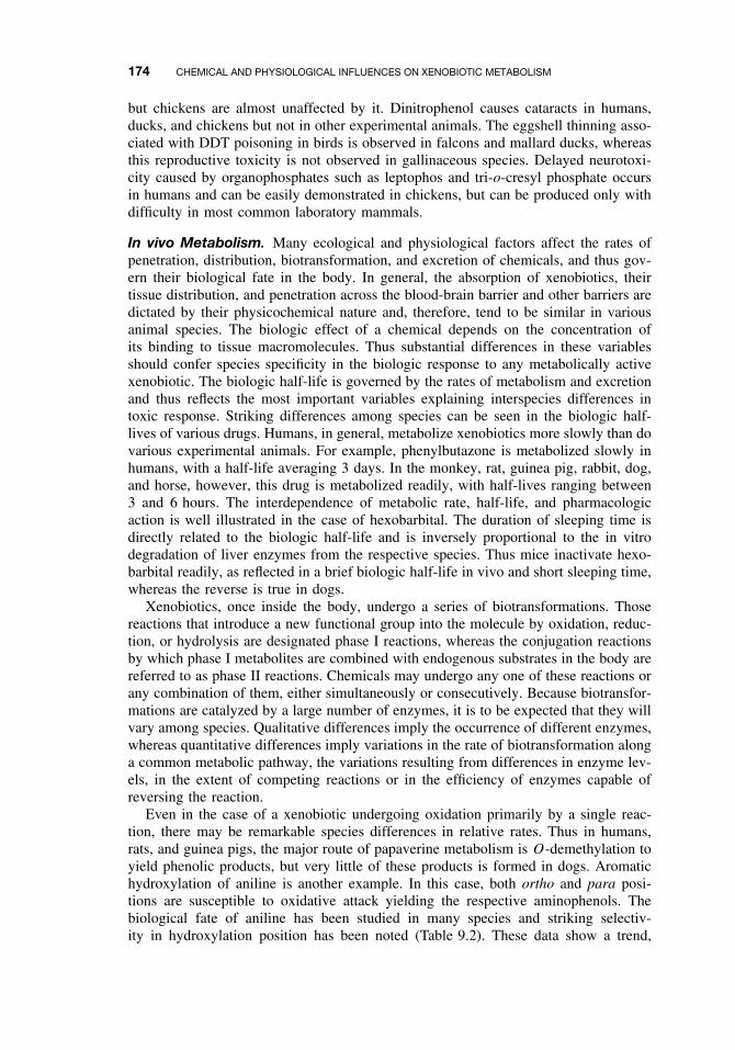
174 CHEMICAL AND PHYSIOLOGICAL INFLUENCES ON XENOBIOTIC METABOLISM
but chickens are almost unaffected by it. Dinitrophenol causes cataracts in humans,ducks, and chickens but not in other experimental animals. The eggshell thinning asso-ciated with DDT poisoning in birds is observed in falcons and mallard ducks, whereasthis reproductive toxicity is not observed in gallinaceous species. Delayed neurotoxi-city caused by organophosphates such as leptophos and tri-o-cresyl phosphate occursin humans and can be easily demonstrated in chickens, but can be produced only withdifficulty in most common laboratory mammals.
In vivo Metabolism. Many ecological and physiological factors affect the rates ofpenetration, distribution, biotransformation, and excretion of chemicals, and thus gov-ern their biological fate in the body. In general, the absorption of xenobiotics, theirtissue distribution, and penetration across the blood-brain barrier and other barriers aredictated by their physicochemical nature and, therefore, tend to be similar in variousanimal species. The biologic effect of a chemical depends on the concentration ofits binding to tissue macromolecules. Thus substantial differences in these variablesshould confer species specificity in the biologic response to any metabolically activexenobiotic. The biologic half-life is governed by the rates of metabolism and excretionand thus reflects the most important variables explaining interspecies differences intoxic response. Striking differences among species can be seen in the biologic half-lives of various drugs. Humans, in general, metabolize xenobiotics more slowly than dovarious experimental animals. For example, phenylbutazone is metabolized slowly inhumans, with a half-life averaging 3 days. In the monkey, rat, guinea pig, rabbit, dog,and horse, however, this drug is metabolized readily, with half-lives ranging between3 and 6 hours. The interdependence of metabolic rate, half-life, and pharmacologicaction is well illustrated in the case of hexobarbital. The duration of sleeping time isdirectly related to the biologic half-life and is inversely proportional to the in vitrodegradation of liver enzymes from the respective species. Thus mice inactivate hexo-barbital readily, as reflected in a brief biologic half-life in vivo and short sleeping time,whereas the reverse is true in dogs.
Xenobiotics, once inside the body, undergo a series of biotransformations. Thosereactions that introduce a new functional group into the molecule by oxidation, reduc-tion, or hydrolysis are designated phase I reactions, whereas the conjugation reactionsby which phase I metabolites are combined with endogenous substrates in the body arereferred to as phase II reactions. Chemicals may undergo any one of these reactions orany combination of them, either simultaneously or consecutively. Because biotransfor-mations are catalyzed by a large number of enzymes, it is to be expected that they willvary among species. Qualitative differences imply the occurrence of different enzymes,whereas quantitative differences imply variations in the rate of biotransformation alonga common metabolic pathway, the variations resulting from differences in enzyme lev-els, in the extent of competing reactions or in the efficiency of enzymes capable ofreversing the reaction.
Even in the case of a xenobiotic undergoing oxidation primarily by a single reac-tion, there may be remarkable species differences in relative rates. Thus in humans,rats, and guinea pigs, the major route of papaverine metabolism is O-demethylation toyield phenolic products, but very little of these products is formed in dogs. Aromatichydroxylation of aniline is another example. In this case, both ortho and para posi-tions are susceptible to oxidative attack yielding the respective aminophenols. Thebiological fate of aniline has been studied in many species and striking selectiv-ity in hydroxylation position has been noted (Table 9.2). These data show a trend,
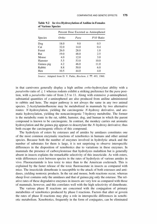
COMPARATIVE AND GENETIC EFFECTS 175
Table 9.2 In vivo Hydroxylation of Aniline in Femalesof Various Species
Percent Dose Excreted as Aminophenol
Species Ortho Para P /O Ratio
Dog 18.0 9.0 0.5Cat 32.0 14.0 0.4Ferret 26.0 28.0 1.0Rat 19.0 48.0 2.5Mouse 4.0 12.0 3.0Hamster 5.5 53.0 10.0Guinea pig 4.2 46.0 11.0Rabbit 8.8 50.0 6.0Hen 10.5 44.0 4.0
Source: Adapted from D. V. Parke, Biochem. J. 77: 493, 1960.
in that carnivores generally display a high aniline ortho-hydroxylase ability with apara/ortho ratio of ≤ 1 whereas rodents exhibit a striking preference for the para posi-tion, with a para/ortho ratio of from 2.5 to 11. Along with extensive p-aminophenol,substantial quantities of o-aminophenol are also produced from aniline administeredto rabbits and hens. The major pathway is not always the same in any two animalspecies. 2-Acetylaminofluorene may be metabolized in mammals by two alternativeroutes: N -hydroxylation, yielding the carcinogenic N -hydroxy derivative, and aro-matic hydroxylation, yielding the noncarcinogenic 7-hydroxy metabolite. The formeris the metabolic route in the rat, rabbit, hamster, dog, and human in which the parentcompound is known to be carcinogenic. In contrast, the monkey carries out aromatichydroxylation and the guinea pig appears to deacetylate the N -hydroxy derivative; thusboth escape the carcinogenic effects of this compound.
The hydrolysis of esters by esterases and of amides by amidases constitutes oneof the most common enzymatic reactions of xenobiotics in humans and other animalspecies. Because both the number of enzymes involved in hydrolytic attack and thenumber of substrates for them is large, it is not surprising to observe interspecificdifferences in the disposition of xenobiotics due to variations in these enzymes. Inmammals the presence of carboxylesterase that hydrolyzes malathion but is generallyabsent in insects explains the remarkable selectivity of this insecticide. As with esters,wide differences exist between species in the rates of hydrolysis of various amides invivo. Fluoracetamide is less toxic to mice than to the American cockroach. This isexplained by the faster release of the toxic fluoroacetate in insects as compared withmice. The insecticide dimethoate is susceptible to the attack of both esterases and ami-dases, yielding nontoxic products. In the rat and mouse, both reactions occur, whereassheep liver contains only the amidases and that of guinea pig only the esterase. The rel-ative rates of these degradative enzymes in insects are very low as compared with thoseof mammals, however, and this correlates well with the high selectivity of dimethoate.
The various phase II reactions are concerned with the conjugation of primarymetabolites of xenobiotics produced by phase I reactions. Factors that alter or governthe rates of phase II reactions may play a role in interspecific differences in xenobi-otic metabolism. Xenobiotics, frequently in the form of conjugates, can be eliminated
176 CHEMICAL AND PHYSIOLOGICAL INFLUENCES ON XENOBIOTIC METABOLISM
through urine, feces, lungs, sweat, saliva, milk, hair, nails, or placenta, although com-parative data are generally available only for the first two routes. Interspecific variationin the pattern of biliary excretion may determine species differences in the relativeextent to which compounds are eliminated in the urine or feces. Fecal excretion of achemical or its metabolites tends to be higher in species that are good biliary excretors,such as the rat and dog, than in species that are poor biliary excretors, such as therabbit, guinea pig, and monkey. For example, the fecal excretion of stilbestrol in therat accounts for 75% of the dose, whereas in the rabbit about 70% can be found inthe urine. Dogs, like humans, metabolize indomethacin to a glucuronide but, unlikehumans that excrete it in the urine, dogs excrete it primarily in the feces—apparentlydue to inefficient renal and hepatic blood clearance of the glucuronide. These differ-ences may involve species variation in enterohepatic circulation, plasma level, andbiologic half-life.
Interspecific differences in the magnitude of biliary excretion of a xenobiotic excre-tion product largely depend on molecular weight, the presence of polar groups in themolecule, and the extent of conjugation. Conjugates with molecular weights of lessthan 300 are poorly excreted in bile and tend to be excreted with urine, whereas thereverse is true for those with molecular weights higher than 300. The critical molecularweight appears to vary between species, and marked species differences are noted forbiliary excretion of chemicals with molecular weights of about 300. Thus the biliaryexcretion of succinylsulfathioazole is 20- to 30-fold greater in the rat and the dog thanin the rabbit and the guinea pig, and more than 100-fold greater than in the pig andthe rhesus monkey. The cat and sheep are intermediate and excrete about 7% of thedose in the bile.
The evidence reported in a few studies suggests some relationship between the evo-lutionary position of a species and its conjugation mechanisms (Table 9.3). In humansand most mammals, the principal mechanisms involve conjugations with glucuronicacid, glycine, glutamine, glutathione and sulfate. Minor conjugation mechanisms in
Table 9.3 Occurrence of Common and Unusual Conjugation Reactions
ConjugatingGroup Common Unusual
Carbohydrate Glucuronic acid (animals) N-Acetylglucosamine (rabbits)Glucose (insects, plants) Ribose (rats, mice)
Amino acids Glycine Glutamine (insects, humans)Glutathione Ornithine (birds)Methionine Arginine (ticks, spiders)
Glycyltaurine (cats)Glycylglycine (cats)Serine (rabbits)
Acetyl Acetyl group fromacetyl-0CoA
Formyl Formylation (dogs, rats)Sulfate Sulfate group from PAPSPhosphate Phosphate monoester formation (dogs, insects)
Source: Modified from A. P. Kulkarni and E. Hodgson, Comparative toxicology, in Introduction to Bio-chemical Toxicology. E. Hodgson and F. E. Guthrie, eds., New York: Elsevier, 1980, p. 115.
COMPARATIVE AND GENETIC EFFECTS 177
mammals include acetylation and methylation pathways. In some species of birds andreptiles, ornithine conjugation replaces glycine conjugation; in plants, bacteria, andinsects, conjugation with glucose instead of glucuronic acid results in the formation ofglucosides. In addition to these predominant reactions, certain other conjugative pro-cesses are found involving specific compounds in only a few species. These reactionsinclude conjugation with phosphate, taurine, N -acetyl-glucosamine, ribose, glycyltau-rine, serine, arginine, and formic acid.
From the standpoint of evolution, similarity might be expected between humans andother primate species as opposed to the nonprimates. This phylogenic relationship isobvious from the relative importance of glycine and glutamine in the conjugation ofarylacetic acids. The conjugating agent in humans is exclusively glutamine, and thesame is essentially true with Old World monkeys. New World monkeys, however, useboth the glycine and glutamine pathways. Most nonprimates and lower primates carryout glycine conjugation selectively. A similar evolutionary trend is also observed in theN -glucuronidation of sulfadimethoxine and in the aromatization of quinic acid; bothreactions occur extensively in human, and their importance decreases with increas-ing evolutionary divergence from humans. When the relative importance of metabolicpathways is considered, one of the simplest cases of an enzyme-related species differ-ence in the disposition of a substrate undergoing only one conjugative reaction is theacetylation of 4-aminohippuric acid. In the rat, guinea pig, and rabbit, the major biliarymetabolite is 4-aminohippuric acid; the cat excretes nearly equal amounts of free acidand its acetyl derivative; and the hen excretes mainly the unchanged compound. In thedog, 4-aminohippuric acid is also passed into the bile unchanged because this speciesis unable to acetylate aromatic amino groups.
Defective operation of phase II reactions usually causes a striking species differencein the disposition pattern of a xenobiotic. The origin of such species variations isusually either the absence or a low level of the enzyme(s) in question and/or itscofactors. Glucuronide synthesis is one of the most common detoxication mechanismsin most mammalian species. The cat and closely related species have a defectiveglucuronide-forming system, however. Although cats form little or no glucuronide fromo-aminophenol, phenol, p-nitrophenol, 2-amino-4-nitrophenol, 1-or 2-naphthol, andmorphine, they readily form glucuronides from phenolphthalein, bilirubin, thyroxine,and certain steroids. Recently polymorphisms of UDP glucuronyl-transferase have beendemonstrated in rat and guinea pig liver preparations; thus defective glucuronidation inthe cat is probably related to the absence of the appropriate transferase rather than thatof the active intermediate, UDPGA or UDP glucose dehydrogenase, which convertsUDP glucose into UDPGA.
Studies on the metabolic fate of phenol in several species have indicated that foururinary products are excreted (Figure 9.5). Although extensive phenol metabolism takesplace in most species, the relative proportions of each metabolite produced varies fromspecies to species. In contrast to the cat, which selectively forms sulfate conjugates, thepig excretes phenol exclusively as the glucuronide. This defect in sulfate conjugationin the pig is restricted to only a few substrates, however, and may be due to the lackof a specific phenyl sulfotransferase because the formation of substantial amounts ofthe sulfate conjugate of 1-naphthol clearly indicates the occurrence of other forms ofsulfotransferases.
Certain unusual conjugation mechanisms have been uncovered during comparativeinvestigations, but this may be a reflection of inadequate data on other species. Future
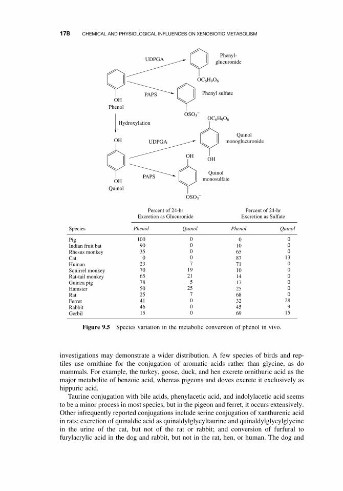
178 CHEMICAL AND PHYSIOLOGICAL INFLUENCES ON XENOBIOTIC METABOLISM
OHPhenol
OC6H9O6
Phenyl-glucuronide
OSO3−
Phenyl sulfate
OHQuinol
OH
OSO3−
OHOH
OC6H9O6
QuinolmonoglucuronideUDPGA
PAPS
UDPGA
PAPS
Hydroxylation
Quinolmonosulfate
Species Phenol PhenolQuinol Quinol
Percent of 24-hrExcretion as Glucuronide
Percent of 24-hrExcretion as Sulfate
PigIndian fruit batRhesus monkeyCatHumanSquirrel monkeyRat-tail monkeyGuinea pigHamsterRatFerretRabbitGerbil
10090350
237065785025414615
00007
19215
257000
0106587711014172568324569
000
13000000
289
15
Figure 9.5 Species variation in the metabolic conversion of phenol in vivo.
investigations may demonstrate a wider distribution. A few species of birds and rep-tiles use ornithine for the conjugation of aromatic acids rather than glycine, as domammals. For example, the turkey, goose, duck, and hen excrete ornithuric acid as themajor metabolite of benzoic acid, whereas pigeons and doves excrete it exclusively ashippuric acid.
Taurine conjugation with bile acids, phenylacetic acid, and indolylacetic acid seemsto be a minor process in most species, but in the pigeon and ferret, it occurs extensively.Other infrequently reported conjugations include serine conjugation of xanthurenic acidin rats; excretion of quinaldic acid as quinaldylglycyltaurine and quinaldylglycylglycinein the urine of the cat, but not of the rat or rabbit; and conversion of furfural tofurylacrylic acid in the dog and rabbit, but not in the rat, hen, or human. The dog and
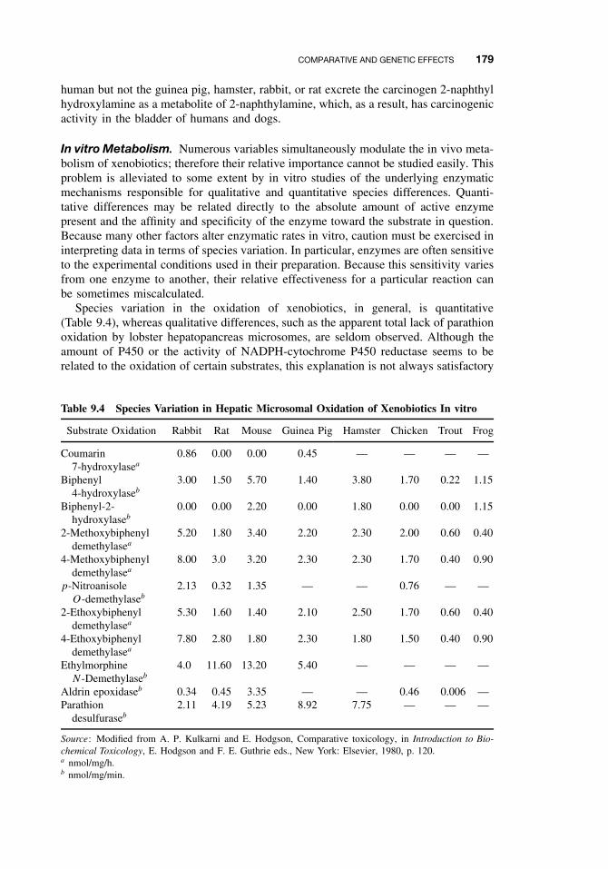
COMPARATIVE AND GENETIC EFFECTS 179
human but not the guinea pig, hamster, rabbit, or rat excrete the carcinogen 2-naphthylhydroxylamine as a metabolite of 2-naphthylamine, which, as a result, has carcinogenicactivity in the bladder of humans and dogs.
In vitro Metabolism. Numerous variables simultaneously modulate the in vivo meta-bolism of xenobiotics; therefore their relative importance cannot be studied easily. Thisproblem is alleviated to some extent by in vitro studies of the underlying enzymaticmechanisms responsible for qualitative and quantitative species differences. Quanti-tative differences may be related directly to the absolute amount of active enzymepresent and the affinity and specificity of the enzyme toward the substrate in question.Because many other factors alter enzymatic rates in vitro, caution must be exercised ininterpreting data in terms of species variation. In particular, enzymes are often sensitiveto the experimental conditions used in their preparation. Because this sensitivity variesfrom one enzyme to another, their relative effectiveness for a particular reaction canbe sometimes miscalculated.
Species variation in the oxidation of xenobiotics, in general, is quantitative(Table 9.4), whereas qualitative differences, such as the apparent total lack of parathionoxidation by lobster hepatopancreas microsomes, are seldom observed. Although theamount of P450 or the activity of NADPH-cytochrome P450 reductase seems to berelated to the oxidation of certain substrates, this explanation is not always satisfactory
Table 9.4 Species Variation in Hepatic Microsomal Oxidation of Xenobiotics In vitro
Substrate Oxidation Rabbit Rat Mouse Guinea Pig Hamster Chicken Trout Frog
Coumarin7-hydroxylasea
0.86 0.00 0.00 0.45 — — — —
Biphenyl4-hydroxylaseb
3.00 1.50 5.70 1.40 3.80 1.70 0.22 1.15
Biphenyl-2-hydroxylaseb
0.00 0.00 2.20 0.00 1.80 0.00 0.00 1.15
2-Methoxybiphenyldemethylasea
5.20 1.80 3.40 2.20 2.30 2.00 0.60 0.40
4-Methoxybiphenyldemethylasea
8.00 3.0 3.20 2.30 2.30 1.70 0.40 0.90
p-NitroanisoleO-demethylaseb
2.13 0.32 1.35 — — 0.76 — —
2-Ethoxybiphenyldemethylasea
5.30 1.60 1.40 2.10 2.50 1.70 0.60 0.40
4-Ethoxybiphenyldemethylasea
7.80 2.80 1.80 2.30 1.80 1.50 0.40 0.90
EthylmorphineN-Demethylaseb
4.0 11.60 13.20 5.40 — — — —
Aldrin epoxidaseb 0.34 0.45 3.35 — — 0.46 0.006 —Parathion
desulfuraseb
2.11 4.19 5.23 8.92 7.75 — — —
Source: Modified from A. P. Kulkarni and E. Hodgson, Comparative toxicology, in Introduction to Bio-chemical Toxicology, E. Hodgson and F. E. Guthrie eds., New York: Elsevier, 1980, p. 120.a nmol/mg/h.b nmol/mg/min.
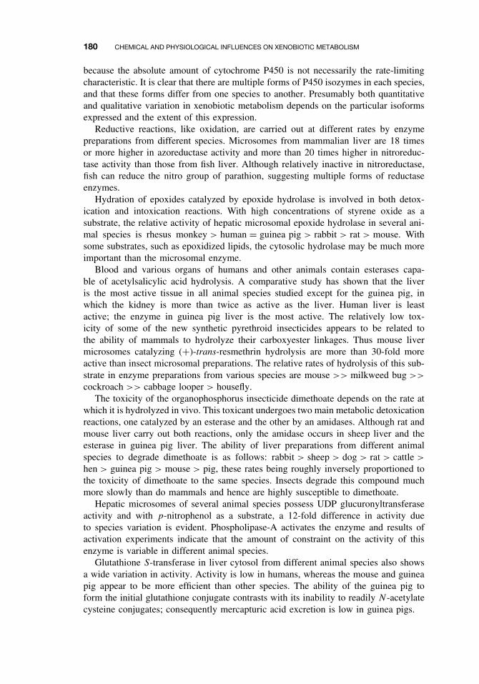
180 CHEMICAL AND PHYSIOLOGICAL INFLUENCES ON XENOBIOTIC METABOLISM
because the absolute amount of cytochrome P450 is not necessarily the rate-limitingcharacteristic. It is clear that there are multiple forms of P450 isozymes in each species,and that these forms differ from one species to another. Presumably both quantitativeand qualitative variation in xenobiotic metabolism depends on the particular isoformsexpressed and the extent of this expression.
Reductive reactions, like oxidation, are carried out at different rates by enzymepreparations from different species. Microsomes from mammalian liver are 18 timesor more higher in azoreductase activity and more than 20 times higher in nitroreduc-tase activity than those from fish liver. Although relatively inactive in nitroreductase,fish can reduce the nitro group of parathion, suggesting multiple forms of reductaseenzymes.
Hydration of epoxides catalyzed by epoxide hydrolase is involved in both detox-ication and intoxication reactions. With high concentrations of styrene oxide as asubstrate, the relative activity of hepatic microsomal epoxide hydrolase in several ani-mal species is rhesus monkey > human = guinea pig > rabbit > rat > mouse. Withsome substrates, such as epoxidized lipids, the cytosolic hydrolase may be much moreimportant than the microsomal enzyme.
Blood and various organs of humans and other animals contain esterases capa-ble of acetylsalicylic acid hydrolysis. A comparative study has shown that the liveris the most active tissue in all animal species studied except for the guinea pig, inwhich the kidney is more than twice as active as the liver. Human liver is leastactive; the enzyme in guinea pig liver is the most active. The relatively low tox-icity of some of the new synthetic pyrethroid insecticides appears to be related tothe ability of mammals to hydrolyze their carboxyester linkages. Thus mouse livermicrosomes catalyzing (+)-trans-resmethrin hydrolysis are more than 30-fold moreactive than insect microsomal preparations. The relative rates of hydrolysis of this sub-strate in enzyme preparations from various species are mouse >> milkweed bug >>
cockroach >> cabbage looper > housefly.The toxicity of the organophosphorus insecticide dimethoate depends on the rate at
which it is hydrolyzed in vivo. This toxicant undergoes two main metabolic detoxicationreactions, one catalyzed by an esterase and the other by an amidases. Although rat andmouse liver carry out both reactions, only the amidase occurs in sheep liver and theesterase in guinea pig liver. The ability of liver preparations from different animalspecies to degrade dimethoate is as follows: rabbit > sheep > dog > rat > cattle >
hen > guinea pig > mouse > pig, these rates being roughly inversely proportioned tothe toxicity of dimethoate to the same species. Insects degrade this compound muchmore slowly than do mammals and hence are highly susceptible to dimethoate.
Hepatic microsomes of several animal species possess UDP glucuronyltransferaseactivity and with p-nitrophenol as a substrate, a 12-fold difference in activity dueto species variation is evident. Phospholipase-A activates the enzyme and results ofactivation experiments indicate that the amount of constraint on the activity of thisenzyme is variable in different animal species.
Glutathione S-transferase in liver cytosol from different animal species also showsa wide variation in activity. Activity is low in humans, whereas the mouse and guineapig appear to be more efficient than other species. The ability of the guinea pig toform the initial glutathione conjugate contrasts with its inability to readily N -acetylatecysteine conjugates; consequently mercapturic acid excretion is low in guinea pigs.
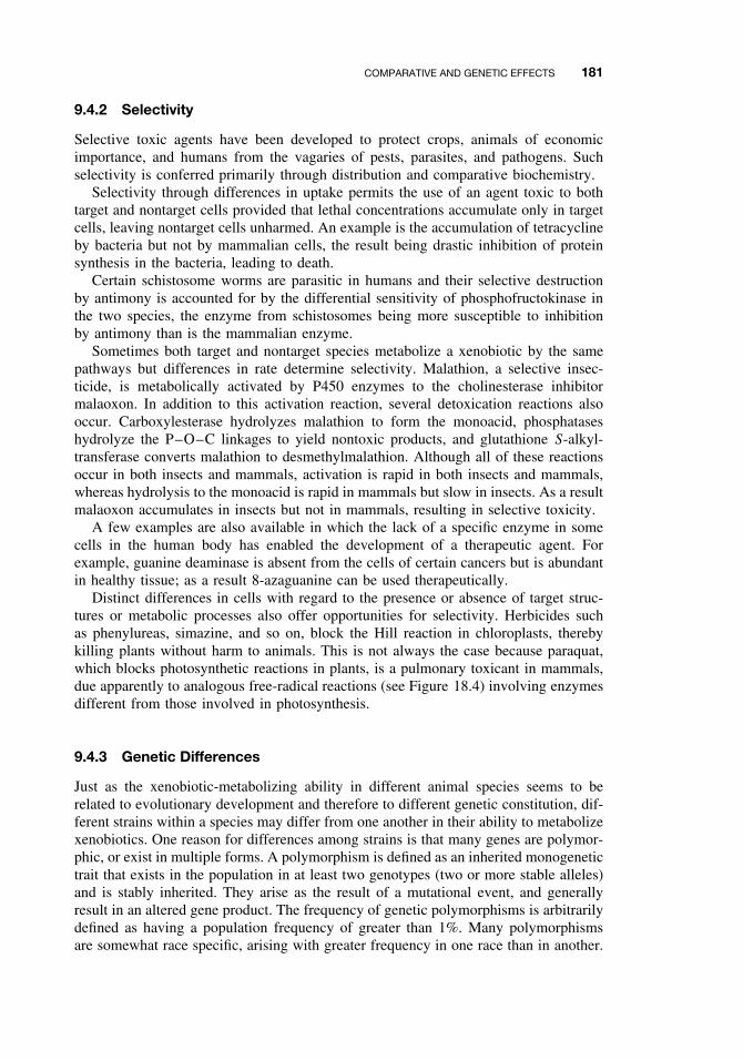
COMPARATIVE AND GENETIC EFFECTS 181
9.4.2 Selectivity
Selective toxic agents have been developed to protect crops, animals of economicimportance, and humans from the vagaries of pests, parasites, and pathogens. Suchselectivity is conferred primarily through distribution and comparative biochemistry.
Selectivity through differences in uptake permits the use of an agent toxic to bothtarget and nontarget cells provided that lethal concentrations accumulate only in targetcells, leaving nontarget cells unharmed. An example is the accumulation of tetracyclineby bacteria but not by mammalian cells, the result being drastic inhibition of proteinsynthesis in the bacteria, leading to death.
Certain schistosome worms are parasitic in humans and their selective destructionby antimony is accounted for by the differential sensitivity of phosphofructokinase inthe two species, the enzyme from schistosomes being more susceptible to inhibitionby antimony than is the mammalian enzyme.
Sometimes both target and nontarget species metabolize a xenobiotic by the samepathways but differences in rate determine selectivity. Malathion, a selective insec-ticide, is metabolically activated by P450 enzymes to the cholinesterase inhibitormalaoxon. In addition to this activation reaction, several detoxication reactions alsooccur. Carboxylesterase hydrolyzes malathion to form the monoacid, phosphataseshydrolyze the P–O–C linkages to yield nontoxic products, and glutathione S-alkyl-transferase converts malathion to desmethylmalathion. Although all of these reactionsoccur in both insects and mammals, activation is rapid in both insects and mammals,whereas hydrolysis to the monoacid is rapid in mammals but slow in insects. As a resultmalaoxon accumulates in insects but not in mammals, resulting in selective toxicity.
A few examples are also available in which the lack of a specific enzyme in somecells in the human body has enabled the development of a therapeutic agent. Forexample, guanine deaminase is absent from the cells of certain cancers but is abundantin healthy tissue; as a result 8-azaguanine can be used therapeutically.
Distinct differences in cells with regard to the presence or absence of target struc-tures or metabolic processes also offer opportunities for selectivity. Herbicides suchas phenylureas, simazine, and so on, block the Hill reaction in chloroplasts, therebykilling plants without harm to animals. This is not always the case because paraquat,which blocks photosynthetic reactions in plants, is a pulmonary toxicant in mammals,due apparently to analogous free-radical reactions (see Figure 18.4) involving enzymesdifferent from those involved in photosynthesis.
9.4.3 Genetic Differences
Just as the xenobiotic-metabolizing ability in different animal species seems to berelated to evolutionary development and therefore to different genetic constitution, dif-ferent strains within a species may differ from one another in their ability to metabolizexenobiotics. One reason for differences among strains is that many genes are polymor-phic, or exist in multiple forms. A polymorphism is defined as an inherited monogenetictrait that exists in the population in at least two genotypes (two or more stable alleles)and is stably inherited. They arise as the result of a mutational event, and generallyresult in an altered gene product. The frequency of genetic polymorphisms is arbitrarilydefined as having a population frequency of greater than 1%. Many polymorphismsare somewhat race specific, arising with greater frequency in one race than in another.
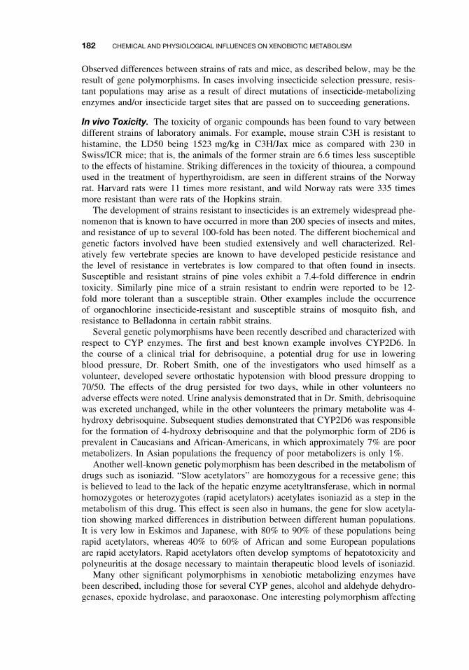
182 CHEMICAL AND PHYSIOLOGICAL INFLUENCES ON XENOBIOTIC METABOLISM
Observed differences between strains of rats and mice, as described below, may be theresult of gene polymorphisms. In cases involving insecticide selection pressure, resis-tant populations may arise as a result of direct mutations of insecticide-metabolizingenzymes and/or insecticide target sites that are passed on to succeeding generations.
In vivo Toxicity. The toxicity of organic compounds has been found to vary betweendifferent strains of laboratory animals. For example, mouse strain C3H is resistant tohistamine, the LD50 being 1523 mg/kg in C3H/Jax mice as compared with 230 inSwiss/ICR mice; that is, the animals of the former strain are 6.6 times less susceptibleto the effects of histamine. Striking differences in the toxicity of thiourea, a compoundused in the treatment of hyperthyroidism, are seen in different strains of the Norwayrat. Harvard rats were 11 times more resistant, and wild Norway rats were 335 timesmore resistant than were rats of the Hopkins strain.
The development of strains resistant to insecticides is an extremely widespread phe-nomenon that is known to have occurred in more than 200 species of insects and mites,and resistance of up to several 100-fold has been noted. The different biochemical andgenetic factors involved have been studied extensively and well characterized. Rel-atively few vertebrate species are known to have developed pesticide resistance andthe level of resistance in vertebrates is low compared to that often found in insects.Susceptible and resistant strains of pine voles exhibit a 7.4-fold difference in endrintoxicity. Similarly pine mice of a strain resistant to endrin were reported to be 12-fold more tolerant than a susceptible strain. Other examples include the occurrenceof organochlorine insecticide-resistant and susceptible strains of mosquito fish, andresistance to Belladonna in certain rabbit strains.
Several genetic polymorphisms have been recently described and characterized withrespect to CYP enzymes. The first and best known example involves CYP2D6. Inthe course of a clinical trial for debrisoquine, a potential drug for use in loweringblood pressure, Dr. Robert Smith, one of the investigators who used himself as avolunteer, developed severe orthostatic hypotension with blood pressure dropping to70/50. The effects of the drug persisted for two days, while in other volunteers noadverse effects were noted. Urine analysis demonstrated that in Dr. Smith, debrisoquinewas excreted unchanged, while in the other volunteers the primary metabolite was 4-hydroxy debrisoquine. Subsequent studies demonstrated that CYP2D6 was responsiblefor the formation of 4-hydroxy debrisoquine and that the polymorphic form of 2D6 isprevalent in Caucasians and African-Americans, in which approximately 7% are poormetabolizers. In Asian populations the frequency of poor metabolizers is only 1%.
Another well-known genetic polymorphism has been described in the metabolism ofdrugs such as isoniazid. “Slow acetylators” are homozygous for a recessive gene; thisis believed to lead to the lack of the hepatic enzyme acetyltransferase, which in normalhomozygotes or heterozygotes (rapid acetylators) acetylates isoniazid as a step in themetabolism of this drug. This effect is seen also in humans, the gene for slow acetyla-tion showing marked differences in distribution between different human populations.It is very low in Eskimos and Japanese, with 80% to 90% of these populations beingrapid acetylators, whereas 40% to 60% of African and some European populationsare rapid acetylators. Rapid acetylators often develop symptoms of hepatotoxicity andpolyneuritis at the dosage necessary to maintain therapeutic blood levels of isoniazid.
Many other significant polymorphisms in xenobiotic metabolizing enzymes havebeen described, including those for several CYP genes, alcohol and aldehyde dehydro-genases, epoxide hydrolase, and paraoxonase. One interesting polymorphism affecting
COMPARATIVE AND GENETIC EFFECTS 183
metabolism of dietary trimethylamines involves FMO3. Individuals with FMO3 poly-morphisms have a condition known as fish odor syndrome, or trimethylaminurea.Individuals with this syndrome exhibit an objectionable body odor resembling rot-ting fish due to their inability to N -oxidize trimethylamines, which are found in manyfoods including meat, eggs, and soybeans. This syndrome often leads to social iso-lation, clinical depression, and even suicide. Other toxicological implications of thispolymorphism are still not known.
Metabolite Production. Strain variations to hexobarbital are often dependant on itsdegradation rate. For example, male mice of the AL/N strain are long sleepers, andthis trait is correlated with slow inactivation of the drug. The reverse is true in CFW/Nmice, which have short sleeping time due to rapid hexobarbital oxidation. This closerelationship is further evidenced by the fact that the level of brain hexobarbital atawakening is essentially the same in all stains. Similar strain differences have beenreported for zoxazolamine paralysis in mice.
Studies on the induction of arylhydrocarbon hydroxylase by 3-methylcholanthrenehave revealed several responsive and nonresponsive mouse strains, and it is now wellestablished that the induction of this enzyme is controlled by a single gene. In theaccepted nomenclature, Ahb represents the allele for responsiveness, whereas Ahd
denotes the allele for nonresponsiveness.In rats, both age and gender seem to influence strain variation in xenobiotic meta-
bolism. Male rats exhibit about twofold variation between strains in hexobarbitalmetabolism, whereas female rats may display up to sixfold variation. In either genderthe extent of variations depend on age. The ability to metabolize hexobarbital is relatedto the metabolism of other substrates and the interstrain differences are maintained.
A well-known interstrain difference in phase II reactions is that of glucuronida-tion in Gunn rats. This is a mutant strain of Wistar rats that is characterized by asevere, genetically determined defect of bilirubin glucuronidation. Their ability to glu-curonidate o-aminophenol, o-aminobenzoic acid, and a number of other substrates isalso partially defective. This deficiency does not seem to be related to an inability toform UDPGA but rather to the lack of a specific UDP glucuronosyl-transferase. It hasbeen demonstrated that Gunn rats can conjugate aniline by N -glucuronidation and canform the O-glucuronide of p-nitrophenol.
Rabbit strains may exhibit up to 20-fold variation, particularly in the case of hexo-barbital, amphetamine, and aminopyrine metabolism. Relatively smaller differencesbetween strains occur with chlorpromazine metabolism. Wild rabbits and Califor-nia rabbits display the greatest differences from other rabbit strains in hepatic drugmetabolism.
Enzyme Differences. Variation in the nature and amount of constitutively expressedmicrosomal P450s have not been studied extensively in different strains of the samevertebrate. The only thorough investigations, those of the Ah Locus, which controlsaryl hydrocarbon hydroxylase induction, have shown that in addition to quantitativedifferences in the amount of P450 after induction in different strains of mice, theremay also be a qualitative difference in the P450 isozymes induced (see Section 9.5.2).
184 CHEMICAL AND PHYSIOLOGICAL INFLUENCES ON XENOBIOTIC METABOLISM
9.5 CHEMICAL EFFECTS
With regard to both logistics and scientific philosophy, the study of the metabolismand toxicity of xenobiotics must be initiated by considering single compounds. Unfor-tunately, humans and other living organisms are not exposed in this way; rather, theyare exposed to many xenobiotics simultaneously, involving different portals of entry,modes of action, and metabolic pathways. Some estimation of the number of chemicalsin use in the United States are given in Table 9.5. Because it bears directly on the prob-lem of toxicity-related interaction among different xenobiotics, the effect of chemicalson the metabolism of other exogenous compounds is one of the more important areasof biochemical toxicology.
Xenobiotics, in addition to serving as substrates for a number of enzymes, mayalso serve as inhibitors or inducers of these or other enzymes. Many examples areknown of compounds that first inhibit and subsequently induce enzymes such as themicrosomal monooxygenases. The situation is even further complicated by the factthat although some substances have an inherent toxicity and are detoxified in the body,others without inherent toxicity can be metabolically activated to potent toxicants. Thefollowing examples are illustrative of the situations that might occur involving twocompounds:
ž Compound A, without inherent toxicity, is metabolized to a potent toxicant. Inthe presence of an inhibitor of its metabolism, there would be a reduction in toxiceffect.
ž Compound A, given after exposure to an inducer of the activating enzymes, wouldappear more toxic.
ž Compound B, a toxicant, is metabolically detoxified. In the presence of an inhibitorof the detoxifying enzymes, there would be an increase in the toxic effect.
ž Compound B, given after exposure to an inducer of the detoxifying enzymes,would appear less toxic.
In addition to the previously mentioned cases, the toxicity of the inhibitor or inducer,as well as the time dependence of the effect, must also be considered because, as
Table 9.5 Some Estimates of the Number of Chemi-cals in Use in the United States
Number TypeSource ofEstimatea
1500 Active ingredients of pesticides EPA4000 Active ingredients of drugs FDA2000 Drug additives (preservatives,
stabilizers, etc.)FDA
2500 Food additives (nutritional value) FDA3000 Food additives (preservatives,
stabilizers, etc.)FDA
50,000 Additional chemicals in commonuse
EPA
aEPA, Environmental Protection Agency; FDA, Food and DrugAdministration.
CHEMICAL EFFECTS 185
mentioned, many xenbiotics that are initially enzyme inhibitors ultimately becomeinducers.
9.5.1 Inhibition
As previously indicated, inhibition of xenobiotic-metabolizing enzymes can causeeither an increase or a decrease in toxicity. Several well-known inhibitors of suchenzymes are shown in Figure 9.6 and are discussed in this section. Inhibitory effectscan be demonstrated in a number of ways at different organizational levels.
Types of Inhibition: Experimental DemonstrationIn vivo Symptoms. The measurement of the effect of an inhibitor on the durationof action of a drug in vivo is the most common method of demonstrating its action.These methods are open to criticism, however, because effects on duration of actioncan be mediated by systems other than those involved in the metabolism of the drug.Furthermore they cannot be used for inhibitors that have pharmacological activitysimilar or opposite to the compound being used.
Previously the most used and most reliable of these tests involved the measurementof effects on the hexobarbital sleeping time and the zoxazolamine paralysis time. Bothof these drugs are fairly rapidly deactivated by the hepatic microsomal monooxygenase
O
O CH2(OC2H4)2OC4H9
C3H7
POC2H5O
C6H5
NO2
N
CC(CH3)2
O
N
(C6H5)2CCO(CH2)2N(C2H5)2
O
C3H7
SKF-525A [P450]2-(Diethylamino)ethyl-2,2-diphenylpentanoate
CHCNH2
CHCH2
(CH3)2CH
O
EPN [esterases]O-Ethyl-O-p-nitrophenylphenylphosphonothioate
CHCOC2H5
Allylisopropylacetamide[P450]
Piperonyl Butoxide [P450]3,4-Methylenedioxy-6-propylbenzyl
n-butyl diethyleneglycol ether
Diethyl maleate[glutathione S-transferase]
1-Aminobenzotriazole (1-ABT)[P450]
Metyrapone[P450]
(C2H5)2NCSSCN(C2H5)2
S S
Disulfiram (Antabuse)[aldehyde dehydrogenase]
O
CH2
C2H5OCCH
O O
N
N
N
NH2
Figure 9.6 Some common inhibitors of xenobiotic-metabolizing enzymes.
186 CHEMICAL AND PHYSIOLOGICAL INFLUENCES ON XENOBIOTIC METABOLISM
system; thus, inhibitors of this system prolong their action. For example, treatmentof mice with chloramphenicol 0.5 to 1.0 hour before pentobarbital treatment pro-longs the duration of the pentobarbital sleeping time in a dose-related manner; itis effective at low doses (< 5 mg/kg) and has a greater than 10-fold effect at highdoses (100–200 mg/kg). The well-known inhibitor of drug metabolism, SKF-525A(Figure 9.6), causes an increase in both hexobarbital sleeping time and zoxazolamineparalysis time in rats and mice, as do the insecticide synergists piperonyl butoxideand tropital, the optimum pretreatment time being about 0.5 hour before the narcoticis given. As a consequence of the availability of single expressed isoforms for directstudies of inhibitory mechanisms, these methods are now used much less often.
In the case of activation reactions, such as the activation of the insecticide azin-phosmethyl to its potent anticholinesterase oxon derivative, a decrease in toxicity isapparent when rats are pretreated with the P450 inhibitor SKF-525A.
Cocarcinogenicity may also be an expression of inhibition of a detoxication reaction,as in the case of the cocarcinogenicity of piperonyl butoxide, a P450 inhibitor, and thecarcinogens, freons 112 and 113.
Distribution and Blood Levels. Treatment of an animal with an inhibitor of foreigncompound metabolism may cause changes in the blood levels of an unmetabolizedtoxicant and/or its metabolites. This procedure may be used in the investigation ofthe inhibition of detoxication pathways; it has the advantage over in vitro methods ofyielding results of direct physiological or toxicological interest because it is carried outin the intact animal. For example, if animals are first treated with either SKF-525A,glutethimide, or chlorcyclizine, followed in 1 hour or less by pentobarbital, it can beshown that the serum level of pentobarbital is considerably higher in treated animalsthan in controls within 1 hour of its injection. Moreover the time sequence of theeffects can be followed in individual animals, a factor of importance when inhibitionis followed by induction—a not uncommon event.
Effects on Metabolism In vivo. A further refinement of the previous technique is todetermine the effect of an inhibitor on the overall metabolism of a xenobiotic in vivo,usually by following the appearance of metabolites in the urine and/or feces. In somecases the appearance of metabolites in the blood or tissue may also be followed. Again,the use of the intact animal has practical advantages over in vitro methods, althoughlittle is revealed about the mechanisms involved.
Studies of antipyrine metabolism may be used to illustrate the effect of inhibitionon metabolism in vivo; in addition, these studies have demonstrated variation amongspecies in the inhibition of the metabolism of xenobiotics. In the rat, a dose of piperonylbutoxide of at least 100 mg/kg was necessary to inhibit antipyrine metabolism, whereasin the mouse a single intraperitoneal (IP) or oral dose of 1 mg/kg produced a significantinhibition. In humans an oral dose of 0.71 mg/kg had no discernible effect on themetabolism of antipyrine.
Disulfiram (Antabuse) inhibits aldehyde dehydrogenase irreversibly, causing anincrease in the level of acetaldehyde, formed from ethanol by the enzyme alcohol dehy-drogenase. This results in nausea, vomiting, and other symptoms in the human—henceits use as a deterrent in alcoholism. Inhibition by disulfiram appears to be irreversible,the level returning to normal only as a result of protein synthesis.
Use of specific metabolic enzyme inhibitors may often provide valuable informationwith respect to the metabolism of a particular drug. For example, quinidine is a potent
CHEMICAL EFFECTS 187
and selective inhibitor of CYP2D6. This drug has been used in clinical studies as apharmacological tool to mimic the lack of CYP2D6 in humans. By demonstrating thatquinidine substantially slows the metabolism of trimipramine (a tricyclic antidepres-sant), investigators have implicated CYP2D6 in its metabolism.
Effects on In vitro Metabolism Following In vivo Treatment. This method of demon-strating inhibition is of variable utility. The preparation of enzymes from animal tissuesusually involves considerable dilution with the preparative medium during homoge-nization, centrifugation, and re-suspension. As a result inhibitors not tightly bound tothe enzyme in question are lost, either in whole or in part, during the preparative pro-cesses. Therefore negative results can have little utility because failure to inhibit andloss of the inhibitor give identical results. Positive results, however, not only indicatethat the compound administered is an inhibitor but also provide a clear indication ofexcellent binding to the enzyme, most probably due to the formation of a covalentor slowly reversible inhibitory complex. The inhibition of esterases following treat-ment of the animal with organophosphorus compounds, such as paraoxon, is a goodexample, because the phosphorylated enzyme is stable and is still inhibited after thepreparative procedures. Inhibition by carbamates, however, is greatly reduced by thesame procedures because the carbamylated enzyme is unstable and, in addition, theresidual carbamate is highly diluted.
Microsomal monooxygenase inhibitors that form stable inhibitory complexes withP450, such as SKF-525A, piperonyl butoxide, and other methylenedioxphenyl com-pounds, and amphetamine and its derivatives, can be readily investigated in this way.This is because the microsomes isolated from pretreated animals have a reduced capac-ity to oxidize many xenobiotics.
Another form of chemical interaction, resulting from inhibition in vivo, that can thenbe demonstrated in vitro involves those xenobiotics that function by causing destructionof the enzyme in question, so-called suicide substrates. Exposure of rats to vinyl chlo-ride results in a loss of cytochrome P450 and a corresponding reduction in the capacityof microsomes subsequently isolated to metabolize foreign compounds. Allyl isopropy-lacetamide and other allyl compounds have long been known to have a similar effect.
In vitro Effects. In vitro measurement of the effect of one xenobiotic on the metabolismof another is by far the most common type of investigation of interactions involvinginhibition. Although it is the most useful method for the study of inhibitory mecha-nisms, particularly when purified enzymes are used, it is of limited utility in assessingthe toxicological implications for the intact animal. The principal reason for this isthat in vitro measurement does not assess the effects of factors that affect absorption,distribution, and prior metabolism, all of which occur before the inhibitory event underconsideration.
Although the kinetics of inhibition of xenobiotic-metabolizing enzymes can be inves-tigated in the same ways as any other enzyme mechanism, a number of problems arisethat may decrease the value of this type of investigation. They include the following:
ž The P450 system, a particulate enzyme system, has been investigated many times,but using methods developed for single soluble enzymes. As a result Lineweaver-Burke or other reciprocal plots are frequently curvilinear, and the same reactionmay appear to have quite a different characteristics from laboratory to laboratory,species to species, and organ to organ.
188 CHEMICAL AND PHYSIOLOGICAL INFLUENCES ON XENOBIOTIC METABOLISM
ž The nonspecific binding of substrate and/or inhibitor to membrane components isa further complicating factor affecting inhibition kinetics.
ž Both substrates and inhibitors are frequently lipophilic, with low solubility inaqueous media.
ž Xenobiotic-metabolizing enzymes commonly exist in multiple forms (e.g., glu-tathione S-transferases and P450s). These isozymes are all relatively nonspecificbut differ from one another in the relative affinities of the different substrates.
The primary considerations in studies of inhibition mechanisms are reversibility andselectivity. The inhibition kinetics of reversible inhibition give considerable insight intothe reaction mechanisms of enzymes and, for that reason, have been well studied. Ingeneral, reversible inhibition involves no covalent binding, occurs rapidly, and canbe reversed by dialysis or, more rapidly, by dilution. Reversible inhibition is usuallydivided into competitive inhibition, uncompetitive inhibition, and noncompetitive inhi-bition. Because these types are not rigidly separated, many intermediate classes havebeen described.
Competitive inhibition is usually caused by two substrates competing for the sameactive site. Following classic enzyme kinetics, there should be a change in the apparentKm but not in Vmax. In microsomal monooxygenase reaction, type I ligands, whichoften appear to bind as substrates but do not bind to the heme iron, might be expectedto be competitive inhibitors, and this frequently appears to be the case. Examplesare the inhibition of the O-demethylation of p-nitronanisole by aminopyrine, aldrinepoxidation by dihydroaldrin, and N -demethylation of aminopyrine by nicotinamide.More recently some of the polychlorinated biphenyls (PCBs), notably dichlorbiphenyl,have been shown to have a high affinity as type I ligands for rabbit liver P450 and tobe competitive inhibitors of the O-demethylation of p-nitronanisole.
Uncompetitive inhibition has seldom been reported in studies of xenobiotic meta-bolism. It occurs when an inhibitor interacts with an enzyme-substrate complex butcannot interact with free enzyme. Both Km and Vmax change by the same ratio, givingrise to a family of parallel lines in a Lineweaver-Burke plot.
Noncompetitive inhibitors can bind to both the enzyme and enzyme-substrate com-plex to form either an enzyme-inhibitor complex or an enzyme-inhibitor-substratecomplex. The net result is a decrease in Vmax but no change in Km. Metyrapone(Figure 9.6), a well-known inhibitor of monooxygenase reactions, can also, under somecircumstances, stimulate metabolism in vitro. In either case the effect is noncompet-itive, in that the Km does not change, whereas Vmax does, decreasing in the case ofinhibition and increasing in the case of stimulation.
Irreversible inhibition, which is much more important toxicologically, can arise fromvarious causes. In most cases the formation of covalent or other stable bonds or the dis-ruption of the enzyme structure is involved. In these cases the effect cannot be readilyreversed in vitro by either dialysis or dilution. The formation of stable inhibitory com-plexes may involve the prior formation of a reactive intermediate that then interacts withthe enzyme. An excellent example of this type of inhibition is the effect of the insecti-cide synergist piperonyl butoxide (Figure 9.6) on hepatic microsomal monooxygenaseactivity. This methylenedioxyphenyl compound can form a stable inhibitory complexthat blocks CO binding to P450 and also prevents substrate oxidation. This complexresults from the formation of a reactive intermediate, which is shown by the fact thatthe type of inhibition changes from competitive to irreversible as metabolism, in the
CHEMICAL EFFECTS 189
presence of NADPH and oxygen, proceeds. It appears probable that the metabolite inquestion is a carbine formed spontaneously by elimination of water following hydrox-ylation of the methylene carbon by the cytochrome (see Figure 7.8 for metabolism ofmethylenedioxyphenyl compounds). Piperonyl butoxide inhibits the in vitro metabolismof many substrates of the monooxygenase system, including aldrin, ethylmorphine, ani-line, and aminopyrine, as well as carbaryl, biphenyl, hexobarbital, and p-nitroanisoleamong many others. Although most of the studies carried out on piperonyl butoxidehave involved rat or mouse liver microsomes, they have also been carried out on pig,rabbit, and carp liver microsomes, and in various preparations from houseflies, cock-roaches, and other insects. Certain classes of monooxygenase inhibitors, in addition tomethylenedioxyphenyl compounds, are now known to form “metabolite inhibitory com-plexes,” including amphetamine and its derivatives and SKF-525A and its derivatives.
The inhibition of the carboxylesterase that hydrolyzes malathion by organophospho-rus compounds, such as EPN is a further example of xenobiotic interaction resultingfrom irreversible inhibition. In this case the enzyme is phosphorylated by the inhibitor.
Another class of irreversible inhibitors of toxicological significance consists of thosecompounds that bring about the destruction of the xenobiotic-metabolizing enzymes,hence the designation “suicide substrates.” The drug allylisopropylacetamide(Figure 9.6), as well as other allyl compounds, has long been known to cause thebreakdown of P450 and the resultant release of heme. More recently the hepatocar-cinogen vinyl chloride has also been shown to have a similar effect, probably alsomediated through the generation of a highly reactive intermediate (see Figure 8.2).Much information has accumulated since the mid-1970s on the mode of action of thehepatotoxicant carbon tetrachloride, which effects a number of irreversible changes inboth liver proteins and lipids, such changes being generated by reactive intermediatesformed during its metabolism (Figure 8.3).
The less specific disruptors of protein structure, such as urea, detergents, strongacids, and so on, are probably of significance only in vitro experiments.
Synergism and Potentiation. The terms synergism and potentiation have beenused and defined in various ways, but in any case, they involve a toxicity that isgreater when two compounds are given simultaneously or sequentially than would beexpected from a consideration of the toxicities of the compounds given alone. Sometoxicologists have used the term synergism for cases that fit this definition, but onlywhen one compound is toxic alone whereas the other has little or no intrinsic toxicity.For example, the nontoxic synergist, piperonyl butoxide is often included in pesticideformulations because of its ability to significantly increase the toxicity of the activepesticide ingredient by inhibiting its detoxication in the target species.
The term potentiation is then reserved for those cases where both compounds haveappreciable intrinsic toxicity, such as in the case of malathion and EPN. Malathion hasa low mammalian toxicity due primarily to its rapid hydrolysis by a carboxylesterase.EPN (Figure 9.6) another organophosphate insecticide, causes a dramatic increasein malathion toxicity to mammals at dose levels, which, given alone, cause essen-tially no inhibition of acetylcholinesterase. The increase in toxicity as a result ofcoadministration of these two toxicants is the result of the ability of EPN, at lowconcentrations, to inhibit the carboxylesterase responsible for malathion degradation.
Unfortunately, the terms synergist and potentiation have often been used by sometoxicologists in precisely the opposite manner. Historically, the term synergist has
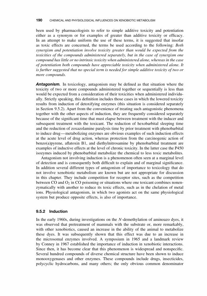
190 CHEMICAL AND PHYSIOLOGICAL INFLUENCES ON XENOBIOTIC METABOLISM
been used by pharmacologists to refer to simple additive toxicity and potentiationeither as a synonym or for examples of greater than additive toxicity or efficacy.In an attempt to make uniform the use of these terms, it is suggested that insofaras toxic effects are concerned, the terms be used according to the following: Bothsynergism and potentiation involve toxicity greater than would be expected from thetoxicities of the compounds administered separately, but in the case of synergism onecompound has little or no intrinsic toxicity when administered alone, whereas in the caseof potentiation both compounds have appreciable toxicity when administered alone. Itis further suggested that no special term is needed for simple additive toxicity of two ormore compounds.
Antagonism. In toxicology, antagonism may be defined as that situation where thetoxicity of two or more compounds administered together or sequentially is less thanwould be expected from a consideration of their toxicities when administered individu-ally. Strictly speaking, this definition includes those cases in which the lowered toxicityresults from induction of detoxifying enzymes (this situation is considered separatelyin Section 9.5.2). Apart from the convenience of treating such antagonistic phenomenatogether with the other aspects of induction, they are frequently considered separatelybecause of the significant time that must elapse between treatment with the inducer andsubsequent treatment with the toxicant. The reduction of hexobarbital sleeping timeand the reduction of zoxazolamine paralysis time by prior treatment with phenobarbitalto induce drug—metabolizing enzymes are obvious examples of such induction effectsat the acute level of drug action, whereas protection from the carcinogenic action ofbenzo(a)pyrene, aflatoxin B1, and diethylnitrosamine by phenobarbital treatment areexamples of inductive effects at the level of chronic toxicity. In the latter case the P450isozymes induced by phenobarbital metabolize the chemical to less toxic metabolites.
Antagonism not involving induction is a phenomenon often seen at a marginal levelof detection and is consequently both difficult to explain and of marginal significance.In addition several different types of antagonism of importance to toxicology that donot involve xenobiotic metabolism are known but are not appropriate for discussionin this chapter. They include competition for receptor sites, such as the competitionbetween CO and O2 in CO poisoning or situations where one toxicant combines nonen-zymatically with another to reduce its toxic effects, such as in the chelation of metalions. Physiological antagonism, in which two agonists act on the same physiologicalsystem but produce opposite effects, is also of importance.
9.5.2 Induction
In the early 1960s, during investigations on the N -demethylation of aminoazo dyes, itwas observed that pretreatment of mammals with the substrate or, more remarkably,with other xenobiotics, caused an increase in the ability of the animal to metabolizethese dyes. It was subsequently shown that this effect was due to an increase inthe microsomal enzymes involved. A symposium in 1965 and a landmark reviewby Conney in 1967 established the importance of induction in xenobiotic interactions.Since then, it has become clear that this phenomenon is widespread and nonspecific.Several hundred compounds of diverse chemical structure have been shown to inducemonooxygenases and other enzymes. These compounds include drugs, insecticides,polycyclic hydrocarbons, and many others; the only obvious common denominator
CHEMICAL EFFECTS 191
is that they are organic and lipophilic. It has also become apparent that although allinducers do not have the same effects, the effects tend to be nonspecific to the extentthat any single inducer induces more than one enzymatic activity. Other enzymesoften coinduced by P450 inducers include glutathione S-transferase, epoxide hydrolasesand others; perhaps as a result of general induction of cellular processes includingproliferative responses of endoplasmic reticulum, peroxisomes, and mitochondria.
Specificity of Monooxygenase Induction. The majority of studies involving mono-oxygenase induction have been conducted in mammals. Mammals have at least 17distinct CYP families, coding for as many as 50 to 60 individual CYP genes inany given species. Many of these CYP families are fairly specific for endogenousmetabolic pathways and are not typically involved in metabolism of foreign chemicals.As discussed in Chapter 7, CYP families 1–4 are the predominant families involved inxenobiotic metabolism. These CYP families are also known for their ability to respondto xenobiotic challenges by increasing their protein levels. Many of the genes withinfamilies 1–4 are transcriptionally activated through one of four receptor-dependentmechanisms. Others, such as CYP2E1, are regulated at the level of mRNA stabiliza-tion and/or protein stabilization. These mechanisms of regulation are discussed later inthis section.
Inducers of monooxygenase activity fall into four principle classes, exemplified byTCDD (inducer of CYP1A1), phenobarbital (inducer of the CYP2B and 3A families),rifampicin (inducer of CYP3A and 2C families), and ethanol (inducer of 2E1). Inducersof the phenobarbital-type tend to share few structural features other than lipophilicity,while TCDD-like inducers are primarily polycyclic hydrocarbons. Other inducers, suchas ethanol, dexamethasone, and clofibrate are more specific. Many inducers requireeither fairly high dose levels or repeated dosing to be effective, frequently >10 mg/kgand some as high as 100 to 200 mg/kg. Some insecticides, however, such as mirex,can induce at dose levels as low as 1 mg/kg, while the most potent inducer known,2,3,7,8-tetrachlorodibenzo-p-dioxin (TCDD), is effective at 1µg/kg in some species.
In the liver, phenobarbital-type inducers cause a marked proliferation of the smoothendoplasmic reticulum as well as an increase in the amount of CYP content. Oftenthese changes are sufficient to result in significant liver weight increases. Phenobarbitalinduction induces a wide range of oxidative activities including O-demethylation ofp-nitroanisole, N -demethylation of benzphetamine, pentobarbital hydroxylation, andaldrin hydroxylation. CYP families that are primarily induced by phenobarbital andphenobarbital-like inducers include CYP2B, CYP2C, and CYP3A subfamilies.
In contrast with phenobarbital, induction by TCDD and polycyclic hydrocarbonsdoes not cause proliferation of the endoplasmic reticulum, although the CYP contentis increased. CYP1A1 is the primary isoform induced, although other non-CYP pro-teins such as uridine diphosphoglucuronyl transferase may also be induced. Inductionof CYP1A1 by polycyclic hydrocarbons results in the induction of a relatively narrowrange of oxidative activities, consisting primarily of reactions involving aryl hydrocar-bon hydroxylase, the best-known reaction being the hydroxylation of benzo(a)pyrene.
Rifampicin and pregnenolone-16α-carbonitrile (PCN) induce members of theCYP3A family and represent a third type of inducer, in that the substrate speci-ficity of the microsomes from treated animals differs from that of the microsomesfrom either phenobarbital-treated or TCDD-treated animals. Inducing substrates of thisclass include endogenous and synthetic glucocorticoids (e.g., dexamethasone), pregnane
192 CHEMICAL AND PHYSIOLOGICAL INFLUENCES ON XENOBIOTIC METABOLISM
compounds (e.g., pregenenolone 16α-carbonitrile, PCN), and macrolide antibiotics(e.g., rifampicin).
Ethanol and a number of other chemicals, including acetone and certain imidazoles,induce CYP2E1. Piperonyl butoxide, isosafrole, and other methylenedioxyphenyl com-pounds are known to induce CYP1A2 by a non-Ah receptor-dependent mechanism.Peroxisome proliferators, including the drug, clofibrate, and the herbicide synergisttridiphane induce a CYP4A isozyme that catalyzes the ω-oxidation of lauric acid.
All inducers do not fall readily into one or the other of these classes. Some oxidativeprocesses can be induced by either type of inducer, such as the hydroxylation of anilineand the N -demethylation of chlorcyclizine. Some inducers, such as the mixture ofPCBs designated Arochlor 1254, can induce a broad spectrum of CYP isoforms. Manyvariations also exist in the relative stimulation of different oxidative activities withinthe same class of inducer, particularly of the phenobarbital type.
It appears reasonable that because several types of CYP are associated with theendoplasmic reticulum, various inducers may induce one or more of them. Because eachof these types has a relatively broad substrate specificity, differences may be caused byvariations in the extent of induction of different cytochromes. Now that methods areavailable for gel electrophoresis of microsomes and identification of specific isoformsby immunoblotting and isoforms-specific antibodies, the complex array of inductivephenomena is being more logically explained in terms of specific isozymes.
Although the bulk of published investigations of the induction of monooxygenaseenzymes have dealt with the mammalian liver, induction has been observed in othermammalian tissues and in nonmammalian species, both vertebrate and invertebrate.Many induced CYPs have now been cloned and/or purified from a variety of species.It is clear that many of these induced CYPs represent only a small percentage of thetotal CYP in the uninduced animal. For this reason the “constitutive” isozymes, thosealready expressed in the uninduced animal, must be fully characterized because theyrepresent the available xenobiotic-metabolizing capacity of the normal animal.
Mechanism and Genetics of Induction in Mammals. Many different mecha-nisms may be involved in CYP induction. These include increased transcription ofDNA, increased mRNA translation to protein, mRNA stabilization, and protein stabi-lization. Induction can only occur in intact cells and cannot be achieved by the additionof inducers directly to cell fractions such as microsomes. It has been known for sometime that in most cases of increase in monooxygenase activity there is a true induc-tion involving synthesis of new enzyme, and not the activation of enzyme alreadysynthesized, since induction is generally prevented by inhibitors of protein synthesis.For example, the protein synthesis inhibitors such as puromycin, ethionine, and cyclo-heximide inhibit aryl hydrocarbon hydroxylase activity. A simplified scheme for geneexpression and protein synthesis is shown in Figure 9.7.
Perhaps the best understood example of induction involves induction of the aromatichydrocarbon receptor (AhR) by compounds such as TCDD and 3-methylcholanthrene.The use of suitable inhibitors of RNA and DNA polymerase activity has shown thatinhibitors of RNA synthesis such as actinomycin D and mercapto(pyridethyl)benzimida-zole block aryl hydrocarbon hydroxylase induction, whereas hydroxyurea, at levelsthat completely block the incorporation of thymidine into DNA, has no effect. Thus itappears that the inductive effect is at the level of transcription and that DNA synthesisis not required.
CHEMICAL EFFECTS 193
RegulatorGene
StructuralGenes
OperatorGene
Transcription
Translation
DNA
mRNA
sRNAsRNA sRNA
Derepression
Proteinsynthesis
Protein(Enzymes)
Figure 9.7 Simplified scheme for gene expression in animals.
These findings imply that compounds that induce xenobiotic-metabolizing enzymesplay a role as derepressors of regulator or other genes in a manner analogous to steroidhormones—namely by combining with a cytosolic receptor followed by movementinto the nucleus and then derepression of the appropriate gene. In the case of the AhR,TCDD, or some other appropriate ligand enters the cell through the plasma membraneand binds to the cytosolic Ah receptor protein (Figure 9.8). After ligand binding, thereceptor translocates to the nucleus where it forms a dimer with another protein knownas ARNT. In the nucleus the transformed receptor interacts with specific sequencesof DNA known as xenobiotic responsive elements (XREs). Two XREs are locatedapproximately 1000 or more base pairs upstream from the transcriptional start site inthe 5′ flanking region of the CYP1A1 gene. A third site is likely to be an inhibitory orsuppressor site (Figure 9.9). The protein-DNA interaction that occurs at the XREs isthought to result in a bending of the DNA, which allows for increased transcription fol-lowed by increased protein synthesis. Another promoter region is located just upstreamfrom the transcriptional start site. Although several transcription factors may interactwith this binding site, including the TATA-binding protein, it has no binding sites forthe AhR/Arnt proteins. Transfection experiments indicate that the TATA-binding siteis essential for promoter function, while the other sites are less important. This pro-moter region is silent unless the upstream XREs have been appropriately activated bythe AhR and Arnt proteins. The fact that several genes may be responsive to CYP1Ainducers is indicative of the fact that similar XREs are found on many Ah-receptorinducible genes.
Although phenobarbital induction has been studied for many years, the mechanismfor induction has only recently been established. In bacteria a key feature of phe-nobarbital induction was demonstrated to involve barbiturate-mediated removal of a
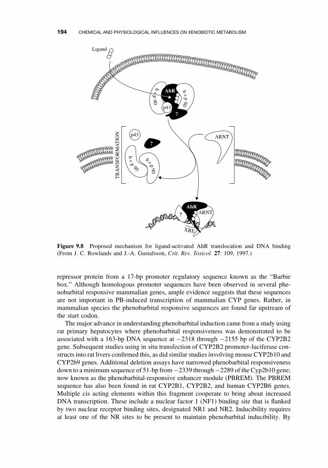
194 CHEMICAL AND PHYSIOLOGICAL INFLUENCES ON XENOBIOTIC METABOLISM
Ligand
hs
p90
h s p90
TR
AN
SFO
RM
AT
ION
?ARNT
ARNT
hs
p9
0
p43
p43
hs
p90
AhR
AhR
?
?
XRE
Figure 9.8 Proposed mechanism for ligand-activated AhR translocation and DNA binding(From J. C. Rowlands and J.-A. Gustafsson, Crit. Rev. Toxicol. 27: 109, 1997.)
repressor protein from a 17-bp promoter regulatory sequence known as the “Barbiebox.” Although homologous promoter sequences have been observed in several phe-nobarbital responsive mammalian genes, ample evidence suggests that these sequencesare not important in PB-induced transcription of mammalian CYP genes. Rather, inmammalian species the phenobarbital responsive sequences are found far upstream ofthe start codon.
The major advance in understanding phenobarbital induction came from a study usingrat primary hepatocytes where phenobarbital responsiveness was demonstrated to beassociated with a 163-bp DNA sequence at −2318 through −2155 bp of the CYP2B2gene. Subsequent studies using in situ transfection of CYP2B2 promoter-luciferase con-structs into rat livers confirmed this, as did similar studies involving mouse CYP2b10 andCYP2b9 genes. Additional deletion assays have narrowed phenobarbital responsivenessdown to a minimum sequence of 51-bp from −2339 through −2289 of the Cyp2b10 gene;now known as the phenobarbital-responsive enhancer module (PBREM). The PBREMsequence has also been found in rat CYP2B1, CYP2B2, and human CYP2B6 genes.Multiple cis acting elements within this fragment cooperate to bring about increasedDNA transcription. These include a nuclear factor 1 (NF1) binding site that is flankedby two nuclear receptor binding sites, designated NR1 and NR2. Inducibility requiresat least one of the NR sites to be present to maintain phenobarbital inducibility. By
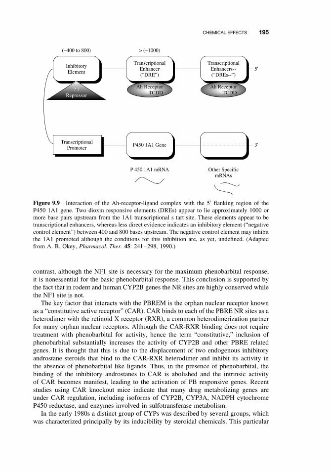
CHEMICAL EFFECTS 195
InhibitoryElement
(−400 to 800)
TranscriptionalEnhancer(“DRE”)
P450 1A1 Gene
P 450 1A1 mRNA
> (−1000)
TranscriptionalEnhancers--(“DREs--”)
Other SpecificmRNAs
5'
3'
? ?Repressor
Ah ReceptorTCDD
Ah ReceptorTCDD
TranscriptionalPromoter
Figure 9.9 Interaction of the Ah-receptor-ligand complex with the 5′ flanking region of theP450 1A1 gene. Two dioxin responsive elements (DREs) appear to lie approximately 1000 ormore base pairs upstream from the 1A1 transcriptional s tart site. These elements appear to betranscriptional enhancers, whereas less direct evidence indicates an inhibitory element (“negativecontrol element”) between 400 and 800 bases upstream. The negative control element may inhibitthe 1A1 promoted although the conditions for this inhibition are, as yet, undefined. (Adaptedfrom A. B. Okey, Pharmacol. Ther. 45: 241–298, 1990.)
contrast, although the NF1 site is necessary for the maximum phenobarbital response,it is nonessential for the basic phenobarbital response. This conclusion is supported bythe fact that in rodent and human CYP2B genes the NR sites are highly conserved whilethe NF1 site is not.
The key factor that interacts with the PBREM is the orphan nuclear receptor knownas a “constitutive active receptor” (CAR). CAR binds to each of the PBRE NR sites as aheterodimer with the retinoid X receptor (RXR), a common heterodimerization partnerfor many orphan nuclear receptors. Although the CAR-RXR binding does not requiretreatment with phenobarbital for activity, hence the term “constitutive,” inclusion ofphenobarbital substantially increases the activity of CYP2B and other PBRE relatedgenes. It is thought that this is due to the displacement of two endogenous inhibitoryandrostane steroids that bind to the CAR-RXR heterodimer and inhibit its activity inthe absence of phenobarbital like ligands. Thus, in the presence of phenobarbital, thebinding of the inhibitory androstanes to CAR is abolished and the intrinsic activityof CAR becomes manifest, leading to the activation of PB responsive genes. Recentstudies using CAR knockout mice indicate that many drug metabolizing genes areunder CAR regulation, including isoforms of CYP2B, CYP3A, NADPH cytochromeP450 reductase, and enzymes involved in sulfotransferase metabolism.
In the early 1980s a distinct group of CYPs was described by several groups, whichwas characterized principally by its inducibility by steroidal chemicals. This particular
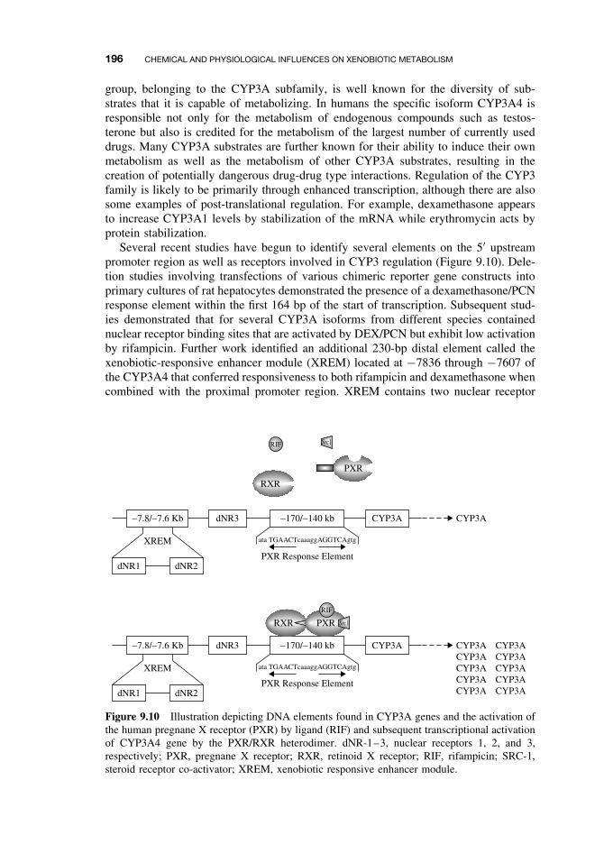
196 CHEMICAL AND PHYSIOLOGICAL INFLUENCES ON XENOBIOTIC METABOLISM
group, belonging to the CYP3A subfamily, is well known for the diversity of sub-strates that it is capable of metabolizing. In humans the specific isoform CYP3A4 isresponsible not only for the metabolism of endogenous compounds such as testos-terone but also is credited for the metabolism of the largest number of currently useddrugs. Many CYP3A substrates are further known for their ability to induce their ownmetabolism as well as the metabolism of other CYP3A substrates, resulting in thecreation of potentially dangerous drug-drug type interactions. Regulation of the CYP3family is likely to be primarily through enhanced transcription, although there are alsosome examples of post-translational regulation. For example, dexamethasone appearsto increase CYP3A1 levels by stabilization of the mRNA while erythromycin acts byprotein stabilization.
Several recent studies have begun to identify several elements on the 5′ upstreampromoter region as well as receptors involved in CYP3 regulation (Figure 9.10). Dele-tion studies involving transfections of various chimeric reporter gene constructs intoprimary cultures of rat hepatocytes demonstrated the presence of a dexamethasone/PCNresponse element within the first 164 bp of the start of transcription. Subsequent stud-ies demonstrated that for several CYP3A isoforms from different species containednuclear receptor binding sites that are activated by DEX/PCN but exhibit low activationby rifampicin. Further work identified an additional 230-bp distal element called thexenobiotic-responsive enhancer module (XREM) located at −7836 through −7607 ofthe CYP3A4 that conferred responsiveness to both rifampicin and dexamethasone whencombined with the proximal promoter region. XREM contains two nuclear receptor
CYP3A
XREM ata TGAACTcaaaggAGGTCAgtg
PXR Response Element
−7.8/−7.6 Kb −170/−140 kbdNR3 CYP3A
dNR2dNR1
CYP3A CYP3A CYP3A CYP3A CYP3A CYP3A CYP3A CYP3A CYP3A CYP3A
XREM ata TGAACTcaaaggAGGTCAgtg
PXR Response Element
−7.8/−7.6 Kb −170/−140 kbdNR3 CYP3A
dNR2dNR1
RXR PXR
RIF
Src1
RXR
PXR
RIF Src1
Figure 9.10 Illustration depicting DNA elements found in CYP3A genes and the activation ofthe human pregnane X receptor (PXR) by ligand (RIF) and subsequent transcriptional activationof CYP3A4 gene by the PXR/RXR heterodimer. dNR-1–3, nuclear receptors 1, 2, and 3,respectively; PXR, pregnane X receptor; RXR, retinoid X receptor; RIF, rifampicin; SRC-1,steroid receptor co-activator; XREM, xenobiotic responsive enhancer module.
CHEMICAL EFFECTS 197
binding sites (dNR1 and dNR2), neither of which is solely responsible for the activityof XREM. An additional nuclear receptor site, dNR3 located several hundred basesdownstream of XREM also appears to have some importance in induction.
Recent work has demonstrated that the nuclear orphan receptor, pregnane X receptor(PXR) is the major determinant of CYP3A gene regulation by xenobiotics. Severallines of evidence support PXR involvement with CYP3A induction. First, both PXRand CYP3A isoforms are predominantly expressed in liver and intestine, with lessexpression found in lungs and kidneys. Second, PXR binds to human and rat CYP3Apromoter regions and can activate expression of CYP3A4 promoter in transfectionassays. Third, many of the same inducers of CYP3A isoforms also activate PXR.Fourth, interspecies differences in response to CYP3A inducers have been demonstratedto be due to the ability of these inducers to activate PXR in these species. Fifth,disruption of the mouse PXR gene eliminated induction of CYP3A by PCN and mice“humanized” with the PXR gene were able to respond to rifampicin induction. Theseobservations suggest that many of the significant differences in CYP3A inductionprofiles between species may be due to differences in the PXR.
Peroxisome proliferators, including hypolipidemic drugs such as clofibrate, phthalateplasticizers, and herbicides bring about the induction of a CYP4A isoform that cat-alyzes the oxidation of many biologically important fatty acids, including arachidonicacid and other eicosanoids. CYP4A expression is part of a pleiotropic response in therodent liver, which includes increased liver weight, proliferation of peroxisomes, andthe elevation of several peroxisomal enzymes such as catalase. Peroxisome prolifera-tors are often epigenetic carcinogens in rodents, but since the effect is primarily seenin rodents, its significance for other species such as humans is unclear. The receptorprotein peroxisome proliferator-activated receptor-α PPARα was first cloned in 1990.PPARα knockout mice exposed to chemicals that normally induce CYP4A as well asperoxisome proliferation do not exhibit these characteristics, demonstrating the essen-tial nature of PPARα for these responses. Like PXR, PPARα, which is constitutivelynuclear, also binds to DNA as a PPARα/RXR heterodimer in response to peroxisomeproliferating chemicals.
CYP2E1 catalyzes metabolism of several low molecular weight xenobiotics includ-ing drugs (e.g., acetaminophen), solvents (e.g., ethanol and carbon tetrachloride), andprocarcinogens (e.g., N -nitrosodimethylamine). Induction of CYP2E1 can occur as aresult of exposure to several xenobiotics including ethanol, acetone, and imidazole, oralternatively, as a result of physiological conditions such as starvation and diabetes. Itsinduction by either fasting or diabetes is believed to be due to the high levels of ketoneslikely to be present in either of these conditions. It might also be noted that althoughCYP2E1 is in the same family as 2B1 and 2B2, it is not induced by phenobarbital-typeinducers. In contrast to many other inducible CYPs, CYP2E1 induction is not accom-panied by high levels of CYP2E1 mRNA, suggesting that regulation is by means ofpost-transcriptional mechanisms.
The regulation of CYP2E1 gene expression involves several mechanisms that donot primarily include increased transcription. Recent studies demonstrated that rapidincreases in CYP2E1 protein levels following birth are due to stabilization of preexist-ing proteins by ketone bodies released at birth. Rats treated with ethanol or acetone canhave three- to sixfold increases in CYP2E1 protein in the absence of increased CYP2E1mRNA. Other studies have demonstrated that substrates including ethanol, imidazole,and acetone had little effects on CYP2E1 transcript content and that these substrates
198 CHEMICAL AND PHYSIOLOGICAL INFLUENCES ON XENOBIOTIC METABOLISM
tend to prevent protein degradation. Thus increased protein expression in response tothese substrates may be due to protein stabilization (e.g., decreased turnover), as aresult of the inhibition of ubiquitin-mediated proteolysis. The ubiquitination processnormally tags proteins with a chain of multiple ubiquitin moieties that can be detectedas smears at the tops of SDS gels. The ubiquitin tags allow for the selective degra-dation of associated proteins by a cytosolic 26S protease, known as the proteasome.In recent studies an antibody prepared against a putative ubiquitination-target site onthe CYP2E1 protein quenched ubiquitination in a concentration-dependent manner.The same antibody also prevented catalysis of chlorzoxazone. These results providea plausible mechanistic explanation for the observation that substrate binding protectsthe CYP2E1 protein from ubiquitin-dependent proteolysis.
In other observations, diabetes is known to increase CYP2E1 expression at boththe mRNA and protein levels in both chemically induced and spontaneous diabeticrats. Elevation of mRNA levels as a result of diabetes has been attributed to mRNAstabilization, which can be reversed by daily insulin treatment. Recent research hasshown that insulin destabilizes CYP2E1 mRNA by binding to a 16-bp sequence withinthe 5′ coding sequence of CYP2E1. The mechanism for regulation by this means isstill uncertain, although other genes have also been reported with similar destabilizingsequences within their coding sequences. Many other possibilities for mRNA stabiliza-tion/destabilization exist within the 5′ and 3′ untranslated regions of the DNA that arebeing explored.
Effects of Induction. The effects of inducers are usually the opposite of those ofinhibitors; thus their effects can be demonstrated by much the same methods, thatis, by their effects on pharmacological or toxicological properties in vivo or by theeffects on enzymes in vitro following prior treatment of the animal with the inducer.In vivo effects are frequently reported; the most common ones are the reduction of thehexobarbital sleeping time or zoxazolamine paralysis time. These effects have beenreported for numerous inducers and can be quite dramatic. For example, in the rat,the paralysis time resulting from a high dose of zoxazolamine can be reduced from 11hours to 17 minutes by treatment of the animal with benzo(a)pyrene 24 hours beforethe administration of zoxazolamine.
The induction of monooxygenase activity may also protect an animal from the effectof carcinogens by increasing the rate of detoxication. This has been demonstrated in therat with a number of carcinogens including benzo(a)pyrene, N -2-fluorenylacetamide,and aflatoxin B1. Effects on carcinogenesis may be expected to be complex becausesome carcinogens are both activated and detoxified by monooxygenase enzymes, whileepoxide hydrolase, which can also be involved in both activation and detoxication,may also be induced. For example, the toxicity of the carcinogen 2-naphthylamine,the hepatotoxic alkaloid monocrotaline, and the cytotoxin cyclophosphamide are allincreased by phenobarbital induction—an effect mediated by the increased populationof reactive intermediates.
Organochlorine insecticides are also well-known inducers. Treatment of rats witheither DDT or chlordane, for example, will decrease hexobarbital sleeping time andoffer protection from the toxic effect of warfarin. Persons exposed to DDT and lindanemetabolized antipyrine twice as fast as a group not exposed, whereas those exposedto DDT alone had a reduced half-life for phenylbutazone and increased excretion of6-hydroxycortisol.
ENVIRONMENTAL EFFECTS 199
Effects on xenobiotic metabolism in vivo are also widely known in both humansand animals. Cigarette smoke, as well as several of its constituent polycyclic hydro-carbons, is a potent inducer of aryl hydrocarbon hydroxylase in the placenta, liver,and other organs. The average content of CYP1A1 in liver biopsies from smokers wasapproximately fourfold higher than that from nonsmokers. Hepatic activity of CYP1A1as measured by phenacetin O-deethylation, was also increased from 54 pmol/min/mgof protein in nonsmokers to 230 nmol/min/mg of protein in smokers. Examination ofthe term placentas of smoking human mothers revealed a marked stimulation of arylhydrocarbon hydroxylase and related activities—remarkable in an organ that, in theuninduced state, is almost inactive toward foreign chemicals. These in vitro differ-ences in metabolism are also observed in vivo, as smokers have been demonstrated tohave increased clearance rates for several drugs metabolized principally by CYP1A1including theophylline, caffeine, phenacetin, fluvoxamine, clozapine, and olanzapine.
Induction of Xenobiotic-Metabolizing Enzymes Other Than Monooxy-genases. Although less well studied, xenobiotic-metabolizing enzymes other thanthose of the P450 system are also known to be induced, frequently by the same inducersthat induce the oxidases. These include glutathione S-transferases, epoxide hydrolase,and UDP glucuronyltransferase. The selective induction of one pathway over anothercan greatly affect the metabolism of a xenobiotic.
9.5.3 Biphasic Effects: Inhibition and Induction
Many inhibitors of mammalian monooxygenase activity can also act as inducers. Inhi-bition of microsomal monooxygenase activity is fairly rapid and involves a directinteraction with the cytochrome, whereas induction is a slower process. Therefore, fol-lowing a single injection of a suitable compound, an initial decrease due to inhibitionwould be followed by an inductive phase. As the compound and its metabolites areeliminated, the levels would be expected to return to control values. Some of the bestexamples of such compounds are the methylenedioxyphenyl synergists, such as piper-onyl butoxide. Because P450 combined with methylenedioxyphenyl compounds in aninhibitory complex cannot interact with CO, the cytochrome P450 titer, as determinedby the method of Omura and Sato (dependent on CO-binding to reduced cytochrome),would appear to follow the same curve.
It is apparent from extensive reviews of the induction of monooxygenase activityby xenobiotics that many compounds other than methylenedioxyphenyl compoundshave the same effect. It may be that any synergist that functions by inhibiting micro-somal monooxygenase activity could also induce this activity on longer exposure,resulting in a biphasic curve as described previously for methylenedioxyphenyl com-pounds. This curve has been demonstrated for NIA 16824 (2-methylpropyl-2-propynylphenylphosphonate) and WL 19255 (5,6-dichloro-1,2,3-benzothiadiazole), although theresults were less marked with R05-8019 [2,(2,4,5-trichlorophenyl)-propynyl ether] andMGK 264 [N -(2-ethylhexyl)-5-norbornene-2,3-dicarboximide].
9.6 ENVIRONMENTAL EFFECTS
Because light, temperature, and other in vitro effects on xenobiotic metabolizingenzymes are not different from their effects on other enzymes or enzyme systems,
200 CHEMICAL AND PHYSIOLOGICAL INFLUENCES ON XENOBIOTIC METABOLISM
we are not concerned with them at present. This section deals with the effects of envi-ronmental factors on the intact animal as they relate to in vivo metabolism of foreigncompounds.
Temperature. Although it might be expected that variations in ambient temperaturewould not affect the metabolism of xenobiotics in animals with homeothermic control,this is not the case. Temperature variations can be a form of stress and thereby producechanges mediated by hormonal interactions. Such effects of stress require an intactpituitary-adrenal axis and are eliminated by either hypothysectomy or adrenalectomy.There appear to be two basic types of temperature effects on toxicity: either an increasein toxicity at both high and low temperature or an increase in toxicity with an increase intemperature. For example, both warming and cooling increases the toxicity of caffeineto mice, whereas the toxicity of D-amphetamine is lower at reduced temperatures andshows a regular increase with increases in temperature.
In many studies it is unclear whether the effects of temperature are mediated throughmetabolism of the toxicant or via some other physiological mechanism. In other cases,however, temperature clearly affects metabolism. For example, in cold-stressed ratsthere is an increase in the metabolism of 2-naphthylamine to 2-amino-1-naphthol.
Ionizing Radiation. In general, ionizing radiation reduces the rate of metabolism ofxenobiotics both in vivo and in enzyme preparations subsequently isolated. This hasoccurred in hydroxylation of steroids, in the development of desulfuration activitytoward azinphosmethyl in young rats, and in glucuronide formation in mice. Pseu-docholinesterase activity is reduced by ionizing radiation in the ileum of both ratsand mice.
Light. Because many enzymes, including some of those involved with xenobioticmetabolism, show a diurnal pattern that can be keyed to the light cycle, light cyclesrather than light intensity would be expected to affect these enzymes. In the case ofhydroxyindole-O-methyltransferase in the pineal gland, there is a diurnal rhythm withgreatest activity at night; continuous darkness causes maintenance of the high level.Cytochrome P450 and the microsomal monooxygenase system show a diurnal rhythmin both the rat and the mouse, with greatest activity occurring at the beginning of thedark phase.
Moisture. No moisture effect has been shown in vertebrates, but in insects it wasnoted that housefly larvae reared on diets containing 40% moisture had four timesmore activity for the epoxidation of heptachlor than did larvae reared in a similarmedium saturated with water.
Altitude. Altitude can either increase or decrease toxicity. It has been suggested thatthese effects are related to the metabolism of toxicants rather than to physiologicalmechanisms involving the receptor system, but in most examples this has not beendemonstrated clearly. Examples of altitude effects include the observations that ataltitudes of ≥ 5000 ft, the lethality of digitalis or strychnine to mice is decreased,whereas that of D-amphetamine is increased.
Other Stress Factors. Noise has been shown to affect the rate of metabolism of 2-napthylamine, causing a slight increase in the rat. This increase is additive with thatcaused by cold stress.
SUGGESTED READING 201
9.7 GENERAL SUMMARY AND CONCLUSIONS
It is apparent from the material presented in this chapter and the previous chaptersrelated to metabolism that the metabolism of xenobiotics is complex, involving manyenzymes; that it is susceptible to a large number of modifying factors, both phys-iological and exogenous; and that the toxicological implications of metabolism areimportant. Despite the complexity, summary statements of considerable importancecan be abstracted:
ž Phase I metabolism generally introduces a functional group into a xenobiotic,which enables conjugation to an endogenous metabolite to occur during phase IImetabolism.
ž The conjugates produced by phase II metabolism are considerably more watersoluble than either the parent compound or the phase I metabolite(s) and henceare more excretable.
ž During the course of metabolism, and particularly during phase I reactions, reac-tive intermediates that are much more toxic than the parent compound may beproduced. Thus xenobiotic metabolism may be either a detoxication or an activa-tion process.
ž Because the number of enzymes involved in phase I and phase II reactions islarge and many different sites on organic molecules are susceptible to metabolicattack, the number of potential metabolites and intermediates that can be derivedfrom a single substrate is frequently very large.
ž Because both qualitative and quantitative differences exist among species, strains,individual organs, and cell types, a particular toxicant may have different effectsin different circumstances.
ž Because exogenous chemicals can be inducers and/or inhibitors of the xenobiotic-metabolizing enzymes of which they are substrates; such chemicals may interactto bring about toxic sequelae different from those that might be expected fromany of them administered alone.
ž Because endogenous factors also affect the enzymes of xenobiotic metabolism,the toxic sequelae to be expected from a particular toxicant will vary with devel-opmental stage, nutritional statue, health or physiological status, stress or envi-ronment.
ž It has become increasingly clear that most enzymes involved in xenobiotic meta-bolism occur as several isozymes, which coexist within the same individual and,frequently, within the same subcellular organelle. An understanding of the bio-chemistry and molecular genetics of these isozymes may lead to an understandingof the variation among species, individuals, organs, sexes, developmental stages,and so on.
SUGGESTED READING
Anderson, K. E., and A. Kappas. Dietary regulation of cytochrome P450. An. Rev. Nutr. 11:141–167, 1991.
Banerjee, A., T. A. Kocarek, and R. F. Novak. Identification of a ubiquitination-target/substrate-interaction domain of cytochrome P-450 (CYP) 2E1. Drug Metabol. Disp. 28: 118–124,2000.
202 CHEMICAL AND PHYSIOLOGICAL INFLUENCES ON XENOBIOTIC METABOLISM
Conney, A. H. Induction of drug-metabolizing enzymes: A path to the discovery of multiplecytochromes P450. An. Rev. Pharmacol. Toxicol. 43: 1–30, 2003.
Denison, M. S., and J. P. Whitlock, Jr. Minireview: Xenobiotic-inducible transcription of cyto-chrome P450 genes. J. Biol. Chem. 270: 18175–18178, 1995.
Goodwin, B., M. R. Redinbo, and S. A. Kliewer. Regulation of CYP3A gene transcription bythe pregnane X receptor. An. Rev. Pharmacol. Toxicol. 42: 1–23, 2002.
Guengerich, F. P. Cytochrome P-450 3A4: Regulation and role in drug metabolism. An. Rev.Pharmacol. Toxicol. 39: 1–17, 1999.
Hodgson, E., and J. A. Goldstein. Metabolism of toxicants: Phase I reactions and pharmaco-genetics. In Introduction to Biochemical Toxicology, 3rd ed., E. Hodgson, R. C. Smart, eds.,New York: Wiley, 2001.
Kakizaki, S., Y. Yamamoto, A. Ueda, R. Moore, T. Sueyoshi, and M. Negishi. Phenobarbitalinduction of drug/steroid-metabolizing enzymes and nuclear receptor CAR. Biochim. Biophys.Acta 1619: 239–242, 2003.
Kocarek, T. A., R. C. Zanger, and R. F. Novak. Post-transcriptional regulation of rat CYP2E1expression: Role of CYP2E1 mRNA untranslated regions in control of translational efficiencyand message stability. Arch. Biochem. Biophys. 376: 180–190, 2000.
Lieber, C. S. Cytochrome P-4502E1: Its physiological and pathological role. Physiol. Rev. 77:517–544, 1997.
Lin, J. H., and A. Y. H. Lu. Interindividual variability in inhibition and induction of cytochromeP450 enzymes. An. Rev. Pharmacol. Toxicol. 41: 535–567, 2001.
Newton, D. J., R. W. Wang, and A. Y. H. Lu. Cytochrome P450 inhibitors: Evaluation of speci-ficities in the in vitro metabolism of therapeutic agents by human liver microsomes. DrugMetab. Disp. 23: 154–158, 1995.
Okey A. B. Enzyme induction in the cytochrome P-450 system. Pharmac. Ther. 45: 241–298,1990.
Ronis, M. J. J., and H. C. Cunny. Physiological factors affecting xenobiotic metabolism. InIntroduction to Biochemical Toxicology, 3rd ed., E. Hodgson, and R. C. Smart, eds. NewYork: Wiley, 2001.
Rose, R. L., and E. Hodgson. Adaptation to toxicants. In Introduction to Biochemical Toxicology,3rd ed. E. Hodgson, and R. C. Smart eds. New York: Wiley, 2001.
Sueyoshi, T., and M. Negishi. Phenobarbital response elements of cytochrome P450 genes andnuclear receptors. An. Rev. Pharmacol. Toxicol. 41: 123–143, 2001.
Smith, C. A. D., G. Smith, and C. R. Wolf. Genetic polymorphisms in xenobiotic metabolism.Eur. J. Cancer 30A: 1935–1941, 1994.
Whitlock, J. P., Jr. Induction of cytochrome P4501A1. An. Rev. Pharmacol. Toxicol. 39:103–125, 1999.
CHAPTER 10
Elimination of Toxicants
GERALD A. LEBLANC
10.1 INTRODUCTION
The ability to efficiently eliminate toxic materials is critical to the survival of a species.The complexity of toxicant elimination processes has increased commensurate with theincreased complexity associated with animal form. For unicellular organisms, passivediffusion can suffice for the elimination of toxic metabolic wastes produced by theorganism. Similarly, as exogenous toxic materials derived from the environment diffuseinto a unicellular organism, they can also readily diffuse out of the organism. Thelarge surface area to mass ratio of these organisms ensures that a toxic chemical withinthe cell is never significantly distanced from a surface membrane across which itcan diffuse.
As organisms evolved in complexity, several consequences of increased complexitycompromised the efficiency of the passive diffusion of toxic chemicals:
1. They increased in size.2. Their surface area to body mass decreased.3. Their bodies compartmentalized (i.e., cells, tissues, organs).4. They generally increased in lipid content.5. They developed barriers to the external environment.
Size. With increased size of an organism, a toxic chemical has greater distance totraverse before reaching a membrane across which it can diffuse to the external envi-ronment. Thus overall retention of the chemical will increase as will propensity for thechemical to elicit toxicity.
Surface Area to Body Mass Ratio. Increased size of an organism is associated with adecrease in the surface area to body mass ratio. Accordingly, the availability of surfacemembranes across which a chemical can passively diffuse to the external environmentdecreases and propensity for retention of the chemical increases.
A Textbook of Modern Toxicology, Third Edition, edited by Ernest HodgsonISBN 0-471-26508-X Copyright 2004 John Wiley & Sons, Inc.
203
204 ELIMINATION OF TOXICANTS
Compartmentalization. With increased complexity comes increased compartmental-ization. Cells associate to form tissues and tissues associate to form organs. Compart-mentalization increases the number of barriers across which chemicals must traversebefore sites of elimination are reached. As different compartments often have differ-ent physicochemical characteristics (i.e., adipose tissue is largely fat while blood islargely aqueous), chemicals are faced with the challenge to be mobile in these variousenvironments.
Lipid Content. As a general but not universal rule, the ability of organisms to storeenergy as fat increases with increased size of the organism. Thus large organismstend to have significant lipid stores into which lipophilic chemicals can be stored forextended periods of time. These stored chemicals tend to be largely immobile anddifficult to release from the adipose tissue.
Barriers to the Environment. Through evolution, increased complexity of organismsled to increased exploitation of various environments. In order to survive in theseenvironments, organisms developed barriers such as skin and scales that protect fromharsh conditions on the outside and minimize loss of vital constituents such as water onthe inside. Likewise these barriers impede the elimination of toxic constituents by theorganisms, requiring the development of specialized membranes and organs throughwhich toxic materials can be eliminated.
A consequence of this hindrance to elimination of toxic materials by complex organ-isms was the development of specialized routes of elimination. These routes generallyevolved in concert (i.e., co-evolved) with biotransformation processes that render chem-icals amenable to these modes of elimination (see Chapters 7–9).
Three major routes of elimination culminate in the specialized organs of elimination,the liver, kidneys, and lungs. The liver serves as a major organ at which lipophilicmaterials are collected from the blood, biotransformed to generally less toxic andmore polar derivatives, then eliminated into the bile. The kidneys complement theliver in that these organs collect wastes and other chemicals in the blood through afiltration process and eliminate these wastes in the urine. The respiratory membranesof the lungs are ideal for the removal of volatile materials from the blood into expiredair. In addition to these major routes of elimination, several quantitatively minor routesexist through which toxic materials can be eliminated from the body. These includethe following:
1. Skin. Skin constitutes the largest organ in the human body, and it spans the inter-face between the body and the external environment. While the skin’s epidermisconstitutes a relatively impervious membrane across which chemical eliminationis difficult, the shear surface area involved requires consideration of this organas a route of elimination. Volatile chemicals are particularly adept at traversingthe skin and exiting the body through this route.
2. Sweat. Humans lose an average of 0.7 L of water per day due to sweating. Thisloss of fluid provides a route for the elimination of water-soluble chemicals.
3. Milk. Mother’s milk is rich in lipids and lipoproteins. Milk thus serves as an idealroute for the elimination of both water-soluble and fat-soluble chemicals from themother’s body. For example, the DDT metabolite DDE, the flame retardant mirex,and the polychlorinated biphenyls (PCBs) often have been detected in mother’s
RENAL ELIMINATION 205
milk. While lactation may provide a benefit to the mother by the elimination oftoxic chemicals, transfer of these toxicants to the suckling infant can have direconsequences.
4. Hair. Growing hair can serve as a limited route through which chemicals canescape the body. Pollutants such as mercury and drugs such as cocaine have beenmeasured in human hair, and hair analyses is often used as a marker of exposureto such materials.
10.2 TRANSPORT
For a chemical to be eliminated from the body at a site of elimination (i.e., kidney) thatis distant from the site of storage (i.e., adipose tissue) or toxicity (i.e., brain), the chem-ical must be transported from the site of origin to the site of elimination. Chemicalsare transported to the site of elimination largely via the circulatory system. Sufficientlywater-soluble chemicals can freely dissolve into the aqueous component of blood andbe transported by both diffusion and blood circulation to sites of elimination. Withdecreasing water solubility and increasing lipid solubility, chemicals are less likely tofreely diffuse into blood and extraction of these chemicals from sites of toxicity orstorage can be more challenging. These materials generally associate with transportproteins in the blood, which either contain binding sites for chemical attachment orlipophilic cores (lipoproteins) into which lipophilic chemicals can diffuse. The bloodcontains various transport proteins that are typically suited for the transport of spe-cific endogenous chemicals. These include albumin, sex steroid-binding globulin, andlipoproteins. Often xenobiotics can utilize these proteins, particularly the nonspecifictransporters, to facilitate mobilization and transport in the aqueous environment of theblood. At the site of elimination, xenobiotics may diffuse from the transport proteinto the membranes of the excretory organ, or the transport protein may bind to surfacereceptors on the excretory organ, undergo endocytosis and intracellular processing,where the xenobiotic is released and undergoes processing leading to elimination.
10.3 RENAL ELIMINATION
The kidneys are the sites of elimination of water-soluble chemicals that are removedfrom the blood by the process of reverse filtration. Two characteristics are primarilyresponsible for determining whether a chemical will be eliminated by the kidneys: sizeand water solubility.
Size. The reverse filtration process requires that chemicals to be removed from theblood are able to pass through 70 to 100 A pores. As a rule, chemicals havinga molecular mass of less than 65,000 are sufficiently small to be subject toreverse filtration.
Water Solubility . Non-water-soluble chemicals will be transported to the kidneysin association with transport proteins. Thus, in association with these proteins,the chemicals will not be able to pass through the pores during reverse filtra-tion. Lipophilic chemicals are generally subject to renal elimination after they
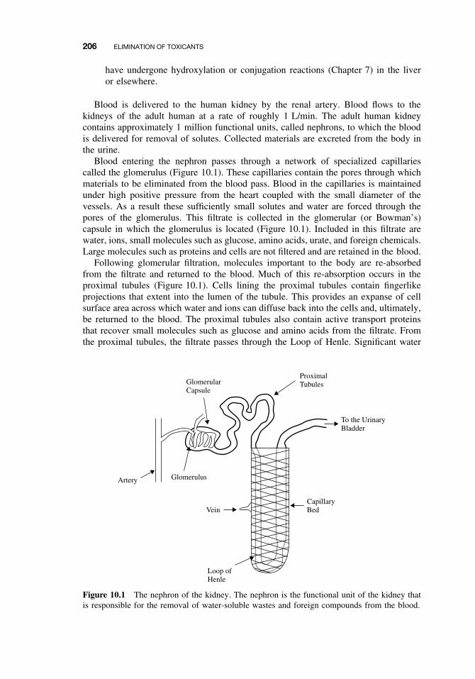
206 ELIMINATION OF TOXICANTS
have undergone hydroxylation or conjugation reactions (Chapter 7) in the liveror elsewhere.
Blood is delivered to the human kidney by the renal artery. Blood flows to thekidneys of the adult human at a rate of roughly 1 L/min. The adult human kidneycontains approximately 1 million functional units, called nephrons, to which the bloodis delivered for removal of solutes. Collected materials are excreted from the body inthe urine.
Blood entering the nephron passes through a network of specialized capillariescalled the glomerulus (Figure 10.1). These capillaries contain the pores through whichmaterials to be eliminated from the blood pass. Blood in the capillaries is maintainedunder high positive pressure from the heart coupled with the small diameter of thevessels. As a result these sufficiently small solutes and water are forced through thepores of the glomerulus. This filtrate is collected in the glomerular (or Bowman’s)capsule in which the glomerulus is located (Figure 10.1). Included in this filtrate arewater, ions, small molecules such as glucose, amino acids, urate, and foreign chemicals.Large molecules such as proteins and cells are not filtered and are retained in the blood.
Following glomerular filtration, molecules important to the body are re-absorbedfrom the filtrate and returned to the blood. Much of this re-absorption occurs in theproximal tubules (Figure 10.1). Cells lining the proximal tubules contain fingerlikeprojections that extent into the lumen of the tubule. This provides an expanse of cellsurface area across which water and ions can diffuse back into the cells and, ultimately,be returned to the blood. The proximal tubules also contain active transport proteinsthat recover small molecules such as glucose and amino acids from the filtrate. Fromthe proximal tubules, the filtrate passes through the Loop of Henle. Significant water
Vein
Glomerulus
Loop of Henle
CapillaryBed
To the UrinaryBladder
Artery
GlomerularCapsule
ProximalTubules
Figure 10.1 The nephron of the kidney. The nephron is the functional unit of the kidney thatis responsible for the removal of water-soluble wastes and foreign compounds from the blood.
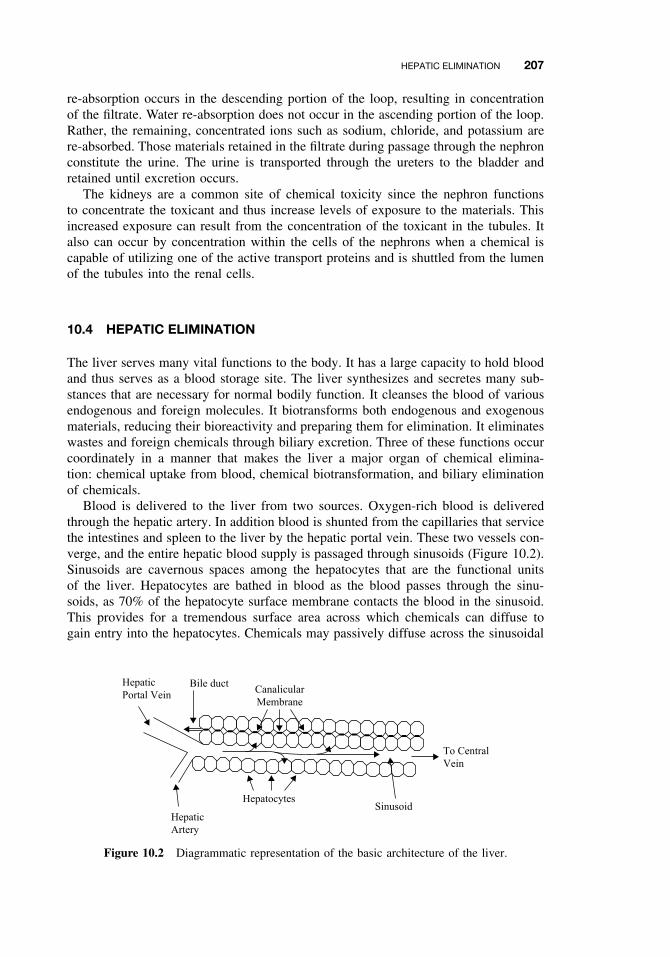
HEPATIC ELIMINATION 207
re-absorption occurs in the descending portion of the loop, resulting in concentrationof the filtrate. Water re-absorption does not occur in the ascending portion of the loop.Rather, the remaining, concentrated ions such as sodium, chloride, and potassium arere-absorbed. Those materials retained in the filtrate during passage through the nephronconstitute the urine. The urine is transported through the ureters to the bladder andretained until excretion occurs.
The kidneys are a common site of chemical toxicity since the nephron functionsto concentrate the toxicant and thus increase levels of exposure to the materials. Thisincreased exposure can result from the concentration of the toxicant in the tubules. Italso can occur by concentration within the cells of the nephrons when a chemical iscapable of utilizing one of the active transport proteins and is shuttled from the lumenof the tubules into the renal cells.
10.4 HEPATIC ELIMINATION
The liver serves many vital functions to the body. It has a large capacity to hold bloodand thus serves as a blood storage site. The liver synthesizes and secretes many sub-stances that are necessary for normal bodily function. It cleanses the blood of variousendogenous and foreign molecules. It biotransforms both endogenous and exogenousmaterials, reducing their bioreactivity and preparing them for elimination. It eliminateswastes and foreign chemicals through biliary excretion. Three of these functions occurcoordinately in a manner that makes the liver a major organ of chemical elimina-tion: chemical uptake from blood, chemical biotransformation, and biliary eliminationof chemicals.
Blood is delivered to the liver from two sources. Oxygen-rich blood is deliveredthrough the hepatic artery. In addition blood is shunted from the capillaries that servicethe intestines and spleen to the liver by the hepatic portal vein. These two vessels con-verge, and the entire hepatic blood supply is passaged through sinusoids (Figure 10.2).Sinusoids are cavernous spaces among the hepatocytes that are the functional unitsof the liver. Hepatocytes are bathed in blood as the blood passes through the sinu-soids, as 70% of the hepatocyte surface membrane contacts the blood in the sinusoid.This provides for a tremendous surface area across which chemicals can diffuse togain entry into the hepatocytes. Chemicals may passively diffuse across the sinusoidal
Bile ductHepatic
Portal VeinCanalicular
Membrane
Hepatocytes Sinusoid
To Central
Vein
Hepatic
Artery
Figure 10.2 Diagrammatic representation of the basic architecture of the liver.
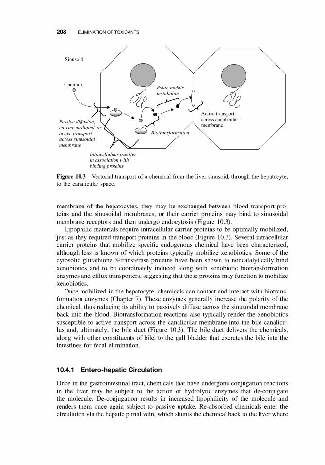
208 ELIMINATION OF TOXICANTS
Passive diffusion,carrier-mediated, oractive transportacross sinusoidalmembrane
Sinusoid
Chemical
Intracelluluar transferin association withbinding proteins
Active transportacross canalicularmembrane
Polar, mobilemetabolite
Biotransformation
Figure 10.3 Vectorial transport of a chemical from the liver sinusoid, through the hepatocyte,to the canalicular space.
membrane of the hepatocytes, they may be exchanged between blood transport pro-teins and the sinusoidal membranes, or their carrier proteins may bind to sinusoidalmembrane receptors and then undergo endocytosis (Figure 10.3).
Lipophilic materials require intracellular carrier proteins to be optimally mobilized,just as they required transport proteins in the blood (Figure 10.3). Several intracellularcarrier proteins that mobilize specific endogenous chemical have been characterized,although less is known of which proteins typically mobilize xenobiotics. Some of thecytosolic glutathione S-transferase proteins have been shown to noncatalytically bindxenobiotics and to be coordinately induced along with xenobiotic biotransformationenzymes and efflux transporters, suggesting that these proteins may function to mobilizexenobiotics.
Once mobilized in the hepatocyte, chemicals can contact and interact with biotrans-formation enzymes (Chapter 7). These enzymes generally increase the polarity of thechemical, thus reducing its ability to passively diffuse across the sinusoidal membraneback into the blood. Biotransformation reactions also typically render the xenobioticssusceptible to active transport across the canalicular membrane into the bile canalicu-lus and, ultimately, the bile duct (Figure 10.3). The bile duct delivers the chemicals,along with other constituents of bile, to the gall bladder that excretes the bile into theintestines for fecal elimination.
10.4.1 Entero-hepatic Circulation
Once in the gastrointestinal tract, chemicals that have undergone conjugation reactionsin the liver may be subject to the action of hydrolytic enzymes that de-conjugatethe molecule. De-conjugation results in increased lipophilicity of the molecule andrenders them once again subject to passive uptake. Re-absorbed chemicals enter thecirculation via the hepatic portal vein, which shunts the chemical back to the liver where
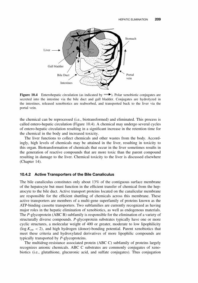
HEPATIC ELIMINATION 209
Liver
Gall bladder
Stomach
Intestines
Portalvein
Bile Duct
Figure 10.4 Enterohepatic circulation (as indicated by ). Polar xenobiotic conjugates aresecreted into the intestine via the bile duct and gall bladder. Conjugates are hydrolyzed inthe intestines, released xenobiotics are reabsorbed, and transported back to the liver via theportal vein.
the chemical can be reprocessed (i.e., biotransformed) and eliminated. This process iscalled entero-hepatic circulation (Figure 10.4). A chemical may undergo several cyclesof entero-hepatic circulation resulting in a significant increase in the retention time forthe chemical in the body and increased toxicity.
The liver functions to collect chemicals and other wastes from the body. Accord-ingly, high levels of chemicals may be attained in the liver, resulting in toxicity tothis organ. Biotransformation of chemicals that occur in the liver sometimes results inthe generation of reactive compounds that are more toxic than the parent compoundresulting in damage to the liver. Chemical toxicity to the liver is discussed elsewhere(Chapter 14).
10.4.2 Active Transporters of the Bile Canaliculus
The bile canaliculus constitutes only about 13% of the contiguous surface membraneof the hepatocyte but must function in the efficient transfer of chemical from the hep-atocyte to the bile duct. Active transport proteins located on the canalicular membraneare responsible for the efficient shuttling of chemicals across this membrane. Theseactive transporters are members of a multi-gene superfamily of proteins known as theATP-binding cassette transporters. Two subfamilies are currently recognized as havingmajor roles in the hepatic elimination of xenobiotics, as well as endogenous materials.The P-glycoprotein (ABC B) subfamily is responsible for the elimination of a variety ofstructurally diverse compounds. P-glycoprotein substrates typically have one or morecyclic structures, a molecular weight of 400 or greater, moderate to low lipophilicity(log Kow < 2), and high hydrogen (donor)-bonding potential. Parent xenobiotics thatmeet these criteria and hydroxylated derivatives of more lipophilic compounds aretypically transported by P-glycoproteins.
The multidrug-resistance associated protein (ABC C) subfamily of proteins largelyrecognizes anionic chemicals. ABC C substrates are commonly conjugates of xeno-biotics (i.e., glutathione, glucuronic acid, and sulfate conjugates). Thus conjugation
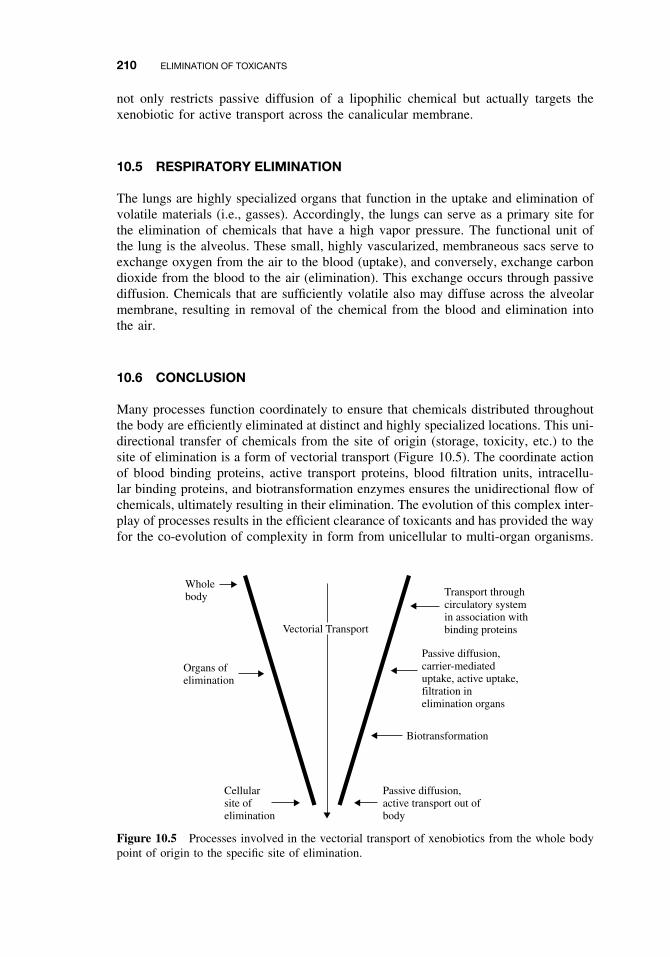
210 ELIMINATION OF TOXICANTS
not only restricts passive diffusion of a lipophilic chemical but actually targets thexenobiotic for active transport across the canalicular membrane.
10.5 RESPIRATORY ELIMINATION
The lungs are highly specialized organs that function in the uptake and elimination ofvolatile materials (i.e., gasses). Accordingly, the lungs can serve as a primary site forthe elimination of chemicals that have a high vapor pressure. The functional unit ofthe lung is the alveolus. These small, highly vascularized, membraneous sacs serve toexchange oxygen from the air to the blood (uptake), and conversely, exchange carbondioxide from the blood to the air (elimination). This exchange occurs through passivediffusion. Chemicals that are sufficiently volatile also may diffuse across the alveolarmembrane, resulting in removal of the chemical from the blood and elimination intothe air.
10.6 CONCLUSION
Many processes function coordinately to ensure that chemicals distributed throughoutthe body are efficiently eliminated at distinct and highly specialized locations. This uni-directional transfer of chemicals from the site of origin (storage, toxicity, etc.) to thesite of elimination is a form of vectorial transport (Figure 10.5). The coordinate actionof blood binding proteins, active transport proteins, blood filtration units, intracellu-lar binding proteins, and biotransformation enzymes ensures the unidirectional flow ofchemicals, ultimately resulting in their elimination. The evolution of this complex inter-play of processes results in the efficient clearance of toxicants and has provided the wayfor the co-evolution of complexity in form from unicellular to multi-organ organisms.
Wholebody
Organs ofelimination
Cellularsite ofelimination
Transport throughcirculatory systemin association withbinding proteins
Passive diffusion,carrier-mediateduptake, active uptake,filtration inelimination organs
Biotransformation
Passive diffusion,active transport out ofbody
Vectorial Transport
Figure 10.5 Processes involved in the vectorial transport of xenobiotics from the whole bodypoint of origin to the specific site of elimination.

SUGGESTED READING 211
SUGGESTED READING
LeBlanc, G. A., and W. C. Dauterman. Conjugation and elimination of toxicants. In Introductionto Biochemical Toxicology, E. Hodgson and R. C. Smart, eds. New York: Wiley-Interscience,2001, pp. 115–136.
Kester, J. E. Liver. In Encyclopedia of Toxicology, vol. 2, P. Wexler, ed. New York: AcademicPress, 1998, pp. 253–261.
Rankin, G. O. Kidney. In Encyclopedia of Toxicology, vol. 2, P. Wexler, ed. New York: Aca-demic Press, 1998, pp. 198–225.
Klaassen, C. D. ed. Casarett and Doull’s Toxicology: The Basic Science of Poisons, 6th ed. NewYork: McGraw-Hill, 2001.


PART IV
TOXIC ACTION

CHAPTER 11
Acute Toxicity
GERALD A. LEBLANC
11.1 INTRODUCTION
Acute toxicity of a chemical can be viewed from two perspectives. Acute toxicity maybe the descriptor used as a qualitative indicator of an incident of poisoning. Considerthe following statement: “methyl isocyanate gas, accidentally released from a chemicalmanufacturing facility in 1984, was acutely toxic to the residents of Bhopal, India.”This statement implies that the residents of Bhopal were exposed to sufficiently highlevels of methyl isocyanate over a relatively short time to result in immediate harm.High-level, short-term exposure resulting in immediate toxicity are all characteristics ofacute toxicity. Alternatively, acute toxicity may represent a quantifiable characteristicof a material. For example, the statement: “the acute toxicity of methyl isocyanate, asmeasured by its LD50 in rats, is 140 mg/kg” defines the acute toxicity of the chemical.Again, the characterization of the quantified effects of methyl isocyanate as being acutetoxicity implies that this quantification occurred during or following short-term dosingand that the effect measured occurred within a short time period following dosing.In terms of these qualitative and quantitative aspects, acute toxicity can be definedas toxicity elicited immediately following short-term exposure to a chemical. By thisdefinition, two components comprise acute toxicity: acute exposure and acute effect.
11.2 ACUTE EXPOSURE AND EFFECT
In contrast to acute toxicity, chronic toxicity is characterized by prolonged exposureand sublethal effects elicited through mechanisms that are distinct from those thatcause acute toxicity. Typically acute and chronic toxicity of a chemical are easilydistinguished. For example, mortality occurring within two days of a single dose of achemical would be a prime example of acute toxicity (Figure 11.1a). Similarly, reducedlitter size following continuous (i.e., daily) dosing of the parental organisms would beindicative of chronic toxicity (Figure 11.1b). However, defining toxicity as being acuteor chronic is sometimes challenging. For example, chronic exposure to a persistent,lipophilic chemical may result in sequestration of significant levels of the chemical
A Textbook of Modern Toxicology, Third Edition, edited by Ernest HodgsonISBN 0-471-26508-X Copyright 2004 John Wiley & Sons, Inc.
215
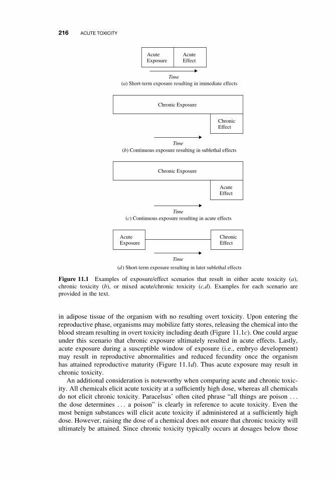
216 ACUTE TOXICITY
Time
Time
Time
Time
(c) Continuous exposure resulting in acute effects
(d ) Short-term exposure resulting in later sublethal effects
(a) Short-term exposure resulting in immediate effects
(b) Continuous exposure resulting in sublethal effects
AcuteExposure
ChronicEffect
AcuteExposure
AcuteEffect
Chronic Exposure
Chronic Exposure
ChronicEffect
AcuteEffect
Figure 11.1 Examples of exposure/effect scenarios that result in either acute toxicity (a),chronic toxicity (b), or mixed acute/chronic toxicity (c,d). Examples for each scenario areprovided in the text.
in adipose tissue of the organism with no resulting overt toxicity. Upon entering thereproductive phase, organisms may mobilize fatty stores, releasing the chemical into theblood stream resulting in overt toxicity including death (Figure 11.1c). One could argueunder this scenario that chronic exposure ultimately resulted in acute effects. Lastly,acute exposure during a susceptible window of exposure (i.e., embryo development)may result in reproductive abnormalities and reduced fecundity once the organismhas attained reproductive maturity (Figure 11.1d). Thus acute exposure may result inchronic toxicity.
An additional consideration is noteworthy when comparing acute and chronic toxic-ity. All chemicals elicit acute toxicity at a sufficiently high dose, whereas all chemicalsdo not elicit chronic toxicity. Paracelsus’ often cited phrase “all things are poison . . .
the dose determines . . . a poison” is clearly in reference to acute toxicity. Even themost benign substances will elicit acute toxicity if administered at a sufficiently highdose. However, raising the dose of a chemical does not ensure that chronic toxicity willultimately be attained. Since chronic toxicity typically occurs at dosages below those
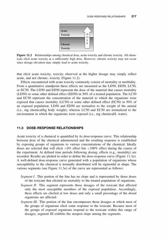
DOSE-RESPONSE RELATIONSHIPS 217
Acute
Toxicity
Chronic
Toxicity
DOSE
Figure 11.2 Relationships among chemical dose, acute toxicity and chronic toxicity. All chem-icals elicit acute toxicity at a sufficiently high dose. However, chronic toxicity may not occursince dosage elevation may simply lead to acute toxicity.
that elicit acute toxicity, toxicity observed at the higher dosage may simply reflectacute, and not chronic, toxicity (Figure 11.2).
Effects encountered with acute toxicity commonly consist of mortality or morbidity.From a quantitative standpoint these effects are measured as the LD50, ED50, LC50,or EC50. The LD50 and ED50 represent the dose of the material that causes mortality(LD50) or some other defined effect (ED50) in 50% of a treated population. The LC50and EC50 represent the concentration of the material to which the organisms wereexposed that causes mortality (LC50) or some other defined effect (EC50) in 50% ofan exposed population. LD50 and ED50 are normalize to the weight of the animal(i.e., mg chemical/kg body weight); whereas LC50 and EC50 are normalized to theenvironment in which the organisms were exposed (i.e., mg chemical/L water).
11.3 DOSE-RESPONSE RELATIONSHIPS
Acute toxicity of a chemical is quantified by its dose-response curve. This relationshipbetween dose of the chemical administered and the resulting response is establishedby exposing groups of organisms to various concentrations of the chemical. Ideallydoses are selected that will elicit >0% effect but <100% effect during the course ofthe experiment. At defined time periods following dosing, effects (e.g., mortality) arerecorded. Results are plotted in order to define the dose-response curve (Figure 11.3a).A well-defined dose-response curve generated with a population of organisms whosesusceptibility to the chemical is normally distributed will be sigmoidal in shape. Thevarious segments (see Figure 11.3a) of the curve are represented as follows:
Segment I . This portion of the line has no slope and is represented by those dosesof the toxicant that elicited no mortality to the treated population of organisms.
Segment II . This segment represents those dosages of the toxicant that affectedonly the most susceptible members of the exposed population. Accordingly,these effects are elicited at low doses and only a small percentage of the dosedorganisms are affected.
Segment III . This portion of the line encompasses those dosages at which most ofthe groups of organisms elicit some response to the toxicant. Because most ofthe groups of exposed organisms respond to the toxicant within this range ofdosages, segment III exhibits the steepest slope among the segments.
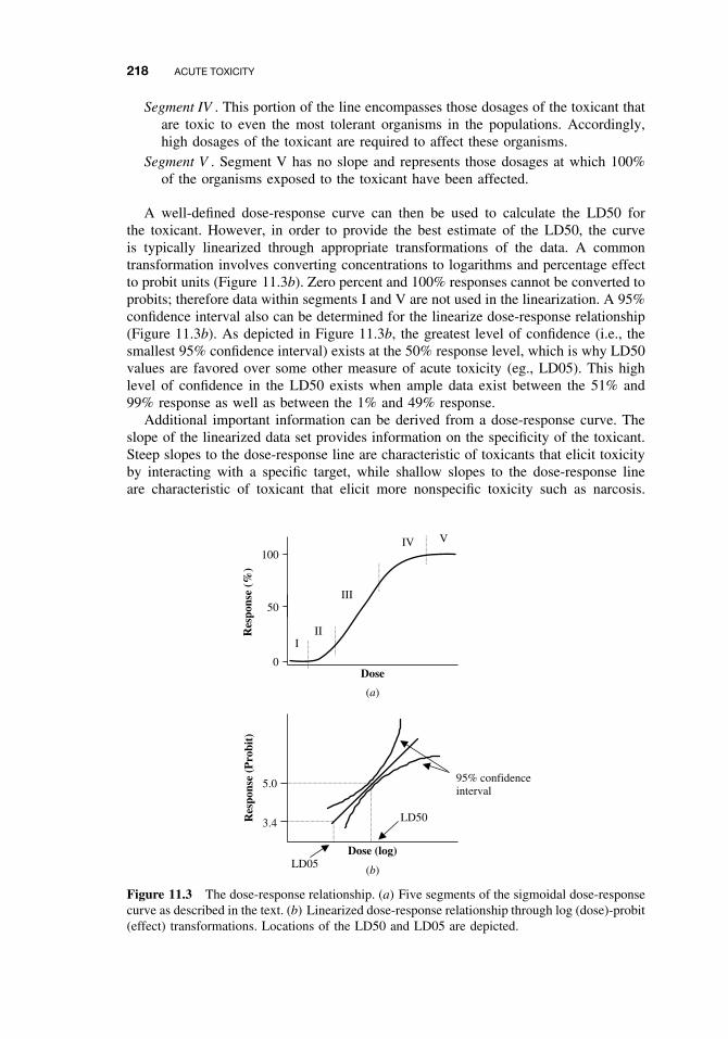
218 ACUTE TOXICITY
Segment IV . This portion of the line encompasses those dosages of the toxicant thatare toxic to even the most tolerant organisms in the populations. Accordingly,high dosages of the toxicant are required to affect these organisms.
Segment V . Segment V has no slope and represents those dosages at which 100%of the organisms exposed to the toxicant have been affected.
A well-defined dose-response curve can then be used to calculate the LD50 forthe toxicant. However, in order to provide the best estimate of the LD50, the curveis typically linearized through appropriate transformations of the data. A commontransformation involves converting concentrations to logarithms and percentage effectto probit units (Figure 11.3b). Zero percent and 100% responses cannot be converted toprobits; therefore data within segments I and V are not used in the linearization. A 95%confidence interval also can be determined for the linearize dose-response relationship(Figure 11.3b). As depicted in Figure 11.3b, the greatest level of confidence (i.e., thesmallest 95% confidence interval) exists at the 50% response level, which is why LD50values are favored over some other measure of acute toxicity (eg., LD05). This highlevel of confidence in the LD50 exists when ample data exist between the 51% and99% response as well as between the 1% and 49% response.
Additional important information can be derived from a dose-response curve. Theslope of the linearized data set provides information on the specificity of the toxicant.Steep slopes to the dose-response line are characteristic of toxicants that elicit toxicityby interacting with a specific target, while shallow slopes to the dose-response lineare characteristic of toxicant that elicit more nonspecific toxicity such as narcosis.
I
LD50
LD05
3.4
95% confidenceinterval
Res
pons
e (P
robi
t)
5.0
Dose (log)
100
0
Res
pons
e (%
)
50
Dose
(a)
(b)
IV V
II
III
Figure 11.3 The dose-response relationship. (a) Five segments of the sigmoidal dose-responsecurve as described in the text. (b) Linearized dose-response relationship through log (dose)-probit(effect) transformations. Locations of the LD50 and LD05 are depicted.
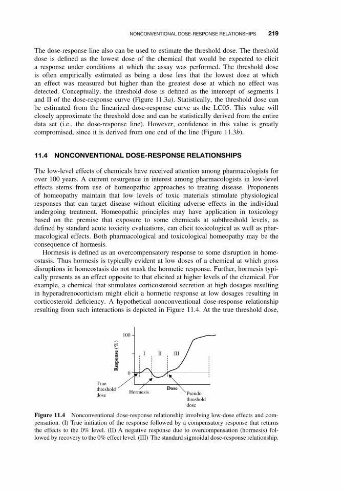
NONCONVENTIONAL DOSE-RESPONSE RELATIONSHIPS 219
The dose-response line also can be used to estimate the threshold dose. The thresholddose is defined as the lowest dose of the chemical that would be expected to elicita response under conditions at which the assay was performed. The threshold doseis often empirically estimated as being a dose less that the lowest dose at whichan effect was measured but higher than the greatest dose at which no effect wasdetected. Conceptually, the threshold dose is defined as the intercept of segments Iand II of the dose-response curve (Figure 11.3a). Statistically, the threshold dose canbe estimated from the linearized dose-response curve as the LC05. This value willclosely approximate the threshold dose and can be statistically derived from the entiredata set (i.e., the dose-response line). However, confidence in this value is greatlycompromised, since it is derived from one end of the line (Figure 11.3b).
11.4 NONCONVENTIONAL DOSE-RESPONSE RELATIONSHIPS
The low-level effects of chemicals have received attention among pharmacologists forover 100 years. A current resurgence in interest among pharmacologists in low-leveleffects stems from use of homeopathic approaches to treating disease. Proponentsof homeopathy maintain that low levels of toxic materials stimulate physiologicalresponses that can target disease without eliciting adverse effects in the individualundergoing treatment. Homeopathic principles may have application in toxicologybased on the premise that exposure to some chemicals at subthreshold levels, asdefined by standard acute toxicity evaluations, can elicit toxicological as well as phar-macological effects. Both pharmacological and toxicological homeopathy may be theconsequence of hormesis.
Hormesis is defined as an overcompensatory response to some disruption in home-ostasis. Thus hormesis is typically evident at low doses of a chemical at which grossdisruptions in homeostasis do not mask the hormetic response. Further, hormesis typi-cally presents as an effect opposite to that elicited at higher levels of the chemical. Forexample, a chemical that stimulates corticosteroid secretion at high dosages resultingin hyperadrenocorticism might elicit a hormetic response at low dosages resulting incorticosteroid deficiency. A hypothetical nonconventional dose-response relationshipresulting from such interactions is depicted in Figure 11.4. At the true threshold dose,
Hormesis Pseudothresholddose
Truethresholddose
100
0
Res
pons
e (%
)
Dose
I II III
Figure 11.4 Nonconventional dose-response relationship involving low-dose effects and com-pensation. (I) True initiation of the response followed by a compensatory response that returnsthe effects to the 0% level. (II) A negative response due to overcompensation (hormesis) fol-lowed by recovery to the 0% effect level. (III) The standard sigmoidal dose-response relationship.
220 ACUTE TOXICITY
the organisms begin to exhibit increased stimulation in corticosteroid secretion. How-ever, at slightly higher doses, a compensatory response occurs whereby corticosteroidsecretion is decreased in order to maintain homeostasis within the organism. Over-compensation may actually result in a decrease in corticosteroid secretion at certaintoxicant dosages. Finally the compensatory abilities of the organism are overcome bythe high doses of the toxicant at the “pseudo” threshold dose, above which the standarddose-response relationship occurs. Nonconventional dose-response relationships havebeen observed with respect to both acute and chronic toxicity and are particularlyrelevant to the risk assessment process when establishing levels of exposure that areanticipated to pose no harm.
11.5 MECHANISMS OF ACUTE TOXICITY
An exhaustive review of the mechanisms by which chemicals cause acute toxicityis beyond the scope of this chapter. However, certain mechanisms of toxicity arerelevant since they are common to many important classes of toxicants. Some of thesemechanisms of acute toxicity are discussed.
11.5.1 Narcosis
Narcosis in toxicology is defined as toxicity resulting from chemicals associating withand disrupting the lipid bilayer of membranes. Narcotics are classified as either non-polar (class 1) or polar (class 2) compounds. Members of both classes of compoundsare lipid soluble. However, class 2 compounds possess constituents that confer somecharge distribution to the compound (i.e., aliphatic and aromatic amines, nitroaromat-ics, alcohols). The aliphatic hydrocarbon (C5 through C8) are examples of powerfulclass 1 narcotics, whereas, ethanol is an example of a class 2 narcotic. The affinityof narcotics to partition into the nonpolar core of membranes (class 1 narcotics) or todistribute in both the polar and nonpolar components of membranes (class 2 narcotics)alters the fluidity of the membrane. This effect compromises the ability of proteins andother constituents of the membranes to function properly leading to various manifes-tation of narcosis. The central nervous system is the prime target of chemical narcosisand symptoms initially include disorientation, euphoria, giddiness, and progress tounconsciousness, convulsion, and death.
11.5.2 Acetylcholinesterase Inhibition
Acetylcholine is a neurotransmitter that functions in conveying nerve impulses acrosssynaptic clefts within the central and autonomic nervous systems and at juncturesof nerves and muscles. Following transmission of an impulse across the synapse bythe release of acetylcholine, acetylcholinesterase is released into the synaptic cleft.This enzyme hydrolyzes acetylcholine to choline and acetate and transmission of thenerve impulse is terminated. The inhibition of acetylcholineasterase results in pro-longed, uncoordinated nerve or muscle stimulation. Organophosphorus and carbamatepesticides (Chapter 5) along with some nerve gases (i.e., sarin) elicit toxicity viathis mechanism.
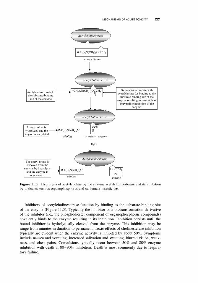
MECHANISMS OF ACUTE TOXICITY 221
choline
(CH3)3N(CH2)2O
Acetylcholinesterase
Acetylcholinesterase
CCH
acetylated enzyme
H2O
(CH3)3N(CH2)2OCCH3
acetylchloline
choline
(CH3)3N(CH2)2O
acetate
HOCCH3
Xenobiotics compete withacetylcholine for binding to the
substrate-binding site of theenzyme resulting in reversible or
irreversible inhibition of theenzyme.
(CH3)3N(CH2)2OCCH3
Acetylcholinesterase
Acetylcholine binds tothe substrate-binding
site of the enzyme
Acetylcholine ishydrolyzed and the
enzyme is acetylated
The acetyl group isremoved from the
enzyme by hydrolysisand the enzyme is
regenerated
Acetylcholinesterase
Figure 11.5 Hydrolysis of acetylcholine by the enzyme acetylcholinesterase and its inhibitionby toxicants such as organophosphorus and carbamate insecticides.
Inhibitors of acetylcholinesterase function by binding to the substrate-binding siteof the enzyme (Figure 11.5). Typically the inhibitor or a biotransformation derivativeof the inhibitor (i.e., the phosphodiester component of organophosphorus compounds)covalently binds to the enzyme resulting in its inhibition. Inhibition persists until thebound inhibitor is hydrolytically cleaved from the enzyme. This inhibition may berange from minutes in duration to permanent. Toxic effects of cholinesterase inhibitiontypically are evident when the enzyme activity is inhibited by about 50%. Symptomsinclude nausea and vomiting, increased salivation and sweating, blurred vision, weak-ness, and chest pains. Convulsions typically occur between 50% and 80% enzymeinhibition with death at 80–90% inhibition. Death is most commonly due to respira-tory failure.
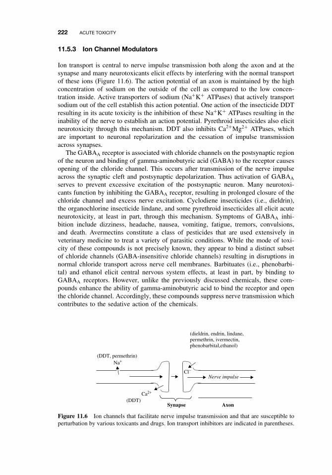
222 ACUTE TOXICITY
11.5.3 Ion Channel Modulators
Ion transport is central to nerve impulse transmission both along the axon and at thesynapse and many neurotoxicants elicit effects by interfering with the normal transportof these ions (Figure 11.6). The action potential of an axon is maintained by the highconcentration of sodium on the outside of the cell as compared to the low concen-tration inside. Active transporters of sodium (Na+K+ ATPases) that actively transportsodium out of the cell establish this action potential. One action of the insecticide DDTresulting in its acute toxicity is the inhibition of these Na+K+ ATPases resulting in theinability of the nerve to establish an action potential. Pyrethroid insecticides also elicitneurotoxicity through this mechanism. DDT also inhibits Ca2+Mg2+ ATPases, whichare important to neuronal repolarization and the cessation of impulse transmissionacross synapses.
The GABAA receptor is associated with chloride channels on the postsynaptic regionof the neuron and binding of gamma-aminobutyric acid (GABA) to the receptor causesopening of the chloride channel. This occurs after transmission of the nerve impulseacross the synaptic cleft and postsynaptic depolarization. Thus activation of GABAA
serves to prevent excessive excitation of the postsynaptic neuron. Many neurotoxi-cants function by inhibiting the GABAA receptor, resulting in prolonged closure of thechloride channel and excess nerve excitation. Cyclodiene insecticides (i.e., dieldrin),the organochlorine insecticide lindane, and some pyrethroid insecticides all elicit acuteneurotoxicity, at least in part, through this mechanism. Symptoms of GABAA inhi-bition include dizziness, headache, nausea, vomiting, fatigue, tremors, convulsions,and death. Avermectins constitute a class of pesticides that are used extensively inveterinary medicine to treat a variety of parasitic conditions. While the mode of toxi-city of these compounds is not precisely known, they appear to bind a distinct subsetof chloride channels (GABA-insensitive chloride channels) resulting in disruptions innormal chloride transport across nerve cell membranes. Barbituates (i.e., phenobarbi-tal) and ethanol elicit central nervous system effects, at least in part, by binding toGABAA receptors. However, unlike the previously discussed chemicals, these com-pounds enhance the ability of gamma-aminobutyric acid to bind the receptor and openthe chloride channel. Accordingly, these compounds suppress nerve transmission whichcontributes to the sedative action of the chemicals.
(dieldrin, endrin, lindane,permethrin, ivermectin,phenobarbital,ethanol)
(DDT)Synapse Axon
Cl−
Ca2+
Na+
Nerve impulse
(DDT, permethrin)
Figure 11.6 Ion channels that facilitate nerve impulse transmission and that are susceptible toperturbation by various toxicants and drugs. Ion transport inhibitors are indicated in parentheses.
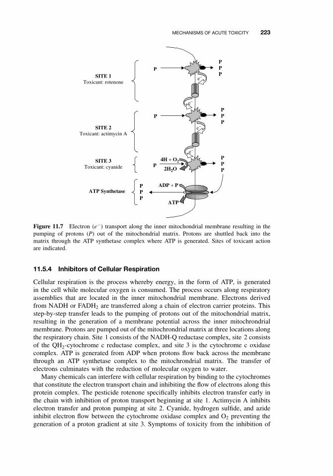
MECHANISMS OF ACUTE TOXICITY 223
PPP
P
2H2O
4H + O2
ATP
e−
e−
e−
e−
ADP + P
P
PPPP
PPP
PPPSITE 1
Toxicant: rotenone
SITE 2Toxicant: actimycin A
SITE 3Toxicant: cyanide
ATP Synthetase
Figure 11.7 Electron (e−) transport along the inner mitochondrial membrane resulting in thepumping of protons (P) out of the mitochondrial matrix. Protons are shuttled back into thematrix through the ATP synthetase complex where ATP is generated. Sites of toxicant actionare indicated.
11.5.4 Inhibitors of Cellular Respiration
Cellular respiration is the process whereby energy, in the form of ATP, is generatedin the cell while molecular oxygen is consumed. The process occurs along respiratoryassemblies that are located in the inner mitochondrial membrane. Electrons derivedfrom NADH or FADH2 are transferred along a chain of electron carrier proteins. Thisstep-by-step transfer leads to the pumping of protons out of the mitochondrial matrix,resulting in the generation of a membrane potential across the inner mitochrondrialmembrane. Protons are pumped out of the mitochrondrial matrix at three locations alongthe respiratory chain. Site 1 consists of the NADH-Q reductase complex, site 2 consistsof the QH2-cytochrome c reductase complex, and site 3 is the cytochrome c oxidasecomplex. ATP is generated from ADP when protons flow back across the membranethrough an ATP synthetase complex to the mitochrondrial matrix. The transfer ofelectrons culminates with the reduction of molecular oxygen to water.
Many chemicals can interfere with cellular respiration by binding to the cytochromesthat constitute the electron transport chain and inhibiting the flow of electrons along thisprotein complex. The pesticide rotenone specifically inhibits electron transfer early inthe chain with inhibition of proton transport beginning at site 1. Actimycin A inhibitselectron transfer and proton pumping at site 2. Cyanide, hydrogen sulfide, and azideinhibit electron flow between the cytochrome oxidase complex and O2 preventing thegeneration of a proton gradient at site 3. Symptoms of toxicity from the inhibition of
224 ACUTE TOXICITY
respiratory chain include excess salivation, giddiness, headache, palpitations, respira-tory distress, and loss of consciousness. Potent inhibitors such as cyanide can causedeath due to respiratory arrest immediately following poisoning.
Some chemicals do not interfere with electron transport leading to the consump-tion of molecular oxygen but rather interfere with the conversion of ADP to ATP.These uncouplers of oxidative phosphorylation function by leaking protons across theinner membrane back to the mitochondrial matrix. As a result a membrane poten-tial is not generated, and energy required for the phosphorylation of ADP to ATP islost. The uncoupling of oxidative phosphorylation results in increased electron trans-port, increased oxygen consumption, and heat production. The controlled uncouplingof oxidative phosphorylation is a physiologically relevant means of maintaining bodytemperature by hibernating animals, some newborn animals, and in some animals thatinhabit cold environments. Chemicals known to cause uncoupling of oxidative phos-phorylation include 2,4-dinitrophenol, pentachlorophenol, and dicumarol. Symptomsof intoxication include accelerated respiration and pulse, flushed skin, elevated tem-perature, sweating, nausea, coma, and death.
SUGGESTED READING
Joy, R. M. Neurotoxicology: Central and peripheral. In Encyclopedia of Toxicology, vol. 2,P. Wexler, ed. New York: Academic Press, 1998, pp. 389–413.
Stryer, L. Biochemistry, 4th ed. San Francisco: W. H. Freeman, 1999.
Eaton, D. L., and C. D. Klaassen. Principles of toxicology In Casarrett and Doull’s Toxicology:The Basic Science of Poisons, 6th ed. C. D. Klaassen, ed. New York: McGraw-Hill, 2001, pp.11–34.
Calabrese, E. J., and L. A. Baldwin. U-shaped dose-responses in biology, toxicology, and publichealth. An. Rev. Public Health 22: 15–33, 2001.
CHAPTER 12
Chemical Carcinogenesis
ROBERT C. SMART
12.1 GENERAL ASPECTS OF CANCER
Carcinogenesis is the process through which cancer develops. Chemical carcinogene-sis is the study of the mechanisms through which chemical carcinogens induce cancerand also involves the development/utilization of experimental systems aimed at deter-mining whether a substance is a potential human carcinogen. An important aspect oftoxicology is the identification of potential human carcinogens. To begin to appreci-ate the complexity of this subject, it is important to first have some understanding ofcancer and its etiologies.
Cancer is not a single disease but a large group of diseases, all of which can becharacterized by the uncontrolled growth of an abnormal cell to produce a populationof cells that have acquired the ability to multiply and invade surrounding and distanttissues. It is this invasive characteristic that imparts its lethality on the host. Epidemi-ology studies have revealed that the incidence of most cancers increase exponentiallywith age (Figure 12.1). Epidemiologist have interpreted this exponential increase incancer incidence to denote that three to seven critical mutations or “hits” within asingle cell are required for cancer development. Molecular analyses of human tumorshave confirmed the accumulation of mutations in critical genes in the development ofcancer. These mutations can be the result of imperfect DNA replication/repair, oxida-tive DNA damage, and/or DNA damage caused by environmental carcinogens. Mostcancers are monoclonal in origin (derived from a single cell) and do not arise froma single critical mutation but from the accumulation of sequential critical mutationsin relevant target genes within a single cell (Figure 12.2). Initially a somatic mutationoccurs in a critical gene, and this provides a growth advantage to the cell and results inthe expansion of the mutant clone. Each additional critical mutation provides a furtherselective growth advantage resulting in clonal expansion of cells with mutations inmultiple critical genes. It often requires decades for a cell clone to accumulate mul-tiple critical mutations and for the progeny of this cell to clonally expand to producea clinically detectable cancer. Thus the time required for accumulation of mutationsin critical genes within a cell is likely related to the observation that cancer incidenceincreases exponentially with age.
A Textbook of Modern Toxicology, Third Edition, edited by Ernest HodgsonISBN 0-471-26508-X Copyright 2004 John Wiley & Sons, Inc.
225
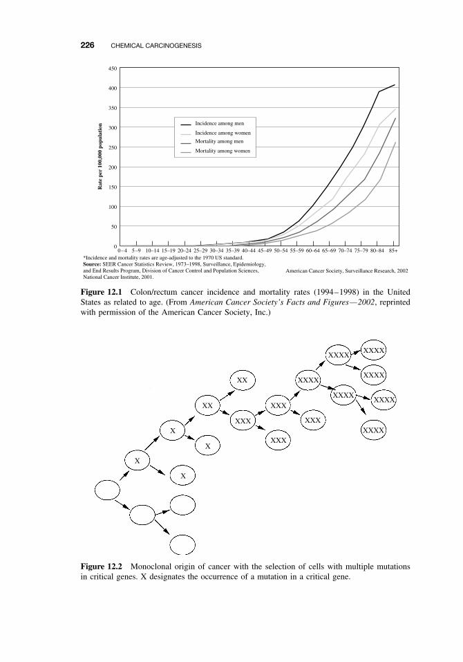
226 CHEMICAL CARCINOGENESIS
450
400
350
300
250
200
150
100
50
0
Rat
e pe
r 10
0,00
0 po
pula
tion
0–4 5–9 10–14 15–19 20–24 25–29 30–34 35–39 40–44 45–49 50–54 55–59 60–64 65–69 70–74 75–79 80–84 85+
Incidence among men
Incidence among women
Mortality among men
Mortality among women
*Incidence and mortality rates are age-adjusted to the 1970 US standard.Source: SEER Cancer Statistics Review, 1973–1998, Surveillance, Epidemiology,and End Results Program, Division of Cancer Control and Population Sciences,National Cancer Institute, 2001.
American Cancer Society, Surveillance Research, 2002
Figure 12.1 Colon/rectum cancer incidence and mortality rates (1994–1998) in the UnitedStates as related to age. (From American Cancer Society’s Facts and Figures—2002, reprintedwith permission of the American Cancer Society, Inc.)
X
X
X
XX
XX
XXX
XXX
XXX
XXX
XXXX
XXXX
XXXX
XXXX
XXXX
XXXXXXXX
X
Figure 12.2 Monoclonal origin of cancer with the selection of cells with multiple mutationsin critical genes. X designates the occurrence of a mutation in a critical gene.
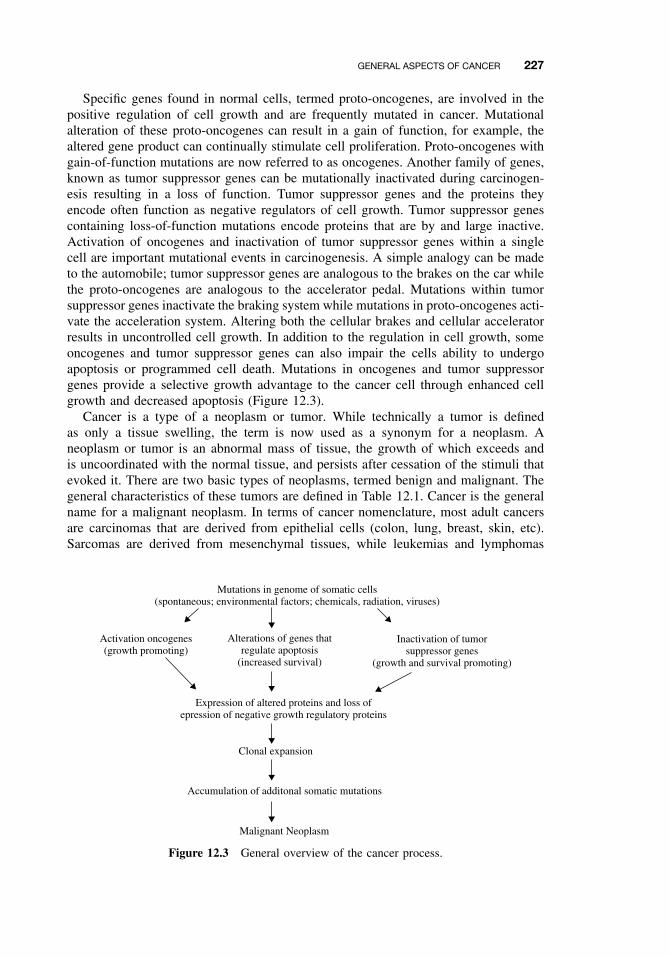
GENERAL ASPECTS OF CANCER 227
Specific genes found in normal cells, termed proto-oncogenes, are involved in thepositive regulation of cell growth and are frequently mutated in cancer. Mutationalalteration of these proto-oncogenes can result in a gain of function, for example, thealtered gene product can continually stimulate cell proliferation. Proto-oncogenes withgain-of-function mutations are now referred to as oncogenes. Another family of genes,known as tumor suppressor genes can be mutationally inactivated during carcinogen-esis resulting in a loss of function. Tumor suppressor genes and the proteins theyencode often function as negative regulators of cell growth. Tumor suppressor genescontaining loss-of-function mutations encode proteins that are by and large inactive.Activation of oncogenes and inactivation of tumor suppressor genes within a singlecell are important mutational events in carcinogenesis. A simple analogy can be madeto the automobile; tumor suppressor genes are analogous to the brakes on the car whilethe proto-oncogenes are analogous to the accelerator pedal. Mutations within tumorsuppressor genes inactivate the braking system while mutations in proto-oncogenes acti-vate the acceleration system. Altering both the cellular brakes and cellular acceleratorresults in uncontrolled cell growth. In addition to the regulation in cell growth, someoncogenes and tumor suppressor genes can also impair the cells ability to undergoapoptosis or programmed cell death. Mutations in oncogenes and tumor suppressorgenes provide a selective growth advantage to the cancer cell through enhanced cellgrowth and decreased apoptosis (Figure 12.3).
Cancer is a type of a neoplasm or tumor. While technically a tumor is definedas only a tissue swelling, the term is now used as a synonym for a neoplasm. Aneoplasm or tumor is an abnormal mass of tissue, the growth of which exceeds andis uncoordinated with the normal tissue, and persists after cessation of the stimuli thatevoked it. There are two basic types of neoplasms, termed benign and malignant. Thegeneral characteristics of these tumors are defined in Table 12.1. Cancer is the generalname for a malignant neoplasm. In terms of cancer nomenclature, most adult cancersare carcinomas that are derived from epithelial cells (colon, lung, breast, skin, etc).Sarcomas are derived from mesenchymal tissues, while leukemias and lymphomas
Mutations in genome of somatic cells(spontaneous; environmental factors; chemicals, radiation, viruses)
Activation oncogenes(growth promoting)
Alterations of genes thatregulate apoptosis
(increased survival)
Inactivation of tumorsuppressor genes
(growth and survival promoting)
Expression of altered proteins and loss ofepression of negative growth regulatory proteins
Clonal expansion
Accumulation of additonal somatic mutations
Malignant Neoplasm
Figure 12.3 General overview of the cancer process.
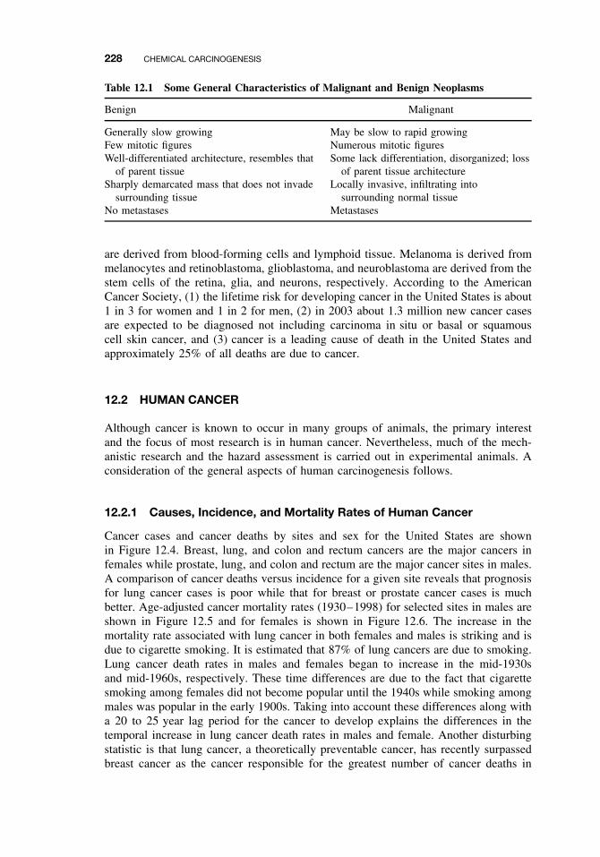
228 CHEMICAL CARCINOGENESIS
Table 12.1 Some General Characteristics of Malignant and Benign Neoplasms
Benign Malignant
Generally slow growing May be slow to rapid growingFew mitotic figures Numerous mitotic figuresWell-differentiated architecture, resembles that
of parent tissueSome lack differentiation, disorganized; loss
of parent tissue architectureSharply demarcated mass that does not invade
surrounding tissueLocally invasive, infiltrating into
surrounding normal tissueNo metastases Metastases
are derived from blood-forming cells and lymphoid tissue. Melanoma is derived frommelanocytes and retinoblastoma, glioblastoma, and neuroblastoma are derived from thestem cells of the retina, glia, and neurons, respectively. According to the AmericanCancer Society, (1) the lifetime risk for developing cancer in the United States is about1 in 3 for women and 1 in 2 for men, (2) in 2003 about 1.3 million new cancer casesare expected to be diagnosed not including carcinoma in situ or basal or squamouscell skin cancer, and (3) cancer is a leading cause of death in the United States andapproximately 25% of all deaths are due to cancer.
12.2 HUMAN CANCER
Although cancer is known to occur in many groups of animals, the primary interestand the focus of most research is in human cancer. Nevertheless, much of the mech-anistic research and the hazard assessment is carried out in experimental animals. Aconsideration of the general aspects of human carcinogenesis follows.
12.2.1 Causes, Incidence, and Mortality Rates of Human Cancer
Cancer cases and cancer deaths by sites and sex for the United States are shownin Figure 12.4. Breast, lung, and colon and rectum cancers are the major cancers infemales while prostate, lung, and colon and rectum are the major cancer sites in males.A comparison of cancer deaths versus incidence for a given site reveals that prognosisfor lung cancer cases is poor while that for breast or prostate cancer cases is muchbetter. Age-adjusted cancer mortality rates (1930–1998) for selected sites in males areshown in Figure 12.5 and for females is shown in Figure 12.6. The increase in themortality rate associated with lung cancer in both females and males is striking and isdue to cigarette smoking. It is estimated that 87% of lung cancers are due to smoking.Lung cancer death rates in males and females began to increase in the mid-1930sand mid-1960s, respectively. These time differences are due to the fact that cigarettesmoking among females did not become popular until the 1940s while smoking amongmales was popular in the early 1900s. Taking into account these differences along witha 20 to 25 year lag period for the cancer to develop explains the differences in thetemporal increase in lung cancer death rates in males and female. Another disturbingstatistic is that lung cancer, a theoretically preventable cancer, has recently surpassedbreast cancer as the cancer responsible for the greatest number of cancer deaths in
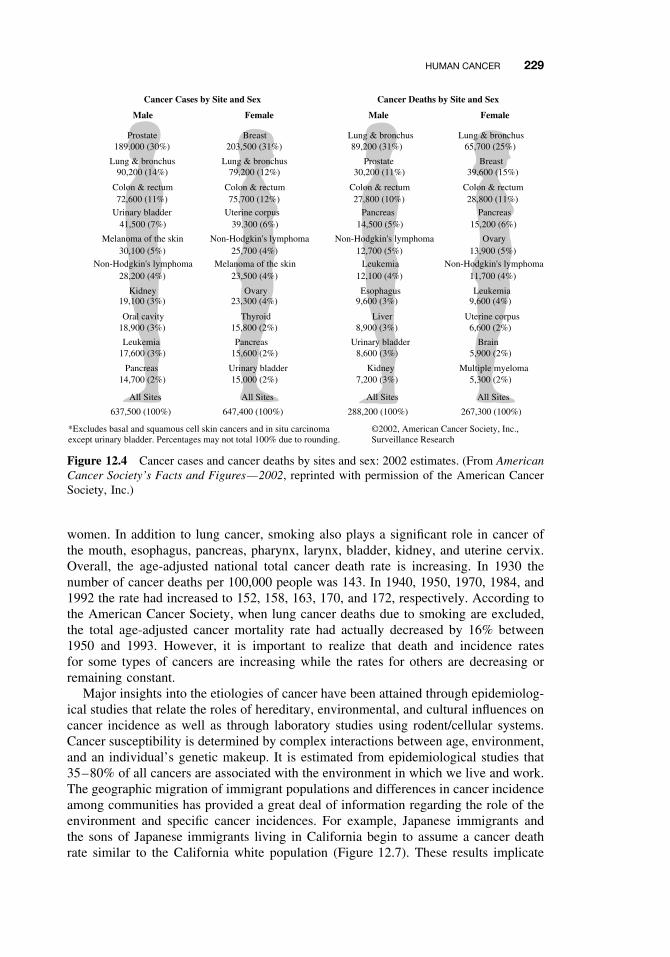
HUMAN CANCER 229
Cancer Cases by Site and Sex
Male Female
Cancer Deaths by Site and Sex
Male Female
Prostate189,000 (30%)
Lung & bronchus90,200 (14%)
Colon & rectum
Urinary bladder
Melanoma of the skin
41,500 (7%)
30,100 (5%)Non-Hodgkin's lymphoma
28,200 (4%)
19,100 (3%)
18,900 (3%)
Kidney
Oral cavity
17,600 (3%)Leukemia
14,700 (2%)
637,500 (100%)
Pancreas
All Sites
72,600 (11%)
Breast203,500 (31%)
Lung & bronchus79,200 (12%)
Colon & rectum
Uterine corpus
Non-Hodgkin's lymphoma
39,300 (6%)
25,700 (4%)Melanoma of the skin
23,500 (4%)
23,300 (4%)
15,800 (2%)
Ovary
Thyroid
15,600 (2%)Pancreas
15,000 (2%)
647,400 (100%)
Urinary bladder
All Sites
75,700 (12%)
Lung & bronchus89,200 (31%)
Prostate30,200 (11%)
Colon & rectum
Pancreas
Non-Hodgkin's lymphoma
14,500 (5%)
12,700 (5%)Leukemia
12,100 (4%)
9,600 (3%)
8,900 (3%)
Esophagus
Liver
8,600 (3%)Urinary bladder
7,200 (3%)
288,200 (100%)
Kidney
All Sites
27,800 (10%)
Lung & bronchus65,700 (25%)
Breast39,600 (15%)
Colon & rectum
Pancreas
Ovary
15,200 (6%)
13,900 (5%)Non-Hodgkin's lymphoma
11,700 (4%)
9,600 (4%)
6,600 (2%)
Leukemia
Uterine corpus
5,900 (2%)Brain
5,300 (2%)
267,300 (100%)
Multiple myeloma
All Sites
28,800 (11%)
©2002, American Cancer Society, Inc.,Surveillance Research
*Excludes basal and squamous cell skin cancers and in situ carcinomaexcept urinary bladder. Percentages may not total 100% due to rounding.
Figure 12.4 Cancer cases and cancer deaths by sites and sex: 2002 estimates. (From AmericanCancer Society’s Facts and Figures—2002, reprinted with permission of the American CancerSociety, Inc.)
women. In addition to lung cancer, smoking also plays a significant role in cancer ofthe mouth, esophagus, pancreas, pharynx, larynx, bladder, kidney, and uterine cervix.Overall, the age-adjusted national total cancer death rate is increasing. In 1930 thenumber of cancer deaths per 100,000 people was 143. In 1940, 1950, 1970, 1984, and1992 the rate had increased to 152, 158, 163, 170, and 172, respectively. According tothe American Cancer Society, when lung cancer deaths due to smoking are excluded,the total age-adjusted cancer mortality rate had actually decreased by 16% between1950 and 1993. However, it is important to realize that death and incidence ratesfor some types of cancers are increasing while the rates for others are decreasing orremaining constant.
Major insights into the etiologies of cancer have been attained through epidemiolog-ical studies that relate the roles of hereditary, environmental, and cultural influences oncancer incidence as well as through laboratory studies using rodent/cellular systems.Cancer susceptibility is determined by complex interactions between age, environment,and an individual’s genetic makeup. It is estimated from epidemiological studies that35–80% of all cancers are associated with the environment in which we live and work.The geographic migration of immigrant populations and differences in cancer incidenceamong communities has provided a great deal of information regarding the role of theenvironment and specific cancer incidences. For example, Japanese immigrants andthe sons of Japanese immigrants living in California begin to assume a cancer deathrate similar to the California white population (Figure 12.7). These results implicate
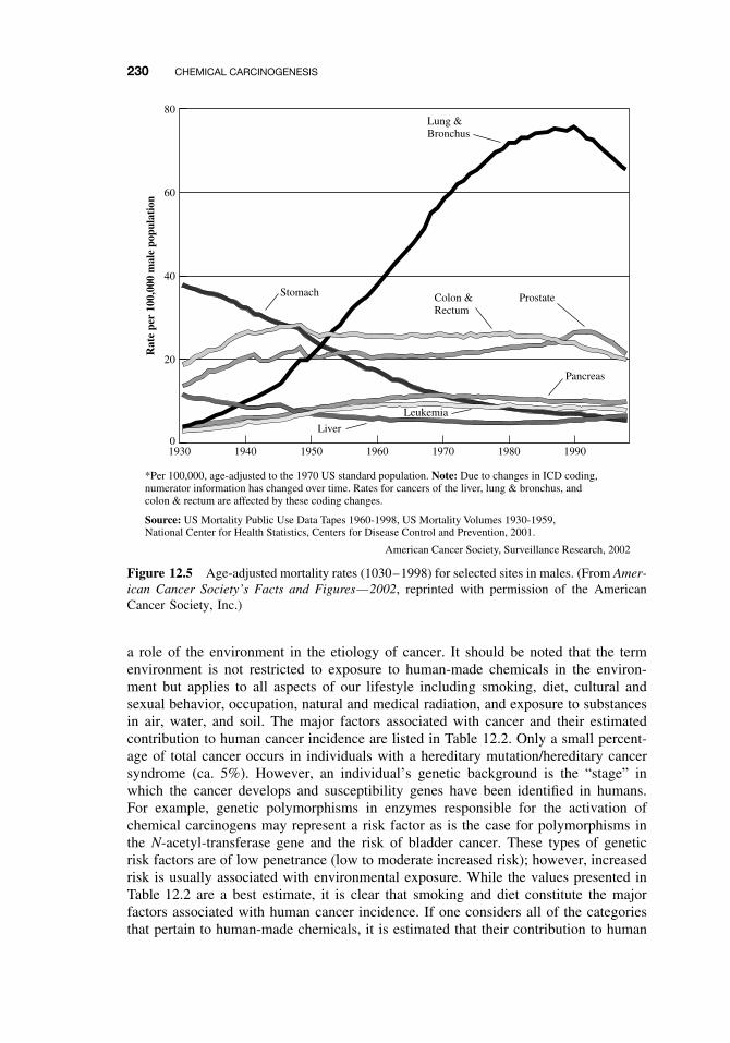
230 CHEMICAL CARCINOGENESIS
1930 1940 1950 1960 1970 1980 19900
20
40
Rat
e pe
r 10
0,00
0 m
ale
popu
lati
on
60
80Lung &Bronchus
Stomach Colon &Rectum
Prostate
Pancreas
Liver
Leukemia
*Per 100,000, age-adjusted to the 1970 US standard population. Note: Due to changes in ICD coding,numerator information has changed over time. Rates for cancers of the liver, lung & bronchus, andcolon & rectum are affected by these coding changes.
Source: US Mortality Public Use Data Tapes 1960-1998, US Mortality Volumes 1930-1959,National Center for Health Statistics, Centers for Disease Control and Prevention, 2001.
American Cancer Society, Surveillance Research, 2002
Figure 12.5 Age-adjusted mortality rates (1030–1998) for selected sites in males. (From Amer-ican Cancer Society’s Facts and Figures—2002, reprinted with permission of the AmericanCancer Society, Inc.)
a role of the environment in the etiology of cancer. It should be noted that the termenvironment is not restricted to exposure to human-made chemicals in the environ-ment but applies to all aspects of our lifestyle including smoking, diet, cultural andsexual behavior, occupation, natural and medical radiation, and exposure to substancesin air, water, and soil. The major factors associated with cancer and their estimatedcontribution to human cancer incidence are listed in Table 12.2. Only a small percent-age of total cancer occurs in individuals with a hereditary mutation/hereditary cancersyndrome (ca. 5%). However, an individual’s genetic background is the “stage” inwhich the cancer develops and susceptibility genes have been identified in humans.For example, genetic polymorphisms in enzymes responsible for the activation ofchemical carcinogens may represent a risk factor as is the case for polymorphisms inthe N-acetyl-transferase gene and the risk of bladder cancer. These types of geneticrisk factors are of low penetrance (low to moderate increased risk); however, increasedrisk is usually associated with environmental exposure. While the values presented inTable 12.2 are a best estimate, it is clear that smoking and diet constitute the majorfactors associated with human cancer incidence. If one considers all of the categoriesthat pertain to human-made chemicals, it is estimated that their contribution to human
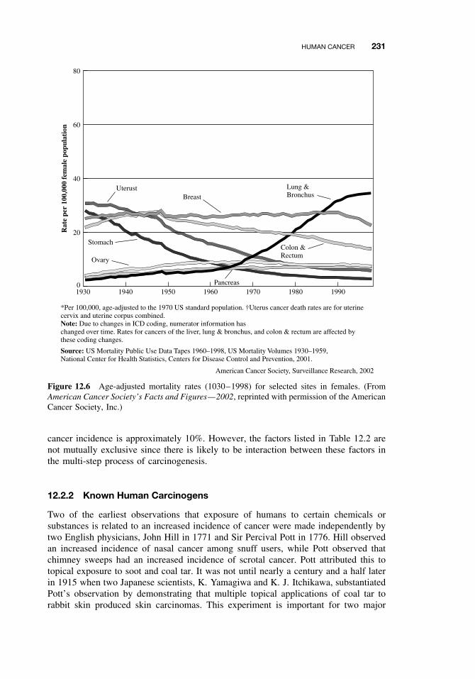
HUMAN CANCER 231
1940 1950 1960 1970 1980 1990
20
40
Rat
e pe
r 10
0,00
0 fe
mal
e po
pula
tion
60
80
Lung &Bronchus
StomachColon &Rectum
Pancreas
*Per 100,000, age-adjusted to the 1970 US standard population. †Uterus cancer death rates are for uterinecervix and uterine corpus combined.Note: Due to changes in ICD coding, numerator information haschanged over time. Rates for cancers of the liver, lung & bronchus, and colon & rectum are affected by these coding changes.
Source: US Mortality Public Use Data Tapes 1960–1998, US Mortality Volumes 1930–1959,National Center for Health Statistics, Centers for Disease Control and Prevention, 2001.
American Cancer Society, Surveillance Research, 2002
UterustBreast
Ovary
19300
Figure 12.6 Age-adjusted mortality rates (1030–1998) for selected sites in females. (FromAmerican Cancer Society’s Facts and Figures—2002, reprinted with permission of the AmericanCancer Society, Inc.)
cancer incidence is approximately 10%. However, the factors listed in Table 12.2 arenot mutually exclusive since there is likely to be interaction between these factors inthe multi-step process of carcinogenesis.
12.2.2 Known Human Carcinogens
Two of the earliest observations that exposure of humans to certain chemicals orsubstances is related to an increased incidence of cancer were made independently bytwo English physicians, John Hill in 1771 and Sir Percival Pott in 1776. Hill observedan increased incidence of nasal cancer among snuff users, while Pott observed thatchimney sweeps had an increased incidence of scrotal cancer. Pott attributed this totopical exposure to soot and coal tar. It was not until nearly a century and a half laterin 1915 when two Japanese scientists, K. Yamagiwa and K. J. Itchikawa, substantiatedPott’s observation by demonstrating that multiple topical applications of coal tar torabbit skin produced skin carcinomas. This experiment is important for two major
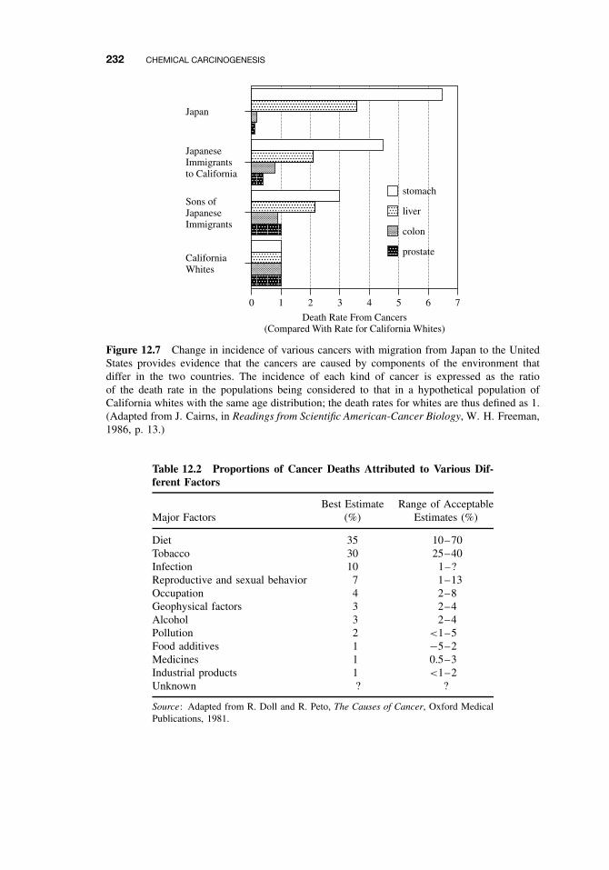
232 CHEMICAL CARCINOGENESIS
0 1 2 3
Death Rate From Cancers(Compared With Rate for California Whites)
4 5 6 7
Japan
JapaneseImmigrantsto California
Sons ofJapaneseImmigrants
CaliforniaWhites
stomach
liver
colon
prostate
Figure 12.7 Change in incidence of various cancers with migration from Japan to the UnitedStates provides evidence that the cancers are caused by components of the environment thatdiffer in the two countries. The incidence of each kind of cancer is expressed as the ratioof the death rate in the populations being considered to that in a hypothetical population ofCalifornia whites with the same age distribution; the death rates for whites are thus defined as 1.(Adapted from J. Cairns, in Readings from Scientific American-Cancer Biology, W. H. Freeman,1986, p. 13.)
Table 12.2 Proportions of Cancer Deaths Attributed to Various Dif-ferent Factors
Major FactorsBest Estimate
(%)Range of Acceptable
Estimates (%)
Diet 35 10–70Tobacco 30 25–40Infection 10 1–?Reproductive and sexual behavior 7 1–13Occupation 4 2–8Geophysical factors 3 2–4Alcohol 3 2–4Pollution 2 <1–5Food additives 1 −5–2Medicines 1 0.5–3Industrial products 1 <1–2Unknown ? ?
Source: Adapted from R. Doll and R. Peto, The Causes of Cancer, Oxford MedicalPublications, 1981.
HUMAN CANCER 233
reasons: (1) it was the first demonstration that a chemical or substance could producecancer in animals, and (2) it confirmed Pott’s initial observation and established arelationship between human epidemiology studies and animal carcinogenicity. Becauseof these important findings, Yamagiwa and Itchikawa are considered the fathers ofexperimental chemical carcinogenesis. In the 1930s Kennaway and coworkers isolateda single active carcinogenic chemical from coal tar and identified it as benzo[a]pyrene,a polycyclic aromatic hydrocarbon that results from the incomplete combustion oforganic molecules. Benzo[a]pyrene has also been identified as one of the carcinogensin cigarette smoke. The p53 tumor suppressor gene can be mutationally inactivated bynumerous carcinogens, including the carcinogenic metabolite of benzo[a]pyrene.
Epidemiological studies have provided sufficient evidence that exposure to a varietyof chemicals, agents, or processes are associated with human cancer. For example, thefollowing causal associations have emerged between exposure and the developmentof specific cancers: vinyl chloride and hepatic cancer, amine dyes and bladder can-cer, benzene and leukemia, diethylstilbestrol and clear cell carcinoma of the vagina,and cigarette smoking and lung cancer. Naturally occurring chemicals or agents suchas asbestos, aflatoxin B1, betel nut, nickel, and certain arsenic compounds are alsoassociated with an increased incidence of certain human cancers. Both epidemiolog-ical studies and rodent carcinogenicity studies are important in the identification andclassification of potential human carcinogens. The strongest evidence for establishingwhether exposure to a given chemical is carcinogenic in humans comes from epi-demiological studies. However, these studies are complicated by the fact that it oftentakes 20 to 30 years after carcinogen exposure for a clinically detectable cancer todevelop. This delay is problematic and can result in inaccurate historical exposureinformation and additional complexity due to the interference of a large number ofconfounding variables. This lag period can also prevent the timely identification of aputative carcinogen and result in unnecessary exposure. Therefore methods to identifypotential human carcinogens have been developed. The long-term rodent bioassay alsoknown as the two-year rodent carcinogenesis bioassay (see Chapter 21) is currentlyused in an attempt to identify potential human carcinogens. It is clear that almost allhuman carcinogens identified to date are rodent carcinogens; however, it is not knownif all rodent carcinogens are human carcinogens. Indeed, identification of possiblehuman carcinogens based on rodent carcinogenicity can be extremely complicated(see below). Table 12.3 contains the list of the known human carcinogens as listedby the International Agency for Research on Cancer (IARC). In addition Table 12.3includes information on carcinogenic complex mixtures and occupations associatedwith increased cancer incidence. In vitro mutagenicity assays are also used to identifymutagenic agents that may have carcinogenic activity (see Chapter 21).
12.2.3 Classification of Human Carcinogens
Identification and classification of potential human carcinogens through the two-yearrodent carcinogenesis bioassay is complicated by species differences, use of high doses(MTD, maximum tolerated dose), the short life span of the rodents, high backgroundtumor incidence in some organs, sample size, and the need to extrapolate from highto low doses for human risk assessment. Although these problems are by no meanstrivial, the rodent two-year bioassay remains the “gold standard” for the classification
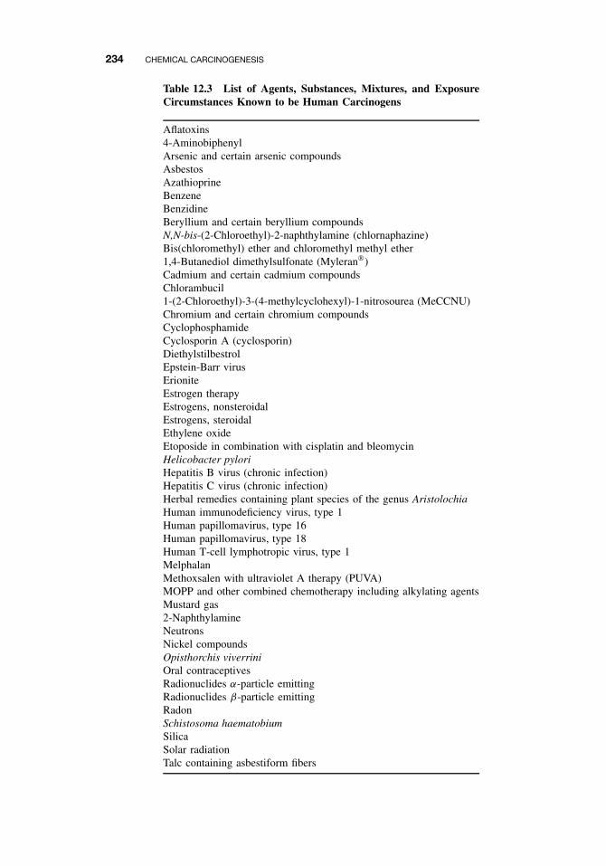
234 CHEMICAL CARCINOGENESIS
Table 12.3 List of Agents, Substances, Mixtures, and ExposureCircumstances Known to be Human Carcinogens
Aflatoxins4-AminobiphenylArsenic and certain arsenic compoundsAsbestosAzathioprineBenzeneBenzidineBeryllium and certain beryllium compoundsN,N-bis-(2-Chloroethyl)-2-naphthylamine (chlornaphazine)Bis(chloromethyl) ether and chloromethyl methyl ether1,4-Butanediol dimethylsulfonate (Myleran)Cadmium and certain cadmium compoundsChlorambucil1-(2-Chloroethyl)-3-(4-methylcyclohexyl)-1-nitrosourea (MeCCNU)Chromium and certain chromium compoundsCyclophosphamideCyclosporin A (cyclosporin)DiethylstilbestrolEpstein-Barr virusErioniteEstrogen therapyEstrogens, nonsteroidalEstrogens, steroidalEthylene oxideEtoposide in combination with cisplatin and bleomycinHelicobacter pyloriHepatitis B virus (chronic infection)Hepatitis C virus (chronic infection)Herbal remedies containing plant species of the genus AristolochiaHuman immunodeficiency virus, type 1Human papillomavirus, type 16Human papillomavirus, type 18Human T-cell lymphotropic virus, type 1MelphalanMethoxsalen with ultraviolet A therapy (PUVA)MOPP and other combined chemotherapy including alkylating agentsMustard gas2-NaphthylamineNeutronsNickel compoundsOpisthorchis viverriniOral contraceptivesRadionuclides α-particle emittingRadionuclides β-particle emittingRadonSchistosoma haematobiumSilicaSolar radiationTalc containing asbestiform fibers
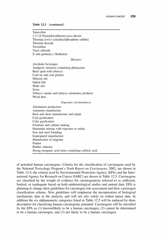
HUMAN CANCER 235
Table 12.3 (continued )
Tamoxifen2,3,7,8-Tetrachlorodibenzo-para-dioxinThiotepa [tris(1-aziridinyl)phosphine sulfide]Thorium dioxideTreosulfanVinyl chlorideX and gamma(γ ) Radiation
Mixtures
Alcoholic beveragesAnalgesic mixtures containing phenacetinBetel quid with tobaccoCoal tar and coal pitchesMineral oilsSalted fishShale oilsSootsTobacco smoke and tobacco smokeless productsWood dust
Exposure circumstances
Aluminium productionAuramine manufactureBoot and show manufacture and repairCoal gasificationCoke gasificationFurniture and cabinet makingHaematite mining with exposure to radonIron and steel foundingIsopropanol manufactureManufacture of magentaPainterRubber industryStrong inorganic acid mists containing sulfuric acid
of potential human carcinogens. Criteria for the classification of carcinogens used bythe National Toxicology Program’s Tenth Report on Carcinogens, 2002, are shown inTable 12.4; the criteria used by Environmental Protection Agency (EPA) and the Inter-national Agency for Research on Cancer (IARC) are shown in Table 12.5. Carcinogensare classified by the weight of evidence for carcinogenicity referred to as sufficient,limited, or inadequate based on both epidemiological studies and animal data. EPA isplanning to change their guidelines for carcinogen risk assessment and their carcinogenclassification scheme. New guidelines will emphasize the incorporation of biologicalmechanistic data in the analysis, and will not rely solely on rodent tumor data. Inaddition the six alphanumeric categories listed in Table 12.5 will be replaced by threedescriptors for classifying human carcinogenic potential. Carcinogens will be classifiedby the EPA as (1) known/likely to be a human carcinogen, (2) cannot be determinedto be a human carcinogen, and (3) not likely to be a human carcinogen.
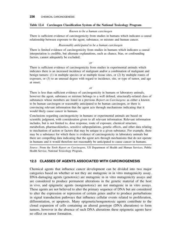
236 CHEMICAL CARCINOGENESIS
Table 12.4 Carcinogen Classification System of the National Toxicology Program
Known to be a human carcinogen
There is sufficient evidence of carcinogenicity from studies in humans which indicates a causalrelationship between exposure to the agent, substance, or mixture and human cancer.
Reasonably anticipated to be a human carcinogen
There is limited evidence of carcinogenicity from studies in humans which indicates a causalinterpretation is credible, but alternate explanations, such as chance, bias, or confoundingfactors, cannot adequately be excluded;
or
There is sufficient evidence of carcinogenicity from studies in experimental animals whichindicates there is an increased incidence of malignant and/or a combination of malignant andbenign tumors: (1) in multiple species or at multiple tissue sites, or (2) by multiple routes ofexposure, or (3) to an unusual degree with regard to incidence, site, or type of tumor, and ageat onset;
or
There is less than sufficient evidence of carcinogenicity in humans or laboratory animals,however the agent, substance or mixture belongs to a well defined, structurally-related class ofsubstances whose members are listed in a previous Report on Carcinogens as either a knownto be human carcinogen or reasonably anticipated to be human carcinogen, or there isconvincing relevant information that the agent acts through mechanisms indicating that itwould likely cause cancer in humans.
Conclusions regarding carcinogenicity in humans or experimental animals are based onscientific judgment, with consideration given to all relevant information. Relevant informationincludes, but is not limited to, dose response, route of exposure, chemical structure,metabolism, pharmacokinetics, sensitive subpopulations, genetic effects, and other data relatingto mechanism of action or factors that may be unique to a given substance. For example, theremay be a substance for which there is evidence of carcinogenicity in laboratory animals butthere are compelling data indicating that the agent acts through mechanisms that do not operatein humans and it would therefore not reasonably be anticipated to cause cancer in humans.
Source: From the Tenth Report on Carcinogens, US Department of Health and Human Services, PublicHealth Service, National Toxicology Program.
12.3 CLASSES OF AGENTS ASSOCIATED WITH CARCINOGENESIS
Chemical agents that influence cancer development can be divided into two majorcategories based on whether or not they are mutagenic in in vitro mutagenicity assay.DNA-damaging agents (genotoxic) are mutagenic in in vitro mutagenicity assays andare considered to produce permanent alterations in the genetic material of the hostin vivo, and epigenetic agents (nongenotoxic) are not mutagenic in in vitro assays.These agents are not believed to alter the primary sequence of DNA but are consideredto alter the expression or repression of certain genes and/or to produce perturbationsin signal transduction pathways that influence cellular events related to proliferation,differentiation, or apoptosis. Many epigenetic/nongenotoxic agents contribute to theclonal expansion of cells containing an altered genotype (DNA alterations) to formtumors, however in the absence of such DNA alterations these epigenetic agents haveno effect on tumor formation.
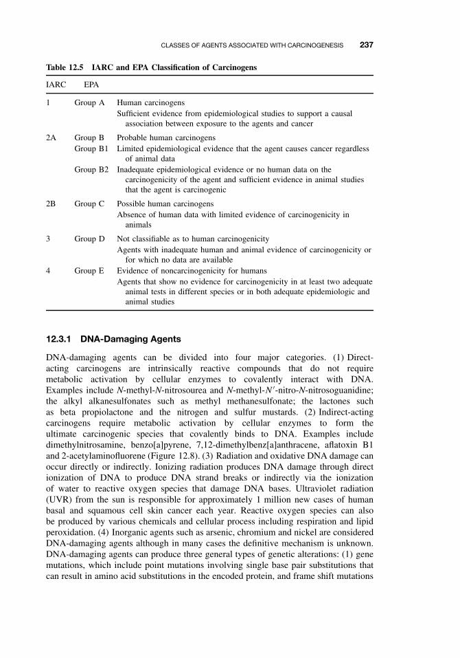
CLASSES OF AGENTS ASSOCIATED WITH CARCINOGENESIS 237
Table 12.5 IARC and EPA Classification of Carcinogens
IARC EPA
1 Group A Human carcinogensSufficient evidence from epidemiological studies to support a causal
association between exposure to the agents and cancer
2A Group B Probable human carcinogensGroup B1 Limited epidemiological evidence that the agent causes cancer regardless
of animal dataGroup B2 Inadequate epidemiological evidence or no human data on the
carcinogenicity of the agent and sufficient evidence in animal studiesthat the agent is carcinogenic
2B Group C Possible human carcinogensAbsence of human data with limited evidence of carcinogenicity in
animals
3 Group D Not classifiable as to human carcinogenicityAgents with inadequate human and animal evidence of carcinogenicity or
for which no data are available4 Group E Evidence of noncarcinogenicity for humans
Agents that show no evidence for carcinogenicity in at least two adequateanimal tests in different species or in both adequate epidemiologic andanimal studies
12.3.1 DNA-Damaging Agents
DNA-damaging agents can be divided into four major categories. (1) Direct-acting carcinogens are intrinsically reactive compounds that do not requiremetabolic activation by cellular enzymes to covalently interact with DNA.Examples include N-methyl-N-nitrosourea and N-methyl-N ′-nitro-N-nitrosoguanidine;the alkyl alkanesulfonates such as methyl methanesulfonate; the lactones suchas beta propiolactone and the nitrogen and sulfur mustards. (2) Indirect-actingcarcinogens require metabolic activation by cellular enzymes to form theultimate carcinogenic species that covalently binds to DNA. Examples includedimethylnitrosamine, benzo[a]pyrene, 7,12-dimethylbenz[a]anthracene, aflatoxin B1and 2-acetylaminofluorene (Figure 12.8). (3) Radiation and oxidative DNA damage canoccur directly or indirectly. Ionizing radiation produces DNA damage through directionization of DNA to produce DNA strand breaks or indirectly via the ionizationof water to reactive oxygen species that damage DNA bases. Ultraviolet radiation(UVR) from the sun is responsible for approximately 1 million new cases of humanbasal and squamous cell skin cancer each year. Reactive oxygen species can alsobe produced by various chemicals and cellular process including respiration and lipidperoxidation. (4) Inorganic agents such as arsenic, chromium and nickel are consideredDNA-damaging agents although in many cases the definitive mechanism is unknown.DNA-damaging agents can produce three general types of genetic alterations: (1) genemutations, which include point mutations involving single base pair substitutions thatcan result in amino acid substitutions in the encoded protein, and frame shift mutations
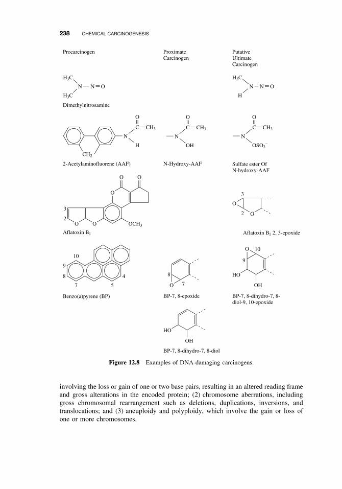
238 CHEMICAL CARCINOGENESIS
10
9
8
7 5
O O
O
OO
OCH3
3
2
OH
HO
OH
HO
O
O
O
N
C
OSO3−
CH3
O
N
C
OH
CH3
O
N N O
H3C
H3C
N N O
H
H3C
CH2
N
C
H
O
CH3
4
Procarcinogen
Dimethylnitrosamine
2-Acetylaminofluorene (AAF)
Aflatoxin B1
Benzo(a)pyrene (BP)
N-Hydroxy-AAF
O 7
8
BP-7, 8-epoxide
BP-7, 8-dihydro-7, 8-diol
ProximateCarcinogen
PutativeUltimateCarcinogen
Sulfate ester OfN-hydroxy-AAF
3
2
Aflatoxin B1 2, 3-epoxide
BP-7, 8-dihydro-7, 8-diol-9, 10-epoxide
9
10
Figure 12.8 Examples of DNA-damaging carcinogens.
involving the loss or gain of one or two base pairs, resulting in an altered reading frameand gross alterations in the encoded protein; (2) chromosome aberrations, includinggross chromosomal rearrangement such as deletions, duplications, inversions, andtranslocations; and (3) aneuploidy and polyploidy, which involve the gain or loss ofone or more chromosomes.
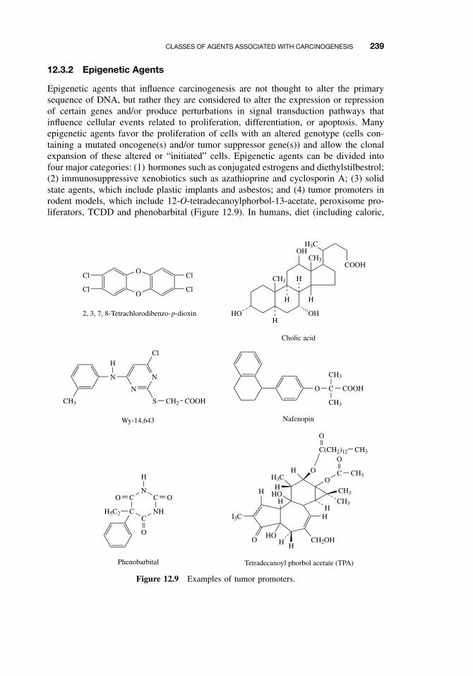
CLASSES OF AGENTS ASSOCIATED WITH CARCINOGENESIS 239
12.3.2 Epigenetic Agents
Epigenetic agents that influence carcinogenesis are not thought to alter the primarysequence of DNA, but rather they are considered to alter the expression or repressionof certain genes and/or produce perturbations in signal transduction pathways thatinfluence cellular events related to proliferation, differentiation, or apoptosis. Manyepigenetic agents favor the proliferation of cells with an altered genotype (cells con-taining a mutated oncogene(s) and/or tumor suppressor gene(s)) and allow the clonalexpansion of these altered or “initiated” cells. Epigenetic agents can be divided intofour major categories: (1) hormones such as conjugated estrogens and diethylstilbestrol;(2) immunosuppressive xenobiotics such as azathioprine and cyclosporin A; (3) solidstate agents, which include plastic implants and asbestos; and (4) tumor promoters inrodent models, which include 12-O-tetradecanoylphorbol-13-acetate, peroxisome pro-liferators, TCDD and phenobarbital (Figure 12.9). In humans, diet (including caloric,
O
I3C
H
HHHO
CH2OH
H
CH3
CH3H
O
HH3C
H
HHO
OC CH3
OC(CH2)12 CH3
O
Tetradecanoyl phorbol acetate (TPA)
CC
NH
CN
C
O
OO
H
H5C2
Phenobarbital
CH3
N
N
N
HCl
S CH2 COOH
O
OCl
Cl
Cl
Cl
2, 3, 7, 8-Tetrachlorodibenzo-p-dioxin
O C COOH
CH3
CH3
Nafenopin
H3COH
COOH
HOH
CH3 H
H
OH
H
CH3
Cholic acid
Wy-14,643
Figure 12.9 Examples of tumor promoters.
240 CHEMICAL CARCINOGENESIS
fat, and protein intake), excess alcohol, and late age of pregnancy are considered tofunction through a promotion mechanism. While smoking and UVR have initiatingactivity, both are also considered to have tumor-promoting activity. By definition,tumor promoters are not classified as carcinogens since they are considered inactivein the absence of initiated cells. However, an altered genotype or an initiated cellcan arise from spontaneous mutations resulting from imperfect DNA replication/repairoxidative DNA damage, or can result from environmental carcinogens. Theoretically,in the presence of a tumor promoter these mutant cells would clonally expand to forma tumor. Therefore the nomenclature becomes somewhat a matter of semantics as towhether the tumor promoter should or should not be classified as a carcinogen. Certainhormones and immunosuppressive agents are classified as human carcinogens, althoughit is generally considered that these agents are not carcinogenic in the absence of initi-ated cells. Rather, like tumor promoters, they may only allow for the clonal expansionof cells with an altered genotype.
Some nongenotoxic/epigenetic agents have been shown to induce DNA damagein vivo, for example, some “nongenotoxic/epigenetic agents” can induce oxidativeDNA damage in vivo through the direct or indirect production of reactive oxygenspecies. For example, certain estrogens may possess this ability and such a character-istic may contribute to their carcinogenicity. Thus, as we gain a better understandingof chemical carcinogenesis, we find that there is functional and mechanistic overlapand interaction between these two major categories of chemical carcinogens.
12.4 GENERAL ASPECTS OF CHEMICAL CARCINOGENESIS
A great deal of evidence has accumulated in support of the somatic mutation theory ofcarcinogenesis, which simply states that mutations within somatic cells is necessary forneoplasia. As stated earlier, cancer development (carcinogenesis) involves the accu-mulation of mutations in multiple critical genes. These mutations can be the result ofimperfect DNA replication/repair, oxidative DNA damage, and/or DNA damage causedby environmental carcinogens. Many chemical carcinogens can alter DNA throughcovalent interaction (DNA adducts) or direct and/or indirect oxidative DNA dam-age. Some chemical carcinogens are intrinsically reactive and can directly covalentlybind to DNA, while others require metabolic via cytochromes P450 to produce reac-tive electrophilic intermediates capable of covalently binding to DNA (Figure 12.10).In the 1950s Elizabeth and James Miller observed that a diverse array of chemicalscould produce cancer in rodents. In an attempt to explain this, they hypothesizedthat many carcinogens are metabolically activated to electrophilic metabolites that arecapable of interacting with nucleophilic sites in DNA. The Millers termed this theelectrophilic theory of chemical carcinogenesis. From this concept of metabolic acti-vation, the important terms parent, proximate, and ultimate carcinogen were developed.A parent carcinogen is a compound that must be metabolized in order to have carcino-genic activity; a proximate carcinogen is an intermediate metabolite requiring furthermetabolism and resulting in the ultimate carcinogen, which is the actual metabolite thatcovalently binds to the DNA. The cell has many defense systems to detoxify the car-cinogenic species, including cellular antioxidants and nucleophiles as well as a wholehost of phase I and phase II enzymes. In addition reactive carcinogenic species maybind to noncritical sites in the cell, resulting in detoxification, or they can undergo spon-taneous decomposition. If the carcinogenic species binds to DNA, the adducted DNA
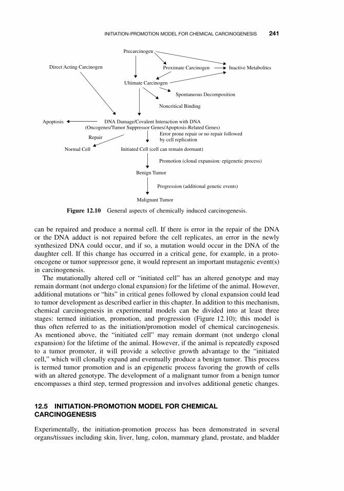
INITIATION-PROMOTION MODEL FOR CHEMICAL CARCINOGENESIS 241
Malignant Tumor
Direct Acting Carcinogen
Precarcinogen
Proximate Carcinogen Inactive Metabolites
Ultimate Carcinogen
Spontaneous Decomposition
Noncritical Binding
Apoptosis DNA Damage/Covalent Interaction with DNA(Oncogenes/Tumor Suppressor Genes/Apoptosis-Related Genes)
RepairError prone repair or no repair followedby cell replication
Normal Cell Initiated Cell (cell can remain dormant)
Promotion (clonal expansion: epigenetic process)
Benign Tumor
Progression (additional genetic events)
Figure 12.10 General aspects of chemically induced carcinogenesis.
can be repaired and produce a normal cell. If there is error in the repair of the DNAor the DNA adduct is not repaired before the cell replicates, an error in the newlysynthesized DNA could occur, and if so, a mutation would occur in the DNA of thedaughter cell. If this change has occurred in a critical gene, for example, in a proto-oncogene or tumor suppressor gene, it would represent an important mutagenic event(s)in carcinogenesis.
The mutationally altered cell or “initiated cell” has an altered genotype and mayremain dormant (not undergo clonal expansion) for the lifetime of the animal. However,additional mutations or “hits” in critical genes followed by clonal expansion could leadto tumor development as described earlier in this chapter. In addition to this mechanism,chemical carcinogenesis in experimental models can be divided into at least threestages: termed initiation, promotion, and progression (Figure 12.10); this model isthus often referred to as the initiation/promotion model of chemical carcinogenesis.As mentioned above, the “initiated cell” may remain dormant (not undergo clonalexpansion) for the lifetime of the animal. However, if the animal is repeatedly exposedto a tumor promoter, it will provide a selective growth advantage to the “initiatedcell,” which will clonally expand and eventually produce a benign tumor. This processis termed tumor promotion and is an epigenetic process favoring the growth of cellswith an altered genotype. The development of a malignant tumor from a benign tumorencompasses a third step, termed progression and involves additional genetic changes.
12.5 INITIATION-PROMOTION MODEL FOR CHEMICALCARCINOGENESIS
Experimentally, the initiation-promotion process has been demonstrated in severalorgans/tissues including skin, liver, lung, colon, mammary gland, prostate, and bladder
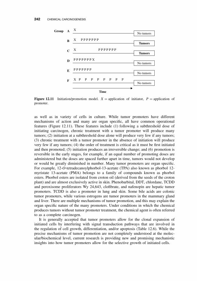
242 CHEMICAL CARCINOGENESIS
No tumorsXA
TumorsXB
TumorsXC
No tumorsP P P P P P P XD
No tumorsP P P P P P PE
No tumorsX P P P P P P P PF
Group
Time
P P P P P P P
P P P P P P P
Figure 12.11 Initiation/promotion model. X = application of initiator, P = application ofpromoter.
as well as in variety of cells in culture. While tumor promoters have differentmechanisms of action and many are organ specific, all have common operationalfeatures (Figure 12.11). These features include (1) following a subthreshold dose ofinitiating carcinogen, chronic treatment with a tumor promoter will produce manytumors; (2) initiation at a subthreshold dose alone will produce very few if any tumors;(3) chronic treatment with a tumor promoter in the absence of initiation will producevery few if any tumors; (4) the order of treatment is critical as it must be first initiatedand then promoted; (5) initiation produces an irreversible change; and (6) promotion isreversible in the early stages, for example, if an equal number of promoting doses areadministered but the doses are spaced further apart in time, tumors would not developor would be greatly diminished in number. Many tumor promoters are organ specific.For example, 12-O-tetradecanoylphorbol-13-acetate (TPA) also known as phorbol 12-myristate 13-acetate (PMA) belongs to a family of compounds known as phorbolesters. Phorbol esters are isolated from croton oil (derived from the seeds of the crotonplant) and are almost exclusively active in skin. Phenobarbital, DDT, chlordane, TCDDand peroxisome proliferators Wy 24,643, clofibrate, and nafenopin are hepatic tumorpromoters. TCDD is also a promoter in lung and skin. Some bile acids are colonictumor promoters, while various estrogens are tumor promoters in the mammary glandand liver. There are multiple mechanisms of tumor promotion, and this may explain theorgan specific nature of the many promoters. Under conditions in which the chemicalproduces tumors without tumor promoter treatment, the chemical agent is often referredto as a complete carcinogen.
It is generally accepted that tumor promoters allow for the clonal expansion ofinitiated cells by interfering with signal transduction pathways that are involved inthe regulation of cell growth, differentiation, and/or apoptosis (Table 12.6). While theprecise mechanisms of tumor promotion are not completely understood at the molec-ular/biochemical level, current research is providing new and promising mechanisticinsights into how tumor promoters allow for the selective growth of initiated cells.
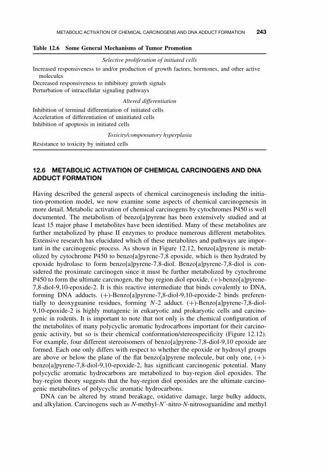
METABOLIC ACTIVATION OF CHEMICAL CARCINOGENS AND DNA ADDUCT FORMATION 243
Table 12.6 Some General Mechanisms of Tumor Promotion
Selective proliferation of initiated cells
Increased responsiveness to and/or production of growth factors, hormones, and other activemolecules
Decreased responsiveness to inhibitory growth signalsPerturbation of intracellular signaling pathways
Altered differentiation
Inhibition of terminal differentiation of initiated cellsAcceleration of differentiation of uninitiated cellsInhibition of apoptosis in initiated cells
Toxicity/compensatory hyperplasia
Resistance to toxicity by initiated cells
12.6 METABOLIC ACTIVATION OF CHEMICAL CARCINOGENS AND DNAADDUCT FORMATION
Having described the general aspects of chemical carcinogenesis including the initia-tion-promotion model, we now examine some aspects of chemical carcinogenesis inmore detail. Metabolic activation of chemical carcinogens by cytochromes P450 is welldocumented. The metabolism of benzo[a]pyrene has been extensively studied and atleast 15 major phase I metabolites have been identified. Many of these metabolites arefurther metabolized by phase II enzymes to produce numerous different metabolites.Extensive research has elucidated which of these metabolites and pathways are impor-tant in the carcinogenic process. As shown in Figure 12.12, benzo[a]pyrene is metab-olized by cytochrome P450 to benzo[a]pyrene-7,8 epoxide, which is then hydrated byepoxide hydrolase to form benzo[a]pyrene-7,8-diol. Benzo[a]pyrene-7,8-diol is con-sidered the proximate carcinogen since it must be further metabolized by cytochromeP450 to form the ultimate carcinogen, the bay region diol epoxide, (+)-benzo[a]pyrene-7,8-diol-9,10-epoxide-2. It is this reactive intermediate that binds covalently to DNA,forming DNA adducts. (+)-Benzo[a]pyrene-7,8-diol-9,10-epoxide-2 binds preferen-tially to deoxyguanine residues, forming N -2 adduct. (+)-Benzo[a]pyrene-7,8-diol-9,10-epoxide-2 is highly mutagenic in eukaryotic and prokaryotic cells and carcino-genic in rodents. It is important to note that not only is the chemical configuration ofthe metabolites of many polycyclic aromatic hydrocarbons important for their carcino-genic activity, but so is their chemical conformation/stereospecificity (Figure 12.12).For example, four different stereoisomers of benzo[a]pyrene-7,8-diol-9,10 epoxide areformed. Each one only differs with respect to whether the epoxide or hydroxyl groupsare above or below the plane of the flat benzo[a]pyrene molecule, but only one, (+)-benzo[a]pyrene-7,8-diol-9,10-epoxide-2, has significant carcinogenic potential. Manypolycyclic aromatic hydrocarbons are metabolized to bay-region diol epoxides. Thebay-region theory suggests that the bay-region diol epoxides are the ultimate carcino-genic metabolites of polycyclic aromatic hydrocarbons.
DNA can be altered by strand breakage, oxidative damage, large bulky adducts,and alkylation. Carcinogens such as N-methyl-N ′-nitro-N-nitrosoguanidine and methyl
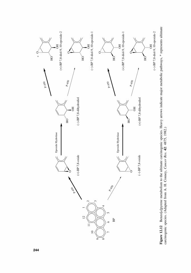
1
2 3
489
1011
12
76
5
O
HO
OH
O
HO
OH
O
HO
OH
O
HO
OH
(+)-
BP
7,8-
diol
-9, 1
0-ep
oxid
e-2
(−)-
BP
7,8-
diol
-9, 1
0-ep
oxid
e-1
(+)-
BP
7,8-
diol
-9, 1
0-ep
oxid
e-1
(−)-
BP
7,8-
diol
-9, 1
0-ep
oxid
e-2
(+)-
BP
7,8-
oxid
e
O
(−)-
BP
7,8-
dihy
drod
iol
HO
OH
Epo
xide
Hyd
rola
se
P-45
0
(−)-
BP
7,8-
oxid
e
O
Epo
xide
Hyd
rola
se
(+)-
BP
7,8-
dihy
drod
iol
HO
OH
P-45
0
P-45
0
P-45
0
P-45
0
P-45
0B
P
*
Fig
ure
12.1
2B
enzo
[a]p
yren
em
etab
olis
mto
the
ultim
ate
carc
inog
enic
spec
ies.
Hea
vyar
row
sin
dica
tem
ajor
met
abol
icpa
thw
ays,
*re
pres
ents
ultim
ate
carc
inog
enic
spec
ies.
(Ada
pted
from
A.
H.
Con
ney,
Can
cer
Res
.42
:48
75,
1982
.)
244
ONCOGENES 245
methanesulfonate alkylate DNA to produce N-alkylated and O-alkylated purines andpyrimidines. Ionizing radiation and reactive oxygen species commonly oxidize guanineto produce 8-oxoguanine. Formation of DNA adducts may involve any of the bases,although the N -7 position of guanine is one the most nucleophilic sites in DNA. Ofimportance is how long the adduct is retained in the DNA. (+)-Benzo[a]pyrene-7,8-diol-9,10-epoxide-2 forms adducts mainly at guanine N -2, while aflatoxin B1 epoxide,another well-studied rodent and human carcinogen, binds preferentially to the N -7position of guanine. For some carcinogens there is a strong correlation between theformation of very specific DNA-adducts and tumorigenicity. Quantitation and iden-tification of specific carcinogen adducts may be useful as biomarkers of exposure.Importantly, the identification of specific DNA-adducts has allowed for the predictionof specific point mutations that would likely occur in the daughter cell provided thatthere was no repair of the DNA-adduct in the parent cell. As will be discussed in a latersection, some of these expected mutations have been identified in specific oncogenesand tumor suppressor genes in chemically induced rodent tumors, providing supportthat the covalent carcinogen binding produced the observed mutation. In several cases,specific base pair changes in p53 tumor suppressor gene in human tumors are associ-ated with a mutational spectrum that is consistent with exposure of the individual toa specific carcinogen. For example, the mutation spectra identified in p53 in humantumors thought to result from the exposure of the individual to ultraviolet radiation(UVR), aflatoxin, and benzo[a]pyrene (from cigarette smoke), are consistent with theobserved specific mutational damage in p53 induced by these agents in experimentalcellular systems.
12.7 ONCOGENES
12.7.1 Mutational Activation of Proto-oncogenes
Much evidence has accumulated for a role of covalent binding of reactive electrophiliccarcinogens to DNA in chemical carcinogenesis. It is known that chemical mutagensand carcinogens can produce point mutations, frameshift mutations, strand breaks, andchromosome aberrations in mammalian cells. If the interaction of a chemical carcino-gen with DNA leading to a permanent alteration in the DNA is a critical event inchemical carcinogenesis, then the identification of these altered genes and the functionof their protein products is essential to our understanding of chemical carcinogene-sis. While specific DNA-carcinogen adducts were isolated in the 1970s and 1980s, itwas not until the early to mid-1980s that the identification of specific genes that weremutationally altered by chemical carcinogens became known. Certain normal cellulargenes, termed proto-oncogenes, can be mutated by chemical carcinogens providing aselective growth advantage to the cell. The mutational activation of proto-oncogenesis strongly associated with tumor formation, carcinogenesis, and cell transformation.Proto-oncogenes are highly conserved in evolution and their expression is tightly reg-ulated. Their protein products function in the control of normal cellular proliferation,differentiation, and apoptosis. However, when these genes are altered by a mutation,chromosome translocation, gene amplification, or promoter insertion, an abnormal pro-tein product or an abnormal amount of product is produced. Under these circumstancesthese genes have the ability to transform cells in vitro, and they are termed oncogenes.
246 CHEMICAL CARCINOGENESIS
Table 12.7 Oncogene Classification
Families Genes
Growth factors sis, hst-1, int-2, wnt-1Growth factor receptor tyrosine kinases EGFR, fms, met/HGFR, ErbB2/neu/HER2, trk/NGFRNonreceptor tyrosine kinases abl, src, fgr, fes, yes, lckGuanine nucleotide binding proteins H-ras, K-ras, N-ras, TC21, GA12
Serine/threonine kinases mos, raf, bcr, pim-1DNA-binding proteins myc, fos, myb, jun, E2F1, ets, rel
Over a 100 oncogenes have been identified with approximately 30 oncogenes havinga major role in human cancer.
Most oncogene protein products appear to function in one way or another in cellularsignal transduction pathways that are involved in regulating cell growth, differentiationor apoptosis. Signal transduction pathways are used by the cells to receive and processinformation to ultimately produce a biological cellular response. These pathways arethe cellular circuitry conveying specific information from the outside of the cell to thenucleus. In the nucleus, specific genes are expressed, and their encoded proteins pro-duce the evoked biological response. Oncogenes encode proteins that are componentsof this cellular circuitry (Table 12.7). If a component of the circuit is altered, then theentire cellular circuit of which the component is a part is altered. It is not difficult toimagine how an alteration in a pathway that regulates cellular growth, differentiation,or apoptosis could have very profound effects on cellular homeostasis. Indeed, this isthe molecular basis of how oncogenes contribute to the cancer process.
12.7.2 Ras Oncogene
Ras genes are frequently mutated in chemically induced animal tumors and are the mostfrequently detected mutated oncogenes in human tumors. Approximately 20–30% ofall human tumors contain mutated ras. The Ras subfamily includes H-ras, K-ras, andN-ras, and all have been found to be mutationally activated in numerous types oftumors from a large variety of species including humans.
Activated ras oncogenes have been detected in a large number of animal tumorsinduced by diverse agents including physical agents, such as radiation, and a largenumber of chemical carcinogens. Some chemical carcinogens bind covalently to DNA,forming specific adducts which upon DNA replication yields characteristic alterationsin the primary sequence of the Ha-ras proto-oncogene. The study of the ras onco-gene as a target for chemical carcinogens has revealed a correlation between specificcarcinogen-DNA adducts and specific activating mutations of ras in chemically inducedtumors. For example, 7,12-dimethylbenz[a]anthracene, a polycyclic aromatic hydro-carbon carcinogen, is metabolically activated to a bay-region diol epoxide that bindspreferentially to adenine residues in DNA. Skin tumors isolated from mice treatedwith DMBA contain an activated H-ras oncogene with an A to T transversion of themiddle base in the 61st codon of H-ras. Therefore the identified mutation in ras isconsistent with the expected mutation based on the DMBA-DNA adducts which havebeen identified. Likewise rat mammary carcinomas induced by nitrosomethylurea con-tain a G to A transition in the 12th codon of H-ras , and this mutation is consistent
TUMOR SUPPRESSOR GENES 247
with the modification of guanine residues by this carcinogen. Based on these events,the alteration of ras by specific chemical carcinogens appears to be an early event incarcinogenesis.
Ras proteins function as membrane-associated molecular switches operating down-stream of a variety of membrane receptors. Ras is in the off position when it is boundto guanosine diphosphate (GDP). However, when stimulated by a growth factor recep-tor, Ras exchanges GTP guanosine triphosphate for GDP, and now Ras is in the onposition. Ras communicates this “on” message to the next protein in the signalingcircuitry, which through a kinase cascade ultimately results in the activation of sev-eral transcription factors. These transcription factors regulate the expression of genesinvolved in cell proliferation, for example. Once Ras has conveyed the “on” message,it turns itself off. Ras has intrinsic GTPase activity that hydrolyzes GTP to form GDP,and Ras is once again in the off position. Another protein, termed GAPp120 (GTPaseactivating protein), aids Ras in GTP hydrolysis. When ras is mutated in certain codons,including the 12th, 13th, or 61st codon, the intrinsic GTPase activity of Ras is greatlydiminished as is its ability to interact with GAP. The net effect is that mutated Ras isessentially stuck in the “on” position continually sending a proliferative signal to thedownstream circuitry.
12.8 TUMOR SUPPRESSOR GENES
12.8.1 Inactivation of Tumor Suppressor Genes
Activation of oncogenes results in a gain of function while inactivation of tumorsuppressor genes results in a loss of function. Tumor suppressor genes have also beentermed anti-oncogenes, recessive oncogenes, and growth suppressor genes. Tumor sup-pressor genes encode proteins that generally function as negative regulators of cellgrowth or regulators of cell death. In addition some tumor suppressor genes func-tion in DNA repair and cell adhesion. The majority of tumor suppressor genes werefirst identified in rare familial cancer syndromes, and some are frequently mutated insporadic cancers through somatic mutation. There are approximately 18 known tumorsuppressor genes (e.g., p53, Rb, APC, p16, and BRCA1) that have been shown to havea role in cancer and another 12 putative tumor suppressors have been identified. Whentumor suppressor genes are inactivated by allelic loss, point mutation, or chromosomedeletion, they are no longer capable of negatively regulating cellular growth leading tospecific forms of cancer predisposition. Generally, if one copy or allele of the tumorsuppressor gene is inactivated, the cell is normal, and if both copies or alleles areinactivated, loss of growth control occurs. In some cases a single mutant allele ofcertain tumor suppressor genes, such as p53, can give rise to an altered intermediatephenotype. However, inactivation of both alleles is required for full loss of functionand the transformed phenotype.
12.8.2 p53 Tumor Suppressor Gene
p53 encodes a 53 kDa protein. p53 is mutated in 50% of all human cancer and isthe most frequently known mutated gene in human cancer. The majority (ca. 80%) ofp53 mutations are missense mutations and p53 is mutated in approximately 70% of
248 CHEMICAL CARCINOGENESIS
colon cancers, 50% of breast and lung cancers, and 97% of primary melanomas. Inaddition to point mutations, allelic loss, rearrangements, and deletions of p53 occurin human tumors. p53 is a transcription factor and participates in many cellular func-tions, including cell cycle regulation, DNA repair, and apoptosis. The p53 protein iscomposed of 393 amino acids, and single missense mutations can inactivate the p53.Unlike ras genes, which have a few mutational codons that result in its activation, thep53 protein can be inactivated by hundreds of different single-point mutations in p53.It has been proposed that the mutation spectrum of p53 in human cancer can aid in theidentification of the specific carcinogen that is responsible for the genetic damage; thatis to say, different carcinogens cause different characteristic mutations in p53. Someof the mutations in p53 reflect endogenous oxidative damage, while others such asthe mutational spectrum in p53 in hepatocellular carcinomas from individuals exposedto aflatoxin demonstrate a mutation spectrum characteristic aflatoxin. In sun-exposedareas where skin tumors develop, the mutations found in p53 in these tumors are char-acteristic of UV light induced pyrimidine dimers, and finally the mutation spectruminduced by (+)-benzo[a]pyrene-7,8-diol-9,10-epoxide-2 in cells in culture is similar tothe mutational spectrum in p53 in lung tumors form cigarette smokers. Thus certaincarcinogens produce a molecular signature that may provide important information inunderstanding the etiology of tumor development.
p53 has been termed the “guardian of genome” because it controls a G1/S check-point, regulates DNA repair, and apoptosis. DNA damage results in the activation ofp53 function and p53 prevents cells with damaged DNA from entering the S-phase ofthe cell cycle until the DNA damage is repaired. If the DNA damage is severe, p53 cancause the cell to undergo apoptosis. Mutation of p53 disrupts these functions leadingto the accumulation of mutations as cells enter S-phase with damaged DNA (mutatorphenotype, genetic instability) and further development of malignant clones.
12.9 GENERAL ASPECTS OF MUTAGENICITY
Mutagens are chemical and physical agents that are capable of producing a mutation.Mutagens include agents such as radiation, chemotherapeutic agents, and many car-cinogens. A mutation is a permanent alteration in the genetic information (DNA) ofthe cell. DNA-damaging agents/mutagens can produce (1) point mutations involvingsingle base pair substitutions that can result in amino acid substitutions in the encodedprotein and frame-shift mutations involving the loss or gain of one or two base pairs,resulting in an altered reading frame and gross alterations in the encoded protein,(2) chromosome aberrations including gross chromosomal rearrangement such as dele-tions, duplications, inversions, and translocations, and (3) aneuploidy and polyploidy,which involve the gain or loss of one or more chromosomes. Point mutations are clas-sified as missense or nonsense mutations. A missense mutation produces an alteredprotein in which an incorrect amino acid has been substituted for the correct aminoacid. A nonsense mutation is an alteration that produces a stop codon and results in atruncated protein. A point mutation can also be characterized based on the mutagen-induced substitution of one base for another within the DNA. When a point mutationproduces a substitution of a purine for another purine (i.e., guanine for adenine) or apyrimidine for another pyrimidine (i.e., thymine for cytosine), the mutation is referred
USEFULNESS AND LIMITATIONS OF MUTAGENICITY ASSAYS 249
as a transition. If a purine is substituted for a pyrimidine, and vice versa (i.e., thyminefor adenine or guanine for cytosine), the mutation is referred to as a transversion.
12.10 USEFULNESS AND LIMITATIONS OF MUTAGENICITY ASSAYSFOR THE IDENTIFICATION OF CARCINOGENS
As mentioned earlier in this chapter, the two-year rodent carcinogenesis bioassay isconsidered the “gold standard” and is utilized to determine whether a test compoundhas carcinogenic potential. Identification and classification of potential human carcino-gens through the two-year rodent carcinogenesis bioassay is complicated by speciesdifferences, use of high doses (MTD, maximum tolerated dose), the short life spanof the rodents, sample size, and the need to extrapolate from high to low doses forhuman risk assessment. In addition the two-year rodent bioassay to is costly to con-duct (>2 million dollars) and takes two to four years before complete results can beobtained. Since many carcinogens are mutagens, short-term test systems to evaluatethe mutagenicity or genetic toxicity of compounds were developed with the idea thatthese tests could be used to quickly and inexpensively detect/identify chemical car-cinogens. Short-term genotoxicity/mutagenicity assays were developed in a variety oforganisms including bacteria, yeast, Drosophila, and human and rodent cells. Thesemutagenic assays or short-term genotoxicity tests directly or indirectly measure pointmutations, frame-shift mutations, chromosomal damage, DNA damage and repair, andcell transformation.
In the 1970s it was reported that mutagenicity could predict rodent carcinogenic-ity 90% of the time. However, after extensive evaluation, it is now considered thatmutagenicity can predict rodent carcinogenicity approximately 60% of the time. Forcertain classes of carcinogens such as the polycyclic aromatic hydrocarbons, short-termmutagenicity tests are generally highly accurate at predicting rodent carcinogenicity.For other classes of carcinogens such as the halogenated hydrocarbons, short-termgenotoxicity tests often fail to detect these rodent carcinogens. Many of these halo-genated hydrocarbons probably function through an epigenetic mechanism/tumor pro-moting mechanism.
In an important study published in 1987 by Tennant et al. (Science 236,pp. 933–941) 73 chemicals previously tested in the rodent two-year carcinogenesisbioassay were examined in four widely used short-term tests for genetic toxicity. Theshort-term assays measured mutagenesis in the Salmonella assay (Ames Assay) andmouse lymphoma assay, and chromosome aberrations and sister chromatid exchangesin Chinese hamster ovary cells. The concordance (% agreement between short-termgenotoxicity test and rodent bioassay results) of each assay with the rodent bioassaydata was approximately 60%. Within the limits of the study there was no evidenceof complementarity among the four tests, and no battery of tests constructed fromthese assays improved substantially on the overall performance of the Salmonellaassay. When interpreting the results of short-term test for genetic toxicity assays, it isimportant to consider (1) the structure and physical properties of the test compound,(2) the 60% concordance between the short-term test for genetic toxicity and rodentcarcinogenicity, (3) epigenetic versus genetic mechanisms of carcinogenesis, and(4) the existence noncarcinogenic mutagens. It is also important to keep in mindthat there is accumulating evidence that some compounds that are negative in
250 CHEMICAL CARCINOGENESIS
short-term tests for mutagenicity can induce oxidative DNA damage in vivo throughthe direct or indirect production of reactive oxygen species. These compounds arein vivo mutagens but are negative in the short-term test of genetic toxicity.
Several bacterial and mammalian short-term tests for genetic toxicity as well as theirbiochemical and genetic rationale are described in Chapter 21 on toxicity testing. Theyinclude the salmonella assay, the Chinese hamster ovary cell/hypoxanthine-guaninephosphoribosyl transferase assay, the mouse lymphoma assay, the mammalian trans-formation assay, sister chromatid exchange, and the chromosome aberration assay.
SUGGESTED READING
10 th Report on Carcinogens, US Department of Health and Human Services, Public HealthService, National Toxicology Program. http : //ehp.niehs.nih.gov/roc.toc10.html
Cancer Facts and Figures, American Cancer Society. www.cancer.org
International Agency for Research on Cancer. www.iarc.fr
Pitot, H. C., and Y. P. Dragan. Chemical Carcinogenesis. In Casarett and Doull’s Toxicology:The Basic Science of Poisons, 6th ed., C. D. Klaassen, ed. New York: McGraw Hill, 2001,pp 241–319.
Smart, R. C., and J. K. Akunda. Carcinogenesis. In An Introduction to Biochemical Toxicology,E. Hodgson and R. C. Smart, eds. New York: Wiley, 2001, pp 343–396.
Tennant, R. W., et al. Prediction of chemical carcinogenicity in rodents from in vitro genetictoxicity tests. Science 236: 933–941, 1987.
CHAPTER 13
Teratogenesis
STACY BRANCH
13.1 INTRODUCTION
Developmental toxicity is any morphological or functional alteration caused by chem-ical or physical insult that interferes with normal growth, homeostasis, development,differentiation, and/or behavior. Teratology is a specialized area of embryology. It isthe study of the etiology of abnormal development (the study of birth defects). Ter-atogens therefore are xenobiotics and other factors that cause malformations in thedeveloping conceptus. Examples of teratogens may include (Figure 13.1): pharmaceu-tic compounds, substances of abuse, hormones found in contraceptive agents, cigarettecomponents, and heavy metals. Also included in this category are viral agents, alteredmetabolic states induced by stress, and nutrient deficiencies (e.g., folic acid deficiency).
13.2 PRINCIPLES OF TERATOLOGY
James Wilson (in 1959) proposed six principles of teratology. A simplified version ofthese is as follows:
1. Susceptibility to teratogenesis depends on the embryo’s genotype that interactswith adverse environmental factors (G × E interaction).
2. The developmental stage of exposure to the conceptus determines the outcome.
3. Teratogenic agents have specific mechanisms through which they exert therepathogenic effects.
4. The nature of the teratogenic compound or factor determines its access to thedeveloping conceptus/tissue.
5. The four major categories of manifestations of altered development are death,malformation, growth retardation, and functional deficits.
6. The manifestations of the altered development increase with increasing dose (i.e.,no effect to lethality).
A Textbook of Modern Toxicology, Third Edition, edited by Ernest HodgsonISBN 0-471-26508-X Copyright 2004 John Wiley & Sons, Inc.
251
252 TERATOGENESIS
Alcohol
Drugs
Hormones
Cigarettes
German Measles
Lead, Mercury
Radiation
PLACENTA
Figure 13.1 Placenta and susceptibility to teratogens.
When describing teratogens, one may think of three basic characteristics of teratogens:
1. A given teratogen may be organ specific.2. It may be species specific.3. It can be dose specific.
Further discussion detailing these characteristics will be encountered later in thechapter when historical examples are addressed.
13.3 MAMMALIAN EMBRYOLOGY OVERVIEW
Prior to specific discussion of teratogenicity, an embryology review is provided tofacilitate the understanding of the principles and descriptions associated with the effectsof teratogen exposure. This section will address the development from the zygote stateto the attainment of the three germ layers (gastrula). Figure 13.2 diagrams the eventsleading to the development of the three-layered embryo, the gastrula. Formation of thezygote marks the beginning of early embryonic development. The embryo proceedsfrom morula to the blastocyst while still within the zona pellucida. The aforementionedmorula will give rise to the structure that attaches the early embryo to the uterusand feeds the embryo (trophoblast). Mammalian development is characterized by theformation of the blastocele-bearing embryo, the blastula (Figure 13.3). The blastulacontains the mass of cells that will give rise to the actual embryo (conceptus). Thesecells, termed the inner cell mass (ICM), differentiate into ectoderm and endodermprior to implantation. The ectoderm will eventually give rise to the epidermis andassociated structures, the brain, and nervous system. The endoderm will give rise toglandular tissue such as the liver and pancreas and the linings of the gastrointestinaland respiratory tracts.
The inner cell mass gives rise to the epiblast (develops into ectoderm) and thehypoblast (develops into endoderm). Cells of the epiblast migrate toward the midlineof the early embryo (Figures 13.4 and 13.5). The primitive streak is active proliferationof the cells with a loss of the basement membrane separating the epiblast and endoderm.The epiblast cells migrate and intermingle with the endoderm cells. The anterior end of
MAMMALIAN EMBRYOLOGY OVERVIEW 253
Zygote
Morula Trophoblast
Blastula
Inner Cell Mass (ICM)will give rise to the embryo.
Epiblast Hypoblast
Migration of cellstoward midline
3 germ layers(Ectoderm, Mesoderm, Endoderm)
Primitive streak(Future longitudinal axis)
[involution]
Figure 13.2 Development of the zygote to a three germ cell layered embryo.
In the blastocyst stage (zona-intact) there is a distinct inner cell mass and an outer layer of trophectoderm cells.
Figure 13.3 Blastula containing the inner cell mass that gives rise to the embryo proper.
the streak is defined by the Hensen’s node. This node (also termed the primitive node)is associated with the organization of the developing embryo. The cellular migration(involution) leads to the creation of the third germ layer (the mesoderm). Somites arederived from the mesoderm. Figure 13.6 demonstrates the early stage embryo withvisible somites and the succeeding embryonic to fetal stages. Somites are blocklike
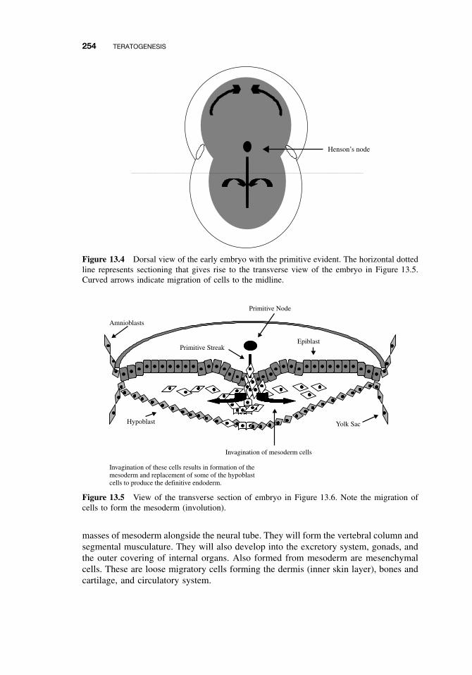
254 TERATOGENESIS
Henson’s node
Figure 13.4 Dorsal view of the early embryo with the primitive evident. The horizontal dottedline represents sectioning that gives rise to the transverse view of the embryo in Figure 13.5.Curved arrows indicate migration of cells to the midline.
Amnioblasts
Primitive Node
Primitive StreakEpiblast
Yolk SacHypoblast
Invagination of mesoderm cells
Invagination of these cells results in formation of themesoderm and replacement of some of the hypoblastcells to produce the definitive endoderm.
Figure 13.5 View of the transverse section of embryo in Figure 13.6. Note the migration ofcells to form the mesoderm (involution).
masses of mesoderm alongside the neural tube. They will form the vertebral column andsegmental musculature. They will also develop into the excretory system, gonads, andthe outer covering of internal organs. Also formed from mesoderm are mesenchymalcells. These are loose migratory cells forming the dermis (inner skin layer), bones andcartilage, and circulatory system.
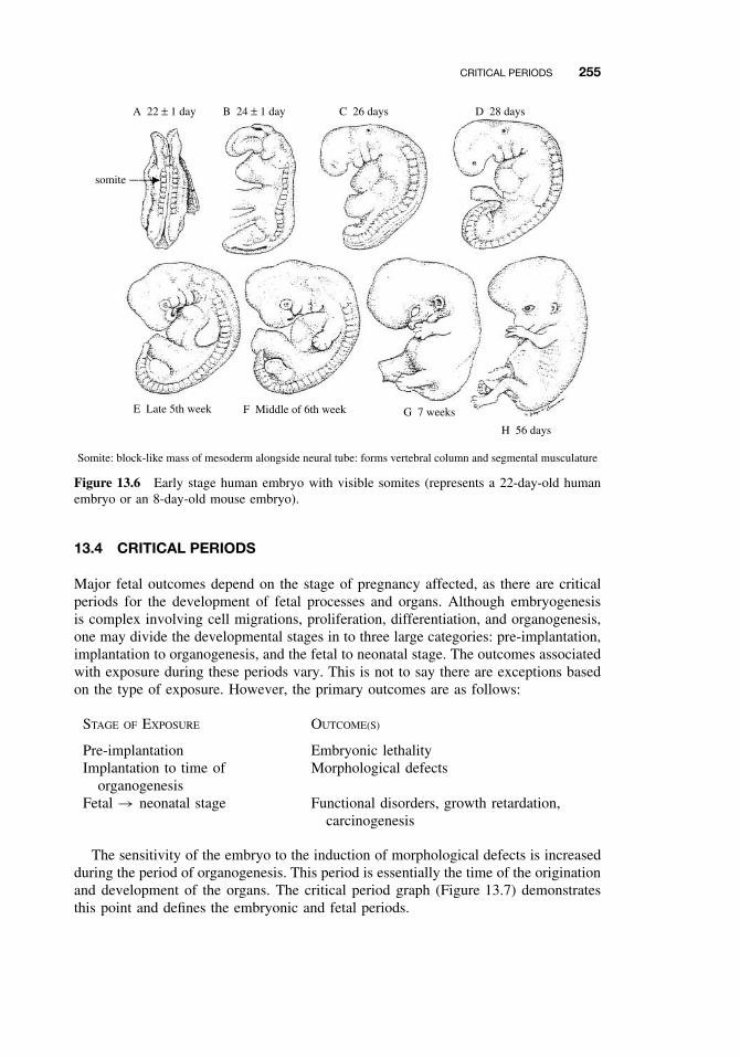
CRITICAL PERIODS 255
A 22 ± 1 day B 24 ± 1 day C 26 days D 28 days
E Late 5th week
Somite: block-like mass of mesoderm alongside neural tube: forms vertebral column and segmental musculature
F Middle of 6th week G 7 weeks
H 56 days
somite
Figure 13.6 Early stage human embryo with visible somites (represents a 22-day-old humanembryo or an 8-day-old mouse embryo).
13.4 CRITICAL PERIODS
Major fetal outcomes depend on the stage of pregnancy affected, as there are criticalperiods for the development of fetal processes and organs. Although embryogenesisis complex involving cell migrations, proliferation, differentiation, and organogenesis,one may divide the developmental stages in to three large categories: pre-implantation,implantation to organogenesis, and the fetal to neonatal stage. The outcomes associatedwith exposure during these periods vary. This is not to say there are exceptions basedon the type of exposure. However, the primary outcomes are as follows:
STAGE OF EXPOSURE OUTCOME(S)
Pre-implantation Embryonic lethalityImplantation to time of
organogenesisMorphological defects
Fetal → neonatal stage Functional disorders, growth retardation,carcinogenesis
The sensitivity of the embryo to the induction of morphological defects is increasedduring the period of organogenesis. This period is essentially the time of the originationand development of the organs. The critical period graph (Figure 13.7) demonstratesthis point and defines the embryonic and fetal periods.
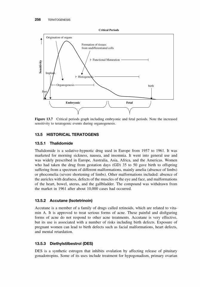
256 TERATOGENESIS
Origination of organs
Formation of tissuesfrom undifferentiated cells
Functional Maturation
Histogenesis
Organogenesis
Embryonic
Sens
itiv
ity
Critical Periods
Fetal
Implant.
Fert
birth
Figure 13.7 Critical periods graph including embryonic and fetal periods. Note the increasedsensitivity to teratogenic events during organogenesis.
13.5 HISTORICAL TERATOGENS
13.5.1 Thalidomide
Thalidomide is a sedative-hypnotic drug used in Europe from 1957 to 1961. It wasmarketed for morning sickness, nausea, and insomnia. It went into general use andwas widely prescribed in Europe, Australia, Asia, Africa, and the Americas. Womenwho had taken the drug from gestation days (GD) 35 to 50 gave birth to offspringsuffering from a spectrum of different malformations, mainly amelia (absence of limbs)or phocomelia (severe shortening of limbs). Other malformations included: absence ofthe auricles with deafness, defects of the muscles of the eye and face, and malformationsof the heart, bowel, uterus, and the gallbladder. The compound was withdrawn fromthe market in 1961 after about 10,000 cases had occurred.
13.5.2 Accutane (Isotetrinoin)
Accutane is a member of a family of drugs called retinoids, which are related to vita-min A. It is approved to treat serious forms of acne. These painful and disfiguringforms of acne do not respond to other acne treatments. Accutane is very effective,but its use is associated with a number of risks including birth defects. Exposure ofpregnant women can lead to birth defects such as facial malformations, heart defects,and mental retardation.
13.5.3 Diethylstilbestrol (DES)
DES is a synthetic estrogen that inhibits ovulation by affecting release of pituitarygonadotropins. Some of its uses include treatment for hypogonadism, primary ovarian
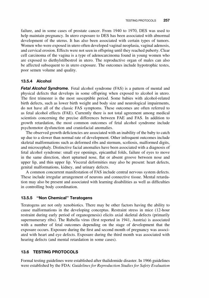
TESTING PROTOCOLS 257
failure, and in some cases of prostate cancer. From 1940 to 1970, DES was used tohelp maintain pregnancy. In utero exposure to DES has been associated with abnormaldevelopment of the uterus. It has also been associated with certain types of tumors.Women who were exposed in utero often developed vaginal neoplasia, vaginal adenosis,and cervical erosion. Effects were not seen in offspring until they reached puberty. Clearcell carcinoma of the vagina is a type of adenocarcinoma found in young women whoare exposed to diethylstilbestrol in utero. The reproductive organ of males can alsobe affected subsequent to in utero exposure. The outcomes include hypotrophic testes,poor semen volume and quality.
13.5.4 Alcohol
Fetal Alcohol Syndrome. Fetal alcohol syndrome (FAS) is a pattern of mental andphysical defects that develops in some offspring when exposed to alcohol in utero.The first trimester is the most susceptible period. Some babies with alcohol-relatedbirth defects, such as lower birth weight and body size and neurological impairments,do not have all of the classic FAS symptoms. These outcomes are often referred toas fetal alcohol effects (FAE). Currently there is not total agreement among medicalscientists concerning the precise differences between FAE and FAS. In addition togrowth retardation, the most common outcomes of fetal alcohol syndrome includepsychomotor dysfunction and craniofacial anomalies.
The observed growth deficiencies are associated with an inability of the baby to catchup due to a slower than normal rate of development. Other infrequent outcomes includeskeletal malformations such as deformed ribs and sternum, scoliosis, malformed digits,and microcephaly. Distinctive facial anomalies have been associated with a diagnosis offetal alcohol syndrome: small eye openings, epicanthal folds, failure of eyes to movein the same direction, short upturned nose, flat or absent groove between nose andupper lip, and thin upper lip. Visceral deformities may also be present: heart defects,genital malformations, kidney, and urinary defects.
A common concurrent manifestation of FAS include central nervous system defects.These include irregular arrangement of neurons and connective tissue. Mental retarda-tion may also be present and associated with learning disabilities as well as difficultiesin controlling body coordination.
13.5.5 ‘‘Non Chemical’’ Teratogens
Teratogens are not only xenobiotics. There may be other factors having the ability tocause malformations in the developing conceptus. Restraint stress in mice (12-hourrestraint during early period of organogenesis) elicits axial skeletal defects (primarilysupernumerary ribs). The Rubella virus (first reported in 1941, Austria) is associatedwith a number of fetal outcomes depending on the stage of development that theexposure occurs. Exposure during the first and second month of pregnancy was associ-ated with heart and eye defects. Exposure during the third month was associated withhearing defects (and mental retardation in some cases).
13.6 TESTING PROTOCOLS
Formal testing guidelines were established after thalidomide disaster. In 1966 guidelineswere established by the FDA: Guidelines for Reproduction Studies for Safety Evaluation
258 TERATOGENESIS
of Drugs for Human Use. Since then (1994) new streamlined testing protocols havebeen developed with international acceptance. This newer approach, ICH (13.6.2),relies on the investigator to determine the model to access reproductive/developmentaltoxicity. However, many scientist currently conduct and publish the FDA version oftesting (i.e., segment studies). Therefore both (FDA & ICH) approaches will be dis-cussed. Further practical details of testing protocols may be found in Chapter 21 aswell as some discussion of the regulatory implications for different agencies.
13.6.1 FDA Guidelines for Reproduction Studies for Safety Evaluationof Drugs for Human Use
Multigenerational Study. This approach involves the continuous exposure of arodent species (usually mice). The parental animals are exposed shortly after weaning(30–40 days of age). At reproductive maturity, the animals are mated. The first gen-eration is produced (F1). From these an F2 is produced and then subsequently an F3
generation. The effects of the test is monitored through each generation. The measuredparameters include fertility, litter size, and neonatal viability. This is a time-consumingeffort that usually takes about two years to complete.
Single-Generation Studies. Single-generation studies are short-term studies con-ducted in three segments:
Segment I: Evaluation of Fertility and Reproductive Performance. Male rodentsare treated for 70 days (to expose for one spermatogenic cycle), and nonpregnantfemales for 14 days (to exposure for several estrous cycles). Treatment is continued inthe females during mating, pregnancy, and lactation. Fifty percent of the females arekilled and the fetuses are examined for presence of malformations. The other 50% areallowed to give birth. After weaning, these offspring are killed and necropsied.
Segment II: Assessment of Developmental Toxicity. This involves the treatment ofpregnant females only during the period covering implantation through organogenesis(typically from gestational days 6 to 15 in mice with 18-day gestational periods). Oneday prior to birth, the animals are killed and fetuses examined for viability, bodyweight, and presence of malformation.
Segment II: Postnatal Evaluation. Pregnant animals are treated from the last trimesterof pregnancy until weaning. Evaluated are parturition process, late fetal development,neonatal survival, and growth as well as presence of any malformations.
13.6.2 International Conference of Harmonization (ICH) of TechnicalRequirements for Registration of Pharmaceuticals for Human Use(ICH)—US FDA, 1994
As previously mentioned, new streamlined testing protocols with international accep-tance have been developed. Below is a description of these guidelines as it relates tosimilarity to a particular segment-type study.
ICH 4.1.1: Fertility Assessment. This study duration is typically shorter than seg-ment I studies. Males are exposed for four weeks before mating and females two weeks
SUGGESTED READING 259
before mating. Male reproductive organs are carefully evaluated: organ weights, his-tological analysis and sperm count, and mobility evaluation. For the females, fertility,litter size, and viability of conceptus are evaluated.
ICH 4.1.2: Postnatal Evaluation and Pregnancy State Susceptibility. Thisstudy protocol is similar to the segment III study. Maternal toxicity is evaluated bycomparing the degree of toxicity of the nonpregnant female to that of the pregnantfemale. Postnatal viability and growth are also evaluated. Offspring are also evaluatedto assess functional development (i.e., presence of behavioral and reproductive deficits).
ICH 4.1.3: Assessment of Developmental Toxicity. This is almost identical tothe segment II study protocol. Pregnant animals are exposed from implantation throughorganogenesis. The parameters measured in the segment II study are similar. However,the study is usually conducted using at least two species. More specifically, at leastone rodent and one nonrodent species.
13.6.3 Alternative Test Methods
A number of alternative test methods have been developed to reduce the number ofwhole animals used in studies and/or to obtain more rapid information concerning thepotential of a compound to be a reproductive/developmental toxicant. Validation ofmany of the methods has been problematic, since they do not address the contributionof maternal factors or multiorgan contributions to outcomes. Some of these alterna-tive methods include the use of cell or embryo culture. For example, the micromassculture involves the use of limb bud cells from rat embryos grown in micromass cul-ture for five days. The processes of differentiation and cell proliferation are assessed.In the Chernoff/Kavlock Assay, pregnant rodents are exposed during organogenesisand allowed to deliver. Postnatal growth, viability, and gross morphology of littersare recorded (detailed skeletal evaluations are not performed). Other alternative testsinvolve the use of nontraditional test species such as Xenopus embryos (FETAX) andHydra. Xenopus embryos are exposed for 96 hours and then evaluated for morpho-logical defects, viability, and growth. The cells of Hydra aggregate to form artificialembryos. The dose response in these “embryos” is compared to that of the adult Hydra.
13.7 CONCLUSIONS
Understanding the mechanisms of the induction of birth defects is key to determinehow to prevent these effects. Further, increasing the accuracy of experimental ani-mal extrapolation will aid in the interpretation of experimental data in order to moreaccurately determine the risk of a given compound to elicit birth defects in humans.
SUGGESTED READING
Ballantyne, B., Marrs, T. and P. Turner, eds. General and Applied Toxicology, college ed., NewYork: Macmillan, 1995.
Klaassen, C. D., ed. Casarett and Doull’s Toxicology: The Basic Science of Poisons, 6th ed.,New York: McGraw-Hill, 2001.
Korach, K. S. Reproductive and Developmental Toxicology. New York: Dekker, 1998.
Hood, R. D. Handbook of Developmental Toxicology. Boca Raton, FL: CRC Press, 1997.


PART V
ORGAN TOXICITY

CHAPTER 14
Hepatotoxicity
ERNEST HODGSON and PATRICIA E. LEVI
14.1 INTRODUCTION
14.1.1 Liver Structure
The basic structure of the liver consists of rows of hepatic cells (hepatocytes orparenchymal cells) perforated by specialized blood capillaries called sinusoids (seeChapter 10, Figures 10.2 and 10.3). The sinusoid walls contain phagocytic cells calledKupffer cells whose role is to engulf and destroy materials such as solid particles,bacteria, dead blood cells, and so on. The main blood supply comes to the liver fromthe intestinal vasculature. These vessels, along with those from the spleen and thestomach, merge with each other to form the portal vein (see Chapter 10, Figure 10.4).On entering the liver, the portal vein subdivides and drains into the sinusoids. Theblood then perfuses the liver and exits by the hepatic veins, which merge into theinferior vena cava and return blood to the heart. The hepatic artery supplies the liverwith oxygenated arterial blood.
Other materials, such as bile acids and many xenobiotics, move from the hepatocytesinto the bile-carrying canaliculi, which merge into larger ducts that follow the portalvein branches. The ducts merge into the hepatic duct from which bile drains into theupper part of the small intestine, the duodenum. The gall bladder serves to hold bileuntil it is emptied into the intestine.
14.1.2 Liver Function
In the liver three main functions occur: storage, metabolism, and biosynthesis. Glucoseis converted to glycogen and stored; when needed for energy, it is converted back toglucose. Fat, fat-soluble vitamins, and other nutrients are also stored in the liver. Fattyacids are metabolized and converted to lipids, which are then conjugated with proteinssynthesized in the liver and released into the bloodstream as lipoproteins. The liver alsosynthesizes numerous functional proteins, such as enzymes and blood-coagulating fac-tors. In addition the liver, which contains numerous xenobiotic metabolizing enzymes,is the main site of xenobiotic metabolism.
A Textbook of Modern Toxicology, Third Edition, edited by Ernest HodgsonISBN 0-471-26508-X Copyright 2004 John Wiley & Sons, Inc.
263
264 HEPATOTOXICITY
14.2 SUSCEPTIBILITY OF THE LIVER
The liver, the largest organ in the body, is often the target organ for chemically inducedinjuries. Several important factors are known to contribute to the liver’s susceptibility.First, most xenobiotics enter the body through the gastrointestinal (GI) tract and, afterabsorption, are transported by the hepatic portal vein to the liver: thus the liver isthe first organ perfused by chemicals that are absorbed in the gut. A second factor isthe high concentration in the liver of xenobiotic metabolizing enzymes, primarily thecytochrome P450-dependent monooxygenase system. Although most biotransforma-tions are detoxication reactions, many oxidative reactions produce reactive metabolites(Chapters 7 and 8) that can induce lesions within the liver. Often areas of damageare in the centrilobular region, and this localization has been attributed, in part, to thehigher concentration of cytochrome P450 in that area of the liver.
14.3 TYPES OF LIVER INJURY
The types of injury to the liver depend on the type of toxic agent, the severity ofintoxication, and the type of exposure, whether acute or chronic. The main types ofliver damage are discussed briefly in this section. Whereas some types of damage—forexample, cholestasis—are liver specific, others such as necrosis and carcinogenesis aremore general phenomena.
14.3.1 Fatty Liver
Fatty liver refers to the abnormal accumulation of fat in hepatocytes. At the same timethere is a decrease in plasma lipids and lipoproteins. Although many toxicants maycause lipid accumulation in the liver (Table 14.1), the mechanisms may be different.Basically lipid accumulation is related to disturbances in either the synthesis or thesecretion of lipoproteins. Excess lipid can result from an oversupply of free fatty acidsfrom adipose tissues or, more commonly, from impaired release of triglycerides fromthe liver into the plasma. Triglycerides are secreted from the liver as lipoproteins (verylow density lipoprotein, VLDL). As might be expected, there are a number of points atwhich this process can be disrupted. Some of the more important ones are as follows(Figure 14.1):
ž Interference with synthesis of the protein moietyž Impaired conjugation of triglyceride with lipoproteinž Interference with transfer of VLDL across cell membranesž Decreased synthesis of phospholipidsž Impaired oxidation of lipids by mitochondriaž Inadequate energy (adenosine triphosphate [ATP] for lipid and protein synthesis
The role that fatty liver plays in liver injury is not clearly understood, and fatty liverin itself does not necessarily mean liver dysfunction. The onset of lipid accumulationin the liver is accompanied by changes in blood biochemistry, and for this reason bloodchemistry analysis can be a useful diagnostic tool.
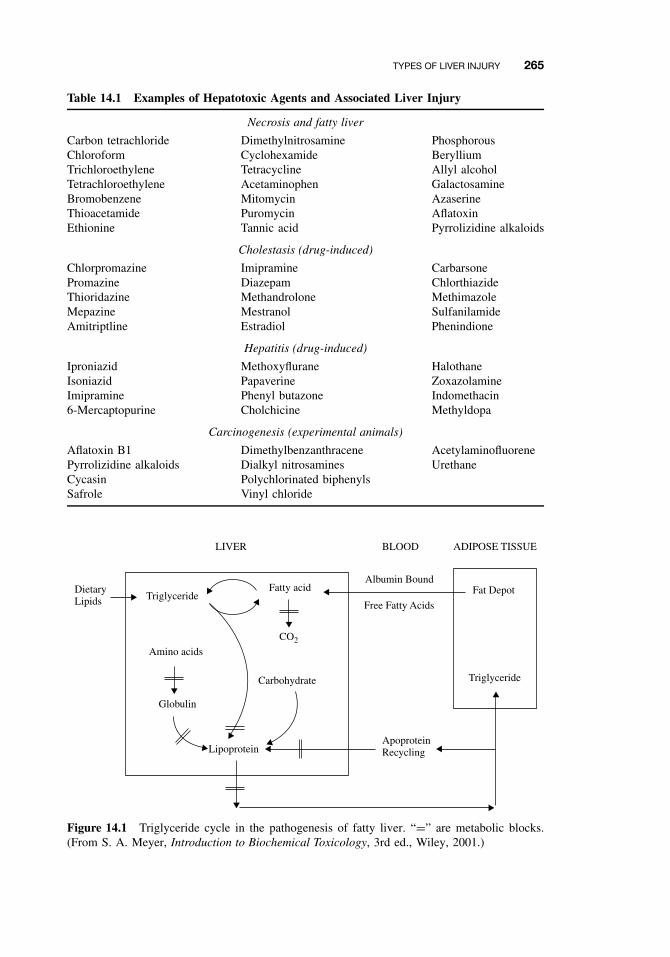
TYPES OF LIVER INJURY 265
Table 14.1 Examples of Hepatotoxic Agents and Associated Liver Injury
Necrosis and fatty liver
Carbon tetrachloride Dimethylnitrosamine PhosphorousChloroform Cyclohexamide BerylliumTrichloroethylene Tetracycline Allyl alcoholTetrachloroethylene Acetaminophen GalactosamineBromobenzene Mitomycin AzaserineThioacetamide Puromycin AflatoxinEthionine Tannic acid Pyrrolizidine alkaloids
Cholestasis (drug-induced)
Chlorpromazine Imipramine CarbarsonePromazine Diazepam ChlorthiazideThioridazine Methandrolone MethimazoleMepazine Mestranol SulfanilamideAmitriptline Estradiol Phenindione
Hepatitis (drug-induced)
Iproniazid Methoxyflurane HalothaneIsoniazid Papaverine ZoxazolamineImipramine Phenyl butazone Indomethacin6-Mercaptopurine Cholchicine Methyldopa
Carcinogenesis (experimental animals)
Aflatoxin B1 Dimethylbenzanthracene AcetylaminofluorenePyrrolizidine alkaloids Dialkyl nitrosamines UrethaneCycasin Polychlorinated biphenylsSafrole Vinyl chloride
DietaryLipids Triglyceride
Amino acids
Globulin
Lipoprotein
Carbohydrate
CO2
Fatty acidAlbumin Bound
Free Fatty Acids
ApoproteinRecycling
Fat Depot
Triglyceride
LIVER BLOOD ADIPOSE TISSUE
Figure 14.1 Triglyceride cycle in the pathogenesis of fatty liver. “=” are metabolic blocks.(From S. A. Meyer, Introduction to Biochemical Toxicology, 3rd ed., Wiley, 2001.)

266 HEPATOTOXICITY
14.3.2 Necrosis
Cell necrosis is a degenerative process leading to cell death. Necrosis, usually anacute injury, may be localized and affect only a few hepatocytes (focal necrosis), orit may involve an entire lobe (massive necrosis). Cell death occurs along with rup-ture of the plasma membrane, and is preceded by a number of morphologic changessuch as cytoplasmic edema, dilation of the endoplasmic reticulum, disaggregation ofpolysomes, accumulation of triglycerides, swelling of mitochondria with disruption ofcristae, and dissolution of organelles and nucleus. Biochemical events that may leadto these changes include binding of reactive metabolites to proteins and unsaturatedlipids (inducing lipid peroxidation and subsequent membrane destruction) disturbanceof cellular Ca+2 homeostasis, inference with metabolic pathways, shifts in Na+ andK+ balance, and inhibition of protein synthesis. Changes in blood chemistry resemblethose seen with fatty liver, except they are quantitatively larger. Because of the regen-erating capability of the liver, necrotic lesions are not necessarily critical. Massiveareas of necrosis, however, can lead to severe liver damage and failure.
14.3.3 Apoptosis
Apoptosis is a controlled form of cell death that serves as a regulation point for bio-logic processes and can be thought of as the counterpoint of cell division by mitosis.This selective mechanism is particularly active during development and senescence.Although apoptosis is a normal physiological process, it can also be induced by a num-ber of exogenous factors, such as xenobiotic chemicals, oxidative stress, anoxia, andradiation. (A stimulus that induces a cell to undergo apoptosis is known as an apogen.)If, however, apoptosis is suppressed in some cell types, it can lead to accumulation ofthese cells. For example, in some instances, clonal expansion of malignant cells andsubsequent tumor growth results primarily from inhibition of apoptosis.
Apoptosis can be distinguished from necrosis by morphologic criteria, using eitherlight or electron microscopy. Toxicants, however, do not always act in a clear-cutfashion, and some toxicants can induce both apoptosis and necrosis either concurrentlyor sequentially.
14.3.4 Cholestasis
Cholestasis is the suppression or stoppage of bile flow, and may have either intrahepaticor extrahepatic causes. Inflammation or blockage of the bile ducts results in retentionof bile salts as well as bilirubin accumulation, an event that leads to jaundice. Othermechanisms causing cholestasis include changes in membranes permeability of eitherhepatocytes or biliary canaliculi. Cholestasis is usually drug induced (Table 14.1) andis difficult to produce in experimental animals. Again, changes in blood chemistry canbe a useful diagnostic tool.
14.3.5 Cirrhosis
Cirrhosis is a progressive disease that is characterized by the deposition of collagenthroughout the liver. In most cases cirrhosis results from chronic chemical injury. The
TYPES OF LIVER INJURY 267
accumulation of fibrous material causes severe restriction in blood flow and in theliver’s normal metabolic and detoxication processes. This situation can in turn causefurther damage and eventually lead to liver failure. In humans, chronic use of ethanolis the single most important cause of cirrhosis, although there is some dispute as towhether the effect is due to ethanol alone or is also related to the nutritional deficienciesthat usually accompany alcoholism.
14.3.6 Hepatitis
Hepatitis is an inflammation of the liver and is usually viral in origin; however, certainchemicals, usually drugs, can induce a hepatitis that closely resembles that producedby viral infections (Table 14.1). This type of liver injury is not usually demonstrable inlaboratory animals and is often manifest only in susceptible individuals. Fortunately,the incidence of this type of disease is very low.
14.3.7 Oxidative Stress
Oxidative stress has been defined as an imbalance between the prooxidant/antioxidantsteady state in the cell, with the excess of prooxidants being available to interact withcellular macromolecules to cause damage to the cell, often resulting in cell death.Although the occurrence of reactive oxygen species in normal metabolism and theconcept of oxidative stress was derived from these studies, it is apparent that oxidativestress can occur in almost any tissue, producing a variety of deleterious effects. To date,a number of liver diseases, including alcoholic liver disease, metal storage diseases,and cholestatic liver disease, have been shown to have an oxidative stress component.
Reactive oxygen and reactive nitrogen radicals can be formed in a number of ways(Figure 14.2), the former primarily as a by-product of mitochondrial electron transport.Superoxide, hydrogen peroxide, singlet oxygen, and hydroxyl can all arise from thissource. Other sources include monooxygenases and peroxisomes. If not detoxified,reactive oxygen species can interact with biological macromolecules such as DNAand protein or with lipids. Once lipid peroxidation of unsaturated fatty acids in phos-pholipids is initiated, it is propagated in such a way as to have a major damagingeffect on cellular membranes. The formation, detoxication by superoxide dismutaseand by glutathione-dependent mechanisms, and interaction at sites of toxic action areillustrated in Figure 14.2.
14.3.8 Carcinogenesis
The most common type of primary liver tumor is hepatocellular carcinoma; other typesinclude cholangiocarcinoma, angiosarcoma, glandular carcinoma, and undifferentiatedliver cell carcinoma. Although a wide variety of chemicals are known to induce livercancer in laboratory animals (Table 14.1), the incidence of primary liver cancer inhumans in the United States is very low.
Some naturally occurring liver carcinogens are aflatoxin, cycasin, and safrole. Anumber of synthetic chemicals have been shown to cause liver cancer in animals,including the dialkylnitrosamines, dimethylbenzanthracene, aromatic amines such as
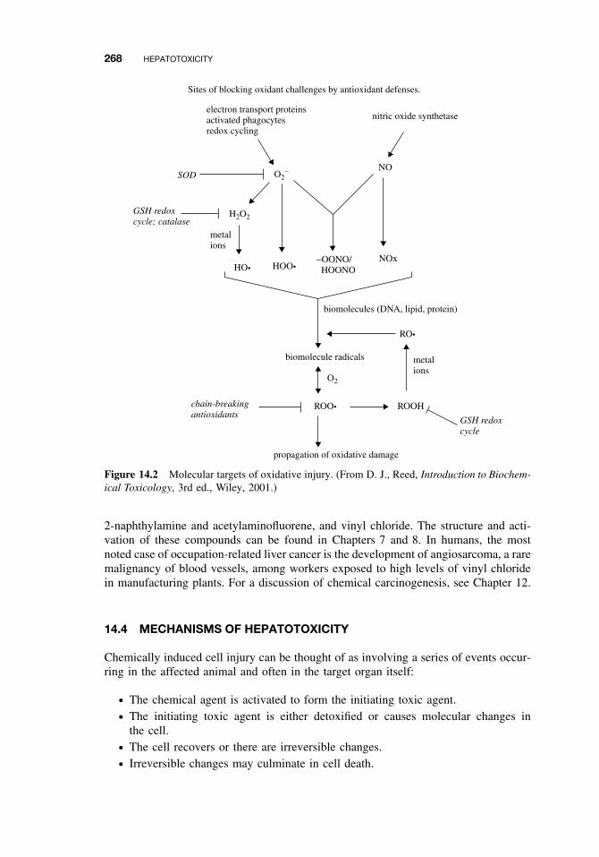
268 HEPATOTOXICITY
Sites of blocking oxidant challenges by antioxidant defenses.
electron transport proteinsactivated phagocytesredox cycling
NO
nitric oxide synthetase
SOD
GSH redoxcycle; catalase
O2−
H2O2
O2
metalions
metalions
GSH redoxcycle
biomolecules (DNA, lipid, protein)
HO•
ROO• ROOH
RO•
HOO•−OONO/ HOONO
NOx
biomolecule radicals
propagation of oxidative damage
chain-breakingantioxidants
Figure 14.2 Molecular targets of oxidative injury. (From D. J., Reed, Introduction to Biochem-ical Toxicology, 3rd ed., Wiley, 2001.)
2-naphthylamine and acetylaminofluorene, and vinyl chloride. The structure and acti-vation of these compounds can be found in Chapters 7 and 8. In humans, the mostnoted case of occupation-related liver cancer is the development of angiosarcoma, a raremalignancy of blood vessels, among workers exposed to high levels of vinyl chloridein manufacturing plants. For a discussion of chemical carcinogenesis, see Chapter 12.
14.4 MECHANISMS OF HEPATOTOXICITY
Chemically induced cell injury can be thought of as involving a series of events occur-ring in the affected animal and often in the target organ itself:
ž The chemical agent is activated to form the initiating toxic agent.ž The initiating toxic agent is either detoxified or causes molecular changes in
the cell.ž The cell recovers or there are irreversible changes.ž Irreversible changes may culminate in cell death.
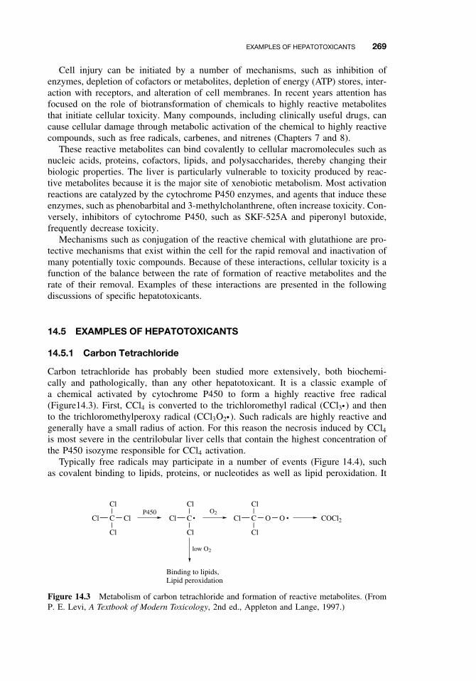
EXAMPLES OF HEPATOTOXICANTS 269
Cell injury can be initiated by a number of mechanisms, such as inhibition ofenzymes, depletion of cofactors or metabolites, depletion of energy (ATP) stores, inter-action with receptors, and alteration of cell membranes. In recent years attention hasfocused on the role of biotransformation of chemicals to highly reactive metabolitesthat initiate cellular toxicity. Many compounds, including clinically useful drugs, cancause cellular damage through metabolic activation of the chemical to highly reactivecompounds, such as free radicals, carbenes, and nitrenes (Chapters 7 and 8).
These reactive metabolites can bind covalently to cellular macromolecules such asnucleic acids, proteins, cofactors, lipids, and polysaccharides, thereby changing theirbiologic properties. The liver is particularly vulnerable to toxicity produced by reac-tive metabolites because it is the major site of xenobiotic metabolism. Most activationreactions are catalyzed by the cytochrome P450 enzymes, and agents that induce theseenzymes, such as phenobarbital and 3-methylcholanthrene, often increase toxicity. Con-versely, inhibitors of cytochrome P450, such as SKF-525A and piperonyl butoxide,frequently decrease toxicity.
Mechanisms such as conjugation of the reactive chemical with glutathione are pro-tective mechanisms that exist within the cell for the rapid removal and inactivation ofmany potentially toxic compounds. Because of these interactions, cellular toxicity is afunction of the balance between the rate of formation of reactive metabolites and therate of their removal. Examples of these interactions are presented in the followingdiscussions of specific hepatotoxicants.
14.5 EXAMPLES OF HEPATOTOXICANTS
14.5.1 Carbon Tetrachloride
Carbon tetrachloride has probably been studied more extensively, both biochemi-cally and pathologically, than any other hepatotoxicant. It is a classic example ofa chemical activated by cytochrome P450 to form a highly reactive free radical(Figure14.3). First, CCl4 is converted to the trichloromethyl radical (CCl3ž) and thento the trichloromethylperoxy radical (CCl3O2ž). Such radicals are highly reactive andgenerally have a small radius of action. For this reason the necrosis induced by CCl4is most severe in the centrilobular liver cells that contain the highest concentration ofthe P450 isozyme responsible for CCl4 activation.
Typically free radicals may participate in a number of events (Figure 14.4), suchas covalent binding to lipids, proteins, or nucleotides as well as lipid peroxidation. It
C
Cl
Cl
Cl
ClP450
C
Cl
Cl
Cl •O2
C
Cl
O
Cl
Cl O • COCl2
low O2
Binding to lipids,Lipid peroxidation
Figure 14.3 Metabolism of carbon tetrachloride and formation of reactive metabolites. (FromP. E. Levi, A Textbook of Modern Toxicology, 2nd ed., Appleton and Lange, 1997.)
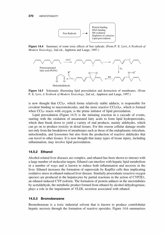
270 HEPATOTOXICITY
Free Radicals
Protein bindingDNA bindingSH oxidationDepletion of cofactorsLipid peroxidation
Figure 14.4 Summary of some toxic effects of free radicals. (From P. E. Levi, A Textbook ofModern Toxicology, 2nd ed., Appleton and Lange, 1997.)
CCl3•
•
•
O2
O O•
RH
O OR•
O O
Polyunsaturatedfatty acid (PUFA)
Malondialdehyde
Figure 14.5 Schematic illustrating lipid peroxidation and destruction of membranes. (FromP. E. Levi, A Textbook of Modern Toxicology, 2nd ed., Appleton and Lange, 1997.)
is now thought that CCl3ž , which forms relatively stable adducts, is responsible forcovalent binding to macromolecules, and the more reactive CCl3O2ž , which is formedwhen CCl3ž reacts with oxygen, is the prime initiator of lipid peroxidation.
Lipid peroxidation (Figure 14.5) is the initiating reaction in a cascade of events,starting with the oxidation of unsaturated fatty acids to form lipid hydroperoxides,which then break down to yield a variety of end products, mainly aldehydes, whichcan go on to produce toxicity in distal tissues. For this reason cellular damage resultsnot only from the breakdown of membranes such as those of the endoplasmic reticulum,mitochondria, and lysosomes but also from the production of reactive aldehydes thatcan travel to other tissues. It is now thought that many types of tissue injury, includinginflammation, may involve lipid peroxidation.
14.5.2 Ethanol
Alcohol-related liver diseases are complex, and ethanol has been shown to interact witha large number of molecular targets. Ethanol can interfere with hepatic lipid metabolismin a number of ways and is known to induce both inflammation and necrosis in theliver. Ethanol increases the formation of superoxide by Kupffer cells thus implicatingoxidative stress in ethanol-induced liver disease. Similarly prooxidants (reactive oxygenspecies) are produced in the hepatocytes by partial reactions in the action of CYP2E1,an ethanol-induced CYP isoform. The formation of protein adducts in the microtubulesby acetaldehyde, the metabolic product formed from ethanol by alcohol dehydrogenase,plays a role in the impairment of VLDL secretion associated with ethanol.
14.5.3 Bromobenzene
Bromobenzene is a toxic industrial solvent that is known to produce centrilobularhepatic necrosis through the formation of reactive epoxides. Figure 14.6 summarizes
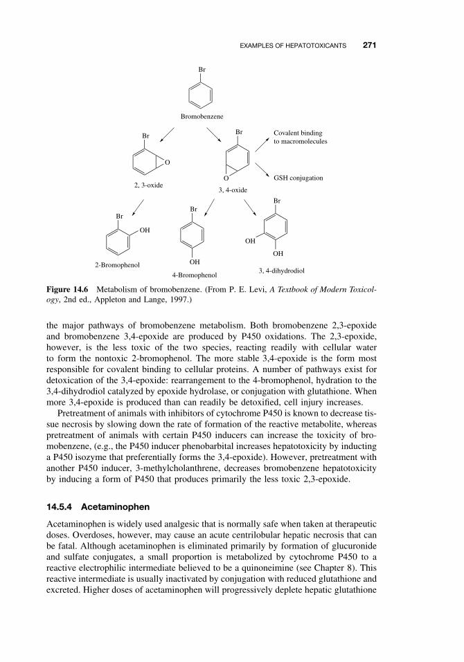
EXAMPLES OF HEPATOTOXICANTS 271
Br
O
O
BrBr
Br
OH
OH
Br
OH
Br
OH2-Bromophenol
4-Bromophenol3, 4-dihydrodiol
3, 4-oxide2, 3-oxide
Bromobenzene
Covalent bindingto macromolecules
GSH conjugation
Figure 14.6 Metabolism of bromobenzene. (From P. E. Levi, A Textbook of Modern Toxicol-ogy, 2nd ed., Appleton and Lange, 1997.)
the major pathways of bromobenzene metabolism. Both bromobenzene 2,3-epoxideand bromobenzene 3,4-epoxide are produced by P450 oxidations. The 2,3-epoxide,however, is the less toxic of the two species, reacting readily with cellular waterto form the nontoxic 2-bromophenol. The more stable 3,4-epoxide is the form mostresponsible for covalent binding to cellular proteins. A number of pathways exist fordetoxication of the 3,4-epoxide: rearrangement to the 4-bromophenol, hydration to the3,4-dihydrodiol catalyzed by epoxide hydrolase, or conjugation with glutathione. Whenmore 3,4-epoxide is produced than can readily be detoxified, cell injury increases.
Pretreatment of animals with inhibitors of cytochrome P450 is known to decrease tis-sue necrosis by slowing down the rate of formation of the reactive metabolite, whereaspretreatment of animals with certain P450 inducers can increase the toxicity of bro-mobenzene, (e.g., the P450 inducer phenobarbital increases hepatotoxicity by inductinga P450 isozyme that preferentially forms the 3,4-epoxide). However, pretreatment withanother P450 inducer, 3-methylcholanthrene, decreases bromobenzene hepatotoxicityby inducing a form of P450 that produces primarily the less toxic 2,3-epoxide.
14.5.4 Acetaminophen
Acetaminophen is widely used analgesic that is normally safe when taken at therapeuticdoses. Overdoses, however, may cause an acute centrilobular hepatic necrosis that canbe fatal. Although acetaminophen is eliminated primarily by formation of glucuronideand sulfate conjugates, a small proportion is metabolized by cytochrome P450 to areactive electrophilic intermediate believed to be a quinoneimine (see Chapter 8). Thisreactive intermediate is usually inactivated by conjugation with reduced glutathione andexcreted. Higher doses of acetaminophen will progressively deplete hepatic glutathione
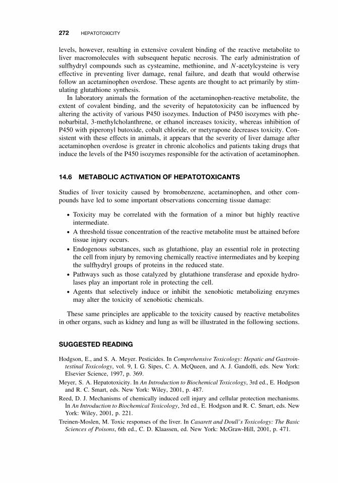
272 HEPATOTOXICITY
levels, however, resulting in extensive covalent binding of the reactive metabolite toliver macromolecules with subsequent hepatic necrosis. The early administration ofsulfhydryl compounds such as cysteamine, methionine, and N -acetylcysteine is veryeffective in preventing liver damage, renal failure, and death that would otherwisefollow an acetaminophen overdose. These agents are thought to act primarily by stim-ulating glutathione synthesis.
In laboratory animals the formation of the acetaminophen-reactive metabolite, theextent of covalent binding, and the severity of hepatotoxicity can be influenced byaltering the activity of various P450 isozymes. Induction of P450 isozymes with phe-nobarbital, 3-methylcholanthrene, or ethanol increases toxicity, whereas inhibition ofP450 with piperonyl butoxide, cobalt chloride, or metyrapone decreases toxicity. Con-sistent with these effects in animals, it appears that the severity of liver damage afteracetaminophen overdose is greater in chronic alcoholics and patients taking drugs thatinduce the levels of the P450 isozymes responsible for the activation of acetaminophen.
14.6 METABOLIC ACTIVATION OF HEPATOTOXICANTS
Studies of liver toxicity caused by bromobenzene, acetaminophen, and other com-pounds have led to some important observations concerning tissue damage:
ž Toxicity may be correlated with the formation of a minor but highly reactiveintermediate.
ž A threshold tissue concentration of the reactive metabolite must be attained beforetissue injury occurs.
ž Endogenous substances, such as glutathione, play an essential role in protectingthe cell from injury by removing chemically reactive intermediates and by keepingthe sulfhydryl groups of proteins in the reduced state.
ž Pathways such as those catalyzed by glutathione transferase and epoxide hydro-lases play an important role in protecting the cell.
ž Agents that selectively induce or inhibit the xenobiotic metabolizing enzymesmay alter the toxicity of xenobiotic chemicals.
These same principles are applicable to the toxicity caused by reactive metabolitesin other organs, such as kidney and lung as will be illustrated in the following sections.
SUGGESTED READING
Hodgson, E., and S. A. Meyer. Pesticides. In Comprehensive Toxicology: Hepatic and Gastroin-testinal Toxicology, vol. 9, I. G. Sipes, C. A. McQueen, and A. J. Gandolfi, eds. New York:Elsevier Science, 1997, p. 369.
Meyer, S. A. Hepatotoxicity. In An Introduction to Biochemical Toxicology, 3rd ed., E. Hodgsonand R. C. Smart, eds. New York: Wiley, 2001, p. 487.
Reed, D. J. Mechanisms of chemically induced cell injury and cellular protection mechanisms.In An Introduction to Biochemical Toxicology, 3rd ed., E. Hodgson and R. C. Smart, eds. NewYork: Wiley, 2001, p. 221.
Treinen-Moslen, M. Toxic responses of the liver. In Casarett and Doull’s Toxicology: The BasicSciences of Poisons, 6th ed., C. D. Klaassen, ed. New York: McGraw-Hill, 2001, p. 471.
CHAPTER 15
Nephrotoxicity
ERNEST HODGSON and PATRICIA E. LEVI
15.1 INTRODUCTION
15.1.1 Structure of the Renal System
The renal system consists of the kidneys and their vasculature and innervation, thekidneys each draining through a ureter into a single median urinary bladder, and thelatter draining to the exterior via a single duct, the urethra. The kidney has three majoranatomical areas: the cortex, the medulla, and the papilla.
The renal cortex is the outermost region of the kidney and contains glomeruli,proximal and distal tubules, and peritubular capillaries. Cortical blood flow is high,the cortex receiving approximately 90% of the renal blood flow. Since blood-bornetoxicants will be delivered preferentially to the cortex, they are more likely to affectcortical functions rather than those of medulla or papilla. The renal medulla is themiddle portion and contains primarily loops of Henle, vasa recta, and collecting ducts.Although the medulla receives only about 6% of the renal blood flow, it may beexposed to high concentrations of toxicants within tubular structures. The papilla isthe smallest anatomical portion of the kidney and receives only about 1% of the renalblood flow. Nevertheless, because the tubular fluid is maximally concentrated andluminal fluid is maximally reduced, the concentrations of potential toxicants in thepapilla my be extremely high, leading to cellular injury in the papillary tubular and/orinterstitial cells.
The nephron is the functional unit of the kidney. It is described in detail in Chapter 10and illustrated in Figure 10.1.
15.1.2 Function of the Renal System
The primary function of the renal system is the elimination of waste products, derivedeither from endogenous metabolism or from the metabolism of xenobiotics. The lat-ter function is discussed in detail in Chapter 10. The kidney also plays an importantrole in regulation of body homeostasis, regulating extracellular fluid volume, and elec-trolyte balance.
A Textbook of Modern Toxicology, Third Edition, edited by Ernest HodgsonISBN 0-471-26508-X Copyright 2004 John Wiley & Sons, Inc.
273
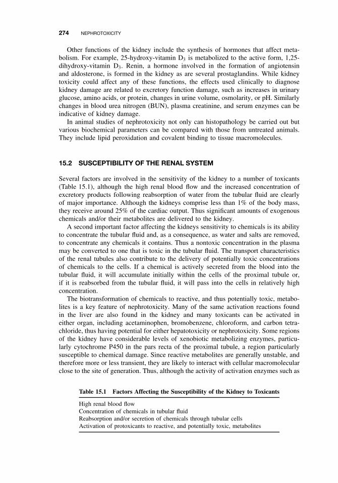
274 NEPHROTOXICITY
Other functions of the kidney include the synthesis of hormones that affect meta-bolism. For example, 25-hydroxy-vitamin D3 is metabolized to the active form, 1,25-dihydroxy-vitamin D3. Renin, a hormone involved in the formation of angiotensinand aldosterone, is formed in the kidney as are several prostaglandins. While kidneytoxicity could affect any of these functions, the effects used clinically to diagnosekidney damage are related to excretory function damage, such as increases in urinaryglucose, amino acids, or protein, changes in urine volume, osmolarity, or pH. Similarlychanges in blood urea nitrogen (BUN), plasma creatinine, and serum enzymes can beindicative of kidney damage.
In animal studies of nephrotoxicity not only can histopathology be carried out butvarious biochemical parameters can be compared with those from untreated animals.They include lipid peroxidation and covalent binding to tissue macromolecules.
15.2 SUSCEPTIBILITY OF THE RENAL SYSTEM
Several factors are involved in the sensitivity of the kidney to a number of toxicants(Table 15.1), although the high renal blood flow and the increased concentration ofexcretory products following reabsorption of water from the tubular fluid are clearlyof major importance. Although the kidneys comprise less than 1% of the body mass,they receive around 25% of the cardiac output. Thus significant amounts of exogenouschemicals and/or their metabolites are delivered to the kidney.
A second important factor affecting the kidneys sensitivity to chemicals is its abilityto concentrate the tubular fluid and, as a consequence, as water and salts are removed,to concentrate any chemicals it contains. Thus a nontoxic concentration in the plasmamay be converted to one that is toxic in the tubular fluid. The transport characteristicsof the renal tubules also contribute to the delivery of potentially toxic concentrationsof chemicals to the cells. If a chemical is actively secreted from the blood into thetubular fluid, it will accumulate initially within the cells of the proximal tubule or,if it is reabsorbed from the tubular fluid, it will pass into the cells in relatively highconcentration.
The biotransformation of chemicals to reactive, and thus potentially toxic, metabo-lites is a key feature of nephrotoxicity. Many of the same activation reactions foundin the liver are also found in the kidney and many toxicants can be activated ineither organ, including acetaminophen, bromobenzene, chloroform, and carbon tetra-chloride, thus having potential for either hepatotoxicity or nephrotoxicity. Some regionsof the kidney have considerable levels of xenobiotic metabolizing enzymes, particu-larly cytochrome P450 in the pars recta of the proximal tubule, a region particularlysusceptible to chemical damage. Since reactive metabolites are generally unstable, andtherefore more or less transient, they are likely to interact with cellular macromolecularclose to the site of generation. Thus, although the activity of activation enzymes such as
Table 15.1 Factors Affecting the Susceptibility of the Kidney to Toxicants
High renal blood flowConcentration of chemicals in tubular fluidReabsorption and/or secretion of chemicals through tubular cellsActivation of protoxicants to reactive, and potentially toxic, metabolites
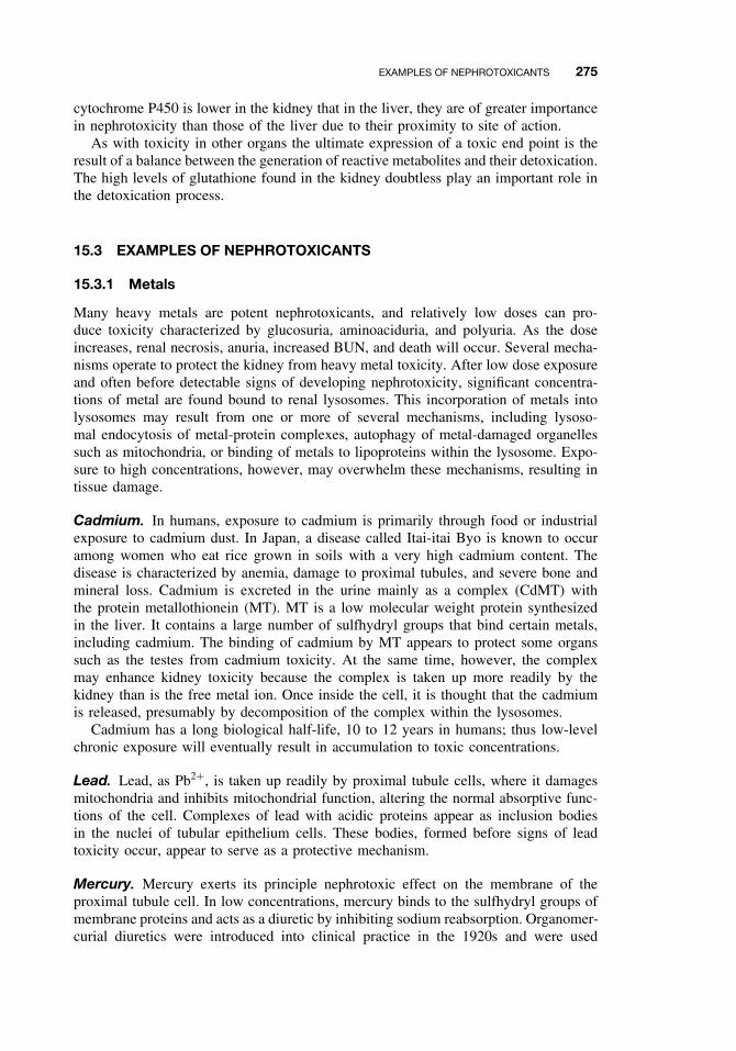
EXAMPLES OF NEPHROTOXICANTS 275
cytochrome P450 is lower in the kidney that in the liver, they are of greater importancein nephrotoxicity than those of the liver due to their proximity to site of action.
As with toxicity in other organs the ultimate expression of a toxic end point is theresult of a balance between the generation of reactive metabolites and their detoxication.The high levels of glutathione found in the kidney doubtless play an important role inthe detoxication process.
15.3 EXAMPLES OF NEPHROTOXICANTS
15.3.1 Metals
Many heavy metals are potent nephrotoxicants, and relatively low doses can pro-duce toxicity characterized by glucosuria, aminoaciduria, and polyuria. As the doseincreases, renal necrosis, anuria, increased BUN, and death will occur. Several mecha-nisms operate to protect the kidney from heavy metal toxicity. After low dose exposureand often before detectable signs of developing nephrotoxicity, significant concentra-tions of metal are found bound to renal lysosomes. This incorporation of metals intolysosomes may result from one or more of several mechanisms, including lysoso-mal endocytosis of metal-protein complexes, autophagy of metal-damaged organellessuch as mitochondria, or binding of metals to lipoproteins within the lysosome. Expo-sure to high concentrations, however, may overwhelm these mechanisms, resulting intissue damage.
Cadmium. In humans, exposure to cadmium is primarily through food or industrialexposure to cadmium dust. In Japan, a disease called Itai-itai Byo is known to occuramong women who eat rice grown in soils with a very high cadmium content. Thedisease is characterized by anemia, damage to proximal tubules, and severe bone andmineral loss. Cadmium is excreted in the urine mainly as a complex (CdMT) withthe protein metallothionein (MT). MT is a low molecular weight protein synthesizedin the liver. It contains a large number of sulfhydryl groups that bind certain metals,including cadmium. The binding of cadmium by MT appears to protect some organssuch as the testes from cadmium toxicity. At the same time, however, the complexmay enhance kidney toxicity because the complex is taken up more readily by thekidney than is the free metal ion. Once inside the cell, it is thought that the cadmiumis released, presumably by decomposition of the complex within the lysosomes.
Cadmium has a long biological half-life, 10 to 12 years in humans; thus low-levelchronic exposure will eventually result in accumulation to toxic concentrations.
Lead. Lead, as Pb2+, is taken up readily by proximal tubule cells, where it damagesmitochondria and inhibits mitochondrial function, altering the normal absorptive func-tions of the cell. Complexes of lead with acidic proteins appear as inclusion bodiesin the nuclei of tubular epithelium cells. These bodies, formed before signs of leadtoxicity occur, appear to serve as a protective mechanism.
Mercury. Mercury exerts its principle nephrotoxic effect on the membrane of theproximal tubule cell. In low concentrations, mercury binds to the sulfhydryl groups ofmembrane proteins and acts as a diuretic by inhibiting sodium reabsorption. Organomer-curial diuretics were introduced into clinical practice in the 1920s and were used
276 NEPHROTOXICITY
clinically into the 1960s. Despite their widespread acceptance as effective therapeuticdiuretics, it was well known that problems related to severe kidney toxicity were pos-sible. However, in the absence of other effective drugs, the organomercurials provedto be effective, sometimes life-saving, therapeutic agents. More recently organomercu-rial chemicals have been implicated as environmental pollutants, responsible for renaldamage in humans and animals.
Uranium. About 50% of plasma uranium is bound, as the uranyl ion, to bicarbonate,which is filtered by the glomerulus. As a result of acidification in the proximal tubule,the bicarbonate complex dissociates, followed by reabsorption of the bicarbonate ion;the released UO2
2+ then becomes attached to the membrane of the proximal tubulecells. The resultant loss of cell function is evidenced by increased concentrations ofglucose, amino acids, and proteins in the urine.
15.3.2 Aminoglycosides
Certain antibiotics, most notably the aminoglycosides, are known to be nephrotoxicin humans, especially in high doses or after prolonged therapy. The group of antibi-otics includes streptomycin, neomycin, kanamycin, and gentamycin. Aminoglycosidesare polar cations that are filtered by the glomerulus and excreted unchanged intothe urine. In the proximal tubule, the aminoglycosides are reabsorbed by binding toanionic membrane phospholipids, followed by endocytosis and sequestration in lyso-somes (Figure 15.1). It is thought that when a threshold concentration is reached, thelysosomes rupture, releasing hydrolytic enzymes that cause tissue necrosis.
15.3.3 Amphotericin B
With some drugs, renal damage may be related to the drugs’ biochemical mechanismof action. For example, the polymycins, such as amphotericin B, are surface-activeagents that bind to membrane phospholipids, disrupting the integrity of the membraneand resulting in leaky cells.
L
AG
AG
AG
AG
AG
AGM
Figure 15.1 Possible cellular interactions of aminoglycosides. AG = aminoglycoside; M =mitochondrion; L = lysosome. (From E. Hodgson and P. E. Levi, eds., A Textbook of ModernToxicology. 2nd ed., Appleton and Lange, Stamford, CT, 1997.)
EXAMPLES OF NEPHROTOXICANTS 277
15.3.4 Chloroform
Chloroform is a common industrial organic solvent that can be a hepatotoxicant ora nephrotoxicant in both humans and animals. As a nephrotoxicant it is both speciesand gender dependent. For example, following chloroform administration male micedevelop primarily kidney necrosis whereas female develop liver necrosis.
As a nephrotoxicant, chloroform most probably undergoes metabolic activation inthe kidney itself. Chloroform is metabolized to phosgene (Figure 15.2) by a cytochromeP450-dependent reaction, probably proceeding via an unstable hydroxylated product,trichloromethanol. Phosgene is capable of binding to cellular proteins to produce thecellular necrosis associated with chloroform toxicity to the kidney. Phosgene canalso be further metabolized by a number of reactions (Figure 15.2), and as withmost chemical-induced toxicity, the final expression of toxicity depends on a balancebetween activation and detoxication.
15.3.5 Hexachlorobutadiene
Hexachlorobutadiene is an industrial solvent and heat-transfer agent. It is a widespreadenvironmental contaminant that is a potent and relatively specific nephrotoxicant. Hex-achlorobutadiene first forms a glutathione conjugate, which is further metabolized bythe mercapturic acid pathway to a cysteine conjugate (see Chapter 7 for details ofglutathione conjugation and the mercapturic acid pathway). In the kidney, the cys-teine conjugate is cleaved to a reactive intermediate by the enzyme, cysteine conjugateβ-lyase.
Cl C
H
Cl
Cl
Cytochrome P-450,
O2, microsomesCl C
OH
Cl
Cl
Cl C
O
Cl
H C
H
C
S
C
OH
N
OH
C
O
H
phosgenecysteine
H2O
2GSHCO2 + 2HCl
GS CO SG + 2HClCovalentbinding
+ 2HClDiglutathionyldithiocarbonate
TOXICITY 2-oxothiazolidine-4-carboxylic acid (OTZ)
HCl
Figure 15.2 Proposed mechanism of chloroform biotransformation. (From J. B. Tarloff in AnIntroduction to Biochemical Toxicology, 3rd ed., E. Hodgson and R. C. Smart eds., Wiley, 2001.)
278 NEPHROTOXICITY
15.3.6 Tetrafluoroethylene
The nephrotoxic mode of action of tetrafluoroethylene is similar to that of hexachloro-butadiene. It is first metabolized to a cysteine conjugate, which is metabolized bycysteine conjugate β-lyase to a reactive product that can bind to cellular macro-molecules.
SUGGESTED READING
Tarloff, J. B. Biochemical mechanisms of renal toxicity. In An Introduction to Biochemical Tox-icology, 3rd ed., E. Hodgson and R. C. Smart, eds. New York: Wiley, 2001, p. 641.
Schnellmann R. G, Toxic Responses of the kidney. In Casarett and Doull’s Toxicology: TheBasic Sciences of Poisons, 6th ed., C. D Klaassen, ed, New York: McGraw-Hill, 2001, p. 491.
CHAPTER 16
Toxicology of the Nervous System
BONITA L. BLAKE
16.1 INTRODUCTION
Many substances alter the normal activity of the nervous system. Sometimes theseeffects are immediate and transient, like the stimulatory effect of a cup of coffee or aheadache from the fresh paint in your office. Other effects can be much more insidious,like the movement disorders suffered by miners after years of chronic manganese intox-ication. Many agents are safe or even therapeutic at lower doses but become neurotoxicat higher levels. Trace metals and pyridoxine (vitamin B-6) fall into this category ofdose-dependent effects. Since these agents affirm the maxim, “the dose makes thepoison,” it becomes necessary to have a meaningful definition of nervous system poi-soning, or neurotoxicity. Neurotoxicity refers to the ability of an agent to adverselyaffect the structural or functional integrity of the nervous system. Structural damage tonervous system components usually results in altered functioning, although the reverseis not always true. Alterations in nervous system function may occur through toxicantinteractions with the normal signaling mechanisms of neurotransmission, resulting inlittle or no structural damage. Nevertheless, it is easier to identify alterations, be theystructural or functional, than it is to define adversity. For example, the stimulant effectof a morning cup of coffee may be too anxiety provoking for some individuals but anecessity to others.
In this chapter a brief introduction to the nervous system is presented and its func-tions are described. A discussion of some of the mechanisms of structural and functionalneurotoxicant effects follows. These descriptions are not exhaustive, they are meant toillustrate the concepts of toxicant interaction with the nervous system. Finally somemethods for testing toxicant effects in the nervous system are explored.
16.2 THE NERVOUS SYSTEM
Most multicellular animals possess a nervous system. In every case the function ofthe nervous system is to receive information about the external and internal envi-ronment, integrate the information, and then coordinate a response appropriate to the
A Textbook of Modern Toxicology, Third Edition, edited by Ernest HodgsonISBN 0-471-26508-X Copyright 2004 John Wiley & Sons, Inc.
279
280 TOXICOLOGY OF THE NERVOUS SYSTEM
environmental stimulus. In addition to these basic vital functions, the nervous systemsof higher organisms are responsible for feeling, thinking, and learning. All of the otherorgan systems of the body are subject to control by the nervous system; thus damage tothis “master” system by toxicants can have far-reaching and even devastating effects.
In vertebrates there are two major components of the nervous system. Althoughthese two systems are anatomically separable, they are contiguous and they functioninteractively. The brain and spinal cord comprise the central nervous system (CNS),and the nervous tissue (ganglia and peripheral nerves) outside the brain and spinal cordcomprise the peripheral nervous system (PNS). The PNS can be further divided intothe somatic and autonomic nervous systems. Somatic afferents carry sensory infor-mation from the skin, muscle, and joints to the CNS, while motor efferents innervateskeletal muscle to cause contractive movement. The autonomic nervous system can bethought of as a motor system for visceral organs, since it projects to these organs toinnervate and control the function of smooth muscle, cardiac muscle, and endocrineand exocrine glands. The autonomic nervous system is further divided anatomicallyand functionally into the sympathetic and parasympathetic subdivisions. Most organsare innervated by both subdivisions, and their influences generally oppose one another.For example, stimulation of sympathetic nerves increases heart rate, while stimulationof the vagus nerve, the primary parasympathetic innervation of the heart, slows its rateof contraction.
16.2.1 The Neuron
The basic unit of the nervous system is the neuron, a type of cell that is structurallyand functionally specialized to receive, integrate, conduct, and transmit information.Although neurons are far more diverse than any other cell type in the body, they dohave some common features. Neurons are polarized cells; that is, they have differentcharacteristics on one end of the cell compared to the other (Figure 16.1). Typicallythe end of the neuron that receives information in the form of neurotransmitter stim-ulation from other neurons is highly branched into a region known as the dendritictree. The branches are sometimes studded with projections, known as spines, whichcontain receptors that recognize and are activated by neurotransmitters. It is here thatthe neuron is in close contact with other neurons via specialized structures calledsynapses. Synapses are areas of close apposition where one neuron, the presynapticneuron, releases neurotransmitter into the gap, or cleft, between the two neurons. Thepostsynaptic neuron then recognizes this chemical signal via receptors that are clus-tered in small densities opposing the presynaptic neuron. Once the neurotransmittersignal is recognized by its receptors, the dendritic region of the neuron transmits theinformation as intracellular and electrochemical signals to the regions of the neuronwhere signal integration takes place. In the typical neuron the arborizations of the den-dritic tree converge on the soma, or cell body, where the nucleus and most of RNA-and protein-synthesizing machinery exist. The cell body usually then gives off a singleaxon, and it is in the region where the axon leaves the cell body (the axon hillock)that signals converge to be integrated into an all-or-none response.
Neurotransmission down an axon is in the form of electrochemical signals. In theresting state the interior of the axon is negatively charged with respect to the exte-rior. The membrane is then said to be polarized, and the charge difference across themembrane in this resting state is approximately −70 mV. Small depolarizing potentials
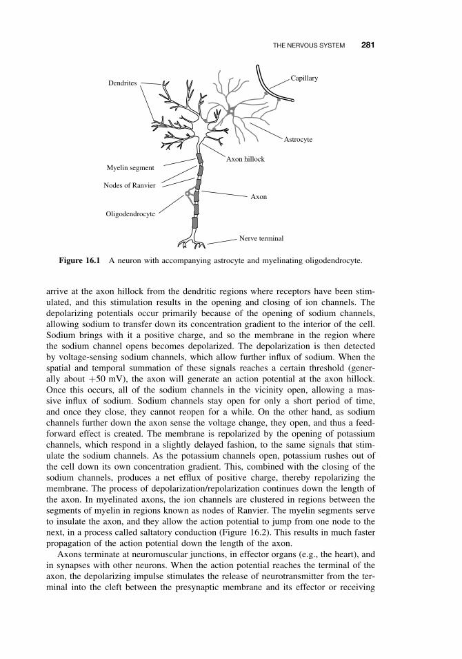
THE NERVOUS SYSTEM 281
Capillary
Astrocyte
Myelin segment
Nodes of Ranvier
Dendrites
Axon hillock
Nerve terminal
Axon
Oligodendrocyte
Figure 16.1 A neuron with accompanying astrocyte and myelinating oligodendrocyte.
arrive at the axon hillock from the dendritic regions where receptors have been stim-ulated, and this stimulation results in the opening and closing of ion channels. Thedepolarizing potentials occur primarily because of the opening of sodium channels,allowing sodium to transfer down its concentration gradient to the interior of the cell.Sodium brings with it a positive charge, and so the membrane in the region wherethe sodium channel opens becomes depolarized. The depolarization is then detectedby voltage-sensing sodium channels, which allow further influx of sodium. When thespatial and temporal summation of these signals reaches a certain threshold (gener-ally about +50 mV), the axon will generate an action potential at the axon hillock.Once this occurs, all of the sodium channels in the vicinity open, allowing a mas-sive influx of sodium. Sodium channels stay open for only a short period of time,and once they close, they cannot reopen for a while. On the other hand, as sodiumchannels further down the axon sense the voltage change, they open, and thus a feed-forward effect is created. The membrane is repolarized by the opening of potassiumchannels, which respond in a slightly delayed fashion, to the same signals that stim-ulate the sodium channels. As the potassium channels open, potassium rushes out ofthe cell down its own concentration gradient. This, combined with the closing of thesodium channels, produces a net efflux of positive charge, thereby repolarizing themembrane. The process of depolarization/repolarization continues down the length ofthe axon. In myelinated axons, the ion channels are clustered in regions between thesegments of myelin in regions known as nodes of Ranvier. The myelin segments serveto insulate the axon, and they allow the action potential to jump from one node to thenext, in a process called saltatory conduction (Figure 16.2). This results in much fasterpropagation of the action potential down the length of the axon.
Axons terminate at neuromuscular junctions, in effector organs (e.g., the heart), andin synapses with other neurons. When the action potential reaches the terminal of theaxon, the depolarizing impulse stimulates the release of neurotransmitter from the ter-minal into the cleft between the presynaptic membrane and its effector or receiving
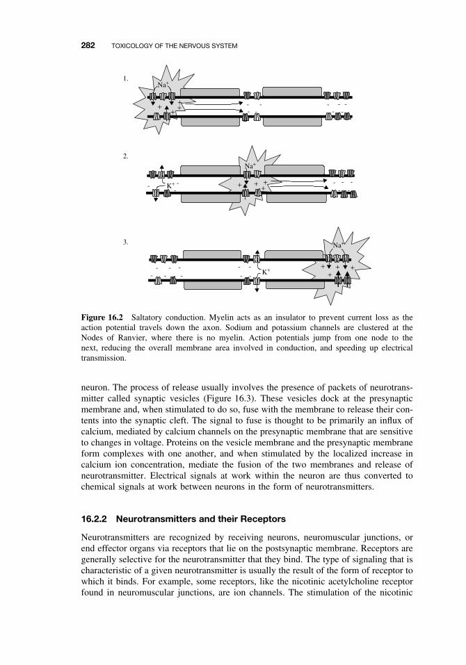
282 TOXICOLOGY OF THE NERVOUS SYSTEM
Na+
+++
--
-- -
- - --
+
Na+
+ ++ + -- - -
-K+- -
-
Na+
+++K+
- ----- -
-- -+
1.
2.
3.
Figure 16.2 Saltatory conduction. Myelin acts as an insulator to prevent current loss as theaction potential travels down the axon. Sodium and potassium channels are clustered at theNodes of Ranvier, where there is no myelin. Action potentials jump from one node to thenext, reducing the overall membrane area involved in conduction, and speeding up electricaltransmission.
neuron. The process of release usually involves the presence of packets of neurotrans-mitter called synaptic vesicles (Figure 16.3). These vesicles dock at the presynapticmembrane and, when stimulated to do so, fuse with the membrane to release their con-tents into the synaptic cleft. The signal to fuse is thought to be primarily an influx ofcalcium, mediated by calcium channels on the presynaptic membrane that are sensitiveto changes in voltage. Proteins on the vesicle membrane and the presynaptic membraneform complexes with one another, and when stimulated by the localized increase incalcium ion concentration, mediate the fusion of the two membranes and release ofneurotransmitter. Electrical signals at work within the neuron are thus converted tochemical signals at work between neurons in the form of neurotransmitters.
16.2.2 Neurotransmitters and their Receptors
Neurotransmitters are recognized by receiving neurons, neuromuscular junctions, orend effector organs via receptors that lie on the postsynaptic membrane. Receptors aregenerally selective for the neurotransmitter that they bind. The type of signaling that ischaracteristic of a given neurotransmitter is usually the result of the form of receptor towhich it binds. For example, some receptors, like the nicotinic acetylcholine receptorfound in neuromuscular junctions, are ion channels. The stimulation of the nicotinic
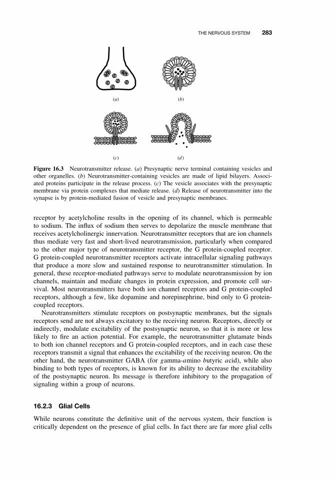
THE NERVOUS SYSTEM 283
(a) (b)
(c) (d )
Figure 16.3 Neurotransmitter release. (a) Presynaptic nerve terminal containing vesicles andother organelles. (b) Neurotransmitter-containing vesicles are made of lipid bilayers. Associ-ated proteins participate in the release process. (c) The vesicle associates with the presynapticmembrane via protein complexes that mediate release. (d) Release of neurotransmitter into thesynapse is by protein-mediated fusion of vesicle and presynaptic membranes.
receptor by acetylcholine results in the opening of its channel, which is permeableto sodium. The influx of sodium then serves to depolarize the muscle membrane thatreceives acetylcholinergic innervation. Neurotransmitter receptors that are ion channelsthus mediate very fast and short-lived neurotransmission, particularly when comparedto the other major type of neurotransmitter receptor, the G protein-coupled receptor.G protein-coupled neurotransmitter receptors activate intracellular signaling pathwaysthat produce a more slow and sustained response to neurotransmitter stimulation. Ingeneral, these receptor-mediated pathways serve to modulate neurotransmission by ionchannels, maintain and mediate changes in protein expression, and promote cell sur-vival. Most neurotransmitters have both ion channel receptors and G protein-coupledreceptors, although a few, like dopamine and norepinephrine, bind only to G protein-coupled receptors.
Neurotransmitters stimulate receptors on postsynaptic membranes, but the signalsreceptors send are not always excitatory to the receiving neuron. Receptors, directly orindirectly, modulate excitability of the postsynaptic neuron, so that it is more or lesslikely to fire an action potential. For example, the neurotransmitter glutamate bindsto both ion channel receptors and G protein-coupled receptors, and in each case thesereceptors transmit a signal that enhances the excitability of the receiving neuron. On theother hand, the neurotransmitter GABA (for gamma-amino butyric acid), while alsobinding to both types of receptors, is known for its ability to decrease the excitabilityof the postsynaptic neuron. Its message is therefore inhibitory to the propagation ofsignaling within a group of neurons.
16.2.3 Glial Cells
While neurons constitute the definitive unit of the nervous system, their function iscritically dependent on the presence of glial cells. In fact there are far more glial cells
284 TOXICOLOGY OF THE NERVOUS SYSTEM
in the nervous system than neurons. Glial cells perform many functions, includingstructural support, insulation, buffering, and guidance of migration during develop-ment. One class of glial cell, called microglia, is responsible for phagocytosis ofcellular debris following injury and infection. The other types of glial cells, collec-tively known as macroglia, are the astrocytes and oligodendrocytes found in the CNS,and the Schwann cells found in the PNS. Oligodendrocytes and Schwann cells formmyelin by wrapping multiple layers of plasma membrane around axons. Astrocytes arethe most numerous of the glial cells, and they help form the blood-brain barrier, take upexcess neurotransmitter and ions, and probably have some nutritive function. Metabolicenzymes expressed within astrocytes also regulate neuronal signaling by catabolizingexcessive amounts of neurotransmitter. Monoamine oxidases, for example, catalyze thebiotransformation of dopamine, norepinephrine, and serotonin into oxidation productsthat are substrates for further enzymatic reactions en route to excretion. The inciden-tal bioactivation of the xenobiotic MPTP to its neurotoxic metabolite MPP+ by theseenzymes will be discussed later in this chapter.
16.2.4 The Blood-Brain Barrier
The blood-brain barrier was conceptualized when it was noted that dyes injected intothe bloodstream of animals stained nearly all tissues except the brain. It is thus thisbarrier and its PNS equivalent, the blood-nerve barrier, that prevents all but a selectfew molecules from entering the nervous system. The barrier itself is not a singleunitary structure, but a combination of unique anatomical features that prevents thetranslocation of blood-borne agents from brain capillaries into the surrounding tissue.As mentioned above, astrocytes help form the barrier, surrounding capillary endothelialcells with extensions of their cytoplasm known as end-feet. There are also pericytes,whose function is not well known, that associate with the capillaries and may helpinduce a functional barrier. Another component of the barrier is the impermeablenature of the endothelial cells that line the interior of capillaries. Capillary endothelialcells in the nervous system are different from those in the periphery in at least threeways. First, brain capillaries form tight junctions of very high resistance between cells.In contrast, peripheral capillaries have low resistance tight junctions, and even open-ings, or fenestrations, that allow compounds to pass between cells. Second, comparedto peripheral endothelial cells, brain endothelial cells are deficient in their ability totransport agents by endocytic mechanisms. Instead, only small lipophilic particles canbe passed transcellularly. For larger molecules, carrier-mediated transport mechanismsare highly selective, and allow only one-way transport. Third, there is an enzymatic bar-rier that metabolizes nutrients and other compounds. Enzymes such as gamma-glutamyltranspeptidase, alkaline phosphatase, and aromatic acid decarboxylase are more preva-lent in cerebral microvessels than in nonneuronal capillaries. Most of these enzymesare present at the lumenal side of the endothelium, Additionally the P-glycoprotein(P-gp) drug efflux transporter is presently thought to exist at the lumenal membranesurface, although some scientists argue that P-gp is actually associated with astrocytes.Finally the CNS endothelial cell displays a net negative charge at its lumenal sideand at the basement membrane. This provides an additional selective mechanism byimpeding passage of anionic molecules across the membrane.
Most of the toxicants that enter the nervous system do so by exploiting mech-anisms designed to allow entry of essential molecules, such as nutrients, ions, and
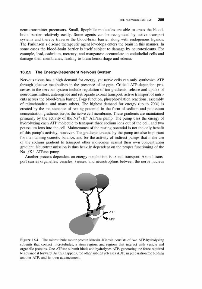
THE NERVOUS SYSTEM 285
neurotransmitter precursors. Small, lipophilic molecules are able to cross the blood-brain barrier relatively easily. Some agents can be recognized by active transportsystems and thereby traverse the blood-brain barrier along with endogenous ligands.The Parkinson’s disease therapeutic agent levodopa enters the brain in this manner. Insome cases the blood-brain barrier is itself subject to damage by neurotoxicants. Forexample, lead, cadmium, mercury, and manganese accumulate in endothelial cells anddamage their membranes, leading to brain hemorrhage and edema.
16.2.5 The Energy-Dependent Nervous System
Nervous tissue has a high demand for energy, yet nerve cells can only synthesize ATPthrough glucose metabolism in the presence of oxygen. Critical ATP-dependent pro-cesses in the nervous system include regulation of ion gradients, release and uptake ofneurotransmitters, anterograde and retrograde axonal transport, active transport of nutri-ents across the blood-brain barrier, P-gp function, phosphorylation reactions, assemblyof mitochondria, and many others. The highest demand for energy (up to 70%) iscreated by the maintenance of resting potential in the form of sodium and potassiumconcentration gradients across the nerve cell membrane. These gradients are maintainedprimarily by the activity of the Na+/K+ ATPase pump. The pump uses the energy ofhydrolyzing each ATP molecule to transport three sodium ions out of the cell, and twopotassium ions into the cell. Maintenance of the resting potential is not the only benefitof this pump’s activity, however. The gradients created by the pump are also importantfor maintaining osmotic balance, and for the activity of indirect pumps that make useof the sodium gradient to transport other molecules against their own concentrationgradient. Neurotransmission is thus heavily dependent on the proper functioning of theNa+/K+ ATPase pump.
Another process dependent on energy metabolism is axonal transport. Axonal trans-port carries organelles, vesicles, viruses, and neurotrophins between the nerve nucleus
ATP
ADP
Figure 16.4 The microtubule motor protein kinesin. Kinesin consists of two ATP-hydrolyzingsubunits that contact microtubules, a stem region, and regions that interact with vesicle andorganelle proteins. One ATPase subunit binds and hydrolyses ATP, generating the force requiredto advance it forward. As this happens, the other subunit releases ADP, in preparation for bindinganother ATP, and its own advancement.
286 TOXICOLOGY OF THE NERVOUS SYSTEM
and the terminal. This distance can be quite long when one considers that the length ofthe sciatic nerve, for example, can be up to one meter. Anterograde transport (from cellbody to terminal) is accomplished by two mechanisms defined by their rate, fast axonaltransport and slow axonal transport. Fast axonal transport proceeds at rates of approx-imately 400 mm/day, and is mediated by the ATP-dependent motor protein kinesin.Kinesin forms cross-bridges between vesicles or organelles and microtubules, and dualprojections of these cross-bridges shift back-to-front along microtubules in a coor-dinated, ATP-dependent manner, such that the entire molecule appears to be walking(Figure 16.4). Slow axonal transport is used to carry cytoskeletal elements such as tubu-lin and neurofilaments to the far ends of the axon, and it proceeds at approximately 0.2to 5 mm/day. Traditionally slow transport has been regarded as passively dependent onaxoplasmic flow; however, recent evidence suggests that the cytoskeletal elements actu-ally move rather quickly but frequently stall in a stop-and-go fashion. Fast axonal trans-port also proceeds retrogradely, mediated by the ATP-dependent motor protein dynein.The rate of retrograde transport is about 200 mm/day. Neurons use retrograde transportfor recycling membranes, vesicles, and their associated proteins. Neurotrophic factors,and some viruses and toxins (e.g., tetanus toxin) are also transported by this mechanism.
16.3 TOXICANT EFFECTS ON THE NERVOUS SYSTEM
Neurotoxicants affect the nervous system in a number of different ways. Some neuro-toxicants damage the distal portions of axons without much effect on the remainder ofthe cell, while others produce outright cell death. Still others affect signaling processesin the nervous system, without causing structural damage. This wide variety of neu-rotoxicant effects is due in part to the unique nature of the different types of neuronsand glia. Neurons may be differentially vulnerable to certain neurotoxicants becauseof their functional characteristics, as in the case of the targeting of substantia nigraneurons by the active metabolite of MPTP, an agent that causes Parkinson’s disease.The substantia nigra, a brain region where neurons that synthesize dopamine are par-ticularly abundant, sends out axons that project to other parts of the brain where thedopamine is released. After release, these neurons take back up the synaptic dopaminevia selective transporters on the nerve endings. The damaging metabolite of MPTP,MPP+, is not distinguished from dopamine by the uptake transporter, so when present,MPP+ is taken up as well. MPP+ kills substantia nigra neurons by affecting mito-chondrial energy production and promoting free radical formation. The death of theseneurons results in a lack of dopamine release in an area of the brain called the stra-tum, which is responsible for the control of movement. The loss of dopamine in thestratum causes the hallmark symptoms of the neurodegenerative disease Parkinsonism:slowed movement, rigidity, and tremors. Because epidemiological studies have linkedParkinsonism in some patients with the agricultural use of pesticides, many of whichare toxic to mitochondria, scientists believe that at least some cases of Parkinson’sdisease may be related to long-term exposure to environmental toxins.
MPTP is one example of a toxicant that causes direct structural damage to neurons,resulting in loss of function. In the following sections, other types of structural andfunctional effects of neurotoxicants are described. Structural effects are divided intothree primary types: effects on myelin formation, primary damage to axons, and directpromotion of cell death. Neurons may also be secondarily affected by neurotoxicants
TOXICANT EFFECTS ON THE NERVOUS SYSTEM 287
that target other cells in the nervous system, disrupting normal homeostatic function andcausing structural or functional damage. Another method by which toxicants may affectthe function of the nervous system is by directly altering synaptic neurotransmission.
16.3.1 Structural Effects of Toxicants on Neurons
Demyelination. The role of myelin in the nervous system is to aid in signal trans-duction. Myelin acts like an electrical insulator by preventing loss of ion current, andintact myelin is critical for the fast saltatory nerve conduction discussed above. Neu-rotoxicants that target the synthesis or integrity of PNS myelin may cause muscleweakness, poor coordination, and paralysis. In the brain, white matter tracts that con-nect neurons within and between hemispheres may be destroyed, in a syndrome knownas toxic leukoencephalopathy. A multifocal distribution of brain lesions is reflected inmental deterioration, vision loss, speech disturbances, ataxia (inability to coordinatemovements), and paralysis.
Demyelination occurs secondary to axonal degeneration, a topic covered in thesection on axonopathy. Neurotoxicants that produce primary demyelination are uncom-mon, but may be divided into those that affect the integrity of the myelin sheathwithout or prior to damage to the myelinating cells, and those that directly injuremyelin-producing cells. The former include agents like hexachlorophene, isoniazid,and the organotins. These compounds cause reversible edema between the layers ofmyelin by a mechanism that is yet unclear. The optic nerve is particularly suscepti-ble to demyelination by hexachlorophene and organic solvents, whereas other cranialnerves, such as the trigeminal and vestibulocochlear, are vulnerable to styrene, xylene,and to trichloroethylene, an agent used in dry-cleaning. The metalloid tellurium dam-ages myelin by inhibiting an enzyme involved in the synthesis of cholesterol, a majorcomponent of myelin.
In contrast to agents that target the integrity of the myelin sheath, chronic expo-sure to cyanide and carbon monoxide is thought to directly injure myelin-producingSchwann cell bodies in the PNS and oligodendrocytes in the CNS. Inorganic lead alsocauses direct damage myelinating cells. Oligodendrocytes appear more sensitive to leadtoxicity than astrocytes or neurons. One mechanism for the devastating developmentaleffects of lead exposure may be the preferential inhibition of oligodendrocyte precursorcell differentiation.
Axonopathy. Axonopathy is a specialized form of neuronal damage, involving degen-eration of the axon, while leaving the cell body intact. When axons die and degenerate,myelin breaks down as well, yet Schwann cells may survive and guide regenerationof the axon in some cases. Some toxicants produce axonal injury by directly targetingthe axon itself. Others are thought to cause degenerative changes in axons by compro-mising the metabolic systems of the neuron. In the latter case the distal portion of theaxon degenerates first, because it is this region that is most heavily dependent on intactaxonal transport mechanisms. Since axonal transport is energy-dependent, toxicants thatinterfere with ATP production, such as the nicotinamide analogue Vacor, may causedistal regions to degenerate initially. Agents that target tubulin, like the vinca alkaloids,also cause this type of injury, because the tubulin-derived microtubules are criticallyimportant for axonal transport. With continued exposure the degeneration progresses
288 TOXICOLOGY OF THE NERVOUS SYSTEM
proximally, and eventually will affect the entire neuron. This distal-to-proximal degen-eration is called “dying back neuropathy,” and it affects the longest and largest diameterneurons most severely. If exposure to the toxicant is discontinued before death of theentire proximal axon and cell body, axons in the PNS will often regenerate, but axonalregeneration does not occur within the CNS. Regeneration in the PNS is dependent onSchwann cells proliferating and guiding growth of regenerating axon tips back to thetarget tissue.
In the 1850s Augustus Waller described the sequence of events that occurred fol-lowing transection of a nerve fiber. These events have subsequently become knownas Wallerian degeneration. The essential features of this type of degeneration includeswelling of the axon at the distal end of the proximal segment of the transected axon,distal axonal dissolution and phagocytosis by inflammatory cells, and dissolution ofmyelin, with preservation and proliferation of Schwann cells along the length of theformer axon. Certain neurotoxicants are capable of chemically transecting an axon,producing Wallerian degeneration similar to that occurring after slicing the nerve inhalf. Hexane, for example, forms covalent adducts with neurofilament proteins. Thischemical crosslinking is thought to result in neurofilamentous axonal swellings thatessentially block transport to regions of the axon distal to the swelling. The distalregions then die due to lack of communication with the neuron cell body, undergoingWallerian degeneration.
Axonopathy can manifest as defects in sensory or motor functions, or a combinationof the two. For most neurotoxicants, sensory changes are noticed first, followed by pro-gressive involvement of motor neurons. One historically important case that illustratesthese effects is that of the epidemic poisoning resulting from the consumption of “Gin-ger Jake” during Prohibition. Extracts of ginger used in tonics were legally requiredto contain 5 grams of ginger per milliliter of alcohol. The Department of Agriculturechecked for compliance with this requirement by sampling the tonics, boiling off theethanol, and weighing the solid content. Bootleggers soon discovered that a good dealof money could be saved by cutting back on the ginger and substituting it with adulter-ating agents like castor oil and molasses. It was such an attempt at adulterating GingerJake that led to the addition of Lyndol, a triorthocresyl phosphate (TOCP)-containingoil used in lacquers and varnishes, to tonic that was consumed by hundreds of thousandsof people. The earliest signs that developed in people who had consumed the productwere noted days to weeks later, and began with tingling and numbness in the handsand feet. In many, this progressed to leg cramps, weakness of the legs and arms, andataxia. Those with minor symptoms improved, but perhaps thousands of people wereleft permanently paralyzed by the incident. Today TOCP is used to study the syndromeof delayed effects caused by some organophosphate compounds, commonly known asorganophosphate-induced delayed neuropathy (OPIDN). The nature of OPIDN is stillpoorly understood. It appears not to be associated with organophosphate inhibition ofacetylcholinesterase, but rather with another neuronal enzyme, the neuropathy targetesterase (NTE). The physiological role of NTE is unknown.
Neuronopathy. Neuronopathy refers to generalized damage to nerve cells, with theprimary damage occurring at the nerve cell body. Axonal and dendritic processes diesecondarily in response to loss of the cell body. Like other cells in the body, neuronsdie by one of two processes distinguished by their morphological and molecular fea-tures: apoptosis and necrosis. (This division is overly simplistic; there is much debate
TOXICANT EFFECTS ON THE NERVOUS SYSTEM 289
over the characteristics of the two categories, and whether there are more than twocategories of cell death. Nevertheless, only these two will be considered here.) Oftenthe same neurotoxicants can promote either form of cell death, depending on the inten-sity of the insult. For example, methylmercury given to rats at a high dose for oneweek causes widespread histopathological damage consistent with necrosis, whereaslower doses spread out over a longer time period results in apoptotic changes restrictedprimarily to cerebellar granule cells. It is thought that the severe and abrupt loss of cel-lular energy production by impairment of mitochondrial activity and plasma membranedisruption are responsible for necrotic death of neurons. This affects surrounding tissuemore than apoptosis, since the dying cells release their contents and localized inflam-matory responses ensue. On the other hand, apoptosis is death that is encoded withinindividual cells. Apoptosis is characterized by a process of cell shrinkage, pyknosisand fragmentation of nuclei, and membrane budding. The dying cell breaks apart intosmall membrane-bound apoptotic fragments that are phagocytosed, and thus collateraldamage is reduced because only cells with activated death programs are affected. Itis important to remember that apoptotic and necrotic mechanisms of cell death canoccur concomitantly or sequentially, and thus are part of a continuum of effects asso-ciated with dose-dependent alterations in cellular energy production and the differentialsensitivity of neuronal subtypes.
Many neurotoxicants produce their effects by promoting cell death in neurons.Neurotoxicant-induced cytotoxicity has been associated with the pathogenesis of anumber of neurodegenerative disorders, including Alzheimer’s disease, Parkinson’sdisease, and amyotrophic lateral sclerosis (ALS), and the weight of evidence sug-gests that toxicant exposure is a risk factor for these diseases. One area of intenseresearch focus has been the toxic effects of excessive signaling by glutamate and otherexcitatory amino acids (EAAs), and the role that EAAs may play in neurodegener-ative disorders. Glutamate activates ion channel receptors that open to allow influxof calcium and other ions into the neuron. This influx of ions, combined with othersecond messenger events that promote further intracellular release of calcium, con-tribute to calcium overload. Signaling cascades are then activated in response to theintracellular calcium, and these pathways eventually lead to activation of oxidativestress and programmed cell death (apoptotic) pathways. EAA-mediated neuronopathicinjury has been extensively studied for its role in ischemic and seizure-induced braindamage. It is furthermore thought that the glutamate receptor agonist, domoic acid, atoxin produced by algae, is responsible for several outbreaks of shellfish poisoning.In one incident in 1987, several people died, and dozens became ill with dizziness,seizures, and memory loss after consuming blue mussels. The mussels were contam-inated with domoic acid, found in high levels following an algae bloom near PrinceEdward Island, Canada. More recently domoic acid produced by algae blooms hasbeen blamed for the abnormal behavior and deaths of pelicans, cormorants, and sealions on the California coast.
16.3.2 Effects of Toxicants on Other Cells
Toxicants may selectively target glial cells for a number of reasons. Myelinating glialcells constantly synthesize cholesterol and cerebroside for myelin production; thustoxicants that affect these synthetic pathways will preferentially affect myelination.The hydrophobic nature of myelin may serve as a reservoir for lipophilic toxicants
290 TOXICOLOGY OF THE NERVOUS SYSTEM
as well. The specialized structures of cells that form myelin also present unique chal-lenges to cellular homeostasis, increasing the vulnerability of these cells to toxicantaction.
In general, a toxic or physical insult to either neurons or glial cells eventuallyleads to changes in the other cell type. Because of this, it is often difficult to deter-mine whether the primary insult was neuronal or glial. Metals such as lead, cadmium,and aluminum are capable of inducing cell death in cultured astrocytes and endothe-lial cells. Methylmercury preferentially accumulates in astrocytes and to some extentin microglia, causing cellular swelling. The swelling is presumably the effect ofmethylmercury interfering with ion channels, because ion channel blockers can reversethis effect. Astrocytes are important reservoirs of excess glutamate, and swollen astro-cytes release glutamate in and around synapses, potentially causing the excitotoxicitydescribed above. Astrocyte swelling also has effects on brain blood flow, since astro-cyte end-feet surround the blood vessels of the CNS. Not only does swelling result inreduced lumen size, but the distances substrates and waste products must diffuse toreach the bloodstream is increased.
Astrocytes and microglial cells become activated secondarily to brain injury, suchas acute trauma or toxic lesioning. This activation, known as “reactive gliosis” is char-acterized by astrocyte hypertrophy and hyperplasia. Reactive astrocytes have greatlyenlarged cytoplasmic processes, and produce increased amounts of a protein known asglial fibrillary acidic protein (GFAP). GFAP is often used as a quantitative histochem-ical marker for toxicant-mediated injury in the nervous system.
In addition to their function as phagocytic cells, glial cells produce neurotrophicfactors to prevent neuronal death and promote axonal growth after injury. Glial cellsalso have xenobiotic biotransforming enzymes, but in some cases glial metabolismresults in xenobiotic activation to a toxic metabolite. As discussed above, MPP+ isthe toxic metabolite of MPTP. MPTP is taken up by astrocytes, where monoamineoxidase B converts MPTP to MPP+. While MPP+ seems to cause no damage tothe astrocytes themselves, the astrocytes release this reactive metabolite into synapses,where it is selectively taken up by dopamine re-uptake transporters on the endings ofneurons that normally release and recycle dopamine. MPP+ then kills the dopamin-ergic neurons, and this insult to the movement-controlling brain circuitry results inthe classic motor symptoms of Parkinson’s disease. The properties of MPTP haveonly become known since the 1980s, when its accidental ingestion by heroin addictsresulted in acute Parkinson-like symptoms. These incidents spurred multiple investiga-tions, leading to much of what is known today about the pathogenesis of Parkinson’sdisease.
16.3.3 Toxicant-Mediated Alterations in Synaptic Function
Nervous system function may be adversely affected by neurotoxicants without neces-sarily causing structural damage to tissue. In many cases neurotoxicants interfere withsignaling processes within the nervous system by activating or inhibiting receptors, oraltering the amount of neurotransmitter available to activate receptors. This type ofneurotoxicity is illustrated by the well-characterized actions of the organophosphatesand carbamates on acetylcholine signaling.
Organophosphates inhibit acetylcholinesterase, the enzyme responsible for break-ing down acetylcholine into acetic acid and choline. After acetylcholine has been
TOXICANT EFFECTS ON THE NERVOUS SYSTEM 291
released into the synapse or the neuromuscular junction, acetylcholinesterase terminatesreceptor-stimulating activity by binding acetylcholine in its active site. Separate siteswithin the binding pocket of acetylcholinesterase bind the quaternary nitrogen of thecholine group, and the carbonyl of the ester group. A hydrolytic reaction results in theloss of choline, leaving an acylated serine residue, which is then rapidly hydrolyzed.The biologically active oxon forms of organophosphates also bind to the active site ofacetylcholinesterase, covalently phosphorylating the serine residue in the catalytic siteof the enzyme. The phosphorylation of acetylcholinesterase creates a relatively stableinactive enzyme that persists for hours to days before hydrolysis of the phosphatemoiety occurs spontaneously, and acetylcholinesterase activity is restored. The rate ofspontaneous hydrolysis is increased with larger alkyl groups attached to the phosphatemoiety. When one or more of these alkyl groups is lost, in a process known as “aging,”spontaneous reactivation of acetylcholinesterase by hydrolysis of the phosphate moietyis impossible, and the enzyme is permanently inactivated. Carbamates similarly inhibitacetylcholinesterase by carbamylating the enzyme active site. The stability of carbamy-lation is much less than phosphorylation, however, and spontaneous reactivation thusoccurs faster than with organophosphates.
The effects of acetylcholinesterase inhibition can be seen throughout the nervoussystem. Acetylcholine and its receptors mediate neurotransmission in sympathetic andparasympathetic autonomic ganglia, in the effector organs where autonomic nervesterminate, in neuromuscular junctions, and in the brain and spinal cord. The signs ofhypercholinergic activity are thus very diverse, and include effects mediated by bothnicotinic and muscarinic types of acetylcholine receptor. Hyperstimulation of nicotinicreceptors in neuromuscular junctions results in muscle weakness, in rapid, localizedcontractions called fasciculations, and in paralysis. Nicotinic receptors are also foundin sympathetic and parasympathetic ganglia, and so stimulation of both divisions of theautonomic system is apparent as hypertension, increased heart rate, and papillary dila-tion. Muscarinic receptors in the PNS mediate postganglionic parasympathetic effectson the smooth muscle present in the end organs such as the lung, gastrointestinaltract, eye, bladder, and secretory glands. Hyperstimulation of these receptors resultsin a pattern of toxicity known by the mnemonic SLUDGE (salivation, lacrimation,urination, defecation, GI upset, emesis). Bronchospasm and bradycardia are also mus-carinic effects. In the CNS, confusion, anxiety, restlessness, ataxia, seizures, and comaare effects of both muscarinic and nicotinic receptor overstimulation. Death generallyoccurs from respiratory paralysis.
Treatment for toxicity by organophosphates and carbamates is directed at coun-teracting hyperstimulation and regenerating acetylcholinesterase enzymatic activity.Atropine is a muscarinic receptor antagonist (it blocks acetylcholine from binding tothe muscarinic receptor), and is used to counteract the effects of cholinergic overactiv-ity. Atropine has no effect at the nicotinic receptor, however, so the skeletal muscularand some of the sympathetic effects of cholinergic hyperstimulation will remain afteradministration of atropine. Inhibition of acetylcholinesterase activity by organophos-phates can be reversed by administration of oxime compounds (e.g., pralidoxime and2-PAM). These compounds contain a quaternary nitrogen that binds to the choline bind-ing site of acetylcholinesterase, positioning the oxime portion of the molecule near theesteratic site. Oximes are themselves reversible inhibitors of acetylcholinesterase, buttheir mechanism of organophosphate reversal is by attack of the covalent phosphoser-ine bond, releasing the phosphate group. Oximes are not effective on dealkylated or
292 TOXICOLOGY OF THE NERVOUS SYSTEM
“aged” enzymes, so they must be administered soon after organophosphate intoxicationin order to be effective. They are also ineffective against carbamate-mediated toxicity,and some researchers believe they actually worsen carbamate effects by stabilizing thecarbamylation of the enzyme.
Many biological toxins produce hyperstimulation of receptors by directly bind-ing and activating them (agonism), or reduce receptor stimulation by prohibiting theendogenous ligand from activating them (antagonism). A number of snake and spidervenoms, mushroom and plant alkaloids, affect nervous system function by these mech-anisms. As the binding of receptors by these agents is usually reversible, their effectsare reversible as well (although some may still cause death by massively altering neu-ronal signaling). Beyond the receptor, an active area of current research is the role ofintracellular signaling molecules in mediating the effects of neurotoxicants. The effectsof a number of metals, in particular, may be related to their ability to act as cofactorsfor proteins involved in intracellular signaling. To date, however, few signal transduc-tion molecules have been shown to be directly affected by neurotoxicants. Notableexceptions are cholera and pertussis toxins, which selectively target G proteins, buttheir primary effects are on the gastrointestinal and respiratory systems, respectively,rather than on the nervous system.
On the other hand, the Clostridium toxins, botulinum (causing botulism) and tetano-spasmin (causing tetanus), block neurotransmission by inhibiting release of neuro-transmitter into synapses and at motor end-plates in muscle. Both of these agents arestructurally similar proteases, but the effects they cause are vastly different. Botulinumtoxin enters presynaptic motor neurons in the PNS, where it cleaves proteins that areinvolved in the fusion of synaptic vesicles with membranes. This cleavage results inthe inhibition of acetylcholine release from the presynaptic terminal, and thus mus-cles cannot be stimulated to contract. The clinical result of botulinum intoxication(usually by ingestion) is a flaccid paralysis. Recovery occurs when the presynapticneuron sprouts new nerve endings that contact the muscle and create new motor end-plates. Tetanus toxin causes a completely different clinical picture, even though itssubstrate specificity for cleavage of proteins is very similar. Once taken up into thepresynaptic nerve endings, tetanus associates with endosomes, and like endosomes, istransported retrogradely toward the neuron cell body. Tetanospasmin then continuesits trek to the dendritic regions of the neurons, where it is released, again retrogradely,into synapses. Usually these synapses are within the spinal cord, where interneuronssend an inhibitory signal (via the neurotransmitters glycine and GABA) to motor neu-rons to slow their activity and prevent massive muscular contraction. The presynapticmembranes of the interneurons take up tetanospasmin, and this is where most of itsactivity occurs. By cleaving release-regulating proteins in the interneuron terminal,tetanospasmin prevents the release of glycine and GABA onto the motor neurons.The motor neurons then become hyperactive, and this results in overstimulation of themotor end-plate with acetylcholine. Clinically this results in a spasms, stiffness, andwhole-body paralysis. Again, the interneurons themselves do not die, but they mustform new synapses with the motor neurons. Fortunately, in all but the most severecases, recovery is complete. The reformation of new synapses by neurons, even in theCNS, is an example of the remarkable plasticity of the nervous system. The continualformation and reformation of synaptic connections allows the organism to change andadapt to an inconstant environment.
NEUROTOXICITY TESTING 293
16.4 NEUROTOXICITY TESTING
A large number of the chemicals used in industry today remain poorly characterizedwith respect to their toxic effects on the nervous system. In order to determine potentialrisks to human and environmental well-being, existing neurotoxicants must be identi-fied, and the approximately 2000 new chemicals introduced each year must be screenedfor their potential neurotoxic effects. Often, a tiered approach is used, with the firsttier consisting of general screening tests to identify acute hazards. The Environmen-tal Protection Agency (EPA) has proposed screening guidelines for tests in rodentsthat include a functional observational battery (FOB, see below) to evaluate sensory,motor, and autonomic effects, tests that identify changes in motor activity, and neu-ropathological assessment. Interpretation of the outcome of tier 1 screening may leadto more selective testing and examining the effects of repeated exposures in the secondtier. Specialized tests for behavioral effects, developmental neurotoxicity, or delayedorganophosphate effects may be required. If necessary, a third tier of testing char-acterizes mechanisms of neurotoxicant-induced injury. Complete and comprehensiveevaluation of potential neurotoxicant effects requires that data from different types ofsources be considered; this can range from molecular interactions to whole animaland human exposure analysis. Below are examples of techniques commonly used fortesting neurotoxic effects.
16.4.1 In vivo Tests of Human Exposure
Historically the first indication of neurotoxic potential by a chemical has often followedaccidental human exposure in the workplace. Case reports of incidents involving indi-viduals, or clusters of individuals, are useful for documentation but generally providea limited amount of information about the specific details of an exposure. Proceduresincluded in most case reports include a patient medical history and clinical neuro-logical exam, sometimes supplemented with psychiatric or neurophysiological tests,and/or neuroimaging. Although the specific tests involved vary depending on the clin-ician, most basic clinical neurological exams rely heavily on evaluation of mentalstatus (level of consciousness, orientation, mood, etc.) and sensorimotor function (gait,coordination, muscle tone, sensitivity to touch, reflexes).
Human epidemiological studies generally represent a deeper investigation into thecausal relationship between an exposure and neurotoxicological effects. Some ofthe methods used to identify neurotoxic effects in epidemiological studies includebehavioral assessments, neurophysiological evaluations, and neuroimaging techniques.Neurobehavioral assessments examine a variety of psychological and cognitivefunctions such as mood, attention, memory, perceptual and visuospatial ability, andpsychomotor performance. In an effort to standardize neurotoxicological testing ofhuman behavioral effects, particularly for studies involving worksite exposure, theWorld Health Organization (WHO) and the US National Institute for OccupationalSafety and Health (NIOSH) devised a the Neurobehavioral Core Test Battery (NCBT).The NCBT (Table 16.1) consists of seven tests that were shown previously tobe sensitive indicators of neurotoxicant exposure. The battery is designed to beadministered one on one by an examiner. Although this battery has a relatively narrow
294 TOXICOLOGY OF THE NERVOUS SYSTEM
Table 16.1 The WHO Neurobehavioral Core Test Battery (NBCT)
Domain Analysis Test Task
Psychomotorperformance
Motor speed,motor steadiness
Pursuit aiming Follow a pattern of smallcircles, placing a dotin each circle around apattern; subject’s scoreis number of taps incircle within oneminute.
Manual dexterity,hand–eyecoordination
Santa AnaDexterity Test
Perform skillfulmovements with handsand arms.
Perceptual codingand perceptualmotor speed
Wechsler DigitSymbol Test
Each number in a list isassociated with asimple symbol. On alist of random digitswith blank spacesbelow them, write thecorrect symbols inblank spaces as fast aspossible.
Attention andshort-termmemory
Attention andresponse speed
Simple reactiontime
Test reactions of handsor feet from visual andauditory signals.
Visual perceptionand memory
Benton VisualRetention Test
Recall and reproducefigures.
Auditory memory Wechsler DigitSpan Test
Recall digits in seriesforwards andbackwardsimmediately afterhearing them.
Mood and affect Profile of MoodStates
Evaluate, byquestionnaire, anger,tension, confusion,depression, etc.
focus, primarily on the effects most commonly seen in CNS toxicity, it also providessuggestions for the selection of further testing depending on the exposure setting. TheNCBT has been widely used because of its ease of administration, relatively low cost,and its large base of control data. A broader battery of cognitive and psychomotortests that is often used is the Neurobehavioral Evaluation System (NES). The NESconsists of a combination of automated (computerized) and hand-administered tests.The sensitivity of the NES to effects caused by neurotoxicants in industrial settingshas been validated internationally.
Neurobehavioral examinations are useful for identifying neurotoxicant-mediateddeficits, but it is often difficult to localize the site of toxic action from such tests.For example, sensorimotor tests of reaction time, manual dexterity, hand-eye coordi-nation, and finger tapping can indicate either neuromuscular or psychomotor damage.
NEUROTOXICITY TESTING 295
The results of these tests thus should be interpreted in the context of other experiments.For example, electrophysiological techniques can help to focus an investigation to thesite of the lesion, and characterize electrical dysfunction within the damaged nerves.Electrophysiological nerve conduction studies can distinguish between proximal anddistal axonal lesions in peripheral nerves and can be performed noninvasively (i.e.,with skin surface electrodes). Characteristic changes in the velocity, duration, ampli-tude, waveform, or refractory period of peripheral nerves may be detected, dependingon the agent. Evoked potentials represent another useful electrophysiological endpoint.These procedures measure the function of an entire system, such as the visual, audi-tory, or motor systems. The specific pathway is stimulated by an evoking stimulus,such as a flash of light or electrical nerve stimulation. The evoked potentials are readas changes in ongoing electroencephalograms (EEGs) in response to the stimulation.Thus the activity of the entire neural circuit is evaluated in the brain after peripheralstimulation. Evoked potentials can be very sensitive indicators of changes in neuralactivity when performed in a carefully controlled environment, and when interpretedin light of behavioral or other physiological findings.
An increasingly popular method of documenting brain pathology is the use of neu-roimaging methods. Computerized axial tomography (CAT) and magnetic resonanceimaging (MRI) can produce images of the brain that can show structural changesin the volume or density of a specific region or ventricle. Other techniques, such aspositron emission tomography (PET) and single photon emission computerized tomog-raphy (SPECT), use radioactive tracer molecules to determine functional biochemicalchanges in processes like glucose utilization or receptor binding. The number of casesso far analyzed with neuroimaging techniques is still relatively small, and thus specifictoxicant-mediated effects are not well characterized. Nevertheless, this growing fieldpromises to contribute significantly to neurotoxicity studies in the future.
16.4.2 In vivo Tests of Animal Exposure
The primary approach currently used to detect and characterize potential neurotoxicantsinvolves the use of animal models, particularly rodents. Behavioral and neurophysio-logical tests, often similar to the ones used in humans, are typically administered. Thesensitivity of these measures to neurotoxicant exposure is widely accepted. Althoughit is often not possible to test toxicant effects on some higher behavioral functions inanimals (e.g., verbal ability, cognitive flexibility), there are other neurobehavioral out-comes such as memory loss, motivational defects, somatosensory deficits, and motordysfunction that can be successfully modeled in rodents. These behaviors are basedon the ability of the nervous system to integrate multiple inputs and outputs, thus theycannot be modeled adequately in vitro. Although the bulk of neurotoxicity data hasbeen collected in rodents, birds and primates are also used to model human behavioraloutcomes.
As mentioned above, a useful screening tool for neurotoxicant exposure is a batteryof observational tests of function known as an FOB. FOBs, like the one developedby the EPA, are used to detect overt changes in behavior and physiology of animalsexposed to neurotoxicants. In the typical exam, an observer documents cageside obser-vations regarding the appearance and activity of the animal. Then the animal is handledand examined for obvious signs such as lacrimation, salivation, or piloerection. Pupil-lary light responses and temperature are recorded, and the ease of handling the animal
296 TOXICOLOGY OF THE NERVOUS SYSTEM
is also noted. The animal is then placed in an open field, such as the top of a labo-ratory cart, and observed for a set period of time, during which the observer recordsexploratory behaviors, excretion rate, and whether or not there are any motor abnor-malities. A number of manipulations are then performed to assess hearing, sensitivityto touch, righting reflex, coordination, and grip strength. The FOB used in the EPAguidelines has shown remarkable consistency in detecting chemical effects in diversetesting laboratories and situations. A test of motor activity can be administered alongwith the FOB, consisting of quantitative evaluations of the animal’s movement in eitheran open field or a maze. Motor activity tests reflect integrative abilities of the nervoussystem to process sensory input, association, and motor output. A number of agents,such as toluene, triadimefon, and chlorinated hydrocarbons, increase or decrease motoractivity in a toxicant-specific manner, unrelated to their general effects on the healthof the animal.
More in-depth behavioral tests are required if dose-related toxicant effects are notedin screening tests. These tests may also be required as part of more selective toxicolog-ical screening, such as for developmental neurotoxicity. Focused tests of neuromotorfunction and activity, sensory functions, memory, attention, and motivation help toidentify sites of toxicant-mediated lesioning, aid in the classification of neurotoxicants,and may suggest mechanisms of action. Some of these tests, like the schedule-controlledoperant behavior tests for cognitive function, require animal training and extensiveoperator interaction with the animals.
16.4.3 In vitro Neurochemical and Histopathological End Points
In vitro methods for studying neurotoxicant effects are a valuable supplement towhole animal and human testing, allowing the researcher to supplement findings, testhypotheses, and reduce the number of animals used for toxicity testing. Much of theneurochemical and histopathological data on neurotoxicant effects in humans and ani-mals is gathered concomitantly with, or immediately after, performing behavioral tests.This may involve collection of bodily fluids or samples from living subjects for thepurpose of analyses such as red blood cell or plasma acetylcholinesterase or NTEactivity, determination of hormone or neurotransmitter concentration, or detection ofthe presence of toxicant or metabolite in the cerebrospinal fluid.
Postmortem tissues can provide a wealth of information about the location, timing,extent, and mechanism of neurotoxicant-induced damage. For example, changes inthe gross morphology and weight of brain or nerves may be seen at higher levels oftoxicant exposure. Microscopically, fixed and stained tissues reveal the type of damageto target cells, such as axonopathic or demyelinating lesions. Degenerative changes incells may be indicative of the injurious process, and whether cells are dying by necrosisor apoptosis. Typical stains such as cresyl violet (Nissl stain) and silver (Golgi stain)are useful for cell morphology and counting. Other stains are selective for damagedcells, like the specialized silver impregnation techniques that are frequently used toidentify neurotoxicant-mediated degeneration of neurons.
Tissue sections may also be processed for immunohistochemical staining. A fre-quently used immunochemical marker for neuropathologic insult is glial fibrillary acidicprotein (GFAP). GFAP is produced in large amounts by reactive astrocytes that prolif-erate in response to tissue injury. Stress proteins, apoptotic signals, and immediate earlygenes are also utilized as markers of neuronal activity and injury. Other protein markers
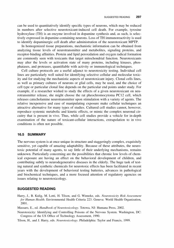
SUGGESTED READING 297
can be used to quantitatively identify specific types of neurons, which may be reducedin numbers after selective neurotoxicant-induced cell death. For example, tyrosinehydroxylase (TH) is an enzyme involved in dopamine synthesis and, as such, is selec-tively expressed in dopamine-containing neurons. Loss of TH immunoreactivity is usedto identify dopaminergic cell death after administration of the neurotoxicant MPTP.
In homogenized tissue preparations, mechanistic information can be obtained fromanalyzing tissue levels of neurotransmitter and metabolites, signaling proteins, andreceptor-binding affinities. Protein and lipid peroxidation and oxygen radical formationare commonly seen with toxicants that target mitochondrial function. Neurotoxicantsmay alter the levels or activation state of many proteins, including kinases, phos-phatases, and proteases, quantifiable with activity or immunological techniques.
Cell culture protocols are a useful adjunct to neurotoxicity testing. Individual celllines are particularly well suited for identifying selective cellular and molecular toxic-ity and for studying the mechanistic aspects of neurotoxicant injury. Clonal cells lines,as well as primary cultures of neurons or glial cells, may be used, and the choice ofcell type or particular clonal line depends on the particular end points under study. Forexample, if a researcher wished to study the effects of a given neurotoxicant on neu-rotransmitter release, she might choose the rat pheochromocytoma PC12 cell, whichreleases catecholamine neurotransmitter upon stimulation with a variety of agents. Therelative inexpensive and ease of manipulating exposure make cellular techniques anattractive alternative for many types of studies. Cultured cell studies cannot, however,reproduce systemic metabolic and kinetic effects, or mimic the complex neuronal cir-cuitry that is present in vivo. Thus, while cell studies provide a vehicle for in-depthexamination of the nature of toxicant-cellular interactions, extrapolation to in vivoconditions is often not possible.
16.5 SUMMARY
The nervous system is at once unique in structure and staggeringly complex, exquisitelysensitive, yet capable of amazing adaptability. Because of these attributes, the neuro-toxic potential of many agents, to say little of their underlying mechanisms, remainsunknown. Particularly concerning are the possibilities that chronic low levels of chem-ical exposure are having an effect on the behavioral development of children, andcontributing subtly to neurodegenerative diseases in the elderly. The huge task of test-ing natural and synthetic chemicals for neurotoxic effects has been facilitated in recentyears with the development of behavioral testing batteries, advances in pathologicaland biochemical techniques, and a more focused attention of regulatory agencies onissues relating to neurotoxicology.
SUGGESTED READING
Harry, J., B. Kulig, M. Lotti, H. Tilson, and G. Winneke, eds. Neurotoxicity Risk Assessmentfor Human Health. Environmental Health Criteria 223. Geneva: World Health Organization,2001.
Massaro, E., ed. Handbook of Neurotoxicology. Totowa, NJ: Humana Press, 2002.
Neurotoxicity: Identifying and Controlling Poisons of the Nervous System. Washington, DC:Congress of the US Office of Technology Assessment, 1990.
Tilson, H., and J. Harry, eds. Neurotoxicology. Philadelphia: Taylor and Francis, 1999.

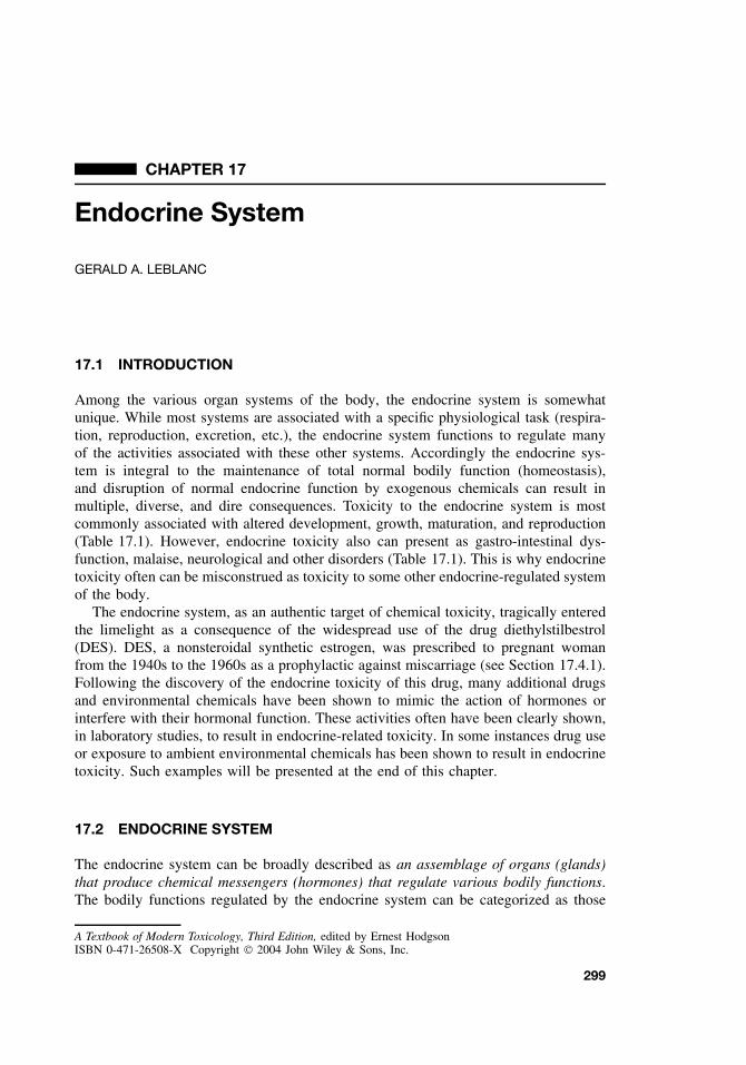
CHAPTER 17
Endocrine System
GERALD A. LEBLANC
17.1 INTRODUCTION
Among the various organ systems of the body, the endocrine system is somewhatunique. While most systems are associated with a specific physiological task (respira-tion, reproduction, excretion, etc.), the endocrine system functions to regulate manyof the activities associated with these other systems. Accordingly the endocrine sys-tem is integral to the maintenance of total normal bodily function (homeostasis),and disruption of normal endocrine function by exogenous chemicals can result inmultiple, diverse, and dire consequences. Toxicity to the endocrine system is mostcommonly associated with altered development, growth, maturation, and reproduction(Table 17.1). However, endocrine toxicity also can present as gastro-intestinal dys-function, malaise, neurological and other disorders (Table 17.1). This is why endocrinetoxicity often can be misconstrued as toxicity to some other endocrine-regulated systemof the body.
The endocrine system, as an authentic target of chemical toxicity, tragically enteredthe limelight as a consequence of the widespread use of the drug diethylstilbestrol(DES). DES, a nonsteroidal synthetic estrogen, was prescribed to pregnant womanfrom the 1940s to the 1960s as a prophylactic against miscarriage (see Section 17.4.1).Following the discovery of the endocrine toxicity of this drug, many additional drugsand environmental chemicals have been shown to mimic the action of hormones orinterfere with their hormonal function. These activities often have been clearly shown,in laboratory studies, to result in endocrine-related toxicity. In some instances drug useor exposure to ambient environmental chemicals has been shown to result in endocrinetoxicity. Such examples will be presented at the end of this chapter.
17.2 ENDOCRINE SYSTEM
The endocrine system can be broadly described as an assemblage of organs (glands)that produce chemical messengers (hormones) that regulate various bodily functions.The bodily functions regulated by the endocrine system can be categorized as those
A Textbook of Modern Toxicology, Third Edition, edited by Ernest HodgsonISBN 0-471-26508-X Copyright 2004 John Wiley & Sons, Inc.
299
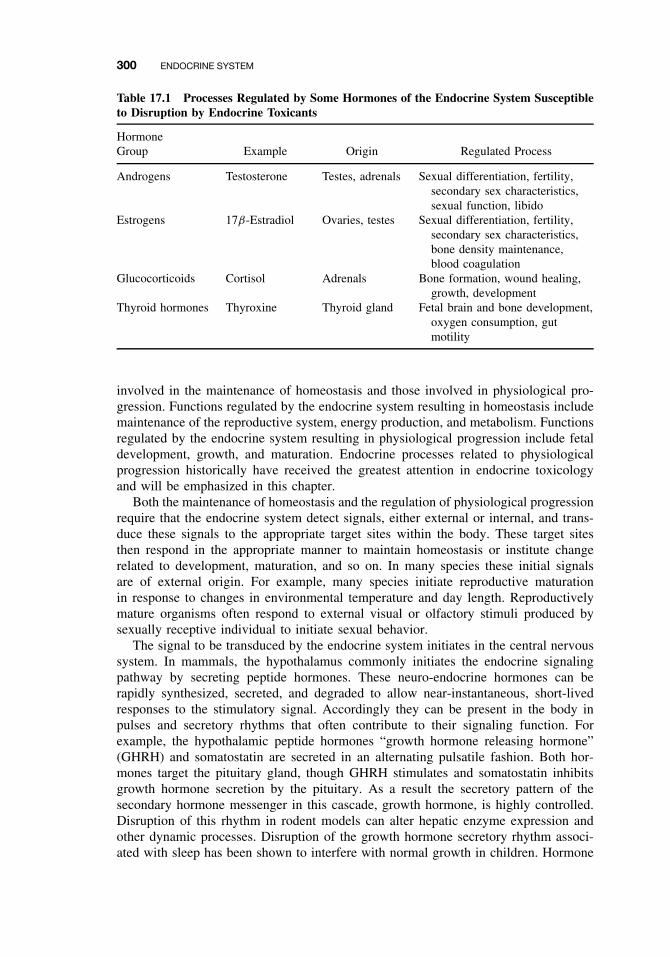
300 ENDOCRINE SYSTEM
Table 17.1 Processes Regulated by Some Hormones of the Endocrine System Susceptibleto Disruption by Endocrine Toxicants
HormoneGroup Example Origin Regulated Process
Androgens Testosterone Testes, adrenals Sexual differentiation, fertility,secondary sex characteristics,sexual function, libido
Estrogens 17β-Estradiol Ovaries, testes Sexual differentiation, fertility,secondary sex characteristics,bone density maintenance,blood coagulation
Glucocorticoids Cortisol Adrenals Bone formation, wound healing,growth, development
Thyroid hormones Thyroxine Thyroid gland Fetal brain and bone development,oxygen consumption, gutmotility
involved in the maintenance of homeostasis and those involved in physiological pro-gression. Functions regulated by the endocrine system resulting in homeostasis includemaintenance of the reproductive system, energy production, and metabolism. Functionsregulated by the endocrine system resulting in physiological progression include fetaldevelopment, growth, and maturation. Endocrine processes related to physiologicalprogression historically have received the greatest attention in endocrine toxicologyand will be emphasized in this chapter.
Both the maintenance of homeostasis and the regulation of physiological progressionrequire that the endocrine system detect signals, either external or internal, and trans-duce these signals to the appropriate target sites within the body. These target sitesthen respond in the appropriate manner to maintain homeostasis or institute changerelated to development, maturation, and so on. In many species these initial signalsare of external origin. For example, many species initiate reproductive maturationin response to changes in environmental temperature and day length. Reproductivelymature organisms often respond to external visual or olfactory stimuli produced bysexually receptive individual to initiate sexual behavior.
The signal to be transduced by the endocrine system initiates in the central nervoussystem. In mammals, the hypothalamus commonly initiates the endocrine signalingpathway by secreting peptide hormones. These neuro-endocrine hormones can berapidly synthesized, secreted, and degraded to allow near-instantaneous, short-livedresponses to the stimulatory signal. Accordingly they can be present in the body inpulses and secretory rhythms that often contribute to their signaling function. Forexample, the hypothalamic peptide hormones “growth hormone releasing hormone”(GHRH) and somatostatin are secreted in an alternating pulsatile fashion. Both hor-mones target the pituitary gland, though GHRH stimulates and somatostatin inhibitsgrowth hormone secretion by the pituitary. As a result the secretory pattern of thesecondary hormone messenger in this cascade, growth hormone, is highly controlled.Disruption of this rhythm in rodent models can alter hepatic enzyme expression andother dynamic processes. Disruption of the growth hormone secretory rhythm associ-ated with sleep has been shown to interfere with normal growth in children. Hormone
ENDOCRINE SYSTEM 301
secretory rhythms have been associated with other physiological processes includingsleep, sexual behavior, and ovulation.
Endocrine signaling pathways from the central nervous system to the target organtypically occur along axes (Figure 17.1). An axis is defined by the endocrine glands thatproduce signaling hormones along the cascade (i.e., hypothalamic–pituitary–gonadalaxis), and sometimes, a terminal target organ of the signaling pathway (i.e., hypotha-lamic–pituitary–gonadal–hepatic axis).
Endocrine signaling cascades offer several advantages over a single hormone signal-ing strategy. Cascades provide several sites at which the signal can be regulated thusensuring maintenance of the appropriate endocrine signal (Figure 17.2). For example,testosterone is secreted by the testis but regulates its own secretion by acting upstreamin the axis at the pituitary gland and hypothalamic gland. Signaling cascades also
Hypothalamus
Pituitarygland
Thyrotropin-ReleasingHormone
Thyrotropin
Thyroid gland
Thyroxine
Gonadotropin-ReleasingHormone
Gonads
Sex steroids
Adrenocorticotropic-ReleasingHormone
Gonadotropin Adrenocorticotropin
Corticosteroids
Adrenal gland
Figure 17.1 Some major neuro–endocrine axes that transduce endocrine signals to targetorgans. Neuro–endocrine signaling is initiated by the secretion of releasing hormones, or insome instances inhibiting hormones, that regulate secretion of the secondary hormone signalby the pituitary. Pituitary hormones then regulate secretion of the tertiary hormone, often asteroid hormone, by the appropriate endocrine gland. The tertiary hormones then stimulate genetranscription at target organs.
302 ENDOCRINE SYSTEM
Sex Steroid Hormones
LuteinizingHormone
Luteinizing HormoneReleasing Hormone
Feedback regulation of sex steroid hormone synthesis
Level I
Level II
Hypothalamus
Pituitarygland
Gonads
Figure 17.2 The hypothalamic–pituitary–gonadal axis. Endocrine signaling cascades providemultiple sites for regulation and ensure optimum signaling.
utilize multiple hormones with differing properties to contribute to the process. Peptidehormones are commonly the intermediate messengers along a signaling cascade, whilethe terminal hormone is often of nonpeptide origin (i.e., steroids). Peptide hormonesoffer advantages as intermediate messengers in that they can be rapidly synthesized anddegraded (i.e., turned “on” and “off”). Peptide hormones also do not require cell entryto elicit activity but rather bind to cell surface receptors. This facilitates a rapid physi-ological response to the hormone. Steroid and other nonpeptide hormones are typicallymore stable, they are maintained in circulation at a relatively constant, physiologi-cally appropriate level, they can be stored as precursor molecules or apolar conjugates,they can be mobilized as polar conjugates, and most often, they require cell entryto interact with its receptor and elicit a response. Accordingly the nonpeptide termi-nal hormones offer the advantages of constant availability but lack the advantages ofrapid modulation.
17.2.1 Nuclear Receptors
Toxicologically the function of the terminal hormones of endocrine cascades (i.e.,steroid, retinoid, thyroid hormones) appear to be most susceptible to disruption bychemicals. This is because many foreign molecules share sufficient characteristics withthese hormone molecules to allow binding to the nuclear receptors of these hormonesin either an agonistic or antagonistic fashion. The binding of the xenobiotic to the
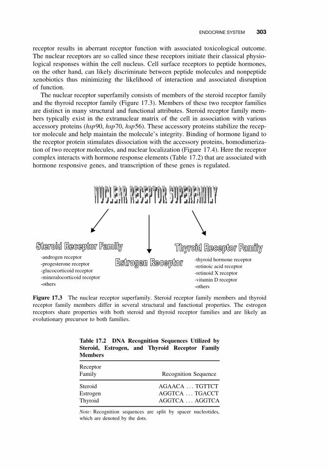
ENDOCRINE SYSTEM 303
receptor results in aberrant receptor function with associated toxicological outcome.The nuclear receptors are so called since these receptors initiate their classical physio-logical responses within the cell nucleus. Cell surface receptors to peptide hormones,on the other hand, can likely discriminate between peptide molecules and nonpeptidexenobiotics thus minimizing the likelihood of interaction and associated disruptionof function.
The nuclear receptor superfamily consists of members of the steroid receptor familyand the thyroid receptor family (Figure 17.3). Members of these two receptor familiesare distinct in many structural and functional attributes. Steroid receptor family mem-bers typically exist in the extranuclear matrix of the cell in association with variousaccessory proteins (hsp90, hsp70, hsp56). These accessory proteins stabilize the recep-tor molecule and help maintain the molecule’s integrity. Binding of hormone ligand tothe receptor protein stimulates dissociation with the accessory proteins, homodimeriza-tion of two receptor molecules, and nuclear localization (Figure 17.4). Here the receptorcomplex interacts with hormone response elements (Table 17.2) that are associated withhormone responsive genes, and transcription of these genes is regulated.
-androgen receptor -progesterone receptor -glucocorticoid receptor -mineralocorticoid receptor -others
-thyroid hormone receptor-retinoic acid receptor-retinoid X receptor -vitamin D receptor-others
Figure 17.3 The nuclear receptor superfamily. Steroid receptor family members and thyroidreceptor family members differ in several structural and functional properties. The estrogenreceptors share properties with both steroid and thyroid receptor families and are likely anevolutionary precursor to both families.
Table 17.2 DNA Recognition Sequences Utilized bySteroid, Estrogen, and Thyroid Receptor FamilyMembers
ReceptorFamily Recognition Sequence
Steroid AGAACA . . . TGTTCTEstrogen AGGTCA . . . TGACCTThyroid AGGTCA . . . AGGTCA
Note: Recognition sequences are split by spacer nucleotides,which are denoted by the dots.
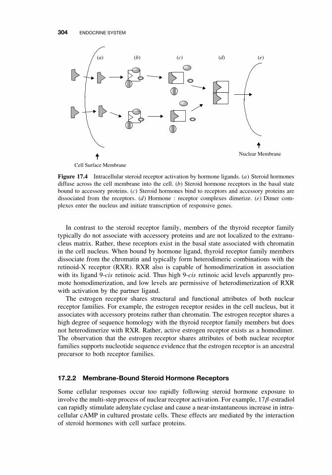
304 ENDOCRINE SYSTEM
(a) (b) (c) (d) (e)
Cell Surface Membrane
Nuclear Membrane
Figure 17.4 Intracellular steroid receptor activation by hormone ligands. (a) Steroid hormonesdiffuse across the cell membrane into the cell. (b) Steroid hormone receptors in the basal statebound to accessory proteins. (c) Steroid hormones bind to receptors and accessory proteins aredissociated from the receptors. (d) Hormone : receptor complexes dimerize. (e) Dimer com-plexes enter the nucleus and initiate transcription of responsive genes.
In contrast to the steroid receptor family, members of the thyroid receptor familytypically do not associate with accessory proteins and are not localized to the extranu-cleus matrix. Rather, these receptors exist in the basal state associated with chromatinin the cell nucleus. When bound by hormone ligand, thyroid receptor family membersdissociate from the chromatin and typically form heterodimeric combinations with theretinoid-X receptor (RXR). RXR also is capable of homodimerization in associationwith its ligand 9-cis retinoic acid. Thus high 9-cis retinoic acid levels apparently pro-mote homodimerization, and low levels are permissive of heterodimerization of RXRwith activation by the partner ligand.
The estrogen receptor shares structural and functional attributes of both nuclearreceptor families. For example, the estrogen receptor resides in the cell nucleus, but itassociates with accessory proteins rather than chromatin. The estrogen receptor shares ahigh degree of sequence homology with the thyroid receptor family members but doesnot heterodimerize with RXR. Rather, active estrogen receptor exists as a homodimer.The observation that the estrogen receptor shares attributes of both nuclear receptorfamilies supports nucleotide sequence evidence that the estrogen receptor is an ancestralprecursor to both receptor families.
17.2.2 Membrane-Bound Steroid Hormone Receptors
Some cellular responses occur too rapidly following steroid hormone exposure toinvolve the multi-step process of nuclear receptor activation. For example, 17β-estradiolcan rapidly stimulate adenylate cyclase and cause a near-instantaneous increase in intra-cellular cAMP in cultured prostate cells. These effects are mediated by the interactionof steroid hormones with cell surface proteins.
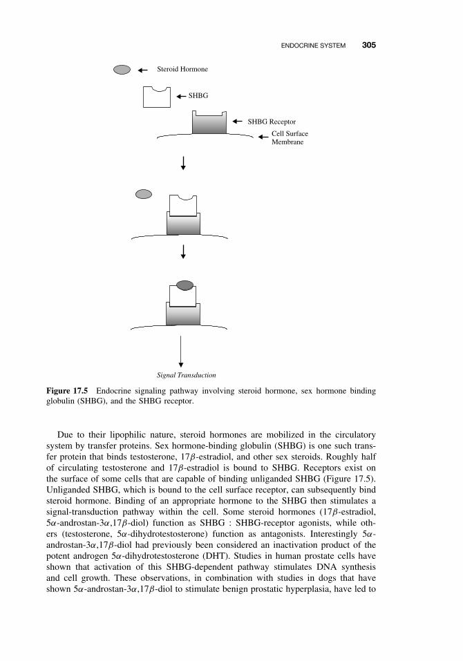
ENDOCRINE SYSTEM 305
Cell SurfaceMembrane
SHBG Receptor
SHBG
Steroid Hormone
Signal Transduction
Figure 17.5 Endocrine signaling pathway involving steroid hormone, sex hormone bindingglobulin (SHBG), and the SHBG receptor.
Due to their lipophilic nature, steroid hormones are mobilized in the circulatorysystem by transfer proteins. Sex hormone-binding globulin (SHBG) is one such trans-fer protein that binds testosterone, 17β-estradiol, and other sex steroids. Roughly halfof circulating testosterone and 17β-estradiol is bound to SHBG. Receptors exist onthe surface of some cells that are capable of binding unliganded SHBG (Figure 17.5).Unliganded SHBG, which is bound to the cell surface receptor, can subsequently bindsteroid hormone. Binding of an appropriate hormone to the SHBG then stimulates asignal-transduction pathway within the cell. Some steroid hormones (17β-estradiol,5α-androstan-3α,17β-diol) function as SHBG : SHBG-receptor agonists, while oth-ers (testosterone, 5α-dihydrotestosterone) function as antagonists. Interestingly 5α-androstan-3α,17β-diol had previously been considered an inactivation product of thepotent androgen 5α-dihydrotestosterone (DHT). Studies in human prostate cells haveshown that activation of this SHBG-dependent pathway stimulates DNA synthesisand cell growth. These observations, in combination with studies in dogs that haveshown 5α-androstan-3α,17β-diol to stimulate benign prostatic hyperplasia, have led to
306 ENDOCRINE SYSTEM
suggestions that the SHBG-receptor pathway is involved in this disease condition. Thesusceptibility of these membrane-bound receptor pathways in endocrine toxicity hasreceived little attention, though conceivably toxicants could perturb these pathways bycompeting with endogenous hormone for binding to SHBG, resulting in the loss ofstimulatory activity (antagonists) or inappropriate stimulation of activity (agonists).
17.3 ENDOCRINE DISRUPTION
Xenobiotics have the ability to disrupt hormone activity through a variety of mecha-nisms, though the predominant mechanisms appear to involve binding to the hormonereceptor, either as an agonist or antagonist, or by modulating endogenous steroid hor-mone levels.
17.3.1 Hormone Receptor Agonists
A hormone receptor agonist is defined as a compound that binds to and activatesa hormone receptor. Endogenous hormones function as agonists to their respectivereceptors. Xenobiotics can act as receptor agonists and stimulate receptor-dependentphysiological processes in the absence of the endogenous receptor ligand (hormone).Such inappropriate stimulation can result in the errant expression of hormone-dependentprocesses such as breast development in males (gynecomastia).
Estrogen Receptor. Among the steroid hormone receptors, the estrogen receptorappears most susceptible to the agonistic action of xenobiotics. Estrogen receptoragonists are quite diverse in molecular structure (Figure 17.6). Several steric consider-ations, associated with the steroid structure, in conjunction with electrostatic (charge)properties of the outer surface of the molecule seem to dictate whether a xenobioticcan fit into the binding-pocket of the receptor and function as a receptor agonist. It isnot clear why the estrogen receptor would be more susceptible to the agonistic actionof xenobiotics as compared to other steroid hormone receptors. High ligand speci-ficity may be an evolutionary trait of most “modern” receptors that is not associatedwith this presumed ancestral precursor to other nuclear receptor superfamily members.The estrogen receptor is often referred to as a promiscuous receptor because of thissusceptibility to agonistic interactions with xenobiotics.
Some drugs are rather potent estrogens (i.e., DES); however, environmental chemi-cals with estrogenic activity are typically weak agonists with activity several orders-of-magnitude less than that of 17β-estradiol (Table 17.3). Because of this weak activity,xenoestrogens are typically not associated with endocrine toxicity to adult femalesowing to the large amount of 17β-estradiol in these individuals. However, adultmales, immature individuals, and embryos all have been shown to exhibit endocrinetoxicity resulting from xeno-estrogen exposure. For example, in utero exposure ofmale or female rodents and humans to DES causes proliferation of epithelial cellsassociated with the reproductive system resulting in abnormalities of this system.Gynecomastia is a common side effect of estrogenic drugs such as DES and fosfestrolwhen administered to adult males. The physiological consequences of xeno-estrogenicactivity is typically characteristic of feminization, that is, the acquisition of femalecharacteristics.
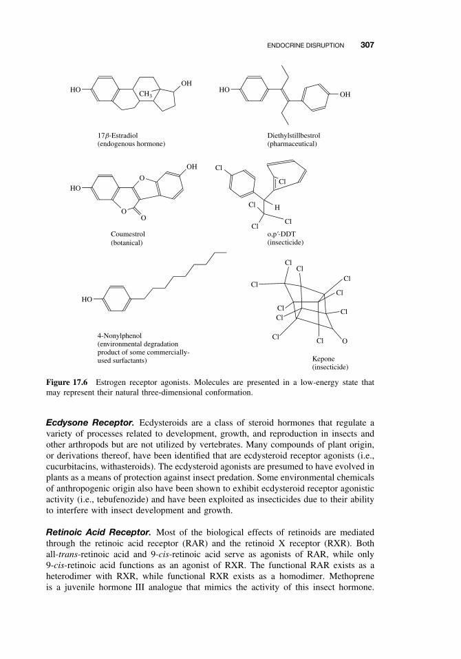
ENDOCRINE DISRUPTION 307
Cl
ClCl
Cl
Cl
Cl
ClCl
ClCl
O
Kepone(insecticide)
HO CH3
OHHO
OH
17b-Estradiol(endogenous hormone)
Diethylstillbestrol(pharmaceutical)
HO
O
4-Nonylphenol(environmental degradationproduct of some commercially-used surfactants)
o,p'-DDT(insecticide)
Coumestrol(botanical)
O
O
OH Cl
HCl
ClCl
Cl
HO
Figure 17.6 Estrogen receptor agonists. Molecules are presented in a low-energy state thatmay represent their natural three-dimensional conformation.
Ecdysone Receptor. Ecdysteroids are a class of steroid hormones that regulate avariety of processes related to development, growth, and reproduction in insects andother arthropods but are not utilized by vertebrates. Many compounds of plant origin,or derivations thereof, have been identified that are ecdysteroid receptor agonists (i.e.,cucurbitacins, withasteroids). The ecdysteroid agonists are presumed to have evolved inplants as a means of protection against insect predation. Some environmental chemicalsof anthropogenic origin also have been shown to exhibit ecdysteroid receptor agonisticactivity (i.e., tebufenozide) and have been exploited as insecticides due to their abilityto interfere with insect development and growth.
Retinoic Acid Receptor. Most of the biological effects of retinoids are mediatedthrough the retinoic acid receptor (RAR) and the retinoid X receptor (RXR). Bothall-trans-retinoic acid and 9-cis-retinoic acid serve as agonists of RAR, while only9-cis-retinoic acid functions as an agonist of RXR. The functional RAR exists as aheterodimer with RXR, while functional RXR exists as a homodimer. Methopreneis a juvenile hormone III analogue that mimics the activity of this insect hormone.
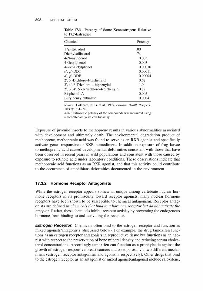
308 ENDOCRINE SYSTEM
Table 17.3 Potency of Some Xenoestrogens Relativeto 17β-Estradiol
Chemical Potency
17β-Estradiol 100Diethylstilbestrol 744-Nonylphenol 0.0054-Octylphenol 0.0034-tert-Octylphenol 0.00036o′, p′-DDT 0.00011o′, p′-DDE 0.000042′, 5′-Dichloro-4-biphenylol 0.622′, 4′, 6-Trichloro-4-biphenylol 1.02′, 3′, 4′, 5′-Tetrachloro-4-biphenylol 0.82Bisphenol A 0.005Butylbenzylphthalate 0.0004
Source: Coldham, N. G. et al., 1997, Environ. Health Perspect.105(7): 734–742.Note: Estrogenic potency of the compounds was measured usinga recombinant yeast cell bioassay.
Exposure of juvenile insects to methoprene results in various abnormalities associatedwith development and ultimately death. The environmental degradation product ofmethoprene, methoprenic acid was found to serve as an RXR agonist and specificallyactivate genes responsive to RXR homodimers. In addition exposure of frog larvaeto methoprenic acid caused developmental deformities consistent with those that havebeen observed in recent years in wild populations and consistent with those caused byexposure to retinoic acid under laboratory conditions. These observations indicate thatmethoprenic acid functions as an RXR agonist, and that this activity could contributeto the occurrence of amphibians deformities documented in the environment.
17.3.2 Hormone Receptor Antagonists
While the estrogen receptor appears somewhat unique among vertebrate nuclear hor-mone receptors in its promiscuity toward receptor agonists, many nuclear hormonereceptors have been shown to be susceptible to chemical antagonism. Receptor antag-onists are defined as chemicals that bind to a hormone receptor but do not activate thereceptor. Rather, these chemicals inhibit receptor activity by preventing the endogenoushormone from binding to and activating the receptor.
Estrogen Receptor. Chemicals often bind to the estrogen receptor and function asmixed agonists/antagonists (discussed below). For example, the drug tamoxifen func-tions as an estrogen receptor antagonists in reproductive tissue but functions as an ago-nist with respect to the preservation of bone mineral density and reducing serum choles-terol concentrations. Accordingly tamoxifen can function as a prophylactic against thegrowth of estrogen-responsive breast cancers and osteoporosis via two different mecha-nisms (estrogen receptor antagonism and agonism, respectively). Other drugs that bindto the estrogen receptor as an antagonist or mixed agonist/antagonist include raloxifene,
ENDOCRINE DISRUPTION 309
ICI 164,384, and toremifene. Environmental estrogen receptor antagonists include somephytochemicals (i.e., flavonoids) and PCBs (i.e., 3, 3′, 4, 4′-tetrachlorobiphenyl). Con-sequences of estrogen receptor antagonism are typically considered de-feminization(loss of female traits). In laboratory animal studies, estrogen receptor antagonists havebeen shown in females to disrupt estrous cycles, impair fertility, increase preimplan-tation loss, and cause embryolethality.
Androgen Receptor. Chemicals that bind to the androgen receptor in an antag-onistic fashion include the pharmaceuticals spironolactone, cimetidine, cyperoteroneacetate, and hydroxyflutamide. Environmental chemicals that have been shown to actas androgen receptor antagonists include the metabolites of the agricultural fungi-cide vinclozolin, the DDT metabolite p, p′-DDE, some hydroxylated PCBs, and theorganophosphate insecticide fenitrothion. The consequence of androgen receptor antag-onism is typically considered demasculinization (loss of male traits). Demasculinizingeffects of antiandrogens in laboratory animal studies have included reductions in thesize of the ventral prostate and seminal vesicle weights along with deformities ofthe penis.
Glucocorticoid Receptor. Some drugs (i.e., mifepristone) elicit antagonistic activ-ity toward the glucocorticoid receptor. This property has been associated with adverseside effects of some drugs and also has been capitalized upon therapeutically for themodulation of the glucocorticoid receptor. Antiglucocorticoids typically are steroidalcompounds that are capable of binding to the receptors but are relatively ineffec-tive in activating the receptor. As such, these compounds are typically mixed ago-nists/antagonists (see below). Glucocorticoid receptor antagonists can adversely affectgrowth, development, and other glucocorticoid-regulated processes (Table 17.1). Lit-tle is known of the ability of environmental chemicals to function as glucocorticoidreceptor antagonists.
Mixed Agonists/Antagonists. Chemicals often can function as either a receptoragonist or antagonist depending on the level of endogenous hormone. A weak agonistmay bind to a receptor and stimulate some low-level receptor-mediated activity in theabsence of the endogenous hormone. However, in the presence of the hormone, bind-ing of the xenobiotic to the receptor may prevent binding of the endogenous hormone,and if the xenobiotic is a much weaker activator of receptor-mediated activity, thenthe net effect is loss of activity. Thus, in the presence of the endogenous hormone,the xenobiotic functions as a receptor antagonist. Whether a weak xeno-agonist func-tions as an agonist or antagonist depends on (1) the concentration of the xeno-agonist,(2) the binding affinity of the xeno-agonist to the receptor, (3) the concentration of theendogenous hormone to the receptor, and (4) the binding affinity of the endogenoushormone to the receptor. These compounds are classified mixed agonists/antagonists.
17.3.3 Organizational versus Activational Effects of Endocrine Toxicants
Effects of receptor agonists or antagonists on endocrine related processes are oftendescribed as being either organizational or activational. An organizational effect ofan endocrine toxicant is one that typically results from neonatal or prenatal exposureduring which time hormones are directing various irreversible aspects of development.

310 ENDOCRINE SYSTEM
Accordingly the disrupting effect of the toxicant also is irreversible. These organi-zational effects may be evident only later in life during maturation or reproduction.Neonatal exposure to DES resulting in proliferation of epithelial cells of the repro-ductive tract at reproductive maturity is an example of an organizational effect of anendocrine toxicant. Organizational effects of endocrine toxicants have been of greatconcern to toxicologists and are the most difficult type of toxicity to diagnose owingto the temporal separation between exposure and effect.
An activational effect of an endocrine toxicant occurs in the same time frame asthe exposure and is the consequence of the toxicant disrupting the immediate role of ahormone in some physiological process. Activational effects are reversible followingcessation of exposure to the toxicant. For example, androgens contribute to maintenanceof the prostate gland in the adult male. Exposure of adult males to an antiandrogen canresult in a decrease in prostate size. Cessation of exposure to the antiandrogen thenresults in restoration of the prostate gland to its normal size.
17.3.4 Inhibitors of Hormone Synthesis
Endocrine toxicants can elicit antihormone activity by lowering levels of endoge-nous hormone in the body. With steroid hormones, chemicals typically elicit thiseffect by inhibiting enzymes necessary for synthesis of the hormone. For example, thecytochrome P450 enzyme CYP19 is responsible for the aromatization of testosteroneto form 17β-estradiol. CYP19 inhibitors such as fadrozol, anastrozole, and letrozole,can lower endogenous 17β-estradiol levels resulting in de-feminization. CytochromeP450s enzymes also are critical to various hydroxylation reactions that contribute tothe synthesis of androgens and other steroid hormones and inhibition of these enzymescan result in a variety of antisteroid hormone effects. For example, the agriculturaland medicinal fungicides propiconazole, ketoconazole, and fenarimol are capable ofinhibiting P450 enzymes and reducing synthesis and circulating levels of testosteroneand other steroid hormones. Toxicological consequences of the lowering of endogenoussteroid hormone levels are typically comparable to those effects elicited by antagonistsof the hormone’s receptor.
17.3.5 Inducers of Hormone Clearance
In most species, steroid and thyroid hormones are inactivated and cleared from thebody by the same biotransformation processes that are involved in chemical detoxifi-cation (see Chapter 7). Predominant among the hormone biotransformation processesin vertebrates are hydroxylation, glucuronic acid conjugation, and sulfate conjugation.The thyroid hormones T3 and T4 are inactivated and cleared following sulfate and glu-curonic acid conjugation, respectively. The glucuronosyl transferase enzymes that areresponsible for the elimination of T4 are induced following exposure to phenobarbital-type inducers and Ah receptor ligands (see Chapter 9). Thus exposure to chemicalssuch as some dioxins and PCBs can result in enhanced clearance of thyroid hor-mone resulting in low circulating thyroid hormone levels. The resulting hypothyroidstate can result in a variety of pathological conditions. In newborn infants, hypothy-roidism is associated with cretinism. This organizational syndrome is characterized bymental retardation, short stature, and various neurological abnormalities. In children,
INCIDENTS OF ENDOCRINE TOXICITY 311
hypothyroidism can cause delayed growth and mental development while advancingthe onset of puberty in adolescents. Hypothyroidism in adults results in various activa-tional abnormalities including impaired cardiovascular, pulmonary, intestinal, and renalfunction. Chronic fatigue, lethargy, and difficulty in concentration are also associatedwith thypothyroidism in adults.
Increased clearance of steroid hormones due to induction of hepatic biotransforma-tion enzymes following chemical exposure often has been cited as a possible mecha-nism by which toxicants could lower circulating testosterone or 17β-estradiol levels.While enhanced clearance of sex steroids has been demonstrated following chemi-cal exposure and induction of hepatic biotransformation enzymes, elegant feedbackcontrol mechanisms tend to ensure that more hormone is produced and homeosta-sis is maintained (Figure 17.2). Enhanced clearance of sex steroids can contribute toendocrine disruption if the toxicity also results in impaired hormone synthesis (i.e.,gonadal toxicity or interference with the feedback control of hormone synthesis).2,3,7,8-Tetrachlorodibenzodioxin appears to lower circulating sex steroid levels viathis dual effect.
17.3.6 Hormone Displacement from Binding Proteins
Steroid and thyroid hormones are typically distributed throughout the body while boundto serum-binding proteins such as sex hormone-binding globulin, corticosteroid-bindingglobulin, thyroxine-binding globulin (transthyretin), and albumin. Most steroid andthyroid hormones (>95%) are present in the blood reversibly bound to proteins. Thisbound hormone is not available for cell entry where it may interact with nuclearreceptors or undergo inactivation/elimination reactions. Rather, the bound hormoneserves as a reservoir from which hormone can be liberated (free hormone) for cell entry.
Some xenobiotics can compete with hormones for binding to the blood proteins. As aresult the circulating hormone reservoir can be depleted and free hormone becomes lim-ited. A variety of phenolic compounds, including hydroxylated metabolites of polychlo-rinated biphenyls (PCBs), chlorophenols, chlorophenoxy acids, and nitrophenols, havebeen shown to interfere with thyroxine binding to thyroxine-binding globulin during invitro experiments. In some instances compounds that displace thyroxine from the bind-ing protein also have been shown to decrease circulating thyroxine levels in exposedanimal models or in humans. In vitro experiments also have revealed that testosteroneand 17β-estradiol can be displaced from sex hormone-binding globulin by some chem-icals such as 4-nonylphenol, 4-tert-octylphenol, bisphenol A, O-hydroxybiphenyl, andpyrethroid insecticides. However, it is not clear whether these chemicals would sig-nificantly displace sex steroids from the binding globulin at concentrations typicallymeasured in human blood.
17.4 INCIDENTS OF ENDOCRINE TOXICITY
17.4.1 Organizational Toxicity
In utero exposure to estrogens or antiandrogens has been shown, in animal models,to elicit a variety of organizational effects associated with development of the repro-ductive system. The best-described example of the organizational effects of a drug
312 ENDOCRINE SYSTEM
administered to humans involves the synthetic estrogen DES. As discussed previously,DES was prescribed to over two million pregnant women in the United States betweenthe 1940s and 1960s to prevent miscarriage. Offspring exposed to DES during fetaldevelopment experienced a variety of problems upon attainment of sexual maturity.DES daughters experience a significantly increased risk of clear cell adenocarcinomaof the vagina and cervix. DES daughters have increased risk of a variety of reproduc-tive disorders including structural abnormalities of the reproductive tract, infertility,ectopic pregnancy, miscarriage, and pre-term delivery.
Less is known of the risks faced by males exposed to DES during fetal develop-ment. Animal studies have revealed that male rodents exposed to DES have increasedincidence of prostatic metaplasia. Epidemiological studies of DES sons have suggestedincreased risk of various testicular abnormalities including epididymal cysts, testicularvaricoceles, and undescended testis. Hyperplasia and metaplasia of the prostatic ductsin DES sons also have been reported.
The effects elicited by fetal exposure to DES appear to be largely the consequence ofthe estrogenic activity of this drug. Estrogens orchestrate organizational events duringfetal development that promote female reproductive tract development. Excess estro-gen exposure resulting from DES treatment of either female or male fetuses resultedin permanent alterations, many of which became evident only upon attainment ofreproductive maturity.
Organizational effects on reproductive development resulting from perinatal expo-sure to endocrine toxicants of environmental origin also have been reported to occur.In 1973 a fire retardant containing polybrominated biphenyls (PBBs) was mistakenlyadded to cattle feed in Michigan. An estimated 4000 people subsequently were exposedto the PBBs by consuming dairy products derived from these cattle. PBBs are long-lived chemicals that are stored in the fat of exposed individuals. PBBs have beenreported to elicit endocrine toxicity-like symptoms in animal models consistent withhypothyroidism. For example, offspring from maternal rats provided PBBs duringgestation and lactation showed signs of neurological deficit and growth retardation.Daughters of mothers that were exposed to PBBs during the Michigan incident weremonitored for possible adverse effects on the female reproductive system. The initiationof menarche (menstruation) among these daughters correlated with the likely severityof PBB exposure. The most highly exposed daughters began menstruating approxi-mately 1 year ahead of females that were less severely exposed. Early initiation ofmenarche is consistent with precocious puberty associated with hypothyroidism. Theinitiation of menarche also is under the regulation of 17β-estradiol and early initiationof menarche may reflect an estrogen-type organizational effect of the PBBs duringperinatal exposure.
17.4.2 Activational Toxicity
Estrogenic Pharmaceuticals. Administration of estrogenic pharmaceuticals to chil-dren or adults can result in a variety of abnormalities associated largely with secondarysex characteristics that are reversible upon cessation of drug treatment.
Gynecomastia, the development of breast tissue in males, is often the consequenceof perturbations in the normal androgen/estrogen ratio. As discussed earlier in thischapter, prolonged administration of drugs with estrogenic or antiandrogenic activitycan cause gynecomastia. Gynecomastia had been reported in the medical literature
INCIDENTS OF ENDOCRINE TOXICITY 313
to occur as a result of frequent intercourse when an estrogen-containing cream wasused as a vaginal lubricant and among morticans who applied estrogen-containing skincreams to corpses without the use of gloves.
Similar to gynecomastia in adult males, activational toxicity from estrogenic drugshas been reported to cause pseudoprecocious puberty in children. Pseudoprecociouspuberty is characterized by the development of some indicators of puberty (pubic orfacial hair, morphological changes in sex organs, breast development, etc.) in pread-olescent individuals. An outbreak of pseudoprecocious puberty was reported among agroup of children ranging in age from 4 months to 2 years of age following applicationof a skin cream to treat dermatitis. Symptoms included pigmentation of the nipples,breast development, the presence of pubic hair, and vaginal discharge and bleedingamong the females. Breast development also was reported in prepubertal boys fol-lowing use of an estrogen-containing hair cream. These reports highlight the fact thatdermal exposure can be adequate to attain a sufficient dose of endocrine-active com-pound to elicit adverse responses. In all of these cases the symptoms of endocrinetoxicity resolved following cessation of exposure to the causative agent.
Environmental Estrogens. Thelarche is defined as the development of breast tis-sue in preadolescent females (typically <8 years of age). Since 1979 physicians havemonitored an epidemic level of thelarche on the island of Puerto Rico. The cause ofthelarche in Puerto Rico is not known; however, evidence strongly implicates exposureto endocrine-disrupting agents. Analyses of blood samples from thelarche and nonthe-larche children for environmental chemicals with known estrogenic activity revealedthat 68% of the thelarche children contained significantly high levels of several typesof phthalate esters. Only a single nonthelarche child contained a significant amountof phthalate ester and only one type of phthalate ester was found in this individual.Phthalate esters are used as plasticizers and are ubiquitous environmental contaminants.They have been shown to cause a variety of endocrine-related effects in animal modelsand some phthalate esters have been shown to be estrogenic in vitro. The associationbetween phthalate ester exposure and the high incidence of thelarche in Puerto Ricodoes not establish causality but has generated concern that environmental agents areresponsible for this condition.
Kepone (chlordecone) is an organochlorine insecticide (Figure 17.5) that was man-ufactured in Hopewell, Virginia, from the mid-1960s to 1975. In 1975 the Center forDisease Control determined that employees of the manufacturing facility and other res-idents of Hopewell, totaling over 200 individuals, had been significantly contaminatedwith this insecticide. Exposed individuals reported a variety of symptoms. Foremost,among the symptoms of “Kepone sickness” were neurological disorders presentingas tremors, weight loss, and nervousness. However, subsequent evaluations revealedthat males exposed to Kepone also experienced testicular dysfunction that was char-acteristic of estrogen exposure. Later laboratory studies demonstrated that Keponewas an estrogen receptor agonist, which could explain its adverse effects on the malereproductive system.
17.4.3 Hypothyroidism
Hypothyroidism describes the clinical state arising from a deficiency in thyroid hor-mone. Toxicity resulting in hypothyroidism is manifested at several organ systems as
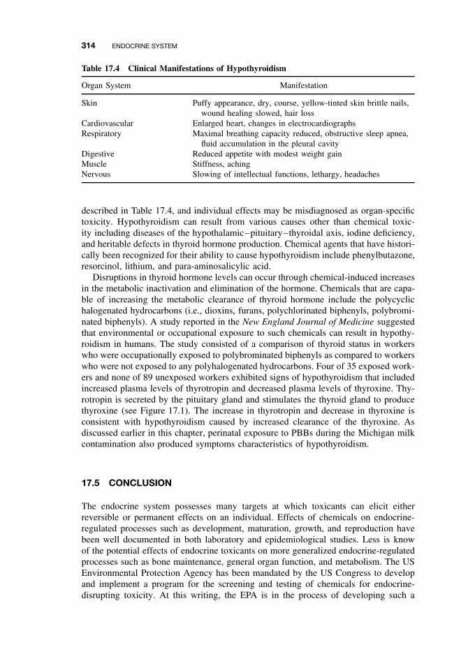
314 ENDOCRINE SYSTEM
Table 17.4 Clinical Manifestations of Hypothyroidism
Organ System Manifestation
Skin Puffy appearance, dry, course, yellow-tinted skin brittle nails,wound healing slowed, hair loss
Cardiovascular Enlarged heart, changes in electrocardiographsRespiratory Maximal breathing capacity reduced, obstructive sleep apnea,
fluid accumulation in the pleural cavityDigestive Reduced appetite with modest weight gainMuscle Stiffness, achingNervous Slowing of intellectual functions, lethargy, headaches
described in Table 17.4, and individual effects may be misdiagnosed as organ-specifictoxicity. Hypothyroidism can result from various causes other than chemical toxic-ity including diseases of the hypothalamic–pituitary–thyroidal axis, iodine deficiency,and heritable defects in thyroid hormone production. Chemical agents that have histori-cally been recognized for their ability to cause hypothyroidism include phenylbutazone,resorcinol, lithium, and para-aminosalicylic acid.
Disruptions in thyroid hormone levels can occur through chemical-induced increasesin the metabolic inactivation and elimination of the hormone. Chemicals that are capa-ble of increasing the metabolic clearance of thyroid hormone include the polycyclichalogenated hydrocarbons (i.e., dioxins, furans, polychlorinated biphenyls, polybromi-nated biphenyls). A study reported in the New England Journal of Medicine suggestedthat environmental or occupational exposure to such chemicals can result in hypothy-roidism in humans. The study consisted of a comparison of thyroid status in workerswho were occupationally exposed to polybrominated biphenyls as compared to workerswho were not exposed to any polyhalogenated hydrocarbons. Four of 35 exposed work-ers and none of 89 unexposed workers exhibited signs of hypothyroidism that includedincreased plasma levels of thyrotropin and decreased plasma levels of thyroxine. Thy-rotropin is secreted by the pituitary gland and stimulates the thyroid gland to producethyroxine (see Figure 17.1). The increase in thyrotropin and decrease in thyroxine isconsistent with hypothyroidism caused by increased clearance of the thyroxine. Asdiscussed earlier in this chapter, perinatal exposure to PBBs during the Michigan milkcontamination also produced symptoms characteristics of hypothyroidism.
17.5 CONCLUSION
The endocrine system possesses many targets at which toxicants can elicit eitherreversible or permanent effects on an individual. Effects of chemicals on endocrine-regulated processes such as development, maturation, growth, and reproduction havebeen well documented in both laboratory and epidemiological studies. Less is knowof the potential effects of endocrine toxicants on more generalized endocrine-regulatedprocesses such as bone maintenance, general organ function, and metabolism. The USEnvironmental Protection Agency has been mandated by the US Congress to developand implement a program for the screening and testing of chemicals for endocrine-disrupting toxicity. At this writing, the EPA is in the process of developing such a
SUGGESTED READING 315
program that will focus on the effects of chemicals on the androgen/estrogen andthyroid hormone regulated processes. Once implemented, this required testing willgreatly expand our knowledge of the extent to which humans are exposed to chemicalsthat interfere with processes regulated by these hormones. However, it is importantto recognize that chemicals have the potential ability to interfere with other hormonecascades, including those involving mineralcorticoids, glucocorticoids, retinoids, andperhaps some peptide hormones. Research is needed to increase our understanding ofthe susceptibility of endocrine signaling pathways involving these hormones to chem-ical toxicity and, ultimately, to our establishing chemical exposure limits that includethese considerations.
SUGGESTED READING
Bahn, A. K., J. L. Mills, P. J. Snyder, et al. Hypothyroidism in workers exposed to polybromi-nated biphenyls. N. Engl. J. Med. 302: 31–33, 1980.
Beas, F., L. Vargas, R. P. Spada, and N. Merchack. Pseudoprecocious puberty in infants causedby dermal ointment containing estrogens. J. Pediatr. 75: 127–130, 1962.
Blanck, H. M., M. Marcus, P. E. Tolbert, C. Rubin, A. K. Henderson, V. S. Hertzberg, R. H.Zhang, and L. Cameron. Age at menarche and tanner stage in girls exposed in utero andpostnatally to polybrominated biphenyls. Epidemiology 11: 641–647, 2000.
Colborn, T., and C. Clement, eds. Chemically-induced alterations in sexual and functional devel-opment: The Wildlife/Human Connection. In Advances in Modern Environmental Toxicology,M. A. Mehlman, ed. Princeton, NJ: Princeton Scientific Publishing, 1992.
DiRaimondo, C. V., A. L. Roach, and C. K. Meador. Gynecomastia from exposure to vaginaestrogen cream. N. Engl. J. Med. 302: 1089–1090, 1980.
Greenspan, F. S., and G. J. Strewler, eds. Basic and Clinical Endocrinology. Stamford, CT:Appleton and Lange, 1997.
Guillette, L, Jr. and D. A. Crain, eds. Environmental endocrine disrupters. New York: Taylorand Francis, 2000.
McLachlan, J. A. Environmental signaling: What embryos and evolution teach us about endocrinedisrupting chemicals. Endocrine Rev. 22: 319–341, 2001.
Wilson, J. D., and D. W. Foster, eds. Textbook of Endocrinology. Philadelphia: Saunders, 1992.

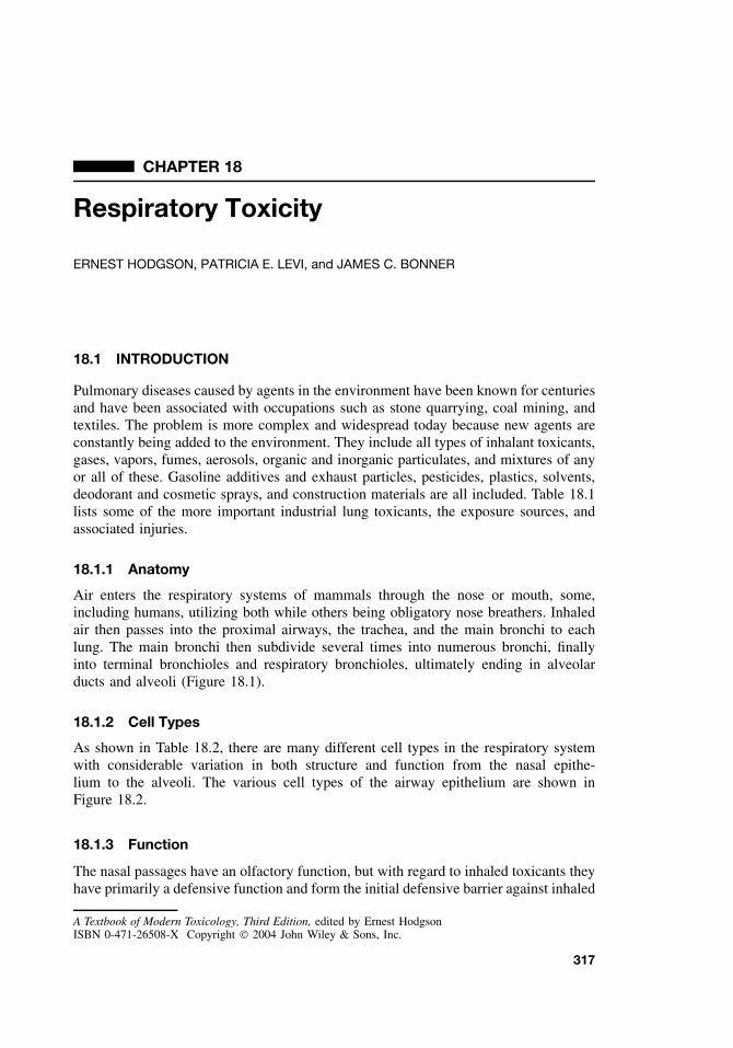
CHAPTER 18
Respiratory Toxicity
ERNEST HODGSON, PATRICIA E. LEVI, and JAMES C. BONNER
18.1 INTRODUCTION
Pulmonary diseases caused by agents in the environment have been known for centuriesand have been associated with occupations such as stone quarrying, coal mining, andtextiles. The problem is more complex and widespread today because new agents areconstantly being added to the environment. They include all types of inhalant toxicants,gases, vapors, fumes, aerosols, organic and inorganic particulates, and mixtures of anyor all of these. Gasoline additives and exhaust particles, pesticides, plastics, solvents,deodorant and cosmetic sprays, and construction materials are all included. Table 18.1lists some of the more important industrial lung toxicants, the exposure sources, andassociated injuries.
18.1.1 Anatomy
Air enters the respiratory systems of mammals through the nose or mouth, some,including humans, utilizing both while others being obligatory nose breathers. Inhaledair then passes into the proximal airways, the trachea, and the main bronchi to eachlung. The main bronchi then subdivide several times into numerous bronchi, finallyinto terminal bronchioles and respiratory bronchioles, ultimately ending in alveolarducts and alveoli (Figure 18.1).
18.1.2 Cell Types
As shown in Table 18.2, there are many different cell types in the respiratory systemwith considerable variation in both structure and function from the nasal epithe-lium to the alveoli. The various cell types of the airway epithelium are shown inFigure 18.2.
18.1.3 Function
The nasal passages have an olfactory function, but with regard to inhaled toxicants theyhave primarily a defensive function and form the initial defensive barrier against inhaled
A Textbook of Modern Toxicology, Third Edition, edited by Ernest HodgsonISBN 0-471-26508-X Copyright 2004 John Wiley & Sons, Inc.
317
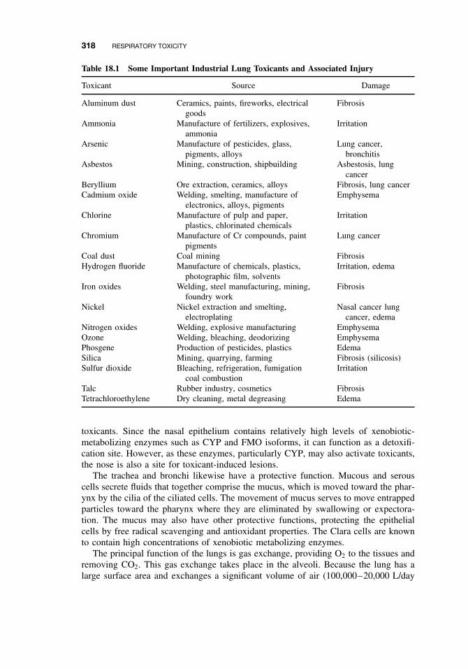
318 RESPIRATORY TOXICITY
Table 18.1 Some Important Industrial Lung Toxicants and Associated Injury
Toxicant Source Damage
Aluminum dust Ceramics, paints, fireworks, electricalgoods
Fibrosis
Ammonia Manufacture of fertilizers, explosives,ammonia
Irritation
Arsenic Manufacture of pesticides, glass,pigments, alloys
Lung cancer,bronchitis
Asbestos Mining, construction, shipbuilding Asbestosis, lungcancer
Beryllium Ore extraction, ceramics, alloys Fibrosis, lung cancerCadmium oxide Welding, smelting, manufacture of
electronics, alloys, pigmentsEmphysema
Chlorine Manufacture of pulp and paper,plastics, chlorinated chemicals
Irritation
Chromium Manufacture of Cr compounds, paintpigments
Lung cancer
Coal dust Coal mining FibrosisHydrogen fluoride Manufacture of chemicals, plastics,
photographic film, solventsIrritation, edema
Iron oxides Welding, steel manufacturing, mining,foundry work
Fibrosis
Nickel Nickel extraction and smelting,electroplating
Nasal cancer lungcancer, edema
Nitrogen oxides Welding, explosive manufacturing EmphysemaOzone Welding, bleaching, deodorizing EmphysemaPhosgene Production of pesticides, plastics EdemaSilica Mining, quarrying, farming Fibrosis (silicosis)Sulfur dioxide Bleaching, refrigeration, fumigation
coal combustionIrritation
Talc Rubber industry, cosmetics FibrosisTetrachloroethylene Dry cleaning, metal degreasing Edema
toxicants. Since the nasal epithelium contains relatively high levels of xenobiotic-metabolizing enzymes such as CYP and FMO isoforms, it can function as a detoxifi-cation site. However, as these enzymes, particularly CYP, may also activate toxicants,the nose is also a site for toxicant-induced lesions.
The trachea and bronchi likewise have a protective function. Mucous and serouscells secrete fluids that together comprise the mucus, which is moved toward the phar-ynx by the cilia of the ciliated cells. The movement of mucus serves to move entrappedparticles toward the pharynx where they are eliminated by swallowing or expectora-tion. The mucus may also have other protective functions, protecting the epithelialcells by free radical scavenging and antioxidant properties. The Clara cells are knownto contain high concentrations of xenobiotic metabolizing enzymes.
The principal function of the lungs is gas exchange, providing O2 to the tissues andremoving CO2. This gas exchange takes place in the alveoli. Because the lung has alarge surface area and exchanges a significant volume of air (100,000–20,000 L/day
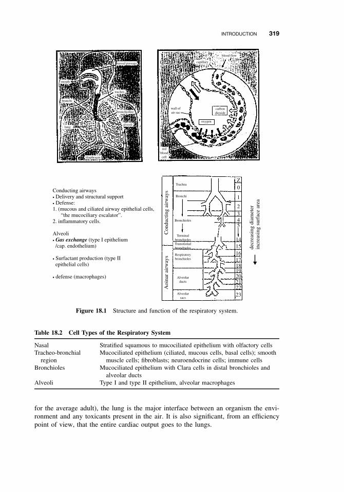
INTRODUCTION 319
mouth
nassal passage
blood flow
capillary
traches
alveoli
bronchiollee
lung
diaphragm
redbloodcell
branchi
wall ofair sac
oxygen
carbondioxide
Trachea
Bronchi
Bronchioles
TerminalbronchiolesTransitionalbronchioles
Respiratorybronchioles
Alveolarducts
Alveolarsacs
Con
duct
ing
airw
ays
Aoi
nar
airw
ays
Z0
1
2345
14151617181920212223
Conducting airways• Delivery and structural support• Defense:1. (mucous and ciliated airway epithelial cells, “the mucociliary escalator”.2. inflammatory cells.
Alveoli• Gas exchange (type I epithelium /cap. endothelium)
• Surfactant production (type II epithelial cells)
• defense (macrophages)
decr
easi
ng d
iam
eter
incr
easi
ng s
urfa
ce a
rea
Figure 18.1 Structure and function of the respiratory system.
Table 18.2 Cell Types of the Respiratory System
Nasal Stratified squamous to mucociliated epithelium with olfactory cellsTracheo-bronchial
regionMucociliated epithelium (ciliated, mucous cells, basal cells); smooth
muscle cells; fibroblasts; neuroendocrine cells; immune cellsBronchioles Mucociliated epithelium with Clara cells in distal bronchioles and
alveolar ductsAlveoli Type I and type II epithelium, alveolar macrophages
for the average adult), the lung is the major interface between an organism the envi-ronment and any toxicants present in the air. It is also significant, from an efficiencypoint of view, that the entire cardiac output goes to the lungs.
320 RESPIRATORY TOXICITY
Nonciliatedcolumnar cell
Ciliatedcolumnar cell
Goblet cell
Blood vessel
Figure 18.2 Cell types of the airway epithelium.
18.2 SUSCEPTIBILITY OF THE RESPIRATORY SYSTEM
18.2.1 Nasal
The nasal epithelia are the first point of contact for respiratory toxicants. Because theycontain xenobiotic metabolizing enzymes, they are susceptible to toxic effects causedby reactive intermediates.
18.2.2 Lung
In addition to being in direct contact with airborne toxicants, the entire body bloodvolume passes through the lung one to five times a minute, exposing the lung totoxicants and drugs within the systemic circulation. Thus the possibility of damagefrom both inhaled and circulating agents is enormous. As with the liver and kidney,the lungs possess significant levels of many xenobiotic metabolizing enzymes and thuscan play a large role in the activation and detoxication of exogenous chemicals.
18.3 TYPES OF TOXIC RESPONSE
Although many different agents may damage the lung, the patterns of cellular injuryand repair are relatively constant, and most fall into one or more of the categoriesdescribed below.
18.3.1 Irritation
Perhaps one of the most obvious and familiar chemical effects is irritation causedby volatile compounds such as ammonia or chlorine gas. Such irritation, especially
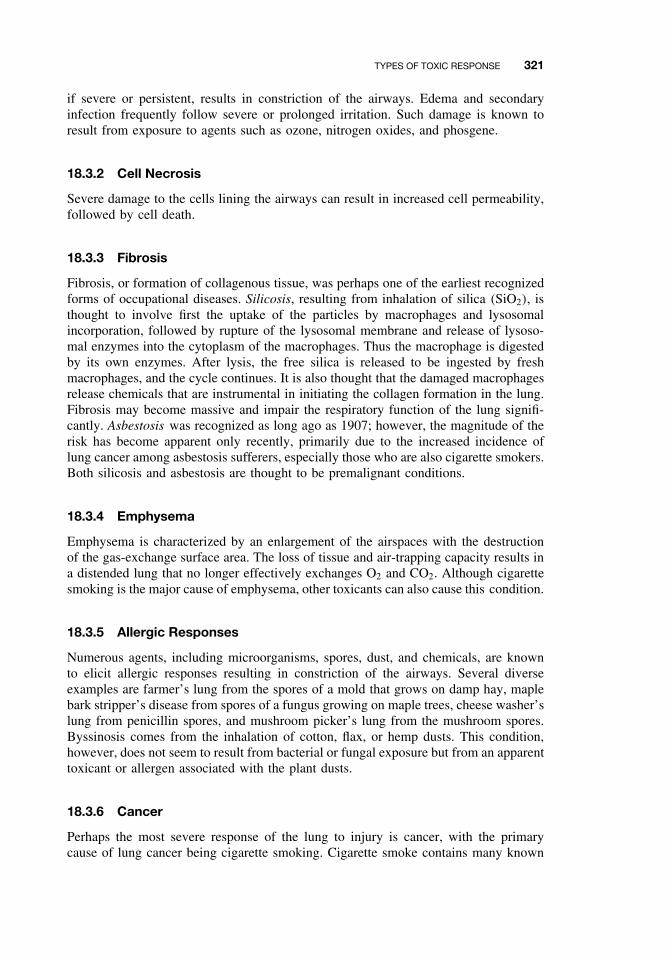
TYPES OF TOXIC RESPONSE 321
if severe or persistent, results in constriction of the airways. Edema and secondaryinfection frequently follow severe or prolonged irritation. Such damage is known toresult from exposure to agents such as ozone, nitrogen oxides, and phosgene.
18.3.2 Cell Necrosis
Severe damage to the cells lining the airways can result in increased cell permeability,followed by cell death.
18.3.3 Fibrosis
Fibrosis, or formation of collagenous tissue, was perhaps one of the earliest recognizedforms of occupational diseases. Silicosis, resulting from inhalation of silica (SiO2), isthought to involve first the uptake of the particles by macrophages and lysosomalincorporation, followed by rupture of the lysosomal membrane and release of lysoso-mal enzymes into the cytoplasm of the macrophages. Thus the macrophage is digestedby its own enzymes. After lysis, the free silica is released to be ingested by freshmacrophages, and the cycle continues. It is also thought that the damaged macrophagesrelease chemicals that are instrumental in initiating the collagen formation in the lung.Fibrosis may become massive and impair the respiratory function of the lung signifi-cantly. Asbestosis was recognized as long ago as 1907; however, the magnitude of therisk has become apparent only recently, primarily due to the increased incidence oflung cancer among asbestosis sufferers, especially those who are also cigarette smokers.Both silicosis and asbestosis are thought to be premalignant conditions.
18.3.4 Emphysema
Emphysema is characterized by an enlargement of the airspaces with the destructionof the gas-exchange surface area. The loss of tissue and air-trapping capacity results ina distended lung that no longer effectively exchanges O2 and CO2. Although cigarettesmoking is the major cause of emphysema, other toxicants can also cause this condition.
18.3.5 Allergic Responses
Numerous agents, including microorganisms, spores, dust, and chemicals, are knownto elicit allergic responses resulting in constriction of the airways. Several diverseexamples are farmer’s lung from the spores of a mold that grows on damp hay, maplebark stripper’s disease from spores of a fungus growing on maple trees, cheese washer’slung from penicillin spores, and mushroom picker’s lung from the mushroom spores.Byssinosis comes from the inhalation of cotton, flax, or hemp dusts. This condition,however, does not seem to result from bacterial or fungal exposure but from an apparenttoxicant or allergen associated with the plant dusts.
18.3.6 Cancer
Perhaps the most severe response of the lung to injury is cancer, with the primarycause of lung cancer being cigarette smoking. Cigarette smoke contains many known
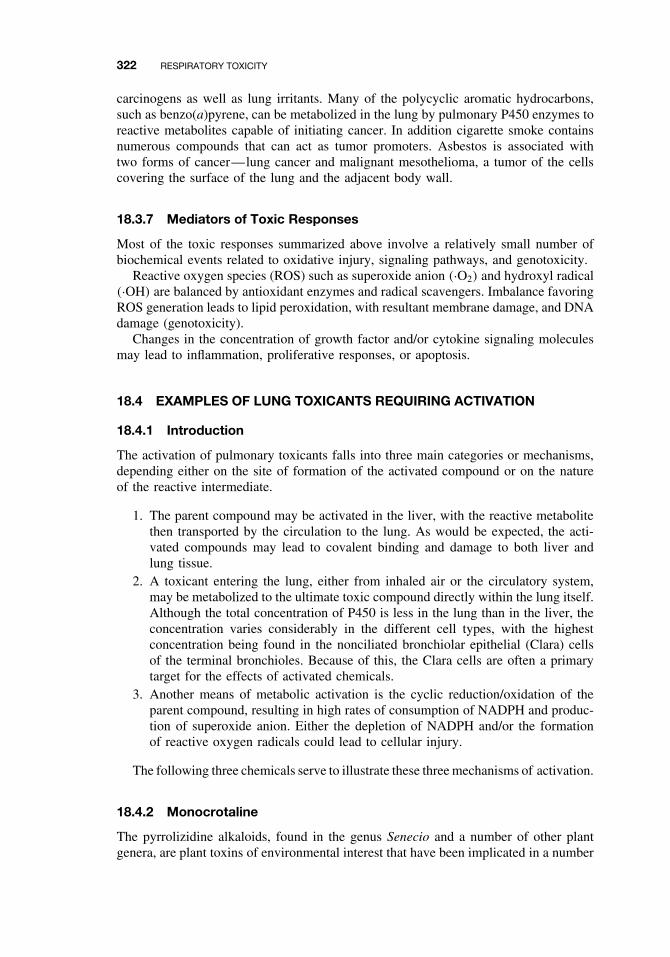
322 RESPIRATORY TOXICITY
carcinogens as well as lung irritants. Many of the polycyclic aromatic hydrocarbons,such as benzo(a)pyrene, can be metabolized in the lung by pulmonary P450 enzymes toreactive metabolites capable of initiating cancer. In addition cigarette smoke containsnumerous compounds that can act as tumor promoters. Asbestos is associated withtwo forms of cancer—lung cancer and malignant mesothelioma, a tumor of the cellscovering the surface of the lung and the adjacent body wall.
18.3.7 Mediators of Toxic Responses
Most of the toxic responses summarized above involve a relatively small number ofbiochemical events related to oxidative injury, signaling pathways, and genotoxicity.
Reactive oxygen species (ROS) such as superoxide anion (·O2) and hydroxyl radical(·OH) are balanced by antioxidant enzymes and radical scavengers. Imbalance favoringROS generation leads to lipid peroxidation, with resultant membrane damage, and DNAdamage (genotoxicity).
Changes in the concentration of growth factor and/or cytokine signaling moleculesmay lead to inflammation, proliferative responses, or apoptosis.
18.4 EXAMPLES OF LUNG TOXICANTS REQUIRING ACTIVATION
18.4.1 Introduction
The activation of pulmonary toxicants falls into three main categories or mechanisms,depending either on the site of formation of the activated compound or on the natureof the reactive intermediate.
1. The parent compound may be activated in the liver, with the reactive metabolitethen transported by the circulation to the lung. As would be expected, the acti-vated compounds may lead to covalent binding and damage to both liver andlung tissue.
2. A toxicant entering the lung, either from inhaled air or the circulatory system,may be metabolized to the ultimate toxic compound directly within the lung itself.Although the total concentration of P450 is less in the lung than in the liver, theconcentration varies considerably in the different cell types, with the highestconcentration being found in the nonciliated bronchiolar epithelial (Clara) cellsof the terminal bronchioles. Because of this, the Clara cells are often a primarytarget for the effects of activated chemicals.
3. Another means of metabolic activation is the cyclic reduction/oxidation of theparent compound, resulting in high rates of consumption of NADPH and produc-tion of superoxide anion. Either the depletion of NADPH and/or the formationof reactive oxygen radicals could lead to cellular injury.
The following three chemicals serve to illustrate these three mechanisms of activation.
18.4.2 Monocrotaline
The pyrrolizidine alkaloids, found in the genus Senecio and a number of other plantgenera, are plant toxins of environmental interest that have been implicated in a number
EXAMPLES OF LUNG TOXICANTS REQUIRING ACTIVATION 323
N
H
O
O
CH3
HO
O
O
CH3HO
HH
H
H
H3C
H
N
O
O
CH3
OH
O
O
CH3OH
HH
H
H
H3C
H
CYP3A4
Figure 18.3 Structure and activation of monocrotaline.
of livestock and human poisonings. Grazing animals may be poisoned by feeding onpyrrolizidine alkaloid-containing pastures, and human exposure may occur throughconsumption of herbal teas and contaminated grains and milk. The chemical structurefor monocrotaline is shown in Figure 18.3.
Monocrotaline, found in the leaves and seeds of the plant Crotalaria spectabilis, hasbeen the most extensively studied of the pyrrolizidine alkaloids. When monocrotalineis given to rats and other animals at high doses, a pronounced liver injury occurs,and animals usually die of acute effects, presumably liver failure. Lower doses, how-ever, that are only mildly hepatotoxic, result in lung injury that is associated withpulmonary hypertension and usually death in several weeks. It is thought that activa-tion of monocrotaline to its dehydro metabolite occurs in the liver and is a reductivereaction mediated by cytochrome P450 3A4 (Figure 18.3). Even though monocrotalineacts as a pneumotoxicant, several lines of evidence indicate that the lung is incapableof activating monocrotaline or can only do so to a limited extent. Furthermore the mainsite of pulmonary injury occurs in the endothelial cells, a target site consistent with areactive metabolite being absorbed from the circulatory system.
18.4.3 Ipomeanol
One of the best-known examples of a toxic compound being activated in the lungis 4-ipomeanol (Figure 18.4). This naturally occurring furan is produced by the moldFusarium solani that infects sweet potatoes. Lung edema in cattle is known to beassociated with the ingestion of mold-damaged sweet potatoes. A similar pulmonarylesion can be produced in a number of species regardless of the route of adminis-tration. Pulmonary injury by 4-ipomeanol is caused, not by the parent compound,but by a highly reactive alkylating metabolite produced in the lung by lung-specificP450 isozymes. In addition these isozymes are highly concentrated in the Clara cells,which are most affected by 4-ipomeanol toxicity. Although the reactive metabolitehas not been identified unambiguously, considerable data suggest a reactive epoxide.
324 RESPIRATORY TOXICITY
O
O
OH
4-Ipomeanol
O
O
OH
O
N NH3C+
CH3
+
Paraquat
e−
N NH3C CH3
+•
O2− O2•
Figure 18.4 Structure and activation of ipomeanol and paraquat.
Other toxic lung furans, such as the atmospheric contaminants 2-methylfuran and 3-methylfuran, may exert their toxicity through the formation of reactive metabolites,probably reactive aldehydes.
18.4.4 Paraquat
Systemic administration of compounds such as the herbicide paraquat (Figure 18.4),bleomycin (a cancer therapeutic agent), and nitroflurantoin (an antibiotic used for uri-nary tract infections) initiate a progression of degenerative and potentially lethal lesionsin the lung by a mechanism known as redox cycling. These compounds are reducedby cytochrome P450 reductase and NADPH, forming a free radical. Although the freeradical could potentially react with tissue macromolecules, one molecule of oxygenis reduced to superoxide that can then be converted to other toxic oxygen species.These reactive compounds may cause peroxidation of cellular membranes. The spe-cific toxicity of paraquat to the lung results from the uptake of this compound by thepolyamine transport system in the lung as well as from the high pulmonary oxygentension. Nitroflurantoin, however, is not actively accumulated in the lung, and its tissuespecificity probably results from the high pulmonary oxygen tension.
18.5 DEFENSE MECHANISMS
There are two important defense mechanisms against inhaled particles. The first ofthese involves the mucociliary escalator and consists of the trapping of particles inmucus followed by the upward movement of the mucus brought about by the upwardbeating of cilia on the airway epithelial airway cells. The material is then either swal-lowed or expectorated. The second mechanism is macrophage mediated. Macrophagesengulf particles and either deposit them on the mucociliary escalator or enter thelymphatic system.
Toxicants may also be detoxified by xenobiotic-metabolizing enzymes such ascytochrome P450 or the flavin-containing monooxygenase. It should be borne in mind,however, that these enzymes may also activate toxicants to more reactive, and poten-tially more toxic, metabolites.
SUGGESTED READING 325
SUGGESTED READING
Bond, J. A. Metabolism and elimination of inhaled drugs and airborne chemicals from the lungs.Pharmacol. Toxicol. 72: 36–47, 1993.
Cho, M., C. Chichester, C. Plopper, and A. Buckpitt. Biochemical factors important in Claracell selective toxicity in the lung. Drug Metabol. Rev. 27: 369–386, 1995.
Dahl, A. R., and J. L. Lewis. Respiratory tract uptake of inhalants and metabolism of xenobi-otics. An. Rev. Pharmacol. Toxicol. 32: 383–407, 1993.
Foth, H. Role of the lung in accumulation and metabolism of xenobiotic compounds-implicationsfor chemically induced toxicity. Crit. Rev. Toxicol. 25: 165–205, 1995.
Henderson, R. J., and K. J. Nikula. Respiratory tract toxicity. In Introduction to BiochemicalToxicology, 3rd ed., E. Hodgson and R. C. Smart, eds. New York: Wiley-Interscience, 2001.
Wheeler, C. W., T. M. Guenthner. Cytochrome P450-dependent metabolism of xenobiotics inhuman lung. J. Biochem. Toxicol. 6: 163–169, 1991.
Witschi, H. R., and J. A. Last. Toxic responses of the respiratory system. In Casarett and Doull’sToxicology: The Science of Poisons, 6th ed., C. D. Klaassen, ed. New York: McGraw-Hill,2001, pp. 515–534.

CHAPTER 19
Immunotoxicity
MARYJANE K. SELGRADE
19.1 INTRODUCTION
A properly functioning immune system is essential to good health. It defends the bodyagainst infectious agents and in some cases tumor cells. Individuals with immunedeficiencies resulting from genetic defects, diseases (e.g., AIDS, leukemia), or drugtherapies are more susceptible to infections and certain types of cancer, the conse-quences of which can be life-threatening. On the other hand, the immune system mayreact to foreign substances that would otherwise be relatively innocuous, such as cer-tain chemicals, pollens, and house dust. The resulting allergic reactions can producean array of pathologies, ranging from skin rashes and rhinitis to more life-threateningasthmatic and anaphylactic reactions. A crucial part of immune function is the abil-ity to distinguish endogenous components (self) from potentially harmful exogenouscomponents (non-self). Failure to make this distinction results in autoimmune disease.
Immunotoxicology is the study of undesired effects resulting from the interactions ofxenobiotics with the immune system (Figure 19.1). There is evidence that some xeno-biotics can cause immune suppression. Xenobiotics can also interact with the immunesystem to either cause or exacerbate allergic disease. Finally there is growing concernthat xenobiotics could have some involvement in autoimmune disease. This chapterprovides a brief overview of the immune system, chemicals associated with immunesuppression and immune pathologies, and approaches to testing for these effects.
19.2 THE IMMUNE SYSTEM
Cells of the immune system include several types of leukocytes (white blood cells)(Table 19.1), which are derived from bone marrow. T lymphocytes, a subset of immune
Disclaimer: This chapter has been reviewed by the National Health and Environmental Effects ResearchLaboratory, US Environmental Protection Agency and approved for publication. Approval does not signifythat the contents necessarily reflects the views and policies of the Agency, nor does mention of trade namesor commercial products constitute endorsement or recommendation for use.
A Textbook of Modern Toxicology, Third Edition, edited by Ernest HodgsonISBN 0-471-26508-X Copyright 2004 John Wiley & Sons, Inc.
327
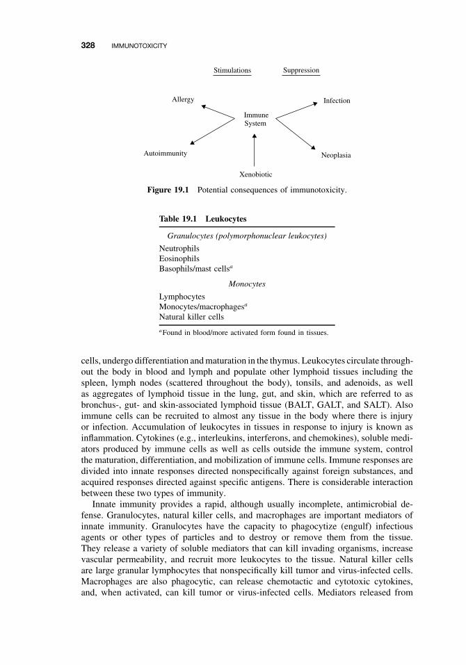
328 IMMUNOTOXICITY
Suppression
Neoplasia
Stimulations
Autoimmunity
ImmuneSystem
InfectionAllergy
Xenobiotic
Figure 19.1 Potential consequences of immunotoxicity.
Table 19.1 Leukocytes
Granulocytes (polymorphonuclear leukocytes)
NeutrophilsEosinophilsBasophils/mast cellsa
Monocytes
LymphocytesMonocytes/macrophagesa
Natural killer cells
aFound in blood/more activated form found in tissues.
cells, undergo differentiation and maturation in the thymus. Leukocytes circulate through-out the body in blood and lymph and populate other lymphoid tissues including thespleen, lymph nodes (scattered throughout the body), tonsils, and adenoids, as wellas aggregates of lymphoid tissue in the lung, gut, and skin, which are referred to asbronchus-, gut- and skin-associated lymphoid tissue (BALT, GALT, and SALT). Alsoimmune cells can be recruited to almost any tissue in the body where there is injuryor infection. Accumulation of leukocytes in tissues in response to injury is known asinflammation. Cytokines (e.g., interleukins, interferons, and chemokines), soluble medi-ators produced by immune cells as well as cells outside the immune system, controlthe maturation, differentiation, and mobilization of immune cells. Immune responses aredivided into innate responses directed nonspecifically against foreign substances, andacquired responses directed against specific antigens. There is considerable interactionbetween these two types of immunity.
Innate immunity provides a rapid, although usually incomplete, antimicrobial de-fense. Granulocytes, natural killer cells, and macrophages are important mediators ofinnate immunity. Granulocytes have the capacity to phagocytize (engulf) infectiousagents or other types of particles and to destroy or remove them from the tissue.They release a variety of soluble mediators that can kill invading organisms, increasevascular permeability, and recruit more leukocytes to the tissue. Natural killer cellsare large granular lymphocytes that nonspecifically kill tumor and virus-infected cells.Macrophages are also phagocytic, can release chemotactic and cytotoxic cytokines,and, when activated, can kill tumor or virus-infected cells. Mediators released from
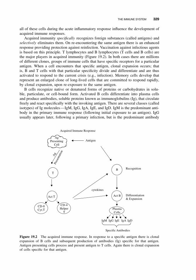
THE IMMUNE SYSTEM 329
all of these cells during the acute inflammatory response influence the development ofacquired immune responses.
Acquired immunity specifically recognizes foreign substances (called antigens) andselectively eliminates them. On re-encountering the same antigen there is an enhancedresponse providing protection against reinfection. Vaccination against infectious agentsis based on this principle. T lymphocytes and B lymphocytes (T cells and B cells) arethe major players in acquired immunity (Figure 19.2). In both cases there are millionsof different clones, groups of immune cells that have specific receptors for a particularantigen. When a cell encounters that specific antigen, clonal expansion occurs; thatis, B and T cells with that particular specificity divide and differentiate and are thusactivated to respond to the current crisis (e.g., infection). Memory cells develop thatrepresent an enlarged clone of long-lived cells that are committed to respond rapidly,by clonal expansion, upon re-exposure to the same antigen.
B cells recognize native or denatured forms of proteins or carbohydrates in solu-ble, particulate, or cell-bound form. Activated B cells differentiate into plasma cellsand produce antibodies, soluble proteins known as immunoglobulins (Ig), that circulatefreely and react specifically with the invoking antigen. There are several classes (calledisotypes) of Ig molecules—IgM, IgG, IgA, IgE, and IgD. IgM is the predominant anti-body in the primary immune response (following initial exposure to an antigen). IgGusually appears later, following a primary infection, but is the predominant antibody
AgPresenting
CellAntigen
Acquired Immune Response
Ag
Pres
enta
tion
T
Act T
CD-8Cyt.T
CD-4Helper
T
B
Act B
PlasmaCells
cytokines
IgM IgG IgE IgA IgD
Specific Antibodies
Recognition
Differentiation& Expansion
Figure 19.2 The acquired immune response. In response to a specific antigen there is clonalexpansion of B cells and subsequent production of antibodies (Ig) specific for that antigen.Antigen presenting cells process and present antigen to T cells. Again there is clonal expansionof cells specific for that antigen.
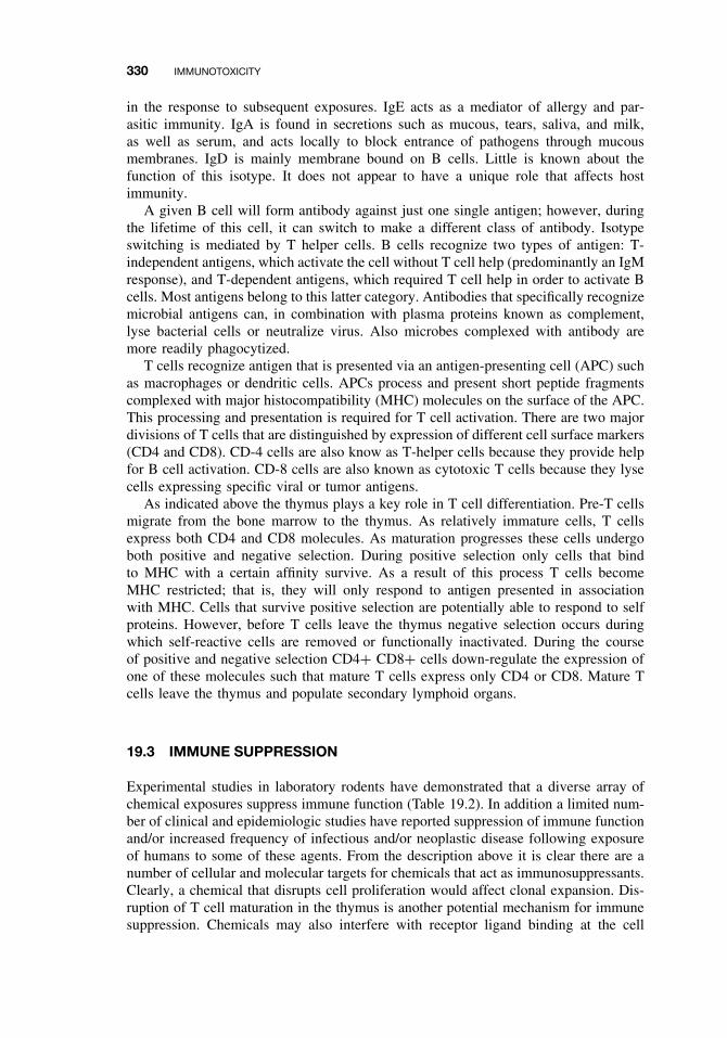
330 IMMUNOTOXICITY
in the response to subsequent exposures. IgE acts as a mediator of allergy and par-asitic immunity. IgA is found in secretions such as mucous, tears, saliva, and milk,as well as serum, and acts locally to block entrance of pathogens through mucousmembranes. IgD is mainly membrane bound on B cells. Little is known about thefunction of this isotype. It does not appear to have a unique role that affects hostimmunity.
A given B cell will form antibody against just one single antigen; however, duringthe lifetime of this cell, it can switch to make a different class of antibody. Isotypeswitching is mediated by T helper cells. B cells recognize two types of antigen: T-independent antigens, which activate the cell without T cell help (predominantly an IgMresponse), and T-dependent antigens, which required T cell help in order to activate Bcells. Most antigens belong to this latter category. Antibodies that specifically recognizemicrobial antigens can, in combination with plasma proteins known as complement,lyse bacterial cells or neutralize virus. Also microbes complexed with antibody aremore readily phagocytized.
T cells recognize antigen that is presented via an antigen-presenting cell (APC) suchas macrophages or dendritic cells. APCs process and present short peptide fragmentscomplexed with major histocompatibility (MHC) molecules on the surface of the APC.This processing and presentation is required for T cell activation. There are two majordivisions of T cells that are distinguished by expression of different cell surface markers(CD4 and CD8). CD-4 cells are also know as T-helper cells because they provide helpfor B cell activation. CD-8 cells are also known as cytotoxic T cells because they lysecells expressing specific viral or tumor antigens.
As indicated above the thymus plays a key role in T cell differentiation. Pre-T cellsmigrate from the bone marrow to the thymus. As relatively immature cells, T cellsexpress both CD4 and CD8 molecules. As maturation progresses these cells undergoboth positive and negative selection. During positive selection only cells that bindto MHC with a certain affinity survive. As a result of this process T cells becomeMHC restricted; that is, they will only respond to antigen presented in associationwith MHC. Cells that survive positive selection are potentially able to respond to selfproteins. However, before T cells leave the thymus negative selection occurs duringwhich self-reactive cells are removed or functionally inactivated. During the courseof positive and negative selection CD4+ CD8+ cells down-regulate the expression ofone of these molecules such that mature T cells express only CD4 or CD8. Mature Tcells leave the thymus and populate secondary lymphoid organs.
19.3 IMMUNE SUPPRESSION
Experimental studies in laboratory rodents have demonstrated that a diverse array ofchemical exposures suppress immune function (Table 19.2). In addition a limited num-ber of clinical and epidemiologic studies have reported suppression of immune functionand/or increased frequency of infectious and/or neoplastic disease following exposureof humans to some of these agents. From the description above it is clear there are anumber of cellular and molecular targets for chemicals that act as immunosuppressants.Clearly, a chemical that disrupts cell proliferation would affect clonal expansion. Dis-ruption of T cell maturation in the thymus is another potential mechanism for immunesuppression. Chemicals may also interfere with receptor ligand binding at the cell
IMMUNE SUPPRESSION 331
Table 19.2 Selected Examples of Immunosuppressive Agents
Drugs
Cyclosporin A, cyclophosphamide, glucocorticoids (Dexamethazone), azothioprine
Metals
Lead, cadmium, methylmercury, organotinsa
Pesticides
Chlorodanea , DDTa , Dieldrina
Industrial compounds
2,3,7,8-Tetrachlorodibenzo-p-dioxin (TCDD), polychlorinated and polybrominated biphenyls(PCBs and PBBs), benzene, poly aromatic hydrocarbonsa
Addictive substances
Cocaine, ethanol, opiates, cannabinoids, nicotine
Air pollutants
Environmental tobacco smoke, ozone, nitrogen dioxide
Microbial toxins
Aflatoxin,b ochratoxin A,b trichothecenes T-2 toxinb
Radiation
Ionizing, UV
Other
Asbestos, diethylstilbestrol (DES), dimethylnitrosamine
aEffects in humans are unknown; for all other compound without superscripts changes have been demon-strated in both rodents and humans.bEffects in humans unknown, but veterinary clinicians have noted immunosuppression in livestock ingestingmycotoxins at levels below those that cause overt toxicity.
surface and/or the cascade of signals that lead to transcription of genes responsible forgenerating and regulating the appropriate immune responses.
Because of the complexity of the immune system, tiered approaches to testingchemicals for immunosuppressive potential have been developed. Like other typesof toxicity testing, the first level of the tier (Table 19.3) frequently relies solely onstructural end points, including changes in the weight of thymus and other lymphoidorgans, histopathology of these organs, or differential blood cell counts. This type ofevaluation is convenient because it can be carried out along with an evaluation forother organ systems during routine toxicity testing using one set of animals. How-ever, although these nonfunctional endpoints may be effective in identifying gross(high dose) immunotoxic effects, they are not very accurate in predicting changes inimmune function or alterations in susceptibility to challenge with infectious agentsor tumor cells at lower chemical doses. Hence the first testing tier (Table 19.3) oftenincludes functional end points designed to assess (1) antibody-mediated responses, (2)T-cell-mediated responses, and (3) NK cell activity. The most commonly used immunefunction assay in laboratory animals assesses the ability of a mouse or rat to respondto challenge with an antigen, usually sheep red blood cells (SRBC) (Figure 19.3). Theresponse is assessed by determining the number of antigen specific antibody (IgM)
332 IMMUNOTOXICITY
Table 19.3 Tier I Tests (Screen) for Immune Suppression Using Laboratory Rodents
Immunopathology Hematology: Complete blood count and differentialWeights: body, spleen, thymusHistology: Spleen, thymus, lymph node
Antibody-mediated immunity IgM plaque-forming cell (PFC) response to T cell-dependentantigen (e.g., SRBC)
Cell-mediated immunity Lymphoproliferative response: T cell mitogens (Con A andPHA) Allogeneic mixed leukocyte response (MLR)
Nonspecific immunity Natural killer (NK) cell activity
Note: For details on specific assays see M. I. Luster et al., Fund Appl. Tox. 10: 2–19, 1988.
IgM Response to SRBCs
1. Inject SRBC IV
3. Assess antibody forming cells
2. Removespleen 4days 2. Or draw
blood forELISA
Figure 19.3 Assessing chemicals for immunosuppressive effects. The most common approachto accomplish this goal is to inject chemical and vehicle treated mice or rats with antigen andassess the antibody response. Most often the antigen injected is sheep red blood cells (SRBC);four days later slides are made with a single cell suspension of spleen cells, sheep red blood cells,and complement immobilized in agar. Slides are incubated and spleen cells making antibodyagainst SRBC lyse the surrounding RBCs generating plaques. Plaques are counted to determinethe number of antibody forming cells. Alternatively, serum can be obtained and an ELISA assayperformed to detect SRBC specific antibody.
forming cells (AFC) in the spleen (Jerne assay) or by assessing antigen specific anti-bodies in serum using an enzyme-linked immunosorbent assay (ELISA). Because theSRBC is a T-dependent antigen, T and B cells, as well as antigen presenting cells, mustbe functional to have a successful immunization. Suppression of this response is highlypredictive of suppression of other immune function tests and also correlates well withtests that assess resistance to challenge with an infectious agent or tumor cells. Thedisadvantage to this test is that it usually requires a dedicated set of animals because ofthe antigen challenge. The most common approach has been to treat the animals for 14to 28 days with the xenobiotic of interest, inject the antigen at the end of that exposure,and collect spleen or serum 4 to 5 days later. Unlike the tests for antibody-mediatedimmunity, tier 1 tests for cell-mediated immunity, and natural killer cell activity canbe done ex vivo and do not require a dedicated set of animals. However, these testsfocus on one cell type and are not as predictive of overall immunocompetence as theantibody assays.
IMMUNE SUPPRESSION 333
Table 19.4 Tier II More Indepth Evaluation of Immunosuppressive Chemicals
Immunopathology Quantitation of B and T cell numbers using flow cytometryAntibody-mediated immunity IgG PFC to SRBC
IgM PFC to T cell-independent antigen (e.g., TNP-LPS)Cell-mediated immunity Cytotoxic T lymphocyte (CTL) cytolysis
Delayed hypersensitivity response (DHR)Nonspecific immunity Macrophage: phagocytosis, bactericidal/tumoricidal activity)
Neutrophil: function (phagocytosis and bactericidal activity)Host resistance models Response to challenge with infectious agent or tumor cells
Note: For details on specific assays see M. I. Luster et al., Fund Appl. Tox. 10: 2–19, 1988.
When immunosuppressive effects are noted in tier 1, an in-depth evaluation usingmore sophisticated tests may be carried out (tier 2, Table 19.4). This might includeenumeration of lymphocyte subsets (B cells, total T cells, and CD4+ and CD8+) usingflow cytometry or assessment of the IgM response to a T-independent antigen in aneffort to determine what portion of the immune response is the actual target. Unliketier 1, tests of cell-mediated immunity in tier 2 require administration of an antigenand subsequent test for cytotoxic T cells (e.g., against an immunizing tumor cell) or adelayed type hypersensitivity response (similar to the response to a tuberculin test). Inorder to understand the mechanism’s underlying immune suppression, a host of othertests can be carried out, including expression of an assortment of cytokines.
Tier 2 also include host resistance models, tests in which an animal is exposedto a xenobiotic and then challenged with an infectious agent or tumor cells. This isconsidered the ultimate test for an adverse effect on the immune system. However, itshould be noted that the amount of immune suppression that can be tolerated is greatlydependent on the dose and virulence of the challenging agent, as well as the geneticsof the host. Manipulation of these variables can affect greatly results obtained in hostresistance tests.
As in animal studies, human clinical data obtained from routine hematology (differ-ential cell counts) and clinical chemistry (serum immunoglobulin levels) may providegeneral information on the status of the immune system in humans. However, as withthe animal studies, these may not be as sensitive nor as informative as assays thattarget specific components of the immune system and/or assess function. The assess-ment of certain lymphocyte surface antigens has been successfully used in the clinic todetect and monitor the progression or regression of leukemias, lymphomas, and HIVinfections, all diseases associated with severe immunosuppression. However, there isconsiderable variability in the “normal” human population, such that the clinical sig-nificance of slight to moderate quantitative changes in the numbers of immune cellpopulations is difficult to interpret. There is consensus within the immunotoxicologycommunity that tests that measure the response to an actual antigen challenge are likelyto be more reliable predictors of immunotoxicity than flow cytometric assays for cellsurface markers because the latter generally only assesses the state of the immunesystem at rest. For ethical reasons it is not possible to immunize humans with SRBC.One approach under consideration is assessing responses to vaccines in chemicallyexposed populations. This approach has been used successfully to demonstrate a linkbetween mild, stress-induced suppression of the antibody response to influenza vaccineand enhanced risk of infectious disease.
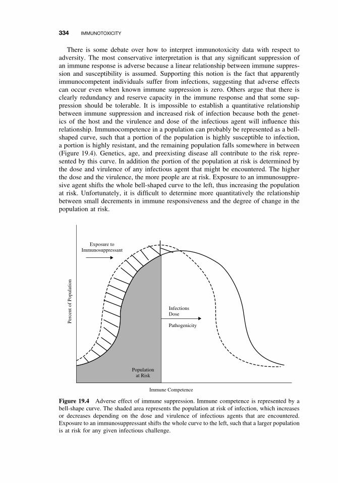
334 IMMUNOTOXICITY
There is some debate over how to interpret immunotoxicity data with respect toadversity. The most conservative interpretation is that any significant suppression ofan immune response is adverse because a linear relationship between immune suppres-sion and susceptibility is assumed. Supporting this notion is the fact that apparentlyimmunocompetent individuals suffer from infections, suggesting that adverse effectscan occur even when known immune suppression is zero. Others argue that there isclearly redundancy and reserve capacity in the immune response and that some sup-pression should be tolerable. It is impossible to establish a quantitative relationshipbetween immune suppression and increased risk of infection because both the genet-ics of the host and the virulence and dose of the infectious agent will influence thisrelationship. Immunocompetence in a population can probably be represented as a bell-shaped curve, such that a portion of the population is highly susceptible to infection,a portion is highly resistant, and the remaining population falls somewhere in between(Figure 19.4). Genetics, age, and preexisting disease all contribute to the risk repre-sented by this curve. In addition the portion of the population at risk is determined bythe dose and virulence of any infectious agent that might be encountered. The higherthe dose and the virulence, the more people are at risk. Exposure to an immunosuppre-sive agent shifts the whole bell-shaped curve to the left, thus increasing the populationat risk. Unfortunately, it is difficult to determine more quantitatively the relationshipbetween small decrements in immune responsiveness and the degree of change in thepopulation at risk.
Exposure toImmunosuppressant
InfectionsDose
Pathogenicity
Immune Competence
Perc
ent o
f Po
pula
tion
Populationat Risk
Figure 19.4 Adverse effect of immune suppression. Immune competence is represented by abell-shape curve. The shaded area represents the population at risk of infection, which increasesor decreases depending on the dose and virulence of infectious agents that are encountered.Exposure to an immunosuppressant shifts the whole curve to the left, such that a larger populationis at risk for any given infectious challenge.
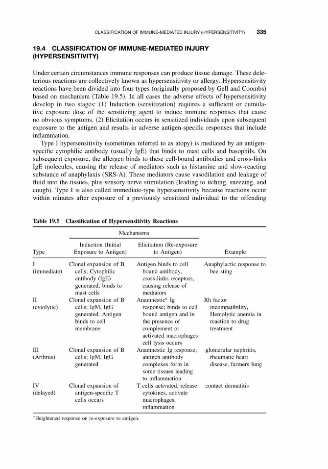
CLASSIFICATION OF IMMUNE-MEDIATED INJURY (HYPERSENSITIVITY) 335
19.4 CLASSIFICATION OF IMMUNE-MEDIATED INJURY(HYPERSENSITIVITY)
Under certain circumstances immune responses can produce tissue damage. These dele-terious reactions are collectively known as hypersensitivity or allergy. Hypersensitivityreactions have been divided into four types (originally proposed by Gell and Coombs)based on mechanism (Table 19.5). In all cases the adverse effects of hypersensitivitydevelop in two stages: (1) Induction (sensitization) requires a sufficient or cumula-tive exposure dose of the sensitizing agent to induce immune responses that causeno obvious symptoms. (2) Elicitation occurs in sensitized individuals upon subsequentexposure to the antigen and results in adverse antigen-specific responses that includeinflammation.
Type I hypersensitivity (sometimes referred to as atopy) is mediated by an antigen-specific cytophilic antibody (usually IgE) that binds to mast cells and basophils. Onsubsequent exposure, the allergen binds to these cell-bound antibodies and cross-linksIgE molecules, causing the release of mediators such as histamine and slow-reactingsubstance of anaphylaxis (SRS-A). These mediators cause vasodilation and leakage offluid into the tissues, plus sensory nerve stimulation (leading to itching, sneezing, andcough). Type I is also called immediate-type hypersensitivity because reactions occurwithin minutes after exposure of a previously sensitized individual to the offending
Table 19.5 Classification of Hypersensitivity Reactions
Mechanisms
TypeInduction (Initial
Exposure to Antigen)Elicitation (Re-exposure
to Antigen) Example
I(immediate)
Clonal expansion of Bcells; Cytophilicantibody (IgE)generated; binds tomast cells
Antigen binds to cellbound antibody,cross-links receptors,causing release ofmediators
Anaphylactic response tobee sting
II(cytolytic)
III(Arthrus)
IV(delayed)
Clonal expansion of Bcells; IgM, IgGgenerated. Antigenbinds to cellmembrane
Clonal expansion of Bcells; IgM, IgGgenerated
Clonal expansion ofantigen-specific Tcells occurs
Anamnestica Igresponse; binds to cellbound antigen and inthe presence ofcomplement oractivated macrophagescell lysis occurs
Anamnestic Ig response;antigen antibodycomplexes form insome tissues leadingto inflammation
T cells activated, releasecytokines, activatemacrophages,inflammation
Rh factorincompatibility,Hemolytic anemia inreaction to drugtreatment
glomerular nephritis,rheumatic heartdisease, farmers lung
contact dermatitis
aHeightened response on re-exposure to antigen.
336 IMMUNOTOXICITY
antigen. Type I reactions include immediate asthmatic responses to allergen, allergicrhinitis (hay fever), atopic dermatitis (eczema), and acute urticaria (hives). The mostsevere form is systemic anaphylaxis (e.g., in response to a bee sting), which results inanaphylactic shock, and potentially death.
Type II hypersensitivity is the result of antibody-mediated cytotoxicity that occurswhen antibodies respond to cell surface antigens. Antibodies bound to antigen on thecell surface activate the complement system and/or macrophages, leading to lysis ofthe target cell. Frequently blood cells are the targets, as in the case of an incompatibleblood transfusion or Rh blood incompatibility between mother and child. The basementmembrane of the kidney or lung may also be a target. Autoimmune diseases can resultfrom drug treatments with penicillin, quinidine, quinine, or acetaminophen. Apparentlythese drugs interact with the cell membrane such that the immune system detects“foreign” antigens on the cell surface. This type of autoimmune disease may also haveunknown etiologies.
Type III reactions are the result of antigen-antibody (IgG) complexes that accumulatein tissues or the circulation, activate macrophages and the complement system, andtrigger the influx of granulocytes and lymphocytes (inflammation). This is sometimesreferred to as the Arthrus reaction and includes postinfection sequelae such as rheumaticheart disease. Farmer’s lung, a pneumonitis caused by molds has been attributed to bothtype III and type IV, and some of the late phase response (4–6 hours after exposure)in asthmatics may be the result of Arthrus-type reactions.
Unlike the preceding three types, type IV, or delayed-type hypersensitivity (DTH),involves T cells and macrophages, not antibodies. Activated T cells release cytokinesthat cause accumulation and activation of macrophages, which in turn cause localdamage. This type of reaction is very important in defense against intracellular infec-tions such as tuberculosis, but is also responsible for contact hypersensitivity responses(allergic contact dermatitis) such as the response to poison ivy. Inhalation of berylliumcan result in a range of pathologies, including acute pneumonitis, tracheobronchitis,and chronic beryllium disease, all of which appear to be due to type IV beryllium-specific immune responses. The expression of type IV responses following challengeis delayed, occurring 24 to 48 hours after exposure.
The different types of immune-mediated injury are not mutually exclusive. Morethan one hypersensitivity mechanism may be involved in the response to a particularantigen. Also the resulting pathology, particularly that caused by type III and IV,reactions may appear very similar, although the mechanisms leading to the effectare different.
19.5 EFFECTS OF CHEMICALS ON ALLERGIC DISEASE
Xenobiotics can affect allergic disease in one of two ways. They can themselves actas antigens and elicit hypersensitivity responses, or they can enhance the developmentor expression of allergic responses to commonly encountered allergens, such as dustmite. Chemicals that act as allergens include certain proteins that can by themselvesinduce an immune response and low molecular weight chemicals (known as haptens)that are too small to induce a specific immune response but may react with a protein toinduce an immune response that is then hapten specific. Haptens have been associatedwith both allergic contact dermatitis (ACD), sometimes called contact hypersensitivity
EFFECTS OF CHEMICALS ON ALLERGIC DISEASE 337
(CHS), and respiratory hypersensitivity. Proteins have been associated with respiratoryhypersensitivity and food allergies. When a chemical is an allergen or a hapten, thereare two doses of concern, the sensitizing dose and the elicitation dose. In general, thedose required for sensitization is greater than that required to elicit a response in asensitized individual. Chemicals that enhance the development of allergic sensitizationare referred to as adjuvants. Air pollutants have been associated both with enhancedsensitization and exacerbation of allergic respiratory symptoms.
19.5.1 Allergic Contact Dermatitis
Allergic contact dermatitis (ACD) or contact hypersensitivity (CHS) is one of the mostcommon occupational health problems and hence is one of the most common problemsassociated with immunotoxicity. It is a type IV response that occurs as a result of dermalexposure to chemicals that are haptens. Following dermal exposure, the chemical reactswith host cell protein at the surface of the skin and is picked up by epidermal dendriticcells, known as Langerhans cells. Cytokines released from the epidermal keratinocytesand from Langerhans cells cause maturation and mobilization of the Langerhans cells,which travel to the draining local lymph node and present antigen to lymphocytes.Clonal expansion occurs, enlarging the number of T lymphocytes specific for that aller-gen and generating memory cells that, in addition to specificity for the allergen, have thepropensity to home to the skin. On re-exposure to the chemical, these specific T cells areactivated, proliferate, home rapidly to the site of exposure, and produce erythema andedema typical of a type IV response. The reaction to poison ivy is the classic example.
Methods to assess chemicals (drugs, pesticides, dyes, cosmetics, and household prod-ucts, etc.) for potential to induce CHS are well established, and several protocols usingguinea pigs have been in use since the 1950s. These protocols assess the actual diseaseend point, skin erythema, and edema, following sensitization and challenge with the testagent. Two commonly used tests are the guinea pig maximization test and the Buehleroccluded patch test. The sensitization procedure for the maximization tests includesintradermal injection of the test chemical with an adjuvant (intended to enhance thesensitization process) as well as topical application. The Buehler test relies on topicalsensitization alone. In both cases, after approximately 2 weeks, animals are challengedat a different site on the skin and erythema and edema are assessed 24 to 48 hourslater. This assessment is somewhat subjective and these tests are fairly expensive.
A chemical is considered to be a sensitizer if 30% (maximization) or 15% (Buehler)of the animals respond. Recently a more economical, less subjective, test for CHS hasbeen developed using mice. This test, the local lymph node assay (LLNA), assessesthe proliferative response of lymphocytes in the draining lymph node following appli-cation of the agent to the ear and is based on our understanding of the immunologicmechanisms underlying CHS; that is, clonal expansion has to occur in the draininglymph node if there is to be allergic sensitization (Figure 19.5). The LLNA is gainingacceptance as a stand-alone alternative to the guinea pig tests and is likely to becomethe assay of choice.
Finally structure activity approaches have recently been developed to identify contactsensitizers. This approach is based on the concept that the biologic mechanisms thatdetermine a chemical’s effect are related to its structure and hence chemicals withsimilar structures will have similar effects. Computer models have been developed tocompare the structure of an unknown chemical to structures in a database for known
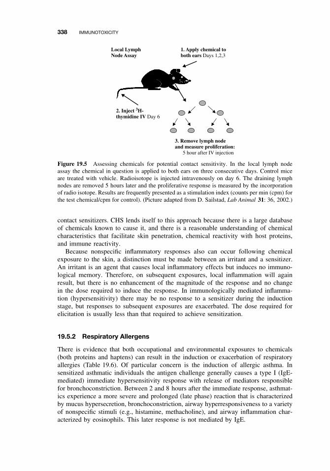
338 IMMUNOTOXICITY
3. Remove lymph nodeand measure proliferation: 5 hour after IV injection
Local LymphNode Assay
1. Apply chemical toboth ears Days 1,2,3
2. Inject 3H-thymidine IV Day 6
Figure 19.5 Assessing chemicals for potential contact sensitivity. In the local lymph nodeassay the chemical in question is applied to both ears on three consecutive days. Control miceare treated with vehicle. Radioisotope is injected intravenously on day 6. The draining lymphnodes are removed 5 hours later and the proliferative response is measured by the incorporationof radio isotope. Results are frequently presented as a stimulation index (counts per min (cpm) forthe test chemical/cpm for control). (Picture adapted from D. Sailstad, Lab Animal 31: 36, 2002.)
contact sensitizers. CHS lends itself to this approach because there is a large databaseof chemicals known to cause it, and there is a reasonable understanding of chemicalcharacteristics that facilitate skin penetration, chemical reactivity with host proteins,and immune reactivity.
Because nonspecific inflammatory responses also can occur following chemicalexposure to the skin, a distinction must be made between an irritant and a sensitizer.An irritant is an agent that causes local inflammatory effects but induces no immuno-logical memory. Therefore, on subsequent exposures, local inflammation will againresult, but there is no enhancement of the magnitude of the response and no changein the dose required to induce the response. In immunologically mediated inflamma-tion (hypersensitivity) there may be no response to a sensitizer during the inductionstage, but responses to subsequent exposures are exacerbated. The dose required forelicitation is usually less than that required to achieve sensitization.
19.5.2 Respiratory Allergens
There is evidence that both occupational and environmental exposures to chemicals(both proteins and haptens) can result in the induction or exacerbation of respiratoryallergies (Table 19.6). Of particular concern is the induction of allergic asthma. Insensitized asthmatic individuals the antigen challenge generally causes a type I (IgE-mediated) immediate hypersensitivity response with release of mediators responsiblefor bronchoconstriction. Between 2 and 8 hours after the immediate response, asthmat-ics experience a more severe and prolonged (late phase) reaction that is characterizedby mucus hypersecretion, bronchoconstriction, airway hyperresponsiveness to a varietyof nonspecific stimuli (e.g., histamine, methacholine), and airway inflammation char-acterized by eosinophils. This later response is not mediated by IgE.
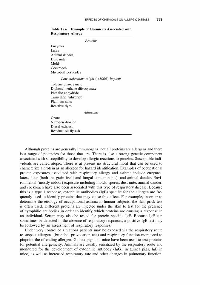
EFFECTS OF CHEMICALS ON ALLERGIC DISEASE 339
Table 19.6 Example of Chemicals Associated withRespiratory Allergy
Proteins
EnzymesLatexAnimal danderDust miteMoldsCockroachMicrobial pesticides
Low molecular weight (<3000 )-haptens
Toluene diisocyanateDiphenylmethane diisocyanatePhthalic anhydrideTrimellitic anhydridePlatinum saltsReactive dyes
Adjuvants
OzoneNitrogen dioxideDiesel exhaustResidual oil fly ash
Although proteins are generally immunogens, not all proteins are allergens and thereis a range of potencies for those that are. There is also a strong genetic componentassociated with susceptibility to develop allergic reactions to proteins. Susceptible indi-viduals are called atopic. There is at present no structural motif that can be used tocharacterize a protein as an allergen for hazard identification. Examples of occupationalprotein exposures associated with respiratory allergy and asthma include enzymes,latex, flour (both the grain itself and fungal contaminants), and animal dander. Envi-ronmental (mostly indoor) exposure including molds, spores, dust mite, animal dander,and cockroach have also been associated with this type of respiratory disease. Becausethis is a type 1 response, cytophilic antibodies (IgE) specific for the allergen are fre-quently used to identify proteins that may cause this effect. For example, in order todetermine the etiology of occupational asthma in human subjects, the skin prick testis often used. Different proteins are injected under the skin to test for the presenceof cytophilic antibodies in order to identify which proteins are causing a response inan individual. Serum may also be tested for protein specific IgE. Because IgE cansometimes be detected in the absence of respiratory responses, a positive IgE test maybe followed by an assessment of respiratory responses.
Under very controlled situations patients may be exposed via the respiratory routeto suspect allergens (broncho- provocation test) and respiratory function monitored topinpoint the offending allergen. Guinea pigs and mice have been used to test proteinsfor potential allergenicity. Animals are usually sensitized by the respiratory route andmonitored for the development of cytophilic antibody (IgG1 in guinea pigs, IgE inmice) as well as increased respiratory rate and other changes in pulmonary function.
340 IMMUNOTOXICITY
The guinea pig intratracheal test has been used to establish the relative potency of dif-ferent detergent enzymes and establish safe occupational exposure levels. As the nameimplies, guinea pigs are sensitized by intratracheal exposure and induction of cytophilicantibodies are assessed. Dose responses obtained for new enzymes are compared to areference enzyme for which safe exposure levels have been established. The relativepotency of the new enzyme to this reference is used to establish a safe exposure levelfor the new enzyme.
Exposure to certain low (<3000) molecular weight compounds (haptens) has also beenassociated with the development of occupational asthma. Highly reactive compoundssuch as the diisocyanates or acid anhydrides have the capacity to react with protein andinduce an immune response. Toluene diisocyanate (TDI) and trimellitic anhydride arethe compounds that have been most extensively studied in this regard. There is a greatdeal of interest in developing a test to screen chemicals for this type of effect in order toavoid induction of immune responses that could lead to occupational asthma. Althoughspecific IgE antibodies have been detected in some individuals with TDI asthma, it hasnot been uniformly present and some of these individuals exhibit the late phase but not theimmediate response. Hence, unlike proteins, there is less certainty about the mechanismsunderlying respiratory allergic responses to low molecular weight compounds.
Structure activity approaches similar to those described for contact sensitizers havebeen developed, but this approach has limitations because the database of knownrespiratory sensitizers is small compared to contact sensitizers and the underlying mech-anisms are less well defined. At the other extreme guinea pigs have been exposed byinhalation for a number of days, rested, and then challenged at a later date by inhala-tion with subsequent monitoring of respiratory responses. Although this approach hasproduced a good model of TDI asthma, it is too cumbersome and expensive for routinetesting. Because the capacity to interact with protein is a pre-requisite to allergenicity,it has been suggested that testing for protein reactivity in vitro could provide an initialscreening test for chemicals. Also, because it appears that respiratory sensitizers area subset of chemicals that produce positive results in a contact sensitivity test, it hasbeen suggested that the LLNA test be used as the first tier in screening chemicals forthis effect. The problem then becomes separating chemicals that are strictly contactsensitizers from those that have the capacity to cause respiratory sensitization. Effortshave been made to determine whether differences in responses to dermal application ofthese chemicals could provide a means for making this distinction. One proposal is toassess total serum IgE following dermal exposure, assuming that respiratory sensitiz-ers would produce a bigger IgE signal. Another approach has been to assess cytokineprofiles in the draining lymph node following dermal exposure. Different subsets ofT helper (Th) cells, have been associated with type I immediate (Th2) and type IVdelayed (Th1) responses. These different populations of T cells are distinguished bydifferent cytokine profile and efforts are underway to use these differing profiles to dis-tinguish respiratory from contact sensitizers. However, there is as yet no well-validated,well-accepted test to assess low molecular weight chemicals for the capacity to inducerespiratory allergy. This remains a subject of intense research.
19.5.3 Adjuvants
An adjuvant is a compound administered in conjunction with an antigen that non-specifically enhances the immune response to that antigen. Adjuvants are used in
EMERGING ISSUES: FOOD ALLERGIES, AUTOIMMUNITY, AND THE DEVELOPING IMMUNE SYSTEM 341
vaccines to promote immunogenicity. There is now growing concern that chemicalsin our environment (particularly, air pollutants) might act as adjuvants for allergicsensitization to common allergens such as dust mite and pollen. Laboratory rodentshave been used to show that nitrogen dioxide, residual oil fly ash, and diesel exhaustenhance allergic sensitization and disease. Enhanced sensitization to an allergen hasalso been demonstrated in rhesus monkeys exposed to ozone and humans exposed todiesel exhaust. The significance of these findings in terms of enhanced burden of res-piratory allergies in the human population is unclear. As in other areas of toxicology,simultaneous environmental exposures to agents that are not the agent of immediateconcern can certainly influence outcomes. Adjuvancy is a concern that likely extendsbeyond air pollution and type 1 responses.
19.6 EMERGING ISSUES: FOOD ALLERGIES, AUTOIMMUNITY,AND THE DEVELOPING IMMUNE SYSTEM
There are several emerging issues in immunotoxicology. These active areas of researchwill be only briefly described here because there are currently more questions thananswers.
Toxicologists have recently been drawn into the area of food allergy by advancesin biotechnology and the need to assess the safety of genetically modified foods interms of potential allergenicity. There is concern that insertion of a novel gene into afood crop (e.g., to increase yield or pest resistance) might inadvertently introduce a newallergen into the food supply. Food allergies are relatively rare, affecting approximately5% of children and 2–3% of adults, and even in these individuals, most proteinsare not food allergens. However, when food allergy does occur, the consequencescan be severe. Anaphylactic (life-threatening) reactions to peanuts provide the bestexample. Unfortunately, the mechanisms underlying food allergies (or the mechanismsthat protect most of people from developing reactions to the foreign proteins they eat),the characteristics that make a protein a food allergen, and the characteristics that makean individual susceptible to food allergies are poorly understood at this time. Theseare some of the issues that need to be resolved in order to develop appropriate safetyassessment tools.
Autoimmune diseases affect about 3% of the population and comprise a diversearray of both organ specific (e.g., type I diabetes, thyroiditis) and systemic (systemiclupus erythematosis) diseases. Susceptibility includes a strong genetic component, andin some cases women appear to be more vulnerable than men. Xenobiotics mightaffect the development or progression of autoimmune disease. A variety of mechanismscould contribute to xenobiotic effects on the development and maintenance of immunetolerance or unmasking or modification of self proteins. There is evidence that exposureto certain drugs, heavy metals, silica, and endocrine disruptors are a concern in thisregard. Current research includes both human and animals studies to determine theextent of risk and ways to assess and control it.
Finally there is growing concern that the developing immune system may be partic-ularly vulnerable to xenobiotic exposures and that perinatal and/or in utero exposuresmay have a lifelong impact on susceptibility to infectious, allergic, or autoimmunedisease. As in other areas of toxicology, tests designed to assess the risk of immuno-toxicity for adults may not be sufficient to protect children and research is currentlyunderway to determine how best to meet this need.
342 IMMUNOTOXICITY
Clearly, exposure to xenobiotics can have a number of effects on the immune systemthat in turn can affect an array of health outcomes. In some areas of immunotoxicologysignificant progress has been made in terms of identifying and understanding the risksassociated with xenobiotic exposure. In other areas more research is needed.
SUGGESTED READING
Benjamini, E., G. Sunshine, and S. Leskowitz. Immunology A Short Course, 4th ed. New York:Wiley, 2000.
Gilmour, M. I., M. J. K. Selgrade, and A. L. Lambert. Enhanced allergic sensitization in ani-mals exposed to particulate air pollution. Inhalation Toxicol. 12 (suppl. 3): 373–380, 2000.
Kimber, I., I. L. Bernstein, M. H. Karol, M. K. Robinson, K. Sarlo, and M. J. K. Selgrade.Workshop overview: Identification of respiratory allergens. Fundam. Appl. Toxicol. 33: 1–10,1996.
Luster, M. I., A. E. Munson, P. T. Thomas, M. P. Holsapple, J. D. Fenters, K. L. White, L. E.Lauer, D. R. Germolec, G. J. Rosenthal, and J. H. Dean. Development of a testing batteryto assess chemical-induced immunotoxicity: National Toxicology Program’s guidelines forimmunotoxicity evaluation in mice. Fundam. Appl. Toxicol. 10: 2–19, 1988.
Luster, M. I., et al. Risk assessment in immunotoxicology I. Sensitivity and predictability ofimmune tests. Fundam. Appl. Toxicol. 18: 200–210, 1992, and II. Relationships betweenimmune and host resistance tests. Fundam. Appl. Toxicol. 21: 71–82, 1993.
Metcalfe, D. E., et al. Assessment of the allergenic potential of foods derived from geneticallyengineered crop plants. Crit. Rev. Food Sci. Nutr. 36S: S165–S186, 1996.
Roitt, I. Roitt’s Essentials in Immunology. London: Blackwell, 2001.
Sailstad, D. M. Murine local lymph node assay: An alternative test method for skin hypersen-sitivity testing. Lab. Animal 31: 36–41, 2002.
Sarlo, K. Human Health Risk Assessment: Focus on Enzymes. In Proceeding of the 3rd WorldConference on Detergents, A. Cahn, ed. Champaign, I: Am. Oil Chem. Soc. Press, 1994,pp. 54–57.
CHAPTER 20
Reproductive System
STACY BRANCH
20.1 INTRODUCTION
What is reproductive toxicity? Reproductive toxicity refers to any adverse effect onany aspect of male or female sexual structure, function, and lactation including effectson the reproductive potential and viability of the offspring. This concept may alsoinclude the following:
Organ toxicity . Can interfere with normal system function.Teratogenicity . Ability to cause dysmorphogenesis in the developing fetus.Behavioral teratogenicity . Ability to adversely affect the mental development of
the fetus.Developmental toxicity . Includes both teratogenicity terms above and abnormal post-
natal development.
This chapter discusses both male and female reproductive toxicity and provides anoverview of reproductive physiology.
20.2 MALE REPRODUCTIVE PHYSIOLOGY
The anterior pituitary is stimulated by the hypothalamus (via the gonadotrophichormone releasing hormone) to release gonadotrophic hormones (leutenizinghormone—LH; and follicle stimulating hormone—FSH). In addition to LH andFSH, prolactin is released by the anterior pituitary. The target of LH and FSH inthe male is the testis. While LH stimulates steroidogenesis, FSH has its primaryeffects on the sertoli cells. The role of prolactin (which is inhibited by dopamine)is to modulate the effects of LH in the testicular tissue. Critical points within thehypothalamic–pituitary–gonadal axis (Figure 20.1) may be susceptible to alterationsby xenobiotics, leading to altered reproductive function and pathology.
A Textbook of Modern Toxicology, Third Edition, edited by Ernest HodgsonISBN 0-471-26508-X Copyright 2004 John Wiley & Sons, Inc.
343
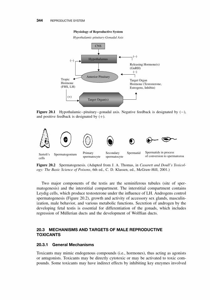
344 REPRODUCTIVE SYSTEM
CNS
Hypothalamic-pituitary-Gonadal Axis
Physiology of Reproductive System
Hypothalamus
Anterior Pituitary
Target Organ(s)
TropicHormone(FHS, LH)
Target OrganHormone (Testosterone,Estrogens, Inhibin)
Releasing Hormone(s)(GnRH)
(+)
(−)
(−)
(−)
Figure 20.1 Hypothalamic–pituitary–gonadal axis. Negative feedback is designated by (−),and positive feedback is designated by (+).
Sertoli’scells
Spermatogonium Primaryspermatocyte
Secondaryspermatocyte
Spermatid Spermatids in processof conversion to spermatozoa
Figure 20.2 Spermatogenesis. (Adapted from J. A. Thomas, in Casarett and Doull’s Toxicol-ogy: The Basic Science of Poisons, 6th ed., C. D. Klassen, ed., McGraw-Hill, 2001.)
Two major components of the testis are the seminiferous tubules (site of sper-matogenesis) and the interstitial compartment. The interstitial compartment containsLeydig cells, which produce testosterone under the influence of LH. Androgens controlspermatogenesis (Figure 20.2), growth and activity of accessory sex glands, masculin-ization, male behavior, and various metabolic functions. Secretion of androgen by thedeveloping fetal testis is essential for differentiation of the gonads, which includesregression of Mullerian ducts and the development of Wolffian ducts.
20.3 MECHANISMS AND TARGETS OF MALE REPRODUCTIVETOXICANTS
20.3.1 General Mechanisms
Toxicants may mimic endogenous compounds (i.e., hormones), thus acting as agonistsor antagonists. Toxicants may be directly cytotoxic or may be activated to toxic com-pounds. Some toxicants may have indirect effects by inhibiting key enzymes involved
MECHANISMS AND TARGETS OF MALE REPRODUCTIVE TOXICANTS 345
in steroid synthesis. Below are examples of how selected toxicants affect various sus-ceptible targets of the male reproductive tract.
20.3.2 Effects on Germ Cells
Epidemiological data have indicated that wives of men exposed to the compoundvinyl chloride experience spontaneous abortion. Vinyl chloride is used to manufacturepolyvinyl chloride (PVC). PVC is a component of various plastic products, includingpipes, furniture, and automobile upholstery. Ethylnitrosourea (ENU) is a mutagenicagent that has been used extensively in mouse mutagenesis. ENU acts on male sper-matogonial stem cells, introducing mutations. It is also capable of inducing reversiblesterility in mice. Actinomycin D is an older chemotherapeutic drug that has been usedin cancer therapy for many years. Actinomycin D is commonly used in the treatment ofgestational trophoblastic cancers, testicular cancer, Wilm’s tumor, and rhabdomyosar-coma. Actinomycin D intercalates with DNA and disrupts the structure and functionof the DNA of the spermatozoa.
20.3.3 Effects on Spermatogenesis and Sperm Quality
The antibiotics and antimetabolites used in cancer treatment (i.e., vinblastine) are sper-matotoxic and affect semen quality. Ionizing radiation is also spermatotoxic (withspermatogonial cells being the most sensitive). Prolonged scrotal heating is a factorthat affects the earlier states of spermatogenesis.
20.3.4 Effects on Sexual Behavior
Anabolic steroids, antidepressants and drugs of abuse affect libido, potency, andejaculatory function. Anabolic steroids are derivatives of testosterone, and have stronggenitotropic effects. There is published evidence indicating that anabolic steroidsincreases sexual desire; however, the frequency of erectile dysfunction is also increased.Treatment with the antidepressant fluoxetine has been associated with sexual sideeffects including delayed or nonexistent ejaculation and hyposexuality. Mice treated inutero with the anitleukemic agent 5-aza-2′-deoxycytidine exhibit abnormal reproductivebehavior and low reproductive capacity.
20.3.5 Effects on Endocrine Function
Cimetidine (for treatment of peptic ulcers) competes with dihydrotestosterone forreceptors in the testis and accessory sex glands. The more common sequelaeare low sperm count and gynacomastia. Epidemiological evidence has shown thatoccupation exposure to oral contraceptives can induce gynacomastia in exposed males.Diethylstilbestrol (DES) antagonizes the activity of fetal testosterone. In the maleoffspring, testicular hypoplasia, abnormal semen parameters, and infertility result.Ketoconazole has be shown to be transported to the seminal fluid and to immobilizethe sperm.
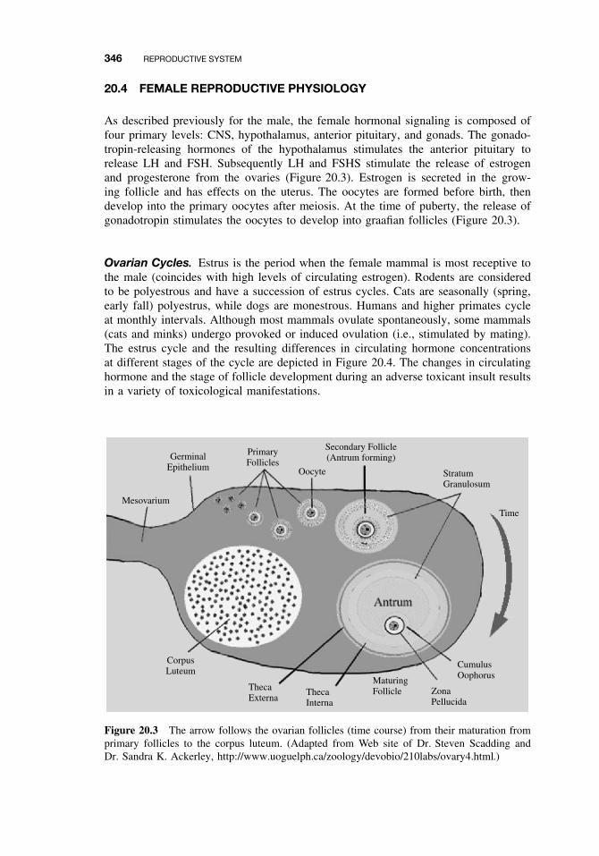
346 REPRODUCTIVE SYSTEM
20.4 FEMALE REPRODUCTIVE PHYSIOLOGY
As described previously for the male, the female hormonal signaling is composed offour primary levels: CNS, hypothalamus, anterior pituitary, and gonads. The gonado-tropin-releasing hormones of the hypothalamus stimulates the anterior pituitary torelease LH and FSH. Subsequently LH and FSHS stimulate the release of estrogenand progesterone from the ovaries (Figure 20.3). Estrogen is secreted in the grow-ing follicle and has effects on the uterus. The oocytes are formed before birth, thendevelop into the primary oocytes after meiosis. At the time of puberty, the release ofgonadotropin stimulates the oocytes to develop into graafian follicles (Figure 20.3).
Ovarian Cycles. Estrus is the period when the female mammal is most receptive tothe male (coincides with high levels of circulating estrogen). Rodents are consideredto be polyestrous and have a succession of estrus cycles. Cats are seasonally (spring,early fall) polyestrus, while dogs are monestrous. Humans and higher primates cycleat monthly intervals. Although most mammals ovulate spontaneously, some mammals(cats and minks) undergo provoked or induced ovulation (i.e., stimulated by mating).The estrus cycle and the resulting differences in circulating hormone concentrationsat different stages of the cycle are depicted in Figure 20.4. The changes in circulatinghormone and the stage of follicle development during an adverse toxicant insult resultsin a variety of toxicological manifestations.
Mesovarium
CorpusLuteum
GerminalEpithelium
PrimaryFollicles
Oocyte
Secondary Follicle(Antrum forming)
StratumGranulosum
Time
ThecaExterna
ThecaInterna
MaturingFollicle Zona
Pellucida
CumulusOophorus
Figure 20.3 The arrow follows the ovarian follicles (time course) from their maturation fromprimary follicles to the corpus luteum. (Adapted from Web site of Dr. Steven Scadding andDr. Sandra K. Ackerley, http://www.uoguelph.ca/zoology/devobio/210labs/ovary4.html.)
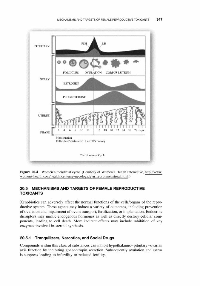
MECHANISMS AND TARGETS OF FEMALE REPRODUCTIVE TOXICANTS 347
PITUITARY
OVARY
UTERUS
PHASE
FSH LH
FOLLICLES
PROGESTERONE
ESTROGEN
OVULATION CORPUS LUTEUM
2
MenstruationFollicular/Proliferative Lufeel/Secretory
The Hormonal Cycle
4 6 8 10 12 16 18 20 22 24 26 28 days
Figure 20.4 Women’s menstrual cycle. (Courtesy of Women’s Health Interactive, http://www.womens-health.com/health center/gynecology/gyn repro menstrual.html.)
20.5 MECHANISMS AND TARGETS OF FEMALE REPRODUCTIVETOXICANTS
Xenobiotics can adversely affect the normal functions of the cells/organs of the repro-ductive system. These agents may induce a variety of outcomes, including preventionof ovulation and impairment of ovum transport, fertilization, or implantation. Endocrinedisruptors may mimic endogenous hormones as well as directly destroy cellular com-ponents, leading to cell death. More indirect effects may include inhibition of keyenzymes involved in steroid synthesis.
20.5.1 Tranquilizers, Narcotics, and Social Drugs
Compounds within this class of substances can inhibit hypothalamic–pituitary–ovarianaxis function by inhibiting gonadotropin secretion. Subsequently ovulation and estrusis suppress leading to infertility or reduced fertility.
348 REPRODUCTIVE SYSTEM
20.5.2 Endocrine Disruptors (EDs)
Endocrine disruptors are compounds (synthetic and naturally occurring) that can alterthe normal hormonal balance and function in animals. The historical ED diethylstilbe-strol (DES) is a classic example of an endocrine disrupter affecting female reproductivehealth. In utero exposure of females to DES is associated with the induction of vaginalcarcinomas apparent after puberty. In experimental mice, estrogenic substances causeaccelerated sexual maturation and irregular estrous cycles and prolonged estrous. Inrats, xenoestrogens such as kepone and methoxychlor cause masculinization of theexposed female rats. These rats do not ovulate, lack stimulation of the LH surge, andexhibit male sexual behavior. In humans, estrogen mimicking compounds can alter nat-ural hormonal cycles and have been associated with breast cancer induction. Certainenvironmental EDs may function as promoters or inducers of carcinogenesis. Poly-chlorinated biphenyls (PCBs) and a trichloroethane compound (DDT) are persistent inthe environment. Serum DDE (a DDT metabolite) levels have been found to correlatewith breast cancer incidence.
20.5.3 Effects on Germ Cells
As previously described for the male reproductive toxicity, the class of toxicants affect-ing germ cells can alter the structure of genetic material (chromosomal aberrations,alterations in meiosis, DNA synthesis, and replication). Mature oocytes have a DNArepair capacity different from that of mature sperm, but this capacity decreases at theperiod of meiotic maturation.
20.5.4 Effects on the Ovaries and Uterus
Cyclophosphamide and vincristine are examples of alkylating agents capable of induc-ing gonadal dysfunction. Premature menopause is a primary outcome of exposure tothese agents. Amenorrhea and abnormal hormonal levels are characteristics of theovarian dysfunction induced by cyclophosphamide.
Premature ovarian failure can be induced in offspring exposed in utero by activemetabolites such as 6-mercaptopurine. Tamoxifen (treatments for breast cancer) andclomiphene (to induce ovulation) are antiestrogens that can inhibit uterine decidualinduction in pseudopregnant rats.
20.5.5 Effects on Sexual Behavior
Normal sexual activity is associated with ovulation in most female mammals. Com-pounds affecting this process can adversely affect female libido. Ovarian failure inducedby xenobiotic compounds has been associated with a decrease in libido in women.Certain types of oral contraceptives as well as drugs of abuse (methadone, cannabis,alcohol) cause decreases in female libido. The treatment for hirsutism, excessivegrowth of hair in both normal and abnormal locations, is the compound cyproteroneacetate. It is an antiandrogen that has the side effect of severely decreasing libidoin women.

SUGGESTED READING 349
SUGGESTED READING
Klaassen, C. D. Cassarett and Doull’s Toxicology: The Basic Science of Poisons, 6th ed.New York: McGraw-Hill, 2001.
Korach, K. S. Reproductive and Developmental Toxicology. New York: Dekker, 1998.
Naz, R. K. Endocrine Disruptors. Boca Raton, FL: CRC Press, 1997.
Ballantyne, B., T. Marrs, and P. Turner, eds. General and Applied Toxicology, college ed.,New York: Macmillan, 1995.


PART VI
APPLIED TOXICOLOGY

CHAPTER 21
Toxicity Testing
HELEN CUNNY and ERNEST HODGSON
21.1 INTRODUCTION
Although testing for toxicity, usually for the purposes of human health risk assess-ment, might be expected to be one of the more routine aspects of toxicology, it isactually one of the more controversial. Among the many areas of controversy are theuse of animals for testing and the welfare of the animals, extrapolation of animaldata to humans, extrapolation from high-dose to low-dose effects, and the increasingcost and complexity of testing protocols relative to the benefits expected. New testsare constantly being devised and are often added to testing requirements already inexistence.
Most testing can be subdivided into in vivo tests for acute, subchronic, or chroniceffects and in vitro tests for genotoxicity or cell transformation, although other testsare used and are described in this chapter. Any chemical that has been introducedinto commerce or that is being developed for possible introduction into commerce issubject to toxicity testing to satisfy the regulations of one or more regulatory agen-cies. Furthermore compounds produced as waste products of industrial processes (e.g.,combustion products) are also subject to testing.
Toxicity assessment is the determination of the potential of any substance to act asa poison, the conditions under which this potential will be realized, and the charac-terization of its action. Risk assessment, however, is a quantitative assessment of theprobability of deleterious effects under given exposure conditions. Both are involvedin the regulation of toxic chemicals. Regulation is the control, by statute, of the man-ufacture, transportation, sale, or disposal of chemicals deemed to be toxic after testingprocedures or according to criteria laid down in applicable laws.
Testing in the United States is carried out by many groups: industrial, governmental,academic, and others. Regulation, however, is carried out by a narrow range of govern-mental agencies, each charged with the formulation of regulations under a particularlaw or laws and with the administration of those regulations. The principal regulatoryagencies for the United States are shown in Table 21.1. Other industrialized countrieshave counterpart laws and agencies for the regulation of toxic chemicals.
A Textbook of Modern Toxicology, Third Edition, edited by Ernest HodgsonISBN 0-471-26508-X Copyright 2004 John Wiley & Sons, Inc.
353
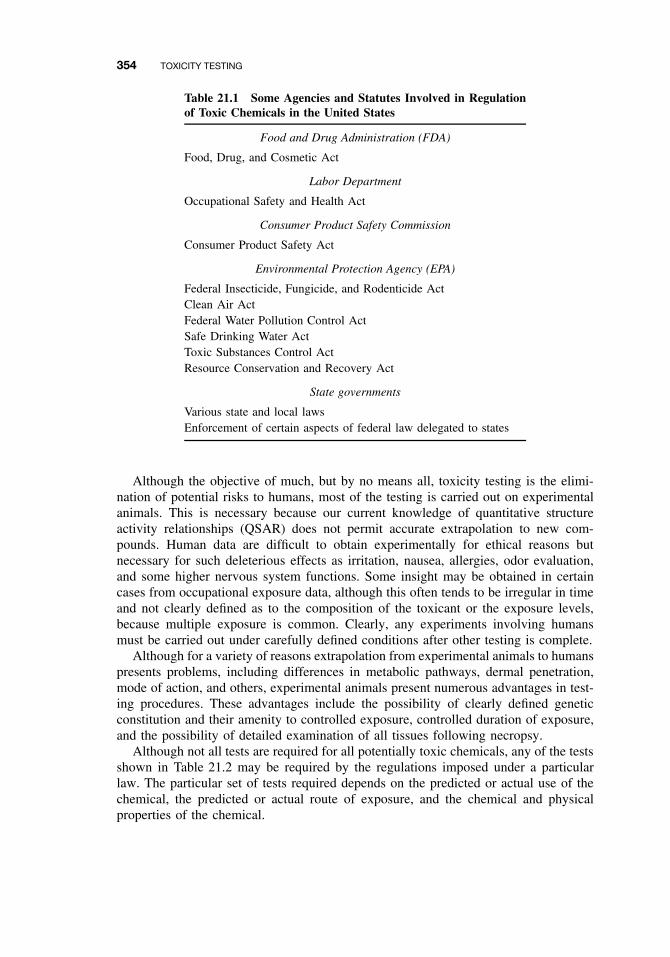
354 TOXICITY TESTING
Table 21.1 Some Agencies and Statutes Involved in Regulationof Toxic Chemicals in the United States
Food and Drug Administration (FDA)
Food, Drug, and Cosmetic Act
Labor Department
Occupational Safety and Health Act
Consumer Product Safety Commission
Consumer Product Safety Act
Environmental Protection Agency (EPA)
Federal Insecticide, Fungicide, and Rodenticide ActClean Air ActFederal Water Pollution Control ActSafe Drinking Water ActToxic Substances Control ActResource Conservation and Recovery Act
State governments
Various state and local lawsEnforcement of certain aspects of federal law delegated to states
Although the objective of much, but by no means all, toxicity testing is the elimi-nation of potential risks to humans, most of the testing is carried out on experimentalanimals. This is necessary because our current knowledge of quantitative structureactivity relationships (QSAR) does not permit accurate extrapolation to new com-pounds. Human data are difficult to obtain experimentally for ethical reasons butnecessary for such deleterious effects as irritation, nausea, allergies, odor evaluation,and some higher nervous system functions. Some insight may be obtained in certaincases from occupational exposure data, although this often tends to be irregular in timeand not clearly defined as to the composition of the toxicant or the exposure levels,because multiple exposure is common. Clearly, any experiments involving humansmust be carried out under carefully defined conditions after other testing is complete.
Although for a variety of reasons extrapolation from experimental animals to humanspresents problems, including differences in metabolic pathways, dermal penetration,mode of action, and others, experimental animals present numerous advantages in test-ing procedures. These advantages include the possibility of clearly defined geneticconstitution and their amenity to controlled exposure, controlled duration of exposure,and the possibility of detailed examination of all tissues following necropsy.
Although not all tests are required for all potentially toxic chemicals, any of the testsshown in Table 21.2 may be required by the regulations imposed under a particularlaw. The particular set of tests required depends on the predicted or actual use of thechemical, the predicted or actual route of exposure, and the chemical and physicalproperties of the chemical.
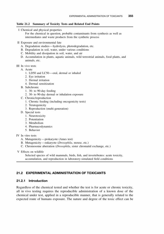
EXPERIMENTAL ADMINISTRATION OF TOXICANTS 355
Table 21.2 Summary of Toxicity Tests and Related End Points
I Chemical and physical propertiesFor the chemical in question, probable contaminants from synthesis as well asintermediates and waste products from the synthetic process
II Exposure and environmental fateA. Degradation studies—hydrolysis, photodegradation, etc.B. Degradation in soil, water, under various conditionsC. Mobility and dissipation in soil, water, and airD. Accumulation in plants, aquatic animals, wild terrestrial animals, food plants, and
animals, etc.
III In vivo testsA. Acute
1. LD50 and LC50—oral, dermal or inhaled2. Eye irritation3. Dermal irritation4. Dermal sensitization
B. Subchronic1. 30- to 90-day feeding2. 30- to 90-day dermal or inhalation exposure
C. Chronic/reproduction1. Chronic feeding (including oncogenicity tests)2. Teratogenicity3. Reproduction (multi-generation)
D. Special tests1. Neurotoxicity2. Potentiation3. Metabolism4. Pharmacodynamics5. Behavior
IV In vitro testsA. Mutagenicity—prokaryote (Ames test)B. Mutagenicity—eukaryote (Drosophila, mouse, etc.)C. Chromosome aberration (Drosophila, sister chromatid exchange, etc.)
V Effects on wildlifeSelected species of wild mammals, birds, fish, and invertebrates: acute toxicity,accumulation, and reproduction in laboratory-simulated field conditions
21.2 EXPERIMENTAL ADMINISTRATION OF TOXICANTS
21.2.1 Introduction
Regardless of the chemical tested and whether the test is for acute or chronic toxicity,all in vivo testing requires the reproducible administration of a known dose of thechemical under test, applied in a reproducible manner, that is generally related to theexpected route of humans exposure. The nature and degree of the toxic effect can be
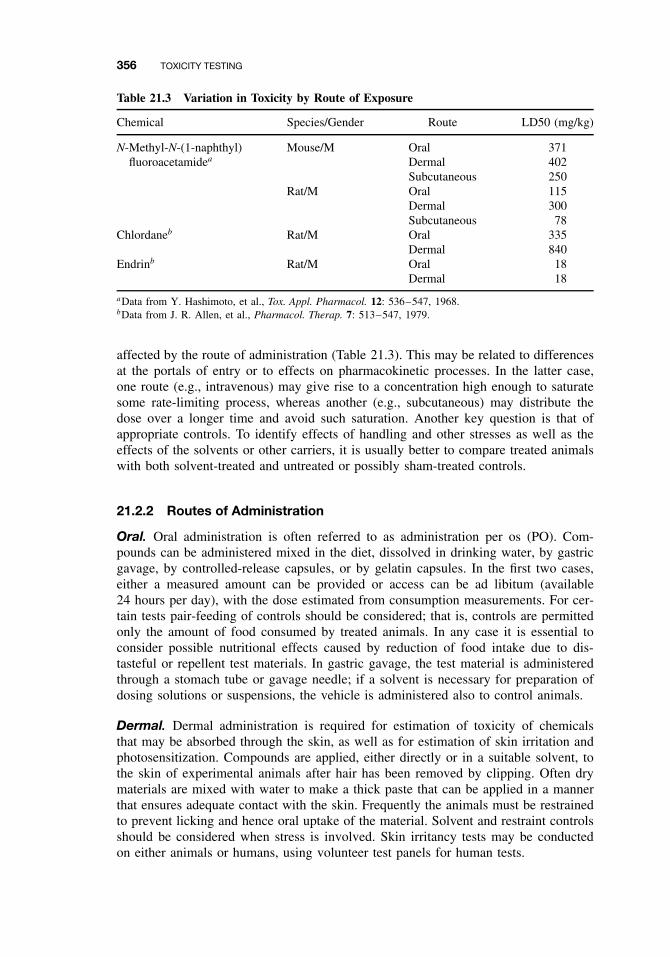
356 TOXICITY TESTING
Table 21.3 Variation in Toxicity by Route of Exposure
Chemical Species/Gender Route LD50 (mg/kg)
N-Methyl-N-(1-naphthyl) Mouse/M Oral 371fluoroacetamidea Dermal 402
Subcutaneous 250Rat/M Oral 115
Dermal 300Subcutaneous 78
Chlordaneb Rat/M Oral 335Dermal 840
Endrinb Rat/M Oral 18Dermal 18
aData from Y. Hashimoto, et al., Tox. Appl. Pharmacol. 12: 536–547, 1968.bData from J. R. Allen, et al., Pharmacol. Therap. 7: 513–547, 1979.
affected by the route of administration (Table 21.3). This may be related to differencesat the portals of entry or to effects on pharmacokinetic processes. In the latter case,one route (e.g., intravenous) may give rise to a concentration high enough to saturatesome rate-limiting process, whereas another (e.g., subcutaneous) may distribute thedose over a longer time and avoid such saturation. Another key question is that ofappropriate controls. To identify effects of handling and other stresses as well as theeffects of the solvents or other carriers, it is usually better to compare treated animalswith both solvent-treated and untreated or possibly sham-treated controls.
21.2.2 Routes of Administration
Oral. Oral administration is often referred to as administration per os (PO). Com-pounds can be administered mixed in the diet, dissolved in drinking water, by gastricgavage, by controlled-release capsules, or by gelatin capsules. In the first two cases,either a measured amount can be provided or access can be ad libitum (available24 hours per day), with the dose estimated from consumption measurements. For cer-tain tests pair-feeding of controls should be considered; that is, controls are permittedonly the amount of food consumed by treated animals. In any case it is essential toconsider possible nutritional effects caused by reduction of food intake due to dis-tasteful or repellent test materials. In gastric gavage, the test material is administeredthrough a stomach tube or gavage needle; if a solvent is necessary for preparation ofdosing solutions or suspensions, the vehicle is administered also to control animals.
Dermal. Dermal administration is required for estimation of toxicity of chemicalsthat may be absorbed through the skin, as well as for estimation of skin irritation andphotosensitization. Compounds are applied, either directly or in a suitable solvent, tothe skin of experimental animals after hair has been removed by clipping. Often drymaterials are mixed with water to make a thick paste that can be applied in a mannerthat ensures adequate contact with the skin. Frequently the animals must be restrainedto prevent licking and hence oral uptake of the material. Solvent and restraint controlsshould be considered when stress is involved. Skin irritancy tests may be conductedon either animals or humans, using volunteer test panels for human tests.
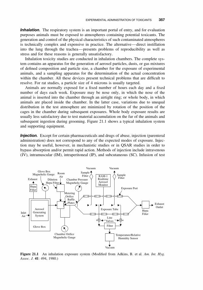
EXPERIMENTAL ADMINISTRATION OF TOXICANTS 357
Inhalation. The respiratory system is an important portal of entry, and for evaluationpurposes animals must be exposed to atmospheres containing potential toxicants. Thegeneration and control of the physical characteristics of such contaminated atmospheresis technically complex and expensive in practice. The alternative—direct instillationinto the lung through the trachea—presents problems of reproducibility as well asstress and for these reasons is generally unsatisfactory.
Inhalation toxicity studies are conducted in inhalation chambers. The complete sys-tem contains an apparatus for the generation of aerosol particles, dusts, or gas mixturesof defined composition and particle size, a chamber for the exposure of experimentalanimals, and a sampling apparatus for the determination of the actual concentrationwithin the chamber. All these devices present technical problems that are difficult toresolve. For rat studies, a particle size of 4 microns is usually targeted.
Animals are normally exposed for a fixed number of hours each day and a fixednumber of days each week. Exposure may be nose only, in which the nose of theanimal is inserted into the chamber through an airtight ring; or whole body, in whichanimals are placed inside the chamber. In the latter case, variations due to unequaldistribution in the test atmosphere are minimized by rotation of the position of thecages in the chamber during subsequent exposures. Whole body exposure results areusually less satisfactory due to test material accumulation on the fur of the animals andsubsequent ingestion during grooming. Figure 21.1 shows a typical inhalation systemand supporting equipment.
Injection. Except for certain pharmaceuticals and drugs of abuse, injection (parenteraladministration) does not correspond to any of the expected modes of exposure. Injec-tion may be useful, however, in mechanistic studies or in QSAR studies in order tobypass absorption and/or permit rapid action. Methods of injection include intravenous(IV), intramuscular (IM), intraperitoneal (IP), and subcutaneous (SC). Infusion of test
AerosolGenerating
System
Glove Box
InletAir
HepaFilter
Exhaust
Glove BoxMagnehelic Gauge Room
AirDilution
Hepa Filter
MeteringOrifice
BafflePlates
Chamber PressureMagnehelic Gauge
Vacuum Vacuum
SampleFilter RAM-1
RealtimeAerosolMonitor
SampleFilter
Exposure Port
ExhaustOutlet
HepaFilter
LineValves
Temperature/RelativeHumidity Sensor
Vaccum
Filter
Chamber OrificeMagnehelic Gauge
Exposure Tube
Figure 21.1 An inhalation exposure system (Modified from Adkins, B. et al. Am. Int. Hyg.Assoc. J. 41: 494, 1980.)
358 TOXICITY TESTING
materials over an extended period is also possible. Again, both solvent controls anduntreated controls are necessary for proper interpretation of the results.
21.3 CHEMICAL AND PHYSICAL PROPERTIES
Although the determination of chemical and physical properties of known or poten-tial toxicants does not constitute a test for toxicity, it is an essential preliminary forsuch tests.
The information obtained can be used in many ways. Structure activity comparisonswith other known toxicants may indicate the most probable hazards. These comparisonsmay also aid identification in subsequent poisoning episodes. Determination of stabilityto light, heat, freezing, and oxidizing or reducing agents may enable preliminary esti-mates of persistence in the environment as well as indicate the most likely breakdownproducts that may also require testing for toxicity. Establishing such properties as thelipid solubility or octanol/water partition coefficient may enable preliminary estimatesof rate of uptake and persistence in living organisms. Vapor pressure may indicatewhether the respiratory system is a probable route of entry. Knowledge of the chem-ical and physical properties must be acquired in order to develop analytical methodsfor the measurement of the compound and its degradation products. If the chemical isbeing produced for commercial use, similar information is needed on intermediates inthe synthesis or by-products of the process because both are possible contaminants inthe final product.
21.4 EXPOSURE AND ENVIRONMENTAL FATE
Data on exposure and environmental fate are needed, not to determine toxicity, but toprovide information that may be useful in the prediction of possible exposure in theevent that the chemical is toxic. These tests are primarily useful for chemicals releasedinto the environment such as pesticides, and they include the rate of breakdown underaerobic and anaerobic conditions in soils of various types, the rates of leaching intosurface water from soils of various types, and the rate of movement toward groundwa-ter. The effects of physical factors on degradation through photolysis and hydrolysisstudies and the identification of the product formed can indicate the rate of loss ofthe hazardous chemical or the possible formation of hazardous degradation products.Tests for accumulation in plants and animals and movement within the ecosystem areconsidered in Section 21.7.
21.5 IN VIVO TESTS
Traditionally the basis for the determination of toxicity has been administration of thetest compound, in vivo, to one or more species of experimental animal, followed byexamination for clinical signs of toxicity and/or mortality in acute tests. In additionpathological examination for tissue abnormalities is also performed, especially in testsof longer duration. The results of these tests are then used by a variety of extrapolation
IN VIVO TESTS 359
techniques to estimate hazard to humans. These tests are summarized in the remainderof this section. While these tests offer many advantages and are widely used, they sufferfrom a number of disadvantages: they require the use of animals, whose numbers areoften deemed unnecessary by both animal rights and animal welfare advocates; they areextremely expensive to conduct, and they are time-consuming. As a result they havebeen supplemented by many specialized in vitro tests, some of which are summarized inSection 21.6, and research is ongoing to further develop tests with fewer disadvantages.
21.5.1 Acute and Subchronic Toxicity Tests
Acute toxicity test methods measure the adverse effects that occur within a short timeafter administration of a single dose of a test substance. This testing is performedprincipally in rodents and is usually done early in the development of a new chemicalor product to provide information on its potential toxicity. This information is used toprotect individuals who are working with the new material and to develop safe handlingprocedures for transport and disposal. The information gained also serves as the basisfor hazard classification and labeling of chemicals in commerce. Acute toxicity datacan help identify the mode of toxic action of a substance and may provide informationon doses associated with target-organ toxicity and lethality that can be used in settingdose levels for repeated-dose studies. This information may also be extrapolated foruse in the diagnosis and treatment of toxic reactions in humans. The results from acutetoxicity tests can provide information for comparison of toxicity and dose-responseamong members of chemical classes and help in the selection of candidate materialsfor further work. They are further used to standardize certain biological products suchas vaccines.
The results of acute toxicity tests have a wide variety of regulatory applications.These include determination of the need for childproof packaging, determination ofreentry intervals after pesticide application, establishment of the requirement and basisfor training workers in chemical use, determination of requirements for protectiveequipment and clothing, and decision making about general registration of pesticidesor their restriction for use by certified applicators. Acute oral toxicity may be used inrisk assessments of chemicals for humans and nontarget environmental organisms.
The various national and international regulatory authorities have used differenthazard classification systems in the past. In light of the importance of hazard classifi-cation, the Organisation for Economic Cooperation and Development (OECD) recentlyharmonized criteria for hazard classification for global use. For example, the five har-monized categories for acute oral toxicity (in mg/kg body weight) are 0–5, 5–50,50–300, 300–2000, and 2000–5000.
Acute Oral Testing. Traditionally acute oral toxicity testing has focused on deter-mining the dose that kills half of the animals (i.e., the median lethal dose or LD50), thetiming of lethality following acute chemical exposure, as well as observing the onset,nature, severity, and reversibility of toxicity. The LD50 concept was developed by Tre-van in 1927. Original testing methods were designed to characterize the dose-responsecurve by using several animals (usually at least 5/sex) at each of several test doses.Data from a minimum of three doses is required. The LD50 values are presented asestimated doses (mg/kg) with confidence limits. The simplest method for the determi-nation of the LD50 is a graphic one and is based on the assumption that the effect is a
360 TOXICITY TESTING
quantal one (all or none), that the percentage responding in an experimental group isdose related, and that the cumulative effect follows a normal distribution. Data froma typical example (see Figure 11.3), their analysis, and implications are discussed inChapter 11 on acute toxicity.
As a result of much recent controversy, the LD50 test has been the subject ofconsiderable regulatory attention and as a result changes in requirements have beenpromulgated. These changes are intended to obtain more information but, at the sametime, use fewer animals.
Criticism of the LD50 Test. The criticisms of the test include:
ž Used uncritically, it is an expression of lethality only, not reflecting other acuteeffects.
ž It requires large numbers of experimental animals to obtain statistically acceptablevalues. Moreover the results of LD50 tests are known to vary with species, strain,sex, age, and so on (Table 21.4); thus the values are seldom closely similar fromone laboratory to another despite the numbers used.
ž Because, for regulatory purposes, the most important information needed concernschronic toxicity, little useful information is derived from the LD50 test. Thesmall amount of information that is acquired could be obtained as well from anapproximation requiring only a small number of animals.
ž Extrapolation to humans is difficult.
Support of the LD50 Test. Continued use of the test has been advocated, however,on the grounds that it is of use in the following ways:
ž Properly conducted, acute toxicity tests yield not only the LD50 but also informa-tion on other acute effects such as cause of death, time of death, symptomatology,nonlethal acute effects, organs affected, and reversibility of nonlethal effects.
ž Information concerning mode of action and metabolic detoxication can be inferredfrom the slope of the mortality curve.
ž The results can form the basis for the design of subsequent subchronic studies.ž The test is useful as a first approximation of hazards to workers.ž The test is rapidly completed.
For the previously listed reasons, there has been a concerted effort in recent yearsto modify the concept of acute toxicity testing as it is embodied in the regulations ofmany countries and to substitute more meaningful methods that use fewer experimentalanimals. The article by Zbinden and Flury-Roversi is an excellent summary of the
Table 21.4 Factors Causing Variation in LD50 Values
Species Health TemperatureStrain Nutrition Time of dayAge Gut contents SeasonWeight Route of administration Human errorGender Housing
IN VIVO TESTS 361
factors affecting LD50 determinations, the advantages and disadvantages of requiringsuch tests, and the nature and value of the information derived. It concludes that theacute toxicity test (single-dose toxicity) is still of considerable importance for theassessment of risk posed by new chemical substances, and for a better control ofnatural and synthetic agents in the human environment. It is not permissible, however,to regard a routine determination of the LD50 in various animal species as a validsubstitute for an acute toxicity study.
Current Status. Recently attention has been focused on developing alternatives to theclassical LD50 test to reduce the number of animals used or refine procedures to makeexposures less stressful to animals. OECD adopted several alternative methods fordetermining acute oral toxicity: a limit test for materials with anticipated low toxicity,a fixed dose procedure, an acute toxic class method, and an up-and-down procedure.The fixed-dose procedure and the acute toxic class method estimate the LD50 within adose range for use in classification and labeling. The up-and-down procedure generatespoint estimates and confidence intervals of the LD50 and therefore may be useful in awider set of applications.
The fixed-dose procedure (Guideline OECD 420) aims to identify the appropriatehazard class for new chemicals; it does not provide a point estimate of the LD50.This method calls for testing animals sequentially at one of four doses: 5, 50, 300, or2000 mg/kg body weight. The test begins with a sighting study in which animals aretested, one at a time, at doses selected from the set doses. Once clear signs of toxicityappear, additional animals (females, or the more sensitive sex) are dosed at that levelfor a total of five animals. Subsequent groups of animals may receive doses at higher orlower levels, if necessary, depending on the outcome of the previous group. Decisioncriteria based on the number of animals surviving or showing evident toxicity providefor classification decisions.
The acute toxic class method (guideline OECD 423) aims to identify the appropriatehazard and labeling classification and provides a range for lethality rather than a pointestimate of the LD50. Groups of three animals (females, or the more sensitive sex)receive one of four or five doses: 5, 50, 300, 2000 and if necessary 5000 mg/kg bodyweight. Depending on the survival or mortality of the first group of animals, three ormore animals may receive the same or a higher or lower dose. The number of animalsthat survive or die determines the classification decisions.
The up-and-down procedure (Guideline OECD 425) employs sequential dosing,using only a single animal at each step, the dosage depending on whether the previ-ously dosed animal lives or dies. The test provides a point estimate of lethality andconfidence intervals, and can be used to evaluate lethality up to 5000 mg/kg. Themain test incorporates elements of range finding and uses a flexible stopping point.A sequential limit test uses up to five animals. Default dose spacing is 3.2 times theprevious dose. The starting dose should be slightly below the estimated LD50. If noinformation is available to estimate the LD50, the starting dose is 175 mg/kg. A com-puter program was developed by the US EPA to simplify both the experimental phaseof the test and the calculation of the LD50 and confidence intervals.
For all three guidelines, selection of a starting dose close to the actual LD50 shoulddecrease the number of animals necessary, reduce study duration, and decrease theamount of test substance needed. Therefore it is desirable that all information on thetest substance be made available to the testing laboratory for consideration prior to
362 TOXICITY TESTING
conducting the study. Such information includes the identity and chemical structure ofthe substance, its physicochemical properties, the results of any other toxicity test testson the substance, toxicological data on structurally related substances, the anticipateduses of the substance, or cytotoxicity data on the substance. This information willaid the testing laboratory in selecting the most appropriate test to satisfy regulatoryrequirements and in choosing the starting dose.
As with the traditional acute oral toxicity methods, the alternative tests involvethe administration of a single-bolus dose of a test substance to fasted healthy youngadult rodents by oral gavage, observation for morbidity/mortality for up to 14 daysafter dosing, with recording of body weight (weekly) and clinical signs (daily), and anecropsy at study termination. At the time of dosing, each animal should be between 8and 12 weeks old and its weight should fall in an interval within +/−20% of the meanweight of all previously dosed animals taken on their day of dosing. Observation ofthe postdosing effects on each animal should be for at least 48 hours or until it is clearwhether the dosed animals will survive. However, depending on the characteristics ofthe test material, investigators can vary this time between dosing, so long as the intervalis sufficient. Only when the results are clear can a decision be made about whether anadditional dose is necessary, and if so, whether to dose the next animal or group ofanimals at the same, higher, or lower dose. The information from every animal, eventhose that die after the initial observation period, is used in the final determination ofthe test outcome.
These newer methods call for testing to be done in a single sex to reduce variabilityin the test population. This reduction in variability in turn minimizes the number of ani-mals needed. Normally females are used. Although there is usually little difference insensitivity between males and females, in those cases where there are observable differ-ences, females are most commonly the more sensitive sex. Normally animal suppliershave an excess of female rats because many researchers order only male rats to avoidphysiological changes associated with estrus cycling in females; therefore preferentialuse of female animals for acute testing should not result in excess male animals.
Eye Irritation. Because of the prospect of permanent blindness, ocular toxicity haslong been a subject of both interest and concern. Although all regions of the eye aresubject to systemic toxicity, usually chronic but sometimes acute, the tests of concernin this section are tests for irritancy of compounds applied topically to the eye. Thetests used are all variations of the Draize test, and the preferred experimental animalis the albino rabbit.
The test consists of placing the material to be tested directly into the conjunctivalsac of one eye, with the other eye serving as the control. The lids are held togetherfor a few seconds, and the material is left in the eye for at least 24 hours. After thattime it may be rinsed out, but in any case, the eye is examined and graded after1, 2, and 3 days. Grading is subjective and based on the appearance of the cornea,particularly as regards opacity; the iris, as regards both appearance and reaction tolight; the conjunctiva, as regards redness and effects on blood vessels; and the eyelids,as regards swelling. Fluorescein dye may be used to assist visual examination becausethe dye is more readily absorbed by damaged tissues, which then fluoresce when theeye is illuminated. Each end point in the evaluation is scored on a numerical scale, andchemicals are compared on this basis. In addition to the “no-rinse” test, some protocolsalso investigate the effect of rinsing the eye one minute after exposure to determine
IN VIVO TESTS 363
if this reduces the potential for irritation. In addition eyes may be graded for up to21 days after administration of an irritating test material to evaluate recovery.
The eye irritation test is probably the most criticized by advocates of animal rightsand animal welfare, primarily because it is inhumane. It has also been criticized onnarrower scientific grounds in that both concentration and volumes used are unrealis-tically high, and that the results, because of high variability and the greater sensitivityof the rabbit eye, may not be applicable to humans. It is clear, however, that becauseof great significance of visual impairment, tests for ocular toxicity will continue.
Attempts to solve the dilemma have taken two forms: to find substitute in vitro testsand to modify the Draize test so that it becomes not only more humane but also morepredictive for humans. Substitute tests consist of attempts to use cultured cells or eyesfrom slaughtered food animals, but neither method is yet acceptable as a routine test.Modifications consist primarily of using fewer animals. Usually one animal is testedfirst and, if the material is severely irritating no further eye testing is conducted. EPAhas reduced the required number of animals from 6 to 3. In addition eye irritationshould never be carried out on materials with a pH of less than 2 or more than 10 asthese materials can be assumed to be potential eye irritants.
Dermal Irritation and Sensitization. There are tests for dermal irritation causedby topical application of chemicals. These fall into four general categories: primaryirritation, cutaneous sensitization, phototoxicity, and photosensitization. Because manyforeign chemicals come into direct contact with the skin, including cosmetics, deter-gents, bleaches, and many others, these tests are considered essential to the properregulation of such products. Less commonly, dermal effects may be caused by sys-temic toxicants.
In the typical primary irritation test, the backs of albino rabbits are clipped free ofhair and an area of about 5 cm2 on each rabbit is used in the test. This area is thentreated with either 0.5 ml or 0.5 g of the compound to be tested and then coveredwith a gauze pad. The entire trunk of the rabbit is wrapped to prevent ingestion. After4 to 24 hours the tape and gauze are removed, the treated areas are evaluated forerythematous lesions (redness of the skin produced by congestion of the capillaries)and edematous lesions (accumulation of excess fluid in SC tissue), each of which isexpressed on a numerical scale. After an additional 24 to 48 hours, the treated areasare again evaluated.
Skin sensitization tests are designed to test the ability of chemicals to affect theimmune system in such a way that a subsequent contact causes a more severe reactionthan the first contact. This reaction may be elicited at a much lower concentrationand in areas beyond the area of initial contact. The antigen involved is presumed to beformed by the binding of the chemical to body proteins, the ligand-protein complex thenbeing recognized as a foreign protein to which antibodies can be formed. Subsequentexposure may then give rise to an allergic reaction. Skin sensitization tests generallyfollow protocols that are modifications of the Buchler (dermal inductions) method or theMagnesson and Kligman (intradermal inductions) method. The test animal commonlyused in skin sensitization tests is the guinea pig; animals are treated with the testcompound in a suitable vehicle, with the vehicle alone, or with a positive controlsuch as 2,4-dinitrochlorobenzene (a relatively strong sensitizer) or cinnamaldehyde (arelatively weak sensitizer) in the same vehicle. During the induction phase the animalsare treated for each of 3 days evenly spaced during a 2-week period. This is followed
364 TOXICITY TESTING
by a 2-week rest period followed by the challenge phase of the test. This consists ofa 24-hour topical treatment carried out as described for primary skin irritation tests.The lesions are scored on the basis of severity and the number of animals responding(incidence). If there is a greater skin reaction in the animals given induction dosescompared to those given the test material for the first time, the compound is consideredto be a dermal sensitizer.
Other test methods include those in which the induction phase is conducted byintradermal injection together with Freund’s adjuvant (a chemical mixture that enhancesthe antigenic response) and the challenge by dermal application, or tests in which bothinduction and challenge doses are topical but the former is accompanied by intradermalinjections of Freund’s adjuvant. It is important that compounds that cause primary skinirritation be tested for skin sensitization at concentrations low enough that the twoeffects are not confused.
Phototoxicity tests are designed to evaluate the combined dermal effects of light(primarily ultraviolet [UV] light) and the chemical in question. Tests have been devel-oped for both phototoxicity and photoallergy. In both cases the light energy is believedto cause a transient excitation of the toxicant molecule, which, on returning to thelower energy state, generates a reactive, free-radical intermediate. In phototoxicitythese organic radicals act directly on the cells to cause lesions, whereas in photoal-lergy they bind to body proteins. These modified proteins then stimulate the immunesystem to produce antibodies, because the modifications cause them to be recognizedas foreign or “nonself” proteins. These tests are basically modifications of the testsfor primary irritation and sensitization except that, following application of the testchemical, the treated area is irradiated with UV light. The differences between theanimals treated and irradiated and those treated and not irradiated is a measure of thephototoxic effect.
Safety Pharmacology Studies. Safety pharmacology studies investigate the poten-tial undesirable pharmacodynamic effects of a test article on physiological functions inrelationship to exposure. These tests are typically conducted as part of the developmentof new drugs. The objectives of safety pharmacology studies are threefold: to identifyundesirable effects of a test article that may have relevance to its use in humans, toevaluate a test article for possible effects observed in toxicology or clinical studies,and to investigate the mechanism underlying any undesirable effects of the test article.Safety pharmacology consists of a core battery of studies with follow-up studies asindicated by preliminary findings. The core studies are designed to target vital organsystems, particularly the central nervous system, cardiovascular system, and pulmonarysystem. These studies are typically conducted using small numbers of rats and dogs. Inthe study for pulmonary function, end points measured are respiratory rate, minute vol-ume, and tidal volume. In the cardiovascular telemetry study, end points include heartrate, blood pressure, and electrocardiogram evaluation. In telemetry studies a radiotransmitter is implanted in all animals to permit continuous monitoring for 24 hourspretest and 24 hours after dosing. A cardiopulmonary study can also be conducted inwhich respiratory rate, minute volume, tidal volume, blood pressure, heart rate, elec-trocardiogram, and body temperature are monitored in restrained animals for typically2 hours after dosing.
Subchronic Tests. Subchronic tests examine toxicity caused by repeated dosing overan extended period, but not one that is so long as to constitute a significant portion of
IN VIVO TESTS 365
the expected life span of the species tested. A 28- or 90-day oral study in the rat or dogwould be typical of this type of study, as would a 21- to 28-day dermal applicationstudy or a 28- to 90-day inhalation study. Such tests provide information on essentiallyall types of chronic toxicity other than carcinogenicity and are usually believed to beessential for establishing the dose regimens for prolonged chronic studies. They arefrequently used as the basis for the determination of the no observed effect level(NOEL). This value is often defined as the highest dose level at which no deleteriousor abnormal effect can be measured, and is often used in risk assessment calculations.Subchronic tests are also useful in providing information on target organs and on thepotential of the test chemical to accumulate in the organism.
Twenty-Eight- to Ninety-Day Tests. Chemicals are usually tested by adminis-tration in the diet, less commonly in the drinking water, and only when absolutelynecessary by gavage, because the last process involves much handling and subsequentstress. Numerous experimental variables must be controlled and biologic variablesevaluated. In addition the number of end points that can be measured is large, and as aconsequence record keeping and data analysis must be carefully planned. If all is donewith care, much may be learned from such tests.
Experimental (Nonbiologic) Variables. Several environmental variables may affecttoxicity evaluations, some directly and others by their effects on animal health. Majordeviations from the optimum temperature and humidity for the species in question cancause stress reactions. Stress can also be caused by housing more than one species ofexperimental animal in the same room. Many toxic or metabolic effects show diurnalvariations that are related to photoperiod. Cage design and the nature of the bed-ding have also been shown to affect the toxic response. Thus the optimum housingconditions are clean rooms, each containing a single species, with the temperature,humidity, and photoperiod being constant and optimized for the species in question.Cages should be the optimum design for the species, bedding should be inert (not causeenzyme induction or other metabolic effect), and cages should not be overcrowded,with individual caging whenever possible.
Dose selection, preparation, and administration are all important variables. Sub-chronic studies are usually conducted using three (less often, four) dose levels. Thehighest should produce obvious toxicity but not high mortality and the lowest no tox-icity (NOEL), whereas the intermediate dose should give effects clearly intermediatebetween these two extremes. Although the doses can be extrapolated from acute test,such extrapolation is difficult, particularly in the case of compounds that accumulatein the body, and frequently a 14-day range-finding study is made. Although the routeof administration should ideally mimic the expected route of exposure in humans, inpractice, the chemical is usually administered ad libitum in the diet because this is,on average, most appropriate. Diets containing known amounts of the test materialare presented to the animals. Measurement of food consumption is recommended toprovide an estimate of the test material consumed. In cases where a highly accuratemeasurement of dose is an important factor in the experimental design, the animalsmay be treated by gavage or by capsules containing the test material.
To avoid effects from nonspecific variations on the diet, enough feed from the samebatch should be obtained for the entire study. Part is set aside for the controls, andthe remainder is mixed with the test chemical at the various dose levels. Care should
366 TOXICITY TESTING
be taken to store all food in such a way that not only does the test chemical remainstable, but also the nutritional value is maintained. The identity and concentration ofthe test chemical should be checked periodically by chemical analysis. Treated dietsmay be prepared at set intervals, such as weekly, depending on the stability of the testmaterial in the diet.
Subchronic studies are usually conducted with 10 to 20 males and 10 to 20 femalesof a rodent species at each dose level and 4 to 8 of each sex of a larger species, such asthe dog, at each dose level. Animals should be drawn from a larger group and assignedto control or treatment groups by a random process, but the larger group should notvary so much that the mean weights and ages of the subgroups vary significantly atthe beginning of the experiment.
Biologic Variables. Subchronic studies should be conducted on two species, ideally arodent and a nonrodent. Although the species chosen should be those with the greatestpharmacokinetic and metabolic similarity to humans for the compound in question,this information is seldom available. In practice, the most common rodent used is therat, and the most common nonrodent used is the dog. It has long been held that inbredrodent strains should be used to reduce variability. This and the search for strainsthat were sensitive to chemical carcinogenesis but did not have an unacceptably highspontaneous tumor rate led to widespread use of the F344 rat and B6C3F1 mouse.Other researchers believe that an outbred strain such as the Sprague Dawley rat ismore robust and prefer to use them.
Although ideally the age should be matched to the expected exposure period in termsof the stage of human development, this is not often done. Young adult or adolescentanimals that are still growing are preferred in almost all cases, and both sexes areroutinely used.
Good animal care is critical at all times because toxicity has been shown to varywith diet, disease, and environmental factors. Animals should be quarantined for sometime before being admitted to the test area, their diet should be optimum for the testspecies, and the facility should be kept clean at all times. Regular inspection by aveterinarian is essential, and any animals showing unusual symptoms not related tothe treatment (e.g., in controls or in low dose but not high dose animals) should beremoved from the test and autopsied.
Results. Although the information required from subchronic tests varies somewhatfrom one regulatory agency to another, the requirements are basically similar(Table 21.5). For explanatory purposes the data obtained from these tests can bedescribed as two types: that obtained from living animals during the course of thetest and that obtained from animals sacrificed either during or at the end of the testperiod. Many of the tests performed on living animals can be carried out first before thetest period begins to provide a baseline for comparison to subsequent measurement. Asatellite group of treated animals can be added to the test for evaluation of “recovery.”For these animals the treated food would be removed at the end of the test period andthey would be returned to the control diet for 21 to 28 days while the various endpoints are followed. This is done to establish whether any effects noted are reversible.Autopsies should be performed on all animals found dead or moribund during thecourse of the test. The following is a list of end points that may be measured duringa 90-day oral toxicity study.

Tabl
e21
.5Su
mm
ary
ofSu
bchr
onic
Test
Gui
delin
esby
Reg
ulat
ory
Age
ncy
Cha
ract
erof
test
s
EPA
Pest
icid
eA
sses
smen
tG
uide
lines
FDA
“Red
Boo
k”
FDA
IND
/ND
APh
arm
acol
ogy
Rev
iew
Gui
delin
esO
EC
D
EPA
Hea
lthE
ffec
tsTe
stG
uide
lines
NT
P
Purp
ose
Pest
icid
ere
gist
ratio
nsu
ppor
tFo
odan
dco
lor
addi
tives
;sa
fety
asse
ssm
ent
IND
/ND
Rph
arm
acol
ogy
revi
ewgu
idel
ines
Ass
essm
ent
and
eval
uatio
nof
toxi
cch
arac
teri
stic
s
Sele
ctch
roni
cdo
sele
vels
Pred
ict
dose
rang
efo
rch
roni
cst
udy
No-
obse
rved
-eff
ect
leve
lN
oob
serv
edad
vers
eef
fect
s,no
-eff
ect
leve
l
Cha
ract
eriz
eph
arm
acol
ogy,
toxi
colo
gy,
phar
mac
okin
etic
s,an
dm
etab
olis
mof
drug
sfo
rpr
ecau
tiona
rycl
inic
alde
cisi
ons
Sele
ctch
roni
cdo
sele
vels
.U
sefu
lin
form
atio
nan
dpe
rmis
sibl
ehu
man
expo
sure
Est
ablis
hsa
fety
crite
ria
for
hum
anex
posu
re,
no-o
bser
ved-
effe
ctle
vel
Spec
ies
Rat
,do
g,m
ouse
Rat
,do
gR
at,
mou
se,
othe
rro
dent
s,do
g,m
onke
y,ot
her
nonr
oden
ts
Rat
,do
gR
at,
dog
Fisc
her
344
rats
B6C
3F1
mic
e
Dos
esT
hree
dose
leve
lsT
hree
dose
leve
lsT
hree
dose
leve
lsT
hree
dose
leve
lsT
hree
dose
leve
lsFi
vedo
sele
vels
End
poin
tsC
linic
alsi
gns
Clin
ical
sign
sO
phth
alm
olog
yO
phth
alm
olog
yO
phth
alm
olog
yO
phth
alm
olog
yO
phth
alm
olog
yH
emat
olog
yH
emat
olog
yH
emat
olog
yH
emat
olog
yH
emat
olog
yC
linic
alch
emis
try
Clin
ical
chem
istr
yC
linic
alch
emis
try
Clin
ical
chem
istr
yC
linic
alch
emis
try
His
topa
thol
ogy
His
topa
thol
ogy
His
topa
thol
ogy
His
topa
thol
ogy
His
tolo
gyW
eigh
tlo
ss,
hist
opat
holo
gyTa
rget
orga
nsTa
rget
orga
nsTa
rget
orga
ns,
beha
vior
alan
dph
arm
acol
ogic
alef
fect
s
Targ
etor
gans
Targ
etor
gans
Targ
etor
gans
Sour
ce:
From
the
Nat
iona
lTo
xico
logy
/Pro
gram
,Was
hing
ton,
DC
:Dep
artm
ento
fH
ealth
and
Hum
anSe
rvic
es.R
epor
toft
heN
TP
Ad
hoc
Pan
elon
Che
mic
alC
arci
noge
nesi
sTe
stin
gan
dE
valu
atio
n.N
ote:
EPA
,E
nvir
onm
enta
lPr
otec
tion
Age
ncy;
FDA
,Fo
odan
dD
rug
Adm
inis
trat
ion;
IND
/ND
A,
inve
stig
ativ
ene
wdr
ug/n
ewdr
ugas
sess
men
t;O
EC
D,
Org
aniz
atio
nfo
rE
cono
mic
Coo
pera
tion
and
Dev
elop
men
t;N
TP,
Nat
iona
lTo
xico
logy
Prog
ram
.
367
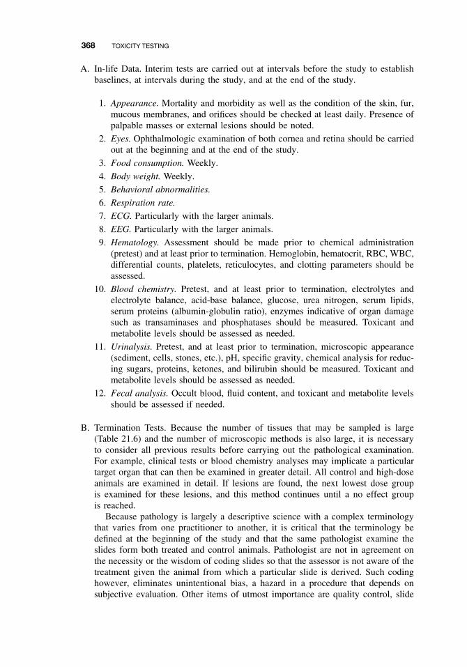
368 TOXICITY TESTING
A. In-life Data. Interim tests are carried out at intervals before the study to establishbaselines, at intervals during the study, and at the end of the study.
1. Appearance. Mortality and morbidity as well as the condition of the skin, fur,mucous membranes, and orifices should be checked at least daily. Presence ofpalpable masses or external lesions should be noted.
2. Eyes. Ophthalmologic examination of both cornea and retina should be carriedout at the beginning and at the end of the study.
3. Food consumption. Weekly.
4. Body weight. Weekly.
5. Behavioral abnormalities.
6. Respiration rate.
7. ECG. Particularly with the larger animals.
8. EEG. Particularly with the larger animals.
9. Hematology. Assessment should be made prior to chemical administration(pretest) and at least prior to termination. Hemoglobin, hematocrit, RBC, WBC,differential counts, platelets, reticulocytes, and clotting parameters should beassessed.
10. Blood chemistry. Pretest, and at least prior to termination, electrolytes andelectrolyte balance, acid-base balance, glucose, urea nitrogen, serum lipids,serum proteins (albumin-globulin ratio), enzymes indicative of organ damagesuch as transaminases and phosphatases should be measured. Toxicant andmetabolite levels should be assessed as needed.
11. Urinalysis. Pretest, and at least prior to termination, microscopic appearance(sediment, cells, stones, etc.), pH, specific gravity, chemical analysis for reduc-ing sugars, proteins, ketones, and bilirubin should be measured. Toxicant andmetabolite levels should be assessed as needed.
12. Fecal analysis. Occult blood, fluid content, and toxicant and metabolite levelsshould be assessed if needed.
B. Termination Tests. Because the number of tissues that may be sampled is large(Table 21.6) and the number of microscopic methods is also large, it is necessaryto consider all previous results before carrying out the pathological examination.For example, clinical tests or blood chemistry analyses may implicate a particulartarget organ that can then be examined in greater detail. All control and high-doseanimals are examined in detail. If lesions are found, the next lowest dose groupis examined for these lesions, and this method continues until a no effect groupis reached.
Because pathology is largely a descriptive science with a complex terminologythat varies from one practitioner to another, it is critical that the terminology bedefined at the beginning of the study and that the same pathologist examine theslides form both treated and control animals. Pathologist are not in agreement onthe necessity or the wisdom of coding slides so that the assessor is not aware of thetreatment given the animal from which a particular slide is derived. Such codinghowever, eliminates unintentional bias, a hazard in a procedure that depends onsubjective evaluation. Other items of utmost importance are quality control, slide
IN VIVO TESTS 369
Table 21.6 Tissues and Organs to Be Examined Histologically in Chronic and SubchronicToxicity Tests
Adrenals Larynx Salivary glandBone and bone marrow Liver Sciatic nerveBrain Lungs and bronchi Seminal vesiclesCartilage Lymph nodes SkinCecum Mammary glands Spinal cordColon Mandibular lymph node SpleenDuodenum Mesenteric lymph node StomachEsophagus Nasal cavity TestesEyes Ovaries Thigh muscleGall bladder Parathyroids ThymusIleum Pituitary Urinary bladderJejunum Prostate UterusKidneys Rectum
identification, and data recording. Many tissues may be examined; consequently aneven larger number of tissue blocks must be prepared. Because each of these mayyield many slides to be stained, comparable quality of staining and the accuratecorrelation of a particular slide with its parent block, tissue, and animal is critical.
1. Necropsy. This must be conducted with care to avoid postmortem damage tothe specimens. Tissues are removed, weighed, and examined closely for grosslesions, masses, and so on. Tissues are then fixed in buffered formalin forsubsequent histologic examination.
2. Histology. The tissues listed in Table 21.6 plus any lesions, masses, or abnormaltissues are embedded, sectioned, and strained for light microscopy. Parafinembedding and staining with hemotoxylin and eosin are the preferred routinemethods, but special stains may be used for particular tissues or for a morespecific examination of certain lesions. Electron microscopy may also be usedfor more specific examination of lesions or cellular changes after their initiallocalization by more routine methods.
Repeated Dose Dermal Tests. Twenty-one to 28-day dermal tests are particularlyimportant when the expected route of human exposure is by contact with the skin, asis the case with many industrial chemicals or pesticides. Compounds to be tested areusually applied daily to clipped areas on the back of the animal, either undiluted orin a suitable vehicle. In the latter case, if a vehicle is used, it is also applied to thecontrols. Selection of a suitable solvent is difficult because many affect the skin, causingeither drying or irritation, whereas others may markedly affect the rate of penetrationof the test chemical. Corn oil, methanol, or carboxymethyl cellulose are preferred todimethyl sulfoxide (DMSO) or acetone. It should also be considered that some of thetest chemical may be ingested as a result of grooming by the animal, although this canbe controlled to some extent by use of restraining collars and/or wrapping.
The criteria for environment, dose selection, and species selection, for example, arenot greatly different from the criteria used for 90-day feeding tests, although the listof end points to be examined is often shorter (e.g., fewer organs may be examined).
370 TOXICITY TESTING
It is necessary, however, to pay close attention to the skin at the point of applicationbecause local effects may be as important as systemic ones.
Twenty-Eight- to Ninety-Day Inhalation Tests. Inhalation studies are indicatedwhenever the route of exposure is expected to be through the lungs. Animals arecommonly exposed for 6 to 8 hours each day, 5 days each week, in chambers of thetype previously discussed. Even in those cases where the animals are maintained inthe inhalation chambers during nonexposure hours, food is always removed duringexposure. Nevertheless, exposure tends to be in part dermal and, due to grooming ofthe fur, in part oral. Environmental and biologic parameters are the same as those forother subchronic tests, as are the routine end points to be measured before, during, andafter the test period. Particular attention must be paid, however, to effects on the tissuesof the nasal cavity and the lungs because these are the areas of maximum exposure.
If the test material is particulate, consideration must be given to the particle sizeand its inhalation potential. Particles of 4 microns in size are considered to be inhal-able; larger particles will be cleared from the respiratory tract by ciliary action andsubsequently swallowed (oral exposure) or expelled by sneezing or expectoration.
21.5.2 Chronic Tests
Chronic tests are those conducted over a significant part of the life span of the testanimal. The duration of a chronic study is generally one year or more. Typically ratand dog are the preferred species; for carcinogenicity studies, rats and mice are used.
Chronic Toxicity and Carcinogenicity. Descriptions of tests for both chronic tox-icity and carcinogenicity are included here because the design is similar—so similarin fact that they can be combined into one test. Chronic toxicity tests are designed todiscover any of numerous toxic effects and to define safety margins to be used in theregulation of chemicals. As with subchronic tests, two species are usually used, oneof which is either a rat or a mouse strain, in which case the tests are run for 2 yearsor 1.5 to 2.0 years, respectively. Data are gathered after 1 year to determine chroniceffects without potential confounding effects of aging. Data are gathered after 1.5 years(mouse) or 2 years (rat) to determine carcinogenic potential. The nonrodent speciesused may be the dog, a nonhuman primate, or, rarely, a small carnivore such as the fer-ret. Chronic toxicity tests may involve administration in the food, in the drinking water,by capsule, or by inhalation, the first being the most common. Gavage is rarely used.The dose used is the maximum tolerated dose (MTD) and usually two lower doses, per-haps 0.25 MTD and 0.125 MTD with the lowest dose being a predicted no effect level.
MTD. The MTD has been defined for testing purposes by the US EnvironmentalProtection Agency (EPA) as:
[T]he highest dose that causes no more than a 10% weight decrement, as compared tothe appropriate control groups; and does not produce mortality, clinical signs of toxicity,or pathologic lesions (other than those that may be related to a neoplastic response) thatwould be predicted to shorten the animals’ natural life span.
This dose is determined by extrapolation from subchronic studies.
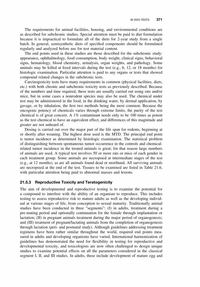
IN VIVO TESTS 371
The requirements for animal facilities, housing, and environmental conditions areas described for subchronic studies. Special attention must be paid to diet formulationbecause it is impractical to formulate all of the diets for 2-year study from a singlebatch. In general, semisynthetic diets of specified components should be formulatedregularly and analyzed before use for test material content.
The end points used in these studies are those described for the subchronic study:appearance, ophthalmology, food consumption, body weight, clinical signs, behavioralsigns, hematology, blood chemistry, urinalysis, organ weights, and pathology. Someanimals may be killed at fixed intervals during the test (e.g., 6, 12, or 18 months) forhistologic examination. Particular attention is paid to any organs or tests that showedcompound related changes in the subchronic tests.
Carcinogenicity tests have many requirements in common (physical facilities, diets,etc.) with both chronic and subchronic toxicity tests as previously described. Becauseof the numbers and time required, these tests are usually carried out using rats and/ormice, but in some cases a nonrodent species may also be used. The chemical undertest may be administered in the food, in the drinking water, by dermal application, bygavage, or by inhalation, the first two methods being the most common. Because theoncogenic potency of chemicals varies through extreme limits, the purity of the testchemical is of great concern. A 1% contaminant needs only to be 100 times as potentas the test chemical to have an equivalent effect, and differences of this magnitude andgreater are not unheard of.
Dosing is carried out over the major part of the life span for rodents, beginning ator shortly after weaning. The highest dose used is the MTD. The principal end pointis tumor incidence as determined by histologic examination. The statistical problemof distinguishing between spontaneous tumor occurrence in the controls and chemical-related tumor incidence in the treated animals is great; for that reason large numbersof animals are used. A typical test involves 50 or more rats or mice of each gender ineach treatment group. Some animals are necropsied at intermediate stages of the test(e.g., at 12 months), as are all animals found dead or moribund. All surviving animalsare necropsied at the end of the test. Tissues to be examined are listed in Table 21.6,with particular attention being paid to abnormal masses and lesions.
21.5.3 Reproductive Toxicity and Teratogenicity
The aim of developmental and reproductive testing is to examine the potential fora compound to interfere with the ability of an organism to reproduce. This includestesting to assess reproductive risk to mature adults as well as the developing individ-ual at various stages of life, from conception to sexual maturity. Traditionally animalstudies have been conducted in three “segments”: (I) in adults, treatment during apre-mating period and optionally continuation for the female through implantation orlactation; (II) in pregnant animals treatment during the major period of organogenesis;and (III) treatment of pregnant/lactating animals from the completion of organogenesisthrough lactation (peri- and postnatal study). Although guidelines addressing treatmentregimens have been rather similar throughout the world, required end points mea-sured in adults and developing organisms have varied. International harmonization ofguidelines has demonstrated the need for flexibility in testing for reproductive anddevelopmental toxicity, and toxicologists are now often challenged to design uniquestudies to examine potential effects on all the parameters considered in the classicalsegment I, II, and III studies. In adults, these include development of mature egg and
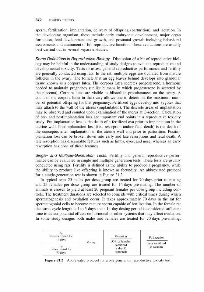
372 TOXICITY TESTING
sperm, fertilization, implantation, delivery of offspring (parturition), and lactation. Inthe developing organism, these include early embryonic development, major organformation, fetal development and growth, and postnatal growth including behavioralassessments and attainment of full reproductive function. These evaluations are usuallybest carried out in several separate studies.
Some Definitions in Reproductive Biology. Discussion of a bit of reproductive biol-ogy may be helpful in the understanding of study designs to evaluate reproductive anddevelopmental toxicity. Tests to assess general reproductive performance and fertilityare generally conducted using rats. In the rat, multiple eggs are ovulated from maturefollicles in the ovary. The follicle that an egg leaves behind develops into glandulartissue known as a corpora lutea. The corpora lutea secretes progesterone, a hormoneneeded to maintain pregnancy (unlike humans in which progesterone is secreted bythe placenta). Corpora lutea are visible as blisterlike protuberances on the ovary. Acount of the corpora lutea in the ovary allows one to determine the maximum num-ber of potential offspring for that pregnancy. Fertilized eggs develop into zygotes thatmay attach to the wall of the uterus (implantation). The discrete areas of implantationmay be observed and counted upon examination of the uterus at C-section. Calculationof pre- and postimplantation loss are important end points in a reproductive toxicitystudy. Pre-implantation loss is the death of a fertilized ova prior to implantation in theuterine wall. Postimplantation loss (i.e., resorption and/or fetal death) is the death ofthe conceptus after implantation in the uterine wall and prior to parturition. Postim-plantation loss can be broken down into early and late resorptions and fetal death. Alate resorption has discernable features such as limbs, eyes, and nose, whereas an earlyresorption has none of these features.
Single- and Multiple-Generation Tests. Fertility and general reproductive perfor-mance can be evaluated in single and multiple generation tests. These tests are usuallyconducted using rats. Fertility is defined as the ability to produce a pregnancy, whilethe ability to produce live offspring is known as fecundity. An abbreviated protocolfor a single-generation test is shown in Figure 21.2.
In typical tests 25 males per dose group are treated for 70 days prior to matingand 25 females per dose group are treated for 14 days pre-mating. The number ofanimals is chosen to yield at least 20 pregnant females per dose group including con-trols. The treatment durations are selected to coincide with critical times during whichspermatogenesis and ovulation occur. It takes approximately 70 days in the rat forspermatogonial cells to become mature sperm capable of fertilization. In the female ratthe estrus cycle length is 4 to 5 days and a 14-day dosing period is considered sufficienttime to detect potential effects on hormonal or other systems that may effect ovulation.In some study designs both males and females are treated for 70 days pre-mating.
F0females treated for
14 days
F0males treated for
70 days
Mating
Gestation
50% of femalessacrificedat day 15(optional)
F1 Lactation
pups sacrificedat weaning
Figure 21.2 Abbreviated protocol for a one generation reproductive toxicity test.
IN VIVO TESTS 373
Treatment of the females is continued through pregnancy (21 days) and until the pupsare weaned. Pups are usually 21 days of age. The test compound is administered atthree dose levels either in the feed, in drinking water, or by gavage. The high dose ischosen to cause some, but not excessive, maternal toxicity (e.g., an approximate 10%decrease in body weight gain, or effects on target organs). Low doses are generallyexpected to be no-effect levels.
After the pre-mating period, the rats are placed in cohabitation, with one male andone female caged together. Mating is confirmed by the appearance of spermatozoain a daily vaginal smear. Day 1 of gestation is the day insemination is confirmed.The females bear and nurse their pups. After birth, the pups are counted, weighed, andexamined for external abnormalities. The litters are frequently culled to a constant num-ber (usually 8–10) after 4 days. At weaning, the pups are killed and autopsied for grossand internal abnormalities. In a multigeneration study, approximately 25/sex/group aresaved to produce the next generation. Brother-sister pairings are avoided. Treatmentis continuous throughout the test, which can be carried out for two, sometimes, threegenerations. An abbreviated protocol for a multiple generation test can be seen inFigure 21.3. Note that the parental generation is known as the F0 generation and the
F0Females
treated for70 days
F0Males
treated for70 days
Mating Gestation
F1A Lactation
Gestation
#1
Mating#2
F1B Lactation
pups sacrificedat weaning
pups sacrificed atweaning—enough
left for nextgeneration
F1BFemales
continuedon test
F1BMales
continuedon test
Mating Gestation
F2A Lactation
Gestation
#1
Mating#2
F2B Lactation
pups sacrificedat weaning
pups sacrificed atweaning—enough
left for nextgeneration
F2BFemales
continuedon test
F2BMales
continuedon test
Mating Gestation
F3A Lactation
Gestation
#1
Mating#2
F3B Lactation
pups sacrificedat weaning
pups sacrificed atweaning—complete
histology
Figure 21.3 Abbreviated protocol for a multigeneration reproductive toxicity test.
374 TOXICITY TESTING
offspring are known as the F1s and F2s. In some studies, parents produce two litters,for example the F1A and F1B litters.
Because both males and females are treated in this type of study design, it isnot possible to distinguish between maternal and paternal effects in the reproductiveperformance. To permit this separation, it is necessary to dose additional animals tothe stage of mating and then breed them to untreated members of the opposite sex.Similarly, if effects are seen postnatally, it may not be possible to distinguish betweeneffects mediated in utero or mediated by lactation. This distinction can be made by“cross-fostering” the offspring of treated females to untreated females, and vice versa.
The end points observed in these types of tests, depending on study design, areas follows:
1. Fertility index, the number of pregnancies relative to the number of matings.2. The number of live births, relative to the number of total births.3. Pre-implantation loss, or number of corpora lutea in the ovaries relative to the
number of implantation sites.4. Postimplantation loss, or the number of resorption sites in the uterus relative to
the number of implantation sites.5. Duration of gestation.6. Effects on male or female reproductive systems.7. Litter size and condition, gross morphology of pups at birth, gender, and anogen-
ital distance.8. Survival of pups.9. Weight gain and performance of adults and pups.
10. Time of occurrence of developmental landmarks, such as eye opening, tootheruption, vaginal opening in females, preputial separation in males.
11. Morphological abnormalities in weanlings.
Results from single and multiple generation tests provide important informationfor assessment of test materials that may perturb a variety of systems including theendocrine system. A number of variations of the single and multiple generation testsexist. For example, a number of weanlings may be left to develop and be tested laterfor behavioral and/or physiological defects (e.g., developmental neurotoxicity testing).
Teratology. Teratology is the study of abnormal fetal development. For an agent to belabeled a teratogen, it must significantly increase the occurrence of adverse structuralor functional abnormalities in offspring after its administration to the female duringpregnancy or directly to the developing organism. In teratology testing, exposure tothe test chemical may be from implantation to parturition, although it has also beenrestricted to the period of major organogenesis, the most sensitive period for inducingstructural malformations. Observations may be extended throughout life, but usuallythey are made immediately prior to birth after a C-section. The end points observedare mainly morphologic (structural changes and malformations), although embryo-fetal mortality is also used as an end point. Figure 21.4 shows an outline of a typicalteratology study.
Teratology studies are carried out in two species, a rodent species (usually the rat)and in another species such as the rabbit (rarely in the dog or primate). Enough females
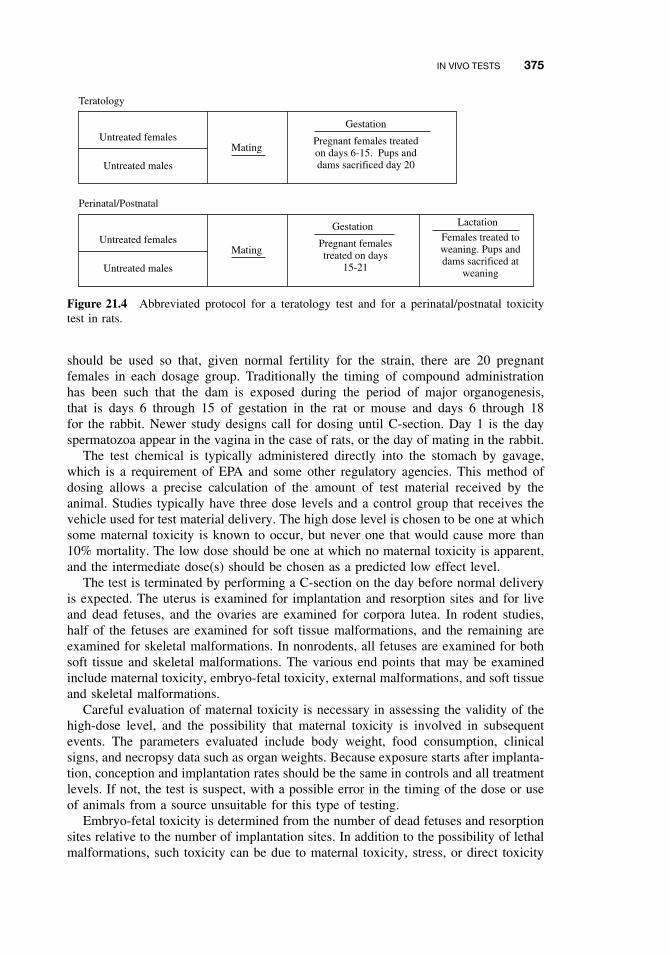
IN VIVO TESTS 375
Untreated females
Untreated males
Teratology
Mating
Gestation
Pregnant females treatedon days 6-15. Pups anddams sacrificed day 20
Untreated females
Untreated males
Perinatal/Postnatal
Mating
Gestation
Pregnant femalestreated on days
15-21
Lactation
Females treated toweaning. Pups anddams sacrificed at
weaning
Figure 21.4 Abbreviated protocol for a teratology test and for a perinatal/postnatal toxicitytest in rats.
should be used so that, given normal fertility for the strain, there are 20 pregnantfemales in each dosage group. Traditionally the timing of compound administrationhas been such that the dam is exposed during the period of major organogenesis,that is days 6 through 15 of gestation in the rat or mouse and days 6 through 18for the rabbit. Newer study designs call for dosing until C-section. Day 1 is the dayspermatozoa appear in the vagina in the case of rats, or the day of mating in the rabbit.
The test chemical is typically administered directly into the stomach by gavage,which is a requirement of EPA and some other regulatory agencies. This method ofdosing allows a precise calculation of the amount of test material received by theanimal. Studies typically have three dose levels and a control group that receives thevehicle used for test material delivery. The high dose level is chosen to be one at whichsome maternal toxicity is known to occur, but never one that would cause more than10% mortality. The low dose should be one at which no maternal toxicity is apparent,and the intermediate dose(s) should be chosen as a predicted low effect level.
The test is terminated by performing a C-section on the day before normal deliveryis expected. The uterus is examined for implantation and resorption sites and for liveand dead fetuses, and the ovaries are examined for corpora lutea. In rodent studies,half of the fetuses are examined for soft tissue malformations, and the remaining areexamined for skeletal malformations. In nonrodents, all fetuses are examined for bothsoft tissue and skeletal malformations. The various end points that may be examinedinclude maternal toxicity, embryo-fetal toxicity, external malformations, and soft tissueand skeletal malformations.
Careful evaluation of maternal toxicity is necessary in assessing the validity of thehigh-dose level, and the possibility that maternal toxicity is involved in subsequentevents. The parameters evaluated include body weight, food consumption, clinicalsigns, and necropsy data such as organ weights. Because exposure starts after implanta-tion, conception and implantation rates should be the same in controls and all treatmentlevels. If not, the test is suspect, with a possible error in the timing of the dose or useof animals from a source unsuitable for this type of testing.
Embryo-fetal toxicity is determined from the number of dead fetuses and resorptionsites relative to the number of implantation sites. In addition to the possibility of lethalmalformations, such toxicity can be due to maternal toxicity, stress, or direct toxicity
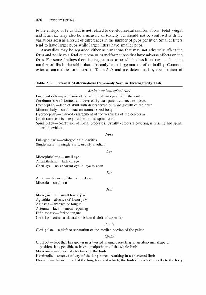
376 TOXICITY TESTING
to the embryo or fetus that is not related to developmental malformations. Fetal weightand fetal size may also be a measure of toxicity but should not be confused with thevariations seen as a result of differences in the number of pups per litter. Smaller litterstend to have larger pups while larger litters have smaller pups.
Anomalies may be regarded either as variations that may not adversely affect thefetus and not have a fetal outcome or as malformations that have adverse effects on thefetus. For some findings there is disagreement as to which class it belongs, such as thenumber of ribs in the rabbit that inherently has a large amount of variability. Commonexternal anomalities are listed in Table 21.7 and are determined by examination of
Table 21.7 External Malformations Commonly Seen in Teratogenicity Tests
Brain, cranium, spinal cord
Encephalocele—protrusion of brain through an opening of the skull.Cerebrum is well formed and covered by transparent connective tissue.Exencephaly—lack of skull with disorganized outward growth of the brain.Microcephaly—small head on normal sized body.Hydrocephaly—marked enlargement of the ventricles of the cerebrum.Craniorachischisis—exposed brain and spinal cord.Spina bifida—Nonfusion of spinal processes. Usually ectoderm covering is missing and spinal
cord is evident.
Nose
Enlarged naris—enlarged nasal cavitiesSingle naris—a single naris, usually median
Eye
Microphthalmia—small eyeAnophthalmia—lack of eyeOpen eye—no apparent eyelid, eye is open
Ear
Anotia—absence of the external earMicrotia—small ear
Jaw
Micrognathia—small lower jawAgnathia—absence of lower jawAglossia—absence of tongueAstomia—lack of mouth openingBifid tongue—forked tongueCleft lip—either unilateral or bilateral cleft of upper lip
Palate
Cleft palate—a cleft or separation of the median portion of the palate
Limbs
Clubfoot—foot that has grown in a twisted manner, resulting in an abnormal shape orposition. It is possible to have a malposition of the whole limb
Micromelia—abnormal shortness of the limbHemimelia—absence of any of the long bones, resulting in a shortened limbPhomelia—absence of all of the long bones of a limb, the limb is attached directly to the body
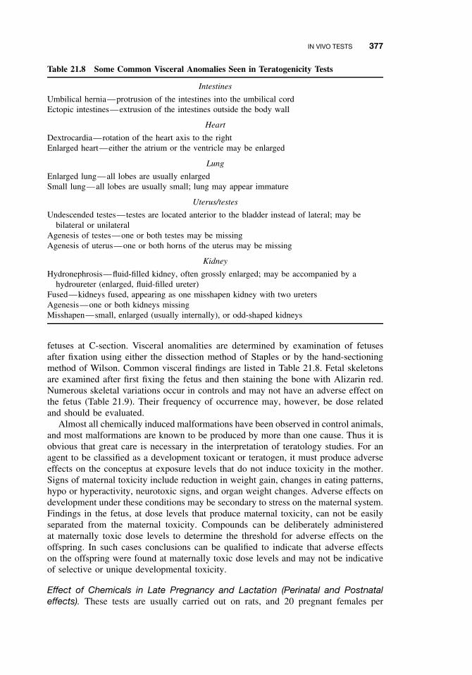
IN VIVO TESTS 377
Table 21.8 Some Common Visceral Anomalies Seen in Teratogenicity Tests
Intestines
Umbilical hernia—protrusion of the intestines into the umbilical cordEctopic intestines—extrusion of the intestines outside the body wall
Heart
Dextrocardia—rotation of the heart axis to the rightEnlarged heart—either the atrium or the ventricle may be enlarged
Lung
Enlarged lung—all lobes are usually enlargedSmall lung—all lobes are usually small; lung may appear immature
Uterus/testes
Undescended testes—testes are located anterior to the bladder instead of lateral; may bebilateral or unilateral
Agenesis of testes—one or both testes may be missingAgenesis of uterus—one or both horns of the uterus may be missing
Kidney
Hydronephrosis—fluid-filled kidney, often grossly enlarged; may be accompanied by ahydroureter (enlarged, fluid-filled ureter)
Fused—kidneys fused, appearing as one misshapen kidney with two uretersAgenesis—one or both kidneys missingMisshapen—small, enlarged (usually internally), or odd-shaped kidneys
fetuses at C-section. Visceral anomalities are determined by examination of fetusesafter fixation using either the dissection method of Staples or by the hand-sectioningmethod of Wilson. Common visceral findings are listed in Table 21.8. Fetal skeletonsare examined after first fixing the fetus and then staining the bone with Alizarin red.Numerous skeletal variations occur in controls and may not have an adverse effect onthe fetus (Table 21.9). Their frequency of occurrence may, however, be dose relatedand should be evaluated.
Almost all chemically induced malformations have been observed in control animals,and most malformations are known to be produced by more than one cause. Thus it isobvious that great care is necessary in the interpretation of teratology studies. For anagent to be classified as a development toxicant or teratogen, it must produce adverseeffects on the conceptus at exposure levels that do not induce toxicity in the mother.Signs of maternal toxicity include reduction in weight gain, changes in eating patterns,hypo or hyperactivity, neurotoxic signs, and organ weight changes. Adverse effects ondevelopment under these conditions may be secondary to stress on the maternal system.Findings in the fetus, at dose levels that produce maternal toxicity, can not be easilyseparated from the maternal toxicity. Compounds can be deliberately administeredat maternally toxic dose levels to determine the threshold for adverse effects on theoffspring. In such cases conclusions can be qualified to indicate that adverse effectson the offspring were found at maternally toxic dose levels and may not be indicativeof selective or unique developmental toxicity.
Effect of Chemicals in Late Pregnancy and Lactation (Perinatal and Postnataleffects). These tests are usually carried out on rats, and 20 pregnant females per
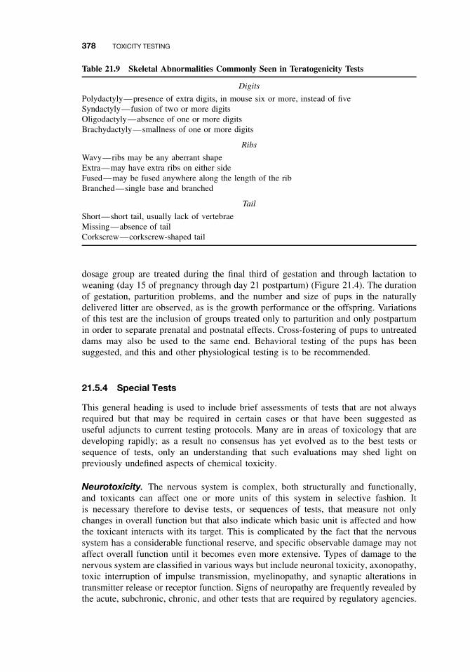
378 TOXICITY TESTING
Table 21.9 Skeletal Abnormalities Commonly Seen in Teratogenicity Tests
Digits
Polydactyly—presence of extra digits, in mouse six or more, instead of fiveSyndactyly—fusion of two or more digitsOligodactyly—absence of one or more digitsBrachydactyly—smallness of one or more digits
Ribs
Wavy—ribs may be any aberrant shapeExtra—may have extra ribs on either sideFused—may be fused anywhere along the length of the ribBranched—single base and branched
Tail
Short—short tail, usually lack of vertebraeMissing—absence of tailCorkscrew—corkscrew-shaped tail
dosage group are treated during the final third of gestation and through lactation toweaning (day 15 of pregnancy through day 21 postpartum) (Figure 21.4). The durationof gestation, parturition problems, and the number and size of pups in the naturallydelivered litter are observed, as is the growth performance or the offspring. Variationsof this test are the inclusion of groups treated only to parturition and only postpartumin order to separate prenatal and postnatal effects. Cross-fostering of pups to untreateddams may also be used to the same end. Behavioral testing of the pups has beensuggested, and this and other physiological testing is to be recommended.
21.5.4 Special Tests
This general heading is used to include brief assessments of tests that are not alwaysrequired but that may be required in certain cases or that have been suggested asuseful adjuncts to current testing protocols. Many are in areas of toxicology that aredeveloping rapidly; as a result no consensus has yet evolved as to the best tests orsequence of tests, only an understanding that such evaluations may shed light onpreviously undefined aspects of chemical toxicity.
Neurotoxicity. The nervous system is complex, both structurally and functionally,and toxicants can affect one or more units of this system in selective fashion. Itis necessary therefore to devise tests, or sequences of tests, that measure not onlychanges in overall function but that also indicate which basic unit is affected and howthe toxicant interacts with its target. This is complicated by the fact that the nervoussystem has a considerable functional reserve, and specific observable damage may notaffect overall function until it becomes even more extensive. Types of damage to thenervous system are classified in various ways but include neuronal toxicity, axonopathy,toxic interruption of impulse transmission, myelinopathy, and synaptic alterations intransmitter release or receptor function. Signs of neuropathy are frequently revealed bythe acute, subchronic, chronic, and other tests that are required by regulatory agencies.
IN VIVO TESTS 379
Neurotoxicity is of great significance in toxicology, however, and tests have beendevised to supplement those routinely required. These include acute and subchronicneurotoxicity studies as well as developmental neurotoxicity studies.
Behavioral and Pharmacological Tests. Behavioral and pharmacological tests involvethe observation of clinical signs and behavior. These include signs of changes inawareness, mood, motor activity, central nervous system excitation, posture, motorincoordination, muscle tone, reflexes, and autonomic functions. If these tests so indi-cate, more specialized tests can be carried out that evaluate spontaneous motor activity,conditioned avoidance responses, operant conditioning, as well as tests for motor inco-ordination such as the inclined plane or rotarod tests.
Tests for specific classes of chemicals include the measurement of transmitter stim-ulated adenyl cyclase and Na/K-ATPase for chemicals that affect receptor functionor cholinesterase inhibition for organophosphates or carbamates. Electrophysiologicaltechniques may detect chemicals such as DDT or pyrethroids, which affect impulsetransmission.
Acute and Subchronic Neurotoxicity Tests. These studies are designed to test a widerange of neurotoxicity effects including CNS stimulation or depression, reflex perturba-tion, peripheral nerve damage, cognitive effects on learning and memory, motor activityeffects and neuropathology. The tests are conducted using rats or sometimes mice.
In the acute neurotoxicity study, approximately 10 to 15 animals per sex per dosegroup are administered a single gavage (bolus) dose of the test material. There are usu-ally three dose groups and a control. Behavioral assessments are made on the day ofdosing, and at 1 and 2 weeks post dose. The assessments include tests on motor activity,a functional observation battery (FOB), and neuropathology (at termination). The FOBscreens for sensorimotor, neuromuscular, autonomic and general physiological effectsof a test compound. Table 21.10 depicts component tests of the FOB. These functionaltests have the advantage over biochemical measures that they permit repeated eval-uation of individual animals over time to determine the onset, progression, duration,and reversibility of neurotoxic effects. Motor activity is also measured over time andcan be evaluated by a variety of devices. One such device that has been frequentlyused is the figure 8 maze, which consists of a series of interconnected alleys (in theshape of the numeral 8) converging on a central open area and covered with transpar-ent acrylic plastic. Motor activity is detected by photobeams, and an activity count isregistered each time a photobeam is interrupted by the animal. Motor activity sessionsare generally 60 minutes in length and each session is divided into 5- to 10-minutereporting intervals (epochs). “Habituation” is an end point evaluated in the motor activ-ity test, and this is defined as a decrement in activity during the test session. Activityis expected to decrease toward the end of the test as the animal’s exploratory activitynormally lessens as the time in the maze increases. Neuropathological examinationsare the same as those described below for the subchronic neurotoxicity test.
Before the acute neurotoxicity study is conducted, it is necessary to conduct apreliminary test to determine the time of peak effect after dosing of the test material.Preliminary tests may evaluate a selected group of end points in the FOB or othersensitive end points, if known, for a particular test material. Results of this preliminarytest will determine the time when observations are performed on the day of dosing inthe acute neurotoxicity study.
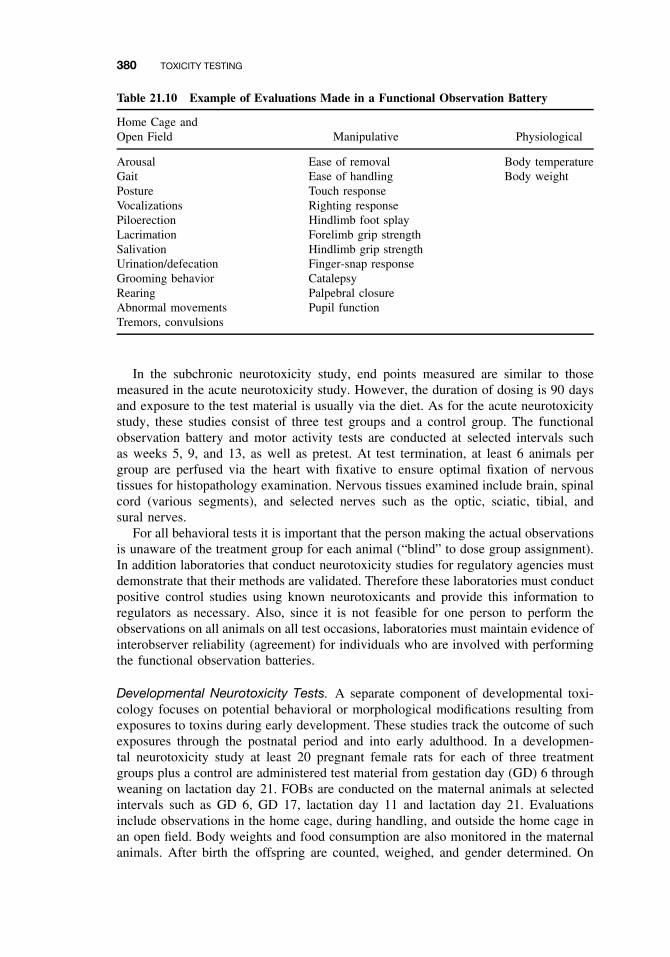
380 TOXICITY TESTING
Table 21.10 Example of Evaluations Made in a Functional Observation Battery
Home Cage andOpen Field Manipulative Physiological
Arousal Ease of removal Body temperatureGait Ease of handling Body weightPosture Touch responseVocalizations Righting responsePiloerection Hindlimb foot splayLacrimation Forelimb grip strengthSalivation Hindlimb grip strengthUrination/defecation Finger-snap responseGrooming behavior CatalepsyRearing Palpebral closureAbnormal movements Pupil functionTremors, convulsions
In the subchronic neurotoxicity study, end points measured are similar to thosemeasured in the acute neurotoxicity study. However, the duration of dosing is 90 daysand exposure to the test material is usually via the diet. As for the acute neurotoxicitystudy, these studies consist of three test groups and a control group. The functionalobservation battery and motor activity tests are conducted at selected intervals suchas weeks 5, 9, and 13, as well as pretest. At test termination, at least 6 animals pergroup are perfused via the heart with fixative to ensure optimal fixation of nervoustissues for histopathology examination. Nervous tissues examined include brain, spinalcord (various segments), and selected nerves such as the optic, sciatic, tibial, andsural nerves.
For all behavioral tests it is important that the person making the actual observationsis unaware of the treatment group for each animal (“blind” to dose group assignment).In addition laboratories that conduct neurotoxicity studies for regulatory agencies mustdemonstrate that their methods are validated. Therefore these laboratories must conductpositive control studies using known neurotoxicants and provide this information toregulators as necessary. Also, since it is not feasible for one person to perform theobservations on all animals on all test occasions, laboratories must maintain evidence ofinterobserver reliability (agreement) for individuals who are involved with performingthe functional observation batteries.
Developmental Neurotoxicity Tests. A separate component of developmental toxi-cology focuses on potential behavioral or morphological modifications resulting fromexposures to toxins during early development. These studies track the outcome of suchexposures through the postnatal period and into early adulthood. In a developmen-tal neurotoxicity study at least 20 pregnant female rats for each of three treatmentgroups plus a control are administered test material from gestation day (GD) 6 throughweaning on lactation day 21. FOBs are conducted on the maternal animals at selectedintervals such as GD 6, GD 17, lactation day 11 and lactation day 21. Evaluationsinclude observations in the home cage, during handling, and outside the home cage inan open field. Body weights and food consumption are also monitored in the maternalanimals. After birth the offspring are counted, weighed, and gender determined. On
IN VIVO TESTS 381
postnatal day (PND) 4, litters are culled to 8 pups per litter. Following culling, at least10 pups/sex/group are assigned to one of the following tests: learning and memory,motor activity, or acoustic startle. Additional pups are assigned for neuropathologyand brain weight evaluations on PND 11 and PND 70 (10/sex/group). FOBs are per-formed on the offspring at selected intervals such as PND 11, 17, 21, 35, and 60/70.Indicators of physical development such as preputial separation (male sexual matura-tion) and vaginal patency (female sexual maturation) are evaluated for all offspring aswell as body weight and food consumption. Learning and memory can be evaluatedwith a variety of tests. Frequently a water maze is used where the rat learns to swimthrough a series of alleys to find a platform it can use to climb out of the water. Thetime it takes for the animal to swim through the maze to the platform (trial latency)and the number of mistakes made are some of the end points evaluated in this test.The trials are conducted over a series of days and the assay provides an index of thedevelopment of both working memory (with-in day performance) as well as referencememory (between-day performance). The startle test measures the animal’s responseto a burst of loud noise and also how quickly it becomes habituated to 10 pulses ofstartle-eliciting tones in 5 blocks. For the startle test special chambers lined with sound-attenuating and vibration-absorbing material are used. These chambers can measure theforce exerted on a platform on which the animal stands during the test procedure. Thestartle test is conducted at 2 time points such as PND 22 and 60. When the offspringare approximately 70 days old, the test is terminated. Selected animals are perfusedwith fixative for neuropathology assessments. In addition to detailed microscopic eval-uation of at least five different sections of the brain, simple morphometric analysis ofthe cerebrum, hippocampus, and cerebellum are conducted.
Delayed Neuropathy (OPIDN). The delayed neurotoxic potential of certain organo-phosphates such as tri-o-cresyl phosphate (TOCP) is usually evaluated by observationof clinical signs (paralysis of leg muscles in hens) or pathology (degeneration of themotor nerves in hens), but a biochemical test involving the ratio between the ability toinhibit cholinesterase and the ability to inhibit an enzyme that has been referred to asthe neurotoxic esterase (NTE) has been suggested. The ability of chemicals to causedelayed neuropathy is generally correlated with their ability to inhibit this nonspecificesterase, found in various tissues, although the role, if any, of NTE in the sequenceof events leading to nerve degeneration is not known. The preferred test organism isthe mature hen because the clinical signs are similar to those in humans, and suchsymptoms cannot be readily elicited in the common laboratory rodents.
Potentiation. Potentiation and synergism represent interactions between toxicantsthat are potential sources of hazard because neither humans nor other species areusually exposed to one chemical at a time. The enormous number of possible combi-nations of chemicals makes routine screening for all such effects not only impracticalbut impossible.
One of the classic cases is the potentiation of the insecticide malathion by anotherinsecticide, EPN, the LD50 of the mixture being dramatically lower than that of eithercompound alone. This potentiation can also be seen between malathion and certain con-taminants that are formed during synthesis, such as isomalathion. For this reason qualitycontrol during manufacture is essential. This example of potentiation involves inhibi-tion, by EPN or isomalathion, of the carboxylesterase responsible for the detoxicationof malathion in mammals.
382 TOXICITY TESTING
It is practical to test for potentiation only when there has been some preliminaryindication that it might occur or when either or both compounds belong to chemicalclasses previously known to cause potentiation. Such a test can be conducted by com-paring the LD50, or any other appropriate toxic end point, of a mixture of equitoxicdoses of the chemicals in question with the same end point measured with the twochemicals administered alone.
In the case of synergism, in which one of the compounds is relatively nontoxic whengiven alone, the toxicity of the toxic compound can be measured when administeredalone or after a relatively large dose of the nontoxic compound.
Toxicokinetics and Metabolism. Routine toxicity testing without regard to themechanisms involved is likely to be wasteful of time and of human, animal, andfinancial resources. A knowledge of toxicokinetics and metabolism can give valuableinsights and provide for testing that is both more efficient and more informative.Such knowledge provides the necessary background to make the most appropriateselection of test animal species and of dose levels, and the most appropriate methodfor extrapolating from animal studies to the assessment of human hazard. Moreoverthey may provide information on possible reactive intermediates as well as informationon induction or inhibition of the enzymes of xenobiotic metabolism, the latter beingcritical to an assessment of possible interaction.
The nature of metabolic reactions and their variations between species is detailedin Chapters 7, 8, and 9 with some aspects of toxicokinetics in Chapter 6. The methodsused for the measurement of toxicants and their metabolites are detailed in Chapter 25.The present section is concerned with the general principles, use, and need for metabolicand toxicokinetics studies in toxicity testing.
Toxicokinetics studies are designed to measure the amount and rate of the absorption,distribution, metabolism, and excretion of a xenobiotic. These data are used to constructpredictive mathematical models so that the distribution and excretion of other doses canbe simulated. Such studies are carried out using radiolabeled compounds to facilitatemeasurement and total recovery of the administered dose. This can be done entirelyin vivo by measuring levels in blood, expired air, feces, and urine; these procedures canbe done relatively noninvasively and continuously in the same animal. Tissue levelscan be measured by sequential killing and analysis of organ levels. It is important tomeasure not only the compound administered but also its metabolites, because simpleradioactivity counting does not differentiate among them.
The metabolic study, considered separately, consists of treatment of the animal withthe radiolabeled compound followed by chemical analysis of all metabolites formedin vivo and excreted via the lungs, kidneys, or bile. Although reactive intermediatesare unlikely to be isolated, the chemical structure of the end products may providevital clues to the nature of the intermediates involved in their formation. The use oftissue homogenates, subcellular fractions, and purified enzymes may serve to clarifyevents occurring during metabolic sequences leading to the end products.
Information of importance in test animal selection is the similarity in toxicodynamicsand metabolism to that of humans. Although all of the necessary information maynot be available for humans, it can often be inferred with reference to metabolismand excretion of related compounds, but it is clearly ill advised to use an animalthat differs from most others in the toxicokinetics or metabolism of the compound inquestion or that differs from humans in the nature of the end products. Dose selection
IN VIVO TESTS 383
is influenced by knowledge of whether a particular dose saturates a physiologicalprocess such as excretion or whether it is likely to accumulate in a particular tissue,because these factors are likely to become increasingly important the longer a chronicstudy continues.
Behavior. Although the primary emphasis in toxicity testing has long been the esti-mation of morphologic changes, much recent interest has focused on more fundamentalevaluations. One such aspect has been the evaluation of chemical effects on behavior.
The categories of methods used in behavioral toxicology fall into two principalclasses, stimulus-oriented behavior, and internally generated behavior. The formerincludes two types of conditioned behavior: operant conditioning, in which animalsare trained to perform a task in order to obtain a reward or to avoid a punishment, andclassical conditioning, in which an animal learns to associate a conditioning stimuluswith a reflex action. Stimulus-oriented behavior also involves unconditioned responsesin which the animal’s response to a particular stimulus is recorded.
Internally generated behavior includes observation of animal behavior in responseto various experimental situations, and includes exploratory behavior, circadian activ-ity, social behavior, and so on. The performance of animals treated with a particularchemical is compared with that of untreated controls as a measure of the effect ofthe chemical.
Many of the variables associated with other types of testing must also be con-trolled in behavioral tests: sex, age, species, environment, diet, and animal husbandry.Behavior may vary with all of these. Norton describes a series of four tests that mayform an appropriate series, inasmuch as they represent four different types of behavior;the series should therefore reflect different types of nervous system activity. They areas follows:
1. Passive avoidance. This test involves the use of a shuttle box, in which animalscan move between a light side and a dark side. After an acclimatization period,in which the animal can move freely between the two sides, it receives a mildelectric shock while in the dark (preferred) side. During subsequent trials, thetime spent in the “safe side” is recorded.
2. Auditory startle. This test involves the response (movement) to a sound stimuluseither without, or preceded by, a light-flash stimulus.
3. Residential maze. Movements of animals in a residential maze are automaticallyrecorded during both light and dark photoperiods.
4. Walking patterns. Gait is measured in walking animals, including such character-istics as the length and width of stride and the angles formed by the placementof the feet.
Problems associated with behavioral toxicology include the functional reserve andadaptability of the nervous system. Frequently behavior is maintained despite clearlyobservable injury. Other problems are the statistical ones associated with multiple tests,multiple measurements, and the inherently large variability in behavior.
The use of human subjects occupationally exposed to chemicals is often attempted,but such tests are complicated by the subjective nature of the end points (dizzi-ness, etc.).
384 TOXICITY TESTING
Covalent Binding. Toxicity has been associated with covalent binding in a num-ber of ways. Organ-specific toxicants administered in vivo bind covalently to macro-molecules, usually at a higher level in the target tissues than in nontarget tissues.Examples include acetaminophen in the liver, carbon tetrachloride in the liver, p-aminophenol in the kidney, and ipomeanol in the lung. Similarly many carcinogensare known to give rise to DNA adducts. In general, covalent binding occurs as a resultof metabolism of the toxicant to highly reactive intermediates, usually, but not always,by cytochrome P450. Because these intermediates are highly reactive electrophiles,they bind to many nucleophilic sites on DNA, RNA, or protein molecules, not justthe site of toxic action. Thus measurement of covalent binding may be a measure oftoxic potential rather than a specific measurement, related directly to a mechanism ofaction. The occurrence of covalent binding at the same time as toxicity is so commonan occurrence, however, that a measurement of covalent binding of a chemical maybe regarded as an excellent although perhaps not infallible indication of potential fortoxicity. Although such tests are not routine, considerable interest has been shown intheir development.
The measurement of DNA adducts is an indirect indication of genotoxic (car-cinogenic) potential, and DNA adducts in the urine are an indication, obtained bya noninvasive technique, of recent exposure. Protein adducts give an integrated mea-sure of exposure because they accumulate over the life span of the protein and, at thesame time, indicate possible organ toxicity.
Tissue protein adducts are usually demonstrated in experimental animals followinginjection of radiolabeled chemicals and, after a period to time, the organs are removedand homogenized, and by rigorous extraction, all the noncovalently bound material isremoved. Extraction methods include lipid solvents, acids and bases, concentrated ureasolutions, and solubilization and precipitation of the proteins. They tend to underesti-mate the extent of covalent binding because even covalent bonds may be broken bythe rigorous procedures used. Newer methods involving dialysis against detergents andseparation of adducted proteins will probably prove more appropriate.
Blood proteins, such as hemoglobin, may be used in tests of human exposurebecause blood is readily and safely accessible. For example, the exposure of mice toethylene oxide or dimethylnitrosamine was estimated by measuring alkylated residuesin hemoglobin. The method was subsequently extended to people exposed occupa-tionally to ethylene oxide by measuring N-3-(2-hydroxyethyl) histidine residues inhemoglobin. Similarly methyl cysteine residues in hemoglobin can be used as a mea-sure of methylation.
DNA-RNA adducts can also be measured in various ways, including rigorousextraction, separation, and precipitation following administration of labeled compoundsin vivo, or use of antibodies raised to chemically modified DNA or RNA.
Although many compounds of different chemical classes have been shown tobind covalently when activated by microsomal preparations in vitro (e.g., aflatoxin,ipomeanol, stilbene, vinyl chloride), these observations have not been developedinto routine testing procedures. Such procedures could be useful in predicting toxicpotential.
Immunotoxicity. Immunotoxicology comprises two distinct types of toxic effects:the involvement of the immune system in mediating the toxic effect of a chemicaland the toxic effects of chemicals on the immune system. The former is shown, for
IN VITRO AND OTHER SHORT-TERM TESTS 385
example, in tests for cutaneous sensitization, whereas the latter is shown in impairmentof the ability to resist infection.
Tests for immunotoxicity are not required by all regulatory agencies, but it is anarea of great interest, both in the fundamental mechanisms of immune function and inthe design of tests to measure impairment of immune function. Both of these aspectsare discussed in detail in Chapter 19.
21.6 IN VITRO AND OTHER SHORT-TERM TESTS
21.6.1 Introduction
The toxicity tests that follow are tests conducted largely in vitro with isolated cell sys-tems. Some are short-term tests carried out in vivo or are combinations of in vivo andin vitro systems. The latter are included because of similarities in approach, mechanism,or intent. In general, these tests measure effects on the genome or cell transformation;their importance lies in the relationship between such effects and the mechanism ofchemical carcinogenesis. Mutagenicity of cells in the germ line is itself an expressionof toxicity, however, and the mutant genes can be inherited and expressed in the nextor subsequent generations.
The theory that the initiating step of chemical carcinogenesis is a somatic mutationis well recognized, and considerable evidence shows that mutagenic potential is cor-related with carcinogenic potential. Thus the intent of much of this type of testing isto provide early warning of carcinogenic potential without the delay involved in con-ducting lifetime chronic feeding studies in experimental animals. Despite the numeroustests that have been devised, regulatory agencies have not yet seen it fit to substituteany of them, or any combination of them, for chronic feeding studies. Instead, theyhave been added as additional testing requirements. One function of such tests shouldbe to identify those compounds with the greatest potential for toxicity and enable theamount of chronic testing to be reduced to more manageable proportions.
21.6.2 Prokaryote Mutagenicity
Ames Test. The Ames test, developed by Bruce Ames and his coworkers at theUniversity of California, Berkeley, depends on the ability of mutagenic chemicals tobring about reverse mutations in Salmonella typhimurium strains that have defectsin the histidine biosynthesis pathway. These strains will not grow in the absence ofhistidine but can be caused to mutate back to the wild type, which can synthesizehistidine and hence can grow in its absence. The postmitochondrial supernatant (S-9fraction), obtained from homogenates of livers of rats previously treated with PCBs inorder to induce certain cytochrome P450 isoforms, is also included in order to providethe activating enzymes involved in the production of the potent electrophiles ofteninvolved in the toxicity of chemicals to animals.
Bacterial tester strains have been developed that can test for either base-pair (e.g.,strain TA-1531) or frameshift (e.g., strains TA-1537, TA-1538) mutations. Other, moresensitive strains such as TA-98 and TA-100 are also used, although they may be lessspecific with regard to the type of mutation caused.
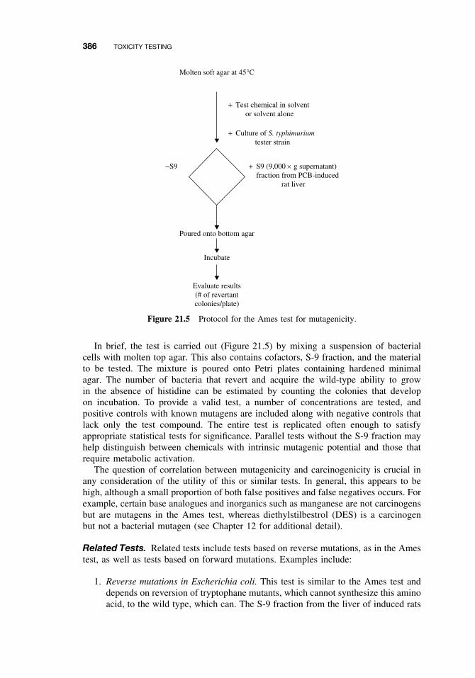
386 TOXICITY TESTING
Molten soft agar at 45°C
Poured onto bottom agar
Incubate
Evaluate results(# of revertantcolonies/plate)
+ Test chemical in solvent or solvent alone
+ Culture of S. typhimurium tester strain
+−S9 S9 (9,000 × g supernatant)fraction from PCB-induced rat liver
Figure 21.5 Protocol for the Ames test for mutagenicity.
In brief, the test is carried out (Figure 21.5) by mixing a suspension of bacterialcells with molten top agar. This also contains cofactors, S-9 fraction, and the materialto be tested. The mixture is poured onto Petri plates containing hardened minimalagar. The number of bacteria that revert and acquire the wild-type ability to growin the absence of histidine can be estimated by counting the colonies that developon incubation. To provide a valid test, a number of concentrations are tested, andpositive controls with known mutagens are included along with negative controls thatlack only the test compound. The entire test is replicated often enough to satisfyappropriate statistical tests for significance. Parallel tests without the S-9 fraction mayhelp distinguish between chemicals with intrinsic mutagenic potential and those thatrequire metabolic activation.
The question of correlation between mutagenicity and carcinogenicity is crucial inany consideration of the utility of this or similar tests. In general, this appears to behigh, although a small proportion of both false positives and false negatives occurs. Forexample, certain base analogues and inorganics such as manganese are not carcinogensbut are mutagens in the Ames test, whereas diethylstilbestrol (DES) is a carcinogenbut not a bacterial mutagen (see Chapter 12 for additional detail).
Related Tests. Related tests include tests based on reverse mutations, as in the Amestest, as well as tests based on forward mutations. Examples include:
1. Reverse mutations in Escherichia coli. This test is similar to the Ames test anddepends on reversion of tryptophane mutants, which cannot synthesize this aminoacid, to the wild type, which can. The S-9 fraction from the liver of induced rats
IN VITRO AND OTHER SHORT-TERM TESTS 387
can also be used as an activating system in this test. Other E. coli reverse mutationtests utilize nicotinic acid and arginine mutants.
2. Forward mutations in S. typhimurium. One such assay, dependent on the appear-ance of a mutation conferring resistance to 8-azaguanine in a histidine revertantstrain, has been developed and is said to be as sensitive as the reverse-mutationtests
3. Forward mutations in E. coli. These mutations depend on mutation of galactosenonfermenting E. coli to galactose fermenting E. coli or the change from 5-methyltryptophane to 5-methyltryptophane resistance.
4. DNA repair. Polymerase-deficient, and thus DNA repair-deficient, E. coli hasprovided the basis for a test that depends on the fact that the growth of a deficientstrain is inhibited more by a DNA-damaging agent than is that of a repair-competent strain. The recombinant assay using Bacillus subtilis is conducted inmuch the same way because recombinant deficient strains are more sensitive toDNA-damaging agents.
21.6.3 Eukaryote Mutagenicity
Mammalian Cell Mutation. The development of cell culture techniques that permitboth survival and replication have led to many advances in cell biology, including theuse of certain of these cell lines for detection of mutagens. Although such cells, ifderived from mammals, would seem ideal for testing for toxicity toward mammals,there are several problems. Primary cells, which generally resemble those of the tis-sue of origin, are difficult to culture and have poor cloning ability. Because of thesedifficulties, certain established cell lines are usually used. These cells, such as Chi-nese hamster ovary cells and mouse lymphoma cells, clone readily and do not becomesenescent with passage through many cell generations. Unfortunately, they have littlemetabolic activity toward xenobiotics and thus do not readily activate toxicants. More-over they usually show chromosome changes, such as aneuploidy (i.e., more or fewerthan the usual diploid number of chromosomes).
The characteristics usually involved in these assays are resistance to 8-azaguanineor 6-thioguanine (the hypoxanthine guanine phosphoribosyl transferase or HGPRTlocus), resistance to bromodeoxyuridine or triflurothymidine (the thymidine kinaseor TK locus), or resistance to ouabain (the OU or Na/K-ATPase locus). HGPRT isresponsible for incorporation of purines from the medium into the nucleic acid synthe-sis pathway. Its loss prevents uptake of normal purines and also of toxic purines suchas 8-azaguanine, which would kill the cell. Thus mutation at this locus confers resis-tance to these toxic purine analogues. Similarly TK permits pyrimidine transport, andits loss prevents uptake of toxic pyrimidine analogues and confers resistance to them.In the absence of HGPRT or TK, the cells can grow by de novo synthesis of purinesand pyrimidines. Ouabain kills cells by combining with the Na/K-ATPase. Mutation atthe OU locus alters the ouabain-binding site in a way that prevents inhibition and thusconfers resistance.
A typical test system is the analysis of the TK locus in mouse lymphoma cells formutations that confer resistance to bromodeoxyuracil. The tests are conducted with andwithout the S-9 fraction from induced rat liver because the lymphoma cells have littleactivating ability. Both positive and negative controls are included, and the parameter
388 TOXICITY TESTING
measured is the number of cells formed that are capable of forming colonies in thepresence of bromodeoxyuridine.
Drosophila Sex-Linked Recessive Lethal Test. The advantages of Drosophila(fruit fly) tests are that they involve an intact eukaryotic organism with all of itsinterrelated organ systems and activation mechanisms but, at the same time, are fast,relatively easy to perform, and do not involve mammals as test animals. The most obvi-ous disadvantages are that the hormonal and immune systems of insects are significantlydifferent from those of mammals and that the nature, specificity, and inducibility ofthe cytochrome P450s are not as well understood in insects as they are in mammals.
In a typical test, males that are 2 days postpuparium and that were raised from eggslaid within a short time period (usually 24 hours) are treated with the test compoundin water to which sucrose has been added to increase palatability. Males from a straincarrying a gene for yellow body on the X chromosome are used. Preliminary testsdetermine that the number of offspring of the survivors of the treatment doses (usu-ally 0.25 LD50 and 0.5 LD50) are adequate for future crosses. Appropriate controls,including a solvent control (with emulsifier if one was necessary to prepare the testsolution), and a positive control, such as ethyl methane sulfonate, are routinely includedwith each test. Individual crosses of each surviving treated male with a series of threefemales are made on a 0- to 2-, 3- to 5-, and 6- to 8-day schedule. The progeny of eachfemale is reared separately, and the males and females of the F1 generation are matedin brother-sister matings. If there are no males with yellow bodies in a particular setof progeny, it should be assumed that a lethal mutation was present on the treated Xchromosomes. A comparison of the F2 progeny derived from females inseminated bymales at different times after treatment allows a distinction to be made between effectson spermatozoa, spermatids, and spermatocytes.
In the Basc (Muller-5) test shown in Figure 21.6, the strain used for the females inthe F1 cross is a multiple-marked strain that carries a dominant gene for bar eyes andrecessive genes for apricot eyes and a reduction of bristles on the thorax (scute gene).(Basc is an acronym for bar, apricot, and scute.)
Related Tests. Many tests related to the two types of eukaryote-mutation tests arediscussed earlier in this section, and many of them are simply variations of the testsdescribed. Two distinct classes are worthy of mention: the first uses yeasts as the testorganisms, and the second is the spot test for mutations in mice.
One group of yeast tests includes tests for gene mutations and strains that canbe used to detect forward mutations in genes that code for enzymes in the purinebiosynthetic pathway; other strains can be used to detect reversions. Yeasts can also beused to test for recombinant events such as reciprocal mitotic recombination (mitoticcrossing over) and nonreciprocal mitotic recombination. Saccharomyces cerevisiae isthe preferred organism in almost all these tests. Although they possess cytochromeP450s capable of metabolizing xenobiotics, their specificity and sensitivity are limitedas compared with those of mammals, and an S-9 fraction is often included, as in theAmes test, to enhance activation.
The gene mutation test systems in mice include the specific locus test, in which wild-type treated males are crossed with females carrying recessive mutations for visiblephenotypic effects. The F1 progeny have the same phenotype as the wild-type parentunless a mutation, corresponding to a recessive mutant marker, has occurred. Such tests
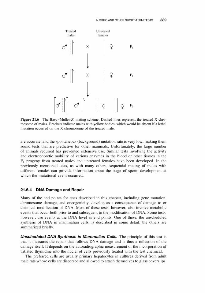
IN VITRO AND OTHER SHORT-TERM TESTS 389
Treatedmales
Untreatedfemales
P1
F1
F2
Figure 21.6 The Basc (Muller-5) mating scheme. Dashed lines represent the treated X chro-mosome of males. Brackets indicate males with yellow bodies, which would be absent if a lethalmutation occurred on the X chromosome of the treated male.
are accurate, and the spontaneous (background) mutation rate is very low, making themsound tests that are predictive for other mammals. Unfortunately, the large numberof animals required has prevented extensive use. Similar tests involving the activityand electrophoretic mobility of various enzymes in the blood or other tissues in theF1 progeny from treated males and untreated females have been developed. In thepreviously mentioned tests, as with many others, sequential mating of males withdifferent females can provide information about the stage of sperm development atwhich the mutational event occurred.
21.6.4 DNA Damage and Repair
Many of the end points for tests described in this chapter, including gene mutation,chromosome damage, and oncogenicity, develop as a consequence of damage to orchemical modification of DNA. Most of these tests, however, also involve metabolicevents that occur both prior to and subsequent to the modification of DNA. Some tests,however, use events at the DNA level as end points. One of these, the unscheduledsynthesis of DNA in mammalian cells, is described in some detail; the others aresummarized briefly.
Unscheduled DNA Synthesis in Mammalian Cells. The principle of this test isthat it measures the repair that follows DNA damage and is thus a reflection of thedamage itself. It depends on the autoradiographic measurement of the incorporation oftritiated thymidine into the nuclei of cells previously treated with the test chemical.
The preferred cells are usually primary hepatocytes in cultures derived from adultmale rats whose cells are dispersed and allowed to attach themselves to glass coverslips.
390 TOXICITY TESTING
From this point on, the test is carried out on the attached cells. Both positive controlswith agents known to stimulate unscheduled DNA synthesis, such as the carcinogenaflatoxin B1 or 2-acetylaminofluorene, and negative controls, which are processedthrough all procedures except exposure to the test compound, are performed routinelywith every test. Cells are exposed by replacing the medium for a short time withone containing the test chemical. The dose levels are determined by a preliminarycell viability test (Trypan blue exclusion test) and consist of several concentrationsthat span the range from no apparent loss of viability to almost complete loss ofviability. Following exposure, the medium is removed, and the cells are washed byseveral changes of fresh medium and finally placed in a medium containing tritiatedthymidine. The cells are fixed and dried, and the coverslip with the cells attached iscoated with photographic emulsion. After a suitable exposure period (usually severalweeks), the emulsion is developed and the cells are stained with hemotoxylin and eosin.The number of grains in the nuclear region is corrected by subtracting nonnucleargrains, and the net grain count in the nuclear area is compared between treated anduntreated cells.
This test has several advantages in that primary liver cells have considerable acti-vation capacity and the test measures an event at the DNA level. It does not, however,distinguish between error-free repair and error-prone repair, the latter being itself amutagenic process. Thus it cannot distinguish between events that might lead to toxicsequelae and those that do not. A modification of this test measures in vivo unsched-uled DNA synthesis. In this modification animals are first treated in vivo, and primaryhepatocytes are then prepared and treated as already described.
Related Tests. Tests for the measurement of binding of the test material to DNAhave already been discussed under covalent binding (Section 21.5.4). Another methodof assessing DNA damage is the estimation of DNA breakage following exposure tothe test chemical; the DNA-strand length is estimated by using alkaline elution orsucrose density gradient centrifugation. This has been done with a number of cell linesand with freshly prepared hepatocytes, in the latter case following treatment eitherin vivo or in vitro. It may be regarded as promising but not yet fully validated. Thepolymerase-deficient E. coli tests as well as recombinant tests using yeasts are alsorelated to DNA repair.
21.6.5 Chromosome Aberrations
Tests for chromosome aberrations involve the estimation of effects on extended regionsof whole chromosomes rather than on single or small numbers of genes. Primarily theyconcern chromosome breaks and the exchange of material between chromosomes.
Sister Chromatid Exchange. Sister chromatid exchange (SCE) occurs between thesister chromatids that together make up a chromosome. It occurs at the same locus ineach chromatid and is thus a symmetrical exchange of chromosome material. In thisregard it is not strictly an aberration because the products do not differ in morphologyfrom normal chromosome. SCE, however, is susceptible to chemical induction andappears to be correlated with the genotoxic potential of chemicals as well as with theironcogenic potential. The exchange is visualized by permitting the treated cells to passthrough two DNA replication cycles in the presence of 5-bromo-2′-deoxyuridine, which
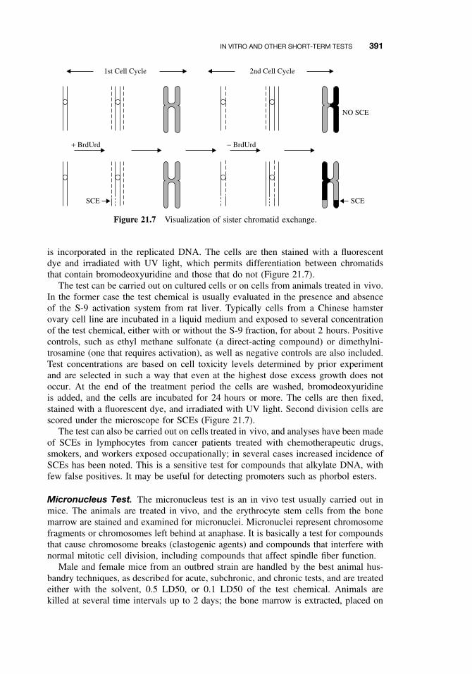
IN VITRO AND OTHER SHORT-TERM TESTS 391
1st Cell Cycle 2nd Cell Cycle
+ BrdUrd
SCE SCE
NO SCE
− BrdUrd
Figure 21.7 Visualization of sister chromatid exchange.
is incorporated in the replicated DNA. The cells are then stained with a fluorescentdye and irradiated with UV light, which permits differentiation between chromatidsthat contain bromodeoxyuridine and those that do not (Figure 21.7).
The test can be carried out on cultured cells or on cells from animals treated in vivo.In the former case the test chemical is usually evaluated in the presence and absenceof the S-9 activation system from rat liver. Typically cells from a Chinese hamsterovary cell line are incubated in a liquid medium and exposed to several concentrationof the test chemical, either with or without the S-9 fraction, for about 2 hours. Positivecontrols, such as ethyl methane sulfonate (a direct-acting compound) or dimethylni-trosamine (one that requires activation), as well as negative controls are also included.Test concentrations are based on cell toxicity levels determined by prior experimentand are selected in such a way that even at the highest dose excess growth does notoccur. At the end of the treatment period the cells are washed, bromodeoxyuridineis added, and the cells are incubated for 24 hours or more. The cells are then fixed,stained with a fluorescent dye, and irradiated with UV light. Second division cells arescored under the microscope for SCEs (Figure 21.7).
The test can also be carried out on cells treated in vivo, and analyses have been madeof SCEs in lymphocytes from cancer patients treated with chemotherapeutic drugs,smokers, and workers exposed occupationally; in several cases increased incidence ofSCEs has been noted. This is a sensitive test for compounds that alkylate DNA, withfew false positives. It may be useful for detecting promoters such as phorbol esters.
Micronucleus Test. The micronucleus test is an in vivo test usually carried out inmice. The animals are treated in vivo, and the erythrocyte stem cells from the bonemarrow are stained and examined for micronuclei. Micronuclei represent chromosomefragments or chromosomes left behind at anaphase. It is basically a test for compoundsthat cause chromosome breaks (clastogenic agents) and compounds that interfere withnormal mitotic cell division, including compounds that affect spindle fiber function.
Male and female mice from an outbred strain are handled by the best animal hus-bandry techniques, as described for acute, subchronic, and chronic tests, and are treatedeither with the solvent, 0.5 LD50, or 0.1 LD50 of the test chemical. Animals arekilled at several time intervals up to 2 days; the bone marrow is extracted, placed on
392 TOXICITY TESTING
microscope slides, dried, and stained. The presence of micronuclei is scored visuallyunder the microscope.
Dominant Lethal Test in Rodents. The dominant lethal test, which is performedusing rats, mice, or hamsters, is an in vivo test to determine the germ-cell risk froma suspected mutagen. The test consists of treating males with the test compound forseveral days, followed by mating to different females each week for enough weeks tocover the period required for a complete spermatogenic cycle. Animals are maintainedunder optimal conditions of animal husbandry and are dosed, usually by a gavage,with several doses of less than 0.1 LD50. The females are killed after two weeks ofgestation and dissected; corpora lutea and living and dead implantations are counted.The end points used to determine the occurrence of dominant lethal mutations in thetreated males are the fertility index (ratio of pregnant females to mated females),preimplantation losses (the number of implantations relative to the number of corporalutea), the number of females with dead implantations relative to the total number ofpregnant females, and the number of dead implantations relative to the total numberof implantations. Mutations in sperm that are dominant and lethal do not result inviable offspring.
Related Tests. Many cells exposed to test chemicals can be scored for chromosomeaberrations by staining procedures followed by visual examination with the aid ofthe microscope. These include Chinese hamster ovary cells in culture treated in aprotocol very similar to that used in the test for SCEs, bone marrow cells from animalstreated in vivo, or lymphocytes from animals treated in vivo. The types of aberrationsevaluated include chromatid gaps, breaks, and deletions; chromosome gaps, breaks,and deletions; chromosome fragments; translocations; and ploidy.
Heritable translocations can be detected by direct examination of cells from male orfemale offspring in various stages of development or by crossing the treated animalsto untreated animals and evaluating fertility, with males with reduced fertility beingexamined for translocations, and so on. Progeny from this or other tests, such as thosefor dominant lethals, can be permitted to survive and then examined for translocationsand other abnormalities.
21.6.6 Mammalian Cell Transformation
Most cell transformation assays utilize fibroblast cultures derived from embryonictissue. The original studies showed that cells from C3H mouse fibroblast culturesdeveloped morphologic changes and changes in growth patterns when treated withcarcinogens. Later similar studies were made with Syrian hamster embryo cells. Thedirect relationship of these changes to carcinogenesis was demonstrated by transplan-tation of the cells into a host animal and the subsequent development of tumors. Therecent development of practical assay procedures involves two cell lines from mouseembryos, Balb/3T3 and C3H/10T1/2, in which transformation is easily recognized andscored. In a typical assay situation, cells, such as Balb/3T3 mouse fibroblasts, will mul-tiply in culture until a monolayer is formed. At this point they cease dividing unlesstransformed. Chemicals that are transforming agents will, however, cause growth tooccur in thicker layers above the monolayer. These clumps of transformed cells areknown as foci. Despite many recommended controls the assay is only semiquantitative.
ECOLOGICAL EFFECTS 393
The doses are selected from the results of a preliminary experiment and range froma high dose that reduces colony formation (but not by >50%) to a low dose that hasno measurable effect on colony formation. After exposure to the test chemical for 1to 3 days, the cells are washed and incubation is continued for up to 4 weeks. At thattime the monolayers are fixed, stained, and scored for transformed foci.
Transformation assays have several distinct advantages. Because transplanted focigive rise to tumors in congenic hosts (those from the same inbred strain from which thecells were derived) whereas untransformed cells do not, cell transformation is believedto be illustrative of the overall expression of carcinogenesis in mammalian tissues.The two cell types used most (Balb/3T3 and C3H/10T1/2) respond to promoters inthe manner predicted by the multistage model for carcinogenesis in vivo and mayeventually be useful in the development of assays for promotion. Unfortunately, alarge number of false negative results are obtained because these cell lines do notshow much activation capacity; it has not proved practical to combine them with theS-9 activation system. Furthermore the cells are aneuploidy and may be preneoplasticin the untreated state. Syrian hamster cells, which do have considerable activationcapacity, have proved difficult to use in test procedures and are difficult to score.
21.6.7 General Considerations and Testing Sequences
Considering all of the tests for acute and chronic toxicity, long and short term, in vivoand in vitro, it is clearly impractical to apply a complete series of tests to all commercialchemicals and all their derivatives in food, water, and the environment. The challengeof toxicity testing is to identify the most effective set or sequence of tests necessaryto describe the apparent and potential toxicity of a particular chemical or mixture ofchemicals. The enormous emphasis on in vitro or short-term tests that has occurredsince the mid-1970s had its roots in the need to find substitutes for lifetime feedingstudies in experimental animals or, at the very least, to suggest a sequence of teststhat would enable priorities to be set for which chemicals should be subjected tochronic tests. Such tests might also be used to eliminate the need for chronic testingfor chemicals that either clearly possessed the potential for toxicity or clearly do not.Although there has been much success in test development, the challenge outlined herehas not been met, primarily because of the failure of scientists and regulatory agencies,worldwide, to agree on test sequences or on the circumstances in which short-term testsmay substitute for chronic tests. Thus not only are short-term tests often required, thesetests are in addition to long-term tests. As an example, the US EPA requirements forthe Federal Insecticide, Fungicide, and Rodenticide Act (FIFRA) include, besides afull battery of acute, subchronic, and chronic tests, tests to address the following threecategories: gene mutations, structural chromosome aberrations, and other genotoxictests as appropriate (e.g., DNA damage and repair and chromosome aberrations). Itis important, however, that test sequences have been suggested and considered byregulatory agencies, but there must also be taken into account the fact that short-termtests do not provide all of the information needed from the longer term tests.
21.7 ECOLOGICAL EFFECTS
Tests for ecological effects include those designed to address the potential of chemi-cals to affect ecosystems and the population dynamics in the environment. The tests
394 TOXICITY TESTING
are conducted to estimate effects on field populations of vertebrates, invertebrates,and plants. The use of environmental risk assessment tests is discussed in detail inChapter 28.
21.7.1 Laboratory Tests
There are two types of laboratory tests: toxicity determinations on wildlife and aquaticorganisms and the use of model ecosystems to measure bioaccumulation and transportof toxicants and their degradation products.
Among the tests included in the first category are the avian oral LD50, the aviandietary LC50, wild mammal toxicity, and avian reproduction. The avian tests areusually carried out on bobwhite quail or mallard ducks, whereas the wild mammalsmay be species such as the pine mouse, Paramyscus. The tests are similar to thosedescribed under acute and chronic testing procedures but suffer from some drawbacks;the standards of animal husbandry used with rats and mice are probably unattainablewith birds or wild mammals, even through bobwhite quail and mallards are easily rearedin captivity. The genetics of the birds and mammals used are much more variable thanare those of the traditional laboratory rodent strains.
Similar tests can be carried out with aquatic organisms (e.g., the LC50 for freshwaterfish such as rainbow trout and bluegills), the LC50 for estuarine and marine organisms,the LC50 for invertebrates such as Daphnia, and the effect of chemicals on the earlystages of fish and various invertebrates.
Model systems, first developed by ecologists to study basic ecological processes,have been adapted to toxicological testing. In toxicology these models were first usedto determine the movement and concentration of pesticides. Typically the model has awater phase containing vertebrates and invertebrates, and a terrestrial phase containingat least one plant species and one herbivore species. First, the 14C-labeled pesticide orother environmental contaminant is applied to the leaves of the terrestrial plant sorghum(Sorgum halpense), and then salt marsh caterpillars (Estigmene acrea) are placed onthe plants. The larvae eat the plants and contaminate the water with feces and theirdead bodies. The aquatic food chain is simulated with plankton (diatoms, rotifers, etc.),water fleas (Daphnia), mosquito larvae (Culex pipiens), and fish (Gambusia affinis).From an analysis of the plants, animals, and substrates for the 14C-labeled compoundand its degradation products, the biologic magnification or rate of degradation can becalculated.
More complex models involving several compartments, simulated rain, simulatedsoil drainage, simulated tidal flow, and so on, have been constructed and their propertiesinvestigated, but none have been brought to the stage of use in routine testing. Similarlyaquatic models using static, re-circulating, and continuous flow have also been used, ashave entirely terrestrial models: again, none have been developed for routine testing.
21.7.2 Simulated Field Tests
Simulated field tests may be quite simple, consisting of feeding treated prey to predatorsand studying the toxic effects on the predator, enabling some predictions concerningeffects to nontarget organisms. In general, however, the term is used for greenhouse,small plot, small artificial pond, or small natural pond tests. These serve to test biologic
THE FUTURE OF TOXICITY TESTING 395
accumulation and degradation under conditions somewhat more natural than in modelecosystems and the test chemicals are exposed to environmental as well as biologicdegradation. Population effects may be noted, but these methods are more useful forsoil invertebrates, plants, and aquatic organisms because other organisms are not easilycontained in small plots.
21.7.3 Field Tests
In field-test situations, test chemicals are applied to large areas under natural condi-tions. The areas are at least several acres and may be either natural or part of someagroecosystem. Because the area is large and in the open, radiolabeled compoundscannot be used, it is not possible to obtain a balance between material applied andmaterial recovered.
The effects are followed over a long period of time and two types of control maybe used: first, a comparison with a similar area that is untreated; and second, and acomparison with the same area before treatment. In the first case it is difficult, if notimpossible, to duplicate exactly a large natural area, and in the second, changes canoccur that are unrelated to the test material.
In either case, studies of populations are the most important focus of this type oftesting, although the disappearance of the test material, its accumulation in variouslife forms, and the appearance, accumulation, and disappearance of its degradationproducts are also important. The population of soil organisms, terrestrial organisms,and aquatic organisms as well as plants all must be surveyed and characterized, bothqualitatively and quantitatively. After application of the test material the populationscan be followed through two or more annual cycles to determine both acute andlong-term population effects.
21.8 RISK ANALYSIS
The preceding tests for various kinds of toxicity can be used to measure adverse effectsof many different chemical compounds in different species, organ, tissues, cells, oreven populations, and under many different conditions. This information can be usedto predict possible toxicity of related compounds from QSAR or of the same chemicalunder different conditions (e.g., mutagenicity as a predictor of carcinogenicity). It isconsiderably more difficult to use this information to predict possible risk to otherspecies, such as humans, because little experimental data on this species is available.Some methods are available to predict risk to humans and to provide the risk factorin the risk-benefit assessment that provides the basis for regulatory action, however.Human health risk assessment is discussed in detail in Chapter 24. The benefit factor islargely economic in nature, and the final regulatory action is not, in the narrow sense,a scientific one. It also involves political and legal aspects and, in toto, representssociety’s evaluation of the amount of risk that can be tolerated in any particular case.
21.9 THE FUTURE OF TOXICITY TESTING
Because of the public awareness of the potentially harmful effects of chemicals, it isclear that toxicity testing will continue to be an important activity and that it will be
396 TOXICITY TESTING
required by regulatory agencies before the use of a particular chemical is permittedeither in commercial processes or for use by the public. Because of the proliferation oftesting procedures, the number of experimental species and other test systems available,as well as the high dose rates usually used, it is clear that eventually some expressionof some type of toxicity will be obtained for most exogenous chemicals. Thus theidentification of toxic effects with the intent of banning any chemical causing sucheffects is no longer a productive mode of attack. The aim of toxicity testing should beto identify those compounds that present an unacceptable potential for risk to humansor to the environment and thus ought to be banned but, at the same time, provide anaccurate assessment of the risk to humans and the environment of less toxic compoundsso that their use may be regulated.
Subjecting all chemicals to all possible tests is logistically impossible, and the futureof toxicity testing must lie in the development of techniques that will narrow the testingprocess so that highly toxic and relatively nontoxic compounds can be identified earlyand either banned or permitted unrestricted use without undue waste of time, funds, andhuman resources. These vital commodities could then be concentrated on compoundswhose fate and effects are less predictable.
Such progress will come from further development and validation of the newertesting procedures and the development of techniques to select, for any given chemical,the most suitable testing methods. Perhaps of most importance is the development ofintegrated test sequences that permit decisions to be made at each step, thereby eitherabbreviating the sequence or making the next step more effective and efficient.
SUGGESTED READING
Balls, M., R. J. Riddell, and A. N. Worden. Animals and Alternatives in Toxicity Testing. Lon-don: Academic Press, 1983.
Clark, B., and D. A. Smith. Pharmacokinetics and toxicity testing. CRC Crit. Rev. Toxicol. 12:343, 1984.
Couch, J. A., and W. J. Hargis Jr. Aquatic animals in toxicity testing. J. Am. Coll. Toxicol. 3:331, 1984.
Dean, J. H., M. I. Luster, M. J. Murray, and L. D. Laver. Approaches and methodology forexamining the immunological effects of xenobiotics. Immunotoxicol. 7: 205, 1983
de Serres F. J., Ashby J. (eds): Evaluation of Short Term Tests for Carcinogens. New York:Elsevier, 1981.
Ecobichon, D. J.. The Basis of Toxicity Testing. Boca Raton, FL: CRC Press, 1992.
Enslein, K., and P. N. Craig. Carcinogenesis: A predictive structure-activity model. J. Toxicol.Environ. Health 10: 521, 1982.
Gorrod, J. W., ed. Testing for Toxicity. London: Taylor and Francis, 1981. Relevant chaptersinclude:
Chapter 3. Brown, V. K. H.: Acute toxicity testing—a critique.
Chapter 4. Roe, F. J. C.: Testing in vivo for general chronic toxicity and carcinogenicity.
Chapter 7. Gorrod, J. W.: Covalent binding as an indication of drug toxicity.
Chapter 15. Dewar, A. J.: Neurotoxicity testing—with particular reference to biochemicalmethods.
Chapter 18. Cobb, L. M.: Pulmonary toxicity.
SUGGESTED READING 397
Chapter 20. Parish, W. E.: Immunological tests to predict toxicological hazards to man.
Chapter 21. Venitt, S.: Microbial tests in carcinogenesis studies.
Chapter 22. Styles, J. A.: Other short-term tests in carcinogenesis studies.
Hayes, A. W., ed. Principles and Methods of Toxicology, 2nd ed. New York: Raven Press. Thisvolume contains the following chapters of particular relevance to this chapter:
Chapter 6. Chan, P. K., and A. W. Hayes: Principles and methods for acute and eye irritancy.
Chapter 7. Mosberg, A. T., and A. W. Hayes: Subchronic toxicity testing.
Chapter 8. Stevens, K. R., and M. A. Gallo: Practical considerations in the conduct of chronictoxicity studies.
Chapter 9. Roberts, J. F., W. W. Piegorsch, and R. L. Schueler: Methods in testing for car-cinogenicity.
Chapter 10. Zenick, H., and E. D. Clegg: Assessment of male reproductive toxicology: a riskassessment approach.
Chapter 11. Manson, J. M., and Y. J. Kang: Test methods for assessing female reproductiveand developmental toxicology.
Chapter 12. Kennedy, G. L., Jr: Inhalation toxicology.
Chapter 13. Patrick, E., and H. I. Maiback: Dermatotoxicology.
Chapter 14. Brusick, D.: Genetic toxicology.
Chapter 17. Burger, G. T., and L. C. Miller: Animal care and facilities.
Chapter 18. Norton, S.: Methods for behavioral toxicology.
Chapter 26. Dean, J. H., et al.: Immune system; evaluation of injury.
Chapter 30. Renwick, A. G.: Pharmacokinetics in toxicology.
Chapter 31. Hogan, M. D., and D. G. Hoel: Extrapolation to humans.
Moser, V. C., G. C. Becking, V. Cuomo, et al. The IPCS collaborative study on neurobehavioralscreening methods: IV. Control data. Neurotoxicol. 18: 947–967, 1997.
Weiss, B., and D. Cory-Slechta. Assessment of behavioral toxicity. In Principles and Methodsof Toxicology, 3rd ed. A. Wallace Hayes, ed. New York: Raven Press, 1994.

CHAPTER 22
Forensic and Clinical Toxicology
STACY BRANCH
22.1 INTRODUCTION
Forensic toxicology refers to the use of toxicology for the purposes of the law.It is considered a hybrid of analytical chemistry and fundamental toxicology. Theefforts or activities conducted to effectuate this purpose include but are not limited tothe following:
ž Urine testing to detect drug usež Regulatory toxicologyž Occupational diseasež Identification of causative agents causing death or injury in humans and animalsž Courtroom testimony and consultation concerning toxicoses
Analytical toxicology in a clinical setting plays a role similar to that in forensic toxi-cology. Therefore, this chapter will be divided into forensic and clinical toxicology.
22.2 FOUNDATIONS OF FORENSIC TOXICOLOGY
Until the 1700s convictions associated with homicidal poisoning were based onlyon circumstantial evidence rather than the identification of the actual toxicant withinthe victim. In 1781, Joseph Plenic stated that the detection and identification of thepoison in the organs of the deceased was the only true sign of poisoning. Years later,1813, Mathieiv Orfila (considered the father of toxicology) published the first completework on the subject of poisons and legal medicine. By 1836, James M. Marsh devel-oped a test for the presence of arsenic in tissue. Then, in 1839, Orfila successfully usedMarsh’s test to identify arsenic extracted from human tissues. Fifty years later, ErnstWilhelm Heinrich Gutzeit developed a method (Gutzeit test) to quantitate arsenic in tis-sues. In this process arsenic compounds are reduced by hydrogen produced when zincand sulfuric acid react. The hydrogen then reduces the arsenic compounds to arsine,which is exposed to paper that has been treated with mercuric chloride solution. This
A Textbook of Modern Toxicology, Third Edition, edited by Ernest HodgsonISBN 0-471-26508-X Copyright 2004 John Wiley & Sons, Inc.
399
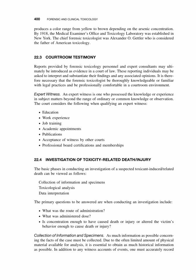
400 FORENSIC AND CLINICAL TOXICOLOGY
produces a color range from yellow to brown depending on the arsenic concentration.By 1918, the Medical Examiner’s Office and Toxicology Laboratory was established inNew York. The chief forensic toxicologist was Alexander O. Gettler who is consideredthe father of American toxicology.
22.3 COURTROOM TESTIMONY
Reports provided by forensic toxicology personnel and expert consultants may ulti-mately be introduced as evidence in a court of law. These reporting individuals may beasked to interpret and substantiate their findings and any associated opinions. It is there-fore necessary that the forensic toxicologist be thoroughly knowledgeable or familiarwith legal practices and be professionally comfortable in a courtroom environment.
Expert Witness. An expert witness is one who possessed the knowledge or experiencein subject matters beyond the range of ordinary or common knowledge or observation.The court considers the following when qualifying an expert witness:
ž Educationž Work experiencež Job trainingž Academic appointmentsž Publicationsž Acceptance of witness by other courtsž Professional board certifications and memberships
22.4 INVESTIGATION OF TOXICITY-RELATED DEATH/INJURY
The basic phases in conducting an investigation of a suspected toxicant-induced/relateddeath can be viewed as follows:
Collection of information and specimensToxicological analysisData interpretation
The primary questions to be answered are when conducting an investigation include:
ž What was the route of administration?ž What was administered dose?ž Is concentration enough to have caused death or injury or altered the victim’s
behavior enough to cause death or injury?
Collection of Information and Specimens. As much information as possible concern-ing the facts of the case must be collected. Due to the often limited amount of physicalmaterial available for analysis, it is essential to obtain as much historical informationas possible. In addition to any witness accounts of events, one must accurately record
INVESTIGATION OF TOXICITY-RELATED DEATH/INJURY 401
information such as the age, sex, and weight of the victim, his medical history, identi-fication of any medications or other drugs/substances taken before death, and the timeinterval that elapsed between the intake of these substances and death.
When collecting the specimens, many different body fluids and organs should becollected since xenobiotics have different affinities for body tissues and therefore mul-tiple extractions (for specific analyses) may be needed. Specimens should be collectedbefore applying processes that may destroy evidence, that is, before embalming. Theprocess of embalming, for example, may destroy or dilute the xenobiotic and mayyield a false positive result, for example, for the presence of ethanol (which is a con-stituent of embalming fluid). It is possible to obtain useful specimens from burnedor burial remains. The tissue often collected under these circumstances include bonemarrow, skeletal muscle, vitreous humor, hair, and maggots. For example, from hairsamples, it is possible to detect the presence of antibiotics, antipsychotics, and drugsof abuse. However, the information is primarily qualitative in nature. From maggots,barbiturates, barbiturates, benzodiazepines, phenothiazines, morphine, and malathioncan be detected.
It is often necessary to add preservatives to specimens to protect against postmortemchanges. For example, the addition of sodium fluoride to a tissue specimen can preventthe production of bacterial ethanol (which can potentially yield a false positive resultfor the presence of ingested ethanol).
22.4.1 Documentation Practices
Labeling and all handling documentation must exist from the beginning of data/specimencollection to analysis. Figure 22.1 is a sample of a typical toxicology worksheet. Thename of the victim (if known) is recorded along with the name of the medical examiner.The condition of the body when found is described and the date of death is recorded.Additionally the date of request of toxicological analyses is also stated on the report.Each collected specimen is identified as well as the tests to be performed. In the resultssection it is necessary that the analyst for each test signs the form identifying the actualresults for the tissue tested and the date the results were obtained.
22.4.2 Considerations for Forensic Toxicological Analysis
The decision concerning which analytical methods to employ depend greatly on anumber of factors. One can imagine that a given method may need more samplevolume or weight than another method. Therefore the amount of specimen avail-able is a critical determinant of methods to chose for proper toxicological analysis.It is necessary to know the nature of the toxicant to test. In a particular case, isit relevant to detect the parent compound, its metabolites, or all of these? Further-more toxicant biotransformation must be taken into account when doing the anal-yses and making interpretations. A low concentration of a toxic parent compoundmay reflect biotransformation as opposed to a low level of exposure. Conversely, alow-level presence of a nontoxic parent compound may be associated with a suffi-cient concentration of a biotransformation product that was high enough to cause theinsult. Furthermore both forms (parent and metabolites) may have contributed to theadverse outcome.
402 FORENSIC AND CLINICAL TOXICOLOGY
Name - Johnston, Gato T.
Medical Examiner - Bahiyah Sushaunna Ngozi, M.D.
Reference number - 38470-38973-2
Brief History - Subject was found with a syringe, white power at scene, a small transparentglass bottle, and 0.5 liters of alcohol.
Date of Death - 8/08/2002
Date of Request - 8/09/02
Decomposed Corpse: Yes No
TOXICOLOGY WORK SHEET
SPECIMENS
RESULTS:
#OF SAMPLES TEST
ANALYSIS DATE TEST
Blood (Heart)Blood (Femoral)Blood (Other: cephalic)UrineBileVitreousKidneyBrainOther: ( )
1111
1
AlcoholAcidic DrugBasic DrugOther: analyze unknown white powder
X
Figure 22.1 Toxicology work sheet.
22.4.3 Drug Concentrations and Distribution
As a rule, the highest concentrations of a poison are found at the site of administration.A large quantity of drug in the GI tract and liver indicates oral ingestion. The gas-trointestinal (GI) tract may contain large amounts of unabsorbed toxicant. Cases thatinvolve the oral administration of toxicants indicate analysis of GI contents. However,the presence of toxic material in the GI tract does not provide sufficient evidence thatthe agent is the cause of death. Absorption and transport of the toxicant to the site ofaction must be demonstrated. Blood and tissue analysis is necessary and would stillbe paramount.
Higher concentrations of drug or toxicant in the lungs compared to other tissuesmay indicate inhalation, while compounds located in tissue surrounding an injection
LABORATORY ANALYSES 403
site indicates a fresh intramuscular or intravenous injection. Detection of drug com-bustion breakdown products within fluids/tissues reveals that smoking was the route ofdrug administration. For example, the primary pyrolysis product of “crack” cocaine isanhydroecgonine methylester. A high concentration of this compound and the parentcocaine indicates smoking as the route of the cocaine administration. Urine analy-sis is also of great value since the kidney is the major organ of excretion for mosttoxicants. The liver is usually the first internal organ to be analyzed. After GI tractabsorption, xenobiotics are transported to the liver. This is a major center of compoundbiotransformation. Finally blood specimens must be collected with care and thought.When collecting blood, it is advantageous to collect both heart and peripheral bloodspecimens. Postmortem blood drug concentrations are site-dependent. This site depen-dency is referred to as “anatomical site concentration differences” or “postmortemredistribution.”
22.5 LABORATORY ANALYSES
The nonspecific initial tests in a series are valuable for determining the presence orabsence of a particular class of compounds. Colorimetric tests to detect the presence ofphenothiazines would give initial information about a drug class present. This wouldbe followed by more specific tests to identify the actual compound as well as providequantitative data. Another example of a type of initial test would be an immunoas-say that determines the presence of barbiturates. Confirmatory tests are mandatory toidentify the particular drug within the class detected.
22.5.1 Colorimetric Screening Tests
These tests require little sample preparation and are usually performed directly on thespecimen. This is a rapid procedure but requires confirmation.
22.5.2 Thermal Desorption
In addition to the analysis of arson crime scene evidence, thermal desorption hasbeen used for the analysis of residual volatile agents in street drugs and the analysisof stains on forensic evidence. Samples are heated to volatilize water and organiccompounds. The organic analytes may then be separated by gas chromatography(Figure 22.2).
22.5.3 Thin-Layer Chromatography (TLC)
An extract of a specimen is spotted on a TLC plate, the plate is placed in a mobilephase. The solvent travels up plate via capillary action and the compounds separatedepending on compound solubility. Detection is by observing color changes or byusing UV light to observe bands. The Rf value is calculated (the distance traveled bythe compound divided by distance traveled by solvent). This value along with colorreactions are used as qualitative results.
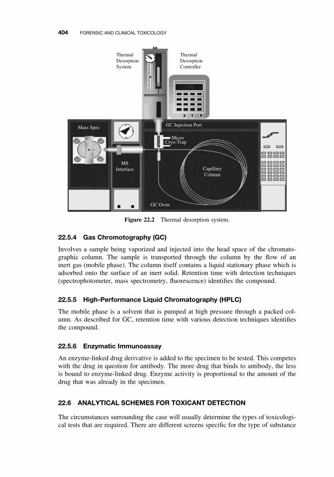
404 FORENSIC AND CLINICAL TOXICOLOGY
ThermalDesorptionSystem
ThermalDesorptionController
Mass Spec
MSInterface
GC Injection Port
MicroCryo-Trap
CapillaryColumn
GC Oven
Figure 22.2 Thermal desorption system.
22.5.4 Gas Chromotography (GC)
Involves a sample being vaporized and injected into the head space of the chromato-graphic column. The sample is transported through the column by the flow of aninert gas (mobile phase). The column itself contains a liquid stationary phase which isadsorbed onto the surface of an inert solid. Retention time with detection techniques(spectrophotometer, mass spectrometry, fluorescence) identifies the compound.
22.5.5 High-Performance Liquid Chromatography (HPLC)
The mobile phase is a solvent that is pumped at high pressure through a packed col-umn. As described for GC, retention time with various detection techniques identifiesthe compound.
22.5.6 Enzymatic Immunoassay
An enzyme-linked drug derivative is added to the specimen to be tested. This competeswith the drug in question for antibody. The more drug that binds to antibody, the lessis bound to enzyme-linked drug. Enzyme activity is proportional to the amount of thedrug that was already in the specimen.
22.6 ANALYTICAL SCHEMES FOR TOXICANT DETECTION
The circumstances surrounding the case will usually determine the types of toxicologi-cal tests that are required. There are different screens specific for the type of substance
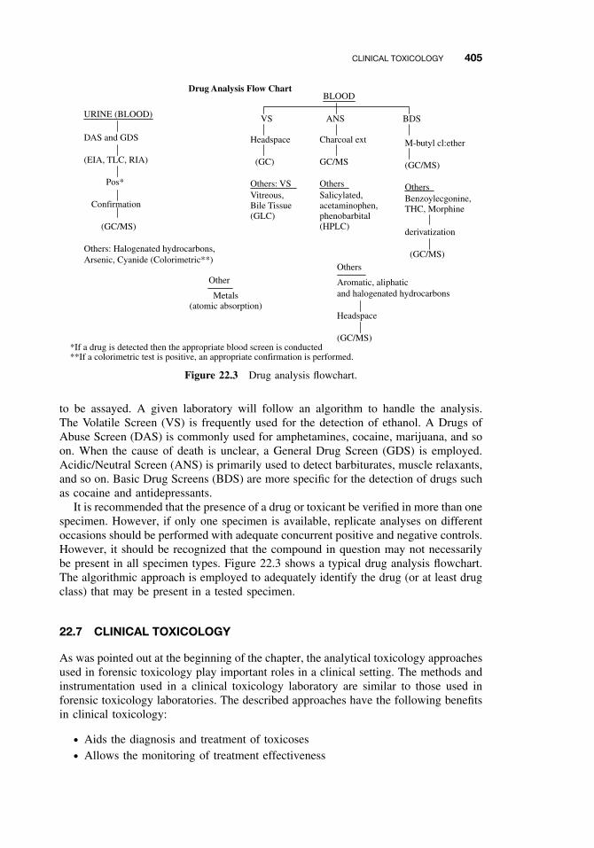
CLINICAL TOXICOLOGY 405
URINE (BLOOD)
Drug Analysis Flow Chart
DAS and GDS
(EIA, TLC, RIA)
Pos*
Confirmation
(GC/MS)
Others: Halogenated hydrocarbons, Arsenic, Cyanide (Colorimetric**)
BLOOD
VS ANS BDS
Headspace
(GC)
Others: VSVitreous,Bile Tissue(GLC)
Charcoal ext
GC/MS
OthersSalicylated,acetaminophen,phenobarbital(HPLC)
M-butyl cl:ether
(GC/MS)
OthersBenzoylecgonine,THC, Morphine
derivatization
(GC/MS)
Other
Metals(atomic absorption)
Others
Aromatic, aliphaticand halogenated hydrocarbons
Headspace
(GC/MS)*If a drug is detected then the appropriate blood screen is conducted**If a colorimetric test is positive, an appropriate confirmation is performed.
Figure 22.3 Drug analysis flowchart.
to be assayed. A given laboratory will follow an algorithm to handle the analysis.The Volatile Screen (VS) is frequently used for the detection of ethanol. A Drugs ofAbuse Screen (DAS) is commonly used for amphetamines, cocaine, marijuana, and soon. When the cause of death is unclear, a General Drug Screen (GDS) is employed.Acidic/Neutral Screen (ANS) is primarily used to detect barbiturates, muscle relaxants,and so on. Basic Drug Screens (BDS) are more specific for the detection of drugs suchas cocaine and antidepressants.
It is recommended that the presence of a drug or toxicant be verified in more than onespecimen. However, if only one specimen is available, replicate analyses on differentoccasions should be performed with adequate concurrent positive and negative controls.However, it should be recognized that the compound in question may not necessarilybe present in all specimen types. Figure 22.3 shows a typical drug analysis flowchart.The algorithmic approach is employed to adequately identify the drug (or at least drugclass) that may be present in a tested specimen.
22.7 CLINICAL TOXICOLOGY
As was pointed out at the beginning of the chapter, the analytical toxicology approachesused in forensic toxicology play important roles in a clinical setting. The methods andinstrumentation used in a clinical toxicology laboratory are similar to those used inforensic toxicology laboratories. The described approaches have the following benefitsin clinical toxicology:
ž Aids the diagnosis and treatment of toxicosesž Allows the monitoring of treatment effectiveness
406 FORENSIC AND CLINICAL TOXICOLOGY
ž Identification of the nature of exposurež Quantification of toxicant
22.7.1 History Taking
Taking thorough general history aids in the effective treatment of an intoxicated patient.Often it may not be possible to communicate directly with the patient due to a lackof consciousness, altered mental state leading to inaccurate information, or deliberatesubmission of misleading information. The type of information that is essential andhelpful include:
ž Identifying what was taken and when, how much, and by what routež Presence of preexisting conditions or allergiesž Whether the patient is currently using any medications or substances of any kindž Whether the patient is pregnant
Historical information can include information obtained from family, friends, lawenforcement and medical personnel, and any observers.
22.7.2 Basic Operating Rules in the Treatment of Toxicosis
The following concepts are central to approaching a toxicosis patient:
ž Ensure airway so that breathing and circulation are adequatež Remove unabsorbed materialž Limit the further absorption of toxicantž Hasten toxicant elimination
Reducing further exposure to a toxicant is crucial and may include removal of thepatient from a toxic environment and the application of decontamination procedures.Immediate decontamination reduces absorption of toxic compounds and represents aprimary essential aspect of the treatment regimen.
External/Skin Decontamination. This entails the complete removal of clothing andgentle washing of the victim with copious amounts of lukewarm water. Mild soaps areoften useful and may increase effectiveness of the removal of the offending substance.
Internal Decontamination. The most recommended methods of internal decontam-ination Include gastric lavage, whole bowel irrigation and administration of acti-vated charcoal.
Lavage. This is utilized if a patient has ingested a life-threatening toxicant. It is rec-ommended for use within a few short hours after ingestion and involves the use of anasogastric or orogastric tube to flush the gastrointestinal tract. A large bore tube isinserted into the stomach and the contents removed with sequential administration andaspiration of small quantities of warm water or saline. This technique is contraindicated
CLINICAL TOXICOLOGY 407
in cases of ingestion of corrosives (i.e., acids, bases) and hydrocarbons (i.e., fuels,essential oils).
Whole-Bowel Irrigation. This involves the infusion by nasogastric or orogastric tubeof a lavage solution consisting of an isosmotic electrolyte (polyethylene glycol elec-trolyte solution is currently recommended). This procedure is indicated after ingestionof metals (e.g., iron, lithium), controlled release medications (terms to describe for-mulations that do not release the active compound immediately after oral ingestion),ingestion of a large amount of an anticholinergic drug (e.g., tricyclics, carbamazepine),and after ingestion of large numbers of tablets. A polyethylene glycol electrolyte solu-tion is administer per hour per os (P.O.) or via a nasogastric tube. Antiemetics may berequired to control vomiting. This is continued until the rectal effluent is clear (approx-imately 3 to 6 hours). The goal is to completely irrigate the gastrointestinal tract toprevent or decrease toxicant absorption. The use of an isosmotic compound such aspolyethylene glycol results in minimal electrolyte loss and fluid changes.
Activated Charcoal. This is considered the most useful agent for the prevention ofabsorption of toxicants. Repeated administration (multiple dose activated charcoal)can impair the enteroenteric-enterohepatic circulation of drugs by binding to drugs thatundergo significant enterohepatic or enteroenteric recycling, including carbamazepine,digoxin, phenobarbitone, theophylline, and verapamil. The prescribed amount of acti-vated charcoal is administered every hour P.O or via nasogastric tube.
Emesis and Catharsis. Emesis is not recommended as a treatment measure for thetoxicosis patient. The danger of aspiration of the gastric contents is great (leading toasphyxiation or aspiration pneumonia). Also a concern is the damage to esophageal andrelated tissue by ingested corrosive substances. Sorbitol is a commonly used cathartic.Often used in charcoal formulations, it increases the gut motility to improve excretionof poison-charcoal complexes. It is not recommended in poisonings by compoundsthat cause profuse diarrhea (e.g., organophosphates, carbamates, and arsenic). The useof a cathartic alone has no value in the management of the poisoned patient. Its useis even controversial as a treatment in combination with activated charcoal. Its useis contraindicated in hypotensive patients, when dehydration or electrolyte balanceis present, when corrosive substances have been ingested and in cases of abdominaltrauma or surgery, and intestinal perforation or obstruction.
22.7.3 Approaches to Selected Toxicoses
A number of antidotes are effective by altering the distribution or metabolism of atoxicant. Reducing the distribution of toxic substances to their sites of action can beachieved by a variety of methods including blocking access of specific poisons totheir receptors with compounds that can compete with these receptors and by usingchelating agents to bind the toxicants (e.g., dimercaprol for arsenic). Biotransformationof a toxic compound into a less toxic form can be achieved by certain agents. Forexample, thiosulphate is used to increase the conversion of cyanide into thiocyanate.
Ethylene Glycol Toxicosis. Ethylene glycol is commonly used as an antifreeze. Itis metabolized by alcohol dehydrogenase to mixed aldehydes, carboxylic acids, and
408 FORENSIC AND CLINICAL TOXICOLOGY
oxalic acid. Toxicosis results in nephrotoxicosis (kidney damage). Administration offomepizole (4-methylpyrazole) or ethanol is effective due to their competition withethylene glycol for the alcohol dehydrogenase enzyme. Although ethanol has a long-term history of clinical experience and is less costly to acquire, fomepizole treatment isassociated with less adverse effects, predictable pharmacokinetics and has a validatedefficacy. Hemodialysis is indicated in patients with severe kidney failure.
Ethanol Toxicosis. Absorption of ethanol by the GI tract absorption is rapid. Peaklevels can be reached 30 to 60 minutes after ingestion. As a rule of thumb, 1 ml of abso-lute ethanol per kilogram weight results in a level of 100 mg/100 ml (0.1%) in 1 hour.Supportive treatment is directed toward the control of acidosis and hypoglycemia.
Organophosphates and Carbamates. The adverse effects of organophosphorous andcarbamate pesticides are mediated through the inhibition of the cholinesterase enzymes.One form, acetylcholinesterase, is located at neurosynaptic junctions while butyrylcholinesterase is primarily located in the plasma and pancreas. Organophosphate pesti-cides inhibit cholinesterase by forming covalent bonds via phosphorylation. Enzymaticregeneration half-lives are long, taking days to months. Organophosphates affect bothred blood cell and plasma cholinesterase activity. Carbamates primarily affect only theplasma derivative. Carbamate insecticides inhibit cholinesterase activity in reversiblemanner. Since carbamates interact with cholinesterase by weak, ionic bonding, thecholinesterases can regenerates itself more readily in matter of minutes to hours.
Since organophosphate toxicosis results in respiratory failure, the treatment approachfor must include the maintenance of a patent airway. Artificial respiration may alsoneed to be employed. The first pharmacological approach is the administration ofatropine. Atropine competes with acetylcholine for its receptor site, thus reducing theeffects of the neurotransmitter. N -methylpyridinium 2-aldoxime (2-PAM) is used inwith atropine therapy as an effective means to restore the covalently bound enzyme toa normal state. It reacts with the phosphorylated cholinesterase enzyme removing thephosphate group. As previously mentioned, carbamates interact with cholinesterase byweak, ionic bonding; thus 2-PAM is of no use to combat toxicosis caused by thesecompounds. However, atropine is effective to prevent the effects on respiration.
Arsenic Toxicosis. Urine arsenic is the best indicator of current or recent exposure.Atomic absorption spectrophotometry is preferred as the detection method. Hair orfingernail sampling may also be helpful. Use of blood is useful if analyzed soon afterexposure or in cases of continuous chronic exposure. After acute exposure, chela-tion therapy is instituted utilizing either (1) Dimercaprol BAL (British Anti-Lewisite)and analogues:
DMSA (dimercaptosuccinic acid)DMPS (dimercaptopropane succinate)
or (2) d-penicillamine. Supportive/symptomatic therapy is also necessary. A higher pro-tein diet and the alleviation of dehydration due to diarrhea and vomiting are beneficial.
Chronic Exposure (Arsenic). Primarily symptomatic treatment is chosen. Chelationtherapy is practiced, but its usefulness in cases of chronic exposure is still questionable.
SUGGESTED READING 409
Supportive treatment is an essential component of the management of the intoxicatedpatient. Monitoring and assessment of all organ systems in conjunction with the use ofappropriate pharmaceutical agents/antidotes increases therapeutic success. The natureof this care will depend on the toxicant in question and the patient’s condition uponpresentation.
SUGGESTED READING
Ballantyne, B., T. Marrs, and P. Turner, eds. General and Applied Toxicology, college ed., NewYork: Macmillan, 1995.
R. E. Ferner, ed. Forensic Pharmacology: Medicines, Mayhem, and Malpractice. New York:Oxford University Press, 1996.
A. Furst, ed. The Toxicologist as Expert Witness. Washington, DC: Taylor and Francis, 1997.
Hardman, J. G., L. E. Limbird, P. B. Molinoff, R. W. Ruddon, and A. Goodman Gilman, eds.Goodman and Gilman’s The Pharmacological Basis of Therapeutics, 10th ed., New York:McGraw-Hill, 2001.
Klaassen, C. D., Cassarett and Doull’s Toxicology: The Basic Science of Poisons, 6th ed. NewYork: McGraw-Hill, 2001.
Trestrail, III, J. H. ed. Criminal Poisoning: Investigational Guide For Law Enforcement, Toxi-cologists, Forensic Scientists, and Attorneys. Totowa, NJ: Humana Press, 2000.

CHAPTER 23
Prevention of Toxicity
ERNEST HODGSON
23.1 INTRODUCTION
It is obvious, but often forgotten, that toxicity is always a consequence of exposure andthat no matter what the results of hazard assessment, without exposure there cannotbe a toxic effect. However, if both hazard and exposure are verified, and the riskappears to be significant, there are a range of possible actions available to reduce thatrisk. These actions range from outright banning, from both production and use, ofthe chemical in question, through measures to restrict exposure, to measures to restricteffect. Exposure can be restricted by prevention of manufacture, control of use patterns,control of application techniques, environmental manipulation, and education. Effectscan be restricted by prophylactic and therapeutic methods and by education. Many ofthese approaches are controlled in whole, or in part, by legislation. All of them, takentogether, comprise the subject matter of this chapter.
Laws and regulations provide the framework for organized efforts to prevent toxicity,and sanctions are necessary to prevent those without social conscience from deliberatelyexposing their fellows to risks from toxic hazards. That is not enough, however, withouta population educated to toxic hazards and their prevention, the laws could never beadministered properly. Moreover, in many circumstances, and particularly in the home,wisdom dictates courses of action not necessarily prescribed by law. The key to toxicityprevention lies in information and education with legislation, regulation, and penaltiesas final safeguards. In all probability, the better the general population is educated andinformed, the less likely are laws to be necessary.
23.2 LEGISLATION AND REGULATION
In the best sense, legislation provides an enabling act describing the areas to be coveredunder the particular law and the general manner in which they are to be regulated, anddesignating an executive agency to write and enforce specific regulations within theintent of the legislative body. For example, the Toxic Substances Control Act (TSCA)was passed by Congress to regulate the introduction of chemicals into commerce, to
A Textbook of Modern Toxicology, Third Edition, edited by Ernest HodgsonISBN 0-471-26508-X Copyright 2004 John Wiley & Sons, Inc.
411
412 PREVENTION OF TOXICITY
determine their hazards to the human population and the environment, and to regulateor ban those deemed hazardous. The task of writing and enforcing specific regulationswas assigned to the Environmental Protection Agency (EPA).
Legislative attempts to write specific regulations into laws usually fail. The resultantlaws lack flexibility and, because they are written by lawyers rather than toxicologists,seldom address the problems in a scientifically rigorous manner.
It should be borne in mind that legislation is a synthesis of science, politics, andpublic and private pressure. It represents a society’s best estimate, at that moment, of therisks it is prepared to take and those it wishes to avoid, as well as the price it is preparedto pay. Such decisions properly include more than science. The task of the toxicologistis to see that the science that is included is accurate and is interpreted logically.
This section is based primarily on regulations in the United States, not becausethese are the best but because, in toto, they are the most comprehensive. In manyrespects they are a complex mixture of overlapping laws and jurisdictions, providingunnecessary work for the legal profession. At the same time few, if any, toxic hazardsin the home, workplace, or environment are not addressed.
23.2.1 Federal Government
Following is a summary of the most important federal statues concerned in whole orin part with the regulation of toxic substances.
Clean Air Act. The Clean Air Act is administered by the EPA. Although the principalenforcement provisions are the responsibility of local governments, overall administra-tive responsibility rests with EPA. This act requires criteria documents for air pollutantsand sets both national air quality standards and standards for sources that create airpollutants, such as motor vehicles, power plants, and so on. Important actions alreadytaken under this law include standards for the now complete phased-out eliminationof lead in gasoline, and the setting of sulfuric acid air emission guidelines for existingindustrial plants.
Clean Water Act. The Clean Water Act, which amends the Federal Water PollutionControl Act, is also administered b the EPA and provides for funding of municipalsewage treatment plants. However, with respect to toxicity prevention, it is more impor-tant that the act regulates emissions from municipal and industrial sources. It has as itsgoal the elimination of discharges of pollutants and the protection of rivers so that theyare “swimmable and fishable” and applies to “waters of the United States” subsequentlydefined to include all waters that reach navigable waters, wetland, and intermittentstreams. Some of the more important actions taken under this statute include settingstandards for emissions of inorganics from smelter operations and publishing prioritylists of toxic pollutants. This act allows the federal government to recover cleanup andother costs as damages from the polluting agency, company, or individual.
Safe Drinking Water Act. (1974, 1986, 1996). Specifically applied to water suppliedfor humans consumption, this act requires the EPA to set maximum levels for contam-inants in water delivered to users of public water systems. Two criteria are establishedfor a particular contaminant: the maximum containment level goal (MCGL) and themaximum contaminant level (MCL). The former, the MCLG, is the level at which no
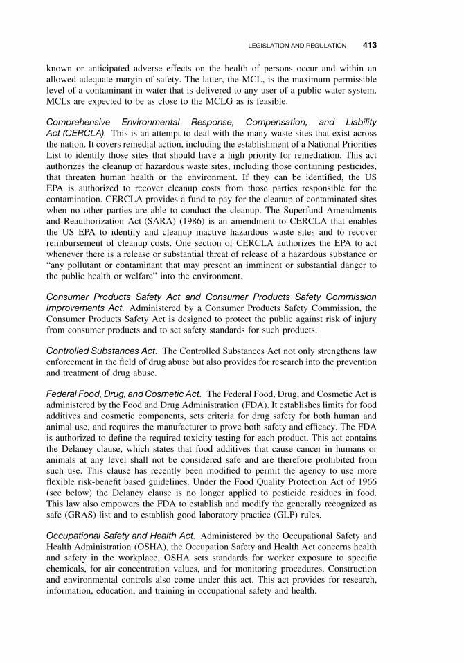
LEGISLATION AND REGULATION 413
known or anticipated adverse effects on the health of persons occur and within anallowed adequate margin of safety. The latter, the MCL, is the maximum permissiblelevel of a contaminant in water that is delivered to any user of a public water system.MCLs are expected to be as close to the MCLG as is feasible.
Comprehensive Environmental Response, Compensation, and LiabilityAct (CERCLA). This is an attempt to deal with the many waste sites that exist acrossthe nation. It covers remedial action, including the establishment of a National PrioritiesList to identify those sites that should have a high priority for remediation. This actauthorizes the cleanup of hazardous waste sites, including those containing pesticides,that threaten human health or the environment. If they can be identified, the USEPA is authorized to recover cleanup costs from those parties responsible for thecontamination. CERCLA provides a fund to pay for the cleanup of contaminated siteswhen no other parties are able to conduct the cleanup. The Superfund Amendmentsand Reauthorization Act (SARA) (1986) is an amendment to CERCLA that enablesthe US EPA to identify and cleanup inactive hazardous waste sites and to recoverreimbursement of cleanup costs. One section of CERCLA authorizes the EPA to actwhenever there is a release or substantial threat of release of a hazardous substance or“any pollutant or contaminant that may present an imminent or substantial danger tothe public health or welfare” into the environment.
Consumer Products Safety Act and Consumer Products Safety CommissionImprovements Act. Administered by a Consumer Products Safety Commission, theConsumer Products Safety Act is designed to protect the public against risk of injuryfrom consumer products and to set safety standards for such products.
Controlled Substances Act. The Controlled Substances Act not only strengthens lawenforcement in the field of drug abuse but also provides for research into the preventionand treatment of drug abuse.
Federal Food, Drug, and Cosmetic Act. The Federal Food, Drug, and Cosmetic Act isadministered by the Food and Drug Administration (FDA). It establishes limits for foodadditives and cosmetic components, sets criteria for drug safety for both human andanimal use, and requires the manufacturer to prove both safety and efficacy. The FDAis authorized to define the required toxicity testing for each product. This act containsthe Delaney clause, which states that food additives that cause cancer in humans oranimals at any level shall not be considered safe and are therefore prohibited fromsuch use. This clause has recently been modified to permit the agency to use moreflexible risk-benefit based guidelines. Under the Food Quality Protection Act of 1966(see below) the Delaney clause is no longer applied to pesticide residues in food.This law also empowers the FDA to establish and modify the generally recognized assafe (GRAS) list and to establish good laboratory practice (GLP) rules.
Occupational Safety and Health Act. Administered by the Occupational Safety andHealth Administration (OSHA), the Occupation Safety and Health Act concerns healthand safety in the workplace, OSHA sets standards for worker exposure to specificchemicals, for air concentration values, and for monitoring procedures. Constructionand environmental controls also come under this act. This act provides for research,information, education, and training in occupational safety and health.
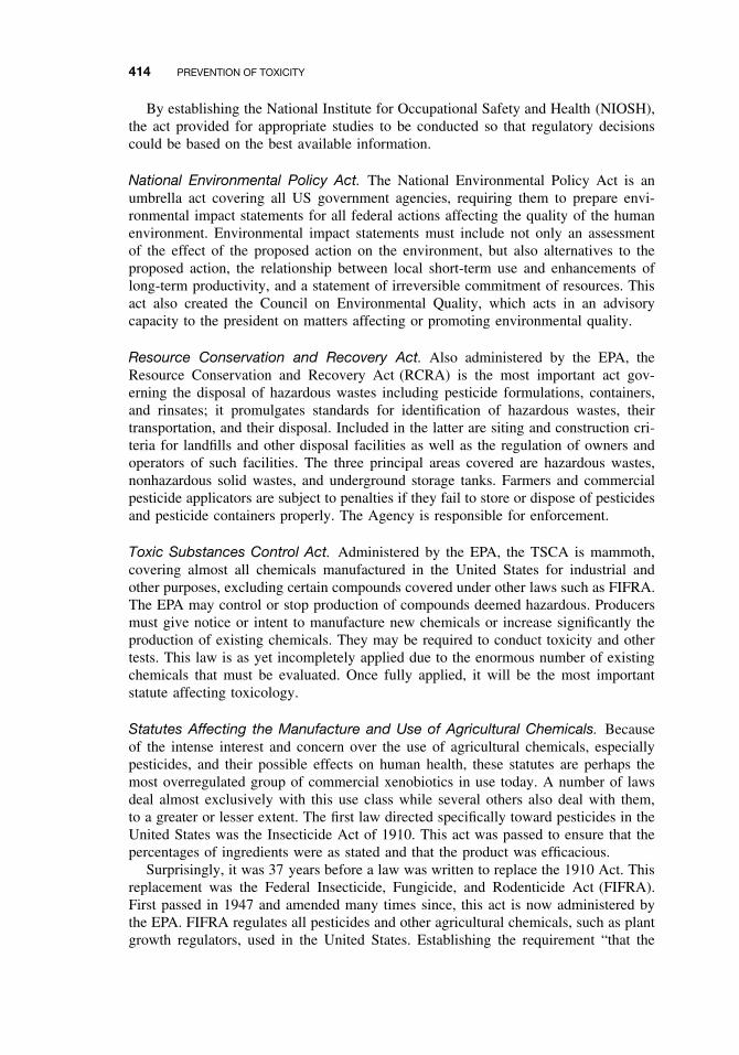
414 PREVENTION OF TOXICITY
By establishing the National Institute for Occupational Safety and Health (NIOSH),the act provided for appropriate studies to be conducted so that regulatory decisionscould be based on the best available information.
National Environmental Policy Act. The National Environmental Policy Act is anumbrella act covering all US government agencies, requiring them to prepare envi-ronmental impact statements for all federal actions affecting the quality of the humanenvironment. Environmental impact statements must include not only an assessmentof the effect of the proposed action on the environment, but also alternatives to theproposed action, the relationship between local short-term use and enhancements oflong-term productivity, and a statement of irreversible commitment of resources. Thisact also created the Council on Environmental Quality, which acts in an advisorycapacity to the president on matters affecting or promoting environmental quality.
Resource Conservation and Recovery Act. Also administered by the EPA, theResource Conservation and Recovery Act (RCRA) is the most important act gov-erning the disposal of hazardous wastes including pesticide formulations, containers,and rinsates; it promulgates standards for identification of hazardous wastes, theirtransportation, and their disposal. Included in the latter are siting and construction cri-teria for landfills and other disposal facilities as well as the regulation of owners andoperators of such facilities. The three principal areas covered are hazardous wastes,nonhazardous solid wastes, and underground storage tanks. Farmers and commercialpesticide applicators are subject to penalties if they fail to store or dispose of pesticidesand pesticide containers properly. The Agency is responsible for enforcement.
Toxic Substances Control Act. Administered by the EPA, the TSCA is mammoth,covering almost all chemicals manufactured in the United States for industrial andother purposes, excluding certain compounds covered under other laws such as FIFRA.The EPA may control or stop production of compounds deemed hazardous. Producersmust give notice or intent to manufacture new chemicals or increase significantly theproduction of existing chemicals. They may be required to conduct toxicity and othertests. This law is as yet incompletely applied due to the enormous number of existingchemicals that must be evaluated. Once fully applied, it will be the most importantstatute affecting toxicology.
Statutes Affecting the Manufacture and Use of Agricultural Chemicals. Becauseof the intense interest and concern over the use of agricultural chemicals, especiallypesticides, and their possible effects on human health, these statutes are perhaps themost overregulated group of commercial xenobiotics in use today. A number of lawsdeal almost exclusively with this use class while several others also deal with them,to a greater or lesser extent. The first law directed specifically toward pesticides in theUnited States was the Insecticide Act of 1910. This act was passed to ensure that thepercentages of ingredients were as stated and that the product was efficacious.
Surprisingly, it was 37 years before a law was written to replace the 1910 Act. Thisreplacement was the Federal Insecticide, Fungicide, and Rodenticide Act (FIFRA).First passed in 1947 and amended many times since, this act is now administered bythe EPA. FIFRA regulates all pesticides and other agricultural chemicals, such as plantgrowth regulators, used in the United States. Establishing the requirement “that the
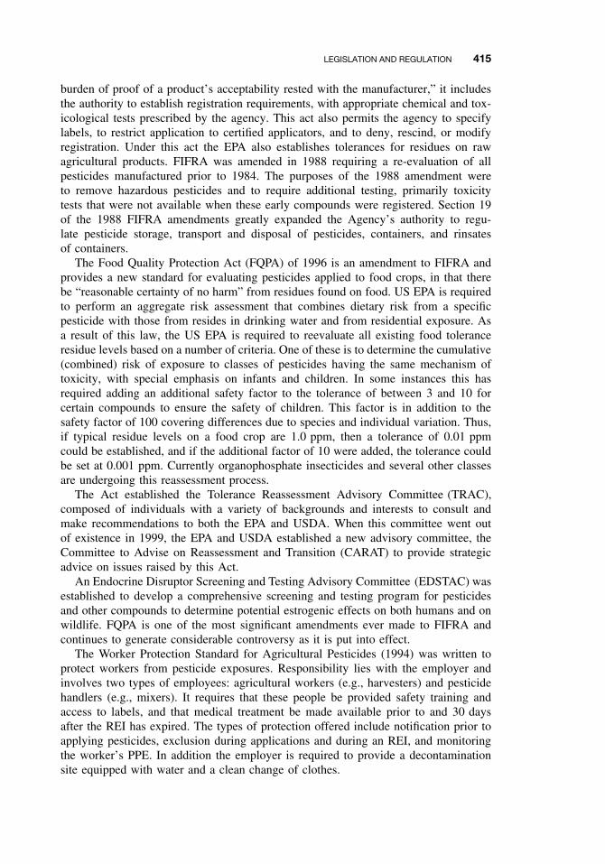
LEGISLATION AND REGULATION 415
burden of proof of a product’s acceptability rested with the manufacturer,” it includesthe authority to establish registration requirements, with appropriate chemical and tox-icological tests prescribed by the agency. This act also permits the agency to specifylabels, to restrict application to certified applicators, and to deny, rescind, or modifyregistration. Under this act the EPA also establishes tolerances for residues on rawagricultural products. FIFRA was amended in 1988 requiring a re-evaluation of allpesticides manufactured prior to 1984. The purposes of the 1988 amendment wereto remove hazardous pesticides and to require additional testing, primarily toxicitytests that were not available when these early compounds were registered. Section 19of the 1988 FIFRA amendments greatly expanded the Agency’s authority to regu-late pesticide storage, transport and disposal of pesticides, containers, and rinsatesof containers.
The Food Quality Protection Act (FQPA) of 1996 is an amendment to FIFRA andprovides a new standard for evaluating pesticides applied to food crops, in that therebe “reasonable certainty of no harm” from residues found on food. US EPA is requiredto perform an aggregate risk assessment that combines dietary risk from a specificpesticide with those from resides in drinking water and from residential exposure. Asa result of this law, the US EPA is required to reevaluate all existing food toleranceresidue levels based on a number of criteria. One of these is to determine the cumulative(combined) risk of exposure to classes of pesticides having the same mechanism oftoxicity, with special emphasis on infants and children. In some instances this hasrequired adding an additional safety factor to the tolerance of between 3 and 10 forcertain compounds to ensure the safety of children. This factor is in addition to thesafety factor of 100 covering differences due to species and individual variation. Thus,if typical residue levels on a food crop are 1.0 ppm, then a tolerance of 0.01 ppmcould be established, and if the additional factor of 10 were added, the tolerance couldbe set at 0.001 ppm. Currently organophosphate insecticides and several other classesare undergoing this reassessment process.
The Act established the Tolerance Reassessment Advisory Committee (TRAC),composed of individuals with a variety of backgrounds and interests to consult andmake recommendations to both the EPA and USDA. When this committee went outof existence in 1999, the EPA and USDA established a new advisory committee, theCommittee to Advise on Reassessment and Transition (CARAT) to provide strategicadvice on issues raised by this Act.
An Endocrine Disruptor Screening and Testing Advisory Committee (EDSTAC) wasestablished to develop a comprehensive screening and testing program for pesticidesand other compounds to determine potential estrogenic effects on both humans and onwildlife. FQPA is one of the most significant amendments ever made to FIFRA andcontinues to generate considerable controversy as it is put into effect.
The Worker Protection Standard for Agricultural Pesticides (1994) was written toprotect workers from pesticide exposures. Responsibility lies with the employer andinvolves two types of employees: agricultural workers (e.g., harvesters) and pesticidehandlers (e.g., mixers). It requires that these people be provided safety training andaccess to labels, and that medical treatment be made available prior to and 30 daysafter the REI has expired. The types of protection offered include notification prior toapplying pesticides, exclusion during applications and during an REI, and monitoringthe worker’s PPE. In addition the employer is required to provide a decontaminationsite equipped with water and a clean change of clothes.
416 PREVENTION OF TOXICITY
The Act was written to cover pesticide use on farms, forests, nurseries, and green-houses. It does not include applications to pastures, golf courses, parks, livestock,right-of-way, or home gardens, nor does it cover treatments for mosquito abatementand rodent control.
Many other legislative acts impact in whole or in part on pesticide use. They includethe Endangered Species Act of 1973, an act written to protect endangered wildlife;it regulates pesticide use around wildlife sanctuaries. Pesticides might injure or killendangered species if allowed to drift onto habitat, or runoff into streams, lakes, orwetlands might be found to significantly degrade endangered wildlife habitat. Alsoincluded are the Clean Water Act, the Safe Drinking Water Act, RCRA, CERCLA,and SARA, all discussed above.
Other Statutes with Relevance to the Prevention of Toxicity. It should be noted thatsome of these statutes have been superseded by others, either in whole or in part.
ž Comprehensive Employment and Training Actž Dangerous Cargo Actž Federal Coal Mine Safety and Health Amendment Actž Federal Caustic Poison Actž Federal Railroad Safety Authorization Actž Hazardous Materials Transport Actž Lead-Based Paint Poison Prevention Actž Marine Protection Research and Sanctuaries Actž Poison Prevention Packaging Actž Ports and Waterways Safety Act
23.2.2 State Governments
Within the United States, states are free to adopt legislation with toxicological signifi-cance, although their jurisdiction does not extend beyond their geographic boundaries.In other cases the states may enforce federal statutes under certain circumstances. Forexample, if state regulations concerning hazardous waste disposal is neither less com-prehensive nor less rigorous than the federal statute, enforcements is delegated to thestates. Similarly certain aspects of FIFRA are enforced by individual states. In somecases (California is notable in this respect) states have passed laws considerably morecomprehensive and more rigorous than the corresponding federal stature.
23.2.3 Legislation and Regulation in Other Countries
It would serve little purpose to enumerate all the laws affecting toxicology, toxicitytesting, and the prevention of toxicity that have been promulgated in all countries thathave such laws. Legislation in this area has been adopted in most countries of westernEurope and in Japan. Although the laws in use in the United States are a complexmixture of overlapping statutes and enforcement agencies, they are probably the mostcomprehensive set of such laws in existence. Most other industrialized countries havelegislation in the same areas, although the emphasis varies widely from one country to
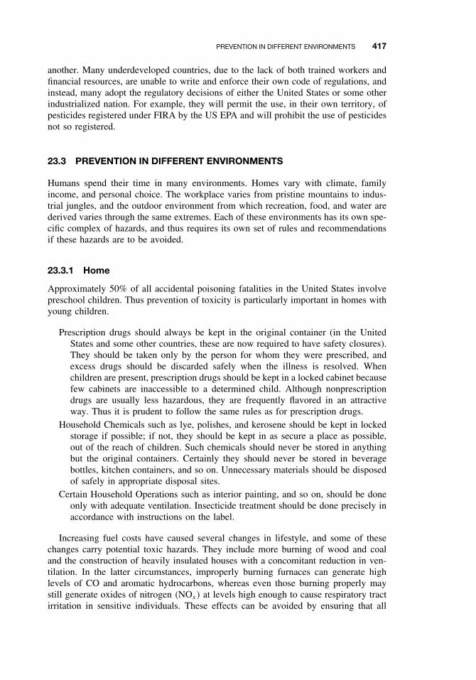
PREVENTION IN DIFFERENT ENVIRONMENTS 417
another. Many underdeveloped countries, due to the lack of both trained workers andfinancial resources, are unable to write and enforce their own code of regulations, andinstead, many adopt the regulatory decisions of either the United States or some otherindustrialized nation. For example, they will permit the use, in their own territory, ofpesticides registered under FIRA by the US EPA and will prohibit the use of pesticidesnot so registered.
23.3 PREVENTION IN DIFFERENT ENVIRONMENTS
Humans spend their time in many environments. Homes vary with climate, familyincome, and personal choice. The workplace varies from pristine mountains to indus-trial jungles, and the outdoor environment from which recreation, food, and water arederived varies through the same extremes. Each of these environments has its own spe-cific complex of hazards, and thus requires its own set of rules and recommendationsif these hazards are to be avoided.
23.3.1 Home
Approximately 50% of all accidental poisoning fatalities in the United States involvepreschool children. Thus prevention of toxicity is particularly important in homes withyoung children.
Prescription drugs should always be kept in the original container (in the UnitedStates and some other countries, these are now required to have safety closures).They should be taken only by the person for whom they were prescribed, andexcess drugs should be discarded safely when the illness is resolved. Whenchildren are present, prescription drugs should be kept in a locked cabinet becausefew cabinets are inaccessible to a determined child. Although nonprescriptiondrugs are usually less hazardous, they are frequently flavored in an attractiveway. Thus it is prudent to follow the same rules as for prescription drugs.
Household Chemicals such as lye, polishes, and kerosene should be kept in lockedstorage if possible; if not, they should be kept in as secure a place as possible,out of the reach of children. Such chemicals should never be stored in anythingbut the original containers. Certainly they should never be stored in beveragebottles, kitchen containers, and so on. Unnecessary materials should be disposedof safely in appropriate disposal sites.
Certain Household Operations such as interior painting, and so on, should be doneonly with adequate ventilation. Insecticide treatment should be done precisely inaccordance with instructions on the label.
Increasing fuel costs have caused several changes in lifestyle, and some of thesechanges carry potential toxic hazards. They include more burning of wood and coaland the construction of heavily insulated houses with a concomitant reduction in ven-tilation. In the latter circumstances, improperly burning furnaces can generate highlevels of CO and aromatic hydrocarbons, whereas even those burning properly maystill generate oxides of nitrogen (NOx) at levels high enough to cause respiratory tractirritation in sensitive individuals. These effects can be avoided by ensuring that all
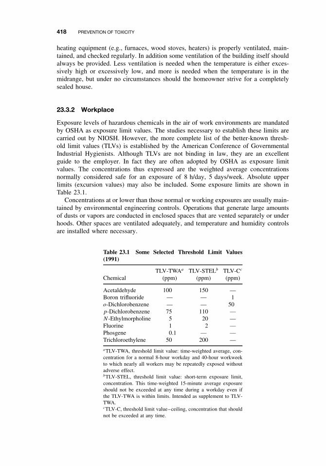
418 PREVENTION OF TOXICITY
heating equipment (e.g., furnaces, wood stoves, heaters) is properly ventilated, main-tained, and checked regularly. In addition some ventilation of the building itself shouldalways be provided. Less ventilation is needed when the temperature is either exces-sively high or excessively low, and more is needed when the temperature is in themidrange, but under no circumstances should the homeowner strive for a completelysealed house.
23.3.2 Workplace
Exposure levels of hazardous chemicals in the air of work environments are mandatedby OSHA as exposure limit values. The studies necessary to establish these limits arecarried out by NIOSH. However, the more complete list of the better-known thresh-old limit values (TLVs) is established by the American Conference of GovernmentalIndustrial Hygienists. Although TLVs are not binding in law, they are an excellentguide to the employer. In fact they are often adopted by OSHA as exposure limitvalues. The concentrations thus expressed are the weighted average concentrationsnormally considered safe for an exposure of 8 h/day, 5 days/week. Absolute upperlimits (excursion values) may also be included. Some exposure limits are shown inTable 23.1.
Concentrations at or lower than those normal or working exposures are usually main-tained by environmental engineering controls. Operations that generate large amountsof dusts or vapors are conducted in enclosed spaces that are vented separately or underhoods. Other spaces are ventilated adequately, and temperature and humidity controlsare installed where necessary.
Table 23.1 Some Selected Threshold Limit Values(1991)
ChemicalTLV-TWAa
(ppm)TLV-STELb
(ppm)TLV-Cc
(ppm)
Acetaldehyde 100 150 —Boron trifluoride — — 1o-Dichlorobenzene — — 50p-Dichlorobenzene 75 110 —N-Ethylmorpholine 5 20 —Fluorine 1 2 —Phosgene 0.1 — —Trichloroethylene 50 200 —
aTLV-TWA, threshold limit value: time-weighted average, con-centration for a normal 8-hour workday and 40-hour workweekto which nearly all workers may be repeatedly exposed withoutadverse effect.bTLV-STEL, threshold limit value: short-term exposure limit,concentration. This time-weighted 15-minute average exposureshould not be exceeded at any time during a workday even ifthe TLV-TWA is within limits. Intended as supplement to TLV-TWA.cTLV-C, threshold limit value–ceiling, concentration that shouldnot be exceeded at any time.
PREVENTION IN DIFFERENT ENVIRONMENTS 419
Other precautions must be taken to prevent accidental or occasional increases inconcentrations. Materials should be transported in “safe” containers, spilled materialremoved rapidly, and floor and wall materials selected to prevent contamination andallow easy cleaning.
Additional methods for the prevention of toxicity in the workplace include the useof personal safety equipment—protective clothing, gloves, and goggles are the mostimportant. In particularly hazardous operations, closed-circuit air masks, gas masks,and so on, may also be necessary.
Pre-employment instruction and pre-employment physical examinations are of crit-ical importance in most work situations involving hazardous chemicals. The formershould make clear the hazards involved, the need to avoid exposure under normalworking conditions, and the mechanisms by which exposure is limited. Furthermoreemployees should understand how and when to contain spills and how and when toevacuate the area around the spill. Locations and use of emergency equipment, show-ers, eye washes, and so on, should also be given, and the most important proceduresshould be posted in the work area.
23.3.3 Pollution of Air, Water, and Land
The toxicological significance of pollution of the environment may be work related, asin the case of agricultural workers, or related to the outside environment encounteredin daily life. In the case of agricultural workers, numerous precautions are necessaryfor the prevention of toxicity. For example:
ž Pesticides and other agricultural chemicals should be kept only in the originalcontainer, carrying the labels prescribed by EPA under FIFRA.
ž Empty containers and excess chemicals should be disposed of properly in safehazardous waste disposal sites, incinerated when possible, or, in some cases,decontaminated.
ž Workers should not re-enter treated areas until the safe re-entry period has elapsed.ž Certain workers such as applicators and those preparing tank mixes should wear
appropriate protection clothing, gloves, face masks, and so forth. The developmentof closed systems for mixing pesticides should help protect mixers and loaders ofpesticides from exposure.
ž Spraying operations should be carried out in such a way as to minimize drift,contamination of water, and so on.
Pesticides have caused a number of fatalities in the past. The current practice in somecountries of restricting the most hazardous chemicals for use only by certified operatorsshould greatly minimize pesticide poisoning in these locations.
Individuals can do little to protect themselves from poisoning by chemicals thatpollute the air and water except to insist that discharge of toxicants into the environmentbe minimized. The exposure levels are low compared with those in acute toxicitycases, and the effects may be indirect, as in the increase in preexisting respiratoryirritation during smog. Thus these effects can be determined only at the epidemiologiclevel. Because many persons are not affected or may not be affected for years, itis often argued that environmental contamination is not very important. However, a
420 PREVENTION OF TOXICITY
small percentage increase may represent a large number of people when the wholepopulation is considered. Furthermore chronic toxicity is not often reversible. Becausein most industrialized countries laws already exist to control emission problems, ifsuch problems exist in these countries, they are usually problems of enforcement.
One of the most critical areas for the prevention of toxicity caused by environmentalcontamination is that of disposal of hazardous wastes. It is now apparent that pastpractices in many industrialized countries have created large numbers of waste sitesin which the waste is often unidentified, improperly stored, and leaching into theenvironment. The task of rectifying these past errors is an enormous one just nowbeing addressed.
The ideal situation for current and future practices is to reduce chemical wasteto an irreducible minimum and then to place the remainder in secure storage. Wastereduction can be accomplished in many ways.
ž Refine plant processes so that less waste is produced.ž Recycle waste into useful products.ž Concentrate wastes.ž Incinerate. The technology is available to incinerate essentially all waste to inor-
ganic slag. Unfortunately, the technology is sophisticated and expensive. Inad-equate incineration is itself a hazard because of the risk of generating dioxinsand other toxicants and releasing them into the environment. Less complex andmore easily maintained incinerators will be essential if this technology is to playa prominent role in waste reduction.
Safe storage for the remaining waste may be in dump sites or in above-groundstorage. In either case such storage ideally should be properly sited, constructed, main-tained, and monitored.
Because of the nature of commerce, probably none of these measures will be suc-cessful unless the laws, penalties, and incentives are manipulated in such a way as tomake safe disposal more attractive economically than unsafe disposal.
23.4 EDUCATION
Because chemicals, many of them hazardous, are an inevitable part of life in industrial-ized countries, education is probably the most important method for the prevention oftoxicity. Unfortunately, it is also one of the most neglected. In a typical public debateconcerning a possible chemical hazard, the principle protagonists tend to fall into twoextreme groups. The “everything is OK” protagonists and the “ban it completely” pro-tagonists. The media seldom seem to educate the public, usually serving only to addfuel to the flames.
The educational role of the toxicologist should be the voice of reason, presenting abalanced view of risks and benefits, and outlining alternatives whenever possible. Thesimple lesson that science deals not in certainty, but rather degrees of certitude, mustbe learned by all involved.
In terms of ongoing educational programs, there should be opportunities at all levels:elementary schools, high schools, university, adult education, and media education.Several approaches can be used to educate the general public in ideal situations:
SUGGESTED READING 421
ž Elementary school. Teach the rudiments of first aid and environmental concerns,proper disposal, and so on.
ž High school. Teach concepts of toxicology (dose response, etc.) and environmen-tal toxicology (bioaccumulation, etc.), as these concepts can be introduced intogeneral science courses.
ž University. In addition to toxicology degrees, general courses for nontoxicologyand/or nonscience majors should stress a balanced approach, with both responsibleuse and toxicity prevention as desirable end points. General Toxicology should bea required course in all chemically related academic programs such as chemistryand chemical engineering.
ž Media. Encourage a balanced approach to toxicity problems. Toxicologistsshould be available to media representatives and, where appropriate, should beinvolved directly.
SUGGESTED READING
American Conference of Governmental Industrial Hygienists. TLVs—Threshold Limit Values forChemical Substances and Physical Agents in the Work Environment with Intended Changes.Cincinnati (published annually).
American Conference of Governmental Industrial Hygienists. Documentation of the ThresholdLimit Values for Substances in Workroom Air. Cincinnati (published annually).
Doull, J. Recommended limits for exposure to chemicals. In Casarett and Doull’s Toxicology:The Basic Science of Poisons, 6th ed., C. D. Klaassen, ed. New York: McGraw-Hill, 2001,pp. 1115–1176.
Dreisbach, R. H. Handbook of Poisoning: Prevention, Diagnosis, and Treatment, 11th ed. LosAltos, CA: Lange Medical, 1983.
Ellenhorn, M. J., and D. G. Barceloux. Medical Toxicology: Diagnosis and Treatment of HumanPoisoning. New York: Elsevier Science, 1998.
Environmental Regulation: An International View. A series of papers on: I. Britain, T. W. Hall;II. European Economic Community, S. P. Johnson; III. The United States, J. B. Ritch, Jr; IV.An Industry View, R. C. Tineknell. Chem. Soc. Rev. 5: 431–771, 1976.
Fan, A. M., and L. W. Chang, eds. Toxicology and Risk Assessment: Principles, Methods andApplications. New York: Dekker, 1996.
Merrill, R. A. Regulatory toxicology. In Casarett and Doull’s Toxicology: The Basic Science ofPoisons, 6th ed., C. D. Klaassen, ed. New York: McGraw-Hill, 2001, pp. 1141–1153.
Sandmeyer, E. E. Regulatory toxicology. In A Guide to General Toxicology. F. Homberger,J. A. Hayes, E. W. Pelikan, eds. Basel: Karger, 1983, ch. 20.

CHAPTER 24
Human Health Risk Assessment
RONALD E. BAYNES
24.1 INTRODUCTION
We often perform toxicological research to better understand the mechanism and associ-ated health risk following exposure to hazardous agents. Risk assessment is a systematicscientific characterization of potential adverse health effects following exposure to thesehazardous agents. Risk assessment activities are designed to identify, describe, and mea-sure qualities and quantities from these toxicological studies, which are often conductedwith homogeneous animal models at doses and exposure duration not encountered ina more heterogeneous human population. Herein lie the challenge of risk assessment.The use of default assumptions because of some level of uncertainty in our extrapola-tions across species, doses, routes, and interindividual variability, the risk assessmentprocess is often perceived as lacking scientific rigor. This chapter will cover tradi-tional practices as well as new and novel approaches that utilize more of the availablescientific data to identify and reduce uncertainty in the process. The advent of pow-erful computers and sophisticated software programs has allowed the development ofquantitative models that better describe the dose-response relationship, refine biologi-cally relevant dose estimates in the risk assessment process, and encourage departurefrom traditional default approaches (Conolly et al., 1999). Although the focus of thischapter is on current and novel risk assessment methods that are scientifically based,it is critical that the reader be aware of the differences between risk assessment andrisk management, which are summarized in Table 24.1.
Results from the risk assessment are used to inform risk management. The risk man-ager uses the risk information in conjunction with factors such as the social importanceof the risk, the social acceptability of the risk, the economic impacts of risk reduc-tion, engineering, and legislative mandates when deciding on and implementing riskmanagement approaches.
The risk assessment may be perceived as the source of a risk management decision,when in fact, social concerns, international issues, trade, public perception, or othernon-risk considerations may be taken into consideration. Finally there is one activ-ity known as risk communication that involves making the risk assessment and risk
A Textbook of Modern Toxicology, Third Edition, edited by Ernest HodgsonISBN 0-471-26508-X Copyright 2004 John Wiley & Sons, Inc.
423
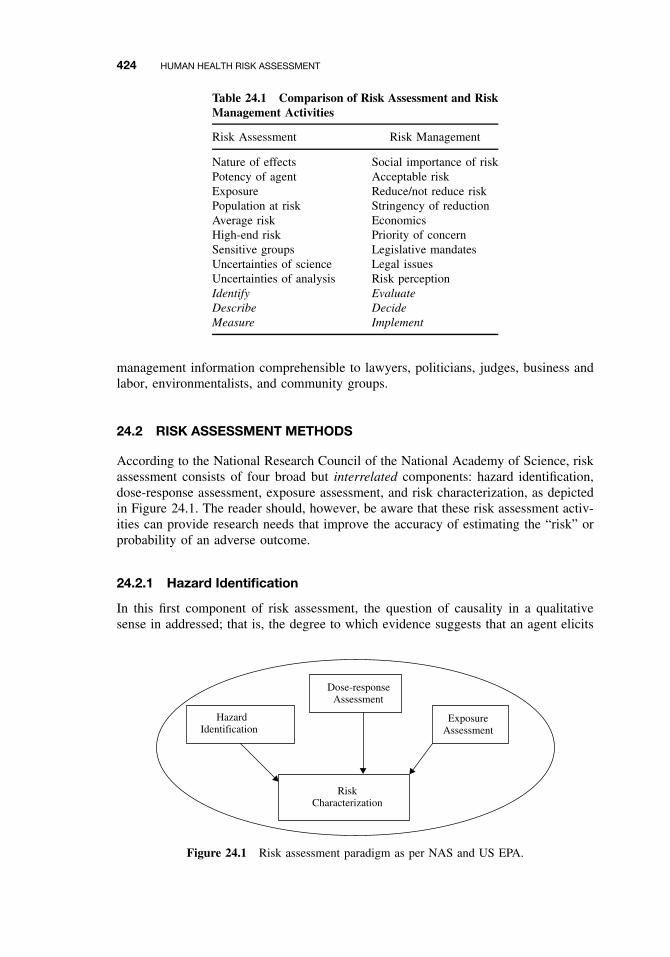
424 HUMAN HEALTH RISK ASSESSMENT
Table 24.1 Comparison of Risk Assessment and RiskManagement Activities
Risk Assessment Risk Management
Nature of effects Social importance of riskPotency of agent Acceptable riskExposure Reduce/not reduce riskPopulation at risk Stringency of reductionAverage risk EconomicsHigh-end risk Priority of concernSensitive groups Legislative mandatesUncertainties of science Legal issuesUncertainties of analysis Risk perceptionIdentify EvaluateDescribe DecideMeasure Implement
management information comprehensible to lawyers, politicians, judges, business andlabor, environmentalists, and community groups.
24.2 RISK ASSESSMENT METHODS
According to the National Research Council of the National Academy of Science, riskassessment consists of four broad but interrelated components: hazard identification,dose-response assessment, exposure assessment, and risk characterization, as depictedin Figure 24.1. The reader should, however, be aware that these risk assessment activ-ities can provide research needs that improve the accuracy of estimating the “risk” orprobability of an adverse outcome.
24.2.1 Hazard Identification
In this first component of risk assessment, the question of causality in a qualitativesense in addressed; that is, the degree to which evidence suggests that an agent elicits
HazardIdentification
RiskCharacterization
Dose-response Assessment
ExposureAssessment
Figure 24.1 Risk assessment paradigm as per NAS and US EPA.
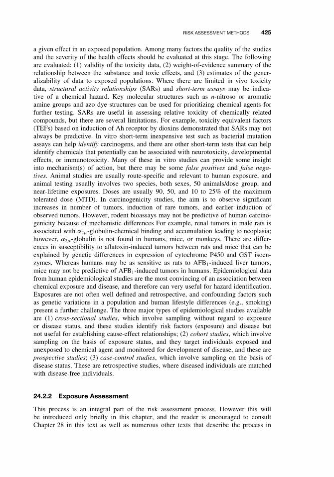
RISK ASSESSMENT METHODS 425
a given effect in an exposed population. Among many factors the quality of the studiesand the severity of the health effects should be evaluated at this stage. The followingare evaluated: (1) validity of the toxicity data, (2) weight-of-evidence summary of therelationship between the substance and toxic effects, and (3) estimates of the gener-alizability of data to exposed populations. Where there are limited in vivo toxicitydata, structural activity relationships (SARs) and short-term assays may be indica-tive of a chemical hazard. Key molecular structures such as n-nitroso or aromaticamine groups and azo dye structures can be used for prioritizing chemical agents forfurther testing. SARs are useful in assessing relative toxicity of chemically relatedcompounds, but there are several limitations. For example, toxicity equivalent factors(TEFs) based on induction of Ah receptor by dioxins demonstrated that SARs may notalways be predictive. In vitro short-term inexpensive test such as bacterial mutationassays can help identify carcinogens, and there are other short-term tests that can helpidentify chemicals that potentially can be associated with neurotoxicity, developmentaleffects, or immunotoxicity. Many of these in vitro studies can provide some insightinto mechanism(s) of action, but there may be some false positives and false nega-tives. Animal studies are usually route-specific and relevant to human exposure, andanimal testing usually involves two species, both sexes, 50 animals/dose group, andnear-lifetime exposures. Doses are usually 90, 50, and 10 to 25% of the maximumtolerated dose (MTD). In carcinogenicity studies, the aim is to observe significantincreases in number of tumors, induction of rare tumors, and earlier induction ofobserved tumors. However, rodent bioassays may not be predictive of human carcino-genicity because of mechanistic differences For example, renal tumors in male rats isassociated with α2µ-globulin-chemical binding and accumulation leading to neoplasia;however, α2µ-globulin is not found in humans, mice, or monkeys. There are differ-ences in susceptibility to aflatoxin-induced tumors between rats and mice that can beexplained by genetic differences in expression of cytochrome P450 and GST isoen-zymes. Whereas humans may be as sensitive as rats to AFB1-induced liver tumors,mice may not be predictive of AFB1-induced tumors in humans. Epidemiological datafrom human epidemiological studies are the most convincing of an association betweenchemical exposure and disease, and therefore can very useful for hazard identification.Exposures are not often well defined and retrospective, and confounding factors suchas genetic variations in a population and human lifestyle differences (e.g., smoking)present a further challenge. The three major types of epidemiological studies availableare (1) cross-sectional studies, which involve sampling without regard to exposureor disease status, and these studies identify risk factors (exposure) and disease butnot useful for establishing cause-effect relationships; (2) cohort studies, which involvesampling on the basis of exposure status, and they target individuals exposed andunexposed to chemical agent and monitored for development of disease, and these areprospective studies; (3) case-control studies, which involve sampling on the basis ofdisease status. These are retrospective studies, where diseased individuals are matchedwith disease-free individuals.
24.2.2 Exposure Assessment
This process is an integral part of the risk assessment process. However this willbe introduced only briefly in this chapter, and the reader is encouraged to consultChapter 28 in this text as well as numerous other texts that describe the process in
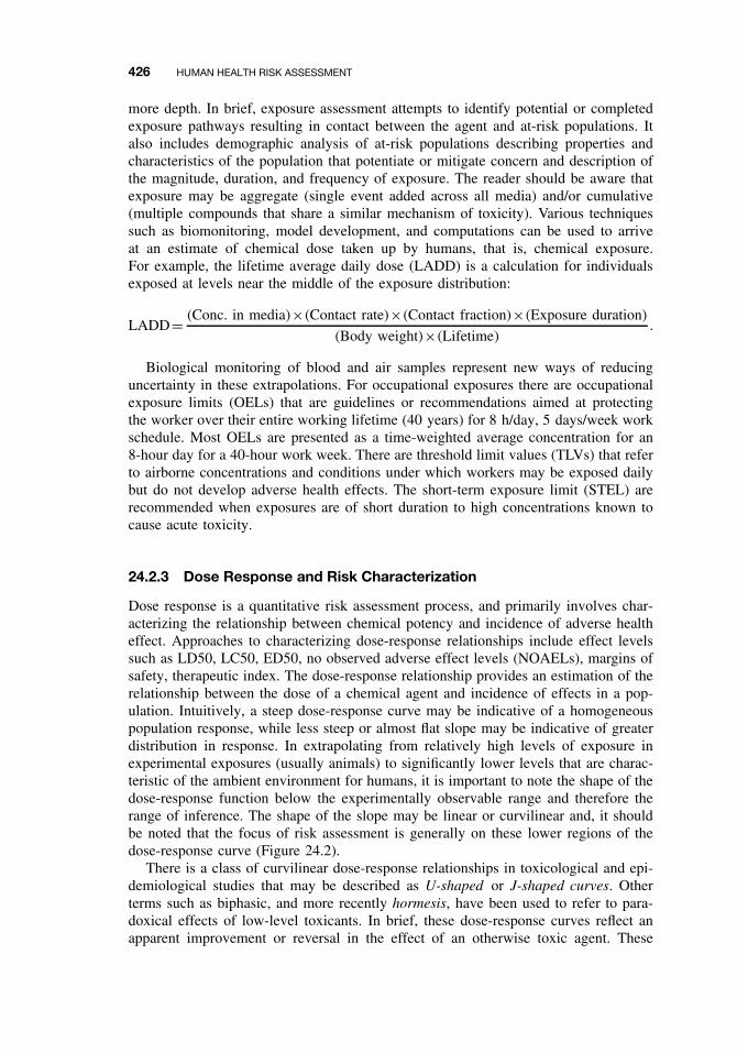
426 HUMAN HEALTH RISK ASSESSMENT
more depth. In brief, exposure assessment attempts to identify potential or completedexposure pathways resulting in contact between the agent and at-risk populations. Italso includes demographic analysis of at-risk populations describing properties andcharacteristics of the population that potentiate or mitigate concern and description ofthe magnitude, duration, and frequency of exposure. The reader should be aware thatexposure may be aggregate (single event added across all media) and/or cumulative(multiple compounds that share a similar mechanism of toxicity). Various techniquessuch as biomonitoring, model development, and computations can be used to arriveat an estimate of chemical dose taken up by humans, that is, chemical exposure.For example, the lifetime average daily dose (LADD) is a calculation for individualsexposed at levels near the middle of the exposure distribution:
LADD= (Conc. in media)×(Contact rate)×(Contact fraction)×(Exposure duration)
(Body weight)×(Lifetime).
Biological monitoring of blood and air samples represent new ways of reducinguncertainty in these extrapolations. For occupational exposures there are occupationalexposure limits (OELs) that are guidelines or recommendations aimed at protectingthe worker over their entire working lifetime (40 years) for 8 h/day, 5 days/week workschedule. Most OELs are presented as a time-weighted average concentration for an8-hour day for a 40-hour work week. There are threshold limit values (TLVs) that referto airborne concentrations and conditions under which workers may be exposed dailybut do not develop adverse health effects. The short-term exposure limit (STEL) arerecommended when exposures are of short duration to high concentrations known tocause acute toxicity.
24.2.3 Dose Response and Risk Characterization
Dose response is a quantitative risk assessment process, and primarily involves char-acterizing the relationship between chemical potency and incidence of adverse healtheffect. Approaches to characterizing dose-response relationships include effect levelssuch as LD50, LC50, ED50, no observed adverse effect levels (NOAELs), margins ofsafety, therapeutic index. The dose-response relationship provides an estimation of therelationship between the dose of a chemical agent and incidence of effects in a pop-ulation. Intuitively, a steep dose-response curve may be indicative of a homogeneouspopulation response, while less steep or almost flat slope may be indicative of greaterdistribution in response. In extrapolating from relatively high levels of exposure inexperimental exposures (usually animals) to significantly lower levels that are charac-teristic of the ambient environment for humans, it is important to note the shape of thedose-response function below the experimentally observable range and therefore therange of inference. The shape of the slope may be linear or curvilinear and, it shouldbe noted that the focus of risk assessment is generally on these lower regions of thedose-response curve (Figure 24.2).
There is a class of curvilinear dose-response relationships in toxicological and epi-demiological studies that may be described as U-shaped or J-shaped curves. Otherterms such as biphasic, and more recently hormesis, have been used to refer to para-doxical effects of low-level toxicants. In brief, these dose-response curves reflect anapparent improvement or reversal in the effect of an otherwise toxic agent. These
NONCANCER RISK ASSESSMENT 427
Response
Range ofInference
ExperimentalObservable Range
Figure 24.2 Dose-response curve, with emphasis on the shape of the dose-response functionbelow the experimentally observable range and therefore the range of inference where peopleare realistically exposed.
U-shaped effects can be explained in terms of homeostatic adjustments or overcor-rections in the operation of feedback mechanisms. Examples of studies with datafitting a U-shaped curve include the hormetic effect of organic lead on body growthin rats (Cragg and Rees, 1984) and peripheral nerve conduction velocity in children atlow doses (Ewert et al., 1986). Similar relationships have been observed with alcoholand nicotine in humans. It has been proposed that because thresholds are inherent inU-shaped dose-response curves, the linear no-threshold extrapolation method is notan appropriate approach for regulating hormetic agents. The current risk assessmentparadigm used by US EPA and other federal agencies does not conflict with the conceptof hormesis, but it has been proposed that the risk assessor’s analyzes make an activeconsideration of the data and the application of that data in the low dose portion ofthe dose-response curve for hormetic agents.
24.3 NONCANCER RISK ASSESSMENT
The noncancer risk assessment process assumes a threshold. For many noncarcinogeniceffects, protective mechanisms are believed to exist that must be overcome beforean adverse effect is manifested. At the cellular level for some toxicant, a range ofexposures exists from zero to some finite value that can be tolerated by the organismwith essentially no chance of expression of adverse effects. The aim here in riskassessment is to identify the upper bound of this tolerance range (i.e., the maximumsubthreshold level). This approach involves obtaining the no observed adverse effectlevel. NOAEL is the highest dose level that does not produce a significant elevatedincrease in an adverse response. Significance refers to biological and statistical criteriaand is dependent on dose levels tested, number of animals, background incidence in theunexposed control groups. Sometimes there is insufficient data to arrive at a NOAEL,and a LOAEL (lowest observed adverse effect level) is derived. The NOAEL is the keydatum obtained from the study of the dose-response relationship. The NOAEL is used tocalculate reference doses (RfD) for chronic oral exposures and reference concentrations(RfC) for chronic inhalation exposures as per EPA. Other agencies, such as the ATSDRand WHO, use the NOAEL to calculate minimum risk levels (MRLs) and acceptabledaily intakes (ADI). The US EPA describes the RfD as an estimate, with uncertaintyspanning an order of magnitude, of a daily exposure to the human population, including
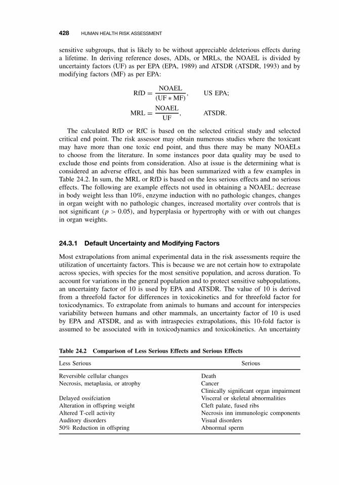
428 HUMAN HEALTH RISK ASSESSMENT
sensitive subgroups, that is likely to be without appreciable deleterious effects duringa lifetime. In deriving reference doses, ADIs, or MRLs, the NOAEL is divided byuncertainty factors (UF) as per EPA (EPA, 1989) and ATSDR (ATSDR, 1993) and bymodifying factors (MF) as per EPA:
RfD = NOAEL
(UF ∗ MF), US EPA;
MRL = NOAEL
UF, ATSDR.
The calculated RfD or RfC is based on the selected critical study and selectedcritical end point. The risk assessor may obtain numerous studies where the toxicantmay have more than one toxic end point, and thus there may be many NOAELsto choose from the literature. In some instances poor data quality may be used toexclude those end points from consideration. Also at issue is the determining what isconsidered an adverse effect, and this has been summarized with a few examples inTable 24.2. In sum, the MRL or RfD is based on the less serious effects and no seriouseffects. The following are example effects not used in obtaining a NOAEL: decreasein body weight less than 10%, enzyme induction with no pathologic changes, changesin organ weight with no pathologic changes, increased mortality over controls that isnot significant (p > 0.05), and hyperplasia or hypertrophy with or with out changesin organ weights.
24.3.1 Default Uncertainty and Modifying Factors
Most extrapolations from animal experimental data in the risk assessments require theutilization of uncertainty factors. This is because we are not certain how to extrapolateacross species, with species for the most sensitive population, and across duration. Toaccount for variations in the general population and to protect sensitive subpopulations,an uncertainty factor of 10 is used by EPA and ATSDR. The value of 10 is derivedfrom a threefold factor for differences in toxicokinetics and for threefold factor fortoxicodynamics. To extrapolate from animals to humans and account for interspeciesvariability between humans and other mammals, an uncertainty factor of 10 is usedby EPA and ATSDR, and as with intraspecies extrapolations, this 10-fold factor isassumed to be associated with in toxicodynamics and toxicokinetics. An uncertainty
Table 24.2 Comparison of Less Serious Effects and Serious Effects
Less Serious Serious
Reversible cellular changes DeathNecrosis, metaplasia, or atrophy Cancer
Clinically significant organ impairmentDelayed ossifciation Visceral or skeletal abnormalitiesAlteration in offspring weight Cleft palate, fused ribsAltered T-cell activity Necrosis inn immunologic componentsAuditory disorders Visual disorders50% Reduction in offspring Abnormal sperm
NONCANCER RISK ASSESSMENT 429
factor of 10 is used when a NOAEL derived from a subchronic study instead of achronic study is used as the basis for a calculation of a chronic RfD (EPA only). Notethat ATSDR does not perform this extrapolation but derive chronic and subchronicMRLs. An uncertainty factor of 10 is used in deriving an RfD or MRL from a LOAELwhen a NOAEL is not available. It should be noted that there are no reference dosesfor dermal exposure, however when there is insufficient dermal absorption data, theEPA uses a default factor of 10% to estimate bioavailability for dermal absorption. Amodifying factor ranging from 1 to 10 is included by EPA only to reflect a qualitativeprofessional assessment of additional uncertainties in the critical study and in the entiredata base for the chemical not explicitly addressed by preceding uncertainty factors.
Refinements of the RfC have utilized mechanistic data to modify the interspeciesuncertainty factor of 10 (Jarabek, 1995). The reader should appreciate that with theinhalation route of exposure, dosimetric adjustments are necessary and can affect theextrapolations of toxicity data of inhaled agents for human health risk assessment. TheEPA has included dosimetry modeling in RfC calculations, and the resulting dosimetricadjustment factor (DAF) used in determining the RfC is dependent on physiochemicalproperties of the inhaled toxicant as well as type of dosimetry model ranging fromrudimentary to optimal model structures. In essence, the use of the DAF can reducethe default uncertainty factor for interspecies extrapolation from 10 to 3.16.
The 1996 Food Quality Protection Act (FQPA) now requires that an additionalsafety factor of 10 be used in the risk assessment of pesticides to ensure the safetyof infants and children, unless the EPA can show that an adequate margin of safety isassured with out it (Scheuplein, 2000). The rational behind this additional safety factoris that infants and children have different dietary consumption patterns than adults andinfants, and children are more susceptible to toxicants than adults. We do know frompharmacokinetics studies with various human pharmaceuticals that drug elimination isslower in infants up to 6 months of age than in adults, and therefore the potential existsfor greater tissue concentrations and vulnerability for neonatal and postnatal effects.Based on these observations, the US EPA supports a default safety factor greater orless than 10, which may be used on the basis of reliable data. However, there arefew scientific data from humans or animals that permit comparisons of sensitivitiesof children and adults, but there are some examples, such as lead, where childrenare the more sensitive population. It some cases qualitative differences in age-relatedsusceptibility are small beyond 6 months of age, and quantitative differences in toxicitybetween children and adults can sometimes be less than a factor of 2 or 3.
Much of the research efforts in risk assessment are therefore aimed at reducing theneed to use these default uncertainty factors, although the risk assessor is limited by dataquality of the chemical of interest. With sufficient data and the advent of sophisticatedand validated physiologically based pharmacokinetic models and biologically baseddose-response models (Conolly and Butterworth, 1995), these default values can bereplaced with science-based factors. In some instances there may be sufficient data tobe able to obtain distributions rather than point estimates.
24.3.2 Derivation of Developmental Toxicant RfD
Developmental toxicity includes any detrimental effect produced by exposures duringembryonic development, and the effect may be temporary or overt physical malfor-mation. Adverse effects include death, structural abnormalities, altered growth, and
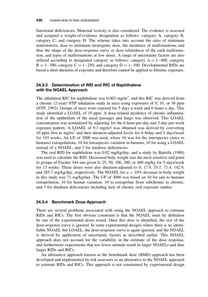
430 HUMAN HEALTH RISK ASSESSMENT
functional deficiencies. Maternal toxicity is also considered. The evidence is assessedand assigned a weight-of-evidence designation as follows: category A, category B,category C, and category D. The scheme takes into account the ratio of minimummaternotoxic dose to minimum teratogenic dose, the incidence of malformations andthus the shape of the dose-response curve or dose relatedness of the each malforma-tion, and types of malformations at low doses. A range of uncertainty factors are alsoutilized according to designated category as follows: category A = 1–400, categoryB = 1–300, category C = 1–250, and category D = 1–100. Developmental RfDs arebased a short duration of exposure and therefore cannot be applied to lifetime exposure.
24.3.3 Determination of RfD and RfC of Naphthalenewith the NOAEL Approach
The inhalation RfC for naphthalene was 0.003 mg/m3, and this RfC was derived froma chronic (2-year) NTP inhalation study in mice using exposures of 0, 10, or 30 ppm(NTP, 1992). Groups of mice were exposed for 5 days a week and 6 hours a day. Thisstudy identified a LOAEL of 10 ppm. A dose-related incidence of chronic inflamma-tion of the epithelium of the nasal passages and lungs was observed. This LOAELconcentration was normalized by adjusting for the 6-hour-per-day and 5-day-per-weekexposure pattern. A LOAEL of 9.3 mg/m3 was obtained was derived by converting10 ppm first to mg/m3 and then duration-adjusted levels for 6 h/day and 5 days/weekfor 103 weeks. An UF of 3000 was used, where 10 was for the interspecies (mice tohumans) extrapolations, 10 for intraspecies variation in humans, 10 for using a LOAELinstead of a NOAEL, and 3 for database deficiencies.
The oral RfD for naphthalene was 0.02 mg/kg/day, and a study by Battelle (1980)was used to calculate the RfD. Decreased body weight was the most sensitive end pointin groups of Fischer 344 rats given 0, 25, 50, 100, 200, or 400 mg/kg for 5 days/weekfor 13 weeks. These doses were also duration-adjusted to 0, 17.9, 35.7, 71.4, 142.9,and 285.7 mg/kg/day, respectively. The NOAEL for a > 10% decrease in body weightin this study was 71 mg/kg/day. The UF of 3000 was based on 10 for rats to humansextrapolation, 10 for human variation, 10 to extrapolate from subchronic to chronic,and 3 for database deficiencies including lack of chronic oral exposure studies.
24.3.4 Benchmark Dose Approach
There are several problems associated with using the NOAEL approach to estimateRfDs and RfCs. The first obvious constraint is that the NOAEL must by definitionbe one of the experimental doses tested. Once this dose is identified, the rest of thedose-response curve is ignored. In some experimental designs where there is no identi-fiable NOAEL but LOAEL, the dose-response curve is again ignored, and the NOAELis derived by application of uncertainty factors as described earlier. This NOAELapproach does not account for the variability in the estimate of the dose response,and furthermore experiments that test fewer animals result in larger NOAELs and thuslarger RfDs and RfCs.
An alternative approach known as the benchmark dose (BMD) approach has beendeveloped and implemented by risk assessors as an alternative to the NOAEL approachto estimate RfDs and RfCs. This approach is not constrained by experimental design
NONCANCER RISK ASSESSMENT 431
as the NOAEL approach, and it incorporates information on the sample size and shapeof the dose-response curve. In fact this approach can be used for both threshold andnonthreshold adverse effects as well as continuous and quantal data sets. This requiresuse of Benchmark Dose Software where the dose-response is modeled and the lowerconfidence bound for a dose at a specified response level (benchmark response) iscalculated. The benchmark response is usually specified as a 1–10% response; that is,it corresponds to a dose associated with a low level of risk such as 1–10%.
Figure 24.3 shows how an effective dose that corresponds to a specific change ofeffect/response (e.g., 10%) over background and a 95% lower confidence bound on thedose is calculated. The latter is often referred to as the BMDL or LBMD, as opposedto the BMD, which does not have this confidence limited associated with it.
Because the benchmark represents a statistical lower limit, larger experiments willtend, on average, to give larger benchmarks, thus rewarding good experimentation.This is not the case with NOAELs, as there is an inverse relationship between NOAELand size of experiments. For example, poorer experiments possessing less sensitivityfor detecting statistically significant increases in risk inappropriately result in higherNOAELs and RfDs, which may have an unknown unacceptable level of risk. In essence,the NOAEL is very sensitive to sample size, and there can also be high variabilitybetween experiments. With the benchmark dose approach, all the doses and slopes ofthe curve influence the calculations, variability of the data is considered, and the BMDis less variable between experiments. In the BMD approach quantitative toxicologicaldata such as continuous data (organ weights serum levels, etc.) and quantal or incidencedata (pathology findings, genetic anomalies, etc.) are fitted to numerous dose-responsemodels described in the literature. The resulting benchmark dose that, for example,corresponds to a tumor risk of 10% generally can be estimated with adequate precisionand not particularly dependent on the dose-response model used to fit the data. Note thatdose intervals are not required for BMD estimation. This will be greatly appreciatedin the cancer risk assessment section of this chapter.
24.3.5 Determination of BMD and BMDL for ETU
The BMD method has been quite extensively in assessing quantal data, and veryoften this has involved analysis of data from developmental and reproductive toxicity
0.1
Excess fraction ofabnormal responses
Upper Confidencelimit on estimated risk
Dose response fittedto experimental data
BMDL BMD Dose
Figure 24.3 Benchmark dose determination from dose response relationship with the BMDLcorresponding to the lower end of a one-sided 95% confidence interval for the BMD.
432 HUMAN HEALTH RISK ASSESSMENT
studies. In this study example (Crump, 1984), rats were exposed to ethylenethiourea(ETU) at 0, 5, 10, 20, 40, and 80 mg/kg doses, and the number affected with fetalanomalies per number of rats were 0/167, 0/132, 1/138, 14/81, 142/178, and 24/24,respectively. The benchmark dose computation can involve utilization of any givendose-response probability model, but in this example the quantal Weibull model wasused and the specified effect was set at 0.01 (1%) with confidence level of 0.95. TheBMD was determined to be 8.9 mg/kg, and the BMDL was 6.9 mg/kg. This valueis close to the NOAEL, which is 5 mg/kg, but it does demonstrate that the NOAELapproximates a lower confidence limit on the BMD corresponding to an excess riskof about 1% for proportions of fetal anomalies. In fact an empirical analysis of some486 developmental toxicity studies has demonstrated that the NOAEL can result in anexcess risk of 5% for proportions of dead or malformed fetuses per litter. The readershould at this stage recognize that the BMD approach can also be used in cancer riskassessment as we are often times working with quantal data that are ideally suited forBMD modeling.
24.3.6 Quantifying Risk for Noncarcinogenic Effects: Hazard Quotient
The measure used to describe the potential for noncarcinogenic toxicity to occur isnot expressed as the probability. Probabilistic approach is used in cancer RA. Fornoncancer RA, the potential for noncarcinogenic effects is evaluated by comparing anexposure level (E) over a specified time period with a reference dose (RfD). This ratiois called a hazard quotient:
Hazard quotient = E
RfD.
In general, the greater the value of E/RfD exceeds unity, the greater is the level ofconcern. Note that this is a ratio and not to be interpreted as a statistical probability.
24.3.7 Chemical Mixtures
Human populations are more likely to be exposed simultaneously or sequentially to amixture of chemicals rather than to one single chemical. Standard default approachesto mixture risk assessment consider doses and responses of the mixture components tobe additive. However, it should also be recognized that components in the mixture canalso result in synergistic, antagonistic, or no toxicological effect following exposureto a chemical mixture. Therefore mixture toxicity cannot always be predicted even ifwe know the mechanisms of all toxic components in a defined mixture. Furthermoretissue dosimetry can be complicated by interactions at the route of entry (e.g., GIT,skin surface) and clearance mechanisms in the body. In essence, there are considerableuncertainties involved in trying to extrapolate effects following exposure to chemicalmixtures. Several PBPK models have been used to quantitate these effects and alsoprovide some information useful for risk assessment of chemical mixtures (Krishnanet al., 1994; Haddad et al. 2001).
The 1996 FQPA has also mandated that the EPA should also consider implementingcumulative risk assessments for pesticides. Cumulative risk assessments usually involve
CANCER RISK ASSESSMENT 433
integration of the hazard and cumulative exposure analysis, and it primarily involvescumulative nonoccupational exposure by multiple routes or pathways to two or morepesticides or chemicals sharing a common mechanism of toxicity.
Calculation procedures differ for carcinogenic and noncarcinogenic effects, but bothsets of procedures assume dose additivity in the absence of information on mixtures:
Cancer risk equation for mixtures : RiskT = �RiskI,
Noncancer hazard index = E1
RfD1+ E2
RfD2+ · · · + Ei
RfDi.
This hazard index (HI) approach as well as other indexes (e.g., relative potency fac-tors) are applied for mixture components that induce the same toxic effect by identicalmechanism of action. In cases where there are different mechanisms, separate HIvalues can be calculated for each end point of concern. As the equation above indi-cates, the HI is easy to calculate, as there is simply scaling of individual componentexposure concentrations by a measure of relative potency such as the RfD or RfC,and adding scaled concentrations to get an indicator of risk from exposure to themixture of concern. However, as noted above, this additivity approach does not takeinto account tissue dosimetry and pharmacokinetic interactions. Recent published riskassessments have utilized mixture PBPK models to account for multiple pharmacoki-netic interactions among mixture constituents. These interaction-based PBPK modelscan quantify change in tissue dose metrics of chemicals during exposure to mixturesand thus improve the mechanistic basis of mixture risk assessment. Finally the readershould be aware that this HI is different from the a term known as the margin of safety(MOS), which is the ratio of the critical or chronic NOAEL for a specific toxicolog-ical end point to an estimate of human exposure. MOS values greater than 100 aregenerally considered protective if the NOAEL is derived from animal data.
24.4 CANCER RISK ASSESSMENT
For cancer risk assessment an assumption is held that a threshold for an adverse effectdoes not exist with most individual chemicals. It is assumed that a small number ofmolecular events can evoke changes in a single cell that can lead to uncontrolledcellular proliferation and eventually to a clinical state of disease. This mechanism isreferred to as “nonthreshold” because there is believed to be essentially no level ofexposure to such a chemical that does not pose a finite probability, however small,of generating a carcinogenic response. That is, no dose is though to be risk free.Therefore, in evaluations of cancer risks, an effect threshold cannot be estimated. Forcarcinogenic effects, the US EPA uses a two-part evaluation: (1) the substance is firstassigned a weight-of-evidence classification and then (2) a slope factor is calculated.
1. Assigning a weight-of-evidence. The aim here is to determine the likelihood thatthe agent is a human carcinogen. The evidence is characterized separately for humanstudies and animal studies as sufficient, limited, inadequate, no data, or evidence ofno effect. Based on this characterization and on the extent to which the chemical hasbeen shown to be a carcinogen in animals or humans or both, the chemical is given aprovisional weight-of-evidence classification. The US EPA classification system (EPA,
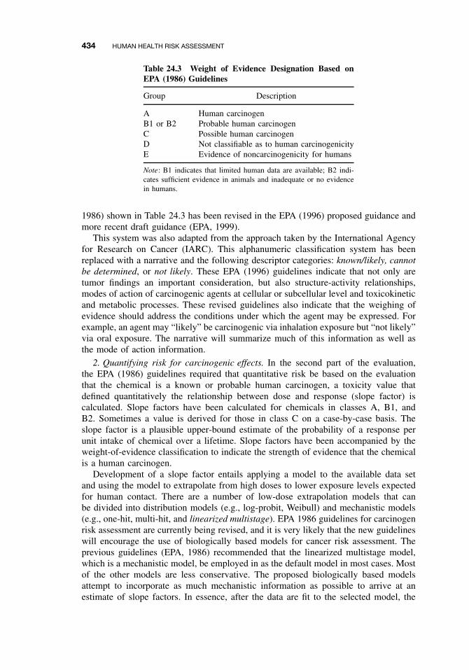
434 HUMAN HEALTH RISK ASSESSMENT
Table 24.3 Weight of Evidence Designation Based onEPA (1986) Guidelines
Group Description
A Human carcinogenB1 or B2 Probable human carcinogenC Possible human carcinogenD Not classifiable as to human carcinogenicityE Evidence of noncarcinogenicity for humans
Note: B1 indicates that limited human data are available; B2 indi-cates sufficient evidence in animals and inadequate or no evidencein humans.
1986) shown in Table 24.3 has been revised in the EPA (1996) proposed guidance andmore recent draft guidance (EPA, 1999).
This system was also adapted from the approach taken by the International Agencyfor Research on Cancer (IARC). This alphanumeric classification system has beenreplaced with a narrative and the following descriptor categories: known/likely, cannotbe determined, or not likely. These EPA (1996) guidelines indicate that not only aretumor findings an important consideration, but also structure-activity relationships,modes of action of carcinogenic agents at cellular or subcellular level and toxicokineticand metabolic processes. These revised guidelines also indicate that the weighing ofevidence should address the conditions under which the agent may be expressed. Forexample, an agent may “likely” be carcinogenic via inhalation exposure but “not likely”via oral exposure. The narrative will summarize much of this information as well asthe mode of action information.
2. Quantifying risk for carcinogenic effects. In the second part of the evaluation,the EPA (1986) guidelines required that quantitative risk be based on the evaluationthat the chemical is a known or probable human carcinogen, a toxicity value thatdefined quantitatively the relationship between dose and response (slope factor) iscalculated. Slope factors have been calculated for chemicals in classes A, B1, andB2. Sometimes a value is derived for those in class C on a case-by-case basis. Theslope factor is a plausible upper-bound estimate of the probability of a response perunit intake of chemical over a lifetime. Slope factors have been accompanied by theweight-of-evidence classification to indicate the strength of evidence that the chemicalis a human carcinogen.
Development of a slope factor entails applying a model to the available data setand using the model to extrapolate from high doses to lower exposure levels expectedfor human contact. There are a number of low-dose extrapolation models that canbe divided into distribution models (e.g., log-probit, Weibull) and mechanistic models(e.g., one-hit, multi-hit, and linearized multistage). EPA 1986 guidelines for carcinogenrisk assessment are currently being revised, and it is very likely that the new guidelineswill encourage the use of biologically based models for cancer risk assessment. Theprevious guidelines (EPA, 1986) recommended that the linearized multistage model,which is a mechanistic model, be employed in as the default model in most cases. Mostof the other models are less conservative. The proposed biologically based modelsattempt to incorporate as much mechanistic information as possible to arrive at anestimate of slope factors. In essence, after the data are fit to the selected model, the
CANCER RISK ASSESSMENT 435
upper 95th percent confidence limit of the slope of the resulting dose response curve iscalculated. This represents the probability of a response per unit intake over a lifetime,or that there is a 5% chance that the probability of a response could be greater than theestimated value on the basis of experimental data and model used. In some cases, theslope factors based on human dose-response data are based on “best” estimate insteadof upper 95th percent confidence limit. The toxicity values for carcinogenic effects canbe expressed in several ways.
The slope factor is expressed as q1∗:
Slope factor = Risk per unit dose
= Risk per mg/kg-day.
The slope factor can therefore be used to calculate the upper bound estimate onrisk (R)
Risk = q1∗[risk × (mg/kg/day)−1] × exposure (mg/kg/day).
Here risk is a unitless probability (e.g., 2 × 10⁻⁵) of an individual developing cancerand exposure is really chronic daily intake averaged over 70 years: mg/kg/day. Thiscan be determined if we can determine the slope factor and human exposure at thewaste site or occupational site. The EPA usually sets a goal of limiting lifetime cancerrisks in the range of 10−6 to 10−4 for chemical exposures, while the FDA typicallyaims for risks below 10−6 for general population exposure. It is therefore quite likelyfor very high exposures for the accepted EPA range of risk to be exceeded. The EPArange is considered protective of the general and sensitive human population. It shouldbe noted that these orders of magnitude are substantially greater than those used inestimating RfD and RfCs in noncancer risk assessment.
Because relatively low intakes (compared to those experienced by test animals)are most likely from environmental exposure at Superfund hazardous waste sites, itgenerally can be assumed that the dose-response relationship will be linear on the low-dose portion of the multistage model dose-response curve. The equation above canapply to these linear low-dose situations. This linear equation is valid only at low risklevels (i.e., below the estimated risk of 0.01). For risk above 0.01 the one-hit equationshould be used:
Risk = 1 − exp(-exposure × slope factor).
As indicated above, biologically based extrapolation models are the preferredapproach for quantifying risk to carcinogens, although it is possible that all thenecessary data will not be available for many chemicals. The EPA (1986) guidelineshave been modified to include the response data on effects of the agent on carcinogenicprocesses in addition to data on tumor incidence. Precursor effects and tumor incidencedata may be combined to extend the dose response curve below the tumor data;that is, below the range of observation. Thus a biologically based or case-specificdose-response model is developed when there is sufficient data, or a standard defaultprocedure is used when there is insufficient data to adequately curve-fit the data.In brief, the dose-response assessment is considered in two parts or steps, range ofobservation and range of extrapolation, and the overriding preferred approach is touse the biologically based or case-specific model for both of these ranges. In the first
436 HUMAN HEALTH RISK ASSESSMENT
step of this process, the lower 95% confidence limit on a dose associated with anestimated 10% increase in tumor or nontumor response (LED10) is identified. Whenhuman real world exposures are outside the range of the observed or experimental data,this serves as the point of departure or marks the beginning for the extrapolating tothese low environmental exposure levels. Note that these procedures are very similarto the benchmark procedure for quantitating risk to noncarcinogenic chemicals. Inthe second step, the biologically based or case-specific model is preferred for use inextrapolations to lower dose levels provided that there are sufficient data. If the latteris not the case, then default approaches consistent with agent chemical mode of actionare implemented with the assumption of linearity or nonlinearity of the dose-responserelationship. The linear default approach is a departure from the 1986 guidelines, whichused the linearlized multistage (LMS) procedure, but is based on mode of action oralternatively if there is insufficient data to support a nonlinear mode of action. In brief,it involves drawing a straight line from the point of departure (LED10) to the origin(i.e., zero). When there is no evidence of linearity or there is a nonlinear mode of action,the default approach is the margin of exposure (MOE) analysis. The MOE approachcomputes the ratio between the LED10 and the environmental exposure, and the analysisbegins from the point of departure that is adjusted for toxicokinetic differences betweenspecies to give a human equivalent dose.
Finally it should be noted that prior to the FQPA in 1996, the Delaney clause pro-hibited the establishment of tolerances or maximum allowable levels for food additivesif it has been shown to induce cancer in human or animal. This is an important changein regulations because pesticide residues were considered as food additives. Becauseof the FQPA, pesticide residues are no longer regarded as food additives, and there isno prohibition against setting tolerances for carcinogens.
24.5 PBPK MODELING
Physiologically based pharmacokinetic (PBPK) modeling has been used in risk assess-ment to make more scientifically based extrapolations, and at the same time to helpexplore and reduce inherent uncertainties. Historically pharmacokinetics has relied onempirical models, and in many instances this process offers little insight into mech-anisms of absorption, distribution, and clearance of hazardous agents and does notfacilitate translation from animal experiments to human exposures. For example, dosescaling using by body weight or size may often time overestimate or underestimatetoxicant levels at the target tissue. PBPK models can help predict tissue concentrationsin different species under various conditions based on independent anatomical, physi-ological, and biochemical parameters. In these analyzes physiological parameters suchas organ volumes, tissue-blood partition coefficients, and blood flow to specific tissuecompartments described by the model, are calculated or obtained from the literatureand integrated into the model. Monte Carlo analysis, a form of uncertainty analysis,can now be performed, and this allows for the propagation of uncertainty through amodel that results in estimation of the variance of model output. This can be achievedby randomly sampling model parameters from defined distributions; some parameterssuch as cardiac output, metabolic, and log P parameters, may have a lognormal dis-tribution, while other parameters may be normal or uniform. In essence, the MonteCarlo analysis when coupled with PBPK characterizes the distribution of potential risk
SUGGESTED READING 437
in a population by using a range of potential values for each input parameter (notsingle values) as well as an estimate of how these values are distributed (Clewell andAndersen, 1996). By these approaches, uncertainty is identifiable and quantifiable, andcan reduce inappropriate levels of concern in reporting the risk of chemical exposure.These mathematical modeling approaches also help identify areas of potential scientificresearch that could improve the human health assessment.
In recent years there have been significant efforts at harmonizating noncancer andcancer risk assessments (Barton et al., 1998; Clewell et al., 2002), and in this respectPKPD modeling can be a very useful tool in the risk assessment process. For example,recall that noncancer risk assessment addresses variability in a population by dividingthe NOAEL by 10, whereas the cancer risk assessment does not address this quantita-tively. PBPK modeling coupled with Monte Carlo analysis is one approach as describedin the previous paragraph that will help address this level of uncertainty in the riskassessment. In conclusion, it should be noted that PBPK modeling has been utilizedwith very few toxicants. It is hoped that risk assessment policy will encourage theuse of this tool as well as other appropriate models to integrate mechanistic informa-tion and the pharmacokinetics (dosimetry), and pharmacodynamics (dose response) oftoxicants. Improved quantitative risk assessments will ultimately provide scientificallysound information that will influence the risk management decision process.
SUGGESTED READING
ATSDR. Guidance for the Preparation of Toxicological Profiles. ATSDR, US Public HealthService. 1993.
Battelle. Subchronic toxicity study: Naphthalene (C52904), Fischer 344 rats. Report to USDepartment of Health and Human Services, National Toxicology Program, Research TrianglePark, NC, by Battelle Columbus Laboratories, Columbus, OH, 1980.
Borton, H. A., M. E. Anderson, and H. J. Clewell. Harmonization: Developing consistentguidelines for applying mode of action and dosimetry information for applying mode ofaction and dosimetry information to cancer and noncancer risk assessment. Hum. Ecol. RiskAssess. 4: 75–115, 1998.
Clewell, H. J., and M. E. Andersen. Use of physiologically based pharmacokinetic modeling toinvestigate individual versus population risk. Toxicol. 111(1–3): 315–329, 1996.
Clewell, H. J., M. E. Andersen, and H. A. Barton. A consistent approach for the application ofpharmacokinetic modeling in cancer and noncancer risk assessment. Envion. Health. Perspect.110: 85–93, 2002.
Conolly, R. B., and B. E. Butterworth. Biologically based dose response model for hepatic tox-icity: a mechanistically based replacment for traditional estimates of noncancer risk. Toxicol.Lett. 82–83: 901–906, 1995.
Conolly, R. B., B. D. Beck, and J. I. Goodman. Stimulating research to improve the scientificbasis of risk assessment. Toxicol. Sci. 49: 1–4, 1999.
Crump, K. S. A new method for determining allowable daily intakes. Fundam. Appl. Toxicol.4: 854–871, 1984.
EPA. Guidelines for Carcinogen Risk Assessment. Fed. Reg. 51: 33992–34003, 1986.
EPA. Proposed Guidelines for Carcinogen Risk Assessment. Fed. Reg. 61: 17960–18011, 1996.
EPA. Guidelines for Carcinogen Risk Assessment. Risk Assessment Forum, US EnvironmentalProtection Agency, NCEA-F-0644, July 1999, Review Draft.
438 HUMAN HEALTH RISK ASSESSMENT
EPA. Risk Assessment Guidance for Superfund, Vol. 1. Human Health Evaluation Manual(Part A). US EPA. 1989.
Haddad, S., M. Beliveau, R. Tardif, and K. Krishnan. A PBPK modeling-based approach toaccount for interactions in the health risk assessment of chemical mixtures. Toxicol. Sci. 63:125–131, 2001.
Jarabek, A. M., The application of dosimetry models to identify key processes and parametersfor default dose-response assessment approaches. Toxicol. Lett. 79(1–3): 171–184, 1995.
Krishnan, K., H. J. Clewell, and M. E. Andersen Physiologically based pharmacokinetic ana-lyzes of simple mixtures. Environ. Health Perspec. 102 (supp. 9): 151–155, 1994.
NTP. National Toxicology Program. Technical report series No. 410. Toxicology and carcinogen-esis studies of naphthalene (CAS No. 91-20-3) in B6C3F1 mice (inhalation studies). ResearchTriangle Park, NC. US Department of Health and Human Services, Public Health Services,National Institutes of Health. NIH Publication No. 92–3141, 1992.
Page, N. P., D. V Singh, W. Farland, J. I. Goodman, R. B. Conolly, M. E Andersen,H. J. Clewell, C. B. Frederick, H. Yamasaki, G. Lucier. Implementation of EPA Revised Can-cer Assessment Guidelines: Incorporation of Mechanistic and Pharmacokinetic Data. Fundam.Appl. Toxicol. 37: 16–36, 1997.
Scheuplein, R. J. Pesticides and infant risk: Is there a need for an additional safety margin?Regul. Toxicol. Pharmacol. 31(3): 267–279, 2000.

PART VII
ENVIRONMENTAL TOXICOLOGY

CHAPTER 25
Analytical Methods in Toxicology
ROSS B. LEIDY
25.1 INTRODUCTION
Some 200,000 chemicals are synthesized annually worldwide, and the toxicity of mostof them is unknown. Few of these chemicals reach the stage of further developmentand use, but those that do usually find their way into the environment. Some arepersistent and remain adsorbed to soil particles or soil organic matter, some find theirway into water through soil movement or aerial deposition, others are metabolized bymicroorganisms into compounds of greater toxicity that move up the food chain. Overtime, their accumulation in higher life forms could result in debilitating alterations inmetabolism, leading to illness. It might be years before such illness could be attributedto specific compounds because of the difficulty involved in identifying and quantitatingthem. The concern over the role of persistent organochlorines in the food chain and theirpossible role as human xenoestrogens is an example. The identification and quantitationof chemicals in both the environment and in living beings relies on the developmentof analytical techniques and instruments.
Advances in analytical techniques continue to multiply in all fields of toxicol-ogy, and as mentioned, many of these focus on the environmental area. Whetherlooking for new techniques to sample water or for an automated instrument to deter-mine quantities of sulfur-containing compounds in air, such devices are available. Inmany instances, developments in environmental analyses are adaptable to experimen-tal work related to drug toxicity, or in forensic medicine, to determine the cause ofpoisoning.
Although new techniques and instruments continue to enter the commercial market,the basic analytical process has not changed: define the research goal(s), develop asampling scheme to obtain representative samples, isolate the compound(s) of inter-est, remove potential interfering components, and quantitate and evaluate the data inrelation to the initial hypothesis. Based on the data generated, many options are avail-able. For example, was the sampling scheme complete? Would further refinement ofthe analytical procedure be required? Should other sample types be analyzed? Thus itis obvious that within these general categories particular methods vary considerablydepending on the chemical characteristics of the toxicant (Table 25.1).
A Textbook of Modern Toxicology, Third Edition, edited by Ernest HodgsonISBN 0-471-26508-X Copyright 2004 John Wiley & Sons, Inc.
441
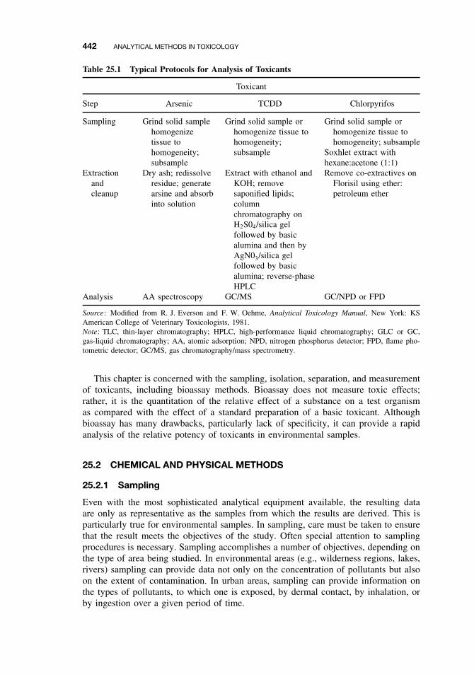
442 ANALYTICAL METHODS IN TOXICOLOGY
Table 25.1 Typical Protocols for Analysis of Toxicants
Toxicant
Step Arsenic TCDD Chlorpyrifos
Sampling Grind solid samplehomogenizetissue tohomogeneity;subsample
Grind solid sample orhomogenize tissue tohomogeneity;subsample
Grind solid sample orhomogenize tissue tohomogeneity; subsample
Soxhlet extract withhexane:acetone (1:1)
Extractionandcleanup
Dry ash; redissolveresidue; generatearsine and absorbinto solution
Extract with ethanol andKOH; removesaponified lipids;columnchromatography onH2S04/silica gelfollowed by basicalumina and then byAgN03/silica gelfollowed by basicalumina; reverse-phaseHPLC
Remove co-extractives onFlorisil using ether:petroleum ether
Analysis AA spectroscopy GC/MS GC/NPD or FPD
Source: Modified from R. J. Everson and F. W. Oehme, Analytical Toxicology Manual, New York: KSAmerican College of Veterinary Toxicologists, 1981.Note: TLC, thin-layer chromatography; HPLC, high-performance liquid chromatography; GLC or GC,gas-liquid chromatography; AA, atomic adsorption; NPD, nitrogen phosphorus detector; FPD, flame pho-tometric detector; GC/MS, gas chromatography/mass spectrometry.
This chapter is concerned with the sampling, isolation, separation, and measurementof toxicants, including bioassay methods. Bioassay does not measure toxic effects;rather, it is the quantitation of the relative effect of a substance on a test organismas compared with the effect of a standard preparation of a basic toxicant. Althoughbioassay has many drawbacks, particularly lack of specificity, it can provide a rapidanalysis of the relative potency of toxicants in environmental samples.
25.2 CHEMICAL AND PHYSICAL METHODS
25.2.1 Sampling
Even with the most sophisticated analytical equipment available, the resulting dataare only as representative as the samples from which the results are derived. This isparticularly true for environmental samples. In sampling, care must be taken to ensurethat the result meets the objectives of the study. Often special attention to samplingprocedures is necessary. Sampling accomplishes a number of objectives, depending onthe type of area being studied. In environmental areas (e.g., wilderness regions, lakes,rivers) sampling can provide data not only on the concentration of pollutants but alsoon the extent of contamination. In urban areas, sampling can provide information onthe types of pollutants, to which one is exposed, by dermal contact, by inhalation, orby ingestion over a given period of time.
CHEMICAL AND PHYSICAL METHODS 443
In industrial areas, hazardous conditions can be detected and sources of pollutioncan be identified. Sampling is used in the process of designing pollution controlsand can provide a chronicle of the changes in operational conditions as controls areimplemented. Another important application of sampling in industrial areas in theUnited States is the documentation of compliance with existing Occupational Safetyand Health Administration (OSHA) and US Environmental Protection Agency (USEPA) regulations. The many methods available for sampling the environment can bedivided into categories of air, soil, water, and tissue sampling. The fourth category isof particular interest in experimental and forensic studies.
Air. Most pollutants entering the atmosphere come from fuel combustion, industrialprocesses, and solid waste disposal. Additional miscellaneous sources, such as nuclearexplosions, forest fires, dusts, volcanoes, natural gaseous emissions, agricultural burn-ing, and pesticide drift, contribute to the level of atmospheric pollution. To affectterrestrial animals and plants, particulate pollutants must be in a size range that allowsthem to enter the body and remain there; that is, they must be in an aerosol (definedas an airborne suspension of liquid droplets) or on solid particles small enough topossess a low settling velocity. Suspensions can be classified as liquids including fogs(small particles) and mists (large particles) produced from atomization, condensation, orentrapment of liquids by gases; and solids including dusts, fumes, and smoke producedby crushing, metal vaporization, and combustion of organic materials, respectively.
At rest, an adult human inhales 6 to 8 L of air each minute (1 L = O.OO1 m3)and, during an 8-hour workday, can inhale from 5 to 20 m3 depending on the levelof physical activity. The optimum size range for aerosol particles to get into the lungsand remain there is 0.5 to 5.0 µm. As instrumentation used to collect atmospheric dusthave become more precise, particulate matter (PM) in the size range of 2.5 to 10 µmhave come under increasing scrutiny, because many potential toxicants are adsorbedto their surfaces. These particles are inhaled and will remain in the lungs and allowthe compounds to pass into the bloodstream.
Thus air samplers have been miniaturized and adsorbents have been developed tocollect either particulate matter in the size range most detrimental to humans or to“trap” organic toxicants from air. An air sampler generally consists of an inlet to directair through a filter to entrap particles that might be of interest (e.g., dust); through theadsorbent, which collects organic vapors, a flowmeter and valve to calibrate airflow, anda pump to pull air through the system. Personnel samplers are run by battery powerand can be attached to an individual’s clothing, thus allowing continual monitoringwhile performing assigned tasks in the work environment. This allows the estimationof individual exposure.
Many air samplers use various types of filters to collect solid particulate matter,such as asbestos, which is collected on glass fiber filters with pores 20 µm or less indiameter. Membrane filters with pores 0.01 to 10 µm in diameter are used to collectdusts and silica. Liquid-containing collectors, called impingers, are used to trap mineraldusts and pesticides. Mineral dusts are collected in large impingers that have flow ratesof 10 to 50 L of air per minute, and insecticides can be collected in smaller “midget”impingers that handle flows of 2 to 4.5 L of air per minute. Depending on the pollutantbeing sought, the entrapping liquid might be distilled water, alcohol, ethylene glycol,hexylene glycol (2-methyl, 2,4-pentane diol) or some other solvent. Because of theease of handling and the rapid desorption of compounds, polyurethane foam (PUF)
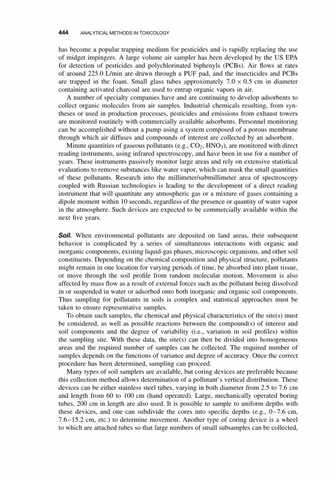
444 ANALYTICAL METHODS IN TOXICOLOGY
has become a popular trapping medium for pesticides and is rapidly replacing the useof midget impingers. A large volume air sampler has been developed by the US EPAfor detection of pesticides and polychlorinated biphenyls (PCBs). Air flows at ratesof around 225.0 L/min are drawn through a PUF pad, and the insecticides and PCBsare trapped in the foam. Small glass tubes approximately 7.0 × 0.5 cm in diametercontaining activated charcoal are used to entrap organic vapors in air.
A number of specialty companies have and are continuing to develop adsorbents tocollect organic molecules from air samples. Industrial chemicals resulting, from syn-theses or used in production processes, pesticides and emissions from exhaust towersare monitored routinely with commercially available adsorbents. Personnel monitoringcan be accomplished without a pump using a system composed of a porous membranethrough which air diffuses and compounds of interest are collected by an adsorbent.
Minute quantities of gaseous pollutants (e.g., CO2, HNO3), are monitored with directreading instruments, using infrared spectroscopy, and have been in use for a number ofyears. These instruments passively monitor large areas and rely on extensive statisticalevaluations to remove substances like water vapor, which can mask the small quantitiesof these pollutants. Research into the millimeter/submillimeter area of spectroscopycoupled with Russian technologies is leading to the development of a direct readinginstrument that will quantitate any atmospheric gas or a mixture of gases containing adipole moment within 10 seconds, regardless of the presence or quantity of water vaporin the atmosphere. Such devices are expected to be commercially available within thenext five years.
Soil. When environmental pollutants are deposited on land areas, their subsequentbehavior is complicated by a series of simultaneous interactions with organic andinorganic components, existing liquid-gas phases, microscopic organisms, and other soilconstituents. Depending on the chemical composition and physical structure, pollutantsmight remain in one location for varying periods of time, be absorbed into plant tissue,or move through the soil profile from random molecular motion. Movement is alsoaffected by mass flow as a result of external forces such as the pollutant being dissolvedin or suspended in water or adsorbed onto both inorganic and organic soil components.Thus sampling for pollutants in soils is complex and statistical approaches must betaken to ensure representative samples.
To obtain such samples, the chemical and physical characteristics of the site(s) mustbe considered, as well as possible reactions between the compound(s) of interest andsoil components and the degree of variability (i.e., variation in soil profiles) withinthe sampling site. With these data, the site(s) can then be divided into homogeneousareas and the required number of samples can be collected. The required number ofsamples depends on the functions of variance and degree of accuracy. Once the correctprocedure has been determined, sampling can proceed.
Many types of soil samplers are available, but coring devices are preferable becausethis collection method allows determination of a pollutant’s vertical distribution. Thesedevices can be either stainless steel tubes, varying in both diameter from 2.5 to 7.6 cmand length from 60 to 100 cm (hand operated). Large, mechanically operated boringtubes, 200 cm in length are also used. It is possible to sample to uniform depths withthese devices, and one can subdivide the cores into specific depths (e.g., 0–7.6 cm,7.6–15.2 cm, etc.) to determine movement. Another type of coring device is a wheelto which are attached tubes so that large numbers of small subsamples can be collected,
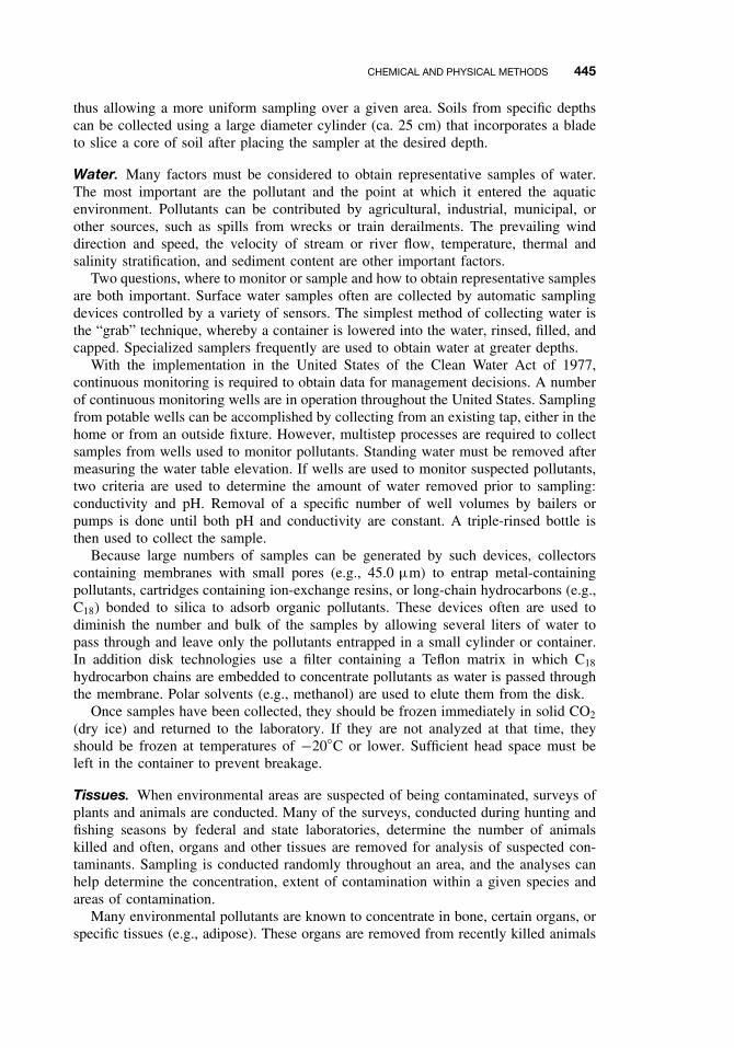
CHEMICAL AND PHYSICAL METHODS 445
thus allowing a more uniform sampling over a given area. Soils from specific depthscan be collected using a large diameter cylinder (ca. 25 cm) that incorporates a bladeto slice a core of soil after placing the sampler at the desired depth.
Water. Many factors must be considered to obtain representative samples of water.The most important are the pollutant and the point at which it entered the aquaticenvironment. Pollutants can be contributed by agricultural, industrial, municipal, orother sources, such as spills from wrecks or train derailments. The prevailing winddirection and speed, the velocity of stream or river flow, temperature, thermal andsalinity stratification, and sediment content are other important factors.
Two questions, where to monitor or sample and how to obtain representative samplesare both important. Surface water samples often are collected by automatic samplingdevices controlled by a variety of sensors. The simplest method of collecting water isthe “grab” technique, whereby a container is lowered into the water, rinsed, filled, andcapped. Specialized samplers frequently are used to obtain water at greater depths.
With the implementation in the United States of the Clean Water Act of 1977,continuous monitoring is required to obtain data for management decisions. A numberof continuous monitoring wells are in operation throughout the United States. Samplingfrom potable wells can be accomplished by collecting from an existing tap, either in thehome or from an outside fixture. However, multistep processes are required to collectsamples from wells used to monitor pollutants. Standing water must be removed aftermeasuring the water table elevation. If wells are used to monitor suspected pollutants,two criteria are used to determine the amount of water removed prior to sampling:conductivity and pH. Removal of a specific number of well volumes by bailers orpumps is done until both pH and conductivity are constant. A triple-rinsed bottle isthen used to collect the sample.
Because large numbers of samples can be generated by such devices, collectorscontaining membranes with small pores (e.g., 45.0 µm) to entrap metal-containingpollutants, cartridges containing ion-exchange resins, or long-chain hydrocarbons (e.g.,C18) bonded to silica to adsorb organic pollutants. These devices often are used todiminish the number and bulk of the samples by allowing several liters of water topass through and leave only the pollutants entrapped in a small cylinder or container.In addition disk technologies use a filter containing a Teflon matrix in which C18
hydrocarbon chains are embedded to concentrate pollutants as water is passed throughthe membrane. Polar solvents (e.g., methanol) are used to elute them from the disk.
Once samples have been collected, they should be frozen immediately in solid CO2
(dry ice) and returned to the laboratory. If they are not analyzed at that time, theyshould be frozen at temperatures of −20◦C or lower. Sufficient head space must beleft in the container to prevent breakage.
Tissues. When environmental areas are suspected of being contaminated, surveys ofplants and animals are conducted. Many of the surveys, conducted during hunting andfishing seasons by federal and state laboratories, determine the number of animalskilled and often, organs and other tissues are removed for analysis of suspected con-taminants. Sampling is conducted randomly throughout an area, and the analyses canhelp determine the concentration, extent of contamination within a given species andareas of contamination.
Many environmental pollutants are known to concentrate in bone, certain organs, orspecific tissues (e.g., adipose). These organs are removed from recently killed animals
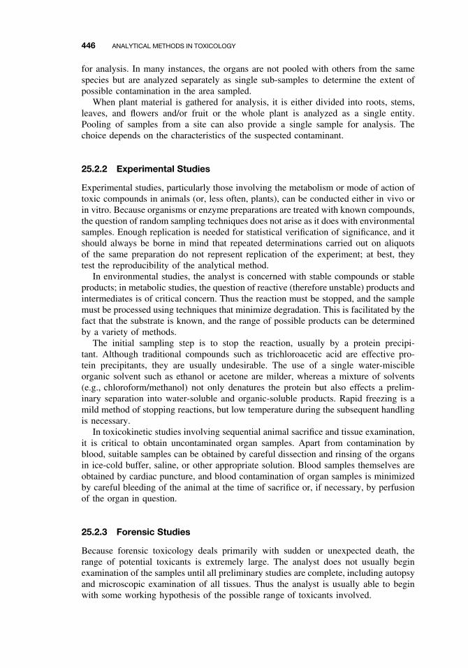
446 ANALYTICAL METHODS IN TOXICOLOGY
for analysis. In many instances, the organs are not pooled with others from the samespecies but are analyzed separately as single sub-samples to determine the extent ofpossible contamination in the area sampled.
When plant material is gathered for analysis, it is either divided into roots, stems,leaves, and flowers and/or fruit or the whole plant is analyzed as a single entity.Pooling of samples from a site can also provide a single sample for analysis. Thechoice depends on the characteristics of the suspected contaminant.
25.2.2 Experimental Studies
Experimental studies, particularly those involving the metabolism or mode of action oftoxic compounds in animals (or, less often, plants), can be conducted either in vivo orin vitro. Because organisms or enzyme preparations are treated with known compounds,the question of random sampling techniques does not arise as it does with environmentalsamples. Enough replication is needed for statistical verification of significance, and itshould always be borne in mind that repeated determinations carried out on aliquotsof the same preparation do not represent replication of the experiment; at best, theytest the reproducibility of the analytical method.
In environmental studies, the analyst is concerned with stable compounds or stableproducts; in metabolic studies, the question of reactive (therefore unstable) products andintermediates is of critical concern. Thus the reaction must be stopped, and the samplemust be processed using techniques that minimize degradation. This is facilitated by thefact that the substrate is known, and the range of possible products can be determinedby a variety of methods.
The initial sampling step is to stop the reaction, usually by a protein precipi-tant. Although traditional compounds such as trichloroacetic acid are effective pro-tein precipitants, they are usually undesirable. The use of a single water-miscibleorganic solvent such as ethanol or acetone are milder, whereas a mixture of solvents(e.g., chloroform/methanol) not only denatures the protein but also effects a prelim-inary separation into water-soluble and organic-soluble products. Rapid freezing is amild method of stopping reactions, but low temperature during the subsequent handlingis necessary.
In toxicokinetic studies involving sequential animal sacrifice and tissue examination,it is critical to obtain uncontaminated organ samples. Apart from contamination byblood, suitable samples can be obtained by careful dissection and rinsing of the organsin ice-cold buffer, saline, or other appropriate solution. Blood samples themselves areobtained by cardiac puncture, and blood contamination of organ samples is minimizedby careful bleeding of the animal at the time of sacrifice or, if necessary, by perfusionof the organ in question.
25.2.3 Forensic Studies
Because forensic toxicology deals primarily with sudden or unexpected death, therange of potential toxicants is extremely large. The analyst does not usually beginexamination of the samples until all preliminary studies are complete, including autopsyand microscopic examination of all tissues. Thus the analyst is usually able to beginwith some working hypothesis of the possible range of toxicants involved.
CHEMICAL AND PHYSICAL METHODS 447
Because further sampling usually involves exhumation and is therefore unlikely or,in the case of cremation, impossible, adequate sampling and sample preservation isessential. For example, various body fluids must be collected in a proper way: bloodby cardiac puncture, never from the body cavity; urine from the urinary bladder; bilecollected intact as part of the ligated gallbladder; and so on. Adequate sample size isimportant. Blood can be analyzed for carbon monoxide, ethanol, and other alcohols,barbiturates, tranquilizers, and other drugs; at least 100 mL should be collected. Urineis useful for analysis of both endogenous and exogenous chemicals and the entirecontent of the bladder is retained. The liver frequently contains high levels of toxicantsand/or their metabolites, and it and the kidney are the most important solid tissues forforensic analysis; 100 to 200 g of the former and the equivalent of one kidney usuallyare retained. DNA analysis has made tremendous strides through the use of polymerasechain reaction (PCR) that allows old samples (e.g., exhumation and sampling of bonemarrow) to be analyzed and compared to living relatives; thus these data providevaluable information to law authorities and others.
An unusual requirement with important legal ramifications is that of possession.An unbroken chain of identifiable possession (i.e., chain of custody) must be main-tained. All transfers are marked on the samples as to time and date of collection,arrival at the laboratory, and all transfers must be signed by both parties. The secu-rity and handling of samples during time of possession must be verifiable as a matterof law.
25.2.4 Sample Preparation
Extraction. In most cases the analysis of a pollutant or other toxicant depends on itsphysical removal from the sample medium. In order to ensure that the sample usedis homogeneous, it is chopped, ground or blended to a uniform consistency and thensubsampled. This subsample is extracted, which involves bringing a suitable solventinto intimate contact with the sample, generally in a ratio of 5 to 25 volumes ofsolvent to l volume of sample. One or more of four different procedures can be used,depending on the chemical and physical characteristics of the toxicant and the samplematrix. Other extraction methods such as boiling, grinding, or distilling the samplewith appropriate solvents are used less frequently.
Blending. The use of an electric or air-driven blender is currently the most commonmethod of extraction of biologic materials. The weighed sample is placed in a container,solvent is added, and the tissue is homogenized by motor-driven blades. Blending for 5to 15 minutes followed by a repeat blending will extract most environmental toxicants.A homogenate in an organic solvent can be filtered through anhydrous sodium sulfateto remove water that might cause problems in the quantitation phase of the analysis.The use of sonication is a popular method for extracting tissue samples, particularlywhen the binding of toxicants to subcellular fractions is of interest. Sonicator probesrupture cells rapidly, thus allowing the solvent to come into intimate contact with allcell components. Differential centrifugation can then be used to isolate fractions ofinterest. Large wattage (e.g., 450 watt) sonifiers are used to extract compounds fromenvironmental samples, and several US EPA methods list sonification as a valid methodof extraction.
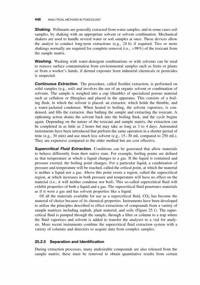
448 ANALYTICAL METHODS IN TOXICOLOGY
Shaking. Pollutants are generally extracted from water samples, and in some cases soilsamples, by shaking with an appropriate solvent or solvent combination. Mechanicalshakers are used to handle several water or soil samples at once. These devices allowthe analyst to conduct long-term extractions (e.g., 24 h) if required. Two or moreshakings normally are required for complete removal (i.e., >98%) of the toxicant fromthe sample matrix.
Washing. Washing with water-detergent combinations or with solvents can be usedto remove surface contamination from environmental samples such as fruits or plantsor from a worker’s hands, if dermal exposure from industrial chemicals or pesticidesis suspected.
Continuous Extraction. The procedure, called Soxhlet extraction, is performed onsolid samples (e.g., soil) and involves the use of an organic solvent or combination ofsolvents. The sample is weighed into a cup (thimble) of specialized porous materialsuch as cellulose or fiberglass and placed in the apparatus. This consists of a boil-ing flask, in which the solvent is placed: an extractor, which holds the thimble, anda water-jacketed condenser. When heated to boiling, the solvent vaporizes, is con-densed, and fills the extractor, thus bathing the sample and extracting the toxicant. Asiphoning action drains the solvent back into the boiling flask, and the cycle beginsagain. Depending on the nature of the toxicant and sample matrix, the extraction canbe completed in as little as 2 hours but may take as long as 3 to 4 days. Automatedinstruments have been introduced that perform the same operation in a shorter period oftime (e.g., 30 min) and use much less solvent (e.g., 15–30 mL compared to 250 mL).They are expensive compared to the older method but are cost effective.
Supercritical Fluid Extraction. Conditions can be generated that allow materialsto behave differently from their native state. For example, boiling points are definedas that temperature at which a liquid changes to a gas. If the liquid is contained andpressure exerted, the boiling point changes. For a particular liquid, a combination ofpressure and temperature will be reached, called the critical point, at which the materialis neither a liquid nor a gas. Above this point exists a region, called the supercriticalregion, at which increases in both pressure and temperature will have no effect on thematerial (i.e., it will neither condense nor boil). This so-called supercritical fluid willexhibit properties of both a liquid and a gas. The supercritical fluid penetrates materialsas if it were a gas and has solvent properties like a liquid.
Of all the materials available for use as a supercritical fluid, CO2 has become thematerial of choice because of its chemical properties. Instruments have been developedto utilize the principles described to effect extractions of compounds from a variety ofsample matrices including asphalt, plant material, and soils (Figure 25.1). The super-critical fluid is pumped through the sample, through a filter or column to a trap wherethe fluid vaporizes and solvent is added to transfer the analyses to a vial for analy-sis. More recent instruments combine the supercritical fluid extraction system with avariety of columns and detectors to acquire data from complex samples.
25.2.5 Separation and Identification
During extraction processes, many undesirable compounds are also released from thesample matrix; these must be removed to obtain quantitative results from certain
CHEMICAL AND PHYSICAL METHODS 449
Data System
CO2 Pump ExtractionChamber
Filteror
Column
SUPER CRITICAL FLUID EXTRACTION
TrapSample
Vial
Figure 25.1 Supercritical fluid extraction.
instruments. These components include plant and animal pigments, lipids, organicmaterial from soil and water, and inorganic compounds. If not removed, the impu-rities decrease the sensitivity of the detectors and columns in the analytical instrument,mask peaks, or produce extraneous peaks on chromatograms. Although some morerecently developed instruments automatically remove these substances and concen-trate the samples to small volumes for quantitative analysis, they are expensive. Thusmost laboratories rely on other methods. These include adsorption chromatography,thin-layer chromatography (TLC), and solvent partitioning. Generally, adsorption chro-matography is the method of choice to remove co-extractives from the compoundin question.
Because most techniques use large volumes of solvent, the solvent must be removedto obtain a working volume (e.g., 5–10 mL) that is easy to manipulate by the analyst.This is accomplished by distillation, evaporation under a stream of air or an inert gassuch as nitrogen, or evaporation under reduced pressure. Once the working volumeis reached, extracts can be further purified by one or more procedures. In addition tothe use of adsorbents, many organic toxicants will distribute between two immisciblesolvents (e.g., chloroform and water or hexane and acetonitrile). When shaken in aseparatory funnel and then allowed to equilibrate into two original solvent layers,some of the toxicant will have transferred from the original extracting solvent into theother layer. With repeated additions (e.g., 4 to 5 volumes), mixing, and removal, mostor all of the compound of interest will have been transferred, leaving many interferingcompounds in the original solvent. Regardless of the separation method or combinationof methods used, the toxicant will be in a large volume of solvent in relation to itsamount that is removed as described. Final volumes used to identify and quantitatecompounds generally range from 250 µL to 10.0 mL.
Recent advances in circuit miniaturization and column technology, the developmentof microprocessors and new concepts in instrument design have allowed sensitive mea-surement at the parts per billion and parts per trillion levels for many toxicants. Thisincreased sensitivity has focused public attention on the extent of environmental pollu-tion, because many toxic materials present in minute quantities could not be detecteduntil technological advances reached the present state of the art. At present, most pol-lutants are identified and quantified by chromatography, spectroscopy, and bioassays.
Once the toxicant has been extracted and separated from extraneous materials, theactual identification procedure can begin, although it should be remembered that thepurification procedures are themselves often used in identification (e.g., peak position
450 ANALYTICAL METHODS IN TOXICOLOGY
in gas-liquid chromatography [GLC] and high-performance liquid chromatography[HPLC]). Thus no definite line can be drawn between the two procedures.
Chromatography. All chromatographic processes, such as TLC, GLC, HPLC, orcapillary electrophoresis (CE), use a mobile and immobile phase to effect a separationof components. In TLC, the immobile phase is a thin layer of adsorbent placed onglass, resistant plastic, or fiberglass, and the mobile phase is the solvent. The mobilephase can be a liquid or gas, whereas the immobile phase can be a liquid or solid.Chromatographic separations are based on the interactions of these phases or surfaces.All chromatographic procedures use the differential distribution or partitioning of oneor more components between the phases, based on the absorption, adsorption, ion-exchange, or size exclusion properties of one of the phases.
Paper Chromatography. When the introduction of paper chromatography to com-mon laboratory use occurred in the mid-1930s, it revolutionized experimental bio-chemistry and toxicology. This technique is still used in laboratories that lack theexpensive instruments necessary for GLC or HPLC. The stationary phase is repre-sented by the aqueous constituent of the solvent system, which is adsorbed onto thepaper; the moving phase is the organic constituents. Separation is effected by partitionbetween the two phases as the solvent system moves over the paper. Although manyvariations exist, including reverse-phase paper chromatography in which the paper istreated with a hydrophobic material, ion-exchange cellulose paper, and so on, all havebeen superseded by equivalent systems involving thin layers of adsorbents bonded toan inert backing.
Thin-Layer Chromatography. Many toxicants and their metabolites can be sep-arated from interfering substances with TLC. In this form of chromatography, theadsorbent is spread as a thin layer (250–2000 µm) on glass, resistant plastic or fiber-glass backings. When the extract is placed near the bottom of the plate and the plate isplaced in a tank containing a solvent system, the solvent migrates up the plate, and thetoxicant and other constituent move with the solvent; differential rates of movementresult in separation. The compounds can be scraped from the plate and eluted fromthe adsorbent with suitable solvents. Recent developments in TLC adsorbents allowtoxicants and other materials to be quantitated at the nanogram (10−9 g) and picogram(10−12 g) levels.
Column: Adsorption, Hydrophobic, Ion Exchange. A large number of adsorbentsare available to the analyst. The adsorbent can be activated charcoal, aluminum oxide,Florisil, silica, silicic acid, or mixed adsorbents. The characteristics of the toxicantdetermine the choice of adsorbent. When choosing an adsorbent, select conditions thateither bind the co-extractives to it, allowing the compound of interest to elute, andvice versa. The efficiency of separation depends on the flow rate of solvent throughthe column (cartridge) and the capacity of the adsorbent to handle the extract placed onit. This amount depends on the type and quantity of adsorbent, the capacity factor (k′)and concentration of sample components, and the type and strength of the solvents usedto elute the compound of interest. Many environmental samples contain a sufficientamount of interfering materials so that the analyst must prepare a column using aglass chromatography tube into which the adsorbent is added. In the most common
CHEMICAL AND PHYSICAL METHODS 451
sequence the column is packed in an organic solvent of low polarity, the sample isadded in the same solvent, and the column is then developed with a sequence ofsolvents or solvent mixtures of increasing polarity. Such a sequence might include(in order of increasing polarity) hexane, benzene, chloroform, acetone, and methanol.Once removed, the eluate containing the toxicant is reduced to a small volume forquantitation.
However, cartridge technologies are improving to allow similar concentrations ofsample to be added that result in a less expensive and more rapid analysis. A number ofminiaturized columns have been introduced since the early 1980s. Most contain 0.5 to2.0 g of the adsorbent in a plastic tube with fitted ends. The columns can be attachedto standard Luer Lock syringes. Other companies have designed vacuum manifoldsthat hold the collecting device. The column is placed on the apparatus, a vacuum isapplied, and the solvent is drawn through the column. Some advantages of these sys-tems include preweighed amounts of adsorbent for uniformity, easy disposal of theco-extractives remaining in the cartridge, no breakage and decreased cost of the analy-sis because less solvent and adsorbent are used. Other forms of column chromatographycan be used. They include ion-exchange chromatography, permeation chromatography,and affinity chromatography. Ion-exchange chromatography depends on the attractionbetween charged molecules and opposite charges on the ion exchanger, usually a resin.Compounds so bound are eluted by changes in pH and, because the net charge dependson the relationship between pH of the solution and the isoelectric point of the com-pounds, compounds of different isoelectric point can be eluted sequentially. Both ionicand anionic exchangers are available. Permeation chromatography utilizes the molec-ular sieve properties of porous materials. Molecules large enough to be excluded fromthe pores of the porous material will move through the column faster than will smallermolecules not excluded, thus separating them. Cross-linked dextrans such as Sephadexor agarose (Sepharose) are commonly used materials. Affinity chromatography is apotent tool for biologically active macromolecules but is seldom used for purifyingsmall molecules, such as most toxicants. It depends on the affinity of an enzyme fora substrate (or substrate analogue) that has been incorporated into a column matrix orthe affinity of a receptor for a ligand.
Gas-Liquid Chromatography (GLC). GLC is used most commonly for the sepa-ration and quantitation of organic toxicants. This system consists of an injector port,oven, detector, amplifier (electrometer), and supporting electronics (Figure 25.2). Cur-rent modern gas chromatographs use a capillary column to effect separation of complexmixtures of organic molecules and has replaced, to a large extent, the “packed” column.Instead of coating an inert support, the stationary phase is coated onto the inside ofthe column. The mobile phase is an inert gas (called the carrier gas), usually heliumor nitrogen that passes through the column.
When a sample is injected, the injector port is at a temperature sufficient to vaporizethe sample components. Based on the solubility and volatility of these componentswith respect to the stationary phase, the components separate and are swept throughthe column by the carrier gas to a detector, which responds to the concentration of eachcomponent. The detector might not respond to all components. The electronic signalproduced as the component passes through the detector is amplified by the electrometer,and the resulting signal is sent to a recorder, computer, or electronic data-collectingdevice for quantitation.
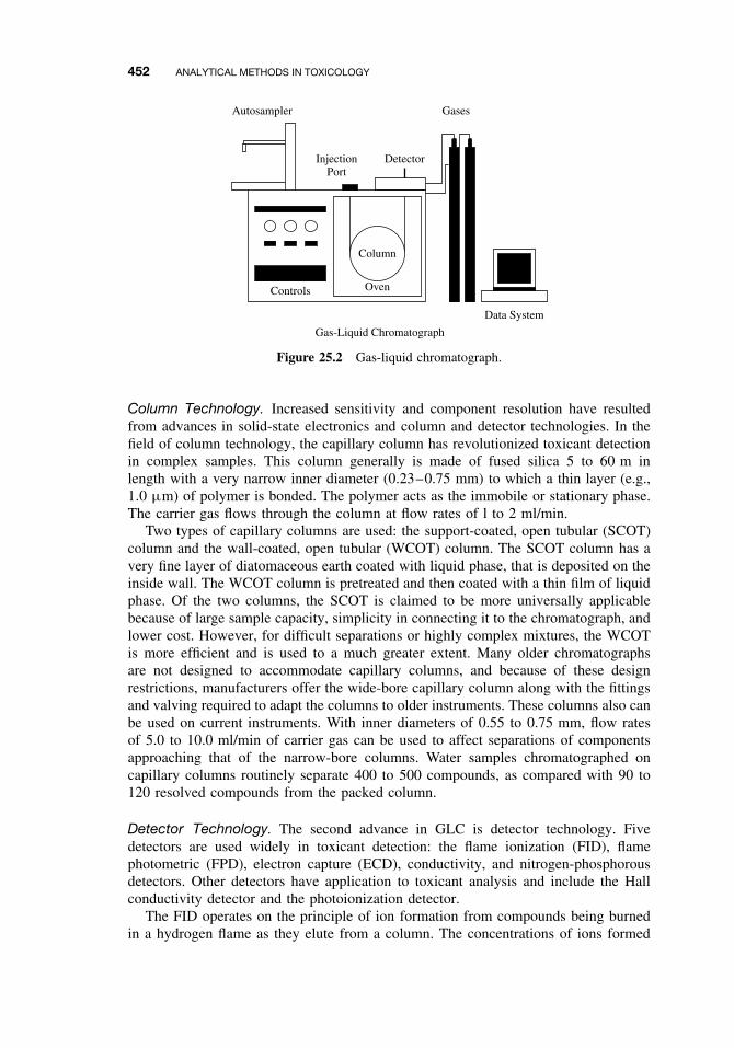
452 ANALYTICAL METHODS IN TOXICOLOGY
Column
Oven
Gas-Liquid Chromatograph
Controls
InjectionPort
Detector
GasesAutosampler
Data System
Figure 25.2 Gas-liquid chromatograph.
Column Technology. Increased sensitivity and component resolution have resultedfrom advances in solid-state electronics and column and detector technologies. In thefield of column technology, the capillary column has revolutionized toxicant detectionin complex samples. This column generally is made of fused silica 5 to 60 m inlength with a very narrow inner diameter (0.23–0.75 mm) to which a thin layer (e.g.,1.0 µm) of polymer is bonded. The polymer acts as the immobile or stationary phase.The carrier gas flows through the column at flow rates of l to 2 ml/min.
Two types of capillary columns are used: the support-coated, open tubular (SCOT)column and the wall-coated, open tubular (WCOT) column. The SCOT column has avery fine layer of diatomaceous earth coated with liquid phase, that is deposited on theinside wall. The WCOT column is pretreated and then coated with a thin film of liquidphase. Of the two columns, the SCOT is claimed to be more universally applicablebecause of large sample capacity, simplicity in connecting it to the chromatograph, andlower cost. However, for difficult separations or highly complex mixtures, the WCOTis more efficient and is used to a much greater extent. Many older chromatographsare not designed to accommodate capillary columns, and because of these designrestrictions, manufacturers offer the wide-bore capillary column along with the fittingsand valving required to adapt the columns to older instruments. These columns also canbe used on current instruments. With inner diameters of 0.55 to 0.75 mm, flow ratesof 5.0 to 10.0 ml/min of carrier gas can be used to affect separations of componentsapproaching that of the narrow-bore columns. Water samples chromatographed oncapillary columns routinely separate 400 to 500 compounds, as compared with 90 to120 resolved compounds from the packed column.
Detector Technology. The second advance in GLC is detector technology. Fivedetectors are used widely in toxicant detection: the flame ionization (FID), flamephotometric (FPD), electron capture (ECD), conductivity, and nitrogen-phosphorousdetectors. Other detectors have application to toxicant analysis and include the Hallconductivity detector and the photoionization detector.
The FID operates on the principle of ion formation from compounds being burnedin a hydrogen flame as they elute from a column. The concentrations of ions formed
CHEMICAL AND PHYSICAL METHODS 453
are several orders of magnitude greater than those formed in the uncontaminated flame.The ions cause a current to flow between two electrodes held at a constant potential,thus sending a signal to the electrometer.
The FPD is a specific detector in that it detects either phosphorous- or sulfur-containing compounds. When atoms of a given element are burned in a hydrogen-rich flame, the excitation energy supplied to these atoms produces a unique emissionspectrum. The intensity of the wavelengths of light emitted by these atoms is directlyproportional to the number of atoms excited. Larger concentrations cause a greaternumber of atoms to reach the excitation energy level, thus increasing the intensity of theemission spectrum. The change in intensity is detected by a photomultiplier, amplifiedby the electrometer, and recorded. Filters that allow only the emission wavelengthof phosphorous (526 nm) or sulfur (394 nm) are inserted between the flame and thephotomultiplier to give this detector its specificity.
The ECD is used to detect halogen-containing compounds, although it will producea response to any electronegative compound. When a negative DC voltage is appliedto a radioactive source (e.g., 63Ni, 3H), low-energy β particles are emitted, producingsecondary electrons by ionizing the carrier gas as it passes through the detector. Thesecondary electron stream flows from the source (cathode) to a collector (anode), wherethe amount of current generated (called a standing current) is amplified and recorded.As electronegative compounds pass from the column into the detector, electrons areremoved or “captured,” and the standing current is reduced. The reduction is relatedto both the concentration and electronegativity of the compound passing through, andthis produces a response that is recorded. The sensitivity of ECD is greater than thatof any other detectors currently available.
Early electrolytic conductivity detectors operated on the principle of componentcombustion, which produced simple molecular species that readily ionized, thus alteringthe conductivity of deionized water. The changes were monitored by a dc bridge circuitand recorded. By varying the conditions, the detector could be made selective fordifferent types of compounds (e.g., chlorine containing, nitrogen containing).
The alkali flame detector can also be made selective. Enhanced response to com-pounds containing arsenic, boron, halogen, nitrogen and phosphorous results whenthe collector (cathode) of an FID is coated with different alkali metal salts such asKBr, KCl, Na2SO4. As with conductivity detectors, by varying gas flow rates, typesof salt, and electrode configuration, enhanced responses are obtained. The nitrogen-phosphorous alkali detector is used widely for analysis of herbicides. Alkali salts areembedded in a silica gel matrix and are heated electrically. The detector allows rou-tine use of chlorinated solvents and derivatizing reagents that can be detrimental toother detectors.
The Hall electrolytic conductivity uses advanced designs in the conductivity cell, fur-nace, and an ac conductivity bridge to detect chlorine, nitrogen, and sulfur-containingcompounds at sensitivities of 0.01 ng. It operates on the conductivity principle describedpreviously. Another detector, the photoionization detector, uses an ultraviolet (UV)light source to ionize molecules by absorption of a photon of UV light. The ion formedhas an energy greater than the ionization potential of the parent compound, and theformed ions are collected by an electrode. The current, which is proportional to concen-tration, is amplified and recorded. The detector can measure a number of organic andinorganic compounds in air, biologic fluids, and water. A number of instrument manu-facturers have introduced portable GLCs that can be transported for use on field sites.
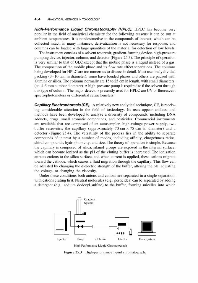
454 ANALYTICAL METHODS IN TOXICOLOGY
High-Performance Liquid Chromatography (HPLC). HPLC has become verypopular in the field of analytical chemistry for the following reasons: it can be run atambient temperatures; it is nondestructive to the compounds of interest, which can becollected intact; in many instances, derivatization is not necessary for response; andcolumns can be loaded with large quantities of the material for detection of low levels.
The instrument consists of a solvent reservoir, gradient-forming device. high-pressurepumping device, injector, column, and detector (Figure 25.3). The principle of operationis very similar to that of GLC except that the mobile phase is a liquid instead of a gas.The composition of the mobile phase and its flow rate effect separations. The columnsbeing developed for HPLC are too numerous to discuss in detail. Most use finely dividedpacking (3–10 µm in diameter), some have bonded phases and others are packed withalumina or silica. The columns normally are 15 to 25 cm in length, with small diameters.(ca. 4.6 mm number diameter). A high-pressure pump is required to fi the solvent throughthis type of column. The major detectors presently used for HPLC are UV or fluorescentspectrophotometers or differential refractometers.
Capillary Electrophoresis (CE). A relatively new analytical technique, CE, is receiv-ing considerable attention in the field of toxicology. Its uses appear endless, andmethods have been developed to analyze a diversity of compounds, including DNAadducts, drugs, small aromatic compounds, and pesticides. Commercial instrumentsare available that are composed of an autosampler, high-voltage power supply, twobuffer reservoirs, the capillary (approximately 70 cm × 75 µm in diameter) and adetector (Figure 25.4). The versatility of the process lies in the ability to separatecompounds of interest by a number of modes, including affinity, charge/mass ratios,chiral compounds, hydrophobicity, and size. The theory of operation is simple. Becausethe capillary is composed of silica, silanol groups are exposed in the internal surface,which can become ionized as the pH of the eluting buffer is increased. The ionizationattracts cations to the silica surface, and when current is applied, these cations migratetoward the cathode, which causes a fluid migration through the capillary. This flow canbe adjusted by changing the dielectric strength of the buffer, altering the pH, adjustingthe voltage, or changing the viscosity.
Under these conditions both anions and cations are separated in a single separation,with cations eluting first. Neutral molecules (e.g., pesticides) can be separated by addinga detergent (e.g., sodium dodecyl sulfate) to the buffer, forming micelles into which
Data SystemDetectorColumn
High Performance Liquid Chromatograph
GradientSystem
PumpInjector
Figure 25.3 High-performance liquid chromatograph.
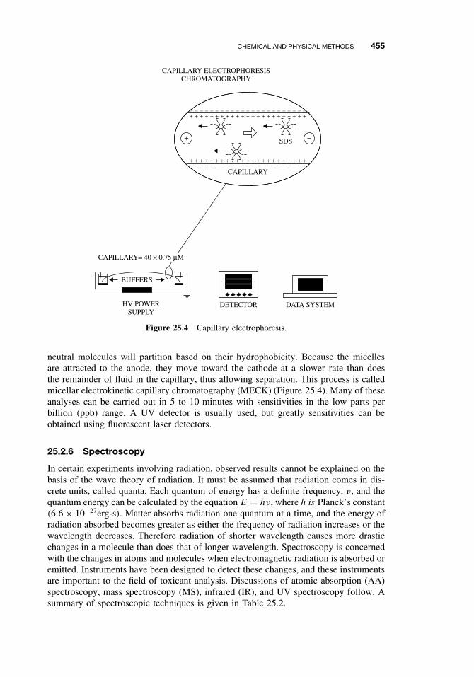
CHEMICAL AND PHYSICAL METHODS 455
− − − − − − − − − − − − − − − − − − − − − − −+ + + + + + + + + + + + + + + + + + + + + + +
− − − − − − − − − − − − − − − − − − − − − − −+ + + + + + + + + + + + + + + + + + + + + + +
+ −SDS
CAPILLARY
BUFFERS
HV POWERSUPPLY
DETECTOR DATA SYSTEM
CAPILLARY= 40 × 0.75 µM
CAPILLARY ELECTROPHORESISCHROMATOGRAPHY
Figure 25.4 Capillary electrophoresis.
neutral molecules will partition based on their hydrophobicity. Because the micellesare attracted to the anode, they move toward the cathode at a slower rate than doesthe remainder of fluid in the capillary, thus allowing separation. This process is calledmicellar electrokinetic capillary chromatography (MECK) (Figure 25.4). Many of theseanalyses can be carried out in 5 to 10 minutes with sensitivities in the low parts perbillion (ppb) range. A UV detector is usually used, but greatly sensitivities can beobtained using fluorescent laser detectors.
25.2.6 Spectroscopy
In certain experiments involving radiation, observed results cannot be explained on thebasis of the wave theory of radiation. It must be assumed that radiation comes in dis-crete units, called quanta. Each quantum of energy has a definite frequency, v, and thequantum energy can be calculated by the equation E = hv, where h is Planck’s constant(6.6 × 10⁻²⁷erg-s). Matter absorbs radiation one quantum at a time, and the energy ofradiation absorbed becomes greater as either the frequency of radiation increases or thewavelength decreases. Therefore radiation of shorter wavelength causes more drasticchanges in a molecule than does that of longer wavelength. Spectroscopy is concernedwith the changes in atoms and molecules when electromagnetic radiation is absorbed oremitted. Instruments have been designed to detect these changes, and these instrumentsare important to the field of toxicant analysis. Discussions of atomic absorption (AA)spectroscopy, mass spectroscopy (MS), infrared (IR), and UV spectroscopy follow. Asummary of spectroscopic techniques is given in Table 25.2.
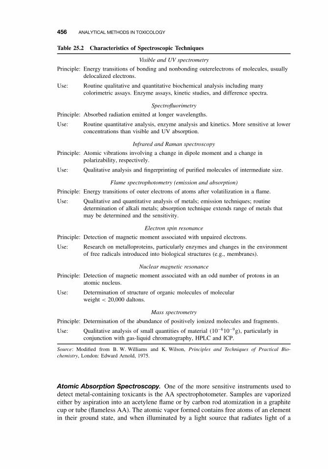
456 ANALYTICAL METHODS IN TOXICOLOGY
Table 25.2 Characteristics of Spectroscopic Techniques
Visible and UV spectrometry
Principle: Energy transitions of bonding and nonbonding outerelectrons of molecules, usuallydelocalized electrons.
Use: Routine qualitative and quantitative biochemical analysis including manycolorimetric assays. Enzyme assays, kinetic studies, and difference spectra.
Spectrofluorimetry
Principle: Absorbed radiation emitted at longer wavelengths.
Use: Routine quantitative analysis, enzyme analysis and kinetics. More sensitive at lowerconcentrations than visible and UV absorption.
Infrared and Raman spectroscopy
Principle: Atomic vibrations involving a change in dipole moment and a change inpolarizability, respectively.
Use: Qualitative analysis and fingerprinting of purified molecules of intermediate size.
Flame spectrophotometry (emission and absorption)
Principle: Energy transitions of outer electrons of atoms after volatilization in a flame.
Use: Qualitative and quantitative analysis of metals; emission techniques; routinedetermination of alkali metals; absorption technique extends range of metals thatmay be determined and the sensitivity.
Electron spin resonance
Principle: Detection of magnetic moment associated with unpaired electrons.
Use: Research on metalloproteins, particularly enzymes and changes in the environmentof free radicals introduced into biological structures (e.g., membranes).
Nuclear magnetic resonance
Principle: Detection of magnetic moment associated with an odd number of protons in anatomic nucleus.
Use: Determination of structure of organic molecules of molecularweight < 20,000 daltons.
Mass spectrometry
Principle: Determination of the abundance of positively ionized molecules and fragments.
Use: Qualitative analysis of small quantities of material (10−610−9g), particularly inconjunction with gas-liquid chromatography, HPLC and ICP.
Source: Modified from B. W. Williams and K. Wilson, Principles and Techniques of Practical Bio-chemistry, London: Edward Arnold, 1975.
Atomic Absorption Spectroscopy. One of the more sensitive instruments used todetect metal-containing toxicants is the AA spectrophotometer. Samples are vaporizedeither by aspiration into an acetylene flame or by carbon rod atomization in a graphitecup or tube (flameless AA). The atomic vapor formed contains free atoms of an elementin their ground state, and when illuminated by a light source that radiates light of a
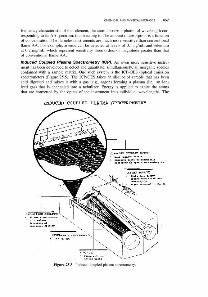
CHEMICAL AND PHYSICAL METHODS 457
frequency characteristic of that element, the atom absorbs a photon of wavelength cor-responding to its AA spectrum, thus exciting it. The amount of absorption is a functionof concentration. The flameless instruments are much more sensitive than conventionalflame AA. For example, arsenic can be detected at levels of 0.1 ng/mL and seleniumat 0.2 mg/mL, which represent sensitivity three orders of magnitude greater than thatof conventional flame AA.
Induced Coupled Plasma Spectrometry (ICP). An even more sensitive instru-ment has been developed to detect and quantitate, simultaneously, all inorganic speciescontained with a sample matrix. One such system is the ICP-OES (optical emissionspectrometer) (Figure 25.5). The ICP-OES takes an aliquot of sample that has beenacid digested and mixes it with a gas (e.g., argon) forming a plasma (i.e., an ion-ized gas) that is channeled into a nebulizer. Energy is applied to excite the atomsthat are converted by the optics of the instrument into individual wavelengths. The
Figure 25.5 Induced coupled plasma spectrometry.
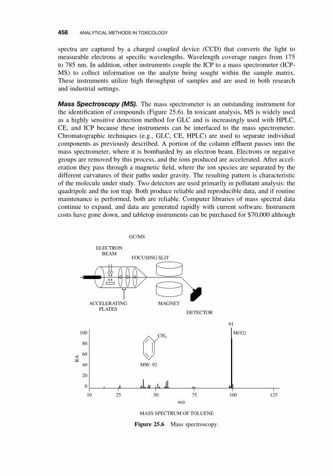
458 ANALYTICAL METHODS IN TOXICOLOGY
spectra are captured by a charged coupled device (CCD) that converts the light tomeasurable electrons at specific wavelengths. Wavelength coverage ranges from 175to 785 nm. In addition, other instruments couple the ICP to a mass spectrometer (ICP-MS) to collect information on the analyte being sought within the sample matrix.These instruments utilize high throughput of samples and are used in both researchand industrial settings.
Mass Spectroscopy (MS). The mass spectrometer is an outstanding instrument forthe identification of compounds (Figure 25.6). In toxicant analysis, MS is widely usedas a highly sensitive detection method for GLC and is increasingly used with HPLC,CE, and ICP because these instruments can be interfaced to the mass spectrometer.Chromatographic techniques (e.g., GLC, CE, HPLC) are used to separate individualcomponents as previously described. A portion of the column effluent passes into themass spectrometer, where it is bombarded by an electron beam. Electrons or negativegroups are removed by this process, and the ions produced are accelerated. After accel-eration they pass through a magnetic field, where the ion species are separated by thedifferent curvatures of their paths under gravity. The resulting pattern is characteristicof the molecule under study. Two detectors are used primarily in pollutant analysis: thequadripole and the ion trap. Both produce reliable and reproducible data, and if routinemaintenance is performed, both are reliable. Computer libraries of mass spectral datacontinue to expand, and data are generated rapidly with current software. Instrumentcosts have gone down, and tabletop instruments can be purchased for $70,000 although
MASS SPECTRUM OF TOLUENE
ELECTRONBEAM
FOCUSING SLIT
GC/MS
ACCELERATINGPLATES
MAGNET
DETECTOR
100
80
60
40
20
0
10 25 50m/e
RA
75 100 125
CH3
MW: 92
91
M(92)
Figure 25.6 Mass spectroscopy.
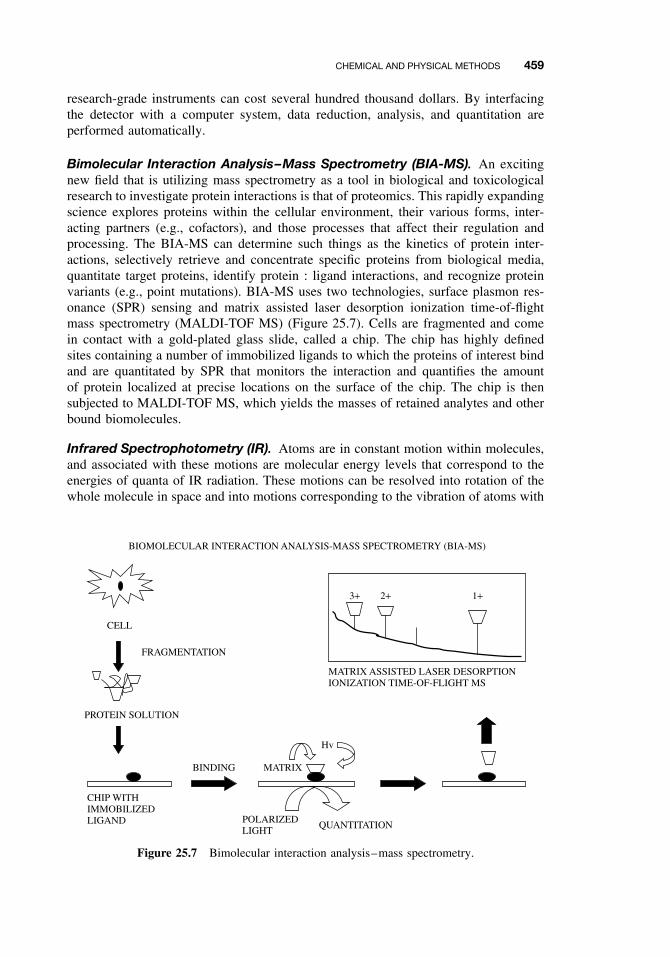
CHEMICAL AND PHYSICAL METHODS 459
research-grade instruments can cost several hundred thousand dollars. By interfacingthe detector with a computer system, data reduction, analysis, and quantitation areperformed automatically.
Bimolecular Interaction Analysis–Mass Spectrometry (BIA-MS). An excitingnew field that is utilizing mass spectrometry as a tool in biological and toxicologicalresearch to investigate protein interactions is that of proteomics. This rapidly expandingscience explores proteins within the cellular environment, their various forms, inter-acting partners (e.g., cofactors), and those processes that affect their regulation andprocessing. The BIA-MS can determine such things as the kinetics of protein inter-actions, selectively retrieve and concentrate specific proteins from biological media,quantitate target proteins, identify protein : ligand interactions, and recognize proteinvariants (e.g., point mutations). BIA-MS uses two technologies, surface plasmon res-onance (SPR) sensing and matrix assisted laser desorption ionization time-of-flightmass spectrometry (MALDI-TOF MS) (Figure 25.7). Cells are fragmented and comein contact with a gold-plated glass slide, called a chip. The chip has highly definedsites containing a number of immobilized ligands to which the proteins of interest bindand are quantitated by SPR that monitors the interaction and quantifies the amountof protein localized at precise locations on the surface of the chip. The chip is thensubjected to MALDI-TOF MS, which yields the masses of retained analytes and otherbound biomolecules.
Infrared Spectrophotometry (IR). Atoms are in constant motion within molecules,and associated with these motions are molecular energy levels that correspond to theenergies of quanta of IR radiation. These motions can be resolved into rotation of thewhole molecule in space and into motions corresponding to the vibration of atoms with
BIOMOLECULAR INTERACTION ANALYSIS-MASS SPECTROMETRY (BIA-MS)
CELL
FRAGMENTATION
PROTEIN SOLUTION
CHIP WITHIMMOBILIZEDLIGAND
BINDING MATRIX
POLARIZEDLIGHT
QUANTITATION
Hv
MATRIX ASSISTED LASER DESORPTIONIONIZATION TIME-OF-FLIGHT MS
3+ 2+ 1+
Figure 25.7 Bimolecular interaction analysis–mass spectrometry.
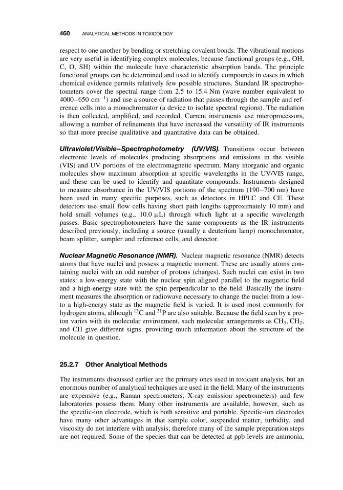
460 ANALYTICAL METHODS IN TOXICOLOGY
respect to one another by bending or stretching covalent bonds. The vibrational motionsare very useful in identifying complex molecules, because functional groups (e.g., OH,C, O, SH) within the molecule have characteristic absorption bands. The principlefunctional groups can be determined and used to identify compounds in cases in whichchemical evidence permits relatively few possible structures. Standard IR spectropho-tometers cover the spectral range from 2.5 to 15.4 Nm (wave number equivalent to4000–650 cm−1) and use a source of radiation that passes through the sample and ref-erence cells into a monochromator (a device to isolate spectral regions). The radiationis then collected, amplified, and recorded. Current instruments use microprocessors,allowing a number of refinements that have increased the versatility of IR instrumentsso that more precise qualitative and quantitative data can be obtained.
Ultraviolet/Visible–Spectrophotometry (UV/VIS). Transitions occur betweenelectronic levels of molecules producing absorptions and emissions in the visible(VIS) and UV portions of the electromagnetic spectrum. Many inorganic and organicmolecules show maximum absorption at specific wavelengths in the UV/VIS range,and these can be used to identify and quantitate compounds. Instruments designedto measure absorbance in the UV/VIS portions of the spectrum (190–700 nm) havebeen used in many specific purposes, such as detectors in HPLC and CE. Thesedetectors use small flow cells having short path lengths (approximately 10 mm) andhold small volumes (e.g., 10.0 µL) through which light at a specific wavelengthpasses. Basic spectrophotometers have the same components as the IR instrumentsdescribed previously, including a source (usually a deuterium lamp) monochromator,beam splitter, sampler and reference cells, and detector.
Nuclear Magnetic Resonance (NMR). Nuclear magnetic resonance (NMR) detectsatoms that have nuclei and possess a magnetic moment. These are usually atoms con-taining nuclei with an odd number of protons (charges). Such nuclei can exist in twostates: a low-energy state with the nuclear spin aligned parallel to the magnetic fieldand a high-energy state with the spin perpendicular to the field. Basically the instru-ment measures the absorption or radiowave necessary to change the nuclei from a low-to a high-energy state as the magnetic field is varied. It is used most commonly forhydrogen atoms, although 13C and 31P are also suitable. Because the field seen by a pro-ton varies with its molecular environment, such molecular arrangements as CH3, CH2,and CH give different signs, providing much information about the structure of themolecule in question.
25.2.7 Other Analytical Methods
The instruments discussed earlier are the primary ones used in toxicant analysis, but anenormous number of analytical techniques are used in the field. Many of the instrumentsare expensive (e.g., Raman spectrometers, X-ray emission spectrometers) and fewlaboratories possess them. Many other instruments are available, however, such asthe specific-ion electrode, which is both sensitive and portable. Specific-ion electrodeshave many other advantages in that sample color, suspended matter, turbidity, andviscosity do not interfere with analysis; therefore many of the sample preparation stepsare not required. Some of the species that can be detected at ppb levels are ammonia,
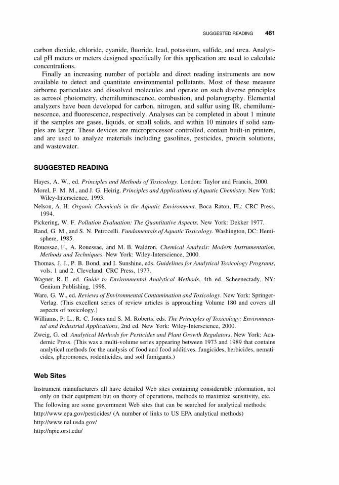
SUGGESTED READING 461
carbon dioxide, chloride, cyanide, fluoride, lead, potassium, sulfide, and urea. Analyti-cal pH meters or meters designed specifically for this application are used to calculateconcentrations.
Finally an increasing number of portable and direct reading instruments are nowavailable to detect and quantitate environmental pollutants. Most of these measureairborne particulates and dissolved molecules and operate on such diverse principlesas aerosol photometry, chemiluminescence, combustion, and polarography. Elementalanalyzers have been developed for carbon, nitrogen, and sulfur using IR, chemilumi-nescence, and fluorescence, respectively. Analyses can be completed in about 1 minuteif the samples are gases, liquids, or small solids, and within 10 minutes if solid sam-ples are larger. These devices are microprocessor controlled, contain built-in printers,and are used to analyze materials including gasolines, pesticides, protein solutions,and wastewater.
SUGGESTED READING
Hayes, A. W., ed. Principles and Methods of Toxicology. London: Taylor and Francis, 2000.
Morel, F. M. M., and J. G. Heirig. Principles and Applications of Aquatic Chemistry. New York:Wiley-Interscience, 1993.
Nelson, A. H. Organic Chemicals in the Aquatic Environment. Boca Raton, FL: CRC Press,1994.
Pickering, W. F. Pollution Evaluation: The Quantitative Aspects. New York: Dekker 1977.
Rand, G. M., and S. N. Petrocelli. Fundamentals of Aquatic Toxicology. Washington, DC: Hemi-sphere, 1985.
Rouessae, F., A. Rouessae, and M. B. Waldron. Chemical Analysis: Modern Instrumentation,Methods and Techniques. New York: Wiley-Interscience, 2000.
Thomas, J. J., P. B. Bond, and I. Sunshine, eds. Guidelines for Analytical Toxicology Programs,vols. 1 and 2. Cleveland: CRC Press, 1977.
Wagner, R. E. ed. Guide to Environmental Analytical Methods, 4th ed. Scheenectady, NY:Genium Publishing, 1998.
Ware, G. W., ed. Reviews of Environmental Contamination and Toxicology. New York: Springer-Verlag. (This excellent series of review articles is approaching Volume 180 and covers allaspects of toxicology.)
Williams, P. L., R. C. Jones and S. M. Roberts, eds. The Principles of Toxicology: Environmen-tal and Industrial Applications, 2nd ed. New York: Wiley-Interscience, 2000.
Zweig, G. ed. Analytical Methods for Pesticides and Plant Growth Regulators. New York: Aca-demic Press. (This was a multi-volume series appearing between 1973 and 1989 that containsanalytical methods for the analysis of food and food additives, fungicides, herbicides, nemati-cides, pheromones, rodenticides, and soil fumigants.)
Web Sites
Instrument manufacturers all have detailed Web sites containing considerable information, notonly on their equipment but on theory of operations, methods to maximize sensitivity, etc.
The following are some government Web sites that can be searched for analytical methods:
http://www.epa.gov/pesticides/ (A number of links to US EPA analytical methods)
http://www.nal.usda.gov/
http://npic.orst.edu/

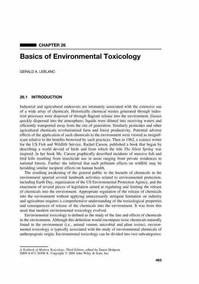
CHAPTER 26
Basics of Environmental Toxicology
GERALD A. LEBLANC
26.1 INTRODUCTION
Industrial and agricultural endeavors are intimately associated with the extensive useof a wide array of chemicals. Historically chemical wastes generated through indus-trial processes were disposed of through flagrant release into the environment. Gassesquickly dispersed into the atmosphere; liquids were diluted into receiving waters andefficiently transported away from the site of generation. Similarly pesticides and otheragricultural chemicals revolutionized farm and forest productivity. Potential adverseeffects of the application of such chemicals to the environment were viewed as insignif-icant relative to the benefits bestowed by such practices. Then in 1962, a science writerfor the US Fish and Wildlife Service, Rachel Carson, published a book that began bydescribing a world devoid of birds and from which the title The Silent Spring wasinspired. In her book Ms. Carson graphically described incidents of massive fish andbird kills resulting from insecticide use in areas ranging from private residences tonational forests. Further she inferred that such pollutant effects on wildlife may beheralding similar incipient effects on human health.
The resulting awakening of the general public to the hazards of chemicals in theenvironment spurred several landmark activities related to environmental protection,including Earth Day, organization of the US Environmental Protection Agency, and theenactment of several pieces of legislation aimed at regulating and limiting the releaseof chemicals into the environment. Appropriate regulation of the release of chemicalsinto the environment without applying unnecessarily stringent limitation on industryand agriculture requires a comprehensive understanding of the toxicological propertiesand consequences of release of the chemicals into the environment. It was from thisneed that modern environmental toxicology evolved.
Environmental toxicology is defined as the study of the fate and effects of chemicalsin the environment. Although this definition would encompass toxic chemicals naturallyfound in the environment (i.e., animal venom, microbial and plant toxins), environ-mental toxicology is typically associated with the study of environmental chemicals ofanthropogenic origin. Environmental toxicology can be divided into two subcategories:
A Textbook of Modern Toxicology, Third Edition, edited by Ernest HodgsonISBN 0-471-26508-X Copyright 2004 John Wiley & Sons, Inc.
463
464 BASICS OF ENVIRONMENTAL TOXICOLOGY
environmental health toxicology and ecotoxicology. Environmental health toxicologyis the study of the adverse effects of environmental chemicals on human health, whileecotoxicology focuses upon the effects of environmental contaminants upon ecosystemsand constituents thereof (fish, wildlife, etc.). Assessing the toxic effects of chemicalson humans involves the use of standard animal models (i.e., mouse and rat) as wellas epidemiological evaluations of exposed human populations (i.e., farmers and fac-tory workers). In contrast, ecotoxicology involves the study of the adverse effects oftoxicants on myriad of organisms that compose ecosystems ranging from microorgan-isms to top predators. Further, comprehensive insight into the effects of chemicals inthe environment requires assessments ancillary to toxicology such as the fate of thechemical in the environment (Chapter 27), and toxicant interactions with abiotic (non-living) components of ecosystems. Comprehensive assessments of the adverse effectsof environmental chemicals thus utilize expertise from many scientific disciplines. Theultimate goal of these assessments is elucidating the adverse effects of chemicals thatare present in the environment (retrospective hazard assessment) and predicting anyadverse effects of chemicals before they are discharged into the environment (prospec-tive hazard assessment). The ecological hazard assessment process is discussed inChapter 28.
Historically chemicals that have posed major environmental hazards tend to sharethree insidious characteristics: environmental persistence, the propensity to accumulatein living things, and high toxicity.
26.2 ENVIRONMENTAL PERSISTENCE
Many abiotic and biotic processes exist in nature that function in concert to eliminate(i.e., degrade) toxic chemicals. Accordingly many chemicals released into the environ-ment pose minimal hazard simply because of their limited life span in the environment.Chemicals that have historically posed environmental hazard (i.e., DDT, PCBs, TCDD)resist degradative processes and accordingly persist in the environment for extremelylong periods of time (Table 26.1). Continued disposal of persistent chemicals into theenvironment can result in their accumulation to environmental levels sufficient to posetoxicity. Such chemicals can continue to pose hazard long after their disposal into theenvironment has ceased. For example, significant contamination of Lake Ontario bythe pesticide mirex occurred from the 1950s through the 1970s. Mass balance stud-ies performed 20 years later revealed that 80% of the mirex deposited into the lake
Table 26.1 Environmental Half-lives of Some Chem-ical Contaminants
Contaminant Half-life Media
DDT 10 Years SoilTCDD 9 Years SoilAtrazine 25 Months WaterBenzoperylene (PAH) 14 Months SoilPhenanthrene (PAH) 138 Days SoilCarbofuran 45 Days Water
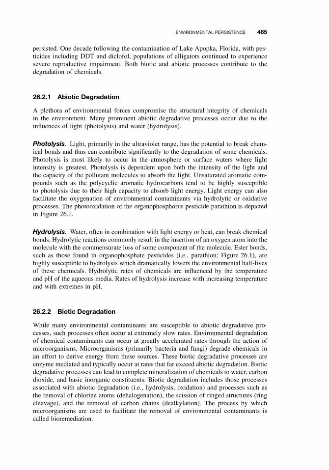
ENVIRONMENTAL PERSISTENCE 465
persisted. One decade following the contamination of Lake Apopka, Florida, with pes-ticides including DDT and diclofol, populations of alligators continued to experiencesevere reproductive impairment. Both biotic and abiotic processes contribute to thedegradation of chemicals.
26.2.1 Abiotic Degradation
A plethora of environmental forces compromise the structural integrity of chemicalsin the environment. Many prominent abiotic degradative processes occur due to theinfluences of light (photolysis) and water (hydrolysis).
Photolysis. Light, primarily in the ultraviolet range, has the potential to break chem-ical bonds and thus can contribute significantly to the degradation of some chemicals.Photolysis is most likely to occur in the atmosphere or surface waters where lightintensity is greatest. Photolysis is dependent upon both the intensity of the light andthe capacity of the pollutant molecules to absorb the light. Unsaturated aromatic com-pounds such as the polycyclic aromatic hydrocarbons tend to be highly susceptibleto photolysis due to their high capacity to absorb light energy. Light energy can alsofacilitate the oxygenation of environmental contaminants via hydrolytic or oxidativeprocesses. The photooxidation of the organophosphorus pesticide parathion is depictedin Figure 26.1.
Hydrolysis. Water, often in combination with light energy or heat, can break chemicalbonds. Hydrolytic reactions commonly result in the insertion of an oxygen atom into themolecule with the commensurate loss of some component of the molecule. Ester bonds,such as those found in organophosphate pesticides (i.e., parathion; Figure 26.1), arehighly susceptible to hydrolysis which dramatically lowers the environmental half-livesof these chemicals. Hydrolytic rates of chemicals are influenced by the temperatureand pH of the aqueous media. Rates of hydrolysis increase with increasing temperatureand with extremes in pH.
26.2.2 Biotic Degradation
While many environmental contaminants are susceptible to abiotic degradative pro-cesses, such processes often occur at extremely slow rates. Environmental degradationof chemical contaminants can occur at greatly accelerated rates through the action ofmicroorganisms. Microorganisms (primarily bacteria and fungi) degrade chemicals inan effort to derive energy from these sources. These biotic degradative processes areenzyme mediated and typically occur at rates that far exceed abiotic degradation. Bioticdegradative processes can lead to complete mineralization of chemicals to water, carbondioxide, and basic inorganic constituents. Biotic degradation includes those processesassociated with abiotic degradation (i.e., hydrolysis, oxidation) and processes such asthe removal of chlorine atoms (dehalogenation), the scission of ringed structures (ringcleavage), and the removal of carbon chains (dealkylation). The process by whichmicroorganisms are used to facilitate the removal of environmental contaminants iscalled bioremediation.
466 BASICS OF ENVIRONMENTAL TOXICOLOGY
Paraoxon+
Photoxidation
NO2OP(OC2H5)2
O
NO2OP(OC2H5)2
S
Parathion
NO2HO
O−P(OC2H5)2
S
Diethylphosphorothioate+
p-nitrophenol
Hydrolysis
Figure 26.1 The effect of sunlight (photooxidation) and precipitation (hydrolysis) on the degra-dation of parathion.
26.2.3 Nondegradative Elimination Processes
Many processes are operative in the environment that contribute to the regional elim-ination of a contaminant by altering its distribution. Contaminants with sufficientlyhigh vapor pressure can evaporate from contaminated terrestrial or aquatic compart-ments and be transferred through the atmosphere to new locations. Such processes ofglobal distillation are considered largely responsible for the worldwide distribution ofrelatively volatile organochlorine pesticides such as lindane and hexachlorobenzene.Entrainment by wind and upper atmospheric currents of contaminant particles or dustonto which the contaminants are sorbed also contribute to contaminant redistribution.Sorption of contaminant to suspended solids in an aquatic environment with com-mensurate sedimentation can result with the removal of contaminants from the water
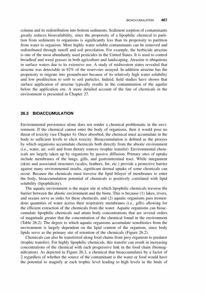
BIOACCUMULATION 467
column and its redistribution into bottom sediments. Sediment sorption of contaminantsgreatly reduces bioavailability, since the propensity of a lipophilic chemical to parti-tion from sediments to organisms is significantly less than its propensity to partitionfrom water to organism. More highly water soluble contaminants can be removed andredistributed through runoff and soil percolation. For example, the herbicide atrazineis one of the most abundantly used pesticides in the United States. It is used to controlbroadleaf and weed grasses in both agriculture and landscaping. Atrazine is ubiquitousin surface waters due to its extensive use. A study of midwestern states revealed thatatrazine was detectable in 92% of the reservoirs assayed. In addition atrazine has thepropensity to migrate into groundwater because of its relatively high water solubilityand low predilection to sorb to soil particles. Indeed, field studies have shown thatsurface application of atrazine typically results in the contamination of the aquiferbelow the application site. A more detailed account of the fate of chemicals in theenvironment is presented in Chapter 27.
26.3 BIOACCUMULATION
Environmental persistence alone does not render a chemical problematic in the envi-ronment. If the chemical cannot enter the body of organisms, then it would pose nothreat of toxicity (see Chapter 6). Once absorbed, the chemical must accumulate in thebody to sufficient levels to elicit toxicity. Bioaccumulation is defined as the processby which organisms accumulate chemicals both directly from the abiotic environment(i.e., water, air, soil) and from dietary sources (trophic transfer). Environmental chem-icals are largely taken up by organisms by passive diffusion. Primary sites of uptakeinclude membranes of the lungs, gills, and gastrointestinal tract. While integument(skin) and associated structures (scales, feathers, fur, etc.) provide a protective barrieragainst many environmental insults, significant dermal uptake of some chemicals canoccur. Because the chemicals must traverse the lipid bilayer of membranes to enterthe body, bioaccumulation potential of chemicals is positively correlated with lipidsolubility (lipophilicity).
The aquatic environment is the major site at which lipophilic chemicals traverse thebarrier between the abiotic environment and the biota. This is because (1) lakes, rivers,and oceans serve as sinks for these chemicals, and (2) aquatic organisms pass tremen-dous quantities of water across their respiratory membranes (i.e., gills) allowing forthe efficient extraction of the chemicals from the water. Aquatic organisms can bioac-cumulate lipophilic chemicals and attain body concentrations that are several ordersof magnitude greater that the concentration of the chemical found in the environment(Table 26.2). The degree to which aquatic organisms accumulate xenobiotics from theenvironment is largely dependent on the lipid content of the organism, since bodylipids serve as the primary site of retention of the chemicals (Figure 26.2).
Chemicals can also be transferred along food chains from prey organism to predator(trophic transfer). For highly lipophilic chemicals, this transfer can result in increasingconcentrations of the chemical with each progressive link in the food chain (biomag-nification). As depicted in Figure 26.3, a chemical that bioaccumulates by a factor of2 regardless of whether the source of the contaminant is the water or food would havethe potential to magnify at each trophic level leading to high levels in the birds of
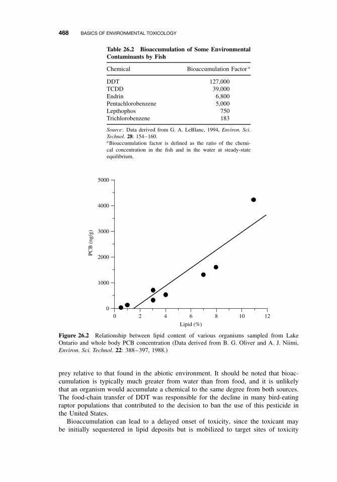
468 BASICS OF ENVIRONMENTAL TOXICOLOGY
Table 26.2 Bioaccumulation of Some EnvironmentalContaminants by Fish
Chemical Bioaccumulation Factor a
DDT 127,000TCDD 39,000Endrin 6,800Pentachlorobenzene 5,000Lepthophos 750Trichlorobenzene 183
Source: Data derived from G. A. LeBlanc, 1994, Environ. Sci.Technol. 28: 154–160.aBioaccumulation factor is defined as the ratio of the chemi-cal concentration in the fish and in the water at steady-stateequilibrium.
0 2 4 6
Lipid (%)
8 10 12
5000
4000
3000
2000
1000
0
PCB
(ng
/g)
Figure 26.2 Relationship between lipid content of various organisms sampled from LakeOntario and whole body PCB concentration (Data derived from B. G. Oliver and A. J. Niimi,Environ. Sci. Technol. 22: 388–397, 1988.)
prey relative to that found in the abiotic environment. It should be noted that bioac-cumulation is typically much greater from water than from food, and it is unlikelythat an organism would accumulate a chemical to the same degree from both sources.The food-chain transfer of DDT was responsible for the decline in many bird-eatingraptor populations that contributed to the decision to ban the use of this pesticide inthe United States.
Bioaccumulation can lead to a delayed onset of toxicity, since the toxicant maybe initially sequestered in lipid deposits but is mobilized to target sites of toxicity

BIOACCUMULATION 469
614
28
28
14
2
1
2
1
2
1
2
4 12
BIOACCUMULATION OF ENVIRONMENTAL CHEMICALS
Figure 26.3 Bioaccumulation of a chemical along a generic food chain. In this simplisticparadigm, the amount of the chemical in the water is assigned an arbitrary concentration of 1, andit is assumed that the chemical will bioaccumulate either from the water to the fish or from onetrophic level to another by a factor of 2. Circled numbers represent the concentration of chemicalin the respective compartment. Numbers associated with arrows represent the concentration ofchemical transferred from one compartment to another.
when these lipid stores are utilized. For example, lipid stores are often mobilized inpreparation for reproduction. The loss of the lipid can result in the release of lipophilictoxicants rendering them available for toxic action. Such effects can result in mortalityof adult organisms as they approach reproductive maturity. Lipophilic chemicals alsocan be transferred to offspring in lipids associated with the yolk of oviparous organismsor the milk of mammals, resulting in toxicity to offspring that was not evident in theparental organisms.
26.3.1 Factors That Influence Bioaccumulation
The propensity for an environmental contaminant to bioaccumulate is influenced byseveral factors. The first consideration is environmental persistence. The degree to
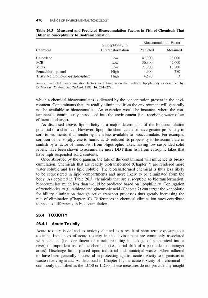
470 BASICS OF ENVIRONMENTAL TOXICOLOGY
Table 26.3 Measured and Predicted Bioaccumulation Factors in Fish of Chemicals ThatDiffer in Susceptibility to Biotransformation
Bioaccumulation FactorSusceptibility to
Chemical Biotransformation Predicted Measured
Chlordane Low 47,900 38,000PCB Low 36,300 42,600Mirex Low 21,900 18,200Pentachloro-phenol High 4,900 780Tris(2,3-dibromo-propyl)phosphate High 4,570 3
Source: Predicted bioaccumulation factors were based upon their relative lipophilicity as described by,D. Mackay, Environ. Sci. Technol. 1982, 16: 274–278.
which a chemical bioaccumulates is dictated by the concentration present in the envi-ronment. Contaminants that are readily eliminated from the environment will generallynot be available to bioaccumulate. An exception would be instances where the con-taminant is continuously introduced into the environment (i.e., receiving water of aneffluent discharge).
As discussed above, lipophilicity is a major determinant of the bioaccumulationpotential of a chemical. However, lipophilic chemicals also have greater propensity tosorb to sediments, thus rendering them less available to bioaccumulate. For example,sorption of benzo[a]pyrene to humic acids reduced its propensity to bioaccumulate insunfish by a factor of three. Fish from oligotrophic lakes, having low suspended solidlevels, have been shown to accumulate more DDT than fish from eutrophic lakes thathave high suspended solid contents.
Once absorbed by the organism, the fate of the contaminant will influence its bioac-cumulation. Chemicals that are readily biotransformed (Chapter 7) are rendered morewater soluble and less lipid soluble. The biotransformed chemical is thus less likelyto be sequestered in lipid compartments and more likely to be eliminated from thebody. As depicted in Table 26.3, chemicals that are susceptible to biotransformation,bioaccumulate much less than would be predicted based on lipophilicity. Conjugationof xenobiotics to glutathione and glucuronic acid (Chapter 7) can target the xenobioticfor biliary elimination through active transport processes thus greatly increasing therate of elimination (Chapter 10). Differences in chemical elimination rates contributeto species differences in bioaccumulation.
26.4 TOXICITY
26.4.1 Acute Toxicity
Acute toxicity is defined as toxicity elicited as a result of short-term exposure to atoxicant. Incidences of acute toxicity in the environment are commonly associatedwith accident (i.e., derailment of a train resulting in leakage of a chemical into ariver) or imprudent use of the chemical (i.e., aerial drift of a pesticide to nontargetareas). Discharge limits placed upon industrial and municipal wastes, when adheredto, have been generally successful in protecting against acute toxicity to organisms inwaste-receiving areas. As discussed in Chapter 11, the acute toxicity of a chemical iscommonly quantified as the LC50 or LD50. These measures do not provide any insight
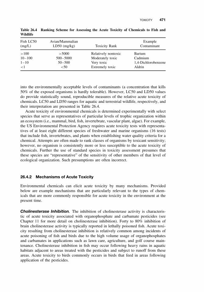
TOXICITY 471
Table 26.4 Ranking Scheme for Assessing the Acute Toxicity of Chemicals to Fish andWildlife
Fish LC50(mg/L)
Avian/MammalianLD50 (mg/kg) Toxicity Rank
ExampleContaminant
>100 >5000 Relatively nontoxic Barium10–100 500–5000 Moderately toxic Cadmium1–10 50–500 Very toxic 1,4-Dichlorobenzene<1 <50 Extremely toxic Aldrin
into the environmentally acceptable levels of contaminants (a concentration that kills50% of the exposed organisms is hardly tolerable). However, LC50 and LD50 valuesdo provide statistically sound, reproducible measures of the relative acute toxicity ofchemicals. LC50 and LD50 ranges for aquatic and terrestrial wildlife, respectively, andtheir interpretation are presented in Table 26.4.
Acute toxicity of environmental chemicals is determined experimentally with selectspecies that serve as representatives of particular levels of trophic organization withinan ecosystem (i.e., mammal, bird, fish, invertebrate, vascular plant, algae). For example,the US Environmental Protection Agency requires acute toxicity tests with representa-tives of at least eight different species of freshwater and marine organisms (16 tests)that include fish, invertebrates, and plants when establishing water quality criteria for achemical. Attempts are often made to rank classes of organisms by toxicant sensitivity;however, no organism is consistently more or less susceptible to the acute toxicity ofchemicals. Further the use of standard species in toxicity assessment presumes thatthese species are “representative” of the sensitivity of other members of that level ofecological organization. Such presumptions are often incorrect.
26.4.2 Mechanisms of Acute Toxicity
Environmental chemicals can elicit acute toxicity by many mechanisms. Providedbelow are example mechanisms that are particularly relevant to the types of chem-icals that are more commonly responsible for acute toxicity in the environment at thepresent time.
Cholinesterase Inhibition. The inhibition of cholinesterase activity is characteris-tic of acute toxicity associated with organophosphate and carbamate pesticides (seeChapter 11 for more detail on cholinesterase inhibition). Forty to 80% inhibition ofbrain cholinesterase activity is typically reported in lethally poisoned fish. Acute toxi-city resulting from cholinesterase inhibition is relatively common among incidents ofacute poisoning of fish and birds due to the high volume usage of organophosphatesand carbamates in applications such as lawn care, agriculture, and golf course main-tenance. Cholinesterase inhibition in fish may occur following heavy rains in aquatichabitats adjacent to areas treated with the pesticides and subject to runoff from theseareas. Acute toxicity to birds commonly occurs in birds that feed in areas followingapplication of the pesticides.
472 BASICS OF ENVIRONMENTAL TOXICOLOGY
Narcosis. A common means by which industrial chemicals elicit acute toxicity, par-ticularly to aquatic organisms, is through narcosis. Narcosis occurs when a chemicalaccumulates in cellular membranes interfering with the normal function of the mem-branes. Typical responses to the narcosis are decreased activity, reduced reactionto external stimuli, and increased pigmentation (in fish). The effects are reversible,and nonmoribund organisms typically return to normal activity once the chemical isremoved from the organism’s environment. Prolonged narcosis can result in death.Approximately 60% of industrial chemicals that enter the aquatic environment elicitacute toxicity through narcosis. Chemicals that elicit toxicity via narcosis typically donot elicit toxicity at specific target sites and are sufficiently lipophilic to accumulatein the lipid phase or the lipid-aqueous interface of membranes to sufficient levels todisrupt membrane function. Chemicals that induce narcosis include alcohols, ketones,benzenes, ethers, and aldehydes.
Physical Effects. Perhaps most graphic among recent incidents of environmentalacute toxicity is the physical effects of petroleum following oil spills. Slicks of oil onthe surface of contaminated waters results in the coating of animals, such as birds andmarine mammals, that frequent the air-water interface. Such a spill of unprecedentedmagnitude and consequence in the United States occurred on March 24, 1989, whenthe hull of the Exxon Valdez was ruptured on Bligh Reef in Prince William Sound,Alaska. Nearly 11 million gallons of crude oil spilled onto the nearshore waters killingmore wildlife than any prior oil spill in history. Thousands of sea birds and mammalssuccumbed to the acute effects of the oil.
Hypothermia is considered a major cause of death of oiled marine birds and mam-mals. These organisms insulate themselves from the frigid waters by maintaining alayer of air among the spaces within their coat of fur or feathers. The oil penetratesthe fur/feather barrier and purges the insulating air. As a result the animals rapidlysuccumb to hypothermia. In addition to hypothermia, these animals can also experi-ence oil toxicosis. Inhalation of oil, as well as ingestion through feeding and preening,can result in the accumulation of hydrocarbons to toxic levels. Toxicity to sea ottershas been correlated to degree of oiling and is characterized by pulmonary emphysema(bubbles of air within the connective tissue of the lungs), gastric hemorrhages, andliver damage.
26.4.3 Chronic Toxicity
Chronic toxicity is defined as toxicity elicited as a result of long-term exposure toa toxicant. Sublethal end points are generally associated with chronic toxicity. Theseinclude reproductive, immune, endocrine, and developmental dysfunction. However,chronic exposure also can result in direct mortality not observed during acute exposure.For example, chronic exposure of highly lipophilic chemicals can result in the eventualbioaccumulation of the chemical to concentrations that are lethal to the organisms. Oras discussed previously, mobilization of lipophilic toxicants from lipid compartmentsduring reproduction may result in lethality. It is important to recognize that, whiletheoretically, all chemicals elicit acute toxicity at a sufficiently high dose, all chemicalsare not chronically toxic. Chronic toxicity is measured by end points such as thehighest level of the chemical that does not elicit toxicity during continuous, prolongedexposure (no observed effect level, NOEL), the lowest level of the chemical that elicits
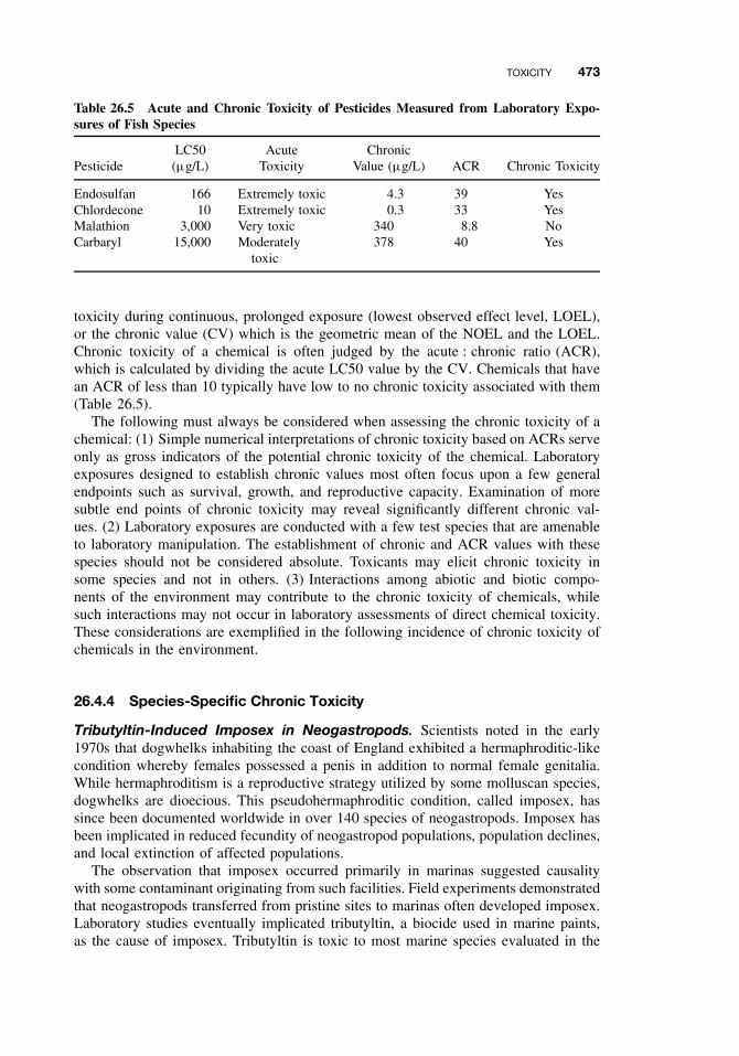
TOXICITY 473
Table 26.5 Acute and Chronic Toxicity of Pesticides Measured from Laboratory Expo-sures of Fish Species
PesticideLC50(µg/L)
AcuteToxicity
ChronicValue (µg/L) ACR Chronic Toxicity
Endosulfan 166 Extremely toxic 4.3 39 YesChlordecone 10 Extremely toxic 0.3 33 YesMalathion 3,000 Very toxic 340 8.8 NoCarbaryl 15,000 Moderately
toxic378 40 Yes
toxicity during continuous, prolonged exposure (lowest observed effect level, LOEL),or the chronic value (CV) which is the geometric mean of the NOEL and the LOEL.Chronic toxicity of a chemical is often judged by the acute : chronic ratio (ACR),which is calculated by dividing the acute LC50 value by the CV. Chemicals that havean ACR of less than 10 typically have low to no chronic toxicity associated with them(Table 26.5).
The following must always be considered when assessing the chronic toxicity of achemical: (1) Simple numerical interpretations of chronic toxicity based on ACRs serveonly as gross indicators of the potential chronic toxicity of the chemical. Laboratoryexposures designed to establish chronic values most often focus upon a few generalendpoints such as survival, growth, and reproductive capacity. Examination of moresubtle end points of chronic toxicity may reveal significantly different chronic val-ues. (2) Laboratory exposures are conducted with a few test species that are amenableto laboratory manipulation. The establishment of chronic and ACR values with thesespecies should not be considered absolute. Toxicants may elicit chronic toxicity insome species and not in others. (3) Interactions among abiotic and biotic compo-nents of the environment may contribute to the chronic toxicity of chemicals, whilesuch interactions may not occur in laboratory assessments of direct chemical toxicity.These considerations are exemplified in the following incidence of chronic toxicity ofchemicals in the environment.
26.4.4 Species-Specific Chronic Toxicity
Tributyltin-Induced Imposex in Neogastropods. Scientists noted in the early1970s that dogwhelks inhabiting the coast of England exhibited a hermaphroditic-likecondition whereby females possessed a penis in addition to normal female genitalia.While hermaphroditism is a reproductive strategy utilized by some molluscan species,dogwhelks are dioecious. This pseudohermaphroditic condition, called imposex, hassince been documented worldwide in over 140 species of neogastropods. Imposex hasbeen implicated in reduced fecundity of neogastropod populations, population declines,and local extinction of affected populations.
The observation that imposex occurred primarily in marinas suggested causalitywith some contaminant originating from such facilities. Field experiments demonstratedthat neogastropods transferred from pristine sites to marinas often developed imposex.Laboratory studies eventually implicated tributyltin, a biocide used in marine paints,as the cause of imposex. Tributyltin is toxic to most marine species evaluated in the
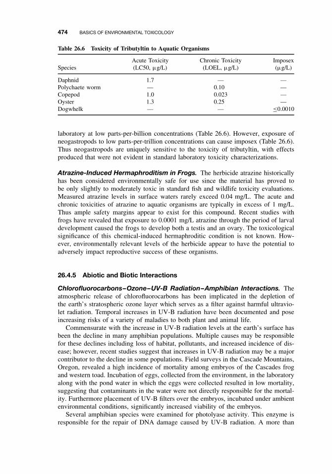
474 BASICS OF ENVIRONMENTAL TOXICOLOGY
Table 26.6 Toxicity of Tributyltin to Aquatic Organisms
SpeciesAcute Toxicity(LC50, µg/L)
Chronic Toxicity(LOEL, µg/L)
Imposex(µg/L)
Daphnid 1.7 — —Polychaete worm — 0.10 —Copepod 1.0 0.023 —Oyster 1.3 0.25 —Dogwhelk — — ≤0.0010
laboratory at low parts-per-billion concentrations (Table 26.6). However, exposure ofneogastropods to low parts-per-trillion concentrations can cause imposex (Table 26.6).Thus neogastropods are uniquely sensitive to the toxicity of tributyltin, with effectsproduced that were not evident in standard laboratory toxicity characterizations.
Atrazine-Induced Hermaphroditism in Frogs. The herbicide atrazine historicallyhas been considered environmentally safe for use since the material has proved tobe only slightly to moderately toxic in standard fish and wildlife toxicity evaluations.Measured atrazine levels in surface waters rarely exceed 0.04 mg/L. The acute andchronic toxicities of atrazine to aquatic organisms are typically in excess of 1 mg/L.Thus ample safety margins appear to exist for this compound. Recent studies withfrogs have revealed that exposure to 0.0001 mg/L atrazine through the period of larvaldevelopment caused the frogs to develop both a testis and an ovary. The toxicologicalsignificance of this chemical-induced hermaphroditic condition is not known. How-ever, environmentally relevant levels of the herbicide appear to have the potential toadversely impact reproductive success of these organisms.
26.4.5 Abiotic and Biotic Interactions
Chlorofluorocarbons–Ozone–UV-B Radiation–Amphibian Interactions. Theatmospheric release of chlorofluorocarbons has been implicated in the depletion ofthe earth’s stratospheric ozone layer which serves as a filter against harmful ultravio-let radiation. Temporal increases in UV-B radiation have been documented and poseincreasing risks of a variety of maladies to both plant and animal life.
Commensurate with the increase in UV-B radiation levels at the earth’s surface hasbeen the decline in many amphibian populations. Multiple causes may be responsiblefor these declines including loss of habitat, pollutants, and increased incidence of dis-ease; however, recent studies suggest that increases in UV-B radiation may be a majorcontributor to the decline in some populations. Field surveys in the Cascade Mountains,Oregon, revealed a high incidence of mortality among embryos of the Cascades frogand western toad. Incubation of eggs, collected from the environment, in the laboratoryalong with the pond water in which the eggs were collected resulted in low mortality,suggesting that contaminants in the water were not directly responsible for the mortal-ity. Furthermore placement of UV-B filters over the embryos, incubated under ambientenvironmental conditions, significantly increased viability of the embryos.
Several amphibian species were examined for photolyase activity. This enzyme isresponsible for the repair of DNA damage caused by UV-B radiation. A more than
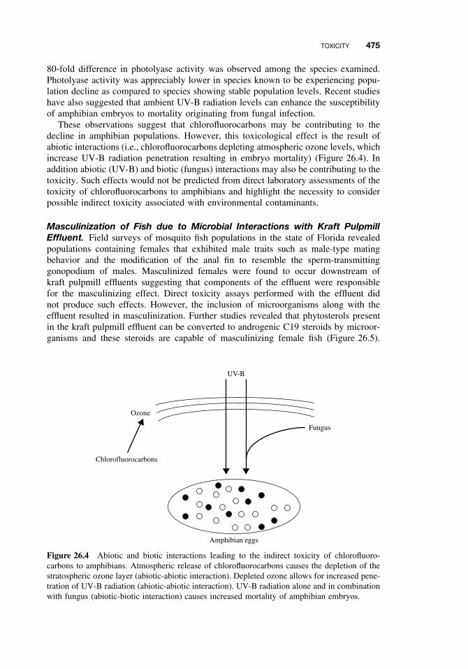
TOXICITY 475
80-fold difference in photolyase activity was observed among the species examined.Photolyase activity was appreciably lower in species known to be experiencing popu-lation decline as compared to species showing stable population levels. Recent studieshave also suggested that ambient UV-B radiation levels can enhance the susceptibilityof amphibian embryos to mortality originating from fungal infection.
These observations suggest that chlorofluorocarbons may be contributing to thedecline in amphibian populations. However, this toxicological effect is the result ofabiotic interactions (i.e., chlorofluorocarbons depleting atmospheric ozone levels, whichincrease UV-B radiation penetration resulting in embryo mortality) (Figure 26.4). Inaddition abiotic (UV-B) and biotic (fungus) interactions may also be contributing to thetoxicity. Such effects would not be predicted from direct laboratory assessments of thetoxicity of chlorofluorocarbons to amphibians and highlight the necessity to considerpossible indirect toxicity associated with environmental contaminants.
Masculinization of Fish due to Microbial Interactions with Kraft PulpmillEffluent. Field surveys of mosquito fish populations in the state of Florida revealedpopulations containing females that exhibited male traits such as male-type matingbehavior and the modification of the anal fin to resemble the sperm-transmittinggonopodium of males. Masculinized females were found to occur downstream ofkraft pulpmill effluents suggesting that components of the effluent were responsiblefor the masculinizing effect. Direct toxicity assays performed with the effluent didnot produce such effects. However, the inclusion of microorganisms along with theeffluent resulted in masculinization. Further studies revealed that phytosterols presentin the kraft pulpmill effluent can be converted to androgenic C19 steroids by microor-ganisms and these steroids are capable of masculinizing female fish (Figure 26.5).
Amphibian eggs
UV-B
Fungus
Chlorofluorocarbons
Ozone
Figure 26.4 Abiotic and biotic interactions leading to the indirect toxicity of chlorofluoro-carbons to amphibians. Atmospheric release of chlorofluorocarbons causes the depletion of thestratospheric ozone layer (abiotic-abiotic interaction). Depleted ozone allows for increased pene-tration of UV-B radiation (abiotic-abiotic interaction). UV-B radiation alone and in combinationwith fungus (abiotic-biotic interaction) causes increased mortality of amphibian embryos.
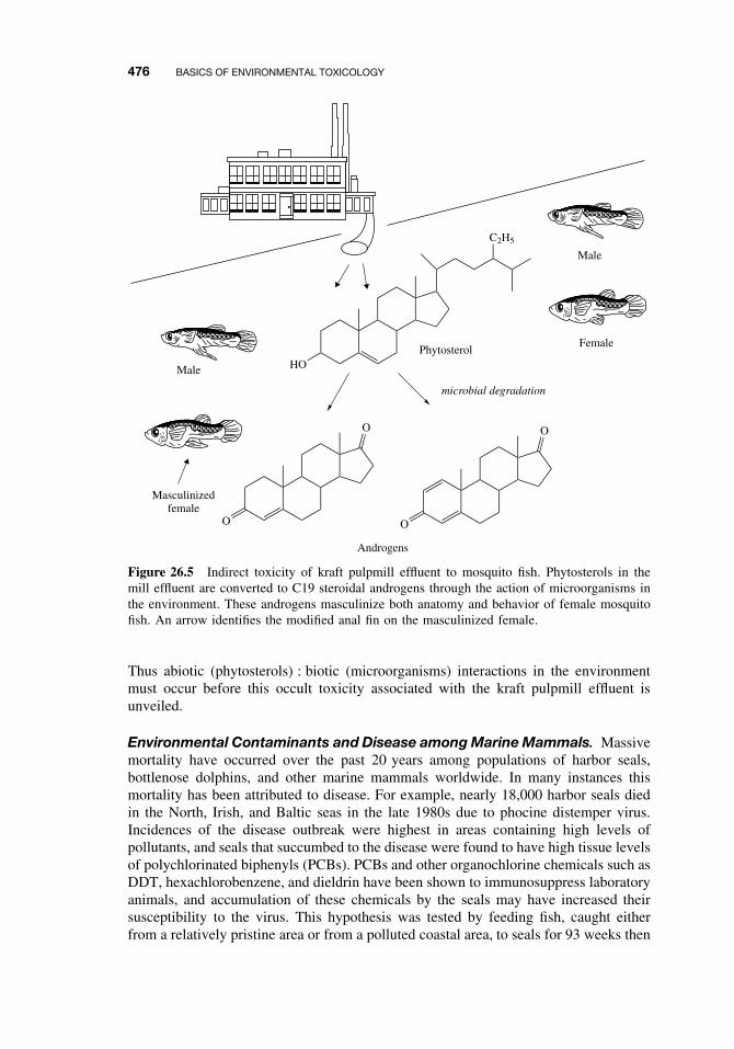
476 BASICS OF ENVIRONMENTAL TOXICOLOGY
Masculinizedfemale
Male
Male
Female
C2H5
HOPhytosterol
microbial degradation
O
O
O
O
Androgens
Figure 26.5 Indirect toxicity of kraft pulpmill effluent to mosquito fish. Phytosterols in themill effluent are converted to C19 steroidal androgens through the action of microorganisms inthe environment. These androgens masculinize both anatomy and behavior of female mosquitofish. An arrow identifies the modified anal fin on the masculinized female.
Thus abiotic (phytosterols) : biotic (microorganisms) interactions in the environmentmust occur before this occult toxicity associated with the kraft pulpmill effluent isunveiled.
Environmental Contaminants and Disease among Marine Mammals. Massivemortality have occurred over the past 20 years among populations of harbor seals,bottlenose dolphins, and other marine mammals worldwide. In many instances thismortality has been attributed to disease. For example, nearly 18,000 harbor seals diedin the North, Irish, and Baltic seas in the late 1980s due to phocine distemper virus.Incidences of the disease outbreak were highest in areas containing high levels ofpollutants, and seals that succumbed to the disease were found to have high tissue levelsof polychlorinated biphenyls (PCBs). PCBs and other organochlorine chemicals such asDDT, hexachlorobenzene, and dieldrin have been shown to immunosuppress laboratoryanimals, and accumulation of these chemicals by the seals may have increased theirsusceptibility to the virus. This hypothesis was tested by feeding fish, caught eitherfrom a relatively pristine area or from a polluted coastal area, to seals for 93 weeks then
SUGGESTED READING 477
assessing the integrity of the immune system in the seals. Seals fed the contaminatedfish did indeed have impaired immune responses lending credence to the hypothesisthat organochlorine contaminants in the marine environment are rendering some speciesimmunodeficient. Mortality occurs, not as a direct result of chemical toxicity, but dueto increased susceptibility to pathogens.
26.5 CONCLUSION
Environmental toxicologists have learned a great deal about the effects of chemicals inthe environment and the characteristics of chemicals that are responsible for the hazardsthey pose. Much of the information gained has been due to retrospective analyzes of theenvironmental consequences of the deposition of chemicals into the environment. Suchanalyzes have resulted in curtailing the release of demonstrated hazardous chemicalsinto the environment and provide benchmark information upon which the regulation ofchemicals proposed for release into the environment can be based. The recognition thatenvironmentally hazardous chemicals commonly share characteristics of persistence,potential to bioaccumulate, and high toxicity has resulted in development and useof chemicals that lack one or more of these characteristics yet fulfill societal needspreviously served by hazardous chemicals. For example, recognition that persistenceand propensity to bioaccumulate were largely responsible for the environmental hazardsposed by many organochlorine pesticides led to the development and use of alternativeclasses of pesticides such as organophosphates, carbamates, and pyrethroids. Whilethese chemicals all possess the toxicity necessary to function as pesticides, their lackof persistence and reduced propensity to bioaccumulate makes them more suitable foruse in the environment.
Such advances in our understanding of the fate and effects of chemicals in theenvironmental does not imply that the role of environmental toxicologists in the twenty-first century will diminish. A dearth of information persist in areas vital to continuedprotection of natural resources against chemical insult. These include understanding(1) the unique susceptibilities of key species to the toxicity of different classes ofchemicals, (2) the interactions of chemical contaminants with abiotic components ofthe environment that lead to increased toxicity, (3) the toxicological consequences ofexposure to complex chemical mixtures, and (4) the consequences of toxicant effectson individuals with respect to ecosystem viability. Additionally continued research isneeded to develop molecular and cellular biomarkers of toxicant exposure and effectthat could be used to predict dire consequences to ecosystem before such effects aremanifested at higher levels of biological organization. The role of the environmen-tal toxicologist undoubtedly will increase in prospective activities aimed at reducingthe risk associated with chemical contaminants in the environments before problemsarise, and hopefully will decrease with respect to assessing damage caused by suchenvironmental contaminants.
SUGGESTED READING
Persistence
Burns, L. A., and G. L. Baughman. Fate modeling. In Fundamentals of Aquatic Toxicology,G. M. Rand, and S. R. Petrocelli, eds. New York: Hemisphere, 1985, pp. 558–586.

478 BASICS OF ENVIRONMENTAL TOXICOLOGY
Larson, R. A., and E. J. Weber. Reaction Mechanisms in Environmental Organic Chemistry.Boca Raton, FL: Lewis Publishers, 1994.
Bioaccumulation
Banerjee, S., and G. A. Baughman. Bioconcentration factors and lipid solubility. Environ. Sci.Technol. 25: 536–539, 1991.
Barron, M. G. Bioconcentration. Environ. Sci. Technol. 24: 1612–1618, 1990.
Barron, M. G. Bioaccumulation and bioconcentration in aquatic organisms. In Handbook ofEcotoxicology, D. J. Hoffman, B. A. Rattner, G. A. Burton Jr., and J. Cairns Jr., eds. BocaRaton, FL: Lewis Publishers, 1995, pp. 652–666.
LeBlanc, G. A. Trophic-level differences in the bioconcentration of chemicals: Implications inassessing environmental biomagnification. Environ. Sci. Technol. 28: 154–160, 1995.
Acute Toxicity
Kelso, D. D., and M. Kendziorek. Alaska’s response to the Exxon Valdez oil spill. Environ. Sci.Technol. 25: 183–190, 1991.
Parrish, P. R. Acute toxicity tests. In Fundamentals of Aquatic Toxicology, G. M. Rand, andS. R. Petrocelli, eds. New York: Hemisphere, 1985, pp. 31–57.
Stansley, W. Field results using cholinesterase reactivation techniques to diagnose acute anti-cholinesterase poisoning in birds and fish. Arch. Environ. Contam. Toxicol. 25: 315–321,1993.
van Wezel, A. P., and A. Opperhuizen. Narcosis due to environmental pollutants in aquaticorganisms: residue-based toxicity, mechanisms, and membrane burdens. Crit. Rev. Toxicol.25: 255–279, 1995.
Wilson, V. S., and G. A. LeBlanc. Petroleum pollution. Rev. Toxicol. 3: 1–36, 1999.
Chronic Toxicity
Adams, W. J. Aquatic toxicology testing methods. In Handbook of Ecotoxicology, D. J. Hoffman,B. A. Rattner, G. A. Burton Jr., and J. Cairns Jr., eds. Boca Raton, FL: Lewis Publishers, 1995,pp. 25–46.
Blaustein, A. R., P. D. Hoffman, D. G. Hokit, J. M. Kiesecker, S. C. Walls, J. B. Hays. UVrepair and resistance to solar UV-B in amphibian eggs: A link to population declines? Proc.Nat. Acad. Sciences. USA 91: 1791–1795, 1994.
Colborn, T., and C. Clement, eds. Chemically-Induced Alterations in Sexual and FunctionalDevelopment: The Wildlife/Human Connection. Princeton, NJ: Princeton Scientific, 1992.
LeBlanc, G. A., and L. J. Bain. Chronic toxicity of environmental contaminants: Sentinels andbiomarkers. Environ. Health Perspect. 105(suppl. 1): 65–80, 1997.
Hayes, T. B., A. Collins, M. Lee, M. Mendoza, N. Noriega, A. A. Stuart, and A. Vonk.Hermaphroditic, demasculinized frogs after exposure to the herbicide atrazine at lowecologically relevant doses. Proc. Nat. Acad. Sciences. 99: 5476–5480, 2002.
CHAPTER 27
Transport and Fate of Toxicantsin the Environment
DAMIAN SHEA
27.1 INTRODUCTION
More than 100,000 chemicals are released into the global environment every yearthrough their normal production, use, and disposal. To understand and predict thepotential risk that this environmental contamination poses to humans and wildlife,we must couple our knowledge on the toxicity of a chemical to our knowledge onhow chemicals enter into and behave in the environment. The simple box modelshown in Figure 27.1 illustrates the relationship between a toxicant source, its fatein the environment, its effective exposure or dose, and resulting biological effects. Aprospective or predictive assessment of a chemical hazard would begin by characteriz-ing the source of contamination, modeling the chemical’s fate to predict exposure, andusing exposure/dose-response functions to predict effects (moving from left to rightin Figure 27.1). A common application would be to assess the potential effects of anew waste discharge. A retrospective assessment would proceed in the opposite direc-tion starting with some observed effect and reconstructing events to find a probablecause. Assuming that we have reliable dose/exposure-response functions, the key tosuccessful use of this simple relationship is to develop a qualitative description andquantitative model of the sources and fate of toxicants in the environment.
Toxicants are released into the environment in many ways, and they can travelalong many pathways during their lifetime. A toxicant present in the environment ata given point in time and space can experience three possible outcomes: it can bestationary and add to the toxicant inventory and exposure at that location, it can betransported to another location, or it can be transformed into another chemical species.Environmental contamination and exposure resulting from the use of a chemical ismodified by the transport and transformation of the chemical in the environment.Dilution and degradation can attenuate the source emission, while processes that focusand accumulate the chemical can magnify the source emission. The actual fate ofa chemical depends on the chemical’s use pattern and physical-chemical properties,combined with the characteristics of the environment to which it is released.
A Textbook of Modern Toxicology, Third Edition, edited by Ernest HodgsonISBN 0-471-26508-X Copyright 2004 John Wiley & Sons, Inc.
479
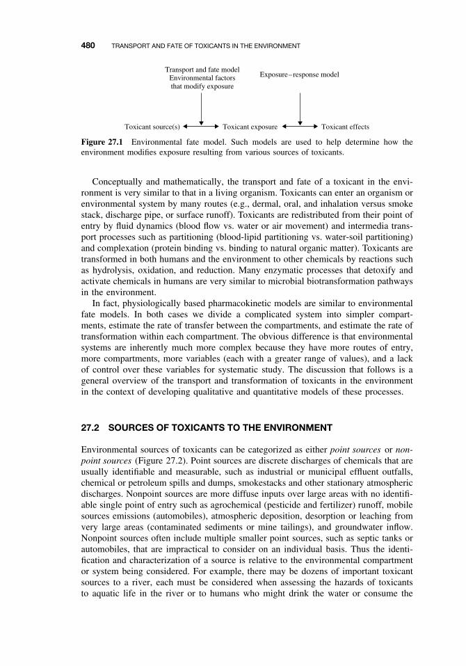
480 TRANSPORT AND FATE OF TOXICANTS IN THE ENVIRONMENT
Toxicant exposureToxicant source(s) Toxicant effects
Exposure–response modelTransport and fate model
Environmental factorsthat modify exposure
Figure 27.1 Environmental fate model. Such models are used to help determine how theenvironment modifies exposure resulting from various sources of toxicants.
Conceptually and mathematically, the transport and fate of a toxicant in the envi-ronment is very similar to that in a living organism. Toxicants can enter an organism orenvironmental system by many routes (e.g., dermal, oral, and inhalation versus smokestack, discharge pipe, or surface runoff). Toxicants are redistributed from their point ofentry by fluid dynamics (blood flow vs. water or air movement) and intermedia trans-port processes such as partitioning (blood-lipid partitioning vs. water-soil partitioning)and complexation (protein binding vs. binding to natural organic matter). Toxicants aretransformed in both humans and the environment to other chemicals by reactions suchas hydrolysis, oxidation, and reduction. Many enzymatic processes that detoxify andactivate chemicals in humans are very similar to microbial biotransformation pathwaysin the environment.
In fact, physiologically based pharmacokinetic models are similar to environmentalfate models. In both cases we divide a complicated system into simpler compart-ments, estimate the rate of transfer between the compartments, and estimate the rate oftransformation within each compartment. The obvious difference is that environmentalsystems are inherently much more complex because they have more routes of entry,more compartments, more variables (each with a greater range of values), and a lackof control over these variables for systematic study. The discussion that follows is ageneral overview of the transport and transformation of toxicants in the environmentin the context of developing qualitative and quantitative models of these processes.
27.2 SOURCES OF TOXICANTS TO THE ENVIRONMENT
Environmental sources of toxicants can be categorized as either point sources or non-point sources (Figure 27.2). Point sources are discrete discharges of chemicals that areusually identifiable and measurable, such as industrial or municipal effluent outfalls,chemical or petroleum spills and dumps, smokestacks and other stationary atmosphericdischarges. Nonpoint sources are more diffuse inputs over large areas with no identifi-able single point of entry such as agrochemical (pesticide and fertilizer) runoff, mobilesources emissions (automobiles), atmospheric deposition, desorption or leaching fromvery large areas (contaminated sediments or mine tailings), and groundwater inflow.Nonpoint sources often include multiple smaller point sources, such as septic tanks orautomobiles, that are impractical to consider on an individual basis. Thus the identi-fication and characterization of a source is relative to the environmental compartmentor system being considered. For example, there may be dozens of important toxicantsources to a river, each must be considered when assessing the hazards of toxicantsto aquatic life in the river or to humans who might drink the water or consume the
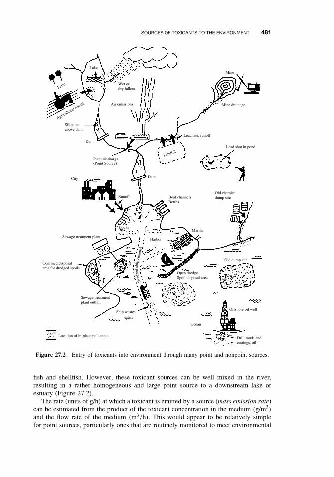
SOURCES OF TOXICANTS TO THE ENVIRONMENT 481
Farm
Lake
Agricultural runoff
Wet ordry fallout
Air emissions
Dam
Dam
Siltationabove dam
Plant discharge(Point Source)
City
Runoff
Landfill
Leachate, runoff
Mine
Mine drainage
Lead shot in pond
Boat channelsBerths
Old chemicaldump site
Marina
Harbor
Docks
Open dredgeSpoil disposal area
Ocean
Offshore oil well
Old dump site
Drill muds andcuttings, oil
Location of in-place pollutants
Ship wastes
Spills
Confined disposalarea for dredged spoils
Sewage treatmentplant outfall
Sewage treatment plant
Dre
dged
ship
chan
nel
Figure 27.2 Entry of toxicants into environment through many point and nonpoint sources.
fish and shellfish. However, these toxicant sources can be well mixed in the river,resulting in a rather homogeneous and large point source to a downstream lake orestuary (Figure 27.2).
The rate (units of g/h) at which a toxicant is emitted by a source (mass emission rate)can be estimated from the product of the toxicant concentration in the medium (g/m3)
and the flow rate of the medium (m3/h). This would appear to be relatively simplefor point sources, particularly ones that are routinely monitored to meet environmental
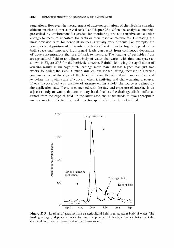
482 TRANSPORT AND FATE OF TOXICANTS IN THE ENVIRONMENT
regulations. However, the measurement of trace concentrations of chemicals in complexeffluent matrices is not a trivial task (see Chapter 25). Often the analytical methodsprescribed by environmental agencies for monitoring are not sensitive or selectiveenough to measure important toxicants or their reactive metabolites. Estimating themass emission rates for nonpoint sources is usually very difficult. For example, theatmospheric deposition of toxicants to a body of water can be highly dependent onboth space and time, and high annual loads can result from continuous depositionof trace concentrations that are difficult to measure. The loading of pesticides froman agricultural field to an adjacent body of water also varies with time and space asshown in Figure 27.3 for the herbicide atrazine. Rainfall following the application ofatrazine results in drainage ditch loadings more than 100-fold higher than just twoweeks following the rain. A much smaller, but longer lasting, increase in atrazineloading occurs at the edge of the field following the rain. Again, we see the needto define the spatial scale of concern when identifying and characterizing a source.If one is concerned with the fate of atrazine within a field, the source is defined bythe application rate. If one is concerned with the fate and exposure of atrazine in anadjacent body of water, the source may be defined as the drainage ditch and/or asrunoff from the edge of field. In the latter case one either needs to take appropriatemeasurements in the field or model the transport of atrazine from the field.
April May June July Aug Sept
2000
1500
1000
500
0
Large rain events
Period of atrazineapplication
Drainage ditch
Edge of field
Atr
azin
e co
ncen
trat
ion
(ng/
L)
Figure 27.3 Loading of atrazine from an agricultural field to an adjacent body of water. Theloading is highly dependent on rainfall and the presence of drainage ditches that collect thechemical and focus its movement in the environment.
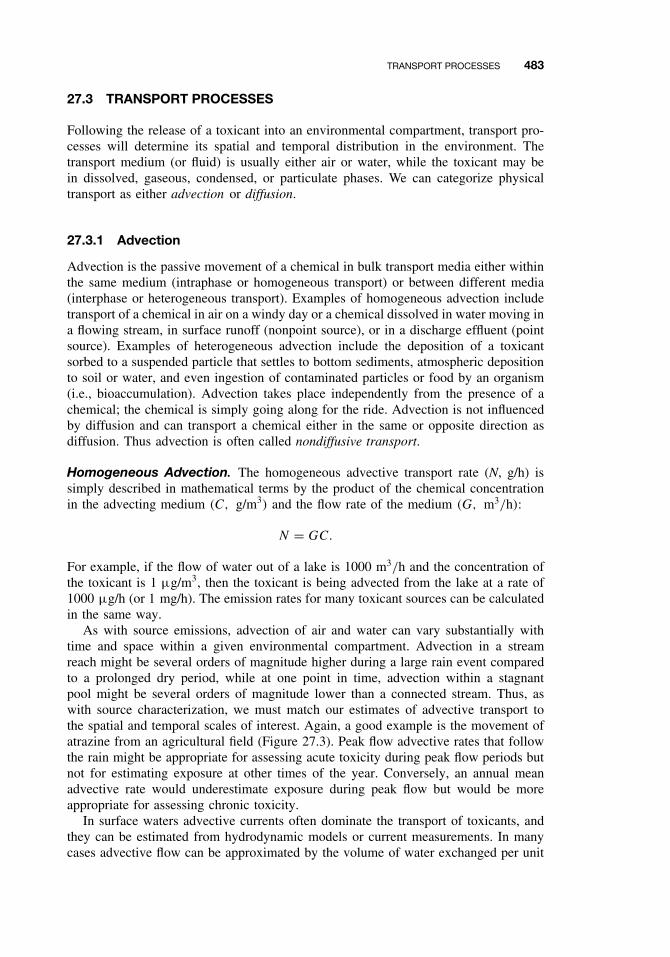
TRANSPORT PROCESSES 483
27.3 TRANSPORT PROCESSES
Following the release of a toxicant into an environmental compartment, transport pro-cesses will determine its spatial and temporal distribution in the environment. Thetransport medium (or fluid) is usually either air or water, while the toxicant may bein dissolved, gaseous, condensed, or particulate phases. We can categorize physicaltransport as either advection or diffusion.
27.3.1 Advection
Advection is the passive movement of a chemical in bulk transport media either withinthe same medium (intraphase or homogeneous transport) or between different media(interphase or heterogeneous transport). Examples of homogeneous advection includetransport of a chemical in air on a windy day or a chemical dissolved in water moving ina flowing stream, in surface runoff (nonpoint source), or in a discharge effluent (pointsource). Examples of heterogeneous advection include the deposition of a toxicantsorbed to a suspended particle that settles to bottom sediments, atmospheric depositionto soil or water, and even ingestion of contaminated particles or food by an organism(i.e., bioaccumulation). Advection takes place independently from the presence of achemical; the chemical is simply going along for the ride. Advection is not influencedby diffusion and can transport a chemical either in the same or opposite direction asdiffusion. Thus advection is often called nondiffusive transport.
Homogeneous Advection. The homogeneous advective transport rate (N, g/h) issimply described in mathematical terms by the product of the chemical concentrationin the advecting medium (C, g/m3) and the flow rate of the medium (G, m3/h):
N = GC.
For example, if the flow of water out of a lake is 1000 m3/h and the concentration ofthe toxicant is 1 µg/m3, then the toxicant is being advected from the lake at a rate of1000 µg/h (or 1 mg/h). The emission rates for many toxicant sources can be calculatedin the same way.
As with source emissions, advection of air and water can vary substantially withtime and space within a given environmental compartment. Advection in a streamreach might be several orders of magnitude higher during a large rain event comparedto a prolonged dry period, while at one point in time, advection within a stagnantpool might be several orders of magnitude lower than a connected stream. Thus, aswith source characterization, we must match our estimates of advective transport tothe spatial and temporal scales of interest. Again, a good example is the movement ofatrazine from an agricultural field (Figure 27.3). Peak flow advective rates that followthe rain might be appropriate for assessing acute toxicity during peak flow periods butnot for estimating exposure at other times of the year. Conversely, an annual meanadvective rate would underestimate exposure during peak flow but would be moreappropriate for assessing chronic toxicity.
In surface waters advective currents often dominate the transport of toxicants, andthey can be estimated from hydrodynamic models or current measurements. In manycases advective flow can be approximated by the volume of water exchanged per unit
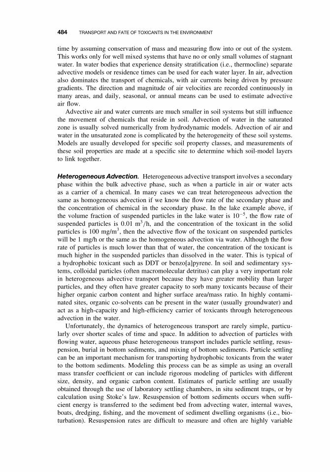
484 TRANSPORT AND FATE OF TOXICANTS IN THE ENVIRONMENT
time by assuming conservation of mass and measuring flow into or out of the system.This works only for well mixed systems that have no or only small volumes of stagnantwater. In water bodies that experience density stratification (i.e., thermocline) separateadvective models or residence times can be used for each water layer. In air, advectionalso dominates the transport of chemicals, with air currents being driven by pressuregradients. The direction and magnitude of air velocities are recorded continuously inmany areas, and daily, seasonal, or annual means can be used to estimate advectiveair flow.
Advective air and water currents are much smaller in soil systems but still influencethe movement of chemicals that reside in soil. Advection of water in the saturatedzone is usually solved numerically from hydrodynamic models. Advection of air andwater in the unsaturated zone is complicated by the heterogeneity of these soil systems.Models are usually developed for specific soil property classes, and measurements ofthese soil properties are made at a specific site to determine which soil-model layersto link together.
Heterogeneous Advection. Heterogeneous advective transport involves a secondaryphase within the bulk advective phase, such as when a particle in air or water actsas a carrier of a chemical. In many cases we can treat heterogeneous advection thesame as homogeneous advection if we know the flow rate of the secondary phase andthe concentration of chemical in the secondary phase. In the lake example above, ifthe volume fraction of suspended particles in the lake water is 10−5, the flow rate ofsuspended particles is 0.01 m3/h, and the concentration of the toxicant in the solidparticles is 100 mg/m3, then the advective flow of the toxicant on suspended particleswill be 1 mg/h or the same as the homogeneous advection via water. Although the flowrate of particles is much lower than that of water, the concentration of the toxicant ismuch higher in the suspended particles than dissolved in the water. This is typical ofa hydrophobic toxicant such as DDT or benzo[a]pyrene. In soil and sedimentary sys-tems, colloidal particles (often macromolecular detritus) can play a very important rolein heterogeneous advective transport because they have greater mobility than largerparticles, and they often have greater capacity to sorb many toxicants because of theirhigher organic carbon content and higher surface area/mass ratio. In highly contami-nated sites, organic co-solvents can be present in the water (usually groundwater) andact as a high-capacity and high-efficiency carrier of toxicants through heterogeneousadvection in the water.
Unfortunately, the dynamics of heterogeneous transport are rarely simple, particu-larly over shorter scales of time and space. In addition to advection of particles withflowing water, aqueous phase heterogeneous transport includes particle settling, resus-pension, burial in bottom sediments, and mixing of bottom sediments. Particle settlingcan be an important mechanism for transporting hydrophobic toxicants from the waterto the bottom sediments. Modeling this process can be as simple as using an overallmass transfer coefficient or can include rigorous modeling of particles with differentsize, density, and organic carbon content. Estimates of particle settling are usuallyobtained through the use of laboratory settling chambers, in situ sediment traps, or bycalculation using Stoke’s law. Resuspension of bottom sediments occurs when suffi-cient energy is transferred to the sediment bed from advecting water, internal waves,boats, dredging, fishing, and the movement of sediment dwelling organisms (i.e., bio-turbation). Resuspension rates are difficult to measure and often are highly variable
TRANSPORT PROCESSES 485
in both time and space. Much as the annual runoff of pesticides from an agriculturalfield may be dominated by a few rain events, annual resuspension rates can be dom-inated by a major storm, and in smaller areas by a single boat or school of bottomfish. Resuspension rates can be estimated from sediment traps deployed just above thesediment surface or from the difference between particle settling and permanent burialor sedimentation. Sedimentation is the net result of particle settling and resuspensionand can be measured using radionuclide dating methods (e.g., 210Pb). Sediment dat-ing itself becomes difficult when there is significant mixing of the surface sediments(e.g., through bioturbation). Thus the heterogeneous transport of toxicants on aqueousparticles can be rather complicated, though many aquatic systems have been modeledreasonably well.
Heterogeneous advective transport in air occurs primarily through the absorption ofchemicals into falling water droplets (wet deposition) or the sorption of chemicals intosolid particles that fall to earth’s surface (dry deposition). Under certain conditionsboth processes can be treated as simple first-order advective transport using a flow rateand concentration in the advecting medium. For example, wet deposition is usuallycharacterized by a washout coefficient that is proportional to rainfall intensity.
27.3.2 Diffusion
Diffusion is the transport of a chemical by random motion due to a state of disequilib-rium. For example, diffusion causes the movement of a chemical within a phase (e.g.,water) from a location of relatively high concentration to a place of lower concentra-tion until the chemical is homogeneously distributed throughout the phase. Likewisediffusive transport will drive a chemical between media (e.g., water and air) until theirequilibrium concentrations are reached and thus the chemical potentials or fugacitiesare equal in each phase.
Diffusion within a Phase. Diffusional transport within a phase can result fromrandom (thermal) motion of the chemical (molecular diffusion), the random turbulentmixing of the transport medium (turbulent diffusion), or a combination of both. Tur-bulent diffusion usually dominates the diffusive (but not necessarily the advective)chemical transport in air and water due to the turbulent motions or eddies that arecommon in nature. In porous media (sediment and soil) the water velocities are typ-ically too low to create eddies, but random mixing still occurs as water tortuouslyflows around particles. This mechanical diffusion is often called dispersion by hydrol-ogists and dispersion on larger scales, such as when groundwater detours around largeareas of less permeable soil, is called macrodispersion. Note, however, that the termdispersion often is used by meteorologists and engineers to describe any turbulentdiffusion.
Although different physical mechanisms can cause diffusive mixing, they all causea net transport of a chemical from areas of higher concentration to areas of lowerconcentration. All diffusive processes are also referred to as Fickian transport becausethey all can be described mathematically by Fick’s first law, which states that the flow(or flux) of a chemical (N, g/h) is proportional to its concentration gradient (dC/dx):
N = −DA
(dC
dx
),
486 TRANSPORT AND FATE OF TOXICANTS IN THE ENVIRONMENT
where D is the diffusivity or mass transfer coefficient (m2/h), A is the area throughwhich the chemical is passing (m2), C is the concentration of the diffusing chemical(g/m3), and x is the distance being considered (m). The negative sign is simply theconvention that the direction of diffusion is from high to low concentration (diffusion ispositive when dC/dx is negative). Note that many scientists and texts define diffusionas an area specific process with units of g/m2h and thus the area term (A) is not includedin the diffusion equation. This is simply an alternative designation that describes trans-port as a flux density (g/m2h) rather than as a flow (g/h). In either case the diffusionequation can be integrated numerically and even expressed in three dimensions usingvector notation. However, for most environmental situations we usually have no accu-rate estimate of D or dx, so we combine the two into a one-dimensional mass transfercoefficient (kM) with units of velocity (m/h). The chemical flux is then the product ofthis velocity, area, and concentration:
N = −kMAC.
Mass transfer coefficients can be estimated from laboratory, mesocosm, and fieldstudies and are widely used in environmental fate models. Mass transfer coefficientscan be derived separately for molecular diffusion, turbulent diffusion, and dispersionin porous media, and all three terms can be added to the chemical flux equation.This is usually not necessary because one term often dominates the transport in spe-cific environmental regions. Consider the vertical diffusion of methane gas generatedby methanogenic bacteria in deep sediments. Molecular diffusion dominates in thehighly compacted and low porosity deeper sediments. Dispersion becomes importantas methane approaches the more porous surface sediments. Following methane gasebulation from the sediment porewater, turbulent diffusion will dominate transport ina well-mixed water column (i.e., not a stagnant pool or beneath a thermocline wheremolecular diffusion will dominate). At the water surface, eddies tend to be dampedand molecular diffusion may again dominate transport. Under stagnant atmosphericconditions (i.e., a temperature inversion) molecular diffusion will continue to dominatebut will yield to more rapid mixing when typical turbulent conditions are reached. Themagnitude and variability of the transport rate generally increase as the methane movesvertically through the environment, except when very stagnant conditions are encoun-tered in the water or air. Modeling the transport of a chemical in air is particularlydifficult because of the high spatial and temporal variability of air movement. Notealso that advective processes in water or air usually transport chemicals at a faster ratethan either molecular or turbulent diffusion.
Diffusion between Phases. The transport of a chemical between phases is some-times treated as a third category of transport processes or even as a transformationreaction. Interphase or intermedia transport is not a transformation reaction becausethe chemical is moving only between phases; it is not reacting with anything or chang-ing its chemical structure. Instead, intermedia transport is simply driven by diffusionbetween two phases. When a chemical reaches an interface such as air–water, parti-cle–water, or (biological) membrane–water, two diffusive regions are created at eitherside of the interface. The classical description of this process is the Whitman two-filmor two-resistance mass transfer theory, where chemicals pass through two stagnantboundary layers by molecular diffusion, while the two bulk phases are assumed to
EQUILIBRIUM PARTITIONING 487
be homogeneously mixed. This allows us to use a first-order function of the concen-tration gradient in the two phases, where the mass transfer coefficient will dependonly on the molecular diffusivity of the chemical in each phase and the thickness ofthe boundary layers. This is fairly straightforward for transfer at the air–water inter-face (and often at the membrane–water interface), but not for the particle–water orparticle–air interfaces.
Diffusive transport between phases can be described mathematically as the productof the departure from equilibrium and a kinetic term:
N = kA(C1 − C2K12),
where N is the transport rate (g/h), k is a transport rate coefficient (m/h), A is theinterfacial area (m2), C1 and C2 are the concentrations in the two phases, and K12 is theequilibrium partition coefficient. At equilibrium K12 equals C1/C2, the term describingthe departure from equilibrium (C1 − C2K12) becomes zero, and thus the net rate oftransfer also is zero. The partition coefficients are readily obtained from thermodynamicdata and equilibrium partitioning experiments. The transport rate coefficients are usuallyestimated from the transport rate equation itself by measuring intermedia transport rates(N ) under controlled laboratory conditions (temperature, wind and water velocities) atknown values of A, C1, C2, and K12. These measurements must then be extrapolated tothe field, sometimes with great uncertainty. This uncertainty, along with the knowledgethat many interfacial regions have reached or are near equilibrium, has led many tosimply assume that equilibrium exists at the interface. Thus the net transport rate iszero and the phase distribution of a chemical is simply described by its equilibriumpartition coefficient.
27.4 EQUILIBRIUM PARTITIONING
When a small amount of a chemical is added to two immiscible phases and then shaken,the phases will eventually separate and the chemical will partition between the twophases according to its solubility in each phase. The concentration ratio at equilibriumis the partition coefficient:
C1
C2= K12.
In the laboratory, we usually determine K12 from the slope of C1 versus C2 overa range of concentrations. Partition coefficients can be measured for essentially anytwo-phase system: air–water, octanol–water, lipid–water, particle–water, and so on.In situ partition coefficients also can be measured where site-specific environmentalconditions might influence the equilibrium phase distribution.
27.4.1 Air–Water Partitioning
Air–water partition coefficients (Kair−water) are essentially Henry’s law constants (H ):
Kair−water = H = Cair
Cwater,
488 TRANSPORT AND FATE OF TOXICANTS IN THE ENVIRONMENT
where H can be expressed in dimensionless form (same units for air and water) orin units of pressure divided by concentration (e.g., Pa m3/mol). The latter is usuallywritten as
H = Pair
Cwater,
where Pair is the partial vapor pressure of the chemical. When H is not measureddirectly, it can be estimated from the ratio of the chemical’s vapor pressure and aqueoussolubility, although one must be careful about using vapor pressures and solubilities thatapply to the same temperature and phase. Chemicals with high Henry’s law constants(e.g., alkanes and many chlorinated solvents) have a tendency to escape from water toair and typically have high vapor pressures, low aqueous solubilities, and low boilingpoints. Chemicals with low Henry’s law constants (e.g., alcohols, chlorinated phenols,larger polycyclic aromatic hydrocarbons, lindane, atrazine) tend to have high watersolubility and/or very low vapor pressure. Note that some chemicals that are consideredto be “nonvolatile,” such as DDT, are often assumed to have low Henry’s law constants.However, DDT also has a very low water solubility yielding a rather high Henry’s lawconstant. Thus DDT readily partitions into the atmosphere as is now apparent from theglobal distribution of DDT.
27.4.2 Octanol–Water Partitioning
For many decades chemists have been measuring the octanol–water partition coeffi-cient (KOW) as a descriptor of hydrophobicity or how much a chemical “hates” to bein water. It is now one of the most important and frequently used physicochemicalproperties in toxicology and environmental chemistry. In fact toxicologists often sim-ply use the symbol P , for partition coefficient, as if no other partition coefficient isimportant. Strong correlations exist between KOW and many biochemical and toxico-logical properties. Octanol has a similar carbon:oxygen ratio as lipids, and the KOW
correlates particularly well with lipid–water partition coefficients. This has led many touse KOW as a measure of lipophilicity or how much a chemical “loves” lipids. This isreally not the case because most chemicals have an equal affinity for octanol and otherlipids (within about a factor of ten), but their affinity for water varies by many ordersof magnitude. Thus it is largely aqueous solubility which determines KOW not octanolor lipid solubility. We generally express KOW as log KOW because KOW values rangefrom less than one (alcohols) to over one billion (larger alkanes and alkyl benzenes).
27.4.3 Lipid–Water Partitioning
In most cases we can assume that the equilibrium distribution and partitioning oforganic chemicals in both mammalian and nonmammalian systems is a function oflipid content in the animal and that the lipid–water partition coefficient (KLW) isequal to KOW. Instances where this is not the case include specific binding sites (e.g.,kepone in the liver) and nonequilibrium conditions caused by slow elimination ratesof higher level organisms or structured lipid phases that sterically hinder accumulationof very hydrophobic chemicals. For aquatic organisms in constant contact with water,the bioconcentration factor or fish-water partition coefficient (KFW) is simply:
KFW = flipidKOW,
EQUILIBRIUM PARTITIONING 489
where flipid is the mass fraction of lipid in the fish (g lipid/g fish). Several studies haveshown this relationship works well for many fish and shellfish species and an aggregateplot of KFW versus KOW for many different fish species yields a slope of 0.048, whichis about the average lipid concentration of fish (5%). Again, nonequilibrium conditionswill cause deviation from this equation. Such deviations are found at both the top andbottom of the aquatic food chain. Phytoplankton can have higher apparent lipid–waterpartition coefficients because their large surface area : volume ratios increase the rela-tive importance of surface sorption. Top predators such as marine mammals also havehigh apparent lipid-water partition coefficients because of very slow elimination rates.Thus the deviations occur not because “there is something wrong with the equation”but because the underlying assumption of equilibrium is not appropriate in these cases.
27.4.4 Particle–Water Partitioning
It has been known for several decades that many chemicals preferentially associatewith soil and sediment particles rather than the aqueous phase. The particle–waterpartition coefficient (KP) describing this phenomenon is
KP = CS
CW,
where CS is the concentration of chemical in the soil or sediment (mg/kg dry weight)and CW is the concentration in water (mg/L). In this form, KP has units of L/kgor reciprocal density. Dimensionless partition coefficients are sometimes used whereKP is multiplied by the particle density (in kg/L). It has also been observed, first bypesticide chemists in soil systems and later by environmental engineers and chemists insewage effluent and sediment systems, that nonionic organic chemicals were primarilyassociated with the organic carbon phase(s) of particles. A plot of KP versus the massfraction of organic carbon in the soil (fOC, g/g) is linear with a near-zero interceptyielding the simple relationship
KP = fOCKOC,
where KOC is the organic carbon–water partition coefficient (L/kg). Studies withmany chemicals and many sediment/soil systems have demonstrated the utility of thisequation when the fraction of organic carbon is about 0.5% or greater. At lower organiccarbon fractions, interaction with the mineral phase becomes relatively more important(though highly variable) resulting in a small positive intercept of KP versus fOC. Thestrongest interaction between organic chemicals and mineral phases appears to be withdry clays. Thus KP will likely change substantially as a function of water content inlow organic carbon, clay soils.
Measurements of KOC have been taken directly from partitioning experiments insediment–and soil–water systems over a range of environmental conditions in both thelaboratory and the field. Not surprisingly, the KOC values for many organic chemicalsare highly correlated with their KOW values. Plots of the two partition coefficients forhundreds of chemicals with widely ranging KOW values yield slopes from about 0.3 to1, depending on the classes of compounds and the particular methods included. Mostfate modelers continue to use a slope of 0.41, which was reported by the first definitive
490 TRANSPORT AND FATE OF TOXICANTS IN THE ENVIRONMENT
study on the subject in the early 1980s. Thus we now have a means of estimating thepartitioning of a chemical between a particle and water by using the KOW and fOC:
KP = fOCKOC = fOC 0.41KOW.
This relationship is commonly used in environmental fate models to predict aqueousconcentrations from sediment measurements by substituting the equilibrium expressionfor KP and rearranging to solve for CW:
KP = CS
CW= fOC 0.41KOW,
CW = CS
fOC0.41KOW.
This last equation forms the basis for the EPA’s sediment quality guidelines that areused to assess the potential toxicity of contaminated sediments. The idea is to simplymeasure CS and fOC, look up KOW in a table, compute the predicted CW, and comparethis result to established water quality criteria for the protection of aquatic life or humanlife (e.g., carcinogenicity risk factors). The use of this simple equilibrium partitioningexpression for this purpose is currently the subject of much debate among scientists aswell as policy makers.
27.5 TRANSFORMATION PROCESSES
The potential environmental hazard associated with the use of a chemical is directlyrelated to it’s persistence in the environment (see Chapter 26), which in turn dependson the rates of chemical transformation reactions. Transformation reactions can bedivided into two classes: reversible reactions that involve continuous exchange amongchemical states (ionization, complexation) and irreversible reactions that permanentlytransform a parent chemical into a daughter or reaction product (photolysis, hydrolysis,and many redox reactions). Reversible reactions are usually abiotic, although biologicalprocesses can still exert great influence over them (e.g., via production of complexingagents or a change in pH). Irreversible reactions can be abiotic or mediated directlyby biota, particularly bacteria.
27.5.1 Reversible Reactions
Ionization. Ionization refers to the dissociation of a neutral chemical into chargedspecies. The most common form of neutral toxicant dissociation is acid-base equilibria.The hypothetical monoprotic acid, HA, will dissociate in water to form the conjugateacid-base pair (H+, A−) usually written as
HA + H2O = H3O+ + A−.
The equilibrium constant for this reaction, the acidity constant (Ka), is defined by thelaw of mass action and is given by
Ka = [H3O+][A−]
[HA].
TRANSFORMATION PROCESSES 491
For convenience we often express equilibrium constants as the negative logarithm, orpK value. Thus the relative proportion of the neutral and charged species, will be afunction of the pKa and solution pH. When the pH is equal to pKa, equal concentrationsof the neutral and ionized forms will be present. When pH is less than the pKa,the neutral species will be predominant; when pH is greater than pKa, the ionizedspecies will be in excess. The exact equilibrium distribution can be calculated fromthe equilibrium expression above and the law of mass conservation.
The fate of a chemical is often a function of the relative abundance of a particularchemical species as well as the total concentration. For example, the neutral chemicalmight partition into biological lipids or organic carbon in soil to a greater extent thanthe ionized form. Many acidic toxicants (pentachlorophenol) exhibit higher toxicitiesto aquatic organisms at lower pH where the neutral species predominates. However,specific ionic interactions will take place only with the ionized species. A classicexample of how pH influences the fate and effects of a toxicant is with hydrogencyanide. The pKa of HCN is about 9 and the toxicity of CN− is much higher thanthat of HCN for many aquatic organisms. Thus the discharge of a basic (high pH)industrial effluent containing cyanide would pose a greater hazard to fish than a lowerpH effluent (everything else being equal). The effluent could be treated to reduce thepH well below the pKa according to the reaction:
CN− + H+ = HCN(aq),
thus reducing the hazard to the fish. However, HCN has a rather high Henry’s lawconstant and will partition into the atmosphere:
HCN(aq) = HCN(air).
This may be fine for the fish, but birds in the area and humans working at the industrialplant will now have a much greater exposure to HCN. Thus both the fate and toxicityof a chemical can be influenced by simple ionization reactions.
Precipitation and Dissolution. A special case of ionization is the dissolution of aneutral solid phase into soluble species. For example, the binary solid metal sulfide,CuS, dissolves in water according to
CuS(s) + H+ = Cu2+ + HS−.
The equilibrium constant for this reaction, the solubility product (Ksp), is given by
Ksp = [Cu2+][HS−]
[H+].
The solubility product for CuS is very low (Ksp = 10−19 as written) so that the pres-ence of sulfide in water acts to immobilize Cu (and many other metals) and reduceeffective exposure. The formation of metal sulfides is important in anaerobic soil andsediment, stagnant ponds and basins, and many industrial and domestic sewage treat-ment plants and discharges. Co-precipitation of metals also can be a very importantremoval process in natural waters. In aerobic systems, the precipitation of hydrous
492 TRANSPORT AND FATE OF TOXICANTS IN THE ENVIRONMENT
oxides of manganese and iron often incorporate other metals as impurities. In anaero-bic systems, the precipitation of iron sulfides can include other metals as well. Theseco-precipitates are usually not thermodynamically stable, but their conversion to stablemineral phases often takes place on geological time scales.
Complexation and Chemical Speciation. Natural systems contain many chemicalsthat undergo ionic or covalent interactions with toxicants to change toxicant specia-tion and chemical speciation can have a profound effect on both fate and toxicity.Again, in the case of copper, inorganic ions (Cl−, OH−) and organic detritus (humicacids, peptides) will react with dissolved Cu2+ to form various metal-ligand com-plexes. Molecular diffusivities of complexed copper will be lower than uncomplexed(hydrated) copper and will generally decrease with the size and number of ligands.The toxicity of free, uncomplexed Cu2+ to many aquatic organisms is much higherthan Cu2+ that is complexed to chelating agents such as EDTA or glutathione. Manytransition metal toxicants, such as Cu, Pb, Cd, and Hg, have high binding constantswith compounds that contain amine, sulfhydryl, and carboxylic acid groups. Thesegroups are quite common in natural organic matter. Even inorganic complexes of OH−and Cl− reduce Cu2+ toxicity. In systems where a mineral phase is controlling Cu2+solubility, the addition of these complexing agents will shift the solubility equilibriumaccording to LeChatelier’s principle as shown here for CuS and OH−, Cl−, and GSH(glutathione):
CuS(s) + H+ = Cu2+ + HS−, Ksp,
Cu2+ + OH− = CuOH+, KCuOH,
Cu2+ + Cl− = CuCl+, KCuCl,
Cu2+ + GSH = CuGS+ + H+, KCuGS.
Each successive complexation reaction “leaches” Cu2+ from the solid mineral phase,thereby increasing the total copper in the water but not affecting the concentration of(or exposure to) Cu2+. These equilibria can be combined into one reaction:
4CuS(s) + 3H+ + OH− + Cl− + GSH = Cu2+ + 4HS−
+ CuOH+ + CuCl+ + CuGS+,
and the overall equilibrium constant derived as shown:
Koverall = (4)Ksp × KCuOH × KCuCl × KCuGS
= [Cu2+] [CuOH+] [CuCl+] [CuGS+] [HS−]4/[H+]3 [OH−] [Cl−] [GS−].
A series of simultaneous equations can be derived for these reactions to computethe concentration of individual copper species, and the total concentration of copper,[Cu]T, would be given by
[Cu]T = [Cu2+] + [CuOH+] + [CuCl+] + [CuGS+].
Thus the total copper added to a toxicity test or measured as the exposure (e.g., byatomic absorption spectroscopy) may be much greater than that which is available toan organism to induce toxicological effects.
TRANSFORMATION PROCESSES 493
Literally hundreds of complex equilibria like this can be combined to model whathappens to metals in aqueous systems. Numerous speciation models exist for this appli-cation that include all of the necessary equilibrium constants. Several of these modelsinclude surface complexation reactions that take place at the particle–water interface.Unlike the partitioning of hydrophobic organic contaminants into organic carbon, met-als actually form ionic and covalent bonds with surface ligands such as sulfhydrylgroups on metal sulfides and oxide groups on the hydrous oxides of manganese andiron. Metals also can be biotransformed to more toxic species (e.g., conversion of ele-mental mercury to methyl-mercury by anaerobic bacteria), less toxic species (oxidationof tributyl tin to elemental tin), or temporarily immobilized (e.g., via microbial reduc-tion of sulfate to sulfide, which then precipitates as an insoluble metal sulfide mineral).
27.5.2 Irreversible Reactions
The reversible transformation reactions discussed above alter the fate and toxicityof chemicals, but they do not irreversibly change the structure or properties of thechemical. An acid can be neutralized to its conjugate base, and vice versa. Copper canprecipitate as a metal sulfide, dissolve and from a complex with numerous ligands,and later re-precipitate as a metal sulfide. Irreversible transformation reactions alterthe structure and properties of a chemical forever.
Hydrolysis. Hydrolysis is the cleavage of organic molecules by reaction with waterwith a net displacement of a leaving group (X) with OH−:
RX + H2O = ROH + HX.
Hydrolysis is part of the larger class of chemical reactions called nucleophilic displace-ment reactions in which a nucleophile (electron-rich species with an unshared pair ofelectrons) attacks an electrophile (electron deficient), cleaving one covalent bond toform a new one. Hydrolysis is usually associated with surface waters but also takesplace in the atmosphere (fogs and clouds), groundwater, at the particle–water interfaceof soils and sediments, and in living organisms.
Hydrolysis can proceed through numerous mechanisms via attack by H2O (neutralhydrolysis) or by acid (H+) or base (OH−) catalysis. Acid and base catalyzed reactionsproceed via alternative mechanisms that require less energy than neutral hydrolysis.The combined hydrolysis rate term is a sum of these three constituent reactions and isgiven by
d[RX]
dt= kobs[RX] = ka[H+][RX] + kn[RX] + kb[OH−][RX],
where [RX] is the concentration of the hydrolyzable chemical, kobs is the macroscopicobserved hydrolysis rate constant, and ka, kn, and kb are the rate constants for the acid-catalyzed, neutral, and base-catalyzed hydrolysis. If we assume that the hydrolysis canbe approximated by first-order kinetics with respect to RX (which is usually true), therate term is reduced to
kobs = ka[H+] + kn + kb[OH−].
Neutral hydrolysis is dependent only on water which is present in excess, so kn is asimple pseudo-first-order rate constant (with units t−1). The acid- and base-catalyzed

494 TRANSPORT AND FATE OF TOXICANTS IN THE ENVIRONMENT
hydrolysis depend on the molar quantities of [H+] and [OH−], respectively, so ka andkb have units of M−1t−1.
The observed or apparent hydrolysis half-life at a fixed pH is then given by
t1/2 = ln 2
kobs.
Compilations of hydrolysis half-lives at pH and temperature ranges encountered innature can be found in many sources. Reported hydrolysis half-lives for organic com-pounds at pH 7 and 298 K range at least 13 orders of magnitude. Many esters hydrolyzewithin hours or days, whereas some organic chemicals will never hydrolyze. For halo-genated methanes, which are common groundwater contaminants, half-lives range fromabout 1 year for CH3Cl to about 7000 years for CCl4. The half-lives of halomethanesfollows the strength of the carbon-halogen bond with half-lives decreasing in theorder F > Cl > Br. Small structural changes can dramatically alter hydrolysis rates.An example is the difference between tetrachloroethane (Cl2HC–CHCl2) and tetra-chloroethene (Cl2C=CCl2) which have hydrolysis half-lives of about 0.5 year and109 years, respectively. In this case the hydrolysis rate is affected by the C–Cl bondstrength and the steric bulk at the site of nucleophilic substitution.
The apparent rate of hydrolysis and the relative abundance of reaction products isoften a function of pH because alternative reaction pathways are preferred at differentpH. In the case of halogenated hydrocarbons, base-catalyzed hydrolysis will result inelimination reactions while neutral hydrolysis will take place via nucleophilic displace-ment reactions. An example of the pH dependence of hydrolysis is illustrated by thebase-catalyzed hydrolysis of the structurally similar insecticides DDT and methoxy-chlor. Under a common range of natural pH (5 to 8) the hydrolysis rate of methoxychloris invariant while the hydrolysis of DDT is about 15-fold faster at pH 8 compared topH 5. Only at higher pH (>8) does the hydrolysis rate of methoxychlor increase. Inaddition the major product of DDT hydrolysis throughout this pH range is the same(DDE), while the methoxychlor hydrolysis product shifts from the alcohol at pH 5–8(nucleophilic substitution) to the dehydrochlorinated DMDE at pH > 8 (elimination).This illustrates the necessity to conduct detailed mechanistic experiments as a functionof pH for hydrolytic reactions.
Photolysis. Photolysis of a chemical can proceed either by direct absorption of light(direct photolysis) or by reaction with another chemical species that has been producedor excited by light (indirect photolysis). In either case photochemical transformationssuch as bond cleavage, isomerization, intramolecular rearrangement, and various inter-molecular reactions can result. Photolysis can take place wherever sufficient lightenergy exists, including the atmosphere (in the gas phase and in aerosols and fog/clouddroplets), surface waters (in the dissolved phase or at the particle–water interface), andin the terrestrial environment (on plant and soil/mineral surfaces).
Photolysis dominates the fate of many chemicals in the atmosphere because of thehigh solar irradiance. Near the earth’s surface, chromophores such as nitrogen oxides,carbonyls, and aromatic hydrocarbons play a large role in contaminant fate in urbanareas. In the stratosphere, light is absorbed by ozone, oxygen, organohalogens, andhydrocarbons with global environmental implications. The rate of photolysis in surfacewaters depends on light intensity at the air–water interface, the transmittance through
TRANSFORMATION PROCESSES 495
this interface, and the attenuation through the water column. Open ocean waters (“bluewater”) can transmit blue light to depths of 150 m while highly eutrophic or turbidwaters might absorb all light within 1 cm of the surface.
Oxidation-Reduction Reactions. Although many redox reactions are reversible,they are included here because many of the redox reactions that influence the fateof toxicants are irreversible on the temporal and spatial scales that are importantto toxicity.
Oxidation is simply defined as a loss of electrons. Oxidizing agents are electrophilesand thus gain electrons upon reaction. Oxidations can result in the increase in the oxi-dation state of the chemical as in the oxidation of metals or oxidation can incorporateoxygen into the molecule. Typical organic chemical oxidative reactions include dealky-lation, epoxidation, aromatic ring cleavage, and hydroxylation. The term autooxidation,or weathering, is commonly used to describe the general oxidative degradation of achemical (or chemical mixture, e.g., petroleum) upon exposure to air. Chemicals canreact abiotically in both water and air with oxygen, ozone, peroxides, free radicals,and singlet oxygen. The last two are common intermediate reactants in indirect pho-tolysis. Mineral surfaces are known to catalyze many oxidative reactions. Clays andoxides of silicon, aluminum, iron, and manganese can provide surface active sites thatincrease rates of oxidation. There are a variety of complex mechanisms associated withthis catalysis, so it is difficult to predict the catalytic activity of soils and sedimentin nature.
Reduction of a chemical species takes place when an electron donor (reductant)transfers electrons to an electron acceptor (oxidant). Organic chemicals typically actas the oxidant, while abiotic reductants include sulfide minerals, reduced metals orsulfur compounds, and natural organic matter. There are also extracellular biochemicalreducing agents such as porphyrins, corrinoids, and metal-containing coenzymes. Mostof these reducing agents are present only in anaerobic environments where anaero-bic bacteria are themselves busy reducing chemicals. Thus it is usually very difficultto distinguish biotic and abiotic reductive processes in nature. Well-controlled, sterilelaboratory studies are required to measure abiotic rates of reduction. These studies indi-cate that many abiotic reductive transformations could be important in the environment,including dehalogenation, dealkylation, and the reduction of quinones, nitrosamines,azoaromatics, nitroaromatics, and sulfoxides. Functional groups that are resistant toreduction include aldehydes, ketones, carboxylic acids (and esters), amides, alkenes,and aromatic hydrocarbons.
Biotransformations. As we have seen throughout much of this textbook, verte-brates have developed the capacity to transform many toxicants into other chemicals,sometimes detoxifying the chemical and sometimes activating it. The same or simi-lar biochemical processes that hydrolyze, oxidize, and reduce toxicants in vertebratesalso take place in many lower organisms. In particular, bacteria, protozoans, and fungiprovide a significant capacity to biotransform toxicants in the environment. Althoughmany vertebrates can metabolize toxicants faster than these lower forms of life, theaggregate capacity of vertebrates to biotransform toxicants (based on total biomass andexposure) is insignificant to the overall fate of a toxicant in the environment. In thissection we use the term biotransformation to include all forms of direct biologicaltransformation reactions.
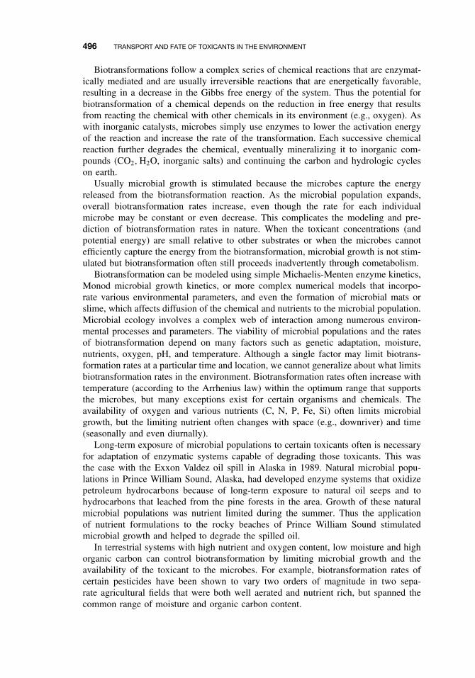
496 TRANSPORT AND FATE OF TOXICANTS IN THE ENVIRONMENT
Biotransformations follow a complex series of chemical reactions that are enzymat-ically mediated and are usually irreversible reactions that are energetically favorable,resulting in a decrease in the Gibbs free energy of the system. Thus the potential forbiotransformation of a chemical depends on the reduction in free energy that resultsfrom reacting the chemical with other chemicals in its environment (e.g., oxygen). Aswith inorganic catalysts, microbes simply use enzymes to lower the activation energyof the reaction and increase the rate of the transformation. Each successive chemicalreaction further degrades the chemical, eventually mineralizing it to inorganic com-pounds (CO2, H2O, inorganic salts) and continuing the carbon and hydrologic cycleson earth.
Usually microbial growth is stimulated because the microbes capture the energyreleased from the biotransformation reaction. As the microbial population expands,overall biotransformation rates increase, even though the rate for each individualmicrobe may be constant or even decrease. This complicates the modeling and pre-diction of biotransformation rates in nature. When the toxicant concentrations (andpotential energy) are small relative to other substrates or when the microbes cannotefficiently capture the energy from the biotransformation, microbial growth is not stim-ulated but biotransformation often still proceeds inadvertently through cometabolism.
Biotransformation can be modeled using simple Michaelis-Menten enzyme kinetics,Monod microbial growth kinetics, or more complex numerical models that incorpo-rate various environmental parameters, and even the formation of microbial mats orslime, which affects diffusion of the chemical and nutrients to the microbial population.Microbial ecology involves a complex web of interaction among numerous environ-mental processes and parameters. The viability of microbial populations and the ratesof biotransformation depend on many factors such as genetic adaptation, moisture,nutrients, oxygen, pH, and temperature. Although a single factor may limit biotrans-formation rates at a particular time and location, we cannot generalize about what limitsbiotransformation rates in the environment. Biotransformation rates often increase withtemperature (according to the Arrhenius law) within the optimum range that supportsthe microbes, but many exceptions exist for certain organisms and chemicals. Theavailability of oxygen and various nutrients (C, N, P, Fe, Si) often limits microbialgrowth, but the limiting nutrient often changes with space (e.g., downriver) and time(seasonally and even diurnally).
Long-term exposure of microbial populations to certain toxicants often is necessaryfor adaptation of enzymatic systems capable of degrading those toxicants. This wasthe case with the Exxon Valdez oil spill in Alaska in 1989. Natural microbial popu-lations in Prince William Sound, Alaska, had developed enzyme systems that oxidizepetroleum hydrocarbons because of long-term exposure to natural oil seeps and tohydrocarbons that leached from the pine forests in the area. Growth of these naturalmicrobial populations was nutrient limited during the summer. Thus the applicationof nutrient formulations to the rocky beaches of Prince William Sound stimulatedmicrobial growth and helped to degrade the spilled oil.
In terrestrial systems with high nutrient and oxygen content, low moisture and highorganic carbon can control biotransformation by limiting microbial growth and theavailability of the toxicant to the microbes. For example, biotransformation rates ofcertain pesticides have been shown to vary two orders of magnitude in two sepa-rate agricultural fields that were both well aerated and nutrient rich, but spanned thecommon range of moisture and organic carbon content.
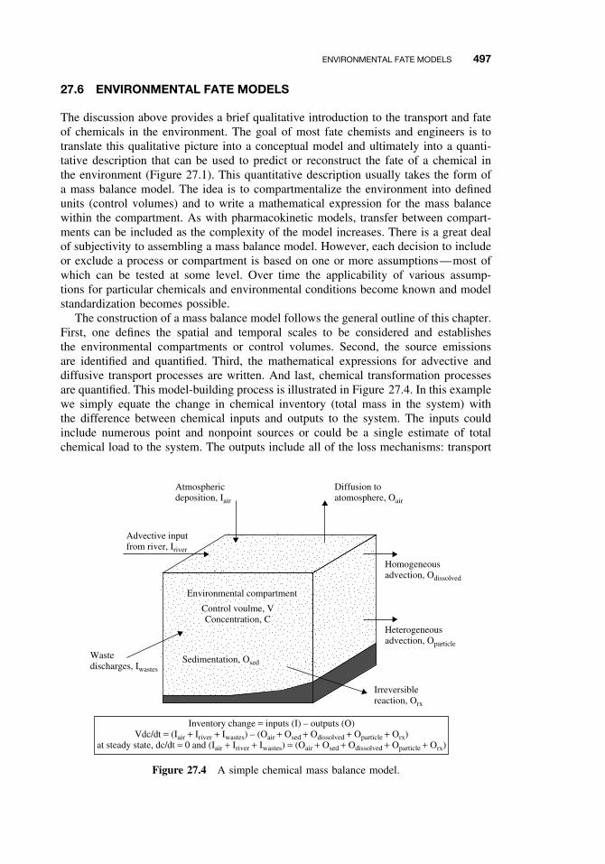
ENVIRONMENTAL FATE MODELS 497
27.6 ENVIRONMENTAL FATE MODELS
The discussion above provides a brief qualitative introduction to the transport and fateof chemicals in the environment. The goal of most fate chemists and engineers is totranslate this qualitative picture into a conceptual model and ultimately into a quanti-tative description that can be used to predict or reconstruct the fate of a chemical inthe environment (Figure 27.1). This quantitative description usually takes the form ofa mass balance model. The idea is to compartmentalize the environment into definedunits (control volumes) and to write a mathematical expression for the mass balancewithin the compartment. As with pharmacokinetic models, transfer between compart-ments can be included as the complexity of the model increases. There is a great dealof subjectivity to assembling a mass balance model. However, each decision to includeor exclude a process or compartment is based on one or more assumptions—most ofwhich can be tested at some level. Over time the applicability of various assump-tions for particular chemicals and environmental conditions become known and modelstandardization becomes possible.
The construction of a mass balance model follows the general outline of this chapter.First, one defines the spatial and temporal scales to be considered and establishesthe environmental compartments or control volumes. Second, the source emissionsare identified and quantified. Third, the mathematical expressions for advective anddiffusive transport processes are written. And last, chemical transformation processesare quantified. This model-building process is illustrated in Figure 27.4. In this examplewe simply equate the change in chemical inventory (total mass in the system) withthe difference between chemical inputs and outputs to the system. The inputs couldinclude numerous point and nonpoint sources or could be a single estimate of totalchemical load to the system. The outputs include all of the loss mechanisms: transport
Atmosphericdeposition, Iair
Advective inputfrom river, Iriver
Diffusion toatomosphere, Oair
Homogeneousadvection, Odissolved
Heterogeneousadvection, Oparticle
Irreversiblereaction, Orx
Wastedischarges, Iwastes
Inventory change = inputs (I) – outputs (O)Vdc/dt = (Iair + Iriver + Iwastes) – (Oair + Osed + Odissolved + Oparticle + Orx)
at steady state, dc/dt = 0 and (Iair + Iriver + Iwastes) = (Oair + Osed + Odissolved + Oparticle + Orx)
Environmental compartment
Sedimentation, Osed
Control voulme, VConcentration, C
Figure 27.4 A simple chemical mass balance model.
498 TRANSPORT AND FATE OF TOXICANTS IN THE ENVIRONMENT
Water column (2 kg)
Particlerecycling?
Desorption?
Outflow
25 kg/y
3 kg/y
Lossprocesses
Mass Budget for Benzo(a)pyrene
Other coastal
2 kg/y
30 kg/y
75 kg/y
64 kg/y
Boston harbor
Atomospheric deposition
Merrimack river
Total input = 171 kg/y Total loss = 198 kg/yNet removal = 27 kg/y
Burial170 kg/y
Surface sediment (2,400 kg)Total sediment (14,500 kg)
Figure 27.5 Information provided by a chemical mass balance model. The annual mass budgetof benzo[a]pyrene in Massachusetts Bay is shown.
out of the compartment and irreversible transformation reactions. If steady state can beassumed (i.e., the chemical’s concentration in the compartment is not changing overthe time scale of the model), the inventory change is zero and we are left with a simplemass balance equation to solve. Unsteady-state conditions would require a numericalsolution to the differential equations.
There are many tricks and shortcuts to this process. For example, rather than compil-ing all of the transformation rate equations (or conducting the actual kinetic experimentsyourself), there are many sources of typical chemical half-lives based on pseudo-first-order rate expressions. It is usually prudent to begin with these “best estimates” ofhalf-lives in air, water, soil, and sediment and perform a sensitivity analysis with themodel to determine which processes are most important. One can return to the mostimportant processes to assess whether more detailed rate expressions are necessary. Anillustration of this mass balance approach is given in Figure 27.5 for benzo[a]pyrene.This approach allows a first-order evaluation of how chemicals enter the environment,what happens to them in the environment, and what the exposure concentrations will bein various environmental media. Thus the chemical mass balance provides informationrelevant to toxicant exposure to both humans and wildlife.
SUGGESTED READING
Hemond, H. F., and E. J. Fechner. Chemical Fate and Transport in the Environment. New York:Academic Press, 1994.
Mackay, D., W. Y. Shiu, and K. C. Ma. Physical-Chemical Properties and Environmental Fateand Degradation Handbook. CRCnetBASE 2000 CR-ROM. Boca Raton, FL: CRC Press,2000.

SUGGESTED READING 499
Mackay, D. Multimedia Environmental Models: The Fugacity Approach, 2nd ed. Boca Raton,FL: Lewis Publishers, 2001.
Rand, G. M., ed. Fundamentals of Aquatic Toxicology: Part II Environmental Fate. Washington,DC: Taylor and Francis, 1995.
Schnoor, J. L. Environmental Modeling: Fate and Transport of Pollutants in Water, Air, and Soil.New York: Wiley, 1996.
Schwarzenbach, R. P., P. M. Gschwend, and D. M. Imboden. Environmental Organic Chem-istry, 2nd ed. New York: Wiley, 2002.

CHAPTER 28
Environmental Risk Assessment
DAMIAN SHEA
28.1 INTRODUCTION
Risk assessment is the process of assigning magnitudes and probabilities to adverseeffects associated with an event. The development of risk assessment methodologyhas focused on accidental events (e.g., an airplane crash) and specific environmentalstresses to humans (exposure of humans to chemicals), and thus most risk assessmentis characterized by discrete events or stresses affecting well-defined endpoints (e.g.,incidence of human death or cancer). This single stress–single end point relationshipallows the use of relatively simple statistical and mechanistic models to estimate riskand is widely used in human health risk assessment. However, this simple paradigmhas only partial applicability to ecological risk assessment because of the inherentcomplexity of ecological systems and the exposure to numerous physical, chemical,and biological stresses that have both direct and indirect effects on a diversity ofecological components, processes, and endpoints. Thus, although the roots of ecologicalrisk assessment can be found in human health risk assessment, the methodology forecological risk assessment is not well developed and the estimated risks are highlyuncertain. Despite these limitations, resource managers and regulators are looking toecological risk assessment to provide a scientific basis for prioritizing problems thatpose the greatest ecological risk and to focus research efforts in areas that will yieldthe greatest reduction in uncertainty.
To this end the US Environmental Protection Agency has issued guidelines forplanning and conducting ecological risk assessments. Because of the complexity anduncertainty associated with ecological risk assessment the EPA guidelines provideonly a loose framework for organizing and analyzing data, information, assumptions,and uncertainties to evaluate the likelihood of adverse ecological effects. However,the guidelines represent a broad consensus of the present scientific knowledge andexperience on ecological risk assessment. This chapter presents a brief overview of theecological risk assessment process as presently described by the EPA.
Ecological risk assessment can be defined as:
The process that evaluates the likelihood that adverse ecological effects may occur or areoccurring as a result of exposure to one or more stressors.
A Textbook of Modern Toxicology, Third Edition, edited by Ernest HodgsonISBN 0-471-26508-X Copyright 2004 John Wiley & Sons, Inc.
501
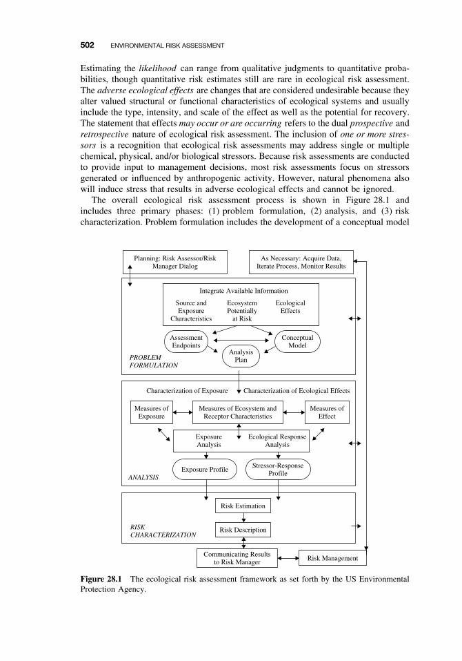
502 ENVIRONMENTAL RISK ASSESSMENT
Estimating the likelihood can range from qualitative judgments to quantitative proba-bilities, though quantitative risk estimates still are rare in ecological risk assessment.The adverse ecological effects are changes that are considered undesirable because theyalter valued structural or functional characteristics of ecological systems and usuallyinclude the type, intensity, and scale of the effect as well as the potential for recovery.The statement that effects may occur or are occurring refers to the dual prospective andretrospective nature of ecological risk assessment. The inclusion of one or more stres-sors is a recognition that ecological risk assessments may address single or multiplechemical, physical, and/or biological stressors. Because risk assessments are conductedto provide input to management decisions, most risk assessments focus on stressorsgenerated or influenced by anthropogenic activity. However, natural phenomena alsowill induce stress that results in adverse ecological effects and cannot be ignored.
The overall ecological risk assessment process is shown in Figure 28.1 andincludes three primary phases: (1) problem formulation, (2) analysis, and (3) riskcharacterization. Problem formulation includes the development of a conceptual model
Planning: Risk Assessor/RiskManager Dialog
As Necessary: Acquire Data,Iterate Process, Monitor Results
AssessmentEndpoints
Integrate Available Information
Source andExposure
Characteristics
EcosystemPotentially
at Risk
EcologicalEffects
AnalysisPlan
ConceptualModel
PROBLEMFORMULATION
ANALYSIS
RISKCHARACTERIZATION
Measures ofExposure
Exposure Profile
Measures ofEffect
Measures of Ecosystem andReceptor Characteristics
Risk Estimation
Risk Management
Risk Description
Stressor-ResponseProfile
Communicating Resultsto Risk Manager
ExposureAnalysis
Ecological ResponseAnalysis
Characterization of Ecological EffectsCharacterization of Exposure
Figure 28.1 The ecological risk assessment framework as set forth by the US EnvironmentalProtection Agency.
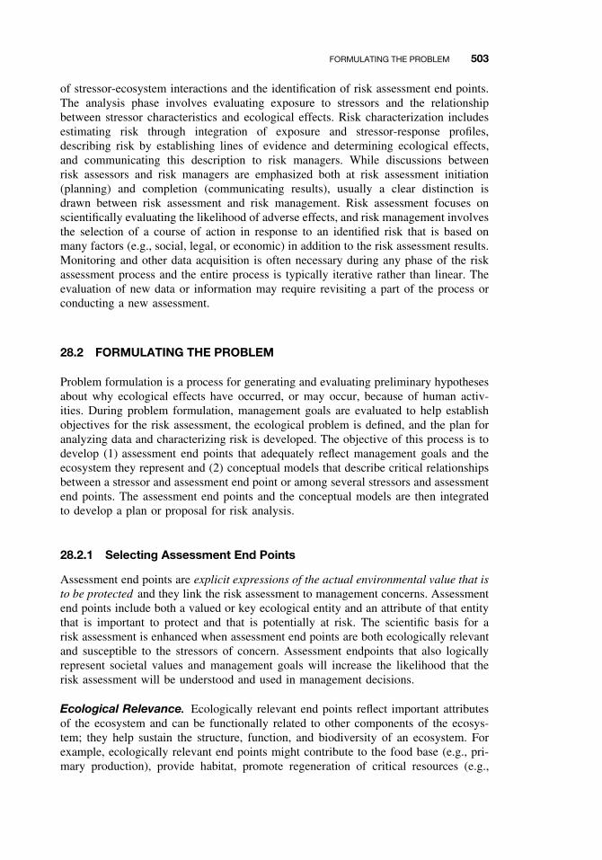
FORMULATING THE PROBLEM 503
of stressor-ecosystem interactions and the identification of risk assessment end points.The analysis phase involves evaluating exposure to stressors and the relationshipbetween stressor characteristics and ecological effects. Risk characterization includesestimating risk through integration of exposure and stressor-response profiles,describing risk by establishing lines of evidence and determining ecological effects,and communicating this description to risk managers. While discussions betweenrisk assessors and risk managers are emphasized both at risk assessment initiation(planning) and completion (communicating results), usually a clear distinction isdrawn between risk assessment and risk management. Risk assessment focuses onscientifically evaluating the likelihood of adverse effects, and risk management involvesthe selection of a course of action in response to an identified risk that is based onmany factors (e.g., social, legal, or economic) in addition to the risk assessment results.Monitoring and other data acquisition is often necessary during any phase of the riskassessment process and the entire process is typically iterative rather than linear. Theevaluation of new data or information may require revisiting a part of the process orconducting a new assessment.
28.2 FORMULATING THE PROBLEM
Problem formulation is a process for generating and evaluating preliminary hypothesesabout why ecological effects have occurred, or may occur, because of human activ-ities. During problem formulation, management goals are evaluated to help establishobjectives for the risk assessment, the ecological problem is defined, and the plan foranalyzing data and characterizing risk is developed. The objective of this process is todevelop (1) assessment end points that adequately reflect management goals and theecosystem they represent and (2) conceptual models that describe critical relationshipsbetween a stressor and assessment end point or among several stressors and assessmentend points. The assessment end points and the conceptual models are then integratedto develop a plan or proposal for risk analysis.
28.2.1 Selecting Assessment End Points
Assessment end points are explicit expressions of the actual environmental value that isto be protected and they link the risk assessment to management concerns. Assessmentend points include both a valued or key ecological entity and an attribute of that entitythat is important to protect and that is potentially at risk. The scientific basis for arisk assessment is enhanced when assessment end points are both ecologically relevantand susceptible to the stressors of concern. Assessment endpoints that also logicallyrepresent societal values and management goals will increase the likelihood that therisk assessment will be understood and used in management decisions.
Ecological Relevance. Ecologically relevant end points reflect important attributesof the ecosystem and can be functionally related to other components of the ecosys-tem; they help sustain the structure, function, and biodiversity of an ecosystem. Forexample, ecologically relevant end points might contribute to the food base (e.g., pri-mary production), provide habitat, promote regeneration of critical resources (e.g.,

504 ENVIRONMENTAL RISK ASSESSMENT
nutrient cycling), or reflect the structure of the community, ecosystem, or landscape(e.g., species diversity). Ecological relevance becomes most useful when it is possibleto identify the potential cascade of adverse effects that could result from a critical ini-tiating effect such as a change in ecosystem function. The selection of assessment endpoints that address both specific organisms of concern and landscape-level ecosystemprocesses becomes increasingly important (and more difficult) in landscape-level riskassessments. In these cases it may be possible to select one or more species and anecosystem process to represent larger functional community or ecosystem processes.Extrapolations like these must be explicitly described in the conceptual model (seeSection 28.2.2).
Susceptibility to Stressors. Ecological resources or entities are considered sus-ceptible if they are sensitive to a human-induced stressor to which they are exposed.Sensitivity represents how readily an ecological entity responds to a particular stres-sor. Measures of sensitivity may include mortality or decreased growth or fecundityresulting from exposure to a toxicant, behavioral abnormalities such as avoidance offood-source areas or nesting sites because of the proximity of stressors such as noise orhabitat alteration. Sensitivity is directly related to the mode of action of the stressors.For example, chemical sensitivity is influenced by individual physiology, genetics, andmetabolism. Sensitivity also is influenced by individual and community life-historycharacteristics. For example, species with long life cycles and low reproductive rateswill be more vulnerable to extinction from increases in mortality than those with shortlife cycles and high reproductive rates. Species with large home ranges may be moresensitive to habitat fragmentation compared to those species with smaller home rangeswithin a fragment. Sensitivity may be related to the life stage of an organism whenexposed to a stressor. Young animals often are more sensitive to stressors than adults.In addition events like migration and molting often increase sensitivity because theyrequire significant energy expenditure that make these organisms more vulnerable tostressors. Sensitivity also may be increased by the presence of other stressors or naturaldisturbances.
Exposure is the other key determinant in susceptibility. In ecological terms, exposurecan mean co-occurrence, contact, or the absence of contact, depending on the stressorand assessment end point. The characteristics and conditions of exposure will influencehow an ecological entity responds to a stressor and thus determine what ecologicalentities might be susceptible. Therefore one must consider information on the proximityof an ecological entity to the stressor along with the timing (e.g., frequency and durationrelative to sensitive life stages) and intensity of exposure. Note that adverse effectsmay be observed even at very low stressor exposures if a necessary resource is limitedduring a critical life stage. For example, if fish are unable to find suitable nesting sitesduring their reproductive phase, risk is significant even when water quality is high andfood sources are abundant.
Exposure may take place at one point in space and time, but effects may not ariseuntil another place or time. Both life history characteristics and the circumstances ofexposure influence susceptibility in this case. For example, exposure of a population toendocrine-modulating chemicals can affect the sex ratio of offspring, but the populationimpacts of this exposure may not become apparent until years later when the cohortof affected animals begins to reproduce. Delayed effects and multiple stressor expo-sures add complexity to evaluations of susceptibility. For example, although toxicity
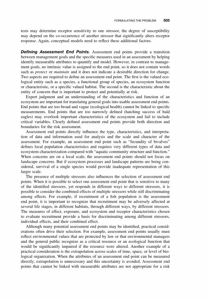
FORMULATING THE PROBLEM 505
tests may determine receptor sensitivity to one stressor, the degree of susceptibilitymay depend on the co-occurrence of another stressor that significantly alters receptorresponse. Again, conceptual models need to reflect these additional factors.
Defining Assessment End Points. Assessment end points provide a transitionbetween management goals and the specific measures used in an assessment by helpingidentify measurable attributes to quantify and model. However, in contrast to manage-ment goals, no intrinsic value is assigned to the end point, so it does not contain wordssuch as protect or maintain and it does not indicate a desirable direction for change.Two aspects are required to define an assessment end point. The first is the valued eco-logical entity such as a species, a functional group of species, an ecosystem functionor characteristic, or a specific valued habitat. The second is the characteristic about theentity of concern that is important to protect and potentially at risk.
Expert judgment and an understanding of the characteristics and function of anecosystem are important for translating general goals into usable assessment end points.End points that are too broad and vague (ecological health) cannot be linked to specificmeasurements. End points that are too narrowly defined (hatching success of baldeagles) may overlook important characteristics of the ecosystem and fail to includecritical variables. Clearly defined assessment end points provide both direction andboundaries for the risk assessment.
Assessment end points directly influence the type, characteristics, and interpreta-tion of data and information used for analysis and the scale and character of theassessment. For example, an assessment end point such as “fecundity of bivalves”defines local population characteristics and requires very different types of data andecosystem characterization compared with “aquatic community structure and function.”When concerns are on a local scale, the assessment end points should not focus onlandscape concerns. But if ecosystem processes and landscape patterns are being con-sidered, survival of a single species would provide inadequate representation of thislarger scale.
The presence of multiple stressors also influences the selection of assessment endpoints. When it is possible to select one assessment end point that is sensitive to manyof the identified stressors, yet responds in different ways to different stressors, it ispossible to consider the combined effects of multiple stressors while still discriminatingamong effects. For example, if recruitment of a fish population is the assessmentend point, it is important to recognize that recruitment may be adversely affected atseveral life stages, in different habitats, through different ways, by different stressors.The measures of effect, exposure, and ecosystem and receptor characteristics chosento evaluate recruitment provide a basis for discriminating among different stressors,individual effects, and their combined effect.
Although many potential assessment end points may be identified, practical consid-erations often drive their selection. For example, assessment end points usually mustreflect environmental values that are protected by law or that environmental managersand the general public recognize as a critical resource or an ecological function thatwould be significantly impaired if the resource were altered. Another example of apractical consideration is the extrapolation across scales of time, space, or level of bio-logical organization. When the attributes of an assessment end point can be measureddirectly, extrapolation is unnecessary and this uncertainty is avoided. Assessment endpoints that cannot be linked with measurable attributes are not appropriate for a risk
506 ENVIRONMENTAL RISK ASSESSMENT
assessment. However, assessment end points that cannot be measured directly but canbe represented by surrogate measures that are easily monitored and modeled can stillprovide a good foundation for the risk assessment.
28.2.2 Developing Conceptual Models
Conceptual models link anthropogenic activities with stressors and evaluate the rela-tionships among exposure pathways, ecological effects, and ecological receptors. Themodels also may describe natural processes that influence these relationships. Con-ceptual models include a set of risk hypotheses that describe predicted relationshipsbetween stressor, exposure, and assessment end point response, along with the ratio-nale for their selection. Risk hypotheses are hypotheses in the broad scientific sense;they do not necessarily involve statistical testing of null and alternative hypothesesor any particular analytical approach. Risk hypotheses may predict the effects of astressor, or they may postulate what stressors may have caused observed ecologi-cal effects.
Diagrams can be used to illustrate the relationships described by the conceptualmodel and risk hypotheses. Conceptual model diagrams are useful tools for commu-nicating important pathways and for identifying major sources of uncertainty. Thesediagrams and risk hypotheses can be used to identify the most important pathways andrelationships to consider in the analysis phase. The hypotheses considered most likelyto contribute to risk are identified for subsequent evaluation in the risk assessment.
The complexity of the conceptual model depends on the complexity of the problem,number of stressors and assessment end points being considered, nature of effects,and characteristics of the ecosystem. For single stressors and single assessment endpoints, conceptual models can be relatively simple relationships. In cases where con-ceptual models describe, besides the pathways of individual stressors and assessmentend points, the interaction of multiple and diverse stressors and assessment end points,several submodels would be required to describe individual pathways. Other modelsmay then be used to explore how these individual pathways interact. An example of aconceptual model for a watershed in shown in Figure 28.2.
28.2.3 Selecting Measures
The last step in the problem formulation phase is the development of an analysis planor proposal that identifies measures to evaluate each risk hypothesis and that describesthe assessment design, data needs, assumptions, extrapolations, and specific methodsfor conducting the analysis. There are three categories of measures that can be selected.Measures of effect (also called measurement end points) are measures used to evaluatethe response of the assessment end point when exposed to a stressor. Measures ofexposure are measures of how exposure may be occurring, including how a stressormoves through the environment and how it may co-occur with the assessment end point.Measures of ecosystem and receptor characteristics include ecosystem characteristicsthat influence the behavior and location of assessment end points, the distribution ofa stressor, and life history characteristics of the assessment end point that may affectexposure or response to the stressor. These diverse measures increase in importanceas the complexity of the assessment increases.
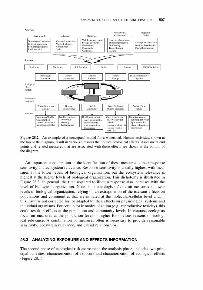
ANALYZING EXPOSURE AND EFFECTS INFORMATION 507
Water control measuresPesticide applicationFertilizer applicationLand alteration
Activities
Stressors
Measures
EcologicalEffectsModels
AssessmentEndpoints
Agricultural Industrial
Chemical waste sitesWaste dischargesConstructionSpills
Mobile sources (autos)Sewage dischargesUrban runoffConstructionWaste sites
Municipal
Recreational/Commercial
Regional/Global
Dredging channelizingShoreline protectionFish/huntingTimber harvestBoating
Atmospheric depositionFossil fuel combustionChlorofluorocarbons
Toxicants Nutrients Soil Particles Noise Disease UV/B Radiation
HydrologicAlteration
Water-DependentWildlife
Populations/HealthAssessment of:
HabitatAlteration
HarvestPressure
ClimateChange
Invasive/IntroducedSpecies
colonial water birdsamphibians/reptiles
BenthicInvertebrates
Pond Invertebrates:abundancediversityhealth indices
FinfishCommunity
Health Assessment:gross abnormalitieshistopathologytoxicant residuesbiomarkers
Water/SedimentQuality Standards
Water Assessment:dissolved oxygenturbidityprimary productivitytoxicant residuesbioassays
Aquatic PlantHabital
Plant Assessment:aquatic plant coverlight attenuationdissolved nutrientsmacroalgae
Figure 28.2 An example of a conceptual model for a watershed. Human activities, shown atthe top of the diagram, result in various stressors that induce ecological effects. Assessment endpoints and related measures that are associated with these effects are shown at the bottom ofthe diagram.
An important consideration in the identification of these measures is their responsesensitivity and ecosystem relevance. Response sensitivity is usually highest with mea-sures at the lower levels of biological organization, but the ecosystem relevance ishighest at the higher levels of biological organization. This dichotomy is illustrated inFigure 28.3. In general, the time required to illicit a response also increases with thelevel of biological organization. Note that toxicologists focus on measures at lowerlevels of biological organization, relying on an extrapolation of the toxicant effects onpopulations and communities that are initiated at the molecular/cellular level and, ifthis insult is not corrected for, or adapted to, then effects on physiological systems andindividual organisms. For certain toxic modes of action (e.g., reproductive toxicity), thiscould result in effects at the population and community levels. In contrast, ecologistsfocus on measures at the population level or higher for obvious reasons of ecolog-ical relevance. A combination of measures often is necessary to provide reasonablesensitivity, ecosystem relevance, and causal relationships.
28.3 ANALYZING EXPOSURE AND EFFECTS INFORMATION
The second phase of ecological risk assessment, the analysis phase, includes two prin-cipal activities: characterization of exposure and characterization of ecological effects(Figure 28.1).
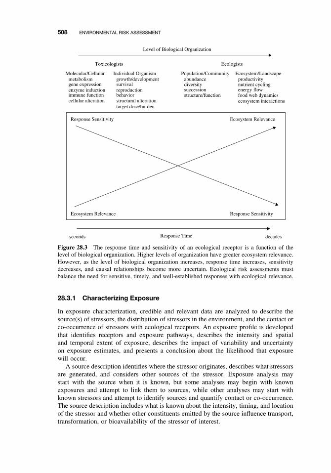
508 ENVIRONMENTAL RISK ASSESSMENT
Level of Biological Organization
Toxicologists
Molecular/Cellularmetabolismgene expressionenzyme inductionimmune functioncellular alteration
Individual Organismgrowth/developmentsurvivalreproductionbehaviorstructural alterationtarget dose/burden
Population/Communityabundancediversitysuccessionstructure/function
Ecosystem/Landscape
Ecosystem RelevanceResponse Sensitivity
Response SensitivityEcosystem Relevance
productivitynutrient cyclingenergy flowfood web dynamicsecosystem interactions
Ecologists
seconds Response Time decades
Figure 28.3 The response time and sensitivity of an ecological receptor is a function of thelevel of biological organization. Higher levels of organization have greater ecosystem relevance.However, as the level of biological organization increases, response time increases, sensitivitydecreases, and causal relationships become more uncertain. Ecological risk assessments mustbalance the need for sensitive, timely, and well-established responses with ecological relevance.
28.3.1 Characterizing Exposure
In exposure characterization, credible and relevant data are analyzed to describe thesource(s) of stressors, the distribution of stressors in the environment, and the contact orco-occurrence of stressors with ecological receptors. An exposure profile is developedthat identifies receptors and exposure pathways, describes the intensity and spatialand temporal extent of exposure, describes the impact of variability and uncertaintyon exposure estimates, and presents a conclusion about the likelihood that exposurewill occur.
A source description identifies where the stressor originates, describes what stressorsare generated, and considers other sources of the stressor. Exposure analysis maystart with the source when it is known, but some analyses may begin with knownexposures and attempt to link them to sources, while other analyses may start withknown stressors and attempt to identify sources and quantify contact or co-occurrence.The source description includes what is known about the intensity, timing, and locationof the stressor and whether other constituents emitted by the source influence transport,transformation, or bioavailability of the stressor of interest.
ANALYZING EXPOSURE AND EFFECTS INFORMATION 509
Many stressors have natural counterparts and/or multiple sources that must be con-sidered. For example, many chemicals occur naturally (e.g., most metals), are generallywidespread due to multiple sources (e.g., polycyclic aromatic hydrocarbons), or mayhave significant sources outside the boundaries of the current assessment (e.g., regionalatmospheric deposition of PCBs). Many physical stressors also have natural coun-terparts such as sedimentation from construction activities versus natural erosion. Inaddition human activities may change the magnitude or frequency of natural distur-bance cycles such as the frequency and severity of flooding. Source characterizationcan be particularly important for new biological stressors (e.g., invasive species), sincemany of the strategies for reducing risks focus on preventing entry in the first place.Once the source is identified, the likelihood of entry may be characterized qualitatively.
Because exposure occurs where receptors co-occur with or contact stressors in theenvironment, characterizing the spatial and temporal distribution of a stressor is a nec-essary precursor to estimating exposure. The stressor’s spatial and temporal distributionin the environment is described by evaluating the pathways that stressors take from thesource as well as the formation and subsequent distribution of secondary stressors. Forchemical stressors, the evaluation of pathways usually follows the type of transport andfate modeling described in Chapter 27. Some physical stressors such as sedimentationalso can be modeled, but other physical stressors require no modeling because theyeliminate entire ecosystems or portions of them, such as when a wetland is filled, aresource is harvested, or an area is flooded.
The movement of biological stressors have been described as diffusion and/or jump-dispersal processes. Diffusion involves a gradual spread from the site of introductionand is a function primarily of reproductive rates and motility. Jump-dispersal involveserratic spreads over periods of time, usually by means of a vector. The gypsy mothand zebra mussel have spread this way; the gypsy moth via egg masses on vehiclesand the zebra mussel via boat ballast water. Biological stressors can use both diffusionand jump-dispersal strategies, which makes it difficult to predict dispersal rates. Anadditional complication is that biological stressors are influenced by their own survivaland reproduction.
The creation of secondary stressors can greatly alter risk. Secondary stressors canbe formed through biotic or abiotic transformation processes and may be of greateror lesser concern than the primary stressor. Physical disturbances can generate sec-ondary stressors, such as when the removal of riparian vegetation results in increasednutrients, sedimentation, and altered stream flow. For chemicals, the evaluation of sec-ondary stressors usually focuses on metabolites or degradation products. In additionsecondary stressors can be formed through ecosystem processes. For example, nutri-ent inputs into an estuary can decrease dissolved oxygen concentrations because theyincrease primary production and subsequent decomposition. A changeover from an aer-obic to an anaerobic environment often is accompanied by the production of sulfide viasulfate-reducing bacteria. Sulfide can act as a secondary stressor to oxygen-dependentorganisms, but it also can reduce exposure to metals through the precipitation of metalsulfides (see Chapter 27).
The distribution of stressors in the environment can be described using measurements,models, or a combination of the two. If stressors have already been released, directmeasurements of environmental media or a combination of modeling and measurementis preferred. However, a modeling approach may be necessary if the assessment isintended to predict future scenarios or if measurements are not possible or practicable.
510 ENVIRONMENTAL RISK ASSESSMENT
28.3.2 Characterizing Ecological Effects
In ecological effects characterization, relevant data are analyzed to evaluate stressor-response relationships and/or to provide evidence that exposure to a stressor causesan observed response. The characterization describes the effects that are elicited bya stressor, links these effects with the assessment endpoints, and evaluates how theeffects change with varying stressor levels. The conclusions of the ecological effectscharacterization are summarized in a stressor-response profile.
Analyzing Ecological Response. Ecological response analysis has three primarycomponents: determining the relationship between stressor exposure and ecologicaleffects, evaluating the plausibility that effects may occur or are occurring as a result ofthe exposure, and linking measurable ecological effects with the assessment end points.
Evaluating ecological risks requires an understanding of the relationships betweenstressor exposure and resulting ecological responses. The stressor-response relation-ships used in a particular assessment depend on the scope and nature of the ecologicalrisk assessment as defined in problem formulation and reflected in the analysis plan.For example, a point estimate of an effect (e.g., an LC50) might be compared withpoint estimates from other stressors. The stressor-response function (e.g., shape of thecurve) may be critical for determining the presence or absence of an effects thresholdor for evaluating incremental risks, or stressor-response functions may be used as inputfor ecological effects models. If sufficient data are available, cumulative distributionfunctions can be constructed using multiple point estimates of effects. Process modelsthat already incorporate empirically derived stressor-response functions also can beused. However, many stressor-response relationships are very complex, and ecologi-cal systems frequently show responses to stressors that involve abrupt shifts to newcommunity or system types.
In simple cases the response will be one variable (e.g., mortality) and quantita-tive univariate analysis can be used. If the response of interest is composed of manyindividual variables (e.g., species abundances in an aquatic community), multivariatestatistical techniques must be used. Multivariate techniques (e.g., factor and clusteranalysis) have a long history of use in ecology but have not yet been extensivelyapplied in risk assessment. Stressor-response relationships can be described using anyof the dimensions of exposure (i.e., intensity, time, space). Intensity is probably themost familiar dimension and is often used for chemicals (e.g., dose, concentration). Theduration of exposure also can be used for chemical stressor-response relationships; forexample, median acute effects levels are always associated with a time parameter (e.g.,24 h, 48 h, 96 h). Both the time and spatial dimensions of exposure can be importantfor physical disturbances such as flooding. Single-point estimates and stressor-responsecurves can be generated for some biological stressors. For pathogens such as bacte-ria and fungi, inoculum levels may be related to the level of symptoms in a host oractual signs of the pathogen. For other biological stressors such as introduced species,developing simple stressor-response relationships may be inappropriate.
Causality is the relationship between cause (one or more stressors) and effect (assess-ment end point response to one or more stressors). Without a sound basis for linkingcause and effect, uncertainty in the conclusions of an ecological risk assessment willbe high. Developing causal relationships is especially important for risk assessmentsdriven by observed adverse ecological effects such as fish kills or long-term declines
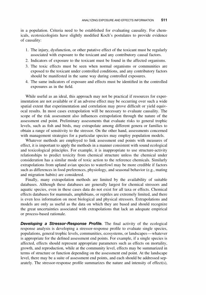
ANALYZING EXPOSURE AND EFFECTS INFORMATION 511
in a population. Criteria need to be established for evaluating causality. For chem-icals, ecotoxicologists have slightly modified Koch’s postulates to provide evidenceof causality:
1. The injury, dysfunction, or other putative effect of the toxicant must be regularlyassociated with exposure to the toxicant and any contributory causal factors.
2. Indicators of exposure to the toxicant must be found in the affected organisms.3. The toxic effects must be seen when normal organisms or communities are
exposed to the toxicant under controlled conditions, and any contributory factorsshould be manifested in the same way during controlled exposures.
4. The same indicators of exposure and effects must be identified in the controlledexposures as in the field.
While useful as an ideal, this approach may not be practical if resources for exper-imentation are not available or if an adverse effect may be occurring over such a widespatial extent that experimentation and correlation may prove difficult or yield equiv-ocal results. In most cases extrapolation will be necessary to evaluate causality. Thescope of the risk assessment also influences extrapolation through the nature of theassessment end point. Preliminary assessments that evaluate risks to general trophiclevels, such as fish and birds, may extrapolate among different genera or families toobtain a range of sensitivity to the stressor. On the other hand, assessments concernedwith management strategies for a particular species may employ population models.
Whatever methods are employed to link assessment end points with measures ofeffect, it is important to apply the methods in a manner consistent with sound ecologicaland toxicological principles. For example, it is inappropriate to use structure-activityrelationships to predict toxicity from chemical structure unless the chemical underconsideration has a similar mode of toxic action to the reference chemicals. Similarlyextrapolations from upland avian species to waterfowl may be more credible if factorssuch as differences in food preferences, physiology, and seasonal behavior (e.g., matingand migration habits) are considered.
Finally, many extrapolation methods are limited by the availability of suitabledatabases. Although these databases are generally largest for chemical stressors andaquatic species, even in these cases data do not exist for all taxa or effects. Chemicaleffects databases for mammals, amphibians, or reptiles are extremely limited, and thereis even less information on most biological and physical stressors. Extrapolations andmodels are only as useful as the data on which they are based and should recognizethe great uncertainties associated with extrapolations that lack an adequate empiricalor process-based rationale.
Developing a Stressor-Response Profile. The final activity of the ecologicalresponse analysis is developing a stressor-response profile to evaluate single species,populations, general trophic levels, communities, ecosystems, or landscapes—whateveris appropriate for the defined assessment end points. For example, if a single species isaffected, effects should represent appropriate parameters such as effects on mortality,growth, and reproduction, while at the community level, effects may be summarized interms of structure or function depending on the assessment end point. At the landscapelevel, there may be a suite of assessment end points, and each should be addressed sep-arately. The stressor-response profile summarizes the nature and intensity of effect(s),
512 ENVIRONMENTAL RISK ASSESSMENT
the time scale for recovery (where appropriate), causal information linking the stressorwith observed effects, and uncertainties associated with the analysis.
28.4 CHARACTERIZING RISK
Risk characterization is the final phase of an ecological risk assessment (Figure 28.1).During risk characterization, risks are estimated and interpreted and the strengths,limitations, assumptions, and major uncertainties are summarized. Risks are estimatedby integrating exposure and stressor-response profiles using a wide range of techniquessuch as comparisons of point estimates or distributions of exposure and effects data,process models, or empirical approaches such as field observational data. Risks aredescribed by evaluating the evidence supporting or refuting the risk estimate(s) andinterpreting the adverse effects on the assessment end point. Criteria for evaluatingadversity include the nature and intensity of effects, spatial and temporal scales, and thepotential for recovery. Agreement among different lines of evidence of risk increasesconfidence in the conclusions of a risk assessment.
28.4.1 Estimating Risk
Risk estimation determines the likelihood of adverse effects to assessment end pointsby integrating exposure and effects data and evaluating any associated uncertainties.The process uses the exposure and stressor-response profiles. Risks can be estimatedby one or more of the following approaches: (1) estimates based on best profes-sional judgment and expressed as qualitative categories such as low, medium, orhigh; (2) estimates comparing single-point estimates of exposure and effects such asa simple ratio of exposure concentration to effects concentration (quotient method);(3) estimates incorporating the entire stressor-response relationship often as a non-linear function of exposure; (4) estimates incorporating variability in exposure andeffects estimates providing the capability to predict changes in the magnitude andlikelihood of effects at different exposure scenarios; (5) estimates based on processmodels that rely partially or entirely on theoretical approximations of exposure andeffects; and (6) estimates based on empirical approaches, including field observationaldata. An example of the first approach, using qualitative categorization, is shown inFigure 28.4.
28.4.2 Describing Risk
After risks have been estimated, available information must be integrated and inter-preted to form conclusions about risks to the assessment endpoints. Risk descriptionsinclude an evaluation of the lines of evidence supporting or refuting the risk estimate(s)and an interpretation of the adverse effects on the assessment end point. Confidence inthe conclusions of a risk assessment may be increased by using several lines of evi-dence to interpret and compare risk estimates. These lines of evidence may be derivedfrom different sources or by different techniques relevant to adverse effects on theassessment end points, such as quotient estimates, modeling results, field experiments,
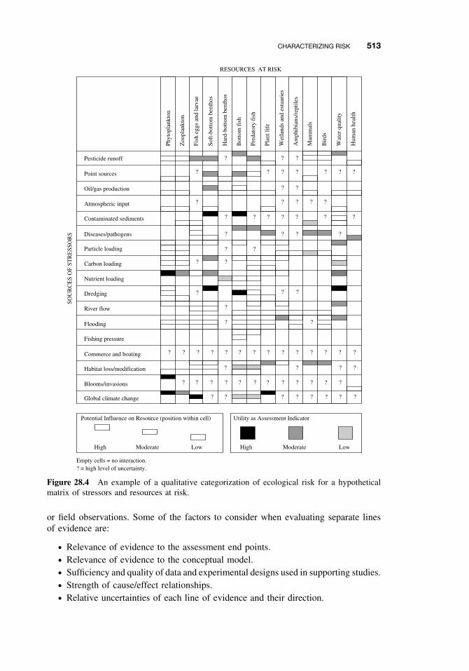
CHARACTERIZING RISK 513
RESOURCES AT RISK
High LowModerate High LowModerate
Empty cells = no interaction.? = high level of uncertainty.
Potential Influence on Resource (position within cell) Utility as Assessment Indicator
SOU
RC
ES
OF
STR
ESS
OR
S
Pesticide runoff
Point sources
Oil/gas production
Atmospheric input
Contaminated sediments
Diseases/pathogens
Particle loading
Carbon loading
Nutrient loading
Dredging
River flow
Flooding
Fishing pressure
Commerce and boating
Habitat loss/modification
Blooms/invasions
Global climate change
Phyt
opla
nkto
n
Zoo
plan
kton
Fish
egg
s an
d la
rvae
Soft
-bot
tom
ben
thos
Har
d-bo
ttom
ben
thos
Bot
tom
fis
h
Pred
ator
y fi
sh
Plan
t lif
e
Wet
land
s an
d es
tuar
ies
Am
phib
ians
/rep
tiles
Mam
mal
s
Bir
ds
Wat
er q
ualit
y
Hum
an h
ealth
? ? ?
??
??
??
? ??
? ?
??
?
? ? ?
??
??
?????
?
?
?
?
? ?
? ? ? ? ? ? ? ? ? ? ? ? ? ?
????
? ? ? ? ? ? ? ? ? ? ? ?
? ???????
?
?
?
Figure 28.4 An example of a qualitative categorization of ecological risk for a hypotheticalmatrix of stressors and resources at risk.
or field observations. Some of the factors to consider when evaluating separate linesof evidence are:
ž Relevance of evidence to the assessment end points.ž Relevance of evidence to the conceptual model.ž Sufficiency and quality of data and experimental designs used in supporting studies.ž Strength of cause/effect relationships.ž Relative uncertainties of each line of evidence and their direction.
514 ENVIRONMENTAL RISK ASSESSMENT
At this point in risk characterization, the changes expected in the assessment endpoints have been estimated and described. The next step is to interpret whether thesechanges are considered adverse and meaningful. Meaningful adverse changes aredefined by ecological and/or social concerns, and thus usually depend on the bestprofessional judgment of the risk assessor. Five criteria have been proposed by EPAfor evaluating adverse changes in assessment end points:
1. Nature of effects
2. Intensity of effects
3. Spatial scale
4. Temporal scale
5. Potential for recovery
The extent to which the five criteria are evaluated depends on the scope and complexityof the ecological risk assessment. However, understanding the underlying assumptionsand science policy judgments is important even in simple cases. For example, whenexceedence of a previously established decision rule such as a benchmark stressor levelor water quality criterion is used as evidence of adversity, the reasons why exceedencesof the benchmark are considered adverse should be clearly understood.
To distinguish ecological changes that are adverse from those ecological events thatare within the normal pattern of ecosystem variability or result in little or no mean-ingful alteration of biota, it is important to consider the nature and intensity of effects.For example, an assessment end point involving survival, growth, and reproductionof a species must consider whether predicted effects involve survival and reproduc-tion or only growth. Or if survival of offspring are affected, the relative loss mustbe considered.
It is important to consider both the ecological and statistical contexts of an effectwhen evaluating intensity. For example, a statistically significant 1% decrease in fishgrowth may not be relevant to an assessment end point of fish population viability, anda 10% decline in reproduction may be worse for a population of slowly reproducingmarine mammals than for rapidly reproducing planktonic algae.
Natural ecosystem variation can make it very difficult to observe (detect) stressor-related perturbations. For example, natural fluctuations in marine fish populations areoften very large and cyclic events (e.g., fish migration) are very important in naturalsystems. Predicting the effects of anthropogenic stressors against this background ofvariation can be very difficult. Thus a lack of statistically significant effects in a fieldstudy does not automatically mean that adverse ecological effects are absent. Rather,factors such as statistical power to detect differences, natural variability, and other linesof evidence must be considered in reaching conclusions about risk.
Spatial and temporal scales also need to be considered in assessing the adversity ofthe effects. The spatial dimension encompasses both the extent and pattern of effect aswell as the context of the effect within the landscape. Factors to consider include theabsolute area affected, the extent of critical habitats affected compared with a largerarea of interest, and the role or use of the affected area within the landscape. Adverseeffects to assessment end points vary with the absolute area of the effect. A largeraffected area may be (1) subject to a greater number of other stressors, increasing thecomplications from stressor interactions; (2) more likely to contain sensitive species or
CHARACTERIZING RISK 515
habitats; or (3) more susceptible to landscape-level changes because many ecosystemsmay be altered by the stressors.
Nevertheless, a smaller area of effect is not always associated with lower risk. Thefunction of an area within the landscape may be more important than the absolutearea. Destruction of small but unique areas, such as submerged vegetation at the land-water margin, may have important effects on local wildlife populations. Also, in riversystems, both riffle and pool areas provide important microhabitats that maintain thestructure and function of the total river ecosystem. Stressors acting on some of thesemicrohabitats may present a significant risk to the entire system. Spatial factors also areimportant for many species because of the linkages between ecological landscapes andpopulation dynamics. Linkages between one or more landscapes can provide refugefor affected populations, and species may require adequate corridors between habitatpatches for successful migration.
The temporal scale for ecosystems can vary from seconds (photosynthesis, prokary-otic reproduction) to centuries (global climate change). Changes within a forest ecosys-tem can occur gradually over decades or centuries and may be affected by slowly chang-ing external factors such as climate. The time scale of stressor-induced changes operateswithin the context of multiple natural time scales. In addition temporal responsesfor ecosystems may involve intrinsic time lags, so responses from a stressor may bedelayed. Thus it is important to distinguish the long-term impacts of a stressor from theimmediately visible effects. For example, visible changes resulting from eutrophicationof aquatic systems (turbidity, excessive macrophyte growth, population decline) maynot become evident for many years after initial increases in nutrient levels.
From the temporal scale of adverse effects we come to a consideration of recovery.Recovery is the rate and extent of return of a population or community to a conditionthat existed before the introduction of a stressor. Because ecosystems are dynamicand even under natural conditions are constantly changing in response to changes inthe physical environment (weather, natural catastrophes, etc.) or other factors, it isunrealistic to expect that a system will remain static at some level or return to exactlythe same state that it was before it was disturbed. Thus the attributes of a “recovered”system must be carefully defined. Examples might include productivity declines inan eutrophic system, re-establishment of a species at a particular density, species re-colonization of a damaged habitat, or the restoration of health of diseased organisms.
Recovery can be evaluated despite the difficulty in predicting events in ecologicalsystems. For example, it is possible to distinguish changes that are usually reversible(e.g., recovery of a stream from sewage effluent discharge), frequently irreversible (e.g.,establishment of introduced species), and always irreversible (e.g., species extinction).It is important to consider whether significant structural or functional changes haveoccurred in a system that might render changes irreversible. For example, physicalalterations such as deforestation can change soil structure and seed sources such thatforests cannot easily grow again.
Natural disturbance patterns can be very important when evaluating the likelihood ofrecovery from anthropogenic stressors. Ecosystems that have been subjected to repeatednatural disturbances may be more vulnerable to anthropogenic stressors (e.g., overfish-ing). Alternatively, if an ecosystem has become adapted to a disturbance pattern, it maybe affected when the disturbance is removed (fire-maintained grasslands). The lackof natural analogues makes it difficult to predict recovery from novel anthropogenicstressors such as exposure to synthetic chemicals.
516 ENVIRONMENTAL RISK ASSESSMENT
The relative rate of recovery also can be estimated. For example, fish populationsin a stream are likely to recover much faster from exposure to a degradable chemicalthan from habitat alterations resulting from stream channelization. It is critical to useknowledge of factors such as the temporal scales of organisms’ life histories, the avail-ability of adequate stock for recruitment, and the interspecific and trophic dynamicsof the populations in evaluating the relative rates of recovery. A fisheries stock orforest might recover in several decades, a benthic infaunal community in years, and aplanktonic community in weeks to months.
28.5 MANAGING RISK
When risk characterization is complete, a description of the risk assessment is com-municated to the risk manager (Figure 28.1) to support a risk management decision.This communication usually is a report and might include:
ž A description of risk assessor/risk manager planning results.ž A review of the conceptual model and the assessment end points.ž A discussion of the major data sources and analytical procedures used.ž A review of the stressor-response and exposure profiles.ž A description of risks to the assessment endpoints, including risk estimates and
adversity evaluations.ž A summary of major areas of uncertainty and the approaches used to address them.ž A discussion of science policy judgments or default assumptions used to bridge
information gaps, and the basis for these assumptions.
After the risk assessment is completed, risk managers may consider whether addi-tional follow-up activities are required. Depending on the importance of the assessment,confidence level in the assessment results, and available resources, it may be advisableto conduct another iteration of the risk assessment in order to facilitate a final man-agement decision. Ecological risk assessments are frequently designed in sequentialtiers that proceed from simple, relatively inexpensive evaluations to more costly andcomplex assessments. Initial tiers are based on conservative assumptions, such as max-imum exposure and ecological sensitivity. When an early tier cannot sufficiently definerisk to support a management decision, a higher assessment tier that may require eitheradditional data or applying more refined analysis techniques to available data may beneeded. Higher tiers provide more ecologically realistic assessments while making lessconservative assumptions about exposure and effects.
Another option is to proceed with a management decision based on the risk assess-ment and develop a monitoring plan to evaluate the results of the decision. For example,if the decision is to mitigate risks through exposure reduction, monitoring will helpdetermine whether the desired reduction in exposure (and effects) is being achieved.Monitoring is also critical for determining the extent and nature of any ecologicalrecovery that may be occurring.
Ecological risk assessment is important for environmental decision making becauseof the high cost of eliminating environmental risks associated with human activitiesand the necessity of making regulatory decisions in the face of uncertainty. Ecologi-cal risk assessment provides only a portion of the information required to make risk
SUGGESTED READING 517
management decisions, but this information is critical to scientifically defensible riskmanagement. Thus ecological risk assessments should provide input to a diverse set ofenvironmental decision-making processes, such as the regulation of hazardous wastesites, industrial chemicals, and pesticides, and improve the management of watershedsaffected by multiple nonchemical and chemical stressors.
SUGGESTED READING
Bartell, S. M., R. H. Gardner, and R. V. O’Neill. Ecological Risk Estimation. Boca Raton, FL:Lewis Publishers, 1992.
Cardwell, R. D., B. R. Parkhurst, W. Warren-Hicks, and J. S. Volosin. Aquatic ecological risk.Water Environ. Technol. 5: 47–51, 1993.
Harwell, M. A., W. Cooper, and R. Flaak. Prioritizing ecological and human welfare risks fromenvironmental stresses. Environ. Manag. 16: 451–464, 1992.
Kendall, R. J., T. E. Lacher, C. Bunck, B. Daniel, C. Driver, C. E. Grue, F. Leighton,W. Stansley, P. G. Watanabe, and M. Whitworth. An ecological risk assessment of lead shotexposure in non-waterfowl avian species: Upland game birds and raptors. Environ. Toxicol.Chem. 15: 4–20, 1996.
National Research Council. A paradigm for ecological risk assessment. In Issues in Risk Assess-ment. Washington, DC: National Academy Press, 1993.
National Research Council. Science and Judgment in Risk Assessment. Washington, DC: NationalAcademy Press, 1994.
National Research Council. Understanding Risk: Informing Decisions in a Democratic Society.Washington, DC: National Academy Press, 1996.
Ruckelshaus, W. D. Science, risk, and public policy. Science 221: 1026–1028, 1983.
Solomon, K. R., D. B. Baker, R. P. Richards, K. R. Dixon, S. J. Klaine, T. W. La Point,R. J. Kendall, C. P. Weisskopf, J. M. Giddings, J. P. Geisy, L. W. Hall, W. M. Williams.Ecological risk assessment of atrazine in North American surface waters. Environ. Toxicol.Chem. 15(1): 31–76, 1996.
Suter, G. W., II. Endpoints for regional ecological risk assessments. Environ. Manag. 14: 19–23,1990.
Suter, G. W., II. Ecological Risk Assessment. Boca Raton, FL: Lewis Publishers, 1993.
Suter, G. W., II. A critique of ecosystem health concepts and indexes. Environ. Toxicol. Chem.12: 1533–1539, 1993.
US Environmental Protection Agency. Summary report on issues in ecological risk assessment.Washington, DC: Risk Assessment Forum, USEPA, 1991. EPA/625/3-91/018.
US Environmental Protection Agency. Framework for ecological risk assessment. Washington,DC: Risk Assessment Forum, USEPA, 1992. EPA/630/R-92/001.
US Environmental Protection Agency. Peer review workshop report on a framework for ecolog-ical risk assessment. Washington, DC: Risk Assessment Forum, USEPA, 1992. EPA/625/3-91/022.
US Environmental Protection Agency. A review of ecological assessment case studies from a riskassessment perspective. Washington, DC: Risk Assessment Forum, USEPA, 1993. EPA/630/R-92/005.
US Environmental Protection Agency. Ecological risk assessment issue papers. Washington,DC: Risk Assessment Forum, USEPA, 1994. EPA/630/R-94/009.
US Environmental Protection Agency. Proposed guidelines for ecological risk assessment. Wash-ington, DC: Risk Assessment Forum, USEPA, 1996. EPA/630/R-95/002B.


PART VIII
SUMMARY

CHAPTER 29
Future Considerations for Environmentaland Human Health
ERNEST HODGSON
29.1 INTRODUCTION
Since the publication of the second edition of this textbook there has been rapid,and in some cases dramatic, progress not only in toxicology but in the sciences thatcontribute methods and insights to toxicology. However, it is still true that speculationconcerning future developments in toxicology can be made only against an assessmentof where the science has come from and its current status. Toxicology, despite its use ofmany state-of-the-art techniques and explorations of the most fundamental molecularmechanisms of toxic action, is, at its heart, an applied science serving the needs ofsociety. Society is served in two principal ways: the protection of human health andthe protection of the environment. In both of these aspects two avenues are explored:studies of chemicals in use and the development of new chemicals that are both safeand effective. These studies range from studies of the mechanisms of toxic action to invivo toxicity testing, but the ultimate goal is a meaningful assessment of risk resultingfrom exposure to the chemicals in question.
The vast increase in public awareness of the potential of chemicals to cause harm-ful effects and the propensity of the print and electronic media to fan the flames ofcontroversy in this area make certain the continued need for toxicologists. We need toask what they will be doing during the next few decades compared to what they havebeen doing in the immediate past.
Through the 1950s and 1960s toxicology tended to be a largely descriptive science,relating the results of in vivo dosing to a variety of toxic end points, in many caseslittle more that the medial lethal dose (LD50) or median lethal concentration (LC50).However, ongoing studies of xenobiotic-metabolizing enzymes were attracting moreattention and techniques for chemical analysis of toxicants were starting to undergoa remarkable metamorphosis. The 1970s were most remarkable for developments inmetabolism and the beginnings of a boom in mode of toxic action studies, whereasthe 1980s and 1990s saw the incorporation of the techniques of molecular biology intomany aspects of toxicology, but perhaps to greatest effect in studies of the mechanismsof chemical carcinogenesis and the induction of xenobiotic-metabolizing enzymes.
A Textbook of Modern Toxicology, Third Edition, edited by Ernest HodgsonISBN 0-471-26508-X Copyright 2004 John Wiley & Sons, Inc.
521
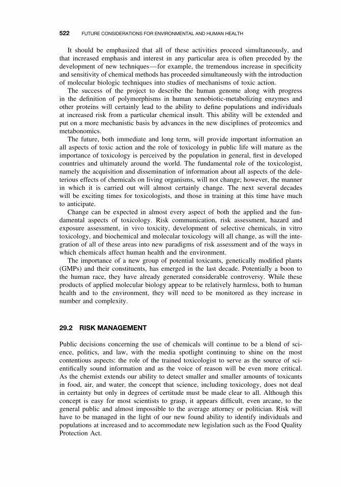
522 FUTURE CONSIDERATIONS FOR ENVIRONMENTAL AND HUMAN HEALTH
It should be emphasized that all of these activities proceed simultaneously, andthat increased emphasis and interest in any particular area is often preceded by thedevelopment of new techniques—for example, the tremendous increase in specificityand sensitivity of chemical methods has proceeded simultaneously with the introductionof molecular biologic techniques into studies of mechanisms of toxic action.
The success of the project to describe the human genome along with progressin the definition of polymorphisms in human xenobiotic-metabolizing enzymes andother proteins will certainly lead to the ability to define populations and individualsat increased risk from a particular chemical insult. This ability will be extended andput on a more mechanistic basis by advances in the new disciplines of proteomics andmetabonomics.
The future, both immediate and long term, will provide important information anall aspects of toxic action and the role of toxicology in public life will mature as theimportance of toxicology is perceived by the population in general, first in developedcountries and ultimately around the world. The fundamental role of the toxicologist,namely the acquisition and dissemination of information about all aspects of the dele-terious effects of chemicals on living organisms, will not change; however, the mannerin which it is carried out will almost certainly change. The next several decadeswill be exciting times for toxicologists, and those in training at this time have muchto anticipate.
Change can be expected in almost every aspect of both the applied and the fun-damental aspects of toxicology. Risk communication, risk assessment, hazard andexposure assessment, in vivo toxicity, development of selective chemicals, in vitrotoxicology, and biochemical and molecular toxicology will all change, as will the inte-gration of all of these areas into new paradigms of risk assessment and of the ways inwhich chemicals affect human health and the environment.
The importance of a new group of potential toxicants, genetically modified plants(GMPs) and their constituents, has emerged in the last decade. Potentially a boon tothe human race, they have already generated considerable controversy. While theseproducts of applied molecular biology appear to be relatively harmless, both to humanhealth and to the environment, they will need to be monitored as they increase innumber and complexity.
29.2 RISK MANAGEMENT
Public decisions concerning the use of chemicals will continue to be a blend of sci-ence, politics, and law, with the media spotlight continuing to shine on the mostcontentious aspects: the role of the trained toxicologist to serve as the source of sci-entifically sound information and as the voice of reason will be even more critical.As the chemist extends our ability to detect smaller and smaller amounts of toxicantsin food, air, and water, the concept that science, including toxicology, does not dealin certainty but only in degrees of certitude must be made clear to all. Although thisconcept is easy for most scientists to grasp, it appears difficult, even arcane, to thegeneral public and almost impossible to the average attorney or politician. Risk willhave to be managed in the light of our new found ability to identify individuals andpopulations at increased and to accommodate new legislation such as the Food QualityProtection Act.
IN VIVO TOXICITY 523
29.3 RISK ASSESSMENT
In the past, risk assessment consisted largely of computer-based models written to startfrom hazard assessment assays, such as chronic toxicity assays on rodents, encompassthe necessary extrapolations between species and between high and low doses, andthen produce a numerical assessment of the risk to human health. Although the hazardassessment tests and the toxic end points are different, an analogous situation exists inenvironmental risk assessment. A matter of considerable importance, now getting somebelated attention, is the integration of human health and environmental risk assessments.
Although many of these risk assessment programs were statistically sophisticated,they frequently did not rise above the level of numbers crunching, and more oftenthan not, different risk assessment programs, starting with the same experimental val-ues, produced very different numerical assessments of risk to human health or to theenvironment. Although having risk assessment become more science based has beena stated goal of regulators for decades, its scientific basis has not been advanced sig-nificantly. The need to incorporate mechanistic data, including mode of action studiesand physiologically based pharmacokinetics, has been realized to some extent. Apartfrom epidemiology and exposure analysis, human studies have not, despite the factthat many such studies can now be performed in noninvasive and ethical experiments.
The immediate future in risk assessment will focus on the difficult but necessarytask of integrating experimental data from all levels into the risk assessment process.A continuing challenge to toxicologists engaged in hazard or risk assessment is thatof risk from chemical mixtures. Neither human beings nor ecosystems are exposed tochemicals one at a time, yet logic dictates that the initial assessment of toxicity startwith individual chemicals. The resolution of this problem will require considerablework at all levels, in vivo and in vitro, into the implications of chemical interactionsfor the expression to toxicity, particularly chronic toxicity.
29.4 HAZARD AND EXPOSURE ASSESSMENT
The enormous cost of multiple-species, multiple-dose, lifetime evaluations of chroniceffects has already made the task of carrying out hazard assessments of all chemicals incommercial use impossible. At the same time, quantitative structure activity relation-ship (QSAR) studies are not yet predictive enough to indicate which chemicals shouldbe so tested and which chemicals need not be tested. In exposure assessment, continueddevelopment of analytical methods will permit ever more sensitive and selective deter-minations of toxicants in food and the environment, as well as the effects of chemicalmixtures and the potential for interactions that affect the ultimate expression of toxic-ity. Developments in QSARs, in short-term tests based on the expected mechanism oftoxic action and simplification of chronic testing procedures, will all be necessary ifthe chemicals to which the public and the environment are exposed are to be assessedadequately for their potential to cause harm.
29.5 IN VIVO TOXICITY
Although developments continue in elucidating the mechanisms of chemical carcino-genicity, much remains to be done with regard to this and other chronic end points,
524 FUTURE CONSIDERATIONS FOR ENVIRONMENTAL AND HUMAN HEALTH
particularly developmental and reproductive toxicity, chronic neurotoxicity, and immuno-toxicity. The further utilization of the methods of molecular biology will bring rapidadvances in all of these areas. It will be a challenge to integrate all of this informationinto useful paradigms for responsible and meaningful risk assessments.
29.6 IN VITRO TOXICITY
In vitro studies of toxic mechanisms will depend heavily on developments in molecularbiology, and great advances can be expected. Many of the ethical problems associatedwith carrying out studies on the effects of toxicants on humans will be circumventedat the in vitro level by the use of cloned and expressed human enzymes, receptors,and so on, although the integration of these data into intact organism models will stillrequire experimental animals. High-throughput technology in genomics, proteomics,and metabonomics will greatly facilitate these studies.
29.7 BIOCHEMICAL AND MOLECULAR TOXICOLOGY
As indicated previously, contributions to all aspects of the mechanistic study of toxicaction from the use of biochemical and molecular techniques can be expected. Nodoubt new techniques will be developed, answers will be found to many questions thatdid not yield to earlier techniques and new questions will be raised. The challenge, asalways, will be to integrate the results form these studies—and reach new levels ofsophistication—into useful and productive approaches to reduce chemical effects onhuman health and the environment.
29.8 DEVELOPMENT OF SELECTIVE TOXICANTS
Almost all aspects of contemporary human society depend on the use of numerouschemicals. Except in the unlikely event that society decides to return to a more sim-plistic and, in fact, more primitive, more unhealthy, and more demanding lifestyle,the challenge is in learning how to live with anthropomorphic chemicals, and not inlearning how to live without them. In many aspects, such as the production of food andfiber and the maintenance of human health, the development of selective pesticides,drugs, and so on, is needed. New techniques in molecular biology, in particular, theavailability of cloned and expressed human enzymes and receptors and new knowl-edge of human polymorphisms, will make this task easier, as will similar knowledgeof target species, including microorganisms causing human disease, and insects andweeds affecting the production of food and fiber, and so on.
High-throughput techniques will not only speed up the search, but in this area, as inother aspects of toxicology, bioinformatics will be necessary, not only for correlatingthe data from many sources but also for reducing it for practical applications.
GLOSSARY
acceptable daily intake (ADI) Amount of exposure determined to be “safe”; usuallyderived from the lowest No-Effect Level in an experimental study, divided by asafety factor such as 100. Also known as the Reference Dose (RfD).
acetylation The addition of an acetyl group from acetyl coenzyme A to a xenobioticor xenobiotic metabolite by the enzyme N-acetyltransferase. Polymorphisms in thisenzyme can be important in the expression of toxicity in humans.
acetylator phenotype Variation in the expression of N-acetyltransferase isoforms inhumans gives rise to two subpopulations—fast and slow acetylators. Slow acety-lators are more susceptible to the toxic effects of toxicants that are detoxified byacetylation.
acid deposition Wet and dry air pollutants that lower the pH of deposition and subse-quently the pH of the environment. Acid rain with a pH of 4 or lower refers to thewet components. Normal rain has a pH of about 5.6. Sulfuric acid from sulfur andnitric acid from nitrogen oxides are the major contributors. In lakes in which thebuffering capacity is low, the pH becomes acidic enough to cause fish kills, and thelakes cannot support fish populations. A contributing factor is the fact that acidicconditions concurrently release toxic metals, such as aluminum, into the water.
activation (bioactivation) In toxicology, this term is used to describe metabolic reac-tions of a xenobiotic in which the product is more toxic than is the substrate. Suchreactions are most commonly monooxygenations, the products of which are elec-trophiles that, if not detoxified by phase II (conjugation) reactions, may react withnucleophilic groups on cellular macromolecules such as proteins and DNA.
active oxygen Used to describe various short-lived highly reactive intermediates in thereduction of oxygen. Active oxygen species such as superoxide anion and hydroxylradical are known or believed to be involved in several toxic actions. Superoxideanion is detoxified by superoxide dismutase.
acute toxicity tests The most common tests for acute toxicity are the LC50 and LD50tests, which are designed to measure mortality in response to an acute toxic insult.Other tests for acute toxicity include dermal irritation tests, dermal sensitization tests,eye irritation tests, photoallergy tests, and phototoxicity tests. See also eye irritationtests; LC50; and LD50.
acute toxicity Refers to adverse effects on, or mortality of, organisms following soonafter a brief exposure to a chemical agent. Either a single exposure or multipleexposures within a short time period may be involved, and an acute effect is generallyregarded as an effect that occurs within the first few days after exposure, usuallyless than two weeks.
A Textbook of Modern Toxicology, Third Edition, edited by Ernest HodgsonISBN 0-471-26508-X Copyright 2004 John Wiley & Sons, Inc.
525
526 GLOSSARY
adaptation to toxicants Refers to the ability of an organism to show insensitivityor decreased sensitivity to a chemical that normally causes deleterious effects. Theterms resistance and tolerance are closely related and have been used in severaldifferent ways. However, a consensus is emerging to use the term resistance tomean that situation in which a change in the genetic constitution of a population inresponse to the stressor chemical enables a greater number of individuals to resist thetoxic action than were able to resist it in the previous unexposed population. Thusan essential feature of resistance is selection and then inheritance by subsequentgenerations. In microorganisms, this frequently involves mutations and inductionof enzymes by the toxicant; in higher organisms, it usually involves selection forgenes already present in the population at low frequency. The term tolerance is thenreserved for situations in which individual organisms acquire the ability to resist theeffect of a toxicant, usually as a result of prior exposure.
Ah locus A gene(s) controlling the trait of responsiveness for induction of enzymes byaromatic hydrocarbons. In addition to aromatic hydrocarbons such as the polycyclics,the chlorinated dibenzo-p-dioxins, dibenzofurans, and biphenyls, as well as thebrominated biphenyls, are involved. This trait, originally defined by studies of induc-tion of hepatic aryl hydrocarbon hydroxylase activity following 3-methylcholanthrenetreatment, is inherited by simple autosomal dominance in crosses and backcrossesbetween C57BL/6 (Ah-responsive) and DBA/2 (Ah-nonresponsive) mice.
Ah receptor (AHR) A protein coded for by a gene of the Ah locus. The initial locationof the Ah receptor is believed to be in the cytosol and, after binding to a ligandsuch as TCDD, is transported to the nucleus. Binding of aromatic hydrocarbonsto the Ah receptor of mice is a prerequisite for the induction of many xenobioticmetabolizing enzymes, as well as for two responses to TCDD; epidermal hyperplasiaand thymic atrophy. Ah-responsive mice have a high-affinity receptor, whereas theAh-nonresponsive mice have a low-affinity receptor.
air pollution In general, the principal air pollutants are carbon monoxide, oxides ofnitrogen, oxides of sulfur, hydrocarbons, and particulates. The principal sourcesare transportation, industrial processes, electric power generation, and the heatingof buildings. Hydrocarbons such as benzo(a)pyrene are produced by incompletecombustion and are associated primarily with the automobile. They are usually notpresent at levels high enough to cause direct toxic effects but are important in theformation of photochemical air pollution, formed as a result of interactions betweenoxides of nitrogen and hydrocarbons in the presence of ultraviolet light, giving riseto lung irritants such as peroxyacetyl nitrate, acrolein, and formaldehyde. Particulatesare a heterogeneous group of particles, often seen as smoke, that are important ascarriers of absorbed hydrocarbons and as irritants to the respiratory system.
alkylating agents These are chemicals that can add alkyl groups to DNA, a reactionthat can result either in mispairing of bases or in chromosome breaks. The mechanismof the reaction involves the formation of a reactive carbonium ion that combines withelectron-rich bases in DNA. Thus alkylating agents such as dimethylnitrosomine arefrequently carcinogens and/or mutagens.
Ames test An in vitro test for mutagenicity utilizing mutant strains of the bacteriumSalmonella typhimurium that is used as a preliminary screen of chemicals for assess-ing potential carcinogenicity. Several strains are available that cannot grow in theabsence of histidine because of metabolic defects in histidine biosynthesis. Muta-gens and presumed carcinogens can cause mutations that enable the strains to regain
GLOSSARY 527
their ability to grow in a histidine deficient medium. The test can be formed inthe presence of the S-9 fraction from rat liver to allow the metabolic activationof promutagens. There is a high correlation between bacterial mutagenicity andcarcinogenicity of chemicals.
antagonism In toxicology, antagonism is usually defined as that situation in which thetoxicity to two or more compounds administered together is less than that expectedfrom consideration of their toxicities when administered alone. Although this def-inition includes lowered toxicity resulting from induction of detoxifying enzymes,antagonism is frequently considered separately because of the time that must elapsebetween treatment with the inducer and subsequent treatment with the toxicant.Antagonism not involving induction is often at a marginal level of detection andis consequently difficult to explain. Such antagonism may involve competition forreceptor sites or nonenzymatic combination of one toxicant with another to reducethe toxic effect. Physiological antagonism, in which two agonists act on the samephysiological system but produce opposite effects, may also occur.
antibody A large protein first expressed on the surface of the B cells of the immunesystem, followed by a series of events resulting in a clone of plasma cells thatsecrete the antibody into body fluids. Antibodies bind to the substance (generally aprotein) that stimulated their production but may cross-react with related proteins.The natural function is to bind foreign substances such as microbes or microbialproducts, but because of their specificity, antibodies are used extensively in researchand in diagnostic and therapeutic procedures.
antidote A compound administered in order to reverse the harmful effect(s) of a tox-icant. They may be toxic mechanism specific, as in the case of 2-pyridine aldoxime(2-PAM) and organophosphate poisoning, or nonspecific, as in the case of syrupof ipecac, used to induce vomiting and, thereby, elimination of toxicants fromthe stomach.
behavioral toxicity Behavior may be defined as an organism’s motor or glandularresponse to changes in its internal or external environment. Such changes may besimple or highly complex, innate or learned, but in any event represent one ofthe final integrated expressions of nervous system function. Behavioral toxicity isadverse or potentially adverse effects on such expression brought about by exoge-nous chemicals.
binding, covalent See covalent binding.bioaccumulation The accumulation of a chemical either from the medium (usually
water) directly or from consumption of food containing the chemical. Biomagnifi-cation is often used as a synonym for bioaccumulation, but it is more correctly usedto describe an increase in concentration of a chemical as it passes from organismsat one tropic level to organisms at higher tropic levels.
bioactivation See activation.bioassay This term is used in two distinct ways. The first and most appropriate is the
use of a living organism to measure the amount of a toxicant present in a sample orthe toxicity of a sample. This is done by comparing the toxic effect of the samplewith that of a graded series of concentrations of a known standard. The second andless appropriate meaning is the use of animals to investigate the toxic effects ofchemicals as in chronic toxicity tests.
burden of proof Responsibility for determining whether a substance is safe or haz-ardous; a range of approaches can be seen when comparing laws. For example, for
528 GLOSSARY
OSHA, regulators show a substance is hazardous before exposure is restricted, withthe government conducting the tests; for FDA, manufacturers must show lack ofhazard before marketing.
biomagnification See bioaccumulation.carcinogen Any chemical or process involving chemicals that induces neoplasms that
are not usually observed, the earlier induction of neoplasms than are commonlyobserved, and/or the induction of more neoplasms than are usually found.
carcinogen, epigenetic Cancer-causing agents that exert their carcinogenic effect bymechanisms other than genetic, such as by immunosuppression, hormonal imbalance,or cytotoxicity. They may act as cocarcinogens or promoters. Epigenetic carcino-genesis is not as well understood a phenomenon as is genotoxic carcinogenesis.
carcinogen, genotoxic Cancer-causing agents that exert their carcinogenic effect bya series of events that is initiated by an interaction with DNA, either directly orthrough an electrophilic metabolite.
carcinogen, proximate See carcinogen, ultimate.carcinogen, ultimate Many, if not most, chemical carcinogens are not intrinsically
carcinogenic but require metabolic activation to express their carcinogenic potential.The term procarcinogen describes the initial reactive compound, the term proximatecarcinogen describes its more active products, and the term ultimate carcinogendescribes the product that is actually responsible for carcinogenesis by its interactionwith DNA.
carcinogenesis This is the process encompassing the conversion of normal cells toneoplastic cells and the further development of these neoplastic cells into a tumor.This process results from the action of specific chemicals, certain viruses, or radi-ation. Chemical carcinogens have been classified into those that are genotoxic andthose that are epigenetic (i.e., not genotoxic).
chronic toxicity This term is used to describe adverse effects manifested after a longtime period of uptake of small quantities of the toxicant in question. The dose issmall enough that no acute effects are manifested, and the time period is frequentlya significant part of the expected normal lifetime of the organism. The most seriousmanifestation of chronic toxicity is carcinogenesis, but other types of chronic toxicityare also known (e.g., reproductive effects, behavioral effects).
chronic toxicity tests Chronic tests are those conducted over a significant part of thelifespan of the test species or, in some cases, more than one generation. The mostimportant tests are carcinogenicity tests, and the most common test species are ratsand mice.
cocarcinogenesis Cocarcinogenesis is the enhancement of the conversion of normalcells to neoplastic cells. This process is manifested by enhancement of carcinogen-esis when the agent is administered either before or together with a carcinogen.Cocarcinogenesis should be distinguished from promotion as, in the latter case, thepromoter must be administered after the initiating carcinogen.
comparative toxicology The study of the variation in the expression of the toxicity ofexogenous chemicals toward organisms of different taxonomic groups or of differentgenetic strains.
compartment In pharmaco(toxico)kinetics a compartment is a hypothetical volume ofan animal system wherein a chemical acts homogeneously in transport and trans-formation. These compartments do not correspond to physiological or anatomic
GLOSSARY 529
areas but are abstract mathematical entities useful for predicting drug concentra-tions. Transport into, out of, or between compartments is described by rate constants,which are used in models of the intact animal.
conjugation reactions See phase II reactions.covalent binding This involves the covalent bond or “shared electron pair” bond.
Each covalent bond consists of a pair of electrons shared between two atoms andoccupying two stable orbitals, one of each atom. Although this is distinguished fromthe ionic bond or ionic valence, chemicals bonds may in fact show both covalent andionic character. In toxicology the term covalent binding is used less precisely to referto the binding of toxicants or their reactive metabolites to endogenous molecules(usually macromolecules) to produce stable adducts resistant to rigorous extrac-tion procedures. A covalent bond between ligand and macromolecule is generallyassumed. Many forms of chronic toxicity involve covalent binding of the toxicantto DNA or protein molecules within the cell.
cross resistance, cross tolerance These terms describe the situation where either resis-tance or tolerance to a particular toxicant (as defined under adaptation to toxicants)is induced by exposure to a different toxicant. This is commonly seen in resistanceof insects to insecticides in which selection with one insecticide brings about a broadspectrum of resistance to insecticides of the same or different chemical classes. Suchcross resistance is usually caused by the inheritance of a high level of nonspecificxenobiotic-metabolizing enzymes.
cytotoxicity Cellular injury or death brought about by chemicals external to the cell.Such chemicals may be soluble mediators produced by the immune system, or theymay be chemicals (toxicants) to which the organism has been exposed.
Delaney Amendment See Food, Drug, and Cosmetics Act.detoxication A metabolic reaction or sequence of reactions that reduces the potential
for adverse effect of a xenobiotic. Such sequences normally involve an increase inwater solubility that facilitates excretion and/or the reaction of a reactive product withan endogenous substrate (conjugation), thereby not only increasing water solubilitybut also reducing the possibility of interaction with cellular macromolecules. Not tobe confused with detoxification. See also detoxification.
detoxification Treatment by which toxicants are removed from intoxicated patients ora course of treatment during which dependence on alcohol or other drugs of abuse isreduced or eliminated. Not to be confused with detoxication. See also detoxication.
distribution The movement of a toxicant from the portal of entry to the tissue and alsothe different concentrations reached in different locations. The toxicant’s movementinvolves the study of transport mechanisms primarily in the blood, and both move-ment and concentration are subject to mathematical analysis in toxicokinetic studies.
dosage The amount of a toxicant drug or other chemical administered or taken ex-pressed as some function of the organism, (e.g., mg/kg body weight/day).
dose The total amount of a toxicant, drug, or other chemical administered to or takenin by the organism.
dose-response relationship In toxicology the quantitative relationship between theamount of a toxicant administered or taken and the incidence or extent of theadverse effect.
dose-response assessment A step in the risk assessment process to characterize therelationship between the dose of a chemical administered to a population of test
530 GLOSSARY
animals and the incidence of a given adverse effect. It involves mathematical mod-eling techniques to extrapolate from the high-dose effects observed in test animalsto estimate the effects expected from exposure to the typically low doses that maybe encountered by humans.
Draize test See eye irritation test.drugs of abuse Although all drugs may have deleterious effects on humans, drugs of
abuse either have no medicinal function or are taken at higher than therapeutic doses.Some drugs of abuse may affect only higher nervous functions (i.e., mood, reactiontime, coordination), but many produce physical dependence and have serious phys-ical effects, with fatal overdose being a common occurrence. The drugs of abuseinclude central nervous system (CNS) depressants such as ethanol, methaqualone(Quaalude), and secobarbital; CNS stimulants such as cocaine, methamphetamine(speed), caffeine, and nicotine; opioids such as heroin and morphine; hallucinogenssuch as lysergic acid diethylamide (LSD), phencyclidine (PCP), and tetrahydro-cannabinol (THC), the most important active principle of marijuana.
drugs, therapeutic All therapeutic drugs can be toxic at some dose. The danger tothe patient is dependent on the nature of the toxic response, the dose necessary toproduce the toxic response, and the relationship between the therapeutic and thetoxic dose. Drug toxicity is affected by all of those factors that affect the toxicityof xenobiotics, including (genetic) variation, diet, age, and the presence of otherexogenous chemicals. The risk of toxic side effects from a particular drug must beweighed against the expected benefits; the use of a quite dangerous drug with only anarrow tolerance between the therapeutic and toxic doses might well be justified ifit is the sole treatment for an otherwise fatal disease. For example, cytotoxic agentsused in the treatment of cancer are known carcinogens.
ecotoxicology See environmental toxicology.
electron transport system (ETS) This term is often restricted to the mitochondrialsystem, although it applies equally well to other systems, including that of micro-somes and chloroplasts. The mitochondrial ETS (also termed respiratory chain orcytochrome chain) consists of a series of cytochromes and other electron carriersarranged in the inner mitochondrial membrane. These components transfer the elec-trons from NADH or FADH2 generated in metabolic oxidations, to oxygen, thefinal electron acceptor, through a series of alternate oxidations and reductions. Theenergy that these electrons lose during these transfers is used to pump H+ fromthe matrix into the intermembrane space, creating an electrochemical proton gra-dient that drives oxidative phosphorylation. The energy is conserved as adenosinetriphosphate (ATP).
electron transport system (ETS) inhibitors The three major respiratory enzyme com-plexes of the mitochondrial electron transport system can all be blocked by inhibitors.For example, rotenone inhibits the NADH dehydrogenase complex, antimycin Ainhibits the b-c complex, and cyanide and carbon monoxide inhibits the cytochromeoxidase complex. Although oxidative phosphorylation inhibitors prevent phospho-rylation while allowing electron transfers to proceed, ETS inhibitors prevent bothelectron transport and ATP production.
electrophilic Electrophiles are chemicals that are attracted to and react with electron-rich centers in other molecules in reactions known as electrophilic reactions. Manyactivation reactions produce electrophilic intermediates such as epoxides, which exert
GLOSSARY 531
their toxic action by forming covalent bonds with nucleophilic centers in cellularmacromolecules such as DNA or proteins.
endoplasmic reticulum The endoplasmic reticulum (ER) is an extensive branchingand anastomosing double membrane distributed in the cytoplasm of eukaryoticcells. The ER is of two types: rough ER (RER) contains attached ribosomes onthe cytosolic surface and smooth ER (SER), devoid of ribosomes are involved inprotein biosynthesis; RER is abundant in cells specialized for protein synthesis.Many xenobiotic-metabolizing enzymes are integral components of both SER andRER, such as the cytochrome P450-dependent monooxygenase system and the flavin-containing monooxygenase, although the specific content is usually higher in SER.When tissue or cells are disrupted by homogenization, the ER is fragmented intomany smaller (ca. 100 nm in diameter) closed vesicles called microsomes, whichcan be isolated by differential centrifugation.
enterohepatic circulation This term describes the excretion of a compound into thebile and its subsequent reabsorption from the small intestine and transport backto the liver, where it is available again for biliary excretion. The most importantmechanism is conjugation in the liver, followed by excretion into the bile. In thesmall intestine the conjugation product is hydrolyzed, either nonenzymatically or bythe microflora, and the compound is reabsorbed to become a substrate for conjugationand reexcretion into the bile.
environmental toxicology This is concerned with the movement of toxicants and theirmetabolites in the environment and in food chains and the effect of such toxicantson populations of organisms.
epigenetic carcinogen See carcinogen, epigenetic.exposure assessment A component of risk assessment. The number of individuals
likely to be exposed to a chemical in the environment or in the workplace is assessed,and the intensity, frequency, and duration of human exposure are estimated.
eye irritation test (Draize test) Eye irritation tests measure irritancy of compoundsapplied topically to the eye. These tests are variations of the Draize test, and theexperimental animal used is the albino rabbit. The test consists of adding the mate-rial to be tested directly into the conjunctival sac of one eye of each of severalalbino rabbits, the other eye serving as the control. This test is probably the mostcontroversial of all toxicity tests, being criticized primarily on the ground that it isinhumane. Moreover, because both concentrations and volumes used are high andshow high variability, it has been suggested that these tests cannot be extrapolated tohumans. However, because visual impairment is a critical toxic end point, tests forocular toxicity are essential. Attempts to solve the dilemma have taken two forms:to find substitute in vitro tests and to modify the Draize test so that it becomes notonly more humane but also more predictive for humans.
Federal Insecticide, Fungicide, and Rodenticide Act (FIFRA) This law is the basicUS law under which pesticides and other agricultural chemicals distributed in inter-state commerce are registered and regulated. First enacted in 1947, FIFRA placedthe regulation of agrochemicals under the control of the US Department of Agricul-ture. In 1970 this responsibility was transferred to the newly created EnvironmentalProtection Agency (EPA). Subsequently FIFRA has been revised extensively by theFederal Environmental Pesticide Control Act (FEPCA) of 1972 and by the FIFRAamendments of 1975, 1978, and 1980. Under FIFRA all new pesticide products usedin the United States must be registered with the EPA. This requires the registrant
532 GLOSSARY
to submit information on the composition, intended use, and efficacy of the prod-uct, along with a comprehensive database establishing that the material can be usedwithout causing unreasonable adverse effects on humans or on the environment. TheFood Quality Protection Act of 1996 is an amendment to FIFRA.
fetal alcohol syndrome (FAS) FAS refers to a pattern of defects in children born toalcoholic mothers. Three criteria for FAS are prenatal or postnatal growth retardation;characteristic facial anomalies such as microcephaly, small eye opening, and thinnedupper lip; and central nervous system dysfunction, such as mental retardation anddevelopmental delays.
food additives Chemicals may be added to food as preservatives (either antibacterialor antifungal compounds or antioxidants) to change the physical characteristics, forprocessing, or to change the taste or odor. Although most food additives are safe andwithout chronic toxicity, many were introduced when toxicity testing was relativelyunsophisticated and some have been shown subsequently to be toxic. The mostimportant inorganic additives are nitrate and nitrite. Well-known examples of foodadditives include the antioxidant butylatedhydroxyanisole (BHA), fungistatic agentssuch as methyl p-benzoic acid, the emulsifier propylene glycol, sweeteners such assaccharin and aspartame, and dyes such as tartrazine and Sunset Yellow.
food contaminants (food pollutants) Food contaminants, as opposed to food addi-tives, are those compounds included inadvertently in foods that are raw, cooked,or processed. They include bacterial toxins such as the exotoxin of Clostridiumbotulinum, mycotoxins such as aflatoxins from Aspergillus flavus, plant alkaloids,animal toxins, pesticide residues, residues of animal food additives such as diethyl-stilbestrol (DES) and antibiotics, and a variety of industrial chemicals such aspolychlorinated biphenyls (PCBs) and polybrominated biphenyls (PBBs).
Food, Drug, and Cosmetics Act The Federal Food, Drug, and Cosmetic Act is admin-istered by the Food and Drug Administration (FDA). It establishes limits for foodadditives, sets criteria for drug safety for both human and animal use, and requiresproof of both safety and efficacy. This act contains the Delaney Amendment, whichstates that food additives that cause cancer in humans or animals at any level shallnot be considered safe and are, therefore, prohibited. The Delaney Amendment hasbeen modified to permit more flexible use of mechanistic and cost-benefit data. Thislaw also empowers the FDA to establish and modify the Generally Recognized asSafe (GRAS) list and to establish Good Laboratory Practice (GLP) rules. As a resultof the Food Quality Protection (1996), the Delaney Amendment no longer appliesto chemicals regulated under FIFRA.
forensic toxicology Forensic toxicology is concerned with the medicolegal aspects ofthe adverse effects of chemicals on humans and animals. Although primarily devotedto the identification of the cause and circumstances of death and the legal issuesarising there from, forensic toxicologists also deal with sublethal poisoning cases.
free radicals Molecules that have unpaired electrons. Free radicals may be producedmetabolically from xenobiotics and, because they are extremely reactive, may beinvolved in interactions with cellular macromolecules, giving rise to adverse effects.Examples include the trichloromethyl radical produced from carbon tetrachlorideor the carbene radical produced by oxidation of the acetal carbon of methylene-dioxyphenyl synergists.
genotoxic carcinogen See carcinogen, genotoxic.
GLOSSARY 533
genotoxicity Genotoxicity is an adverse effect on the genetic material (DNA) of livingcells that, on the replication of the cells, is expressed as a mutagenic or a carcinogenicevent. Genotoxicity results from a reaction with DNA that can be measured eitherbiochemically or in short-term tests with end points that reflect DNA damage.
Good Laboratory Practice (GLS) In the United States this is a code of laboratory pro-cedures laid down under federal law and to be followed by laboratories undertakingtoxicity tests whose results will be used for regulatory or legal purposes.
Generally Regarded as Safe (GRAS) list See Food, Drug, and Cosmetics Act.hazard identification Considered the first step in risk assessment, hazard identification
involves the qualitative determination of whether exposure to a chemical causesan increased incidence of an adverse effect, such as cancer or birth defects, in apopulation of test animals and an evaluation of the relevance of this information tothe potential for causing similar effects in humans.
hepatotoxicity Hepatotoxicants are those chemicals causing adverse effects on theliver. The liver may be particularly susceptible to chemical injury because of itsanatomic relationship to the most important portal of entry, the gastrointestinal (GI)tract, and its high concentration of xenobiotic-metabolizing enzymes. Many of theseenzymes, particularly cytochrome P450, metabolize xenobiotics to produce reactiveintermediates that can react with endogenous macromolecules such as proteins andDNA to produce adverse effects.
immunotoxicity This term can be used in either of two ways. The first refers totoxic effects mediated by the immune system, such as dermal sensitivity reactions tocompounds like 2,4-dinitrochlorobenzene. The second, and currently most accept-able definition, refers to toxic effects that impair the functioning of the immunesystem—for example, the ability of a toxicant to impair resistance to infection.
in vitro tests Literally these are tests conducted outside of the body of the organism.In toxicity testing they would include studies using isolated enzymes, subcellularorganelles, or cultured cells. Although technically the term would not include testsinvolving intact eukaryotes (e.g., the Ames test), it frequently is used by toxicologiststo include all short-term tests for mutagenicity that are normally used as indictorsof potential carcinogenicity.
in vivo tests Tests carried out on the intact organism, although the evaluation of thetoxic end point almost always requires pathological or biochemical examination ofthe test organism’s tissues. They may be acute, subchronic, or chronic. The bestknown are the lifetime carcinogenesis tests carried out on rodents.
induction The process of causing a quantitative increase in an enzyme as a result ofde novo protein synthesis following exposure to an inducing agent. This can occureither by a decrease in the degradation rate or an increase in the synthesis rate orboth. Increasing the synthesis rate is the most common mechanism for induction byxenobiotics. Coordinate (pleiotypic) induction is the induction of multiple enzymesby a single inducing agent. For example, phenobarbital can induce isozymes of bothcytochrome P450 and glutathione S-transferase.
industrial toxicology A specific area of environmental toxicology dealing with thework environment; it includes risk assessment, establishment of permissible levelsof exposure, and worker protection.
inhibition In its most general sense, inhibition means a restraining or a holding back.In biochemistry and biochemical toxicology, inhibition is a reduction in the rate of
534 GLOSSARY
an enzymatic reaction, and an inhibitor is any compound causing such a reduction.Inhibition of enzymes important in normal metabolism is a significant mechanism oftoxic action of xenobiotics, whereas inhibition of xenobiotic-metabolizing enzymescan have important consequences in the ultimate toxicity of their substrates. Inhibi-tion is sometimes used in toxicology in a more general and rather ill-defined wayto refer to the reduction of an overall process of toxicity, as in the inhibition ofcarcinogenesis by a particular chemical.
initiation The initial step in the carcinogenic process involving the conversion of anormal cell to a neoplastic cell. Initiation is considered to be a rapid and essen-tially irreversible change involving the interaction of the ultimate carcinogen withDNA; this change primes the cell for subsequent neoplastic development via thepromotion process.
intoxication In the general sense, this term refers primarily to inebriation with ethylalcohol, secondarily to excitement or delirium cased by other means, including otherchemicals. In the clinical sense it refers to poisoning or becoming poisoned. Intoxicology it is sometimes used as a synonym for activation—that is, the productionof a more toxic metabolite from a less toxic parent compound. This latter use ofintoxication is ambiguous and should be abandoned in favor of the aforementionedgeneral meanings, and activation or bioactivation used instead.
isozymes (isoenzymes) Isozymes are multiple forms of a given enzyme that occurwithin a single species or even a single cell and that catalyze the same generalreaction but are coded for by different genes. Different isozymes may occur at dif-ferent life stages and/or in different organs and tissues, or they may coexist withinthe same cell. The first well-characterized isozymes were those of lactic dehydroge-nase. Several xenobiotic-metabolizing enzymes exist in multiple isozymes, includingcytochrome P450 and glucuronosyltransferase.
LC50 (median lethal concentration) The concentration of a test chemical that, whena population of test organisms is exposed to it, is estimated to be fatal to 50% ofthe organisms under the stated conditions of the test. Normally used in lieu of theLD50 test in aquatic toxicology and inhalation toxicology.
LD50 (median lethal dose) The quantity of a chemical compound that, when applieddirectly to test organisms, is estimated to be fatal to 50% of those organisms underthe stated conditions of the test. The LD50 value is the standard for comparison ofacute toxicity between toxicants and between species. Because the results of LD50determinations may vary widely, it is important that both biological and physicalconditions be narrowly defined (e.g., strain, gender, and age of test organism; timeand route of exposure; environmental conditions). The value may be determinedgraphically from a plot of log dose against mortality expressed in probability units(probits) or, more recently, by using one of several computer programs available.
lethal synthesis This term is used to describe the process by which a toxicant, similarin structure to an endogenous substrate, is incorporated into the same metabolic path-way as the endogenous substrate, ultimately being transformed into a toxic or lethalproduct. For example, fluoroacetate simulates acetate in intermediary metabolism,being transformed via the tricarboxylic acid cycle to fluorocitrate, which then inhibitsaconitase, resulting in disruption of the TCA cycle and energy metabolism.
lipophilic The physical property of chemical compounds that causes them to be sol-uble in nonpolar solvents (e.g., chloroform and benzene) and, generally, relativelyinsoluble in polar solvents such as water. This property is important toxicologically
GLOSSARY 535
because lipophilic compounds tend to enter the body easily and to be excretableonly when they have been rendered less lipophilic by metabolic action.
maximum tolerated dose (MTD) The MTD has been defined for testing purposes bythe US environmental Protection Agency as the highest dose that causes no morethan a 10% weight decrement, as compared to the appropriate control groups, anddoes not produce mortality, clinical signs of toxicity, or pathologic lesions (otherthan those that may be related to a neoplastic response) that would be predicted toshorten the animals’ natural life span. It is an important concept in chronic toxicitytesting; however, the relevance of results produced by such large doses has becomea matter of controversy.
mechanism of action See mode of action.membranes Membranes of tissues, cells, and cell organelles are all basically similar
in structure. They appear to be bimolecular lipid leaflets with proteins embedded inthe matrix and also arranged on the outer polar surfaces. This basic plan is presentdespite many variations, and it is important in toxicological studies of uptake oftoxicants by passive diffusion and active transport.
microsomes Microsomes are small closed vesicles (ca.110 nm in diameter) that rep-resent membrane fragments formed from the endoplasmic reticulum when cells aredisrupted by homogenization. Microsomes are separated from other cell organellesby differential centrifugation. The cell homogenate contains rough microsomes thatare studded with ribosomes and are derived from rough endoplasmic reticulum, andsmooth microsomes that are devoid of ribosomes and are derived from smooth endo-plasmic reticulum. Microsomes are important preparations for studying the manyprocesses carried out by the endoplasmic reticulum, such as protein biosynthesisand xenobiotic metabolism.
mode of action (mode of toxic action) Terms used to describe the mechanism(s)that enables a toxicant to exert its toxic effect. The term(s) may be narrowly usedto describe only those events at the site of action (perhaps better referred to asmechanism of action), or more broadly, to describe the sequence of events fromuptake from the environment, through metabolism, distribution, and so on, up toand including events at the site of action.
monooxygenase (mixed-function oxidase) An enzyme for which the co-substratesare an organic compound and molecular oxygen. In reactions catalyzed by theseenzymes, one atom of a molecule of oxygen is incorporated into the substrate,whereas the other atom is reduced to water. Monooxygenases of importance intoxicology include cytochrome P450 and the flavin-containing monooxygenase,both of which initiate the metabolism of lipophilic xenobiotics by the introduc-tion of a reactive polar group into the molecule. Such reactions may representdetoxication or may generate reactive intermediates of importance in toxic action.The term mixed-function oxidase is now considered obsolete and should not beused. The term multifunction oxidase was never widely adopted and also shouldnot be used.
mutagenicity Mutations are heritable changes produced in the genetic informationstored in the DNA of living cells. Chemicals capable of causing such changes areknown as mutagens, and the process is known as mutagenesis.
mycotoxins Toxins produced by fungi. Many, such as aflatoxins, are particularlyimportant in toxicology.
536 GLOSSARY
nephrotoxicity A pathologic state that can be induced by chemicals (nephrotoxicants)and in which the normal homeostatic functioning of the kidney is impaired. It isoften associated with necrosis of the proximal tubule.
neurotoxicity This is a general term referring to all toxic effects on the nervous system,including toxic effects measured as behavioral abnormalities. Because the nervoussystem is complex, both structurally and functionally, and has considerable functionalreserve, the study of neurotoxicity is a many-faceted branch of toxicology. It involveselectrophysiology, receptor function, pathology, behavior, and other aspects.
No Observed Effect Level (NOEL) This is the highest dose level of a chemical that,in a given toxicity test, causes no observable effect in the test animals. The NOELfor a given chemical varies with the route and duration of exposure and the nature ofthe adverse effect (i.e., the indicator of toxicity). The NOEL for the most sensitivetest species and the most sensitive indicator of toxicity is usually employed forregulatory purposes. Effects considered are usually adverse effects, and this valuemay be called the No Observed Adverse Effects Level (NOAEL).
Occupational Safety and Health Administration (OSHA) In the United States,OSHA is the government agency concerned with health and safety in the workplace.Through the administration of the Occupational Safety and Health Act, OSHA setsthe standards for worker exposure to specific chemicals, for air concentration val-ues, and for monitoring procedures. OSHA is also concerned with research (throughthe National Institute for Occupational Safety and Health, NIOSH), information,education, and training in occupational safety and health.
oncogenes Oncogenes are genes that, when activated in cells, can transform the cellsfrom normal to neoplastic. Sometimes oncogenes are carried into normal cells byinfecting viruses, particularly, RNA viruses or retroviruses. In some cases, however,the oncogene is already present in the normal human cell, and it needs only amutation or other activating event to change it from a harmless and possible essentialgene, called proto-oncogene, into a cancer-producing gene. More than 30 oncogeneshave been identified in humans.
oxidative phosphorylation The conservation of chemical energy extracted from fueloxidations by the phosphorylation of adenosine diphosphate (ADP) by inorganicphosphate to form adenosine triphosphate (ATP) is accomplished in several ways.The majority of ATP is formed by respiratory chain-linked oxidative phosphorylationassociated with the electron transport system in the mitochondrial inner membrane.The oxidations are tightly coupled to phosphorylations through a chemiosmoticmechanism in which H+ are pumped across the inner mitochondrial membrane.Uncouplers of oxidative phosphorylation serve as H+ ionophores to dissipate theH+ gradient, and thus uncouple the phosphorylations from the oxidations.
oxidative stress Damage to cells and cellular constituents and processes by reactiveoxygen species generated in situ. Oxidative stress may be involved in such toxicinteractions as DNA damage, lipid peroxidation, and pulmonary and cardiac toxicity.Because of the transitory nature of most reactive oxygen species, although oxidativestress is often invoked as a mechanism of toxicity, rigorous proof may be lacking.
partition coefficient This is a measure of the relative lipid solubility of a chemi-cal and is determined by measuring the partitioning of the compound between anorganic phase and an aqueous phase (e.g., octanol and water). The partition coeffi-cient is important in studies of the uptake of toxicants because compounds with high
GLOSSARY 537
coefficients (lipophilic compounds) are usually taken up more readily by organismsand tissues.
pharmacokinetics The study of the quantitative relationship between absorption, dis-tribution, and excretion of chemicals and their metabolites. It involves derivationof rate constants for each of these processes and their integration into mathemat-ical models that can predict the distribution of the chemical throughout the bodycompartments at any point in time after administration. Pharmacokinetics have beencarried out most extensively in the case of clinical drugs. When applied specificallyto toxicants, the term toxicokinetics is often used.
phase I reactions These reactions introduce a reactive polar group into lipophilicxenobiotics. In most cases this group becomes the site for conjugation during phase IIreactions. Such reactions include microsomal monooxygenations, cytosolic and mito-chondrial oxidations, cooxidations in the prostaglandin synthetase reaction, reduc-tions, hydrolyses, and epoxide hydrolases. The products of phase I reactions may bepotent electrophiles that can be conjugated and detoxified in phase II reactions or thatmay react with nucleophilic groups on cellular constituents, thereby causing toxicity.
phase II reactions Reactions involving the conjugation with endogenous substratesof phase I products and other xenobiotics that contain functional groups such ashydroxyl, amino, carboxyl, epoxide, or halogen. The endogenous metabolites includesugars, amino acids, glutathione, and sulfate. The conjugation products, with rareexceptions, are more polar, less toxic, and more readily excreted than are their par-ent compounds. There are two general types of conjugations: type I (e.g., glycosideand sulfate formation), in which an activated conjugating agent combines with sub-strate to yield the conjugated product, and type II (e.g., amino acid conjugation), inwhich the substrate is activated and then combines with an amino acid to yield aconjugated product.
poison (toxicant) A poison (toxicant) is any substance that causes a harmful effectwhen administered to a living organism. Due to a popular connotation that poisonsare, by definition, fatal in their effects and that their administration is usually involvedwith attempted homicide or suicide, most toxicologists prefer the less prejudicial termtoxicant. Poison is a quantitative concept. Almost any substance is harmful at somedose and, at the same time, is harmless at a very low dose. There is a range ofpossible effects, from subtle long-term chronic toxicity to immediate lethality.
pollution This is contamination of soil, water, food, or the atmosphere by the dis-charge or admixture of noxious materials. A pollutant is any chemical or substancecontamination the environment and contributing to pollution.
portals of entry The sites at which xenobiotics enter the body. They include the skin,the gastrointestinal (GI) tract, and the respiratory system.
potentiation See synergism and potentiation.procarcinogen See carcinogen, ultimate.promotion The facilitation of the growth and development of neoplastic cells into a
tumor. This process is manifested by enhancement of carcinogenesis when the agentis given after a carcinogen.
pulmonary toxicity This term refers to the effects of compounds that exert their toxiceffects on the respiratory system, primarily the lungs.
quantitative structure activity relationships (QSAR) The relationship between thephysical and/or chemical properties of chemicals and their ability to cause a particular
538 GLOSSARY
effect, enter into particular reactions, and so on. The goal of QSAR studies intoxicology is to develop procedures whereby the toxicity of a compound can bepredicted from its chemical structure by analogy with the properties of other toxicantsof known structure and toxic properties.
reactive intermediates (reactive metabolites) Chemical compounds, produced duringthe metabolism of xenobiotics, that are more chemically reactive than is the parentcompound. Although they are susceptible to detoxication by conjugation reactions,these metabolites, as a consequence of their increased reactivity, have a greaterpotential for adverse effects than does the parent compound. A well-known exampleis the metabolism of benzo(a)pyrene to its carcinogenic dihydrodiol epoxide deriva-tive as a result of metabolism by cytochrome P450 and epoxide hydrolase. Reactiveintermediates involved in toxic effects include epoxides, quinones, free radicals,reactive oxygen species, and a small number of unstable conjugation products.
reference dose (RfD) See acceptable daily intake (ADI).resistance See adaptation to toxicants.Resource Conservation and Recovery Act (RCRA) Administered by the EPA, the
RCRA is the most important act governing the disposal of hazardous wastes in theUnited States; it promulgates standards for identification of hazardous wastes, theirtransportation, and their disposal. Included in the last are siting and constructioncriteria for landfills and other disposal facilities as well as the regulation of ownersand operators of such facilities.
risk assessment (risk analysis) The process by which the potential adverse healtheffects of human exposure to chemicals are characterized; it includes the develop-ment of both qualitative and quantitative expression of risk. The process of riskassessment may be divided into four major components: hazard identification, dose-response assessment (high-dose to low-dose extrapolation), exposure assessment,and risk characterization.
risk, toxicologic The probability that some adverse effect will result from a givenexposure to a chemical is known as the risk. It is the estimated frequency of occur-rence of an event in a population and may be expressed in absolute terms (e.g., 1in 1 million) or in terms of relative risk (i.e., the ratio of the risk in question to thatin an equivalent unexposed population).
safety factor (uncertainty factor) A number by which the no observed effect level(NOEL) is divided to derive the reference dose (RfD), the reference concentration(RfC) or minimum risk level (MRL) of a chemical from experimental data. Thesafety factor is intended to account for the uncertainties inherent in estimating thepotential effects of a chemical on humans from results obtained with test species.The safety factor allows for possible difference insensitivity between the test speciesand humans, as well as for variations in the sensitivity within the human population.The size of safety factor (e.g., 100–1000) varies with confidence in the databaseand the nature of the adverse effects. Small safety factors indicate a high degree ofconfidence in the data, an extensive database, and/or the availability of human data.Large safety factors are indicative of an inadequate and uncertain database and/orthe severity of the unexpected toxic effect.
selectivity (selective toxicity) A characteristic of the relationship between toxic chem-icals and living organisms whereby a particular chemical may be highly toxic to onespecies but relatively innocuous to another. The search for and study of selective
GLOSSARY 539
toxicants is an important aspect of comparative toxicology because chemicals toxicto target species but innocuous to nontarget species are extremely valuable in agri-culture and medicine. The mechanisms involved vary from differential penetrationrates through different metabolic pathways to differences in receptor molecules atthe site of toxic action.
solvents In toxicology this term usually refers to industrial solvents. These belong tomany different chemical classes and a number of these are known to cause prob-lems of toxicity to humans. They include aliphatic hydrocarbons (e.g., hexane),halogenated aliphatic hydrogens (e.g., methylene chloride), aliphatic alcohols (e.g.,methanol), glycols and glycol ethers (e.g., propylene and propylene glycol), andaromatic hydrocarbons (e.g., toluene).
subchronic toxicity Toxicity due to chronic exposure to quantities of a toxicant thatdo not cause any evident acute toxicity for a time period that is extended but is notso long as to constitute a significant part of the lifespan or the species in question.In subchronic toxicity tests using mammals, a 30- to 90-day period is consideredappropriate.
synergism and potentiation The terms synergism and potentiation have been vari-ously used and defined but in any case involve a toxicity that is greater when twocompounds are given simultaneously or sequentially than would be expected from aconsideration of the toxicities of the compounds given alone. In an attempt to makethe use of these terms uniform, it is a suggested that, insofar as toxic effects are con-cerned, they be used as defined as follows: both involve toxicity greater that wouldbe expected from the toxicities of the compounds administered separately, but inthe case of synergism one compound has little or no intrinsic toxicity administeredalone, whereas in the case of potentiation both compounds have appreciable toxicitywhen administered alone.
teratogenesis This term refers to the production of defects in the reproduction pro-cess resulting in either reduced productivity due to fetal or embryonic mortality orthe birth of offspring with physical, mental, behavioral, or developmental defects.Compounds causing such defects are known as teratogens.
therapy Poisoning therapy may be nonspecific or specific. Nonspecific therapy istreatment for poisoning that is not related to the mode of action of the particulartoxicant. It is designed to prevent further uptake of the toxicant and to maintainvital signs. Specific therapy, however, is therapy related to the mode of action of thetoxicant and not simply to the maintenance of vital signs by treatment of symptoms.Specific therapy may be based on activation and detoxication reactions, on mode ofaction, or on elimination of the toxicant. In some cases more than one antidote, withdifferent modes of action, is available for the same toxicant.
threshold dose This is the dose of a toxicant below which no adverse effect occurs.The existence of such a threshold is based on the fundamental tenet of toxicologythat, for any chemical, there exists a range of doses over which the severity ofthe observed effect is directly related to the dose, the threshold level representingthe lower limit of this dose range. Although practical thresholds are considered toexist for most adverse effects, for regulatory purposes it is assumed that there is nothreshold dose for carcinogens.
threshold limit value (TLV) The upper permissive limit of airborne concentrationsof substances represents conditions under which it is believed that nearly all work-ers may be exposed repeatedly, day after day, without adverse effect. Threshold
540 GLOSSARY
limits are based on the best available information from industrial experience, fromexperimental human and animal studies, and, when possible, from a combination ofthe three.
threshold limit value–ceiling (TLV-C) This is the concentration that should not beexceeded even momentarily. For some substances (e.g., irritant gases), only oneTLV category, the TLV-C, may be relevant. For other substances, two or three TLVcategories may need to be considered.
threshold limit value–short-term exposure limit (TLV-STEL) This is the maximalconcentration to which workers can be exposed for a period up to 15 minutes contin-uously without suffering from (1) irritation, (2) chronic or irreversible tissue change,or (3) narcosis of sufficient degree to increase accident proneness.
threshold limit value–time-weighted average (TLV-TWA) This is the TWA con-centration for a normal 8-hour workday or 40-hour workweek to which nearly allworkers may be exposed repeatedly day after day, without adverse effect. Time-weighted averages allow certain permissible excursions above the limit, providedthat they are compensated by equivalent excursions below the limit during the work-day. In some instances the average concentration is calculated for a workweek ratherthan for a workday.
tolerance See adaptation to toxicants.
Toxic Substances Control Act (TSCA) Enacted in 1976, the TSCA provides theEPA with the authority to require testing and to regulate chemicals, both old andnew, entering the environment. It was intended to supplement sections of the CleanAir Act, the Clean Water Act, and the Occupational Safety and Health Act thatalready provide for regulation of chemicals. Manufacturers are required to submitinformation to allow the EPA to identify and evaluate the potential hazards of achemical prior to its introduction into commerce. The act also provides for theregulation of production, use, distribution and disposal of chemicals.
toxicant See poison.toxicokinetics See pharmacokinetics.toxicology Toxicology is defined as that branch of science that deals with poisons
(toxicants) and their effects; a poison is defined as any substance that causes aharmful effect when administered, either by accident or design, to a living organism.There are difficulties in bringing a more precise definition to the meaning of poisonand in the definition and measurement of toxic effect. The range of deleterious effectsis wide and varies with species, gender, developmental stage, and so on, while theeffects of toxicants are always dose dependent.
toxin This is a toxicant produced by a living organism. Toxin should never be usedas a synonym for toxicant.
transport In toxicology this term refers to the mechanisms that bring about movementof toxicants and their metabolites from one site in the organism to another. Transportusually involves binding to either blood albumins or blood lipoproteins.
ultimate carcinogen See carcinogen, ultimate.venom A venom is a toxin produced by an animal specifically for the poisoning of
other species via a mechanism designed to deliver the toxin to its prey. Examplesinclude the venom of bees and wasps, delivered by a sting, and the venom of snakes,delivered by fangs.
GLOSSARY 541
water pollution Water pollution is of concern in both industrialized and nonindustri-alized nations. Chemical contamination is more common in industrialized nations,whereas microbial contamination is more important in nonindustrialized areas. Sur-face water contamination has been the primary cause for concern, but since thediscovery of agricultural and industrial chemicals in groundwater, contamination ofwater from this source is also a problem. Water pollution may arise from runoffof agricultural chemicals, from sewage, or from specific industrial sources. Agri-cultural chemicals found in water include insecticides, herbicides, fungicides, andnematocides; fertilizers, although less of a toxic hazard, contribute to such envi-ronmental problems as eutrophication. Other chemicals of concern include lowmolecular-weight halogenated hydrocarbons such as chloroform, dichloroethane,and carbon tetrachloride; polychlorinated biphenyls (PCBs); chlorophenols; 2,3,7,8-tetrachlorodibenzo-p-dioxin (TCDD); phthalate ester plasticizers; detergents; and anumber of toxic inorganics.
xenobiotic A general term used to describe any chemical interacting with an organismthat does not occur in the normal metabolic pathways of that organism. The use ofthis term in lieu of “foreign compound,” among others, has gained wide acceptance.

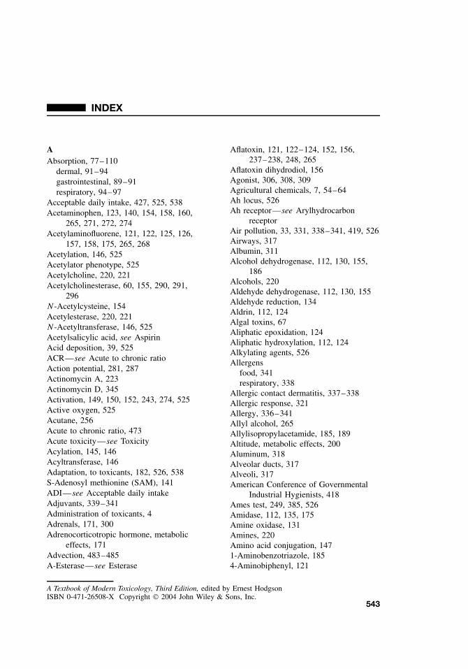
INDEX
AAbsorption, 77–110
dermal, 91–94gastrointestinal, 89–91respiratory, 94–97
Acceptable daily intake, 427, 525, 538Acetaminophen, 123, 140, 154, 158, 160,
265, 271, 272, 274Acetylaminofluorene, 121, 122, 125, 126,
157, 158, 175, 265, 268Acetylation, 146, 525Acetylator phenotype, 525Acetylcholine, 220, 221Acetylcholinesterase, 60, 155, 290, 291,
296N-Acetylcysteine, 154Acetylesterase, 220, 221N-Acetyltransferase, 146, 525Acetylsalicylic acid, see AspirinAcid deposition, 39, 525ACR—see Acute to chronic ratioAction potential, 281, 287Actinomycin A, 223Actinomycin D, 345Activation, 149, 150, 152, 243, 274, 525Active oxygen, 525Acutane, 256Acute to chronic ratio, 473Acute toxicity—see ToxicityAcylation, 145, 146Acyltransferase, 146Adaptation, to toxicants, 182, 526, 538S-Adenosyl methionine (SAM), 141ADI—see Acceptable daily intakeAdjuvants, 339–341Administration of toxicants, 4Adrenals, 171, 300Adrenocorticotropic hormone, metabolic
effects, 171Advection, 483–485A-Esterase—see Esterase
Aflatoxin, 121, 122–124, 152, 156,237–238, 248, 265
Aflatoxin dihydrodiol, 156Agonist, 306, 308, 309Agricultural chemicals, 7, 54–64Ah locus, 526Ah receptor—see Arylhydrocarbon
receptorAir pollution, 33, 331, 338–341, 419, 526Airways, 317Albumin, 311Alcohol dehydrogenase, 112, 130, 155,
186Alcohols, 220Aldehyde dehydrogenase, 112, 130, 155Aldehyde reduction, 134Aldrin, 112, 124Algal toxins, 67Aliphatic epoxidation, 124Aliphatic hydroxylation, 112, 124Alkylating agents, 526Allergens
food, 341respiratory, 338
Allergic contact dermatitis, 337–338Allergic response, 321Allergy, 336–341Allyl alcohol, 265Allylisopropylacetamide, 185, 189Altitude, metabolic effects, 200Aluminum, 318Alveolar ducts, 317Alveoli, 317American Conference of Governmental
Industrial Hygienists, 418Ames test, 249, 385, 526Amidase, 112, 135, 175Amine oxidase, 131Amines, 220Amino acid conjugation, 1471-Aminobenzotriazole, 1854-Aminobiphenyl, 121
A Textbook of Modern Toxicology, Third Edition, edited by Ernest HodgsonISBN 0-471-26508-X Copyright 2004 John Wiley & Sons, Inc.
543
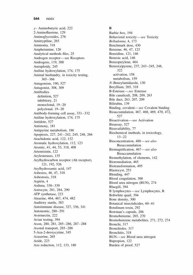
544 INDEX
γ - Aminobutyric acid, 2222-Aminofluorene, 129Aminoglycosides, 276Amitryptline, 265Ammonia, 318Amphetamine, 126Analytical methods files, 25Androgen receptor—see ReceptorsAndrogens, 119, 300Aneuploidy, 245Aniline hydroxylation, 174, 175Animal husbandry, in toxicity testing,
365–366Antagonism, 190, 527Antagonist, 308, 309Antibodies
definition, 527inhibitory, 21monoclonal, 19–20polyclonal, 19–20
Antibody-forming cell assay, 331–332Aniline hydroxylation, 174, 175Antidote, 527Antimony, 181Antipyrine metabolism, 186Apoptosis, 227, 241–242, 245, 248, 266Arachidonic acid, 122, 132Aromatic hydroxylation, 112, 123Arsenic, 41, 44, 53, 318, 408Artemisinin, 122Arylesterases, 135Arylhydrocarbon receptor (Ah receptor),
121, 192, 526Arylhydroxamic acid, 147Asbestos, 46, 47, 318Asbestosis, 318Aspirin, 4Asthma, 338–339Astrocyte, 281, 284, 290ATP synthetase, 223Atrazine, 464, 467, 474, 482Auditory startle, 383Autoimmune disease, 327, 336, 341Autonomic, 280–291Avermectin, 222Avian testing, 394Axon, 280, 281, 285–286, 287–288Axonal transport, 285–2865-Aza-2-deoxycytine, 345Azaserine, 265Azide, 223Azo reduction, 112, 133, 180
BBarbie box, 194Behavioral toxicity—see ToxicityBelladonna, 4, 173Benchmark dose, 430Benzene, 46, 47, 123Benzidine, 121, 146Benzoic acid, 146Benzoperylene, 464Benzo(a)pyrene, 237, 243–245, 248,
322activation, 158metabolism, 159
N-Benzylamidazole, 130Beryllium, 265, 318B-Esterase—see EsteraseBile canaliculi, 208, 209, 263Bile duct, 263, 207, 209Bilirubin, 139Binding, covalent—see Covalent bindingBioaccumulation, 467, 468, 469, 470, 472,
527Bioactivation—see ActivationBioassay, 527Bioavailability, 77Biochemical methods, in toxicology,
13–22Bioconcentration, 488—see also
BioaccumulationBiomagnification, 467—see also
BioaccumulationBiomethylation, of elements, 142Bioremediation, 465Biotransformation, 495Blastocyst, 253Blending, 447Blood coagulation, 300Blood urea nitrogen (BUN), 274Bluegill, 394B lymphocytes—see Lymphocytes, BBobwhite quail, 394Bone density, 300Botanical insectidicides, 60–61Botulinum toxin, 292Bowman’s capsule, 206Bromobenzene, 265, 270Bromobenzene metabolites, 271, 272, 274Bronchi, 317Bronchioles. 317Bronchitis, 318BUN—see Blood urea nitrogenBupropion, 122Burden of proof, 527
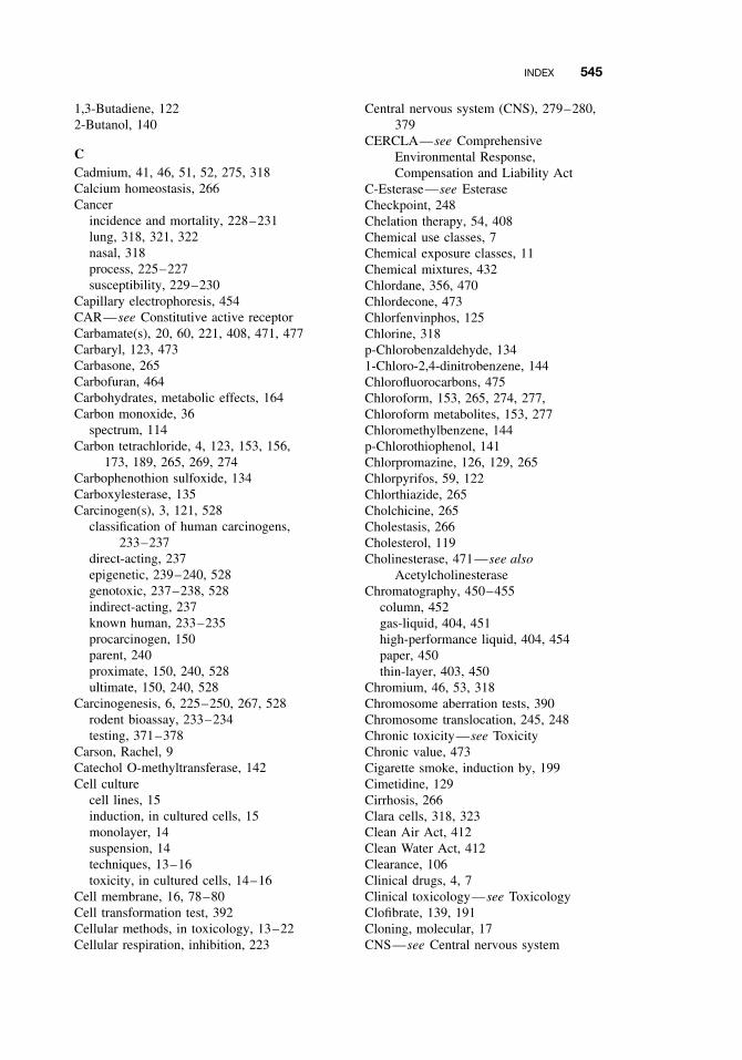
INDEX 545
1,3-Butadiene, 1222-Butanol, 140
CCadmium, 41, 46, 51, 52, 275, 318Calcium homeostasis, 266Cancer
incidence and mortality, 228–231lung, 318, 321, 322nasal, 318process, 225–227susceptibility, 229–230
Capillary electrophoresis, 454CAR—see Constitutive active receptorCarbamate(s), 20, 60, 221, 408, 471, 477Carbaryl, 123, 473Carbasone, 265Carbofuran, 464Carbohydrates, metabolic effects, 164Carbon monoxide, 36
spectrum, 114Carbon tetrachloride, 4, 123, 153, 156,
173, 189, 265, 269, 274Carbophenothion sulfoxide, 134Carboxylesterase, 135Carcinogen(s), 3, 121, 528
classification of human carcinogens,233–237
direct-acting, 237epigenetic, 239–240, 528genotoxic, 237–238, 528indirect-acting, 237known human, 233–235procarcinogen, 150parent, 240proximate, 150, 240, 528ultimate, 150, 240, 528
Carcinogenesis, 6, 225–250, 267, 528rodent bioassay, 233–234testing, 371–378
Carson, Rachel, 9Catechol O-methyltransferase, 142Cell culture
cell lines, 15induction, in cultured cells, 15monolayer, 14suspension, 14techniques, 13–16toxicity, in cultured cells, 14–16
Cell membrane, 16, 78–80Cell transformation test, 392Cellular methods, in toxicology, 13–22Cellular respiration, inhibition, 223
Central nervous system (CNS), 279–280,379
CERCLA—see ComprehensiveEnvironmental Response,Compensation and Liability Act
C-Esterase—see EsteraseCheckpoint, 248Chelation therapy, 54, 408Chemical use classes, 7Chemical exposure classes, 11Chemical mixtures, 432Chlordane, 356, 470Chlordecone, 473Chlorfenvinphos, 125Chlorine, 318p-Chlorobenzaldehyde, 1341-Chloro-2,4-dinitrobenzene, 144Chlorofluorocarbons, 475Chloroform, 153, 265, 274, 277,Chloroform metabolites, 153, 277Chloromethylbenzene, 144p-Chlorothiophenol, 141Chlorpromazine, 126, 129, 265Chlorpyrifos, 59, 122Chlorthiazide, 265Cholchicine, 265Cholestasis, 266Cholesterol, 119Cholinesterase, 471—see also
AcetylcholinesteraseChromatography, 450–455
column, 452gas-liquid, 404, 451high-performance liquid, 404, 454paper, 450thin-layer, 403, 450
Chromium, 46, 53, 318Chromosome aberration tests, 390Chromosome translocation, 245, 248Chronic toxicity—see ToxicityChronic value, 473Cigarette smoke, induction by, 199Cimetidine, 129Cirrhosis, 266Clara cells, 318, 323Clean Air Act, 412Clean Water Act, 412Clearance, 106Clinical drugs, 4, 7Clinical toxicology—see ToxicologyClofibrate, 139, 191Cloning, molecular, 17CNS—see Central nervous system
546 INDEX
CoA—see Coenzyme ACoal dust, 318Cobalt, 4Cocaine, 205Cocarcinogenesis, 528Coenzyme A (CoA), 146Columns, 450, 451Combustion products, 7Compartment, 106, 528Compartmentalization, 204Comprehensive Environmental Response,
Compensation and Liability Act(CERCLA), 413
Conjugation reactions—see Phase IIreactions
Constitutive active receptor (CAR), 195Consumer Products Safety Act, 354, 413Consumer Products Safety Commission
Improvements Act, 413Contact hypersensitivity, 337–338Controlled Substances Act, 413Cooxidation, 132Copper, 4, 165, 166, 492,Cortisol, 300Cosmetics, 71Coumaphos, 126Coumarin, 122, 138Courtroom testimony, 399, 400Covalent binding, 274, 384, 527COX-1, 132COX-2, 132Creatinine, plasma, 274Critical periods, 255Cross resistance, 529Crotalaria spectabilis, 323Cyanide, 173, 223Cycasin, 159, 160, 265Cyclodiene insecticides, 222Cyclohexamide, 265Cyclooxygenases, 132Cyclophosphamide, 122Cyclosporine A, 123CYP—see Cytochrome P450Cysteamine, 129Cytochrome b5, 115Cytochrome P450 (CYP), 271, 272, 274,
277, 310, 318, 322, 323, 324classification, 117distribution, 116CYP1, 119CYP1A1, 119, 121CYP1A1, induction, 191, 199CYP1A2, 119, 121
CYP1A2, induction, 192CYP1B1, 119CYP2, 119, 122CYP2A6, 122CYP2B, regulation, 191, 194CYP2B6, 122CYP2C, 122CYP2C8, 122CYP2C9, 122CYP2C19, 122CYP2D6, 182CYP2E1, 123, 170CYP2E1, regulation, 192, 197CYP3A, regulation, 191, 196, 197CYP3A4, 123, 323CYP19, 310evolution, 117families, 119–123induction, 123multiplicity, 116nomenclature, 117purification, 116reactions, 112, 123–128
Cytochrome P450 reductase, 114, 1320,324
Cytotoxicity—see ToxicityCysteine conjugate β-lyase—see β-LyaseCytosol, 113
DDDE, 174, 204, 348DDT, 57, 58, 137, 198, 204, 222, 307,
309, 348, 464, 465, 468DDT dehydrochlorinase, 136Deacetylation, 147Dealkylation (O-, N-, and S-), 112, 124,
465Debrisoquine, and polymorphisms, 182Degradation
abiotic, 465biotic, 465
Dehalogenation, 112, 465Dehydrogenation, 112Delaney Amendment—see Food, Drug
and Cosmetics ActDemeton, 126Dendrites, 280Dermal
irritation tests, 363sensitization tests, 363subchronic toxicity studies, 369
DES—see DiethylstilbestrolDesulfuration, 112, 127
INDEX 547
Detectorsconductivity, 453electron capture, 453flame ionization, 452flame photometric, 453gas-chromatographic, 452
Detoxication, 529Detoxification, 529Development, 300Developmental toxicity—see ToxicityDevelopmental toxicology—see
ToxicologyDexamethasone, 123, 191, 196Diabetes, CYP2E1 induction in, 197, 198Dialkyl nitrosamines, 265Diamine oxidase, 132Diazepam, 265Diclofenac, 1232,6-Dichlorobenzonitrile, 122Dicofol, 465Dicoumarol, 224Dieldrin, 57, 58, 124, 222, 476Dietary administration, in toxicity testing,
365Diethylmaleate, 144, 185Diethylstilbestrol (DES), 256, 299, 306,
310, 312, 345Diethyltoluamide (DEET), 122Diffusion, 80
between phases, 486in environment, 485–487passive, 80
Dihydrotestosterone, 345Diffusion, passive, 203Digitoxin, 123Dimethoate, 175Dimethylamine128Dimethylaniline, 129Dimethylbenzanthracene, 121, 265Dimethylnitrosamine, 265Dimethyl sulfoxide, 1262,4-Dinitrophenol, 224
and cataracts, 174Dioxin(s), 43, 311, 314Diphenylmethylphosphine, 127Disease, metabolic effects, 171Dispersion, 485Distribution, 77–110, 529Disulfide reduction, 133Disulfiram, 122, 133, 134, 185, 186Diurnal rhythms, metabolic effects, 172DNA
adducts, 240–241, 243–245, 384
binding, 270damage, 237–238, 322, 389cDNA libraries, 17recombinant, 16repair, 240–241, 245, 389unscheduled synthesis test, 389
Dominant lethal test, 392Dopamine, 140Dosage, 529Dose extrapolation, 249, 233–234Dose-response, 4, 10, 217–220, 426–427,
529Dose selection, for chronic studies, 370Draize test—see Eye irritation testDrospophila, 388Drug analysis, 405Drug analysis flowchart, 405Drugs of abuse, 7, 71, 72, 530Drugs, therapeutic, 70, 417, 530
EEC50, 217ED50, 217Ecdysone receptor—see ReceptorsE. coli, 386Ecotoxicology—see ToxicologyEdema, 318EDSTAC—see Endocrine Disruptor
Screening and Testing AdvisoryCommittee
Education, 420Electrolyte balance, 273Electron transport system
definition, 530inhibitors, 530
Electrophilic, 149, 240, 530Electrophoresis, capillary, 454Elimination of toxicants—see ToxicantsEmbryology, mammalian, 252Emphysema, 318, 321Endoplasmic reticulum, 16, 113, 531Endocrine disruptors, 347, 415Endocrine Disruptor Screening and
Testing Advisory Committee(EDSTAC), 415
Endocrine system, 299–315Endocytosis, 276Endosulfan, 126, 473Enterohepatic circulation, 208–209, 531Environmental fate, 479–499Environmental fate models, 479, 497–498Environmental Protection Agency, 354,
411–416
548 INDEX
Environmental transport, 479–499Epidemiology, 6Epigenetic carcinogen—see CarcinogenEpinephrine, 142EPN, 185, 189Epoxidation, 112, 123, 151Epoxide hydrolase, 112, 135, 1801,2-Epoxy-3-(p-nitrophenoxy) propane,
144Erythromycin, 123Esterase(s), 112, 175,
A, 135B, 135C, 135and selectivity, 180
Ester cleavage, 127Estradiol, 265, 300, 304, 305, 306, 307,
311, 312Estrogen receptor—see ReceptorsEstrogens, 119, 300Estrone, 140Ethanol, 123, 191, 257, 270, 408Ethinyl estradiol, 123Ethionine, 265Ethylene glycol, 407Ethylmorphine, 125Ethylnitrosourea, 345Excretion
Biliary, 176Expert witness, 400Exposure, 216Exposure to environmental stress,
508–509Exposure assessment, 425, 507–509, 523,
531Extraction, 447
supercritical fluid, 448Extrapolation, animal to human, 354, 362Eye irritation test, 531
FFatty acid(s), 153FDA—see Food and Drug AdministrationFederal Insecticide, Fungicide and
Rodenticide Act (FIFRA), 393, 414,531
Fenarimol, 310Fertility, 258, 300Fetal alcohol syndrome, 257, 532Fibrosis, 318, 321Field tests, 394–395FIFRA—see Federal Insecticide,
Fungicide and Rodenticide Act
First order kinetics, 82Fish odor syndrome, 128, 183Flavin-containing monooxygenase (FMO),
112, 169, 170, 183, 318, 324Fluoroacetamide, 175FMO—see Flavin-containing
monooxygenaseFOB—see Functional observation batteryFonofos, 129Food additives, 7, 64–65, 532Food allergy, 341Food and Drug Administration (FDA),
258, 354, 413Food chains, environmental, 12Food contaminants, 64–65, 532Food, Drug and Cosmetics Act, 413, 532Food Quality Protection Act, 415Formaldehyde, 152Formic acid, 152Forensic toxicology—see ToxicologyFumigants, 64Function Observational Battery, 293, 295Fungicides, 63Furans, 314Fusarium solani, 323Free radicals, 151, 153, 270, 532Functional observation battery (FOB),
379–380Future considerations, environmental, 521Future considerations, human health, 521
GGABA—see γ -aminobutyric acidGait, 383Galactosamine, 265Gall bladder, 263Gastrula, 252Gender, metabolic effects, 168Gene
amplication, 245expression, 19function, 19regulation, 19
Genetic alterations, 237–239, 243Genetic instability, 248Genetic polymorphisms—see
PolymorphismsGenomic libraries, 17Genotoxic carcinogen—see CarcinogenGenerally regarded as safe (GRAS), 533Genotoxicity—see ToxicityGenotoxicity assays, short assays, 249,
385–393
INDEX 549
Gentamycin, 276Germ cells, 345GFAP—see Glial Fibrillary Acidic ProteinGinger jake, 288Glial Fibrillary Acidic Protein (GFAP),
290, 296Glomerulus, 206Glomerular capsule, 206Glucocorticoid receptor—see ReceptorsGlucocoricoids, 300Glucuronic acid, 209Glucuronidation, developmental effects,
167Glucuronide conjugation, 138Glucuronosyl transferase, 138Glucoside conjugation, 139Glutathione, 143, 209Glutathione conjugation, 143, 144, 167,
180, 277, 208Glutathione S-transferase—see
Glutathione conjugationGlycine, 146Glycine conjugation, 167P-Glycoprotein, 209Good laboratory practice, 533GRAS—see Generally regarded as safeGrowth, 300Growth hormone, 168, 300Gynacomastia, 306, 312, 313
HHair, 205Halogenated hydrocarbons, 249Halothane, 122, 265Hazard assessment, 464, 523Hazard identification, 424Hazard quotient, 432Henry’s law, 487–488Hepatic artery, 207, 263Hepatic elimination, 207–210Hepatic portal vein, 263Hepatitis, 267Hepatocytes, 207, 263–272,Hepatotoxicant(s), 3, 4, 263–272Hepatotoxicity—see ToxicityHerbicides, 62Hexachlorobenzene, 466, 476Hexachlorobutadiene, 277Hexane, 288Hexobarbital, 174, 183, 186HGPRT test, 387Histamine, 182Homeostasis, 273
Hormesis, 219, 426Hormone, 239, 240, 242
clearance, 310synthesis, 310
Household chemicals, 417Human genome project, 16Human health risk assessment—see RiskHydrogen fluoride, 318Hydrogen sulfide, 223Hydrolysis, 135, 465, 493Hydroxyacetanilide, 141N-Hydroxy-2-acetylaminofluorine, 139Hydroxylation, 112, 206Hydroxyl radical, 322Hypersensitivity, 335Hypothalamus, 170, 300, 301, 302Hypothalamus-pituitary-gonadal axis, 344Hypothyroidism, 310, 311, 313, 314
IIbuprofen, 123ICH, 258Ifosfamide, 122Imipramine, 122, 129, 138, 265Immune suppression, 330–334Immune system, 327–330Immune system, developing, 341Immunoaffinity purification, 20Immunoassay, 404Immunochemical techniques, 19–21Immunolocalization, 20Immunosuppresive agents, 239Immunotoxicity—see ToxicityImposex, 473, 474Indomethacin, 176, 265Induction, 190, 198, 310, 533Industrial chemicals, 7Industrial toxicology—see ToxicologyInhalation toxicity testing, 357Inhibition, 60, 155, 185–188, 224, 310,
533Initiation, 241, 534Initiation-promotion model, 241–242Inner cell mass, 252Instrument calibration, 26Insulin, 171Intoxication, 534Involution, 254Ion channels, 222Ionization, 86–87, 490Ionizing radiation, 200Ipomeanol, 323Iproniazid, 265
550 INDEX
Iron, 4, 165, 166, 318Irritation, 318, 320Isoniazid, 265
and polymorphisms, 182Isotetrinoin, 256Isozymes, 534In vitro, 523, 533In vivo, 523, 533
KKanamycin, 276Kepone, 307, 313Ketaconazole, 310Ketamine, 122Ketone reduction, 134Kidney, 273, 205, 206, see also Renal
cortex, 273damage, 274medulla, 273papilla, 273
Koch’s postulates, 511Kupffer cells, 263
LLaboratory Information Management
System (LIM), 25LC50, 217, 470, 471, 525, 534LD50, 215, 217, 359–362, 470, 471, 525,
534Lead, 38, 41, 47, 51, 275, 285, 287, 290Legislation, 411Lethal synthesis, 534Libido, 300Lidocaine, 123Light, metabolic effects, 200Lindane, 222, 466Lipid, 204
effect on metabolism, 164Lipid oxidation, 264Lipid peroxidation, 153, 157, 266, 269,
270, 274, 322Lipophilic, 467, 534Lipoproteins, 264, 270Liver
and elimination, 206. 207, 209fatty, 264–265function, 263injury, 263–268structure, 263susceptibility to toxicants, 264
Local lymph node assay, 337–338LOAEL—see Lowest observed adverse
effect level
Loop of Henle, 206, 273Lovastatin, 123Lowest observed adverse effect level
(LOAEL), 427, 473Lungs, see Respiratory systemβ-Lyase, 145, 277, 278Lymphocytes, B, 19–20Lysosomes, 16, 275
MMagnesium, 4Malathion, 181, 473Malformations, developmental, 376–378Mallard duck, 394Manganese, 4, 386Maximum containment level (MCL), 412Maximum containment level goal
(MCGL), 412Maximum tolerated dose (MTD), 223,
249, 370, 535MCGL—see Maximum containment level
goalMCL—see Maximum containment levelMechanism of action, 535Membranes, 83, 270, 276, 322, 535Mepazine, 265Mephenytoin, 122Mephobarbital, 122Mercaptopurine, 265Mercapturic acids, 143, 166Mercury, 41, 44, 47, 52, 205, 275Mesocosm, 394Mesothelioma, 321, 322,Mestranol, 265Metabolic activation—see ActivationMetabolism of toxicants—see toxicantsMetabolism testing, 382Metallothionein, 51, 53, 275Metals, 4, 49–54
and nephrotoxicity, 275–276speciation, 492–493
Methadone, 122Methandrolone, 265Methanol, 152, 155Methimazole, 265Methiocarb, 126, 129Methiochlor, 126Methionine, 129Methoprene, 308Methoprinic acid, 308Methoxyfluorane, 122, 265N-, O-, S-Methylation, 141, 142Methyldopa, 265
INDEX 551
Methylenedioxyphenyl synergists, 4Methylenedioxy ring cleavage, 127Methyl mercaptan, 125Methylmercury, 289, 2901-Methyl-4-phenyl-1,2,3,6-
tetrahydropyridine (MPTP), 129,286, 290, 297
6-Methylthiopurine, 125Methyltransferase, 141Metyrapone, 185Microbial toxins, 66Microglia, 284, 290Micronucleus test, 391Micronutrients, effects on metabolism, 164Microsomes, 122, 535Midazolam, 123Milk, 204Minimum risk levels (MRL), 427Mirex, 464, 470Mitochrondria, 16, 275Mitomycin, 265Mode of action, 535Moisture, effects on metabolism, 200Molecular methods, in toxicology, 13–22Molybdenum hydroxylases, 112Monoamine oxidase, 131, 284, 290Monoclonal, 225–226Monocrotaline, 322Monooxygenase, 535Motility, gut, 300MPTP—see 1-Methyl-4-phenyl-1,2,3,6-
tetrahydropyridineMRL—see Minimum risk levelMTD—see Maximum tolerated doseMultidrug-resistance associated protein,
209Multigeneration study, 372–374Mutagenesis, 6, 535Mutagenicity assays, 249, 385–393Mutagenicity study requirements, under
FIFRA, 393Mutagens, 121Mutation(s), 237–239, 248–249
transition, 248–249translation, 248–249
Mycotoxins, 66, 535Myelin, 281, 287, 289–290
NNaphthalene, 121, 123, 1371-Naphthalene methyl sulfate, 1442-Naphthamine, 1381-Naphthol, 138
Naphthylamine, 121, 268Narcosis, 220, 472National Environmental Policy Act, 414National Institute for Occupational Safety
and Health (NIOSH), 414, 418NCTB—see Neurobehavioral core test
batteryNecrosis, 266, 321Neomycin, 276Nephron, 206, 273Nephrotoxicity—see ToxicityNeurobehavioral core test battery (NCTB),
293–294Neurotoxicity—see ToxicityNeurotransmitter
receptors, 280, 282–283, 289, 297release, 281–282, 283, 292, 297reuptake, 285, 290
Nevirapine, 122Niacin, 165, 166Nickel, 318Nicotine, 122, 129, 130, 141, 173Nifedipine, 123NIOSH—see National Institute for
Occupational Safety and HealthNitrates, 42p-Nitroanisole, 124Nitroaromatics, 220Nitrobenzene, 134Nitrogen oxides, 37, 3184-Nitrophenol, 140Nitro reduction, 112, 133, 180NOAEL—see No observed adverse effect
levelNonpoint sources, 480–482Nonsteroidal anti-inflammatory drugs
(NSAIDs), 139No observed adverse effect level
(NOAEL), 426No observed effect level (NOEL), 10, 472,
525, 536Noradrenaline, 142Norepinephrine, 142Nornicotine, 141Northern blot analysis, 18Nuclear magnetic resonance, 460Nuclear receptors—see ReceptorsNucleophile, 149Nucleus, 16
OOccupational Safety and Health
Act, 413
552 INDEX
Occupational Safety and HealthAdministration, 354, 413, 418, 536
OECD, 367Oligodendrocyte, 281, 284Omeprazole, 122Oncogenes, 227, 245–247, 536OPIDN—see Organophosphate-induced
delayed neuropathyOrganochlorine insecticides, 57–58Organophosphate-induced delayed
neuropathy (OPIDN), 288Organophosphorus insecticides, 58–60,
220, 221, 288, 290–292, 408, 471,477
aging, 291–292Organogenesis, 371, 374–378Organomercurials, 275Organ toxicity, 6Ornithine, 178OSHA—see Occupational Safety and
Health AdministrationOvaries, 300, 346Oxazepam, 138Oxidation, N-, S-, and P-, 112, 126, 127Oxidative deamination, 126Oxidative phosphorylation, 536Oxidative stress, 267, 268, 536Oxygen consumption, 300Ozone, 37, 318
Pp53, 245, 247–248Paclitaxel, 123Pantothenic acid, 165, 166Papaverine, 265Paracelsus, 9, 216Paraoxon, 127, 152Paraquat, 181Parasympathetic nervous system, 280, 291Parathion, 58, 59, 127, 152, 155, 465, 466Parkinson’s Disease, 285, 286, 289, 290Partition coefficient, 81, 87–88, 536Partitioning, 487
equilibrium, 487lipid-water, 488octanol-water, 488particle-water, 489
Passive avoidance, 383Passive diffusion—see DiffusionPBB, 312PBPK—see Physiologically based
pharmacokinetic modelPCB—see Polychlorinated biphenyls
PCR—see Polymerase chain reactionPentachlorophenol, 224Peroxisome proliferators, 197Peroxisome proliferators-activated
receptor-α (PPARα), 197Persistence, environmental, 464Pesticides, 42, 54–64
classification, 56Petroleum, 472P-Glycoprotein, 209Pharmacokinetics, 78, 537Phase I reactions, 111–137, 174, 537Phase II reactions, 137–148, 165, 166,
174, 176, 177, 206, 529, 537Phenanthrene, 464Phenindione, 265Phenobarbital, 123, 191, 222, 239, 242Phenobarbital-responsive enhancer module
(PBREM), 194Phenol, 177, 178, 311Phenylbutazone, 174, 265Phenytoin, 123Phorate, 126, 129Phorbol 12-myristate 13 acetate (PMA),
239, 2423′-Phosphoadenosine -5′-phosphosulfate
(PAPS), 140Phosphate conjugation, 147Phospholipids, 264, 276Phosgene, 156, 277, 318Phosphorus, 265Photolysis, 465, 494Phthalate, 313Physiologically based pharmacokinetic
model (PBPK), 436Phytotoxins, 68Piperonyl butoxide, 4, 127, 185, 189Pituitary, 301, 302, 170, 171PMA—see phorbol 12-myristate 13
acetatePoint sources, 480–482Poison—see also Toxicant
definition, 3, 537Pollution, 33, 40, 419, 537, 541Polychlorinated biphenyls (PCBs), 43,
204, 265, 309, 311, 314, 464, 468,470, 476
Polycyclic aromatic hydrocarbons, 71, 73,121, 243, 249
Polymerase chain reaction (PCR), 18Polymorphisms, 181, 230Polyploidy, 248Polyvinyl chloride, 345
INDEX 553
Poor metabolizers (PM), 122Portals of entry, 4, 537Potentiation—see Synergism and
potentiationPregnancy, effects on metabolism, 171Pregnane X receptor (PXR), 196, 197Pregnenolone-16α-carbonitrile, 191Prevention of toxicity, 411–421Primitive streak, 252Procarcinogen—see CarcinogenProcedural manuals, 24Progesterone, 119Progression, 241Proguanil, 122Promazine, 265Promethazine, 129Promotion, 241–243, 537Promoter insertion, 245Propanolol, 138Propiconazole, 310Propofol, 122Prospective assessment, 479–480Prostaglandins, 132, 274Prostaglandin synthase, 132,
and cooxidation, 112Protein
binding, 97, 270effects on metabolism, 163stabilization, 198synthesis, 17, 264, 266
Proto-oncogene, 245Pulmonary toxicity—see ToxicityPuromycin, 265Pyrethroid insecticides, 61, 222, 477Pyridoxine, 165, 166Pyrrolizidine alkaloids, 265Physicochemical properties, and diffusion,
85–88
Q
QA/QC manuals, 24QSAR—see Quantitative structure
activity relationshipsQuality assurance, 27Quality control, 27Quantitation, 26Quantitative structure activity relationships
(QSAR), 537Quinidine, 123, 187Quinones, 151
RRadiation, 237
Radioimmunoassay, 21Rainbow trout, 394Rapamycin, 123Ras, 246–247RCRA—see Resource Conservation and
Recovery ActReactive oxygen, 149, 151, 267, 322Reactive intermediates (metabolites),
149–161, 269, 538Receptors
androgen, 309ecdysone, 307ecological, 505–506, 508estrogen, 303, 306, 308glucocorticoid, 309hormone receptor agonists, 306hormone receptor antagonists, 308nuclear, 302–303retinoic acid, 307retinoid X (RXR), 195, 196, 304, 308,
315steroid, 303thyroid, 303
Red book, 367Reduction reactions, 133–135Reference Dose (RfD)—see Acceptable
Daily IntakeRegulation, of toxic chemicals, 353–354,
411Regulatory agencies, 354Renal elimination, 205–207Renal system—see also Kidney
blood flow, 274cortex, 273function, 273structure, 273susceptibility, 274–275tubules, 273, 274, 275
Renin, 274Reproductive physiology
female, 346–347male, 343–344
Reproductive toxicants—see ToxicantsReproductive toxicity—see ToxicityResistance—see Adaptation to
toxicantsResource Conservation and Recovery Act
(RCRA), 414, 538Respiratory system
anatomy, 317and elimination, 210cell types, 317function, 317
554 INDEX
Respiratory system (continued )lungs, 210susceptibility, 320
Respiratory toxicity—see ToxicityRetinoic acid, 122, 304Retinoic acid receptor—see ReceptorsRetinol, 122Retrospective assessment, 479–480RfD—see Acceptable Daily IntakeRiboflavin, 165, 166Rifampicin, 123, 191, 196Risk
analysis, 538assessment, 8, 423–438, 521, 538assessment, environmental, 501–517assessment, non-cancer, 427–428characterization, 426–427communication, 8management, 8, 503, 521toxicologic, 538
RNAhnRNA, 17mRNA, 17processing, 17translation, 17
Rodenticides, 63Rotenone, 223Rubella virus, 257RXR—see Receptors
SS9, 113, 385Safe Drinking Water Act, 412Safety factor, 538Safety pharmacology studies, 364Safrole, 127, 265Salmonella assay, 249Sample preparation, 447–448Sampling,
air, 443soil, 444tissue, 445water, 445
Sarin, 200Schwann cells, 284, 287–288Segment II study, 371, 374–378Selectivity (selective toxicity), 172, 181,
524, 538Selegiline, 122Senescence, effects on metabolism, 167Serotonin, 142Serum enzymes, 274Sexual
behavior, 345, 348characteristics, secondary, 300differentiation, 300function, 300
SHBG, 305, 306Signal transduction, 242, 246Silica, 318Silicosis, 321Sinusoids, 207Sister chromatid exchange, 390SKF 525A, 185, 186Skin, 204Slope factor, 433Soil pollutants, 40Solvents, 70, 539Somite, 253Southern blot analysis, 18Spectrometry
bimolecular interaction analysis-mass,459
induced coupled plasma, 457Spectrophotometry
infrared, 459ultraviolet/visible, 460
Spectroscopyatomic absorption, 456mass, 458
Spermatogenesis, 344Sperm quality, 345Standard operating procedures, 24Starvation, effects on metabolism, 165Steroid receptor—see ReceptorsStilbestrol, 176Streptomycin, 276Stress, effects on metabolism, 200Stressors
ecological, 502response profile, 510, 512sources, 507, 513susceptibility, 504
Structure-activity relationships, 6, 354,395
Styrene 7,8-oxide, 137Subchronic toxicity—see ToxicitySuicide inhibitor, 155Suicide substrate, 151, 189Sulfanilamide, 265Sulfate, 209Sulfate conjugation, 139Sulfhydryl oxidation, 270Sulfotransferase (SULT), 139, 140Sulfoxide reduction, 135Sulfur dioxide, 318
INDEX 555
Sulfur oxides, 36Superoxide anion, 322, 525Superoxide dismutase, 525Surface area, 203Susceptibility, ecosystem, 504Sweat, 204Synergism and potentiation, 189, 381, 539Sympathetic nervous system, 280, 291Synapses, 280, 282, 290
postsynaptic, 280, 281presynaptic, 280, 282, 290
TTalc, 318Tamoxifen, 123, 129, 308Tannic acid, 265Taxol, 122TCDD, 191, 239, 242, 464, 468, 476Temperature, effects on metabolism, 200Teratogenesis, 6, 251–259, 343, 539
multi-generation study, 258single-generation study, 258
Teratology, 251, 374–378Testes, 170, 300Testing guidelines, 257–258Testosterone, 169, 300, 305, 310, 311Tetanus toxin, 292Tetrachloroethylene, 265, 318Tetrachloromethane, 153Tetracycline, 181, 26512-O-Tetradecanoyl phorpol-13-acetate,
239, 242Tetrafluoroethylene, 278Tetrodotoxin, 173Thalidamide, 256Thelarche, 313Therapy, 539Thermal desorption, 403–404Thiobenzamide, 129Thiophenol, 138Thioridazine, 265Thiourea, 182Threshold, 219, 220Threshold dose, 10, 539Threshold Limit Value(s) (TLV), 44, 418,
539ceiling (TLV-C), 418, 539short-term exposure limit (TLV-STEL),
418, 539time-weighted average (TLV-TWA),
539, 418Thyroid, 300, 303, 304, 310, 311Thyroid hormones, 169, 300
Thyroid receptor—see ReceptorsThyroxine, 300, 301TK locus, 387TLV—see Threshold Limit ValueTOCP see Tri-o-cresylphosphateTolbutamide, 123Tolerance—see Adaptation to toxicantsTolerance Reassessment Advisory
Committee (TRAC), 415Toxic action
mechanism of, 5mode of, 5
Toxicant(s)chemical effects, on metabolism,
184–199comparative effects, on metabolism,
172–183definition, 3, 537, 540developmental effects, on metabolism,
166distribution, 97–105elimination, 203–211environmental effects, on metabolism,
199–200environmental movement, 11fate and effect, 5food chains, 11genetic effects, on metabolism,
172–183measurement of, 6metabolism, 111–148nutritional effects, on metabolism,
163–166physiological effects, on metabolism,
166–172reproductive, 344–348selective, 524
Toxicityacute, 215–224, 470, 471, 525acute toxicity tests, 359–363, 525assessment, definition, 353behavioral, 343, 527behavioral toxicity testing, 379–381,
383chronic, 472, 528chronic toxicity tests, 370–371, 528cytotoxicity, 529definition, 3developmental, 343, 371–378, 380genotoxicity, 322, 533genotoxicity testing, 385–393hepatotoxicity, 263–272, 274, 277, 533immunotoxicity, 327–338, 533
556 INDEX
Toxicity (continued )inhalation, 357measurement of, 6nephrotoxicity, 273–278, 536neurotoxicity, 174, 279–297, 536neurotoxicity testing, 378–381prevention 411–421pulmonary, 537reproductive, 343–349reproductive toxicity testing, 371–378respiratory, 317–325selective, 538subchronic, 539subchronic toxicity testing, 363–370testing, 6, 353–397, 533testing requirements, summary, 355testing, routes of administration, 356testing, selection of test species, 366
Toxicokinetics, 105–109, 382, 540Toxicologic pathology, 6Toxicology
analytical, 6, 441–461applied, 6behavioral, 5, 527biochemical, 4, 524clinical, 6, 399–409comparative, 528definition and scope, 3, 540developmental, 251ecotoxicology, 464environmental, 7, 530forensic, 7, 399–409, 532history, 8, 399industrial, 7, 533molecular, 5, 524nutritional, 5occupational, 399regulatory, 7, 399relation to other sciences, 8veterinary, 6
Toxicology work sheet, forensic, 402Toxic Substances Control Act (TSCA),
414, 540Toxin(s), 65–69, 67, 540TPA—see 12-O-Tetradecanoyl
phorpol-13-acetateTRAC—see Tolerance Reassessment
Advisory CommitteeTranscription, 16Transmethylation, 311Transport, 205, 540
carrier-mediated, 84–85Fickian, 485
in environment, 483–487mechanisms, 80
Triazolam, 123Tributyl tin, 473Trichloroethylene, 265Trichloromethanol, 277Trichloromethyl radical, 269Trichloromethylperoxy radical, 269Tri-o-cresylphosphate (TOCP), 288Triglyceride cycle, 265Trimethylacetophenone, 126Trimethylamine, 128, 129Trimethylaminourea, 128, 183Tryptamine, 142TSCA—see Toxic Substances Control ActTumors
benign, 227–228malignant, 227–228promoters, 239–243suppressor genes, 227, 247–248
Type I ligands, 114Type II ligands, 114Type III ligands, 114
UUltimate carcinogen—see CarcinogenUncertainty, 423Uncertainty factor—see Safety factorUranium, 276Ureter, 273Urethane, 265Urethra, 273Uridine diphospho glucuronic acid
(UDPGA), 138Uridine glucuronosyl transferase (UGT),
139Urinary bladder, 273Urine, 206, 399Uterine effects, 348UVR, 237, 245, 248
VValproic acid, 122, 139Venom, 68, 540Vinclozalin, 309Vinyl chloride, 3, 152, 155, 189, 265, 268Vitamin D, 274Volatile organic compounds, 38, 43Volume of distribution, 103
WWallerian degeneration, 288Warfarin, 123Waste reduction, 420

INDEX 557
Water pollution, 419, 541Weight-of-evidence, 433Western blot analysis, 21Worker Protection Standard for
Agricultural Pesticides, 415Wound healing, 300
XXenobiotic, 541
Xenobiotic-metabolizing enzymes, 4Xenobiotic-responsive element (XRE),
193Xenobiotic-responsive enhancer module
(XREM), 196
ZZinc, 4Zoxazolamine, 198, 265