inhalation toxicology research institute
TRANSCRIPT
DECEMBER 1983
INHALATION TOXICOLOGYRESEARCH INSTITUTE
ANNUAL REPORT
1982-1983
by theStaff of the
Inhalation Toxicology Research Institute
INHALATION TOXICOLOGY RESEARCH INSTITUTELOVELACE BIOMEDICAL & ENVIRONMENTAL
RESEARCH INSTITUTE
P.O. Box 5890 Albuquerque, NM 87185
Prepared for the Office of Health and Environmental Researchof the U. S. Department of Energy
under Contract Number DE-ACO4-76EV01013.
This report was prepared as an account of work
sponsored by the United States Government. Neither the
United States nor the United States Department of
Energy, nor any of their employees, nor any of their
contractors, subcontractors, or their employees, makes
any warranty, expressed or implied, or assumes any
legal liability or responsibility for the accuracy,
completeness or usefulness of any information,
apparatus, product or process disclosed, or represents
that its use would not infringe privately owned rights.
The research described in this report involved
animals maintained in animal care facilities fully
accredited by the American Association for
Accreditation of Laboratory Animal Care.
Prlnted In the United States of America
Available from
National Technical Information ServiceU. S. Department of Commerce5285 Port Royal RoadSpringfield, VA 22161
LMF-107
CATEGORY: UC-48
ANNUAL REPORT OF THE
INHALATION TOXICOLOGY RESEARCH INSTITUTE
OPERATED FOR THE
UNITED STATES DEPARTMENT OF ENERGY
BY THE
LOVELACE BIOMEDICAL AND ENVIRONMENTAL RESEARCH INSTITUTE
OCTOBER 1, 1982, through SEPTEMBER 30, 1983
by the
Staff of the
Inhalation Toxicology Research Institute
R. O. McClellan, Director
Scientific Editors
T. C, Marshall
R. A. Guilmette
Technical Editor
R. L. Byers
Editorial Assistant
B. S. Martinez
December 1983
Prepared for the office of Health and Environmental Research of the United States
Department of Energy under Contract Number DE-AC04-76EV01013.

INHALATION TOXICOLOGY RESEARCH INSTITUTE
ANNUAL REPORT- OCTOBER 1982 THROUGH SEPTEMBER 1983
TABLE OF CONTENTS
INTRODUCTION .............................................................................
PHYSICAL AND CHEMICAL CHARACTERIZATION OF ENERGY TECHNOLOGY AEROSOLS .....................
Size Distribution of Fine Particle Emissions from a Steam Plant with aFluidized Bed Coal Combustor ........................................................
Adsorption of Nitrogen and M-Xylene by Coal Combustion Fly Ash .......................
Surface Area Adsorption and Desorption Studies on Indoor Dust Samples ...............
Chemical Characterization of Compounds Adsorbed Onto Indoor Dust Particles ...........
Formation of Potential Aerosols from Fusion Energy Systems ...........................
LABORATORY STUDIES OF AEROSOL GENERATION AND CHARACIERIZATION ...........................
Nose-Only System for Inhalation Exposures of Small Animals to Large Particles .......
Powder Dispersing Properties of a Fluidized Bed Aerosol Generator ...................
Generation and Characterization of l-Nitropyrene Aerosols ............................
Studies of Lithium Aerosols .........................................................
In Vitro Dissolution Studies of Ultrafine Mixed-Matrix Metal Aerosols ...............
Densities of Fused Clay Aerosols - A Comparison of Aerodynamic Particle Sizerand Aerosol Centrifuge ...............................................................
Optical Diameters of Aggregate Aerosols .............................................
Experimental Responses of Two Optical Particle Counters .............................
Evaluation of the Aerodynamic Particle Size Analyzer .................................
Use of Wire Screens as a Model Filter ...............................................
IN VITRO PREDICTORS OF TOXICITY .........................................................
Effects of Aromatic Fuel Additives on Diesel Engine Emissions .......................
Analysis of Diesel Soot for Nitro-PAHS by Gas Chromatography/Mass Spectrometry ........
Contribution of Primary Aromatic Amines to the Mutagenicity of Gasifier Tarsand Coal Oils .......................................................................
Environmental Transformation of Benzo(a)pyrene and Nitropyreneon Glass Surfaces ....................................................................
Metabolism and Mutagenesis of l-Nitropyrene in Rat Liver, Lung,and Nasal Tissue .....................................................................
Metabolism of (14C)-l-Nitropyrene in Isolated Perfused Rat Lungs .....................
Inhibitors of Rabbit Nasal Cytochrome P=450 Dependent Enzyme Activities .............
In Vitro Investigation of the Possible Interaction of Formaldehydewith Glutathione .....................................................................
The Interaction of Antlpain and Chemical Mutagens in Production of Mutationsin the CHO Cells/HGPRT Mutation Assay ................................................
Page
v
1
3
lO
16
20
24
29
31
37
42
46
49
53
58
63
67
72
79
Bl
86
91
94
98
lOl
I06
lOg
Ill
Hypermutability of CHO Cells Measured as Mutations at the HGPRT Locus and as SisterChromatid Exchanges .................................................................
Induction of Sister Chromatid Exchanges in Lung Cells Exposedto 3-Methylcholanthrene ..............................................................
DEPOSITION AND FATE OF INHALED MATERIALS ........................... ......................
Deposition and Retention of Monodisperse Aluminosilicate Particles Inhaledby Guinea Pigs .......................................................................
Deposition and Retention Patterns for 3-, 9-, and i5-pm Latex MicrospheresInhaled by Rats ......................................................................
Deposition of Ultrafine 67Ga203 Particles in Exercising Rats .........................
Dose to Cell Nuclei from Inhaled PuO2 in the Lungs of Dogs ...........................
The Effect of Inhaled Burden of 239pu02 on Its Retention in Beagle Dog Lung .........
Radiation Dose Patterns in Immature and Aged Beagle Dogs After Inhalationof 241Am02 ...........................................................................
Radiation Dose Patterns in Cynomolgus Monkeys After Inhalation of 241Am02 ...........
Effect of the Chemical Form of Inhaled Curium on its Blokinetlcs in Dogs .............
Reducing Curium Translocatlon from Lung with DTPA Therapy ...........................
Tissue Distribution and Metabolic Fate of 2-Aminoanthracene In RatsAfter Inhalation .....................................................................
l-Nitropyrene Metabolism and Covalent Binding of Metabolitesto Mouse Lung DNA ....................................................................
Pulmonary Retention of Benzo(a)pyrene as Influenced by Amount Instilled .............
DOSE-RESPONSE RELATIONSHIPS FOR INHALED RADIONUCLIDES ...................................
Toxicity Studies of Inhaled Beta-Emittlng Radionuclides - Status Report .............
Toxicity of Inhaled gOSrCl2 in Beagle Dogs. XVII ...................................
Toxicity of Inhaled 91ycI3 In Beagle Dogs. XVII .....................................
Toxicity of Inhaled 144CeC13 in Beagle Dogs. XVl ...................................
Toxicity of Injected 137CsCl in Beagle Dogs. XVI ...................................
Toxicity of gOy in a Relatively Insoluble Form Inhaled by Beagle Dogs. XV ...........
Toxicity of gly Inhaled in a Relatively Insoluble Form by Beagle Dogs. XIV .........
Toxicity of 144Ce Inhaled in a Relatively insoluble Form by Beagle Dogs. XVI .......
Toxicity of 144Ce Inhaled in a Relatively Insoluble Form by ImmatureBeagle Dogs. XII ...................................................................
Toxicity of 144Ce Inhaled in a Relatively Insoluble Form by AgedBeagle Dogs. XII ......................... ; .........................................
Toxicity of gOsr Inhaled in a Relatively Insoluble Form by Beagle Dogs. XIV .........
Biological Effects of Repeated Inhalation Exposure of Beagle Dogsto Relatively Insoluble Aerosols of 144Ce. IX ......................................
Toxicity of inhaled Alpha-Emittlng Radionuclides - Status Report .....................
Toxicity of Inhaled 23Bpu02 in Beagle Dogs: A. Monodisperse 1.5 ~m AMADParticles B. Monodisperse 3.0 pm AMAD Particles. X ...............................
If4
lit
121
123
128
133
137
144
149
153
156
15g
163
167
171
175
177
183
190
194
198
203
208
213
220
224
228
232
237
243

Toxicity of Inhaled 239Pu02 in Beagle Dogs: A. Monodisperse 0.75 gm AMADParticles B. Monodisperse 1.5 pm AMAO Particles C. Monodisperse 3.0 gmAMAD Particles. VI .................................................................
Toxicity of Inhaled 239pu02 in Immature Beagle Dogs. V .............................
Toxicity of Inhaled 239Pu02 in Aged Beagle Dogs. V .................................
Repeated Inhalation Exposure of Beagle Dogs to Aerosols of 239pu, VII ...............
Repeated Inhalation Exposure of Rats to Aerosols of 23gPu02. II .....................
Toxic Effects of Inhaled 244Cm203 in Rats. Ill .....................................
The Retention, Distribution, and Cytogenetic Effects of Inhaled 23gPu(N03)4in the Cynomolgus Monkey .............................................................
The Induction of Chromosome Aberrations in the Liver of the Chinese Hamsterby Injected Thorotrast ...............................................................
The Influence of Age and 239pu02 Exposure on the Pulmonary ImmuneResponse of Dogs .....................................................................
Pulmonary Procoagulant Activity of Dogs with Lung Tumors .............................
DOSE-RESPONSE RELATIONSHIPS FOR INHALED CHEMICAL TOXICANTS ...............................
Life-Span Study of Rodents Inhaling Diesel Exhaust:Results Throughout 30 Months .........................................................
Proliferative and Morphological Response of Rat Lungs and Lung-AssociatedLymph Nodes to Inhaled Fly Ash .......................................................
Testicular Toxicity from Subchronlc Inhalation Exposure of Beagle Dogsto 2,2,2-Trifluoroethanol ............................................................
BIOLOGICAL FACTORS THAT INFLUENCE DOSE-RESPONSE RELATIONSHIPS ...........................
Immune Phagocytosls by Canine Alveolar Macrophages ...................................
Cell-Mediated Immunity of the Dog Lung ...............................................
Effects of Age on Immune Responses After Localized Lung Immunization .................
Monkey Lung Immunity: Response to Locally Deposited Antigen .........................
Immune Responses in Rabbits After Localized Lung Immunization .......................
An Improved Method for Calculating Labeling Indices of Lung Epithelial Cells .........
Respiration of Rats in Nose-Only Exposure Tubes .....................................
Toxicological Aspects of the Long-Term Depletion of Reduced G1utathionein Mice Given L-Buthionine-S,R-Sulfoximine ...........................................
RISK ASSESSMENT .........................................................................
Human Risk Relationships Derived from Epidemiology and Laboratory Studies ...........
Potential Health and Environmental Effects of the Fluidized Bed Combustionof Coal - Final Report ...............................................................
Health Risks from the Disposal of Solid FBC Wastes In the Environment ...............
Use of Fractal Mathematics to Estimate Environmental Dilution Factors ............. \.
APPENDICES
A. Status of Longevity and Sacrifice Experiments in Beagle Dogs ...................
B, Organization of the Inhalation Toxicology Research Institute ...................
252
260
264
269
274
278
283
288
292
298
303
3O5
311
321
325
327
331
335
339
343
348
352
35"/
36l
363
372
377
381
385
427
iii

C. Publication of Technical Reports ...............................................
D. Publications in the Open Literature .............................................
E. Presentations Before Regional or National Scientific Meetingsand Educational and Scientific Seminars .........................................
F. Seminars Presented by Visiting Scientists .......................................
INDEX OF PRINCIPAL AUTHORS ...............................................................
433
435
447
455
457
iv
INTRODUCTION
The mission of the Inhalation Toxicology Research Institute (ITRI) is to investigate the
nature and magnitude of human health effects that might result from inhalation of airborne
materials encountered in the work place or the general environment. Special attention is directed
toward airborne particulate and gaseous emissions released by various energy technologies or from
national defense activities. The new knowledge obtained from this research program is essential
for determining realistic operatlonal guidelines and occupational health practices. The
Institute’s program involves research in all of the areas one must consider as steps in moving
from a source of emissions to evaluation of induced disease. This report is arranged along those
generic lines.
The first section includes five papers on the physical and chemical characterization of energy
technoloqy aerosols. In the second section, closely related laboratory studies of aerosol
qeneration and characterization are reported in lO papers. To an increasing extent, it has proved
useful to conduct studies to correlate physical and chemical parameters with in vitro predictors
of toxicity, as reported in II papers in the third section. The next step in assessing toxicity
is to determine the disposition and fate of inhaled materials. In the fourth section are 12
papers on this subject.
In the fifth section, 24 papers report the results of studies on dose-response relationships
for inhaled radionuclides. These include studies ranging from two to sixteen years in duration,
with the primary finding being an increased incidence of cancers of the nasal cavity, lung,
skeleton, and liver. Three papers in the sixth section report on dose-response relationships for
inhaled chemical toxicants. The major focus of this section is a report on the effects of chronic
exposure to diesel exhaust. Research is also being conducted on biological factors that influence
dose-response relationships, as reported in eight papers in the seventh section of the report. In
the last section of the report, four papers are concerned with risk assessment.
For those who would like to quickly obtain an overview of the Institute’s research, a brief
summary has been provided at the beginning of each section. Those highlight the most important
findings reported in each section.
A review of the individual reports shows that many of them provide information useful in
considering the potential health effects of emissions from several energy technologies even though
the work may have been carried out to address a concern related to a specific technology. Indeed,
it is apparent that in many cases the basic findings have potential wide application in evaluating
the toxicity of airborne materials encountered in situations other than energy production or use.
Although the report is presented with this generic orientation, I am confident that the reader
interested in a specific technology, such as nuclear power or use of diesel powered vehicles, will
not have any difficulty finding the papers of greatest relevance to that particular technology.
This report also includes several appendices. Appendix A provides detailed information on the
status of the long-term dog studies under way at this Institute. Although some of the data
included are preliminary and subject to further analysis and modification, they are included
because of their value to the scientists who are following these studies. Appendix B provides a
current roster of the Institute’s staff. Appendices C and D list publications, and Appendix E
lists scientific presentations given by the Institute staff. Reprints of most of the manuscripts
are available upon request. Appendix F lists seminars given at the Institute by visiting
scientists during the past year.
Although this report primarily summarizes research being conducted for the U. S. Department of
Energy’s Office of Health and Environmental Research, about one-third of the research conducted at
the Institute is for other agencies under interagency agreements. Therefore, the reader interested
in a comprehensive view of the Institute’s activities is referred to the published reports and
open literature manuscripts in Appendices C and D.
ITRI research is multidisciplinary, and each investigation is supported by the work of the
entire staff. Principal investigators are listed with each report to aid readers who wish to
contact staff members for further information. The listing by no means includes all who
contributed to the work. Many highly skilled technical, animal care, maintenance, shop,
administrative, and secretarial personnel are not named, but are essential to a productive
research program.
This report is intended to provide government agencies and other scientists with an overview
of the current status of research in progress at ITRI and, in the case of long-term studies, to
provide an update on a continuing effort well in advance of actual completion of the study. Many
of the data are preliminary and subject to further interpretation before a final report can be
made. Final reports will be made in the form of open literature publications.
Roger O. McClellan, DVM
Director
vi
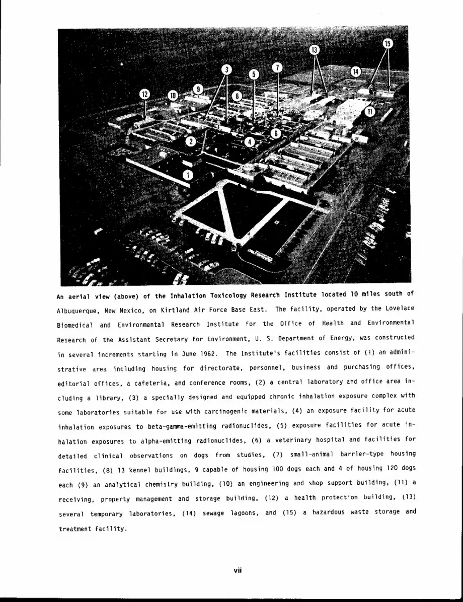
An aertal view (above) of the Inhalation Toxlcology Research Instltute located 10 miles south
Albuquerque, New Mexico, on Kirtland Air Force Base East. The facility, operated by the Lovelace
Biomedical and Environmental Research Institute for the O[fice of Health and Environmental
Research of the Assistant Secretary for Environment, U. S. Department of Energy, was constructed
in several increments starting in June 1962. The Institute’s facilities consist of (1) an admini-
strative area including housing for directorate, personnel, business and purchasing offices,
editorial offices, a cafeteria, and conference rooms, (2) a central laboratory and office area in-
cluding a library, (3) a specially designed and equipped chronic inhalation exposure complex with
some laboratories suitable for use with carcinogenic materials, (4) an exposure facility for acute
inhalation exposures to beta-gamma-emitting radionuclides, (5) exposure facilities for acute in-
halation exposures to alpha-emitting radionuclides, (6) a veterinary hospital and facilities for
detailed clinical observations on dogs from studies, (7) small-animal barrier-type housing
facilities, (B) 13 kennel buildings, g capable of housing lO0 dogs each and 4 of housing 120 dogs
each (9) an analytical chemistry building, (lO) an engineering and shop support building, (ll)
receiving, property management and storage building, (12) a health protection building, (13)
several temporary laboratories, (14) sewage lagoons, and (15) a hazardous waste storage
treatment facility.
vii
PREVIOUS ANNUAL REPORTS
1 ¯ Selective Summary of Studies on the Fission Product Inhalation Program from July 1964 through
June 1965, LF-28, lg65.
2. Selective Summary of Studies on the Fission Product Inhalation Program from July 1965 through
June 1966, LF-33, 1966.
3, Fission Product Inhalation Program Annual Report 1966-1967
4. Fission Product Inhalation Program Annual Report 1967-196B
LF-38, 1967.
LF-39, 1968.
5. Fission Product Inhalation Program Annual Report 196811969, LF-41, 1969.
6. Fission Product Inhalation Program Annual Report 1969-1970, LF-43, 1970.
7. Fission Product Inhalation Program Annual Report 1970-1971 LF-44, 1971.
8. Fission Product Inhalation Program Annual Report 1971-1972 LF-45, 1972.
9. Inhalation Toxicology Research Institute Annual Report 1972-1973, LF-46, 1973.
lO. Inhalation Toxicology Research Institute Annual Report 197311974, LF=4g, 1974.
II. Inhalation Toxicology Research Institute Annual Report 1974-1975, LF-52 1975.
12. Inhalation Toxicology Research Institute Annual Report 1975-1976 LF-56 1976.
13. Inhalation Toxicology Research Institute Annual Report 1976~1977 LF-SB 1977.
14. Inhalation Toxicology Research Institute Annual Report 1977-1978 LF-60 1978.
15. Inhalation Toxicology Research Institute Annual Report 1978-1979, LF-69 IgTg.
16. Inhalation Toxicology Research Institute Annual Report 1979-1980 LMF-84, 1980.
17. Inhalation Toxicology Research Institute Annual Report Ig80-1981 LMF-gl, 1981.
18. Inhalation Toxicology Research Institute Annual Report 1981~1982, LMF-]02, 1982.
viii
PHYSICAL AND CHEMICAL CHARACTERIZATION
OF ENERGY TECHNOLOGY AEROSOLS
A complete inhalation toxicological investigation of effluents associated with production and
use of different sources of energy requires a detailed understanding of the physical and chemical
properties of the materials that might be inhaled. This Institute has had an active program
associated with performing these characterizations on particles and vapors collected in the field
or in laboratory test systems. Information of this type makes it possible to understand the
disposition and fate and the associated dose-response relationships resulting from inhalation of
airborne materials.
The results of effluent particle size characterization from an atmospheric fluidized bed
combustor have been analyzed. Samples were collected from a demonstration plant with a 200-ft bed
at Georgetown University. The distribution of particle sizes was bimodal. The fraction of
submicron particles was 2.3% before the baghouse but increased to 24% in the stack because of
particle size effects in the baghouse collection efficiency. Most of the fly ash (99.98%) was
removed in the baghouse.
One of the critical toxicological issues relating to inhaled fly ash is the degree to which
organic chemicals may be adsorbed on their surfaces and retained in the pulmonary region with
longer effective half=lives than if they were inhaled without the fly ash matrix. Specific
surface area and m-xylene adsorption isotherm measurements have been made with three different fly
ash samples. At low surface coverages, up to one monolayer, it was found that fly ash particles
will adsorb hydrocarbons onto active sites during the time required for stack taversal.
Other studies were conducted on the adsorption and desorptlon of nitrogen and formaldehyde
from room dust. The desorption of formaldehyde was slow, indicating that vapors adsorbed on room
dust could be deposited in the pulmonary region, along with the vector material. Parallel effort
was devoted to chemical characterization of chemicals that might be present on room dust samples.
Several aromatic compounds were found.
Another area of field sampling has involved the collection and analysis of particulate samples
associated with nuclear fusion-related devices. These samples, which have been collected from
either operational or maintenance operations, have been found to be largely ultrafine particles
consisting of branched chain aggregates. Further research is required on the toxicological
implications of these areosol forms for the materials that may be involved.
1/2

SIZE DISTRIBUTION OF FINE PARTICLE EMISSIONS FROM A STEAM PLANT
WITH A FLUIDIZED BED COAL COMBUSTOR
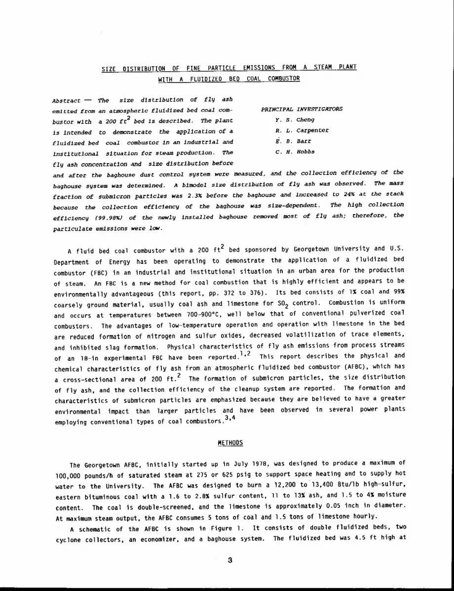
Abstract -- The slze dlstrlbutlon of fl~ ash
emitted from an atmospheric fluld~zed bed coal tom-
buster wlth a 200 ft 2 bed is descrlbed. The plant
is intended to demonstrate the app11caclon of a
fluldlzed bed coal combustor in an indus~r~al and
instltutlonal sltuatlon for steam productlon. The
fly ash concentration and slze dlstrlbutlon before
PRINCIPAL INVESTIGATORS
Y, S. cheng
R, L. Carpenter
~. B. Barr
c. H. Hobbs
and after the baghouse dust control s~stem were measured, and the collectlon efflclency of the
baghouse system was determined. A blmodel slze dlstrlbu~on of fl~ ash was observed. The mass
fractlon of submlcron partlcles was 2.3~ before the baghouse and Increased to 24~ at the stack
because the collectlon eff~clency of the baghouse was slze-dependent. The high collectlon
efficiency (99.98~) of the newl9 Installed baghouse removed most of fly ash; therefore, the
parClculate emlsslons were low.
A fluid bed coal combustor with a 200 ft 2 bed sponsored by Georgetown University and U.S.
Department of Energy has been operating to demonstrate the application of a fluidized bed
combustor (FBC) in an industrial and institutional situation in an urban area for the production
of steam. An FBC is a new method for coal combustion that is highly efficient and appears to be
environmentally advantageous (this report, pp. 372 to 376). Its bed consists of 1% coal and 99~
coarsely ground material, usually coal ash and limestone for SO2 control. Combustion is uniform
and occurs at temperatures between 700-900°C, well below that of conventional pulverized coal
combustors. The advantages of low-temperature operation and operation with limestone in the bed
are reduced formation of nitrogen and sulfur oxides, decreased volatilization of trace elements,
and inhibited slag formation. Physical characteristics of fly ash emissions from process streams
of an 1B-in experimental FBC have been reported. 1’2 This report describes the physical and
chemical characteristics of fly ash from an atmospheric fluidized bed combustor (AFBC), which has
a cross-sectional area of 200 ft. 2 The formation of submicron particles, the size distribution
of fly ash, and the collection efficiency of the cleanup system are reported. The formation and
characteristics of submicron particles are emphasized because they are believed to have a greater
environmental impact than larger particles and have been observed in several power plants
employing conventional types of coal combustors.3’4
METHODS
The Georgetown AFBC, initially started up in 3uly 1978, was designed to produce a maximum of
lO0,O00 pounds/h of saturated steam at 275 or 625 psig to support space heating and to supply hot
water to the University. The AFBC was designed to burn a 12,200 to 13,400 Btu/Ib high-sulfur,
eastern bituminous coal with a 1.6 to 2.8% sulfur content, ll to 13% ash, and 1.5 to 4% moisture
content. The coal is double-screened, and the limestone is approximately 0.05 inch in diameter.
At maximum steam output, the AFBC consumes 5 tons of coal and 1.5 tons of limestone hourly.
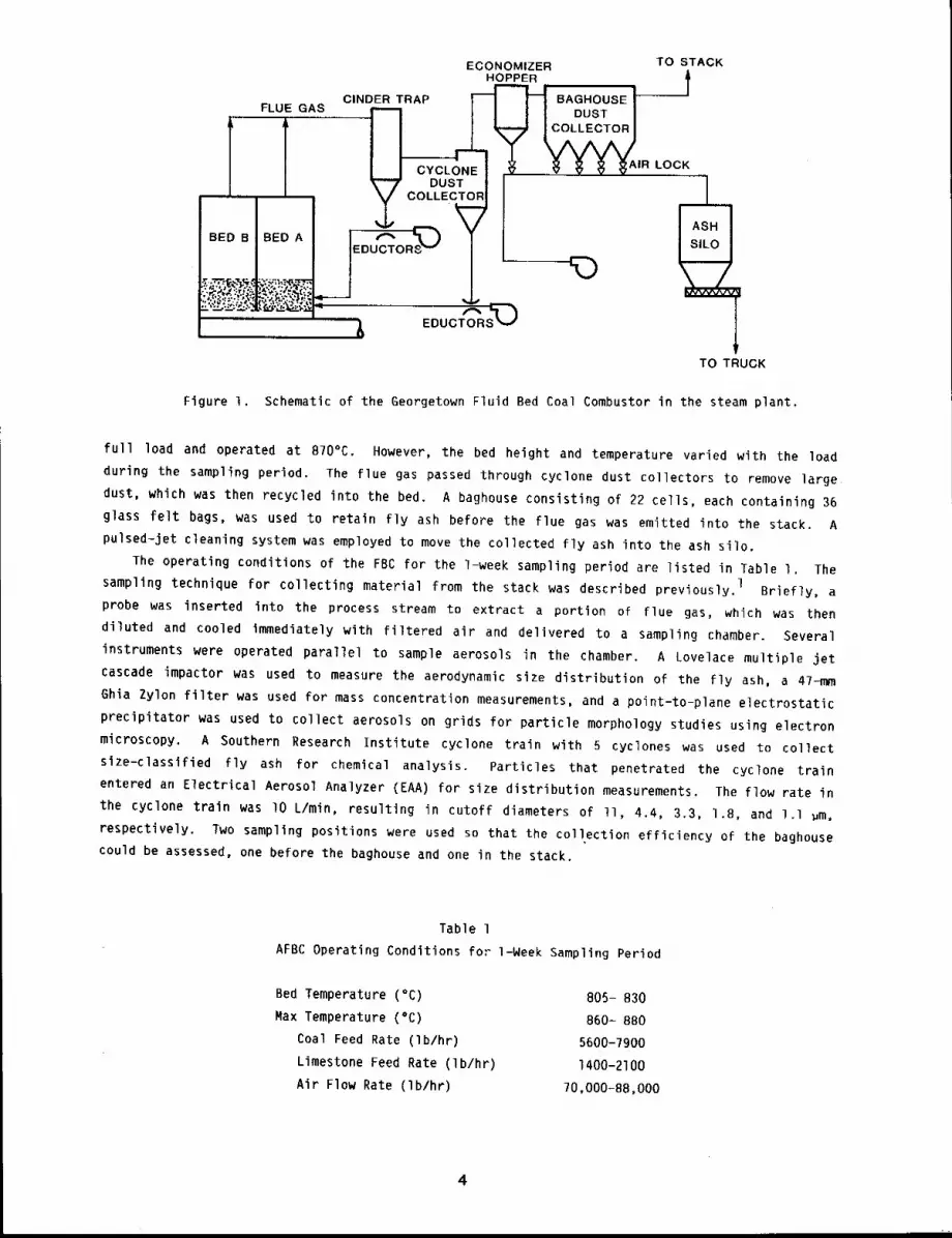
A schematic of the AFBC is shown in Figure l. It consists of double fluidized beds, two
cyclone collectors, an economizer, and a baghouse system. The fluidized bed was 4.5 ft high at
8
FLUE GASA
ECONOMIZERHOPPER
CINDER TRAP "~’ BAGHOUSEIl
II OUST I
,~ ~ ~ ~AIRLOCK
__
TO STACK
BED B I BED A
lTO TRUCK
Figure I. Schematic of the Georgetown Fluid Bed Coal Combustor in the steam plant.
full load and operated at 870°C. However, the bed height and temperature varied with the load
during the sampling period. The flue gas passed through cyclone dust collectors to remove large
dust, which was then recycled into the bed. A baghouse consisting of 22 cells, each containing 36
glass felt bags, was used to retain fly ash before the flue gas was emitted into the stack. A
pulsed-jet cleaning system was employed to move the collected fly ash into the ash silo.
The operating conditions of the FBC for the l-week sampling period are listed in Table I. The
sampling technique for collecting material from the stack was described previously. 1 Briefly, a
probe was inserted into the process stream to extract a portion of flue gas, which was then
diluted and cooled immediately with filtered air and delivered to a sampling chamber. Several
instruments were operated parallel to sample aerosols in the chamber. A Lovelace multiple jet
cascade impactor was used to measure the aerodynamic size distribution of the fly ash, a 47-mm
6hia Zylon filter was used for mass concentration measurements, and a point-to-plane electrostatic
precipitator was used to collect aerosols on grids for particle morphology studies using electron
microscopy. A Southern Research Institute cyclone train with 5 cyclones was used to collect
size-classlfied fly ash for chemical analysis. Particles that penetrated the cyclone train
entered an Electrical Aerosol Analyzer (EAA) for size distribution measurements. The flow rate
the cyclone train was lO L/min, resulting in cutoff diameters of II, 4.4, 3.3, 1.8, and l.l ~m,
respectively. Two sampling positions were used so that the collection efficiency of the baghouse
could be assessed, one before the baghouse and one in the stack.
Table 1
AFBC Operating Conditions fm- l-Week Sampling Period
Bed Temperature (°C)
Max Temperature (°C)
Coal Feed Rate (Ib/hr)
Limestone Feed Rate (Ib/hr)
Air Flow Rate (Ib/hr)
805- 830
860= 8BO
5600-7900
1400-2100
70,000-B8,000
4
RESULTS
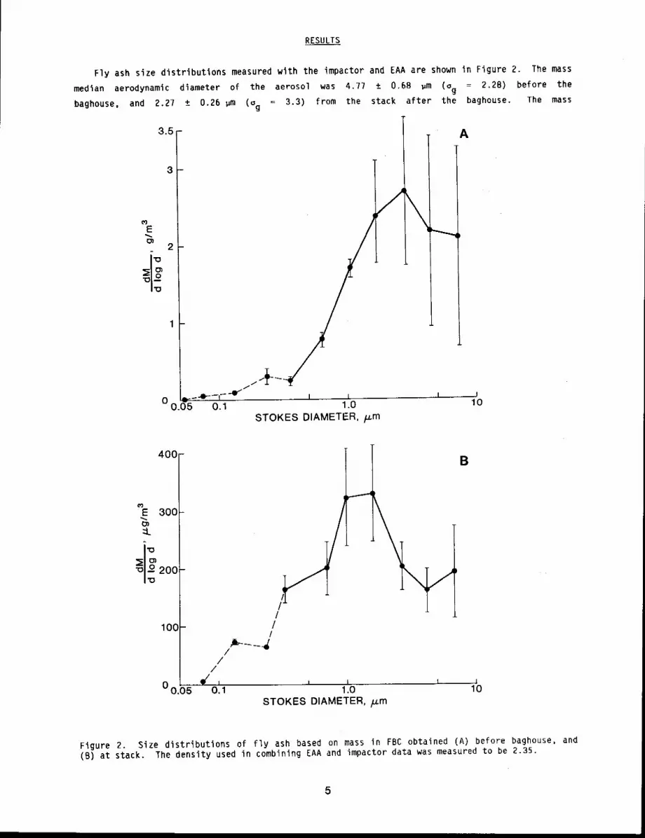
Fly ash size distributions measured with the impactor and EAA are shown in Figure 2. The mass
median aerodynamic diameter of the aerosol was 4.77 ± 0.68 ~m (Og = 2.28) before the
baghouse, and 2.27 ± 0.26 ~m (~g = 3.3) from the stack after the baghouse. The mass
3.5
3
E
2
).05 0.1 1.0STOKES DIAMETER, /..cm
A
I
I0
400
E 300
::k
100
B
_ /I
//
I/, I I t l0 0.05 O. 1 1.0 10
STOKES DIAMETER, /J.m
Figure 2. Size distributions of fly ash based on mass in FBC obtained (A) before baghouse, and(B) at stack. The density used in combining EAA and impactor data was measured to be 2.35.
concentrations (from impactor and filter samples) were 2.96 ± 1.5 g/m3 before the baghouse and
0.352 ± 0.09 mg/m3 after it. This indicates a 99.99% overall collection efficiency for the
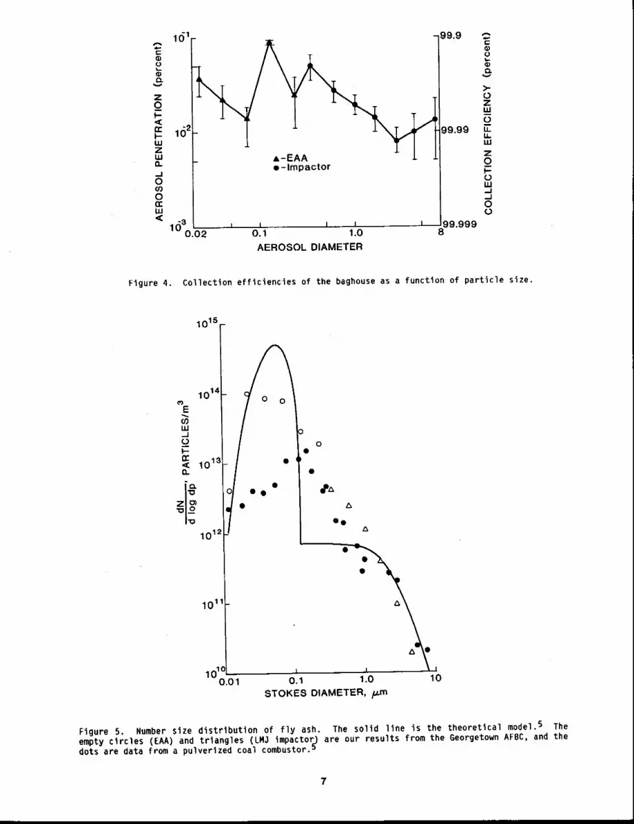
baghouse system. The baghouse efficiencw as a function of particle size (Fig. 3) was determined
by comparing the mass concentration and particle size distribution before and after the baghouse.
The minimum collection efficiency for particle sizes was between 0.I to 0.4 ~m for fly ash.
Size distributions of fly ash were bimodal. A small peak in the size distribution curve was found
between 0.15 to 0.25 ~m. The mass fraction of submicron aerosol was 2.3% before the baghouse
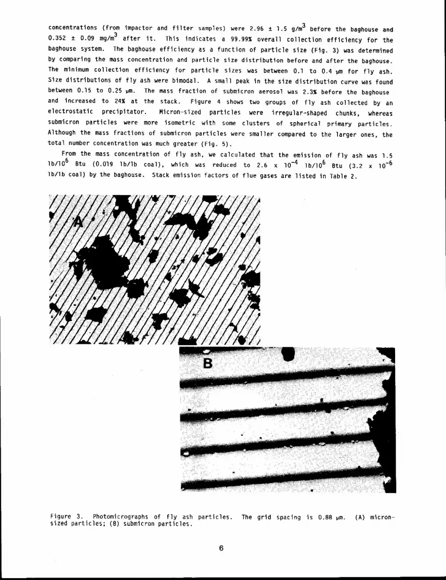
and increased to 24% at the stack. Figure 4 shows two groups of fly ash collected by an
electrostatic precipitator. Micron-sized particles were irregular-shaped chunks, whereas
submicron particles were more isometric with some clusters of spherical primary particles.
Although the mass fractions of submicron particles were smaller compared to the larger ones, the
total number concentration was much greater (Fig. 5).
From the mass concentration of fly ash, we calculated that the emission of fly ash was 1.5
Ib/lO 6 Btu (0.019 Ib/Ib coal), which was reduced to 2.6 x -4 Ib /lO 6 Bt u (3 .2 x 10-6
Ib/Ib coal) by the baghouse. Stack emission factors of flue gases are listed in Table 2.
Figure 3. Photomicrographs of fly ash particles. The grid spacing is 0.88 um. (A) micron-sized particles; (B) submicron particles.
6
1(~1 -99.9
e-lmpactor
99.99
llJ3 , , i ...... I ...... I 99.9990.02 0.1 1.0 8
AEROSOL DIAMETER
f-0;Ot-0;e~
>-OZLUOLLU.UJ
ZOmL~LU.J
O
Figure 4. Collection efficiencies of the baghouse as a function of particle size.
E
UJ_JL~W-rrn
Z
1015
1014
101~It/e ¯ e1012
101
10"0.01
0
0°
I
oe
i I i
0.1 1.0 10STOKES DIAMETER, /~m
Figure 5, Number size distribution of fly ash, The solid line is the theoretical model. 5 Theempty circles (EAA) and triangles (LM3 impactor=) are our results from the Georgetown AFBC, and dots are data from a pulverized coal combustor,u
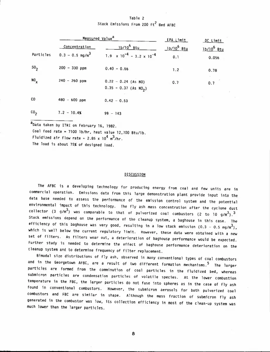
Table 2
Stack Emissions From 200 ft 2 Bed AFBC
Measured Valuea
Concentration
Particles 0.3 - 0.5 mg/m3lb/lO 6 Btu
1.9 x I0 =4 - 3.2 x lO-4
SO2200 - 330 ppm 0.40 - 0.66
NOx 240 = 260 ppm 0.22 - 0.24 (As NO)
0.35 - 0.3? (As NO2)
CO 480 - 600 ppm 0.42 - 0.53
CO2 7.2 - I0.4% 99 - 143
aData taken by ITRI on February 16, 1982.
Coal feed rate = 7100 Ib/hr, heat value 12,700 Btu/Ib.
Fluidized air flow rate = 2.85 x 10⁴ m3/hr.
The load is about ?5% of designed load.
EPA Limit DC Limit
~b/106 Btu Ib/lO 6 Btu
O.l 0.056
1.2 0.78
0.7 0.7
DISCUSSION
The AFBC is a developing technology for producing energy from coal and few units are in
commercial operation. Emissions data from this large demonstration plant provide input into the
data base needed to assess the performance of the emission control system and the potential
environmental impact of this technology. The fly ash mass concentration after the cyclone dust
collector (3 g/m3) was comparable to that of pulverized coal combustors (2 to lO g/m3).3
Stack emissions depend on the performance of the cleanup system, a baghouse in this case. The
efficiency of this baghouse was very good, resulting in a low stack emission (0.3 - 0.5 mg/m3),
which is well below the current regulatory limit. However, these data were obtained with a new
set of filters. As filters wear out, a deterioration of baghous# performance would be expected.
Further study is needed to determine the effect of baghouse performance deterioration on the
cleanup system and to determine frequency of filter replacement.
Bimodal size distributions of fly ash, observed in many conventional types of coal combustors
and in the Georgetown AFBC, are a result of two different formation mechanisms. 5 The larger
particles are formed from the comminution of coal particles in the fluidized bed, whereas
submicron particles are condensation particles of volatile species. At the lower combustion
temperature in the FBC, the larger particles do not fuse into spheres as in the case of fly ash
found in conventional combustors. However, the submicron aerosols for both pulverized coal
combustors and FBC are similar in shape. Although the mass fraction of submicron fly ash
generated in the combustor was low, its collection efficiency in most of the clean-up system was
much lower than the larger particles.
8

REFERENCES
l. Newton, G. 3., R. L. Carpenter, H. C. Yeh, and E. R. Peele, Respirable Aerosols from FluidizedBed Coal Combustion. l. Sampling Methodology for an IB-inch Experimental Fluidized Bed Coal
Combustor, Environ. Sci. Technol. 14: 849-B53, IgSO.
2. Carpenter, R. L., G. J. Newton, S. J. Rothenberg, and P. B. DeNee, Respirable Aerosols fromFluidized Bed Coal Combustion. 2. Physical Characteristics of Fly Ash, Environ. Sci.Technol. 14: 854-B59, Ig80.
3. McElroy, M. W., R. C. Carr, D. S. Ensor, and G. R. Markowski, Size Distribution of FineParticles from Coal Combustion, Science 215: 13-19, 1982.
4. Schmidt, E. W., 3. A. Gieseke, and J. M. Allen, Size Distribution of Fine ParticulateEmissions from a Coal-Fired Power Plant, Atmos. Environ. lO: I065-1069, 1976.
5. Flagan, R. C., and S. K. Friedlander, Particle Formation in Pulverized Coal Combustion v AReview, in Recent Developments in Aerosol Science (D. T. Shaw, ed.), Wiley, New York, NY, pp.25-60, IgTB.
ADSORPTION OF NITROGEN AND M-XYLENE BY COAL COMBUSTION FLY ASH
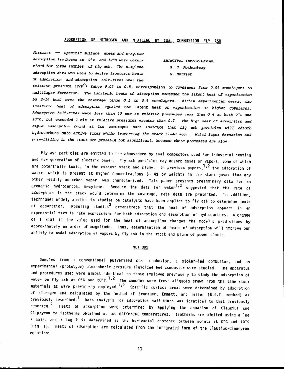
Abstract -- Specific surface areas and m-xglene
adsorption isotherms at O°c and lO°c were deter- PRINCIPAL INVESTIGATORSmined for three samples of fly ash. The m-xylene S.J. Rothenbergadsorption data was used to derive isosterlc heats G. Metzlerof adsorption and adsorption half-tlmes over the
relative pressure (p/pO) range 0.05 to 0.8, corresponding to coverages from 0.05 monolagers
multllayer formation. The isosterlc heats of adsorption exceeded the latent heat of vaperlzatlon
by 2-10 kcal over the coverage range 0.1 to 0.9 monolayers. Wlthln experimental error, the
isosterlc heat of adsorption equaled the latent heat of waporlzatlon a~ higher coverages.
Adsorption half-tlmes were less than 10 sec at relative pressures less than 0.4 at both 0°C and
10°C, but exceeded 3 mln at relative pressures greater than 0.7. The high beat of adsorption and
rapid adsorptlon found at low coverages both indicate that fly ash particles will adsorb
hydrocarbons onto active sites while traversing the stack (i-40 sec). Multl-layer formation and
pore-fllllng In the stack are probably not slgnlflcant, because these processes are slow.
Fly ash particles are emitted to the atmosphere by coal combustors used for industrial heating
and for generation of electric power. Fly ash particles may adsorb gases or vapors, some of which
are potentially toxic, in the exhaust stack and plume. In previous papers,l’2 the adsorption of
water, which is present at higher concentrations (~ 4% by weight) in the stack gases than any
other readily adsorbed vapor, was characterized. This paper presents preliminary data for an
aromatic hydrocarbon, m-xylene. Because the data for water 1’2 suggested that the rate of
adsorption in the stack would determine the coverage, rate data are presented. In addition,
techniques widely applied to studies on catalysts have been applied to fly ash to determine heats
of adsorption. Modeling studies 3 demonstrate that the heat of adsorption appears in an
exponential term in rate expressions for both adsorption and desorption of hydrocarbons. A change
of l kcal in the value used for the heat of adsorption changes the model’s predictions by
approximately an order of magnitude. Thus, determination of heats of adsorption will improve our
ability to model adsorption of vapors by fly ash in the stack and plume of power plants.
METHODS
Samples from a conventional pulverized coal combustor, a stoker-fed combustor, and an
experimental (prototype) atmospheric pressure fluidized bed combustor were studied. The apparatus
and procedures used were almost identical to those employed previously to study the adsorption of
water on fly ash at O°C and 20°C. 1’2 The samples were fresh aliquots drawn from the same stock
materials as were previously employed. I’2 Specific surface areas were determined by adsorption
of nitrogen and calculated by the method of Brunauer, Emmett, and Teller (B.E.T. method)
previously described.1Data analysis for adsorption half-times was identical to that previously
2reported. Heats of adsorption were determined by applying the equation of Clausius and
Clapeyron to isotherms obtained at two different temperatures. Isotherms are plotted using a log
P axis, and a Log P is determined as the horizontal distance between points at 0% and lO°C
(Fig. l). Heats of adsorption are calculated from the integrated form of the Clausius-Clapeyron
equation:
I0
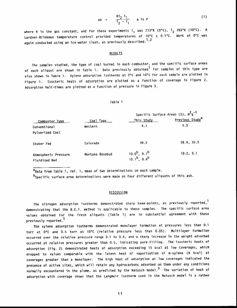
RT2 T1 &InPAH ~ T2 _ T1(i)
where R is the gas constant, and for these experiments T1 was 273°K (O°C), 2 283°K ( lO°C) ¯ A
Lardner-Brinkman temperature control provided temperatures of lO°C ± O.l°C. Work at O°C was1,2
again conducted using an Ice-water slush, as previously described.
RESULTS
The samples studied, the type of coal burned in each combustor, and the specific surface areas
of each aliquot are shown in Table I. Data previously obtained I for samples of this type are
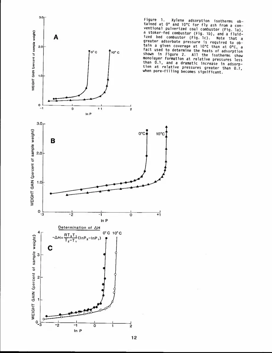
also shown in Table 1. Xylene adsorption isotherms at O°C and lO°C for each sample are plotted in
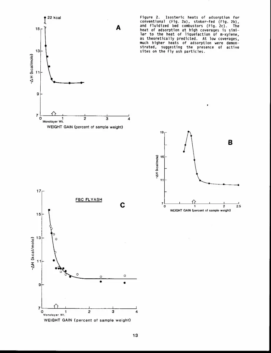
Figure I. Isosteric heats of adsorption are plotted as a function of coverage in Figure 2.
Adsorption halE-times are plotted as a function of pressure in Figure 3.
Table 1
Combustor Type Coal
Conventional Western
Pulverized Coal
Specific Surface Areas (S), m2g-l
This Stud~( Prevlous Stu~a
4.7 5.3
Stoker Fed Colorado 39.2 38.9, 35.5
Atmospheric Pressure Montana Rosebud lO.Ob, 9.7b
Fluidized Bed lO-lb, 9.4b
10.2, 5.1
aData from Table l, ref. I, mean of two determinations on each sample.
bspecific surface area determinations were made on four different aliquots of this ash.
DISCUSSION
The nitrogen adsorption isotherms demonstrated sharp knee-points, as previously reported,l
demonstrating that the B.E.T. method is applicable to these samples. The specific surface area
values obtained for the fresh aliquots (Table l) are in substantial agreement with thosel
previously reported.
The xylene adsorption isotherms demonstrated monolayer formation at pressures less than O.l
torr at O°C and 0.5 torr at lO°C (relative pressure less than 0.05). Multilayer formation
occurred over the relative pressure range O.l to 0.4, and a sharp increase in the weight adsorbed
occurred at relative pressures greater than 0.5, indicating pore-filling. The isosteric heats of
adsorption (Fig. 2) demonstrated heats of adsorption exceeding 15 kcal at low coverages, which
dropped to values comparable with the latent heat of vaporization of m-xylene (9 kcal)
coverages greater than a monolayer. The high heat of adsorption at low coverages indicated the
presence of active sites, which will retain any hydrocarbons adsorbed on them under any conditions
normally encountered in the plume, as predicted by the Natusch model. 3 The variation of heat of
adsorption with coverage shows that the Langmuir isotherm used in the Natusch model is a rather
11
2.0
_z<
1.o,-r(.9[]
3.0
o
A
’0° C
0 +IInP
Figure I. Xylene adsorption isotherms ob-tained at 0° and IO°C for fly ash from a con-ventional pulverized coal combustor (Fig. la),a stoker-fed combustor (Fig. Ib), and a fluid-ized bed combustor (Fig. Ic). Note that greater adsorbate pressure is required to ob-tain a given coverage at lO°C than at O°C, afact used to determine the heats of adsorptionshown in Figure 2. All the isotherms showmonolayer formation at relative pressures lessthan 0.I, and a dramatic increase in adsorp-tion at relative pressures greater than 0.7,when pore-filling becomes significant.
3.0--
~2.0
P
z 1.0
(-9IJJ
o-3
q
O°C~ 10°C
B
,
...... i. I-2 -1
In P
Determination of AH0°C 10°C
Z~ RT2TI- H=T---~-~_Tl(InP2-1nP+) ,)
C
4r.
° l-~3E
~6
C.1i_.(1)CL
(.9 1-l--3:_olU
o3 , , t ,-2 -1 0 1
In P
I /o +1
J2
12
15
13-$-6
~11"r
9
t22 kcal
A
7 ’~ i i i0 1 2 3
Monolayer Wt.
WEIGHT GAIN (percent of sample weight)
17-
15
,-,13(D"5E
O
11.v
FBC FLYASH
C
oo
O O
(ll
7 ’ I I I l I
0 1 2 3Monolayer Wt,
WEIGHT GAIN (percent of sample weight)
Figure 2. Isosteric heats of adsorption forconventional (Fig. 2a), stoker-fed (Fig. 2b),and fluidized bed combustors (Fig. 2c). Theheat of adsorption at high coverages is simi-lar to the heat of liquefaction of m-xylene,as theoretically predicted. At low coverages,much higher heats of adsorption were demon-strated, suggesting the presence of activesites on the fly ash particles.
19-
_~ 15-
-r
,<11
A
A, II ~ li 2
WEIGHT GAIN (percent of sample weight)
B
2.5
13
oocILl
< 0 I 2TPRESSURE (torr)
A
j10°C
~_ _~3 4
10
CD
"5rE
uJ 5
I14-.J<1"
~29.2 min.
O°C
:oo;;0 1 2 3
PRESSURE (tort)
B
2°F==E
wlO-
iu__J< 5I
00
,0 °C 1st Determination C
eO°C 2nd Determination
10°C
1PRESSURE (torr)
(
f2 3 3.5
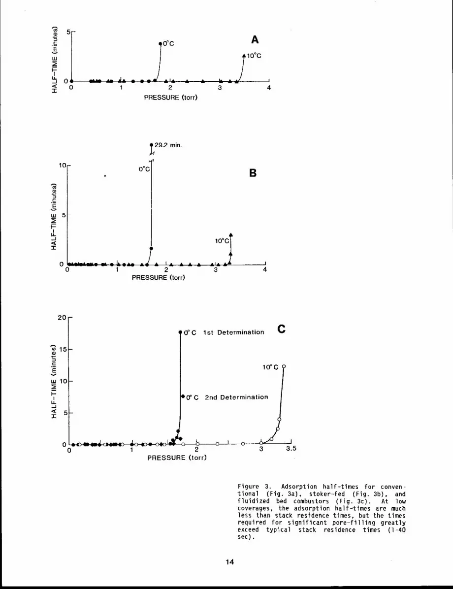
Figure 3. Adsorption half-tlmes for conven-tional (Fig. 3a), stoker-fed (Fig. 3b), fluidized bed combustors (Fig. 3c). At lowcoverages, the adsorptlon hail-times are muchless than stack residence times, but the timesrequired for significant pore-filling greatlyexceed typical stack residence times (I-40sec).
14
drastic approximation. The Langmuir isotherm is really applicable only if the heat of adsorption
is independent of coverage.
Adsorption half-times at O°C (Fig. 3) were less than lO sec at relative pressures less than
0.3, but rose sharply to values over 5 min at relative pressures greater than 0.6. These
pressures correspond to those at which multi-layer formation and pore-filling occur (Fig. 1).
Similar behavior was shown at lO°C. These findings are similar to those previously reported for
water, 2 and suggest transport limited adsorption at high coverages. The adsorption half-times2
at low coverages were smaller than most stack residence times (I-40 sec), so the most active
sites on the fly-ash will adsorb and retain a mixture of water and hydrocarbons in a proportion
determined by kinetic competition in the stack. Stack residence times did not appear to be long
enough for significant pore-filling to occur, as previously predicted on the basis of water2
adsorption kinetics.
REFERENCES
1. Rothenberg, S. J., Coal Combustion Fly Ash Characterization: Adsorption of Nitrogen and
Water, Atmos. Environ. 14: 445-456, 1980.
2. Rothenberg, S. J. and Y. S. Cheng, Coal Combustion Fly Ash Characterization: Rates ofAdsorption and Desorption of Water, ~ Chem. B4: 1644-1649, 1980.
3. Natusch, D. F. S. and B. A. Tomkins, in Carcinpqenesis - A Comprehensive Survey, Vol. 3, P. W.Jones and R. I. Freudenthal, Eds., Raven Press, New York, 1978, pp. 145-153.
15
SURFACE AREA ADSORPTION AND DESORPTION STUDIES ON INDOOR DUST SAMPLES
Abstract --- The adsorptlon properties of two dust
samples, taken from a llbrary and a prefabrlcaCed
building, were studled after collecclon from shelves
five or ~re feet above the floor. Weight loss
curves, nitrogen and formaldehyde adsorptlon and
desorptlon isotherms, and adsorption klnetlcs were
studied using a vacuum mlcrobalance. Samples were
PRINCIPAL INVESTIGATORS
S. J. Rothenberg
P. A. Nagy
J. A. P~ckrell
c. H. Hobbs
further characterlzed by scannlng electron mlcroscopy and energy dlsperslve x-ra 9 analgsls"
Samples had specific surface areas between I m2/gm-I and 20 m2gm-I. The formaldehyde
isotherms showed marked hysteresis, suggesting porosity of the dusts. The desorpt~on of
formaldehyde was slow, suggestlng that vapors adsorbed onto room dust. samples will remain
assoclated wlth partlcles for several days after adsorptlon and tha,t organic vapors adsorbed to
~nhaled subm~cron room dust would be deposited in the respiratory tract along with the particle.
The concentrations of both particulate and gaseous pollutants encountered in the indoor
environment frequently exceed those found outdoors by factors of 2-fold to 20-fold. l Because
most people spend 70-90~ of their time indoors, l study of indoor dusts and vapors is necessary
to predict potential health effects in man from these indoor pollutants. Inhaled vapors may be
deposited in the upper respiratory tract if they are readily soluble in mucus or water or highly
reactive, but the same vapors adsorbed on respirable particles would be deposited in other
locations in the respiratory tract, dependent on the particles. Thus, the adsorption properties
of indoor dust particles are of interest. In this study, we report the results for two samples of
indoor dust for nitrogen adsorption (a standard adsorbate) and for formaldehyde adsorption (an
indoor pollutant of concern).
METHODS
Dust samples were collected from shelves five feet or more above the floor to ensure that they
were not coarse dust brought into the room by pedestrian traffic. Samples were collected from the
top of mobile stacks in the Instltute’s library (Sample A) and from shelves and ductwork in
prefabricated building (Sample B), built from wall-board and sheet metal and previously used
temporary office and laboratory space. Camel hair brushes and spatulas were used to remove
samples from the shelves. Weight loss curves, adsorption, and desorption of nitrogen and
formaldehyde were studied, and hysteresis loops were determined for formaldehyde. The apparatus
and procedures used to study adsorption isotherms were almost identical to those used previously
to study adsorption and desorption of water by fly ash at O°C.2’3 The flask previously used to
hold water had paraformaldehyde powder placed in it. One hour before the measurement of each
formaldehyde adsorption isotherm, the flask was heated to 60°C, creating sufficient pressure of
formaldehyde vapor for the adsorption study.
RESULTS
When heated overnight at lO -5 torr, two different allquots of Sample A lost 3.6%, 1.9% at
50°C, 6.0%, 4.6% at 1SO°C, and 25.6%, 22.9% at 300°C. Two aliquots of Sample B lost 2.1%, 3.9% at
150°C, and 5.8%, 9.0% at 300°C (Sample B was not heated overnight at 50°C). The nitrogen
adsorption isotherms for the dusts had sharp knee-points, demonstrating that the method of
calculation of surface area proposed by Brunauer, Emmett, and Teller 4 (B.E.T. method)
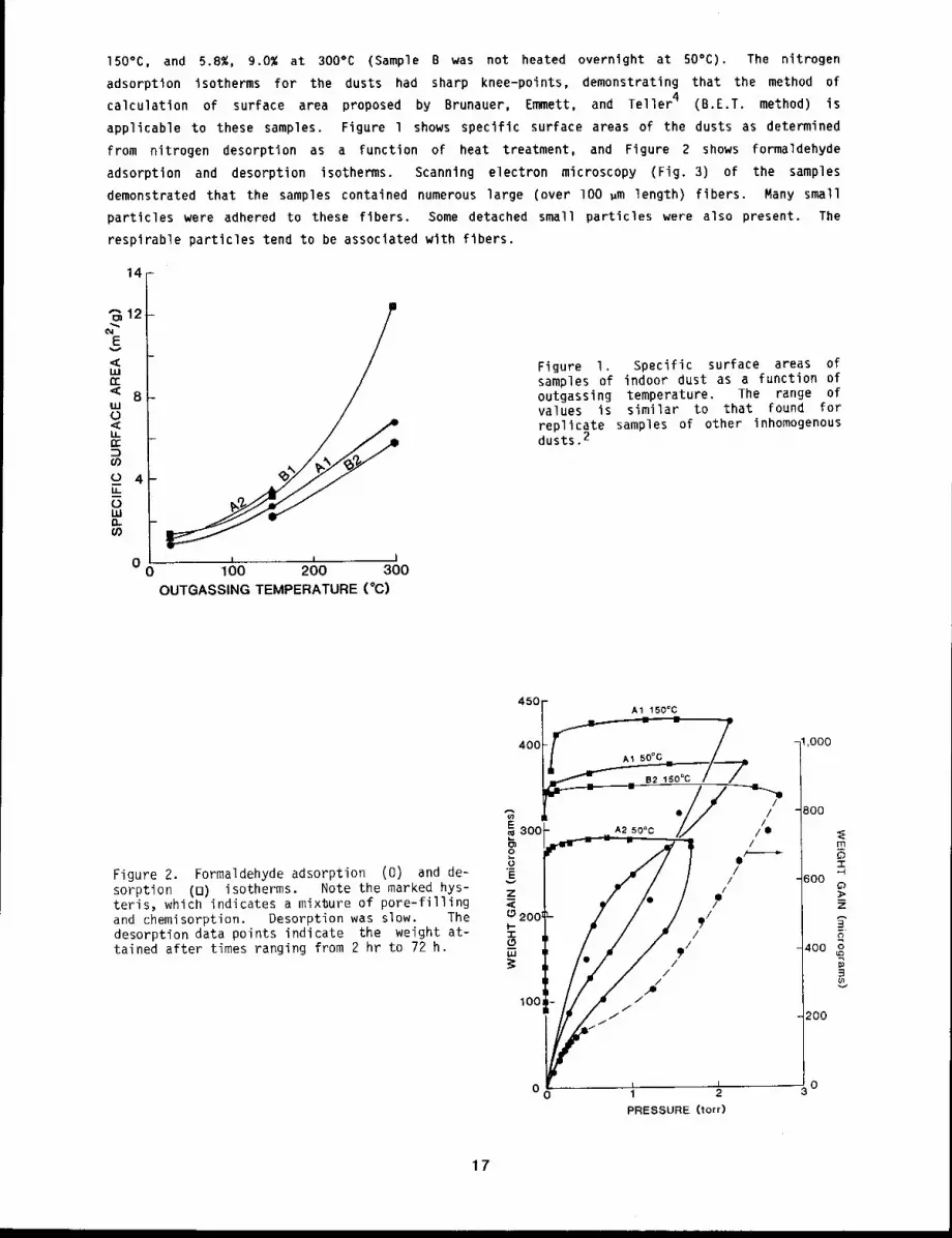
applicable to these samples. Figure l shows specific surface areas of the dusts as determined
from nitrogen desorption as a function of heat treatment, and Figure 2 shows formaldehyde
adsorption and desorption isotherms. Scanning electron microscopy (Fig. 3) of the samples
demonstrated that the samples contained numerous large (over lO0 ~m length) fibers. Many small
particles were adhered to these fibers. Some detached small particles were also present. The
respirable particles tend to be associated with fibers.
14
"~12
Ev<ILln-
8ILl0LLtr
0 4LL0LUn
00
I I I100 200 300
OUTGASSING TEMPERATURE (°C)
Figure I. Specific surface areas ofsamples of indoor dust as a function ofoutgassing temperature. The range ofvalues is similar to that found forreplicate samples of other inhomogenousdusts.2
Figure 2. Formaldehyde adsorption (0) and de-sorption (B) isotherms. Note the marked hys-teris, which indicates a mixture of pore-fillingand chemisorption. Desorption was slow. Thedesorption data points indicate the weight at-tained after times ranging from 2 hr to 72 h.
450A1 150°C
~ = - /"400
A’~ 5o°c /
.///E300- A2 50°C ¯// /
o,oo, //,,/:,l--
-r
/.///100 - /
O0 1 2PRESSURE (tort)
1,000
800
mo!
600
400 o
200
17
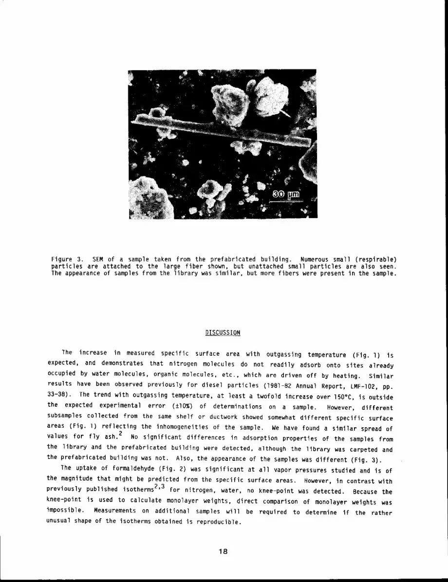
Figure 3. SEM of a sample taken from the prefabricated building. Numerous small (respirable)particles are attached to the large fiber shown, but unattached small particles are also seen.The appearance of samples from the library was similar, but more fibers were present in the sample.
DISCUSSION
The increase in measured specific surface area with outgassing temperature (Fig. l)
expected, and demonstrates that nitrogen molecules do not readily adsorb onto sites already
occupied by water molecules, organic molecules, etc., which are driven off by heating. Similar
results have been observed previously for diesel particles (1981-82 Annual Report, LMF-I02, pp.
33-38). The trend with outgassing temperature, at least a twofold increase over 150°C, is outside
the expected experimental error (±I0%) of determinations on a sample. However, different
subsamples collected from the same shelf or ductwork showed somewhat different specific surface
areas (Fig. l) reflecting the inhomogeneities of the sample. We have found a similar spread
values for fly ash. 2 No significant differences in adsorption properties of the samples from
the library and the prefabricated building were detected, although the library was carpeted and
the prefabricated building was not. Also, the appearance of the samples was different (Fig. 3).
The uptake of formaldehyde (Fig. 2) was significant at all vapor pressures studied and is
the magnitude that might be predicted from the specific surface areas. However, in contrast with
previously published isotherms 2’3 for nitrogen, water, no knee-point was detected. Because the
knee-point is used to calculate monolayer weights, direct comparison of monolayer weights was
impossible. Measurements on additional samples will be required to determine if the rather
unusual shape of the isotherms obtained is reproducible.
18
The adsorption/desorption isotherms showed marked hysteresis (Fig. 2). No weight loss was
detected when the formaldehyde vapor pressure was reduced from 2 torr to l torr, but significant
weight loss occurred (without heating the sample) when the pressure was reduced to less than
lO-4 torr. Further studies will be required to determine whether this indicates porosity of the
dust samples and pore-filling or the chemical reaction of formaldehyde with the surface of the
particles (chemisorption). However, the desorption was slow, and even when one sample was held
10-5 torr for 72 h, only ~ 60% of the formaldehyde initially adsorbed was desorbed. Thus,
these preliminary data suggest that chemisorption and pore-filling are about equally significant.
Also, until we have quantitated the water content of the samples as a function of outgassing
temperature, we can only speculate on the extent to which formaldehyde is reacting with adsorbed
water. For all the samples, the amount of formaldehyde adsorbed did not significantly decrease
(Fig. 2) with increase in outgassing temperature. Because heating the particles strongly drives
off water vapor, formaldehyde adsorption on the surface of the particles or in pores in the
particles must be significant.
The very slow desorption of formaldehyde suggests that vapors adsorbed onto room dust samples
will remain associated with the particles for hours or even days and that formaldehyde and perhaps
other organic vapors adsorbed to inhaled submicron room dust would be deposited in the deep lung
and other portions of the respiratory tract along with the particles. This would alter their
distribution of deposition in the respiratory tract as compared to their inhalation as vapors.
REFERENCES
I. Spengler, J. D. and K. Sexton, Indoor Air Pollution: A Public Health Perspective, Science221: 9-17, 1983.
2. Rothenberg, S. J., Coal Combustion Fly Ash Characterization: Adsorption of Nitrogen andWater, Atmos. Environ. 14: 445-456, 19BO.
3. Rothenberg, S. J. and Y. S. Cheng, Coal Combustion Fly Ash Characterization: Rates ofAdsorption and Desorption of Water, J. Phys. Chem. 84: 1644-1649, 1980.
4. Brunauer, S., P. H. Emmett, and E. Teller, Adsorption of Gases in Multi-Molecular Layers, J=.Amer. Chem. Soc. 60: 309-319, 193B.
19
CHEMICAL CHARACTERIZATION OF COMPOUNDS ADSORBED ONTO INDOOR DUST PARTICLES
Abstract -- Indoor dust from the library and a
laboratory at ITRI was collected and chemlca11y
characterized. Dlcbloromethane extraction and vac-
uum desorptlon/vacuum cryogenic dlst111atlon were
used to separate the soluble and volatlle compounds
from the particles, respectively. Characterlzat~on
of the less volatile organic components adsorbed
on the dusts was based on analyses of CH2CI2 ex-
PRINCIPAL INVESTIGATORS
R. L. Hanson
c. A. Rowehl
S. J. Rothenberg
A. R. Dahl
C. H. Hobbs
tracts. Several aromatic compounds were found in the volatile vacuum-desorbed mater~aZ from the
indoor dusts.
The indoor environment has received increasing attention as an important area where man may be
exposed to levels of indoor air pollutants that could affect health. Particulates in the indoor
environment are one pollutant of concern because they may be inhaled and deposited in the
respiratory tract. Also other materials present in the atmosphere such as formaldehyde, NO2,
and organic compounds may be adsorbed onto these particles and be deposited at the same sites in
the respiratory tract. The purpose of these preliminary studies was to define the chemical nature
of compounds adsorbed onto indoor dust and to compare two methods of isolating them from the
particles for subsequent chemical analysis.
METHODS
Two samples of indoor dust were collected from (1) the top of shelves for bound journals
the library (Sample A) and (2) the top of air ducts and file cabinets of a temporary building
containing offices and an aerosol science laboratory (Sample B) at ITRI. Volatile compounds were
vacuum-desorbed from the dust and analyzed using infrared spectroscopy and gas chromatography/mass
spectrometry (GC/MS). Adsorbed organic compounds were also removed by CH2CI2 extraction and
analyzed by GC/MS. Surface area data on these dust samples is in this report, pp. 16 to Ig.
Compounds adsorbed to the dust particles were isolated using either dichloromethane extraction
with ultrasonic agitation or vacuum desorption/vacuum cryogenic distillation, l For the
extraction, a 1-g aliquot of each dust sample was extracted twice with 5 ml of CH2C]2 in a
scintillation vial by agitation for 1 h in an ultrasonic bath. The extracts from each sample were
concentrated to 1 ml. Additional aliquots [Sample A (9.1 g) and Sample B (17.2 g)] were heated
150°C and vacuum-desorbed overnight at a pressure of iO-2 Tort. The desorbed material was
collected in traps cooled with liquid nitrogen. The desorbed material was then fractionated by
warming and allowing the material to diffuse through four traps submerged in cryogenic slushes at
O°C, -45°C, and -86°C and liquid nitrogen at -Ig6°c.
The contents of the -86°C and -196°C traps were transferred to an infrared gas cell, and
infrared spectra taken using a Perkin-Elmer Model 283B infrared spectrometer. The contents of all
four traps were analyzed by GC/MS using a Finnigan Model 4023 with model 4500 source. The
20
CH2Cl2 extracts were also analyzed by GC/MS. Standards were run by GC/MS to confirm the
identity of selected components. Tentative identifications of specific compounds were based upon
the retention characteristics and mass spectral matches to the NBS library spectra.
RESULTS
For Sample B, 3.1% of the mass was vacuum desorbed. Outgassing experiments (this report,
pp. 16 to 19) on two other aliquots of this sample had 3.g% and 2.1% of the mass desorbed at
150°C. Vacuum grease entered the desorptlon flask of Sample A upon removal from the vacuum
desorptlon apparatus, so the weight desorbed was not obtained. The outgassing experiments on two
other aliquots of Sample A had I0.9% and 5.7% of the mass desorbed at 150°C.
The infrared spectra of the -86°C fractions from both samples indicated that organic compounds
with carbonyl functionality were present. Carbon dioxide and nitrogen dioxide also appeared to be
present in the infrared spectra of the -196°C fractions.
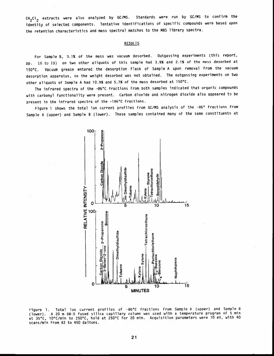
Figure l shows the total ion current profiles from GC/MS analysis of the -B6° fractions from
Sample A (upper) and Sample B (lower). These samples contained many of the same constituents
100
>-I.-f./):Z
_zuJ:> 100-I--5
2
~ w
5
cr
c ~ c
10 15
"0m
"0
E w
O~ X
5
00
2
v..ou
¯ ,t!._,10 15
MINUTES
Figure I. Total ion current profiles of -B6°C fractions from Sample A (upper) and Sample (lower). A 20 m DB-5 fused silica capillary column was used with a temperature program of 5 minat 35°C, lO°C/min to 250°C, hold at 250°C for 20 min. Acquisition parameters were 70 eV, with 40scans/min from 42 to 450 daltons.
21
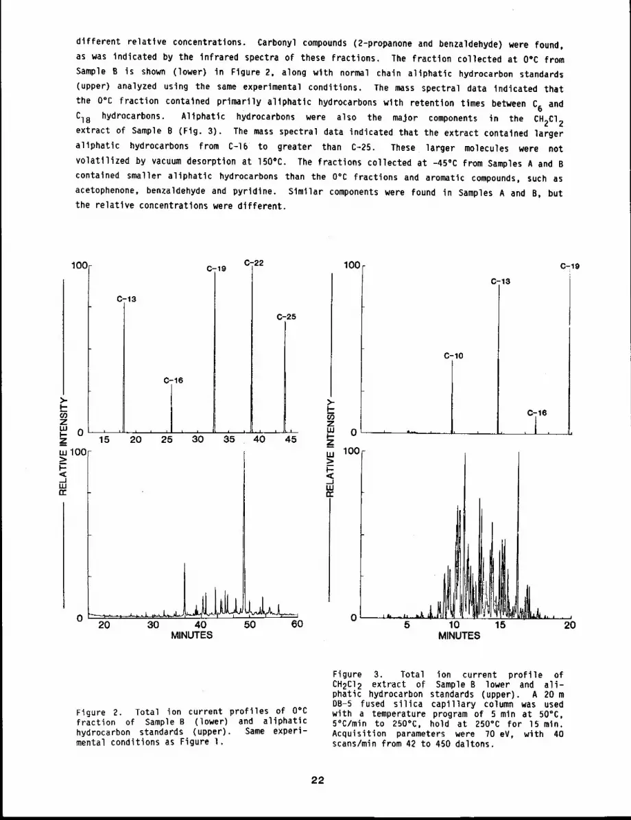
different relative concentrations. Carbonyl compounds (2-propanone and benzaldehyde) were found,
as was indicated by the infrared spectra of these fractions. The fraction collected at O°C from
Sample B is shown (lower) in Figure 2, along with normal chain altphatic hydrocarbon standards
(upper) analyzed using the same experimental conditions. The mass spectral data indicated that
the 0% fraction contained primarily allphatic hydrocarbons wlth retention times between C6 and
C18 hydrocarbons. Allphatic hydrocarbons were also the major components in the CH2C12extract of Sample B (Fig. 3). The mass spectral data indicated that the extract contained larger
allphatic hydrocarbons from C-16 to greater than C~25. These larger molecules were not
volatilized by vacuum desorption at 150°C. The fractions collected at -45°C from Samples A and B
contained smaller altphatlc hydrocarbons than the O°C fractions and aromatic compounds, such as
acetophenone, benzaldehyde and pyridine. Slmllar components were found in Samples A and B, but
the relative concentrations were different.
100- c 19 c .22 100
c-13
0-16
c-25
)- >_i.- I,-z zLu LUI’- 0 ...._ 15 20 2’5 3’0 35 4C) 4’5 l-
Z,,, 100 w_> >,< ,,~--J __1I.U I.Urr rr
20 30 40MINUTES
50 60
0
100
0
C-10
5 10MINUTES
C-13
15
C.19
20
Figure 2. Total ion current profiles of O°Cfraction of Sample B (lower) and aliphatlchydrocarbon standards (upper). Same experi-mental conditions as Figure I.
Figure 3. Total ion current profile ofCH2CI2 extract of Sample B lower and ali-phatic hydrocarbon standards (upper). A 20 DB-S fused silica capillary column was usedwith a temperature program of 5 mln at 50°C,5°C/min to 250°C, hold at 250°C for 15 min.Acquisition parameters were 70 eV, with 40scans/min from 42 to 450 daltons.
22
DISCUSSION
Many aliphatic hydrocarbons were found on the dust samples. Solvent extraction removed the
higher molecular weight compounds that were not volatile at 150°C in the vacuum desorption
apparatus. To minimize thermal degradation of components, the samples were not desorbed at a
higher temperature. Complementary data were obtained by using both CH2CI2 extraction and
vacuum desorption to remove the organic compounds associated with dust, since the larger aliphatlc
hydrocarbons were found in the extract and the more volatile compounds in the vacuum-desorbed
fractions.
Vacuum desorption was effective for collecting volatile compounds adsorbed on dust particles.
The following compounds were identified based upon analyses of standards: phenol, pyridine,
naphthalene, benzene, styrene, and toluene. Inhalation of particles containing these potentially
toxic compounds could change their deposition and retention patterns in the respiratory tract,
compared to inhalation of vapors of these compounds. Future plans involve collection and analysis
of compounds on dust samples obtained in homes.
REFERENCES
Dahl, A. R., J. M. Benson, R. L. Hanson, and S. J. Rothenberg, The Fractionization ofEnvironmental Samples According to Volatility by Vacuum Line Cryogenic Distillation, Amer.Indust. Hyg. Assoc. 3. (in press).
23
FORMATION OF POTENTIAL AEROSOLS FROM FUSION ENERGY SYSTEMS
Abstract --This report descrlbes the collectlon of
airborne parclcles from a number of fuslon-related PRINCIPAL INVESTIGATORS
processes includlng cutting stainless steel, vapor- M.D. Hoover
Izlng targets in two inertial confinement fuslon F.A. SeileI
devices, flaklng of material from the walls of a G.J. Newtonmagnetic confinement device, and spraying of plas- S.J. Rothenberg
ma for coatlngmetals. Such ~nformatlon on parti-
cle formation is needed to plan adequate resplrator 9 protectlon for workers, predict the 11fe
expectanc9 and performance of reactor components, and develop effectlve cleanup and maintenance
procedures.
Little is known about how neutron activation products and other toxic materials in fusion
reactor systems may become airborne in routine or accidental disruptive processes. Examples of
disruptive processes are accidental contact of the fusion plasma with the reactor first wall,
thermal or radiation stress-induced damage to the first wall, and maintenance or decommissioning
activities such as cutting up of used reactor components, or severe accidents such as fires
involving a lithium coolant. Characteristics of particles which are of interest for an improved
health risk assessment include: geometric and aerodynamic particle size distributions, particle
morphology and composition, and expected biological behavior of particles after inhalation and
deposition in the respiratory tract. Our objective in this first study was to investigate the
mechanisms of particle formation in a number of fusion-related activities.
METHODS
Electron microscope grids were placed inside the Particle Beam Fusion Accelerator at Sandia
National Laboratories to capture particles created when a light ion beam interacted with a
diagnostic target. This was facilitated by Mr. Paul Miller and colleagues at Sandia National
Laboratories. Grids placed in the bottom of the accelerator cavity also collected larger
particles deposited by sedimentation. The samples obtained from single tests were compared to
debris collected from the chamber walls after approximately lO0 tests.
Electron microscope grids were also placed inside the Helios device at Los Alamos National
Laboratory to collect particles created when CO2 laser beams interacted with targets such asaluminum. Mr. Jay Howland and colleagues at Los Alamos National Laboratory coordinated our
sampling with their on-going studies. These samples represent particles formed under vacuum from
target debris or from beams accidentally striking the chamber walls.
Dr. John Glowienka provided us with a sample of debris that had come from the chamber wall
material (6061-T6 aluminium) of the Elmo Bumpy Torus Tokamak Fusion Reactor at Oak Ridge National
Laboratory. This sample represents particles that may have to be periodically cleaned out of the
Tokamak fusion reactors.
We conducted an electron microscopic examination of samples of titanium metal feed powder and
titanium particulate residue from inside the plasma spray coating device at Sandia National
Laboratories. Collection of these samples was arranged by Dr. Donald Mattox at Sandia National
Laboratories. This device is used to vaporize a metal powder and apply it as a uniform coating to
other materials. This process was of special Interest because It may be used to prepare
beryllium-coated components for testing In magnetic confinement fusion reactors. The residue
particles formed in the spraying process are particles to which workers might be exposed.
RESULTS
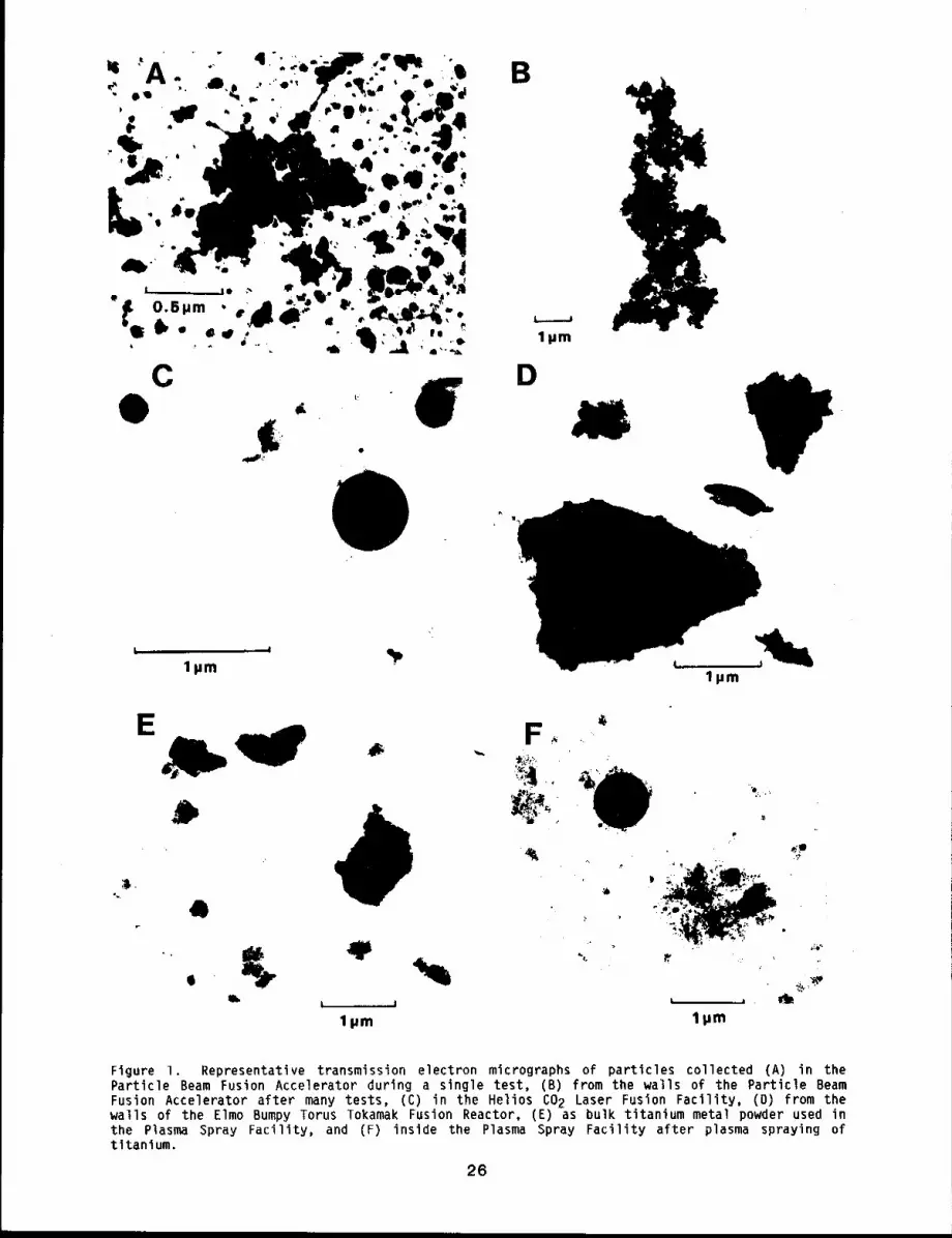
Figure l shows representative transmission electron micrographs for particles collected in the
study. Particles collected on substrates placed in the Particle Beam Fusion Accelerator during a
single test (Fig. IA) had diameters of about O.l ~m and larger with occasional splatter-like
streaks of material. Material resuspended from the walls after many shots (Fig. IB), consisted
irregularly shaped agglomerates of these smaller particles. The particles collected in the Helios
(Fig. IC) contained spheres of about 0.5 ~m in diameter and ultrafine aggregates composed
primary particles with diameters of about 0.05 ~m.
The particles from the walls of the Elmo Bumpy Torus Tokamak Fusion Reactor (Fig. ID) were
irregularly shaped and the size extended from the respirable particles shown in Figure ID to large
flakes having thickness of about O.l pm and length or width that could be measured in
centimeters.
The bulk titanium powder used in the plasma spraying operation (Fig. IE) consisted
irregularly shaped particles with diameters generally larger than l ~m, but with a number of
smaller particles. The particles from the plasma spraying process (Fig. IF) Included both spheres
with diameters of about 0.75 ~m, smaller particles of irregular shape, and branched-chain
aggregates of ultrafine particles.
DISCUSSION
We compared these particle samples to those obtained in our previous studies of aerosols
created in mechanical and high temperature cutting of stainless steel.l’2 Cutting tools tested
in that study included a number of mechanical saws and grinding wheels as well as three
melting-vaporization techniques: oxy-acetylene torch, electric arc cut-rod, and plasma torch.
These aerosols are representative of those that might be generated during the replacement of
fusion reactor first wall or during reactor decommissioning.
The results of these tests indicate at least four particle formation mechanisms at work. The
first is the formation of irregularly shaped particles by mechanical actions such as grinding or
cutting. The irregularly shaped particles of the bulk titanium powder (Fig. IE) are another
example of particles formed by a mechanical process and are similar in shape to those found in the
study of aerosols from mechanical cutting of stainless steel.2
The second aerosol formation process involves vaporization of material, followed by
condensation into ultrafine particles and coagulation into branched, chain agglomerates. Examples
are the particles formed by vaporization of the titanium powder in the plasma spraying device
(Fig. IF). These particles are similar to those formed during cutting of stainless steel with
high temperature devices such as the plasma torch.2
Particles collected in the Particle Beam Fusion Accelerator (Fig. IA) appear to result from
third aerosol formation process: melting and dispersion of molten droplets. The splattering of a
molten droplet on the collection substrate would account for the presence of the chain-like
streaks in Figure IA. The melting and dispersal of molten droplets also appears to be the source
of the larger spheres seen in the aerosol from the CO2 laser vaporization (Fig. 1C), and from
the plasma spraying operation (Fig. IF).
25
Clpm
D
lpm
E
°~.
b
4
F
,~,~.
~ .~
lpm
m. L 1 ilpm
/
lpm
Figure i. Representative transmission electron micrographs of particles collected (A) in theParticle Beam Fusion Accelerator during a single test, (B) from the walls of the Particle BeamFusion Accelerator after many tests, (C) in the Helios 2 Laser Fu sion Fa cility, (D ) fr om thwalls of the Elmo Bumpy Torus Tokamak Fusion Reactor, (E) as bulk titanium metal powder used the Plasma Spray Facility, and (F) inside the Plasma Spray Facility after plasma spraying titanium.
26
Particles from the walls of the Elmo Bumpy Torus Tokamak Fusion Reactor represent still
another particle formation process. There are at least three possibllites for this last
mechanism: peeling of deposited material, blistering of a surface layer during the release of
trapped gas, or detachment of a surface layer as a result of thermal shock. Further work is
needed to identify the mechanism.
This study has demonstrated that particles which could potentially be inhaled by workers and
others are created in a number of fusion-related activities. Formation of these particles also
has implications for predicting the lifetime of reactor components and the cooling of plasma by
the introduction of wall material. Respiratory protection for workers against the entire range of
particle sizes will be appropriate, especially if particles contain toxic materials such as
neutron activation products or beryllium, or irritant materials such as lithium. Additional work
is under way to gain a better understanding of the particle formation mechanisms in fusion-related
activities, the chemical composition of the particles, and their disposition if inhaled by people.
REFERENCES
I. Newton, G. 3., M. D. Hoover, E. B. Barr, B. A. Wong, and P. D. Ritter, Aerosols from MetalCutting. Techniques Typical of Decommissioning Nuclear Facilities - Experimental System forCollection and Characterization, in Proceedings of the 1982 International Decommissioning~, Seattle, WA, 1982.
2. Hoover, M. D., G. J. Newton, E. B. Barr, and B. A. Wong, Aerosols from Metal CuttingTechniques Typical of Decommissioning Nuclear Facilites - Inhalation Hazards and WorkerProtection, in Proceedings of the 1982 International Decommissioninq Symposium, Seattle, WA,1982.
27128

LABORATORY STUDIES OF AEROSOL GENERATION AND CHARACTERIZATION
Conduct of inhalation toxicological studies in the laboratory requires methods to generate
airborne test materials having the proper physical and chemical properties and concentration
levels required by the experimental protocols. Such generation systems must also demonstrate much
stability and reproducibility to ensure adequate control of the exposure environments. Because
these generation systems are frequently called upon to produce exposure atmospheres that closely
mimic occupational or environmental exposures, a substantial amount of ingenuity may be required
to satisfy all of the experimental demands. The same is true for characterization methods for
airborne particles and vapors. The behavior and characteristics of exposure atmospheres can be
well documented through appropriate characterization methodology.
The first paper in this section describes a system for exposing rodents to large (3.0, 9.0, or
15.0 ~m) particles. Special design features were necessary to minimize particle loss by
impactlon and sedimentation before reaching the animals ~ breathing zones. Concentrations up to 37
mg/m3 have been achieved with less than I0% variation in mass distribution across the unit.
The next three papers deal with toxicant generation systems. Measurements were made of the
particle size distribution of the airborne particles from a fluidized bed generator. This
distribution was found to be similar, but not identical, to that of the feed powder. A dynamic
generation system for 14C.-nitropyrene has been developed. The air concentration can be
controlled by the temperature used for vaporization without affecting particle size. This system
will now be used to study the metabolism and pharmacokinetics of 14C-nitropyrene. Preliminary
work was performed on the generation of aerosols containing lithium in different chemical forms.
This research is being performed to investigate possible inhalation hazards associated with the
use of lithium in fusion or space nuclear reactors.
The next three papers deal with different aspects of aerosol characterization. The first
describes preliminary results of in vitro dissolution tests on mixed matrix aerosols. Tests of
this kind provide one measure of the possible dissolution characteristics of these materials after
deposition in the body. Results to date indicate that different dissolution rates were observed
for particles that were composed of mixtures or coatings as compared with single-element
compositions. Additional work on the density of Fused aluminosilicate particles is presented in
the next paper. Good agreement was observed between measurements made with the spiral duct
aerosol centrifuge and the Aerodynamic Particle Sizer. When dealing with optical counters, it is
important to know the relationship between the light-scattering diameter and other measures of
diameter commonly used. The next report in this section discusses the optical equivalent
diameters of cluster aggregates of uniform spheres.
Evaluations of other instruments and approaches are described in the last three reports. The
first compares the response of two different types of optical particle counters, filament lamp and
laser, for particles of different sizes and refractive indices. These factors must be well
understood to obtain proper interpretations of the observed data. An in-depth evaluation of the
Aerodynamic Particle Sizer is reported next. A combination of laboratory calibrations and
theoretical analyses has yielded a method for generating appropriate calibration curves without
actually doing the calibrations. The final report describes experimental measurements and
theoretical analyses of the use of wire screens as particle filter models. These results" are
discussed in relation to their use in the design and operation of diffusion battery sampling
devices.
29130

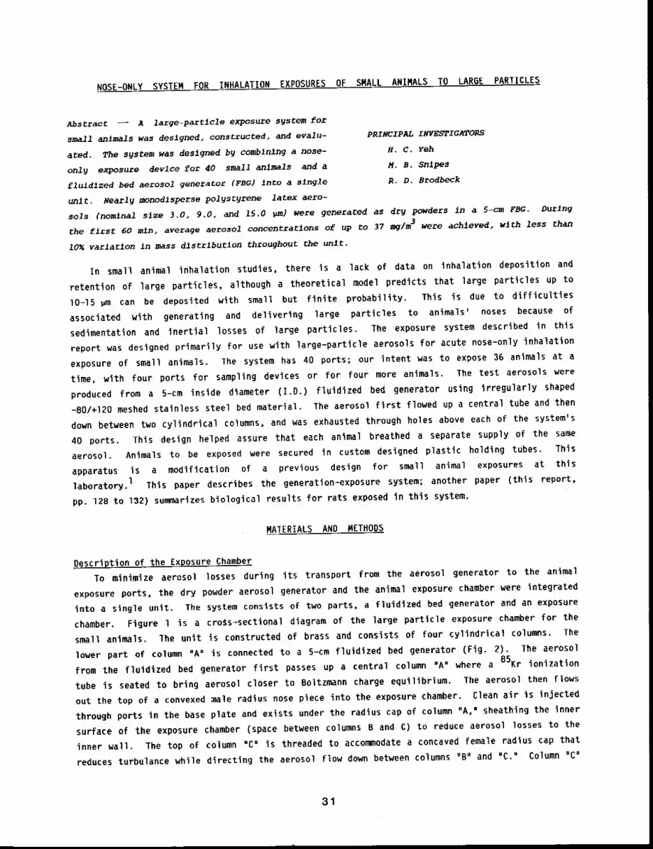
NOSE-ONLY SYSTEM FOR INHALATION EXPOSURES OF SMALL ANIMALS TO LAR6E PARTICLES
Abstract ~ A large-partlcle exposure sgsCem for
small animals was deslgned, constructed~ and evalu-pR/NClPAL INVESTIGATORS
ated. The system was deslgned b9 combining a nose- H, C. Yeh
only exposure devlce for 40 small anlmals and a M.S. 3nipes
fluldlzed bed aerosol generator (FBG) into a single R.D. Brodbeck
unit. Nearly monodlsperse pol~st~rene latex aero-
sols (nomlnal slze 3.0, 9.0, and 15.0 ~m) were generated as dry powders in a 5-cm FBG. During
the flrst 60 mln, average aerosol concentraclons of up co 37 mg/m3 were achleved, wlth less than
10~ varlaClon In mass dlstrlbuCion throughout the unlt.
In small animal inhalation studies, there is a lack of data on inhalation deposition and
retention of large particles, although a theoretical model predicts that large particles up to
lO-15 ~m can be deposited with small but finite probability. This is due to difficulties
associated with generating and delivering large particles to animals’ noses because of
sedimentation and inertial ’losses of large particles. The exposure system described in this
report was designed primarily for use with large-particle aerosols for acute nose-only inhalation
exposure of small animals. The system has 40 ports; our intent was to expose 36 animals at a
time, with four ports for sampling devices or for four more animals. The test aerosols were
produced from a 5-cm inside diameter (I.D.) fluidized bed generator using irregularly shaped
-80/+120 meshed stainless steel bed material. The aerosol first flowed up a central tube and then
down between two cylindrical columns, and was exhausted through holes above each of the system’s
40 ports. This design helped assure that each animal breathed a separate supply of the same
aerosol. Animals to be exposed were secured in custom designed plastic holding tubes. This
apparatus is a modification of a previous design for small animal exposures at this
laboratory, l This paper describes the generation-exposure system; another paper (this report,
pp. 128 to 132) summarizes biological results for rats exposed in this system.
MATERIALS AND METHODS
Description of the Exposure ChamberTo minimize aerosol losses during its transport from the aerosol generator to the animal
exposure ports, the dry powder aerosol generator and the animal exposure chamber were integrated
into a single unit. The system consists of two parts, a fluidlzed bed generator and an exposure
chamber. Figure I is a cro~s-sectional diagram of the large particle exposure chamber for the
small animals. The unit is constructed of brass and consists of four cylindrical columns. The
lower part of column "A" is connected to a 5-cm fluidized bed generator (Fig. 2). The aerosol
from the fluidized bed generator first passes up a central column "A" where a 85Kr ionization
tube is seated to bring aerosol closer to Boltzmann charge equilibrium. The aerosol then flows
out the top of a convexed ~ale radius nose piece into the exposure chamber. Clean air is injected
through ports in the base plate and exists under the radius cap of column "A," sheathing the inner
surface of the exposure chamber (space between columns B and C) to reduce aerosol losses to the
inner wall. The top of column "C" is threaded to accommodate a concaved female radius cap that
reduces turbulance while directing the aerosol flow down between columns "B" and "C." Column "Cu
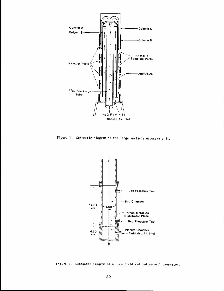
Column A
Column B
/=
Exhaust
Ports~ "!,!
85Kr Discharge-Tube
Column C
,Column D
!~SAnimal &ling Ports
]~-~ AEROSOL
Sheath Air Inlet
Figure I. Schematic diagram of the large particle exposure unit,
Pressure Tap
Chamber
~-~ Porous Metal AirDistributor Plate
Pressure Tap
Chamberg Air Inlet
Figure 2. Schematic diagram of a 5-cm fluidized bed aerosol generator.
32
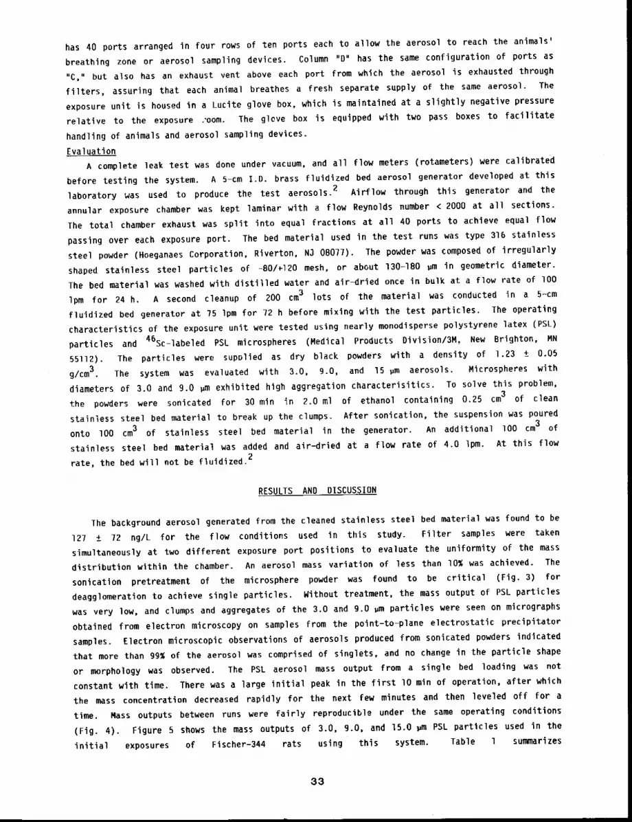
has 40 ports arranged in four rows of ten ports each to allow the aerosol to reach the animals’
breathing zone or aerosol sampling devices. Column "D" has the same configuration of ports as
"C," but also has an exhaust vent above each port from which the aerosol is exhausted through
filters, assuring that each animal breathes a fresh separate supply of the same aerosol. The
exposure unit is housed in a Lucite glove box, which is maintained at a slightly negative pressure
relative to the exposure ,’oom. The gleve box is equipped with two pass boxes to facilitate
handling of animals and aerosol sampling devices.
Evaluation
A complete leak test was done under vacuum, and all flow meters (rotameters) were calibrated
before testing the system. A 5-cm I.D. brass fluidized bed aerosol generator developed at this
laboratory was used to produce the test aerosols. 2 Airflow through this generator and the
annular exposure chamber was kept laminar with a flow Reynolds number < 2000 at all sections.
The total chamber exhaust was split into equal fractions at all 40 ports to achieve equal flow
passing over each exposure port. The bed material used in the test runs was type 316 stainless
steel powder (Hoeganaes Corporation, Riverton, N3 08077). The powder was composed of irregularly
shaped stainless steel particles of -BO/~120 mesh, or about 130-1BO ~m in geometric diameter.
The bed material was washed with distilled water and air-dried once in bulk at a flow rate of lO0
Ipm for 24 h. A second cleanup of 200 cm3 lots of the material was conducted in a 5-cm
fluidized bed generator at 75 Ipm for 72 h before mixing with the test particles. The operating
characteristics of the exposure unit were tested using nearly monodisperse polystyrene latex (PSL)
particles and 46Sc-labeled PSL microspheres (Medical Products Division/3M, New Brighton, MN
55112). The particles were supDlied as dry black powders with a density of 1.23 ± 0.05
g/cm 3. The system was evaluated with 3.0, 9.0, and 15 ~m aerosols. Microspheres with
diameters of 3.0 and 9.0 ~m exhibited high aggregation characterisitics. To solve this problem,
the powders were sonicated for 30 min in 2.0 ml of ethanol containing 0.25 cm3 of clean
stainless steel bed material to break up the clumps. After sonication, the suspension was poured3
onto lO0 cm3 of stainless steel bed material in the generator. An additional lO0 cm of
stainless steel bed material was added and air-dried at a flow rate of 4.0 Ipm. At this flow
rate, the bed will not be fluidized.2
RESULTS AND DISCUSSION
The background aerosol generated from the cleaned stainless steel bed material was found to be
127 ± 72 ng/L for the flow conditions used in this study. Filter samples were taken
simultaneously at two different exposure port positions to evaluate the uniformity of the mass
distribution within the chamber. An aerosol mass variation of less than 10% was achieved. The
sonication pretreatment of the microsphere powder was found to be critical (Fig. 3) for
deagglomeration to achieve single particles. Without treatment, the mass output of PSL particles
was very low, and clumps and aggregates of the 3.0 and g.o ~m particles were seen on micrographs
obtained from electron microscopy on samples from the point-to-plane electrostatic precipitator
samples. Electron microscopic observations of aerosols produced from sonicated powders indicated
that more than 99% of the aerosol was comprised of singlets, and no change in the particle shape
or morphology was observed. The PSL aerosol mass output from a single bed loading was not
constant with time. There was a large initial peak in the first lO min of operation, after which
the mass concentration decreased rapidly for the next Few minutes and then leveled off for a
time. Mass outputs between runs were fairly reproducible under the same operating conditions
(Fig. 4). Figure 5 shows the mass outputs of 3.0, 9.0, and 15.0 ~m PSL particles used in the
initial exposures of Fischer-344 rats using this system. Table l summarizes
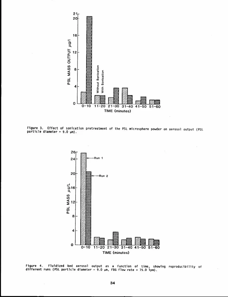
:Lk-
O_
00303<(
.J03EL
11-20 21-30 31-40 41-50 51-60TIME (minutes)
Figure 3. Effect of sonication pretreatment of the PSL microsphere powder on aerosol output (PSLparticle diameter = 9.0 ~m).
20-
011-20 21-30 31-40 41-50 51-60
TIME (minutes)
Figure 4, F1uldized bed aerosol output as a function of time, showing reproducibility ofdifferent runs (PSL particle diameter = 9.0 gm, FB6 flow rate = 75.0 lpm).
34
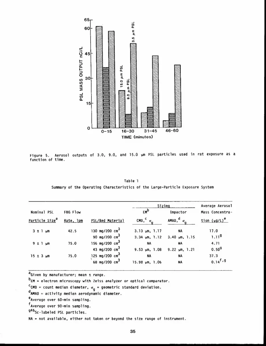
65-
60
45
30-
15-
031-45
TIME (minutes)
46-60
Figure 5. Aerosol outputs of 3.0, 9.0, and 15.0 ~m PSL particles used in rat exposure as afunction of time.
Table 1
Summary of the Operating Characteristics of the Large-Particle Exposure System
Nominal PSL FB6 Flow
Particle Sizea Rate, l~m
3 ± l ~m 42.5
9 +_ l ~m 75.0
15 + 3 ,m 75.0
Sizing Average Aerosol
EMb Impactor Mass Concentra=
PSL/Bed Material CMD,cog AMAD,d~g tion (,g/L)e
130 mg/200 cm3 3.13 ,m, 1.17 NA 17.0
90 mg/200 cm3 3.34 ~m, 1.12 3.40 pm, 1.15 1.11g
156 mg/200 cm3 NA NA 4.71
43 mg/200 cm39.53 ~m, 1.08 9.22 ~m, 1.21 0.50g
125 mg/200 cm3NA NA 37.3
~68 mg/200 cm315.98 gm, 1.06 NA 0.14f’g
aGiven by manufacturer; mean ± range.
bEM = electron microscopy with Zeiss analyzer or optical comparator.
CCMD = count median diameter, o = geometric standard deviation.g
dAMAD = activity median aerodynamic diameter.
eAverage over 60-min sampling.
fAverage over 90-min sampling.
g46sc-labeled PSL particles.
NA = not available, either not taken or beyond the size range of instrument.
35
the operating characteristics of the larger particle exposure system. Average mass concentrations
up to 37 mg/m3 during the first 60 min of operation are achievable. For radioactive aerosols,
this level of concentration is more than enough for most inhalation studies (see Fig. 5 and
Table l).
REFERENCES
Raabe, O. G., J. E. Bennick, M. E. Light, C. H. Hobbs, R. L. Thomas, and M. I. Tillery, AnImproved Apparatus for Acute Inhalation Exposure of Rodents to Radioactive Aerosols, Toxicol.AppI. Pharmacoi. 26: 264-273, 1973.
2. Carpenter, R. L. and K. L. Yerkes, Relationship Between Fluid Bed Aerosol Generator Operationand the Aerosol Produced, Am. Ind. Hvq. AssQc, J. 41: 888-B94, 1980.
36
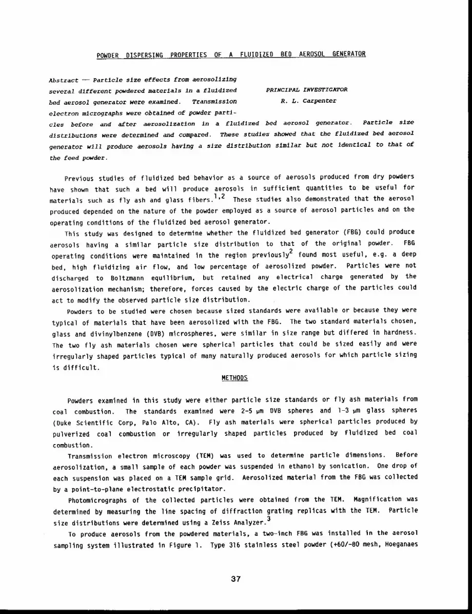
POWDER DISPERSING PROPERTIES OF A FLUIDIZED BED AEROSOL GENERATOR
Abstract --Partlcle size effects from aerosolizlng
several dlfferenC powdered macerlals in a fluld~zed PRINCIPAL INVESTIGATOR
bed aerosol generator were examlned. Transmlsslon R.L. Carpenter
electron mlcrographs were obtalned of powder parti-
cles before and after aerosol~zat~on in a fluldlzed bed aerosol generator. Partlcle slze
dlstrlbu~1ons were determined and compared. These studles showed that the flu~dlzed bed aerosol
generator w111 produce aerosols havlng a slze dlstrlbu~1on slmllar but not IdenClcal to that of
the feed powder.
Previous studies of fluidized bed behavior as a source of aerosols produced from dry powders
have shown that such a bed will produce aerosols in sufficient quantities to be useful for
materials such as fly ash and glass fibers. 1’2 These studies also demonstrated that the aerosol
produced depended on the nature of the powder employed as a source of aerosol particles and on the
operating conditions of the fluidized bed aerosol generator.
This study was designed to determine whether the fluidized bed generator (FBG) could produce
aerosols having a similar particle size distribution to that of the original powder. FBG
operating conditions were maintained in the region previously 2 found most useful, e.g. a deep
bed, high fluidizing air flow, and low percentage of aerosolized powder. Particles were not
discharged to Boltzmann equilibrium, but retained any electrical charge generated by the
aerosolization mechanism; therefore, forces caused by the electric charge of the particles could
act to modify the observed particle size distribution.
Powders to be studied were chosen because sized standards were available or because they were
typical of materials that have been aerosolized with the FBG. The two standard materials chosen,
glass and divinylbenzene (DVB) microspheres, were similar in size range but differed in hardness.
The two fly ash materials chosen were spherical particles that could be sized easily and were
irregularly shaped particles typical of many naturally produced aerosols for which particle sizing
is difficult.
METHODS
Powders examined in this study were either particle size standards or fly ash materials from
coal combustion. The standards examined were 2-5 ~m DVB spheres and 1-3 ~m glass spheres
(Duke Scientific Corp, Palo Alto, CA). Fly ash materials were spherical particles produced
pulverized coal combustion or irregularly shaped particles produced by fluidized bed coal
combustion.
Transmission electron microscopy (TEM) was used to determine particle dimensions. Before
aerosolization, a small sample of each powder was suspended in ethanol by sonication. One drop of
each suspension was placed on a TEM sample grid. Aerosolized material from the FBG was collected
by a point-to-plane electrostatic precipitator.
Photomicrographs of the collected particles were obtained from the TEM. Magnification was
determined by measuring the line spacing of diffraction grating replicas with the TEM. Particle
size distributions were determined using a Zeiss Analyzer.3
To produce aerosols from the powdered materials, a two-inch FBG was installed in the aerosol
sampling system illustrated in Figure l. Type 316 stainless steel powder (+60/-80 mesh, Hoeganaes
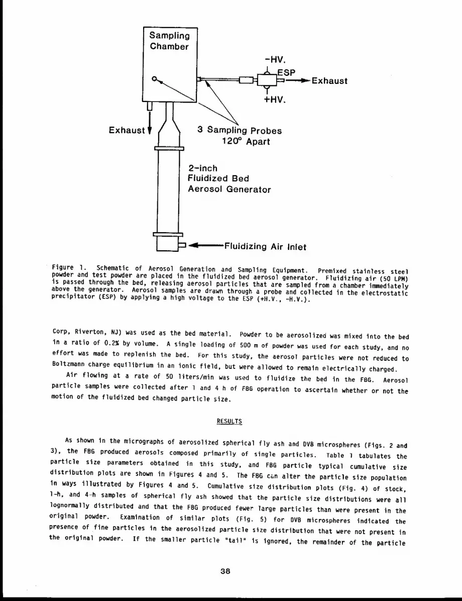
SamplingChamber
-HV.
~ SPc = - Exhaust
Ul
+HV.
Exhaust~ / \ 3 Sampling Probes120° Apart
I
2-inchFluidized BedAerosol Generator
¯ ’Fluidizing Air Inlet
Figure 1. Schematic of Aerosol Generation and Sampling Equipment. Premixed stainless steelpowder and test powder are placed in the fluidized bed aerosol generator. Fluidizing air (50 LPM)is passed through the bed, releasing aerosol particles that are sampled from a chamber immediatelyabove the generator. Aerosol samples are drawn through a probe and collected in the electrostaticprecipitator (ESP) by applying a high voltage to the ESP (+H.V., -H.V.).
Corp, Riverton, NO) was used as the bed material. Powder to be aerosolized was mixed into the bed
in a ratio of 0.2% by volume. A single loading of 500 m of powder was used for each study, and no
effort was made to replenish the bed. For this study, the aerosol particles were not reduced to
Boltzmann charge equilibrium in an ionic field, but were al]owed to remain electrically charged.
Air flowing at a rate of 50 liters/min was used to fluidize the bed in the FBG. Aerosol
particle samples were collected after l and 4 h of FBG operation to ascertain whether or not the
motion of the fluidized bed changed particle size.
RESULTS
As shown in the micrographs of aerosolized spherical fly ash and DVB microspheres (Figs. 2 and
3), the FBG produced aerosols composed primarily of single particles. Table i tabulates the
particle size parameters obtained in this study, and FBG particle typical cumulative size
distribution plots are shown in Figures 4 and 5. The FBG c~n alter the particle size population
in ways illustrated by Figures 4 and 5. Cumulative size distribution plots (Fig. 4) of stock,
l-h, and 4-h samples of spherical fly ash showed that the particle size distributions were all
lognormally distributed and that the FBG produced fewer large particles than were present in the
original powder. Examination of similar plots (Fig. 5) for DVB microspheres indicated the
presence of fine particles in the aerosolized particle size distribution that were not present in
the original powder. If the smaller particle "tail" is ignored, the remainder of the particle
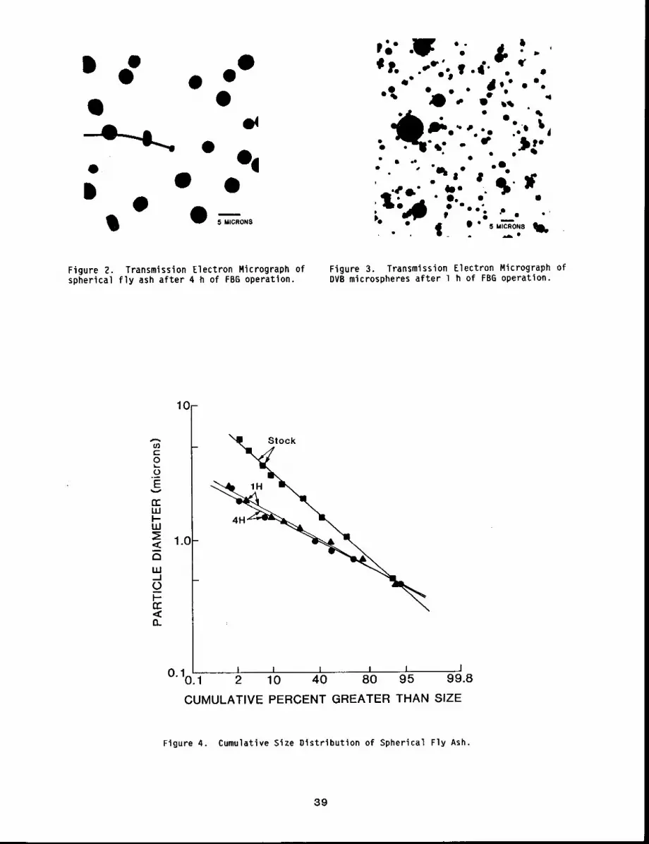
Figure Z. Transmission Electron Micrograph ofspherical fly ash after 4 h of FB6 operation.
Figure 3. Transmission Electron Micrograph ofDVB microspheres after 1 h of FBG operation.
10-
8b
1.0
C)
.......... J J ~ J I JO. 1011~2 10 40 80 95 99.8
CUMULATIVE PERCENT GREATER THAN SIZE
Figure 4. Cumulative Size Distribution of Spherical Fly Ash.
39
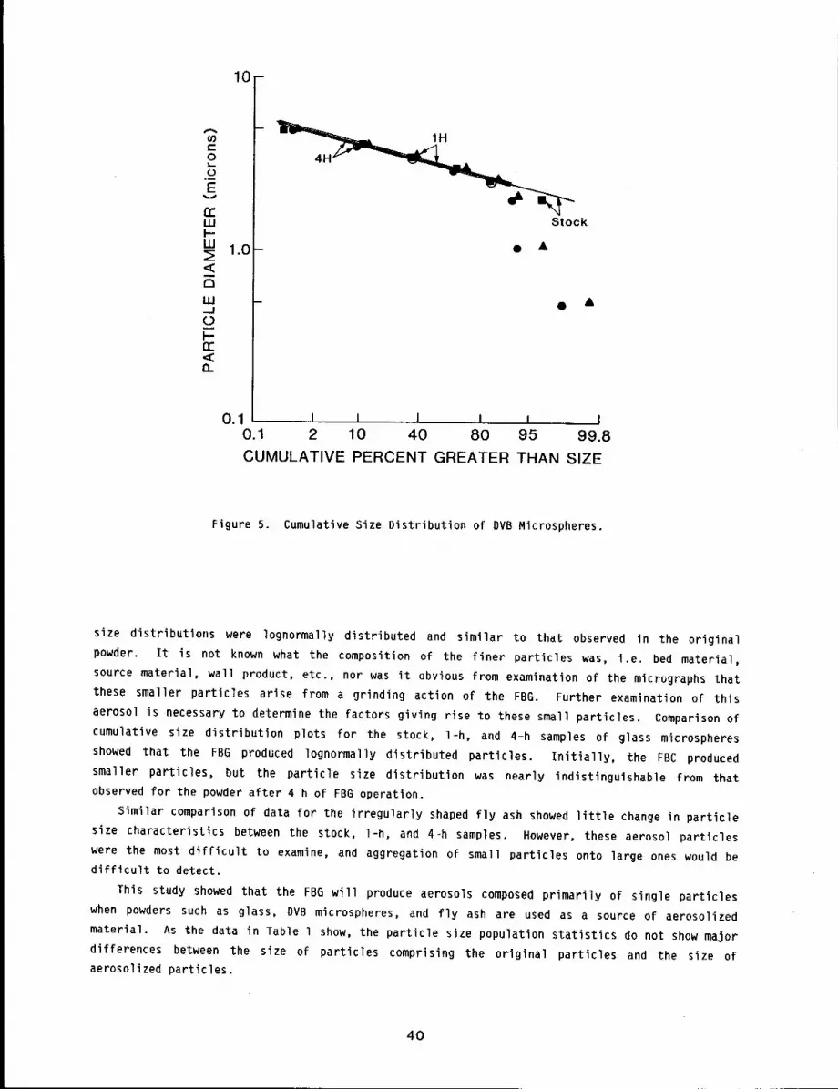
10-
1H
4H
Stock
1.0- ¯ &
- ¯ &
0.1 I I I I I I0.1 2 10 40 80 95 99.8
CUMULATIVE PERCENT GREATER THAN SIZE
Figure 5. Cumulative Size Distribution of DVB Microspheres.
size distributions were lognormally distributed and similar to that observed in the original
powder. It is not known what the composition of the finer particles was, i.e. bed material,
source material, wall product, etc., nor was it obvious from examination of the micrugraphs that
these smaller particles arise from a grinding action of the FBG. Further examination of this
aerosol is necessary to determine the factors giving rise to these small particles. Comparison of
cumulative size distribution plots for the stock, l-h, and 4-h samples of glass microspheres
showed that the FBG produced lognormally distributed particles, initially, the FBC produced
smaller particles, but the particle size distribution was nearly indistinguishable from that
observed for the powder after 4 h of FBG operation.
Similar comparison of data for the irregularly shaped fly ash showed little change in particle
size characteristics between the stock, l-h, and 4-h samples. However, these aerosol particles
were the most difficult to examine, and aggregation of small particles onto large ones would be
difficult to detect.
This study showed that the FBG will produce aerosols composed primarily of single particles
when powders such as glass, DVB microspheres, and fly ash are used as a source of aerosolized
material. As the data in Table l show, the particle size population statistics do not show major
differences between the size of particles comprising the original particles and the size of
aerosolized particles.
4O
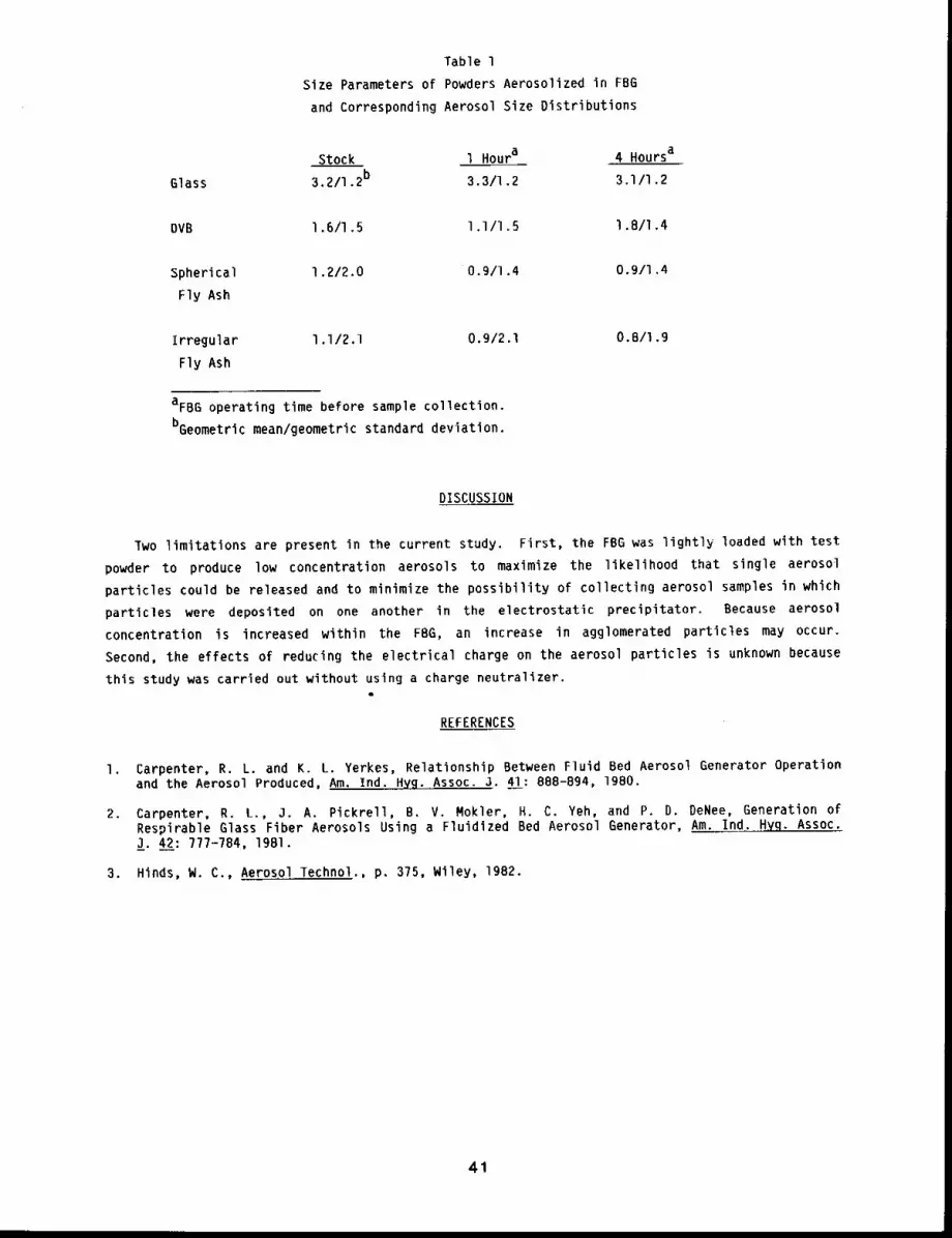
Table l
Size Parameters of Powders Aerosolized in FBG
and Corresponding Aerosol Size Distributions
Stock l Houra 4 Hoursa
Glass 3.2/I .2b 3.3/1.2 3,1/I .2
DVB l .6/I .5 l .I/l .5 l .8/I .4
Spherical I .2/2.0 0.9/1.4
Fly Ash
0.9/I .4
Irregular l.I/2.1 O.g/2.1 O.B/l.g
Fly Ash
aFBG operating time before sample collection.
bGeometric mean/geometric standard deviation.
DISCUSSION
Two limitations are present in the current study. First, the FBG was lightly loaded with test
powder to produce low concentration aerosols to maximize the likelihood that single aerosol
particles could be released and to minimize the possibility of collecting aerosol samples in which
particles were deposited on one another in the electrostatic precipitator. Because aerosol
concentration is increased within the FBG, an increase in agglomerated particles may occur.
Second, the effects of reducing the electrical charge on the aerosol particles is unknown because
this study was carried out without using a charge neutralizer.a
REFERENCES
I. Carpenter, R. L. and K. L. Yerkes, Relationship Between Fluid Bed Aerosol Generator Operationand the Aerosol Produced, Am. Ind. Hyg. Assoc. J. 41: 888-894, 1980.
2. Carpenter, R. L., J. A. Pickrell, B. V. Mokler, H. C. Yeh, and P. D. DeNee, Generation ofRespirable Glass Fiber Aerosols Using a Fluidized Bed Aerosol Generator, Am. Ind..HYq- Assoc.J. 42: 777-784, 19Bl.
3. Hinds, W. C., Aerosol Technol., p. 375, Wiley, 1982.
41

GENERATION AND CHARACTERIZATION OF I-NITROPYRENE AEROSOLS
Abstract --Thls report describes the dynamlc gen-
eration of 14C-nltropyrene (14c-NP) aerosols PRINCIPAL INVESTIGATORS
self-condensatlon of vaporlzed 14C-NP. Results J.D. Sun
showed that the air concentration of the 14C-NP S.J. Stoner
aerosols (0.008 to 0.5 ~g/L of a~r) can be con-
trolled by var~ing the temperature (140 to 2Z0°C) at which It is being vaporized without affectlng
the slze dlstr~butlon of the aerosols (0.12 to 0.18 wm VMD). A/so. heat vaporization of the
Z4C-NP for aerosol generation was shown not to thermally degrade this radlolaheled compound.
Thus. a system has been developed and characterized that can produce resp~rahle aerosols of
14C-NP over a w~de concentration range suitable for future Inhalatlon studles to investlgaCe the
dlsposltlon of inhaled NP.
Many airborne environmental pollutants contain significant amounts of organic compounds. The
potential health risk that these airborne agents pose to man depends on the biological fate of
these compounds after they are inhaled. However, very little is known about the metabolism and
ultimate disposition of many of these organic compounds after inhalation. A class of organic
compounds that has received recent attention is the nitro-substituted polycyclic aromatic
hydrocarbons (NO2-PAHs) because of their demonstrated mutagenicity in many short-term bioassays
and carcinogenicity in different laboratory animal models. 1-Nitropyrene (NP) is one example of
NO2-PAH found in atmospheric pollutants that has been shown to be both mutagenic and
carcinogenic. In fact, it has been estimated that the NP found on diesel exhaust particles is
responsible for - 20% of the total bacterial mutagenicity contained in organic solvent extracts
of diesel soot.l
This report describes the development and detailed characterization of a system to generate
respirable aerosols of 14C-NP at different air concentrations. This system will be used to
conduct detailed metabolism and pharmacokinetlc studies by exposing rats to various concentrations
of the 14C-NP aerosols by nose-only inhalation.
METHODS
Figure l illustrates the aerosol generating system used. 14C-NP (0.43 mCi/mmole) was first
vaporized (140°-210°C) in a tube furnace, and the vaporized 14C-NP driven out of the sample
vessel with nitrogen (N2). 14C-NP vapor was then entrained in the 1.8 L/mln air flow to
cooling chamber, where the 14C-NP aerosol formed by self-condensation. Characterization of this
system was performed by testing the effect of different vaporization temperatures (14O°-210°C)
a constant N2 flow of 200 ml/min or different N2 flow rates (]0-250 ml/min) at a constantvaporization temperature of 180°C on the 14C-NP aerosol being generated. During each test,, the
]4C-NP aerosol concentration was measured by liquid scintillation counting of triplicate glass
fiber filter samples, and the aerosol size distribution was determined using an electrical aerosol
analyzer (EAA). All samples obtained for aerosol characterization measurements were taken from
within the aerosol generating system during aerosol generation.
42
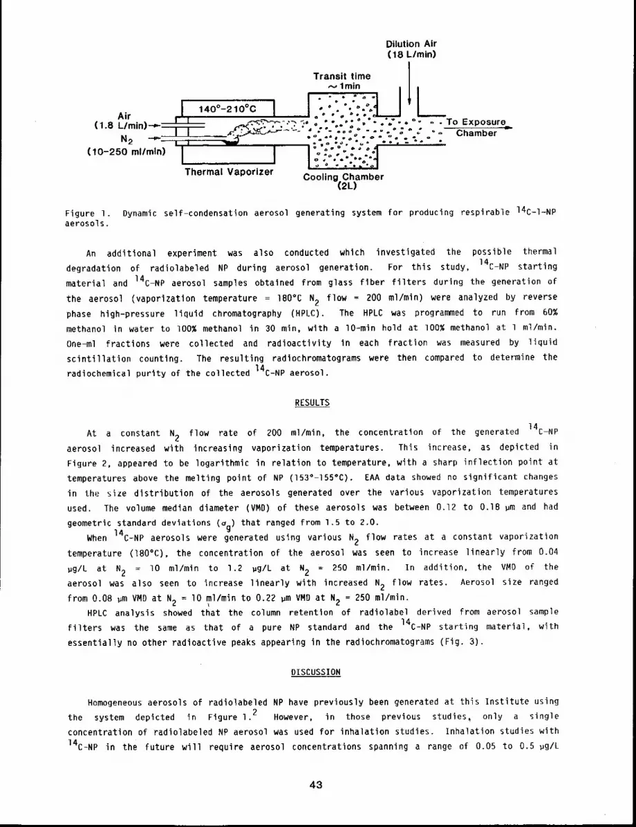
Dilution Air(18 L/mln)
Transit time I~., lmin I I
I !"";-:1 II 140°-210°C % °." °~ .=Air , , ° , o °~’ .,, °(1 R //m"T~U,- I = .~<~P.-’~--~-’~ ~,O O "’o.°* , "o = "O " " - TO Exposure............ t "----l--T-- _P.~c",~-:.~’~-" J Z°. .,,~ ..... ° " ," " ° - ",,.~ g"
i~l~ __ ~ v e~ e ¯ ~ ~
(10-250 ml/mln) I I I "o :::’.:" :.°1I I I °" "" .*% 1E o ~ o ~ e~ ~oiThermal Vaporizer Cooling Chamber
(2L)
Figure I. Dynamic self-condensation aerosol generating system for producing respirable 14C-I-NPaerosols.
An additional experiment was also conducted which investigated the possible thermal
degradation of radiolabeled NP during aerosol generation. For this study, 14C-NP starting
material and 14C-NP aerosol samples obtained from glass fiber filters during the generation of
the aerosol (vaporization temperature = 180°C 2 f low = 200 ml /min) we re an alyzed by rev erse
phase high-pressure liquid chromatography (HPLC). The HPLC was programmed to run from 60%
methanol in water to 100% methanol in 30 min, with a lO-min hold at I00% methanol at l ml/min.
One-ml fractions were collected and radioactivity in each fraction was measured by liquid
scintillation counting. The resulting radiochromatograms were then compared to determine the
radiochemical purity of the collected 14C-NP aerosol.
RESULTS
At a constant N2 flow rate of 200 ml/min, the concentration of the generated 14C-NP
aerosol increased with increasing vaporization temperatures. This increase, as depicted in
Figure 2, appeared to be logarithmic in relation to temperature, with a sharp inflection point at
temperatures above the melting point of NP (153°-155°C). EAA data showed no significant changes
in tile size distribution of the aerosols generated over the various vaporization temperatures
used. The volume median diameter (VMD) of these aerosols was between 0.12 to 0.18 ~m and had
geometric standard deviations (Og) that ranged from 1.5 to 2.0.
When 14C-NP aerosols were generated using various N2 flow rates at a constant vaporization
temperature (180°C), the concentration of the aerosol was seen to increase linearly from 0.04
gg/L at N2 = lO ml/min to 1.2 gg/L at N2 = 250 ml/min. In addition, the VND of the
aerosol was also seen to increase linearly with increased N2 flow rates. Aerosol size ranged
from 0.08 ~m VMD at N2 = lO ml/min to 0.22 ~m VMD at N2 - 250 ml/min.
HPLC analysis showed that the column retention of radiolabel derived from aerosol sample
filters was the same as that of a pure NP standard and the 14C-NP starting material, with
essentially no other radioactive peaks appearing in the radiochromatograms (Fig. 3).
DISCUSSION
Homogeneous aerosols of radiolabeled NP have previously been generated at this Institute using
the system depicted in Figure 1.2 However, in those previous studies~ only a single
concentration of radiolabeled NP aerosol was used for inhalation studies. Inhalation studies with
14C-NP in the future will require aerosol concentrations spanning a range of 0.05 to 0.5 pg/L
43
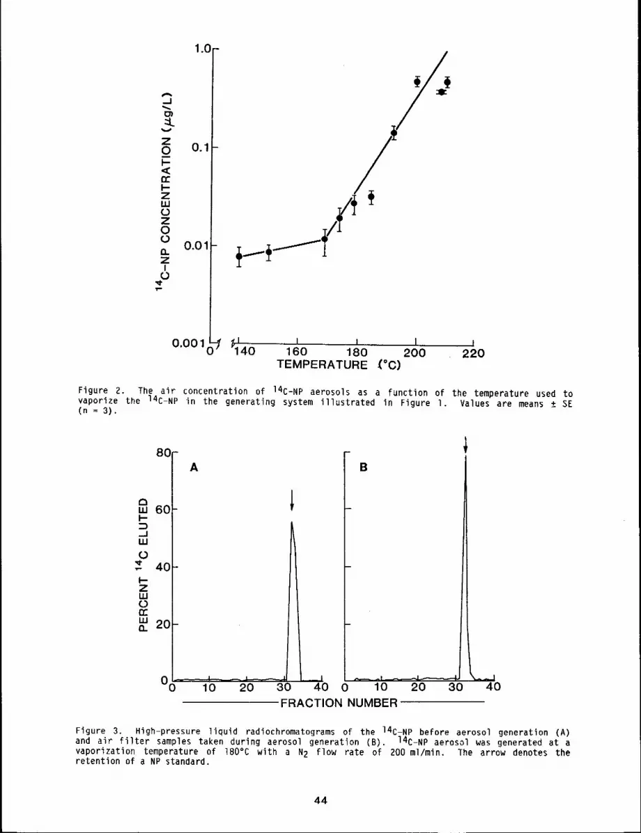
zL
Z0I-<
Z1.0
Z0
O.ZI
1.0--
0.1
0.01
0.001
_0¯ i
1,11 I II I
40 160 180 200 220TEMPERATURE -(°C)
Figure 2, The air concentration of 14C-NP aerosols as a function of the temperature used tovaporize the 14C-NP in the generating system illustrated in Figure I, Values are means + SE(n -- 3).
80-A B
60
,,=,0.
0 10 20 30FRACTION NUMBER
40
Figure 3. High-pressure liquid radiochromatograms of the 14C-NP before aerosol generation (A)and air filter samples taken during aerosol generation (B). 14C-NP aerosol was generated at vaporization temperature of 180°C with a N2 flow rate of 200 ml/min. The arrow denotes theretention of a NP standard.
44
of air. In characterization studies described here, it was shown that aerosols of 14C-NP could
be generated within the necessary concentration range by varying either the vaporization
temperature of the 14C-NP or varying the N2 flow rate that carries the vaporized 14C-NP out
of the sample vessel. However, it was found that the aerosol size distribution (VMD) was also
changed by altering the N2 flow rate to control the aerosol concentration. This situation of
changing particle size would be undesirable for a series of inhalation studies attempting to
observe a dose-response relationship, because inhaled particles of different sizes would have
different efficiencies of lung deposition. Therefore, for this generating system, the most
effective method for controlling 14C-NP aerosol concentration would be to vary the temperature
at which the 14C-NP is vaporized. Also, HPLC analysis showed that heating of 14C-NP for
aerosol generation did not cause thermal degradation of this radiolabeled compound. The
radiochemical purity of the 14C-NP aerosol was virtually the same as that of the 14C-NP
starting material. Thus, these studies showed that the aerosol generation system described in
this report will be useful for the production of 14C-NP aerosols required for inhalation
exposures using 14C-NP. Also, the factors involved in generating these aerosols that affect
aerosol characteristics may also be generally applicable in the generation of other PAH aerosols.
REFERENCES
I. Salmeen, I., A. M. Durisin, T. J. Prater, T. Riley, and D. Schuetzle, Contribution ofl-Nitropyrene to Direct-Acting Ames Assay Mutagenicities of Diesel Particulate Extracts,Murat. Res. 104: 17-23, 1982.
2. Sun, 3. D., R. K. Wolff, H. M. Aberman, and R. O. McClellan, Inhalation of l-NitropyreneAssociated with Ultrafine Insoluble Particles or as a Pure Aerosol: A Comparison ofDeposition and Biological Fate, Toxicol. Appl. Pharmacol. 69: 185-198, 1983.
45
STUDIES OF LITHIUM AEROSOLS
Abstract -- L1thlum was burned ~n alr, reacted wlth
water, and vaporlzed along wlth a stainless steel
tube at high temperatures to slmulate accldents in-
volvlng a space nuclear reactor or a fuslon reactor~
Partlcles of a resplrable s~ze w~re formed ~n these
processes. Inhalation exposure studies were inltl-
ated wlth rats and, although irrltatlon of the upper
PRINCIPAL INVESTIGATORS
M. D. Hoover
F. A. Seller
S. J. Rothenberg
R. F, Henderson
resplrator9 tract was noted, there was no evldence of inflammat~on in the deep lung after
inhalation of llthlum combustion aerosols. Studies are contlnulng to better understand the
toxlclty of 11th~ aerosols.
Lithium metal is a potential coolant for space nuclear reactors and fusion reactors. It may
also be used in the lithium hydride form as a radiation shield in space reactor systems. There is
concern for the health of workers and others who may inhale lithium aerosols after accidents such
as burn-up of a space reactor system during a launch pad accident, or during re-entry to the
atmosphere, or after leaking of hot lithium compounds out of a fusion reactor system, with
subsequent combustion in air. Lithium above its melting point (327°C) burns readily in air, and
the combustion aerosol is expected to have a caustic reaction with tissue. Only limited data are
available on this damage process.1 The purpose of this study was to characterize lithium
aerosols generated under a variety of conditions and to conduct a pilot inhalation study in rats.
METHODS
Three methods were used for generating lithium aerosols. In the first tests, lithium metal
was heated to its melting point in a stainless steel boat and an equal mass of water at room
temperature was added to the boat to allow a lithium-water reaction to occur. Particles were
collected for analysis by electron microscopy using a point-to-plane electrostatic precipitator
and should represent those from a lithium-water accident. In the second tests, lithium metal
alone and lithium metal in a stainless steel tube were vaporized with a plasma torch to create a
high-temperature, mixed-matrix aerosol that could result from a reactor accident. The plasma gas
was nitrogen (lO L/min) and the cooling gas was compressed air (20 L/min). Background
measurements were made of aerosol produced when the torch was operated alone. Measurements of the
NO2 levels produced by the torch were also made. In the third" tests, lithium metal, heated to
600°C, was allowed to burn in room air as it might if a reactor coolant line were to rupture. The
concentration of lithium generated was measured by collecting the lithium in a O.1-M HCI impinger
solution and analyzing the solution by flame photometry. Concentrations ranged from a few mg/m3
to several hundred mg/m3, depending on the rate at which lithium was introduced to the
combustion zone. This last method was used to generate aerosols for a pilot inhalation exposure
study in rats. Animals were exposed for 30 min at concentration levels of lO and 120 mg/m3.
Animals were sacrificed a day after exposure and examined for evidence of tissue damage in the
upper respiratory tract and inflammation of the deep lung.
RESULTS
Representative electron photomicrographs of particles generated in the study are shown in
Figures IA-ID. Aerosols from the lithium-water reaction (Fig. IA) contained a range of particle
46
A
0.5 pm
B
,. ¯ Z.,r
0.1 pm
~
~$..,.,
-C." ~*,-
¢. (
C e’~
’e
! .
. Pb~
1urn q~’
Figure I. Particles collected with a point-to-plane electrostatic precipitator during (A) lithium-water reaction, (B) during operation of the plasma torch only, (C) during vaporization lithium metal in a stainless steel tube with the plasma torch, and (D) during burning of lithiummetal heated to 600°C In air.
47
sizes, with larger agglomerates having diameters of 0.2 ~m and a background of very small
particles with diameters of about O.O1 pm. The background aerosol from operation of the plasma
torch (Fig. 1B) was an ultrafine aerosol of copper from vaporization of the torch head orifice
itself. When lithium metal was vaporized with the torch, this background aerosol could be seen
along with larger lithium particles. The presence of two dlstinct aerosol forms is clearly
evident in Figure IC, which shows aerosol formed from plasma torch vaporization of a stainless
steel tube containing lithium metal. The branched chain ultrafine aggregates of stainless steel
had a primary particle size of about O.O1 pm. The larger spheres seen in Figure 1C are believed
to be lithium because they could be evaporated under the electron beam, leaving behind the
ultrafine stainless steel particles. Measurements of NO2 levels from the plasma torch showed
greater than 50 ppm NO2 present.
Particles from combustion of lithium in air (Fig. ID) consisted of agglomerates of individual
lithium particles having geometric diameters of about 0.2 ~m. Rats exposed two at a time for
30 min in a l-L nose-only exposure chamber gasped occasionally. At necropsy, there was gross
reddening of the tissues lining the nasal cavity of animals exposed at 120 mg/m3. The animals
had no evidence of inflammation of the deep lung at l day, as evidenced by analyses of
bronchopulmonary lavage fluid.2
DISCUSSION
These preliminary studies show that respirable aerosols of lithium are produced under
conditions that represent potential accidents in space nuclear or fusion reactor systems.
Exposure atmospheres generated with a plasma torch contained concentrations of NO2 that would,
by themselves, cause biological damage after inhalation. Thus, the plasma torch would be useful
only for an exposure designed to study the combined effects of NO2 and lithium aerosols, A
reduction in the NO2 level could, however, be obtained by using large amounts of dilution air.
The lack of inflammation in the deep lung of animals exposed to the lithium combustion aerosol
may indicate the aerosol did not reach this region of the respiratory tract. The gross reddening
of the tissues lining the nasal cavity and the lack of damage to the tissues of the deep lung are
similar to that observed after the inhalation of other irritant aerosols, such as H2SO4.Detailed chemical analyses of the composition of the lithium aerosols were not done in this
study, but are part of the continuing work. It is expected that the chemical composition of the
lithium aerosol will include the monoxide, hydroxide, and carbonate forms of lithium in relative
concentrations dependent upon the amounts of water vapor, oxygen, and carbon dioxide in the
combustion air. Current studies involve the use of a system designed to control carefully the
composition of the combustion air.
REFERENCES
I. Spiegl, C. J., 3. K. Scott, H. Steinhardt, L. J. Leach, and H. C. Lodge, Acute InhalationToxicity of Lithium Hydride, A.M.A. Arch. Ind. Health 14: 468-470, 1956.
2. Henderson, R. F., A. H. Rebar, J. A. Pickrell, and G. J. Newton, Early Damage Indicators inthe Lung. III. Biochemical and Cytological Response of the Lung to Inhaled Metal Salts,Toxicol. Appl. Pharmacol. 50: 123-136, 1979.
48
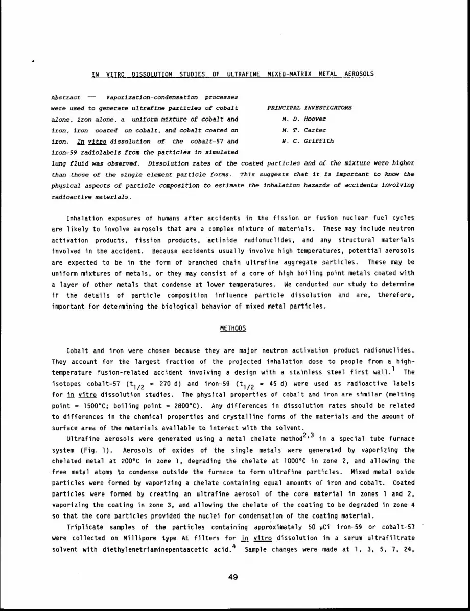
IN VITRO DISSOLUTION STUDIES OF ULTRAFINE MIXED-MATRIX METAL AEROSOLS
Abstract -- vaporlzatlon-condensatlon processes
were used to generate ultraf~ne particles of cobalt PRINCIPAL INVESTIGATORS
alone, iron alone, a unlform mixture of cobalt and M.D. Hoover
iron, iron coated on cobalc, and cobalt coated on M.T. Carter
Iron. In vitro dlssolutlon of the cobalt-57 and w.c. Grlfflth
1ton-59 radlolabels from the particles in simulated
lung fluld was observed. D1ssolutlon rates of the coated particles and of the mixture were higher
than those of the slngle element partJcle forms. Thls suggests That it ~s Important to know the
ph~slcal aspects of particle composition to estlmate the Inhalatlon hazards of accidents Involvlng
rad~oact~we materials.
Inhalation exposures of humans after accidents in the fission or fusion nuclear fuel cycles
are likely to involve aerosols that are a complex mixture of materials. These may include neutron
activation products, fission products, actinide radionuclides, and any structural materials
involved in the accident. Because accidents usually involve high temperatures, potential aerosols
are expected to be in the form of branched chain ultrafine aggregate particles. These may be
uniform mixtures of metals, or they maw consist of a core of high boiling point metals coated with
a layer of other metals that condense at lower temperatures. We conducted our study to determine
if the details of particle composition influence particle dissolution and are, therefore,
important for determining the biological behavior of mixed metal particles.
METHODS
Cobalt and iron were chosen because they are major neutron activation product radionuclides.
They account for the largest fraction of the projected inhalation dose to people from a high-
temperature fusion-related accident involving a design with a stainless steel first wall.1 The
isotopes cobalt-57 (tl/2 = 270 d) and iron-Sg (tl/2 = 45 d) were used as radioactive labels
for in vitro dissolution studies. The physical properties of cobalt and iron are similar (melting
point = 1500°C; boiling point = 2800°C). Any differences in dissolution rates should be related
to differences in the chemical properties and crystalline forms of the materials and the amount of
surface area of the materials available to interact with the solvent.
Ultrafine aerosols were generated using a metal chelate method2’3 in a special tube furnace
system (Fig. l). Aerosols of oxides of the single metals were generated by vaporizing the
chelated metal at 200°C in zone l, degrading the chelate at lO00°C in zone 2, and allowing the
free metal atoms to condense outside the furnace to form ultrafine particles. Mixed metal oxide
particles were formed by vaporizing a chelate containing equal amounts of iron and cobalt. Coated
particles were formed by creating an ultrafine aerosol of the core material in zones l and 2,
vaporizing the coating in zone 3, and allowing the chelate of the coating to be degraded in zone 4
so that the core particles provided the nuclei for condensation of the coating material.
Triplicate samples of the particles containing approximately 50 ~Ci iron-S9 or cobalt-57
were collected on Milllpore type AE filters for in vitro dissolution in a serum ultrafiltrate
solvent with diethylenetriaminepentaacetic acid. 4 Sample changes were made at l, 3, 5, 7, 24,
49
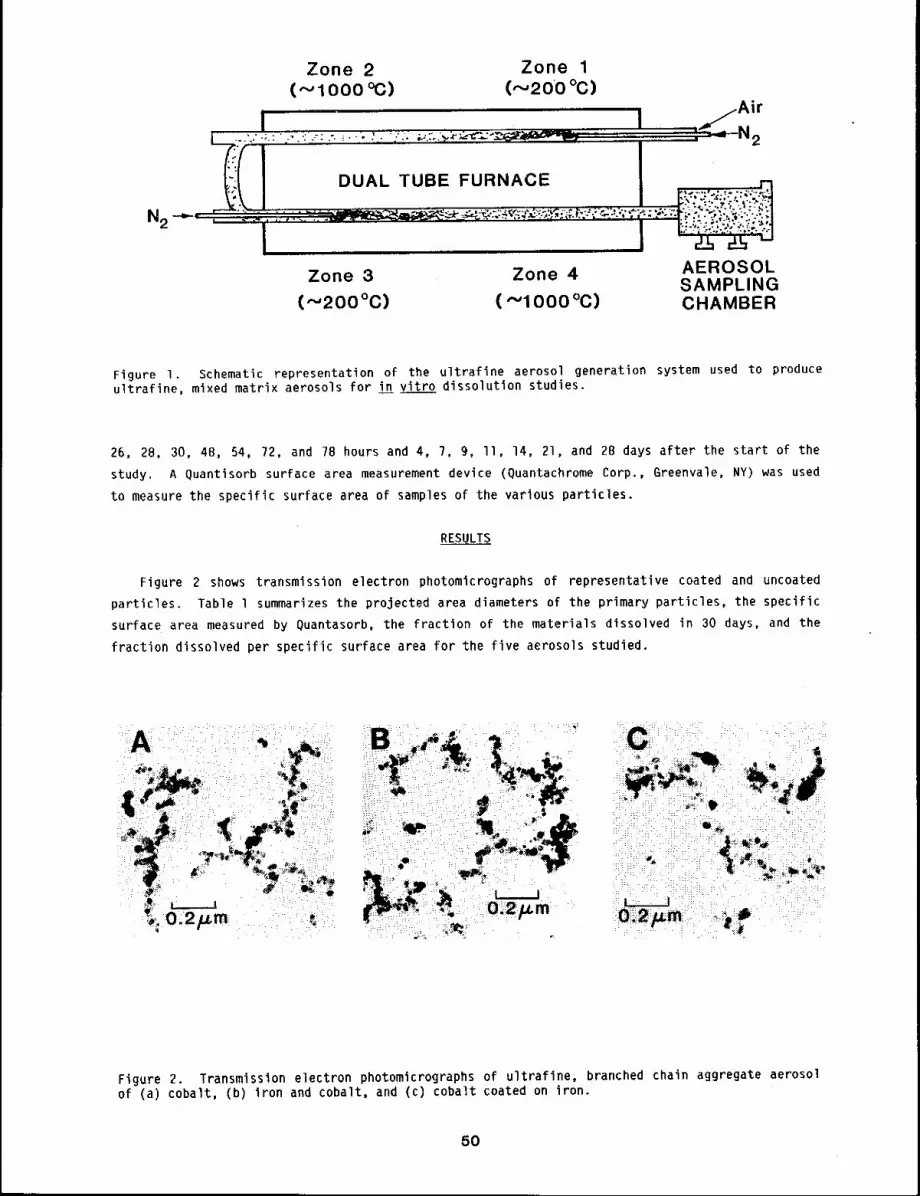
N2
Zone 2 Zone 1
( "-.’ 1000 °C) (,--,200 °C)
DUAL TUBE FURNACE
2
Zone 3 Zone 4 AEROSOLSAMPLING
(,-,-,200 °C) ( "-’1000 °C) CHAMBER
Figure I. Schematic representation of the ultrafine aerosol generation system used to produceultrafine, mixed matrix aerosols for in vitro dissolution studies.
26, 28, 30, 48, 54, 72, and 78 hours and 4, 7, 9, II, 14, 2l, and 28 days after the start of the
study. A Quantisorb surface area measurement device (Quantachrome Corp., Greenvale, NY) was used
to measure the specific surface area of samples of the various particles.
RESULTS
Figure 2 shows transmission electron photomicrographs of representative coated and uncoated
particles. Table l summarizes the projected area diameters of the primary particles, the specific
surface area measured by Quantasorb, the fraction of the materials dissolved in 30 days, and the
fraction dissolved per specific surface area for the five aerosols studied.
Figure 2. Transmission electron photomicrographs of ultrafine, branched chain aggregate aerosolof (a) cobalt, (b) iron and cobalt, and (c) cobalt coated on
5O
Ta
ble
1
Su
mm
ary
of
the
P
art
icle
S
ize
, S
pe
cific
S
urf
ace
A
rea
an
d F
ract
ion
D
isso
lve
d
in
30
ga
ys
for
the
U
ltra
fine
M
eta
l O
xid
e A
ero
sols
S
tud
ied
Pro
ject
ed
A
rea
Co
mp
osi
tion
o
f D
iam
ete
r o
f
Me
tal
Oxi
de
Prim
ary
P
art
icle
s
Pa
rtic
les
(win
± S
D~
Iro
n0
.03
3
± 0
.00
9
Co
ba
lt0
.02
8
± 0
.00
8
Iro
n
an
d C
ob
alt
0.0
20
± 0
.00
9
Iro
n
on
Co
ba
lt0
.02
9
± 0
.01
4
Co
ba
lt o
n I
ron
0.0
35
± O
.Oll
Sp
eci
ficF
ract
ion
Ir
on
Fra
ctio
n
Co
ba
ltF
ract
ion
Ir
on
Fra
ctio
n
Co
ba
lt
Su
rfa
ceD
isso
lve
dD
isso
lve
dD
isso
lve
d
Pe
rD
isso
lve
d
Pe
r
Are
ain
3
0 D
ays
in
30
Da
ysS
pe
cific
S
urf
ace
A
rea
Sp
eci
fic
Su
rfa
ce
Are
a
(m2
/q ±
SD
)(f
ract
ion
± S
D)
(fra
ctio
n
± S
O)
(fra
ctio
n/(
m2
/q)
±
(fra
ctio
n/(
m2
/~)
±
28
.B ±
3.0
0.0
05
± O
.OO
l--
0.0
00
2
± 0
.00
00
4--
16
.2
± I
.I--
0.0
2
± 0
.01
--0
.00
1
±
.00
06
14
.4
± 2
.60
.02
0
±
O.O
lO0
.15
±
0.0
20
.00
1
± 0
.00
07
0.0
1
± .
00
2
21
.1
± 1
.40
.02
0
± O
.OO
10
.04
± 0
,01
0.0
01
±
O.O
00
0B
0.0
02
±
.00
05
8.I
± 1
.00
.00
7
± 0
.00
10
.08
± 0
.02
O.O
Ol ±
0.0
00
20
.01
±
.00
3
ast
an
da
rd
de
via
tion
s b
ase
d o
n t
rip
lica
te
sam
ple
s.
DISCUSSION
Th~ projected area diameters of the primary ultrafine particles were statistically the same
for the five aerosols studied, although the trend appeared to be for the coated particles to be
larger than the uncoated particles (see Table l). The addition of a coating material (e,g, iron
onto cobalt or cobalt onto iron) resulted in an increase in the standard deviation of the primary
particle sizes, indicating a nonuniform condensation process. It would be useful if some method
could be developed to quantltate the fraction of the surface area of the core particles that is
coated. Surface area studies are continuing.
In light of the similarity of the primary particle sizes, the specific surface areas of the
five aerosols would have been expected to be similar. They range over a factor of nearly three,
with the coated particles generally having lower specific surface area than the uncoated
particles. This could result from a smoothing of available surface during the condensation of the
coating.
The dissolution rates of the core materials were statistically similar to those of the
corresponding single materials. This was to be expected because formation conditions of the core
were independent of a later addition of coating. The presence of a coating did not appear to
retard the dissolution of the core, apparently confirming that the core was not uniformly coated
and shielded from the solvent. The dissolution of the iron or cobalt coating, however, was
enhanced by a factor of 4 relative to that of the corresponding single materials. This is
explained in part by the theoretically higher surface-to=volume ratio of a coating. If the
addition of a uniform coating doubles the volume of the particle, the particle diameter increases
by 26% and the surface-to-volume ratio increases by 58%. A second mechanism for the increased
dissolution may be the imperfect nature of the crystal structure established at the interface
between the core and coating.
The apparent influence of a disruption of crystal structure can be seen in the fourfold and
eightfold increases in the dissolution of the cobalt and the iron, respectively, in the mixed
matrix particles as compared to the single composition particles. The study is continuing, with
investigation by x-ray diffraction of the crystal structure of the five aerosols studied. The
results to date indicate that details of particle composition can influence dissolution behavior
by a factor of four or more. This could strongly influence the biological behavior of moderately
soluble materials and should be studied further.
REFERENCES
1 ¯
2.
3.
Piet, S., M. Kazimi, and L. Lidsky, Potential Consequences of Tokamak Fusion Reactor Accidents:The Materials Impact, DOE UC-2OC,D,E, Massachusetts Institute of Technology, 3une 1982.
Kanapilly, G. M., K. W. Tu, T. B. Larson, G. R. Fogel, and R. 3. Luna, Controlled Productionof Ultrafine Metallic Aerosols by Vaporization of an Organic Chelate of the Metal, O. ColloidInterface Sci. 65: 533-547, 197B.
Hoover, M. D. and 3. Lee, Aerosols from Fusion Energy Systems, LMF-I02, pp. 71-73, December1982.
Kanapilly, G. M., O. G. Raabe, and H. A. Boyd, A Method for Determining the DissolutionCharacteristics of Accidentally Released Radioactive Aerosols, in Proceedings of 3rd Interna-tional.Congress, IRPA, USAEC, Oak Ridge, TN, 1974.
52
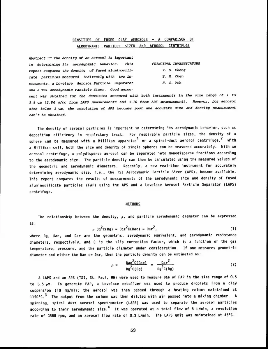
DENSITIES OF FUSED CLAY AEROSOLS - A COMPARISON OF
AERODYNAMIC PARTICLE SIZER AND AEROSOL CENTRIFUGE
Abstract -- The denslty of an aerosol is important
in determlnlng its aerodynamlc behavior. This PRINCIPAL INVESTIGATORS
report compares the density of fused alum~nosill- Y.S. Cheng
care partlcles measured Indlrectly wlth two in- T.H. Chen
struments, a Lovelace Aerosol Particle separator H.C. Yeh
and a TSI Aerodynamic Partlcle Slzer. Good agree-
ment was obtalned for the densltles measured with both ~nstruments in the slze range of i to
3.5 ~m (2.84 g/cc from LAPS measurements and 3.Z0 from APS ~asurement). However, for aerosol
slze below 1 ~m, the resolution of APS becomes poor and accurate slze and denslt~ ~asurement
can’t be obtained.
The density of aerosol particles is important in determining its aerodynamic behavior, such as
deposition efficiency in respiratory tract. For respirable particle sizes, the density of a
sphere can be measured with a Millikan apparatus I or a spiral-duct aerosol centrifuge. 2 With
a Millikan cell, both the size and density of single spheres can be measured accurately. With an
aerosol centrifuge, a polydisperse aerosol can be separated into monodisperse fractions according
to the aerodynamic size. The particle density can then be calculated using the measured values of
the geometric and aerodynamic diameters. Recently, a new real-tlme instrument for accurately
determining aerodynamic size, i.e., the TSI Aerodynamic Particle Sizer (APS), became available.
This report compares the results of measurements of the aerodynamic size and density of fused
aluminosilicate particles (FAP) using the APS and a Lovelace Aerosol Particle Separator (LAPS)
centrifuge.
METHODS
The relationship between the density, p, and particle aerodynamic diameter can be expressed
as:p Dg2C(Dg) = Dae2C(Dae) = 2, (1)
where Dg, Dae, and Dar are the geometric, aerodynamic equivalent, and aerodynamic resistance
diameters, respectively, and C is the slip correction factor, which is a function of the gas
temperature, pressure, and the particle diameter under consideration. If one measures geometric
diameter and either the Dae or Dar, then the particle density can be estimated as:
Dae2C(Dae) Oar2_ (2)p = Dg2C(Dg)
Dg2C(Dg)
A LAPS and an APS (TSI, St. Paul, MN) were used to measure Dae of FAP in the size range of 0.5
to 3.5 ~m. To generate FAP, a Lovelace nebulizer was used to produce droplets from a clay
suspension (lO mg/ml); the aerosol was then passed through a heating column maintained
ll50°C. 3 The output from the column was then diluted with air passed into a mixing chamber. A
spinning, spiral dust aerosol spectrometer (LAPS) was used to separate the aerosol particles
according to their aerodynamic size. 4 It was operated at a total flow of 5 L/min, a revolution
rate of 3580 rpm, and an aerosol flow rate of 0.3 L/min. The LAPS unit was maintained at 45%.
53
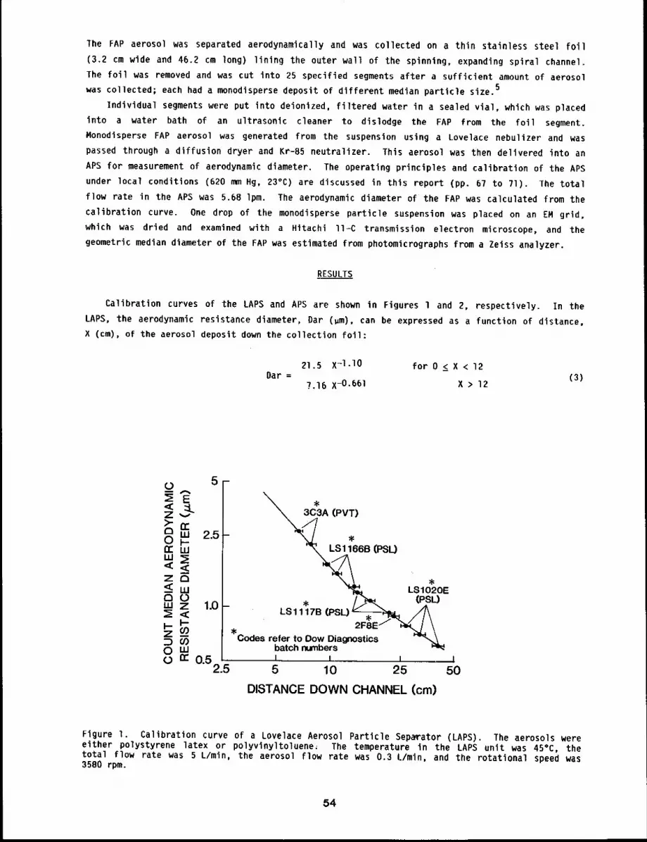
The FAP aerosol was separated aerodynamically and was collected on a thin stainless steel foil
(3.2 cm wide and 46.2 cm long) lining the outer wall of the spinning, expanding spiral channel.
The foil was removed and was cut into 25 specified segments after a sufficient amount of aerosol
was collected; each had a monodisperse deposit of different median particle size.5
Individual segments were put into deionized, filtered water in a sealed vial, which was placed
into a water bath of an ultrasonic cleaner to dislodge the FAP from the foil segment.
Monodisperse FAP aerosol was generated from the suspension using a Lovelace nebulizer and was
passed through a diffusion dryer and Kr-85 neutralizer. This aerosol was then delivered into an
APS for measurement of aerodynamic diameter. The operating principles and calibration of the APS
under local conditions (620 mm Hg, 23°C) are discussed in this report (pp. 67 to 71). The total
flow rate in the APS was 5.68 Ipm. The aerodynamic diameter of the FAP was calculated from the
calibration curve. One drop of the monodisperse particle suspension was placed on an EM grid,
which was dried and examined with a Hitachi ll=C transmission electron microscope, and the
geometric median diameter of the FAP was estimated from photomicrographs from a Zeiss analyzer.
RESULTS
Calibration curves of the LAPS and APS are shown in Figures l and 2, respectively. In the
LAPS, the aerodynamic resistance diameter, Dar (um), can be expressed as a function of distance,
X (cm), of the aerosol deposit down the collection foil:
21.5 X-I-I0 for 0 _< X < 12Dar = (3)
7.16 X-0.66 1 X > 12
0 5-m
E< ~LZv)-n ~ 2.5OF"tr Ww~E
Z a-<wC)w 70 1.0
z_mom0 n’0.5
2.5
A (PV~)
-
"’k~166B (PSL)
LSIO20E_ , / ~L. (PSL)
¯ 11 7B /A¯ " _ . \
m I I i5 10 25 50
DISTANCE DOWN CHANNEL (cm)
Figure I. Calibration curve of a Lovelace Aerosol Particle Separator (LAPS). The aerosols wereeither polystyrene latex or polyvinyltoluene~ The temperature in the LAPS unit was 45°C, thetotal flow rate was 5 L/min, the aerosol flow rate was 0.3 L/min, and the rotational speed was3580 rpm.
54
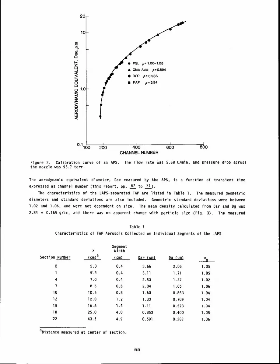
20-
& Oleic Acid p=0.894
f ¯ DOP p=0.986
IIIIIwI
II FAP p= 2.84
0.1100 I = I200 400 600 800CHANNEL NUMBER
Figure 2. Calibration curve of an APS. The flow rate was 5.68 L/min, and pressure drop acrossthe nozzle was 96.7 torr.
The aerodynamic equivalent diameter, Dae measured by the APS, is a function of transient time
expressed as channel number (this report, pp. 67 to 71}.
The characteristics of the LAPS-separated FAP are listed in Table I. The measured geometric
diameters and standard deviations are also included. Geometric standard deviations were between
1.02 and 1.06, and were not dependent on size. The mean density calculated from Dar and Dg was
2.84 ± 0.165 g/cc, and there was no apparent change with particle size (Fig. 3). The measured
Table l
Characteristics of FAP Aerosols Collected on Individual Segments of the LAPS
SegmentX Width
Section Number (cm)a (cm) Dar (vm) Dq (um)
B 5.0 0.4 3.66 2.06
l ~.8 0,4 3.11 1.71
4 7.0 0.4 2.53 1.37
7 8.5 0.6 2.04 1.05I0 10.6 0.8 1.60 0.853
12 12.8 1.2 1.33 0.70915 16.8 1.5 I.II 0.57318 25.0 4.0 0.853 0.400
22 43.5 4.9 0.591 0.26?
aDistance measured at center of section.
g
l 05
l 05
l 02
1 06
l 04
l 04
1.04
l .05
l .06
55
8- Figure 3. Measured FAP density as a functionof geometric particle dlameter.
_
mAPS
00,1"J I o
1.0 5PARTICLE DIAMETER, Dg, /u.m
channel number and the corresponding aerodynamic equivalent diameter, Dae, of FAP measured with
the APS are listed in Table 2. Geometric standard deviations given by the APS increased from 1.05
to 1.23, with decreasing particle size. The calculated densities are shown in Figure 3. There
was a constant density between Dae of 3.66 and l.O? ~m, with a mean and standard deviation of
3.10 ± O.lO g/cc. However, for Dae less than 1.O pm, the measured density increased to 6 g/cc.
Table 2
Aerodynamic Size Distributions of FAP Aerosols Measured with an APS
LAPS
Section Number Mean Channel Dae(cm) ag
B 338 3.66 1.05
l 308 3.10 1.05
4 282 2.51 1.09
7 255 1.97 1.08
lO 234 1.58 l.]O
12 218 1.29 l.ll
15 207 1.07 l+15
18 198 0.878 l.lg
22 196 0.820 1.23
DISCUSSION
Both the APS and LAPS can be used to measure the aerodynamic diameter of particles. Together
with the geometric diameter, the density of monodisperse spherical aerosols can be estimated.
Geometric standard deviations of LAPS-separated FAP aerosols determined by Zeiss analysis are
generally on the order 1,05 and independent of size, whereas those measured by the APS are equal
56
to or larger than 1.05 and increased with decreasing particle size. This indicates that the
resolution of the APS decreased with particle size. FAP densities measured by APS were about 10%
higher than those measured with the LAPS for sizes larger than l.O ~m. At smaller particle
sizes, the APS measured a much larger density than the LAPS because the size resolution of the APS
for smaller particles is not good, and, therefore, it gave inaccurate values of Dae and p.
REFERENCES
I. Raabe, O. 6., Measurement of Aerosol Particle Density with the Millikan Apparatus, FissionProduct Inhalation Program Annual Report, LF-41 Lovelace Foundation lg6B-1969, pp. 70-74.
2. Kotrappa, P. and C. J. Wilkinson, Densities in Relation to Size of Special Aerosols Producedby Nebulization and Heat Degradation, Am. I nd: Hyg. Assoc. J. 33: 449-453, 1972.
3. Raabe, O. G., G. M. Kanapilly, and B. J. Newton, New Methods for the Generation of Aerosols ofInsoluble Particles for Use in Inhalation Studies, in Inhaled Particles Ill, Unwin, England,pp. 3-18, 1971.
4. Kotrappa, P. and M. E. Light, Design and Performance of the Lovelace Aerosol ParticleSeparator, Rev. Sci. Instr. 43: ll06wlll2, 1972,
5. Raabe, O. G., H. A. Boyd, G. M. Kanapilly, C. J, Wilkinson, and G. J. Newton, Development andUse of a System for Routine Production of Monodisperse Particles of 238pu02 and Evaluationof Gamma-Emitting Labels, Health Phys. 28: 655-667, 1975.
57
OPTICAL DIAMETERS OF AGGREGATE AEROSOLS
Abstract -- The optlcal equivalent dlameters of
cluster aggregates composed of uniform polystyrene PRINCIPAL INVESTIGATORS
latex (PSL) spheres were Investlgated with Rogco 226, T.B. Chen
Royco 236, and Cllmet 208A optical particle counters. Y.S. Cheng
Primary PSL particles between 0.3 and 2.4 ~m were
used. For cluster aggregates havlng volume eq~l~valent dlameters smaller than i ~m, the light
scattering d~ameter measured by the Rogco counters was shown to be approxlmatelg equal to the
volume equivalent diameter. For doublet aggregates composed of uniform PSL spheres wlth prJmar9partlcle dlameters larger than 0.76 ~, the light-scatterlng dla/neter measured b9 the C1~met
208A was very close to the area equlvalent diameter.
Behavior of irregularly-shaped particles has been of interest for aerosol scientists. The
equivalent diameters of aggregate aerosols obtained from instruments that measure aerodynamic
diameter, l electrical mobility, 2 and diffusion coefficient, 3 respectively, have been
reported. A few experiments have been performed to measure the response of simple cluster
aggregates (close-packed, three dimensional aggregates) in the optical particle counterS. Gebhart
et al.4 reported the light-scattering equivalent diameter of cluster aggregates composed of
polystyrene latex (PSL) spheres in terms of geometric diameter. These experiments are difficult
to repeat because the instruments are not commercially available. Since some sophisticated
optical particle counters (OPCs) based on light-scattering techniques have been commercially
available and widely used, similar experiments can be done to understand more about the optical
equivalent diameters of PSL cluster aggregates produced by nebulization. With this intent, three
commercial optical counters: a Royco 226, Royco 236, and Climet 208A were studied, and the
results were compared with previous data.
METHODS
Aerosol Generation
PSL spheres in the size range between 0.3 and 2.4 ~m (Dow Chemical Company, Indianapolis,
IN) were used. To increase the fraction of aggregates of uniform PSL spheres in the latex
suspension, the emulsifier (surfactant) used to prevent the latex spheres from aggregating-in the
suspension was removed by centrifugatlon. A highly concentrated suspension of latex spheres was
prepared for the Lovelace nebulizer. After nebulization, the aerosol was dried by a diffusion
dryer and was exposed to a Kr-85 bipolar-ion source. Before entering the optical particle
counter, the aerosol was diluted and mixed with filtered compressed air in a plenum chamber. An
aerosol sample was taken from the chamber with an electrostatic precipitator for scanning electron
microscopy (SEM).
Data Acquisition
Signal output from each OPC was fed into a multi-channel analyzer (Canberra 4100, Meriden, CT)
to classify the voltage pulses into discrete voltage channels. The pulse height spectrum was
displayed on an oscilloscope and recorded. Each channel can be related to a specific particle
size.6
58
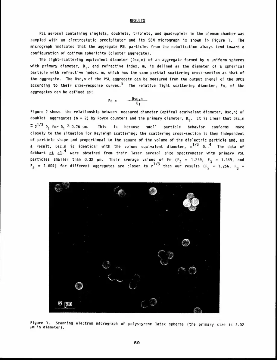
RESULTS
PSL aerosol containing singlets, doublets, triplets, and quadruplets in the plenum chamber was
sampled with an electrostatic precipitator and its SEM micrograph is shown in Figure I. The
micrograph indicates that the aggregate PSL particles from the nebulization always tend toward a
configuration of optimum sphericity (cluster aggregate).
The light-scattering equivalent diameter (Dsc,n) of an aggregate formed by n uniform spheres
with primary diameter, Dl, and refractive index, m, is defined as the diameter of a spherical
particle with refractive index, m, which has the same partial scattering cross-section as that of
the aggregate. The Dsc,n of the PSL aggregate can be measured from the output signal of the OPCs
according to their size-response curves. 5 The relative light scattering diameter, Fn, of the
aggregates can be defined as:
Fn ~ Dsc,nDl
Figure 2 shows the relationship between measured diameter (optical equivalent diameter, Dsc,n)
doublet aggregates (n = 2) by Royco counters and the primary diameter, I. I t i s c lear t hat D sc,n
Z 21/3 Ol for D1 ~ 0.76 um. This Is because small particle behavior conforms more
closely to the situation for Rayleigh scattering; the scattering cross-section is then independent
of particle shape and proportional to the square of the volume of the dielectric particle and, as
a result, Dsc,n is identical with the volume equivalent diameter, n1/3 Di.4 The data of
Gebhart et al. 4 were obtained from their laser aerosol size spectrometer with primary PSL
particles smaller than 0.32 ~m. Their average values of Fn (F 2 = 1.259, F3 = 1.449, and
F4 = 1.604) for different aggregates are closer to I/3 t han o ur r esults ( 2 = 1. 256, F 3=
Figure I. Scanning electron mlcrograph of polystyrene latex spheres (the primary size is 2.02~m in diameter).
59
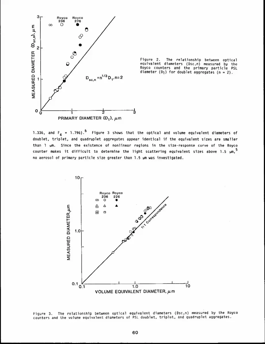
Oo
Royco Royco236 226 /
°y_ 1/3 -2
Dsc,n =nl/3 Dl,n-
I i1 2
PRIMARY DIAMETER (D1),/_cm
Figure 2. The relationship between opticalequivalent diameters (Dsc,n) measured by theRoyco counters and the primary particle PSL
diameter (Dl) for doublet aggregates (n =
and F 4 = 1.796). 6 Figure 3 shows that the optical and volume equivalent diameters of1.336,
doublet, triplet, and quadruplet aggregates appear identical if the equivalent sizes are smaller
than 1 wm. Since the existence of nonlinear regions in the size-response curve of the Royco
5counter makes it difficult to determine the light scattering equivalent sizes above 1.5 gm,
no aerosol of primary particle size greater than 1.5 ~m was investigated.
10
I.(]
Royco Royco236 226 /
co 0 Q/
I I
°’1 o.:1 .... 1.o lOVOLUME EQUIVALENT DIAMETER, ~m
Figure 3. The relationship between optical equivalent diameters (Dsc,n) measured by the Roycocounters and the volume equivalent diameters of PSL doublet, triplet, and quadruplet aggregates.
60
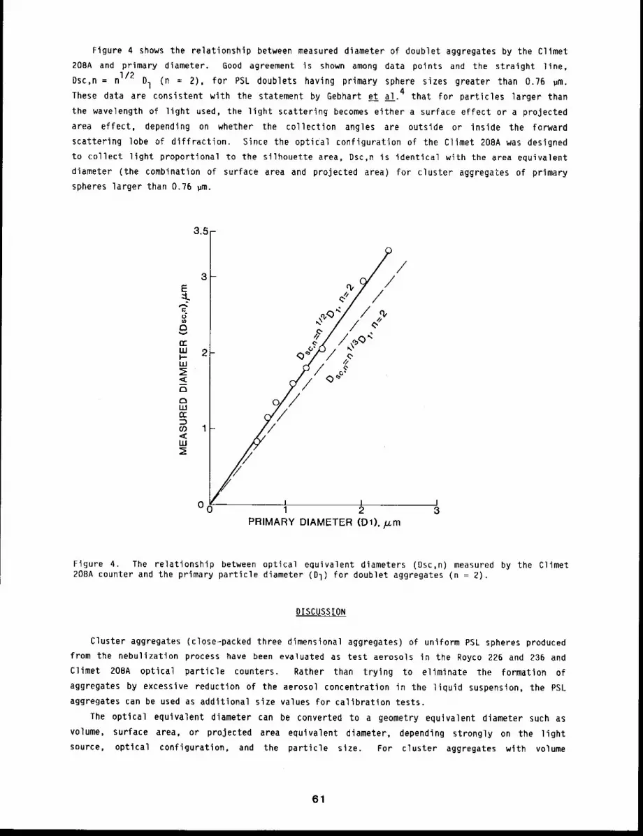
Figure 4 shows the relationship between measured diameter of doublet aggregates by the Cllmet
208A and primary diameter. Good agreement is shown among data points and the straight line,
Dsc,n = nI/2 D1 (n = 2), for PSL doublets having primary sphere sizes greater than 0.76 ~m.These data are consistent with the statement by Gebhart et al. 4 that for particles larger than
the wavelength of light used, the light scattering becomes either a surface effect or a projected
area effect, depending on whether the collection angles are outside or inside the forward
scattering lobe of diffraction. Since the optical configuration of the Climet 208A was designed
to collect light proportional to the silhouette area, Dsc,n is identical with the area equivalent
diameter (the combination of surface area and projected area) for cluster aggregates of primary
spheres larger than 0.76 ~m.
3,~--
3-
~.. 2 ~o" -- ~;
<_ o~o~" .Dt~ILl¢r
03 1<LU
_// ,,
PRIMARY DIAMETER (D1),/zm
Figure 4. The relationship between optical equivalent diameters (Dsc,n) measured by the Climet208A counter and the primary particle diameter (DI) for doublet aggregates (n =
DISCUSSION
Cluster aggregates (close-packed three dimensional aggregates) of uniform PSL spheres produced
from the nebulization process have been evaluated as test aerosols in the Royco 226 and 236 and
Climet 208A optical particle counters. Rather than trying to eliminate the formation of
aggregates by excessive reduction of the aerosol concentration in the liquid suspension, the PSL
aggregates can be used as additional size values for calibration tests.
The optical equivalent diameter can be converted to a geometry equivalent diameter such as
volume, surface area, or projected area equivalent diameter, depending strongly on the light
source, optical configuration, and the particle size. For cluster aggregates with volume
61
equivalent diameter smaller than 1 ~m, the light-scattering diameter measured by the Royco
counters can be considered equal to the volume equivalent diameter. For doublet aggregates of
uniform spheres larger than 0.76 ~m, the light-scattering diameter measured by the Climet 208A
can be considered equal to the area equivalent diameter. The data presented here can be extended
to serve as a guide for the optical equivalent diameter of irregular, nonspherical particles if
the test particles are within the suitable size range.
REFERENCES
I. St~ber, W., A. Berner, and R. Blaschke, The Aerodynamic Diameter of Aggregates of UniformSpheres, a. Colloid Interface Sci. 2_99: 710, 1969.
2. St~ber, W., C. Boose, and V. Prodi, Ober Die Orientierung Und Den Dynamischen Formfaktor VonKettenf~rmigen Aerosolteilchen In Ladungsspektrometern, Water, Air & Soil Pollution 3,: 493,1974.
3. Heyder, J. and G. Scheuch, Diffusional Transport of Nonspherical Aerosol Particles, AerosolSci. Technol. 2: 41, 1983.
4. Gebhart, J., J. Heyder, C. Roth, and W. Stahlhofen, Optical Aerosol Size Spectrometry Belowand Above the Wavelength of Light, in Fine Particles (B. Y. H. Liu, ed.), Academic Press, NewYork, p. 794, 1976.
5. Chen, T. B., Y. S. Cheng, and H. C. Yeh, Experimental Responses of Two Optical ParticleCounters, J. Aerosol Sci. (submitted).
6. Chen, T. B. and Y. S. Cheng, Optical Diameters of Aggregate Particles, 5. Aerosol Sci.(submitted).
EXPERIMENTAL RESPONSES OF TWO OPTICAL PARTICLE COUNTERS
Abstract -- The response of two commerclally avail-
able optlcal partlcle counters has been studled PRINCIPAL INVESTIGATORS
using several monodlsperse aerosols. The aerosol T.B. then
used included olelc acid, dloctylphthalate, and Y.S. Cheng
pol~st~rene latex particles. Diameters of these H.C. Yeh
particles ranged from 0.3 to I0 ~m, and refractive
Indlces were from 1.46 to 1.6. The response curve of the filament lamp counter (CllmeC 208A) was
found to be independent of the particle refractive ~ndex for partlcles larger than 1.7 ~m in
diameter. The laser optlcal counter (Royco 236), however, showed a more pronounced partlcle
refractive index effect. Moreover, its response curve had periodic fluctuations in the slzes
between 1.5 and 5.0 ~m. Calibration curves of both optlcal counters obtained from other
research groups are compared and discussed.
Single particle light scattering is one of the most frequently used methods to detect and
measure aerosol size and number concentration. The Climet 208A uses an elliptical mirror optical
system to attain both great convergence of the illumination and a broad range (15 ° ~ I05 °) of
collection angles, thus avoiding the problem of multiple values in the size-response curve. The
ROyCO 236 employs a helium-neon laser cavity source and an aerodynamically focused flow system for
uniform illumination in the sensitive volume. Also, a wide scattering angle optics (35° ~ 120°)
is used to collect most of the scattered light. Both instruments have been studied with
monodisperse aerosols of limited size range. 1-5 The objective of this study was to thoroughly
calibrate both instruments over the applicable size range of monodisperse oleic acid (OA),
dioctylphthalate (DOP), polystyrene latex (PSL) aerosols. These three aerosols were also used
investigate refractive index effects on both counters.
METHODS
Aerosol Preparation and Classification
Monodisperse aerosols of three different materials were used. PSL spheres between 0.3 and
5 ~m (DOW Chemical Company, Indianapolis, IN) were generated from a Lovelace nebulizer. The
nominal sizes of PSL particles supplied by the factory have been compared with the result from
transmission electron microscopy (TEM) after sampling with an electrostatic precipitator.
vibrating-orlfice generator was used to generate larger DOP and OA monodisperse aerosols. The
combination of a Collison nebulizer and an electrostatic classifier was used to produce smaller
monodisperse DOP and OA aerosols’6.
Data Acquisition
Output from the counter was fed directly into a multi-channel analyzer (MCA) (Canberra 4100,
Meriden, CT) to classify the voltage pulses into discrete voltage ranges (channels), and the pulse
height spectrum was displayed on a scope and printed out. A scale of 2048 channels in the MCA was
used to investigate the possible multiple values in the size-response curve of the counters and,
also, to increase the resolution of both counters since they use less channels in their
68
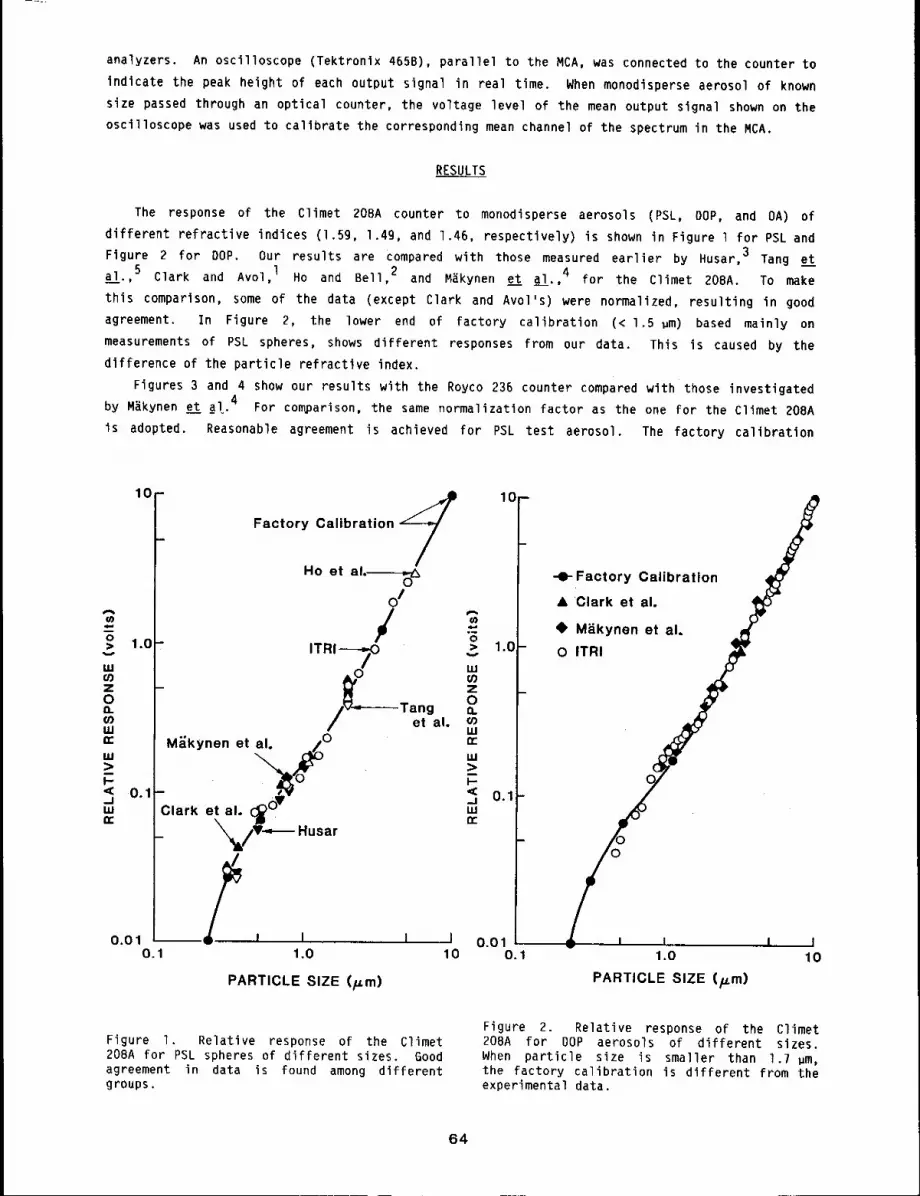
analyzers. An oscilloscope (Tektronix 465B), parallel to the MCA, was connected to the counter
indicate the peak height of each output signal in real time. When monodisperse aerosol of known
size passed through an optical counter, the voltage level of the mean output signal shown on the
oscilloscope was used to calibrate the corresponding mean channel of the spectrum in the MCA.
RESULTS
The response of the Climet 20BA counter to monodisperse aerosols (PSL, DOP, and OA)
different refractive indices (1.59, 1.4g, and 1.46, respectively) is shown in Figure I for PSL and
Figure 2 for DOP. Our results are compared with those measured earlier by Husar, 3 Tang e t
a__!l.,5 Clark and Avol, l Ho and Bell, 2 and MAkynen et al.,4 for the Climet 208A. To make
this comparison, some of the data (except Clark and Avol’s) were normalized, resulting in good
agreement. In Figure 2, the lower end of factory calibration (< 1.5 pm) based mainly
measurements of PSL spheres, shows different responses from our data. This is caused by the
difference of the particle refractive index.
Figures 3 and 4 show our results with the Royco 236 counter compared with those investigated
by M~kynen e__tt a!.. 4 For comparison, the same normalization factor as the one for the Climet 208A
is adopted. Reasonable agreement is achieved for PSL test aerosol. The factory calibration
10
mo> 1.0
g.Iu)zOQ.
uJmruJ::>DI--< 0.1._1mr
Factory Calibration =~
Ho et al.-~O
OI
M;;kynen et al. /0
Clark et al. C~0
_ ’~/V~m Husar
0.01 --~// i I I I0.1 1.0 10
"6ITRI-~
0 tuz0T%~gal° o.
¢,o111
PARTICLE SIZE (p.m)
10=
1.0-
uJ;>i-< 0.1-.=I
-0- Factory Calibration
& ’Clark et al.
@ Miikynen et al.
O ITRI
D
0.010.1 1.0
PARTICLE SIZE (p.m)
I10
Figure I. Relative response of the Climet208A for PSL spheres of different sizes. Goodagreement in data is found among differentgroups.
Figure 2. Relative response of the Climet208A for DOP aerosols of different sizes.When particle size is smaller than 1.7 pm,the factory calibration is different from theexperimental data.
64
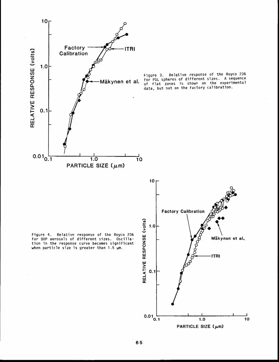
I0
0v
1.0W
Z0nGoLU
LU
.JW
0.010.
FactoryCalibration
ynen et al.
I I I ....1.0
PARTICLE SIZE (/.Lm)
Figure 3. Relative response of the Royco 236for PSL spheres of different sizes. A sequenceof flat zones is shown on the experimentaldata, but not on the factory calibration.
I10
1o-
Figure 4. Relative response of the Royco 236for DOP aerosols of different sizes. Oscilla=tion in the response curve becomes significantwhen particle size is greater than 1.5 gm.
o 1.0-:>
uJof)z013.(ouJn-uJ::>P 0.1-_.1Lu(z:
m
O.Ol0.1
Factory Calibration
00
\M~.kynen et air
I i I1.0
PARTICLE SIZE (Fm)
I10
65
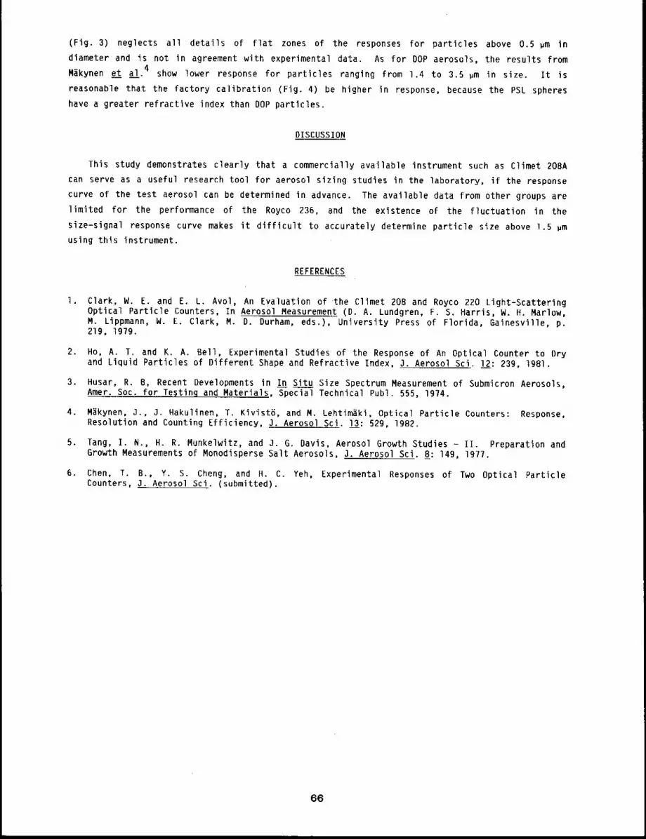
(Fig. 3) neglects all details of flat zones of the responses for particles above 0.5 #m
diameter and is not in agreement with experimental data. As for DOP aerosols, the results from
M~kynen et al.4 show lower response for particles ranging from 1.4 to 3.5 #m in size. It is
reasonable that the factory calibration (Fig. 4) be higher in response, because the PSL spheres
have a greater refractive index than DOP particles.
DISCUSSION
This study demonstrates clearly that a commercially available instrument such as Climet 208A
can serve as a useful research tool for aerosol sizing studies in the laboratory, if the response
curve of the test aerosol can be determined in advance. The available data from other groups are
limited for the performance of the Royco 236, and the existence of the fluctuation in the
size-signal response curve makes it difficult to accurately determine particle size above 1.5 ~m
using this instrument.
REFERENCES
l ¯ Clark, W. E. and E. L. Avol, An Evaluation of the Climet 208 and Royco 220 Light-ScatteringOptical Particle Counters, In Aerosol Measurement (D. A. Lundgren, F. S. Harris, W. H. Marlow,M. Lippmann, W. E. Clark, M. D. Durham, eds.), University Press of Florida, Gainesville, p.219, 197g.
2. Ho, A. T. and K. A. Bell, Experimental Studies of the Response of An Optical Counter to Dryand Liquid Particles of Different Shape and Refractive Index, 3. Aerosol Sci. I__22: 239, 1981.
3. Husar, R. B, Recent Developments in In Situ Size Spectrum Measurement of Submicron Aerosols,Amer. Soc. for Testinq and Materials, Special Technical Publ. 555, 1974.
4. M~kynen, J., 3. Hakulinen, T. Kivist~, and M. Lehtim~ki, Optical Particle Counters: Response,Resolution and Counting Efficiency, J. Aerosol Sci. 13: 529, IgB2.
5, Tang, I. N., H. R. Munkeiwitz, and J. G. Davis, Aerosol Growth Studies - II. Preparation andGrowth Measurements of Monodisperse Salt Aerosols, 3. Aerosol Sci. 8: 14g, 197/.
6. Chert, T. B., Y. S. Cheng, and H. C. Yeh, Experimental Responses of Two Optical ParticleCounters, 3. Aerosol Sci. (submitted).
66
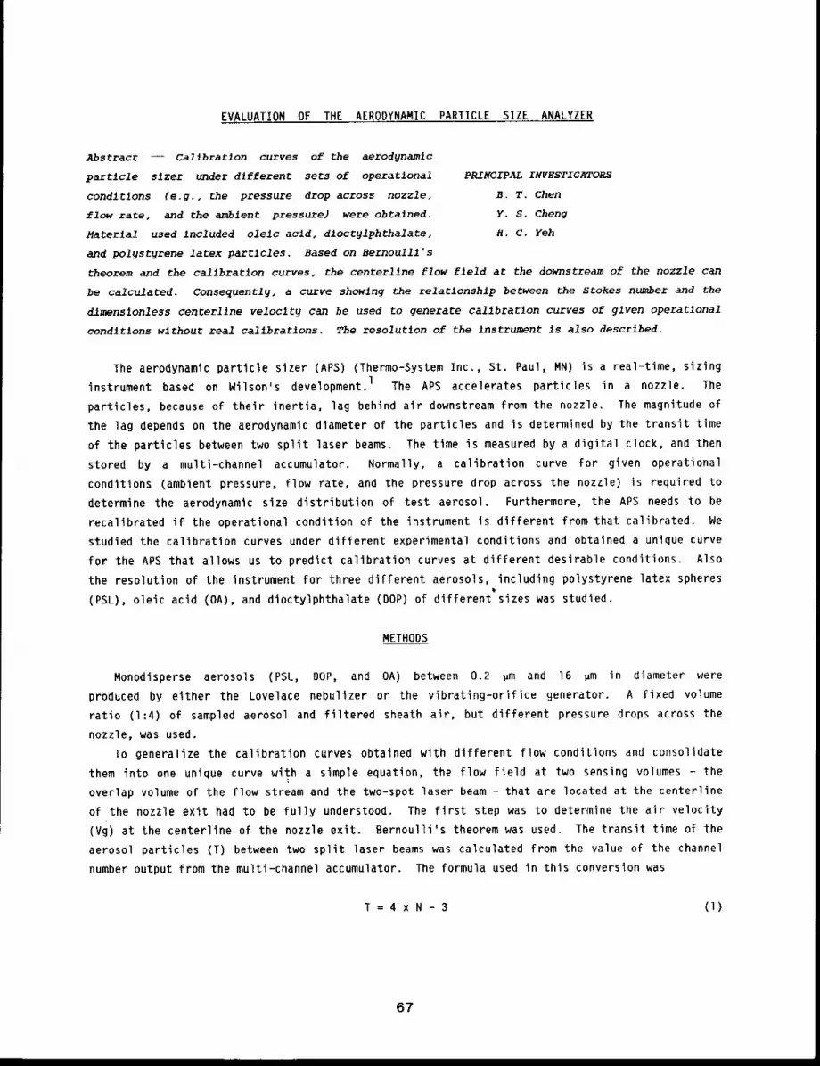
EVALUATION OF THE AERODYNAMIC PARTICLE SIZE ANALYZER
Abstract -- CallbraClon curves of the aerodynamlc
particle slzer under dlfferent sets of operational PRINCIPAL INVESTIGATORS
conditions (e.g., the pressure drop across nozzle, S.T. Chert
flow rate, and the ambient pressure) were obtained. Y. 5. Cheng
Material used included olelc acid, dloctylphthalate, H.C. Yeh
and polystyrene latex particles. Based on Bernoulli’s
theorem and the callbratlon curves, the centerllne flow field at the downstream of the nozzle can
be calculated. Consequently, a curve showing the relationship between the Stokes number and the
dimensionless centerl~ne velocity can be used to generate callbrat~on curves of given operatlonal
condltlons without real callbratlons. The resolution of the instrument is also descrlbed.
The aerodynamic particle sizer (APS) (Thermo-System Inc., St. Paul, MN) is a real-time, sizing
instrument based on Wilson’s development. 1 The APS accelerates particles in a nozzle. The
particles, because of their inertia, lag behind air downstream from the nozzle. The magnitude of
the lag depends on the aerodynamic diameter of the particles and is determined by the transit time
of the particles between two split laser beams. The time is measured by a digital clock, and then
stored by a multl-channel accumulator. Normally, a calibration curve for given operational
conditions (ambient pressure, flow rate, and the pressure drop across the nozzle) is required
determine the aerodynamic size distribution of test aerosol. Furthermore, the APS needs to be
recalibrated if the operational condition of the instrument is different from that calibrated. We
studied the calibration curves under different experimental conditions and obtained a unique curve
for the APS that allows us to predict calibration curves at different desirable conditions. Also
the resolution of the instrument for three different aerosols, including polystyrene latex spheres
(PSL), oleic acid (OA), and dioctylphthalate (DOP) of different sizes was studied.
METHODS
Monodisperse aerosols (PSL, DOP, and OA) between 0.2 ~m and 16 ~m in diameter were
produced by either the Lovelace nebulizer or the vibrating-orifice generator. A fixed volume
ratio (1:4) of sampled aerosol and filtered sheath air, but different pressure drops across the
nozzle, was used.
To generalize the calibration curves obtained with different flow conditions and consolidate
them into one unique curve with a simple equation, the flow field at two sensing volumes - the
overlap volume of the flow stream and the two-spot laser beam - that are located at the centerline
of the nozzle exit had to be fully understood. The first step was to determine the air velocity
(Vg) at the centerline of the nozzle exit. Bernoulli’s theorem was used. The transit time of the
aerosol particles (T) between two split laser beams was calculated from the value of the channel
number output from the multi-channel accumulator. The formula used in this conversion was
T=4xN-3 (l)
67
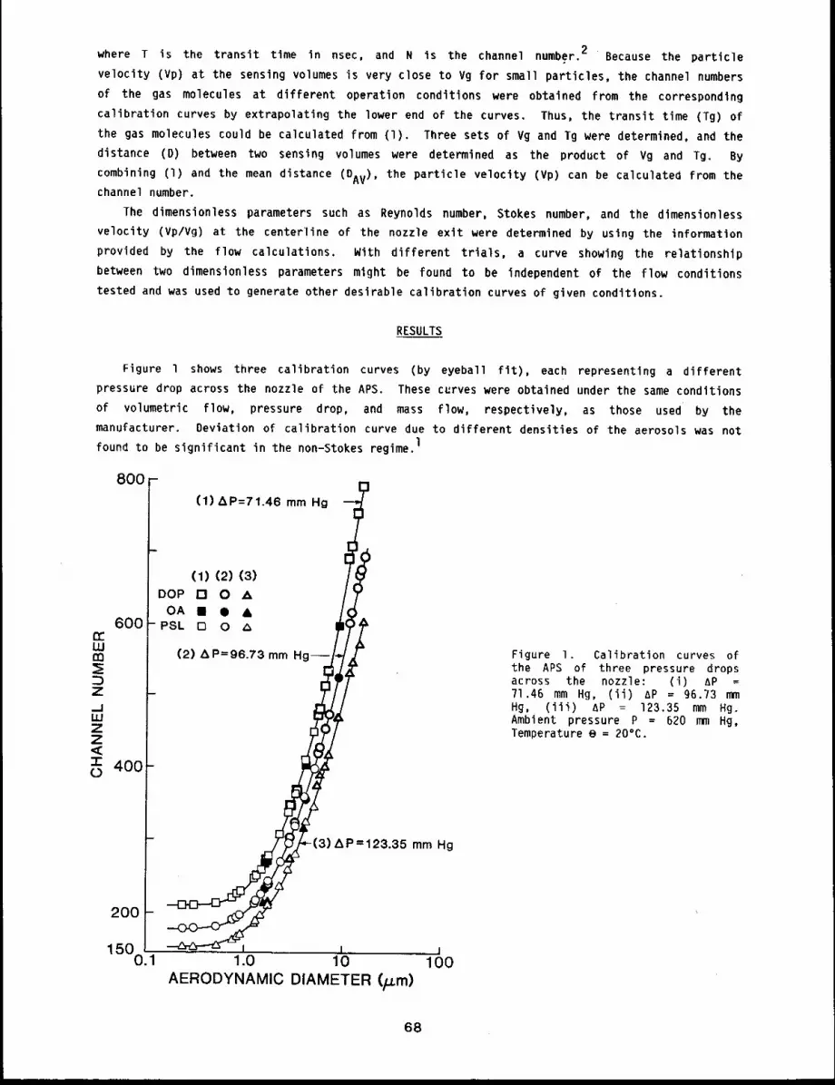
where T is the transit time in nsec, and N is the channel number. 2 Because the particle
velocity (Vp) at the sensing volumes is very close to Vg for small particles, the channel numbers
of the gas molecules at different operation conditions were obtained from the corresponding
calibration curves by extrapolating the lower end of the curves. Thus, the transit time (Tg)
the gas molecules could be calculated from (1). Three sets of Vg and Tg were determined, and the
distance (D) between two sensing volumes were determined as the product of Vg and Tg.
combining (I) and the mean distance (DAv), the particle velocity (Vp) can be calculated from
channel number.
The dimensionless parameters such as Reynolds number, Stokes number, and the dimensionless
velocity (Vp/Vg) at the centerline of the nozzle exit were determined by using the information
provided by the flow calculations, With different trials, a curve showing the relationship
between two dimensionless parameters might be found to be independent of the flow conditions
tested and was used to generate other desirable calibration curves of given conditions.
RESULTS
Figure 1 shows three calibration curves (by eyeball fit), each representing a different
pressure drop across the nozzle of the APS. These curves were obtained under the same conditions
of volumetric flow, pressure drop, and mass flow, respectively, as those used by the
manufacturer. Deviation of calibration curve due to different densities of the aerosols was not
found to be significant in the non-Stokes regime.1
800 -
600rrUJrn
ZlwZZ<3- 400(5
200
1500.1
(1) AP=71.46 mm
(1) (2) DOP [] 0 A
OA ̄ ¯ &PSL 0 0
(2) AP=96.73 mm
=123.35 mm Hg
i I i1.0 10 IO0
AERODYNAMIC DIAMETER(~m)
Figure I. Calibration curves ofthe APS of three pressure dropsacross the nozzle: (i) AP 71.46 mm Hg, (ii) AP = 96.73 Hg, (iii) AP = 123.35 mm Hg.Ambient pressure P = 620 mm Hg,Temperature e = 20°C.
68
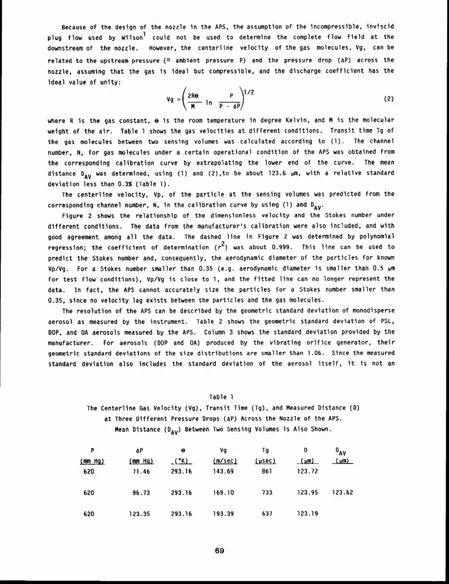
Because of the design of the nozzle in the APS, the assumption of the incompressible, inviscid
plug flow used by Wilson I could not be used to determine the complete flow field at the
downstream of the nozzle, However, the centerline velocity of the gas molecules, Vg, can be
related to the upstream pressure (= ambient pressure P) and the pressure drop (AP) across
nozzle, assuming that the gas is ideal but compressible, and the discharge coefficient has the
ideal value of unity:
Vg =In P - a
where R is the gas constant, e is the room temperature in degree Kelvin, and M is the molecular
weight of the air. Table l shows the gas velocities at different conditions. Transit time Tg of
the gas molecules between two sensing volumes was calculated according to (1). The channel
number, N, for gas molecules under a certain operational condition of the APS was obtained from
the corresponding calibration curve by extrapolating the lower end of the curve. The mean
distance DAV was determined, using (1) and (2),to be about 123.6 ~m, with a relative standard
deviation less than 0.3% (Table l).
The centerline velocity, Vp, of the particle at the sensing volumes was predicted from the
corresponding channel number, N, in the calibration curve by using (I) and DAV.
Figure 2 shows the relationship of the dimensionless velocity and the Stokes number under
different conditions. The data from the manufacturer’s calibration were also included, and with
good agreement among all the data. The dashed line in Figure 2 was determined by polynomial
regression; the coefficient of determination (r 2) was about o.ggg. This line can be used to
predict the Stokes number and, consequently, the aerodynamic diameter of the particles for known
Vp/Vg. For a Stokes number smaller than 0.35 (e.g. aerodynamic diameter is smaller than 0.5 ~m
for test flow conditions), Vp/Vg is close to l, and the fitted line can no longer represent the
data. In fact, the APS cannot accurately size the particles for a Stokes number smaller than
0.35, since no velocity lag exists between the particles and the gas molecules.
The resolution of the APS can be described by the geometric standard deviation of monodisperse
aerosol as measured by the instrument. Table 2 shows the geometric standard deviation of PSL,
DOP, and OA aerosols measured by the APS. Column 3 shows the standard deviation provided by the
manufacturer. For aerosols (DOP and OA) produced by the vibrating orifice generator, their
geometric standard deviations of the size distributions are smaller than 1.06. Since the measured
standard deviation also includes the standard deviation of the aerosol itself, it is not an
Table 1
The Centerline 6as Velocity (Vg), Transit Time (Tg), and Measured Distance
at Three Different Pressure Drops (AP) Across the Nozzle of the APS.
Mean Distance (DAv) Between Two Sensing Volumes is Also Shown.
P AP e Vg Tg O DAV
(~Hg) ~ _(=K) (m/sec} (.sec) (.m) (um)
620 71.46 293.16 143.69 861 123.72
620 96.73 293.16 169.10 733 123.95 123.62
620 123.35 293.16 193.39 637 123.19
69
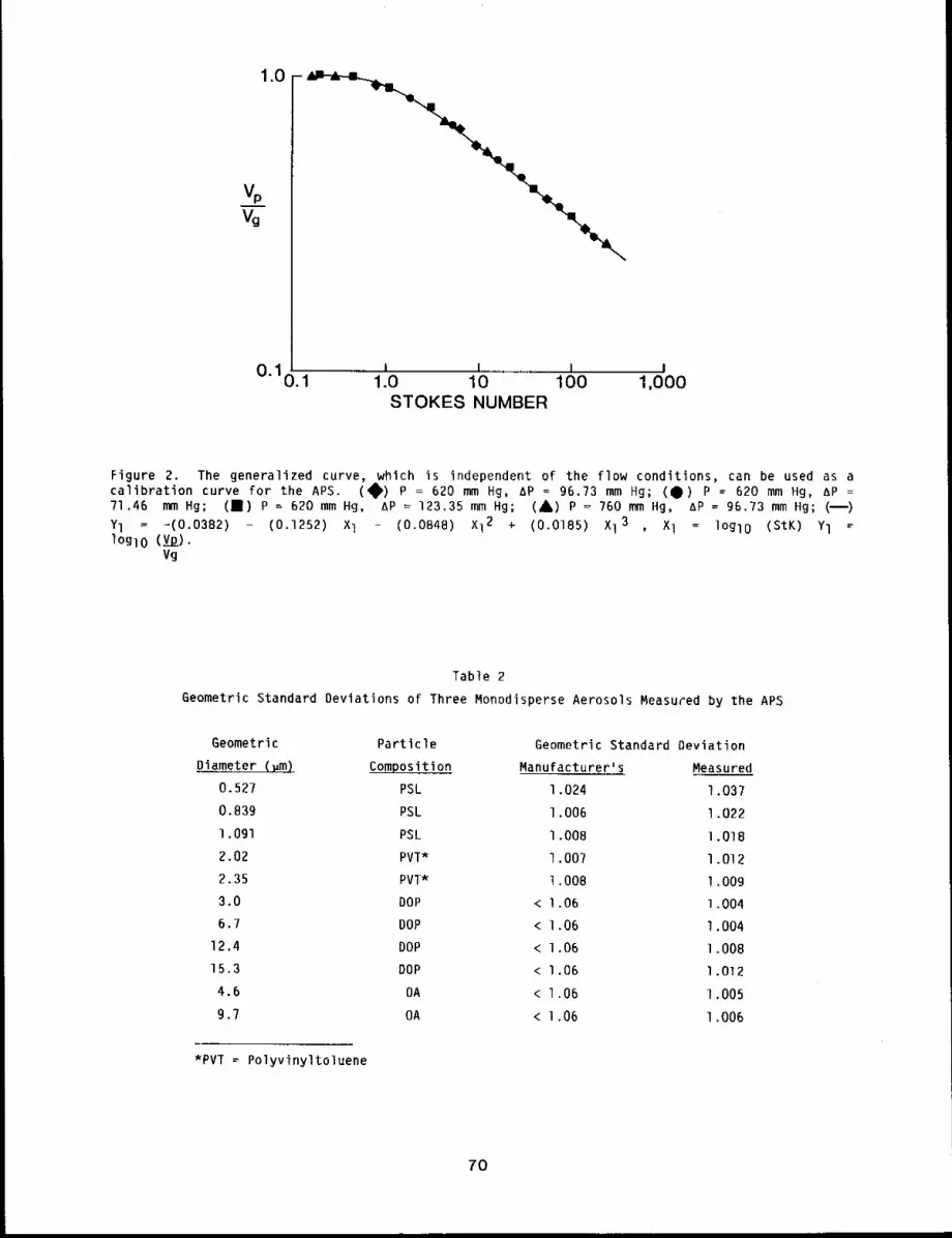
1.0
0"10 1i J j i
. 1.0 10 100 1,000STOKES NUMBER
Figure 2. The generalized curve, which is independent of the flow conditions, can be used as acalibration curve for the APS. (~) P = 620 mm Hg, AP -- 96.73 mm Hg; (4)) P = 620 mm Hg, =
71.46 mm Hg; (1) = 620 mmHg,AP =123.3 5 mm Hg; ( A) P = 76 0 mmHg, AP = 96 .73 mmHg; (B
Yl = -(0.0382) - (0.1252) l - (0 .0848) Xl 2 + (0 .0185) Xl 3 , Xl = l OglO (St K) Yl log10 (vp).
Vg
Table 2
Geometric Standard Deviations of Three Monodisperse Aerosols Measured by the APS
Geometric Particle Geometric Standard Deviation
Diameter (~m) Composition Manufacturer’s Measured
0.527 PSL 1.024 1.037
0.839 PSL 1.006 1.022l.Ogl PSL 1.008 l.OIB
2,02 PVT* 1.007 1.0122.35 PVI* 1.008 1.009
3.0 DOP < 1.06 1.004
6.7 DOP < 1.06 1.00412.4 DOP < 1.06 1.00815.3 DOP < 1.06 1.0124.6 OA < 1.06 1.005
9.7 OA < 1.06 1.006
*PVI’ = Polyvinyltoluene
7O
indication of the absolute resolution of the APS. However, the APS does show good resolution for
monodisperse PSL, DOP, and OA aerosols that are normally used for instrumentation calibration.
DISCUSSION
It was shown that rapid, in situ measurements of aerodynamic diameter between 0.5 ~m and
16 ~m can be made, with good resolution, using the APS. Uncertainty of the size measurement
caused by the difference of the particle density in the non-Stokes regime was not found to be
significant among test aerosols.
The calibration curves of the APS under different operational conditions were combined and
represented by one unique curve that can predict calibration curves of other desirable
conditions. Further, the procedure described here can be used in any APS to determine its own
unique curve, which can then be used to predict the aerodynamic size distribution of the aerosol
under reasonable operational conditions. Moreover, responses related to irregular or aggregate
particles at the downstream of the accelerating nozzle can be studied because the velocity of the
particles and the air molecules can be predicted well.
REFERENCES
I. Wilson, J. C., Aerodynamic Particle Size Measurement by LaserThesis, University of Minnesota, Minneapolis, MN, 1978.
DQRpler Velocimetry, Ph.D.
2. Aerodynamic Particle Size Ana!~.zer, Instruction Manual, Thermo-System Inc., St. Paul, MN, 1983.
71
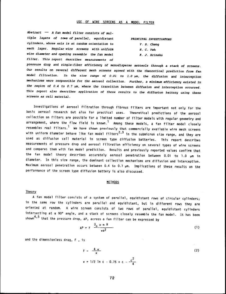
USE OF WIRE SCREENS AS A MODEL FILTER
Abstract -- A fan model f11ter consists of mul-
tiple layers of rows of parallel, equidistant
cylinders, whose axls is at random orlentat~on to
each layer. Regular wire screens wlth unlform
wlre diameter and opening resemble the fan model
filter. Thls report descrlbes measurements of
PRINCIPAL INVESTIGATORS
Y. S. Cheng
H. C. Yeh
K. J. Brlnsko
pressure drop and slngle-flber eff~clencg of monodlsperse aerosols through a stack of screens.
Our results on several different mesh screens agreed wlth the theoretlcal prediction from fan
model filtration. In the size range of 0.01 to 1.0 ~uD, the diffusion and interception
mechanisms were responsible for the aerosol collection. Further, a minimum efficiency existed In
the region of 0.4 to 0,7 ~m, where the trans~tlon between dlffuslon and interception occurred.
Thls report also describes app11catlon of these results to the dlffus~on battery using these
screens as cell material,
Investigations of aerosol filtration through fibrous filters are important not only for the
basic aerosol research but also for practical uses. Theoretical predictions of the aerosol
collection on filters are possible for a limited number of filter models wlth regular geometry and
arrangement, where the flow field is known, l Among these models, a fan filter model closely
resembles real filters, l We have shown previously that commercially available wire mesh screens
with uniform diameter behave like fan model filters 2’3 in the submicron size range, and they are
used as diffusion cell material in screen type diffusion batteries. This report describes
measurements of pressure drop and aerosol filtration efficiency on several types of wire screens
and compares them with fan model prediction. Results and previously reported values confirm that
the fan model theory describes accurately aerosol penetration between O.Ol to l.O ~m in
diameter. In this size range, the dominant collection mechanisms are diffusion and interception.
Maximum aerosol penetration occurs between 0.4 to 0.? pm. Implications of these results on the
performance of the screen type diffusion battery is also discussed.
METHODS
Theory
A fan model filter consists of a system of parallel, equidistant rows of circular cylinders;
in the same row the cylinders are parallel and equidistant, but in different rows they are
oriented at random. A wire screen consists of two rows of parallel, equidistant cylinders
intersecting at a 90° angle, and a stack of screens closely resemble the fan model. It has been
shown4’5 that the pressure drop, AP, across a fan filter can be expressed by
U ~ ~ HAP = F o (1)
~a2
and the dimensionless drag, F , is
2K = i/2 In c - 0.?5 + c - c
4
72
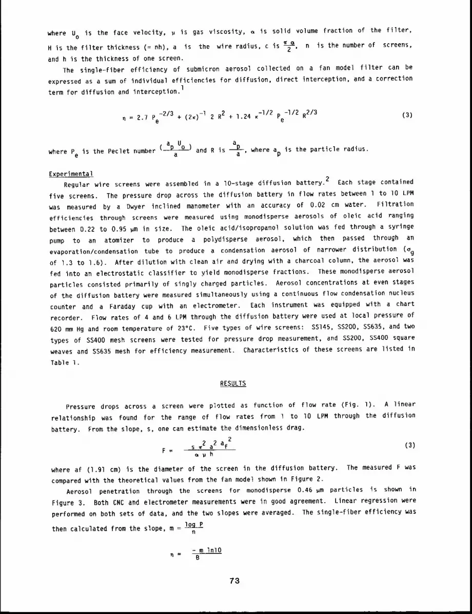
where U is the face velocity, ~ is gas viscosity, ~ is solid volume fraction of the filter,o
H is the filter thickness (= nh), a is the wire radius, c is ~, n is the number of screens,
and h is the thickness of one screen,
The single-fiber efficiency of submicron aerosol collected on a fan model filter can be
expressed as a sum of individual efficiencies for diffusion, direct interception, and a correction
term for diffusion and interception.1
n = 2.7 Pe-213 + (2K)-i 2 R2 + 1.24 K-I/2 P =I12 R213 (3)
e
awhere Pe is the Peclet number ( p u )a and R is ~a ’ where ap is the particle radius.
Experimental 2
Regular wire screens were assembled in a lO-stage diffusion battery. Each stage contained
five screens. The pressure drop across the diffusion battery in flow rates between 1 to lO LPM
was measured by a Dwyer inclined manometer with an accuracy of 0.02 cm water. Filtration
efficiencies through screens were measured using monodisperse aerosols of oleic acid ranging
between 0.22 to 0.95 ~m in size. The oleic acid/isopropanol solution was fed through a syringe
pump to an atomizer to produce a polydisperse aerosol, which then passed through an
evaporation/condensation tube to produce a condensation aerosol of narrower distribution (~gof 1.3 to 1.6). After dilution with clean air and drying with a charcoal column, the aerosol was
fed into an electrostatic classifier to yield monodisperse fractions. These monodisperse aerosol
particles consisted primarily of singly charged particles. Aerosol concentrations at even stages
of the diffusion battery were measured simultaneously using a continuous flow condensation nucleus
counter and a Faraday cup with an electrometer. Each instrument was equipped with a chart
recorder. Flow rates of 4 and 6 LPM through the diffusion battery were used at local pressure of
620 mm Hg and room temperature of 23°C. Five types of wire screens: SS145, SS200, SS635, and two
types of SS400 mesh screens were tested for pressure drop measurement, and SS200, SS400 square
weaves and SS635 mesh for efficiency measurement. Characteristics of these screens are listed in
Table 1.
RESULTS
Pressure drops across a screen were plotted as function of flow rate (Fig. 1). A linear
relationship was found for the range of Flow rates from 1 to lO LPM through the diffusion
battery. From the slope, s, one can estimate the dimensionless drag.
2s 2 a2 af (3)
F= ~ph
where af (l.gl cm) is the diameter of the screen in the diffusion battery. The measured F was
compared with the theoretical values from the fan model shown in Figure Z.
Aerosol penetration through the screens for monodisperse 0.46 gm particles is shown in
Figure 3. Both CNC and electrometer measurements were in good agreement. Linear regression were
performed on both sets of data, and the two slopes were averaged. The single=fiber efficiency was
logthen calculated from the slope, m = n
- m InlOn- B
78
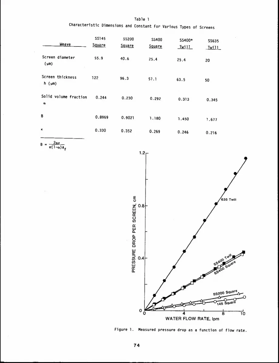
Table 1
Characteristic Dimensions and Constant for Various Types of Screens
Weave
SS145 SS200 SS400 SS400- SS635Square S__q_uare ~ Twill Twill
Screen diameter
(.m)
55.9 40.6 25,4 25.4 20
Screen thickness
h (~m)
122 96.3 57.1 63.5 50
Solid volume fraction 0.244 0.230 0.292 0.313 0.345
B = ....
0.8969 0.9021 1.180 1.450 1.677
0.330 0.352 0.269 0.246 0.216
Figure I.
0 I I I I0 4 8 10
WATER FLOW RATE, Ipm
Measured pressure drop as a function of flow rate.
74
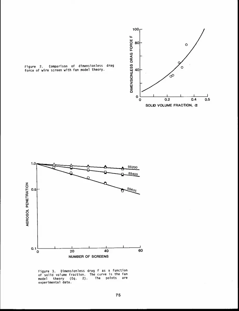
Figure 2. Comparison of dimensionless dragforce of wire screen with fan model theory,
100 -
0
0
I I I ....... i ]0 0.2 0.4 0.5
SOLID VOLUME FRACTION, r/
1.0
0.5n-
0,10
I I I I I20 40
NUMBER OF SCREENS
I60
Figure 3. Dimensionless drag F as a functionof solid volume fraction. The curve is the fanmodel theory (Eq. 2). The points areexperimental data,
75
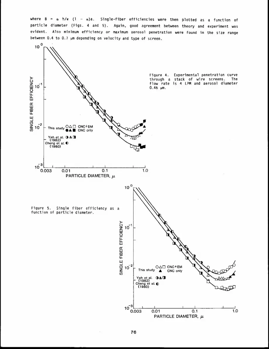
where B -- ~ h/~ (I - ~)a. Single-fiber efficiencies were then plotted as a function
particle diameter (Figs. 4 and 5). Again, good agreement between theory and experiment was
evident. Also minimum efficiency or maximum aerosol penetration were found in the size range
between 0.4 to 0.7 ~m depending on velocity and type of screen.
o1o
@0n [] CNC+EMThis study I CNC only
Yeh et al. @A[](1982)
3heng et al. tD(1980)
0.01 0.1PARTICLE DIAMETER,
10o
Figure 4. Experimental penetration curvethrough a stack of wire screens. Theflow rate is 4 LPM and aerosol diameter0.46 ~m.
Figure 5. Single fiber efficiency as afunction of particle diameter.
O~I-I CNC+EMThis study A CNC only
Yeh et al. @All(1982)
Cheng et al. ~)(1980)
I I I I0.01 0,1 1.0PARTICLE DIAMETER, /J.
76

DISCUSSION
Our results show that the tested screen fit the description of the fan model. The pressure
drop across the screen mesh agreed wlth the fan model only qualitatively because it is shown to be
functions of ~ and the ratio of distance between neighboring screens.4 In the case of wire
screen, this ratio approached zero. However, the measured single fiber efficiencies agreed with
the theoretical prediction within ± 15% for entire size range between O.Ol to about l ~m. In
this range, the interception and diffusion mechanism dominated the aerosol collection. Further,
between 0.4 to 0.7 wm minimum collection efficiency points were shown to be a function of both
screen type and flow rate; thus, single-fiber efficiency is a double-valued curve.
Implications of our results in the diffusion battery where these screens are used, are: l)
the fan model can be used as the calibration curve, 2) the range of operation can be extended to
0.4 to 0.7 ~m depending on the screen type and flow rate, and 3) a precutter with cutoff
diameters at minimum collecting size is needed to minimize the effects of large particles.
REFERENCES
I. Kirsch, A. A. and I. B. Stechkina, The Theory of Aerosol Filtration with Fibrous Filters, inFundamentals of Aerosol Science, Wiley, New York, NY, pp. 165-256, 197B.
2. Cheng, Y. S., J. A. Keating, and G. M. Kanapilly, Theory and Calibration of a Screen-TypeDiffusion Battery, 3. Aerosol Sci. l l: 549-556, 19BO.
3. Yeh, H. C., Y. S. Cheng, and M. M. Orman, Evaluation of Various Types of Wire Screens asDiffusion Battery Cells, O. Colloid Interface Sci. 86: 12-16, 1982.
4. Kirsch, A. A. and N. A. Fuchs, Studies of Fibrous Aerosol Filters ~ II. Pressure Drops inSystems of Parallel Cylinders, Ann. Occup~ H~g. lO: 23-30, 1967.
5. Kirsch, A. A. and N. A. Fuchs, Studies of Fibrous Aerosol Filters - III. DiffusionalDeposition of Aerosols in Fibrous Filters, Ann. Occup. Hyg. l l: 299-304, 196B.
77/78

/N VITRO PREDICTORS OF TOXICITY
In vitro systems are being used increasingly as relatively rapid and less time-consuming means
of predicting the potential toxicity to man from exposure to environmental pollutants. The II
papers in this section report on studies that have used in vitro techniques to determine factors
that may be important in determining the ultimate toxicity of materials to man.
In the first three papers of the section, the Salmonella mutagenicity assay system has been
used in conjunction with chemical analysis to examine the formation and differences in the
chemical composition of the specific chemical compounds that appear to be responsible for the
mutagenic activity of diesel exhaust, coal gasification tars, and coal oil. Addition of selected
polycyclic hydrocarbons to the fuel did influence the resulting mutagenicity of the exhaust from a
diesel engine using the fuel. The mutagenicity of the exhaust was also increased by adding
l-nitropyrene or 3-nitropyrene to the fuel, but the mutagenicity of the exhaust was not Increased
by adding pyrene or fluoranthene to the fuel. The contribution of primary amines to the
mutagenicity of gasifier tar and coal oil was determined to be greater for coal oil. These
studies also illustrate the relatively high contribution of nitrogen containing organics (i.e.,
nitropyrene) to the mutagenic activity in bacterial in vitro systems from these complex chemical
mixtures.
The fourth paper reports on studies designed to determine the rate of breakdown of two
chemicals (benzo[a]pyrene and nitropyrene) often present in combustion effulents in the
environment. Nitropyrene breakdown was faster than that for benzo[a]pyrene.
The fifth and sixth papers report on in vitro studies of nitropyrene metabolism by mammalian
tissues. These studies showed that rat liver, lung, and nasal tissues do have the capacity for
bioactivation of nitropyrene. The studies provide an important link between the in vitro studies
with bacteria and the ultimate toxicity of this class of compounds in in vivo mammalian tests for
toxicity (e.g., carcinogenicity).
Nasal tissues have been identified as important in the respiratory tract for the metabolism of
inhaled compounds. The seventh paper reports on the potential inhibition of this metabolism by 24
compounds likely to be inhaled by man.
Formaldehyde toxicity in man has received increased attention in the past few years. The
eighth paper reports on studies of the reaction of formaldehyde with reduced glutathione in tissue
homogentates and isolated perfused lung and liver. Formaldehyde decreased the level of reduced
glutathione in isolated perfused liver, but not in isolated perfused lung or tissue homogenates.
These studies could provide insight into possible mechanisms of some types of formaldehyde
toxicity.
The next two papers report on the use of cell cultures of Chinese hamster ovary cells to study
factors involved in the mutagenicity of environmental pollutants. In the first, an antiprotease
was shown to affect the mutagenic response of these cells to benzo[a]pyrene. In the second, the
studies suggest that radiation-induced "hypermutability" may be specific for selected chemicals
and biological endpoints.
The final paper in the section reports on the induction of sister chromatid exchanges in vivo
and In vitro in lung cells after exposure to the potent mutagen 3-methylcholanthrene. Sister
chromatid exchanges were induced by this compound both after in vitro exposure of cultured cells
and in cells isolated from animals exposed by inhalation.
79180

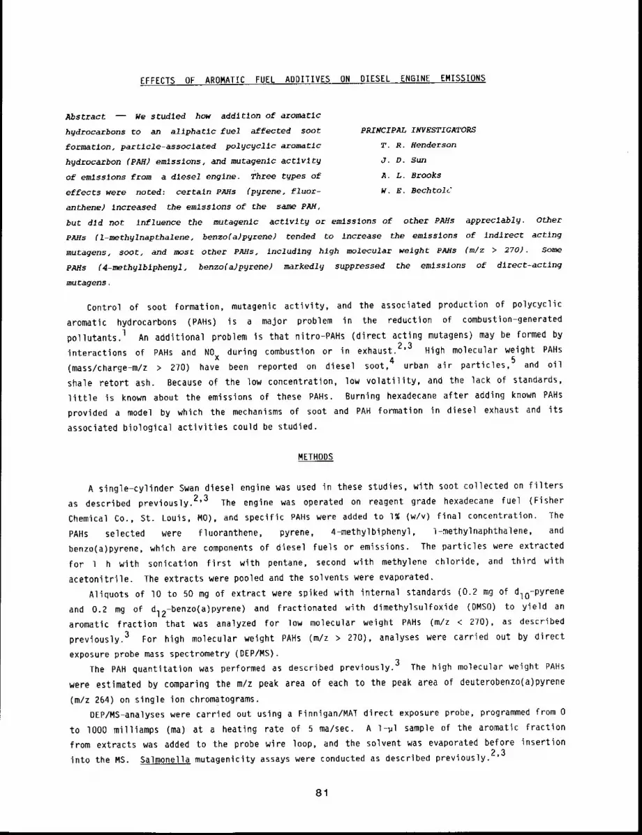
EFFECTS OF AROMATIC FUEL ADDITIVES ON DIESEL ENGINE EMISSIONS
Abstract -- We studled how addltlon of aromatlc
hydrocarbons to an aliphatlc fuel affected soot PRINCIPAL INVESTIGATORS
formatlon, partlcle-assoclated pol~c~cllc aromatic T.R. Henderson
hydrocarbon (PAH) emlsslons, and mutagenlc actlv~ J.D. Sun
of emlsslons from a dlesel englne. Three types of A.L. Brooks
effects were noted: certaln PAHs (pyrene, fluor- w.E. BechtolU
anthene) increased the emlsslons of the same PAH,
but dJd not influence the mutagenlc actlvlty or emlsslons of other PAHs appreclabl~. Other
PAHs (l-meth~lnapthalene~ benzo(a)pyrene) tended to increase the emlss~ons of indlrect actlng
mutagens, soot, and most other pAHs, includlng hlgh molecular weight PAMs (m/z > 270). Some
PAHs (4-methylblphen~l, benzo(a)pyrene) markedly suppressed the emlsslons of dlrect-actlng
mutagens.
Control of soot formation, mutagenic activity, and the associated production of polycyclic
aromatic hydrocarbons (PAHs) is a major problem in the reduction of combustion-generated
pollutants.l An additional problem is that nitro-PAHs (direct acting mutagens) may be formed
interactions of PAHs and NO during combustion or in exhaust.2’3 High molecular weight PAHsx
(mass/charge-m/z > 270) have been reported on diesel soot,4 urban air particles, 5 and oil
shale retort ash. Because of the low concentration, low volatility, and the lack of standards,
little is known about the emissions of these PAHs. Burning hexadecane after adding known PAHs
provided a model by which the mechanisms of soot and PAH formation in diesel exhaust and its
associated biological activities could be studied.
METHODS
A single-cylinder Swan diesel engine was used in these studies, with soot collected on filters
as described previously. 2’3 The engine was operated on reagent grade hexadecane fuel (Fisher
Chemical Co., St. Louis, MO), and specific PAHs were added to l~ (w/v) final concentration.
PAHs selected were fluoranthene, pyrene, 4-methylbiphenyl, 1-methylnaphthalene, and
benzo(a)pyrene, which are components of diesel fuels or emissions. The particles were extracted
for 1 h with sonication first with pentane, second with methylene chloride, and third with
acetonitrile. The extracts were pooled and the solvents were evaporated.
Aliquots of lO to 50 mg of extract were spiked with internal standards (0.2 mg of dlo-pyrene
and 0.2 mg of dl2-benzo(a)pyrene) and fractionated with dimethylsulfoxide (DMSO) to yield
aromatic fraction that was analyzed for low molecular weight PAHs (m/z < 270), as described
previously. 3 For high molecular weight PAHs (m/z > 270), analyses were carried out by direct
exposure probe mass spectrometry (DEP/MS).
The PAB quantitatlon was performed as described previously. 3 The high molecular weight PAHs
were estimated by comparing the m/z peak area of each to the peak area of deuterobenzo(a)pyrene
(m/z 264) on single ion chromatograms.
DEP/MS-analyses were carried out using a Finnigan/MAl direct exposure probe, programmed from 0
to 1000 milliamps (ma) at a heating rate of 5 ma/sec. A 1-~l sample of the aromatic fraction
from extracts was added to the probe wire loop, and the solvent was evaporated before insertion
into the MS. Salmonella mutagenicity assays were conducted as described previously.2’3
81
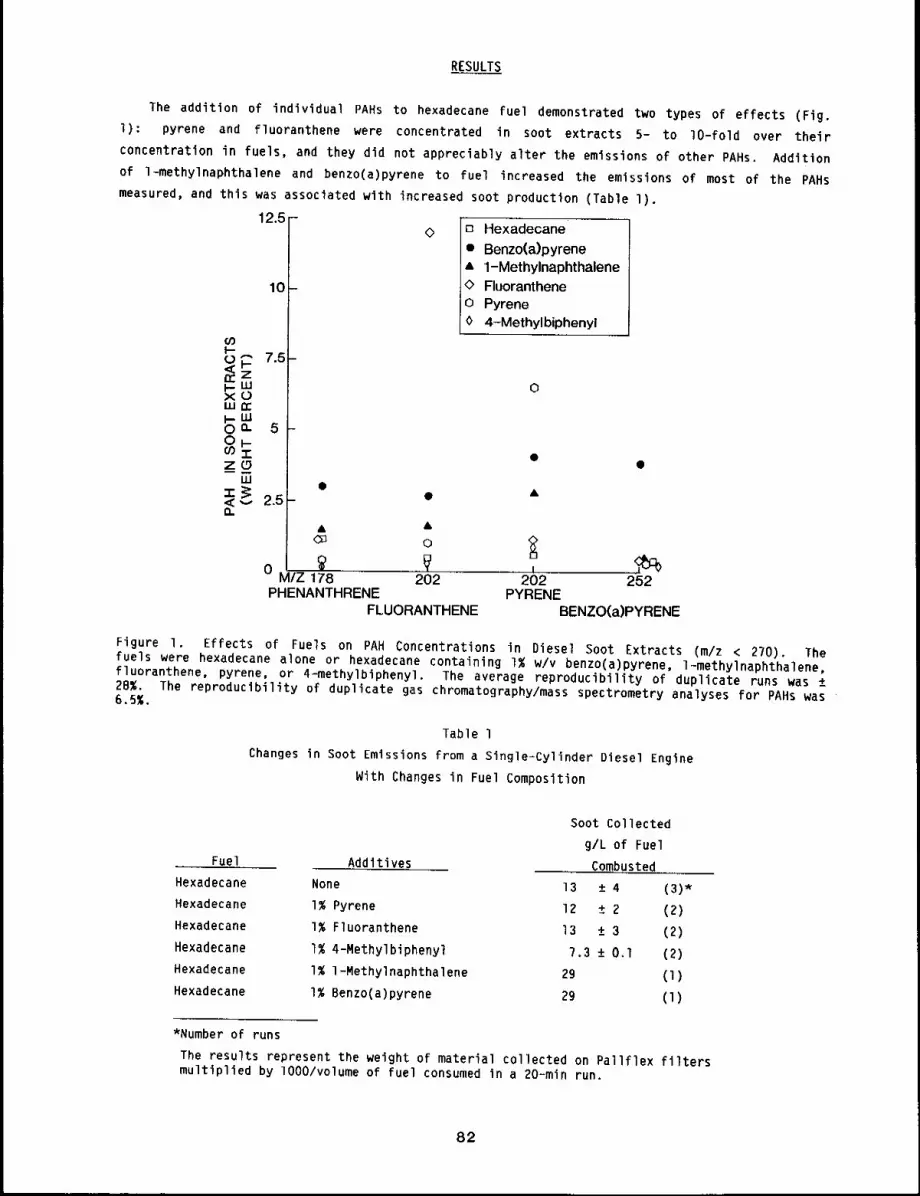
RESULTS
The addition of individual PAHs to hexadecane fuel demonstrated two types of effects (Fig.
l): pyrene and fluoranthene were concentrated in soot extracts 5- to lO-fold over their
concentration in fuels, and they did not appreciably alter the emissions of other PAHs. Addition
of l-methylnaphthalene and benzo(a)pyrene to fuel increased the emissions of most of the PAHs
measured, and this was associated with increased soot production (Table 1).
12.5 -
10-
cOI--
n,- <O~ 7.5-I--xOwn,"
0 50
zow
2.5&
C~
0 ~ ,M/Z 178
PHENANTHRENE
O
&0
,,~202
FLUORANTHENE
[3 Hexadecane
¯ Benzo(a)pyrene’= 1-Methylnaphthalene
Fluorantheneo Pyrene0 4-Methylbiphenyl
202 252PYRENE
BENZO(a)PYRENE
Figure I. Effects of Fuels on PAH Concentrations in Diesel Soot Extracts (m/z < 270). Thefuels were hexadecane alone or hexadecane containing I% w/v benzo(a)pyrene, l-methylnaphthalene,fluoranthene, pyrene, or 4-methylbiphenyl. The average reproducibility of duplicate runs was ±28~. The reproducibility of duplicate gas chromatography/mass spectrometry analyses for PAHs was6.5%.
Table 1Changes in Soot Emissions from a Single-Cylinder Diesel Engine
With Changes in Fuel Composition
Fuel Additives
Soot Collected
g/L of Fuel
CombustedHexadecane None 13 ± 4 (3)*Hexadecane I% Pyrene 12 ± 2 (2)Hexadecane 1% Fluoranthene 13 ± 3 (2)Hexadecane I% 4-Methylbiphenyl 7.3 ± 0.1 (2)Hexadecane l~ l-Methylnaphthalene 29 (1)Hexadecane I% Benzo(a)pyrene 29 (1)
*Number of runs
The results represent the weight of material collected on Pallflex filtersmultiplied by lO00/volume of fuel consumed in a 20=min run.
82
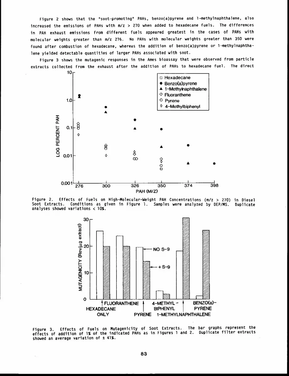
Figure 2 shows that the "soot-promoting" PAHs, benzo(a)pyrene and l-methylnaphthalene, also
increased the emissions of PAHs with m/z > 270 when added to hexadecane fuels. The differences
in PAH exhaust emissions from different fuels appeared greatest in the cases of PAHs with
molecular weights greater than m/z 276. No PAHs with molecular weights greater than 350 were
found after combustion of hexadecane, whereas the addition of benzo(a)pyrene or l-methylnaphtha-
lene yielded detectable quantities of larger PAHs associated with soot.
Figure 3 shows the mutagenic responses in the Ames bioassay that were observed from particle
extracts collected from the exhaust after the addition of PAHs to hexadecane fuel. The direct
10[] Hexadecane¯ Benzo(a)pyrene¯ 1-Methylnaphthalene
tt @ Fluoranthene1.0 o Pyrene
¯ 0 4-Methylbiphenyl&
-r,,( ¯
~- 0.1 &Z ¯LU0 0n"LU13_
~ ¯ ¯(.9o o.oi o $
013
0.001 ~ ~ i i I I276 300 326 350 374 398
PAH (M/Z)
Figure 2. Effects of Fuels on High-Molecular-Weight PAH Concentrations (mlz > 270) in DieselSoot Extracts. Conditions as given In Figure l. Samples were analyzed by DEP/MS. Duplicateanalyses showed variations < I0%.
30-
0
XQ}
-:;=,
¢)E>-F’-z 10-
o
CCC~
~4
NN N
l FLUORANTHENEHEXADECANE
ONLY
"--- NO S-9
+s-9
4-METHYL-BIPHENYL
y//
i///
/i/i////
//1"/71%
///i//11////
BENZO(a)-PYRENE
PYRENE 1-METHYLNAPHTHALENE
Figure 3. Effects of Fuels on Mutagenicity of Soot Extracts. The bar graphs represent theeffects of addition of I% of the indicated PAHs as in Figures l and 2. Duplicate filter extractsshowed an average variation of ± 41%.
83
mutagenicity (no liver S-9 enzymes added) was altered least by the addition of pyrene,
l-methylnaphthalene, or fluoranthene to fuel, but was suppressed by the addition of methylbiphenyl
or b~nzo(a)pyrene. The combustion of hexadecane fuel containing l-methylnaphthalene
benzo(a)pyrene resulted In increased indirect mutagenicity in soot extracts.
DISCUSSION
In characterization of diesel emissions, it has been noted that aromatic hydrocarbons increase
the smoking tendencies of fuels (naphthalenes > benzenes > olefins > paraffins), l and that
reduction of the aromatic content of diesel fuels reduces soot emissions I In this study" m
combustion of aliphatic fuels spiked with PAHs was used to estimate soot formation and to
determine which PAHs were formed during diesel combustion processes. The addition of known PAHs
allowed ranking of their relative resistance to combustion.
Pyrene and fluoranthene in the fuel resulted in the formation of nitro-PAHs, which are known
to have high mutagenic specific activity and may be responsible for much of the direct-acting
mutagenic activities detected by the Ames bioassay. 3 That increased emissions of these PAHs was
not associated with increased mutagenicity is surprising, but may be due to only limited amounts
of NOx being available for reaction in exhaust streams.2 This agrees with the finding that
addition of nitropyrene or nitrofluoranthene to diesel fuel increases the emissions of nitro-PAHs
and the direct mutagenicity of extracts (this report, pp. 86 to 90). The addition
4-methylbiphenyl to fuel resulted in very low mutagenicity in extracts, as observed with
phenanthrene supplemented fuel, 4 and may reflect the formation of nitro-PAHs that are not as
stable under exhaust and collection conditions.3
Previously, benzo(a)pyrene and l-methylnaphthalene (and to a lesser extent acenaphthene)
found to increase twofold to fourfold the soot emissions/volume of fuel combusted. 3 The present
study showed that combustion of these PAHs increased the emission of high molecular weight PAHs
detected by DEP. The addition of liver enzymes (S-g) to these extracts increased the mutagenic
activity and thus appears to reflect the increased concentrations of benzo(a)pyrene and other high
molecular weight PAHs (Fig. 2). The increased concentrations of other PAHs in soot extracts from
the combustion of l-methylnaphthalene or benzo(a)pyrene may be due to intermediates in soot
formation that are proposed to be reactive aromatic moieties (ions, radicals, etc.) in equilibrium
with PAHs.3
Previous methods for detection of high molecular weight PAHs (m/z > 270) have used
concentration by column chromatography followed by high-pressure liquid chromotography or high
temperature gas chromatography for detection.4’5 DMSO fractiona~ion of extracts followed by DEP
analyses, as reported here, is a simpler technique for screening combustion and/or environmental
samples for these compounds. It may be important to determine the biological activity of these
high molecular weight PAHs if they are present in significant concentrations.
REFERENCES
I. Levy, A., Unresolved Problems in SOx, NOx and Soot Control, in Combustion, .l.9th S~mposiumon Combustion, The Combustion Institute, Pittsburgh, PA, p. 1223, 1983.
2. Henderson, T. R., J. D. Sun, R. E. Royer, C. R. Clark, A. P. Li, T. M. Harvey, and D. F. Hunt,Triple Quadrupole Mass Spectrometry Studies of Nitroaromatic Emissions from Different DieselEngines, Environ. Sci. Technol. 17: 443-449, 1983.
84

3. Henderson, T. R., 3. O. Sun, A. P. Lt, R. L. Hanson, W. E. Bechtold, 3. S. Dutcher, T. M.Harvey, J. Shabonowitz, and O. F. Hunt, GC/MS and MS/MS Studies on Diesel Exhaust Mutagenlcltyand Emlssions from Chemically Defined Fuels, Environ. Sct. Technol. (in press) 1983.
4. Oenoyer, E., Ph. O. Oissertatlon, Colorado State University, 1982.
5. Romanowski, T., W. Funcke, 3. Koenig, and E. Balfanz, Isolation of Polycyclic AromaticHydrocarbons in Air Particulate Matter by Liquid Chromatography, Anal. Chem. 54: 1285-1287,1983.
85
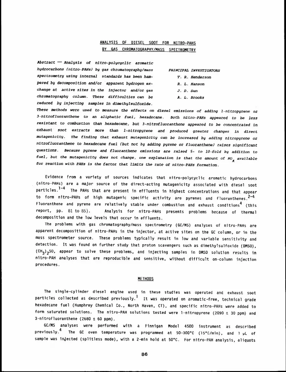
ANALYSIS OF DIESEL SOOT FOR NITRO-PAHS
BY GAS CHROMATOGRAPHY/MASS SPECTROMETRY
Abstract --Analysis of nltro-po19cycllc aromatic
hydrocarbons (nltro-PAHs) by gas chromatography/mass PRINCIPAL INVESTIGATORS
spectrometry using Internal standards has been ham- T.R. Hendersonpered by decomposition and/or apparent hydrogen ex- R.L. Hansonchange at active sites in the Injector and/or gas J.D. Sun
chromatography column. These dlff~cultles can be A.L. Brooks
reduced by inJectlng samples in dlmethy1sulfoxlde.
These methods were used to measure the effects on diesel emissions of adding l-nltrop~rene ol
3-nltrofluoranthene to an ailphatlc fuel, hexadecane. Both nltro-PAHs appeared to be less
resistant to combustion than hexadecane, but 3-nltrofluoranthene appeared to be concentrated ~n
exhaust soot extracts more than l-nltropyrene and produced greater changes in direct
mutagenlclty. The finding that exhaust mutagenlclty can be increased by adding nltropyrene or
n~trofluoranthene to hexadecane fuel (but noc by adding pyrene or fluoranthene) raises slgn~flcant
questions. Because p~rene and fluoranthene emlss~ons are raised 5- to 10-fold by addition to
fuel, but the mutagen~clty does not changes one explanation is that the amount of NO availableX
for reaction with PAHs is the factor that limits the rate of nitro-PAHs formation.
Evidence from a variety of sources indicates that nitro-polycyclic aromatic hydrocarbons
(nitro-PAHs) are a major source of the dlrect-acting mutagenicity associated with diesel soot
particles.I-4The PAHs that are present in effluents in highest concentrations and that appear
to form nitro-PAHs of high mutagenlc specific activity are pyrenes and fluoranthenes.2-4
Fluoranthene and pyrene are relatively stable under combustion and exhaust conditions 4 (this
report, pp. 81 to 85). Analysis for nitro-PAHs presents problems because of thermal
decomposition and the low levels that occur in effluents.
The problems with gas chromatography/mass spectrometry (GC/MS) analyses of nitro-PAHs are
apparent decomposition of nitro-PAHs in the injector, at active sites on the GC column, or in the
mass spectrometer source. These problems typically result in low and variable sensitivity and
detection. It was found on further study that proton scavengers such as dimethylsulfoxide (DMSO),
(CH3)2S0, appear to solve these problems, and injecting samples in DMSO solution results
nitro-PAH analyses that are reproducible and sensitive, without difficult on-column injection
procedures.
METHODS
The single-cylinder diesel engine used in these studies was operated and exhaust soot
particles collected as described previously, l It was operated on aromatic-free, technical grade
hexadecane fuel (Humphrey Chemical Co., North Haven, CT), and specific nitro-PAHs were added
form saturated solutions. The nitro-PAH solutions tested were l-nitropyrene (2090 ± 30 ppm) and
3-nitrofluoranthene (2680 ± 60 ppm).
GC/MS analyses were performed with a Finnigan Model 4500 instrument as described
previously. 4 The GC oven temperature was programmed at 50-300°C (15°C/min), and l ~L
sample was injected (splitless mode), with a 2~min hold at 50°C. For nitro-PAH analysis, aliquots
86
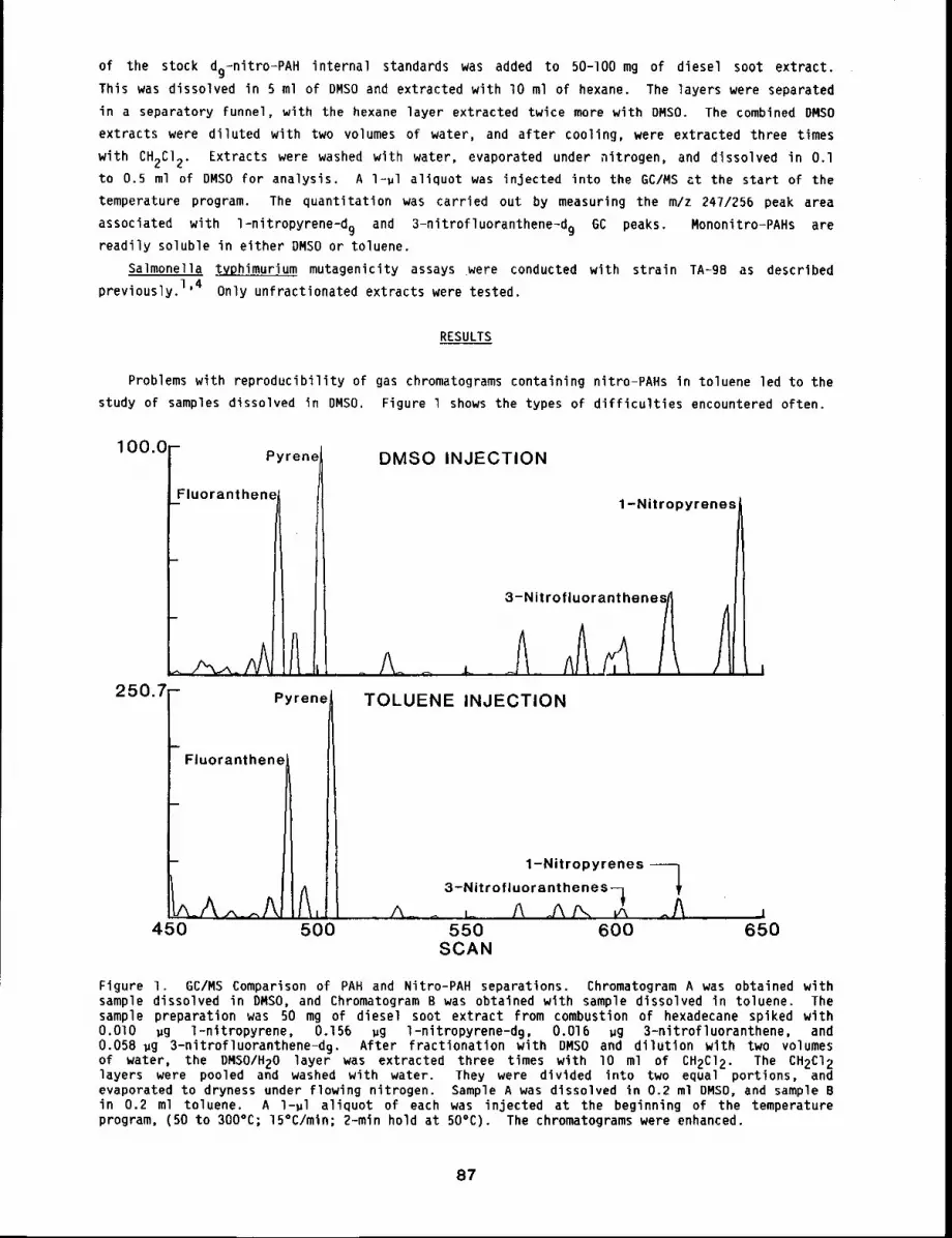
of the stock dg-nitro-PAH internal standards was added to 50-]00 mg of diesel soot extract.This was dissolved in 5 ml of DMSO and extracted with lO ml of hexane. The layers were separated
in a separatory funnel, with the hexane layer extracted twice more with DMSO. The combined DMSO
extracts were diluted with two volumes of water, and after cooling, were extracted three times
with CH2CI2. Extracts were washed with water, evaporated under nitrogen, and dissolved in O.lto 0.5 ml of DMSO for analysis. A l=~l aliquot was injected into the GC/MS at the start of the
temperature program. The quantitation was carried out by measuring the m/z 247/256 peak area
associated with l-nitropyrene-d 9 and 3-nitrofluoranthene-d9 GC peaks. Mononitro-PAHs arereadily soluble in either DMSO or toluene.
Salmonella t_V_phimurium mutagenicity assays were conducted with strain TA-98 as described
previously. 1’4 Only unfractlonated extracts were tested.
RESULTS
Problems with reproducibility of gas chromatograms containing nitro-PAHs in toluene led to the
study of samples dissolved in DMSO. Figure l shows the types of difficulties encountered often.
100,0 - Pyrene
Fluoranthene
250.7
Fluoranthene
Pyrene
DMSO INJECTION
1-Nitropyrenes/
3-Nitrofluorant henes/~
TOLUENE INJECTION
1-Nitropyrenes --1
3-Nitrofluoranthenes-~i,, A_ ,_ ,. A ¯450 500 550 600 650
SCAN
Figure I. GC/MS Comparison of PAH and Nitro-PAH separations. Chromatogram A was obtained withsample dissolved in DMSO, and Chromatogram B was obtained with sample dissolved in toluene. Thesample preparation was 50 mg of diesel soot extract from combustion of hexadecane spiked with0,010 ~g l-nitropyrene, 0.156 ~g l-nitropyrene-d 9, 0.016 wg 3-nitrofluoranthene, and0.058 ~g 3-nitrofluoranthene-d9. After fractionation with DMSO and dilution with two volumesof water, the DMSO/H20 layer was extracted three times with I0 ml of CH2C]2. The CH2C]2layers were pooled and washed with water. They were divided into two equal portions, andevaporated to dryness under flowing nitrogen. Sample A was dissolved in 0.2 ml DMSD, and sample Bin 0.2 ml toluene. A l-~l aliquot of each was injected at the beginning of the temperatureprogram, (50 to 300°C; 15°C/min; 2-min hold at 50°C). The chromatograms were enhanced.
87

Chromatogram A (sample injected in DMSO solution), shows that 3-nltrofluoranthene-d 9 and
3-nitrofluoranthene (peaks 618 and 620) are well resolved from l-nitropyrene-d 9 and
l-nitropyrene (peaks 641 and 643) and are similar in peak height, compared to fluoranthene and
pyrene (peaks 4B7 and 501). In Chromatogram B (sample injected in toluene solution),
l-nitropyrene and 3-nitrofluoranthenes are reduced greatly in relative peak height and total
intensity, compared to the PAHs. Similar effects appear with nitro-PAH standards In the absence
of diesel soot extracts. In addition to smaller nitro-PAH peaks when the samples were injected in
toluene, compared to DMSO, the reproducibility of the H/D molecular Ion ratio (247/256) was much
more variable.
The fuels saturated with l-nitropyrene and 3-nitrofluoranthene respectively, were analyzed
with deuterated nitro-PAH internal standards. It was found that hexadecane saturated with
l-nitropyrene contained 2090 ~ 30 PPM l-nltropyrene; hexadecane saturated with
3-nitrofluoranthene contained 2680 ± 60 PPM 3-nitrofluoranthene. l-nltropyrene was undetectable
in the 3-nitrofluoranthene-saturated fuel, and 3=nitrofluoranthene was undetectable in the
l-nitropyrene-saturated fuel. The stock hexadecane fuel contained less than O.l ppm of both
nitropyrenes and nitrofluoranthenes.
The nitro-PAH analyses and mutagenicity data reported in Tables 1 and 2 were obtained when
these fuels were combusted in the single cylinder diesel engine. Neither l-nitropyrene nor
3-nitrofluoranthene was concentrated in soot extracts over their concentrations in starting fuels
(Table l). The 3-nitrofluoranthene extract/fuel ratio is 0.5, whereas the 1-nitropyrene
extract/fuel is only 0.2, which suggests somewhat greater stability of the nitrofluoranthenes. In
both cases, however, addition of nitro-PAHs to the fuel increased greatly the concentration of
nitro-PAHs (i.e., the other nitro-PAH) in the soot extracts, compared to that in the combustion
hexadecane alone.
The mutagenicity of extracts and total mutagenicity/liter of fuel combusted also were more
markedly affected by addition of nitrofluoranthene to fuel than by nitropyrene addition
(Table 2). However, the fourfold increase in observed mutagenlcity does not seem to be what would
be predicted from the lO0-fold increase in l=nitropyrene concentration observed in Table l from
addition of nitropyrene to fuel.
Table l
Effects of Fuel Additives on Diesel Nitro-PAH Emissions
Fuela
Hexadecane
Total Recovered
~g/g Extract (ug7Liter Fuel Combusted)
I-NP 3-NF I-NP 3-NF
5.0 < O.l 13.4 < 0.2
Hexadecane with
2090 ppm l=Nitropyrene 414 245 1294 766
Hexadecane with
2680 ppm 3-Nltrofluoran-
thene
350 1360 1400 5440
aI-NP and 3-NF are abbreviations for l=nitropyrene and 3-nitrofluoranthene,respectively. Reproducibility of 6C/MS analyses was ± 5%.
88
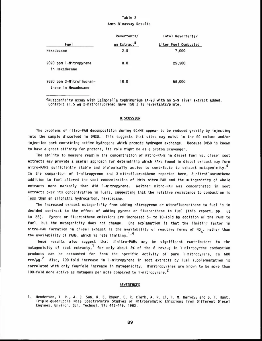
Table 2Ames Bioass3y Results
Fuel
Revertants/
ug Extracta
Total Revertants/
Liter Fuel Combusted
Hexadecane 2.5 7,000
2090 ppm 1-Nitropyrene B.O 25,500
in Hexadecane
2680 ppm 3-Nitrofluoran-
thene in Hexadecane
18.0 65,000
aMutagenicity assay with Salmonella typhimurium TA-gB with no s-g liver extract added.Controls (1.5 ug 2-nitrofluorene) gave 158 ± 12 revertants/plate.
DISCUSSION
The problems of nitro-PAH decomposition during GC/MS appear to be reduced greatly by injecting
into the sample dissolved in DMSO. This suggests that sites may exist in the GC column and/or
injection port containing active hydrogens which promote hydrogen exchange. Because DMSO is known
to have a great affinity for protons, its role might be as a proton scavenger.
The ability to measure readily the concentration of nitro-PAHs in diesel fuel vs. diesel soot
extracts may provide a useful approach for determining which PAHs found in diesel exhaust may form4nitro-PAHS sufficiently stable and biologically active to contribute to exhaust mutagenicity.
In the comparison of l-nitropyrene and 3-nitrofluoranthene reported here~ 3-nitrofluoranthene
addition to fuel altered the soot concentration of this nitro-PAH and the mutagenicity of whole
extracts more markedly than did l-nitropyrene. Neither nitro-PAH was concentrated in soot
extracts over its concentration in fuels, suggesting that the relative resistance to combustion is
less than an aliphatic hydrocarbon, hexadecane.
The increased exhaust mutagenicity from adding nitropyrene or nitrofluoranthene to fuel is in
decided contrast to the effect of adding pyrene or fluoranthene to fuel (this report, pp. 81
to 85). Pyrene or fluoranthene emissions are increased 5- to lO-fold by addition of the PAHs to
fuel, but the mutagenicity does not change. One explanation is that the limiting factor in
nitro-PAH formation in di(,sel exhaust is the availability of reactive forms of NOx, rather than
the availability of PAHs, which is rate limiting.1’4
These results also suggest that dinitro-PAHs may be significant cuntributors to the
mutagenicity of soot extract~,l for only about 3~ of the 8 rev/#g in l-nitropyrene combustion
products can be accounted for from the specific activity of pure l-nitropyrene, ca 600
rev/~g. 2 Also, lO0-fold increase in 1wnitropyrene in soot extracts by fuel supplementation is
correlated wlth only fourfold increase in mutagenicity. Dinitropyrenes are known to be more than
lO0-fold more active as mutagens per mole compared to l~nitropyrene.2
REFERENCES
I. Henderson, T. R., J. D. Sun, R. E. Royer, C. R. Clark, A. P. Li, T. M. Harvey~ and D. F. Hunt,Triple-quadrupole Mass Spectrometry Studies of Nitroaromatic Emissions from Different DieselEngines, Environ. Sci. Technol. !~: 443-449, 1983.
89
2. Rosenkranz, H. S. and R. Mermelstein, Mutagenicity and Genotoxicity of Nitroarenes, All Nitro-Containing Chemicals were not Created Equal, Murat. Res. ll4: 217-267, 1983.
3. Tokiwa, H., R. Nakagawa, and Y. Ohnishi, Mutagenicity of Nitro Derivatives Induced by Exposureof Aromatic Compounds to Nitrogen Dioxide, Murat. Res. B5: 195-205, 1981.
4. Henderson, T. R., J. D. Sun, A. P. Li, R. L. Hanson, T. M. Harvey, J. Shabanowitz, and D. F.Hunt, GC/MS and MS/MS Studies of Diesel Exhaust Mutagenicity and Emissions from ChemicallyDefined Fuels, Environ. Sci. Technol. (in press, 1983).
90
CONTRIBUTION OF PRIMARY AROMATIC AMINES TO THE MUTAGENICITY
OF 6ASIFIER TARS AND COAL OILS
Abstract --Two reactlons that chemlcallg alter
prlmary aromatic amlnes were used to assess the PRINCIPAL IWVESTIGATORS
contrlbutlon of these compounds to ~he indlrect W.E. Bechtold
bacterlal mutagenlclcy of tar from an experimental J.S. Dutcher
low Btu gaslfler tar trap. Nitrous acld generated A.L. Brooks
direct mutagenlc activity from the tar at a pH of R.F. Henderson
1.2, llmltlng the interpretation of resul~s. No
dlrect activity was generated at a pH of 2.5, and vlrtua11y aZl of the indirect actlvlty for
primary amines standards was eliminated. For low Btu gaslfler tar, 61~ of the indirect actlvlty
was eliminated, whereas more than 90~ was ellmlnated from a coal oil. Acetylatlon reduced the
indlrect actlvlty of mosc primary amine standards by more than 85~. Acetylatlon ellmlnated part
of the tar’s mutagenlclty and most of the coal oil’s mutagenlcltg. These resulcs Indicated that a
much lower percent of the bacterlal mutagenlc actlvlcy of low Btu coal tar samples was due go
primary aromaClc amines than was the case for coal o11.
Reaction with nitrous acid has been a successful technique for determining the contribution of
primary aromatic amines (PAA) to the bacterial mutagenicity of process solvents (PS) and heavy
distillates (HD) from solvent refined coal (SRC).I The observation that more than 90% of
indirect activity for PS and HD disappears after reaction with nitrous acid led Pelroy and
Stewart I to conclude that PAA are quantitatively important in determining the mutagenicity of
these mixtures.
The contribution of PAA to the mutagenicity of low Btu gasifier tars is also of interest
because the basic fraction in which PAA would be found has high specific indirect mutagenicity.
We report two methods to assess the contribution of PAA to the mutagenicity of tar from an
experimental low Btu gasifier. A coal oil similar in nature to SRC II liquids was used for
comparison to validate the methods.
MATERIALS AND METHODS
Gasifier tars were collected from the tar trap of an experimental low Btu gasifier unit at the
Morgantown Energy Technology Center (METC), Morgantown, WV. Coal oil samples were comparative
research materials acquired from the Fossil Fuels Research Matrix Program (Oak Ridge National
Laboratory). All other chemicals were obtained from commercial sources.
Chemical standards, samples, and reaction products were tested for biological activity in
Salmonella typhimurium strain TA 98, as previously published.2
initial nitrosation reaction conditions (Method I) were essentially those of Haugen et al.3
Actual experimental conditions have been described previously. 4 Nitrosation reactions were also
produced by methods similar to those of Tsuda et al 5 (Method If). Unlike Method I Method ¯ p
involved use of buffers (citrate and phosphate) to maintain higher pHs (2.5 + 4.0). All solutions
were filter sterilized before use in the bacterial mutagenicity test.4Acetylatlon conditions have also been previously described.
91
RESULTS
Changes in Mutaqenicity by Nitrosation (Method I~
As previously noted, when Method I was applied to tar from the low Btu gasifer, no reduction
in indirect mutagenicity was noted. 4 This may be due to simultaneous generation of direct
mutagenicity. In fact, within 20 s of the onset of the reaction, almost two revertants per ,g
of direct activity were generated, and direct activity continued to rise with time, as did
indirect activity.
Chanqes in MutaQenicity by Nitrosation (Method If)
Nitrosation reaction conditions were sought that would reduce the indirect mutagenicity of PAA
standards without generation of direct activity for tar trap tar. Time (0.5 and 5 h) and pH (2.5
and 4) were the two variables investigated. Results showed that the reaction time of 0.5 h at pH
2.5 proved optimum for reducing the indirect mutagenicity of the aromatic amine standards without
inducing direct mutagens. Indirect activity for the 2-aminoanthracene was reduced by g5%, but no
direct activity was generated. Reaction conditions of pH 2.5 and 0.5 h were used to investigate
the effect of nitrous acid on several standards. In all cases, nitrous acid reduced the
mutagenicities of PAA standards to less than 16% of the control value. Mutagenicities for a
secondary amine, carbazole, and an azaarene, phenanthridine, were not affected by nitrous acid
treatment. When the coal oil sample was subjected to HCI without NAN02, little change in
mutagenicity was noted. However, addition of nitrite reduced the indirect activity to g%, of the
value observed in HCI alone.
Treatment of tar trap tar with nitrous acid decreased but did not eliminate the indirect
mutagenicity. After treatment, 39% of the mutagenicity remained. The administration of
HCI/NaNO2 had no effect on cell viability either in conjunction with PAA standards or with
complex mixtures. Viability studies were run simultaneously with mutagenicity assays and used the
same mixtures at the same concentrations.
Chanqes in Mutaqenicity by AcetNlation
After acetylation, the mutagenicities of five of the six PAA standards were reduced to less
than 20%. However, the activity of l-acetylaminopyrene rose to more than six times the level of
the free amine. Acetylation of the coal oil sample with acetic anhydride reduced the observed
mutagenicity to less than 30% of the unreacted value. Again, reaction controls were similiar to
those in the sample in dimethylsulfoxide, and values were extracted from the linear portion of the
dose-response curve. After acetylation, low Btu tar showed a 52% decrease in mutagenicity.
DISCUSSION
Two reaction schemes were used to assess the contribution of PAA to the mutagenicity of tar
trap tar from a low Btu gasification process. Nitrous acid was effective in eliminating nearly
all of the S-9-dependent mutagenicity of PAA standards and a coal oil sample. Likewise, nitrous
acid reduced the indirect mutagenicity of a low Btu coal gasifier tar, although 39% of the
mutagenicity remained. Acetylation reduced the indirect mutagenicity to only 52% of the
original. These results indicate that coal gasification tars, unlike coal liquefaction fuels,
contain significant amounts of indirect acting mutagens other than PAA.
REFERENCES
I. Pelroy, R. A. and D. L. Stewart, The Effects of Nitrous Acid on the Mutagenicity of Two CoalLiquids and Their Genetically Active Chemical Fractions, Mutat. Res. go: 2g?-30B, 1982.
2. Ames, B. N., J. McCann, and E. Yamasaki, Methods for Detecting Carcinogens and Mutagens withthe Salmonella/Mammalian-Microsome Mutagenicity Test, Murat. Res. 31: 347-364, 1975.
3, Haugen, D. A., M. J. Peak, and C. A. Reilly, Jr., Use of Nitrous Acid-Dependent Decrease inMutagenicity as an Indication of the Presence of Mutagenic Primary Aromatic AminesNon-Specific Reactions with Phenols and Benzo(a)pyrene, Mutat. Res. B2: 59-67, 19Bla.
4o Bechtold, W. E., J. S. Dutcher, A. P. Li, G. J. Newton, and R. F. Henderson, Reductions in theMutagenicities of Coal Tars and SRC-II Materials by Nitrosation and Acetylation, in InhalationToxicoloqy Research Institute Annual Report, pp. 129-132, DOE Research and Development Report,LMF-I02, National Technical Information Center, Springfield, VA, 1982.
5. Tsuda, M., M. Nagao, T. Hirayama, and T. Sugimura, Nitrite Converts 2-Amino-:-Carboline, aNon-Mutagen, and 2-Hydroxy-3-Nitroso.-~-Carboline, a Direct Mutagen, Mutat. Res. 83: 61-68,IgSl.
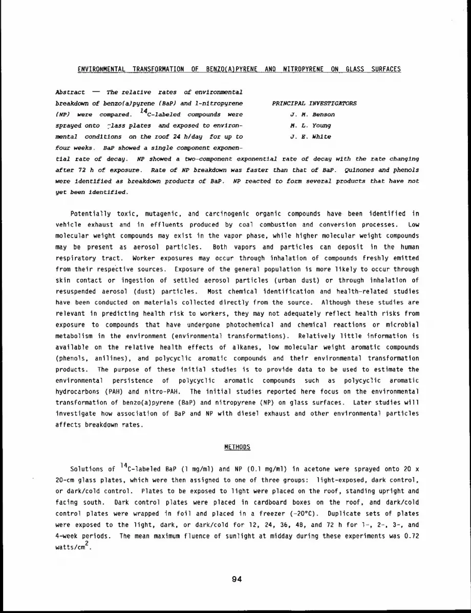
ENVIRONMENTAL TRANSFORMATION OF BENZO(A)PYRENE AND NITROPYRENE ON GLASS SURFACES
Abstract -- The relatlve rates of environmental
breakdown of benzo(a)pyrene (SAP) and 1-nltropyrene PRINCIPAL INVESTIGATORS
(NP) were compared. 14c-labeled compounds were J.N. Benson
sprayed onto 71ass plates and exposed to environ- M.L. Young
mental conditions on the roof 24 h/day for up to J.E. WhlEe
four weeks. BaP showed a single component exponen-
tlal rate of decay. NP showed a two-component exponential rate of decay w~th the rate changing
after 72 h of exposure. Rate of NP breakdown was faster than that of BaP. Qulnones and phenols
were iden~fled as breakdown products of BaP. NP reacted to form several products that have not
yet been Identlfled.
Potentially toxic, mutagenic, and carcinogenic organic compounds have been identified in
vehicle exhaust and in effluents produced by coal combustion and conversion processes. Low
molecular weight compounds may exist in the vapor phase, while higher molecular weight compounds
may be present as aerosol particles. Both vapors and particles can deposit in the human
respiratory tract. Worker exposures may occur through inhalation of compounds freshly emitted
from their respective sources. Exposure of the general population is more likely to occur through
skin contact or ingestion of settled aerosol particles (urban dust) or through inhalation
resuspended aerosol (dust) particles. Most chemical identification and health-related studies
have been conducted on materials collected directly from the source. Although these studies are
relevant in predicting health risk to workers, they may not adequately reflect health risks from
exposure to compounds that have undergone photochemical and chemical reactions or microbial
metabolism in the environment (environmental transformations). Relatively little information
available on the relative health effects of alkanes, low molecular weight aromatic compounds
(phenols, anilines), and polycyclic aromatic compounds and their environmental transformation
products. The purpose of these initial studies is to provide data to be used to estimate the
environmental persistence of polycyclic aromatic compounds such as polycyc]ic aromatic
hydrocarbons (PAH) and nitro-PAH. The initial studies reported here focus on the environmental
transformation of benzo(a)pyrene (BaP) and nitropyrene (NP) on glass surfaces. Later studies
investigate how association of BaP and NP with diesel exhaust and other environmental particles
affects breakdown rates.
METHODS
Solutions of 14C-labeled BaP (1 mg/ml) and NP (O.l mg/ml) in acetone were sprayed onto
20-cm glass plates, which were then assigned to one of three groups: light-exposed, dark control,
or dark/cold control. Plates to be exposed to light were placed on the roof, standing upright and
facing south. Dark control plates were placed in cardboard boxes on the roof, and dark/cold
control plates were wrapped in foil and placed in a freezer (-20°C). Duplicate sets of plates
were exposed to the light, dark, or dark/cold for 12, 24, 36, 48, and 72 h for l-, 2-, 3-, and
4-week periods. The mean maximum fluence of sunlight at midday during these experiments was 0.72
watts/cm2.
94
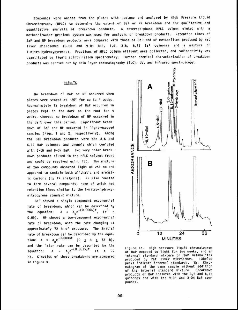
Compounds were washed from the plates with acetone and analyzed by High Pressure Liquid
Chromatography (HPLC) to determine the extent of BaP or NP breakdown and for qualitative and
quantitative analysis of breakdown products. A reversed-phase HPLC column eluted with a
methanol/water gradient system was used for analysis of breakdown products. Retention times of
BaP and NP breakdown products were compared with those of BaP and NP metabolites produced by rat
liver microsomes (3-OH and 9-OH BaP, 1,6, 3,6, 6,12 BaP quinones and a mixture of
l-nitro-hydroxypyrenes). Fractions of HPLC column effluent were collected, and radioactivity was
quantitated by liquid scintillation spectrometry. Further chemical characterization of breakdown
products was carried out by thin layer chromatography (TLC), UV, and infrared spectroscopy.
X2
RESULTS
No breakdown of BaP or NP occurred when
plates were stored at -20° for up to 4 weeks.
Approximately l~ breakdown of BaP occurred in
plates kept in the dark on the roof for 4
weeks, whereas no breakdown of NP occurred in
the dark over this period. Significant break-
down of BaP and NP occurred in light-exposed
samples (Figs. l and 2, respectively). Among
the BaP breakdown products were the 3,6 and
6,12 BaP quinones and phenols which coeluted
with 3-OH and g-OH BaP. Two very polar break-
down products eluted in the HPLC solvent front
and could be resolved using TLC. The mixture
of two compounds absorbed light at 254 nm and
appeared to contain both aliphatic and aromat-
ic carbons (by IR analysis). NP also reacted
to form several compounds, none of which had
retention times similar to the l-nitro-hydroxy-
nitropyrene standard mixture.
BaP showed a single component exponential
rate of breakdown, which can be described by
the equation: A = Aoe-(O’OOO4)t, (r2
0.89). NP showed a two-component exponential
rate of breakdown, with the rate changing at
approximately 72 h of exposure. The initial
rate of breakdown can be described by the equa-
tion: A = A e-0"0033t (0 < t < 72 h),O -- --
and the later rate can be described by the
equation: A = Aoe-(O’OOl5)t (t 72
h). Kinetics of these breakdowns are compared
in Figure 3.
Figure la. High pressure liquid chromatogramof BaP exposed to light for two weeks, and aninternal standard mixture of BaP metabolitesproduced by rat liver microsomes. Labeledpeaks indicate internal standards, lb. Chro-matogram of the same sample without additionof the internal standard mixture. Breakdownproducts of BaP coeluted with the 3,6 and 6,12quinones and with the 9-OH and 3-OH BaP com-pounds.
95
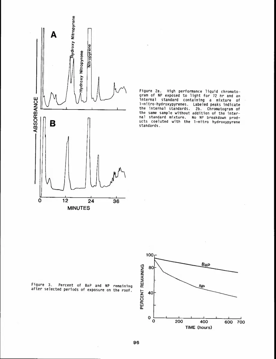
A
B
I
9
0 12 24 ;36MINUTES
Figure 2a. High performance liquid chromato-gram of NP exposed to light for 72 hr and aninternal standard containing a mixture ofl-nitro-hydroxypyrenes. Labeled peaks indicatethe internal standards. 2b. Chromatogram ofthe same sample without addition of the inter-nal standard mixture. No NP breakdown prod-ucts coeluted with the l-nitro hydroxypyrenestandards.
Figure 3. Percent of BaP and NP remainingafter selected periods of exposure on the roof.
0 ,,L ~, = ,,~, ~ j j0 200 400 600 700
TIME (hours)
96
DISCUSSION
Both BaP and NP exposed to light underwent chemical transformations. Because little breakdown
occurred in the dark control samples placed on the roof, we conclude that the breakdown that
occurred was due to photochemical processes.
BaP showed a single component rate of breakdown, whereas NP showed two-component exponential
rates of breakdown. To our knowledge, this is the first report of a two-component exponential
rate of photochemical breakdown of a polycyclic aromatic compound. The observed kinetics may be
due to the presence of the nitro group on the pyrene molecule.
The more rapid rates of breakdown of NP may be due to the reactivity of the nitro group.
Chapman et al. l have studied the photochemical reactions of 9-nitroanthracene and have reported
early formation of the nitrite ester and ultimate cleavage of the nitro group from the ring.
Reaction products included the lO,lO’ bianthrone dimer, anthraquinone and nitroanthrone. Similar
processes may be occurring with I-NP in our studies. It is surprising that we did not identify
hydroxy breakdown products of NP, because Yasuhara 2 identified 1-nitro-2-hydroxy pyrene as a
photolysis product of l-nitropyrene in acetonitrile solution.
REFERENCES
1. Chapman, O. L., A. A. Griswold, E. Hoganson, G. Lenz, and J. Reasoner, Photochemistry ofUnsaturated Nitrocompounds, Pure and Applied Chemistry g: 585-590, 1964.
2. Yasuhara, A., Formation of 1-Nitro-2-hydroxypyrene from l-Nitropyrene by Photolysis, Chem.Lett. IgB3: 347-348, 1983.
97
METABOLISM AND MUTA6ENESIS OF I-NITROPYRENE IN RAT LIVER, LUNG, AND. NASAL TISSUF
Abstract --Rat liver, lung, and nasal tissues were
used as enzyn~e-activatlng systems to invesClgate PRINCIPAL INVESTIGATORthe mutagenlc potentlal of 1-n~tropyrene (NP) J.A. Bond
Salmonella t~phlmurlum and to measure the rates of
1-nltropyrene metabolism. These tissues bloact~vated NP to mutagens that were detected in the
salmonella Ames bacterial test system. In both strains, TA-98 and TA-100, about 1.0 mg/ml liver
or nasal tissue S-9 appeared to be the optimal concentration Chat resulted in the largest
mutagenlc response to NP, whereas 2.0 mg/ml of lung S-9 was necessary for optimal responses. When
NP was incubated with liver, lung, or nasal tissue S-9 and straln TA-98 nltroreductase deflclent,
mutagenlc responses were significantly decreased, compared to the response ~n TA-98. Total
formation rates of NP ~abolltes for nasal tissue, liver, and lung S-9 were 650, 300, and 60
pmol/mg proce~n/mln, res~ectlvely. These results suggest that the respiratory tract, In
partlcular the nasal tlssue, may be an important s~te for in vlvo bloactlvatlon of inhaled NP.
Many nitrated polycyclic aromatic hydrocarbons (PAH), are both bacterial and mammalianlmutagens. These compounds have been detected in the environment from such sources as diesel
exhaust emissions and coal-combustion fly ash. A predominant mutagenic nitrated PAH in diesel
exhaust samples is l-nitropyrene (NP). NP, in addition to being a bacterial and mammalian
mutagen, is also carcinogenic in male rats at the site of injection.
Because inhalation is a major route of potential exposure to NP, it is important to
investigate the capability of respiratory tract tissue, in addition to hepatic tissue, to
bioactivate and biotransform NP. The purpose of this investigation was to study the mutagenic
potential of NP in Salmonella typhimurium using rat liver, lung, and nasal tissue as the enzyme-
activating sources and to measure the rates of metabolism of NP in these same tissues. The
results demonstrate that rat liver, lung, and nasal tissue are capable of bioactivating NP to
species mutagenic in bacteria and that the observed mutagenicity roughly parallels the observed
rate of metabolism of NP in these tissues.
METHODS
Male Fischer-344 rats (18-20 weeks; 300 g) were used in all studies. Food and water were
provided ad libitum. All rats were asphyxiated with C02, the livers, lungs, and nasal tlssuewere removed, and homogenates were prepared. Pooled livers, lungs, and nasal tissue were
homogenized separately and 9000 x g supernatant fractions collected, as previously described.2
Nasal tissue, liver, and lung S-9 were tested for their ability to bioactivate NP to mutagens
using the Ames Salmonella/mammalian microsome assay. 3 Tissues were assayed for bioactivation
capacity in tester strains TA-98, TA-IO0, and TA-gBNR (nitroreductase deficient) at four
concentrations of NP (0.25, 0.50, 0.75, and l.O ~g/plate) and four concentrations of tissue S-9
protein (0.5, l.O, 1.5, and 2.0 mg protein/ml). The concentrations of NP were previously shown
be below the toxic level for these tester strains. Analysis of the rates of formation of NP
metabolites using rat liver, lung, and nasal tissue were determined according to a previously4published method.
98
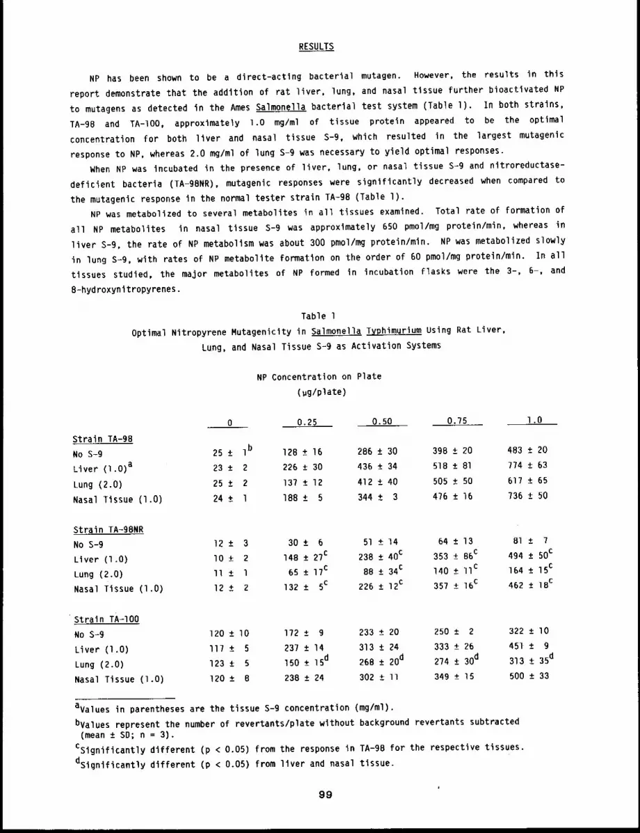
RESULTS
NP has been shown to be a direct-actlng bacterial mutagen. However, the results in this
report demonstrate that the addition of rat liver, lung, and nasal tissue further bioactivated NP
to mutagens as detected in the Ames Salmonella bacterial test system (Table l). In both strains,
TA-98 and TA-IO0, approximately l.O mg/ml of tissue protein appeared to be the optimal
concentration for both liver and nasal tissue S-9, which resulted in the largest mutagenic
response to NP, whereas 2.0 mg/ml of lung S-9 was necessary to yield optimal responses.
When NP was incubated in the presence of liver, lung, or nasal tissue S-9 and nitroreductase-
deficient bacteria (TA-gBNR), mutagenic responses were significantly decreased when compared
the mutagenlc response in the normal tester strain TA-g8 (Table l).
NP was metabolized to several metabolites in all tissues examined. Total rate of formation of
all NP metabolites in nasal tissue S-9 was approximately 650 pmol/mg protein/min, whereas in
liver S-9, the rate of NP metabolism was about 300 pmol/mg protein/min. NP was metabolized slowly
in lung s-g, with rates of NP metabolite formation on the order of 60 pmol/mg protein/min. In all
tissues studied, the major metabolites of NP formed in incubation flasks were the 3-, 6-, and
8=hydroxynltropyrenes.
Table 1
Optimal Nltropyrene Mutagenicity in Salmonella Typhimurium Using Rat Liver,
Lung, and Nasal Tissue s-g as Activation Systems
NP Concentration on Plate
(gg/plate)
0 0.25 0.50 0.75 l.O
Strain TA-gB
No S-9 25 ± Ib 12B ± 16 286 ± 30 398 ± 20
Liver (l.O) a 23 ± 2 226 ± 30 436 ± 34 518 ± 81
Lung (2.0) 25 ± 2 137 ± 12 412 ± 40 505 ± 50
Nasal Tissue (l.O) 24 ± l 18B ± 5 344 ± 3 476 ± 16
483 ± 20
774 ± 63
617 ± 65
736 ± 50
Strain TA-g8NR
No S-9 12 ± 3 30 ± 6 51 ± 14 64 ± 13
Liver (I,0) I0 ± 2 148 ± 27c 238 ± 40c 353 ± B6c
Lung (2.0) II ± l 65 ± 17c 8B ± 34c 140 ± IIc
Nasal Tissue (l.O) 12 ± 2 132 ± 5c 226 ± 12c 357 ± 16C
Bl ± 7
494 ± 50c
164 ± 15c
462 ± IBc
Strain TA-IO0
No S-9 120 ± lO 172 ± 9 233 ± 20 250 ± 2
Liver (l.O) ll7 ± 237 ± 14 313 ± 24 333 ± 26
Lung (2,0) 123 ± 5 150 ± 15d 26B ± 20d 274 ± 30d
Nasal Tissue (1.O) 120 ± 8 238 ± 24 302 ± ll 349 ± 15
322 ± l O
451 ± g
313 ± 35d
500 ± 33
avalues in parentheses are the tissue s-g concentration (mg/ml).
bvalues represent the number of revertants/plate without background revertants subtracted(mean ± SD; n = 3).
CSignificantly different (p < 0.05) from the response in TA-gB for the respective tissues.
dsignificantly different (p < 0.05) from liver and nasal tissue.
99
DISCUSSION
The experiments described in this report were designed to assess the capacity of liver, lung,
and nasal tissue to bioactivate and biotransform NP. Both liver and nasal tissue were capable of
increasing the mutagenic potency of NP and, in all strains tested, were about 50% to I00% greater
than that observed in lung tissue s-g. The observation that the mutagenic response to NP in all
tissues examined was significantly decreased when strain TA-98NR was used suggests that at least a
portion of the mutagenic response toward NP in the normal tester strain TA-98 may be the result of
reduction in the bacteria of the nitro group to mutagenic intermediates. The observation that the
presence of liver, lung, and nasal tissue with TA-gBNR yields mutagenic responses above those seen
in the absence of these tissues suggests that other routes of metabolism of NP in these tissues
may contribute to the overall mutagenic response seen in normal strains of bacteria.
Liver, lung, and nasal tissue metabolized NP to several metabolites. Nasal tissue metabolized
NP at a rate of about twice that seen in rat liver and more than lO times that observed in rat
lung. The metabolites of NP detected were the 6- and 8-hydroxynitropyrenes, with smaller
quantities of 3-hydroxynitropyrene measured. The contribution of these metabolites to the
observed mutagenicity remains to be determined. These data suggest that ring oxidation may be
involved in the mutagenic activity of NP. Presumably, the formation of hydroxynitropyrenes, as
catalyzed by liver, lung, and nasal tissue S-9, proceeds by way of epoxide intermediates.
The data in this report point to the potential importance of both the nasal tissue and the
lung in activation of NP to reactive intermediates. The observation that nasal tissue is capable
of metabolizing NP at rates nearly twice that of liver and lO times that of lung suggests that
nasal tissue may be a very important site for in vivo metabolism of inhaled NP and may play an
important role in determining the metabolic fate of inhaled promutagens.
REFERENCES
I. Mermelstein, R., D. K. Kiriazides, M. Butler, E. C. McCoy, and H. S. Rosenkranz, TheExtraordinary Mutagenicity of Nitropyrenes in Bacteria, Mutat. Res. 8_99: 18?-Ig6, Ig8].
2. Bond, 3. A. and A. P. Li, Rat Nasal Tissue of Benzo(a)pyrene and 2-Aminoanthracene to Mutagensin Salmonella t_v_phimurium, Environ. Mutagen. 5: 311-318, 1983.
3. Ames, B. N., J. McCann, and E. Yamasaki, Methods for Detecting Carcinogens and Mutagens withthe Salmonella/Mammalian Microsome Mutagenicity Test, Mutat. Res. 3l: 34?-364, 1975.
4. Bond, J. A., C. J. Omiecinski, and M. R. Juchau, Kinetics, Activation, and Induction of AorticMonooxygenases-Biotransformation of Benzo(a)pyrene, Biochem. Pharmacol. 28: 305-311, IgTg.
100
METABOLISM OF (14C)-I-NITROPYRENE IN ISOLATED PERFUSED RAT LUNGS
Abstract -- l-Nitropyrene (I-NP), In addJtlon
being both a bacterlal and man.allan mutagen. Is PRINCIPAL INVESTIGATORS
carcinogenic In rats. The purpose of thls study J.R. Bond
was to quantltate 1-NP metabolism and macromolec- J. L, Hauderly
ular covalent binding in the isolated perfused rat
lung. Rat lungs were perfused wlth 2. 5, or 24 ~NI4C-I-NP for 90 mln. Respiration and lung
mechanics were monltored contlnually throughout perfuslon. Lungs from control, phenobarbltal
(PB)-, and 3-methylcholanthrene (3-Nc)-treated rats metabollzed 14C-I-NP to oxldlzed, reduced.
and conjugated metabolltes. Treatment with PB resulted in a 60~ ~ncrease in the total metabolism
of 14C-I-NP, whereas treatment of rats wlth 3-HC resulted in a 10-fold increase in the rate of
metabolism of 14C-I-NP when compared to controls. In all experiments, both d~namlc lung
compliance and tidal volume declined in a nearly l~near manner and were approximately 60~ of the
inltlal value at the end of 90 mln of perfus~on, The results from these studies point to the
potential importance of lung mecabollsm in contributing to the metabolic fate of lnhaled I-NP.
Nitro polycyclic aromatic hydrocarbons (nitro-PAH) originating from sources such as diesel
exhaust emissions and coal-combustion fly ash have been detected in the environment. A
predominant mutagenic nitro-PAH in diesel engine exhaust samples is l-nitropyrene (I-NP).
addition to being a potent bacterial mutagen, I-NP is also mutagenic in mammalian cells andl
carcinogenic in male rats.
Very little information about the mammalian metabolism of I-NP is available. Since inhalation
is the most likely route of human exposure to I-NP, it is important to evaluate the role of intact
lungs in determining the metabolic fate of inhaled I-NP. The purpose of this investigation was to
quantitate 1-NP metabolism and macromolecular covalent binding in the isolated perfused/ventilated
rat lung. The results indicate that I-NP is metabolized in lungs from control rats and that both
metabolism and macromolecular covalent binding of I-NP are increased lO- to 20~fold after
treatment of rats with 3-methylcholanthrene (3-MC). The data suggest that the lung may play
important role in determining the metabolic fate of inhaled I-NP.
MATERIALS AND METHODS
Male specific pathogen~free Fischer-344 rats (15-20 weeks, 250~300 g) were used in all
studies. Food (Lab Blox, Allied Mills, Chicago, IL) and water from bottles were provided a
libitum.
Rats were anesthetized with halothane and the tracheas were cannulated. The pulmonary artery
was then cannulated and the lungs immediately perfused with fortified Krebs-Ringer bicarbonate
buffer (37°C, pH 7.4). The lungs were then removed from the rat and attached to a lung perfusion
apparatus. 2 The lungs were perfused (recirculating; approximately 25 ml/g lung/min) with 50
of the fortified Krebs-Ringer bicarbonate buffer for lO min and then 2, 5, or 24 ~M 14C-I-NP
was added to the perfusate. One ml aliquots were removed from the perfusate reservoir at 5, lO,
15, 20, 30, 45, 60, and 90 min after addition of 14C-I-NP for analyses of I-NP metabolites by
101
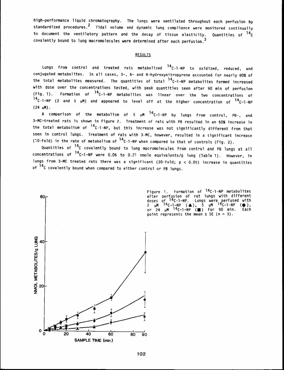
high-performance liquid chromatography. The lungs were ventilated throughout each perfusion by
standardized procedures. 2 Tidal volume and dynamic lung compliance were monitored continually
to document the ventilatory pattern and the decay of tissue elasticity. Quantities of 14Ccovalently bound to lung macromolecules were determined after each perfusion.3
RESULTS
Lungs from control and treated rats metabolized 14C-I-NP to oxidized, reduced, and
conjugated metabolites. In all cases, 3-, 6- and B-hydroxynitropyrene accounted for nearly 80% of
the total metabolites measured. The quantities of total 14C-I-NP metabolites formed increased
with dose over the concentrations tested, with peak quantities seen after 90 min of perfuslon
(Fig. l). Formation of 14C-I-NP metabolites was linear over the two concentrations
14C-I-NP (2 and 5 gM) and appeared to level off at the higher concentration of 14C-l-NP
(24 gM).
A comparison of the metabolism of 5 ~M 14C-I-NP by lungs from control, PB-, and
3-MC-treated rats is shown in Figure 2. Treatment of rats with PB resulted in an 60% increase in
the total metabolism of 14C-I-NP, but this increase was not significantly different from that
seen in control lungs. Treatment of rats with 3-MC, however, resulted in a significant increase
(~O-fold) in the rate of metabolism of 14C-I-NP when compared to that of controls (Fig.
Quantities of 14C covalently bound to lung macromolecules from control and PB lungs at all
concentrations of 14C-I-NP were 0.06 to 0.21 nmole equivalents/g lung (Table l). However,
lungs from 3-MC treated rats there was a significant (20-fold; p < 0.05) increase in quantities
of 14C covalently bound when compared to either control or PB lungs.
60-
z 403
0 20 40 60 80 90SAMPLE TIME (rain.)
Figure I. Formation of 14C-I-NP metabolitesafter perfusion of rat lungs with differentdoses of 14C-I-NP. Lungs were perfused with2 gM 14C-I-NP (&), 5 ~M 14C-I-NP (0),or 24 ~M 14C-I-NP (m) for 90 min. Eachpoint represents the mean ± SE (n = 3).
102
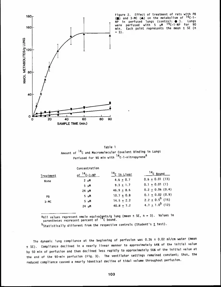
180
160
z,_1
’~120o9uJI-
0g3,<I-IJ, J:~ 80._105:z
4O
00 20 40 60 80 90
SAMPLE TIME (min.)
Figure 2. Effect of treatment of rats with PB(B) and 3-MC (&) on the metabolism of 14C-l-NP in perfused lungs (control: ¯ ). Lungswere perfused with 5 gM 14C-I-NP for 90min. Each point represents the mean ± SE (n
3).
Table l
Amount of 14C and Macromolecular Covalent Binding in Lungs
Perfused for go min with 14C-l-nitropyrenea
Treatment
None
PB
3-MC
Concentration
of 14C-I-NP 14C in Liver 14C Bound
2 ~M 4.6 ± 0.7 0.6 ± O.Ol (13)
5 ~M 9.3 + 1.7 O.l ± O.Ol (1)
24 gM 46.9 ± 8.6 0.2 + 0,06 (0.4)
5 ~M I0.7 +_ O.B O.1 + 0.02 (o.g)
5 ~M 14.5 +_ 2.2 2.2 +_ 0.5b (15)
24 ~M 40.8 +_ 7.2 4.7 ± l.O b (12)
aAll values represent nmole equiva~nts/g lung (mean ± SE, n = 3). Values parentheses represent percent of C bound.
bstatlstically different from the respective controls (Student’s ! test).
The dynamic lung compliance at the beginning of perfusion was 0.36 ± 0.02 ml/cm water (mean
± SE). Compliance declined in a nearly linear manner to approximately 64% of the initial value
by 50 min of perfusion and then declined less rapidly to approximately 56% of the initial value at
the end of the 90-min perfusion (Fig. 3). The ventilator settings remained constant; thus, the
reduced compliance caused a nearly identical decline of tidal volume throughout perfusion.
103
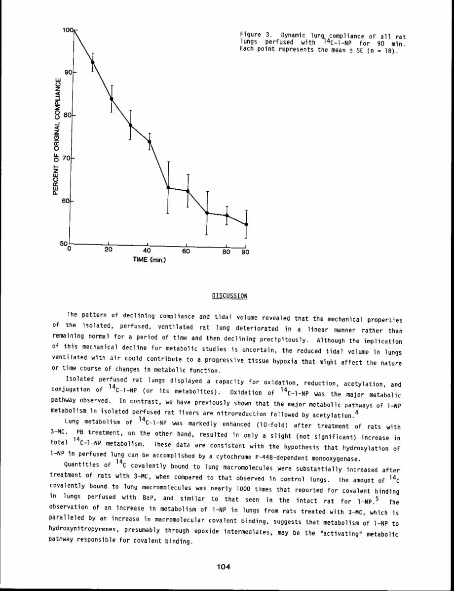
Figure 3¯ Dynamic lung compliance of all ratlungs perfused with 94C_l=NP for 90 min.Each point represents the mean ± SE (n = 18).
50 I I I I I0 20 40 60 80 90TIME (rain.)
DISCUSSION
The pattern of declining compliance and tidal volume revealed that the mechanical properties
of the isolated, perfused, ventilated rat lung deteriorated in a linear manner rather than
remaining normal for a period of time and then declining precipitously. Although the implication
of this mechanical decline for metabolic studies is uncertain, the reduced tidal volume in lungs
ventilated with air could contribute to a progressive tissue hypoxia that might affect the nature
or time course of changes in metabolic function.
Isolated perfused rat lungs displayed a capacity for oxidation, reduction, acetylation, and
conjugation of 14C-1-NP (or its metabolites). Oxidation of 14C-I-NP was the major metabolic
pathway observed. In contrast, we have previously shown that the major metabolic pathways of I-NP
metabolism in isolated perfused rat livers are nitroreduction followed by acetylation 4
Lung metabolism of 14C-I-NP was markedly enhanced (lO-fold) after treatment of rats with
3-MC. PB treatment, on the other hand, resulted in only a slight (not significant) increase
total 14C-I-NP metabolism¯ These data are consistent with the hypothesis that hydroxylation of
I-NP in perfused lung can be accomplished by a cytochrome P-448-dependent monooxygenase.
Quantities of 14C covalently bound to lung macromolecules were substantially increased after
treatment of rats with 3-MC, when compared to that observed in control lungs¯ The amount of 14C
covalently bound to lung macromolecules was nearly lO00 times that reported for covalent binding
in lungs perfused with BaP, and similar to that seen in the intact rat for I-NP. 5 The
observation of an increase in metabolism of I~NP in lungs from rats treated with 3~MC, which is
paralleled by an increase in macromolecular covalent binding, suggests that metabolism of I~NP to
hydroxynitropyrenes, presumably through epoxide intermediates, may be the "activating" metabolic
pathway responsible for covalent binding.
104
The pervasiveness of known inducing agents of the 3-MC type, such as BaP, in our environment
indicates that lungs are likely to be exposed to inducers through inhalation. Inhalation of
inducing agents could conceivably affect the metabolism of various xenobiotics. The data
presented in this study point to the importance of lung metabolism in contributing to the
metabolic fate of inhaled I-NP and the formation of reactive intermediates that are capable of
covalently binding to lung macromolecules.
REFERENCES
I. Ohgaki, H., N. Matsukura, K. Morino, T. Kawachi, T. Susimura, K. Morita, H. Tokiwa, andI. Hirota, Carcinogenicity in Rats of the Mutagenic Compounds l-Nitropyrene and3-Nitrofluoranthene, Cancer Lett. 15: 1-7, 1982.
2. Smith, B. R. and J. R. Bend, Lung Perfusion Techniques For Xenobiotic Metabolism and ToxicityStudies, in Methods in Enzymology ]_7: I05-120, Academic Press, Inc. New York, NY, IgSl.
3. Sun 3. D. and 3. G. Dent, A New Method for Measuring Covalent Binding of Chemicals to CellularMacromolecules, Chem. Biol. Interac. 32: 41-61, 1980.
4. Bond, 3. A., 3. S. Dutcher, and M. A. Medinsky, Metabolism of 14C-i-NitroPyrene in IsolatedPerfused Rat Lung and Liver, The Pharmacologist 25: 109, 1983.
5. Mitchell, C. E., Effect of Enzyme Induction on the Binding of Aromatic Hydrocarbons to MouseLung DNA, Chem. Biol. Interac. (submitted).
105
INHIBITORS OF RABBIT NASAL CYTOCHROME P-450 DEPENDENT ENZYME ACTIVITIES
Abstract -- A total of 34 compounds were screened
for the ab111ty to inhlblt rabbit nasal cytochrome PRINCIPAL INVESTIGATORSP-450. PrecJse Inhlbltlon potencles (I50s) were
A.R. Dahldetermlned for seven of the most potent inhlbltors. D.A. Brez~nsklThe I50s for these compounds ranged from 2-45 pM.
These inhlbltors lnclude common compounds llkelg to be inhaled b9 man, thus potentlally altering
the metabellsm of other ~nhaled compounds.
The presence of high concentrations of cytochromes P-450 in the nasal mucosa has been
established in a variety of species. 1 Because these enzymes have been implicated in the
activation of procarcinogens to carcinogens, 2 it was important to follow up on an earlier report
indicating that the activity of these enzymes could be chemically inhibited. 3 It was the
purpose of this work to investigate a variety of chemicals having structures indicating they might
inhibit nasal cytochrome P-450. Included were methylenedioxyphenyl (MDP) compounds, commonly used
as fragrances or insecticide synergists; organic sulfur (OS) compounds, commonly found air
pollutants and trivalent oxygenated phosphorus ligands (TOPLs). Compounds tested are shown
Table I.
METHODS
Nasal microsomes from LOV:(NZW) rabbits were prepared as previously described, l The
hexamethylphosphoramide (HMPA) N-demethylase activity was measured, 2 as was the screening of
compounds for spectrum formation with cytochrome P-450. 3 Calculations of the 50% inhibition
concentrations (I50) from double reciprocal plots were done as described.4
Each of 18 MDP compounds, nine TOPLs and seven OS compounds were screened for spectrum forming
capability with rabbit nasal microsomal cytochrome P-450. Those producing the largest spectra
were then examined for the ability to inhibit nasal cytochrome P-450 HMPA N-demethylase, and 150values for each compound were calculated.
RESULTS AND DISCUSSION
The results of the screening procedure are related in Table I. Seven compounds from Table l
which gave stable spectra with nasal P-450 - indicating tight ligand binding to the enzyme - and
which in subsequent screening assays inhibited HMPA N-demethylase by >20% at 5 pM inhibitor
concentration, were scheduled for measurement of 150 values. The 150s shown in Table 2, were
from 2.38 to I0.55 pM for the MDP compounds, whereas for tris(2-chloroethyl) phosphite - the
only TOPL for which an i50 was determined - the I50 was 41.40 ~M. All of these 150 values
indicate more potent inhibition of nasal microsomes relative to inhibition of liver
microsomes.4
These experiments identified seven powerful inhibitors of nasal cytochrome P-450 (Table 2).
Each inhibitor has sufficient vapor pressure at room temperature to allow exposure of animals to
106
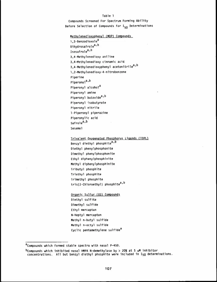
Table 1
Compounds Screened For Spectrum Forming Ability
Before Selection of Compounds for I50 Determinations
Methylenedioxyphenyl (MOP) Compounds
1,3-Benzodioxolea
Dihydrosafrolea’b
isosafrolea,b
3,4-MethylenedioxY aniline
3,4-Methylenedioxy cinnamic acid
3,4-Methylenedioxyphenyl acetonitrilea’b
1,2-Methylenedioxy-4-nitrobenzene
Piperine
Piperonala’b
Piperonyl alcohola
Piperonyl amine
Piperonyl butoxidea’b
Piperonyl isobutyrate
Piperonyl nitrile
l-Piperonyl piperazine
Piperonylic acid
Safrolea,b
Sesamol
Trivalent Oxygenated Phosphorus..L~qands (TOPL)
Benzyl diethyl phosphitea’b
Oiethyl phenylphosphonite
Dimethyl phenylphosphonite
Ethyl diphenylphosphinite
Methyl diphenylphosphinite
Tributyl phosphite
Triethyl phosphite
Trimethyl phosphite
tris(2-Chloroethyl) phosphitea’b
Orqanic Sulfur (OS) Compounds
Diethyl sulfite
Dimethyl sulfide
Ethyl mercaptan
N-Heptyl mercaptan
Methyl n-butyl sulfide
Methyl n-octyl sulfide
Cyclic pentamethylene sulfidea
acompounds which formed stable spectra with nasal P-450.
bcompounds which inhibited nasal HMPA N-demethylase by > 20% at 5 ~M inhibitor
concentrations. All but benzyl diethyl phosphite were included in I50 determinations.
107
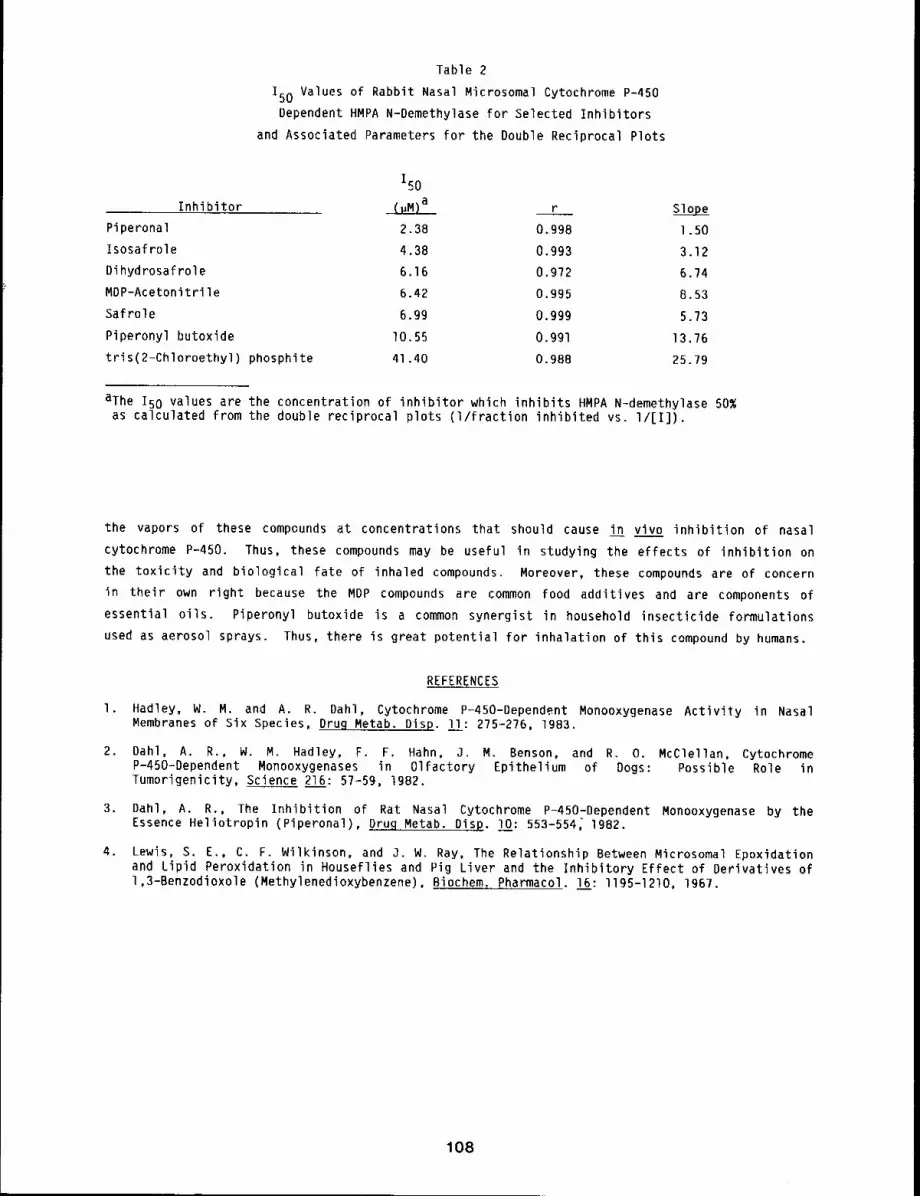
Table 2
150 Values of Rabbit Nasal Microsomal Cytochrome P-450Dependent HMPA N-Demethylase for Selected Inhibitors
and Associated Parameters for the Double Reciprocal Plots
150
Inhibitor (~M)a r SlopePiperonal 2.38 0.998 1.50Isosafrole 4.38 0.993 3.12Dihydrosafrole 6.16 0.972 6.74MDP-Acetonitrile 6.42 0.995 8.53
Safrole 6.9g 0.999 5.73
Piperonyl butoxide I0.55 0.991 13.76
tris(2-Chloroethyl) phosphite 41.40 0.988 25.79
aThe 150 values are the concentration of inhibitor which inhibits HMPA N-demethylase 50%as calculated from the double reciprocal plots (I/fraction inhibited vs. I/[I]).
the vapors of these compounds at concentrations that should cause in vivo inhibition of nasal
cytochrome P-450. Thus, these compounds may be useful in studying the effects of inhibition on
the toxicity and biological fate of inhaled compounds. Moreover, these compounds are of concern
in their own right because the MDP compounds are common food additives and are components of
essential oils. Piperonyl butoxide is a common synergist in household insecticide formulations
used as aerosol sprays. Thus, there is great potential for inhalation of this compound by humans.
REFERENCES
I. Hadley, W. M. and A. R. Dahl, Cytochrome P-450-Dependent Monooxygenase Activity in NasalMembranes of Six Species, Drug Metab. Disp. l_!l: 275-2?6, 1983.
2. Dahl, A. R., W. M. Hadley, F. F. Hahn, J. M. Benson, and R. O. McClellan, CytochromeP-450-Dependent Monooxygenases in Olfactory Epithelium of Dogs: Possible Role inTumorigenicity, Science 216: 5?-59, 1982.
3. Oahl, A. R., The Inhibition of Rat Nasal Cytochrome P-450-Dependent Monooxygenase by theEssence Heliotropin (Piperonal), D~g Metab. Disp. I0: 553-554~ 1982.
4. Lewis, S. E., C. F. Wilkinson, and J. W. Ray, The Relationship Between Microsomal Epoxidationand Lipid Peroxidation in Houseflies and Pig Liver and the inhibitory Effect of Derivatives of1,3-Benzodioxole (Methylenedioxybenzene), Biochem. Pharmacol. 16: I195-1210, 1967.
108
IN VITRO INVESTIGATION OF THE POSSIBLE INTERACTION OF FORMALDEHYDE WITH GLUTATHIONE
Abstract -- The interaction of formaldehyde w~th
reduced glutathlone (GSH) was evaluated using buf- PRINCIPAL INVESTIGATORS
fered solutlons, tlssue homogenaCes, and isolated P.H. Agres
perfused lungs and livers. Assay of GSH concen- T.C. Marshall
tratlon in aqueous solution, tissue homogenates, J.A. Bond
and isolated perfused lung revealed CH20 did not J.D. Sun
change GSH concentrations. However, perfuslon of
isolated livers wlth formaldehyde (CH20) caused a depletion of GSH concentrations
approximately 50~ those of controls. Depletion of GSH by CH20 in the isolated perfused liver,
hut not In tissue homogenates, suggests that metabollc processes in intact cells are Eespon~lble
for the interaction of CH20 with GSH. Lack of depletion of GSH in the isolated perfused lung
may indicate that the lung does not possess the same metabolic ablllty for interaction of CH20
wlth GSH that is available in the l~ver.
Formaldehyde (CH20) has been found to cause nasal cancer in rats after inhalation exposure.
Recent reports indicate that a possible mechanism for some types of CH20-induced toxicity is the
depletion of endogenous reduced glutathione (GSH). These studies have reported that CH20 causesa depletion of GSH in the lung and in isolated hepatocytes. I-4 However, implications from these
studies are unclear because of the use of technical grade CH20 containing methanol as a
stabilizer or because of lack of CH20 quantitation. The primary objective of our study was to
investigate the interaction of pure CH20 with GSH in aqueous solutions, cell-free tissuehomogenates, and the intact perfused lung and liver.
METHODS
CH20 was prepared by heating a solution of paraformaldehyde at llO°C for 5 days. The
concentration of CH20 was standardized by titration with sodium sulfite and hydrochloric
acid. 5 The concentration of CH20 in reaction mixtures was quantitated by using the5pararosaniline-blsulfite assay. Concentrations of GSH were determined by reaction with
ophthaldehyde at pH 8 and measurement of fluorescence emission at 420 nm, with excitation at
350 nm.6
CH20 (0-500 ~g/ml) was added to an aqueous solution containing 1 ~M GSH in Tris-HCL
buffer at pH 7.4. The reaction mixture was incubated for 4 h at 37°C. Aliquots of the reaction
mixture were taken for determination of GSH at O.5-h intervals for the first 2 h, and at hourly
intervals thereafter. Tissue homogenates of lung and liver from Fischer-344 rats were prepared by
homogenizing the organs in 50 mM Tris-HCl buffer (lO ml/g) pH 7.4. CH20 (0-250 ~g/ml) added to the tissue homogenates and then incubated for 4 h at 37°C. Aliquots of the homogenates
for GSH analysis were taken at O.5-h intervals for the first 2 h, and at hourly intervals
thereafter.
Isolated lungs were perfused with Kreb’s-Henseleitt buffer containing CH20 (150 ~g/ml) for
45 min. Livers were perfused in situ with the buffer solution for 45 min. After perfusion, the
lungs and livers were homogenized for GSH analysis.
109
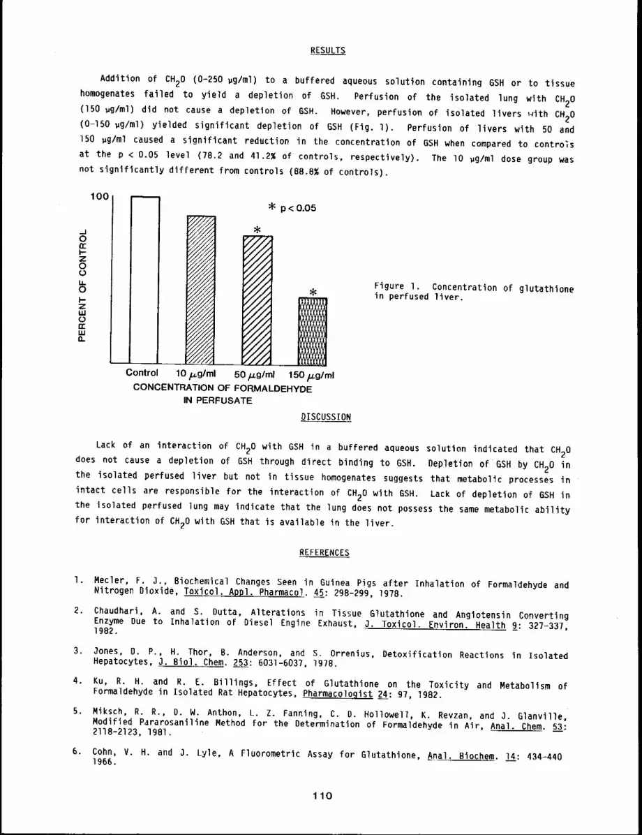
RESULTS
Addition of CH20 (0-250 ~g/ml) to a buffered aqueous solution containing GSH or to tissue
homogenates failed to yield a depletion of GSH. Perfusion of the isolated lung with CH20
(150 wg/ml) did not cause a depletion of GSH. However, perfusion of isolated livers ~vith CH20
(0-150 ~g/ml) yielded significant depletion of GSH (Fig. l). Perfusion of livers with 50
150 ~g/ml caused a significant reduction in the concentration of 6SH when compared to controls
at the p < 0.05 level (78.2 and 41.2% of controls, respectively). The lO ~g/ml dose group was
not significantly different from controls (B8.8% of controls),
0n-
Z0
U.0
zi,i
n-W(i.
I00=p < 0.05
Figure I. Concentration of glutathionein perfused liver.
DISCUSSION
Lack of an interaction of CH20 with GSH in a buffered aqueous solution indicated that CH20
does not cause a depletion of GSH through direct binding to GSH. Depletion of GSH by CH20 in
the isolated perfused liver but not in tissue homogenates suggests that metabolic processes in
intact cells are responsible for the interaction of CH20 with GSH, Lack of depletion of GSH inthe isolated perfused lung may indicate that the lung does not possess the same metabolic ability
for interaction of CH20 with GSH that is available in the liver.
REFERENCES
I. Mecler, F. 3,, Biochemical Changes Seen in Guinea Pigs after Inhalation of Formaldehyde andNitrogen Dioxide, Toxicol. Appl. Pharmacol. 45: 29B-299, 1978.
2. Chaudhari, A. and S. Dutta, Alterations in Tissue Glutathione and Angiotensin ConvertingEnzyme Due to Inhalation of Diesel Engine Exhaust, J. Toxicol. Environ. Health 9: 327-337,1982.
3. 3ones, D. P., H. Thor, B. Anderson, and S. Orrenius, Detoxification Reactions in IsolatedHepatocytes, J. Biol. Chem. 253: 6031-6037, 1978.
4. Ku, R. H. and R. E. Billings, Effect of Glutathione on the Toxicity and Metabolism ofFormaldehyde in Isolated Rat Hepatocytes, Pharmacologist 24: 97, 1982.
5, Miksch, R. R., D. N. Anthon, L, Z. Fanning, C. D. Hollowell, K. Revzan, and 3. Glanville,Modified Pararosaniline Method for the Determination of Formaldehyde in Air, Anal. Chem. 53:211B-2123, 1981.
6. Cohn, V. H. and J. Lyle, A Fluorometric Assay for Glutathione, Anal. Biochem. 14:434-4401966.
110
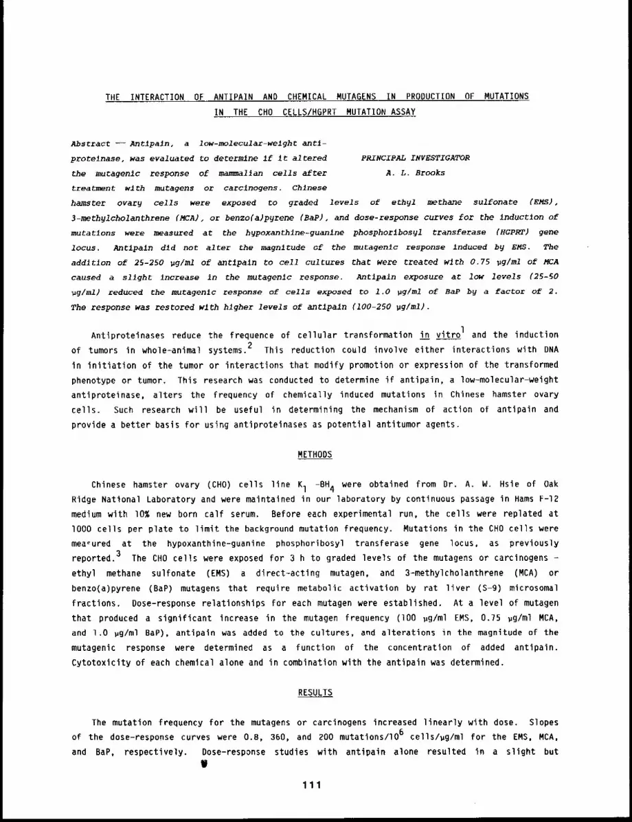
THE INTERACTION OF ANTIPAIN AND CHEMICAL MUTAGENS IN PRODUCTION OF MUTATIONS
IN THE CHO CELLS/HGPRT MUTATION ASSAY
Abstract --Antipa~n, a low-molecular-welght antl-
prote~nase, was evaluated to determine if ~t altered PRINCIPAL INVESTIGATOR
the mutagenlc response of mammal~an cells after A.L. Brooks
treatment wlth mutagens or carclnogens. Chlnese
hamster ovar9 cells were exposed to graded levels of ethyl methane sulfonate (EMS),
3-methylcholanthrene (MCA), or benzo(a)pgrene (BaP), and dose-response curves for the inductlon
mutatlons were measured at the hypoxanthlne-guanlne phosphorlbosyl transferase (HGPRT) gene
locus. Antlpaln d~d not alter the magnitude of the mutagenlc response induced by EMS. The
addltlon of 25-250 pg/ml of antipaln to cell cultures that were treated with 0.75 ~g/ml of MCA
caused a sllght increase in the mutagenlc response. Antlpain exposure at low levels (25-50
~g/ml) reduced the mutagenic response of cells exposed to 1.0 pg/ml of BaP by a factor of 2.
The response was restored wlth higher levels of antlpaln (100-250 pg/ml).
Antiproteinases reduce the frequence of cellular transformation in vitro I and the induction
of tumors in whole-animal systems. 2 This reduction could involve either interactions with DNA
in initiation of the tumor or interactions that modify promotion or expression of the transformed
phenotype or tumor. This research was conducted to determine if antipain, a low-molecular-weight
antiproteinase, alters the frequency of chemically induced mutations in Chinese hamster ovary
cells. Such research will be useful in determining the mechanism of action of antipain and
provide a better basis for using antiproteinases as potential antitumor agents.
METHODS
Chinese hamster ovary (CHO) cells line l - BH4 were o btained f rom D r. A . W . H sie o f O akRidge National Laboratory and were maintained in our laboratory by continuous passage in Hams F-12
medium with 10% new born calf serum. Before each experimental run, the cells were replated at
lO00 cells per plate to limit the background mutation frequency. Mutations in the CHO cells were
measured at the hypoxanthine-guanine phosphoribosyl transferase gene locus, as previously
reported. 3 The CHO cells were exposed for 3 h to graded levels of the mutagens or carcinogens
ethyl methane sulfonate (EMS) a direct-acting mutagen, and 3-methylcholanthrene (MCA)
benzo(a)pyrene (BaP) mutagens that require metabolic activation by rat liver (S-9) microsomal
fractions. Dose-response relationships for each mutagen were established. At a level of mutagen
that produced a significant increase in the mutagen frequency (lO0 ~g/ml EMS, 0.75 ~g/ml MCA,
and 1.O ~g/ml BaP), antipain was added to the cultures, and alterations in the magnitude of the
mutagenic response were determined as a function of the concentration of added antipain.
Cytotoxicity of each chemical alone and in combination with the antipain was determined.
RESULTS
The mutation frequency for the mutagens or carcinogens increased linearly with dose. Slopes
of the dose-response curves were O.B, 360, and 200 mutations/lO 6 cells/ug/ml for the EMS, MCA,
and BaP, respectively. Dose-response studies with antipain alone resulted in a slight but
W
111
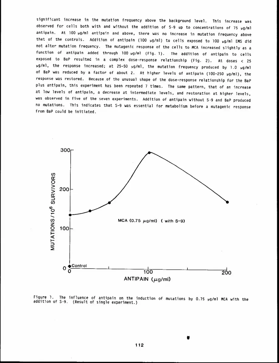
significant increase in the mutation frequency above the background level. This increase was
observed for cells both with and without the addition of S-9 up to concentrations of 75 ~g/ml
antipain. At lO0 ~g/ml antipain and above, there was no increase in mutation frequency above
that of the controls. Addition of antipain (lO0 ~g/ml) to cells exposed to lO0 ~g/ml EMS did
not alter mutation frequency. The mutagenic response of the cells to MCA increased slightly as a
function of antipain added through lO0 ~g/ml (Fig. l). The addition of antipain to cells
exposed to BaP resulted in a complex dose-response relationship (Fig. 2). At doses <
~g/ml, the response increased; at 25-50 ~g/ml, the mutation frequency produced by l.O ~g/ml
of BaP was reduced by a factor of about 2. At higher levels of antipain (I00-250 ~g/ml), the
response was restored. Because of the unusual shape of the dose-response relationship for the BaP
plus antipain, this experiment has been repeated 7 times. The same pattern, that of an increase
at low levels of antipain, a decrease at intermediate levels, and restoration at higher levels,
was observed in five of the seven experiments. Addition of antipain without S-9 and BaP produced
no mutations. This indicates that S-9 was essential for metabolism before a mutagenic response
from BaP could be initiated.
300-
O0
0>- 20>n"
0
t.~ MCA (0.75 p.g/ml) ( with S-9)Z0 100 -I-<I--
:E
oC°ntr°l i I l I0 01 O0 200
ANTIPAIN (FLg/ml)
Figure I. The influence of antipain on the induction of muzations by 0.75 pg/ml MCA with theaddition of S-9. Result of single experiment.)
112
(0n-O>
>n.- 400C)C.B
O
~0ZO 20CI-
I---23
°°°I
Control ¯
I0 100 200 300
ANTIPAIN (H.g/ml)
Figure 2. The influence of antipain on the induction of mutations by l.O ~g/ml BaP with S-9added. (S.E. are shown on points with multiple data sets.)
DISCUSSION
This research suggests that the antiproteinase, antipain, can induce a low frequency of
mutations in CHO cells with and without the addition of S-9. The molecule may thus be interacting
in some way with the genetic material. The mechanism of this interaction requires further
research. One possible mechanism is that the antipain changes plating efficiency of mutated
cells. Another is that antipain is altering some of the proteinase activity involved in
activation or regulation of genes. The major observations from this study are that antipain did
not alter the mutation frequency in cells treated with the direct-acting mutagen EMS, and did
produce changes in the frequency of mutations induced by indirect-acting mutagens MCA and BaP.
Antipain may be interacting to alter the cells directly or it may be interacting with S-9 to
modify the activation and metabolism of MCA and BaP. Additional research is being conducted to
determine if the mechanism of~ action of antipain is on the metabolism of BaP and MCA, or if it
protects cellular DNA from the formation of DNA interactions that result in mutations.
REFERENCES
1 . Kennedy, A. R. and R. R. Weichselbaum, Effects of 17 Beta-estradiol on RadiationTransformation In Vitro; Inhibition of Effects by Protease Inhibitors, Carcinogenesis 2:67-69, 1981.
2. Troll, W., A. Klassen, and A. 3anoff, Tumorgenesis in Mouse Skin: Inhibition by SyntheticInhibitors of Proteases, Science 169: 1211-1213, 1970.
3. Li, A. P., Simplification of the CHO/HGPRT Mutation Assay Through the Growth of ChineseHamster Ovary Cells as Unattached Cultures, Mutat. Res. 85: 165-175, 1981.
113
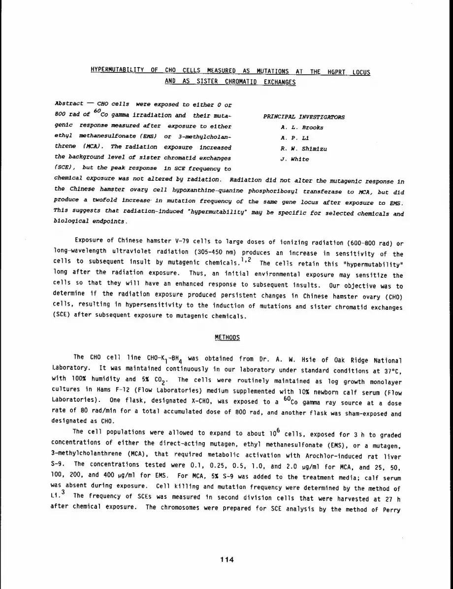
HYPERMUTABILITY OF CHO CELLS MEASURED AS MUTATIONS AT THE H6PRT LOCUS
AND AS SISTER CHROMATID EXCHANGES
Abstract -- CHO cells were exposed to either 0 or
800 rad of 60Co gamma Irradlatlon and their muta-PRINCIPAL INVESTIGATORS
genlc response measured after exposure to either A.L. Brooksethyl methanesulfonate (EMS) or 3-methylcholan- A.P. LIthrene (MCA). The radiation exposure increased R.W. Shlmlzuthe background level of sister chromatld exchanges J. White{SCE), but the peak response In SCE frequency to
chemical exposure was not altered by radiation. Radiation did not alter the mutagenlc response in
the Chinese hamster ovary cell hypoxanthlne-quanlne phosphorlhosyl transferase to MCA, but dld
produce a twofold increase, in mutation frequency of the same gene locus after exposure to EMS.
This suggests that radlatlon-lnduced "hypermutabillty" may be specific for selected chemicals and
biological endpolnts.
Exposure of Chinese hamster V-79 cells to large doses of ionizing radiation (600-800 rad)
long-wavelength ultraviolet radiation (305-450 nm) produces an increase in sensitivity of the
cells to subsequent insult by mutagenic chemicals. 1,2 The cells retain this "hypermutability"
long after the radiation exposure. Thus, an initial environmental exposure may sensitize the
cells so that they will have an enhanced response to subsequent insults. Our objective was to
determine if the radiation exposure produced persistent changes in Chinese hamster ovary (CHO)
cells, resulting in hypersensitivity to the induction of mutations and sister chromatid exchanges
(SCE) after subsequent exposure to mutagenic chemicals.
METHODS
The CHO cell line CHO-KI-BH4 was obtained from Dr. A. W. Hsie of Oak Ridge NationalLaboratory. It was maintained continuously in our laboratory under standard conditions at 37°C,
with lOO% humidity and 5% CO2. The cells were routinely maintained as log growth monolayer
cultures in Hams F-12 (Flow Laboratories) medium supplemented with I0% newborn calf serum (Flow
Laboratories). One flask, designated X-CHO, was exposed to a 60Co gamma ray source at a dose
rate of 80 rad/min for a total accumulated dose of 800 rad, and another flask was sham-exposed and
designated as CHO.
The cell populations were allowed to expand to about lO6 cells, exposed for 3 h to graded
concentrations of either the direct-acting mutagen, ethyl methanesulfonate (EMS), or a mutagen,
3-methylcholanthrene (MCA), that required metabolic activation with Arochlor-induced rat liver
S-9. The concentrations tested were O.l, 0.25, 0.5, l.O, and 2.0 wg/ml for MCA, and 25, 50,
lOO, 200, and 400 wg/ml for EMS. For MCA, 5% S-9 was added to the treatment media; calf serum
was absent during exposure. Cell killing and mutation frequency were determined by the method of
Li. 3 The frequency of SCEs was measured in second division cells that were harvested at 27 h
after chemical exposure. The chromosomes were prepared for SCE analysis by the method of Perry
114
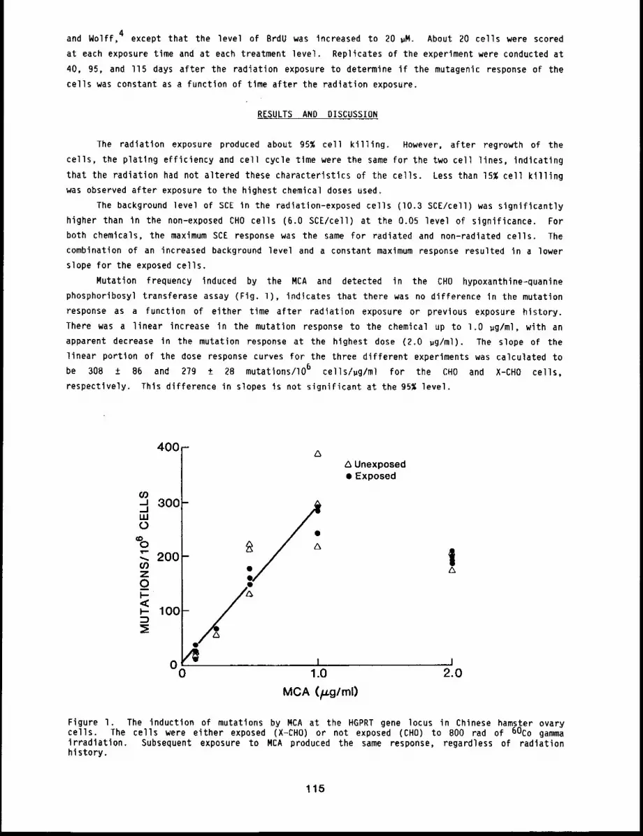
and Wolff, 4 except that the level of BrdU was increased to 20 ~M. About 20 cells were scored
at each exposure time and at each treatment level. Replicates of the experiment were conducted at
40, 95, and ll5 days after the radiation exposure to determine if the mutagenic response of the
cells was constant as a function of tlme after the radiation exposure.
RESULTS AND DISCUSSION
The radiation exposure produced about 95% cell killing. However, after regrowth of the
cells, the plating efficiency and cell cycle time were the same for the two cell lines, indicating
that the radiation had not altered these characteristics of the cells. Less than 15% cell killing
was observed after exposure to the highest chemical doses used.
The background level of SCE in the radiatlon-exposed cells (I0.3 SCE/cell) was significantly
higher than in the non-exposed CHO cells (6.0 SCE/cell) at the 0.05 level of significance. For
both chemicals, the maximum SCE response was the same for radiated and non-radiated cells. The
combination of an increased background level and a constant maximum response resulted in a lower
slope for the exposed cells.
Mutation frequency induced by the MCA and detected in the CHO hypoxanthine-quanine
phosphoribosyl transferase assay (Fig. 1), indicates that there was no difference In the mutation
response as a function of either time after radiation exposure or previous exposure history.
There was a linear increase in the mutation response to the chemical up to 1.O ug/ml, with an
apparent decrease in the mutation response at the highest dose (2.0 ug/ml). The slope of the
linear portion of the dose response curves for the three different experiments was calculated to
be 308 ± 86 and 279 ± 28 mutations/lO 6 cells/ug/ml for the CHO and X-CHO cells,
respectively. This difference in slopes is not significant at the 95% level.
400
..u 300
._JILl0
£ 200
Unexposed¯ Exposed
i 101.0 2.0
MCA (p.g/ml)
Figure I. The induction of mutations by MCA at the HGPRT gene locus in Chinese hamster ovarycells. The cells were either exposed (X-CHO) or not exposed (CHO) to 800 rad of 60Co gammairradiation. Subsequent exposure to MCA produced the same response, regardless of radiationhistory.
115
Figure 2 illustrates the frequency of mutations induced in both cell lines by EMS. The data
were evaluated by first plotting the mutation frequency, mutations/lO 6 surviving cells, against
both the concentration of EMS and time after radiation exposure. This plot indicated little or no
trend for changes in the mutation response to EMS in either radiated or non-radiated cells over
time. Multiple regression techniques were used to test formally for the presence of a trend over
time, and at level of significance 0.05, the change in mutation response with time was not
significant. The data for all three sampling times after radiation, 40, 95, and ll5 days, could
thus be ~rouped, and the dose-response relationships for EMS estimated. F-test on the differences
in the slopes of the dose-response relationships between exposed and unexposed cells indicated
that the slope for the unradiated cells (0.51 ± 0.04 mutations/lO 6 cells/~g/ml) was, at the
0.05 level, significantly lower than the slope for the cells exposed to the 60Co gamma rays
(0.89 ± 0.04 mutations/lO 6 cells/~g/ml). Thus, previous radiation exposure caused
significant, almost twofold, increase in the sensitivity of the cells to the induction of
mutations with EMS.400 ¯
This research suggests that radiation
exposure apparently causes an increase in the
mutagenic responsiveness of CHO cells to the
direct alkylating agent EMS and that this
response is persistent with time. The magni-
tude of the difference in responsiveness to
the alkylating agent EMS, while significant,
is not large, compared to that reported by
Frank and Williams 2 for UV light and 8-metho-
xypsoralen. On the other hand, the radiation
did not increase the sensitivity of mutation
induction in the cells exposed to the indirect
alkylating agent MCA or increase the maximum
response of the cells to the induction of SCE
with either of the two mutagens. Thus, the
data indicate that "hypermutability" may not
be a general phenomenon for all genetic end-
points, chemical mutagens, and cell lines.
l ¯
2°
REFERENCES
Burger, P. M. and 3. W. Simons, Mutagen-icity of 8-Methyoxypsoralen and LongWave Length Ultraviolet Irradiation inV-79 Chinese Hamster Cells, Mutat. Res.60: 381-38g, 1979.
300
LU0
0
"" 200-Z0I--<I.-
100-
Frank, J. P. and J. R. Williams, X-rayInduction of Persistent Hypersensitivityto Mutation, Science 216: 30?-308, 19B2.
A Unexposed¯ Exposed
0 0100 200 300 400
EMS (~g/ml)Figure 2. The inductions of mutations by EMSat the HGPRT gene locus in Chinese hamsterovary cells. The cells were either exposed(X-CHO) or not exposed to 800 rad of 60Cogamma irradiation. The difference between themutagenic response to EMS of the two celllines was significant at the 5% level wherethe data from all three times were combined.
3. Li, A. P., Simplification of the CHO/HGPRT Mutation Assay Through the Growth of ChineseHamster Ovary Cells as Unattached Cultures, Mutat. Res. BS: 165-175, 1981.
Perry, P. and S. Wolff, New Giemsa Method for the Differential Staining of SisterChromatids, Nature 251: 156-158, 1974.
116
INDUCTION OF SISTER CHROMATID EXCHANGES IN LUN6 CELLS EXPOSED TO 3-METHYLCHOLANTHRENE
Abstract --- Cells derived from Chinese hamster
lungs were establ~shed in culture, and the induction PRINCIPAL INVESTIGATORS
of sister chromatld exchanges (SCE) was determined R.W. Shlmlzu
after inhalation or in vitro exposure to 3-methyl- A.L. Brooks
cholanthrene (HCA). A reproduclhle dose-response J.D. Sun
relatlonshlp for SCE induction in vltro by MCA was G.J. Newton
observed wlth cultures established at four different
monthly intervals. A slngle inhalation exposure to MCA (350 pg/L) produced a slgnlf~canC
increase in SCR frequency, whereas repeated exposure to a lower concentration (250 pg/L} dld noE
significantly alter frequency. The measurement of SCEs in lung cells provides a potentlally rapid
method to estlmate genotoxlc interaction of MCA after elther inhalatlon or In vltro exposure.
Short-term genotoxicity assays have shown positive correlations between genotoxicity and
carcinogenicity using a variety of endpoints and target cells, l Many short-term tests have the
disadvantage of using target cells not at risk in human exposure.
We have been examining the potential for genotoxic effects in cells of the respiratory tract.
To evaluate the interaction of chemicals with lung cells, a genotoxicity assay using sister
chromatid exchanges (SCE) as an endpoint has been developed and characterized for use after either
in vitro or in vivo exposures. The lung cell system reported here has the advantage of using
normal target cells that still retain the metabolic capacity to activate promutagens deposited in
lung after inhalation exposure.
MATERIALS AND METHODS
3-methylcholanthrene (MCA) was obtained from the Tridom Chemical Company, Hauppauge, NY.
Carbon-14 labeled MCA was obtained from New England Nuclear, Boston, MA (0.2 mCi/mg).
Female Chinese hamsters CHI:(CHN) approximately lO0 days old were exposed to MCA by nose-only
inhalation and were used as the source of primary lung cell cultures. The lung cell cultures were
obtained using a collagenase (ll5 units/ml) and elastase (l unit/ml) enzyme system, grown at
in Ham’s F-12 medium (Flow Lab, Inglewood, CA) supplemented with I0% newborn calf serum,
penicillin, and streptomycin (K. C. Biological, Lenexa, KS). Primary isolates were designated
passage O, with each subsequent passage occurring after growth to confluency involving a l:l
dilution of cells, so that each passage represented approximately one populatlon doubling.
In vitro treatment cultures were established from passage l cultures. MCA was delivered in
graded concentrations 0.25, 0.5, 1.0, 2.0, and 4.0 ug/ml of medium in dimethyl sulfoxide (DMSO,
Burdick and Jackson Lab, Muskegon, MI). NO S-9 was added to the cultures. All in vitro
treatments lasted 3 h and were done in medium containing lO ~M bromodeoxyuridine (BrdU; Sigma
Chemical Co., St. Louis, MO) for sister chromatid differentiation. Treatment medium was then
replaced with growth medium containing BrdU, and cultures were incubated 24 h. Chromosome cells
were collected at metaphase, differentially stained, 2 slides were coded, and SCE frequency was
scored.
A condensation aerosol generation system for MCA was developed using a design similar to that
developed for pyrene. 3 Three groups of five lO0-day-old female Chinese hamsters were exposed
117
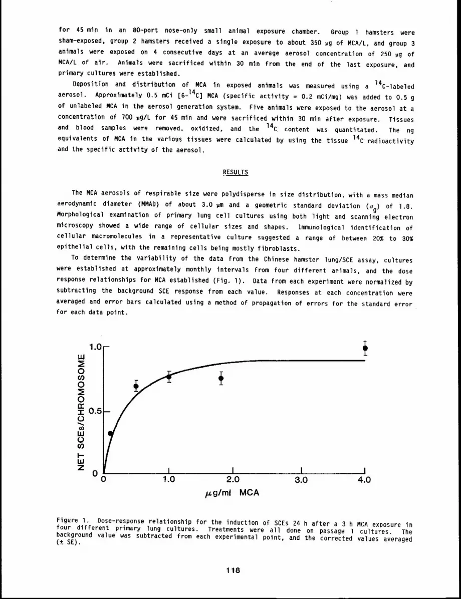
for 45 mln in an 80~port nose-only small animal exposure chamber. Group l hamsters were
sham-exposed, group 2 hamsters received a single exposure to about 350 ~g of MCA/L, and group 3
animals were exposed on 4 consecutive days at an average aerosol concentration of 250 pg of
MCA/L of air. Animals were sacrificed within 30 mln from the end of the last exposure, and
primary cultures were established.
Deposition and distribution of MCA in exposed animals was measured using a 14C-labeled
aerosol. Approximately 0.5 mCi [6-14C] MCA (specific activity = 0.2 mCi/mg) was added to 0.5
of unlabeled MCA in the aerosol generation system. Five animals were exposed to the aerosol at a
concentration of 700 ~g/L for 45 min and were sacrificed within 30 min after exposure. Tissues
and blood samples were removed, oxidized, and the 14C content was quantitated. The ng
equivalents of MCA in the various tissues were calculated by using the tissue 14C-radioactivity
and the specific activity of the aerosol.
RESULTS
The MCA aerosols of respirable size were polydisperse in size distribution, with a mass median
aerodynamic diameter (MMAD) of about 3.0 ~m and a geometric standard deviation (~g) of
Morphological examination of primary lung cell cultures using both light and scanning electron
microscopy showed a wide range of cellular sizes and shapes. Immunological identification of
cellular macromolecules in a representative culture suggested a range of between 20% to 30~
epithelial cells, with the remaining cells being mostly fibroblasts.
To determine the variability of the data from the Chinese hamster lung/SCE assay, cultures
were established at approximately monthly intervals from four different animals, and the dose
response relationships for MCA established (Fig. l). Data from each experiment were normalized
subtracting the background SCE response from each value. Responses at each concentration were
averaged and error bars calculated using a method of propagation of errors for the standard error
for each data point.
1.0
oo
)
mZ . 1
0 1.0 2.0 3.0 4.0
/~g/ml MCA
Figure I. Dose-response relationship for the induction of SCEs 24 h after a 3 h MCA exposure infour different primary lung cultures. Treatments were all done on passage 1 cultures. Thebackground value was subtracted from each experimental point, and the corrected values averaged(± SE).
118
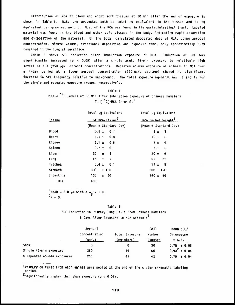
Distribution of MCA in blood and eight soft tissues at 30 min after the end of exposure is
shown in Table I. Data are presented both as total ng equivalent in the tissue and as ng
equivalent per gram wet weight. Most of the MCA was found in the gastrointestinal tract. Labeled
material was found in the blood and other soft tissues in the body, indicating rapid absorption
and disposition of the material. Of the total calculated deposited dose of MCA, using aerosol
concentration, minute volume, fractional deposition and exposure time, only approximately 3.3~
remained in the lung at sacrifice.
Table 2 shows SCE induction after inhalation exposure of MCA. Induction of SCE was
significantly increased (p < 0.05) after a single acute 45-min exposure to relatively high
levels of MCA (350 ~g/L aerosol concentration). Repeated 45-min exposure of animals to MCA over
a 4-day period at a lower aerosol concentration (250 ~g/L average) showed no significant
increase in SCE frequency relative to background. The total exposure mg.min/L was 16 and 45 for
the single and repeated exposure groups, respectively.
Table l
Tissue 14C Levels at 30 Min After Inhalation Exposure of Chinese Hamsters
To [14C]-MCA AerosolsI
Tissue
Blood
Heart
Kidney
Spleen
Liver
Lung
Trachea
Stomach
Intestine
TOTAL
Total gg Equivalent
of MCA/Tissue2
Total gg Equivalent
MCA .qm Wet Weight2
(Mean + Standard Dev) (Mean ± Standard Dev)
0.8 ± 0.7 2 _+ l
1.5 ± 0.8 lO _+ 3
2.1 +_ 0.8 7 ± 4
0.2 + 0.I 3 _+ 2
20 +_ 5 20 +_ 6
15 + 5 65 +_ 25
0.4 ± 0.I 17 +_ 9
300 + l O0 300 ± 150
150 ± 60 190 + 96
490
IMMAD = 3.0 ~m with a ~g = l.B.
2N = 5.
Table 2
SCE Induction in Primary Lung Cells from Chinese Hamsters
6 Days After Exposure to MCA AerosolsI
Sham
Single 45-min exposure
4 repeated 45-mln exposures
Aerosol Cell Mean SCE/
Concentration Total Exposure Number Chromosome
(uq/L) . (m~.mln/L) Counted ± S.E.
0 0 30 0.75 ± 0.05
350 16 60 0.932 ± 0.04
250 45 42 0.79 ± 0.04
Iprimary cultures from each animal were pooled at the end of the sister chromatid labelingperiod.
2Signiflcantly higher than sham exposure (p < 0.05).
119
DISCUSSION
The use of primary cultures to ascertain genotoxic risk to specific organs after in vivo
exposure may be more appropriate than the use of microsomal preparations coupled with transformed
fibroblasts as target cells. Primary cultures contain a mixed population of cell types, but the
SCE response to MCA for primary cultures isolated from four different animals was very similar.
Such reproducibility in SCE response is essential for a genotoxicity assay.
The distribution of inhaled MCA in Chinese hamsters showed that a large fraction of the
deposited chemical was cleared rapidly from the site of deposition to other tissues. Our early
distribution data for MCA suggest that MCA has clearance kinetics similar to the rapid clearance
of benzo(a)pyrene.4 Such rapid clearance decreases the effective dose to lung cells.
A single acute exposure of Chinese hamsters to the MCA aerosol with a total exposure of 16
mg.min/L was effective in inducing SCE formation in primary lung cells isolated from these
animals. However, no change in SCE frequency was detected after multiple exposure, which resulted
in a greater total exposure 45 mg.min/L. Similar findings have been reported by Schmid e~
a l. 5 They postulated that with protracted exposure, damaged cells may be removed from the cell
population. It is also possible that repeated exposures induced detoxification enzyme pathways
that decreased the effective dose of reactive metabolites to the lung cells. It is also possible
during the time between exposure and sampling repair of SCE lesions resulted in a decrease in
response.
Comparison of the in vivo and in vitro data indicate that in vitro techniques are much more
sensitive in detecting genotoxic damage than in vivo techniques. Although in vivo testing is less
sensitive, it may be more realistic in predicting possible health risks to exposed humans. Before
in vivo inhalation exposure becomes useful as a short-term screening assay, further
characterization of mechanisms of interaction of the chemical in the exposed animals, the dose to
the lung cells, and the toxification and detoxification pathways and rates for chemical are needed
to understand the response of the animal to the genotoxicant.
REFERENCES
I. Russell, L. B. and B. E. Matter, Whole-Mammal Mutagenicity Tests: Evaluation of Five Methods,Mutat. Res. 75: 2?9-302, 1980.
2. Wolff, S., Sister Chromatid Exchange, Ann. Rev. Genet. l_!l: 183-201, 1977.
3. Tu, K. W. and G. M. Kanapilly, A High Capacity Condensation Aerosol Generation System,Environ. Sci. and Technol. 13: 69B-701, 1979.
4. Mitchell, C. E., Distribution and Retention of Benzo(a)pyrene in Rats After inhalation,Toxicol. Lett. l_]_l: 35-42, 1982.
5. Schmid, W., D. T. Arakaki, N. A. Breslau, and J. C. Cuthbertson, Chemical Mutagenesis: TheHamster Bone Marrow as an In Vivo Test System, Hum. Genet. ll: I03-118, 1971.
120
DEPOSITION AND FATE OF INHALED MATERIALS
The conduct of inhalation toxicity studies and their use for predicting the health risks to
man from inhaled toxicants require an accurate knowledge of the dose to the tissues at risk.
Factors influencing this dose are deposition patterns of the inhaled material in the respiratory
tract, its retention in the respiratory tract, the metabolism of the material in the respiratory
tract and its disposition after its translocation from the respiratory tract. All of these
factors may be influenced by the species of animals used for the study. To predict dose to
critical tissues in man for estimating health risks, the influence of these factors must be known
for the material in question. Twelve papers in this Section present results of studies designed
to determine the dose to critical tissues after inhalation of particles.
The first three reports deal with the influence of three different factors; species, particle
size, and exercise, on the deposition and retention of inhaled particles. Previous results of
aerosol deposition and retention studies in guinea pigs have indicated that this species deserve
further investigation as a useful experimental model. The first paper in this series describes
results obtained when guinea pigs inhaled 134Cs-labeled fused aluminosilicate particles.
Results through 64 days after exposure suggest a prolonged pulmonary retention llke that in dogs,
a major difference from patterns seen in rats. The newly developed exposure system for large
particles was used to expose rats to microspheres having geometric diameters of 3, 9, or 15 gm.
Results to date suggest that the 9- and 15-~m particles did not reach the deep lung in
significant numbers, but further research is required to define the level of deposition and
retention more accurately. When rats were exposed by inhalation to 67GaO3 particles while
exercising on a treadmill, the relative deposition in the nasopharyngeal region was increased and
that in the pulmonary region was decreased, compared to patterns observed in non-exercising rats.
The fourth report describes preliminary results of local dosimetry analyses for cell nuclei in
dogs that inhaled 239pu02 particles. Such analyses are needed to relatethe lungs of three
the alpha emissions from discrete, non-uniformly deposited 239pu02 particles and the doses
received by critical cell nuclei. A study of the influence of the magnitude of a lung burden of
239pu02 on its pulmonary retention is the subject of a study reported in the fifth paper. At
one year after exposure, the clearance patterns seen in dogs that inhaled 0.72 pm AMAD
239pu02 particles are indistinguishable among dogs with initial lung burdens of 0.07, 0.07 and
0.0007 ~Ci 23gPu/kg body weight.
The sixth and seventh papers present results of examining the effects of age at exposure and
species on the disposition and dosimetry of 242Am inhaled as 241AmO2. Immature and aged
dogs were used to study age=related effects. The study is still in its early stages, and no
definitive differences have yet emerged but a trend toward greater skeletal uptake of 241Am in
skeleton vs. liver appears to be developing for immature dogs than for aged dogs. To expand our
understanding of the link between results in dogs and people, selected studies are being carried
out in a non-human primate, the cynomolgus monkey. One such study has been started in which a
small group of these monkeys was exposed to 241Am02 by inhalation. Results from the first
animals sacrificed in the early post-exposure period yielded tissue distribution patterns slmlar
to those seen in dogs.
The eighth and ninth papers deal with factors influencing the patterns of dose received by
dogs from inhaled 243’244Cm aerosols. A study comparing the deposition and retention of Cm in
dogs that inhaled either a relatively soluble form, Cm(N03)3, or a relatively insoluble form,
Cm203, is underway; its current status is presented. The efficicacy of reducing the amount of
Cm reaching other body organs after being absorbed from the lung was examined by comparing the
121
results after continuous DTPA administration from an implanted osmotic pump with repeated discrete
DTPA injections. Although both methods were effective, the continuous infusion approach was
slightly better at removing Cm from the skeleton.
The last three papers in this section deal with factors influencing the tissue distribution
and metabolic fate of several inhaled organic chemicals. Studies with inhaled
[3H]-2-aminoanthracene (2-AA) showed that radioactivity was widely distributed in body tissues.
Analysis of organic soluble radioactivity in the excreted urine indicated inhaled 2-AA is
extensively metabolized by rats to conjugated materials and excreted as N-glucuronides,
O-glucuronides, and sulfates. Another study dealt with the effect of pre-treatment with
benzo(a)pyrene (BaP) on the binding of 14C-l-nitropyrene and its metabolites to mouse lung
The results indicate that in the mouse lung BaP induces enzymes that metabolize nitropyrene to
products that bind covalently to DNA. Other work with BaP dealt with the question of how the
amount of BaP instilled into the lung may affect the subsequent clearance to blood. As the amount
of BaP instilled was increased, a larger fraction was cleared in the rapidly cleared component.
These dose-related effects need to be considered when dealing with the retention of low total
quantities of BaP.
122
DEPOSITION AND RETENTION OF MONODISPERSE ALUMINOSILICATE PARTICLES
INHALED BY GUINEA PIGS
Abstract -- Gulnea p~gs were exposed nose-onlg to
an aerosol contalnlng 2.0 ~m actlvlty median aero- PRINCIPAL INVESTIGATORS
dgnamlc diameter 134Cs-labeled fused alumlnoslllcate M.B. Snlpes
particles. Whole body, excreta, and tlssue content R.O. McClellan
of 194Cs are being analyzed to define the fate of
the initial burden of inhaled radlolabeled particles. Comparlsons are made wlth models from a
stud 9 wlth rats and dogs that inhaled similar aerosols. Results through da9 64 after exposure
suggest: (i) fractlonal deposition patterns are approxlmatel9 the same for guinea pigs and rats,
(2) lung clearance patterns for deposited particles are similar for gulnea pigs and dogs, and
(3) clearance of particles from the lung to lung-assoclated 19mph nodes of guinea p~gs has the
same pattern as that of dogs (constant transfer rate), but occurs at a rate approximately 10 rlmes
slower than that for dogs.
Guinea pigs have been used in many inhalation studies, but none of the studies was designed to
examine long-term retention patterns for inhaled particles. Guinea pigs represent gentle,
relatively long-lived rodents (life span 6 to 8 years) that have considerable potential for use
chronic inhalation exposure studies. They have deposition and early retention patterns more
similar to those of dogs than of rats.l’2 This study was designed to determine the deposition
and long-term retention patterns for a well characterized, much used aerosol inhaled by guinea
pigs. The study duration is 3 years, with emphasis on pulmonary retention, accumulation of
particles in lung-associated lymph nodes, and mechanical clearance by way of the mucociliary
escalator to the gastrointestinal tract. The study uses relatively insoluble 134Cs-labeled
fused aluminosilicate particles to allow a direct comparison with data from studies done at this
Institute using mice, rats, and dogs.3
METHODS
Forty-five female guinea pigs (CrI:(HA)BR), lO weeks old and weighing 460 to 610 grams
used. They were housed individually in polycarbonate cages with hardwood chip bedding. A diet of
guinea pig pellets (Allied Mills, Chicago, IL) and water were available ad libitum; the diet was
supplemented twice per week with raw cabbage. Temperature was maintained at 20°C to 22°C, with
relative humidity at 40~ to 60% with a 12-h light/dark cycle. They were exposed nose-only to an
aerosol of 134Cs-labeled fused aluminosilicate particles using the multiport small animal
inhalation exposure systems commonly used at this Institute. The aerosol was monodisperse, with
an activity median aerodynamic diameter (AMAD) of 2.0 ~m as characterized with a Mercer
impactor. Exposure time was 75 minutes.3
The guinea pigs were randomly assigned to a sacrifice schedule in which three animals were
killed one hour after inhalation exposure or 4, 16, 34, 64, 128, 256, 384, 512, 640, 768, 896, or
I096 days after exposure. The other six guinea pigs will be assigned for sacrifice at the later
times in the schedule or times beyond I096, depending on results through day 640. All of the
animals were weighed and underwent whole-body counting in a large-volume liquid scintillation
counting system immediately after inhalation exposure; all live animals were weighed and counted
123
again on days 4, 8, 16, 34, 64, 96, 128, and every two months thereafter’ until the end of the
study or sacrifice. In all cases, animals were counted or will also be counted on the day of
sacrifice.
Urine and feces collections were made daily for the first 16 days after exposure and are being
made periodically for the six guinea pigs scheduled to be killed at 896 or I096 days after
inhalation exposures. Stainless steel metabolism cages that segregate urine and feces are used.
The later collections are made for three consecutive days at approximately the same times as
listed for whole body counting.
Guinea pigs are killed with an intraperitoneal injection of 2 ml of T-61 euthanasia solution.
Cardiac puncture is used to obtain 5 ml of whole blood; the guinea pigs are then dissected to
obtain specific organs and tissue samples. Emphasis is placed on careful segregation of
components of the respiratory tract, but the gastrointestinal tract, liver, kidneys, muscle
sample, pelt, and carcass are collected and counted for 134Cs to account for the body burden of
134Cs. Results from these counts are used to define and model the fate of the inhaled
radiolabeled particles as a function of time after exposure.
RESULTS AND DISCUSSION
Initial body burdens of 134Cs averaged 0.87 #Ci (range was 0.45 to 1.61 #Ci); initial
lung burdens averaged 0.15 #Ci (range was 0.058 to 0.28 ~Ci). The initial body burdens were
based on whole-body counts made immediately after inhalation exposure. About I0% of the initial
body burden was external contamination, primarily associated with the external nares. The initial
lung burdens were estimated as follows: lung burdens for guinea pigs sacrificed on the day of
exposure or day 4 were assumed equal to the initial lung burden; initial lung burdens for the
other guinea pigs were estimated from whole body counts and the ratio of 134Cs in lung vs. whole
body with time after exposure. An average of 15% of the initial body burden was in the deep
’lung. Figure l contains whole--body retention data through day 64 expressed as percent of the
initial body burden. This figure includes data for all guinea pigs alive as of day 64. After the
early clearance phase (~ day lO), the whole body retention could be represented by a single
exponential expression having an effective half=tif,le of 175 days.
Data for lung and lymph nodes are presented in Figures 2 and 3, also expressed as percent of
the initial body burden. Figures 4 and 5 contain data for urine and feces, expressed as percent
of the initial body burden per day. Data are presented in this way to allow use of the materials3balance simulation model used previously for mice, rats, and dogs. Results from this study
with guinea pigs are still being analyzed, so it is premature go present a definitive materials
balance model in this report. However, for a preliminary comparison, the models for rats and
dogs3 were included in Figures 2, 3, 4, and 5. Initial deposition values in the rat and dog
model Were adjusted to reflect 15% lung deposition observed for the guinea pigs; also, the
translocation rate of particles from lung to lung-associated lymph nodes was changed from 0.0002
day-l for dogs to 0.00003 day-1 for the guinea pigs. Therefore, the lines in Figures 2, 3, 4,
and 5 are approximations of the models for rats and dogs applied to guinea pigs. This preliminary
comparison suggests that modeling results for the guinea pigs will be much more similar to dogs
than to rats. A complete materials balance model for the guinea pigs will be presented when the
study is completed.
Based on early results and the above tentative comparison with models for dogs and rats: (l)
deposition patterns are basically the same in rats and guinea pigs exposed nose-only in this
Institute’s multiport exposure systems, (2) lung clearance patterns are similar for Beagle dogs
and guinea pigs, with lung clearance obviously much slower for guinea pigs than for rats, and (3)
124
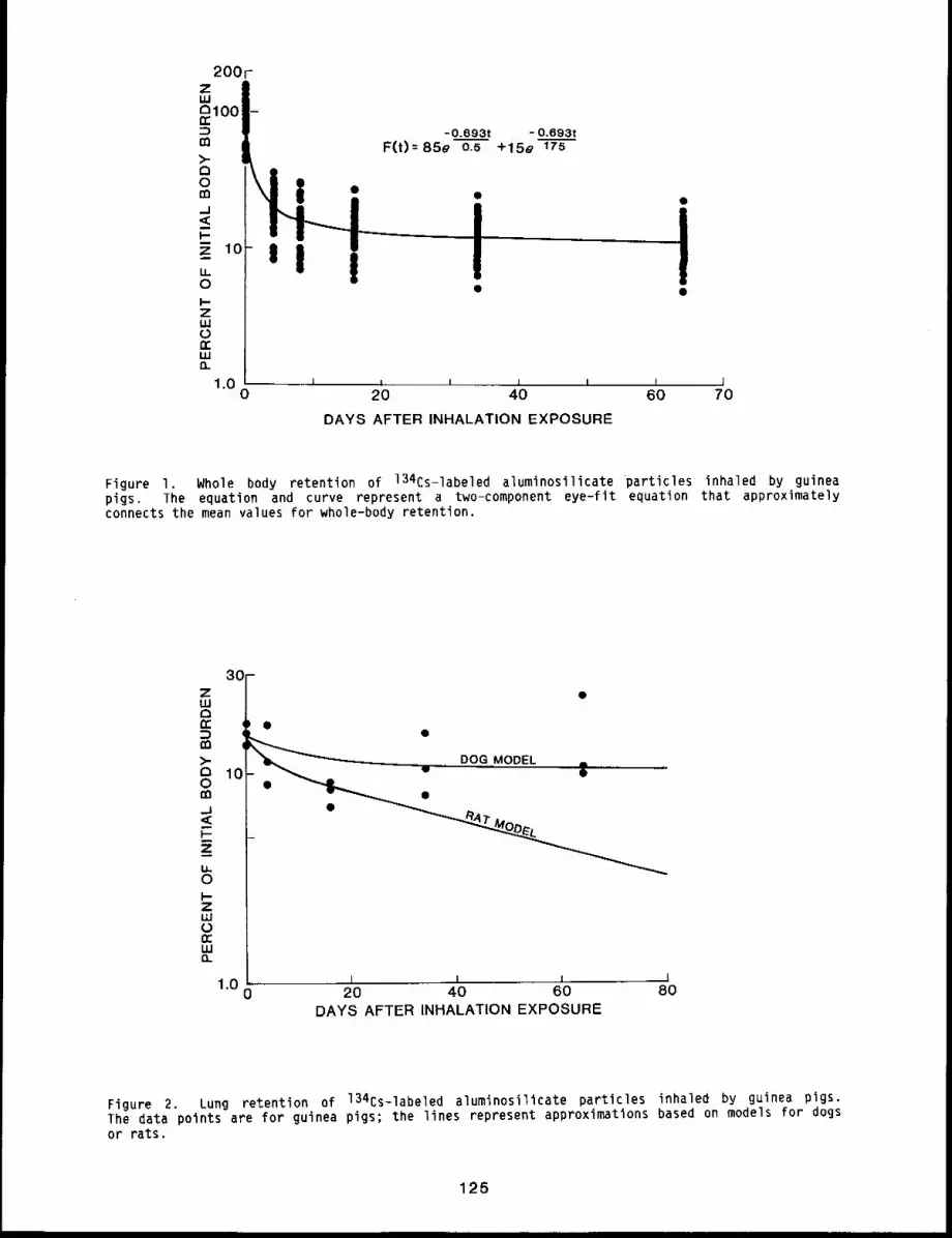
!
III
-0.693t - 0.693tF(t)=85e 0.5 +15e 175
I I | I i
20 40
DAYS AFTER INHALATION EXPOSURE
I I60 70
Figure I. Whole body retention of 134Cs-labeled aluminosilicate particles inhaled by guineapigs. The equation and curve represent a two-component eye-fit equation that approximatelyconnects the mean values for whole-body retention.
,,,=3°Fotr
>-r~ 100
_J<I-
I&,oI--ZUJ0frI,LI13_
i DOG MODEL ;
1.0 I I 1 l20 40 60 80
DAYS AFTER INHALATION EXPOSURE
Figure 2. Lung retention of 134Cs-labeled aluminosilicate particles inhaled by guinea pigs.The data points are for guinea pigs; the lines represent approximations based on models for dogs
or rats.
125
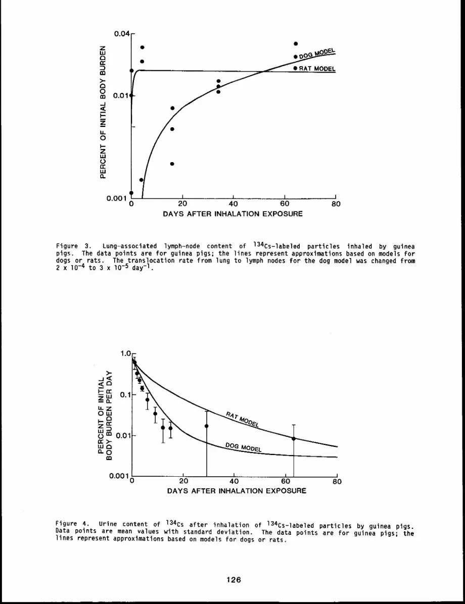
0.04 -
ZLU0n~
m>-C~0m 0.01.J
Z
LL0I-ZLU
n"LUO_
r¯ RAT MODEl
t0.001 I , = I
0 20 40 60 80DAYS AFTER INHALATION EXPOSURE
Figure 3. Lung-associated lymph-node content of 134Cs-labeled particles inhaled by guineapigs. The data points are for guinea pigs; the lines represent approximations based on models fordogs or rats The translocatlon rate from lung to lymph nodes for the dog model was changed from2 x lO -4 to 3 x lO-5 day-l.
1.0--
=J<40r-rr o.1LLZOW
o 0.01nr>-
II1
10 i I . I I I0.00
20 40 60 80DAYS AFTER INHALATION EXPOSURE
Figure 4. Urine content of 134Cs after inhalation of 134Cs-labeled particles by guinea pigs.Data points are mean values with standard deviation. The data points are for guinea pigs; thelines represent approximations based on models for dogs or rats.
126
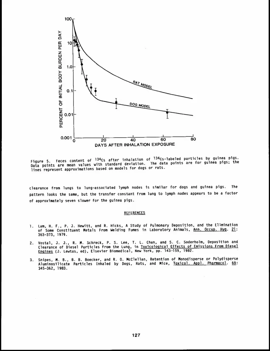
100
<C~~: 10wQ.ZtuDri-
m 1.0>-o0n-=._1<C
0.1ziJ.0I-Zw 0.01-0n.-LUO.-
0
0.001 0 20 40 60 80
DAYS AFTER INHALATION EXPOSURE
Figure 5. Feces content of 134Cs after inhalation of 134Cs-labeled particles by guinea pigs.Data points are mean values with standard deviation. The data points are for guinea pigs; thelines represent approximations based on models for dogs or rats.
clearance from lungs to lung-associated lymph nodes is similar for dogs and guinea pigs. The
pattern looks the same, but the transfer constant from lung to lymph nodes appears to be a factor
of approximately seven slower for the guinea pigs.
REFERENCES
I. Lam, H. F., P, 3. Hewitt, and R. Hicks, A Study of Pulmonary Deposition, and the Eliminationof Some Constituent Metals From Welding Fumes in Laboratory Animals, Ann. Occur, 2__ll:363-373, 197g.
2. Vostal, 3. J., R. M, Schreck, P. S. Lee, T. L. Chart, and S. C. Soderholm, Deposition andClearance of Diesel Particles From the Lung, in Toxicological Effects of Emissions From DieselEngines (3. Lewtas, ed), Elsevier Biomedical, New York, pp, 143-15g, 1982.
3. Snipes, M. B., B. B. Boecker, and R. O. McClellan, Retention of Monodisperse or PolydisperseAluminosilicate Particles Inhaled by Dogs, Rats, and Mice, Toxicol. Appl. Pharmacol. 6_9g:345-362, 1983.
127
DEPOSITION AND RETENTION PATTERNS FOR 3-, g-, AND lS-pm _LATEX MICROSPHERES
INHALED BY RATS
Abstract -- Rats were exposed by inhalation to
radlolabeled 3-, 9-, or 15-~m polystyrene latex PRINCIPAL INVESTIGATORSmlcrospheres. Approxlmatel9 2.1~ of the inlClal M.B. Snipesbody burden of 3-~m mlcrospheres was in the deep T. R~ Olsonlung; less than 0.01~ of the 9- or 15-~m mlcro- H.C. Yehspheres was in the deep l~g. Data are being
analyzed to define the fate of these mlcrospheres after inhalation. Results to date suggest that
9- and l$-~m mlcrospheres dld not reach the deep lung in significant numbers; 3-~m
mlcrospheres were deposlted and cleared by the respiratory tract as would be expected based on
other inhalation exposures of rats to aerosols of similar or smaller size.
Although large particles (> lO ~m diameter) have a small but non-zero probability
reaching the deep lung by inhalation, their subsequent fate is unknown. Large particles deposit
primarily in the upper respiratory tract and are quickly cleared, passed through the
gastrointestinal tract, and excreted. Because large particles may deposit in the deep lung during
inhalation exposure, it is prudent to study their deposition and retention patterns in the
respiratory tract with emphasis on the deep lung. Large particles containing hazardous materials
could present a significant local dosimetry problem in the deep lung because of their large mass
and potential for injury to sensitive cells in their proximity.
In a previous study, 3-, 9-, and 15-~m polystyrene latex microspheres were instilled into
the lungs of Fischer-344 rats to study the fate of large particles deposited in lung. l The
results suggested that particles approximately 9-~m diameter might represent the size limit for
translocation from lung to lung-associated lymph nodes and demonstrated that 9- and 15-~m
diameter microspheres, once deposited in the deep lung, clear very slowly from rats. However,
that study used intratracheal instillation; the study reported here was designed to repeat that
study, but with inhalation as the method of exposure.
METHODS
Three sizes of polystyrene latex microspheres were obtained in dry form from 3M Company, St.
Paul, MN. They were 3-, 9-, and 15-pm geometric diameter, uniformly labeled with 46Sc. A
fluid bed generation system was used to produce inhalation atmospheres in an integrated
generation-exposure system specially designed for this purpose (see this report, pp. 31 to 36 for
a description of the system).
Fischer-344 rats, laboratory-reared, specific pathogen-free were used. Rats were lO to 12
weeks old when exposed, Groups of 36 rats, equal numbers of males and females, were exposed for
90 min (15 ~m) or 60 min (g and 3 ~m) in a nose-only exposure system. Whole-body counts
done immediately after exposure in a large volume liquid scintillation counting system. Counts
were done again on live rats on every assigned sacrifice day (0.2, I, 2, 4, 8, 16, 32, 64, 128, or
192). Rats were sacrificed in groups Of three, using an intraperitoneal injection of l ml of T-61
euthanasia solution. Tissues and organs were taken to allow defining the fate of the deposited
microspheres and any 46Sc that might have dissolved from the microspheres. Samples collected
128
for 46Sc analysis were head skin and nares, pelt with paws and tail, nasal turbinates, skull
(unfleshed), trachea and larynx, separate lung lobes, gastrointestinal tract and contents,
lung=associated lymph nodes, liver, muscle sample, blood sample, and carcass remains. The 46Sc
in nasal turbinates and skull was assumed to be associated with the upper respiratory tract.
Excreta were collected for the six rats from each group assigned for sacrifice at 128 or Ig2
days after exposure. Collections were m~de using stainless steel metabolism cages designed to
segregate urine and feces. Collections were made daily for the first lO days after exposure, then
weekly for 3-day collection intervals.
Excreta results were unavailable for this report but will be included in the final analysis of
data and materials balance model which will be developed to define the fate of these inhaled
microspheres.
RESULTS AND DISCUSSION
Values for initial body burden and initial lung burden are presented in Table I. Initial body
burdens were based on whole body counts; initial lung burdens were assumed equal to lung content
of 46Sc for rats killed on the day of exposure or days l, 2, and 4 after exposure. Initial lung
burdens for rats killed after day 4 were based on the ratio of lung burden to body burden at day 4
and the day 4 whole body counts for these rats. The ratio in Table l indicates 2.1% of the
initial body burden of 3-~m microspheres was in the deep lung and very little, if any, of the 9-
or 15-#m microspheres reached the deep lung. An average of lO~ of the initial body burden for
all three groups was external contamination; this would have been mainly on the facial region,
where it would have been preened and swallowed.
Figure l shows the whole body retention for these radiolabeled microspheres. The patterns
indicate rapid clearance of the inhaled microspheres during the first 16 days. This pattern is
typical for relatively insoluble materials inhaled by rats. Figures 2, 3, and 4 present early
data for the lung, upper respiratory tract, and lung-associated lymph nodes. Lines in Figures I.-4
represent computer-fit curves for individual or grouped data sets to indicate trends in these
early results. Data have not been corrected for 46Sc content, which resulted from dissolution
and redeposition of 46Sc from the radiolabeled microspheres. Nor has cross=organ contamination
been investigated, a real possibility when trying to measure levels ~ lO =5 initial body
burden. However, based on data obtained from the analysis of some excreta samples and data from
other tissues, the degree of dissolution could have been only a few percent at most. The amount
of 46Sc in the lymph nodes of rats that inhaled the 3-~m microspheres was what would be
predicted, 2 so it appears that most of that 46Sc was associated with microspheres.
Table l
Initial Deposition Conditions for Radiolabeled Polystyrene Latex Microspheres
Inhaled by Fischer=344 Rats
#Ci IBBa
#Ci ILBb
ILB/IBB
Microsphere Size
3-~m _ 9-~m 15-~m __
l.Ol ± 0.31 2.35 ± 0.56 3.Bl ± 0.34
0.022 ± 0.013 0.000079 ± 0.00012 0.000046 ± 0.000036
0.021 ± 0.0091 0.000041 ± 0.000077 0.000012 ± 0.000010
aInitial body burden.
bInitial lung burden.
129
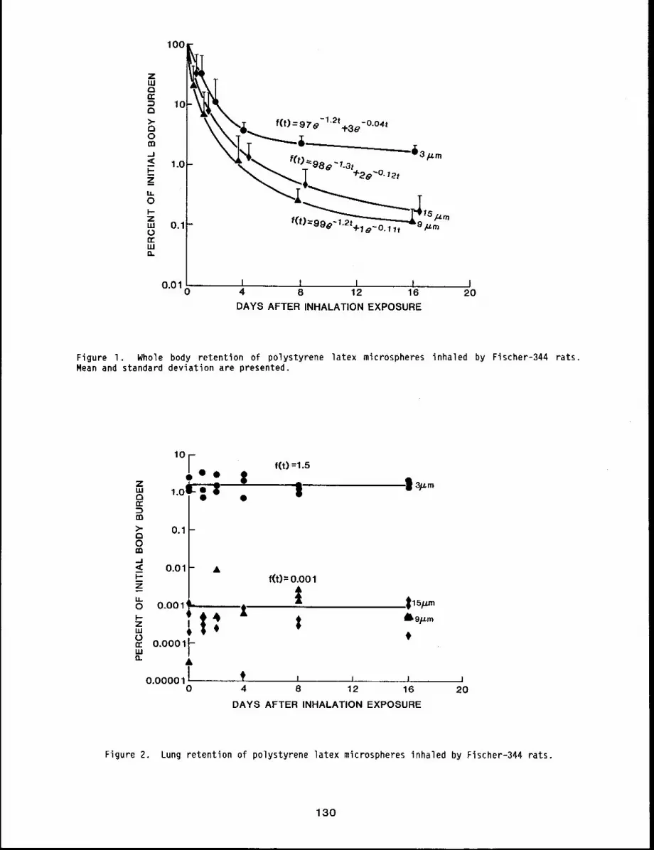
100zLU0n,""~ 10Q
f(t)-97 -lk2t -0 04t>" \\ - e +3~ "0m --
--43
_--"1.o- eSe’7.a~
z_
Z f(t ~,~, -,I-19~U. l lt r-,.,m 0.1-
fn,.wi1.
0.01 i 1 I ! I0 4 8 12 16 20
DAYS AFTER INHALATION EXPOSURE
Figure I. Whole body retention of polystyrene latex microspheres inhaled by Fischer-344 rats.Mean and standard deviation are presented.
,oF¯ 0 0
,.o’ c~ ~ ̄
0.1
f(t) =1.5
i
< 0.01 &f(t) = 0.001
~ o.oo 1’ ---.----,. t
uJ 1I¢0n- 0.0001LU /
0.00001 I0
’ ’ 1 14 8 12 16 20
DAYS AFTER INHALATION EXPOSURE
Figure 2. Lung retention of polystyrene latex microspheres inhaled by Fischer-344 rats.
130
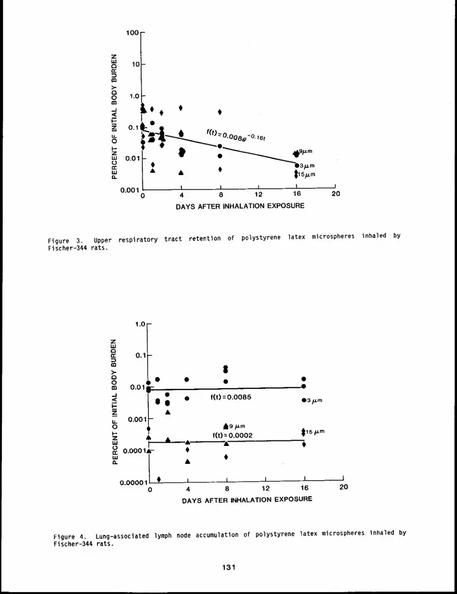
100
z 10
==
O 1.0b
O. 1-- ~0
O.Ol ~"0 0 ~D3p.m
0.001 i I I I0 4 8 12 16
DAYS AFTER INHALATION EXPOSURE
I20
Figure 3. Upper respiratory tract retention of polystyrene latex microspheres inhaled byFis’cher-344 rats,
1.0 --
ZLUE3n- 0.1-m)-
Om O.01r
u- 0.001o 0I.,.- iZ &"’ irr 0.0001A-
a.
0.00001 0 .0
&
$
¯ f(t) = 0.0085
I I I I 14 8 12 16 20
DAYS AFTER INHALATION EXPOSURE
Figure 4, Lung-associated lymph node accumulation of polystyrene latex microspheres inhaled byFischer-344 rats.
131
Burdens of 46Sc in the upper respiratory tract dropped rapidly because of early clearance
during the first few hours after exposure, then dropped more slowly (Fig. 3). This suggests that
although most microspheres cleared as might be predicted, O.Ol to l~ of the initial body burden
for all three microsphere sizes cleared relatively slowly, having a biological retention half-time
of approximately 4 days. Most of the 46Sc cleared from these rats was in feces. This
represented most of the external contamination (i0% of the initial body burden), clearance
microspheres deposited in the upper respiratory tract and large airways, and some clearance from
the deep lung.
CONCLUSIONS
(1) Deposition of inhaled 3-#m polystyrene latex microspheres represented an average
2.1% of the initial body burden; less than 0.01% of the g- and 15-~m microspheres was in the
pulmonary region.
(2) A small percentage of all three sizes of microspheres was retained in the upper
respiratory tract with a clearance half-time of approximately 4 days.
(3) Few, if any, of the 9- or 15-#m microspheres were translocated from the lung
lung-associated lymph nodes.
REFERENCES
I. Snipes, M. B. and M. F. Clem, Retention of Microspheres in the Rat Lung After IntratrachealInstillation, Environ. Res. 24: 33-41, Ig81.
2. Snipes, M. B., B. B. Boecker, and R. O. McClellan, Retention of Monodisperse or PolydisperseAluminosilicate Particles Inhaled by Dogs, Rats, and Mice, Toxicol. Appl. Pharmacol. 6j:345-362, 1983.
132
DEPOSITION OF ULTRAFINE 67Ga203 PARTICLES IN EXERCISING RATS
Abstract -- Male Flscher-344 rats were exposed to
67Ga03 partlcles (0.10 ~un activity medlan diameter)PRINCIPAL IWVESTIGATORS
for 30 mln, elther while running on a treadmill or G.R. Hesseltlne
while at rest. Ten exere~slng and 10 resting rats R.K. Wolff
were sacr~flced at 2 h and at 12 days after expo- J.L. Mauderl9
sure to assess 67Ga retention in respiratory tls- Y.S. cheng
sues. Exercising rats had more 67Ga activity In
the nasal passages (20 ± 12 vs 4.7 f 1.3 nanocur~es) and a lower (p < 0.05) estimated
deposlt~on efficiency in lung lobes (2.8 ± 0.4 vs 10.1 ± 2.2 percent).
The influence of exercise on the deposition, fate, and effects of inhaled materials is of
interest, recognizing that many occupational or environmental exposures to potentially toxic
materials may occur when individuals are exercising. This study was performed to determine the
effect of treadmill exercise on inhaled particle deposition in rats.
MATERIALS AND METHODS
The aerosol used was 67Ga203 (TI/2 = 3.25 days), an ultrafine particle (O.lO
activity median diameter; 0.14 ~m standard deviation), similar in size and deposition properties
to chain aggregate particles produced by many industrial flame processes and internal combustion
engines. The aerosol was used because it was well defined I and because the gamma-emitting
67Ga isotope can be quantitated with external detection methods.
A lO-lane, variable-speed treadmill was built for exposing rodents during exercise. The
system had ports for aerosol sampling at various places in the treadmill lanes and had an
electrified wire grid behind the running surface to facilitate training of rodents.
Fifty male Fischer-344 rats, laboratory-reared, specific pathogen-free and 12 weeks of age,
were trained five days/week for four weeks to run on the treadmill at 30 m/min for 30 min. Forty
rats were assigned to a resting (R) or exercising (E) group (20 per group) and exposed
67&a203 (0.26 ~Ci/liter) for 30 min in the treadmill. The R rats were confined
cylindrical wire mesh cages, and the E rats ran at 30 m/min. Half of the rats in each group were
randomly assigned to an immediate (I) sacrifice group and asphyxiated with 2 2 h af ter
exposure. The remaining rats from each group were the delayed (D) sacrifice group and were
euthanized 12 days after exposure. The lung lobes, trachea with mainstem bronchi, head and larynx
(without pelt) and gastrointestinal (GI) tract were isolated at necropsy. The radioactivity
these tissues was measured with a low gamma bulk spectrometer, converted to nanocuries, and
normalized for the animal’s body weight and individual exposure concentration. Means and standard
deviations of activity in each tissue were determined for E and R immediate-sacrifice rats. The
differences between means were compared for statistical significance (p < 0.05) using the
student’s ! test. Activities in the GI tract and head were added under the assumption that most
67Ga203 in the GI tract was from rapid nasal mucociliary clearance. Activity in lung lobes
of D animals was used to evaluate pulmonary deposition,l
133
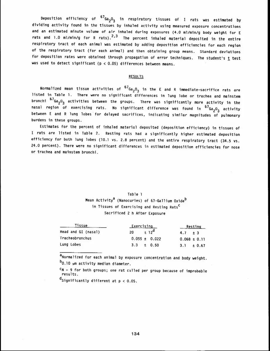
Deposition efficiency of 67Ba203 in respiratory tissues of I rats was estimated by
dividing activity found in the tissues by inhaled activity using measured exposure concentrations
and an estimated minute volume of air inhaled during exposures (4.0 ml/mln/g body weight for
rats and l.O ml/min/g for R rats). 2’3 The percent inhaled material deposited in the entire
respiratory tract of each animal was estimated by adding deposition efficiencies for each region
of the respiratory tract (for each animal) and then obtaining group means. Standard deviations
for deposition rates were obtained through propagation of error techniques. The student’s t test
was used to detect significant (p < 0.05) differences between means.
RESULTS
Normalized mean tissue activities of 67Ga203 in the E and R immediate-sacrifice rats arelisted in Table I. There were no significant differences in lung lobe or trachea and mainstem
bronchi 67Ga203 activities between the groups. There was significantly more activity in the
nasal region of exercising rats. No significant difference was found in 67Ga203 activitybetween E and R lung lobes for delayed sacrifices, indicating similar magnitudes of pulmonary
burdens in these groups.
Estimates for the percent of inhaled material deposited (deposition efficiency) in tissues
i rats are listed in Table 2. Resting rats had a significantly higher estimated deposition
efficiency for both lung lobes (lO.l vs. 2.8 percent) and the entire respiratory tract (34.5
24.0 percent). There were no significant differences in estimated deposition efficiencies for nose
or trachea and mainstem bronchi.
Table l
Mean Activity a (Nanocuries) of 67-Gallium Oxideb
in Tissues of Exercising and Resting Ratsc
Sacrificed 2 h After Exposure
Tissue Exercisin~ Restlnq
Head and GI (nasal) 20 ± 12d 4.7 ± 3
Tracheobronchus 0.055 ± 0.022 0.068 ± O.llLung Lobes 3.3 ± 0.50 3.1 ± 0.67
aNormalized for each animal by exposure concentration and body weight.
bo.lO ~m activity median diameter.
CN = 9 for both groups; one rat culled per group because of improbableresults.
dsignificantly different at p < 0.05.
134
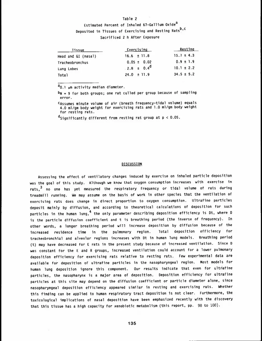
Table 2
Estimated Percent of Inhaled 67-Gallium Oxidea
Deposited in Tissues of Exercising and Resting Ratsb’c
Sacrificed 2 h After Exposure
Tissue Exercising Resting
Head and GI (nasal) 16.6 ± II.8 15.7 ± 4.3
Tracheobronchus 0.05 ± 0.02 0,9 ± 1.9
Lung Lobes 2,B ± 0.4d lO.l ± 2.2
Total 24.0 ± ll.9 34.5 ± 5.2
aO,l gm activity median diameter.
bN = 9 for both groups; one rat culled per group because of samplingerror.
CAssumes minute volume of air (breath frequency-tldal volume) equals4.0 ml/gm body weight for exercising rats and l.O ml/gm body weightfor resting rats.
dsignlficantly different from resting rat group at p < 0.05.
DISCUSSION
Assessing the effect of ventilatory changes induced by exercise on inhaled particle deposition
was the goal of this study. Although we know that oxygen consumption increases with exercise in
rats, 3 no one has yet measured the respiratory frequency or tidal volume of rats during
treadmill running. We may assume on the basis of work in other species that the ventilation of
exercising rats does change in direct proportion to oxygen consumption. Ultrafine particles
deposit mainly by diffusion, and according to theoretical calculations of deposition for such
particles in the human lung, 4 the only parameter describing deposition efficiency is Dt, where D
is the particle diffusion coefficient and t is breathing period (the inverse of frequency).
other words, a longer breathing period will increase deposition by diffusion because of the
increased residence time in the pulmonary region. Total deposition efficiency for
tracheobronchial and alveolar regions increases with Dt in human lung models. Breathing period
(t) may have decreased for E rats in the present study because of increased ventilation. Since
was constant for the E and R groups, increased ventilation could account for a lower pulmonary
deposition efficiency for exercising rats relative to resting rats. Few experimental data are
available for deposition of ultrafine particles in the nasopharyngeal region. Most models for
human lung deposition ignore this component. Our results indicate that even for ultrafine
particles, the nasopharynx is a major area of deposition. Deposition efficiency for ultraflne
particles at this site may depend on the diffusion coefficient or particle diameter alone, since
nasopharyngeal deposition efficiency appeared similar in resting and exercising rats. Whether
this finding can be applied to human respiratory tract deposition is not clear. Furthermore, the
toxicological implications of nasal deposition have been emphasized recently with the discovery
that this tissue has a high capacity for xenobiotic metabolism (this report, pp. 98 to 100).
135

REFERENCES
I. Wolff, R. K., L. C. Griffis, C. H. Hobbs, and R. O. McClellan, Deposition and Retention ofO.l ~m 67Ga203 Aggregate Aerosols in Rats Following Whole Body Exposures Fundam.Appl. Toxicol. 2: 195-200, 1982.
2. Gleeson, T. T. and K. M. Baldwin, Cardiovascular Response to Treadmill Exercise in UntrainedRats, 3. Appl. Physiol. 50: 1206-1211, 1981.
3. Brooks, G. A. and T. P. White, Determination of Metabolic and Heart Rate Responses of Rats toTreadmill Exercise, 3. AppI. Physiol. 45: I009-I015, lg78.
4. Yu, C. P. and O. P. HU, Diffusional Deposition of Ultrafine Particles in the Human Lung, in:Aerosols in the Mining and Industrial Work Environment~, (V. A. Marple and B. Y. H. Liu,eds.), Vol. I, Fundamentals and Status, pp. 13g-149. Ann Arbor Science, Ann Arbor, MI, 1983.
136
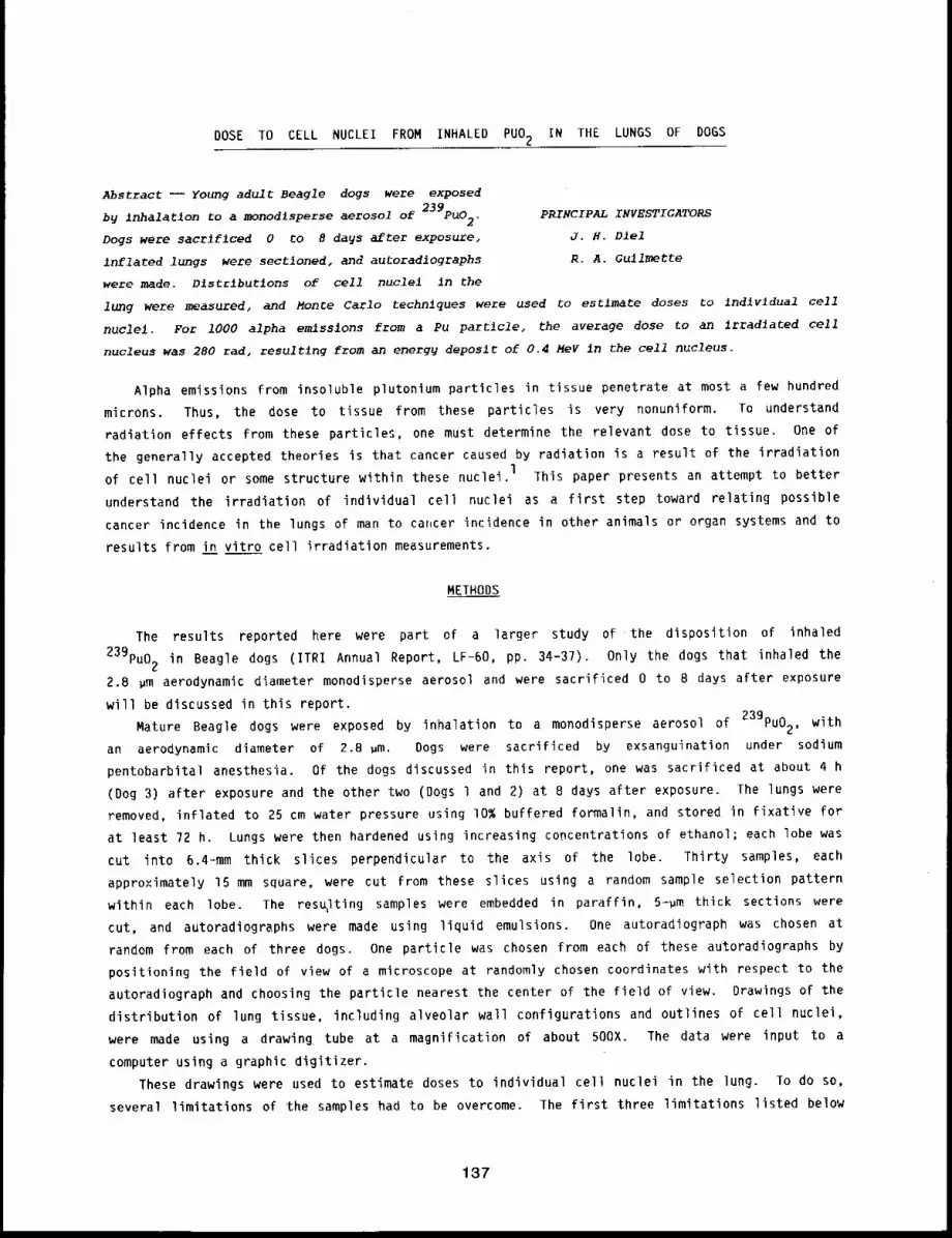
DOSE TO CELL NUCLEI FROM INHALED PUO2 IN THE LUNGS OF DOGS
Abstract -- Young adult Beagle dogs were exposed
b9 inhalatlon to a monodlsperse aerosol of 239pu02. PRINCIPALINVESTIGATORS
Dogs were sacr~flced 0 to 8 days after exposure, J.H. Dlel
inflated lungs were sectioned, and autoradlographs R.A. Gullmette
were made. Dlstrlbutlons of cell nuclel in the
lung were measured, and Monte Carlo techniques were used to estimate doses Co Indlv~dual cell
nuclel. For 1000 alpha emissions from a Pu partlcle, the average dose to an irradlated cell
nucleus was 280 rad, resulting from an energy deposit of 0.4 MeV ~n the cell nucleus.
Alpha emissions from insoluble plutonium particles in tissue penetrate at most a few hundred
microns. Thus, the dose to tissue from these particles is very nonuniform. To understand
radiation effects from these particles, one must determine the relevant dose to tissue. One of
the generally accepted theories is that cancer caused by radiation is a result of the irradiation
of cell nuclei or some structure within these nuclei, l This paper presents an attempt to better
understand the irradiation of individual cell nuclei as a first step toward relating possible
cancer incidence in the lungs of man to cahcer incidence in other animals or organ systems and to
results from in vitro cell irradiation measurements.
METHODS
The results reported here were part of a larger study of the disposition of inhaled
239pu02 in Beagle dogs (ITRI Annual Report, LF-60, pp. 34-37). Only the dogs that inhaled the
2.8 ~m aerodynamic diameter monodisperse aerosol and were sacrificed 0 to 8 days after exposure
will be discussed in this report.
Mature Beagle dogs were exposed by inhalation to a monodisperse aerosol of 239Pu02, with
an aerodynamic diameter of 2.8 ~m. Dogs were sacrificed by exsanguination under sodium
pentobarbital anesthesia. Of the dogs discussed in this report, one was sacrificed at about 4 h
(Dog 3) after exposure and the other two (Dogs l and 2) at 8 days after exposure. The lungs
removed, inflated to 25 cm water pressure using i0% buffered formalin, and stored in fixative for
at least 72 h. Lungs were then hardened using increasing concentrations of ethanol; each lobe was
cut into 6.4-mm thick slices perpendicular to the axis of the lobe. Thirty samples, each
approximately 15 mm square, were cut from these slices using a random sample selection pattern
within each lobe. The res~Iting samples were embedded in paraffin, 5-~m thick sections were
cut, and autoradiographs were made using liquid emulsions. One autoradiograph was chosen at
random from each of three dogs. One particle was chosen from each of these autoradiographs by
positioning the field of view of a microscope at randomly chosen coordinates with respect to the
autoradiograph and choosing the particle nearest the center of the field of view. Drawings of the
distribution of lung tissue, including alveolar wall configurations and outlines of cell nuclei,
were made using a drawing tube at a magnification of about 500X. The data were input to a
computer using a graphic digitizer.
These drawings were used to estimate doses to individual cell nuclei in the lung. To do so,
several limitations of the samples had to be overcome. The first three limitations listed below
137
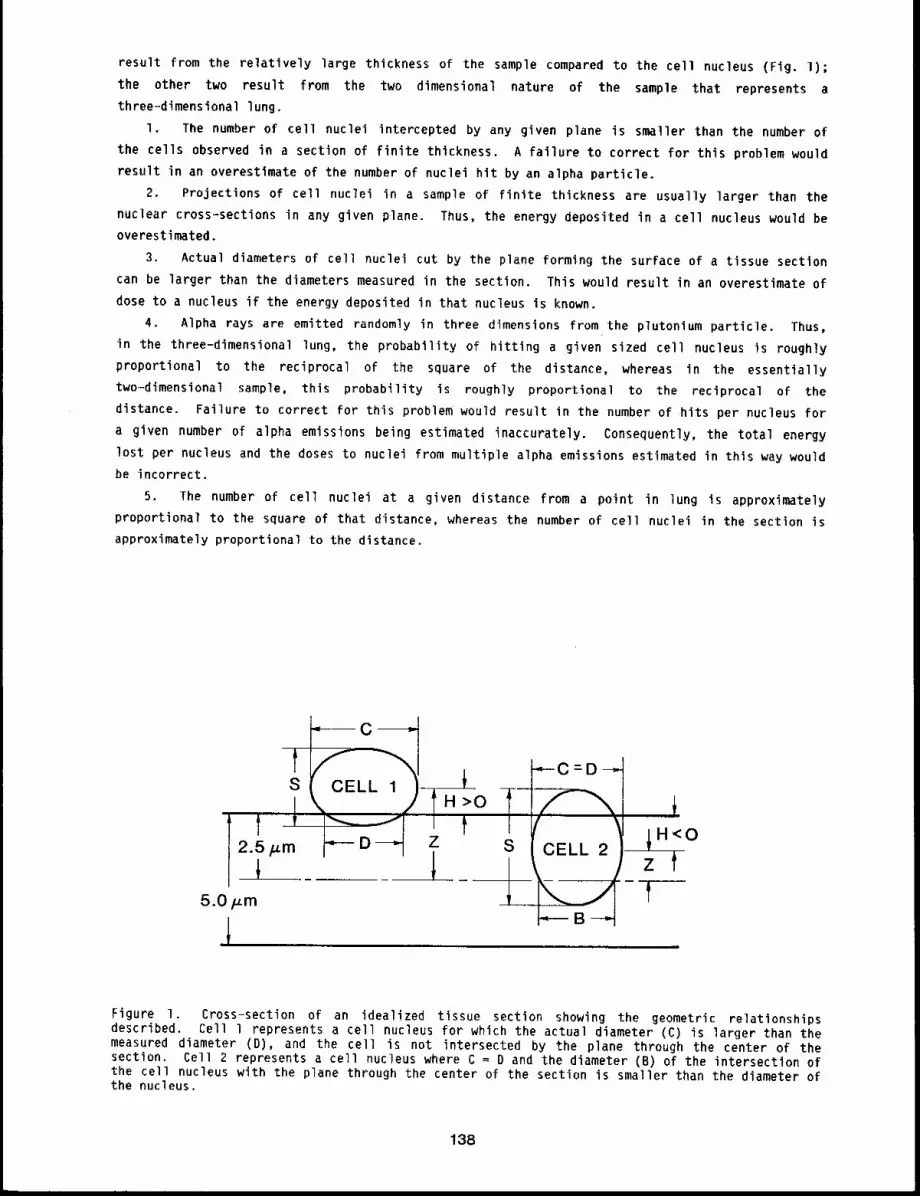
result from the relatively large thickness of the sample compared to the cell nucleus (Fig. I);
the other two result from the two dimensional nature of the sample that represents a
three=dimensional lung.
I. The number of cell nuclei intercepted by any given plane is smaller than the number of
the cells observed in a section of finite thickness. A failure to correct for this problem would
result in an overestimate of the number of nuclei hit by an alpha particle.
2. Projections of cell nuclei in a sample of finite thickness are usually larger than the
nuclear cross-sections in any given plane. Thus, the energy deposited in a cell nucleus would be
overestimated.
3. Actual diameters of cell nuclei cut by the plane forming the surface of a tissue section
can be larger than the diameters measured in the section. This would result in an overestimate of
dose to a nucleus if the energy deposited in that nucleus is known.
4. Alpha rays are emitted randomly in three dimensions from the plutonium particle. Thus,
in the three-dimensional lung, the probability of hitting a given sized cell nucleus is roughly
proportional to the reciprocal of the square of the distance, whereas in the essentially
two-dimensional sample, this probability is roughly proportional to the reciprocal of the
distance. Failure to correct for this problem would result in the number of hits per nucleus for
a given number of alpha emissions being estimated inaccurately. Consequently, the total energy
lost per nucleus and the doses to nuclei from multiple alpha emissions estimated in this way would
be incorrect.
5. The number of cell nuclei at a given distance from a point in lung is approximately
proportional to the square of that distance, whereas the number of cell nuclei in the section is
approximately proportional to the distance.
C LI
~-C=D--
It ± D If I2.5 p.m Z S
5.0Fm
CELL 2
--f
Figure 1. Cross-section of an idealized tissue section showing the geometric relationshipsdescribed. Cell l represents a cell nucleus for which the actual diameter (C) is larger than themeasured diameter (D), and the cell is not intersected by the plane through the center of thesection. Cell 2 represents a cell nucleus where C = D and the diameter (B) of the intersection the cell nucleus with the plane through the center of the section is smaller than the diameter ofthe nucleus.
138
For any single simulated alpha emission, the problems described in 5. and 6. above should
have no effect because microscopic distribution of lung tissue is essentially independent of
section orientation. In taking averages over many emmissions, the opposite effects of number of
nuclei at a given distance and the probability of hitting a given nucleus should cancel.
As a result of these limitations in direct use of measured data, a Monte Carlo procedure was
used to predict nuclear cross-sectlon configurations from the measured configurations. It was
based on the following assumptions:
I. The centers of all nuclei observed are within the section or within a distance from the
section equal to the average measured major diameter of a nucleus~
2. The nuclei are all ellipsoids with two axes parallel to the plane of the section and the
length of the axis perpendicular to the plane of the section equal to: (1) the major axis measured
parallel to the plane of the section - oblate ellipsoids or disk shaped, (2) the minor axis
measured parallel to the plane of the section - prolate ellipsoids or cigar shaped, or (3) the
geometric average of the major and minor axes lengths in the plane of the section - pleomorphic
triaxial ellipsoids.
Further, assume that the nuclei are randomly oriented so that, to a first approximation,
these three cases occur at about equal frequency.
Let D be the measured maximum distance across the cell nucleus; A, the measured projected
area of the nucleus; E = ~ D2/A, the eccentricity in the plane of the section; H, the height
of the ellipsoid center above the top of the section; and Z = H + 2.5, the distance from the
center of the section to the center of the ellipsoid (Fig. 1). For the above assumptions, the
actual major diameter, C, of the nucleus is:
C=D
C= V~D2 + 4 H2
C = ~D2 * 4(E H)2
c= JD2÷4(E 2)
; H < 0 (all cases)
; H > 0 (case (1))
; H > 0 (case (2))
; H > 0 (case (3))
the diameter, S, perpendicular to the plane of the section is:
S = C ; case (1)
S = C/E ; case (2)
S = C/ ~ ; case (3)
and the major diameter, B, of the ellipse produced by the intersection of the nucleus
with the plane at the center of the section is:
B = 0 ; Z > S (all cases)
B = ~C2 - 4(Z2) ; Z < S (case (1))
B = ~C2 - 4(E Z)2 ; Z < S (case (2))
B = ~C2 - 4(E 2) ; Z < S (case (3))
These assumptions result in the following procedure for estimating the actual dimensions of
the nuclei represented by the measured nuclear diameters.
I. Choose a distance from the center of the section at random not to exceed the average
major diameter measured (3.26 pm) plus the half=thickness of the section (2.5 ~m).
2. Choose a random number between 0 and I. If this number is less than I/3, case (1)
applies; if it is between I/3 and 2/3, case (2) applies; if it is between 2/3 and l, case (3)
applies.
3. Compute C, S, and B using the above equations for the appropriate case.
4. Average C/D over all cell nuclei observed, average B/D over all nuclei for which B > O,
and compute the fraction of nuclei for which B > O.
139
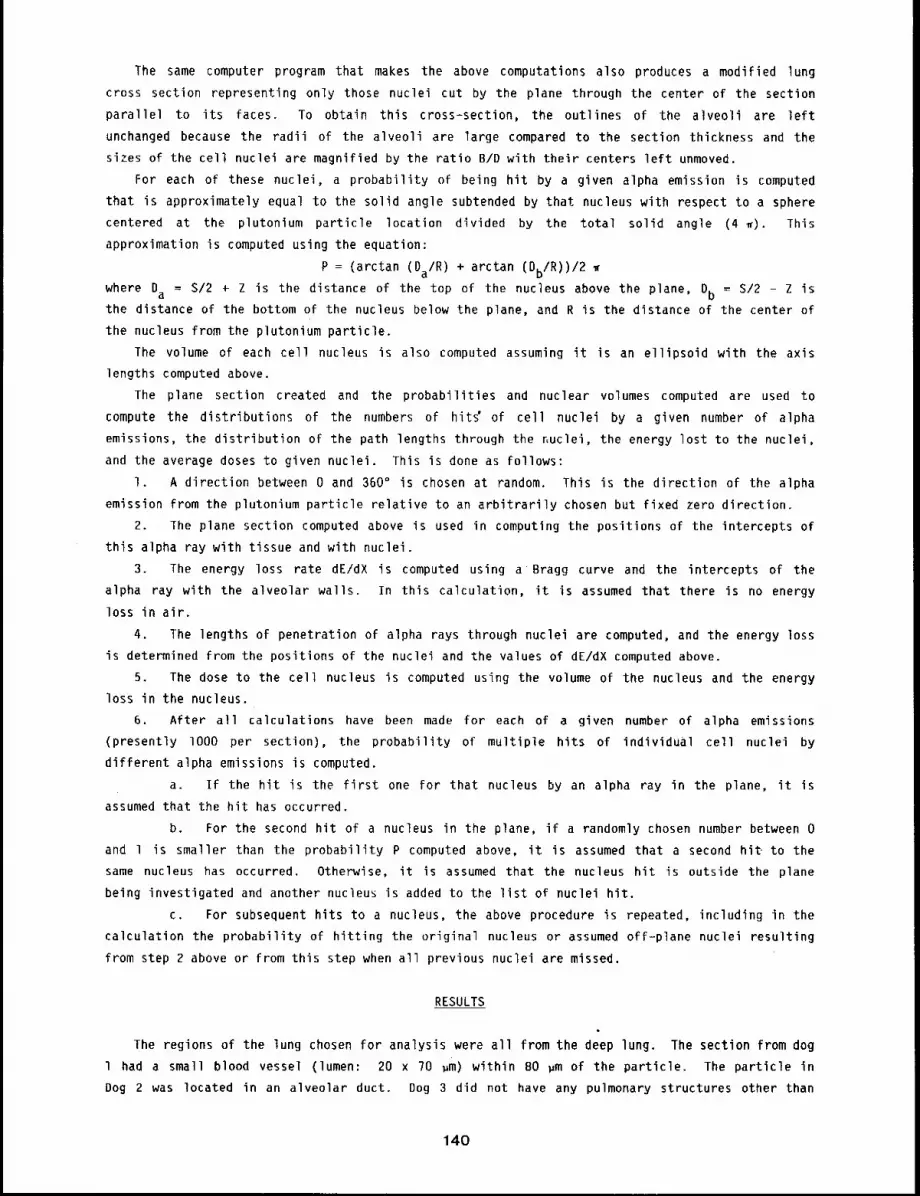
The same computer program that makes the above computations also produces a modified lung
cross section representing only those nuclei cut by the plane through the center of the section
parallel to its faces. To obtain this cross-section, the outlines of the alveoli are left
unchanged because the radii of the alveoli are large compared to the section thickness and the
sizes of the cell nuclei are magnified by the ratio B/D with their centers left unmoved.
For each of these nuclei, a probability of being hit by a given alpha emission is computed
that is approximately equal to the solid angle subtended by that nucleus with respect to a sphere
centered at the plutonium particle location divided by the total solid angle (4 ~). This
approximation is computed using the equation:
P = (arctan (Da/R) + arctan (Db/R))/2
where Da = S/2 ÷ Z is the distance of the top of the nucleus above the plane, Db = S/2 - Z isthe distance of the bottom of the nucleus below the plane, and R is the distance of the center of
the nucleus from the plutonium particle.
The volume of each cell nucleus is also computed assuming it is an ellipsoid with the axis
lengths computed above.
The plane section created and the probabilities and nuclear volumes computed are used to
compute the distributions of the numbers of hitC of cell nuclei by a given number of alpha
emissions, the distribution of the path lengths through the Nuclei, the energy lost to the nuclei,
and the average doses to given nuclei. This is done as follows:
I. A direction between 0 and 360" is chosen at random. This is the direction of the alpha
emission from the plutonium particle relative to an arbitrarily chosen but fixed zero direction.
2. The plane section computed above is used in computing the positions of the intercepts of
this alpha ray with tissue and with nuclei.
3. The energy loss rate dE/dX is computed using a Bragg curve and the intercepts of the
alpha ray with the alveolar walls. In this calculation, it is assumed that there is no energy
loss in air.
4. The lengths of penetration of alpha rays through nuclei are computed, and the energy loss
is determined from the positions of the nuclei and the values of dE/dX computed above.
5. The dose to the cell nucleus is computed using the volume of the nucleus and the energy
loss in the nucleus.
6. After all calculations have been made for each of a given number of alpha emissions
(presently lO00 per section), the probability of multiple hits of individual cell nuclei
different alpha emissions is computed.
a. If the hit is the first one for that nucleus by an alpha ray in the plane, it is
assumed that the hit has occurred.
b. For the second hit of a nucleus in the plane, if a randomly chosen number between 0
and l is smaller than the probability P computed above, it is assumed that a second hit to the
same nucleus has occurred. Otherwise, it is assumed that the nucleus hit is outside the plane
being investigated and another nucleus is added to the list of nuclei hit.
c. For subsequent hits to a nucleus, the above procedure is repeated, including in the
calculation the probability of hitting the original nucleus or assumed off-plane nuclei resulting
from step 2 above or from this step when all previous nuclei are missed.
RESULTS
The regions of the lung chosen for analysis were all from the deep lung. The section from dog
l had a small blood vessel (lumen: 20 x 70 ~m) within BO ~m of the particle. The particle
Dog 2 was located in an alveolar duct. Dog 3 did not have any pulmonary structures other than
140
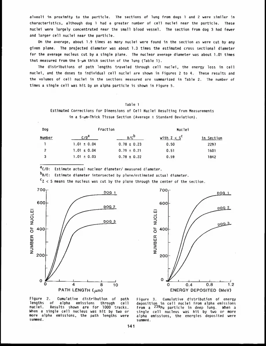
alveoll in proximity to the particle. The sections of lung from dogs 1 and 2 were similar in
characteristics, although dog 1 had a greater number of cell nuclei near the particle. These
nuclei were largely concentrated near the small blood vessel. The section from dog 3 had fewer
and larger cell nuclei near the particle.
On the average, about 1.9 times as many nuclei were found in the section as were cut by any
given plane. The projected diameter was about 1.3 times the estimated cross sectional diameter
for the average nucleus cut by a single plane. The nuclear average diameter was about l.Ol times
that measured from the 5-~m thick section of the lung (Table l).
The distributions of path lengths traveled through cell nuclei, the energy loss in cell
nuclei, and the doses to individual cell nuclei are shown in Figures 2 to 4. These results and
the volumes of cell nuclei in the sections measured are summarized in Table 2. The number of
times a single cell was hit by an alpha particle is shown in Figure 5.
Table l
Estimated Corrections for Dimensions of Cell Nuclei Resulting from Measurements
in a 5-~m-Thick Tissue Section (Average ± Standard Deviation).
Dog
Number
Fraction Nuclei
C/Da B/Cb with Z < Sc in Section
l.Ol +_ 0.04 0.78 _+ 0.23 0.50 2297
l.Ol +_ 0.04 0.79 ± 0.21 0.51 1601
1.Ol +_ 0.03 0.78 +_ 0.22 0.59 1842
aC/D: Estimate actual nuclear diameter/ measured diameter.
bB/C: Estimate diameter intersected by plane/estimated actual diameter.
Cz < S means the nucleus was cut by the plane through the center of the section.
700
600
=oz
~) 400
200
0
- 700 -DOG 1 DOG 1
DOG 2
DOG 3 ~)~Z
~) 400
2OO
, I 0 ,i I I I ¯ 1
0 4 8 10 0 0.4 0.8 1.2
PATH LENGTH (/.t.m) ENERGY DEPOSITED (MeV)
Figure 2. Cumulative distribution of pathlengths of alpha emissions through cellnuclei. Results shown are for lO00 tracks.When a single cell nucleus was hit by two ormore alpha emissions, the path lengths weresummed.
141
Figure 3. Cumulative distri,bution of energydeposition in cell nuclei from alpha emissionsfrom a 239pu particle in deep lung. When asingle cell nucleus was hit by two or morealpha emissions, the energies deposited weresummed.

7001 DOG 1
DOG 2
40O
2OO
0 ~ = I ,j,, I I i l I0 300 600 900 1200
DOSE (rad)
Figure 4. Cumulative distribution of totaldoses to individual cell nuclei from alphaemissions from a 239pu particle in the deeplung of a dog. When a single cell nucleus washit by two or more alpha emissions, the doseswere summed.
Table 2
Characteristics of Cell Nuclei and Their Interaction With Alpha Particles From a 239pu Particle
Deposited in Deep Lung. Values Shown are for lO00 Alpha Emissions and Include
Results of Multiple Hits to a Single Cell Nucleus
(Mean ± Standard Deviation).
Number Path Length Energy Total Volume
Dog Nuclei in Nucleus Deposited Dose of Nucleus
Number Hit (um) (MeV) ~ _ (~m3)
l 671 2.1 ± 1.4 0,30 ± 0,36 260 ± 500 26 ± 22
2 567 2.3 ± 1.7 0.40 ± 0.86 4!0 ± 1800 25 ± 21
3 449 3.0 ± 1.7 0.49 ± 0.75 160 ± 250 39 ± 32
Figure 5.
850I-
8001-450~
300 -
150- z ~Ezz_.Ez
; ! [ ,,., I JDOG 1 DOG 2 O(3
Number of hits to cell nuclei from lO00 alpha emissions from a Pu particle in deep lung.
142
DISCUSSION
The sizes of cell nuclei reported are somewhat smaller than those reported by
investigators 2 (for rat lung), and there is some controversy about the shapes of
nuclei. 2’3 The three possible shapes chosen in this study include all of those commonly used
although it might be argued that the third shape (pleomorphic triaxial ellipsoids) should
represent a larger fraction of the nuclei.2
Individual values of path length, energy loss, and dose per nucleus were highly variable as
would be expected for a random process (Table 2). Their distributions were similar for all three
sections; th~ differences that occurred are attributable to the numbers and differences in sizes
among the nuclei for the three sections. Whether these differences are related to differences in
individual dogs, time after exposure, or simply choice of sample area must await processing of
further samples; whether they are of practical significance must await application of these
results in cancer induction models.
MOSt of the irradiated nuclei were hit by only one of the lO00 simulated alpha emissions,
whereas the number of cell nuclei irradiated per alpha emission averaged much less than one,
indicating that localized plutonium particles are very inefficient in irradiating cell nuclei in
dog lungs.
The results presented in this report are preliminary. For example, they do not take into
account (1) the motion of particles in the lung, (2) the different cell types in the lung, (3)
presence and irradiation of circulating blood lymphocytes in the lung, and (4) possible changes
lung morphometry with age or resulting from irradiation. For application of these results to lung
cancer induction, the above listed items must be taken into account along with at least (1)
estimates or measurements of cell killing and cell transformation probabilities at different
levels of irradiation of the cell nucleus for the different cell types, (2) numbers and types
cells that must be transformed for a tumor to be initiated, (3) relevant irradiation times and
cancer development times, and, if comparisons among species are to be made, (4) differences
these parameters in different species.
REFERENCES
l ¯ Lloyd, E. L., M. A. Gemmell, C. B. Henning, D. S. Gemmel, and B. J. Zabransky, Transformationof Mammalian Cells by Alpha Particles, Int. J. Radiat. Biol. 3_66: 467-478, 1979.
2. Crapo, J. D. and D. A. Greeley, Estimation of the Mean Caliper Diameter of Cell Nuclei, 3~.Microsc. 78: 425-443, 19?l.
3. Haies, D. M., J. Gil, and E. R. Weibel, Morphometric Study of Rat Lung Cells, Am. Rev. Respir.Dis. 123: 533-541, 1981.
143
THE EFFECT OF 1NHALED BURDEN OF 239pu02 ON ITS REIENTION IN BEAGLE DOG LUNG
Abstract --To determine the effect of inhaled pul-
monary burden of 239pu09 on the subsequent clear- PRINCIPAL INVESTIGATORS
ance and translocatlon of 239pu from lung, 9o~ngR.A. Gu11~ette
adult Beagle dogs were exposed to monodlsperse J.H. Dlel
239pu02 (0.72 pm and 2.8 ~m AMAD) B.A. Muggenburgaerosols of
at levels of 0.007 end 0.0007 pCl/kg bod9 welght
for comparison wlth results from dogs exposed at the level of 0.07 ~C1/kg hod 9 weight. These
animals are belng sacrlflced at tlmes after exposure ranglng from elght days to eight Years.
Prellmlnary results cbtalned for dogs exposed to 0,72 pm AMAD aerosol and sacrlflced through one
year after exposure suggest that clearance of 239pu from lung is not enhanced when compared to
results obtalned previously w~th dogs exposed to a s~mJlar aerosol at a level of 0.07 pCl/kg
body weight. Results for dogs receiving 2.8 ~m AMAD 239pu0~aerosol are not yet avallable .
Previous studies of the biokinetics of 239pu02 inhaled by Beagle dogs have shown that the
amount of 23gPu initially deposited in the pulmonary region can influence its subsequent
retention within that biological compartment. In particular, for dogs that inhaled 1.5 ,m
activity median aerodynamic diameter (AMAD) or 3.0 ~m AMAD particles at high levels (O.l
1.8 ~Ci/kg body weight), retention of 239pu in the lung was clearly longer than that predicted
from results obtained in a parallel study in which dogs receiving 0.07 ,Ci/kg body weight
239pu02 were serially sacrificed to measure temporal radiation dose patterns (19BO-8l AnnualReport, LMF-91, pp. 46-48). It was postulated that the increased retention of 239pu in the
lungs of dogs receiving these very high doses was due to physical entrapment of the particles
within the lung structure, which had been severely damaged as a result of radiation pneumonitis
and pulmonary fibrosis. These dogs died between lO0 and 800 days after exposure. If high levels
of inhaled 239pu02 can affect its subsequent pulmonary retention, then it is important to know
if levels significantly below that used in the previous biokinetics study (i.e.,
0.07 ~Ci/kg) would also modify lung retention of 239pu0~ particles. To address this
question, we exposed Beagle dogs to monodisperse aerosols of~23gPu02 at levels one-tenth and
one-hundredth that of the original biokinetics study.
MATERIALS AND METHODS
Monodisperse aerosols of 239pu02 were prepared using methods for the preparation of
238pu02 .l Briefly, 23gPu(IV) was precipitated as a colloidal hydroxide, dispersed
0.6 M HCI, and nebulized using a Lovelace nebulizer. The resulting droplets were dried by passing
them through a 320°C heat column and then oxidized to PuO2 by heat treatment at llSO°C. This
polydisperse aerosol was directed into a Lovelace Aerosol Particle Separator (LAPS) for separation
into monodisperse particle fractions on a stainless steel foil. The collection foil segments
containing monodisperse particles of sizes 0.72 and 2.86 ~m AMAD were isolated. The respective
particles were then resuspended within 24 h of animal exposure in water made pH lO by addition of
dilute NH4OH. The nebulized suspension was dried at 150°C, and the dogs were exposed
individually in an apparatus that has been described previously.2
144
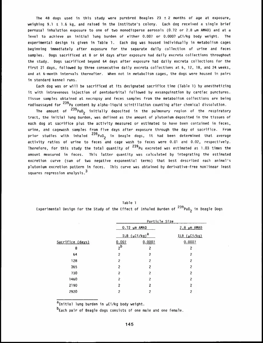
The 48 dogs used in this study were purebred Beagles 23 ± 2 months of age at exposure,
weighing 9.1 ± 1.6 kg, and raised In the Institute’s colony. Each dog received a single brief
pernasal inhalation exposure to one of two monodisperse aerosols (0.72 or 2.8 ~m AMAD) and at
level to achieve an initial lung burden of either 0.007 or 0.0007 pCi/kg body weight. The
experimental design is given in Table I. Each dog was housed individually in metabolism cages
beginning immediately after exposure for the separate daily collection of urine and feces
samples. Dogs sacrificed at 8 or 64 days after exposure had daily excreta collections throughout
the study. Dogs sacrificed beyond 64 days after exposure had daily excreta collections for the
first 21 days, followed by three consecutive daily excreta collections at 6, 12, 18, and 24 weeks,
and at 6-month intervals thereafter. When not in metabolism cages, the dogs were housed in pairs
in standard kennel runs.
Each dog was or will be sacrificed at its designated sacrifice time (Table l) by anesthetizing
it with intravenous injection of pentobarbital followed by exsanguination by cardiac punctures.
Tissue samples obtained at necropsy and feces samples from the metabolism collections are being
radioassayed for 239pu content by alpha-liquid scintillation counting after chemical dissolution.
The amount of 239pu02 initially deposited in the pulmonary region of the respiratory
tract, the initial lung burden, was defined as the amount of plutonium deposited in the tissues of
each dog at sacrifice plus the activity measured or estimated to have been contained in feces,
urine, and cagewash samples from five days after exposure through the day of sacrifice. From
studies with inhaled 239pu02 in Beagle dogs, it had been determined thatprior average
activity ratios of urine to feces and cage wash to feces were O.Ol and 0.02, respectively.
Therefore, for this study the total quantity of 239pu excreted was estimated as 1.03 times the
amount measured in feces. This latter quantity was calculated by integrating the estimated
excretion curve (sum of two negative exponential terms) that best described each animal’s
plutonium excretion pattern in feces. This curve was obtained by derivative-free nonlinear least
squares regression analysis.3
Table l
for the Study of the Effect of Inhaled Burden of 239pu02 inExperimental Design Beagle Dogs
Particle Size
0.72 ~m AMAD 2.8 ~m AMAD
ILB (~Ci/kg)a ILB (uCi/kg)
0.007 0.0007 0.0007
8 2b 2 2
64 2 2 2
128 2 2 2
365 2 2 2
730 2 2 2
1460 2 2 2
2190 2 2 2
2920 2 2 2
alnitial lung burden in ~Ci/kg body weight.
bEach pair of Beagle dogs consists of one male and one female.
145
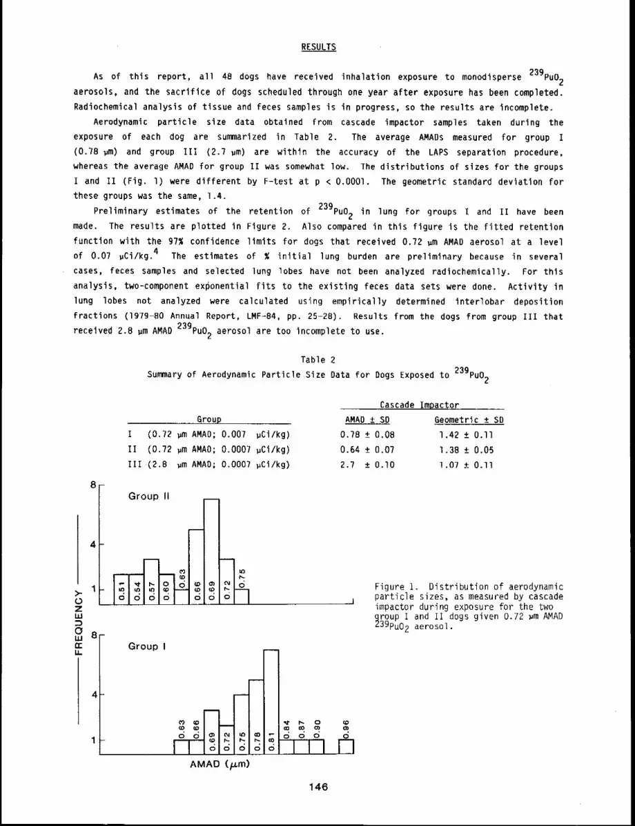
RESULTS
As of this report, all 48 dogs have received inhalation exposure to monodisperse 239pu02
aerosols, and the sacrifice of dogs scheduled through one year after exposure has been completed.
Radiochemical analysis of tissue and feces samples is in progress, so the results are incomplete.
Aerodynamic particle size data obtained from cascade impactor samples taken during the
exposure of each dog are summarized in Table 2. The average AMADs measured for group I
(0.78 ~m) and group III (2.7 ~m) are within the accuracy of the LAPS separation procedure,
whereas the average AMAD for group II was somewhat low. The distributions of sizes for the groups
I and II (Fig. l) were different by F-test at p < O.O001. The geometric standard deviation for
these groups was the same, 1.4.
Preliminary estimates of the retention of 239pu02 in lung for groups i and II have been
made. The results are plotted in Figure 2. Also compared in this figure is the fitted retention
function with the 97% confidence limits for dogs that received 0.72 ~m AMAD aerosol at a level
of 0.07 ~Ci/kg. 4 The estimates of % initial lung burden are preliminary because in several
cases, feces samples and selected lung lobes have not been analyzed radiochemically. For this
analysis, two=component exponential fits to the existing feces data sets were done. Activity in
lung lobes not analyzed were calculated using empirically determined interlobar deposition
fractions (1979-B0 Annual Report, LMF-84, pp. 25-28). Results from the dogs from group Ill that
~m AMAD 23gPuo2 aerosol are too incomplete to use.received 2.8
Table 2
Summary of Aerodynamic Particle Size Data for Dogs Exposed to 239pu02
>-0ZUJ
0uJrr
U.
8
4
Group
I (0.72 ~m AMAD; 0.007 ~Ci/kg)
II (0.72 ~m AMAO; 0.0007 ~Ci/kg)
Ill (2.8 ~m AMAD; 0.0007 ~Ci/kg)
Group ii
5
4
Group I
Cascade Impactor
AMAD +_ SD Geometric ± SD
0.78 +_ 0.08 1.42 _+ 0.II
0.64 ± 0.07 1.38 +_ 0.05
2.? +_ 0.I0 1.07 + O.ll
Figure i. Distribution of aerodynamicparticle sizes, as measured by cascadeimpactor during exposure for the two
~roup I and II dogs given 0.72 ~m AMAD39pu02 aerosol.
(O (:O libo o d o
V1
146
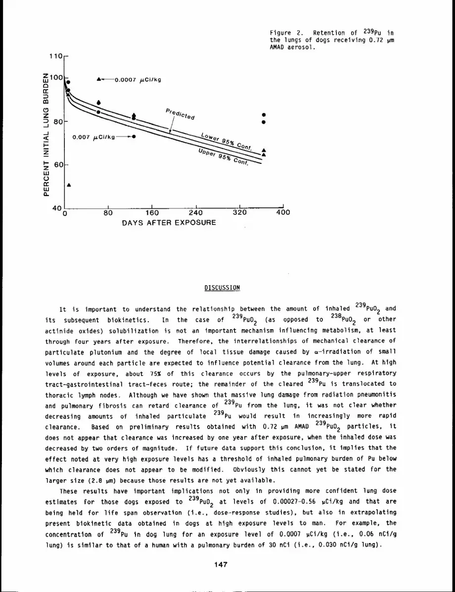
40 , I l I i0 80 160 240 320 400
DAYS AFTER EXPOSURE
Figure 2. Retention of 239pu inthe lungs of dogs receiving 0.72 ~mAMAD aerosol.
DISCUSSION
It is important to understand the relationship between the amount of inhaled 239pu02 and
its subsequent biokinetics. In the case of 239pu02 (as opposed to 238pu02 or other
actinide oxides) solubilization is not an important mechanism influencing metabolism, at least
through four years after exposure. Therefore, the interrelationships of mechanical clearance of
particulate plutonium and the degree of local tissue damage caused by =-irradiation of small
volumes around each particle are expected to influence potential clearance from the lung. At hlgh
levels of exposure, about 75% of this clearance occurs by the pulmonary-upper respiratory
tract-gastrointestinal tract-feces route; the remainder of the cleared 239pu is translocated to
thoracic lymph nodes. Although we have shown that massive lung damage from radiation pneumonitis
and pulmonary fibrosis can retard clearance of 239pu from the lung, it was not clear whether
decreasing amounts of inhaled particulate 23gPu would result in increasingly more rapid
Based on preliminary results obtained with 0.72 ~m AMAD 239pu02 particles,clearance. it
does not appear that clearance was increased by one year after exposure, when the inhaled dose was
decreased by two orders of magnitude. If future data support this conclusion, it implies that the
effect noted at very high exposure levels has a threshold of inhaled pulmonary burden of Pu below
which clearance does not appear to be modified. Obviously this cannot yet be stated for the
larger size (2.8 ~m)because those results are not yet available.
These results have important implications not only in providing more confident lung dose
estimates for those dogs exposed to 239pu02 at levels of 0,00027-0.56 ~Ci/kg and that are
being held for life span observation (i.e., dose-response studies), but also in extrapolating
present biokinetic data obtained in dogs at high exposure levels to man. For example, the
concentration of 239pu in dog lung for an exposure level of 0.0007 ~Ci/kg (i.e., 0.06 nCi/g
lung) is similar to that of a human with a pulmonary burden of 30 nCi (i.e., 0.030 nCi/g lung).
147
REFERENCES
Rcabe, O. G., H. A. Boyd, G. M, Kanapilly, G. J. Wilkinson, and G. J. Newton, Development andUse of a System for Routine Production of Monodisperse Particles of 238pu02 and Evaluationof Gamma Emitting Labels, Health Phys. 28: 655-667, 1975.
2. Mewhinney, J. A. and J. H. Diel, Retention of Inhaled 238pu02 in Beagles: A MechanisticApproach to Description, HealS. 45: 39-60, 1983.
3. Ralston, M., BMD PAR in BMDP Statistical Software (W. J. Dixon, ed.), University CaliforniaPress, Berkeley, CA, 305, 1981.
4. Guilmette, R. A., J. H. Diel, B. A. Muggenburg, J. A. Mewhinney, B. B. Boecker, and R. O.McClellan, Biokinetics of Inhaled 239pu02 in the Beagle Dog: Effect of Aerosol ParticleSize, Int. J. Radiat. Biol. (submitted for publication).
148
RADIATION DOSE PATTERNS IN IMMATURE AND AGED BEAGLE DOGS AFTER INHALATION OF 241Am02
Abstract --Twelve immature and 12 aged Beagle dogs
aerosols of 241AmO2 to PRINCIPAL INVESTIGATORwere exposed to polydlsperse
determine the role of age at time of exposure upon J.A. Hewh~nneg
the sUbsequent dlscrlbutlon of radiation dose.
Dogs are being sacrificed in pairs, one of each sex, at 8 to 730 days after inhalatlon exposure to
determine lung re~entlon, cissue distribution, and excretion rates for 241Am. Prellmlnarg
results are available for a few dogs sacrificed at 8, 32, and 64 days after inhalation. The
llmlted data describing ~issue dlstrlbu~ion in the immature and aged dogs, when compared to data
from an earlier stud9 that used young adult Beagles show no clear differences; however, a trend
toward greater uptake of 241Am in skeleton relative to liver is being observed in the immature
Beagles when compared to the aged dogs.
The distribution of radiation dose to tissues after inhalation exposure of young adult Beagles
to monodisperse and po]ydisperse aerosols of 241Am02 has been determined, l A
biomathematical model was developed from those studies and was found to provide good descriptions
for several cases of adult human inhalation exposures involving 241Am.2 In a single case, the
Beagle data and the model based on that data were found not to apply to a human exposure involving
lO-year-old-boy. 2 To determine the effect of age at time of exposure to 241Am02 on thea
distribution and excretion of this material, a study was initiated in which 12 immature (82 to IO0
days of age at exposure) and 12 aged (4.5 to 6.B years of age at exposure) Beagles received
brief exposure to polydisperse aerosols of 241Am02. Animals were scheduled forsingle
sacrifice in pairs. Three young adult (I.7 to 1.B years of age at exposure) Beagles were included
in this series of exposures to provide a direct link to the earlier studies.l The results of
this study will provide a firm basis for modification of the biomathematical model to provide
improved predictions of the radiation dose to tissues for the heterogenous age distribution in a
human population.
METHODS
The experimental design of this study is summarized in Table 1. The Beagles used were born
and reared in the ITRI colony. Details of the aerosol production and Beagle inhalation exposure
system have been described, l One modification to the inhalation exposure procedure was used
when the immature dogs were exposed. This was the anesthetization of each dog before and
throughout the exposure, together with the emplacement of an endotracheal tube through which the
exposure took place (197B-1979 Annual Report, LF-6g, pp. 145-149).
During each inhalation exposure, the aerosol was characterized using cascade impactors to
determine the aerodynamic size and size distribution, point-to-plane electrostatic precipatators
to collect particles on electron microscope grids to determine particle morphology and physical
sizing, and membrane filters to determine the average aerosol concentration. All dogs were
exposed to achieve an initial lung burden of 0.075 ~Ci/kg body weight.
For 2 weeks before inhalation exposure, the dogs were housed individually in metabolism cages
for acclimatization. Immediately after exposure, each dog was returned to a metabolism cage for a
149
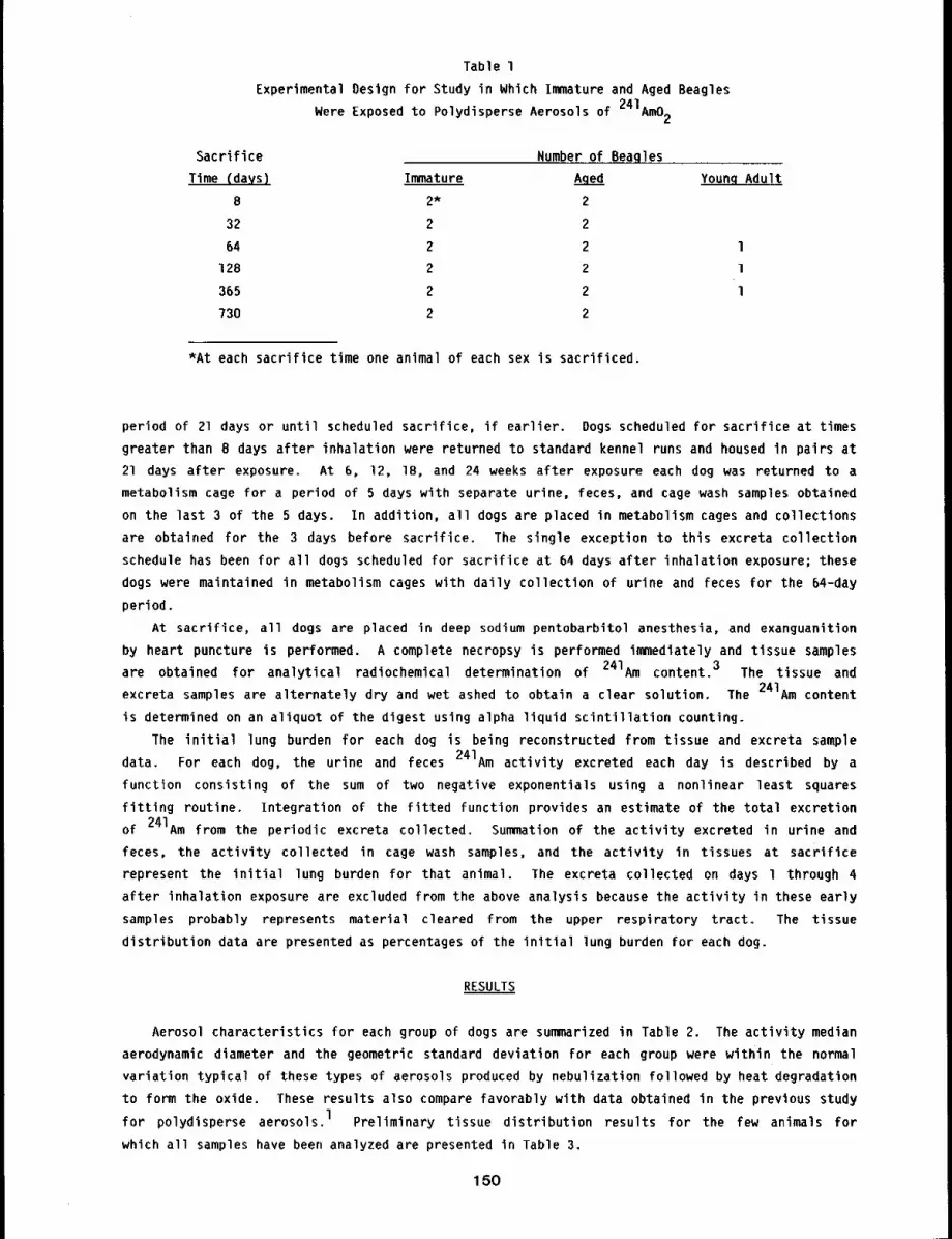
Table 1
Experimental Oesign for Study in Which Immature and Aged Beagles
Were Exposed to Polydisperse Aerosols of 241Am02
Sacrifice Number of Beagles
Time (days) Immature Aged Young Adult
8 2* 2
32 2 2
64 2 2 l
128 2 2 l
365 2 2 l
730 2 2
*At each sacrifice time one animal of each sex is sacrificed.
period of 21 days or until scheduled sacrifice, if earlier. Dogs scheduled for sacrifice at times
greater than 8 days after inhalation were returned to standard kennel runs and housed in pairs at
21 days after exposure. At 6, 12, 18, and 24 weeks after exposure each dog was returned to a
metabolism cage for a period of 5 days with separate urine, feces, and cage wash samples obtained
on the last 3 of the 5 days. In addition, all dogs are placed in metabolism cages and collections
are obtained for the 3 days before sacrifice. The single exception to this excreta collection
schedule has been for all dogs scheduled for sacrifice at 64 days after inhalation exposure; these
dogs were maintained in metabolism cages with daily collection of urine and feces for the 64-day
period.
At sacrifice, all dogs are placed in deep sodium pentobarbitol anesthesia, and exanguanition
by heart puncture is performed. A complete necropsy is performed immediately and tissue samples
are obtained for analytical radiochemical determination of 241Am content. 3 The tissue and
excreta samples are alternately dry and wet ashed to obtain a clear solution. The 241Am content
is determined on an aliquot of the digest using alpha liquid scintillation counting.
The initial lung burden for each dog is being reconstructed from tissue and excreta sample
data. For each dog, the urine and feces 241Am activity excreted each day is described by a
function consisting of the sum of two negative exponentials using a nonlinear least squares
fitting routine. Integration of the fitted function provides an estimate of the total excretion
of 241Am from the periodic excreta collected. Summation of the activity excreted in urine and
feces, the activity collected in cage wash samples, and the activity in tissues at sacrifice
represent the initial lung burden for that animal. The excreta collected on days l through 4
after inhalation exposure are excluded from the above analysis because the activity in these early
samples probably represents material cleared from the upper respiratory tract. The tissue
distribution data are presented as percentages of the initial lung burden for each dog.
RESULTS
Aerosol characteristics for each group of dogs are summarized in Table 2. The activity median
aerodynamic diameter and the geometric standard deviation for each group were within the normal
variation typical of these types of aerosols produced by nebulization followed by heat degradation
to form the oxide. These results also compare favorably with data obtained in the previous study
for polydisperse aerosols. 1 Preliminary tissue distribution results for the few animals for
which all samples have been analyzed are presented in Table 3.
150
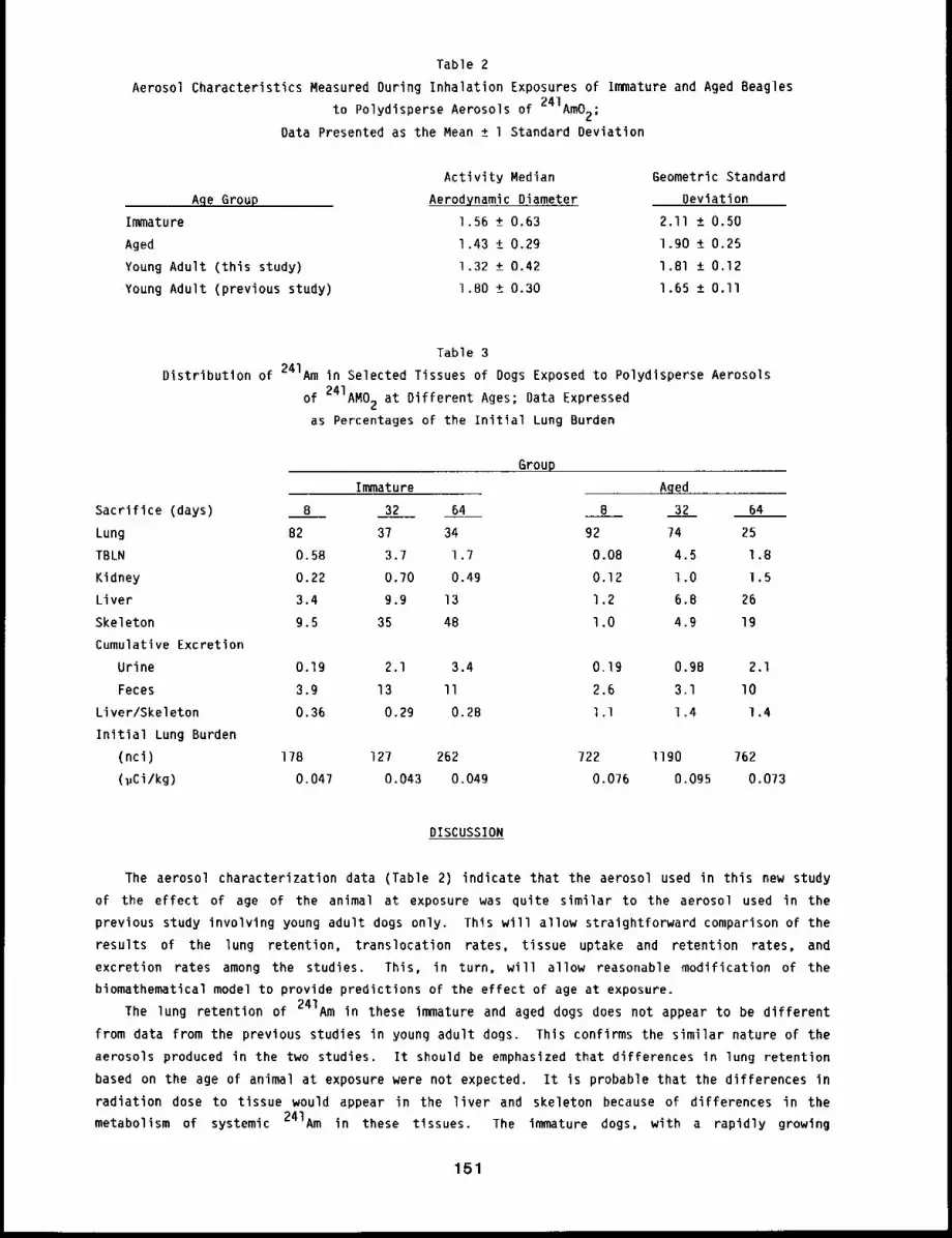
Table 2
Aerosol Characteristics Measured During Inhalation Exposures of Immature and Aged Beagles
to Polydlsperse Aerosols of 241Am02;
Data Presented as the Mean ± l Standard Deviation
Age Group
Immature
Aged
Young Adult (this study)
Young Adult (previous study)
Activity Median
Aerodynamic Diameter
l .56 +_ 0.63
l .43 +_ 0.29
1.32 + 0.42
1 .BO + 0.30
Geometric Standard
Deviation
2.11 +_ 0.50
1.90 + 0.25
1.81 +_ 0.12
1.65 +_ O.ll
Table 3
Distribution of 241Am in Selected Tissues of Dogs Exposed to Polydisperse Aerosols
of 241AM02 at Different Ages; Data Expressed
as Percentages of the Initial Lung Burden
Immature
Group
Sacrifice (days) B 32 64
Lung 82 37 34
TBLN 0.58 3.7 1.7
Kidney 0.22 0.70 0.49
Liver 3.4 9.9 13
Skeleton 9.5 35 48
Cumulative Excretion
Urine 0.19 2.1 3.4
Feces 3.9 13 II
Liver/Skeleton 0.36 0.29 0.28
Initial Lung Burden
(nci) 178 127 262
(~Ci/kg) 0.047 0.043 0.049
Aged
B 32 64
92 74 25
0.08 4.5 1.8
0.12 1.0 l ,5
1.2 6.8 26
l .0 4.9 19
o. Ig O.gB 2.1
2.6 3.1 lO
l .l 1.4 l ,4
722 ll90 762
0.076 0.095 0.073
DISCUSSION
The aerosol characterization data (Table 2) indicate that the aerosol used in this new study
of the effect of age of the animal at exposure was quite similar to the aerosol used in the
previous study involving young adult dogs only. This will allow straightforward comparison of the
results of the lung retention, translocation rates, tissue uptake and retention rates, and
excretion rates among the studies. This, in turn, will allow reasonable modification of the
biomathematical model to provide predictions of the effect of age at exposure.
The lung retention of 241Am in these immature and aged dogs does not appear to be different
from data from the previous studies in young adult dogs. This confirms the similar nature of the
aerosols produced in the two studies. It should be emphasized that differences in lung retention
based on the age of animal at exposure were not expected. It is probable that the differences in
radiation dose to tissue would appear in the liver and skeleton because of differences in the
metabolism of systemic 241Am in these tissues. The immature dogs, with a rapidly growing
151
skeleton might be expected to deposit more 241Am in skeleton than would elther young adult dogs,
which are growing less rapidly, or than would aged dogs whose skeleton has completed its growth
phase. One gauge of altered uptake in skeleton with age of the anfmal may be the ratio of liver
to skeletal deposition. Table 3 shows this ratio for those dogs for which analysis is complete to
date. Although rigorous statistical analysis is not yet possible on these limited data, there
appears to be a trend showing a lower liver to skeleton ratio in the immature dogs than in theaged or young adult dogs.
REFERENCES
I. Mewhinney, J. A., W. C. Griffith, and B. A. Muggenburg, The Influence of Aerosol Size onRetention and Translocatlon of 241Am Following Inhalation of 241Am02 by Beagles, HealthPhys. 42: 611-627, 1982.
2. Mewhinney, J. A. and W. C. Griffith, Models of Am Metabolism in Beagles and Humans, HealthPhys. 42: 629-644, 1982.
3. Guilmette, R. A. and A. Bay, Radioassay of Americium and Curium in Biological Material byIsooctyl Acid Phosphate Solvent Extraction and ~ Liquid Scintillation Counting, Anal. Chem.53: 2351-2354, 1981.
152
RADIATION DOSE PATTERNS IN CYNOMOLGUS MONKEYS AFTER INHALATION OF 241Am02
Abstract --S~xteen cynomolgus monkeys were glven
brlef, nose-only exposure to polydlsperse aerosols PRINCIPAL INVESTIGATOR
of 241AmO2 to provlde a comparison of the dlstrl- J.A. Mewh l nney
butlon of radiation dose to tissues of the monkey
wlth rhat measured In immature, young adult, and aged dogs, and with that observed in accldently
exposed humans. Anlmals are scheduled for sacrlflce, in palrs~ at 8 to 1460 days after inhalatlon
exposure. The exposures have been completed satlsfactorllg, and prellmlnary results on the
dlstrlbutlon and excretlon of 241Am from two monkeys, one sacrlflced at 8 days and the second
sacrlflced aT 32 after inhalation, show patterns that are slmllar to those obtalned from young
adult and aged Beagles sacrlflced at Iden~Ical tlmes after lnhalatlon.
This study is designed to determine the temporal distribution of radiation dose to tissues of
non-human primates after inhalation of 241AmO2. Within this context, several specific
scientific questions are being addressed. Does the non-human primate serve as a "better" model of
retention, distribution, and excretion in humans after inhalation of 241Am than do other animal
species? Can excretion rates for 241Am in non-human primates be used to estimate body burdens
at various times after inhalation exposure? What is the relationship of these rates of excretion
to the burdens in specific tissues? Do the quantities and concentrations of 241Am in tissues of
the non-human primate approximate those measured in humans in vivo or for a single case in which
complete analysis of the body content was accomplished after death? l Data from this study will241provide a firmer basis for determination of risk estimators for humans after inhalation of Am.
At present, only preliminary data are available for two cynomolgus monkeys sacrificed at 8 and
32 days after inhalation exposure to polydisperse aerosols of 24lAm02. As data are
accumulated, direct comparisons will be made among the data from the study and similar studies in
Beagle dogs (this report, pp. 149 to 152). In addition, the biomathematical model developed from
previous studies in Beagles 2 will be modified to incorporate the results of this study to
provide a basis for comparing and contrasting the retention, distribution, and excretion of
2¢IAm in several animal species and humans.
METHODS
Table l summarizes the experimental design of this study. The aerosol production techniques
used are identical to those used in the study in which immature and aged Beagles were exposed to
241Am02 and are fully described elsewhere. 3 Briefly, 241Am is precipitated as the
hydroxide, the precipitate is dispersed in O.Ol M nitric acid, and the suspension is nebulized to
form droplets. The droplets are heat-treated in two stages. The first dries the droplets at241
350°F, and the second (at ll50°C) degrades the particles to AmO2. The inhalation exposure
of monkeys uses the dog exposure system, with minor modifications. During each exposure, the
aerosol is characterized using cascade impactors to determine particle size and size distribution,
point-to-plane electrostatic precipitators to obtain particles on an electron microscope grids for
visualization to determine particle morphology and physical sizing, and membrane filters to
determine aerosol concentration. All monkeys were exposed to achieve an initia’l lung burden of
0.075 ~Ci/kg body weight.
153
Table ’I
Summary of the Experimental Design for the Study in Which Cynomolgus Monkeys
Were Exposed to Polydisperse Aerosols of 241AmO2
Sacrifice Time (days) Number of Animals
8 2
32 2
64 2
12B 2
180 2
365 2
730 2
1460 2
The cynomolgus monkeys used in this study (wild-caught) were purchased from Primate Imports,
Port Washington, NY. The monkeys were placed in quarantine for 90 days, with frequent monitoring
for tuberculosis before inhalation exposure. Ketamine hydrochloride was used to anesthetize all
monkeys before exposure. Immediately after exposure, monkeys were placed in individual metabolism
cages and remain there for the duration of the experimental period. Separate collection of urine
and feces samples was accomplished daily for the first 21 days after exposure, for 3-day periods
at 6, 12, 18, and 24 weeks after exposure, and twice annually thereafter. One monkey of each sex
was randomly assigned to sacrifice, according to the schedule shown in Table I. Monkeys were
sacrificed by anesthesizing the animal with ketamine hydrochloride, and then exanguanation by
heart puncture. Complete necropsy was performed immediately.
Determination of the 241 Am content in excreta and tissue samples was accomplished by
alternate dry and wet ashing to obtain a clear digest. The digest is quantified by solvent4extraction and alpha-liquid scintillation counting.
Reconstruction of the initial lung burden for individual monkeys was accomplished by fitting
the urine and feces activity in daily samples (nCi/day) to a two=component sum of negative
exponential function using a nonlinear least squares method. Integration of the fitted function
over the period from five days after exposure to day of death yields a good estimate of the total
activity excreted by that mode. Excreta data were excluded for days 1 through 4 because of upper
respiratory tract clearance during that period. The quantities excreted in urine and feces were
added to the activity found in tissues at death to obtain the initial lung burden. The data for
tissues were then expressed as percentages of the initial lung burden.
RESULTS
The results of characterization of the exposure aerosols were an activity median aerodynamic
diameter of 1.4 ± 0.4 (mean ± standard deviation) and a geometric standard deviation of 1.9
± 0.2. These values are q~ite similar to those measured for identically produced aerosols in
Beagle dog exposures (this report, pp. 149 to 152). Electron microscopy showed the particles to
reasonably round smooth spheres.
The distribution of 241Am in tissues of two monkeys, one sacrificed at 8 days and the second
at 32 days after inhalation exposure, is shown in Table 2. These data are comparable to similar
data derived for studies in young adult 3 and aged dogs (this report, pp. 149 to 152.) exposed
quite similar aerosols of 241Am02. Total quantities of 241Am excreted by the monkey are
also in general agreement with the amounts excreted by young adult and aged Beagles.
154
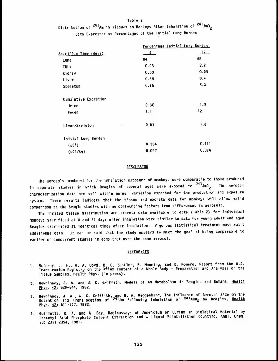
Table 2
Distribution of 241Am in Tissues on Monkeys After Inhalation of 241AmO2.Data Expressed as Percentages of the Initial Lung Burden
Percentage Initial Lung Burden
Sacrifice Time (days) B 32
Lung B4 68
TBLN 0.03 2.2
Kidney 0.03 0.’09
Liver 0.65 8.4
Skeleton 0.96 5.3
Cumulative Excretion
Urine 0.30 1.9
Feces 6.1 12
Liver/Skeleton 0.67 1.6
Initial Lung Burden
(~Ci) 0.364 O.411
(uCi/kg) 0.092 0.094
DISCUSSION
The aerosols produced for the inhalation exposure of monkeys were comparable to those produced
in separate studies in which Beagles of several ages were exposed to 241Am02. The aerosol
characterization data are well within normal variation expected for the production and exposure
system. These results indicate that the tissue and excreta data for monkeys will allow valid
comparison to the Beagle studies with no confounding factors from differences in aerosols.
The limited tissue distribution and excreta data available to date (Table 2) for individual
monkeys sacrificed at B and 32 days after inhalation were similar to data for young adult and aged
Beagles sacrificed at identical times after inhalation. Vigorous statistical treatment must await
additlonal data. It can be said that the study appears to meet the goal of being comparable to
earlier or concurrent studies in dogs that used the same aerosol.
REFERENCES
1. Mclnroy, 3. F., H. A. Boyd, B. C. Eastler, R. Manning, and D. Romero, Report from the U.S.Transuranium Registry on the 241Am Content of a Whole Body - Preparation and Analysis of theTissue Samples, Health Phys. (in press).
2. Mewhinney, 3. A. and W. C. Griffith, Models of Am Metabolism in Beagles and Humans, Health
Phys. 42: 629-644, 1982.
3, Mewhinney, 3. A., W. C. Griffith, and B. A. Muggenburg, The Influence of Aerosol Size on theRetention and Translocation of 241Am Following Inhalation of 241Am02 by Beagles, HealthPhys. 42: 611-627, 1982.
4. 6uilmette, R. A. and A, Bay, Radioassays of Americium or Curium in Biological Material byIsooctyl Acld Phosphate Solvent Extraction and ~ Liquid Scintillation Counting, Anal. Chem.53: Z351-2354, 19Bl.
155
EFFECT OF THE CHEMICAL FORM OF INHALED CURIUM ON ITS BIOKINETICS IN DOGS
Abstract --- To determlne the effect of phgslcochemlcal
form of inhaled curium aerosols on subsequent recent. PRINCIPAL INVESTIGATOR
tion, translocat~on, dlstrlbutlon, and excretion, 18 R.A. Gullmette
young adult Beagle dogs received single pernasal ~nha-
latlon exposures to a monodlsperse aerosol (1.38 ~m activity median aerodynamic diameter [AMAD])
of Cm203, and 18 dogs inhaled a pol~dlsperse aerosol (1.14 ~m AMAD; ~ = 1.7) ofgCm[N03)3.
Through measurement of curlum in the tlssues and excreta of dogs being serlall9sacrificed from 0.17 to 730 days after exposure, the bloklnetlcs of curium are being elucidated as
a functlon of physlcochemlcal form of the inhaled aerosol.
A previous study done at this Institute in which rats received single inhalation exposures to
a curium oxide aerosol heat-treated at 1150°C showed that this particular physical-chemical form
was less soluble than curium oxide aerosols used in other previously reported animal
studies. I-4 Because curium can exist as one of several stoichiometic or nonstoichiometric
oxides with differing crystal structures, it is important to determine the influence of the
physical-chemical state of an inhaled curium compound on its biological fate after inhalation
exposure. We expect that differences in biokinetics of curium oxides will be related to their
intrinsic solubility in biological fluids. A systematic study of the biokinetics of curium within
the context of the curium-oxygen pr~ysicochemical system has been initiated In Beagle dogs. This
study has been structured such that the forms hypothesized to be most soluble (Cm(N03)3),
the least soluble (Cm203), are being studied first. The second phase of this study, which
will use the CmO2 and CmTOl2 forms, will be initiated after results of the Phase l study
have been evaluated. When feasible, monodisperse aerosols are being used to provide a better
comparison of the intrinsic solubility of each aerosol without the complication of heterogeneity
of particle size and, therefore, specific surface area.
METHODS
The source material used in this study is an isotopic mixture of 243Cm (8% by mass), 244Cm
(88%), 245Cm and 246Cm (2.5%). The experimental design for t~his study is shown in Table
Young adult Beagle dogs are being exposed once by inhalation to monodisperse aerosols (1.5 ~m
activity median aerodynamic diameter [AMAD]) of Cm203, Cm02, Cm7012, or to
polydisperse aerosol of Cm(N03) 3 with AMAD = 1.5 ~m and geometric standard deviation
(~g) - 2. There are 18 dogs (9 male, 9 female) in each aerosol group. The desired initiallung burden is 0.04 ~Ci Cm/kg body weight. Physical-chemical characterization of the aerosols
is being done by electron diffraction for crystallographic identification, cascade impactor
sampling as well as transmission electron micrography for physical and aerodynamic particle sizing
and morphologic determination.
Urine and feces samples are periodically collected from each dog to define the pattern of
excretion of curium. Each dog is sacrificed according to the schedule in Table l, and detailed
necropsies are done. The sacrifice schedule has been structured to provide more data during earl)/
times after exposure (< l year), when most of the translocation and excretion is anticipated
occur, and will also provide data comparable to other dose pattern studies in dogs being done at
this Institute with 238pu, 239pu, and 241Am. In addition to radioassay of most soft tissue
15B
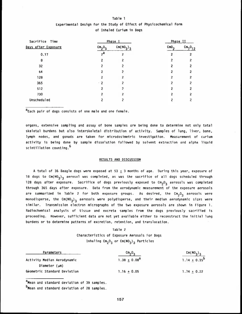
Table 1
Experimental Design for the Study of Effect of Physlcochemtcal Form
of Inhaled Curium in Bogs
Sacrifice Time
Days after Exposure
0.17
B
32
64
128
365
512
730
Unscheduled
Phase I Phase II
Cm203 Cm(N03)3 CmO2 CmTOl2
2a 2 2 2
2 2 2 2
2 2 2 2
2 2 2 2
2 2 2 2
2 2 2 2
2 2 2 2
2 2 2 2
2 2 2 2
aEach pair of dogs consists of one male and one female.
organs, extensive sampling and assay of bone samples are being done to determine not only total
skeletal burdens but also interskeletal distribution of activity. Samples of lung, liver, bone,
lymph nodes, and gonads are taken for microdosimetric investigation. Measurement of curium
activity is being done by sample dissolution followed by solvent extraction and alpha liquid
scintillation counting.6
RESULTS AND DISCUSSION
A total of 36 Beagle dogs were exposed at 53 ± 3 months of age. During this year, exposure of
18 dogs to Cm(N03)3 aerosol was completed, as was the sacrifice of all dogs scheduled through
128 days after exposure. Sacrifice of dogs previously exposed to Cm203 aerosols was completedthrough 365 days after exposure. Data from the aerodynamic measurement of the exposure aerosols
are summarized in Table 2 for both exposure groups. As desired, the Cm203 aerosols were
monodisperse, the Cm(N03)3 aerosols were polydisperse, and their median aerodynamic sizes weresimilar. Transmission electron micrographs of the two exposure aerosols are shown in Figure I.
Radiochemical analysis of tissue and excreta samples from the dogs previously sacrified is
proceeding. However, sufficient data are not yet available either to reconstruct the initial lung
burdens or to determine patterns of excretion, retention, and translocation.
Table 2
Characteristics of Exposure Aerosols For Dogs
Inhaling Cm203 or Cm(N03)3 Particles
Parameters
Activity Median Aerodynamic
Diameter (~m)
Geometric Standard Deviation
aMean and standard deviation of 39 samples.
bMean and standard deviation of 2B samples.
Cm203 Cm(N03)3
l .38 +_ 0.08a l .]4 _+ 0.35b
1.16 + 0.05 1.74 + 0.22
157
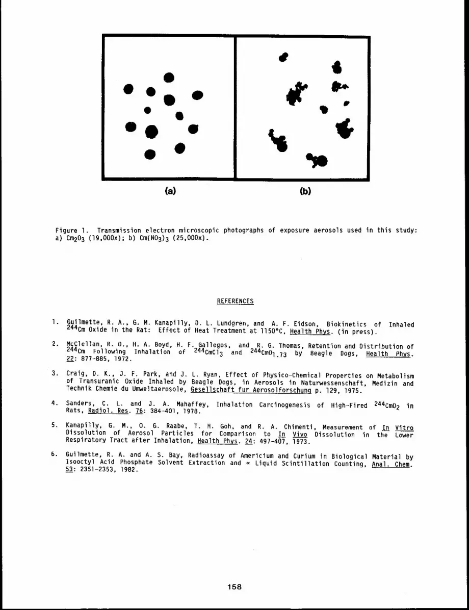
IIIIII
e
e
Illll Ill
(a) (b)II I IIIII
Figure I. Transmission electron microscopic photographs of exposure aerosols used in this study:a) Cm203 (19,000x); b) Cm(N03)3 (25,000x).
REFERENCES
I. Guilmette, R. A., G. M. Kanapilly, D. L. Lundgren, and A. F. Eidson, Biokinetics of Inhaled244Cm Oxide in the Rat: Effect of Heat Treatment at llSO°C, Health Phys. (in press).
2. McClellan, R. 0., H. A. Boyd, H. F. Gallegos, and R. G. Thomas, Retention and Distribution of244Cm Following Inhalation of 244CMCI3 and 244Cm01.73 by Beagle Dogs, Health Phys.2~: 877-B85, 1972.
3. Craig, D. K., 3. F. Park, and O. L. Ryan, Effect of Physico-Chemical Properties on Metabolismof Transuranic Oxide Inhaled by Beagle Dogs, in Aerosols in Naturwessenschaft, Medizin andTechnik Chemie du Umweltaerosole, Gesellschaft fur Aerosolforschung p. 129, 1975.
4. Sanders, C. L. and J. A. Mahaffey, Inhalation Carcinogenesis of High-Fired 244Cm02 inRats, Radiol. Res. 76: 384-401, 1978.
5. Kanapilly, G. M., O. G. Raabe, T. H. Goh, and R. A. Chimenti, Measurement of In VitroDissolution of Aerosol Particles for Comparison to In Vivo Dissolution in the LowerRespiratory Tract after Inhalation, Health Phys. 24: 497-407, 1973.
6. Guilmette, R. A. and A. S. Bay, Radioassay of Americium and Curium in Biological Material byIsooctyl Acid Phosphate Solvent Extraction and = Liquid Scintillation Counting, Anal. Chem.L3: 2351-2353, 1982.
158
REDUCING CURIUM TRANSLOCATION FROM LUNG WITH DTPA THERAPY
Abstract -- To reduce the translocatlon of Cm from
lung to bone and liver, chelatlon with dleth~lene- PRINCIPAL INVESTIGATORS
trlamlnepentaacetlc acld DTPA was started 24 hours R.A. Guilmette
after inhalation exposure of rats to a moderatel~ B.A. Muggenburg
soluble aerosol of 243’244Cm203. Continuous admin-
istratlon was accompllshed using subcutaneously implanted osmotlc pumps. Expressed as percentages
of the organ burdens In Cm-exposed, sallne-treated controls, rats treated with continuous DTPA had
60~ of the lung burden, less than i~ of The liver burden, and 4~ of the skeletal burden 14 days
after exposure. In comparison, discrete intraperltoneal injections of DTPA in rats produced
somewhat less removal efflclenc~ from bone, although, ~n general, that mode of treatment was also
effective.
In cases of accidental inhalation of actinide aerosols at levels sufficient to cause concern
about potential biological effects, it is important to remove the radioactive materials from the
body as soon as possible. If the aerosol is highly or even moderately soluble, the primary
therapeutic goal will be the prevention of translocation of the dissolved actinide from lung to
bone and liver. In this study, rats inhaled a moderately soluble curium oxide aerosol, and the
effectiveness of continuously administered diethylenetriaminepentaacetic acid (DTPA) in preventing
such translocation was evaluated.
METHODS
A total of 21 male and 21 female Fischer-344 specific pathogen-free rats from this Institute’s
colony received a single pernasal inhalation exposure to a monodisperse aerosol of
243’244Cm203 (0.90 ~m activity median aerodynamic diameter; 1.13 geometric standard
deviation), using previously described techniques, l Immediately after exposure, each rat was
placed in a stainless steel metabolism cage and maintained with food and water ad libitum for the
14-day duration of the study. All urine and feces were collected, as well as the solutions used
in the washing of the cages.
The experimental design is shown in Table I. Each group consisted of three male and three
female rats. Treatment was begun one day after exposure. To determine the distribution of curium
in the rat at the beginning of therapy, six animals were sacrificed at one day after exposure.
Discrete administration of DTPA consisted of four intraperitoneal (IP) injections. Continuous
administration of DTPA was accomplished by subcutaneously implanting an Alzet osmotic pump (Alza
Corp., Palo Alto, CA) in the dorsal area of the rat. Two sizes of osmotic pumps were used to
achieve DTPA dose delivery rates of 6 or 40 wmoles Zn DTPA per day.
All rats were sacrificed at l or 14 days after exposure by IP injection of T-61 euthanasia
solution, The liver, lung, tracheobronchial lymph nodes, pelt, and remaining viscera were removed
at necropsy; the remaining carcass (not including skull, paws, and tail) was analyzed as a single
sample. Estimation of the skeletal content of curium was done using the relationship [total
skeletal curium] = [carcass curium] x 1.7. Radioassay of the curium content of the tissue and
159
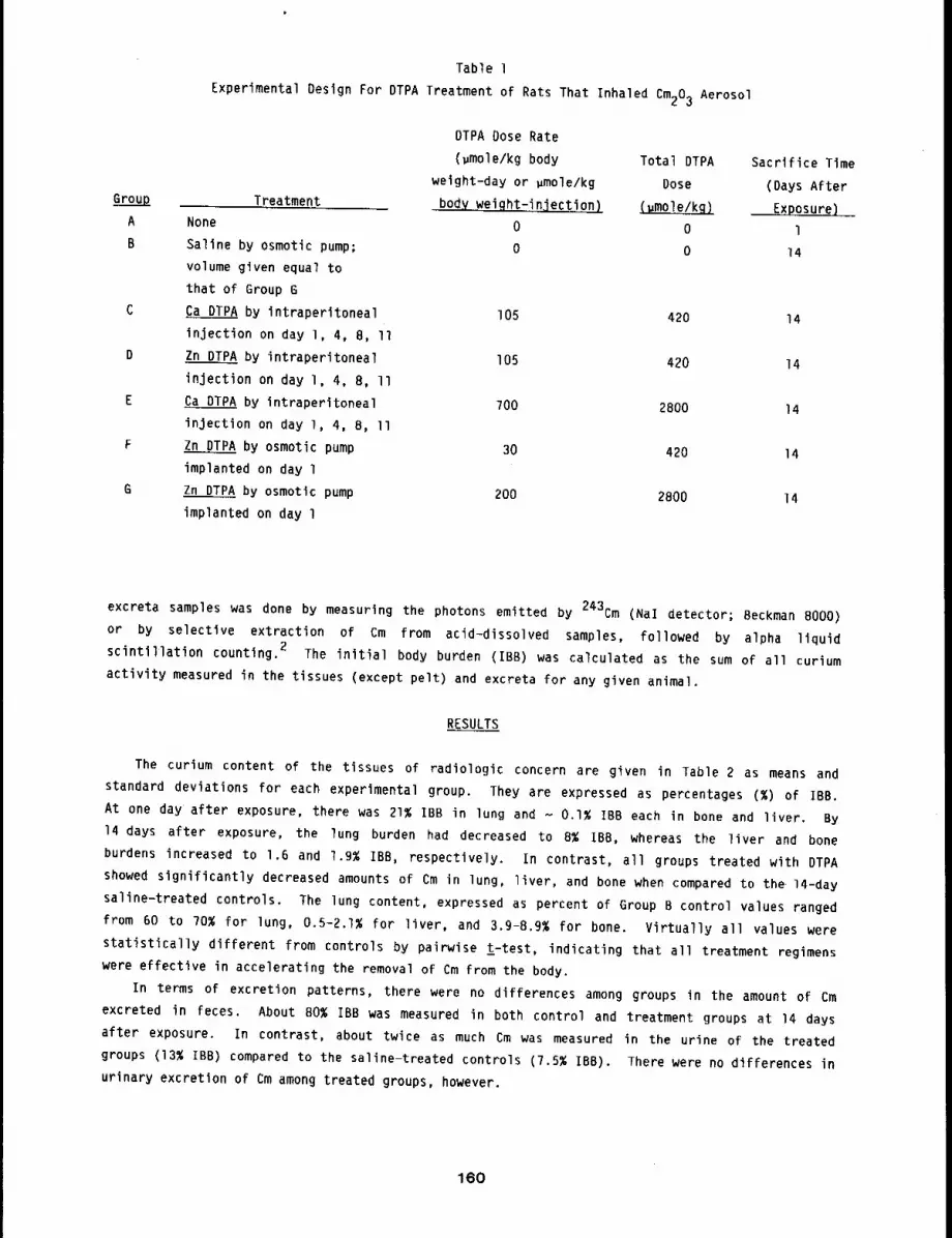
Group
A
B
Table l
Experimental Design For DTPA Treatment of Rats That Inhaled Cm203 Aerosol
Treatment
None
Saline by osmotic pump;
volume given equal to
that of Group G
Ca DTPA by intraperltoneal
injection on day l, 4, 8, II
Zn DTPA by intraperitoneal
injection on day l, 4, 8, II
Ca DTPA by intraperitoneal
injection on day l, 4, 8, II
Zn DTPA by osmotic pump
implanted on day l
Zn DTPA by osmotic pump
implanted on day l
DTPA Dose Rate
(~mole/kg body Total DTPA Sacrifice Timeweight-day or pmole/kg Dose (Days After
body weiqht-injection) ~ Exposure}
0 0 10 0 14
105 420 14
I05 420 14
700 2800 14
30 420 14
200 2800 14
excreta samples was done by measuring the photons emitted by 243Cm (Nal detector; Beckman 8000)
or by selective extraction of Cm from acid-dissolved samples, followed by alpha liquid
scintillation counting. 2 The initial body burden (IBB) was calculated as the sum of all curium
activity measured in the tissues (except pelt) and excreta for any given animal.
RESULTS
The curium content of the tissues of radiologic concern are given in Table 2 as means and
standard deviations for each experimental group. They are expressed as percentages (%) of IBB.
At one day after exposure, there was 21% IBB in lung and - 0.1% IBB each in bone and liver. By
14 days after exposure, the lung burden had decreased to 8% IBB, whereas the liver and bone
burdens increased to 1.6 and 1.9% IBB, respectively. In contrast, all groups treated with DTPA
showed significantly decreased amounts of Cm in lung, liver, and bone when compared to the 14-day
saline-treated controls. The lung content, expressed as percent of Group 8 control values ranged
from 60 to 70% for lung, 0.5-2.1% for liver, and 3.9-8.9% for bone. Virtually all values were
statistically different from controls by pairwise ~-test, indicating that all treatment regimens
were effective in accelerating the removal of Cm from the body.
In terms of excretion patterns, there were no differences among groups in the amount of Cm
excreted in feces. About 80% IBB was measured in both control and treatment groups at 14 days
after exposure. In contrast, about twice as much Cm was measured in the urine of the treated
groups (13% IBB) compared to the saline-treated controls (7.5% IBB). There were no differences
urinary excretion of Cm among treated groups, however.
160
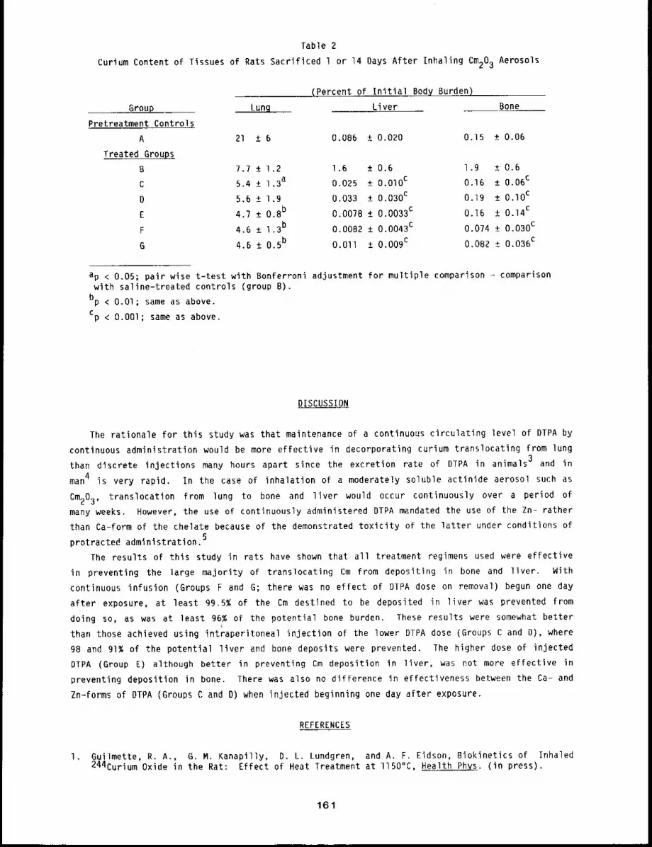
Table 2
Curium Content of Tissues of Rats Sacrificed 1 or 14 Days After Inhaling Cm203 Aerosols
Group Lun~
Pretreatment Controls
A 21 ± 6
Treated Group~
B 7.7 + 1.2
C 5.4 +_ 1.3a
D 5.6 ± 1.9
E 4.7 ± 0.8b
F 4.6 ± 1.3b
G 4.6 ± 0.5b
(Percent of Initial Body Burden)
Liver Bone
0.086 ± 0.020 0.15 + 0.06
1.6 +_ 0.6 l.g + 0.6
0.025 + 0.010 C 0.16 ± 0.06c
0.033 ± 0.030c 0.19 + 0.I0C
0.0078 + 0.0033c 0.16 ± 0.14c
0.0082 +_ 0.0043c 0.0?4 ± 0.030c
O.Oll ± 0.009c 0.082 ± 0.036c
ap < 0.05; pair wise t-test with Bonferroni adjustment for multiple comparison- comparisonwith saline-treated controls (group B).
bp < O.Ol; same as above.cp < O.OOl; same as above.
DISCUSSION
The rationale for this study was that maintenance of a continuous circulating level of DTPA by
continuous administration would be more effective in decorporating curium translocating from lung
than discrete injections many hours apart since the excretion rate of DTPA in animals 3 and in
man4 is very rapid. In the case of inhalation of a moderately soluble actinide aerosol such as
Cm203, translocation from lung to bone and liver would occur continuously over a period of
many weeks. However, the use of continuously administered DTPA mandated the use of the Zn- rather
than Ca-form of the chelate because of the demonstrated toxicity of the latter under conditions of
protracted administration.5
The results of this study in rats have shown that all treatment regimens used were effective
in preventing the large majority of translocating Cm from depositing in bone and liver. With
continuous infusion (Groups F and G; there was no effect of DIPA dose on removal) begun one day
after exposure, at least 99.5% of the Cm destined to be deposited in liver was prevented from
doing so, as was at least 96% of the potential bone burden. These results were somewhat better
than those achieved using intraperitoneal injection of the lower DTPA dose (Groups C and D), where
98 and 91% of the potential liver and bone deposits were prevented. The higher dose of injected
DTPA (Group E) although better in preventing Cm deposition in liver, was not more effective
preventing deposition in bone. There was also no difference in effectiveness between the Ca- and
Zn-forms of DTPA (Groups C and D) when injected beginning one day after exposure.
REFERENCES
I. Guilmette, R. A., G. M. Kanapilly, D. L. Lundgren, and A. F. Eidson, Biokinetics of Inhaled244Curium Oxide in the Rat: Effect of Heat Treatment at I150°C, Health Phys. (in press).
161
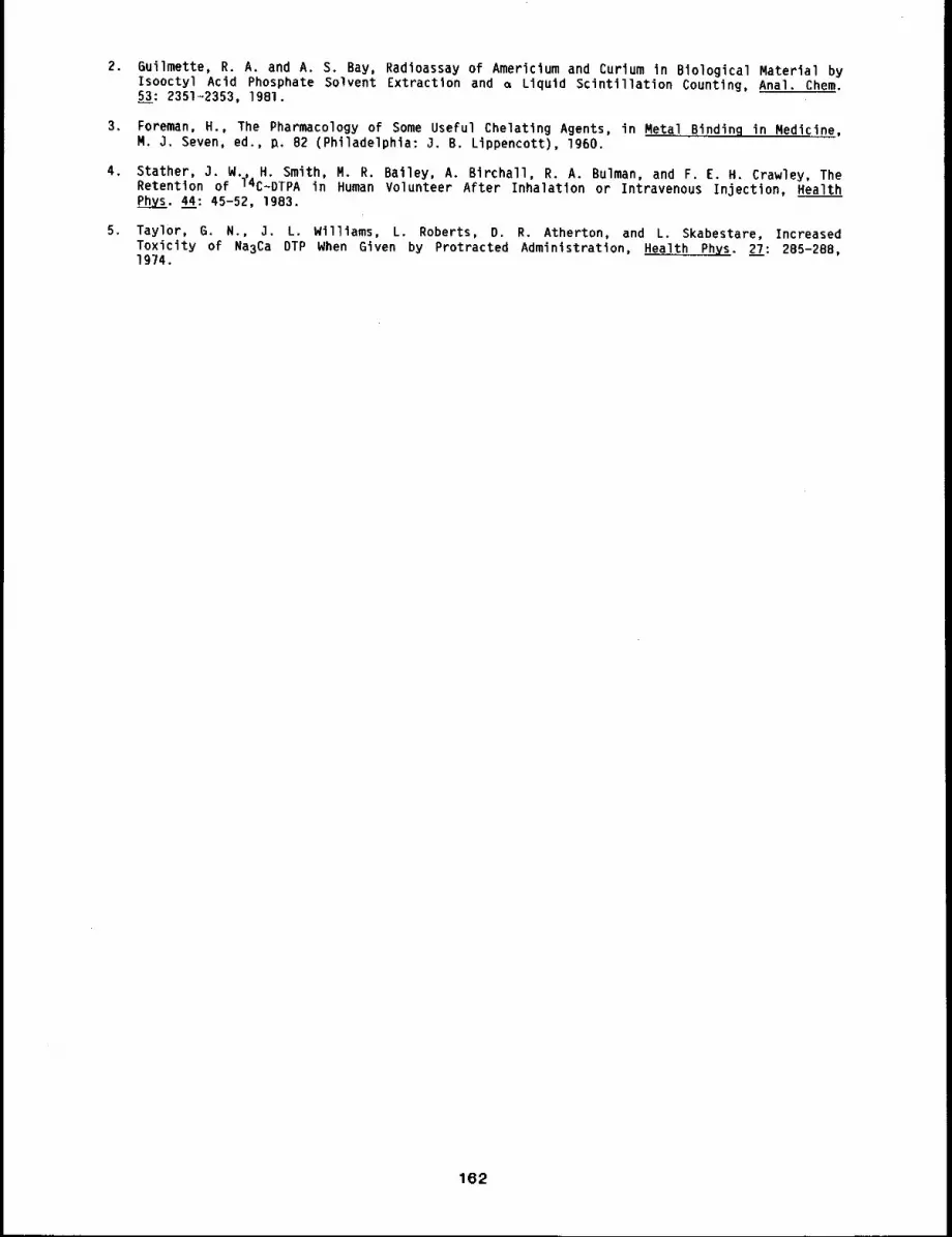
2. 6uilmette, R. A. and A. S. Bay, Radfoassay of Americium and Curium in Blological Material byIsooctyl Acid Phosphate Solvent Extraction and ~ Llquld Scintillation Counting, Anal. Chem.53: 2351~2353, 1981.
3. Foreman, H., The Pharmacology of Some Useful Chelatlng Agents, in Metal Binding in Medicine,N. j. Seven, ed., ~. 82 (Philadelphla: 3. B. Lippencott), 1960.
4. Stather, 3. N., H. Smith, R. R. Bailey, A. Birchall, R. A. Bulman, and F. E. H. Crawley, TheRetention of 14C~DTPA in Human Volunteer After Inhalation or Intravenous Injection, HealthPhys. 44: 45-52, 1983.
5. Taylor, G. N., J. L. Williams, L. Roberts, D. R. Atherton, and L. Skabestare, IncreasedToxicity of Na3Ca DTP When Given by Protracted Administration, Health Phys. 27: 285-288,1974.
162

TISSUE DISTRIBUTION AND METABOLIC FATE OF 2-AMINOANTHRACENE IN RATS AFTER INHALATION
Abstract -- The distribution and fate of [3H]-2-
amlnoanthracene (2-AA) was determined in male PRINCIPAL INVESTIGATORS
Fischer-344 rats after inhalation exposure (70 ~g/1; C.E. Hitchell
actlvlty mass median diameter = 2.1 ~in) for 30 mln. R.F. Henderson
Radloactlvlty was found in the turblnates, trachea, R.O. McClellan
lung, liver, kidney, and gastrointestinal tract 20
mln after exposure. Smaller quantltles of radioactivity were found in fat, brain, and testis.
Inhaled 2-AA was excreted in feces (80~) and urine (20~). Organic soluble radloactlvlty
released from urinary conjugates after treatment of urine with acid and 8-glucuronldase. This
suggests the presence of both N-glucuronldes and o-glucuronldes of 2-AA. The organic soluble
radioactivity found after hydrolysis also indicated that inhaled 2-AA is extensively metabolized
by rats to conjugated materials and excreted as N-glucuronldes, O-glucuronldes, and sulfates.
2-Aminoanthracene (2-AA) is a tricyclic primary aromatic amine (PAA). PAAs are found in
tar, cigarette smoke, waste streams from petroleum facilities, and in petroleum substitutes from
coal and crude oil. l 2-AA is a potent mutagen and carcinogen. Of note are its carcinogenic
activity in rat epithelial tissues, as opposed to mice tissue, and its ability to produce
histologically varied malignant tumors in rats. 2 Although the carcinogenic and mutagenic
properties of 2-AA are known, little, if any, information is available on the deposition,
distribution, and fate of inhaled 2-AA. Because of the potential for inhalation of aromatic
amines (including 2-AA) after release into the environment and because of the unusual effects
2-AA on epithelial tissues (the primary site of deposited material), the present study was carried
out to provide information on the deposition, distribution, and fate of inhaled amines to help
assess potential hazards associated with release.
METHODS
Specific pathogen-free, male Fischer~344 rats, B to lO weeks old, were exposed nose-only for
30 min to 2-amino-[G-3H]anthracene (Amersham/Searle Corp., Arlington Heights, IL), diluted with
unlabeled 2-AA (g8~ pure, Aldrich Chemical Co., Milwaukee, WI) to a specific activity of 32
lO 3 dpm/~g. The aerosols (70 ~g/l; activity mass aerodynamic diameter, 2.1 ~m) were
generated by heating a pyrex boat to 260°C. The flow rates of the driving gas and carrier gas
were 0.4 L/min and 3 L/min, respectively. Twenty minutes after exposure, the rats were either
sacrificed i~ediately or transferred to a small animal housing area and sacrificed later.
Animals were sacrificed by CO2 asphyxiation at 0.33, 3, 6, 12, 24, 48, or g6 h after exposure.
Four rats were placed in metabolism cages (MC 3000 metabolism monitor, Stanford Glassblowing Lab,
Palo Alto, CA), and feces and urine were collected every 12 h. Duplicate tissue and feces samples
were oxidized directly in a sample oxidizer (Model B306 Packard Instruments, Downers Grove, IL).
Aliquots of urine were added to scintillation cocktail, and total 3H-radioactivity was
determined by scintillation counting (Packard Tri-Carb 2660 Packard Instruments, Downers Grove,
163
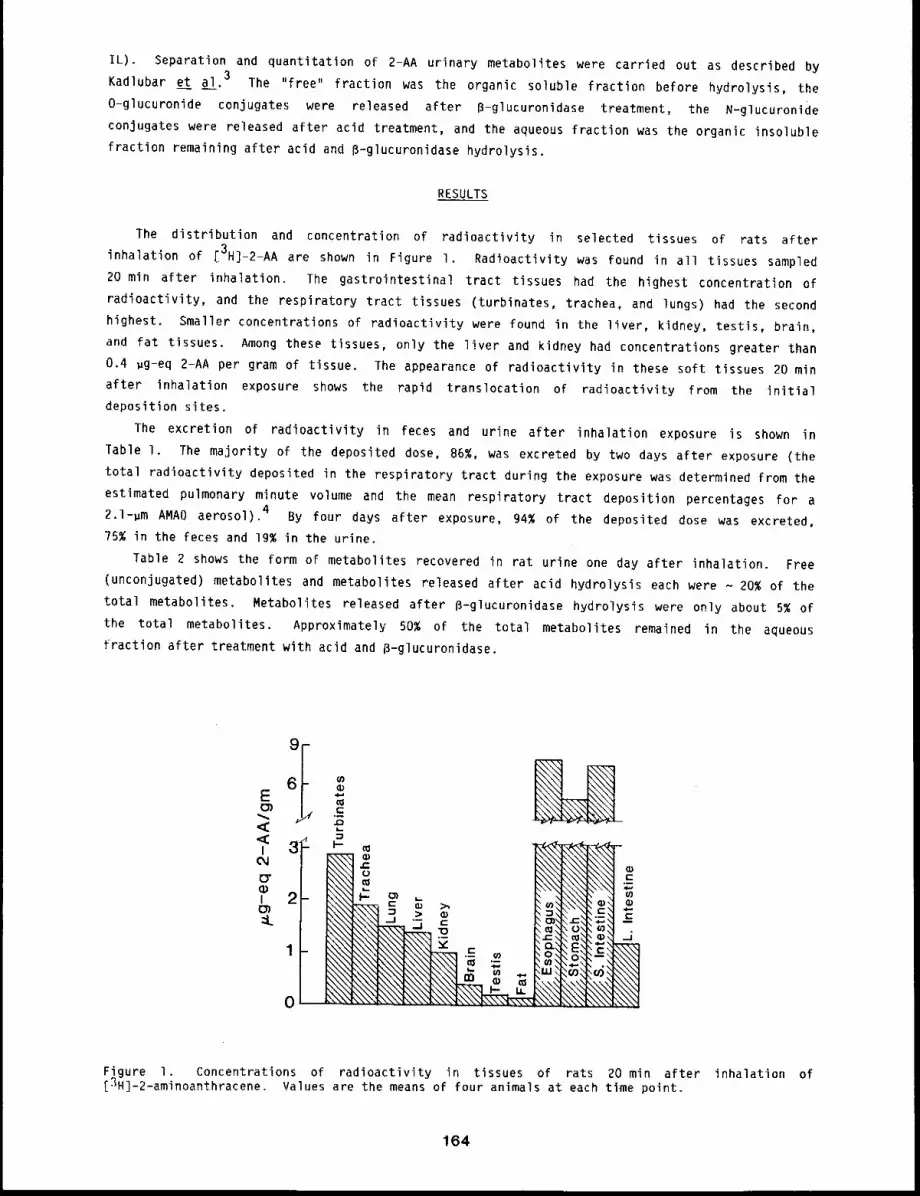
IL). Separation and quantitation of 2-AA urinary metabolites were carried out as described by
Kadlubar et al. 3 l’he "free" fraction was the organic soluble fraction before hydrolysis, the
O-glucuronide conjugates were released after B-glucuronidase treatment, the N-glucuronide
conjugates were released after acid treatment, and the aqueous fraction was the organic insoluble
fraction remaining after acid and B-glucuronidase hydrolysis.
RESULTS
The distribution and concentration of radioactivity in selected tissues of rats after
inhalation of [3H]-2-AA are shown in Figure I. Radioactivity was found in all tissues sampled
20 min after inhalation. The gastrointestinal tract tissues had the highest concentration of
radioactivity, and the respiratory tract tissues (turbinates, trachea, and lungs) had the second
highest. Smaller concentrations of radioactivity were found in the liver, kidney, testis, brain,
and fat tissues. Among these tissues, only the liver and kidney had concentrations greater than
0.4 ~g-eq 2-AA per gram of tissue. The appearance of radioactivity in these soft tissues 20 min
after inhalation exposure shows the rapid translocation of radioactivity from the initial
deposition sites.
The excretion of radioactivity in feces and urine after inhalation exposure is shown in
Table l. The majority of the deposited dose, 86%, was excreted by two days after exposure (the
total radioactivity deposited in the respiratory tract during the exposure was determined from the
estimated pulmonary minute volume and the mean respiratory tract deposition percentages for a
2.1-~m AMAD aerosol). 4 By four days after exposure, 94% of the deposited dose was excreted,
?5% in the feces and 19% in the urine.
fable 2 shows the form oF metabolites recovered in rat urine one day after inhalation. Free
(unconjugated) metabolites and metabolites released after acid hydrolysis each were - 20% of the
total metabolites. Metabolites released after B-glucuronidase hydrolysis were only about 5% of
the total metabolites. Approximately 50% of the total metabolites remained in the aqueous
fraction after treatment with acid and B-glucuronidase.
_
6i-E o..--
6
I 3- I.- m
.... I,-, ~ ,_I 2- ~\\~_~ ~ >,
Figure I. Concentrations of radioactivity in tissues of rats 20 min after inhalation of[3H]-2-aminoanthracene. Values are the means of four animals at each time point.
164
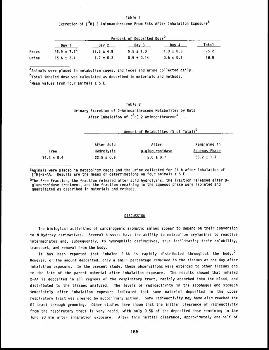
Table 1Excretion of [3H]m2-Amlnoanthracene From Rats After Inhalation Exposurea
Percent of, De.posited Doseb
Day 1 ~2 Day 3 .,Day 4 Total
Feces 45.9 +_ 7.7c 22.5 + 4.9 5.5 ± 1.0 1.3 + 0.3 75.2
Urine 15.6 +_ 3.1 1.7 ± 0.3 0.9 ± 0.14 0.6 ± 0.I 18.8
aAnimals were placed in metabolism cages, and feces and urine collected daily.
bTotal inhaled dose was calculated as described in materials and methods.
CMean values from four animals ± S.E.
Table 2
Urinary Excretion of 2-Amlnoanthracene Metabolites by Rats
After Inhalation of [3H]-2-Aminoanthracenea
Amount of Metabolites (~ of Total)b
Free
19.3 ± 0.4
After Acid After Remaining in
13-gl ucuronidase Aqueous Phase
22.5 ± 0.9 5.0 +_ 0.7 53.2 + 1.7
aA~imals were placed in metabolism cages and the urine collected for 24 h after inhalation of[ H]-2-AA. Results are the means of determinations on four animals ± S.E.
bThe free fractlon, the fraction released after acid hydrolysis, the fraction released after P-glucuronidase treatment, and the fraction remaining In the aqueous phase were isolated andquantitated as described in materials and methods.
DISCUSSION
The biological activities of carcinogenic aromatic amines appear to depend on their conversion
to N-hydroxy derivatives. Several tissues have the ability to metabolize arylamlnes to reactive
intermediates and, subsequently, to hydrophilic derivatives, thus facilitating their solubility,
transport, and removal from the body.5
It has been reported that inhaled 2-AA is rapidly distributed throughout the body.
However, of the amount deposited, only a small percentage remained in the tissues at one day after
inhalation exposure. In the present study, these observations were extended to other tissues and
to the fate of the parent material after inhalation exposure. The results showed that inhaled
2-AA is deposited in all regions of the respiratory tract, rapidly absorbed into the blood, and
distributed to the tissues analyzed. The levels of radioactivity in the esophagus and stomach
immediately after inhalation exposure indicated that some material deposited in the upper
respiratory tract was cleared by mucociliary action. Some radioactivity may have also reached the
GI tract through grooming. Other studies have shown that the initial clearance, of radioactivity
from the respiratory tract is very rapid, with only 0.5% of the deposited dose remaining in the
lung 20 min after inhalation exposure. After this initial clearance, approximately one-half of
165
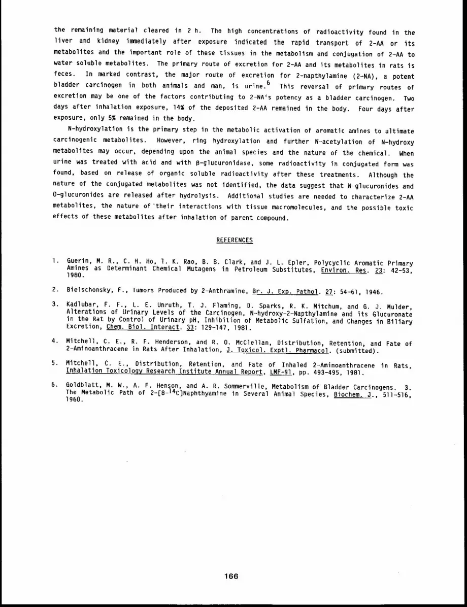
the remaining material cleared in 2 h. The high concentrations of radioactivity found in the
liver and kidney immediately after exposure indicated the rapid transport of 2-AA or its
metabolites and the important role of these tissues in the metabolism and conjugation of 2-AA to
water soluble metabolites. The primary route of excretion for 2-AA and its metabolites in rats is
feces. In marked contrast, the major route of excretion for 2-napthylamine (2-NA), a potent
bladder carcinogen in both animals and man, is urine. 6 This reversal of primary routes of
excretion may be one of the factors contributing to 2-NA’s potency as a bladder carcinogen. Two
days after inhalation exposure, 14~ of the deposited 2-AA remained in the body. Four days after
exposure, only 5% remained in the body.
N-hydroxylation is the primary step in the metabolic activation of aromatic amines to ultimate
carcinogenic metabolites. However, ring hydroxylation and further N-acetylation of N-hydroxy
metabolites may occur, depending upon the animal species and the nature of the chemical. When
urine was treated with acid and with B-glucuronidase, some radioactivity in conjugated form was
found, based on release of organic soluble radioactivity after these treatments. Although the
nature of the conjugated metabolites was not identified, the data suggest that N-glucuronides and
O-glucuronides are released after hydrolysis. Additional studies are needed to characterize 2-AA
metabolites, the nature of’their interactions with tissue macromolecules, and the possible toxic
effects of these metabolites after inhalation of parent compound.
REFERENCES
I. Guerin, M. R., C. H. Ho, T. K. Rao, B. B. Clark, and 3. L. Epler, Polycyclic Aromatic PrimaryAmines as Determinant Chemical Mutagens in Petroleum Substitutes, Environ. Res. 23: 42-53,1980.
2. Bielschonsky, F., Tumors Produced by 2-Anthramine, Br. J..Exp. Pathol. 27: 54-61, 1946.
3, Kadlubar, F. F., L. E. Unruth, T. 3. Flaming, D. Sparks, R. K. Mitchum, and G. J. Mulder,Alterations of Urinary Levels of the Carcinogen, N-hydroxy-2-Napthylamine and its Glucuronatein the Rat by Control of Urinary pH, Inhibition of Metabolic Sulfation, and Changes in BiliaryExcretion, Chem. Biol. Interact. 3__33: 129-147, 1981.
4. Mitchell, C. E., R. F. Henderson, and R. O. McClellan, Distribution, Retention, and Fate of2-Aminoanthracene in Rats After Inhalation, J. Toxicol. Exptl. Pharmacol. (submitted).
5. Mitchell, C. E., Distribution, Retention, and Fate of Inhaled 2-Aminoanthracene in Rats,Inhalation Toxicoloqy Research Institute Annual Report, LMF-91, pp. 493-495, 1981.
6, Goldblatt, M. W., A. F. Henson, and A. R. Sommerville, Metabolism of Bladder Carcinogens. 3.The Metabolic Path of 2-[8-14C]Naphthyamine in Several Anima’l Species, Biochem. J., 511-516,1960.
166
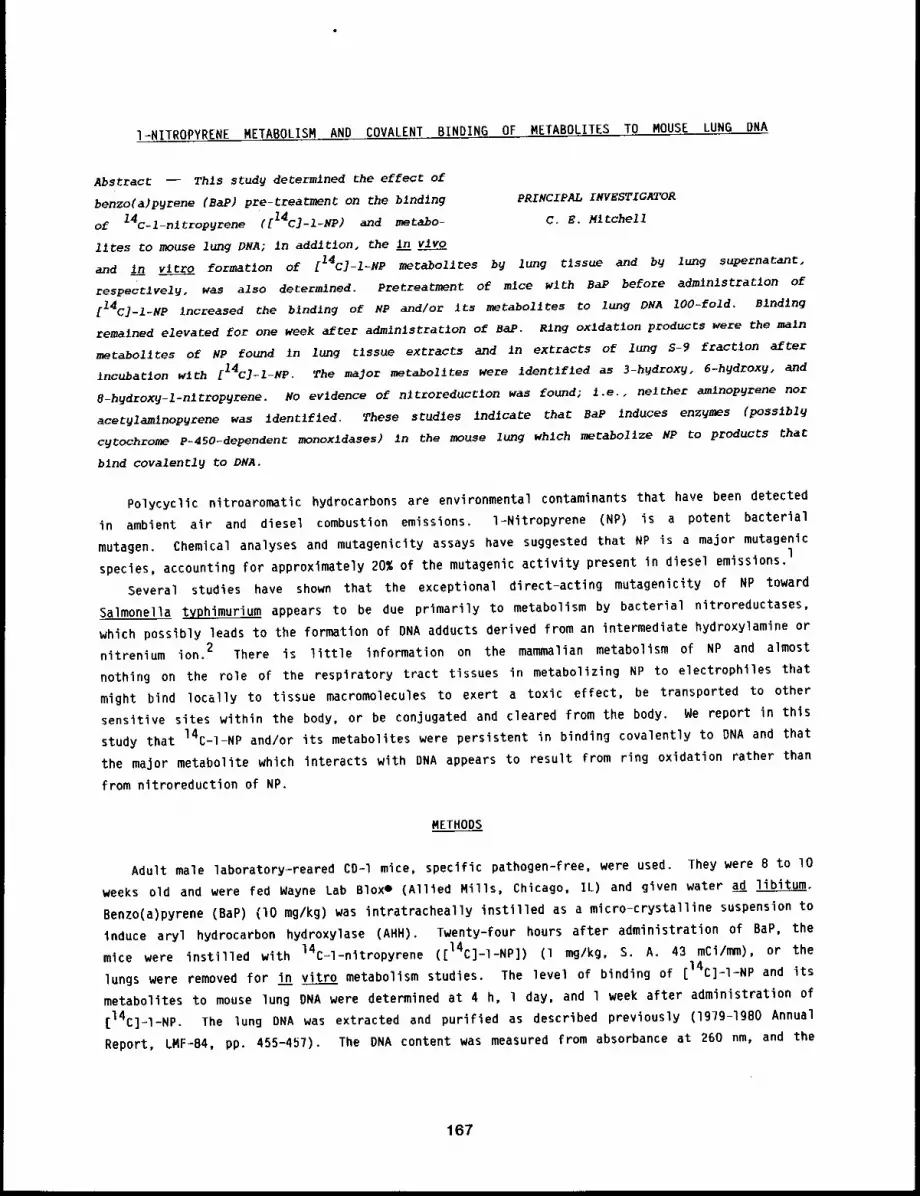
I=NITROPYRENE METABOLISM AND COVALENT BINDING OF METABOLITES TO MOUSE LUNG DNA
Abstract -- Thls study determined the effect of
benzo(a)pyrene (BaP) pre-treatment on the binding PRINCIPAL INVESTIGATOR
of 14c-l-nltropyrene ([I4C] -I-NP) and metabo- C.E. Mitchell
flies co mouse lung DNA; in addltlon, the in vlvo
and in vitro formation of [14C]--ILMP motabolltes by lung tlssue and by lung supernatant,
respectively, was also determined. Pretreatmont of mice wlth BaP before administration of
[14C]-I-NP increased the blnd~ng of NP and/or its motabolltes to lung DNA lO0-fold, slndlng
remained elevated for one week after admlnlstratlon of BaP. Ring oxldatlon products were the main
metabolltes of NP found in lung t~ssue extracts and in extracts of lung S-9 fraction after
incubation wlth [14C]-Z-NP. The major metabo1~tes were Identlfled as 3-h~droxy, 6-hydroxy, and
8-hgdroxg-l-nltropgrene. No evidence of nltroreductlon was found; i.e., neither am~nopyrene nor
acetylamlnopyrene was identlfled. These studies indlcate that BaP induces enzymes (posslbl9
cgtochrome P-450-dependent monoxldases) in the mouse lung which metabolize NP to products that
bind covalently to DNA.
Polycyclic nitroaromatic hydrocarbons are environmental contaminants that have been detected
in ambient air and diesel combustion emissions, l=Nitropyrene (NP) is a potent bacterial
mutagen. Chemical analyses and mutagenicity assays have suggested that NP is a major mutagenic
species, accounting for approximately 20% of the mutagenic activity present in diesel emissions,l
Several studies have shown that the exceptional direct-acting mutagenicity of NP toward
Salmonella typhimurium appears to be due primarily to metabolism by bacterial nitroreductases,
which possibly leads to the formation of DNA adducts derived from an intermediate hydroxylamine or
nitrenium ion. 2 There is little information on the mammalian metabolism of NP and almost
nothing on the role of the respiratory tract tissues in metabolizing NP to electrophiles that
might bind locally to tissue macromolecules to exert a toxic effect, be transported to other
sensitive sites within the body, or be conjugated and cleared from the body. We report in this
study that 14C-I-NP and/or its metabolites were persistent in binding covalently to DNA and that
the major metabolite which interacts with DNA appears to result from ring oxidation rather than
from nitroreduction of NP.
METHODS
Adult male laboratory-reared CD-I mice, specific pathogen-free, were used. They were 8 to lO
weeks old and were fed Wayne Lab Bloxe (Allied Mills, Chicago, IL) and given water ad libitum.
Benzo(a)pyrene (BaP) (lO mg/kg) was intratracheally instilled as a micro-crystalline suspension
induce aryl hydrocarbon hydroxylase (AHH). Twenty-four hours after administration of BaP, the
mice were instilled with 14C-l-nitropyrene ([14C]-I-NP]) (l mg/kg, S. A. 43 mCi/mm), or
lungs were removed for in vitro metabolism studies. The level of binding of [14C]-I-NP and its
metabolites to mouse lung DNA were determined at 4 h, i day, and l week after administration of
[14C]-l-NP. The lung DNA was extracted and purified as described previously (197g-lgBO Annual
Report, LMF-B4, pp. 455-457). The DNA content was measured from absorbance at 260 nm, and the
167
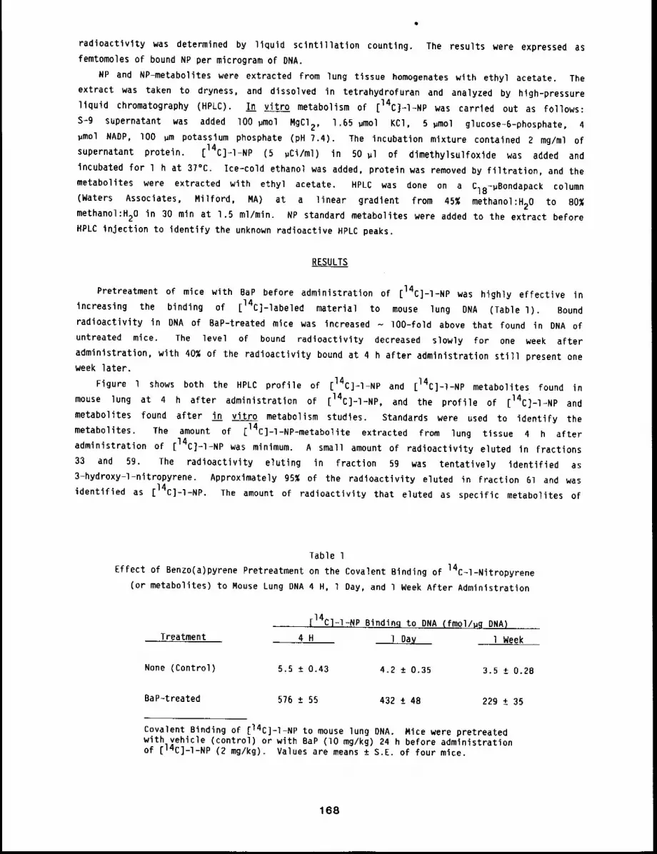
radioactivity was determined by liquid scintillation counting. The results were expressed as
femtomoles of bound NP per microgram of DNA.
NP and NP-metabolites were extracted from lung tissue homogenates with ethyl acetate. The
extract was taken to dryness, and dissolved in tetrahydrofuran and analyzed by high-pressure
liquid chromatography (HPLC). In vitro metabolism of [14C]~I-NP was carried out as follows:
s-g supernatant was added lO0 ~mol MgCI2, 1.65 ~mol KCl, 5 ~mol glucose-6-phosphate, 4~mol NADP, I00 gm potassium phosphate (pH 7.4). The incubation mixture contained 2 mg/ml
supernatant protein. [14C]=I-NP (5 pCi/ml) in 50 ~l of dimethylsulfoxide was added and
incubated for l h at 37°C. Ice-cold ethanol was added, protein was removed by filtration, and the
metabolites were extracted with ethyl acetate. HPLC was done on a CiB1gBondapack column
(Waters Associates, Milford, MA) at a linear gradient from 45% methanol:H20 to BO%
methanol:H20 in 30 min at 1.5 ml/min. NP standard metabolites were added to the extract before
HPLC injection to identify the unknown radioactive HPLC peaks.
RESULTS
Pretreatment of mice with BaP before administration of [14C]-l-NP was highly effective in
increasing the binding of [14C]-labeled material to mouse lung DNA (Table l). Bound
radioactivity in DNA of BaP-treated mice was increased - lO0-fold above that found in DNA of
untreated mice. The level of bound radioactivity decreased slowly for one week after
administration, with 40% of the radioactivity bound at 4 h after administration still present one
week later.
Figure l shows both the HPLC profile of [14C]=I-NP and [14C]-l-NP metabolites found in
mouse lung at 4 h after administration of [14C]-I-NP, and the profile of [14C]-I=NP and
metabolites found after in vitro metabolism studies. Standards were used to identify the
metabolites. The amount of [14C]-l-NP-metabolite extracted from lung tissue 4 h after
administration of [14C]-I-NP was minimum. A small amount of radioactivity eluted in fractions
33 and 59. The radioactivity eluting in fraction 59 was tentatively identified as
3-hydroxy-l-nitropyrene. Approximately g5~ of the radioactivity eluted in Fraction 61 and was
identified as [14C]-I-NP. The amount of radioactivity that eluted as specific metabolites of
Table 1
Effect of Benzo(a)pyrene Pretreatment on the Covalent Binding of 14C11-Nitropyrene
(or metabolites) to Mouse Lung DNA 4 H, l Day, and I Week After Administration
Treatment
_ [14C]-I%NP Binding to DNA (fmol/gg DNA)
4 H 1 Day 1 Week
None (Control) 5.5 ± 0.43 4.2 ± 0.35 3.5 ± 0.28
BaP-treated 576 ± 55 432 ± 48 229 ± 35
Covalent Binding of [14C]-I-NP to mouse lung ONA. Mice were pretreatedwith vehicle (control) or with BaP (lO mg/kg) 24 h before administrationof [14C]-I-NP (2 mg/kg). Values are means ± S.E. of four mice.
168
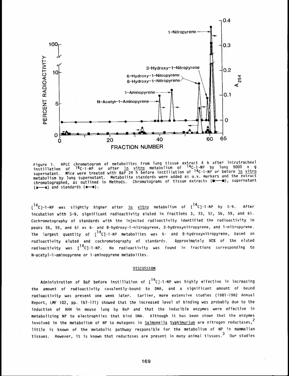
0.4
1-Nitropyrene
3-Hydroxy- 1-Nitropyrene
6-Hydr oxy- 1-Nit ropyrene8-Hydroxy- 1-Nitropyrene
1-Arninopyrene ....
N-Acetyl- 1-Amin°pyrene--l~ l~
" -12-.--2O 40
FRACTION NUMBER
ii,-i,-
0.3
- 0.2
<--o.1
-o
60 65
Figure I. HPLC chromatogram of metabolites from lung tissue extract 4 h after intratrachealinstillation of 14C-1-NP or after in vitro metabolism of 14C-I-NP by lung 9000 x gsupernatant. Mice were treated with BaP 24 h before instillation of 14C-I-NP or before in vitrometabolism by lung supernatant. Metabolite standards were added as u.v. markers and the extractchromatographed, as outlined in Methods. Chromatograms of tissue extracts (H), supernatant(A A) and standards (¢ =).
[14C]-I-NP was slightly higher after in vitro metabolism of [14C]-I-NP by S-9. After
incubation with S=9, significant radioactivity eluted in fractions 3, 33, 52, 56, 59, and 61.
Cochromatography of standards with the injected radioactivity identified the radioactivity in
peaks 56, 59, and 61 as 6- and 8-hydroxy-l=nitropyrene, 3-hydroxynitropyrene, and l-nitropyrene.The largest quantity of [14C]-I-NP metabolites was 6- and 8..-hydroxynitropyrene, based on
radioactivity eluted and cochromatography of standards. Approximately 80% of the elutedradioactivity was [14C]-]-NP. No radioactivity was found in fractions corresponding to
N-acetyl-l-aminopyrene or 1-aminopyrene metabolites.
DISCUSSION
Administration of BaP be#ore instillation of [14C]-l=NP was highly effective in increasing
the amount of radioactivity covalently-bound to DNA, and a significant amount of bound
radioactivity was present one week later. Earlier, more extensive studies (1981-1982 Annual
Report, LMF I02, pp. 167-171) showed that the increased level of binding was probably due to the
induction of AHH in mouse lung by BaP and that tile inducible enzymes were effective in
metabolizing NP to electrophiles that bind DNA. Although it has been shown that the enzymes2
involved in the metabolism of NP to mutagens in Salmonella typhimurium are nitrogen reductases,
little is known of the metabolic pathway responsible for the metabolism of NP in mammaliantissues. However, it is known that reductases are present in many animal tissues.3 Our studies
169
showed that the enzymes that metabolize NP to DNA adducts are inducible in mouse lung after BaP
pretreatment, and although it is not known which class or classes of enzymes are induced, other
data suggest that cytochrome P-450 enzymes are among the enzymes induced. To obtain some
information on whether nitroreductive or ring oxidative enzymes were responsible for the enhanced
binding of [14C]-I-NP to lung DNA, NP-metabolites were characterized using HPLC chromatography.
Small quantities of metabolites were recovered from mouse lung after instillation of
[14C]-I-NP. It was felt that the small quantities recovered may result from the binding of
metabolites to macromolecules and remain unextractable with organic solvent, or result from an
efficient conjugating system whereby metabolites are conjugated to water-soluble products and
subsequently cleared from the body. Thus, in vitro metabolism of [14C]-I-NP by the lung
supernatant was carried out (the conjugating enzymes may be ineffective or absent). This latter
approach was effective in obtaining higher quantities of [14C]-I-NP metabolites, which were
primarily ring oxidation products, with little evidence for nitroreduction products. However,
there were very small quantities of unidentified peaks that may represent both nitro-reductive and
ring oxidative metabolites. The small quantity of radioactivity recovered suggests that a
significant amount of total tissue radioactivity may be bound to macromolecules, as evidenced by
the large amount of [14C]-I-NP bound to DNA (~ 50 times more NP-bound than BaP-bound).4
Recently, it was reported that isolated perfused lung metabolized [14C]-I-NP primarily to
ring oxidation products. 5 Thus, the present in vivo and in vitro studies are in agreement with
the findings in the isolated perfused lung. More work is needed to characterize the enzymes
involved in the metabolism of NP to electrophilic intermediates (they appear more inclined toward
NP metabolism than BaP metabolism) and to characterize specific DNA-adducts in lung after
administration of NP,
REFERENCES
I. Pederson, T. C. and J. C. Siak, The Role of Nitroaromatic Compounds in the Direct-ActingMutagenicity of Diesel Particle Extracts, 5. Appl. Toxicol. l: 54-60, 19Bla.
2. Mermelstein, R., D. K. Kiriazides, M. Butler, E. C. McCoy, and H. S. Rosenkranz, TheExtraordinary Mutagenicity of Nitropyrene in Bacteria, Mutat. Res. 89: 187-196, 1981.
3. Howard, P. C., R. H. Heflich, F. E. Evans and F. A. Beland, Formation of DNA Adducts In Vitroand in Salmonella typhimurium Upon Metabolic Reduction of the Environmental Mutagenl-Nitropyrene, Cancer Res. 43: 2052-2058, 1983.
4. Mitchell, C. E., Effect of Enzyme Induction on the Binding of Aromatic Hydrocarbons to MouseLung DNA, Chemico-Biol Interac. (submitted).
5. Bond, a. A. and 5. L. Mauderly, Metabolism and Macromolecular Covalent Binding of [14C]-I-NPin Isolated Perfused/Ventilated Rat Lungs, Cancer Res. (submitted).
170
PULMONARY RETENTION OF BENZO(A)PYRENE AS INFLUENCED BY AMOUNT INSTILLED
Abstract -- The amount of Benzo(a)pyrene (BaP)
instllled In the lungs can affect the rate at PRINCIPAL INVESTIGATORS
which BaP is cleared Into the blood. Fischer-344 M.A. Medlnsk9
rats were g~ven 16, 90, 390, or 6400 ng of 14C-BaP S.J. Kampclk
(29.7 ~CI/~M) per rat by Jntratracheal instll-
latlon. At varlous tln)es after ~nst~llatlon, lung lobes and trachea were analyzed for
radioactivity. As the amount of BaP inst111ed ~ncreased, a larger percent (89 to 99.7) was
cleared wlth a TI/2 of less than i h. A progresslvely smaller percent (ll to 0.3) was cleared
wlth a T1/2 of greater than I day, indlcatlng that a small amount (2-16 ng) of radloactlvlty was
retained in the lungs, regardless of the amount inst111ed. Thls may have impllcatlons for
estlmat~ng health rlsks to people exposed to BaP in the environment at very low concentrations,
since extrapolatlon from experlments uslng large quantltles of BaP may underestimate rlsk.
Studies at this Institute have been directed toward obtaining information on the disposition
of a wide range of polynuclear aromatic (PNA) compounds after inhalation. 1 These investigations
have involved a variety of inhaled concentrations, particle sizes, and exposure durations, with
compounds inhaled ,as chemically homogenous aerosols and as coatings on various insoluble
particles. All PNA compounds investigated to date are rapidly cleared from the respiratory tract
after inhalation. Clearance is biphasic, with one component clearing with a TI/2 < l day and
another with a TI/2 > 1 day. l Some of the studies on the disposition of PNA compounds
suggest an association between the amount of an organic compound inhaled and deposited, and the
fraction of the dose that is retained in the lung after 24 h.l
The purpose of this study was to determine the effect that instillation of various
concentrations of a PNA would have on the fraction retained in the lung. The PNA instilled was
14C-benzo(a)pyrene (14C-BaP).
METHODS
14C-BaP (29 ~Ci/~mole) was administered to lO0 Fischer-344 female rats by intratracheal
instillation. Rats received one of four instillation solutions, containing 4000, 22,750,
lO0,O00, and 1,650,000 dpm/250 ~l or 16, 90, 390, and 6400 ng/rat, respectively. 14C-BaP was
dissolved in dimethyl sulfoxide and diluted with sterile normal (0.9%) saline.
Three to four rats were euthanized with T61 euthanasia solution at 0.5 (6400 ng only), l,
6, 24 (390 ng ended here), 48, 72, 96, and 168 h after instillation of 14C-BaP. Immediately
after sacrifice, the thoracic cavity was opened and the lungs and trachea, including the larynx,
were removed intact. The heart, thymus, and thyroid glands were cut away, and the surface of the
lungs was washed in cold saline to remove any blood. One sample, which included the larynx to the
bronchi, was weighed and frozen until digestion. The left lobe, the large posterior right lobe,
and the remaining lung tissue were also weighed and frozen.
Tissue samples were digested with l.O ml of tetraethyl ammonium hydroxide (25% in methanol)
60°C for 2 h. After digestion, samples were neutralized and decolorized, and scintillation fluid
was added for determination of total radioactivity per tissue by scintillation spectrometry.
171
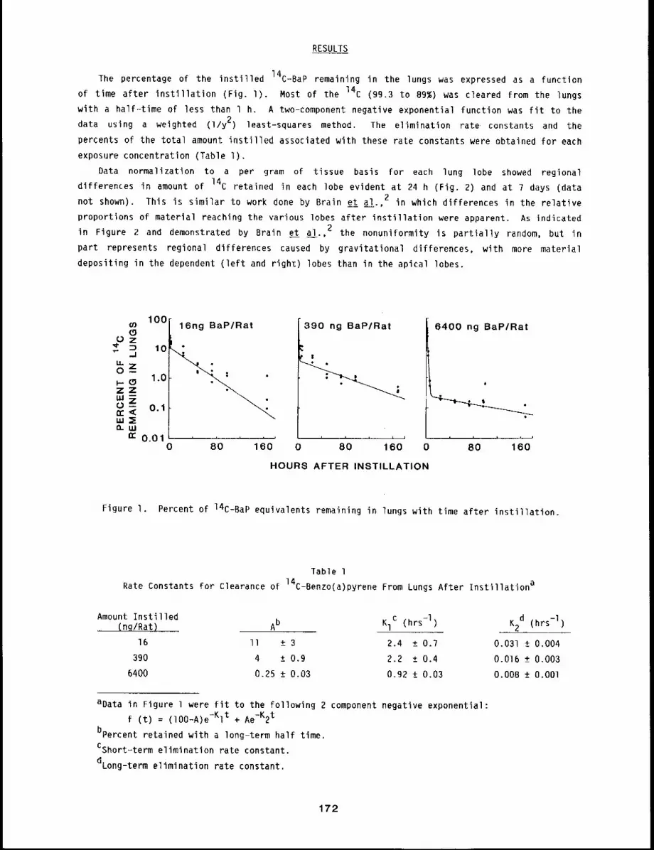
RESULTS
The percentage of the instilled 14C-BaP remaining in the lungs was expressed as a function
of time after instillation (Fig. l). Most of the 14C (99.3 to 89%) was cleared from the lungs
with a half--time of less than 1 h. A two-component negative exponential function was fit to the
data using a weighted (1/y 2) least-squares method. The elimination rate constants and the
percents of the total amount instilled associated with these rate constants were obtained for each
exposure concentration (Table I).
Data normalization to a per gram of tissue basis for each lung lobe showed regional
differences in amount of 14C retained in each lobe evident at 24 h (Fig. 2) and at 7 days (data
not shown). This is similar to work done by Brain et al.,2 in which differences in the relative
proportions of material reaching the various lobes after instillation were apparent. As indicated
in Figure 2 and demonstrated by Brain et al.,2 the nonuniformity is partially random, but in
part represents regional differences caused by gravitational differences, with more material
depositing in the dependent (left and righz) lobes than in the apical lobes.
IO0¢n 16ng BaP/Rat(9
Oz~" ~ 10 "
tU -~zl-- (gZ 1"0tO-- 0.1n-<,,,=Eo-uJ
n~ 0.010 80 160
390 ng BaP/Rat
!
6400 ng BaP/Rat
0 80 160 0 80 160
HOURS AFTER INSTILLATION
Figure I. Percent of 14C-BaP equivalents remaining in lungs with time after instillation.
fable 1
Rate Constants for Clearance of 14C-Benzo(a)pyrene From Lungs After Instillationa
Amount Instilled.... (ng/Rat) Ab KlC (hrs-l) K2d (hrs-l)
16 II + 3 2.4 + 0.7 0.031 + 0.004
390 4 + 0.9 2.2 +_ 0.4 0.016 + 0.003
6400 0.25 _+ 0.03 0.92 +_ 0.03 0.008 + O.OOl
aData in Figure 1 were fit to the following 2 component negative exponential:
f (t) = (lO0-A)e-Kl t + Ae-K2t
bpercent retained with a long-term half time.
CShort=term elimination rate constant.
dLong-term elimination rate constant.
172
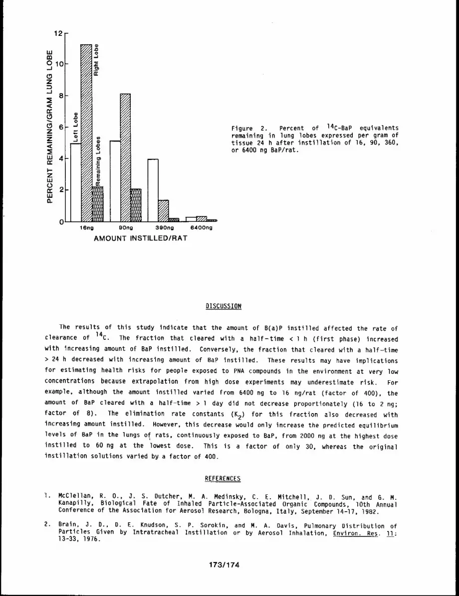
12-
i
Yl/A 0 .....
"///_/2 = "//~ --////A C ;;;~’//d "E ....
.-- ///A////J m "///~"///d E ....
16ng O0ng 300no 6400ng
AMOUNT INSTILLED/RAT
Figure 2. Percent of 14C-BaP equivalentsremaining in lung lobes expressed per gram oftissue 24 h after instillation of 16, 90, 360,or 6400 ng BaP/rat.
DISCUSSION
The results of this study indicate that the amount of B(a)P instilled affected the rate
clearance of 14C. The fraction that cleared with a half-time < 1 h (first phase) increased
with increasing amount of BaP instilled. Conversely, the fraction that cleared with a half-time
> 24 h decreased with increasing amount of BaP instilled. These results may have implications
for estimating health risks for people exposed to PNA compounds in the environment at very low
concentrations because extrapolation from high dose experiments may underestimate risk. For
example, although the amount instilled varied from 6400 ng to 16 ng/rat (factor of 400), the
amount of BaP cleared with a half-time > l day did not decrease proportionately (16 to 2 rig;
factor of B). The elimination rate constants (K2) for this fraction also decreased with
increasing amount instilled. However, this decrease would only increase the predicted equilibrium
levels of BaP in the lungs o~ rats, continuously exposed to BaP, from 2000 ng at the highest dose
instilled to 60 ng at the lowest dose. This is a factor of only 30, whereas the original
instillation solutions varied by a factor of 400.
REFERENCES
I. McClellan, R. 0., J. S. Dutcher, M. A. Medinsky, C. E. Mitchell, 3. D. Sun, and G. M.Kanapilly, Biological Fate of Inhaled Particle-Associated Organic Compounds, lOth AnnualConference of the Association for Aerosol Research, Bologna, Italy, September 14-17, 1982.
2. Brain, 3. D., D. E. Knudson, S. P. Sorokin, and M. A. Davis, Pulmonary Distribution ofParticles Given by Intratracheal Instillation or by Aerosol Inhalation, Environ. Res. ll:13-33, 1976,
173/174

DOSE-RESPONSE RELATIONSHIPS FOR INHALED RADIONUCLIDES
Long-term dose-response studies with beta- and alpha-emittlng radlonuclides are being
conducted to establish qualitative and quantitative relationships between single, acute inhalation
exposures to graded levels and types of radionuclide aerosols and the resultant biological
effects. These studies, involving a limited number of radionuclides in aerosols with different
physical and chemical properties, involve observations of animals for their entire life span to
assess parameters of radiation dose that are significant in biological response. Eight studies
involving young adult dogs that inhaled betalemitting radionuclides are in progress: four with
relatively solubl~ forms (90SrCI~, 144CeC13, 91yc1 and 137CsCl) and four with
relatively insoluble form (gOy, 9ty, 144Ce and gOsr 3in fused aluminosilicate particles).
Taken together, these studies are yielding valuable information on the interrelationships of dose,
dose rate and organs at risk in the production of long-term biological effects. Studies also in
progress involve young adult dogs that inhaled graded activity levels of monodisperse aerosols of
238pu02 or 239pu02. Potential dose-effect modifying factors being studied with these
alpha-emitting radionuclides include the (a) elemental characteristics of the radionuclide, (b)
chemical form, (c) specific activity, (d) particle size distribution, (e) fraction of
irradiated, and (f) host variables such as species or pre-existing disease. Because age
exposure could also be a modifying factor in long-term effects, dogs were also exposed to either
144Ce or 239pu02 at 3 months of age or at 8.0 to 10.5 years of age. Recognizing thatFAP
there are conceivable circumstances under which people could be repeatedly exposed to plutonium,
Beagles are being exposed repeatedly by inhalation to aerosols of 239pu02 to study the
relative effectiveness of repeated versus single exposures.
The studies of dogs that inhaled soluble beta-emitting radionuclides are nearing completion.
young adults ~o 90SrCI 2 or glycI3 or aged dogs exposed to 144Ce inAll dogs exposed as
fused aluminosilicate particles are now dead. Five dogs remain alive in the 144CeC13 and
137CsCl studies. These studies have shown that all organs in which significant quantities of
radionuclide are deposited are susceptible to radiation-induced tumors. Most prevalent have been
tumors in the lung, liver, skeleton, and nasal cavity. In dogs that inhaled relatively insoluble
forms of beta-emitting radionuclides, the type of lung cancer and its time of appearance are being
shown to be a function of the rate at which dose rate changed after exposure. Also,
late-occurring cancer is developing in organs with little or essentially no radiation dose. The
extent to which the cancer incidence in these organs is different than in control animals remains
to be determined.
In the studies of dogs that inhaled alpha-emitting radlonuclides, the difference in organs at
23Bpuo2 inhalation compared with those after 239pu02 inhalation is striking.risk after
Although a few cancers have been seen after 238pu02 inhalation, bone cancer has been much more
prevalent. In contrast, lung cancer has been the only late=occurring cancer seen to date in the
dogs that inhaled 239pu0^. The studies of age=related effects in immature or aged dogs that
inhaled 144Ce FAP or 23~Pu02 were continued during the year, as were observations on dogs
that were exposed repeatedly to 144Ce FAP or 23gPu02. Observations were also continued in239
the studies involving rats exposed once to Cm203 or repeatedly to "PuO~. Chromosomeaberration measurements are being made in cynomolgus monkeys that inhaled 239pu(NO ) and
34Chinese hamsters injected with Thorotrast. As part of an investigation of the pulmonary
immunological aspects of radionuclide inhalation, the effects of age and 239pu02 inhalation on
the humoral immune response of the lungs of dogs after local deposition of antigen were
investigated and are reported here. Other studies reported here involved measurements of the
175

procoagulant activity and spontaneous macrophage migration of lung lavage cells from dog lung
lobes that had radiographically proven lung tumors, compared with control values. Both the
procoagulant activity of the cells and fluid washed from the lobe with a tumor and the spontaneous
migration area of cells from the tumor lobe were greater than seen in non-tumor-bearing control
lung lobes.
176
TOXICITY STUDIES OF INHALED BETA-EMITTING RADIONUCLIDES STATUS REPORT
Abstract --- The Influence of total dose and dose
rate on the effects of lnhaled beta-emlttlng radlo-
nuclldes is being studled in laboratory anlmals.
The radlonuclldes are inhaled elther ~n a relatlvel~
soluble form (90Src12, 144CoC13~ 91yC13, or 137CsCl),
or in a relativel~ insoluble form in fused alumlno-
s111cate partlcles. Whlch organs are affected de-
pends on the solublllt 9 and chemlcal characterlsClcs
of the radlonucl~des. Studles wlth 9oung adult dogs
are complemented b9 comparable studles in other spew
cles (mlce, rats~ and S~rlan hamsters), wlth animals
of dlfferent ages and anlmals repeatedly exposed to
144Ce"
PRINCIPAL INVESTIGATORS
F. F. Hahn
R. O. McClellan
B, B, Boecker
C. H. Hobbs
R. K. Jones
D, L, Lundgren
J. L. Mauderly
B. A. Muggenburg
J. A. Pickrell
H. C. Redman
B. R. Scott
M. B. Snlpes
lhe objective of these studies is to establish the dose-response relationships resulting from
inhalation of different quantities of beta-emitting radionuclides in various physical and chemical
forms by animals of different species and ages. Extrapolation of this information to man helps
determine safe operating procedures and exposure limits essential to the orderly development and
use of nuclear energy. It is impossible to simulate with experimental animal exposures all the
potential dose patterns from inhaled beta-emitting radionuclides that might be encountered in
accidents. Thus, a more basic approach is being used in which the relationship of radiation dose
to biological response is being studied for a few selected dose patterns that span the likely
range of dose patterns that could be encountered. Various dose patterns are achieved by exposing
laboratory animals to radioactive aerosols having different physical and chemical characteristics
and radionuclide content. In this manner, the importance of several radiation dose parameters on "
the dose-response relationship for long-term effects can be evaluated, l a~ well as the relative
radiation sensitivities of various organs.
The fission product radionuclides being studied are present in substantial quantities in many
processes involving nuclear reactor fuel and thus are potential airborne pollutants. Equally
important, the radionuclides and forms chosen present a broad range of physical and effective
half-lives in the body. This range provides an opportunity to assess the influence of radiation
dose rate and total radiation dose on the dose-response relationships for inhaled radionuclides.
Because all of these radionuclides and their associated daughters are energetic beta emitters and
a large number of particles are deposited in the lung, there is nearly uniform beta irradiation of
lung.
The dose-response longevity studies with beta-emitting radionuclides that have been completed
or are in progress are shown in Table l. Particle solubility in body fluids has a definite effect
on the translocation of radionuclides from the lung and influences which organs receive
significant radiation doses. With inhaled 90SrCl2* or 137CsCl,* the radionuclide is rapidly ab-
sorbed from the lung and deposited in other organs (skeleton or whole-body, respectively),
producing a significant radiation dose to these tissues. With inhaled 91yCl3 and
144CeC13,* the radionuclide is absorbed from the lung at a slower rate, resulting in significant
*90Sr, 137Cs, and 144Ce refer to equilibrium mixtures of 90Sr-9Oy, 137Cs-137mBa, or 144Ce-144pr,
respectively.
177
radiation dose to lung as well as to the organs in which these radionuclides are finally deposited
(liver and skeleton). The four radionuclides inhaled in relatively insoluble fused
aluminosilicate particles result primarily in irradiation of the lung and associated thoracic
structures, such as the heart and tracheobronchial lymph nodes.
The different temporal radiation dose patterns to the lung from these aerosols are shown in
Figures l and 2. In Figure l, the change in radiation dose rates as a function of time is shown
for the levels of exposure selected to produce initial dose rates of lO0 rad/day. The dose
patterns in Figure 1 required similar activity levels for initial lung burdens, because the beta
energies are similar for the four radionuclides. These lung burdens result in a marked difference
in the potential 5000-day radiation dose to the lung (370 rad for 90y to 93,000 rad for90Sr). The differences in radiation dose patterns among the different radionuclides are also
evident in Figure 2, where cumulative dose curves resulting in infinite doses of 2000 rad to lung
required 1300 ~Ci at 530 rad/day initially for 90y to 7.1 ~Ci at 5.? rad/day initially forgOsr. These differences result from effective half-lives in lung that range from - 2 days for90y to N 500 days for 90Sr.
Results of studies using multiple laboratory animal species are also shown in Table I. These
studies are allowing more precise extrapolation of dose-response relationships among species, and
provide the basis for projection to human exposures to these or similar radionuclides.
Experimental details of these studies are given in the following section of this document. A
tabular summary of the studies involving dogs is shown in Table 2.
Studies have also been conducted with mice, Syrian hamsters, rats, and dogs repeatedly exposed
to aerosols of a relatively insoluble form of 144Ce. Repeated exposure, every two months for 15
to 50% of the animals’ life span, results in complex dose patterns to the lung that mimic repeated
or chronic exposures that may occur in occupational situations. In studies with rodents, the lung
tumor incidence correlated better with cumulative dose to lung rather than the protraction of the
dose rate resulting from repeated exposure. 2-4 Results in the study with dogs indicate that
lung tumors occur later in life after the initiation of repeated exposures than after single
exposures (1980-1981 Annual Report, LMF-91, pp. 134-137).
For dogs exposed once, there is a general relationship between survival time distribution for
death from early effects and the specific radionuclide exposure pattern. For a particular
cumulative dose, each radionuclide has a different initial dose rate and response.6 For
example, the earliest deaths from radiation pneumonitis and pulmonary fibrosis were observed with
90y. Those animals received high dose rates initially that decreased rapidly. Dogs receivingsimilar lung doses from 91y, 144Ce’ or 90Sr with lower initial dose rates and a longer
protraction of dose lived longer. Likewise, at times when deaths with all four radionuclides were
noted (150 to 250 days), 90y-exposed dogs died with the lowest doses, the 91y-exposed dogs
died at intermediate doses, and the 144Ce- and gOsr-exposed dogs died at the highest doses.
The radiation dose pattern also influences the incidence of lung tumors. 7 Protracted
irradiation of the lung from 90Sr or 144Ce resulted in a relatively high cumulative radiation
dose and produced more total lung tumors but fewer lung tumors per tad than less protracted
irradiation from 90y or gly. At lO years after inhalation exposure, the difference in riskper rad among the dffferent dose patterns was a factor of 4 to 8, indicating that the different
radiation dose patterns from inhaled beta emitters do influence lung tumor risk factors at least
at high (> 20,000 rad) doses to lung. In addition, at high radiation doses, more
hemangiosarcomas of the lung were found than carcinomas, which were more prevalent at lower doses.
The observations in these studies are also important in considering potential new target
organs for the late effects of inhaled beta emitters. Hepatic degeneration occurred in a number
of dogs that inhaled 144CeCl 3. This appears to be a dose-related phenomenon. Degenerative
178
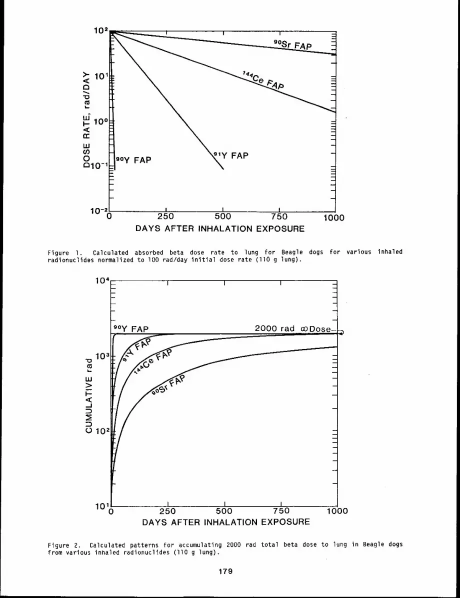
102 ....
,0-! ....... I .......... I I
250 500 750 1000
9oy FAP’~Y FAP _
DAYS AFTER INHALATION EXPOSURE
Figure I. Calculated absorbed beta dose rate to lung for Beagle dogs for various inhaledradionuclides normalized to lO0 rad/day initial dose rate (llO g lung).
I I I
10419oy FAP
ml.
_Y3
"-’-----"--
0102
2000 r a d,.,,_,o0, ..,D. o s e_’
I I I
m
m
1010 250 500 750 1000DAYS AFTER INHALATION EXPOSURE
Figure 2. Calculated patterns for accumulating 2000 rad total beta dose to lung in Beagle dogsfrom various inhaled radionuclides (]lO g lung).
179
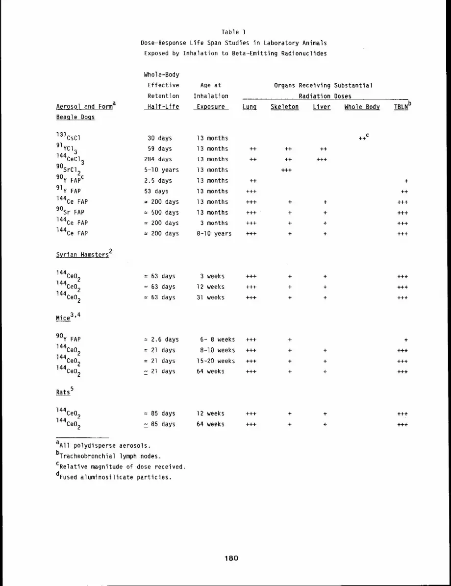
Table 1
Dose-Response Life Span Studies in Laboratory Animals
Exposed by Inhalation to Beta-Emitting Radionuclides
Aerosol and Forma
Beagle Dogs
Whole-Body
Effective Age at
Retention Inhalation
Half-Life Exposure
137CsCl 30 days 13 monthsgIyCl. 59 days 13 months144~ ~.
~eu/3 284 days 13 months
gOSrCl2 5-10 years 13 months90y FApC 2.5 days 13 months
91y FAP 53 days 13 months
144Ce FAP : 200 days 13 months
90Sr FAP ~ 500 days 13 months
144Ce FAP = 200 day~ 3 months
144Ce FAP ~ 200 days 8-I0 years
Syrian Hamsters2
144Ce02 ~ 63 days 3 weeks
144Ce02 = 63 days 12 weeks
144Ce02 = 63 days 31 weeks
Mice3,4
90y FAP ~ 2.6 days 6- 8 weeks
144Ce02 ~ 21 days 8-10 weeks
144Ce02 : 21 days 15-20 weeks
144Ce02 ~ 21 days 64 weeks
Rats5
144Ce02 = 85 days 12 weeks
144Ce02 z 85 days 64 weeks
aAll polydisperse aerosols.
bTracheobronchial lymph nodes.
CRelative magnitude of dose received.
dFused aluminosilicate particles.
Organs Receiving Substantial
Radiation Doses
Lung Skeleton Liver Whole Body
++ ++ 4-+
4-4- 4-4- 4-++
4-++
+4-
+++
4-4-4- 4- {
4-++ 4- +
+++ + +
4-+4- + 4-
4-++ 4- +
4-4-4- 4- 4-
++4- 4- 4-
4-4-4- +
4-4-+ 4- 4-
4-+4- 4- 4-
4-4-+ 4- 4-
+4-4- 4- 4-
+4-4"- + 4-
TBLNb
4-
4-4-
+4-4-
++4-
+4-4-
4-4-+
4-4-4-
4-++
4-4-4-
+
4-+4-
+4-4-
4-4-4-
4-4-4-
4-4-4-
180
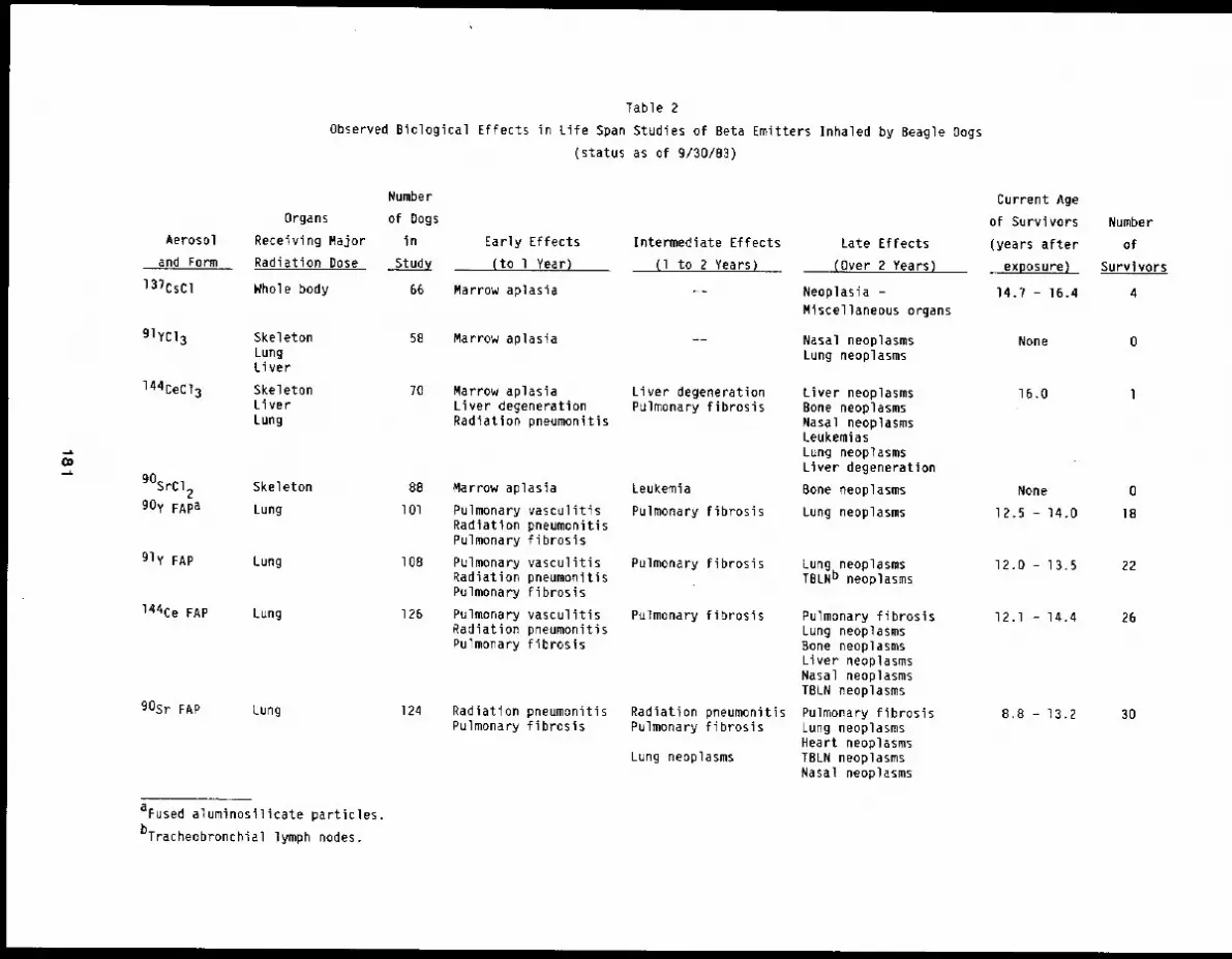
Table 2
Observed Biological Effects in Life Span Studies of Beta Emitters Inhaled by Beagle Dogs
(status as of 9/30/83)
Organs
Aerosol Receiving Major
and Form Radiation Dose
137CsCl Whole body
glycI3 SkeletonLungLiver
144CeC13 SkeletonLiverLung
gOSrCl2 Skeleton
90y FApa Lung
91y FAP Lung
144Ce FAP Lung
90Sr FAP Lung
aFused aluminosilicate particles.
bTracheobronchial lymph nodes.
Number
of Dogs
in
Study
66
58
?0
88
lOl
IOB
126
124
Early Effects
(to l Year)
Marrow aplasia
Marrow aplasia
Marrow aplasiaLiver degenerationRadiation pneumonitis
Marrow aplasia
Pulmonary vasculitisRadiation pneumonitisPulmonary fibrosis
Pulmonary vasculitisRadiation pneumonitisPulmonary fibrosis
Pulmonary vasculitisRadiation pneumonitisPulmonary fibrosis
Radiation pneumonitisPulmonary fibrosis
Intermediate Effects
(I to 2 Years)
Liver degenerationPulmonary fibrosis
Leukemia
Pulmonary fibrosis
Pulmonary fibrosis
Pulmonary fibrosis
Radiation pneumonitisPulmonary fibrosis
Lung neoplasms
Late Effects
(Over 2 Years)
Neoplasia -
Miscellaneous organs
Nasal neoplasmsLung neoplasms
Liver neoplasmsBone neoplasmsNasal neoplasmsLeukemiasLung neoplasmsLiver degeneration
Bone neoplasms
Lung neoplasms
LungneoplasmsTBLNb neoplasms
Pulmonary fibrosisLung neoplasmsBone neoplasmsLiver neoplasmsNasal neoplasmsTBLN neoplasms
Pulmonary fibrosisLung neoplasmsHeart neoplasmsTBLN neoplasmsNasal neoplasms
Current Age
of Survivors
(years after
exposure}
14.7 - 16.4
None
16.0
None
12,5 - 14.0
12.0 - 13.5
12.1 - 14.4
8.8 - 13.2
Number
of
Survivors
4
l
0
IB
22
26
30
changes are not usually emphasized as late effects of chronic irradiation. In the case of
144Ce, however, which is translocated to liver, degenerative changes may represent important
health effects, in addition, liver tumors have been found in dogs that inhaled 144CeC13.
The tracheobronchial lymph nodes have been identified as a primary site for tumor induction.
Blood vascular tumors (hemangiosarcomas) were found in the tracheobronchial lymph nodes, but not
in other tissues of two dogs exposed to aerosols of 144Ce in fused aluminosilicate particles.
The tumors had not metastasized, but one was anaplastic in appearance. This lends further
evidence that hemangiosarcomas may arise in these nodes and that many of the mediastinal
hemangiosarcomas arise from these nodes.B
Nasal carcinomas, which have occurred in dogs after inhalation of glyCl3, 144CEC13, or
gOsrcI2, arise from the epithelium lining the maxilloturbinates. Focal concentrations of
radionuclide can be found in nasal epithelium beyond the normal upper respiratory tract clearance
times and may be one of the significant factors in the induction of nasal tumors.
Many of the exposed dogs are still alive, and continued observation should yield further
In the past year, all of the remaining dogs in the 91yCl3 longevity studycorrelations.
the 144CeC13 longevity study is alive. The results of these inhalationdied. Only one dog on
studies are being compared with results from other studies of internally-deposited beta-emitting
radioisotopes conducted at Argonne National Laboratory, University of California-Davis, and
University of Utah, in which other routes of administration are being used. At Argonne National
Laboratories and University of California-Davis, the health effects of fallout radionuclides are
being studied in dogs either fed or injected with radionuclides. At the University of Utah, the
health effects in bone of strontium and the transuranic actinides are being studied for direct com-
parison with a human population exposed to radium, the radium dial painters. Correlation of these
studies will make a stronger base for predicting health effects in potentially exposed humans.
REFERENCES
I. McClellan, R. 0., J. E. Barnes, B. B. Boecker, T. L. Chiffelle, C. H. Hobbs, R. K. Jones, J. L.Maudery, 3. A. Pickrell, and H, C. Redman, Toxicity of Beta-Emitting Radionuclides Inhaled inFused Clay Particles - an Experimental Approach, in Morphology of Experimental Respirator~Carcinogenesis, AEC Symposium Series 21 (CONF-700501), pp. 395-415, National Technical Infor-mation Service, Springfield, VA, Ig?O.
2. Lundgren, D. L., F. F. Hahn, and R. O. McClellan, Effects of Single and Repeated InhalationExposure of Syrian Hamsters to Aerosols of 144Ce02, Radiat. Res. 90: 374-394, 1982.
3. Hahn, F. F., D. L. Lundgren, and R. O. McClellan, Repeated Inhalation Exposure of Mice to144Ce02 II, Biologic Effects, Radiat. Res. 82: 123-137, 1980.
Lundgren, D. L,, F. F. Hahn, and R. O. McClellan, Toxicity of gOy in Relatively InsolubleFused Aluminosilicate Particles When Labeled by Mice, Radiat. Res. 8B: 510-533, 1981.
5, Lundgren, D. L., F. F. Hahn, and R. O. McClellan, Effects of Repeated Inhalation Exposure ofRats to Aerosols of 144~e02: A Preliminary Report, in Current Concepts in Lung Dosimetry(D. R. Fischer, ed.), pp. 83-89, CONF-820492-Pt l, PNL-SA-l1049, National TechnicalInformation Center, Springfield, VA, 1983.
6. Scott, B. R., A Model for Early Death Caused by Radiation Pneumonitis and Pulmonary FibrosisAfter Inhaling Insoluble Radioactive Particles, Bull. Math. Biol. 42: 447-459, 1980.
7. Hahn, F. F., B. B. Boecker, R. G. Cuddihy, C. H. Hobbs, R. O. McClellan, and M. B. Snipes,Influence of Radiation Dose Patterns on Lung Tumor Incidence in Dogs that Inhaled BetaEmitters, in Somatic and Genetic Effects (I. 3. Broerse, G. W. Barendsen, H. B. Kal, A. J.VanderKogel, eds.), pp. C7-03-C?04, Martin Vijkoff Publishers, Amsterdam, 1983.
8, Hahn, F. F. and B. B. Boecker, Tumors of the Tracheobronchial Lymph Nodes in Beagle Dogs AfterInhalation of a Relatively Insoluble Form of Cerium-144, in Pulmonary Toxicology of Respirableparticles, DOE Symposium Series 53 (CONF-?gIO02), pp. 5gi-600, National Technical InformationService, Springfield, VA, 1980.
4,
182

TOXICITY OF INHALED 90SrCl2 IN BEAGLE DOGS. XVlI
Abstract -- Beagle dogs were exposed to aerosols
contalnlng 90SrCI2 to obtaln inltlal body burdensranging from 2.5 to 250 ~Ci 90Sr/kg body weight.
These dogs were maintained for lifetime ohserva-
tlon or assigned to a sacrifice schedule. Dogs
have d~ed from a variety of causes related to rad-
latlon exposure, ~ncludlng bone marrow aplasla and
bone-related neoplasms. During the past few
years, most deaths have been due to conditions
PRINCIPAL INVESTIGATORS
R. O. McClellan
W. C. Grlff~th
B. A. Muggenburg
F. F. Hahn
B. B. Boecker
R. K. Jones
assoclated with aging, such as renal insufficiency and a varlety of tumors In organs recelv~ng low
doses of radiation relative To the skeleton, which is the prlmar9 target of radiation exposure for
internally deposited 90st. All dogs in the study are now dead, and analyses of the
dose-response relationships are in progress. Preliminary analyses yield a risk estimate for
90St in man of 2.4 x 10⁻⁶ to 12 x 10-6 skeletal cancers per person-tad to the skeleton.
Strontium-gO* is one of the fission product radionuclides that predominates in a nuclear
reactor inventory after a period of sustained operation. It was selected for study because of its
high probability for release in certain types of reactor accidents as well as its long physical
half-life, sustained dose rate, and energetic beta emissions. In this study, dogs were given a
single inhalation exposure to an aerosol of 90SrCl 2 in a CsCI vector. The experimental
protocol for the 90SrCl2 comprised two types of studies (Figs. l and 2): (1) a longevity
study consisting of graded exposure levels in which dogs are being maintained for life-span
observations and (2) a sacrifice study in which dogs were serially sacrificed to provide tissue
specimens for histopathologic examinations, radionuclide analysis, and other studies. Details of
the experimental design, metabolism, and dosimetry have been presented (1966-67 Annual Report,
LF-3B, pp. 1-18; 1967-68 Annual Report, LF-39, pp. 1-13). Detailed data for each dog in the
longevity study are presented in Appendix A.
STATUS
All 24 dogs from the sacrifice study, all 48 dogs from the life-span study, and all 25 control
dogs died or were euthanized or sacrificed before September 30, 1982 (Tables l and 2). Figure
presents survival time for all dogs in the life-span and sacrifice studies as a function of
long-term retained body burden of 90Sr.
DISCUSSION
Preliminary analyses are under way on the results of this study. Table 3 summarizes the
findings of the study, grouping the deaths into the categories of skeletal neoplasms, other
*Strontium-90 refers to 90Sr-gOY in secular equilibrium.
183
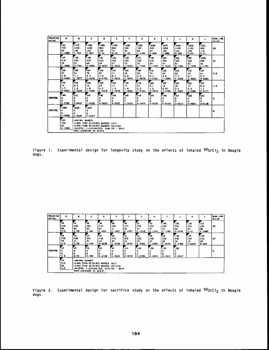
LP’ne,lCTtDUC|,/RG
R B C D E F I J K
~P"seB950100E-3]
1200lO0E-BUN
JP~57E12gO12DE-75,9,
1¯59096098E-JB
~BSB11DO1000128
JlTB SC990JIBD-2t
99
320Li.IO-J¼Oq
~6~RGOD
SDE-1702
~E583.9E-SBTB
~’22D626,Oo-5qqo
2P"D B
5!?.lE-gO??
2¯8C
?B9.9D-2qSB
3~FOB657.90-q795
rSOC710.3E-qq53
rOD586.6E-D37q
~39C799,1E -5261
YqOE6O9.6E-399q
GO7.7E-sq39
538.1D-3926
~2FqlS. 7B-SB’¢q
7,B
~’E ...... ~,,R191.70-2~1i7
293.2E-q97B
%o]3t.gE-SO?9
2qBIq1.6E =5q96
l.B[-9662
~TO232.2
’ £-¼7qq
1~30[8.}].D0-3B33
~’30F
]Bo.R7E-qTO6
3~30G
]62.2E-q~7
P~SE172,$O-U~lil
~TF
B.S1.1E-Ill 17
8.0I,O£-99q8
he
O0D-2?tlO
¯It00o-qsB2
"2.qE;o!O
E-qSBB
00D-5qB3
FAR0BD-SSB5
3YORD0E-Sq~]9
;OO~:~B~..
~R0OE-q236
3Y$80O0-5109
Y35FDo0-3012
qOD0BO-~SBq
~’Y2CCI0o-sz3p
CaHTROL B
liSBoN00E-SqD2
~B2E
BDE-q767
ill¯soNOBE-SOB8
:CONTROl. 0
75003E-1099
=RNTHRL NURBER-LQNG.-TERH RETRINEO BURDEN luCJ)-LONG-TERN RETAINED BURDEN [UC//KG}-OwDERO. E=EUTHBNIEED, R-RL]VE - OATSPBST-EXPBBURE RT DEATH
Figure 1. Expertmenta] design for longevity study on the effects of inhaled 905rC12 in Beagledogs.
E G R L RERN LlnBU¢]/KG
e,na~cyED,UCIIKG
A B O E ] J K
%qlO60S-S
q80653-I’D
%510
67E" 920
%qBO51E-1362
9D37099E- 1~157
37037E-1318
%q2Oq7E-1916
~P’~BR5~OssE-limB
lOB35Oqqo-2o3q
~50q7E-31
~E300=6E =2~l[IB
36Oa65-3BI
SO
27O29S=S
qO290315-29
92035S-379
%JqOJ6D-2620
%270
E-3738
6C2BB2qE-3U72
22029£-2762
%2903q[- 22GP,
22020E-3233
3303OE- 27~B
Jlrl2c150IS~-q222
JJP’2D
210200-505 ....
27
o0S-3RO
%Ro00-2B19
II~c0B0-351o
o05-6
00S-2q
0BE*q720
00E-2638
JOEBoD-SGTB
%,0oO-ZFTB6
I¯~B!o!o: E-5057
CONTROL B
qlo6OS-S
=flNINRL NUHBER=LBNG-TERR BEIRINED BURDEN {UCI)"LONGITERR RE1RIREB BURDEN (UCI/KG)-O-DERO. E.EU]HRNIZED, R=RLIY£ - OflTSPBS]-EXPOSURE RI OERTH
Figure 2. Experimental design for sacrifice study on the effects of inhaled 90SrCI 2 in Beagledogs.
184
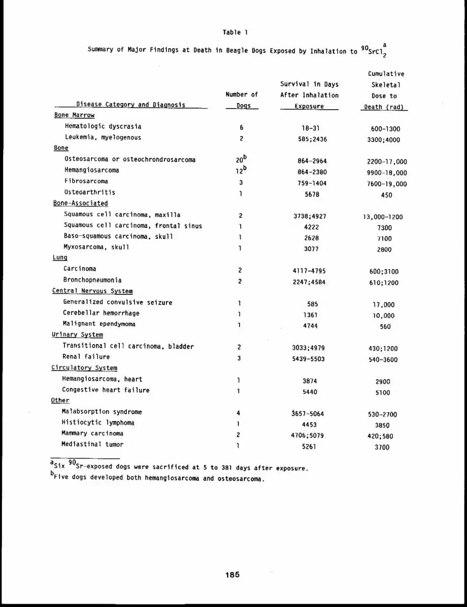
Table I
Summary of Major Findings at Death in Beagle Dogs Exposed by Inhalation to gOsrcI~
Disease Category and Diagnosis
Bone Marrow
Hematologic dyscrasla
Leukemia, myelogenous
Bone
Osteosarcoma or osteochrondrosarcoma
Hemangiosarcoma
Fibrosarcoma
Osteoarthritis
Bone-Assoclated
Squamous cell carcinoma, maxilla
Squamous cell carcinoma, frontal sinus
Baso-squamous carcinoma, skull
Myxosarcoma, skull
Lunq
Carcinoma
Bronchopneumonia
Central Nervous System
Generalized convulsive seizure
Cerebellar hemorrhage
Malignant ependymoma
Urinary System
Transitional cell carcinoma, bladder
Renal failure
Circulatory System
Hemangiosarcoma, heart
Congestive heart failure
Other
Malabsorption syndrome
Histiocytic lymphoma
Mammary carcinoma
Mediastinal tumor
Cumulative
Survival in Days Skeletal
Number of After Inhalation Dose toDogs Exposure Death (rad)
6 18-31 600-1300
2 585;2436 3300;4000
20b 864-2964 2200-17,000
12b 864-2380 9900-18,000
3 759-1404 7600-19,000
l 5678 450
2 3738;4927 13,000-1200
1 4222 7300
1 2628 7100
l 3077 2800
2 4117-4795 600;3100
2 2247;4584 610;1200
1 585 17,000
1 1361 I0,000
1 4744 560
2 3033;4979 430;1200
3 5439-5503 540-3600
l 3874 2900
1 5440 5100
4 ~657-5064 530-2700
1 4453 38502 4706;5079 420;580l 5261 3700
asix 90Sr-exposed dogs were sacrificed at 5 to 381 days after exposure.
bFive dogs developed both hemangiosarcoma and osteosarcoma.
185
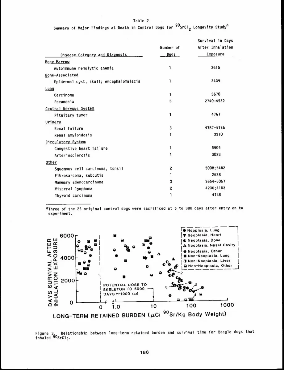
Table 2
Summary of Major Findings at Death in Control Dogs for 90SRC12 Longevity Studya
Diseas.~.Cateqory and Diaqnosls
Bone Marrow
Autoimmune hemolytic anemia
Bone-Assoclated
Epidermal cyst, skull; encephalomalacia
Carcinoma
Pneumonia
Central Nervous System
Pituitary tumor
Urinary
Renal failure
Renal amyloidosis
Circulatory System
Congestive heart failure
Arteriosclerosis
Other
Squamous cell carcinoma, tonsil
Fibrosarcoma, subcutis
Mammary adenocarcinoma
Visceral lymphoma
Thyroid carcinoma
Survival In Days
Number of After Inhalation
Exposure
1 2615
l 3439
l 3670
3 2740-4532
1 4767
3 4787-5136
1 3310
1 5505
1 3023
2 5008;5482
l 2638
3 3654-5057
2 4236;4103
1 4738
aThree of the 25 original control dogs were sacrificed at 5 to 380 days after entry on toexperiment.
6000,,=,==
[ ¯ Neoplasia, Lung ]_ , ,[]
IID
Heart=a [] ~ Bone
%¯~Q~¯ I Q ~ ,i,,t Neoplasia. Nasal Cavity i
Neoplasia, Other I¯ 0 AI m Non-Neoplasia, Lung I
[] ~iO ¯ & I11 Non-Neoplasia, Liver I_ =am ~
g) ¯ [] Non-Neoplasia, Other ]aa g
i SKELETON TO 5000 ~ 3co ._I I DAYS ,’.’1900 rad
~[]
<-r- A m Joz 0 ~ ~1 , -
- 0 1.O 10 100 1000
LONG-TERM RETAINED BURDEN (p.Ci 9°Sr/Kg Body Weight)
Figure 3. Relationship between long-term retained burden and survival time for Beagle dogs thatinhaled 90SrCl2.
186
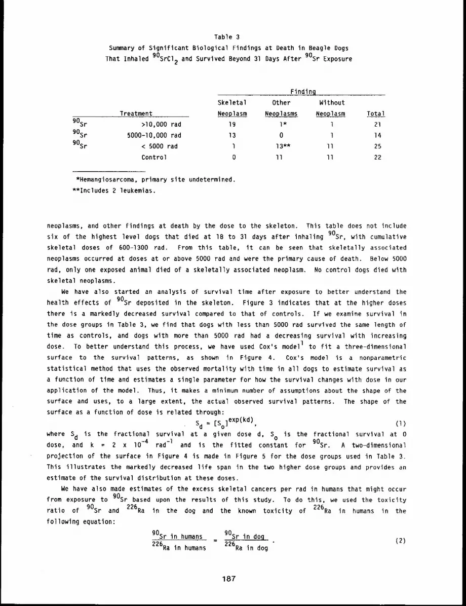
Table 3
Summary of Significant Biological Findings at Death in Beagle Dogs
That Inhaled 90SrCl2 and Survived Beyond 31 Days After 90Sr Exposure
90Sr
90Sr
90Sr
Finding
Skeletal Other Without
Treatment Neoplasm Neoplasms Neoplasm Total
>I0,000 rad 19 l* l 21
5000-I0,000 rad 13 0 l 14
< 5000 rad 1 13"* II 25
Control 0 II II 22
*Hemangiosarcoma, primary site undetermined.
**Includes 2 leukemias.
neoplasms, and other findings at death by the dose to the skeleton. This table does not include
six of the highest level dogs that died at 18 to 31 days after inhaling 90Sr, with cumulative
skeletal doses of 600-1300 tad. From this table, it can be seen that skeletally associated
neoplasms occurred at doses at or above 5000 rad and were the primary cause of death. Below 5000
rad, only one exposed animal died of a skeletally associated neoplasm. No control dogs died with
skeletal neoplasms.
We have also started an analysis of survival time after exposure to better understand the
health effects of gOsr deposited in the skeleton. Figure 3 indicates that at the higher doses
there is a markedly decreased survival compared to that of controls. If we examine survival in
the dose groups in Table 3, we find that dogs with less than 5000 rad survived the same length of
time as controls, and dogs with more than 5000 rad had a decreasing survival with increasing
dose. To better understand this process, we have used Cox’s modelI to fit a three-dimenslonal
surface to the survival patterns, as shown in Figure 4. Cox’s model is a nonparametric
statistical method that uses the observed mortality with time in all dogs to estimate survival as
a function of time and estimates a single parameter for how the survival changes with dose in our
application of the model. Thus, it makes a minimum number of assumptions about the shape of the
surface and uses, to a large extent, the actual observed survival patterns. The shape of the
surface as a function of dose is related through:
Sd = [So ]exp(kd), (l)
where Sd is the fractional survival at a given dose d, SO is the fractional survival at 0dose, and k = 2 x lO -4 rad -I and is the fitted constant for gOsr. A two-dimensional
projection of the surface in Figure 4 is made in Figure 5 for the dose groups used in Table 3.
This illustrates the markedly decreased life span in the two higher dose groups and provides an
estimate of the survival distribution at these doses.
We have also made estimates of the excess skeletal cancers per rad in humans that might occur
from exposure to gOsr based upon the results of this study. To do this, we used the toxicity
ratio of 90Sr and 226Ra in the dog and the known toxicity of 226Ra in humans in the
following equation:
gOsr in humans gOsr in dog- (Z)
226Ra in humans 226Ra in dog
187
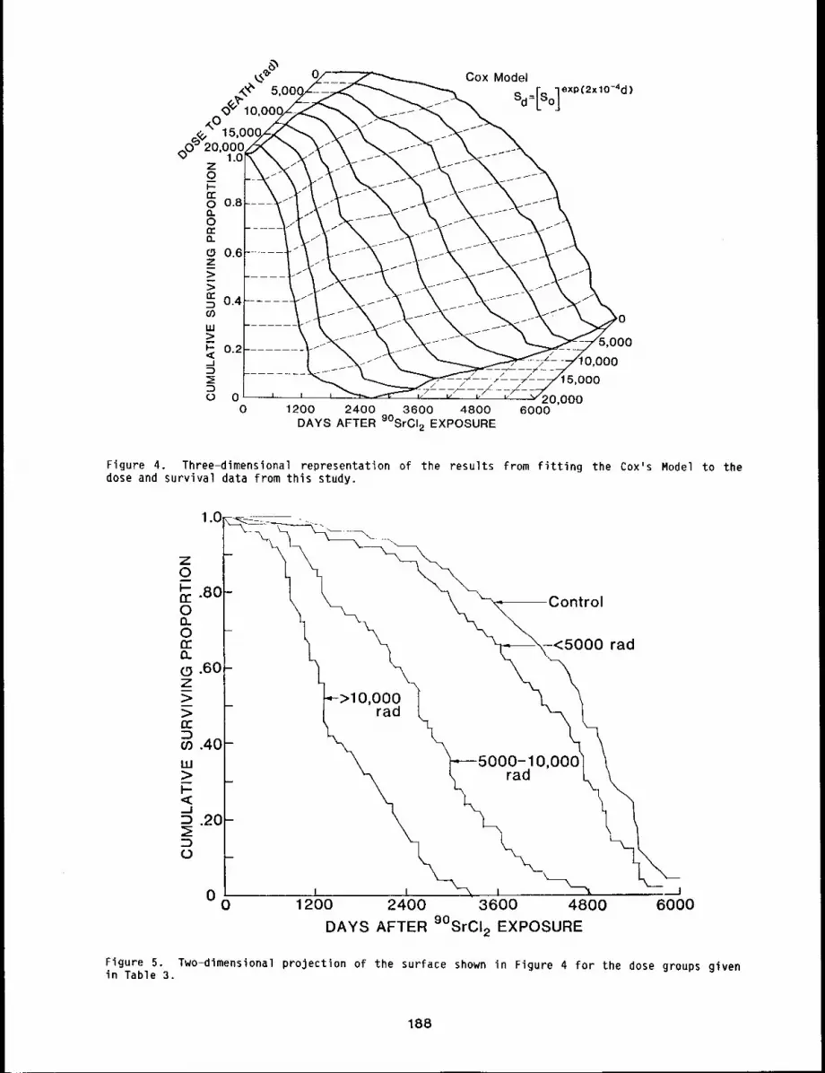
Cox Model
Sd=[so]eXp (2x,O~’d)
1200 2400 3600 4800DAYS AFTER 9OSrCI2 EXPOSURE
000
000
5,OOO
2O,OO06O00
Figure 4. Three-dimensional representation of the results from fitting the Cox’s Model to thedose and survival data from this study.
Figure 5.in Table 3.
Oa_d10,O00
0 1200 2400 3600 4800 6000
DAYS AFTER 9°SrCI2 EXPOSURE
rad
Two-dimensional projection of the surface shown in Figure 4 for the dose groups given
188
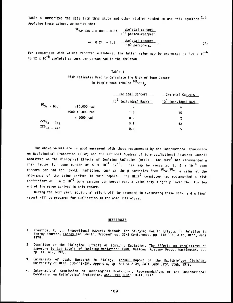
Table 4 summarizes the data from this study and other studies needed to use this equation.2,3
Applying these values, we derive that
90St Man = 0.008 - 0.07
or 0.24 - 1.2
skeletal cancers105 person-rad/year
skeletal cancerslO5 person-rad (3)
For comparison with values reported elsewhere, the latter value may be expressed as 2.4 x lO-6
to 12 x lO-6 skeletal cancers per person-rad to the skeleton.
90Sr - Dog
226Ra - Dog
226Ra - Man
Table 4
Risk Estimates Used to Calculate the Risk of Bone Cancer
in People that Inhaled 90SrCl2
>lO,O00 rad
5000-I0,000 rad
< 5000 rad
Skeletal Cancers
I05 Individual Rad/Yr
Skeletal Cancers
lO5 Individual Rad
1.2 6
1.7 I0
0.2 2
5.1 42
0.2 5
The above values are in good agreement with those recommended by the International Commission
on Radiological Protection (ICRP) and the National Academy of Sciences/National Research Council
Committee on the Biological Effects of Ionizing Radiation (BEIR). The ICRP4 has recommended a
risk factor for bone cancer of 5 x lO -4 Sv-l. This may be converted to 5 x lO -6 bone
cancers per rad for low-LET radiation, such as the B particles from 90Sr-9Oy, a value at the
mid-range of the value derived in this report. The BEIR2 committee has recommended a risk
coefficient of 1.4 x I0 -6 bone sarcoma per person-rad, a value only slightly lower than the low
end of the range derived in this report.
During the next year, additional effort will be expended in evaluating these data, and a Final
report will be prepared for publication in the open literature.
REFERENCES
l ¯ Prentice, R. L., Proportional Hazards Methods for Studying Health Effects in Relation toEnergy Sources, Enerqy and Health, Proceedings, SIMS Conference, pp. I18-133, Alta, Utah, June1978.
2. Committee on the Biological Effects of Ionizing Radiation, The Effects on Populations ofExposure to Low Levels of Ionizin~ Radiation: 19BO, National Academy Press, Washington, DC,pp. 416-417, 1980.
3. University of Utah, Research in Biology, Annual Report of .the Radiobiology Division,University of Utah, C00-I19-254, Appendix, pp. A-I to A-39, Salt Lake City, Utah,i 1979.
4. International Commission on Radiological Protection, Recommendations of the InternationalCommission on Radiological Protection, Ann. IRCP 1(3): lO-ll, 1977.
189
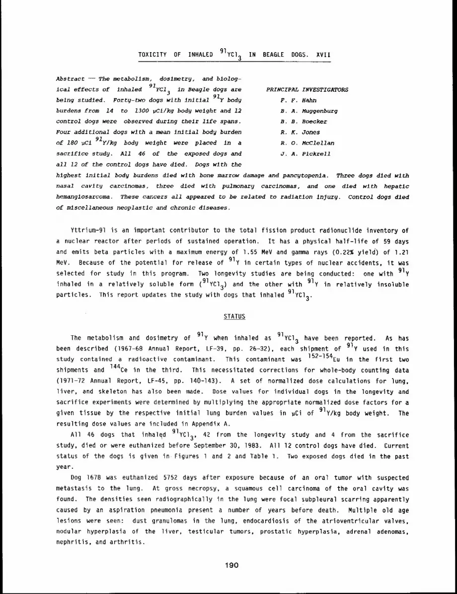
91yCl3TOXICITY OF INHALED IN BEAGLE DOGS. XVll
Abstract --The metabollsm, doslmecry, and blolog-
ical effects of inhaled 91yC13 in Beagle dogs are
belng studled. Forty-two dogs with inltlal 91y body
burdens from 14 to 1300 pCl/kg body welghc and 12
control dogs were observed durlng thelr llfe spans.
Four addltlonal dogs wlth a mean In~tlal body burden
of 180 uCl 91y/kg body weight were placed in a
sacrlflce study. All 46 of the exposed dogs and
all 12 of the control dogs have died. Dogs with the
PRINCIPAL INVESTIGATORS
F. F. Hahn
B, A. Muggenburg
B. B. Boecker
R. K. Jones
R. O. McClellan
J. A. P1ckrell
hlghest Inltlal body burdens dled with bone marrow damage and pancgtopenla. Three dogs dled wlth
nasal cavity carcinomas, three dled wlth pulmonary carclnomas, and one dled wlth hepatic
hemanglosarcoma. These cancers all appeared to be related to radiation injury. Control dogs dled
of mlscellaneous neoplastlc and chronlc dlseases.
Yttrium-91 is an important contributor to the total fission product radionuclide inventory of
a nuclear reactor after periods of sustained operation. It has a physical half-life of 59 days
and emits beta particles with a maximum energy of 1.55 MeV and gamma rays (0.22% yield) of 1.21
MeV. Because of the potential for release of 91y in certain types of nuclear accidents, it was
selected for study in this program. Two longevity studies are being conducted: one with 91y
inhaled in a relatively soluble form (Ylycl3) and the other with 91y in relatively insoluble
particles. This report updates the study with dogs that inhaled 91yCl3.
STATUS
dosimetry of 91y when inhaled as 91yCl3 have been reported. As hasThe metabolism and
been described (1967-68 Annual Report, LF-39, pp. 26-32), each shipment of 91y used in thisstudy contained a radioactive contaminant. This contaminant was 152-154Eu in the First two
shipments and 144Ce in the third. This necessitated corrections for whole-body counting data
(1971-72 Annual Report, LF-45, pp. 140-143). A set of normalized dose calculations for lung,
liver, and skeleton has also been made. Dose values for individual dogs in the longevity and
sacrifice experiments were determined by multiplying the appropriate normalized dose factors for a
given tissue by the respective initial lung burden values in ~Ci of gIy/kg body weight. The
resulting dose values are included in Appendix A.
All 46 dogs that inhaled 91yCl3, 42 from the longevity study and 4 from the sacrifice
study, died or were euthanized before September 30, 1983. All 12 control dogs have died. Current
status of the dogs is given in Figures l and 2 and Table 1. Two exposed dogs died in the past
year.
Dog 167B was euthanized 5752 days after exposure because of an oral tumor with suspected
metastasis Co the lung. At gross necropsy, a squamous cell carcinoma of the oral cavity was
found. The densities seen radiographically in the lung were focal subpleural scarring apparently
caused by an aspiration pneumonia present a number of years before death. Multiple old age
lesions were seen: dust granulomas in the lung, endocardiosis of the atrioventricular valves,
nodular hyperplasia of the liver, testicular tumors, prostatic hyperplasia, adrenal adenomas,
nephritis, and arthritis.
190
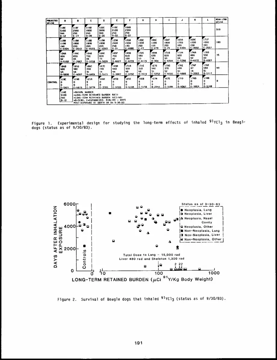
CONTROL
8 1 J K L
1900 1100 1800 1700 1~00 fOOD 100500 I IS 200 t68 150 160E - 2 .~3 0-075 O-2t[ 0-q 300 E- 31~92 E-2~57 ,,
~BO 600 650 600 880 550 ?S53 58 90 68 95 60E~qi15 0-36q E-q3tlq. E-5399 n-t1272 0-q567
3 ] 0 500 3"/0 280 97 qS9 ~0q8 5[ 52 q] ~6 51n=3Gi~l E-5752 E-0722 F-q06(~ D-2Gg-~I 0~511?
$o0 go,9 %. %0 %° ~,,-0 0 0 8 0 0 o
0 o 0 0 o 0
E-51 ~,0 , D:,2201 E-5 I1~q 0"q002 E- 395. 9,..,, 0-5109
Figure i, Experimental design for studying the long-term effects of inhaled 91yc13 in Beagl~dogs (status as of 9/30/83).
6000Z0
<~ 4000
IBO’)
o
o
0__%
Status aa of 9-30-83
[] Neoplasia. Lung 1
[] [] ~ ¢~ ¢= Neoplasia, Liver i
~ ¯ () ~1 I A~ Neoplasla. N~sal [~ Cavity
I[]1~ ~
I~i~ i~ Neoplasia. Other
&~ lib Non~Neoplasia, Lung [
Irll Non~Neoplasia. Liver ]
[] A i~ Non-Neoplasia, OtherJ
~tTotal Dose to Lung - 15,000 tad
Liver 480 rad and Skeleton t,300 rad5
l~I ~2 22
,l [] ........... ~ = J~h-~ = = ,
"10 100 1000LONG-TERM RETAINED BURDEN (FCi 91y/Kg Body Weight)
Figure 2. Survival of Beagle dogs that inhaled 91yCl3 (status as of 9/30/83),
191

Table 1
Findings in Beagle Dogs Exposed by Inhalation to 91yCl3 and Control Dogs
(status as of 9/30/83)
Diaqnosis
Exposed dogsa
Neoplasia
Lung
Nasal Cavity
Liver
Other Organs
Days After
Number Exposure Cumulative Dose (r~d)
at Death Lung___ Liver Skeleton
3 2109-4563 1400-3300 440-1900 1200-29003 2012-3352 1000-3300 310-1000 860-2900l 4627 1200 390 1100
lO 225B-5752 620-3100 190- 970 530-6600
Non-Neoplasia
Lung
Liver
Marrow
Other Organs
2 4567-5121 220- 910 65- 290 180- 780
I 4563 3300 i000 290012 12-3887 670-4300 210- 530 570- 91014 2663-5635 240-2400 76- 760 210-2100
Control Dogs
Neoplasia
Liver l 4366
Other Organs 5 3705-5455
L~
Non-Neoplasla
Lung l 2241
Liver l 4146
Other Organs 4 3959-5286
alncludes four dogs from sacrifice study that were allowed to live out their normal life span.
Dog 164D died 5635 days after exposure. Clinical signs of osteoarthritis first developed
about 6 years before death. Initially, the elbow Joints were involved, but during the next year
the knee joints showed similar changes and spondylosis deformans developed in a number of
vertebral joints. About 2 weeks before death, the dog was brought in and treated for diarrhea.
At gross necropsy a severe segmental hemorrhagic enteritis involving the ileum and jejunum was
found. The body was dehydrated.
DISCUSSION
The results of this study are of interest because the pattern of doses resulting from
inhalation of 91yCl 3 resulted in irradiation of the respiratory tract, liver, and skeleton
over a relatively short period of time. The total dose to organs from each gCi/kg long-term
retained burden of 91yCl3 was lung 15 rad, nasal turbinates 65 rad, liver 4.8 rad, and
skeleton 13 rad.
192
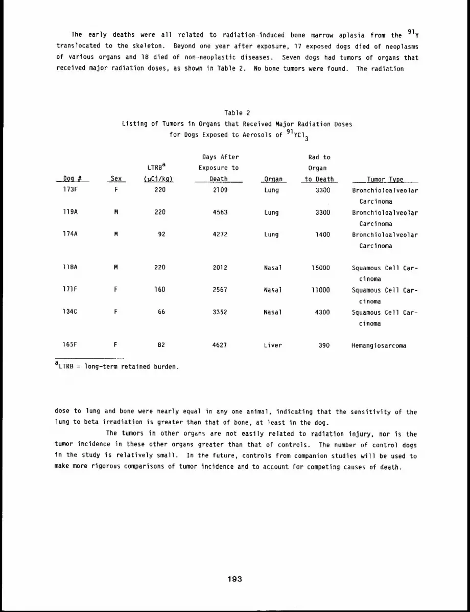
The early deaths were all related to radiation-induced bone marrow aplasia from the 91y
translocated to the skeleton. Beyond one year after exposure, 17 exposed dogs died of neoplasms
of various organs and 18 died of non-neoplastic diseases. Seven dogs had tumors of organs that
received major radiation doses, as shown in Table 2. No bone tumors were found. The radiation
Table 2
Listing of Tumors in Organs that Received Major Radiation Doses
for Dogs Exposed to Aerosols of 91yCl3
Days After Rad to
LTRBa Exposure to Organ
Sex ~ Death ~ to Death Tumor Type...
173F F 220 2109 Lung 3300 Bronchioloalveolar
Carcinoma
ll9A M 220 4563 Lung 3300 Bronchioloalveolar
Carcinoma
l?4A M 92 4272 Lung 1400 Bronchioloalveolar
Carcinoma
llBA M 220 2012 Nasal 15000 Squamous Cell Car-
cinoma
171F F 160 256? Nasal llO00 Squamous Cell Car-
cinoma
134C F 66 3352 Nasal 4300 Squamous Cell Car-
cinoma
165F F B2
aLTRB = long-term retained burden.
4627 Liver 390 Hemangiosarcoma
dose to lung and bone were nearly equal in any one animal, indicating that the sensitivity of the
lung to beta irradiation is greater than that of bone, at least in the dog.
The tumors in other organs are not easily related to radiation injury, nor is the
tumor incidence in these other organs greater than that of controls. The number of control dogs
in the study is relatively small. In the future, controls from companion studies will be used to
make more rigorous comparisons of tumor incidence and to account for competing causes of death.
193
TOXICITY OF INHALED 144CeCl IN BEA6LE DOGS. XVl3
Abstract --- The metabolism, doslmetrg, and biological
144CeCi 3 in Beagle dogs are belngeffects of inhaled
studied to assess the consequences of inhaling 144Ce.
Studles have shown that 144Ce deposited in lung as
144CeCi 3 is trenslocated at a moderately rapid rate
to liver and skeleton and that significant radiation
doses are accumulated by all ~hree organs. The dogs
exposed to 144Ce had long-term retalned burdens that
ranged from 2.6 to 360 ~CI 144Ce/kg hod 9 weight.
and 16 of 17 control dogs have dled.
years) after entr 9 into thls study.
PRINCIPAL INVESTIGATORS
B. B. Boecker
F. F. Hahn
B. A. Muggenburg
R. O, McClellan
J. A. Pickrell
All 55 of the dogs exposed to 144CeCZ3
There Is one control dog surviving at 5828 days (- Z6
Cerium-144 is one of the fission product radionuclides that predominates in a reactor
inventory after sustained reactor operation. It has a half-life of 284 days and decays to
144pr, which has a half-life of 17.3 min. During their decay, 144Ce* and 144pr emit
several gamma rays. They also emit a number of beta particles with an average energy of 1.27
MeV. Because of the potential release of 144Ce in certain types of nuclear accidents and
because of its radiological characteristics, it was selected as one of the radionuclides for study
in this program. One form selected for use was 144CeC13, which translocates from lung to
liver and skeleton at a moderately rapid rate, resulting in significant radiation doses to a]l
three organs. Two studies were initiated involving Beagle dogs: (1) a radiation dose pattern
study in which 27 dogs were exposed to 144CeC13 and serially sacrificed from 2 to 512 days
after inhalation exposure, and (2) a life span study to evaluate the relationship between
radiation dose and biological response for 55 dogs exposed to achieve graded initial body burdens
of 144CeCl 3. These exposed dogs and 17 control dogs were maintained for observation over
their life span (Fig. l). Information on the metabolism and dosimetry for 144CeC13 inhalation
exposures has been published.l This report briefly summarizes the current status of the life-
span study.
STATUS
Of the 72 dogs in the life-span study, 55 exposed and 16 controls have died or were euthanized
(Fig. 2). Table 1 summarizes the diagnoses of the exposed and control dogs that have died. One
control dog was euthanized during the past year. Clinical and pathological observations of this
dog are given below.
Dog 201A, a control, was euthanized 5895 days after exposure because of chronic renal
failure. Although this dog had numerous minor clinical problems, it lived 17.8 years. An
elevated blood urea nitrogen was first noted about 32 months before death and was moderately
elevated until death. At gross necropsy, a chronic nephritis was found. The parathyroid glands
were hyperplastic, indicative of renal secondary hyperparathyroidism. Nodular hyperplasia of the
liver with hepatocellular degeneration was also found. Miscellaneous incidental old age lesions
were thyroid gland adenoma, adrenal gland adenoma, interstitial cell tumor of the testes, heart
base (chemoreceptor) tumor, and arthritis of the hips and elbows.
*144Ce refers to an equilibrium mixture of 144Ce-144pr.
194
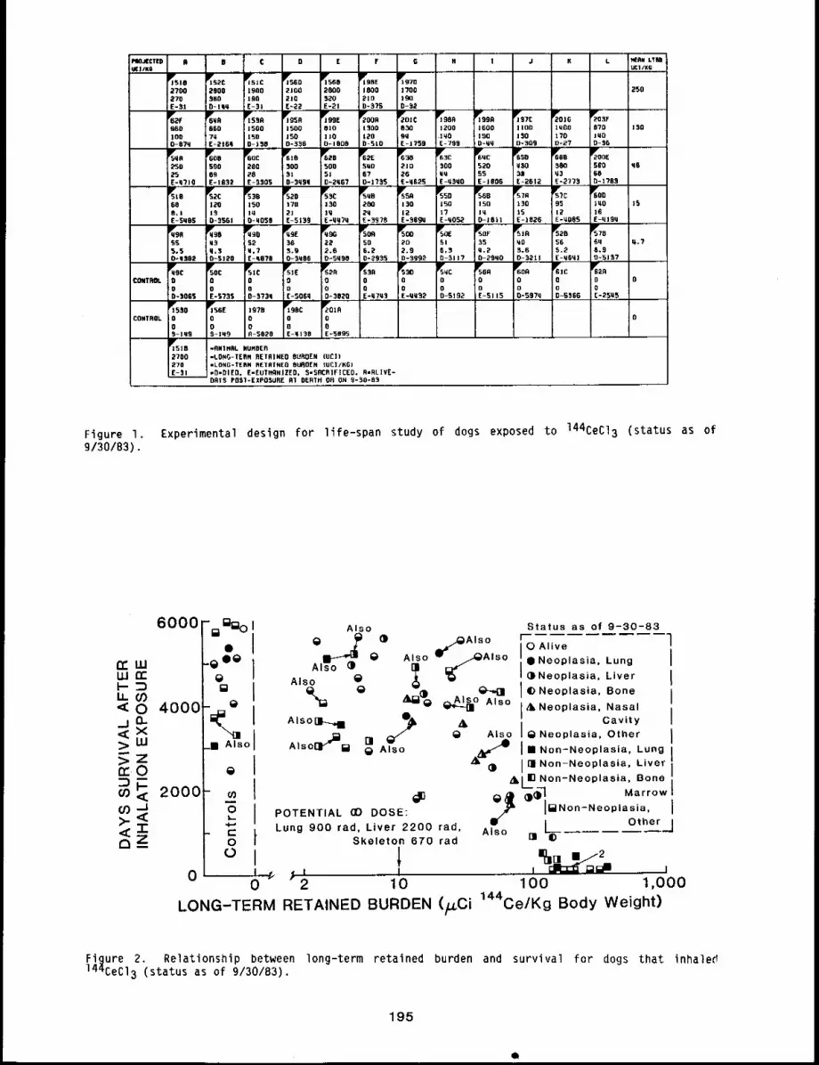
!¢ D [ F G N J K LA 8
1900190[-31
2]002]0
~5602800520E-2J
250
P’*69o1600190O=qq
Ir90A12oo
iMoE-799
~"]57C11001500-309
~016lqQOlTO0-27
lY95R
15001500-9~o
~99E810]100-]808
1 300IZOO SlO
F~01¢0309M[-1759
6r6URNGD7UE-216q
~qC52055E-1806
~658~30
E-2612
6P*285005J0=2q6"/
5U0670-17~5
60855065E-IBS~’
~60C200
28£-5905
1901::’E-SBgu
Y55D150[?[-q052
5r56B150lU0-1611
~R13015E-1826
~5"tC9512E-UORS
698.1E-SUBS
5Y2C
I~0
D-556!
~558
IS0lu0-q058
F-’520I "to
2][-5159
IS
5YOF35q.20-25~0
B.60-521 !
~57B6q6,9o-5157
~OE
565.90-3q86
~~G
222.60-5q90
SOft506.20-2935
202.90-999~
~90SS5,50-U302
52!u.?[-qN?B
5r56900E-S] 15
6YOO000=597q
~1C
OoD-5366
rsoc00E-5755
5~1C00D-575U
00[-S06q
0CONTROL
00E-SO95
00S-lU9
~rss¢00S-lU9
~98C0OE-q158
197B00R-$B20
0CONTROL
~15162700270E-31
-flHIMOL NURBEfl=LONG-TEflH OETR]NEO BUROEN (UCI)-LONG-TERN RETRINEO BUROEN IUC]/KG)-O=DIED, E-EUTNRN]ZED, 5=SOCRIFICED, R=GLIVE-DRTS POSI-EXPOSURE R10ERTH 0R ON 9-~0-83
Figure I. Experimental design for life-span study of dogs exposed to 144CeC13 (status as of9/30/83).
6000
CCUJ
4o0o:>ILl
;< ooo
0
"~ ~)1 Also Status as of 9-30-83
O Z ,v~- I o AliveI " ;.Neooasa Lung IAlso ~;) ~
~ ’ ~Neoplasia. Liver [
I -- ® 14)Neoplasia Bone iASOO Also I
-~~
I ~Ne°plasla’ NasaliI~ I
Alsol]-_~ ~ ,t Cavity
~sI
= _ ~ e A,soI e Neoplasia Othero Also_...-- ~ ~ j , ’ m
ur B ~ Also ~l~- liB Non-Neoplasia, Lung IA~()
I rm Non-Neoplasia, Liver I~kL~ Non-Neoplasia, Bone I
-- ~ e# Q) ~-’~,Marrow
~O 1
POTENTIAL (D DOSE:" 0 red W-~
Ii~N°n-Ne°plasia’ iOther
Lung 900 rad, Liver 22 0 , Also L._ ~-,JSkeleton 670 rad UI 4)
° i , ,0 2 10 100 1,000
LONG-TERM RETAINED BURDEN (~Ci ~44Ce/Kg Body Weight)
Figure 2, Relationship between long-term retained burden and survival for dogs that inhaler!
144CeC13 (status as of 9/30/83).
195
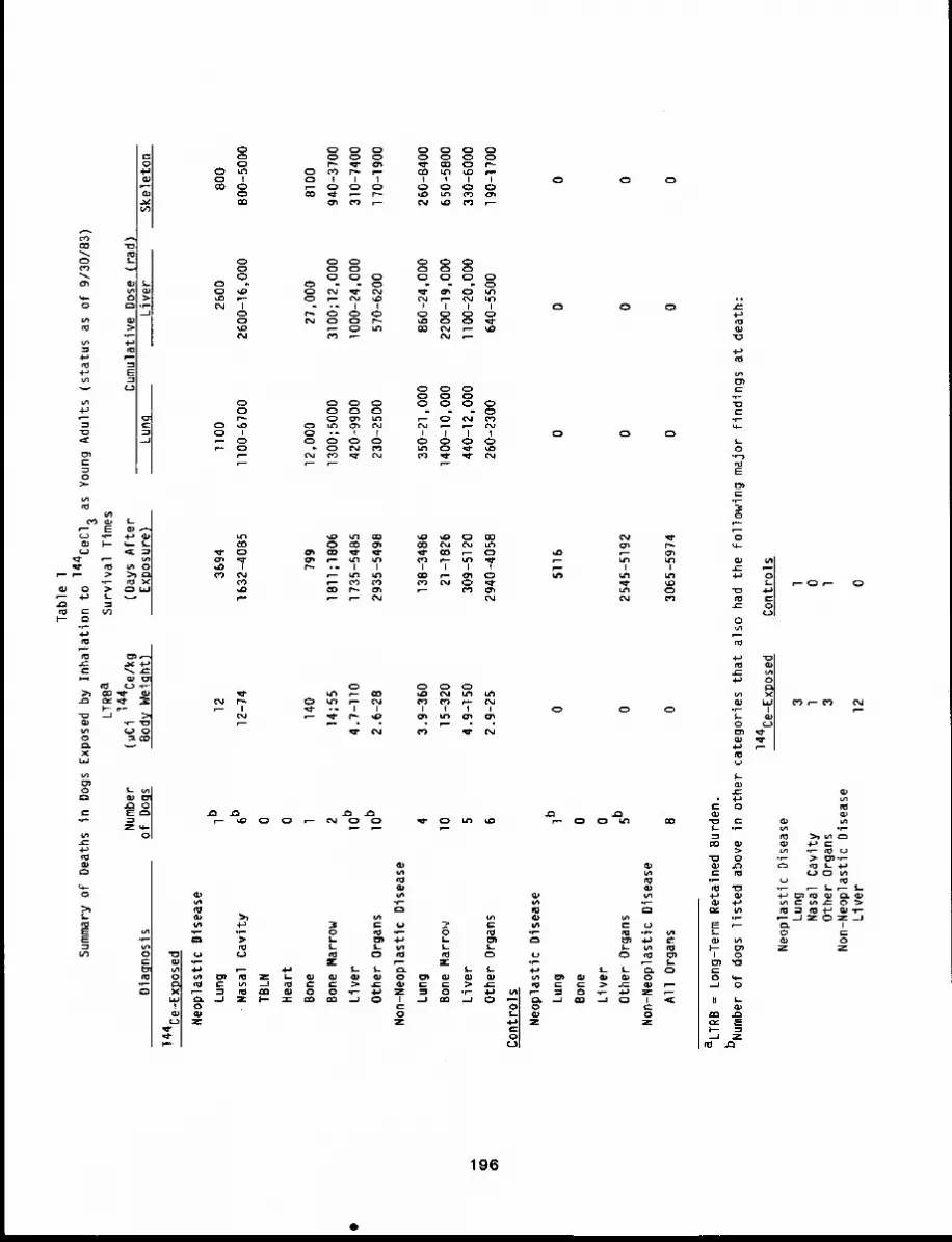
(o o)
fabl
e 1
of
Dea
ths
in
Dog
s E
xpos
ed b
y In
hala
tion
to
144C
EC
I3
Sum
mar
y
Oia
qn
osi
s
LT
R8
aS
urv
iva
l T
ime
s
Nu
mb
er
(~C
i 1
44
Ce
/kg
(Da
ys A
fte
ro
f D
og
sB
od
y W
eig
ht)
Exp
osu
re}
14
4C
e-E
xpo
sed
Ne
op
last
ic
Dis
ea
se
Lu
ng
1b1
23
69
4
Na
sal
Ca
vity
6b1
2-7
41
63
2-4
08
5
TB
LN
0
He
art
0
Bon
e1
140
799
Bo
ne
Ma
rro
w2
14
;55
18
11
;18
06
Liv
er
lOb4
.7-I
I01
73
5-5
48
5
Oth
er
Org
an
sIOb
2.6
-28
29
35
-54
98
No
n-N
eo
pla
stic
D
ise
ase
Lu
ng
43
.9-3
60
13
8-3
48
6
Bo
ne
Ma
rro
~v
I01
5-3
20
21
-18
26
Liv
er
54
.9-1
50
30
9-5
12
0
Oth
er
Org
an
s6
2.9
-25
29
40
-40
58
Co
ntr
ols
Ne
op
last
ic
Dis
ea
se
Lu
ng
Ib0
51
16
Bon
e0
Live
r0
Oth
er
Org
an
s5b
02
54
5-5
19
2
No
n-N
eo
pla
stic
D
ise
ase
All
Org
an
s8
03
06
5-5
97
4
as
Yo
un
g A
du
lts
(sta
tus
as
of
9/3
0/8
3)
Cu
mu
lativ
e D
ose
(ra
d)
Lu
ng
Liv
er
Ske
leto
n
11
00
26
00
80
0
11
00
-67
00
26
00
-16
,00
0B
00
-50
00
12
,00
02
7,0
00
81
00
13
00
;50
00
31
00
;12
,00
09
40
-37
00
42
0-9
90
01
00
0-2
4,0
00
31
0-7
40
0
23
0-2
50
05
70
-62
00
17
0-1
90
0
35
0-2
1,0
00
86
0-2
4,0
00
26
0-8
40
0
14
00
-10
,O0
02
20
0-1
9,0
00
65
0-5
80
0
44
0-1
2,0
00
llO0
-20
,O0
03
30
-60
00
26
0-2
30
06
40
-55
00
19
0-1
70
0
00
0
00
0
00
0
aL
TR
B =
Lo
ng
-Te
rm R
eta
ine
d B
urd
en
.
bN
um
be
r of
do
gs
liste
d
ab
ove
in
o
the
r ca
teg
orie
s th
at
als
o
ha
d t
he
fo
llow
ing
m
ajo
r fin
din
gs
at
de
ath
:
14
4C
e-E
xpo
sed
Co
ntr
ols
Ne
op
last
ic
Dis
ea
seL
un
g3
lN
asa
l C
avi
tyl
0O
the
r O
rga
ns
31
No
n-N
eo
pla
stic
D
ise
ase
Liv
er
12
0
DISCUSSION
The biological effects seen in this study reflect the patterns of deposition, retention, and
dosimetry of 144Ce after inhalation in a relatively soluble form. After rapid mucociliary
clearance of the nasopharyngeal and tracheobronchial regions by way of the gastrointestinal tract
during the first week after exposure, the pulmonary-deposited 144Ce constituted the main part of
the long-term retained burden. Most of the 144Ca was translocated during the first week,
primarily to the liver and skeleton. High local concentrations were also noted in the nasal
turbinates, either from 144Ce deposited there at the time of exposure or translocated there
after absorption from the pulmonary region.1’2
Although the prevalent early effect of inhaled 144Ce was bone marrow aplasia in the dogs
exposed at the highest level, early effects were also seen in the lung (radiation pneumonitis and
pulmonary fibrosis) and liver (necrosis). Dogs that survived this early phase had non-eventful
clinical courses (except for a single case of vertebral osteosarcoma) until cancers began
appear beyond four years after exposure.
Cancers or benign neoplasms were observed in all four organs or tissues receiving the largest
relative doses: lung, liver, bone, and nasal epithelium. Three adenocarcinomas and an adenoma
were seen in the lung. In the liver, there were seven hemangiosarcomas, one hepatocellular
carcinoma, one bile duct carcinoma, and one bile duct cystadenoma. One osteosarcoma and two cases
of myelogenous leukemia were observed. In the nasal cavity were one hemangiosarcoma and six
squamous cell carcinomas. One of these latter tumors was found in situ. Cancers found in other
organs were also observed in control dogs and are assumed not to be associated with the radiation
exposures. From Table l, it is apparent that non-neoplastic disease in the liver was also a major
long-term finding, presumably relating to the deposition and long-term retention of 144Ce in the
liver.
Analyses are in progress to determine the risks of these diseases in these different organs as
a function of absorbed beta dose. The results of these analyses, when extrapolated to human
exposures, should clarify which organs are at greatest risk after an inhalation exposure to
144Ce in a relatively soluble form. These results will be compared with current ICRP
recommendations to determine whether the recommendations provide adequate protection for workers.
Because the nasal cavity is not specifically covered in the current ICRP recommendations, the
nasal cavity cancers seen in this study indicate that they should be given further
consideration.2
REFERENCES
I. Boecker, B. B. and R. G. Cuddihy, Toxicity of 144Ca Inhaled as 144CeC13 by the Beagle:Metabolism and Dosimetry, Radiat. Res. 60: 133-154, 1974.
2. Boecker, B. B., F. F. Hahn, R. G. Cuddihy, M. B. Snipes, and R. O. McClellan, Is the HumanNasal Cavity at Risk from Inhaled Radionuclides? To be published in the proceedings of theTwenty-Second Hanford Life Sciences Symposium, Richland, WA, September 1983.
197
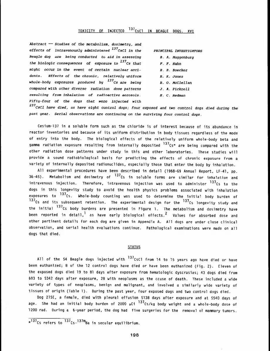
TOXICITY OF INJECTED 137CsCI IN BEAGLE DOGS. XVI
Abstract --Studies of the metabolism, doslmetry, and
effects of intravenously administered 137CsCl in the
Beagle dog are belng conducted to aid in assessing
the blologlc consequences of exposure to 137Cs that
might occur in the event of certain nuclear acci-
dents. Effects of the chronic, relatively ~iform
whole-body exposures produced by 137Cs are being
compared wlth other d~verse radlaclon dose patterns
resultlng from inhalation of radioactive aerosols.
Fifty-four of the dogs that were injected wlth
PRINCIPAL INVESTIGATORS
B. A. Muggenburg
F. F. Hahn
B. B. Boecker
R. K. Jones
R. O. McClellan
J. A. P~ckrell
H. C. Redman
237CSCI have died. as have eight control dogs; four exposed and two control dogs died during the
past year. Serial observatlons are contlnulng on the survlvlng four control dogs.
Cesium-137 in a soluble form such as the chloride is of interest because of its abundance in
reactor inventories and because of its uniform distribution in body tissues regardless of’the mode
of entry into the body. The biological effects of the relatively uniform whole-body beta and
gamma radiation exposure resulting from internally deposited 137Cs* are being compared with the
other radiation dose patterns under study in this and other laboratories. These studies will
provide a sound radiobiological basis for predicting the effects of chronic exposure from a
variety of internally deposited radionuclides, especially those that enter the body by inhalation.
All experimental procedures have been described in detail (1968-69 Annual Report, LF-41, pp.
36-45). Metabolism and dosimetry of 13?Cs in soluble forms are similar for inhalation and
intravenous injection. Therefore, intravenous injection was used to administer 137Cs to the
dogs in this longevity study to avoid the health physics problems associated with inhalation
exposures to 137Cs. Whole-body counting was used to determine the initial body burden of
137Cs and its subsequent retention. The experimental design for the 13?Cs longevity study and
the initial 137Cs body burdens are presented in Figure i. The metabolism and dosimetry have
been reported in detail, l as have early biological effects. 2 Values for absorbed dose and
other pertinent details for each dog are given in Appendix A. All dogs are under close clinical
observation, and serial health evaluations continue. Pathological examinations were made on all
dogs that died.
STATUS
All of the 54 Beagle dogs injected with 137CsCl from 14 to 15 years ago have died or have
been euthanized; B of the 12 control dogs have died or have been euthanized (Fig. 2). Eleven
the exposed dogs died 19 to 81 days after exposure from hematologic dyscrasias; 43 dogs died from
693 to 5342 days after exposure, 28 with neoplasms as the cause of death. These included a wide
variety of types of neoplasms, benign and malignant, and involved a similarly wide variety of
tissues of origin (Table 1). During the past year, four exposed dogs and two control dogs died.
Dog 275E, a female, died with pleural effusion 513B days after exposure and at 5540 days of
age. She had an initial body burden of 2000 ~Ci 137Cs/kg body weight and a whole-body dose of
1200 rad. During a 6-year period, the dog had five surgeries for the removal o~ mammary tumors.
*137Cs refers to 137Cs-137mBa in secular equilibrium.
198
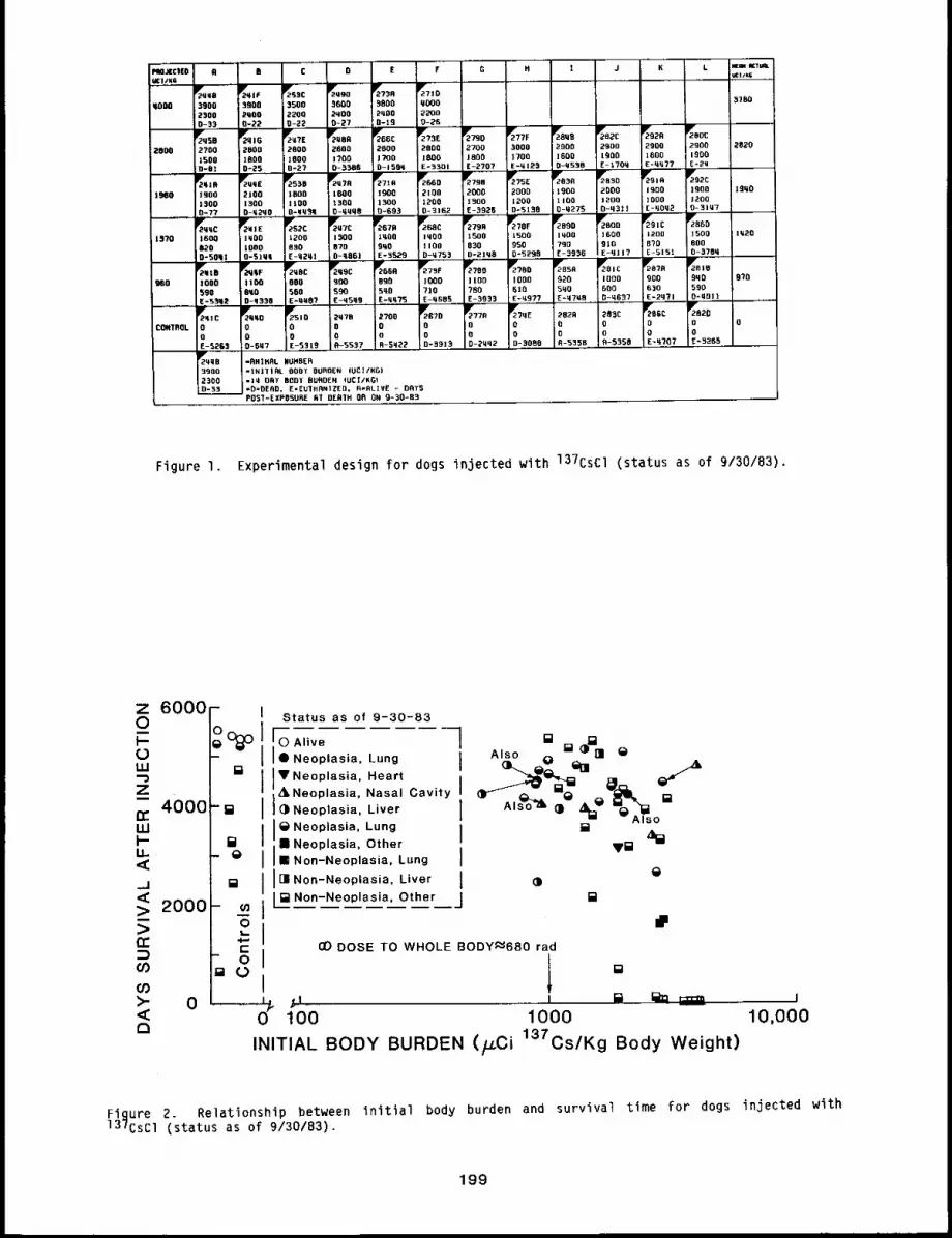
PMJcr|o A BUCI/KG
~qM8qOO0 3900
2~000-33,
~se2800 2700
ZSOOD-91
~9,81960 1900
1570
7~,, ~.c"- ~.,0 7,,, ~,1o3900 3500 3600 3800 qo00 0780
~qO0 2200 2800 2qO0 ~2000-22 0-~2 D-27 :9-19 0-~6 .... ,_,,
~qlG Tq’E T"B’ ~EUC T?3E 7790 T?,F TBOB 7B+E 792A TBOC
2800 +800 2600 2800 0000 2700 3000 +900 090o 2900 2900 28+0
1800 1800 1700 1700 1800 1809 1700 1600 1900 1600 1900
0-~5 1=27 0-3388 D-1589 E-3301 E-2707, E-q123 0-9538 E-170q E-¼q?7 E-2~
%. T,3, ~,7~ 0%105.° %. %BE %+~ ~B.0 r.,o %2:2100 1600 1000 1900 2100 2000 2000 1900 2000 1900 1500 19q0
1300 1100 t300 1300 1200 1900 1200 1100 1200 ]000 1200o-q2qo +-qq3q O-qqq8 0-593 0-3162 E-3526 D-5138 0-M275 0-831| , E-~Oq2 0-30~7
+,;r" +’+’52~
7"7E ~8+" %0~ 7,,0 %r TB.o +’;0o079,: 78~oIqO0 12o0 1500 1q00 1mOO 15o0 15o0 1mOO 1600 12oo +500
10o0 890 870 5qO ]10o 030 950 790 910 0~0 BOO
1500D-777qqC I92016008200-5081 0-5101 E-9291 0-8881 E-9525 0-8753 0-2188 0-5290 E-3536 E-qll? E-5151 0-3789,
gqtB gqqF 2Y,8C 799C ~2869 Y27,F 21k";88 g780 Jlr2BsR 2~01C ~P"97R 781e
1000 1100 880 900 890 1000 llO0 IODO 920 1000 900 9q0 978
590 890 560 590 590 710 780 610 590 600 63o 590
E-5~8~ D-9338 . E-q087 E-9599 E-qq?5 E+9685 E-3933 E-99.77 E=97¼8 . G-9637 E-2971 !0=9011
2829 2B~C 2~’86C 2~8200 0 0 O 00 o 0 0R-5358., A-5358 E-qTO? ~-~265
~qlC 7990 ~510 2976 2700 ~2670 ~770 ~?qE
CONIROL O 0 0 O 0 0 0 0
O 0 0 0 o 0 0 0
IF i28~8 -RRIRAL NUMBER3900 -INJT|RL BOOT OUROEN (UCI/KGI2300 =]q ORT BODY BURDEN +UCI/KGt~=O=OERD, E-EUIBANI~ED. R*RLIVE - DAT5
PO$7=[XPOSURE Bl DERIH OR OR 9+30-83
Figure I. Experimental design for dogs injected with 137CsCl (status as of 9/30/83).
z 6000-O O
4000LUI’-IJ..<_J,<> 2000>n"
(/3CO>" 0<a
I Status as of 9-30-83
1 11 Neoplasia, Lung Also O ~ nl¯ Neoplasia, Heart ~ ~dl
¯ Neoplasia, Nasal Cavity ~l~ll_L,,,e," I AlsoNeoplasia, Lung
I []I; IB Neoplasia, Other V~BI Non-Neoplasia, Lung I[] Non-Neoplasia, Liver [ ()¢= Non-Neoplasia, Other J
CO DOSE TO WHOLE BODY--680 rad
1 °100 1000
INITIAL BODY BURDEN (~CJ 137Cs/Kg Body Weight)
10,000
Figure ~. Relationship between initial body burden and survival time for dogs injected with137CsCl (status as of 9/30/83).
199
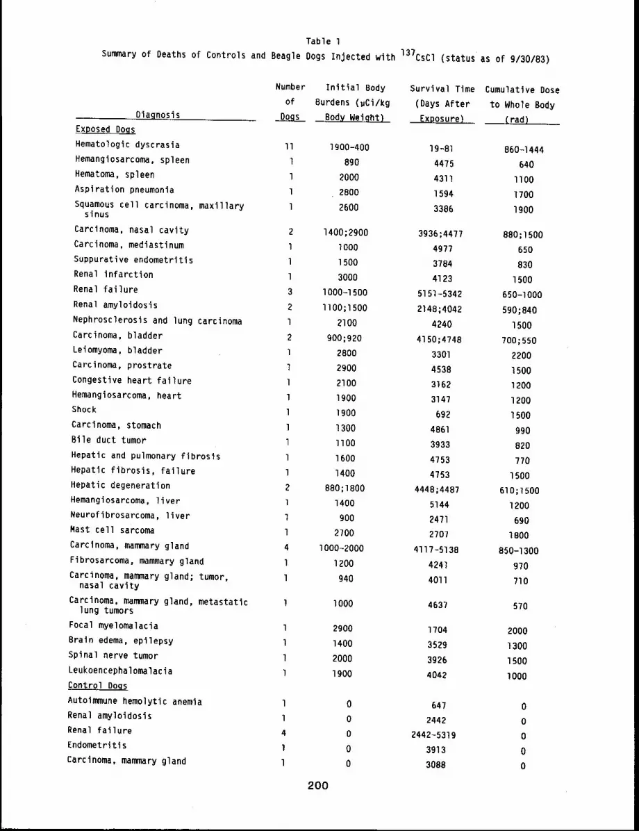
Table 1
Summary of Deaths of Controls and Beagle Dogs Injected with 137CSCI (status as of 9/30/83)
Diagnosis
Exposed Doqs
Hematologic dyscrasia II 1900-400Hemangiosarcoma, spleen 1 890Hematoma, spleen l 2000Aspiration pneumonia l 2800Squamous cell carcinoma, maxillary l 2600
sinus
Carcinoma, nasal cavity 2 1400;2900
Carcinoma, mediastinum l lO00Suppurative endometritis l 1500Renal infarction l 3000
Renal failure 3 1000-1500Renal amyloidosis 2 llO0;1500Nephrosclerosis and lung carcinoma l 2100Carcinoma, bladder 2 900;920Leiomyoma, bladder l 2800Carcinoma, prostrate l 2900Congestive heart failure 1 2100Hemangiosarcoma, heart l 1900Shock l 1900Carcinoma, stomach l 1300Bile duct tumor l llO0Hepatic and pulmonary fibrosis l 1600Hepatic fibrosis, failure l 1400Hepatic degeneration 2 880;1800Hemangiosarcoma, liver l 1400
Neurofibrosarcoma, liver l 900Mast cell sarcoma l 2700Carcinoma, mammary gland 4 I000-2000Fibrosarcoma, mammary gland l 1200Carcinoma, mammary gland; tumor, l 940
nasal cavity
Carcinoma, mammary gland, metastatic l I000lung tumors
Focal myelomalacia l 2900Brain edema, epilepsy l 1400Spinal nerve tumor l 2000Leukoencephalomalacia l 1900Control Dogs
Autoimmune hemolytic anemia lRenal amyloidosis lRenal failure 4
Endometritis lCarcinoma, mammary gland l
Number initial Body Survival Time Cumulative Doseof Burdens (~Ci/kg (Days After to Whole Body
Body Weight) ExPOsure) (rad)
19-81 860~1444
4475 640
4311 llO0
1594 1700
3386 1900
3936;4477 880;1500
4977 650
3784 830
4123 1500
5151-5342 650-1000
2148;4042 590;840
4240 1500
4150;4748 700;550
3301 2200
4538 1500
3162 1200
3147 1200
692 1500
4861 990
3933 820
4753 7704753 1500
4448;4487 610;1500
5144 1200
2471 690
2707 IBO0
4117-5138 850-1300
4241 9704011 710
4637 570
1704 2000
3529 1300
3926 15DO
4042 lO00
0 647 00 2442 0
0 2442-5319 0
0 3913 00 3088 0
200
Five of the 14 tumors removed were classified histologically as malignant. At gross necropsy,
both pleural and abdominal fluids were present. Cytological preparations from these fluids showed
clusters of large, highly vacuolated epithelial cells with a high nuclear cytoplasmic ratio
consistent with mammary adenocarcinoma. The lungs were filled with numerous small tumor nodules.
A right ventricular hypertrophy and other common aging lesions were found.
Dog 278F, a female, died after anesthesia for radiography 5290 days after exposure and at 5681
days of age. She had an initial body burden of 1500 ~Ci 137Cs/kg body weight and a whole-body
dose of I000 tad. The dog had a variety of unrelated, chronic, recurring problems. Among these
was a hysterectomy for a pyometra four years before death. Vaginal tumors were removed three
years ago and subsequently reappeared. Chronic renal failure was also evident clinically. At
gross necropsy, a chronic interstitial nephritis and multiple vaginal tumors were found. Numerous
other aging changes were found, such as skin papillomas, nodular hyperplasia of the liver, and
endocardiosis.
Dog 291C was a male that was euthanized 5151 days after exposure and 5533 days of age with
renal failure. He had an initial body burden of 1200 #Ci 137Cs/kg body weight and a
whole-body dose of 990 rad. At gross necropsy, renal cortical fibrosis was found along with
parathyroid hyperplasia, lesions consistent with renal failure. In addition, multiple infarcts
were found in the heart and lungs, which played a role in the dog’s severe illness. Massive
nodules were present in the liver. These appeared to be hyperplasia.
Dog 241B, a male, was euthanized in renal failure 5342 days after exposure and at 5760 days of
age. He had an initial lung burden of lO00 ~Ci 137Cs/kg body weight and a whole body dose of
650 rad. The dog had a persistently elevated BUN for about 8 years. In the months before
euthanasia, the BUN was greatly elevated and no longer responded to treatment. A chronic
pyelonephritis and parathyroid hyperplasia were found at gross necropsy. The primary cause of
death was severe renal disease. Age-associated lesions in this dog included dust granulomas of
the lung, valvular endocarditis, adrenal gland hyperplasia, interstitial cell tumors of the
testes, leiomyomas of the stomach, fibrosis of the prostate, hemangiomas of the skin, and
dust-laden tracheobronchial lymph nodes.
Dog 241C, a male control, was euthanized in chronic renal failure 5263 days after entering the
experiment and at 5681 days of age. At necropsy, a severe diffuse chronic interstitial nephritis
was found. The immediate cause for the euthanasia, however, was a subacute bronchopneumonia
caused by the aspiration of foreign material. Age-related lesions included nodular hyperplasia of
the liver, cystic hyperplasia of the gall bladder, endocardiosis of the atrioventricular valves,
nodular hyperplasia of the spleen, interstitial cell tumor of the testes, and adenomas of the
adrenal glands.
Dog 251D, a female control, was euthanized 531g days after entering the experiment and at 5727
days of age. She had a long history of elevated serum enzymes related to the liver and of
elevated blood urea nitrogen. At necropsy, a chronic interstitial nephritis and severe nodular
hyperplasia of the liver were found as indicated by the clinical findings. A pituitary tumor and
bilateral hyperplasia of the adrenal cortices were also found. The adrenal hyperplasia may be the
result of an adrenocortical trophic hormone-secreting tumor in the pituitary. A chronic
mesenteric lymphangitis of undetermined origin was also present. Many other age-related lesions
were noted.
DISCUSSION
The remaining dogs in this study are all over 15 years of age. The findings of tumors and
chronic renal failure clinically and at necropsy reflect this increasing age. Tumors found in
201
this study have been associated with mammary glands, nasal passages, bladder, spleen, prostate
gland, stomach, intestines, and lung. Several tumors have occurred at an unexpectedly high rate:
four cancers of the liver, three in the nasal passages, two of the lung, and one in the
mediastinum. Because of the general distribution of 137Cs throughout the body, careful review
of the data will be done to examine the relationship between radiation dose and the incidence of
cancers and other diseases.
REFERENCES
I. Boecker, B. B., Toxicity of 137CsCl in the Beagle: Metabolism and Dosimetry, Radiat. Res.50: 556-573, 1972.
2. Redman, H. C., R. O. McClellan, R. K. 3ones, B. B. Boecker, T. L. Chiffelle, J. A. Pickrell,and E. W. Rypka, Toxicity of 137Cs in the Beagle. Early Biological Effects, Radiat. Res.50: 629-648, 1972.
202
TOXICITY OF 90y IN A RELATIVELY INSOLUBLE FORM INHALED BY BEAGLE DOGS. XV
Abstract -- The mecabollsm, doslmetrg, and blolog-
ical effects of inhaled 90y In fused alumlnosill-
cate partlcles are being studied in Beagle dogs.
Eighty-nine dogs wlch inlclal lung burdens ranging
from 80 to 5200 ~cI 90y/kg body weight and 12 con-
trol dogs are belng malntalned for llfe-span ob-
servaElons. The lung burdens achieved, short
physical half-llfe, and relative insolubility of
PRINCIPAL INVESTIGATORS
F. F. Hahn
C. H. Hobbs
R. O. NcC1ellan
J. L. Mauder1~
J. A. Plckrell
the 90y in thls vector resulted In a relatlvely hlgh, rapld19 decreasing radlation dose rate tothe lung. To date, 76 exposed dogs have d~ed; 38 from radiation pneumonltis, 7 with pulm~narg
neoplasms, and 31 from other causes, seven control dogs have died. Thirteen exposed and flve
control dogs still survlve at 4600 to 5118 days after entry onto the study.
This study is one of a series designed to determine the relationships of total radiation dose
and radiation dose rate from inhaled beta-emitting radionuclides to biological effects. Dogs have
been exposed by inhalation to yttrium-90 (90y) in fused aluminosilicate particles, l The
relative insolubility of 90y in fused aluminosilicate particles, its short physical half-life,
and the dose levels achieved resulted in radiation exposure of the lung and associated structures
at a relatively high, but rapidly decreasing, dose rate. The long-term biological effects ofgOy in this relatively insoluble form are being compared with the biological effects oflonger-lived beta emitters also under study (gIy, 144Ce’ and gOsr in fused aluminosilicate
particles).2
Eighty-nine dogs were exposed to 90y in fused aluminosilicate particles to achieve graded
initial lung burdens to evaluate dose-response relationships during the life of the dogs. Twelve
dogs were exposed to aerosols of stable yttrium in fused aluminosilicate particles to serve as
controls. The experimental design for this longevity study is depicted in Figure I.
STATUS
Details of the experimental procedures, the results of the radiation dose-pattern studies, and
the early biological effects observed have been reported in previous Annual Reports. This report
briefly summarizes and updates the results from the dose-response study.
As of September 30, 1983, a total of 76 of the B9 exposed dogs had died between 7.5 and 4762
days after inhalation exposure (Fig. 2) with initial lung burdens that ranged from 80 to 5200
~Ci 90y/kg body weight and cumulative doses to lung ranging from 1300 to 70,000 rad. Seven
control dogs have died. The significant findings at death are summarized in Table I. More
details on the individual dogs within this study are presented in Appendix A. Seven exposed and
three control dogs died during the last wear.
Dog 400U was euthanized 4507 days after exposure because of multiple tumor metastases to the
lungs. The dose to lung was 6200 rad. During her lifetime, only minor clinical problems were
noted except for four mammary nodules removed on two occasions before the terminal episode. One
of these was a complex adenocarcinoma, one was a mixed mammary tumor, and two were lobular
hyperplasia. Two small pulmonary masses were noted radiographically before death. A large
203
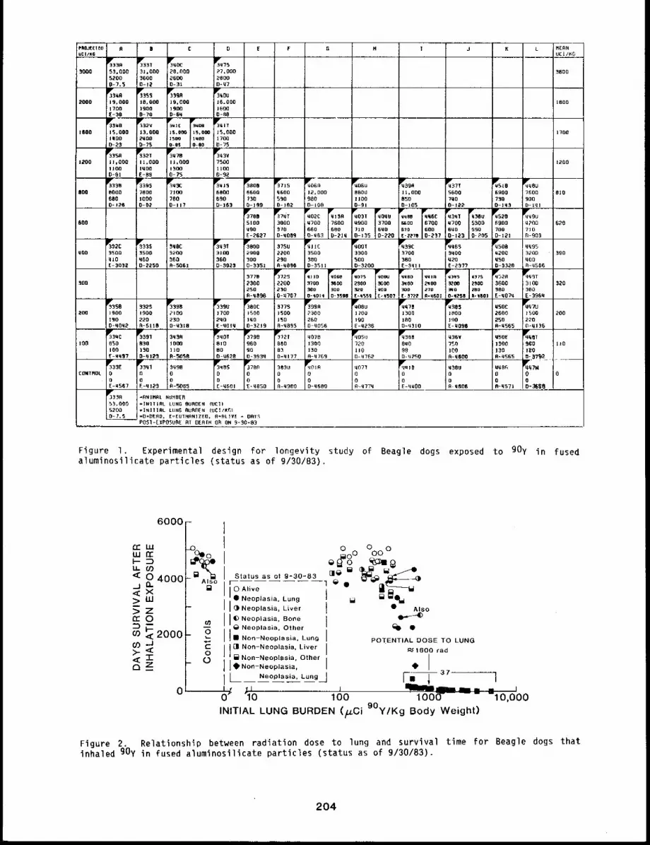
2OOO
1800
1200
C
~oc53,000 31,060 ;~0 flCO5200 3600 ! 26006-7.5 D-]2 0-31
Ile’35¼8 g,~5 g,¢JR
19,000 ]fl,O00 ]9,0001700 1900 1900E-30 0-?0 0-6q
15,000 13,000 ]5.000 13, O00I’00 2q00 15~ IWO06-25 0-?5 0-~
5350 5321 g~TB
il,O00 11,000 ]l,O00liao IqOO 13Qa0-91 E-89 fl-?5
33958000 7800 7100680 zOO0 "/600=126 0-02 0-117
880
800
............. %2:%, .0~qO0 ~50a 3500 3200
~10 ~60 ~60E-3052 0-2250 A-506!
O E
27,00020000-,7
~,ou16.0001600O-~
15,00017 O06-95
~,3v?50011000-32
---%;-- r---3.~,J--~i;366. . ....
F G H | J 6 L NEONUC|/KG
3600
1200
%6° ¢.0 %. ~510¢,o~680(] 6600 0600 12,000 B80U ll,OOO 5600 6900 7600 6|0690 ?50 550 900 1100 850 7,0 ;930 9000-163 0-199 0-162 6-106 0-91 0-105 0-122 O-J,, D-1q1
0, 57,., ~oo:.,, No,?,o,o;. ,,66~,,, ,36o~T;--~5100 3000 ~700 7600 ~900 3700 ~00 6700 ’700 5300 6300 ~p00 620NO0 570 660 660 710 600 670 600 6qO 590 700 710E-2627 0-,069 D-.qG3 -2]q 0-155 -220 E-2~7, -257 0-123 0-P05 6-12! N-905
~11C ~001 ~39C ~063 ~508 qq953500 3300 3700 ,NO0 0200 3200 " 590560 500 380 q20 qSo ,OO
3P~,31 ~BOO ,7503jO0 2900 2200,60 500 2900-~g23 0-3351 A-,096 0-5511 0-5208 E-Sqtl
,976 ~25 ~)]O MOEB q075 qOOU qqBO qqlR2’00 2200 3700 ~00 ~300 3000 ~00 2~OO250 230 ~80 ~OD ~.D qoo 300 2700-,096 0-q707 O-qOtq ~598 [-q553 ,’qSOTE, ST~ g-q601
g3.D" %5. %6° ~,,380C 5775
E-23"/? 0-5526 ~-0566
t3q5 I]75 ’52R~oo 2500 ,600 ,3 i O0~O 2BO 380 3800,, qz’ R, I’01 E-,070 E-396,
~.38=t USOC gq 71J
,20
~,SB 3325 ~339B ’200 ]SO0 1900 PIO0
190 220 230D-qoq2 R-SIIB 0-0318
IV35UC g391 5,,R:180 050 830 1000
]OO 1,0 ]tO~-~099 O-q12~ A-50SB
g3,E 3PF3ql :509BCONTROL 0 fl 0
0 0 I0E-~567 E-,125 0-S065
-ANIMRL NUHBER=|N]TIAL LUNG BURDEN (UCI)
tlJN6 BURDEN (061/~1=D=DERO,[oEUTNRNIZEO, fl-RL]VE - OATSPOSI-EXPOSURE AT DERIN o~ ON 9-50-63
]706 1500 ]500 2300 1;00 1506 1600 2600 1500 2002,0 100 150 260 100 |00 190 250 220E-qOtq 0-32]9 R-0895 D--[1056 E-q~’6 D’-’1310 E-~086 R-’565 0.~13~511e~qOTg?90 11~92T ~07B ~OSU 535B ~36¥ NSOE q~q~T6~0 060 060 13~0 )~0 0,0 950 1~00 ~00 II06O gO 83 130 1]0 98 100 I~0 1206.-0628 O,3930 0-q179 A-q7E9 0-11762 0-,2~0 R-qBo0 R=,565 6,~7~ ...................
%~ ~7003,3u%~0 .°7, g.,o .5o~ ,.~6og~7.0 O 0 O 0 0 O O 0 00 0 0 0 0 0 0 0 0E-0601 E-0850 0-’900 D-qBBo R,.’77’ E-’~OD 0-’606 R-q571 D+,~
Figure I. Experimental design for longevity study of Beagle dogs exposed to gOy in fusedaluminosilicate particles (status as of 9/30/83).
6ooo[II
"’ ~L-~ rO-’O° %oO
<::u" O¢n 4000 i Status as ot 9-.30-83~ ¯ ~;~--~’~ ~-~--°’q~
<-~ x =" Irl? *,,.e g~-..ooo .z0 I I 0 Neoplasia, Bone O"r~
-~ - -~ i_~.ee~,~,o, at,e, ~. ¯2ooo-o 1,11 Non-Neoplasia, Lung
PO]~ENTIAL DOSE TO LUNGo.>_ <f ~ 1600 tad,¢~ 7- ~ Non-Neop aS a, Other
~__. - I e aon-Neoplasia, ~I’
70 lOO lOO0- -lO,000INITIAL LUNG BURDEN (/LCi 9°y/Kg Body Weight)
Figure 2. Relationship between radiation dose to lung and survival time for Beagle dogs thatinhaled gOy in fused aluminosilicate particles (status as of 9/30/83).
204
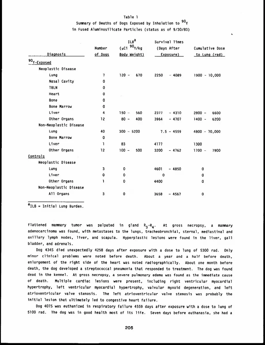
Table 1
Summary of Deaths of Dogs Exposed by Inhalation to 90yin Fused Alumtnosilicate Particles (status as of 9/30/83)
ILBa
Number (pCl 90y/kg
Diagnosis of Dogs Body Weight)
90y-Exposed
Neoplastic Disease
Lung 7 120 - 670Nasal Cavity 0
TBLN 0
Heart 0
Bone 0
Bone Marrow 0
Liver 4 150 - 560
Other Organs 12 80 - 400
Non-Neoplastlc Disease
Lung 40 300 - 5200
Bone Marrow 0
Liver 1 83
Other Organs 12 lO0 - 500
Controls
Neoplastic Disease
Lung 3 0
Liver 0 0
Other Organs 1 0
Non-Neoplastic Disease
All Organs 3 0
Survival Times
(Days After
Exposure)
Cumulative Dose
to Lung (rad)
aILB = Initial Lung Burden.
2250 - 4089 1900 - lO,O00
2377 - 4310 2800 - 66003964 - 4707 1400 - 6200
7.5 - 4559 4800 - 70,000
4177 1300
3200 - 4762 1700 - ?900
4601 - 4850 0
0 0
4400 0
3658 - 4567 0
flattened mammary tumor was palpated in gland R3-R 4. At gross necropsy, a mammaryadenocarcinoma was found, with metastases to the lungs, tracheobronchial, sternal, mediastinal and
axillary lymph nodes, liver, and scapula. Hyperplastic lesion~ were found in the liver, gall
bladder, and adrenals.
Dog 434S died unexpectedly 4258 days after exposure with a dose to lung of 5300 rad. Only
minor clinical problems were noted before death. About a year and a half before death,
enlargement of the right side of the heart was noted radiographically. About one month before
death, the dog developed a streptococcal pneumonia that responded to treatment. The dog was found
dead in the kennel. At gross necropsy, a severe pulmonary edema was found as the immediate cause
of death. Multiple cardiac lesions were present, including right ventricular myocardial
hypertrophy, left ventricular myocardial hypertrophy, valvular myxoid degeneration, and left
atrioventricular valve stenosis. The left atrioventricular valve stenosis was probably the
initial lesion that ultimately led to congestive heart failure.
Dog 407S was euthanized in respiratory failure 4559 days after exposure with a dose to lung of
5100 rad. The dog was in good health most of its life. Seven days before euthanasia, she had a
205
greatly increased respiratory rate of 136/min. Radiographs showed severe interstitial pneumonia.
The dog did not respond to treatment. At gross necropsy, severe pulmonary edema with interstitial
fibrosis was found. Moderate endocardiosis was found on the left and right atrioventricular
~valves.
Dog 372S died after laparotomy for an intussusception at 4707 days after exposure with a dose
to lung of 3600 rad. Ten days before death, the dog was examined for anorexia, vomiting, and
depression. Abdominal radiographs revealed an obstruction that was surgically removed. This
proved to be an adenocarcinoma of the jejunum. At gross necropsy, a severe acute peritonitis was
"~und. Numerous old age lesions were also found.
Dog 447B died 4310 days after inhalation exposure with a dose to lung of 2800 rad. A day
before death, the dog developed diarrhea, depression, and icterus. A clinical examination
suggested a severe hepatic disease with bile duct obstruction and renal failure. The dog died
before euthanasia could be performed. At gross necropsy, a perforated pyloric ulcer was found
with an associated severe peritonitis that was the immediate cause of death. Histologic
examination revealed massive infiltration of the liver with anaplastic mononuclear cells. Similar
cellular infiltrates were found in the kidneys, spleen, and adrenals. Lymph nodes and bone marrow
were unaffected. Morphologically, this was a histiocytic lymphosarcoma.
Dog 405U died unexpectedly 4762 days after exposure with a dose to lung of 1700 rad. Just
before death, the dog was being observed in the hospital because of a poor appetite. She
developed a severe bloody diarrhea and died that night. At gross necropsy, a severe hemorrhagic
enteritis was found. Numerous other aging lesions were found including adrenal cortical adenomas,
hepatic capsular fibrosis, nodular hyperplasia of the pancreas, and mammary tumors.
Dog 438B died 4250 days after exposure with a dose to lung of 1500 rad. Four days before
death, an abdominal mass was palpated, and bloody fluid was withdrawn from the abdominal cavity.
A splenic tumor was suspected. The dog died the next day, before surgery could be performed. At
gross necropsy, a massively enlarged spleen, about thirty times its normal weight, was found. The
spleen was 20 cm long and contained a firm red mass in the midportion that measured I0 x lO x
15 cm. Blood was found in the peritoneal cavity, apparently hemorrhage from the tumor.
Histologically, the splenic mass was a histiocytic lymphosarcoma.
Dog 378A, a control dog, was euthanized 4850 days after exposure because of lung masses and
persistent pleural effusion. At gross necropsy, a tumor mass filled the right apical lobe
extending it to 7 x 6 x 2 cm. The mass had invaded the mediastinum, parietal pleura, intercostal
muscles, and pericardium. It had metastasized to the sternal and mediastinal lymph nodes, and
kidneys. Histologically, the tumor was a papillary adenocarcinoma of the lung.
Dog 401A, a control dog, died 4680 days while under anesthesia for radiographs of a lung
mass. No major clinical problems were noted after exposure until about 9 months before death,
when a radiographic density was noted in the left apical lung lobe. A carcinoma was confirmed
with bronchial cytology. The dog was examined periodically by cytology and radiographs. At gross
necropsy, a 4 x 3 x 2 cm mass was found in the dorsal portion of the left apical lobe. The mass
replaced the parenchyma. Three small nodules were found in the left diaphragmatic lobe; left
ventricular myocardial hypertrophy and left atrial fibroelastosis were also found.
Dog 441B, a control dog, was euthanized 4400 days after exposure. About a month before death,
the dog had lost weight, and it became ataxic in the hind limbs. A bony proliferation was
palpated on the ventral surface of the sacrum. The mass grew and began to obstruct the rectum.
At gross necropsy, a very firm mass was found growing from the vertebral bodies of vertebra L4
to S3. The mass invaded adjacent muscles, but not the spinal cord. Histologically, this was awell differentiated osteosarcoma of periosteal origin. A complication was a prostatic carcinoma
206
that had metastasized to the osteosarcoma and proliferated in vascular and marrow spaces. In
addition, prostatic carcinoma metastases were found in the right popliteal, left axillary,
external iliac lymph nodes, and the lung. Other lesions were related to aging.
DISCUSSION
Radiation doses to the lungs of dogs that inhaled 90y in fused aluminosilicate particles
were delivered at an initially higher and more rapidly decreasing dose rate than those deliveredto the lungs of dogs that inhaled longer-lived beta emitters (91y, 144Ce’ and 90Sr) in fused
aluminosilicate particles. I’3 In all of these studies, dogs that received high initial lung
burdens died relatively early with acute pulmonary injury. Dogs that died from pulmonary injury
after inhalation of 90y in fused aluminosilicate particles died with lower total cumulative
doses to lung. It is probable, Judging from histopathologic findings in the lungs of dogs that
died at later times, that many of the survivors have some degree of pulmonary fibrosis. Three of
the bronchioloalveolar carcinomas observed were associated with pulmonary scars. It will be
important in the future to examine the degree of correlation of this tumor type with pulmonary
fibrosis.
An unusual finding in this study is the occurrence of lymphosarcomas of the liver. These
tumors were considered primary in the liver because of the massive involvement there and little
involvement elsewhere. The role of thoracic irradiation in the initiation of these tumors is
uncertain.
The next few years will provide more information on the relative importance of total radiation
dose and radiation dose rate for tumor incidence, type, and time of occurrence. Five different
pulmonary tumor types (adenocarcinoma, bronchioloalveolar carcinoma, adenosquamous carcinoma,
squamous cell carcinoma, and fibrosarcoma) have occurred in the seven dogs that died with
pulmonary neoplasia. The occurrence of three pulmonary carcinomas in control dogs indicates that
control populations will be very important in assessing the significance of lung tumors in aged
dogs.
REFERENCES
I. Hobbs, C. H., J. E. Barnes, R. O. McClellan, T. L. Chiffelle, R. K. Jones, D. L. Lundgren,J. L. Mauderly, O. A. Pickrell, and E. W. Rypka, Toxicity in the Dog of Inhaled 90y in FusedClay Particles: Early Biological Effects, Radiat. Res, 49: 430-460, 1972.
2. McClellan, R. 0., 3. E. Barnes, B. B. Boecker, T. L. Chiffelle, C. H. Hobbs, R. K. 3ones,3. L. Mauderly, J. A. Pickrell, and H. C. Redman, Toxicity of Beta-Emitting RadionuclidesInhaled in Fused Clay Particles - An Experimental Approach, in Morphology of Experimental~is (P. Nettesheim, M. G. Hanna, 3r. and O. W. Deatherage, Jr., eds.), AECSymposium Series 21 (CONF-700501), National Technical Information Service, Springfield, VA,pp. 395-415, Ig70.
3. Barnes,~J’oyE" R. O. McClellan, C. H. Hobbs, and G. M. Kanapilly, Toxicity in the Dog ofInhaled in Fused Clay Particles: Distribution, Retention Kinetics and Dosimetry,Radiat. Res. 49: 416-429, 1972.
207
TOXICITY OF gly INHALED IN A RELATIVELY INSOLUBLE FORM BY BEAGLE DOBS. XIV
Abstract --Beagle dogs were exposed by Inhalatlon
to aerosols of 9Zy in fused alumlnoslllcate partl-
cles, resulting In inltlal lung burdens ranging
from ii to 300 pcl 9ly/kg body weight. These 96
dogs, plus 12 controls, were malntalned for l~fe-
span observation or assigned to a sacrlflce sched-
ule to study radlat~on dose patterns. Dogs wlth
higher lung burdens died at early times wlth radl-
PRINCIPAL INVRSTIGATORS
F. P. Hahn
B. A. Muggenburg
C. H. SOhbS
R. O. McClellan
J. A. Plckrell
atlon pneumonltls. At later tlmes, tumors were seen in lung, nasal cavity, heart, and
tracheobronchlal lymph nodes; these organs recelved the highest radlatlon doses. Observations are
continuing on the 16 exposed and 6 control dogs that remain alive 12 to 13.5 years after exposure.
Yttrlum-gl, an important contributor to the total fission product radionuclide inventory of a
reactor after sustained operation, is an energetic beta emitter with a physical half-life of 59
days. It is one of four radioisotopes being used in a series of studies designed to determine
dose-response relationships in Beagle dogs after inhalation of beta-emltting radionuclides having
different physical half-lives. Comparison of the results from this study and those from thestudies with 90y, 144Ce’ and 90Sr in fused aluminosilicate particles will allow assessment
of the relationship between total radiation dose, dose rate, and the biological consequences of
chronic irradiation of the lung.
A radiation dose pattern study in which 30 dogs were sacrificed at intervals from 0 to 320
days after inhalation exposure to 91y fused aluminosilicate particles has been completed. The
dose-response study is in progress in which 96 dogs were exposed to gIy in fused aluminosilicate
particles to achieve graded levels of initial lung burdens, and 12 dogs were exposed to stable
yttrium in fused aluminosilicate particles to serve as controls. The experimental design for the
dose-response study is illustrated in Figure I.
Detailed descriptions of the experimental procedures, the results of the dose pattern study,
and early biological effects were reported in the Ig6g-70 Annual Report (LF-43, pp. 163-182).
This paper briefly summarizes previous findings and updates the results of the dose-response study.
STATUS
As of September 30, 1983, 80 dogs with initial lung burdens of 18 to 360 wCi 91y/kg body
weight and cumulative doses to lung at death of 2400 to 60,000 rad had died or were euthanized at
ll3 to 4810 days after inhalation exposure. Six control dogs died at times from 3029 days to 4779
days after exposure. A summary of the pathologic diagnoses, doses to lung, and survival times is
shown in Table I. Additional data for individual dogs are presented in Appendix A. Detailed
discussions of the clinicopathological findings on the dogs that died in earlier years have been
presented in previous annual reports. Forty of these dogs, with cumulative radiation doses to the
lung between B300 and 60,000 tad, died with radiation pneumonitis and/or pulmonary fibrosis at ll3
to loll days after inhalation exposure. Twenty-eight dogs that died between Ill5 and 4713 days
after exposure had cumulative radiation doses to lung ranging from 3100 to 25,000 rad and
developed pulmonary carcinomas. Two of these dogs also had a mediastinal hemangiosarcoma, which
208
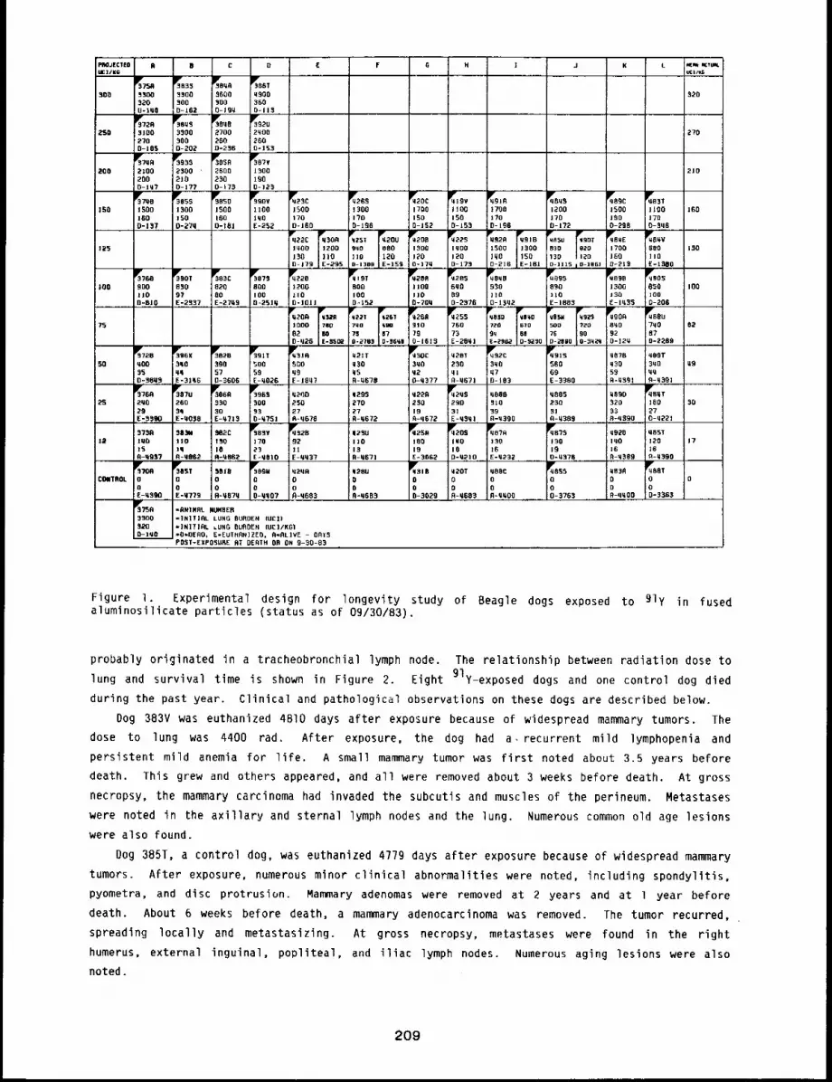
E F 0C O J K L
~3750B30O32Oo-t~O
35oo300D-162
sPro~9360930O0-190
~386T0900359O-ll$,,,
SO0 320
~009
27092009-236
~2U2YO02090-133
V~72931092700-105
Y30u533003000-202
2S9 270
~033
2309 "2100-177
Iv305026002300-~73
~’387V13001900-123
~709
2100290D-iq7
20O 219
] 3001700-190
~510
1700!17o
~05513001500-270
~050
1500Z600-18|
1109lqO[-~52
~23C
1500179D.,100
~20C
1700IS00-152
~19VJlOO1500-153
qPvBq$12001700-172
D-]]15 I D-]BK]
Y089C15001300-208p---~B~E17001300~213
~Y~e3Tl 1091700-306
80011oE-t~IIO
~’370D159o1600-137
ISO 160
130
~-198
0~20
~BIB;1500 I~O0
500-210 ~E-I81
~22C
~309
iqO0 1200130 ||00-,i79 -295
~15; ~20U
gqO 88011o 120O-13OB -159
~200
13001200-170
q~223
1o001200-173
123
~760
000I10O-BIO
~90T
03097E-2337
~3c820
100I E,,27~ 9
r3873800I000,2510
qP’Z201200110D-lOll
~qt9TB00I000,152
~30
1100I100-700
~2856q0090-2376
~800
:]10o-13q2
~9589o110C-1883
qB301300]30
~90S
850]oo0-206
IIO0 I00
92O-12tt
~~U709
070-2289
02991000
82D-q2G
~32A760
~02217NO750-2703
3261q~6?0-|600
Yq260910
790-16l$
~5576073E-28q]
~030
~BqO,7z0 57000 00[-296~’ -3250
092372090
0-~890 -Sq;~q
0275
3~8~’8399570-3606
qY[30C3q0q20-0377
q20T
23001R-q671
q~192cJ3qO!070-103
56969E-3380
~72B090350-30U~
3Y’361(3009qE-31q6
~391T50059E-0026
~31fl500q9E-ION7
q21Tq~O050-~678
q370~30
59
qe9T3No u95o
20029
~0702603qE:~qSa.
~069
33030E-~?,13
300030-0751
qV2q529031E-¼3ql,
~qB~,T180270-q221
0200250279-0670
0293270279-0672
02292~019~-0672
0809
39R-N390
080523031fl-q38~
0890320
8-0390
23 30
a~83v
179
E-qOlO
~289211E-qq3~
q~’2~A
18019E-3062
q~20SlUO180-¼210
~o7013016E-q232
qPrs?51o0190-03~6
085T12016R-q390
B759IUO159-q957
38~4110Iq9-08~2
302C13010A-q862
~23U110
9-0671
0920lqO169-3389
12 17
~88T000-3.563
00£-03~0
~ST00E-0779
~09H
00O-qq07
q~31B
000-3029
~55000-3763
qB3R00A-qqOO
3OrBo09-0870
q2qR0oA-0663
q28u009-0603
q20T00~-0693
086C00A-qqO0
COMTROL 0
3300320O-lqO
"RNIMRL NI.N4BER"]NIT]~L LUNG OUROEN lOCI)*]NITIRL ~UflO OUAOEN (OC)/KGI"0=0100~ E’EUTHP~IZEO, 9~flL]VE - OAT5POST-EXPOSURE AT OE~TH OR ON 9-30-83
Figure 1. Experimental design for longevity study of Beagle dogs exposed to 91y in fusedalumfnosilicate particles (status as of 09/30/83).
probably originated in a tracheobronchial lymph node. The relationship between radiation dose tolung and survival time is shown in Figure 2. Eight 91y-exposed dogs and one control dog died
during the past year. Clinical and pathological observations on these dogs are described below.
Dog 383V was euthanized 4810 days after exposure because of widespread mammary tumors. Thedose to lung was 4400 rad. After exposure, the dog had a. recurrent mild ]ymphopenia and
persistent mild anemia for life. A small mammary tumor was first noted about 3.5 years before
death. This grew and others appeared, and all were removed about 3 weeks before death. At gross
necropsy, the mammary carcinoma had invaded the subcutis and muscles of the perineum. Metastases
were noted in ~he axillary and sternal lymph nodes and the lung. Numerous common old age lesions
were also found.
Dog 385T, a control dog, was euthanized 4779 days after exposure because of widespread mammarytumors. After exposure, numerous minor clinical abnormalities were noted, including spondylitis,
pyometra, and disc protrusion. Mammary adenomas were removed at 2 years and at 1 year before
death. About 6 weeks before death, a mammary adenocarcinoma was removed. The tumor recurred,
spreading locally and metastasizlng. At gross necropsy, metastases were found in the right
humerus, external inguinal, popliteal, and iliac lymph nodes. Numerous aging lesions were also
noted.
209
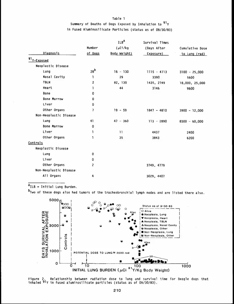
Table 1
Summary of Deaths of Dogs Exposed by Inhalation to 91yin Fused Aluminosilicate Particles (status as of 0g/30/83)
Dlagnosis
91y-Exposed
Neoplastic Disease
Lung 2Bb
Nasal Cavity l
TBLN 2
Heart l
Bone 0
Bone Marrow 0
Liver 0
Other Organs 7
Non-Neoplastic Disease
Lung 41
Bone Marrow 0
Liver l
Other Organs l
Controls
Neoplastic Disease
Lung 0
Liver 0
Other Organs 2
Non-Neoplastic Disease
All Organs 4
ILBa Survival Times
Number (~Ci/kg (Days After Cumulative Dose
of Dogs ~ Exposure) to Lunq (rad~...
16 - 130 Ill5 - 4713 3100 - 25,000
29 3390 5600
82, 130 1435, 2749 18,000, 25,000
44 3146 9600
19 - 59 1847 - 4810 3900 - 12,000
47 - 360 I13 - 2890 8300 - 60,000
II 4437 2400
35 3843 6200
3749, 4779
3029, 4407
aILB= Initial Lung Burden.
bTwo of these dogs also had tumors of the tracheobronchial lymph nodes and are listed there also.
5000
3000
1000
5~oooII
~a°°~ j
[]
¢.1
OO O ~/C)¯OO O OO Status as of 9-30-83
o.r s° I °.eo ,oola...,og I¯ ¯ ¯ J V Neoplasle. Heart J
At J & Nooplasia, TBLN I
Neoplasia, Other J
Non-Neoplasia, Other jAlso
/ ¯POTENTIAL DOSE TO LUNG~ 2000 red
0 I ~ ,(, I ...... ii0 10 100 1000
INITIAL LUNG BURDEN (~Gi 91y/Kg Body Weight)
Ij35
Figure 2. Relationship between radiation dose to lung and survival time for Beagle dogs thatinhaled 91y in fused aluminosilicate particles (status as of 09/30/83).
210
Dog 386A was euthanized 4713 days after exposure to a dose to lung of 6200 rad. About 6 weeks
before death, lameness and swelling of the tarsal and carpal Joints was noted. A 6 cm diameter
mass was noted radtographtcally in the left diaphragmatic lobe. In addition, pertosteal new bone
proliferation was noted around the carpals, tarsals, metacarpals, and metatarsals. These were
typical of hypertrophic osteopathy. During the next few weeks, the lung tumor enlarged and
lameness became much worse. At gross necropsy, a soft yellow mass occupied much of the anterior
portion of the left diaphragmatic lobe. Metastases were noted in the tracheobronchia],
mediasttnal and sternal lymph nodes, mediastlnum, pericardium, and diaphragm.
Dog 396S died in cardlopulmonary arrest during anesthesia 4751 days after exposure. The dose
to lung was 7000 cad. The dog had numerous minor clinical problems after exposure, including
salivary cyst, epidermal cyst, mammary adenomas, and pyometra. A mammary adenocarcinoma was
removed about a year before death. At gross necropsy, the lungs were atelectatic, and a 1.8 cm
diameter papillary adenocarclnoma was found in the right diaphragmatic lung lobe. Chronic p~ssive
congestion of the liver was also present and may have predisposed the animal to an anesthetic
death.
Dog 4245 was euthanized 4341 days after exposure to a dose to lung of 6200 rad. After
exposure, the dog had numerous minor clinical problems. About 2 months before death, an
ulcerative oral tumor was noted that proved to be a malignant melanoma. The tumor was
inoperable. Multiple tumors were found at gross necropsy. The melanoma had eroded into bone but
had not metastasized. An adrenal cortical carcinoma was found that had metastaslzed to theliver. Two small squamous cell carcinomas were found in the lung. Thus, three primary malignant
tumors were found in this dog, but the lung tumors were incidental findings.
Dog 430C died unexpectedly 4377 days after exposure, The dose to lung was 7900 rad. After
exposure, numerous minor intermittent problems were noted, such as bite wounds, ocular discharge,
scrota1 dermatitis, enlarged prostate, and enlarged left testicle. Lymphopenia was first noted 7
years after exposure. One day before death, the animal was examined and treated for dehydration,
anorexia, and diarrhea. On the morning of death, the dog was weak but alert. At gross necropsy,
a carcinoma of the colon was found. It had eroded a 1 x 2 cm focus of the mucosa and had invadedthe wall of the colon. No metastases were found. The immediate cause of death was an aspiration
pneumonia.
Dog 432B was euthanized 4437 days after exposure. Dose to lung was 2400 rad. No significant
clinical problems were noted until three days before death, when the dog was noted as icteric.
Clinical chemistry indicated high total bilirubin and liver enzymes in the serum consistent with
obstructive Jaundice. Exploratory laparotomy revealed adhesions of the quadrate lobe to the
diaphragm. At gross necropsy, the liver was enlarged, firm, and greenish brown. The gall bladder
was obscured by fibrosis, apparently as a result of earlier rupture. The common bile duct
contained a soft yellow-brown gall stone. The appearance of the liver was one of chronic
cholangiohepatitis.
Dog 484T died 4221 days after exposure to a lung dose of 4800 rad. After exposure, no
significant clinical findings were noted until 2 weeks before death, when the dog presented with
swollen carpal and tarsal joints and depression. A large lung tumor was seen radiographically,
and a diagnosis of hypertrophic osteopathy was made. Two days before death, pneumonia developed.
At gross necropsy, a large tumor was found in the right diaphragmatic lobe. It had invaded the
tracheobronchlal lymph nodes, causing constriction of the right mainstem bronchus. Metastases
were noted in the sternal and mediastinal lymph node.
Dog 487A was euthanized in dyspnea 4232 days after exposure. Dose to lung was 3100 rad.
Numerous minor clinical problems were noted before the terminal episode. About six weeks before
death, a pleural effusion and consolidation of the cardiac lobes were present. The dog had
211
pneumonia, which was treated with antibiotics. Numerous anaplastic epithelial cells existed in
bronchial lavages, prompting a diagnosis of pulmonary carcinoma and a recommendation for
euthanasia. At gross necropsy, a tumor mass was found completely filling the left lung.
Metastases were noted in the tracheobronchial, mediastinal, and sternal lymph nodes. Pleural
effusion was also present.
Dog 487S was euthanized 43B6 days after exposure to a dose to lung of 3800 rad. After
exposure, the dog had several epileptic seizures and was treated with phenobarbital daily. About
3 years before death, a leiomyoma was surgically removed from the urinary bladder wall. Eleven
days before death, the dog developed ascites, which was treated. Three days before death, this
returned and a large tumor was noted radiographically. At gross necropsy, a large mass 5.1 x 5.8
x 6.2 cm was found in the right intermediate lung lobe. Metastasis was not noted. The pressure
of the tumor mass on the posterior vena cava may have been responsible for the ascites.
DISCUSSION
A preponderance of pulmonary carcinomas has been observed in this study, compared to those in
which dogs were exposed to either of the longer lived radionuclides, 144Ce or 90Sr, in which
there has been a preponderance of hemangiosarcomas. The effect appears to depend largely on the
total radiation dose to lung, with higher doses producing more hemangiosarcomas. In this study,
no dogs have developed pulmonary hemangiosarcomas. However, no dogs have survived longer than 2
years with cumulative radiation doses to lung > 25,000 rad. In studies with dogs exposed to
144Ce or gOsr, few dogs with cumulative lung doses as low as 25,000 rad developed
hemangiosarcomas. Most of the dogs with pulmonary hemangiosarcomas had doses > 40,000 rad.
Another interesting comparison is the relative lack of hemangiosarcomas in the
tracheobronchial lymph nodes and heart. These findlngs are consistent with the hypothesis that
large, protracted radiation doses are generally required to produce hemangiosarcomas with
beta-emitting radionuclides. The relatively short half-llfe of 91y does not allow enough time
for significant amounts of radioactive material to translocate to the tracheobronchial lymph nodes
and deliver a significant radiation dose.
212
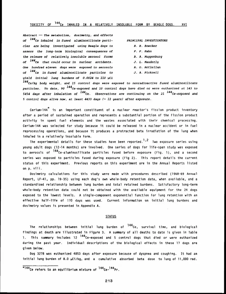
TOXICITY OF 144Ce INHALED IN A RELATIVELY INSOLUBLE FORM BY BEAGLE DOGS. xvI
Abstract --The metabollsm, doslmetry, and effects
of 144Ce inhaled in fused alumlnoslllcate parti-
cles are being investigated using Beagle dogs to
assess the long-term blologlcal consequences of
the release of relatlvely insoluble aerosol forms
of 144Ce that could occur In nuclear accidents.
One hundred eleven dogs were exposed to aerosols
of 144Ce in fused alumlnoslllcate particles to
9~eld in~tlal lung burdens of 0.0024 to 210 ~Cl
PRINCIPAL INVESTIGATORS
B. B. Boecker
F. F. Hahn
B. A. Muggenburg
J. L. Mauderl9
R. O. McClellan
J. A. P1ckrell
144Ce/kg body weight, and 15 control dogs were exposed Co nonradloactlve fused alumlnosllicate
partlcles. To date, 90 144ce-exposed and 10 control dogs have dled or were euthanlzed at 143 to
5454 days after inhalation of 144Ce. Observations are contlnu~ng on the 21 144Ce-exposed and
5 control dogs alive now, at least 4433 days (~ 12 years) after exposure.
Cerium-144 is an important constituent of a nuclear reactor’s fission product inventory
after a period of sustained operation and represents a substantial portion of the fission product
activity in spent fuel elements and the wastes associated with their chemical processing.
Cerium-144 was selected for study because it could be released in a nuclear accident or in fuel
reprocessing operations, and because it produces a protracted beta irradiation of the lung when
inhaled in a relatively insoluble form.
The experimental details for these studies have been reported. 1’2 Two exposure series using
young adult dogs (12-14 months) are involved. One series of dogs for life-span study was exposed
to aerosols of 144Ce-aluminosllicate particles fused before exposure (Fig. l), and a second
series was exposed to particles fused during exposure (Fig 2). This report details the current
status of this experiment. Previous reports on this experiment are in the Annual Reports listed
on p. viii.
Dosimetry calculations for this study were made with procedures described (1968-69 Annual
Report, LF-41, pp. 19-35) using each dog’s own whole-body retention data, when available, and
standardized relationship between lung burden and total retained burdens. Satisfactory long-term
whole-body retention data could not be obtained with the available equipment for the 24 dogs
exposed to the lowest levels. A slngle-component exponential function for lung retention with an
effective half-life of 170 days was used. Current information on initial lung burdens and
dosimetry values is presented in Appendix A.
STATUS
The relationships between initial lung burden of 144Ce, survival time, and biological
findings at death are illustrated in Figure 3. A summary of all deaths to date is given in Table
1. This summary includes 12 ]44Ce-exposed and 5 control dogs that died or were euthanized
during the past year. Individual descriptions of the biological effects in these 17 dogs are
given below.
Dog 327B was euthanized 4853 days after exposure because of dyspnea and coughing. It had an
initial lung burden of 8.0 ,Ci/kg, and a cumulative absorbed beta dose to lung of ll,O00 rad.
*144Ce refers to an equilibrium mixture of 144Ce-144pr.
213
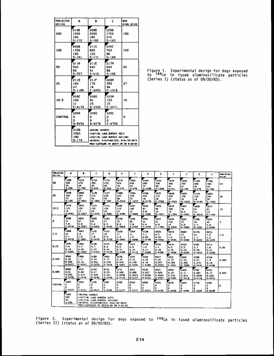
PROJECTEDUC|/KG
R B C
882000leo0-182
T::’BA1700210D-lq3
ISO01900-173
200 190
981700190D*181
TIIG
8901200-171
~26C
7~096D-18g
I00 11~0
T1?R
60068o-186
5oFigure I. Experimental design for dogs exposedto 144Ce in fused aluminosilicate particles
(Series I) (status as of 09/30/83).T110
J9027D-]185
T1~F170IgE-3::’SO
2725
TOBD
9115D~2396
12.5
~2090
000-Y578
CONTROL 0
TIOB
1500190D-173
e’RO~¢I[U R B C 0 E F G H ] J K L ~ K1mL
50 590 M?B 380 q?O S2O YMO 200 230 MOO ~30 qBO ~60 M765 N6 35 06 56 53 32 ~9 QJ ~1 Y~ 5~¯ 0-2~q E-?SR 0-916 B-2ye 0-273 0-279 B-l~2B D-|220 O-~ll 0-155 0-275 E-790%. .0o g,. ~o, ~3o°%0. -.~, T., ~.0 ~o0~g,,R %o~25 320 O 210 230 220 220 220 llO 2SO 190 lSO 2OR i26~3 25 2q ~6 5q ~7 19 10 27 ~ 19 2YD-195 E-~OOI D-]095 E-?G5 0-2313 E-IO?? 0-2179 E-2720 fl-1525 O-iOlO E-2185 E-)253;978 ~Oq5 Ilrs,, B ;,5U T26C Pv328, %E qP’rs’~ ~67R qlFBO, qP’OOB ~67112.5 llO 120 130 150 !)lO ]qO 87 gs 1160 ?O 9J BI l~12 l? lq 18 ;~2 |$ 9.8 10 :I~ 9.5 ii 130-250! 0-2~27 E-IgTQ 0-3901 :E-~280 E-$005 E-2505 0-1527 D-170~ E-350! E-$919 O-JTy9to,, 30. %° ;oR ~,~ %,, ,%° ~55,.~o,R",0,, ~?B.T,o~6 51 67 55 57 72 OR 51 51 J30 37 5q qZ ??,6 9.8 5,Y 6.3 8,0 ?.B 5.1 q.9 12 5.0 ~.7 5.8E=2570 fl-5~G? O-QBY3 E-q205 E-YB~3 0-2)07 E-2755 E-1795 D-2RY9.,. 0=3955 0-250~ E-q39~~980 %,T ~V5|,C ~J3S %JR %,V ;536 q520 ~61B ,66, ,??R YGTU"1.2 I~l 12 17 tg II |! S.l Q.2 13 15 12 ?.5 I,q~.~ 1,6 i.B 2.u 1.3 1.5 0.63 0.52 1,~ 2.0 1.1 1,2o-qqIO E-$Bq] 0-2q79 B-~?Ug 0-2950 E-qGgB E-~199 R-9552 E-SqOq 9-M~OZ 0-qM33 R-qUa5
TR?fl %6U T130 ,l~S T6,9 ,835 q?,C ,?IS~2gc "’%2V ;570 q53U0.23 2.o 3.7 3.7 ~.3 5.9 2,0 J.q Ii.l q.3 5.5 2,7 1o0 0.95O. IB O. MW 0.37 B.~5 0.71 0.52 O.|7 I0. J0 g.~5 0.5~ B.$O 0.25E-~6B5 0-2882 E:M553. n-52~9 E-~266 0-5113 E-q~BGETY592 E-$69q B-Yq32 A-qq$3 B-qq33
O.O~5 0,50 1.2 0.59 :0.77 O. JY 0.$3 0.20 0.~5 O.?S 0.50 0,67 I.~ 0.063O.GqM 0,12 0.05] IU.076 0,015 0.039 0.10 0.057 0,003 O.OB~ 0.079 O,l~R-5~57 E-Q337 R-5229 E-5BIB 0-~087 B-~oo~ E-q390 R-qSSg E-59ql D-3368 R-q~3~ R-YqSq
0.005 0,31 0.08 0,27 0,$~ O.OgO 0,003 0.02? 0.0~ 0.20 O.l) o,OOl o. 17 0.0220.03] O,O?? 0.025 O.O~l 0.0096 O. OOB~ 0.0030 O.O02q 0.010 O.OIq 0.009~ 0.0~0R-5~R E-3578 R-5~30 R~230 0-5113 R-5177 R-~553 R-M55~ R-~qBY O-YlSO D-2Bl~~oBO .....
E-q2lR~’~O5V ~]OB i~OOT 3~qB 3220 qSOR ~5~$ ~67B ~P~rBqu ~770 ~?RTCONTROL 0 0 0 B O O 0 O 0 O 0 0 0
o o o o o o o o o o o o0-5~,~, 0-q151 O-UOl~::O-~lRq A-SI?~..,R-5I?? =A-Y557 DZ~lR5 E-Y~96 EFq~Rq R-qq$8 R-qq~e
r1~529~5 -9NIN~L NUHBER500 -INItIRL LUNG BUROEN IUC|)0i05 =INITIAL LUNG BURDEN (UE]/KB)
=O=DERD. [-EUTHRNIZED. R=RLIVE-OBT5POST-EXPOSURE AT BERTH OR ON 9-30-85
Figure 2. Experimental design for dogs exposed to 144Ce in fused aluminosilicate particles
(Series II) (status as of 09/30/83).
214
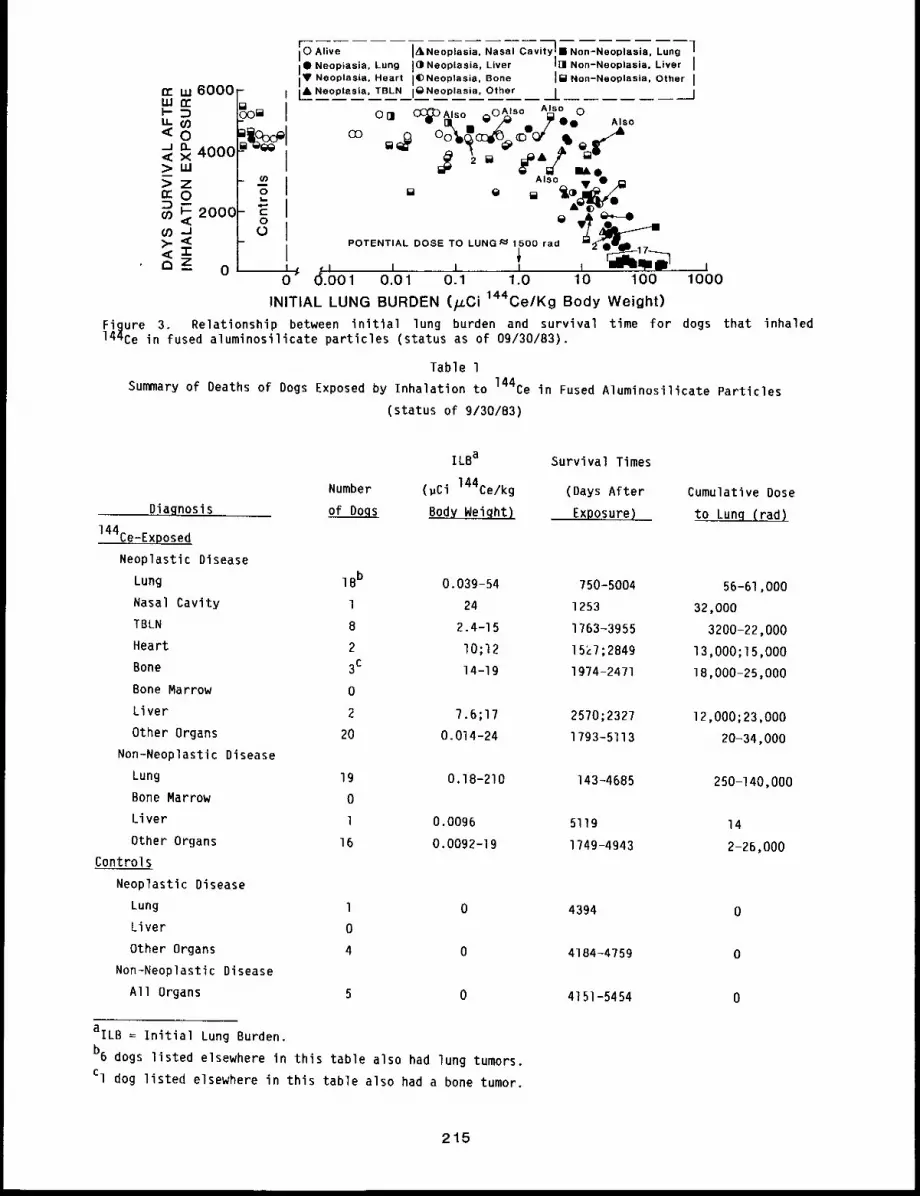
n- Lu 6000uJ t~
u-oo<0--J o-< X 4000~>LU
zCOO
o) ~ 200C(/) >. ,<<-1-c~Z_ 0
- I
I(/)
I
I
0 AliveI ¯Ne°piasia’ Lung ]~Neoplasia, LiverIV Neoplasla. Heart JC)Neoplasia, Bone[ANeoplasia, TBLN iQNeoplasia, Other
hll Non-Neoplasia. Liver J
Jl~ Non-Neoplasia, Other J
JCC~ Also ~,oA~ Is° Also 0O01 qll, []I -- v /~ _ ~’¯¯ Also
iAlso ¯ -0 m
AIL~
POTENTIAL DOSE TO LUNG~ 1500 rad
6.001 0.01 0.1 1.0 10 100 1000
INITIAL LUNG BURDEN (/.LCi144Ce/Kg Body Weight)
Figure 3 Relationship between initial lung burden and survival time for dogs that inhaled144Ce in fused aluminosilicate particles (status as of 09/30/83).
Table l
Summary of Deaths of Dogs Exposed by Inhalation to 144Ce in Fused Aluminosilicate Particles
(status of 9/30/B3)
Number
Diagnosis of Dogs
144Ce-Ex~osed
Neoplastic Disease
Lung 18b
Nasal Cavity 1
TBLN 8
Heart 2
Bone 3c
Bone Marrow 0
Liver 2
Other Organs 20
Non-Neoplastic Disease
Lung 19
Bone Marrow 0
Liver l
Other Organs 16
Controls
Neoplastic Disease
Lung 1
Liver 0
Other Organs 4
Non-Neoplastic Disease
All Organs 5
ILBa Survival Times
(~Ci 144Ce/kg (Days After Cumulative Dose
Exposure) to Lunq (rad)
0.039-54 750-5004 56-61,000
24 1253 32,000
2.4-15 1763-3955 3200-22,000
I0;12 15~7;2849 13,000;15,000
14-19 1974-2471 1B,000-25,000
7.6;17 2570;2327 12,000;23,000
0.014-24 1793-5113 20-34,000
0.18-210 143-4685 250-140,000
0.0096 5119 14
0.0092-19 1749-4943 2-26,000
0 4394 0
0 4184-4759 0
0 4151-5454 0
aILB = Initial Lung Burden.
b6 dogs listed elsewhere in this table also had lung tumors.
Cl dog listed elsewhere in this table also had a bone tumor.
215
Several minor clinical problems were noted before a 4-cm diameter tumor mass was seen
radiographically in the left diaphragmatic lung lobe 13 years after exposure and about 14 months
before death. At gross necropsy, a large tumor was found in the left diaphragmatic lobe that had
mestastasized to other lung lobes and to the local lymph nodes. In addition, a carotid body tumor
and numerous aging lesions were found.
Dog 46gs had an initial lung burden of 5.8 ~Ci/kg weight, with a cumulative absorbed beta
dose of 8600 rad to the lung. It was euthanized 4394 days after exposure. The dog had been
treated for renal failure for several weeks. Despite treatment, the BUN and creatlnine remained
markedly elevated. Other clinical problems include a melanoma removed from the jaw about g months
before death, and a persistent anemia and hypoalbumenla existent for about 1.5 years. Gross
necropsy revealed a chronic interstitial nephritis and a moderate parathyroid hyperplasla, both
consistent with chronic renal failure, in addition, a small 1.6 cm diameter nodule was present in
the right intermediate lobe and appeared to be a primary lung tumor.
Dog 308B died 4943 days after an inhalation exposure that had produced an initial lung burden
of 5.4 ~Ci/kg body weight and a subsequent cumulative absorbed beta dose to lung of 7700 rad.
The dog had numerous minor clinical problems after exposure. Cardiac abnormalities were first
noted about 9.5 years after exposure when left and right ventricular enlargement were documented
radiographically. A grade III systolic heart murmur was noted a year later. Heart failure
developed about 13 years after exposure and was treated with digitoxin. The dog apparently died
of heart failure. At gross necropsy, a chronic suppurative pneumonia was found in the left apical
lobe of the lung. A 1.5 cm diameter tumor, which appeared to be a carcinoma, was found in the
right intermediate lobe. The ventricles of the heart were dilated and the myocardium degenerate.
The myocardial lesions probably predisposed the dog to the pneumonia. The lung tumor was an
incidental finding at necropsy.
Dog 453B was euthanized 4199 days after exposure because of metastatic thyroid tumors, its
initial lung burden of 0.63 gCi/kg body weight resulted in a cumulative absorbed beta dose to
lung of 910 rad. No major clinical problems were noted until the right thyroid was found
nonfunctional about four months before death. The thyroid was excised and diagnosed as a solid
carcinoma of the thyroid. Radiographs revealed that there was already metastasis to the lungs.
At gross necropsy, the left thyroid was found to be atrophic. Many arteries of the heart and
thyroids were mineralized. Many common aging lesions were found, including spondylitis,
esophageal lelomyomas, pancreatic adenoma, and testicular tumors.
Dog 322V died 5113 days after exposure. Its initial lung burden of 0.32 gCi/kg body weight
resulted in a cumulative absorbed beta dose to lung of 470 rad. Anemia of undetermined cause was
first noted about three years before death. Shortly before death, an abdominal mass was noted,
and an exploratory laparotomy was performed, revealing an enlarged spleen with multiple masses of
hemangiosarcoma. The dog died shortly after. At gross necropsy, tumor mestastases were found in
the liver, lungs, greater omentum, brain, and ribs. Numerous other incidental lesions were found.
Dog 453U had an initial lung burden of 0.18 gCi/kg body weight that produced a cumulative
absorbed beta dose to lung of 310 rad. It was euthanized 4392 days after exposure. The dog was
hospitalized shortly before euthanasia because of discovery on a routine physical examination of a
mass in the right axillary region. An adenocarcinoma of the mammary gland had been removed about
1 year earlier. Radiographs showed pulmonary tumors, presumably mestastatic sites. Recovery from
anesthesia was poor and the dog became recumbent. At gross necropsy, massive tumor metastases and
a subacute suppurative pneumonia were found in the lungs. Cardiac and renal degenerative lesions
were seen probably due to hypoxia and electrolyte imbalance.
Dog 457B was euthanized 4466 days after inhalation exposure. Its initial lung burden of 0.17
~Ci/kg body weight resulted in a cumulative absorbed beta dose to lung of 270 tad. No
216
significant clinical findings were noted until about 2 weeks before death, when the dog presented
with stertorous breathing. Bronchoscopic examination revealed no airway abnormalities below the
larynx. Soon after, the dog began circling to the right. It was treated with antibiotics for
otitis media because otitis externa was present. The severity of the circling increased, and the
dog was unresponsive to antibiotics. At gross necropsy, a pituitary tumor measuring 1.5 x 1.2 x
1.2 cm was found. The tumor compressed the hypothalmus and brainstem, causing malacia. Other
lesions included parathyroid hyperplasia, hepatocellular focal hyperplasia, cystic hyperplasia of
the gall bladder, testicular atrophy, and adrenal cortical hyperplasia.
Dog 323T had an initial lung burden of 0.039 ~Ci/kg body weight and a cumulative absorbed
beta dose to lung of 56 rad. It died 5004 days after exposure. Lung tumors were first diagnosed
in this dog about 13 years after exposure and about 9 months before death. From initial diagnosis
until death, there was little enlargement of the tumor. The only other major clinical problem was
a pyometra that was removed about a year before death. At gross necropsy, a tumor mass was found
enlarging the right apical lobe about two times normal size. It had apparently metastasized to
the tracheobronchial lymph nodes, but not beyond. No other significant lesions were found.
Dog 450D had an initial lung burden of 0.018 ~Ci/kg body weight and a cumulative absorbed
beta dose to lung of 26 rad. It was euthanized 4390 days after exposure because of tonsilar
carcinoma. No significant clinical findings were present until a pharyngeal mass was noted about
2 weeks before euthanasia. The mass grew rapidly, causing obstruction and difficulty in
swallowing. At gross necropsy, a tonsilar carcinoma was found in the left tonsil that
metastasized to the regional lymph nodes. No other significant lesions were found.
Dog 462S had an initial lung burden of 0.014 ~Ci/kg body weight and a cumulative absorbed
beta dose to lung of 20 rad. It was euthanized 4218 days after exposure. Only minor medical
problems were found in the dog until about 3 months before death, when it was found ataxic and
depressed. Cervical disc disease was diagnosed, based on clinical signs and a history of cervical
spondylitis. About 2 weeks before death, the dog began circling left. An electroencephalogram
showed a left parietal abnormality. The dog continued to deteriorate and was euthanized. At
gross necropsy, a large grey friable mass was found replacing the pituitary. It measured 3 x 3 x
l cm and compressed the gre,/ matter of the left .pyriform lobe. Other gross lesions noted included
spondylitis of C4_5 and TI2-L6, nodular hyperplasia of the liver, thyroid adenoma, and
cystic ovaries.
Dog 327C died 5119 days after an inhalation exposure that resulted in an initial lung burden
of 0.0096 ~Ci/kg body weight and a cumulative absorbed beta dose to lung of 14 rad. The dog had
numerous clinical problems, but all seemed minor. Shortly before death, a routine radiographic
survey revealed a left c~xofemoral luxation. At necropsy, no cause for death could be found.
There was severe passive congestion in the liver, so hepatic degeneration may have played a role
in the death. Among lesions found were adrenal tumor, subcutaneous abscess, lymph node
hyperplasia, splenic nodular hyperplasia, renal fibromas, testicular atrophy, and spondylitis.
Dog 4V8C died 4150 days after inhalation exposure. Its initial lung burden of 0.0092 ~Ci/kg
body weight resulted in a cumulative absorbed beta dose to lung of 13 rad. Before the terminal
episode, the only clinically significant disease noted was a recurrent hematuria related to
cystitis and prostatitis. This responded to antibiotic treatment. Five days before death, the
dog was thoroughly examined because of a 2.8-kg weight loss over the previous month, depression,
and a bloody stool. Hematologic examination indicated an anemia - neutrophilia and clinical
chemistry was consistent with acute renal failure. A tentative diagnosis was made of acute renal
failure caused by gastrointestinal disease or hemangiosarcoma. The dog was treated with high
levels of antibiotic, but died before a definitive diagnosis could be made. At necropsy, a
chronic enteritis and acute renal tubular necrosis were found. The bacterium Salmonella s~. was
isolated from the intestine.
217
Dog 208A, a control, died unexpectedly 5454 days after exposure. The dog had relatively few
clinical problems during its lifetime. A mild persistent anemia was noted about a year and a half
before death. The serum phosphorus and blood urea nltrogen levels were also slightly elevated,
indicating mtld renal disease. At gross necropsy, a severe polar fibrosis with atrophy was notedin the left kidney and a hypertrophy in the right. The parathyrold glands were hypertrophied,
indicating there was a renal insufficiency. Numerous other common aging lesions were found but
none were severe, except for spondylities involving most of the lumbar and thoracic vertebrae.
Dog 306D, a control, died 5233 days after exposure and after several months of treatment for
chronic malabsorption related to pancreatic failure. About g months before death, a medullary
carcinoma was removed from the right thyroid gland. At gross necropsy, an atrophic, fibrotic
pancreas was found. There was no evidence of recurrence of the thyroid medullary carcinoma.
Other lesions included hepatic lipidosis and polar renal cortical fibrosis.
Dog 464U, a control, was euthanized 4394 days after inhalation exposure. Few clinical
problems were noted until lung densities were noted about a year before death. The tumors slowly
progressed in size, eventually necessitating euthanasia. At gross necropsy, a tumor mass about 6
x 4 x 3.5 cm was present in the anterior portion of the left diaphragmatic lung lobe surrounding
the mainstem bronchus. Numerous other tumors, 0.5-2 cm diameter, were scattered through the lung
lobes. Metastases were present In the left and middle tracheobronchial lymph nodes.
Dog 467B, a control, was euthanized 4296 days after exposure. Numerous clinical problems had
been noted during the dog’s lifetime, including enlarged prostate, spondylitis, intermittent
lymphopenia, and testicular atrophy. In the terminal episode, the dog was found depressed with a
high respiratory rate 2 days before death. Generalized peripheral lymphadenopathy was present.
Radiographs showed a pleural effusion. Thorocentesis and imprints from a lymph node supported the
diagnosis of multlcentric lymphoblastic lymphosarcoma. At gross necropsy, all lymph nodes of the
body were found to be involved. In addition, the spleen was enlarged about ten times normal and
the femoral shaft marrow was reddened. These findings added support for the diagnosis.
Dog 452S, a control, died 4195 days after exposure. About one week before death, a mass was
palpated in the abdomen, and an exploratory laparotomy was performed. The spleen was enlarged and
adherent to the stomach. A metastatic adenocarcinoma was found in the spleen. At gross necropsy,
adenocarcinomas were found in the stomach and mammary glands.
DISCUSSION
The findings at death have become more dlverse as this experiment has progressed, Sixteen
dogs that died or were euthanized during the first 500 days after inhalation exposure had
radiation pneumonitis and pulmonary fibrosis. The next prominent necropsy finding in dogs that
survived the early mortality phase was pulmonary hemangiosarcoma, which occurred in eight dogs
that died or were euthanized between 750 and 1318 days after exposure. No additional pulmonary
hemangiosarcomas have been seen beyond that time. Pulmonary carcinomas, primarily
bronchioloalveolar carcinomas, first appeared in a dog that died 916 days after inhalation
exposure. They have continued to occur to the present time. During the past year, four more lung
tumors were observed, two of which were major findings at death and two which were incidental
findings.
Although no pulmonary hemangiosarcomas have been observed since 1318 days after" exposure,
hemangiosarcomas in tissues either within or external to the thorax have been frequent in dogs
that died or were euthanized between 1527 and 4266 days after exposure. 3 The occurrence of
hemangiosarcomas in the mediastinum and the possible origin in the tracheobronchial lymph nodes
were discussed in detail In the 197B-79 Annual Report, LF-69 (pp. 88-gi). Hemangiosarcomas have
218
also occurred in other organs such as bone, liver, or spleen. Only one hemanglosarcoma was
observed in the 17 dogs that died or were euthanlzed during the past year. This appeared to arise
from the spleen.
The findings at death and the survlval time are related to both the dose rate and totalabsorbed beta dose associated with each exposure level. 4 The final analysls of these results,
together with results from the studies of other radionuclides incorporated into fused
aluminostltcate particles, will provide critical information about the interactions of dose rate
and total dose for chronic irradiation from Internally deposited, beta-emittlng radlonuclides.
Observations are continuing on the 2l 144re-exposed and 5 control dogs allve at this time. The
youngest surviving dog is at 4433 days (~ 12 years), and the oldest is at 5118 days (14 years)
after exposure.
REFERENCES
I. Hahn, F. F., S. A. Benjamin, B. B. Boecker, T. L. Chiffelle, C. H. Hobbs, R. K. Jones, R. O.McClellan, J. A. Pickrell, and H. C. Redman, Primary Pulmonary Neoplasms in Beagle DogsExposed to Aerosols of.144Ce in Fused Clay Particles, J. Natl. Cancer Inst. 50: 675-69B,1973.
2. McClellan, R. 0., J. E. Barnes, B. B. Boecker, T. L. Chiffelle, C. H. Hobbs, R. K. Jones,J. L. Mauderly, J. A. Pickrell, and H. C. Redman, Toxicity of Beta-Emitting RadionuclidesInhaled in Fused Clay Particles - An Experimental Approach, in MorpholoQy of ExperimentalRespiratory Carclnoqenesis (P. Nettesheim, M. G. Hanna, Jr. and J. W. Deatherage, eds.), pp.395~415, USAEC Symposium Series 21 (CONF-?O0501), 1970.
3. Benjamin, S. A., F. F. Hahn, T. L. Chiffelle, B. B. Boecker, C. H. Hobbs, R. K. Jones, R. O.McClellan, and M. B. Snipes, Occurrence of Hemangiosarcomas in Beagles with InternallyDeposited Radlonuclldes, Cancer Res. 35: 1745-1755, 1975.
4. Hahn, F. F., B. B. Boecker, R. G. Cuddihy, C. H. Hobbs, R. O. McClellan, and M. B. Snipes,Influence of Radiation Dose Patterns on Lung Tumor Incidence in Dogs that Inhaled BetaEmitters: A Preliminary Report, Radiat. Res. (submitted).
219
TOXICITY OF 144Ce INHALED IN A RELATIVELY INSOLUBLE FORM
BY IMMATURE BEAGLE DOGS. XlI
Abstract -- Immature Beagle dogs (3 months old) were
exposed once by Jnhalatlon to an aerosol of 144Ce
Incorporated in fused alumlnos111cate particles,
The ~nfluence of thls age on the dose-response rela-
tlonshlps Is belng compared to that of 13-mo-old and
8 to 10.5-yr-old dogs. Thls study involves 49 dogs
that recelved graded Jnltlal lung burdens from 0.004
to 140 pC1 144ce/kg body weight and fJve control
PRINCIPAL INVESTIGATORS
B. B. Boecker
B. A. Muggenburg
F. F. Hahn
J. L. Mauderl9
R. O. McClellan
dogs. To date, 19 of the 144Ce-exposed dogs and one of the controls have dled. Dogs wJth the
highest in~tlal lung burdens of 144Ce dled durlng the first 4 months w1~h radlatlon pneumonltis,
pulmonar9 flbrosls, and congestlve heart fallure. Pulmonary hemanglosarcoma was the prlmary
flndlng in dogs that died at 1.5 to 2 years after exposure. Deaths beyond that time have been due
primarily to extrapulmonarg hemanglosarcomas. Observations are contlnu~ng on the surviving 30
144Ce-exposed and four control dogs at 7.0 ~o II.2 years after exposure.
It is important to study the influence of age on the disposition and long-term biological
effects of inhaled radionuclides because a population of people that might be exposed in a nuclear
incident could contain individuals differing widely in age. Three studies are in progress to
examine possible age-related differences. In all three studies, Beagle dogs have been exposed
once, by inhalation, to a relatively insoluble form of a beta-emitting radionuclide with a
relatively long physical half-life (144Ce,* Tp = 284 days). Fused aluminosilicate particles
were used as the aerosol matrix. Dogs in this study were exposed when they were 3 months old.
Companion studies involve dogs that were exposed either as young adults (12 to 14 months of age)
(this report, pp. 213 to 219), or as aged dogs (8 to 10.5 years of age) (this report, pp.
to 227).
The randomized block experimental design (Fig. l) of this study resembles that used for most
of the other dog longevity studies in progress at this Institute. Dogs were entered into the
study by blocks, each containing 9, lO, or II dogs of the same sex. Dogs in each block were
exposed to graded levels of 144Ce in a fused aluminosilicate aerosol, and control dogs were
exposed to a fused aluminosilicate aerosol containing only stable cerium. The projected initial
lung burden levels included the eight levels used in the companion study with dogs exposed when
they were 12 to 14 months old, and also two higher levels, 75 and lO0 ~Ci 144Ce/kg body
weight. These two higher levels were included to compensate for the more rapid decrease in dose
rate during the first year after exposure in immature dogs because of the increasing weight of the
growing lung compared with dose rates seen in dogs exposed as young adult or aged dogs. Previous
information regarding the exposure, metabolism, dosimetry, and biological effects has been given
in the Annual Reports listed on p. viii.
STATUS
The current status of this study is shown in the experimental design given schematically in
Figure 1. Exposure information and dosimetry values are given for all of these dogs in Appendix A.
*144Ce refers to an equilibrium mixture of 144Ce-144pr.
220
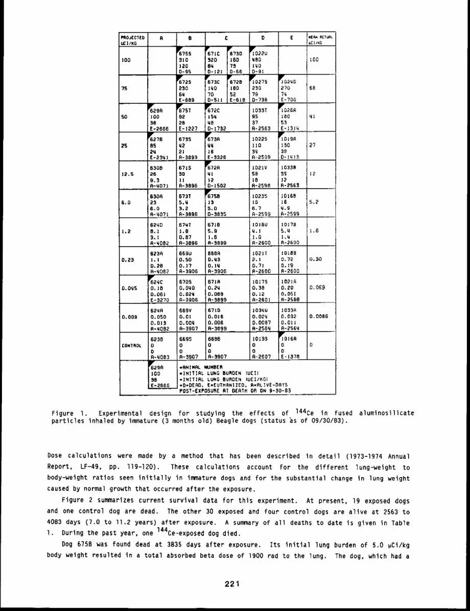
RPROJECTED=’;/KG
D EB C
~75931o
D-95
6"/80160780-66
TO2~G2707~E-700
262306~E-689
678Clq0700-51]
y672818052E-618
6876
~6629R]0o96E-2666
~?5T9228E-1227
1033T9537A-2563
5O
qq16E-3326
TolgA13038D-lq]8
~627B852qE-23~]
10223llO
8qA-2599
6785~22JR-3899
2755
6~72Rql120-J502
1083885itR-256~
671S901!R-8896
1021V5818R-2598
6806126,9.3R-qOT]
1212.9
~758
5.00-8885
|016816
A-2599
650628
IL°o, 673T5.q3.2A-3896
lO2~S166.7fl-2599
5.56,0
101785,z]I.qA-2600
67~T1.80.87R-3896
67~85,9|.6R-8899
IOIBUq.ll,OR=2600
62q08.19.19-q082
1,61,2
zOlB80,750.19R-2600
623Rl.i0.58R-q082
669U0.500.17R-8906
6689O.q3O. lqR-3906
z021I2.10.71R-2600
)0.23 0.30
y629CO. lO0.061E-3270
1021~0.200.05!R-2598
670SO. OqO0.02qR-8906
67190.2~0.089R-3899
JO]750,380.12R-260!
0.0£90.0~5
62qRO.OSO0.018R-qO82
108990.032O.OllR-256q
669V0.61O.OOqR-3907
87100.0160.006R-3699
lO3qu0.02q0.0087R-256q
0,00660.009
169ooE~ 1378
6688ooR-8907
1019500R-2607
669800R-3907
623800R-q063
0CONXFlOL
6Y6299ZOO86E-2666
-RN]NRL NUMBER-INIIIRL LUNG BURDEN (UCI)mIFlITIRL LUNG BURDEN (UC]/KG)=0=0E90, EmEUTNRN]ZEOo R=RL]VE-DRTSPOST-EXPO5URE RT DERTM Ok ON 9-30-83
Figure I. Experimental design for studying the effects of 144Ce in fused aluminosilicateparticles inhaled by immature (3 months old) Beagle dogs (status ~s of 09/30/B3).
Dose calculations were made by a method that has been described in detail (1973-1974 Annual
Report, LF-49, pp. I19-120). These calculations account for the different lung-weight tobody-weight ratios seen initially in immature dogs and for the substantial change in lung weightcaused by normal growth that occurred after the exposure.
Figure 2 summarizes current survival data for this experiment. At present, 19 exposed dogsand one control dog are dead. The other 30 exposed and four control dogs are alive at 2563 to
4083 days (7.0 to ll.2 years) after exposure. A summary of all deaths to date is given in TableI. During the past year, one 144Ce-exposed dog died.
Dog 675B was found dead at 3835 days after exposure. Its initial lung burden of 5.0 pCi/kgbody weight resulted in a total absorbed beta dose of 1900 rad to the lung. The dog, which had a
221
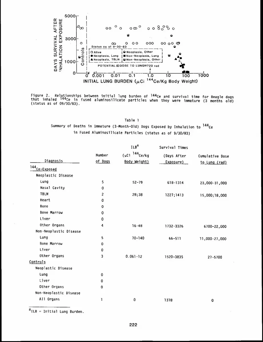
50001oo
3000o
1000
o o o°°o oQ
000 O0 o oQ
0 0O0 0 000
t~
oo 0 O0Status as of 9-30-83
l t l-6 I OAlive I Q Neoplasia, Other I QiI
~ l’Ne°p’as’~. Lung ," NoR-Ne°p’asi~. Lung ,=
A~
I A Neoplasia, TBLN j [] Non-Neoplasia, Other ] A
POTENTIALOODOSE TO LUNG~720 rad
__~ ~L I I I I 8 Io" (~.001 0.01 0.1 1.0 10 100INITIAL LUNG BURDEN (/xCi 144Ce/Kg Body Weight)
0 i1000
Figure 2. Relationships between initial lung burden of 144Ce and survival time for Beagle dogsthat inhaled 144ce in fused aluminosilicate particles when they were immature (3 months old)(status as of 09/30/83).
Table 1
Summary of Deaths in Immature (3-Month-Old) Dogs Exposed by Inhalation to 144Ce
in Fused Aluminosilicate Particles (status as of 9/30/83)
Diagnosis
144Ce-Exposed
Neoplastic Disease
Lung
Nasal Cavity
TBLN
Heart
Bone
Bone Marrow
Liver
Other Organs
Non-Neoplastic Disease
Lung
Bone Marrow
Liver
Other Organs
Controls
Neoplastic Disease
Lung
Liver
Other Organs
Non-Neoplastic Disease
All Organs
aILB = Initial Lung Burden.
ILBa Survival Times
Number (gCi 144Ce/kg (Days After
of Dogs Body Weight) Exposure)
5 52-79 618-1314
0
2 28;38 1227;1413
0
0
0
0
4 16-48 1732-3326
5 70-140 66-511
0
0
3 0.061-12 1520-3835
0
0
0
1 0 1378
Cumulative Dose
to Lung (rad)
23,000-31,000
15,000;18,000
6700-22,000
II,000-27,000
27-5700
0
222
history of pancreatic insufficiency for about 6 months before death, was treated with dietary
pancreatic enzymes, but he continued to lose weight. At necropsy, he was severely emaciated,
apparently because of malabsorption associated with the pancreatic insufficiency. Extreme edema
was found in the body cavities and in the areolar connective tissues in the body. The pancreas
was atrophic, as were the liver, spleen, and testes. A small (4 mm), yellowish nodule was found
under the pleura under the right cardiac lobe. The nature of this lesion awaits histologic
examination,
DISCUSSION
The results of this study to date agree qualitatively with results observed in dogs exposed to
a similar aerosol when they were young adults (12 to 14 months old). Several differences have
emerged, however. The first was development of congestive heart failure in three of four immature
dogs that died shortly after exposure. These dogs died with radiation pneumonitis and pulmonary
fibrosis. Congestive heart failure was not prominent in any of the dogs that died after being
exposed as young adults.
The second difference concerns pulmonary hemangiosarcomas. They occurred in immature dogs
earlier after the inhalation exposure (I.7 to 2,0 years) than in the young adult dogs (2.1 to
years). Also, the cumulative absorbed beta doses to lung at death in the immature dogs that had
pulmonary hemangiosarcomas (23,000 to 31,000 rad) were lower than in most young adult dogs with
pulmonary hemangiosarcomas (29,000 to 61,000 rad). Another difference was that bronchioloalveolar
carcinomas were also seen in the study using young adult dogs, a study that has been in progress
longer than this study with immature dogs. Finally, the one adenocarcinoma is the first pulmonary
carcinoma of any kind in this study. If more of these tumors appear in the immature dogs, it will
be interesting to compare the time and dose at occurrence with corresponding data in the study
with young adult dogs.
A similarity between the studies with immature and young adult dogs has been the predominance
of apparently extrapulmonary hemangiosarcomas at later times after exposure. The implications of
these findings remain to be determined. These results, comparing the long-term dose-response
patterns for dogs exposed at different ages, provide invaluable input to risk assessments for
human populations of mixed ages that might be exposed by inhalation to a radioactive aerosol
released in the course of a nuclear accident.
223
TOXICITY OF 144Ce INHALED IN A RELATIVELY INSOLUBLE FORM BY AGED BEAGLE DOGS. XII
Abstract -- The toxlclty of relatlvely ~nsoluble
144Ce inhaled by 8- to 10.5-year-old Beagle dogs
is being investigated to determine possible age-
related differences in long-term blologlcal re=
sponses. Forty-two dogs were exposed to aerosols
of 144Ce in fused alumlnosillcate particles to
yield inltlal lung burdens of 2.2 to 75 pCi
144Ce/kg body welght, and 12 control dogs were ex-
posed to non-radloactlve fused alumlnos111cate
PRINCIPAL INVESTIGATORS
B. B. Boecker
F. F. Hahn
B. A. Muggenburg
J. L. Mauderly
R. O. McClellan
J. A. Plckrell
particles. All 144Ce-exposed and control dogs have dled or were euthanlzed between 197 and 2726
days after the Inhalatlon exposure. Prominent findings in the 144Ce-exposed dogs were radla£1on
pneumonltls in 19 of the 23 dogs that died during the flrst 943 days after exposure, and
neoplastic disease in 13 of the 20 dogs that died beyond 904 days after exposure. Pulmonary
tumors were found in flve of ~hese dogs. In contrast to the study with young adult dogs, in whlch
pulmonary hemanglosarcomas were one of the prominent flndlngs, all of these tumors were carcinomas.
Long-term dose-response studies are being conducted in this laboratory to provide information
for predicting dose-response relationships in man. Most of these studies have used young adult
animals to represent the most physiologically fit individuals in a population. Thus,
dose-response prediction for man based on data obtained from exposure of young adults may
underestimate the risk of exposure for older members of the population. To obtain information
relevant to this problem, a longevity study is being conducted with 8- to lO.5-year-old Beagle
dogs exposed to 144Ce* in a relatively insoluble form, incorporated into fused aluminosilicate
particles. This study complements two others with 144Ce in the same form, one using dogs
exposed at 12 to 14 months of age (this report, pp. 213 to 219) and the other using dogs exposed
at approximately 3 months of age (this report, pp. 220 to 223). Previous reports on this study
have been presented in the Annual Reports listed on p. viii.
The procedures for exposure have been described (1971-72 Annual Report, LF-45, pp. 172+176).
Forty=two Beagle dogs exposed to 144Ce in fused aluminosilicate aerosols received initial lung
burdens ranging from 2.2 to 75 ~Ci 144Ce/kg body weight (Fig. l). Detailed clinical
evaluations were performed before exposing the dogs and throughout their life spans. Absorbed
beta doses to the lung have been calculated for each dog using the same method as for the
young adult dogs exposed to 144Ce in fused aluminisolicate particles (1970=71 Annual Report,
LF-44, p. ]69). These doses are based on a lung weight (including blood) to body weight ratio
O.Oll. Using a fixed lung-weight-to-body-weight ratio derived from young adult dogs for older
dogs may not be accurate. Consequently, when the lung burden is expressed as ,Ci 144Ce/kg
body weight, some of the implied lung weights may be too large because some of the older dogs were
obese at the time of exposure. Corresponding dose calculations based on lung weight will then be
underestimated by the same amount.
*144Ce refers to an equilibrium mixture of 144Ce-144pr.
224
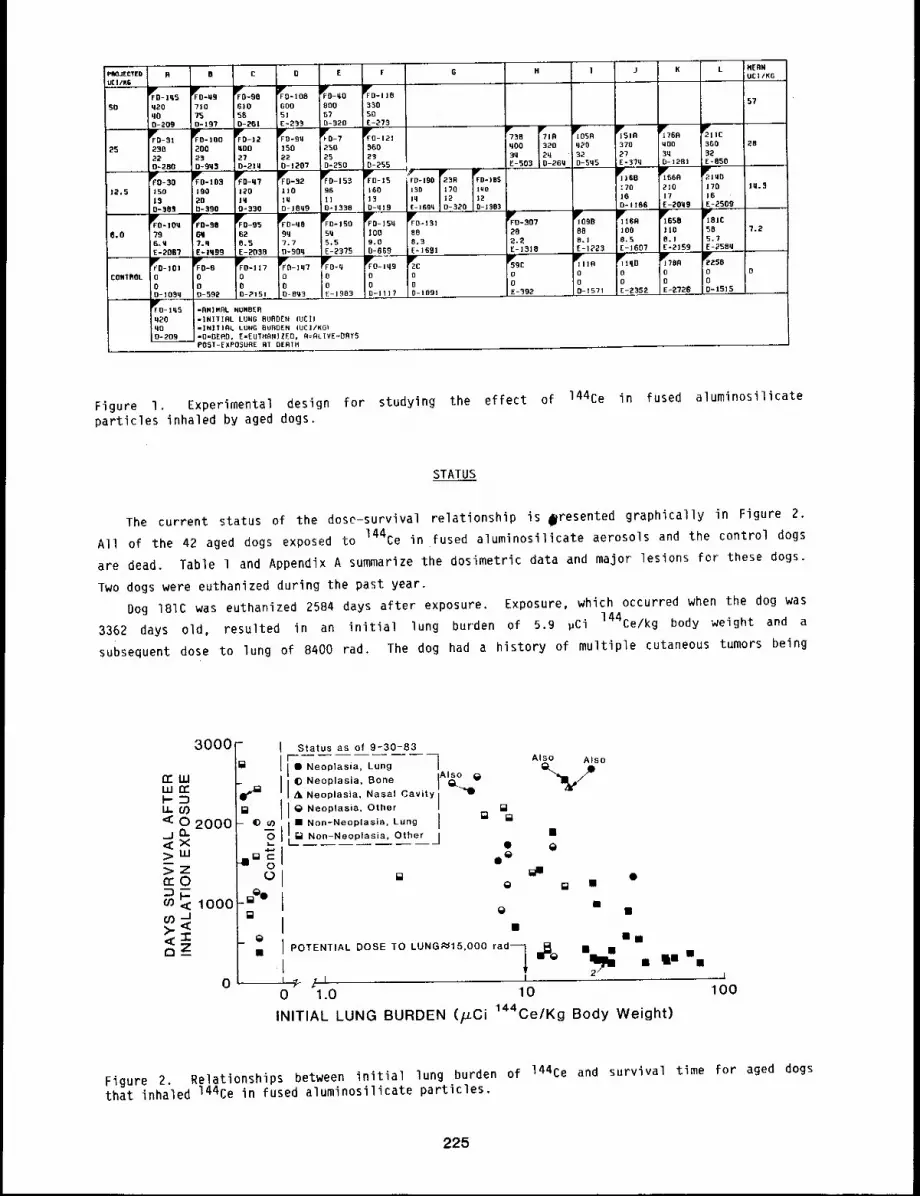
,1::’. 5
8,0
] j K L IH[RNUCI/5G
57
25IWJ os° ~51° ~765 ~1 !cq~O 370 MOO 5GO32 27 3q 32D-SN5 E-3?q [l- 12BI E-858
]r]85 r665 ~’3 | rid~*!0 !’70 1M.$;70
]6 17 !6D= 1158 E-20q9 5-2509
r,°,5 ;,,." %, gs,~B° !OO | iO 58 ?.28.1 8,5 5. l :5.7E-1723 E-1607 E=2159 E-25Bq ....
~,,° ~! qO %85 gz5B
0 0 0 O O
O 0 O 0D-iS71 E T;>..352 E~2721~ 0-15!5
Figure I. Experimental design for studying the effect of 144Ce in fused aluminosilicateparticles inhaled by aged dogs.
STATUS
The current status of the doso-survival relationship is ~resented graphically in Figure 2.
All of the 42 aged dogs exposed to 144Ce in fused aluminosilicate aerosols and the control dogs
are dead. Table 1 and Appendix A summarize the dos!metric data and major lesions for these dogs.
Two dogs were euthanized during the past year.
Dog 181C was euthanized 2584 days after exposure. Exposure, which occurred when the dog was
3362 days old, resulted in an initial lung burden of 5.9 pC! 144Ce/kg body weight and a
subsequent dose to lung of 8400 rad. The dog had a history of multiple cutaneous tumors being
30O0
CEllLLIr¢I"- :~.,~ 0")o 2000
ooo
Status as of 9-30-83Also Also
iL,o.¯ Neoplasia, Lung
O Neoplasia. BoneI GL~,I)
A Neoplasia, Nasal Cavity
I 0 Neoplasia, Other II" Nan-Neo,,as,a. L.no i = ~
[] Non-Neop as a Other / ¯l .... ’ .... O @
e@
Q ¯ ¯¯
¯IPOTENTIAL DOSE TO LUNG~15,000 rad~ I~0
I~¯ .I In
I !
0 1.0 10INITIAL LUNG BURDEN (/LCi 144Ce/Kg Body Weight)
100
Figure 2. Relationships between initial lung burden of 144Ce and survival time for aged dogsthat inhaled 144Ce in fused aluminosilicate particles.
225

Table 1
Sumary of Deaths in Aged Beagle Dogs Exposed by Inhalation to 144Ce
in Fused Aluminosilicate Particles
Diagnosis
ILBa Survival Times
Number (~Ci144 Ce/kg (Days After
of Dogs Body Weight) Exposure)
Cumulative Dose
to Lung (rad)
144Ce-Exposed
Neoplastic Disease
Lung Ib
Nasal Cavity 1
TBLN 0
Heart 0
Bone 0
Bone Marrow 0
Liver 0
Other Organs 9c
Non-Neoplastic Disease
Lung 22
Bone Marrow 0
Liver 0Other Organs 9
Controls
Neoplastic Disease
Lung l
Bone l
Liver 0
Other Organs 2
Non-Neoplastic Disease
All Organs 8
8
35 1281 49,000
17 2409 24,000
5.5 - 14 330 - 2584 8000 - 22,000
9.0 = 75 197 - 2509 ll,O00 - 74,000
2.4 - 16 383 = 215g 3600 - 23,000
0 I091 0
0 1983 0
0 592; llTl 0
0 392 - 2726 0
aILB = Initial Lung Burden
bFour dogs listed elsewhere in this table also had lung tumors.
CTwo dogs listed under non-neoplastic disease also had neoplasms in "other organs" as a majorfinding at death.
removed and diagnosed as sebaceous adenomas, adenocarcinomas, and basal cell tumors. About one
year before death, a sweat gland adenocarcinoma was removed from the left hock. Because of the
extensive local invasion, the left hind leg was amputated. Several days before death, radiographs
confirmed the presence of multiple pulmonary masses, and euthanasia was recommended. At gross
necropsy, tumor masses were found in the lungs, right biceps, heart, and mediastinal lymph node.
All these tumors were histologically similar to the sweat gland adenocarcinoma removed from the
left hock. In addition, numerous incidental lesions were found, including endocardiosis, nodular
hyperplasia of the liver, interstitial nephritis, seminomas, spondylitis, and cystic hyperplasia
of the prostate.
226
Dog 17BA, a control dog, was euthanlzed in cardiopulmonary failure 2726 days after exposure.
Exposure occurred when the dog was 9.3 years of age. Numerous minor clinical problems were noted
both before and shortly after exposure, including skin cysts, spondylitis, testicular atrophy, and
cataracts. About six months after exposure, a mitral valve murmur was detected. About four
months before death, the dog was found depressed and anorexic. Mitral valve insufficiency and
mild renal failure were diagnosed, and the dog was placed under close observation. On the day of
death, the dog had syncope and went into cardiopulmonary arrest. At gross necropsy, lesions of
chronic heart failure (right ventrlcular hypertrophy and pulmonary fibrosis) and acute heart
failure (ascites and pulmonary edema) were found. Incidental lesions included interstitial cell
tumor of the testis, exocrine adenomas of the pancreas, fibromas of the kidneys, and spondylitis.
DISCUSSION
The most notable difference between the results seen in dogs exposed to 144Ce when they were
B to 10.5 years of age, compared with those exposed at about one year of age, has been the type of
lung cancers seen. In the 144Ce-exposed aged dogs, five pulmonary carcinomas have been seen,
but no hemangiosarcomas. This experience contrasts with that of young adult dogs exposed to
aerosols of 144Ce in fused alumlnosilicate particles and is probably related to the radiation
doses received. Exposed young adult dogs developed pulmonary hemangiosarcomas with doses to lung
ranging from 29,000 to 61,000 rad. Aged dogs with similar radiation doses to lung died at early
times with radiation pneumonitis and pulmonary fibrosis. Young adult dogs that died with
bronchioloalveolar carcinoma as the major finding at death had an absorbed radiation dose to lung
of 25,000 to 44,000 rad. Aged dogs with similar tumors died with radiation doses of 8300 to
49,000 rad. Because all dogs are now dead in this study, dose-response analyses can now be
completed.
227
TOXICITY OF 90Sr INHALED IN A RELATIVELY INSOLUBLE FORM
BY BEAGLE DOGS. XIV
Abstract --- Beagle dogs were exposed by inhalatlon
to 90st in a relatively insoluble form to study
long-term health effects. Lung burdens ranged from
0.12 to 96 ~Ci of 90Sr/kg body weight. Hlgh levels
of exposure resulted in radlatlon pneumonlC1s and
pulmonary flbrosls; lower level exposures caused
long-term effects, including cancer of the lung,
heart, and tracheobronchlal lymph nodes. Most of
these tumors were hemanglosarcomas. Two control
PRINCIPAL INVESTIGATORS
M. B. Snlpes
F. F. Hahn
B. A. Muggenbucg
J. L. Mauderl9
R. O. McClellan
J. A. P1ckrell
dogs and two 90St-exposed dogs died or were euthanlzed durlng the past gear. One exposed dog
dled with hemanglosarcoma of the spleen; the other exposed dog died wlth pulmonary thrombosis.
One control dog dled of pulmonary carcinoma, and the other control dog dledof a carcinoma of the
bladder. The remaining 19 exposed and II control dogs are belng maintained for llfetlme
observation.
This longevity study was initiated to determine the potential long-term effects for humans
exposed by inhalation to 90Sr in a relatively insoluble form. Strontium-90* was chosen for
study because of l) its 28-year physical half-life, 2) its presence in nuclear fallout and its
high probability for release in certain nuclear accidents, 3) its chemical characteristics, which
mimic calcium in the body, 4) its energetic beta emissions (from 90y), and 5) its
probability for injury to humans if encountered in sufficient Quantity in the environment.
This study includes I06 dogs exposed once to achieve initial lung burdens of 0.12 to 96 ~Ci
90Sr/kg body weight. The particle matrix was aluminosilicate, heat-treated to produce
relatively insoluble particles. Eighteen control dogs were exposed to stable strontium in the
fused aluminosilicate particles. The experimental design for this study is shown in Figure I.
Experimental procedures were reported in detail (1970-71 Annual Report, LF-44, pp. 181-182;
1974-75 Annual Report, LF-52, pp, 173-177; 1980-81 Annual Report, LF-9I, pp. 90-94).
All inhalation exposures are completed, and surviving dogs are being maintained for life-span
observation. Of the 106 exposed dogs, 19 were alive, along with II control dogs, on September 30,
1983. Detailed data for each dog are presented in Appendix A. The relationship between initial
lung burden of 90Sr and survival time for dogs that inhaled 90Sr in fused aluminosilicate
particles is shown in Figure 2. Causes of death and radiation doses to lung for dogs that have
died in this study are summarized in Table I. Two dogs exposed to 90Sr and two control dogs
died or were euthanized during the past year; the following information pertains to those four
dogs.
Dog 755S was euthanized with hemoperitoneum 3155 days after exposure to 90Sr-labeled fused
aluminosilicate particles. Its initial lung burden of 0.90 ,Ci 90Sr/kg body weight resulted
in a radiation dose to lung to death of 4900 rad. Numerous minor clinical problems were noted
*Strontium-g0 refers to an equilibrium mixture of 90sr-gOY.
228
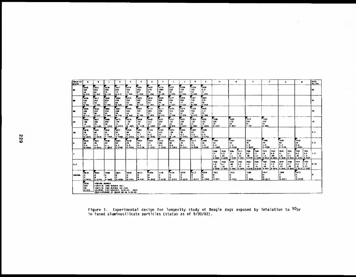
0 .PF IE G H J LI C D
I((i)DS~-IgS
~[[Iga~OnGD42fJ
?10,75E -~2
630
0-255
~3R5207ID4,
~03Um~I?
~’~l~tT
57D-27e
~977G~3t1~4la
~55140
~g ~z~4?SO
049
6II
eKi
[J- J.el+
z6O70E -26?
e9o7~E43]
~$2T
G5o-2r~
~~qn
5OB-510
~;e-G 2D
~oomoo S,VQ
63D-Eq!
650~~lpuqsD
9o-aPat
~ITqlON
~;BS510
o+!++SlE-776
~3~
30
~97B310
~02Azooq~ qz
~lX
q~Feoi
ll0~31;T27;O
0-lIB
~SAII
1702~
~5
21[-lms~
N~C12D15(-I~i
13GiI
?r~lR
I)! E -?ill
~399u~qo19E-I]~
17@-IKI
~I$Czze
[-L:~IB
ISO~33C23O2q[ -623
ZSO2lE -~79
]L~O15
IFoIBZ70IID-IOZ5
~AZIG15[-I21!
I5O- I Iris
2O0J|[-105S
2O
[I IST0LS0-2596
~J~l)B~SOz~
i~’151zloz~
~SR
L2
~70V’,+,6BIGE -ZI~5
NF~TIo556.11D-1707
~o
~$71
7.gC-7~
~P~o 5,1mw9.14E-~31)
~H7
7.7O-]7070-ElLIS
~7
9.104mz~,S
55
’[-1~5
3Qi%1i%1 E -2~3
~77¢
iq.IE-2Z~I
~TOU7q7,5E-2’QTG
!,,25.7E-210q
~SSU
2.Sq25.2[-1~3
~75’,¢T~79~5WI 57
5.7IE-ITG~
~17C
0.5E-~
676.SQ-.z~q7
SS ;0q.l[-~qs
~1..q$eoII.2[-276~
217,gE-26Z~S
+iSS.IO-;PI~I
7 S.T,,~3e7.5
S.O73v.9D-~Sm
¢.97-Z’F~D?
S
R 42227,111ILS,I
D-15t5
t21.S~-7~
II+I.0[-zs~S
I.O
~3217
’J~ID75z.S
I.S+~i
II 17].Sil-a+,+~,5
7+2is.! 1.213
aii5.oi.
1.8
0-~27
:;’.7;.a7
[-.Z’S£7
?.S1.2,17
7~M 75a7.? 2IS
.31(k)~lll
1.s48R-7~:ZO
)rAmI.l I.S
m--~?l
+,.7.12m-:tlS~l
I+Z.IS
O.2
~7675o0O-2570
000E4?H
00E42~7
00o -,,,17 Z~.,
~0 .~x400o-:~nn
00E-qlnS
7~0OA-3~17
?S~To0(i-~,? I
?SN4Z00+++-525l
7SZ$ElD~-~71
758R0oR -1.2| I
qzI(o0,m+ll-¢~
q371
R-q~71
p’llO0n-411~
a~Tu
~-qH9 R-qTO1
11~150Gl+llll~l
0.1111.
F-~77-lWlTl& LING NJ~IW IlLICIt..LilITZlL ttu; II~[~a ilII~]/KCI,,tB-Kn~I. E’.{UTn~U[7[~., 71-RLI¥[ - ~T5P011-LZPiI50~I. ~1 ~FJ|H OR ~ g4~,-I.7
Figure I. Experimental design for longevity study of Beagle dogs exposed by inhalation to 90Stin fused aluminosili¢ate particles (status as of 9/30/73).
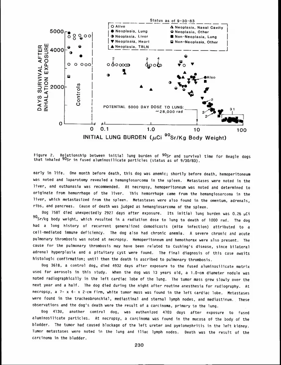
5000
CCw
w ~ 4000I--1,09<O
Q.-Jx<>UJ
n’Om F- 2000O)<t:CO->-<<’I-oz
@
@@
0 0 00011
cO0.I..Ac-O
0
Status as of 9=30-83I
O Alive A Neoplasia, Nasal CavityIi@ Neoplasia, Lung @ Neoplasia, Other
@ Neoplasia, Liver ¯ Non-Neoplasia, Lung II V Neoplasia, Heart ~ Non-Neoplasia, Other
IL~ Ne_ oplasia__~_, TBL. N ....... j
2 2 4
%oo @
@
0 ¯
A @
~ 3It
POTENTIAL 5000 DAY DOSE TO LUNG:’"28,000 rad
0 I~ ~l I I I0 0.1 1.0 10 100
INITIAL LUNG BURDEN (/_LCi 9°Sr/Kg Body Weight)
Figure 2. Relationship between initial lung burden of 90Sr and survival time for Beagle dogsthat inhaled 90Sr in fused aluminosilicate particles (status as of 9/30/83).
early in life. One month before death, this dog was anemic; shortly before death, hemoperitoneum
was noted and laparotomy revealed a hemanglosarcoma in the spleen. Metastases were noted in the
liver, and euthanasia was recommended. At necropsy, hemoperitoneum was noted and determined to
originate from hemorrhage of the liver. This hemorrhage came from the hemangiosarcoma in the
liver, which metastasized from the spleen. Metastases were also found in the omentum, adrenals,
ribs, and pancreas. Cause of death was judged as hemangiosarcoma of the spleen.
Dog 758T died unexpectedly 2927 days after exposure. Its initial lung burden was 0.26 ~Ci
90Sr/kg body weight, which resulted in a radiation dose to lung to death of 1000 rad. The dog
had a long history of recurrent generalized demodicosis (mite infection) attributed to
cell-mediated immune deficiency. The dog also had chronic anemia. A severe chronic and acute
pulmonary thrombosis was noted at necropsy. Hemoperitoneum and hemothorax were also present. The
cause for the pulmonary thrombosis may have been related to Cushing’s disease, since bilateral
adrenal hyperplasia and a pituitary cyst were found. The final diagnosis of this case awaits
histologic confirmation; until then the death is ascribed to pulmonary thrombosis.
Dog 361B, a control dog~ died 4932 days after exposure to the fused aluminosilicate matrix
used for aerosols in this study. When the dog was 13 years old, a 1.O-cm diameter nodule was
noted radiographically in the left cardiac lobe of the lung. The tumor mass grew slowly over the
next year and a half. The dog died during the night after routine anesthesia for radiography. At
necropsy, a 7- x 4- x 2-cm firm, white tumor mass was found in the left cardiac lobe. Metastases
were found in the tracheobronchial, mediastinal and sternal lymph nodes, and mediastinum. These
observations and the dog’s death were the result of a carcinoma, primary to the lung.
Dog 413U, another control dog, was euthanized 4703 days after exposure to fused
aluminosilicate particles. At necropsy, a carcinoma was found in the mucosa of, the body of the
bladder. The tumor had caused blockage of the left ureter and pyelonephritis in the left kidney.
Tumor metastases were noted in the lung and iliac lymph nodes. Death was the result of the
carcinoma in the bladder.
230
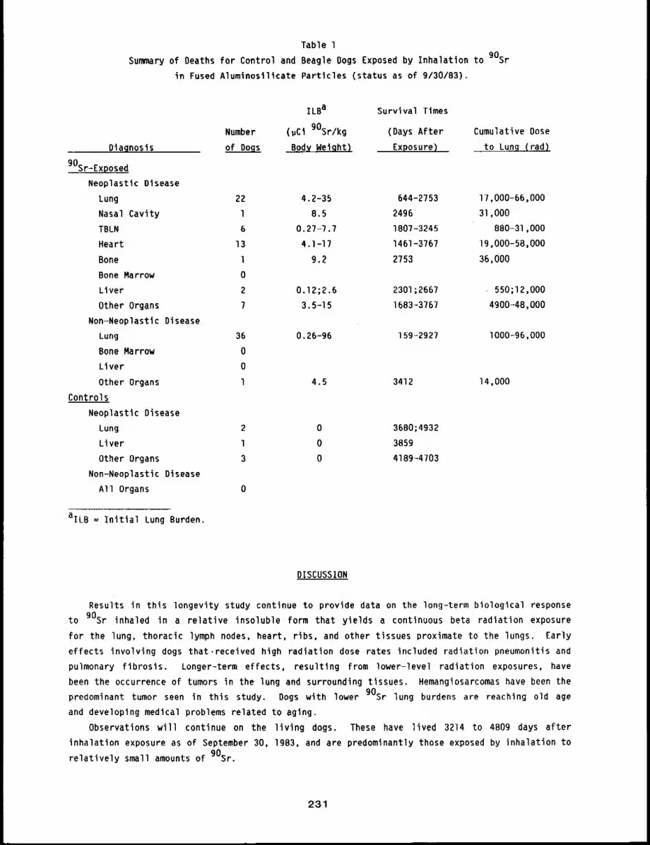
Table l
Summary of Deaths for Control and Beagle Oogs Exposed by Inhalation to 90Sr
in Fused Aluminosilicate Particles (status as of 9/30/83).
ILBa Survival Times
Number (gCi gOSr/kg (Days After
Diagnosis of Dogs Body Weiqht) Exposure)
gOSr-Exposed
Neoplastic Disease
Lung 22 4.2-35 644-2753
Nasal Cavity l 8.5 2496
TBLN 6 0.27-7.7 1807-3245
Heart 13 4.1-17 1461-3767
Bone l 9.2 2753
Bone Marrow 0
Liver 2 0.12;2.6 2301;2667
Other Organs 7 3.5-15 1683~3767
Non=Neoplastic Disease
Lung 36 0.26-96 159-2927
Bone Marrow 0
Liver 0
Other Organs 1 4.5 3412
Controls
Neoplastic Disease
Lung 2 0 3680;4932
Liver l 0 3859
Other Organs 3 0 4189-4703
Non-Neoplastic Disease
All Organs 0
Cumulative Dose
to Lung (rad)
17,000-66,000
31,000
880-31,000
19,000-58,000
36,000
550;12,000
4900-48,000
I000=96,000
14,000
aILB = Initial Lung Burden.
DISCUSSION
Results in this longevity study continue to provide data on the long-term biological response
to gOsr inhaled in a relative insoluble form that yields a continuous beta radiation exposure
for the lung, thoracic lymph nodes, heart, ribs, and other tissues proximate to the lungs. Early
effects involving dogs that,received high radiation dose rates included radiation pneumonitis and
pulmonary fibrosis. Longer-term effects, resulting from lower-level radiation exposures, have
been the occurrence of tumors in the lung and surrounding tissues. Hemangiosarcomas have been the
predominant tumor seen in this study. Dogs with lower gOsr lung burdens are reaching old age
and developing medical problems related to aging.
Observations will continue on the living dogs. These have lived 3214 to 4B09 days after
inhalation exposure as of September 30, 1983, and are predominantly those exposed by inhalation to
relatively small amounts of 90Sr.
231
BIOLOGICAL EFFECTS OF REPEATED INHALATION EXPOSURE OF BEAGLE DOGS
TO RELATIVELY INSOLUBLE AEROSOLS OF 144Ce. IX
Abstract --Beagle dogs were exposed repeatedly to
a relatlvely Insoluble form of 144ce to study the
depos~tlon, retention, and long-term blologlcal ef-
fects for comparison with data from dogs that were
exposed only once to a slmllar aerosol. Four
groups of nine dogs each were exposed once every
8 weeks for 2 years (13 exposures). Exposure
goals were: to increase the lung burden by 2.5
~Ci 144Ce/kg body weight wlth each exposure; to
PRINCIPAL INVESTIGATORS
B. B. Boeckel
F. F. Hahn
B. A. Muggenburg
R.’O. McClellan
J. L. Mauderly
J. A. Pickrell
re-establlsh lung burdens of 9 or 4.5 ~Cl 144Ce/kg body weight; and to expose controls to
fused alumlnosillcate partlcles containing nonradioactive Ce. To date, 25 exposed dogs and 3
control dogs have dled or were euthanlzed. The most prevalent findings have been pulmonary
carclnomas (nine dogs), and hemanglosarcomas in the tracheobronchlal lymph nodes (five dogs).
Observations are continuing on the two surviving 144Ce-exposed and slx control dogs Chat are now
at approxlmately 3590 days (9.8 years) after the flrsc exposure.
Persons may be exposed to radioactive aerosols in single, repeated, or chronic inhalation
situations. This study documents the deposition, retention, and long-term biological effects of
repeated inhalation exposures to 144Ce* in fused aluminosilicate particles. Beagle dogs were
chosen to permit direct comparison of the results with those being obtained in dogs that were
exposed once, as young adults, to a similar 144Ce aerosol.
A single inhalation exposure resulting in an initial lung burden of 25 ~Ci 144Ce/kg body
weight yields an absorbed beta dose to lung of about 35,000 rad in 2 years and has a high
probability of producing pulmonary neoplasia. Two other exposure regimens that would yield 35,000
rad in 2 years were selected for comparative study. Each involved repeating the inhalation
exposure every 8 weeks; one exposure regimen produced a relatively constant (within 20%) dose rate
throughout the 2-year period, and the other produced an increasing dose rate because of increasing
lung burden. These were (1) repeated inhalation exposures to re-establish the lung burden
9 uCi/kg body weight, and (2) repeated inhalation exposures to increase the lung burden by 2.5
uCi/kg body weight at each exposure time. In addition, another group was repeatedly exposed to
re-establish a lung burden of 4.5 ~Ci 144Ce/kg body weight.
STATUS
A list of all dogs in this study and their exposure regimens is in Appendix A. Included in
the table are dose rate and cumulative absorbed beta doses for each dog calculated from its own
whole-body counting data, The methodology for these calculations was described in the 1975-76
Annual Report, LF-56, pp. 279-280. To date, 25 144Ce-exposed dogs and 3 control dogs have died
or were euthanized. Details of these deaths are summarized in Table I. Below are specific
comments on the three 144Ce-exposed dogs and one control dog that died during the past year.
Refers to equilibrium mixture of 144Ce-144pr.
232
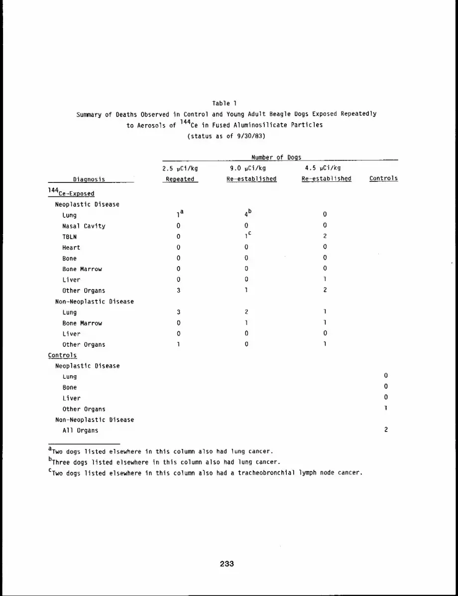
Table l
Summary of Deaths Observed in Control and Young Adult Beagle Dogs Exposed Repeatedly
to Aerosols of 144Ce in Fused Aluminosilicate Particles
(status as of 9/30/83)
Diagnosis
144Ce-Exposed
Neoplastic Disease
Lung
Nasal Cavity
TBLN
Heart
Bone
Bone Marrow
Liver
Other Organs
Non-Neoplastic Disease
Lung
Bone Marrow
Liver
Other Organs
Controls
Neoplastic Disease
Lung
Bone
Liver
Other Organs
Non-Neoplastic Disease
All Organs
2.5 ~Ci/kg
Repeated
Number of Dogs
9.0 ~Ci/kg 4.5 ~Ci/kg
Re-established Re-estabi~shed
Ia 4b 0
0 0 0
0 Ic 2
0 0 0
0 0 0
0 0 0
0 0 l
3 l 2
3 2 1
0 1 1
0 0 0
1 0 l
Controls
aTwo dogs listed elsewhere in this column also had lung cancer.
bThree dogs listed elsewhere in this column also had lung cancer.
CTwo dogs listed elsewhere in this column also had a tracheobronchial lymph node cancer.
233
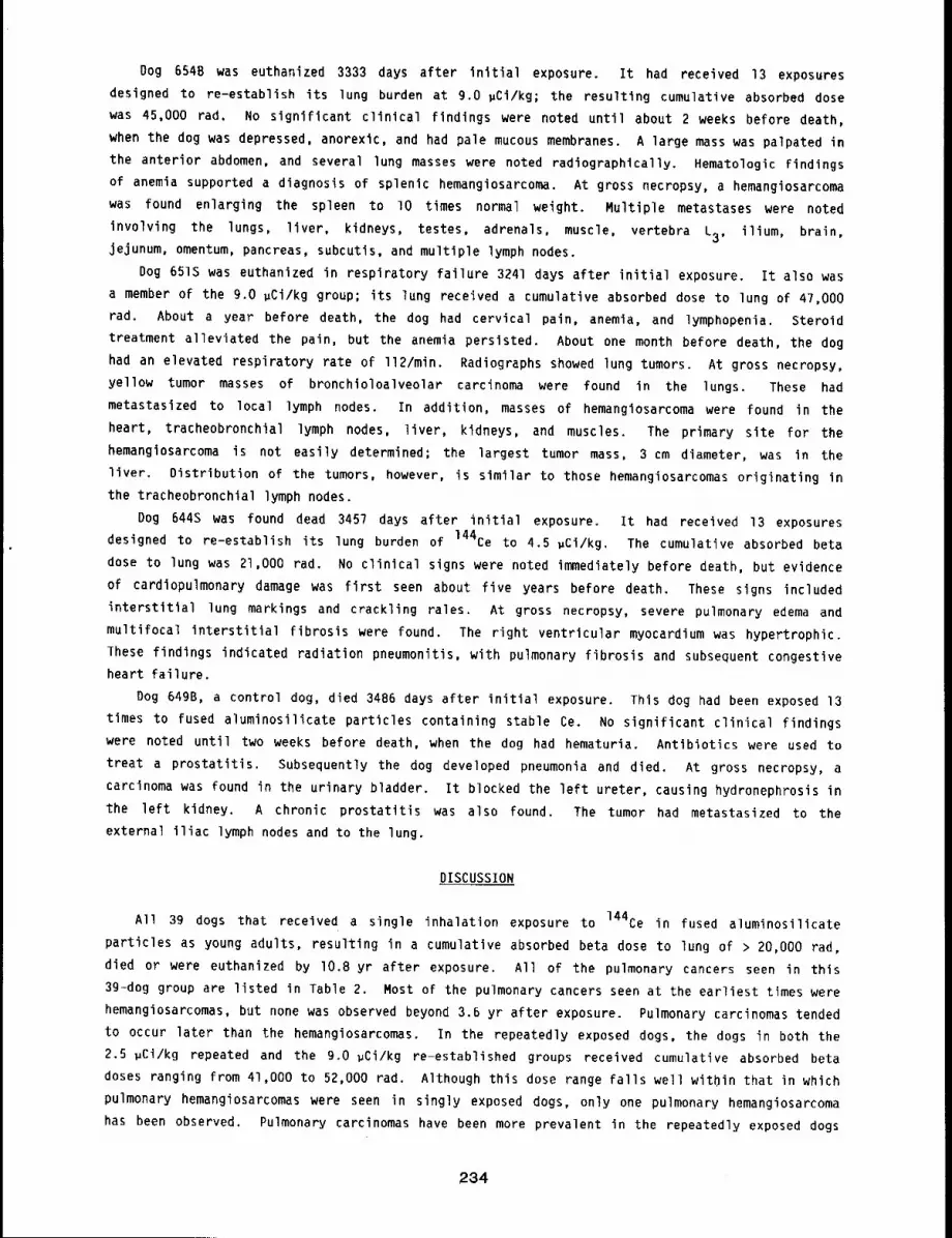
Dog 654B was euthanized 3333 days after initial exposure. It had received 13 exposures
designed to re-establish its lung burden at g.o gCi/kg; the resulting cumulative absorbed dose
was 45,000 rad. No significant clinical findings were noted until about 2 weeks before death,
when the dog was depressed, anorexic, and had pale mucous membranes. A large mass was palpated in
the anterior abdomen, and several lung masses were noted radiographically. Hematologic findings
of anemia supported a diagnosis of splenic hemangiosarcoma. At gross necropsy, a hemangiosarcoma
was found enlarging the spleen to lO times normal weight. Multiple metastases were noted
involving the lungs, liver, kidneys, testes, adrenals, muscle, vertebra L3, ilium, brain,
jejunum, omentum, pancreas, subcutis, and multiple lymph nodes.
Dog 651S was euthanized in respiratory failure 3241 days after initial exposure. It also was
a member of the 9.0 ~Ci/kg group; its lung received a cumulative absorbed dose to lung of 47,000
rad. About a year before death, the dog had cervical pain, anemia, and lymphopenia. Steroid
treatment alleviated the pain, but the anemia persisted. About one month before death, the dog
had an elevated respiratory rate of ll2/min. Radiographs showed lung tumors. At gross necropsy,
yellow tumor masses of bronchioloalveolar carcinoma were found in the lungs. These had
metastasized to local lymph nodes. In addition, masses of hemangiosarcoma were found in the
heart, tracheobronchial lymph nodes, liver, kidneys, and muscles. The primary site for the
hemangiosarcoma is not easily determined; the largest tumor mass, 3 cm diameter, was in the
liver. Distribution of the tumors, however, is similar to those hemangiosarcomas originating in
the tracheobronchial lymph nodes.
Dog 644S was found dead 3457 days after initial exposure. It had received 13 exposures
designed to re-establish its lung burden of 144Ce to 4.5 gCi/kg. The cumulative absorbed beta
dose to lung was 21,000 tad. No clinical signs were noted immediately before death, but evidence
of cardiopulmonary damage was first seen about five years before death. These signs included
interstitial lung markings and crackling rales. At gross necropsy, severe pulmonary edema and
multifocal interstitial fibrosis were found. The right ventricular myocardium was hypertrophic.
These findings indicated radiation pneumonitis, with pulmonary fibrosis and subsequent congestive
heart failure.
Dog 649B, a control dog, died 3486 days after initial exposure. This dog had been exposed 13
times to fused aluminosilicate particles containing stable Ce. No significant clinical findings
were noted until two weeks before death, when the dog had hematuria. Antibiotics were used to
treat a prostatitis. Subsequently the dog developed pneumonia and died. At gross necropsy, a
carcinoma was found in the urinary bladder. It blocked the left ureter, causing hydronephrosis in
the left kidney. A chronic prostatltis was also found. The tumor had metastasized to the
external iliac lymph nodes and to the lung.
DISCUSSION
All 39 dogs that received a single inhalation exposure to 144Ce in fused aluminosilicate
particles as young adults, resulting in a cumulatlve absorbed beta dose to lung of > 20,000 rad,
died or were euthanized by I0.8 yr after exposure. All of the pulmonary cancers seen in this
3g-dog group are listed in Table 2. Most of the pulmonary cancers seen at the earliest times were
hemangiosarcomas, but none was observed beyond 3.6 yr after exposure. Pulmonary carcinomas tended
to occur later than the hemangiosarcomas. In the repeatedly exposed dogs, the dogs in both the
2.5 gCi/kg repeated and the 9.0 ~Ci/kg re-established groups received cumulative absorbed beta
doses ranging from 41,000 to 52,000 rad. Although this dose range falls well within that in which
pulmonary hemangiosarcomas were seen in singly exposed dogs, only one pulmonary hemangiosarcoma
has been observed. Pulmonary carcinomas have been more prevalent in the repeatedly exposed dogs
234
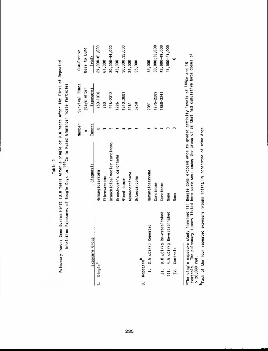
Ta
ble
2
Pu
lmo
na
ry T
um
ors
Se
en
Du
rin
g
First
I0
.8
Ye
ars
Afte
r a
Sin
gle
o
r 9
.B Y
ea
rs
Afte
r th
e
First
o
f R
ep
ea
ted
Inh
ala
tion
E
xpo
sure
s o
f B
ea
gle
Do
gs
to
14
4C
e in
F
use
d A
lum
ino
silic
ate
P
art
icle
s
Exp
osu
re G
rou
p
A.
Sin
gle
a B.
Re
pe
ate
db
I.
2.5
vC
i/kg
R
ep
ea
ted
II.
9.0
p
Ci/k
g
Re
-est
ab
lish
ed
Ill.
4.5
p
Ci/k
g
Re
-est
ab
lish
ed
IV.
Co
ntr
ols
Nu
mb
er
Su
rviv
al
Tim
es
of
(Da
ys A
fte
r
Dia
gn
osi
sT
um
ors
Exp
osu
re)
He
ma
ng
iosa
rco
ma
B7
50
-13
1B
Fib
rosa
rco
ma
l7
50
Bro
nch
iolo
alv
eo
lar
carc
ino
ma
59
16
-23
13
Bro
nch
og
en
ic
carc
ino
ma
l1
22
6
Mix
ed
tu
mo
r2
IBlO
;30
01
Ad
en
oca
rcin
om
al
39
61
Ost
eo
sarc
om
ol
32
50
Cum
ulat
ive
Dos
e to
Lu
ng
(ra
d)
29
00
0-6
1,0
00
61
00
0
3000
0-44
,000
4300
0
3000
0;32
,000
2400
0
25
00
0
He
ma
ng
iosa
rco
ma
l2
09
15
2,0
00
Ca
rcin
om
a2
19
75
-22
89
50
,00
0;5
2,0
00
Ca
rcin
om
a7
18
60
-32
41
43
,00
0-4
9,0
00
No
ne
02
1,0
00
-27
,00
0
No
ne
00
aT
he
sin
gle
e
xpo
sure
st
ud
y in
volv
ed
Ill
B
ea
gle
d
og
s e
xpo
sed
on
ce t
o
gra
de
d a
ctiv
ity
leve
ls
of
14
4C
e a
nd
15
con
tro
ls.
Th
e p
ulm
on
ary
tu
mo
rs
liste
d
he
re
we
re s
ee
n a
mo
ng
the
g
rou
p o
f 3
9 th
at
ha
d c
um
ula
tive
b
eta
d
ose
s o
f>
20
,00
0 r
ad
.
bE
ach
of
the
fo
ur
rep
ea
ted
e
xpo
sure
g
rou
ps
initi
ally
co
nsi
ste
d
of
nin
e
do
gs.
particularly in the g.o #Ci/kg re-established group, in which seven of nine dogs had them. Dogs
in the group that received one-half of that exposure have not developed lung tumors of any kind.
These results re-emphasize the importance of dose rate patterns in the production of lung cancer
by beta-emitting radionuclides. Hemangiosarcomas of the tracheobronchial lymph nodes have also
been a notable Finding in the 9.0 and 4.5 relestablished groups, but not in the 2.5 #Ci/kg
repeated group. Because only two 144Ce-exposed dogs and six control dogs remain alive, this
experiment will soon be at a stage where final analyses can be completed.
236
TOXICITY OF INHALED ALPHA-EMITTING RADIONUCLIDES - STATUS REPORT
Abstract --A series of interrelated dose-response
studies In which Beagle dogs and rodents are being
exposed to aerosols of transuranlc alpha-emlttlng
radlonuclldes Is described. Nonodlsperse aerosols
of oxides of 239pu, 238pu, 241Am, and 244Cm are
used in studles to measure the relative importance
of average organ dose, local dose around particles,
specific activity, particle size, and number of In-
haled partlcles to the development of biologlcal
effects. The elemental characteristics, chemical
form, species of anlmal, and age of animal at the
time of inhalation exposure are also being studied
because they may influence the toxlclt9 of these
radlonuclldes.
PRINCIPAL INVESTIGATORS
B. A. Muggenburg
J. A. Newhlnneg
R. A. Gullmette
F. F. Hahn
8. B. Boecker
J. L. Mauder19
D. L. Lundgren
c. S. blobbs
R. O. McClellan
The toxicity of plutonium and other transuranic alpha-emitting radionuclides is due to a
number of interrelated physical and biological factors. With the continued use of light water
reactors as energy sources, large quantities of these radionuclides will be present in the nuclear
inventory. Thus, there is a significant potential for the exposure of man to these radionuclides
at both the environmental and industrial levels. Inhalation is the most likely route of internal
deposition in man.
Dose-response studies with inhaled transuranic elements are in progress at this Institute.
The studies constitute a series of integrated experiments with laboratory animals exposed to
well=defined aerosols to better understand the relationships among exposure atmosphere, deposition
and retention patterns, radiation dose patterns, and resulting biological effects. This approach
will provide information that can be applied to a wide variety of possible exposure situations,
including those that may be encountered in accidents in the nuclear industry.
The basic experimental philosophy of the dose-response studies with alpha-emitting
radionuclides initiated to date is presented here. Detailed .progress reports for all of the
studies are presented elsewhere in this document. Some of this information and a more detailed
discussion of the relationships of these dose-response studies to other studies being conducted
have been published previously,l
EXPERIMENTAL APPROACH
For an inhalation exposure, a number of factors may influence the dose to lung and other
tissues and the subsequent biological effects. For inhaled alpha-emitting radionuclides, factors
associated with the aerosols include the elemental characteristics of the material, chemical form,
specific activity, and particle size distribution. Factors associated with the host include the
age at the time of exposure, laboratory animal species, health status, and individual response.
The major dog study in progress involves groups of young adult dogs exposed to graded activity
levels of 238pu02 or 239pu02 in monodisperse particles of different sizes. A schematic
presentation of the experimental design for this study is shown in Figure l, where each cube
237
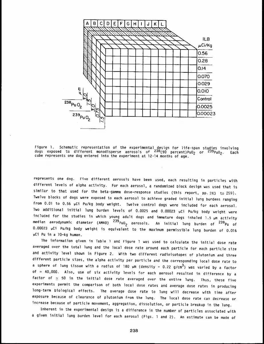
B
238pu 02
239pu02
\
ILB/~Ci/Kg
0.560.280.14
0.0700.0290.010
Control
0.0025
0.00025
Figure I. Schematic representation of the experimental design for life-span studies involvingdogs exposed to different monodisperse aerosols of 238(90 percent)PuO 2 or 239pu02. Eachcube represents one dog entered into the experiment at 12-14 months of age.
represents one dog. Five different aerosols have been used, each resulting in particles with
different levels of alpha activity. For each aerosol, a randomized block design was used that is
similar to that used for the beta-gamma dose-response studies (this report, pp. 243 to 259).
Twelve blocks of dogs were exposed to each aerosol to achieve graded initial lung burdens ranging
from O.Ol to 0.56 ~ci Pu/kg body weight. Twelve control dogs were included for each aerosol.
Two additional initial lung burden levels of 0.0025 and 0.00023 ~Ci Pu/kg body weight were
included for the studies in which young adult dogs and immature dogs inhaled 1.5 ~m activity
median aerodynamic diameter (AMAD) 239pu02 aerosols. An initial lung burden of 239pu 0.00023 ~Ci Pu/kg body weight is equivalent to the maximum permissible lung burden of 0.016
~Ci Pu in a ?O-kg human.
The information given in Table 1 and Figure l was used to calculate the initial dose rate
averaged over the total lung and the local dose rate around each particle for each particle size
and activity level shown in Figure 2. With two different radioisotopes of plutonium and three
different particle sizes, the alpha activity per particle and the corresponding local dose rate toa sphere of lung tissue with a radius of 180 ~m (density = 0.22 g/cm3) was varied by a factor
of = 40,000. Also, use of six activity levels for each aerosol resulted in difference by a
factor of ~ 50 in the initial dose rate averaged over the entire lung. Thus, these five
experiments permit the comparison of both local dose rates and average dose rates in producing
long-term biological effects. The average dose rate to lung will decrease with time after
exposure because of clearance of plutonium from the lung. The local dose rate can decrease or
increase because of particle movement, aggregation, dissolution, or particle breakup in the lung.
inherent in the experimental design is a difference in the number of particles associated with
a given initial lung burden level for each aerosol (Figs. l and 2). An estimate can be made
238
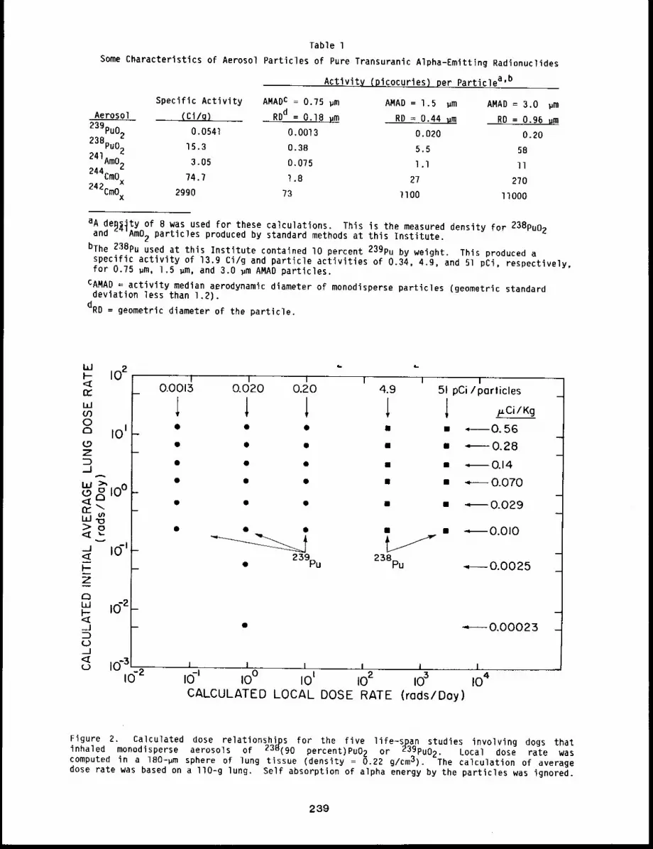
Table l
Some Characteristics of Aerosol Particles of Pure Transuranic Alpha-Emitting Radionuclides
Activity (picocuries) per Particlea’b
Specific Activity AMADc = 0.75 ~m AMAD = 1.5 pm AMAD = 3.0 ~mAerosol (Ci/q) RDd = 0.18 Hm RD = 0.44 ~m RD = 0.96 um
239pu02 0.0541 0.0013 0.020 0.20238pu02 15.3 0.38 5.5 58
241Am02 3.05 0.075 l.l ll244CmOx 74.7 1.8 27 270
242CmOx 2990 ?3 llO0 /tO00
aA deB~ty of B was used for these calculations. This is the measured density for 238pu02and ~’AmO2 particles produced by standard methods at this Institute.
bThe 238pu used at this Institute contained tO percent 239pu by weight. This produced aspecific activity of 13.9 Ci/g and particle activities of 0.34, 4.9, and 51 pCi, respectively,for 0.75 ~m, 1.5 ~m, and 3.0 ~m AMAD particles.
CAMAD = activity median aerodynamic diameter of monodisperse particles (geometric standarddeviation less than 1.2).
dRD = geometric diameter of the particle.
I I t 1 14.9
i ¯
¯ 239pu 238pu
I
I51 pCi/particles
l p.Ci/Kg
i -.--~0. 56= ¯ 0.28
¯ ", 0.14
= ~ 0.070
¯ ¯ 0.029
¯ - 0.010
m
~- 0.0025
m
------0.00023 _
I I 1 I I 1i0-I i0° I01 102 i03 104.CALCULATED LOCAL DOSE RATE (r(]ds/Doy)
Figure 2. Calculated dose relationships for the five life-span studies involving dogs thatinhaled monodisperse aerosols of 238(90 percent)PuO 2 or 239pu02. Local dose rate wascomputed in a 180-pm sphere of lung tissue (density = 0.22 g/cmJ). The calculation of averagedose rate was based on a llO-g lung. Self absorption of alpha energy by the particles was ignored.
239
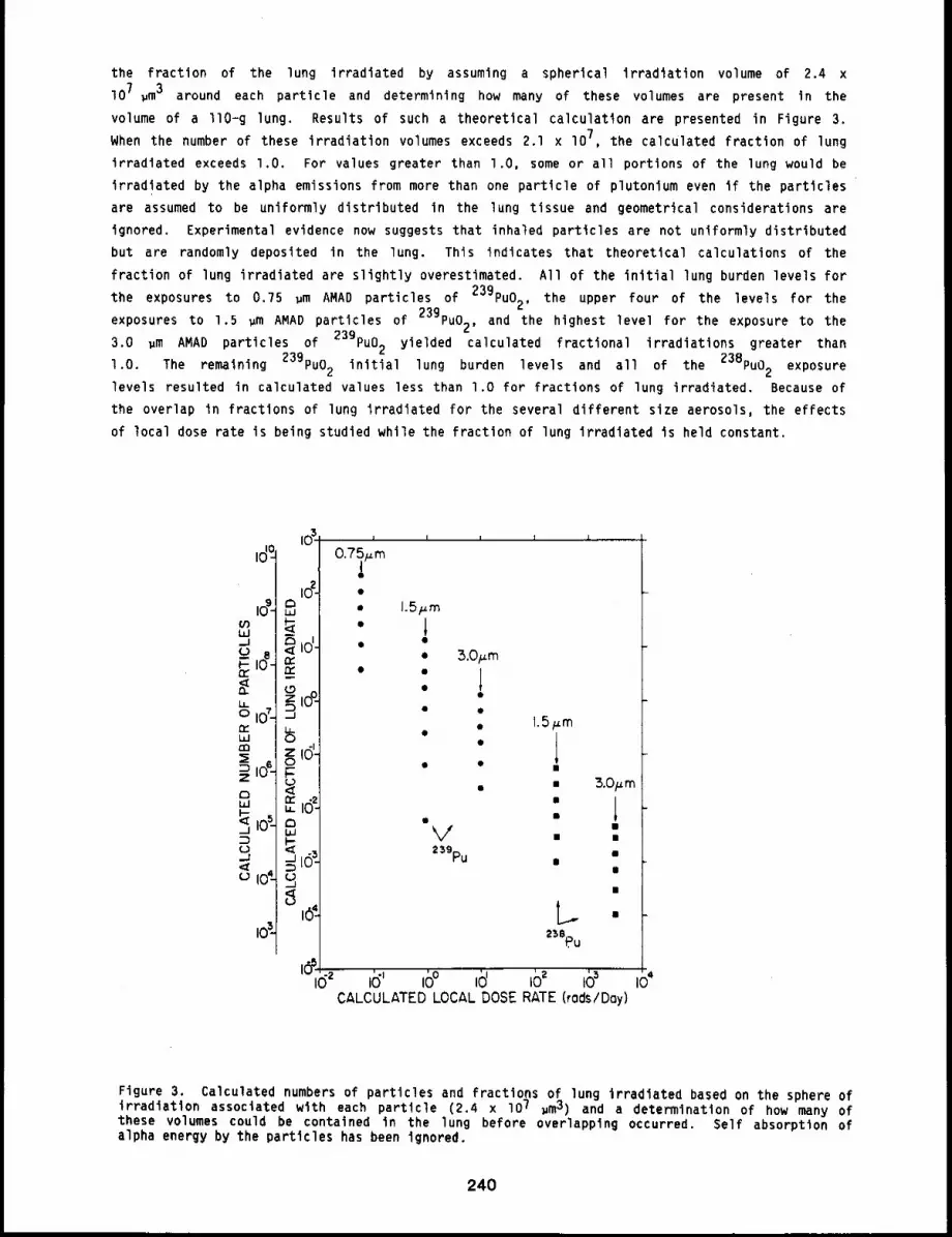
the fraction of the lung irradiated by assuming a spherical irradiation volume of 2.4 x
lO ? ~m3 around each particle and determining how many of these volumes are present In the
volume of a llO-g lung. Results of such a theoretical calculation are presented in Figure 3.
When the number of these irradiation volumes exceeds 2.1 x lO7, the calculated fraction of lung
irradiated exceeds l.O. For values greater than l.O, some or all portions of the lung would be
irradiated by the alpha emissions from more than one particle of plutonium even if the particles
are assumed to be uniformly distributed in the lung tissue and geometrical considerations are
ignored. Experimental evidence now suggests that inhaled particles are not uniformly distributed
but are randomly deposited in the lung. This indicates that theoretical calculations of the
fraction of lung irradiated are slightly overestimated. All of the initial lung burden levels for
exposures to 0.75 wm AMAD particles of 239pu02, the upper four of the levels for thethe
exposures to 1.5 ~m AMAD particles of 239pu02, and the highest level for the exposure to the
~m AMAD particles of 239pu02 yielded calculated fractional irradiations greater than3.0
l.O. The remaining 239pu02 initial lung burden levels and all of the 238pu02 exposure
levels resulted in calculated values less than l.O for fractions of lung irradiated. Because of
the overlap in fractions of lung irradiated for the several different size aerosols, the effects
of local dose rate is being studied while the fraction of lung irradiated is held constant.
io’-
J I I I I,
0.7~/.~mO
O
O
O
1.5Fm
10
3.0Fro
" 10Q
I ¯
¯ 1.5 p.mO " 1Q ¯
III
¯ B
V -Z39pu
II
&O/zm
|
,o" ,o°’’ ,o’ ,6’ ,o~ "CALCULATED LOCAL DOSE RATE (rods/Day)
Figure 3. Calculated numbers of particles and fractions of lung irradiated based on the sphere ofirradiation associated with each particle (2.4 x lO7 ~m3) and a determination of how many ofthese volumes could be contained In the lung before overlapping occurred. Self absorption ofalpha energy by the particles has been ignored.
240
The experiments with dogs exposed to 238pu02 are now from 8 to lO years after exposure
(this report, pp. 243 to 251), and those with dogs exposed to 239pu are ? to 8 years after
exposure (this report, pp. 252 to 259). All exposures for young adult dogs have been completed,
and the dogs are being maintained for life-span observation. Lung and bone cancers are being
observed in these dogs.
The exposures of aged and immature dogs to the 1.5 ~m aerodynamic diameter particles of
239pu02 aerosols are completed. These studies focus on the importance of age at the time of
exposure (this report, pp. 260 to 263 and pp. 264 to 268). This is important for the evaluation
of possible effects of accidental exposure of a general population, which would include
individuals of all ages. Results to date suggest the aged dogs at the time of exposure are more
susceptible to radiation injury than are young adult or immature dogs.
Several major studies are being conducted in rodents to complement the dog studies. The first
rodent experiments were conducted in Syrian hamsters [SCH:(SYR)]. These included animals exposed
238pu02 (1975-76 Annual Report, LF-56, pp. 238-244), monodisperseto monodi sperse aerosols of
241Am02 (1975176 Annual Report, LF-56, pp. 251-258), and polydisperse aerosols aerosols of
239pu02 (1975-76 Annual Report, LF-56, pp. 245-250), The effect of age at exposure was also
addressed in Syrian hamsters exposed to polydisperse aerosols of 239Pu02 either as immature
(~ 4 weeks of age), young adult (= 12 to 14 months of age), or aged animals (~ l year
(1975-76 Annual Report, LF-56, pp. 245-250).
The experiments with laboratory-reared, specific pathogen-free, Fischer-344 rats exposed to
~m aerodynamic diameter particles of 239pu02 are continuing. These animalsl.O and 2.85
have been exposed to one of three different activity levels at which lung tumors are anticipated.
These studies are designed to evaluate changes in tumor incidence as the experimental variables
are changed. In addition, the effect of age at exposure is also being studied in a group of
Fischer-344 rats exposed at 1B months of age. A study with graded lung burdens of 244Cm203is in progress. Lung burdens were selected based on predicted biological effects in rats.2
In all of the studies described above, animals were given single, brief inhalation exposures
to the radionuclide being studied. There is the potential that humans working or living in a
contaminated environment may be exposed by inhalation to alpha-emitting radionuclides more thanonce. Several studies are in progress to determine the effects of similar cumulative radiation
doses after repeated exposures by inhalation in addition to single exposures. In studies with
rodents, Syrian hamsters, mice, and rats have been or are being exposed to 239pu02 every othermonth for one year (seven exposures) (this report, pp. 274 to 277). A study is also in progress
dogs exposed to 239pu02 every 6 months for lO years (this report, pp. 269 to 273).with
To strengthen the interspecies comparison for the biological effects of inhaled plutonium
aerosols, a small group of non-human primates were exposed" to a polydisperse aerosol of
239Pu02 to achieve graded activity levels in the lung. These animals are being observed over
their life span. Data from this study will enhance our ability to extrapolate information from
animal studies to man (this report, pp. 283 to 287).
The above studies complement those under way at other laboratories. At the Pacific Northwest
Laboratories, Beagle dogs, rats, and Syrian hamsters have been exposed to graded activity levels
of polydisperse aerosols of 238pu02 and 239pu02.3’ 4 Other studies with alpha-emitting
radionuclides include plutonium mixed with sodium or uranium mixed with 239pu(N03) 4 and
studies of inhaled radon daughters and uranyl nitrate. At Los Alamos National Laboratory (LANL),
studies were conducted in which Syrian hamsters were given intravenous injections of
plutonium-labeled microspheres of sufficiently large size that they lodged in the lung
capillaries, resulting in chronic irradiation of the lung tissue. 5 Other studies at LANL
involve the inhalation exposure of Syrian hamsters to polydisperse aerosols of 238pu=zirconium
241
particles. The studies with monodisperse and polydlsperse aerosols of these materlals at our
Institute, in conjunction with the studies in other laboratories, are providing information on the
toxlclty of these materlals in various species of experimental animals, making extrapolation of
these data to people more reliable.
REFERENCES
I. McClellan, R. 0., Progress in Studies with Transuranlc Elements at the Lovelace Foundation,Health Phys. 222: B15-822, 1972.
2. McClellan, R. 0., H. A. Boyd, A. F. Gallegos, and R. 6. Thomas, Retention and Distribution of244Cm Followlng Inhalation of 244CMC13 and 244Cm01.73 by Beagle Dogs, Health Phys. 22: 877-885,1972.
3. Park, 3. F., W. J. Bair, and R. H. Busch, Progress In Beagle Dog Studies with TransuranlumElements at Battelle-Northwest, Health Phys. 2-2: 803-810, 1972.
4. Drucker, H., Pacific Northwest Laborator~ Annual Report for IgBO to the DOE AssistantSecretary for Environment and Safety Part I. Biomedical sciQnces, PNL-3700, 1981.
Anderson, E. C., L. M. Holland, O. R. Prine, and R. G. Thomas, Lung Response to LocalizedIrradiation from Plutonium Microspheres, in Inhaled Particles IV Part 2 (W. H. Walton, ed.),Pergamon Press, New York, pp. 615-623, 1977.
242
TOXICITY OF INHALED 23Bpuo2 IN BEAGLE DOGS: A. MONODISPERSE 1.5 ~m AMAD PARTICLES
B. MONODISPERSE 3.0 um AMAD PARTICLES. X
Abstract -- Beagle dogs have received an Inhalation
exposure to one of two sizes of monodlsperse aerosols
238pu02 to achieve graded levels of initial lungof
burden. All dogs are being studied over their 11fe
span, Ninety dogs have died wlth inltlal lung bur-
dens ranglng from 0.01 to 1.5 ~Ci 238pu/kg body
weight; 8 dled of rad~atlon pneumonltls and pulmo-
nary flbrosls, 4 dled of lung tumors, 64 dled of
bone tumors, idled of a liver tumor, and 12 d~ed of
miscellaneous causes, Four" control dogs have dled.
Flfty-four exposed and 20 control dogs survive at
2388-3414 days after exposure.
PRINCIPAL IWVESTIGRTORS
J. A. Newh~nneg
F. F. Hahn
B. A. Muggenburg
J. E. White
J. s. Dlel
J. L. Mauder19
B. B. Boecker
R. O. NcClellan
The studies reported here are a portion of a larger effort in which dogs were exposed by
inhalation to monodisperse aerosols of 23Bpuo 2 or 239pu02 to measure dose-response
relationships. The studies include several types of lung dose distributions in which the number
of inhaled particles, local and average dose to lung, and fraction of lung irradiated are the
primary variables. The complementary dose-response studies using 239pu02 aerosols are
detailed elsewhere (this report, pp. 252 to 259).
In the studies reported here, 72 Beagles inhaled 1.5 wm activity median aerodynamic diameter
of 238pu02, and 72 other Beagles inhaled 3.0 wm AMAD(AMAD) monodisperse aerosols
monodisperse aerosols. In each study, six graded levels of initial lung burden (planned levels of
0.56, 0.28, 0o14, 0.07, 0.029, and O,Ol ~Ci 23Bpu/kg body weight) were used, with 12 dogs
exposed at each level. For each study, 12 additional dogs inhaled the aerosol diluent material
and serve as controls. All dogs are being observed over their life span.
A recent analysis of dose-response data accumulated to date for these studies has been
presented that provides a measure of the relative risk of bone tumors after internal deposition of
bone-seeking radionuclides in dogs. l The analysis used the Proportional Hazards Model2 and
compared studies in which 90Sr and 23Bpu were inhaled or 239pu and 226Ra were injected.
Continued investigations using the Proportional Hazards Model applied to the data from the studies
that are the subject of this report have been pursued; some preliminary results of the role of sex
of the animal are reported.
METHODS
Details of this study have been presented in past annual reports of thls Institute. The
experimental design and present status of the two studies as of September 30, 1983, are
illustrated in Figure l for the study with 1.5 pm AMAD monodisperse particles and in Figure 2
for the study with 3.0 ~m AMAD monodisperse particles. The initial lung burdens were estimated
from whole-body counts of 169yb, a gamma emitter incorporated into the 238pu02 particles.
Estimates of the tissue doses received by dogs in these dose-response studies have been made
using the biomathematical metabolism and dosimetry model developed from data produced in the
243
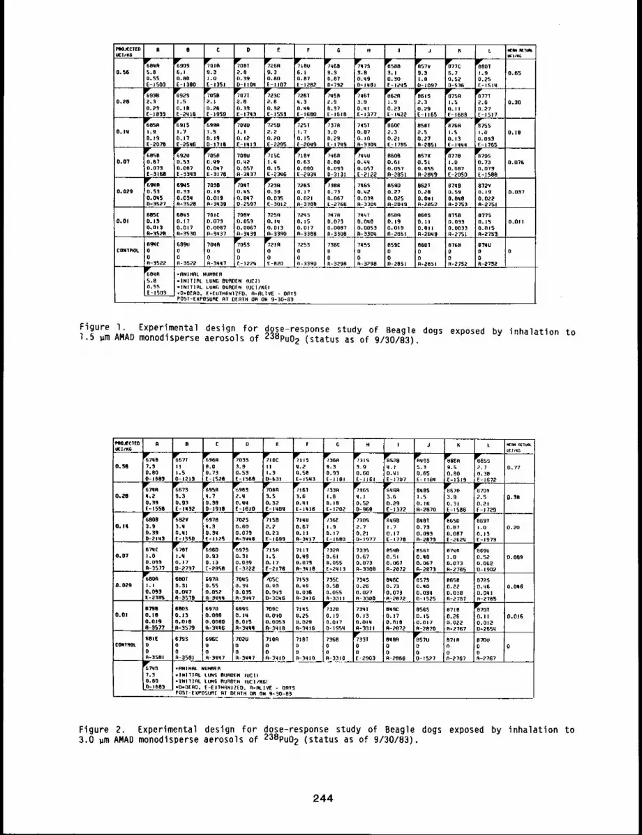
F’NJ~CIEUU¢I/KG
P~JECIED 0 B C D E F H ] J L ~nCTUW,IJI¢IfKO
~J OCut]/K~
6790 ~STT 6~’96N "~//035 ~115 1~360 7~315 ~8520 ~8955 PBGBB ~ss
O,56 7.5 11 0.0 3.9 II q,2 9.3 3.9 q,I 5.3 9.S 6.770.80 0.7~ 0.55 1~3 0,50 0.83 0.60 O.UI 0.65 0.80 0.3BO-i 603 0-1215 £-~528 E-1560 D~63! E-ISU3 E-]ISI [-JlGI E-]707 E- 11611 E-i3J9 E-1672
6~95R UgBS ~u00 l~?161 ~3B "/~3 GS ~8~GR OqBS ~r67R P’r77oV0,2~I 9.3 q.? 2.q 5.5 3,6 1.0 q.I ;3.6 1,5 3.9 2.5 0.56
0.39 0.03 0.50 o.qq 0.52 O.ql 0.18 0, 52 0.29 0. IB 0.31 0.21E= 1556 [-iq32 0-]910 E-1610 E-lqOB E-IqI6 E=IPOP 0-960 E-1372 FI-~870 E-1588 f- 17296~800 ~B2V %0 ;o-;,- ’/IqB 1~/35E ~3OS ~*8960 8q01 ~50 8GBT
O. Iq 3.9 ‘9.9 q.5 0.60 2°2 0,07 1.9 ~’.7 I,? 0.73 0.07 1.0 1.200.39 O.MI O. 3~ 0,0"/5 0.23 0.11 0.17 0,21 O.17 0.063 0,087 0.130-2193 E-]SSO E-112~ N-3~qO E-1699 0-5912 (-J600 O- 1977 F- 1770 0-~8"/$ (-=~E2q [-1973
B~C ~r~01 ~96D 65"/S ~7t50 ?lie 1~7328 "/‘935 8598 0S6T 87qR 869U0,07 1,0 I.q 0,913 0.31 1,5 O.q9 0.51 0.67 O.Sl 0.90 1.0 0.5~ 0.009
0.055 O.17 0,]3 0.0‘99 0.17 0.073 0.055 0.073 0.06? 0.067 0,073 0.0620-‘957"/ 0-2737 E=295B E-3222 E-21"/0 0-‘9q18 E-2913 ~-3300 R-2072 O-28"/‘9 R- 2765 0-1902
6008 680T 657~ ?Oq5 ?3uS 0q6C ~0578 BGSB 87250.029 1.1 0,51 0.55 0.3U O. qO O,q6 0.50 O.ZB 0.73 0.10 0.22 0.q6 O.O~S
0 ̄ 093 o.oq7 0,052 0,035 0.6q5 0.036 0.O55 0.097 0,07‘9 O.03q O.010 o.oulIE-2365 0-35’/8 R-SqqM B-3uq7 O-30q6 0-3Wi6 ~-3311 R-3300 0-2872 0-1525 8-2707 A-ZTSS
0"/98 6003 6370 65BS "t00C 2195 r773~B 73qT 8~C 8565 871B ~TOl0.01 0,10 0.13 0.080 O. Iq o,0qO 0.25 0.]9 0.15 0.17 0.15 0.26 0.11 0.016
0.019 0.010 0,0080 0.013 0.0053 0,029 0.017 O,oiq 0.B]0 O.017 0.022 0.0120-5577 B-3579 B-39|6 fl-3qqO R-‘Bqt6 0-195q N-3311 R-2872 O-2070 R-2767 0-255q
BOSE 6795 GgBC 702V 7160 ?lBT 7360 7351 BqBB ~857U 8716 07OUCONTROL0 O B o 0 0 0 0 0 0 R 0 0
0 0 0 0 0 o 0 O 0 0 O R8-3501 0-3581 R-3qq? o-5~7 ~-33ro R-‘9910 R-3‘910 E-2903 B-2606 O-15P? R-2767 5-27676’/qB =ANIMAL NUMBER7.3 -I#I]IBL LUN0 0UROEN IUCil0.80 =INITIBL LUNG NUOOFN IUCItKGI0~1E03 "0*~E60, [-[UIHRN]ZEO. R=RLIV[ - ORT5
0 0 C D E F G ]iM J 6
~OqA
5,00.$SE-1503
6Y6905
6.!0.80E-1360
TYro]R9"3J.OE--1351
~7O8T2,8O. 690-1 ]Oq
~069,30.80E-1107
~1BU6.1Q.87E-1262
P~Y6B19.S:0.870-792
r77q75~.B0.q90-1W01
8~5883.10.30E - 1¢’95
857V0.3i,G0-1097
077C6.70,520-536
0.$6 1.90.26E-ISlq
0.65
6¯9362,30.25£-1035
¯923
0.10E-2ql6
7o80!2,]o.26E- 1959
~7O7T
::%80.33E-120‘9
~7723C
2,8Q. 32E-155‘9
I1~7~582.90,37E-1610
3.00.qlE=1377
F61S2,30.29E-II65
87501,50.11E-1560
~877T2.6D.27E-1517
1,00,093E-1’/65
0,20 0.30
0,10
6~1r05A1.90.13E-2070
1,70.]7£-2596
~99R1.50.J90-]716
1~7250
2.20.20E-2295
?q5T0,070+ iUB-5309
8S8T2.50.2"1B-205]
~’60560.670.073E-3108
70500,09O.Oq?E-3176
~715Ci.U0.15E-2396
7YTIBV0.630. 000E-2939
~?9600.800.09‘9
; 0-31 ‘91
8Y7701.0O.OB?£-2050
~795
0.730,07~E-1506
7oeu0.~20.05"/R-3U37
86080,610.0570-205]
BS?X0.510,055A-20q9
0o07 0.0?6
~7~BA
I 0.29;0.067E-2706
~66~B0.5’90.0550-3527
6~30.330,039A-3520
7030O,190.0130-3q39
72650*iTO.O21B-3308
79650.920,039R-330q
65000.2’/0,025R-2Bq9
062T0.28O*OqlB-2852
97900.590,098B-275‘9
072V0.190.022B~2751
0,029
!0.07‘910.O0070-3300
605C0. t50,013fl-3510
68950.17O.Ot?R=3530
?OlCD,0730.00870=3,u37
708¥0.0530.006"/A-3q39
72q50.150.017B-~308
7qqTO.OqO0,0055R-330q
BSBA0.19O.OI90-2051
86050.110.01]R-~BU3
07580.0330,005‘9U=275!
07?50.150.0158-275~
0.01 0.011
~’~’7085
0oE-1229
~7210
0oE-R20
609U000-3522
7253000-3‘990
730C00h-3~90
059C000-205]
8601Oo8-~051
876000
CONTROL 0
VOOq8S.BR,SSE-t505
=flN]HRL NUMBER"INII]GL LUNG BURDEN (UCI)=INNIfL LtIN~ BUROEN (UCI/RGI-D=6£RD. E*EUTHANITEO, A=RL1VE - 00T5P05T-EXP05UB[ RT DERTH UB ON 9-30-03
Figure I. Experimental design for dose-response study of Beagle dogs exposed by inhalation to1.5 pm AMAD monodisperse aerosols of 238pu02 (status as of 9/30/83).
POST-EXPOSURE RT BFRIH O0 ON 9-50-85
Figure 2. Experimental design for dose-response study of Beagle dogs exposed by inhalation to3.0 pm AMAD monodisperse aerosols of 23Bpu02 (status as of 9/30/83).
244
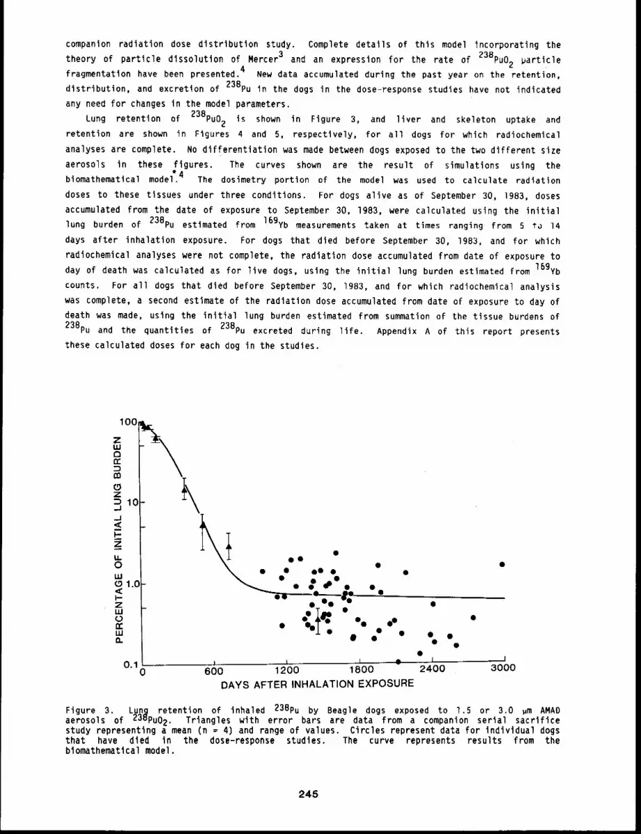
companion radiation dose distribution study. Complete details of this model incorporating the
theory of particle dissolution of Mercer 3 and an expression for the rate of 238pu02 ~article4fragmentation have been presented. New data accumulated during the past year on the retention,
distribution, and excretion of 238pu in the dogs in the dose-response studies have not indicated
any need for changes in the model parameters.
Lung retention of 238pu02 is shown in Figure 3, and liver and skeleton uptake and
retention are shown in Figures 4 and 5, respectively, for all dogs for which radiochemical
analyses are complete. No differentiation was made between dogs exposed to the two different size
aerosols in these figures. The curves shown are the result of simulations using the
biomathematical model~4 The dosimetry portion of the model was used to calculate radiation
doses to these tissues under three conditions. For dogs alive as of September 30, 1983, doses
accumulated from the date of exposure to September 30, 1983, were calculated using the initial
lung burden of 23Bpu estimated from 16gYb measurements taken at times ranging from 5 to 14
days after inhalation exposure. For dogs that died before September 30, 1983, and for which
radiochemical analyses were not complete, the radiation dose accumulated from date of exposure to
day of death was calculated as for live dogs, using the initial lung burden estimated from 169yb
counts. For all dogs that died before September 30, IgB3, and for which radiochemical analysis
was complete, a second estimate of the radiation dose accumulated from date of exposure to day of
death was made, using the initial lung burden estimated from summation of the tissue burdens of
23Bpu and the quantities of 238pu excreted during life. Appendix A of this report presents
these calculated doses for each dog in the studies.
100
ZuJaoc
Z1C_J
_J<I-
u.0LU
1.CI-ZLU
LUO.
1 i ~ i IO. 10
600 1200 1800 2400 3000
DAYS AFTER INHALATION EXPOSURE
Figure 3. Lunq retention of inhaled 23Bpu by Beagle dogs exposed to 1.5 or 3.0 ~m AMADaerosols of 238pu02. Triangles with error bars are data from a companion serial sacrificestudy representing a mean (n = 4) and range of values. Circles represent data for individual dogsthat have died in the dose-response studies. The curve represents results from thebiomathematical model.
245
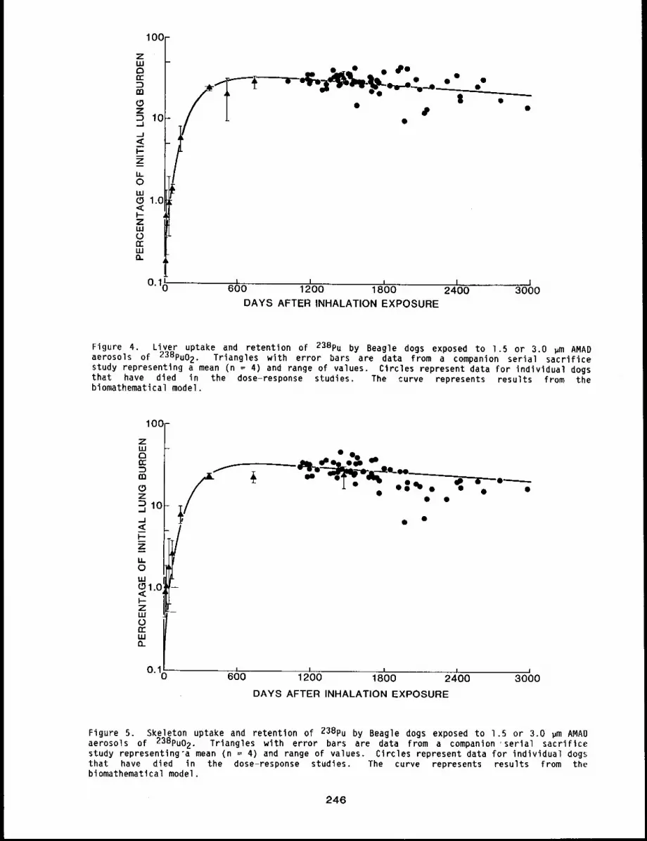
,,,71 O0Ic~ __ e~_o ¯ ~e ¯
z ¯
¯¯ ¯
10..I.....J<I’--
h0
0 1.0.<i-Zl.UouJ
0.10I n i n i
600 1200 1800 2400 3000DAYS AFTER INHALATION EXPOSURE
Figure 4. Liver uptake and retention of 238pu by Beagle dogs exposed to 1.5 or 3.0 ~m AMADaerosols of 23Bpu02. Triangles with error bars are data from a companion serial sacrificestudy representing a mean (n = 4) and range of values. Circles represent data for individual dogsthat have died in the dose-response studies. The curve represents results from thebiomathematical model.
10(]
F-
ouJ~1.0I-ZLUorrLU0_
~ ~ -~-~ ,_ ~ ,--0----j. ,t,,
° O0"O ° 0 ¯ °
/¯ ¯
~, , ¯
/
0.10I I I i I
600 1200 1800 2400 3000DAYS AFTER INHALATION EXPOSURE
Figure 5. Skeleton uptake and retention of 238pu by Beagle dogs exposed to 1.5 or 3.0 ~m AMADaerosols of 238pu02. Triangles with error bars are data from a companion ’serial sacrificestudy representing’a mean (n = 4) and range of values. Circles represent data for individual dogsthat have died in the dose-response studies. The curve represents results from thebiomathematical model.
246
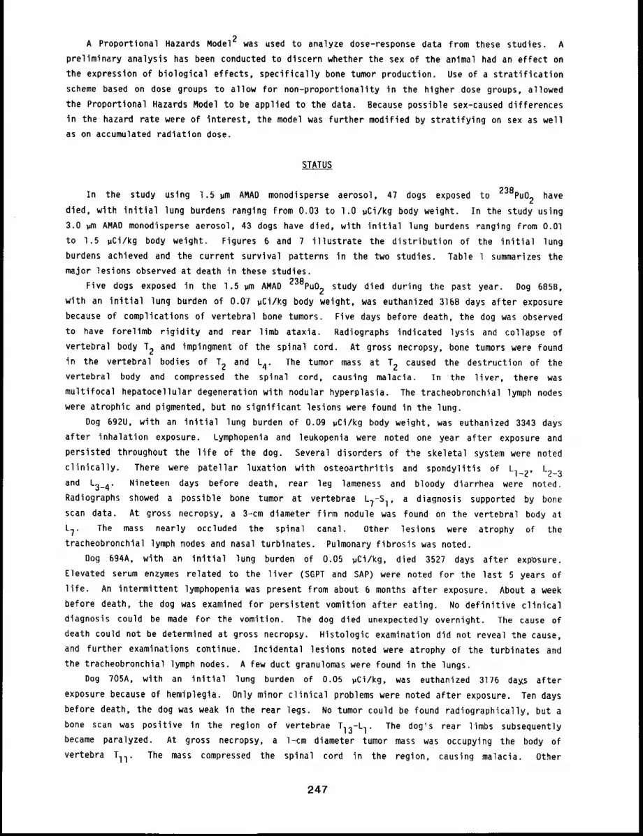
A Proportional Hazards Model2 was used to analyze dose-response data from these studies. A
preliminary analysis has been conducted to discern whether the sex of the animal had an effect on
the expression of biological effects, specifically bone tumor production. Use of a stratification
scheme based on dose groups to allow for non-proportionality in the higher dose groups, allowed
the Proportional Hazards Model to be applied to the data. Because possible sex-caused differences
in the hazard rate were of interest, the model was further modified by stratifying on sex as well
as on accumulated radiation dose,
STATUS
In the study using 1,5 um AMAD monodisperse aerosol, 47 dogs exposed to 238pu02 have
died, with initial lung burdens ranging from 0.03 to l.O uCi/kg body weight. In the study using
3.0 gm AMAD monodisperse aerosol, 43 dogs have died, with initial lung burdens ranging from O.Ol
to 1.5 uCi/kg body weight. Figures 6 and 7 illustrate the distribution of the initial lung
burdens achieved and the current survival patterns in the two studies. Table 1 summarizes the
major lesions observed at death in these studies.
~m AMAD 238pu02 study died during the past year. Dog 685B,Five dogs exposed in the 1.5
with an initial lung burden of 0.07 gCi/kg body weight, was euthanized 3168 days after exposure
because of complications of vertebral bone tumors. Five days before death, the dog was observed
to have forelimb rigidity and rear limb ataxia. Radiographs indicated lysis and collapse of
vertebral body T2 and impingment of the spinal cord. At gross necropsy, bone tumors were found
in the vertebral bodies of T2 and L4. The tumor mass at T2 caused the destruction of thevertebral body and compressed the spinal cord, causing malacia. In the liver, there was
multifocal hepatocellular degeneration with nodular hyperplasia. The tracheobronchial lymph nodes
were atrophic and pigmented, but no significant lesions were found in the lung.
Dog 692U, with an initial lung burden of o.og ~Ci/kg body weight, was euthanized 3343 daYs
after inhalation exposure. Lymphopenia and leukopenia were noted one year after exposure and
persisted throughout the life of the dog. Several disorders of the skeletal system were noted
clinically. There were patellar luxation with osteoarthritis and spondylitis of Ll_2, 1.2..3
and L3_4. Nineteen days before death, rear leg lameness and bloody diarrhea were noted.
Radiographs showed a possible bone tumor at vertebrae LT-SI, a diagnosis supported by bone
scan data. At gross necropsy, a 3-cm diameter firm nodule was found on the vertebral body at
L7. The mass nearly occluded the spinal canal. Other lesions were atrophy of the
tracheobronchial lymph nodes and nasal turbinates. Pulmonary fibrosis was noted.
Dog 694A, with an initial lung burden of 0.05 ~Ci/kg, died 3527 days after exposure.
Elevated serum enzymes related to the liver (SGPT and SAP) were noted for the last 5 years
life. An intermittent lymphopenia was present from about 6 months after exposure. About a week
before death, the dog was examined for persistent vomition after eating. No definitive clinical
diagnosis could be made for the vomition. The dog died unexpectedly overnight. The cause of
death could not be determined at gross necropsy. Histologic examination did not reveal the cause,
and further examinations continue. Incidental lesions noted were atrophy of the turbinates and
the tracheobronchial lymph nodes. A few duct granulomas were found in the lungs.
Dog 705A, with an initial lung burden of 0.05 ~Ci/kg, was euthanized 3176 da~s after
exposure because of hemiplegla. Only minor clinical problems were noted after exposure. Ten days
before death, the dog was weak in the rear legs. No tumor could be found radiographically, but a
bone scan was positive in the region of vertebrae TI3-L I. The dog’s rear limbs subsequently
became paralyzed. At gross necropsy, a 1-cm diameter tumor mass was occupying the body of
vertebra Tll. The mass compressed the spinal cord in the region, causing malacia. Other
247
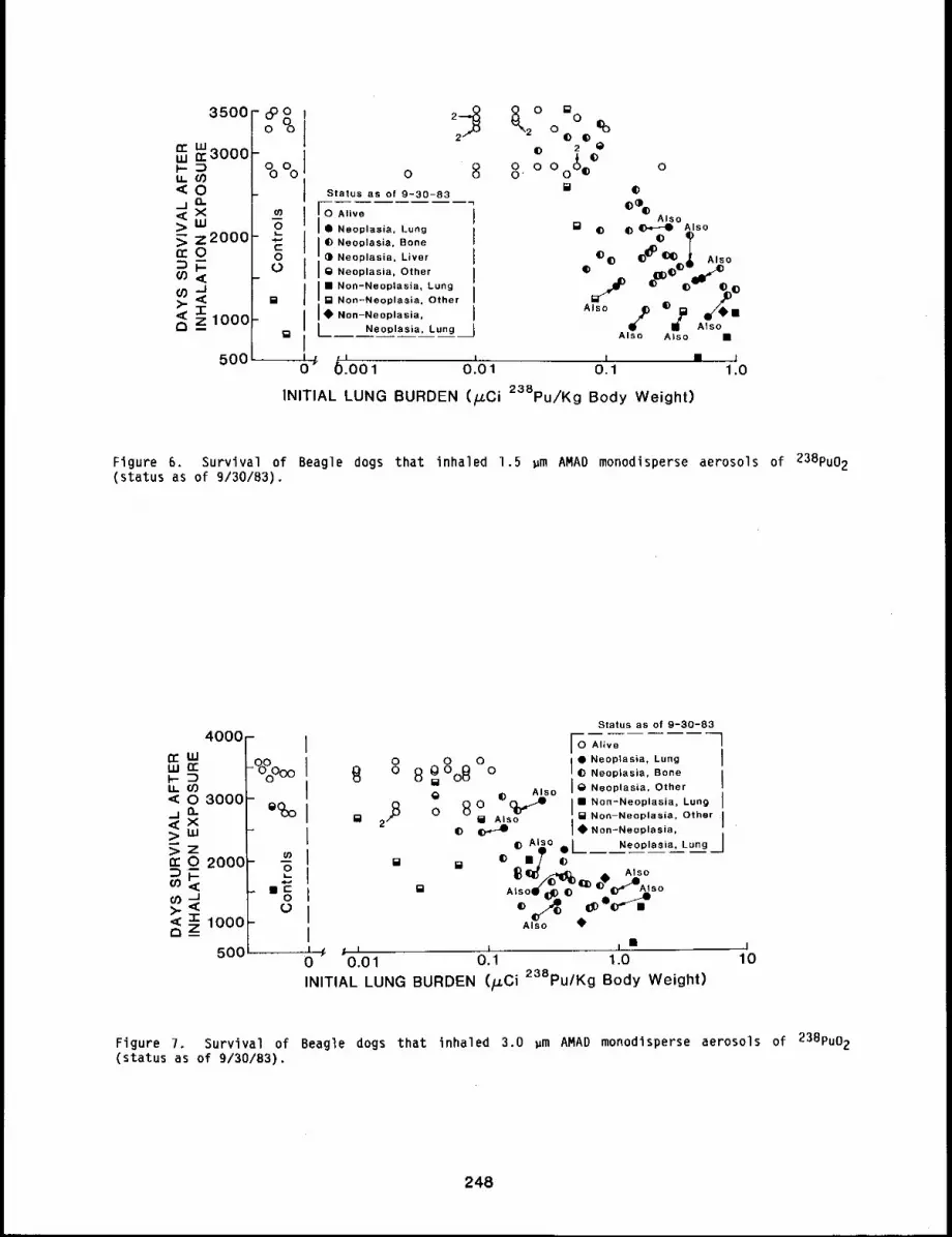
n-LU
IJ.,<
;>
n-
(0
>-<
I0 Alive
Q Neoplasia. Lung
Ij(D Neoplasia, Bone
¯ Neoplasia, Liver
g Neoplasia, Other
I~Non-Neoplasia, Lung
Non-Neoplasia, Other
Non-Neoplasia,
Neoplasia. LungAlso Also ¯
i ¯ 61
O~I I [] 1
.001 0.01 0.1 1.0
INITIAL LUNG BURDEN (/u.Ci 238pu/Kg Body Weight)
Figure 6. Survival of Beagle dogs that inhaled 1.5 pm AMAD monodisperse aerosols of 238pu02
(status as of 9/30/B3).
4000
n-uJLU ri-
Ll. 09< o 3000.j Q-<(X~.LU>Zrro 2000o),,¢
>.< z~ lO0OQ_
o o
B o 80=°08 oAlso
,B o 8°¢= 2 [] Also
[]
Status as of 9-30-83
[ 0 Alive ]
¯ Neoplasia, Lung II E) Neoplasia, Bone
IIS Neoplasia, Other
I ¯ Non-Neoplasia, Lung Ii~Non-Neoplasia. Other INon-Neoplasia,
(D AI~o.L Neoplasia, Lung J© m[ ~ -----
o %,soAlso ~1.
I, ¯I I I500 0 0.01 0.1 1.0 10
INITIAL LUNG BURDEN (/J.Ci 23SPu/Kg Body Weight)
Figure 7. Survival of Beagle dogs that inhaled 3.0 ~m AMAD monodisperse aerosols of 238pu02
(status as of 9/30/83).
248
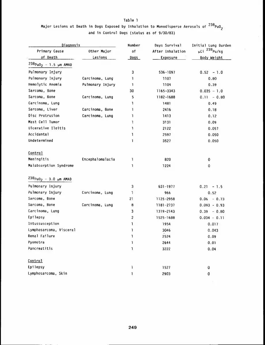
Table 1
Major Lesions at Death in Dogs Exposed by Inhalation to Monodisperse Aerosols of 23Bpuo2and in Control Dogs (status as of 9/30/83)
Diagnosis
Primary Cause
of Death
238pu02 - 1.5 ~m AMAD
Pulmonary Injury
Pulmonary Injury
Hemolytic Anemia
Sarcoma, Bone
Sarcoma, Bone
Carcinoma, Lung
Sarcoma, Liver
Disc Protrusion
Mast Cell Tumor
Ulcerative Ileitis
Accidental
Undetermined
Number Days Survival Initial Lung Burden
Other Major of After Inhalation ~Ci 238pu/kg
Lesions Dogs Exposure Body Wgtqht __
Carcinoma, Lung
Pulmonary Injury
Carcinoma, Lung
Carcinoma, Bone
Carcinoma, Lung
3 536-1097 0.52 - 1.0
1 II07 0.80
1 1104 0.39
30 I165-3343 0.035 - l.O
5 I182-1688 O.ll - 0.80l 1481 0.49
1 2416 0.18
1 1413 0.12
l 3131 o.og
1 2122 0.057
l 2597 0.050
l 352? 0.050
Control
Meningitis
Malabsorption Syndrome
Encephalomalacia 1 820 0
1 1224 0
238pu02 - 3.0 ~m AMAD
Pulmonary Injury
Pulmonary Injury
Sarcoma, Bone
Sarcoma, Bone
Carcinoma, Lung
Epilepsy
Intussusception
Lymphosarcoma, Visceral
Renal Failure
Pyometra
Pancreatitls
Carcinoma, Lung
Carcinoma, Lung
3
l21
8
3
2
l
1
l
l
l
631-1977
966
1125-2958
I181-2737
131g-2143
1525-1688
1954
3046
2524
2644
3222
0.21 - 1.5
0.52
0.06 - 0.73
0.093 .- 0.93
0.39 - 0.80
0.034 - O.ll
0.017
O. 043o.og
O.O1
0.04
Control
Epilepsy
Lymphosarcoma, Skin
1527
2903
249
radiation-related lesions were atrophy and pigmentation of the tracheobronchial lymph nodes and
atrophy of the turbinates.
Dog 746A, with an initial lung burden of 0.09 ~Ci/kg, died unexpectedly 3131 days after
exposure. One year after exposure, lymphopenia was noted and persisted for the life of the dog.
An elevated SGPT was first noted 7 years after exposure and persisted. On the morning of death,
the dog was normal, but was found comatose in the afternoon and died shortly thereafter. A
massively enlarged spleen (about twenty times normal size) was found at gross necropsy.
addition, the liver was enlarged to about four tlmes normal size. Imprints from these organs
indicated a mast cell tumor. Gastric ulcers, typical of histamine-producing mast cell tumors,
were also found.
Three dogs in the group exposed to 3.0 ~m AMAD 23Bpu02 ae,rosol died during the year.
Dog 697S, with an initial lung burden of 0.04 ~Ci/kg, was euthanized 3222 days after exposure.
A number of minor clinical problems were noted in the first 8 years after exposure. Five days
before death, the dog was found with tachypnea and anorexia, Early radiation pneumonitis was
suspected, Chest radiographs showed severe pleural effusion. Euthanasia was recommended. At
gross necropsy, a severe pleural effusion was found that caused almost complete atelectasls of the
lung. Focal hepatic necrosis and chronic pancreatitis were also found.
Dog 865D, with an initial lung burden of 0.09 ~Ci/kg, was euthanized in renal failure 2524
days after exposure. The dog had miscellaneous minor clinical findings for the first 3.5 years of
life, when an increased respiratory rate was noted and persisted for the rest of life. Shortly
before death, the dog lost its appetite and was depressed. At that same time, serum chemistry
values for blood urea nitrogen and creatinine were elevated, indicating renal failure. At gross
necropsy, both kidneys were atrophic and fibrotic consistent with renal failure.
Radiation-related lesions were atrophy of the tracheobronchial lymph nodes and the turbinates.
Miscellaneous lesions found were a polyp in the frontal sinus, nodular hyperplasia of the liver,
atrophy of the testes, and prostatic hyperplasia.
Dog 870T, with an initial lung burden of O.Ol ~Ci/kg, died 2644 days after exposure. The
dog had minor clinical problems until about two weeks before death, when signs of pyometra
developed. An ovariohysterectomy was performed six days before death. The dog did not recover
well after surgery despite antibiotic treatment. At gross necropsy, a septic valvular
endocarditis was found with multiple petechae in most of the major organs. The immediate cause of
death was bacteremia resulting from complications of pyometra.
DISCUSSION
The retention, distribution, and excretion of 238pu for the dogs in these studies continues
to be predicted well by the biomathematical model developed from data available in previous
years. The new data for the eight dogs for which radiochemical analyses were completed during the
current year confirm and extend the model predictions. Certainly, no indication for a change in
the model parameters is present in these new data.
Of the eight dogs that died during the past year, only three died of bone tumors, with the
remaining five dogs succumbing to several different causes of death, probably not related to
238pu02 exposure. The pattern of deaths will become less predictable because the dogs are
aging (current time span since inhalation exposure of all dogs is from 6.5 to 9.4 years). Several
dogs in these studies are now displaying clinical signs of aging.
An interesting preliminary result of the current dose-response analyses is the apparent
decreased survival of female dogs compared to male dogs when only dogs dying of bone tumors are
considered (all other causes of death are censored in the analyses). The observation holds when
250
one considers small dose groupings or large groupings. The exact cause for this difference is
under continued investigation. S’tratification by. dose and by sex has not significantly changed
the estimate of the parameter k published earlier, 3 but this stratification is necessary to meet
the assumptions of the model.
These studies continue to indicate that inhalation exposure to 23Bpu02 results in a
complex, distribution of radiation dose to tissue. This is expressed in the complex biological
responses observed to date, such as the relatively frequent occurrence of two primary tumor types
present in single animals (Table l). The studies clearly point to the need for consideration
all tissues at risk when establishing radiation protection guidelines.
REFERENCES
I. Mewhinney, J. A., W. C. Griffith, F. F. Hahn, M. B. Snipes, B. B. Boecker, and R. O.McClellan, Incidence of Bone Cancer in Beagles After Inhalation of 90SrCl2 or238Pu02: Implications for Estimation of Risk to Humans, in Proceedings of the 22ndHanford Life-Sciences Symposiumm Life-Sp~D Radiation Effects Studies in Animals: What CanThey Tell Us?, September 2?-29, 1983, Richland, WA (in press).
2. Kalbfleisch, J. D. and R. L. Prentice, The Statistical Analysis of Failure Time Data, Wileyand Sons, New York, 1980.
3. Mercer, I. T., On the Role of Particle Size in the Dissolution of Lung Burdens, Health Phys.13: 1211-1221, 1967.
4. Mewhinney, 3. A. and 3. H, Diel, Retention of Inhaled 238pu02 in Beagles: A MechanisticApproach to Description, Health Phys. 45: 39-60, 1983.
251
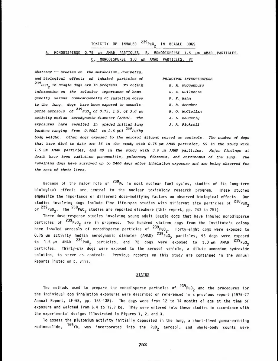
TOXICITY OF INHALED 239pu02 IN BEAGLE DOGS
A. MONODISPERSE 0.75 ~m AMAD PARTICLES. B. MONODISPERSE 1.5 um AMAD PARTICLES.
C. MONODISPERSE 3.0 um AMAD PARTICLES. VI
Abstract -- Studies on the metabollsm, doslmetry,
and b~ologlcal effects of inhaled partlcles of
239pu02 in Beagle dogs are in progress. TO obtain
informatlon on the relative importance of homo-
genelty versus nonhomogenelty of radlatlon doses
CO the lung, dogs have been exposed to monodls-
perse aerosols of 239pu02 of 0.75, 1.5, or 3.0 pm
actlvlty median aerodynamic diameter (AMAD), The
exposures have resulted in graded initial lung
burdens ranging from 0.0002 to 2.6 pCl 239pu/kg
PRINCIPAL INVESTIGATORS
B. A. Muggenburg
R. A. Gullmette
F. F. Hahn
B. B. Boecker
R. O. McClellan
J. L. Mauderly
J. A. P1ckrell
body weight. Other dogs exposed to the aerosol d~luent served as controls. The number of dogs
that have died to date are 16 ~n the study wlth 0.75 ~m AMAD particles, 55 in the study with
1.5 Bm AMAD particles, and 40 in the study with 3.0 pm AMAD particles. Major findings at
death have been radiation pneumonltls, pulmonarg flbrosls, and carcinomas of ~he lung. The
remalnlng dogs have survived up to 2400 da~s after ~nhalarlon exposure and are being observed for
the rest of their lives.
Because of the major role of 239pu in most nuclear fuel cycles, studies of its long-term
biological effects are central to the nuclear toxicology research program. These studies
emphasize the importance of different dose-modifying factors on observed biological effects. Our
studies involving dogs include five life-span studies with different size particles of 238pu02
or 239pu02. The 23Bpu02 studies are reported elsewhere (this report, pp. 243 to 251).
Three dose-response studies involving young adult Beagle dogs that have inhaled monodisperse
particles of 239pu02 are in progress. Two hundred sixteen dogs from the Institute’s colony
have inhaled aerosols of monodisperse particles of 239pu0~. Forty-eight dogs were exposed toz
0.75 ~m activity median aerodynamic diameter (AMAD) 239pu02 particles, 96 dogs were exposed
to 1.5 ~m AMAD 239pu02 particles, and 72 dogs were exposed to 3.0 ~m AMAD 239pu02
particles. Thirty-six dogs were exposed to the aerosol vehicle, a dilute ammonium hydroxide
solution, to serve as controls. Previous reports on this study are contained in the Annual
Reports listed on p. viii.
STATUS
prepare the monodisperse particles of 239pu02 and the procedures forThe methods used to
the individual dog inhalation exposures were described or referenced in a previous report (1976-77
Annual Report, LF-58, pp. 135-138). The dogs were from 12 to 14 months of age at the time of
exposure and weighed from 6.4 to ]2.7 kg. They were entered into these studies in accordance with
the experimental designs illustrated in Figures l, 2, and 3.
To assess the plutonium activity initially deposited in the lung, a short-lived gamma-emitting
radionuclide, 169yb, was incorporated into the PuO2 aerosol, and whole-body counts were
252

P90J~CI|DUCl/XO ] A
~3E0~ 19 O. 73
0.07E-2106
903F0.07 O. q7
0,09!0-295"/
97000.029 0.27
0 ̄ 0260=2Y50
969n0,010 0.25
0.025A-2qS"/
9510CONTROLO
00-2950
063£0.730.007R-Zq57
B C 0 E F G 0 ] J K L
%0, %2. 996070000102..160,E%92,%0. r,,2,97139~,r1.90.93 1,7 0.52 0.67 1.5 0.07 t.S L9 {.2 ].0 1,"/0.003 0.16 0.073 0,000 0.15 0,10 0,16 0.18 0,15 0.17 0,19E-1570 0-1035 0=292y E-1961 0-2298 0=1990 E-1371 D-1520 E-1281 0-091 E-1101
oo2, %0: ooo~Too= 1o2001o0. 10969%70 V, 122771309,,,G,0.21 0.51 0,51 0.69 0.07 0.63 0.37 1.2 0.07 1.0 0.900,021 0.055 0,063 0,062 0.10 0.073 O,OqO O. lO 0. il 6,21 0.093A=2929 0=2006 R=2325 D-2085 R-2297 0~19Y0 R-1959 E-1797 E~1525 0-196? 0-1601
970T f930~ ~001T 10060 1023W 1097C {0960 {1000 11219 11308 tI~3T6.20 0.q5 0.63 0.39 0.51 0.97 0,20 0.27 0,3~ O.Sq 0.370.019 0.097 0.059 0.095 0.059 0.091 0,0~5 0.0~7 0,091 0.051 O. Oql0"2~30 E-1710 E’2081 0I~395 0-~2q7 fl-{OqO A-1359 A-1933 0-{855 R-1773 R-1681
;g?"/$ 900C 9967 1005D 10289 {00GG 109~T 11110 11251 11390 11Y3~
{O.t~ O,uB? 0.073 0.1~ 0.033 0.073 0.11 0.20 0.17 0.40 0.{50.017 0.009 0.009 0,099 0+0]0 0.007 0.010 0.021 0.020 0,090 0.015R-2q20 0-2399 0-232~ fl-2395 9-22117 fl-13qO 0=1359 A-1933 0-1655 A-{773 R-{681
" I I { I 1 1000~ 9920 9990 1007C 10220 10990 10957 {tOUR 11217 11310 119~
0 0 o 0 0 0 o o 0 0 06-23z9 o-2~090-~33~o-,0~ .0 s_ 0-’.9 L~L,. n-,"/"/, n-16~
l-AN R~L NUMBER:INITIAL LUNG BURDEN IUC|I=]N]T]RL LUNG BUnOEN IUC]/~t=G=OEOD, E=EUTNnNI1EO, R=~LIVE - 00Y~PO$T EXPOSURE AT OLRTI{ OR 00 9-30-83
0.089
0.05{
0.022
I’0
Figure 1. Experimental design for dog study with 0.75 gm AMAD monodisperse particles of239pu02 (status as of 9/30/83),
PROJfCI[OUCI/KG
EC 0 F G H I J K LA
I~] 375
9.30,930-299
~139"02.00,19E-152
~72A
5.70,57E-561
l~l 1707.70,000-221 i
~50fl
5,10.99E-593
~930U6,70.000-907
Y]Ol5Bq.t0,970-399
]023X2.S0.30E~652
~ll 1DLI0.71.00=206
0,670.98
1~92"/0q,0O.U50-052 i
P’O09T1,70.16E-1931
~096C13,70.58D-909
I~r090B9.6O.qqE-387
I~119 ]02.20,33D~793
V~1300
1.10,11D-973
~"{{ 55T~.Y0.51E-522
~ T7B3.60,310-503
Prg611 l1.3O, ITE-1977
995Cq.OO.UO9-13~9
9~9992,70.290-720
O, 3"/0.20
~0920~I,0:0.91E-737
1~02991.50,1~0-17117
YiO2OT1,$0.19O-ll3q
PTo99v2,10.23E-gq?
]~’] ]q 151.90.190-995
1.00,150-1950
~96531.7O.l"/E-1981
997C2.U0.290-1266
~9091"1.30.190-16811
{{2900,610.0"/3A-]779
0,21O* 19
~132G
O + 73O. 06 q0-705
1.10,11E-2P60
~i20RI.O0,11E-1719
~11160I
O. IqE-153q
~001.50.150-1009
9~96611.00.100-1002
~9~MT,].i0.130-,E’010
~1 oo7o
0.670,073E-{GM{
113900.0~0.10R~1608
99501.20. II0-2359
109581.30.19A- 1B9~1
0.0"/ 0,tl
~1290 ’
0.600,0560-062
{153TO.q70.057A-{068
1~099£0.6~O,OGO0-1735
~725
0,300.0q60-2190
1130T0.070.0930-1769
9700o.2q0.0200-2q95
99270,90O.Oq30-2309
102500.920,0390-2305
1022To.q30,097A-2291
11096Di0.22O.O~l0-1983
1099T1.60,190-1893
D,OGO
irOlOtO, IW0.0130-{109
~lrlt1200.930.11E-lOflG
9~9200,100.0170-2295
97OF0,150.0190-2qq9
98650.007O. OtiA-2309
100"/00.900.0~50-2305
I092CO. tlO.Otl0-]98~
111900.250.096R-IBSG i
119950. i60,0100-1766
I130C0.930.0979-1779
115350,110.012~-{660
96960.150.0160-2952
0.010 0,030
9B800,0620.00670-2390
{01790,210.0210-2~06
1097A0.0550 ̄ 0062R-198~
111050.360,0~50-1992
11100O,08O0,00930-1896
IIMGT0.130.0150-1689
{19200.0590,00600-1790
115q9O.O~{0.0035A-1669
97200,0330.00q00-2996
950U0.0930.0100-2952
99980.0636.0069A-2355
|0100O.OY50.60930-2292
0,0120 ̄ 0025
g‘/IC0.020O.OOZ$6-2916
97050.0730,0079R-2952
98850,0210,0021N-2390
101q[0,120.615R-2306
1022V0.00670.00057R-2292
109500.015O,O0{u0-1959
1112U0.150.0170-]893
flOOR0.0530.00630-1895
113050,0330,00910- t ]66
11~100.0250.002~0-{780
l|qgT0.0250.0033A=1660
99700.0190.0,017R-2355
0.00530,00025
11075
111099 111365
O 0 0O O 0A-IRql A-109q A-1770
97700oA-2qqq
960100o-2q50
1010900o-23oq
102150oU-2290
10930000-1999
I]31000A-1780
{{5250g~-1069
ggs~000-2397
0CONTROL
~97205,70.57E-561
=ONIMgt NUMBER=INIIIAL LONG BUROEN (UCI)~INITIRL LUNG OUACWN IU¢IIKGI=O=OCAO, [=[U]~RGIZ[D, A=OL]¥[ - DOY5P091FXPOSUA~ 010(GIM OR ON 9-30-03
Figure 2. Experimental design for dog study with 1.5 pm AMAD monodisperse particles of239pu02 (status as of g/30/83),
253
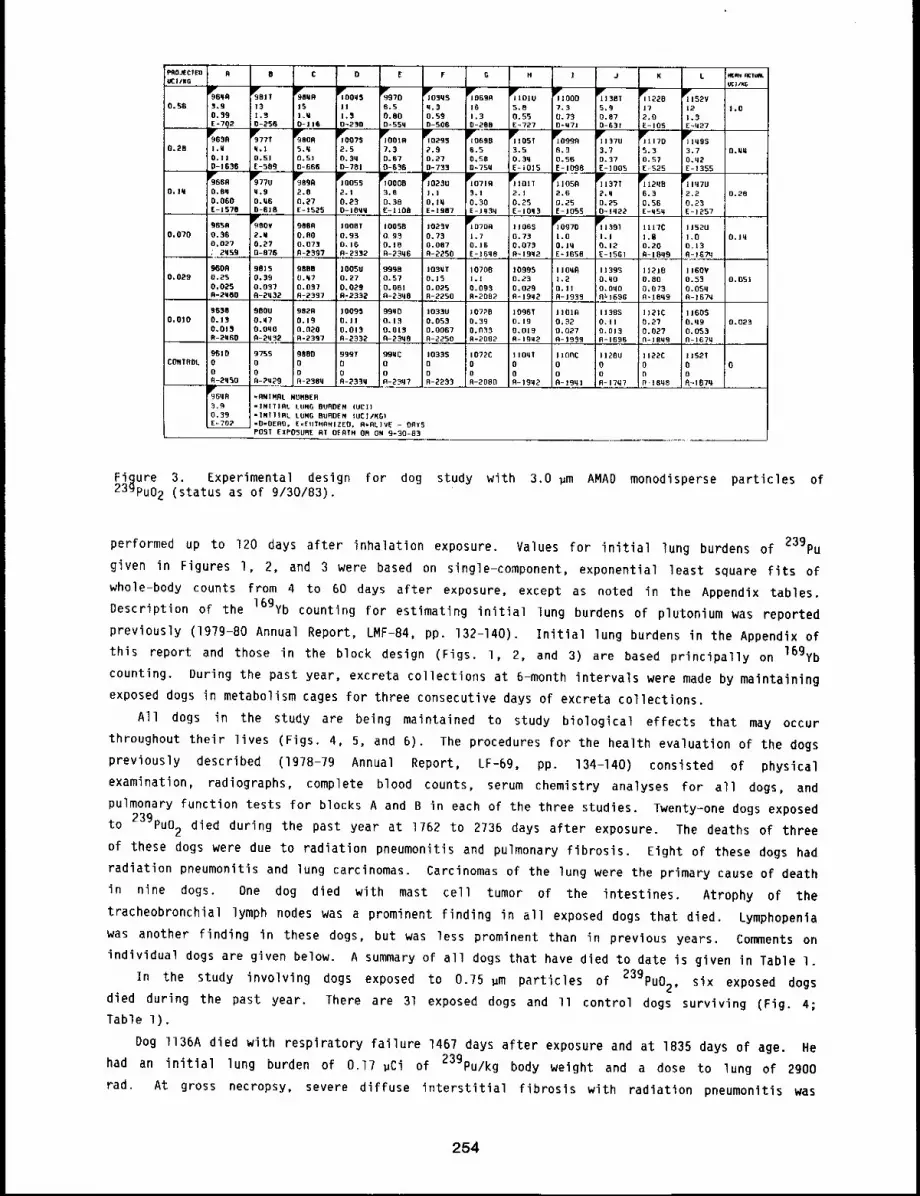
~OJtC1[:o 9 9 C D E F G N ! J Ku£1/NG . I.
~9839tc}lxG
~82~ ~zi9 100=45 ~99"/D ~GYB P~] 101U ~"11000 ~BX I 1229 ~1152VO. Y6 5.9 13 15 Jl 6.5 q.3 i6 5.B 7.3 5,5 J7 12 1.0
0.59 1.5 1.5 0.80 0.59 I+3 O, 55 O. 73 0.87 2+0 1.3E*7qa 0-2’59 O-ii9 0-250 0-553 0-506 0-2’88 [:~727 0-1171 0-631 E-J05 E-I]27
II~+’9659 VS77T 919r8OR T] 0075 ~IOOTR lYo255 z0690 ~’rl 109T ~1099R jilt 37u 11170 ~’1149$
0.28 q.I 5.q ;2.5 7,3 2.9 6.5 3.5 6+3 3.7 5.3 3.7 fl+G~0. II 0.51 0.5] :O.$q 0.57 0.23 0.50 O. 3q 0.56 0.37 0.57 O.q2D-1639 [:-509 0.,668 0-791 0-650 0-733 D=TSMJ~ E--~OI5 [:- 1098 [-1005 [-.525%;-- ~,999 i~J E-1355
0055 11~071R VJ I01T lYJ 2q9 ~lq7UO. Iq 0.93 q.9 2.8 :2.1 _3. B )O230J.l 5.1 2. J 2.6 2.q 6.3 2,2 0.28
0.060 O.Y6 0.2’7 0.2’5 0.98 D, IW O. 30 O. ~5 0.25 0.25 0,56 0.23E-1570 0-019 E+152’5 O-10qq E+1109 EIt3N7 E-]YSq E-IOq3 [:- io9+ 0+1~22 E=W5q [:-1257965A 9090 IV1008]" 10050 i023V 107OR 11069 vr10970 IV] 391 IilTC I J52U
0.070 0.56 2.M O,flO ’0.93 09~ 0.73 1.7 0.73 1.0 1.I I.U 1.0 O.Jq0.027 0,2’7 0.073 0,16 0.10 0.087 0. J6 0,073 O. lq 0.12’ 0.20 D.13zqs9 D-879 0-~597 0-2312 R-23q6 0-2’250 [-]GqB R-19q2 E=iDSB E- 15E;I 91-i8+~.~ ............A-IG?R
9600 9825 BBBB ]O05U 9999 IO3qT 10700 10995 i I OqA 11395 I1PIB 1160VO.O~9 0.25 0.30 6.Y7 0.27 0.57 0.15 I.l 0.23 1,2 O.qO O,BO 0.53 0,051
0.02’5 0.037 0.037 0+0~9 0.061 0.025 0,095 0.029 O. il O.GqO O, 073 O.05qA-2,SO 0-2’q32’ 0-2337 B-2332’ A-2’SqB R-2250 R-20B2 fl+lBq~ 0-1933 fl~t696 fl-,IBq9 D-1G7M
565B 900U 082B 10095 99qD 1033U 107~B 10~6T JIOlO 1138S I]21C 11605O.OlO 0.15 O.q7 0. i9 0.11 0.13 0.053 0.39 0.19 0,32’ O. II 0.27 O.q9 0.023
O.Ol~ O.oqo 0.020 0.013 0.01~ 0.0067 0.0~3 0.015 0,027 0,013 0.02? 0.053R92q50 0-2’q32 0-2337 D-2332’ A-2’~q8 R-2250 R=2082 B-Igq~ 0-1939 R-IGBG N+16~9 N-167~
9610 9755 9800 999T -qBwc 10335 107~C iiOqT 1100C 11200 112’2C 11521CON?OOL0 0 0 0 0 0 0 0
o0 0 0 0 0
0 o 0 o 0 D 0 0 o o D0-2q50 R-?~9 R-23Oq 0-~33q o-2_~7 R-2235 R-~OBg 0-19q~ O-19ql R-17q7 R l8q8 ~--t67qr96qfl *ANIMRL NUHBER3,3 "INITIRL EUNG BURO[:N IUCI)0.39 "INI]IRL LUNG BURDEN IUCI/KO)E~702 =0"0E09. E=FliTN~N]ZEO. R.AL]VE - ORY5
POST [:XPO5URE RT Of RTN 05 ON 9-30-83
Figure 3. Experimental design for dog study with 3.0 pm AMAD monodisperse particles of239pu02 (status as of 9/30/83).
performed up to 120 days after inhalation exposure. Values for initial lung burdens of 239pu
given in Figures 1, 2, and 3 were based on single-component, exponential least square fits of
whole-body counts from 4 to 60 days after exposure, except as noted in the Appendix tables.
Description of the 169yb counting For estimating initial lung burdens of plutonium was reported
previously (1979-80 Annual Report, LMF-84, pp. 132-140). Initial lung burdens in the Appendix
this report and those in the block design (Figs. l, 2, and 3) are based principally on 16gYb
counting. During the past year, excreta collections at 6-month intervals were made by maintaining
exposed dogs in metabolism cages for three consecutive days of excreta collections.
All dogs in the study are being maintained to study biological effects that may occur
throughout their lives (Figs. 4, 5, and 6). The procedures for the health evaluation of the dogs
previously described (1978-79 Annual Report, LF-69, pp. 134-140) consisted of physical
examination, radiographs, complete blood counts, serum chemistry analyses for all dogs, and
pulmonary function tests for blocks A and B in each of the three studies. Twenty-one dogs exposed
to 239pu02 died during the past year at 1762 to 2736 days after exposure. The deaths of three
of these dogs were due to radiation pneumonitis and pulmonary fibrosis. Eight of these dogs had
radiation pneumonitis and lung carcinomas. Carcinomas of the lung were the primary cause of death
in nine dogs. One dog died with mast cell tumor of the intestines. Atrophy of the
tracheobronchial lymph nodes was a prominent finding in all exposed dogs that died. Lymphopenia
was another finding in these dogs, but was less prominent than in previous years. Comments on
individual dogs are given below. A summary of all dogs that have died to date is given in Table l.
In the study involving dogs exposed to 0.75 pm particles of 239pu02, six exposed dogs
died during the past year. There are 31 exposed dogs and ll control dogs surviving (Fig. 4;
Table 1).
Dog ll36A died with respiratory failure 1467 days after exposure and at 1835 days of age. He
had an initial lung burden of O.l? pCi of 23gPu/kg body weight and a dose to lung of 2900
rad. At gross necropsy, severe diffuse interstitial fibrosis with radiation pneumonitis was
254
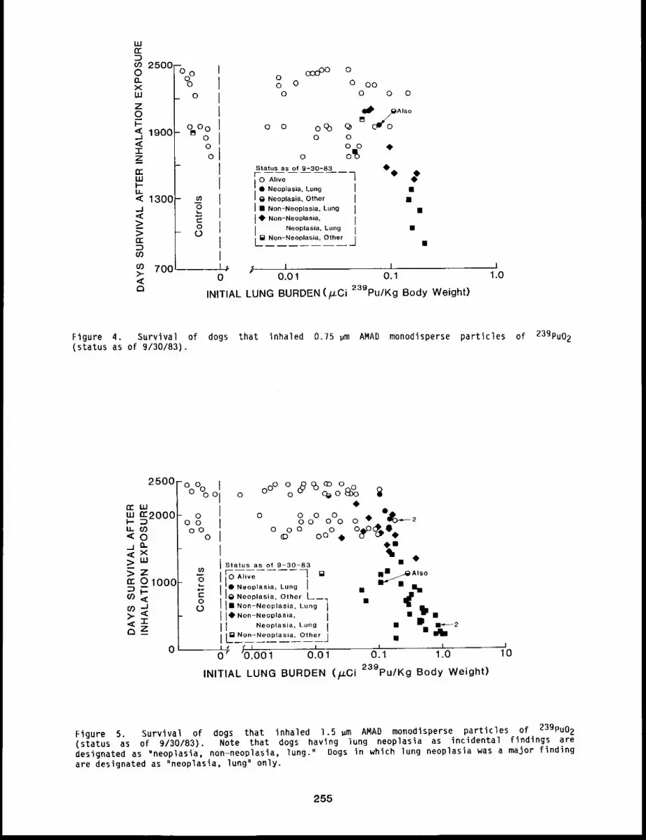
w
2500OXWZo
1900
IZm
W
< 1300J<>m
700<
Oo
o
°~°ooO
o8O
030O0 000 0 0 O00 0 0 0
o o 0(~) (~ I~JPZAls °
O OO O ¯
Status as of 9-30-83 0¯=1--0 Alive
Neoplasia, LungNeoplasia, Other
I ¯ Non-Neoplasia, LungI ¯ Non-Neoplasia,
Neoplasia, Lung
L..~ Non-Ne0plasia, Other
0 0.01 0.1
INITIAL LUNG BURDEN (/.tCi 239pu/Kg Body Weight)
I1.0
Figure 4. Survival of dogs that inhaled 0.75 ~m AMAD monodisperse particles of 239pu02(status as of 9/30/83).
2500Fo o )~°%oIm 2000LI--I.L.<o o
~ m~
nr 0 1000 "6
O9
,~’r"~_z
ooOO o ~ ~oo~
o qa
0000 ¯ @~20 000 0
o oo o ooO. o o@%
Status as of 9-30-83Alive ] []
]]:Neoplasia, Lung ]Neoplasia, Other ~__.¯ Non-Neoplasia, Lung
]¯ Non-Neoplasia,Neoplasia, Lung
L~Non-Neopla sia, Other¢ I I~ "o.oo i 0.01
¯ I., ___-~ Also
¯ ¯ r I¢~
¯m
i J0 0.1 1.0 10
INITIAL LUNG BURDEN (~Ci 239pu/Kg Body Weight)
Figure 5. Survival of dogs that inhaled 1.5 ,m AMAD monodisperse particles of 239pu02(status as of 9/30/83). Note that dogs having lung neoplasia as incidental findings aredesignated as "neoplasia, non-neoplasia, lung." Dogs in which lung neoplasia was a major findingare designated as "neoplasia, lung" only.
255
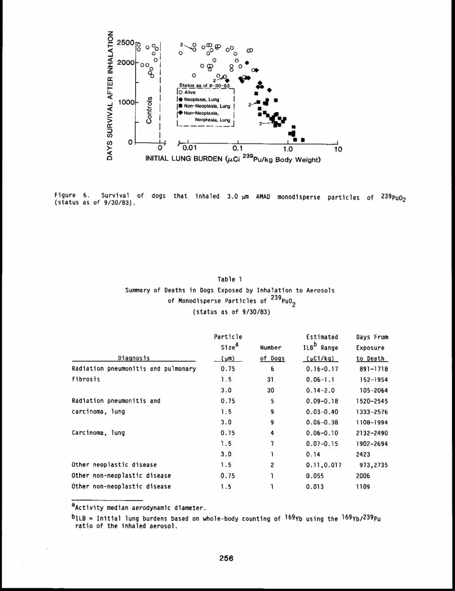
Z
<,=<zn-
LU
u.<
<>>nr
o9DO>-<(3
25001"0 ~ o II o ~oTM
20001oo~°
1000
0
¢0
+8
2 o Oo%@DOo o
o 8 °¯o+~c)StatusAli~e as of 9-30-83 ] ~
I¯ Neoplasia, Lung II" No.-.oop,as+a. L.,ng I ~’= ~ ̄l¯ N°n-N°op’a~ia’ I
!~dP[Neoptasia, Lung j
10INITIAL LUNG BURDEN (p.Ci 239pu/kg Body Weight)
Figure 6. Survival of dogs that inhaled 3.0 wm AMAD monodisperse particles of 239pu02(status as of 9/30/83).
Table 1
Summary of Deaths in Dogs Exposed by Inhalation to Aerosols
of Monodisperse Particles of 239pu02
(status as of 9/30/83)
Diagnosis
Radiation pneumonitis and pulmonary
fibrosis
Radiation pneumonitis and
carcinoma, lung
Carcinoma, lung
Other neoplastic disease
Other non-neoplastic disease
Other non-neoplastic disease
Particle Estimated Days From
Sizea Number ILBb Range Exposure
(~m) of Dogs (uCl/kq) to Death
0.75 6 0.16-0.17 891-1718
1.5 31 0.06-I.I 152-1954
3.0 30 0.14-2.0 I05-2064
0.75 5 0.09-0.18 1520-2545
1.5 g 0.03-0.40 1333-2576
3.0 g 0.06-0.38 II08-1994
0.75 4 0.06-0.I0 2132-2490
1.5 7 0.07-0.15 1902-2694
3.0 l 0.14 2423
1.5 2 0.11,0.017 973,2735
0.75 l 0.055 2006
1.5 l 0.013 II09
aActivity median aerodynamic diameter.
bILB = initial lung burdens based on whole-body counting of 169yb using the 169yb/239puratio of the inhaled aerosol.
256
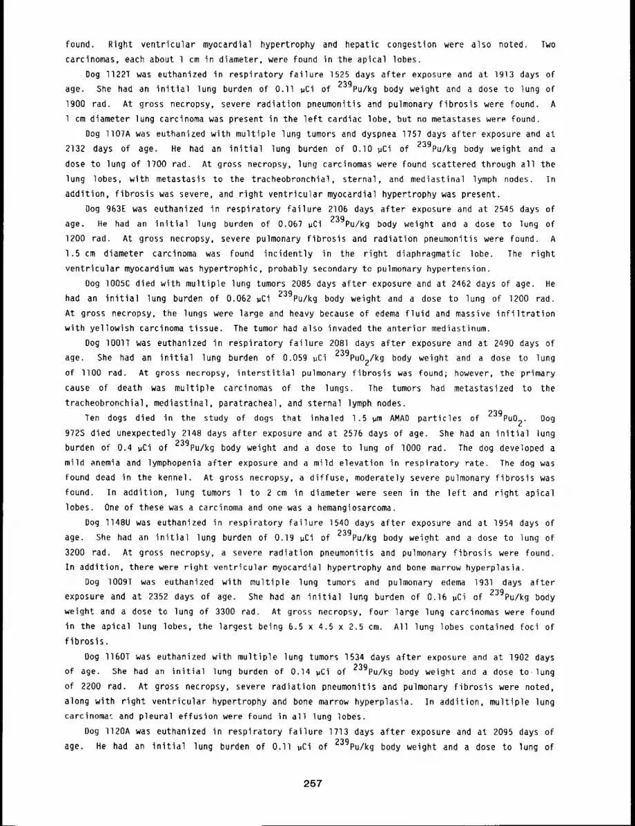
found. Right ventricular myocardial hypertrophy and hepatic congestion were also noted. Two
carcinomas, each about l cm in diameter, were found in the apical lobes.
Dog ll22T was euthanized in respiratory failure 1525 days after exposure and at igl3 days of
age. She had an initial lung burden of 0.II #Ci of 239pu/kg body weight and a dose to lung of
1900 rad. At gross necropsy, severe radiation pneumonitis and pulmonary fibrosis were found. A
l cm diameter lung carcinoma was present in the left cardiac lobe, but no metastases were found.
Dog ll07A was euthanized with multiple lung tumors and dyspnea 1757 days after exposure and at
2132 days of age. He had an initial lung burden of O.lO #Ci of 23gPu/kg body weight and a
dose to lung of 1700 rad. At gross necropsy, lung carcinomas were found scattered through all the
lung lobes, with metastasis to the tracheobronchial, sternal, and mediastinal lymph nodes. In
addition, fibrosis was severe, and right ventricular myocardial hypertrophy was present.
Dog 963E was euthanized in respiratory failure 2106 days after exposure and at 2545 days of
age. He had an initial lung burden of 0.067 ~Ci 23gPu/kg body weight and a dose to lung of
1200 rad. At gross necropsy, severe pulmonary fibrosis and radiation pneumonitis were found. A
1.5 cm diameter carcinoma was found incidently in the right diaphragmatic lobe. The right
ventricular myocardium was hypertrophic, probably secondary to pulmonary hypertension.
Dog lO05C died with multiple lung tumors 2085 days after exposure and at 2462 days of age. He
had an initial lung burden of 0.062 ~Ci 239pu/kg body weight and a dose to lung of 1200 rad.
At gross necropsy, the lungs were large and heavy because of edema fluid and massive infiltration
with yellowish carcinoma tissue. The tumor had also invaded the anterior mediastinum.
Dog IO01T was euthanized in respiratory failure 2081 days after exposure and at 2490 days of
age. She had an initial lung burden of 0.059 ~Ci 239puO2/kg body weight and a dose to lung
of llO0 rad. At gross necropsy, interstitial pulmonary fibrosis was found; however, the primary
cause of death was multiple carcinomas of the lungs. The tumors had metastasized to the
tracheobronchial, mediastinal, paratracheal, and sternal lymph nodes.
Ten dogs died in the study of dogs that inhaled 1.5 ~m AMAD particles of 23gPuo2. Dog
972S died unexpectedly 2148 days after exposure and at 2576 days of age. She had an initial lung
burden of 0.4 ~Ci of 239pu/kg body weight and a dose to lung of lO00 rad. The dog developed a
mild anemia and lymphopenia after exposure and a mild elevation in respiratory rate. The dog was
found dead in the kennel. At gross necropsy, a diffuse, moderately severe pulmonary fibrosis was
found. In addition, lung tumors 1 to 2 cm in diameter were seen in the left and right apical
lobes. One of these was a carcinoma and one was a hemangiosarcoma.
Dog ll48U was euthanized in respiratory failure 1540 days after exposure and at 1954 days of
age. She had an initial lung burden of 0.19 ~Ci of 239pu/kg body weight and a dose to lung of
3200 tad. At gross necropsy, a severe radiation pneumonitis and pulmonary fibrosis were found.
In addition, there were right ventricular myocardial hypertrophy and bone marrow hyperplasia.
Dog I009~ was euthanized with multiple lung tumors and pulmonary edema 1931 days after
exposure and at 2352 days of age. She had an initial lung burden of 0.16 #Ci of 239pu/kg body
weight and a dose to lung of 3300 rad. At gross necropsy, four large lung carcinomas were Found
in the apical lung lobes, the largest being 6.5 x 4.5 x 2.5 cm. All lung lobes contained loci of
fibrosis.
Dog ll60T was euthanized with multiple lung tumors 1534 days after exposure and at 1902 days
of age. She had an initial lung burden of 0.14 #Ci of 239pu/kg body weight and a dose to,lung
of 2200 rad. At gross necropsy, severe radiation pneumonitis and pulmonary fibrosis were noted,
along with right ventricular hypertrophy and bone marrow hyperplasia. In addition, multiple lung
carcinoma~ and pleural effusion were found in all lung lobes.
Dog ll20A was euthanized in respiratory failure 1713 days after exposure and at 2095 days of
age. He had an initial lung burden of O.ll #Ci of 239pu/kg body weight and a dose to lung of
257
2000 tad. At gross necropsy, severe widespread radiation pneumonitis and pulmonary fibrosis were
found. A 4 x 4 x 3 cm carcinoma was present in the right apical lung lobe and had metastasized to
the tracheobronchial lymph node. Right ventricular myocardial hypertrophy and bone marrow
hyperplasia were also present.
Dog lO08S was euthanized in respiratory failure 2269 days after exposure and at 2694 days of
age. She had an initial lung burden of O.ll ~Ci of 239pu/kg body weight and a dose to lung of
2400 rad. Extensive pulmonary fibrosis and a large 5 cm diameter carcinoma in the right
intermediate lobe were found. In addition, two smaller nodules were found in other lobes. No
metastases were found outside of the lungs.
Dog lO07A was euthanized because of radiation pneumonitis and multiple lung tumors 1941 days
after exposure and at 2354 days of age. He had an initial lung burden of 0.0?3 ~Ci of
239pu/kg body weight and a dose to lung of 1500 rad. At gross necropsy, extensive pulmonary
fibrosis was present. Carcinomas were found in the right apical and left cardiac lung lobes. The
largest tumor measured 1.3 x 3 x 4 cm. No metastases were found.
Dog I099C died 1735 days after exposure and at 2186 days of age. There was a prolonged period
of dyspnea. He had an initial lung burden of 0.06 ~Ci of 239pu/kg body weight and a dose to
lung of llO0 rad. At gross necropsy, severe diffuse radiation pneumonitis and pulmonary fibrosis
were found, along with right ventricular myocardial hypertrophy. Nodules of pulmonary carcinoma
were present in the right apical lung lobe.
Dog 1094B was euthanized in respiratory failure 1646 days after exposure and at 201B days of
age. He had an initial lung burden of 0.02? ~Ci of 239pu/kg body weight and a dose to lung of
550 rad. At gross necropsy, a severe diffuse radiation pneumonitis was found along with right
ventricular myocardial hypertrophy. A small yellow mass was found in the right apical lobe, but
no metastases were found.
Dog 992D died unexpectedly 2295 days after exposure and at 2735 days of age. He had an
initial lung burden of 0.017 ~Ci of 239pu/kg body weight and a dose to lung of 400 rad. At
necropsy, an acute hemorrhagic septic peritonitis was found secondary to a perforation of the
jejunum. The intestinal wall was thickened at the site of perforation. This thickening was due
to an infiltration of anaplastic mononuclear cells, apparently mast cells. Metastases of these
cells were apparent in the heart and the anterior mesenteric lymph node.
Five dogs died in the study of dogs that inhaled 3.0 ~m AMAD particles of 239pu02.
Dog ll49S was euthanized in respiratory failure 1355 days after exposure and at 1762 days of
age. She had an initial lung burden of 0.42 ~Ci of 239pu and a dose to lung of 8000 rad. At
gross necropsy, severe diffuse interstitial pneumonitis and fibrosis were found. The right
ventricular myocardium was hypertrophic and the bone marrow hyperplastic.
Dog l137T died 1422 days after exposure and at 1862 days of age. She had an initial lung
burden of 0.25 ~Ci of 23gPu/kg body weight and a dose to lung of 4800 rad. At gross necropsy,
a severe pulmonary fibrosis was found, accompanied by right ventricular hypertrophy. Lung
carcinomas were found in the cardiac lobes, but no metastases were present.
Dog 1023U was euthanized with a lung tumor 1987 days after exposure and at 2423 days of age.
She had an initial lung burden of 0.14 ~Ci of 239pu/kg body weight and a dose to lung of 3600
rad. At gross necropsy, a large 4 x 4 x 2.5 cm mass of carcinoma was present in the right apical
lung lobe. All lung lobes contained streaks of fibrosis, but no metastases were evident.
Dog lO97D was euthanized in respiratory failure 1658 days after exposure and at 2064 days of
age. He had an initial lung burden of 0.14 ~Ci of 239pu/kg body weight and a dose to lung of
2300 rad. At gross necropsy, severe and widespread radiation pneumonltis and pulmonary fibrosis
were found. Right ventricular myocardial hypertrophy was also found.
258
Dog II3gT was euthanized in respiratory failure 1561 days after exposure and at 1994 days of
age. She had an initial lung burden of 0.12 ~Ci of 239pu/kg body weight and a dose to lung of
2300 rad. At gross necropsy, a severe diffuse interstitial pneumonitis and pulmonary fibrosis
were present. A hard 1.5 cm diameter nodule of carcinoma was found in the right diaphragmatic
lobe.
DISCUSSION
Seventeen of the 21 dogs that died this past year had primary lung tumors. They were the
cause of death in nine of these dogs. The dogs that died or were euthanized with pulmonary
failure from radiation pneumonitis continue to be dogs with the highest initial lung burdens of
239pu02. Dogs are continuing to develop severe pulmonary fibrosis and radiation pneumonitis
to 2500 days after inhalation exposure. This is in sharp contrast to the studies in dogs exposed
by inhalation to beta-gamma-emitting radionuclides (1976-77 Annual Report, LF-S8, pp. 139-143).
In those studies, death associated with radiation pneumonitis was rare later than 500 days after
exposure. There is a trend that the dogs exposed to the 3.0 ~m AMAD particles of 239pu02
are dying at later times and at higher doses to lung than the dogs exposed to the 0.75 ~m AMAD
and 1.5 pm AMAD particles. This may reflect the higher degree of nonuniformity of pulmonary
irradiation that occurred in these dogs.
The surviving dogs will continue to be observed during the coming year. Several dogs
presently have radiation pneumonitis and lung tumors. The appearance of more lung tumors is
expected in the exposed dogs.
259
IOXICITY OF INHALED 23gPu02 IN IMMATURE BEAGLE DOGS. V
Abstract --Immature Beagle dogs have been exposed
by inhalatlon to a monodlsperse aerosol of 239pu02
(1.5 ~m AMAD) to compare the blologlcal effects
with those observed ln 9oung adult and aged dogs
exposed to a slmllar aerosol. The study includes
96 dogs exposed to 239pu02 and 12 controls. The
lung burdens ~n the plutonium-exposed dogs ranged
from 0.00030 to 0.80 ~Cl/kg body welght. Durlng
the past year, two dogs d~ed from causes apparent-
PRINCIPAL INVESTIGATORS
R. A. Gullmette
B. A. Muggenburg
F. F. Hahn
J. L. Mauderl9
B. B. Boecker
R. O. McClellan
ly unrelated to radlatlon effects. With a median tlme on study of 700 days, there have not yet
been any deaths related to alpha ~rrad~at~on of the lung, even though iI dogs have cumulatlve lung
doses greater than 1000 rad.
Exposure of the general population to airborne radioactive materials from a nuclear incident
would probably involve persons of widely differing ages. Therefore, it is important to understand
the role of age at exposure in modifying the dose pattern and resulting biological effects. Two
series of experiments are in progress at this Institute to investigate the effect of age at
exposure on the biological effects subsequent to inhaling radioactive aerosols. The first series
of dose-response studies involves inhalation of the beta-emitter 144Ce in a relatively insoluble
fused aluminosilicate clay matrix by immature (90 day), young adult (12-14 month), and
(8-I0.5 years) Beagle dogs. The second series of studies involves single inhalation exposures
similarly aged immature, young adult, and aged Beagle dogs to the alpha-emitter 239pu as the
insoluble dioxide. This report concerns the toxicity of inhaled 239pu02 in immature Beagle
dogs.
The randomized block experimental design of this study (Fig. l) is similar to that used for
most of other large animal dose-response studies in progress at this Institute. Dogs were entered
into the experiment by block, with each block containing eight dogs of the same sex exposed to
graded levels of 239pu02 aerosols, and one control dog exposed to the dilute ammonium
hydroxide aerosol vehicle only. The projected lung burden levels correspond to the same eight
levels (in ~Ci 239pu/kg body weight at exposure) used in the 239pu02 radiation
dose-response study in young adult (13 ± l month old) Beagle dogs. Each dog received a single
inhalation exposure to a monodisperse aerosol of 239pu02 with a particle size of 1.5pernasal
~m activity median aerodynamic diameter (AMAD). The gamma-emitting radionuclide 169yb was
incorporated into the aerosol particles and provided a means for establishing body burdens of
plutonium by external whole-body counting techniques described previously (IgTB-IgTg Annual
Report, LF-69, pp. 146-149). In cases where the whole-body count data did not provide a reliable
estimate of initial lung burden, this latter quantity was estimated using the measured exposure
aerosol concentration of 23gPu, and an average deposition fraction of 0.25. The details of the
methods used for aerosol generation, animal selection and clinical workup, inhalation exposure,
after-exposure sampling, and measurement have been described previously (197B-lgTg Annual Report,
LF-6g, pp. 146-149).
260
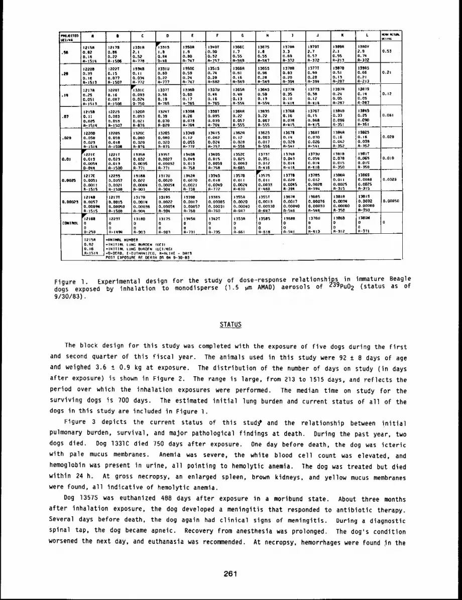
eng~CltD R II D [ F G H I J K L v~mw ~I~tgl/K6 KI/w5
12150 12179 1331A 13315 13SOR 13SLOT ! 366C 13675 ] ~700 tS?gT 13006 1380V0.82 0,66 2,1 1,0 l,g O. 00 I.? 1.0 3,3 2,7 2.1 2.9 0,$30.18 0.22 O. 52 0,£H O. 00 0,~2 0,55 0.5"3 0.50 0.S? 0.55 O, 71iR-1510 0-15O0 0-773 . 0-~0 0-?07 0-757 0-5q0 0-5q? 0-3"/2 n-372 0-3?2
12200 1222T 155qB 13310 1~50C 13S15 13660 13650 13700 1377T 15070 I$S0S.20 0.39 O.lS 0.1! 0.00 0.59 0.70 0.61 0.96 0.03 o.gg O.Sl 0.66 0.21
0,16 0.077 O.03q 0.22 0.20 0,20 0.16 0,28 0.20 0.20 0.13 0.21R-ISi3 n-tSO7 0-772 R-777 0-7~7 R-682 R-Sq9 0-5W9 R-3gq A-39q 0-20? fl-213
12170 122OT 1r~331C 1337T 13360 1337U 10650 136~$ 15776 1~770 13070 1307S.16 0,23 0.16 0,093 0.50 0.60 0,00 0.U9 0.$9 O. 35 0.30 0.20 0.10 0,12
0.051 0.007 0.020 0,19 O.J? 0.10 0.13 0.13 0,10 0.12 0.05 0.050-1513 A-ISO6 0-750 0-70S ..... 0-705 A-765 A-55~ RN55q R-qlq 0-31q 0-287 0-207
12106 12225 13200 1320T 13396 1330T 136UR 13033 13760 1076T 13000 J 30qg.07 O. ll 0,063 0.003 0.39 0.~6 0.095 0.22 0.22 0.16 0,15 0.33 0,25 O.O&i
0.025 0.050 0.021 0.070 0,075 0.039 0.057 0.067 0,076 0.060 0.096 0.090R-lSlq 0-1307 0-0?0 R-029 R-70q R-?6q 0-555 0-555 R-MI3 0-015 ~-351 R-3Sl
t2200 12205 1320C 13205 133q0 13Ul5 |3B~0 13625 13676 1360T 13000 ISBZ5,020 O.OSO 0.059 0.O00 0.060 0.12 0.062 0.12 0.063 O,iq 0.070 0.16 0.10 0.020
0.023 0.018 0.020 0.020 0.055 0,020 0.028 0.017 0.023 0.026 O.Oq2 0,039A-I 5OQ A-O?G 0-075 0-772 ....... o-2~7 0-556 0-556 0=5~1 0~5~1 0-352 n-352
~]22J£ |~21T 13350 13351 13q00 13005 1352C 1373T 137qfl 1373U 13018 i381T0,01 0.013 0.023 0.032 0.0027 0,003 0.015 0.0,?.5 0.001 O.Ou~; O.OSq 0.078 0.065 10.010
0.0030 0.013 O.oog~ 0.00032 0.013 0.0059 0.0063 0.012 O,OIq O.Ol~ 0.015 0.015D-Oqq 0-1500 0-771 0-771 0-750 0-750 0-605 0-q10 0-¢i 16 fl-ql6 0-350 0-]50
1217£ 12253 15186 13170 13q20 ]3905 13570 1~3570 13776 13785 13060 136600 ¯ 0025 0.0051 0.0057 0.022 0.0020 0.0070 0.010 O.Oll 0.01] 0.020 0.0i2 0.0~1 0.0008 O. 0029
0.0011 0,0021 0,006~ 0.0005q 0.0021 O.OOq9 O, o020 0,0033 0.00q5 0.0020 0.0025 0.002SR-13O0 A-9O3 0-900 0-730 0-772 R-630 E-q00 0-39q 0-39¼ R-315 0-315
121q| 12171 13170 10195 1339~ |3385 13SSR 1355T 13676 13005 13010 13eis0.000,~ 0.O0~? 0.0015 O.O01W 0.0022 O.OOi? 0.00085 0.0020 0.0013 0.0017 0.00076 O,o03q o.oo32 0,00050
0.00090 0.00Q’~0 0.00030 0.0005q 0,00057 0.00031 O,O00qO 0,00030 O.O00qO 0,00030 0.O0060 0.000000-1000 fl-90q 0-900 0=700 0=700 R-007 0-607 0-506 0-5M0 0-350 0~350
T21. 12231 i3100 13175 ~3q59 13q2T 13530 1350~ 13606 13700 13860 130OMC~I(TROLo 0 0 0 0 0 0 0 o 0 0 o
0 o 0 o o o 0 0 0 0 o]0-250 fl-lq’30 0-903 n-9o~ 0-731 0-735 A-661 0-610 0-501 0-q13 0-312 0-071
*RNIHRL NUHO[6=IN]TIRL LUNG BURDEN (UCI)=INITIRL LUNG OLIBOFN IUC1/KGI=O’OERO, [=EUTHRNIZ[D, R=RL]Y[ - DRT3POST EIPOSUR[ ~T DEOTH OR ON 9-30-83
Figure 1. Experimental design for the study of dose=response relationships in immature Beagledogs exposed by inhalation to monodisperse (1.5 gm AMAD) aerosols of Z3gPu02 (status as 9/30/83).
STATUS
The block design for this study was completed with the exposure of five dogs during the first
and second quarter of this fiscal year. The animals used in this study were 92 ± 8 days of age
and weighed 3.6 ± 0.9 kg at exposure. The distribution of the number of days on study (in days
after exposure) is shown in Figure 2. The range is large, from 213 to 1515 days, and reflects the
period over which the inhalation exposures were performed. The median time on study for the
surviving dogs is 700 days. The estimated initial lung burden and current status of all of the
dogs in this study are included in Figure 1.
Figure 3 depicts the current status of this stud~ and the relationship between initial
pulmonary burden, survival, and major pathological findings at death. During the past year, two
dogs died. Dog 1331C died 750 days after exposure. One day before death, the dog was icteric
with pale mucus membranes. Anemia was severe, the white blood cell count was elevated, and
hemoglobin was present in urine, all pointing to hemolytic anemia. The dog was treated but died
within 24 h. At gross necropsy, an enlarged spleen, brown kidneys, and yellow mucus membranes
were found, all indicative of hemolytic anemia.
Dog 1357S was euthanized 488 days after exposure in a moribund state. About three months
after inhalation exposure, the dog developed a meningitis that responded to antibiotic therapy.
Several days before death, the dog again had clinical signs of meningitis. During a diagnostic
spinal tap, the dog became apneic. Recovery from anesthesia was prolonged. The dog’s condition
worsened the next day, and euthanasia was recommended. At necropsy, hemorrhages were found ~n the
261
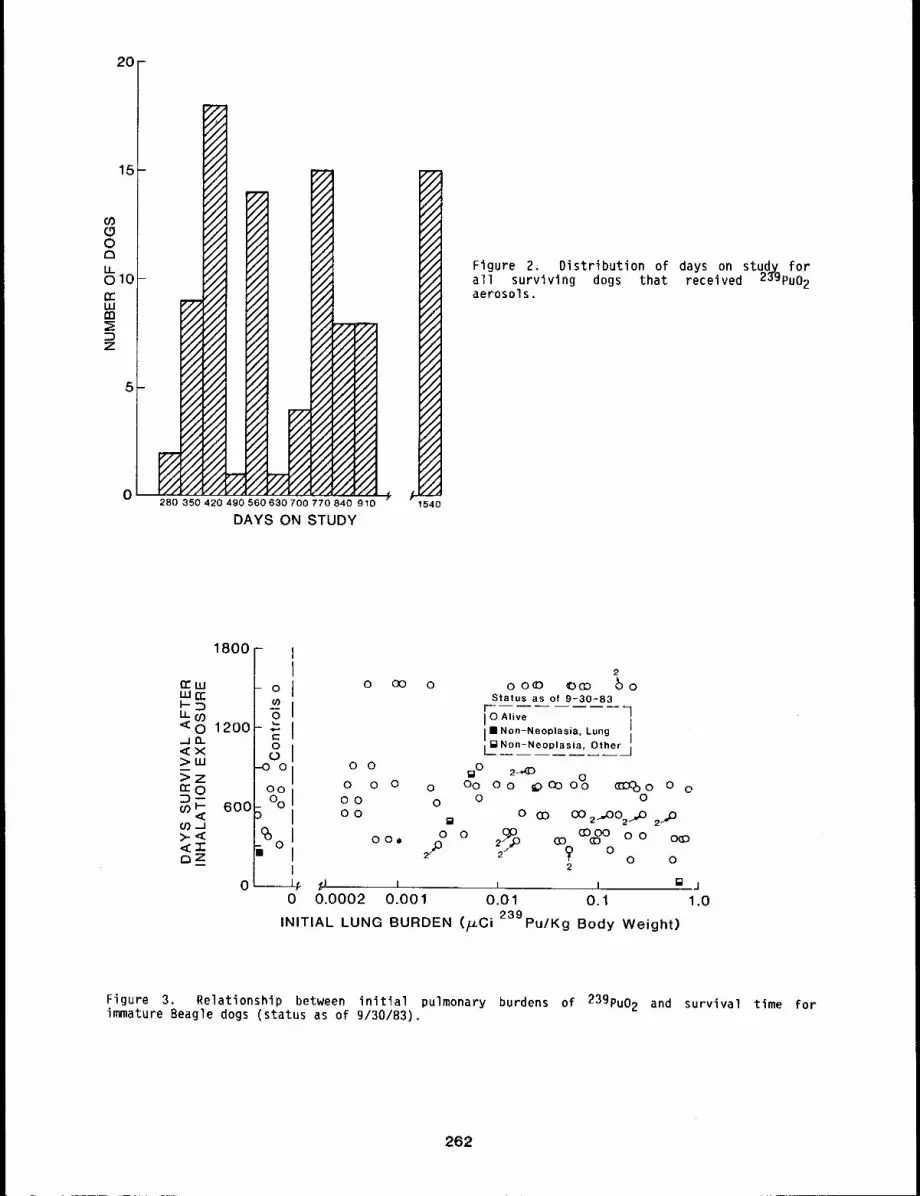
20¸-
15-
o3OO1:3LL010 -CCLUm
Z
.5-
Figure 2. Distribution of days on study forall surviving dogs that received 2~gPu02aerosols.
1800
1200
600
OU)
, O
~O
i o
o
I 0 Alive II II Non-Neoplasi~, Lung I
[laNon-Neoplasia, Other j
O OliD 2-~(D
o o o o Do Do ~>coo~ c~o ooO0 0 0 0o o
~ o co oo ~oo ~o ~,Doo./ o oooo o
2
~I ~ t t []0.0002 0.001 0.01 0.1
INITIAL LUNG BURDEN (/u.Ci 239pu/Kg Body Weight)
Figure 3. Relationship between initial pulmonary burdens of 239pu02 and survival time forimmature Beagle dogs (status as of 9/30/83).
262
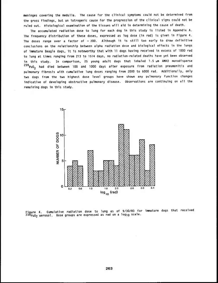
meninges covering the medulla. The cause for the clinical symptoms could not be determined from
the gross findings, but an iatrogenic cause for the progression of the clinical signs could not be
ruled out. Histological examination of the tissues will aid in determining the cause of death.
The accumulated radiation dose to lung for each dog in this study is listed in Appendix A.
The frequency distribution of these doses, expressed as log dose (in rad) is given in Figure
The doses range over a factor of - 200. Although it is still too early to draw definitive
conclusions on the relationship between alpha radiation dose and biological effects in the lungs
of immature Beagle dogs, it is noteworthy that with II dogs having received in excess of lO00 rad
to lung at times ranging from 213 to 1514 days, no radiatlon-related deaths have yet been observed
in this study. In comparison, 25 young adult dogs that inhaled 1.5 um AMAD monodisperse
239pu02 had died between lO0 and lO00 days after exposure from radiation pneumonitis and
pulmonary fibrosis with cumulative lung doses ranging from 2000 to 6000 rad. Additionally, only
two dogs from the two highest dose level groups have shown any pulmonary function changes
indicative of developing obstructive pulmonary disease, Observations are continuing on all the
remaining dogs in this study.
15
Figure 4. Cumulative radiation dose to lung as of 9/30/83 for immature dogs that received239pu02 aerosol. Dose groups are expressed as rad on a lOglO scale.
263
TOXICITY OF INHALED 239pu02 IN AGED BEAGLE DOGS. V
Abstract --Studies to determine the effects of age
at exposure on the metabolism, doslmetzy, and blo-
logical effects of inhaled partlcles of 239pu02 are
in progress in aged Beagle dogs (8.0 to 10.5 years
of age at exposure). Forty-elght dogs have inhaled
239pu02 having an activity medianparticles of
aerodgnamlc dlar~eter of 1.5 ~m to achieve initial
lung burdens ranging from 0.02 to 0.66 ~Ci 239pu/
PRINCIPAL INVESTIGATORS
B. A. Muggenburg
F. F. Hahn
R. A. Guilmette
B. B. Boeeker
R. O. McClellan
kg bodg weight. Twelve dogs exposed to the aerosol diluent serve as controls. Thlrty-flve
exposed dogs have dled~ including 10 in the past 9ear. The principal causes of death were
radiation pneumonltls and pulmonary fibrosis. Three control dogs have dled. The 13 surviving
exposed dogs and 9 controls are contlnulng to be observed.
This is one of a series of studies on the toxicity of inhaled alpha-emitting radionuclides
being conducted at this laboratory. The importance of a study such as this in aged animals can be
seen by examining the results of previous work done at this laboratory on the effects of inhaled
radioactive aerosols in aged animals. Aged Beagle dogs exposed by inhalation to aerosols of
144ce incorporated into fused aluminosilicate particles proved to be more sensitive to beta
irradiation than were young adult dogs, and they died with pulmonary injury, radiation
pneumonitis, and fibrosis at lower total doses (1978-1979 Annual Report, LF-69, pp. 69-I00).
Beagle dogs, 8.0-I0.5 years of age at the time of exposure, from the Institute’s colony were
exposed once to a monodisperse aerosol of 239pu02 having a 1.5 gm activity median
aerodynamic diameter (AMAD). Dogs are being maintained for life-span observation to relate age
exposure to the dose-response relationships for this inhaled material. The experimental design is
given in Figure I. This study is closely related to two others in progress at this Institute in
which dogs inhaled similar monodisperse aerosols of 239pu02. These other studies are: (1)
study using young adults (12 to 14 months of age at exposure) exposed to 0.75 ~m, 1.5 ~m,
3.0 ~m AMAD particles (this report, pp. 252 to 259), and (2) a study involving immature dogs
± lO days of age at exposure) exposed to 1.5 pm AMAD 239pu02 particles (this report,
pp. 260 to 263). Together, these studies will provide important information about potential
consequences of accidental inhalation exposure of 239pu02 by a heterogenously aged human
population.
STATUS
Forty-eight dogs were exposed to 23gPuo2 aerosols. Thirty-five exposed dogs have died to
date, lO during the past year. The principal causes of death have been radiation pneumonitis and
pulmonary fibrosis induced by inhaled 239pu02 (Fig. 2; Table l). Of the 12 control dogs
entered into this study, 3 have died.
Dog 637A died unexpectedly 152 days after exposure, at 3BIB days of age. He had an initial
lung burden of 0.32 ~Ci of 239pu/kg body weight and a dose to lung of BOO rad. The dog had
been hospitalized ]B days before death for radiation pneumonitis. At gross necropsy, severe
pulmonary edema was found associated with radiation pneumonitis and pulmonary fibrosis. The
264

] J K L0 [ F 6 HA B C
~qO2S5.1o.r-qE =506
rOoT5,10.52.E-Iql
Y?~gD2. ]$9O,~UE-190
JP’09302.rio0.23[-zq?
~’r~ I 2C7. JO. 600-~07
~’303T2.00.20D-208
IV3SSBq.60.390-520
O. qO0,20
|/70.10D-qB?
F]SU2.50.25E-U02
F828J .230.1|D-]67
~785
1.60.130-99q
~S[C
0.26D-731
rso~81.30.10E-O~’?
O. 20O. lq
0.65U.D7E-Ill?
J~835
1.7O. IqE-00|
~80 T0.080.110-936
~65
1.2O. 100-970
3S900.650.00E-1~66
qY600O.SS0.09[-1~50
P~SOTB0.8"/0.070-I012
785B0.60~0.066A- q72
0.10O, O?
0.3]0.02
~375T0,7B0.07E-870
639SO.qSO.O~R-]Z92
qB?~O.qOO.O3R-1589
71000.2790.02?0- q76
O. OS0.029
~955000-585
~SSOCI00E-]579
713R00u- q70
625S00A-iISq
0550o0A- qTO
510000~-IqqO
373500~-I621
38?8O0~-1631
361500R-IS31
0C~NTROL
-RNIN~. NUMBER=INIIIRL LUNG BURDEN IUEI)ulNlrI~L LUN0 BLIROEN LUC]/XG)=O-OERD, E=EUTNANIZEU, R=RLIVE - OUTSPOST-EXPOSURE OT DEOTH OR ON 9-30-83
Figure 1.exposed by09/30/B3).
Experimental design for the study of dose-response relationships in aged Beagle dogsinhalation to monodisperse particles (1.5 ~m AMAD) of 239pu02 (status as
1750
1500Q:UJ
I-
<0
< 1000>LJJ
r~O
if)o) ~: 500>--r<zC)--
o o o
o
o_o
o
0 Status as of 9-30-830 i .....
0 ¯ I 0 AliveI (]1)Neoplasia, Liver
0 I ~ Neoplasia. Other
¯i ¯ Non-Neoplasia, Lungo o¯ I [] Non-Neopolasia Other
[] Im~m ̄
oo oo I
0101
0-.-2 o 0--2
Hi
In¯ ¯
¯~m
I
¯ ¯
o
i
I0 0.1 1.0
INITIAL LUNG BURDEN (FLCi 239pu/Kc3 Bodv Weight)
Figure 2. Relationship between initial lung burden of 239pu and survival time for aged Beagledogs (status as of 09/30/83).
265
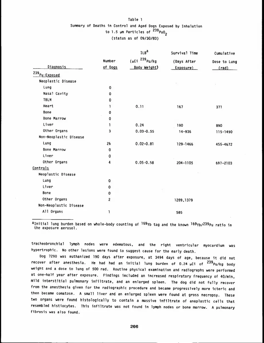
Table 1
Summary of Deaths in Control and Aged Dogs Exposed by Inhalation
to 1.5 ~m Particles of 239pu02
(status as of 09/30/83)
Diaqnosis
239pu-Exposed
Neoplastic Disease
Lung 0
Nasal Cavity 0
TBLN 0
Heart 1
Bone 0
Bone Marrow 0
Liver l
Other Organs 3
Non-Neoplastic Disease
Lung 26
Bone Marrow 0
Liver 0
Other Organs 4
Controls
Neoplastic Disease
Lung 0
Liver 0
Bone 0
Other Organs 2
Non-Neoplastic Disease
All Organs l
ILBa Survival Time Cumulative
Number (~Ci 239pu/kg (Days After Dose to Lung
Bod~) Exposure) (rad)
0.II 167 371
0.24
0.03-0.55
190 890
14-936 I15-1490
0.02-0.81 129-1466 455-4672
0.05-0.58 204-II05 697-2103
1289,1379
585
aInitial lung burden based on whole-body counting of 169yb tag and the known 169yb/23gPu ratio inthe exposure aerosol.
tracheobronchial lymph nodes were edematous, and the right ventricular myocardium was
hypertrophic. No other lesions were found to suggest cause for the early death.
Dog 729D was euthanized 190 days after exposure, at 3494 days of age, because it did not
recover after anesthesia. He had had an initial lung burden of 0.24 gCi of 239pu/kg body
weight and a dose to lung of 500 rad. Routine physical examination and radiographs were performed
at one-half year after exposure, Findings included an increased respiratory frequency of 40/min,
mild interstitial pulmonary infiltrate, and an enlarged spleen. The dog did not fully recover
from the anesthesia given for the radiographic procedure and became progressively more icteric and
then became comatose. A small liver and an enlarged spleen were found at gross necropsy. These
two organs were found histologically to contain a massive infiltrate of anaplastic cells that
resembled histiocytes. This infiltrate was not found in lymph nodes or bone marrow. A pulmonary
fibrosis was also found.
266
Dog 693A was euthanized 147 days after exposure, at 3758 days of age, because of respiratory
distress. He had had an initial lung burden of 0.23 pCi of 23gPu/kg body weight and a dose to
lung of 500 rad. At gross necropsy, severe pulmonary edema and pulmonary fibrosis were found,
along with myocardial hypertrophy.
Dog 590S was euthanized 1008 days after exposure, at 4030 days of age, because of pulmonary
failure. She had had an initial lung burden of 0.17 gCi of 239pu/kg body weight and an
estimated dose to lung of 2300 fads. Signs of radiation pneumonitis were seen two years before
euthanasia. The respiratory frequency slowly increased with time to 160/min. Severe radiation
pneumonitis and pulmonary fibrosis were found at gross necropsy. The tracheobronchial lymph nodes
were atrophic, and the right ventricular myocardium was hypertrophic.
Dog 638A died 212 days after exposure, at 3873 days of age. He had had an initial lung burden
of 0.12 ~Ci of 23gPu/kg body weight and a dose to lung of about 500 rad. About four weeks
before death, chest radiographs showed an enlarged heart. One day before death, radiographs were
taken again to evaluate progression of the cardiac disease. At gross necropsy, radiation
pneumonitis and pulmonary fibrosis were found, along with hypertrophic right ventricular
myocardium and passive congestion of the liver. The history and lesions suggest chronic heart
failure.
Dog 723B was euthanized 457 days after exposure, at 3758 days of age, because of signs of
respiratory failure. He had had an initial lung burden of 0.12 gCi of 23gPu/kg body weight
and a dose to lung of 300 rad. Signs of radiation pneumonitits were evident clinically for about
9 months before euthanasia. Radiation pneumonitis, pulmonary fibrosis, and severe pulmonary
fibrosis with pulmonary edema were found at gross necropsy. The right ventricular myocardium was
hypertrophic.
Dog 682B died unexpectedly 167 days after exposure, at 3649 days of age. He had an initial
lung burden of O.ll ~Ci of 23gPu/kg body weight and a dose to lung of about 400 rad. No
clinical abnormalities were observed before death. At gross necropsy, a severe pulmonary edema
was found as the immediate cause of death. A moderate pulmonary fibrosis was found. More
significantly, however, a hemangiosarcoma was found in the right atrium, with metastasis to the
lungs, liver, spleen, kidneys, lymph nodes, and muscles.
Dog 466A was euthanized 1258 days after exposure, at 4669 days of age, because of dyspnea and
persistant pulmonary edema. He had had an initial lung burden of o.og gCi of 23gPu/kg body
weight and a dose to lung of 1400 rad. Tachypnea and diarrhea were observed 6 weeks before
death. The dog’s condition deteriorated rapidly. Just before euthanasia, the respiratory
frequency was 144/min and oxygen therapy was required. At gross necropsy, radiation pneumonitis
and pulmonary fibrosis were found, as were fibrosis and pigmentation of the tracheobronchial and
mediastial lymph nodes. Right ventricular hypertrophy and bone marrow hyperplasia were found,
indicating hypoxia.
Dog 359D was euthanized 1466 days after exposure, at 4234 days of age, because of dyspnea and
tachypnea. He had had an initial lung burden of O.OB gCi of 23gPu/kg body weight and a dose
to lung of 1400 rad. Radiation pneumonitis was first noted about one year before euthanasia. At
gross necropsy, severe radiation pneumonitis and pulmonary fibrosis were found, along with atrophy
of the tracheobronchial lymph nodes and spleen.
Dog 595T was euthanized Ill7 days after exposure, at 4271 days of age, because of pulmonary
failure. She had had an initial lung burden of 0.07 wCi of 23gPu/kg body weight and a dose to
lung of go0 rad. At gross necropsy, severe pulmonary edema, radiation pneumonitis, and pulmonary
fibrosis were found. The tracheobronchial lymph nodes were edematous, and the right ventricular
myocardium hypertrophic.
267
Two control dogs also died. Dog 398C was euthanized 137g days after entering the experiment,
at 4954 days of age, because of a large mass in the throat. At gross necropsy, the large mass was
identified as a squamous cell carcinoma of the left tonsil. Metastases were found in the local
lymph nodes. A mild multifocal interstitial pulmonary fibrosis was found. It was probably a
lesion resulting from aging. Other age-related lesions were a interstitial cell tumor of the
testes, prostatic hyperplasia, nodular hyperplasia of the liver, endocarditis, and leiomyomas of
the stomach.
Dog 459U died 12B9 days after sham exposure, at 4608 days of age. An oral malignant melanoma
had been surgically removed about 4 months before death. No metastases were found at the time of
surgery. At three months after surgery, the dog became anorexic, and ataxic and was bleeding from
the gastrointestinal tract. At gross necropsy, a carcinoma was found in the right kidney.
Multiple infarcts were in the left kidney. The renal carcinoma had not metastasized, but did
cause renal failure. The dog also had a thyroid carcinoma.
DISCUSSION
The potential health risks associated with inhalation of plutonium may be related to age of
the individual at the time of exposure. Age changes in pulmonary function, for example, occur
both in man and dogs. l To date, the clinical presentation of radiation pneumonitis from inhaled
239pu02 in aged dogs has occurred earlier and with slightly lower initial lung burdens of
plutonium relative to what has been seen in young adult dogs. Also, as with the young adult dogs,
the tracheobronchial lymph nodes were severely damaged in the aged dogs in this study. The
lesions in the lung appeared to be the same in the aged and young adult dogs. As expected, there
have already been complications from concurrent disease problems such as cardiac failure,
lymphosarcoma, peritonitis, and cancer in non-irradiated organs (Table l).
The other dogs exposed in this study are alive from 471 to 1634 days after exposure, with
initial lung burdens ranging from 0.066 to 0.03 ~Ci 239pu/kg body weight. Observations will
be continued on the 13 surviving dogs. It is expected that some dogs in this study will develop
lung tumors over the next 12 months.
REFERENCE
I. Mauderly, J. L., Effect of Age on Pulmonary Structure and Function of Immature and AdultAnimals and Man, Fed. Proc. 38: 173-177, 1979.
268
REPEATED INHALATION EXPOSURE OF BEAGLE DOGS TO AEROSOLS OF 239pu02. Vli
Abstract -- Dogs were exposed once or repea~edlg by
inhalation to aerosols of 239pu02 to study the
relative doses and effects of these two types of
exposures. Dogs stlll alive in thls study have
been exposed twelve t~mes and malntalned at least
5.7 years after their flrst exposure. To date, 14
dogs have died from radlatlon pneumonltls and pulmo-
nary fibrosis, and of these, three had pulmonary carclnomas at death.
pulmonary carcinoma.
PRINCIPAL INVESTIGATORS
J. H. Dlel
F. F. Hahn
R. A. Gullmerte
D. L. Lundgren
One dog dled from a
Humans exposed by inhalation to radioactive materials in the work place may receive a single
acute exposure or may be exposed repeatedly to low levels of material during their working
lifetime, Persons exposed to environmental sources are more likely to receive repeated
exposures. The use of single-exposure effects to predict the effects of repeated exposures
requires a knowledge of the relationship between the effects of single and multiple exposures.
A design study was done to determine appropriate exposure levels for a repeated exposure study
(1976-77 Annual Report, LF=S8, pp. 167-171). Levels of exposure were chosen such that expected
incidence of lung tumors would be moderate. The lowest level used was about 30 times the activity
concentration for the occupational annual limit of intake by inhalation for humans,l
METHODS
A monodisperse aerosol with activity median aerodynamic diameter of 0,75 ,m is being used
for this study. A short-lived, gamma-emitting radionuclide (169yb) is incorporated into the
239pu02 particles for in vivo whole-body counting. All exposures are by inhalation,
nose-only, using techniques previously described.2
The study consists of 12 blocks of seven dogs each (Fig. l). All dogs received their first
exposure at 12-14 months of age. Repeatedly exposed dogs have added lung burdens at each exposure
at the levels indicated. All dogs within a block are exposed within l week to either the
plutonium aerosol or the resuspension medium. All exposures will continue seml-annually for lO
years. Three dogs per time were scheduled for sacrifice l, 2, 5, or lO years after their initial
exposure, and 6 months after their most recent exposure. Because of early deaths of dogs in the
sacrifice group, the final group of sacrifices was a group of three dogs sacrificed at 5 years
after their first exposure. Each dog was sacrificed by exsanguination under pentobarbital
anesthesia. Measurements of 239pu content in periodically collected excreta and tissue samples
taken at necropsy are by radiochemical analysis.
STATUS
The status of this study is summarized in Figure 2. All living dogs have passed 5.7 years
after their first exposure, and most of them have been exposed 12 times. The exception is the one
living dog repeatedly exposed to the highest level. It was exposed ten times, but has been Judged
incapable of surviving further exposures. The average exposure level for the singly exposed dogs
269
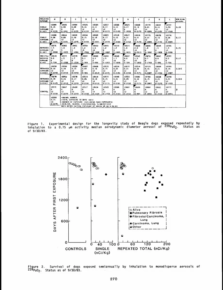
GPROJECIEDEXPOSURE
SINGLEEXPGSL~IE0.10C]
91NGLEEXPO~UflEO, tUCI
REPER1£DEXPOSOREO.)UC!
8 8 0C E F ! J K L
100050.0990-1651
lY06250=178E-|030
~073T0.59~2E-1920
]07700,26120-2089
107051.910R-2QBB
102BR0.15120-2235
102880,00t2fl-2265
)0505O. tqt2R-219~
IOSOAO. tt[2R-222t
1055T0=1712A=2173
10508D.]S12R-2106
1077Y0,2l|20-9089
]063EO. IIt2A-2tS2
0.18
0.17
0.15
1025A0,19120-2230
1055H0,15120-2168
105090.1812A-2221
106050.]?i20~2176
105180.15120-2195
106180.91]20-2]76
1077S0.2!120-2089
106700,10120-2]52
].S9Eht 829
~027C].S
tOE-2008
~OqOC
9E-1609
I~Oq 50
i,5100-1908
~10999
1.2RE-lq82
iPr06282.0]00-2013
]06051.89
; D= t B92l~Oq t R0,6q5-728
11~0~ 771.590-1698
iOqOO0.3gS- 360
1.5:7I E=1267
~105901.7tO9-1038
i~065T0.GqS-728
iP~o59 Ct.9
]os-1862
~067U0.70$-1799
II~0381
0,5q5-759
0.17
~610.0920-66q
11~.5B0.16120-2296
t02900.18~2R-2219
106500.]519Ai2236
i09080.16120-22t9
IOS~$;0.]5
o-2t7!
i057R0,25120-2t91
106710.2!]29-2150
t051A0,9912R-217t
107150.15t20~2087
tOBSR0.2t12R-2150
10789O. lq120-2087
R~PEREEOEXPOSUREO.OtOCl
0.0]5
~rto~?8O. t8t20-2t~
10950O.Iq12R-2219
105700,1712fl-2233
105150,2612R-2191
109180.]812A-2Pt9
10973O. t5i2R-2tT!
t05980.2112R-2tOI
I09950.26]2R-2150
I058CO.)q]2A-2171
10700O. tS129-2007
106580.21t2A-2t5O
0.0|8
I~0620
060-969
1008V 1062CO O12 12R’205q R,.2152
t077T 0o]20-205q
tO37Eot2R-2233
1051TO
0-2193
10q300t2R-2220
]05050]20-2159
]05OR0t29-2193
iO66T0t20-2152CONTROL
102680.15I0R-2295
=RNIHOL NUM~ER-I010L EXPOSURE 10 DATE fUEl)=NUMBER OF EXP0SURE liNE]tiDiNG SHAM EWPOSURE~I=R=RLIVE, O=OEAD, E=EUTHflN]ZED, 9=SRCRIFICEBORTS AFTER INITIAL EXPOSURE RT DERTO 08 ON 9-90-83
Figure I. Experimental design for the longevity study of Beagle dogs exposed repeatedly byinhalation to a 0.75 ~m activity median aerodynamic diameter aerosol of 239pu02. Status asof 9/30/83.
2400-
w
D 1800
0
xW
~ 120C
600
0
CONTROLS
[]
0
0
¯ i( I4~
¯ ¯¯ ¯
,~oA ve lliPulmonary Fibrosis I14,FibrosislCarcinoma, II Lun~ I[~ Carcinoma, Lung [L~Other ~=J
[]
I [ i I I I 1 l I I
40 100 0 60 120 200SINGLE REPEATED TOTAL (nCi/Kg)
(nCi/Kg)
Figure 2. Survival of dogs exposed semiannually by inhalation to monodisperse aerosols of23gPu02. Status as of 9/30/B3.
270
was 0.17 ± O.lO ~Ci; for the high-level repeatedly exposed dogs, it was 0.16 ± 0.07 uCi
per exposure; for dogs exposed to the lower levels, the average was 0.016 ± O.OlO ~Ci per
exposure.
Nine dogs were sacrificed at the times given above to study the effect of repeated exposures
on the retention of 23gPu02 in lung. The last three of these were sacrificed during the past
year, and no results beyond those reported in the open literature 3 are available.
Fourteen dogs have died from radiation pneumonitis and pulmonary fibrosis, and three of these
also had pulmonary carcinomas. One other dog also died of a pulmonary carcinoma. At1 dogs that
dled of radiation effects were in the high-level repeated exposure group, except one. It received
a single expcsure to 0.6 ~Ci of 23gPu. The dose to death for this dog was similar to that for
the repeatedly exposed dogs that died from the same cause. This supports the previous conclusion
that similar doses to singly and repeatedly exposed dogs have similar effects (1981-1982 Annual
Report, LMF-I02, pp. 364-369). Animal numbers, survival times, doses, and major findings at
necropsy are summarized in Table I.
During the past year, lO dogs in the study died or were euthanized. Of these lO dogs, l was
exposed once, 2 were exposed repeatedly at about O.Ol ~Ci per exposure, and the rest were
exposed repeatedly at about O.1 ~Ci per exposure. The medical histories and Findings at
necropsy for these dogs are summarized below.
Dog 1027B, exposed 12 times to result in a total lung burden of O.16 uCi of 239pu, died
unexpectedly at 2125 days after initial exposure. At gross necropsy, an acute peritonitis
resulting from a ruptured gall bladder was found. The common bile duct was patent, but the gall
bladder was enlarged to 4 cm diameter by 7 cm long by a greenish, gelatinous, semisolid material.
This material was found in the 300 cc of fluid in the peritoneal cavity. No evidence of radiation
pneumonitis or lung tumors could be found grossly.
Dog 1073U, exposed 12 times to achieve a total lung burden of 0.21 uCi of 23gPu, died
during its last exposure, 1933 days after the first exposure. The only significant clinical
abnormalities were persistent lymphpenia that developed about a year after initial exposure and
mildly increased interstitial lung densities seen on radiographs 4 years after initial exposure.
Gross necropsy showed no lesions that might cause or predispose the animal to sudden death, lhe
only lung lesion was mild pleural fibrosis.
Dog I073T, exposed once to achieve a lung burden of 0.6 ~Ci of 239pu, was euthanized in
dyspnea Ig20 days after initial exposure. Radiographic signs of radiation pneumonitis were first
noted 3 years after initial exposure. The signs became progressively worse, and euthanasia was
recommended. At gross necropsy, severe radiation pneumonitis and pulmonary fibrosis were noted.
In addition, there was atrophy of the tracheobronchial lymph nodes and hypertrophy of the right
ventricular myocardium. A tumor nodule, 2 cm in diameter, was found in the right apical lobe. It
appeared grossly to be a carcinoma of the lung.
Dog I027C, exposed lO times to achieve a total lung burden of 1.5 uCi of 239pu, was
euthanized 2008 days after initial exposure because of severe dyspnea. Signs of pulmonary
fibrosis were first noted about 4 years after initial exposure. These signs became more severe
over the subsequent 2 years. At gross necropsy, a severe radiation pneumonitis and pulmonary
fibrosis were found, along with atrophy of the tracheobronchial lymph nodes and right ventricular
myocardial hypertrophy. No tumors were found.
Dog 1045D, exposed lO times to achieve a total lung burden of 1.5 ~Ci of 23gpu, died 1908
days after initial exposure while under treatment Eor radiation pneumonitis and pulmonary
fibrosis. An elevated respiratory rate was first noted about 4.5 years after initial exposure.
The signs progressively worsened, and the dog died before euthanasia. At gross necropsy, a severe
diffuse radiation pneumonitis and pulmonary fibrosis were noted. The tracheobronchial lymph nodes
271
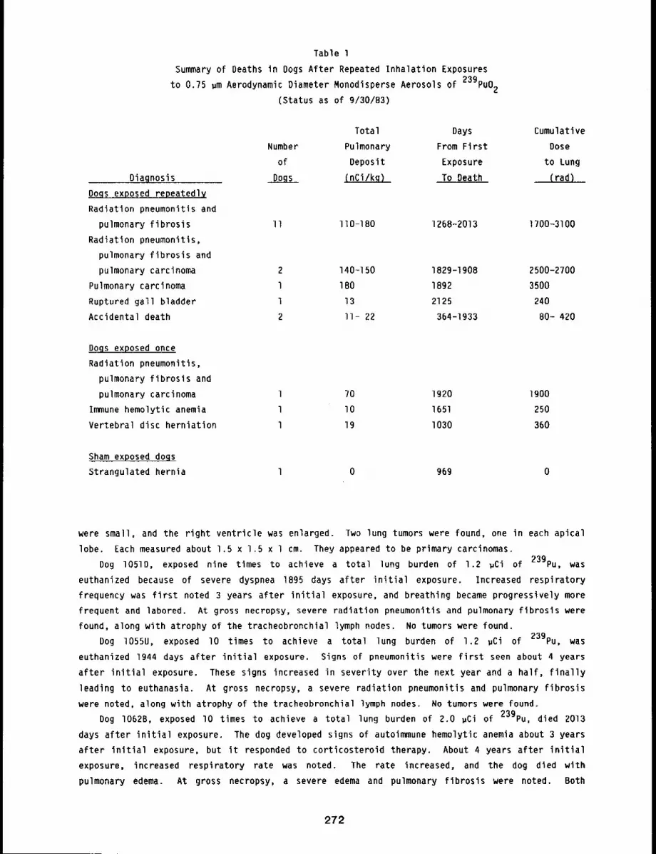
Table 1
Summary of Deaths in Dogs After Repeated Inhalation Exposures
to 0.75 pm Aerodynamic Diameter Monodisperse Aerosols of 239pu02
(Status as of 9/30/83)
Diagnosis
Dongs exposed repeated~
Radiation pneumonitis and
pulmonary fibrosis
Radiation pneumonitls,
pulmonary fibrosis and
pulmonary carcinoma
Pulmonary carcinoma
Ruptured gall bladder
Accidental death
Total Days Cumulative
Number Pulmonary From First Dose
of Deposit Exposure to Lung
Dogs (nCi/kq) To Death (rad)
II 110-180 1268-2013 1700-3100
2 140-150 1829-1908
l 180 1892
l 13 2125
2 ll- 22 364-1933
Dogs exposed once
Radiation pneumonitis,
pulmonary fibrosis and
pulmonary carcinoma
Immune hemolytic anemia
Vertebral disc herniation
1 ?0 1920 1900
l lO 1651 250
l 19 I030 360
Sham exposed dogs
Strangulated hernia l 0 969 0
2500-2?00
3500
240
80- 420
were small, and the right ventricle was enlarged. Two lung tumors were found, one in each apical
lobe. Each measured about 1.5 x 1.5 x l cm. They appeared to be primary carcinomas.
Dog 1051D, exposed nine times to achieve a total lung burden of 1.2 ~Ci of 239pu, was
euthanized because of severe dyspnea 1895 days after initial exposure. Increased respiratory
frequency was first noted 3 years after initial exposure, and breathing became progressively more
frequent and labored. At gross necropsy, severe radiation pneumonitis and pulmonary fibrosis were
found, along with atrophy of the tracheobronchial lymph nodes. No tumors were found.
Dog 1055U, exposed lO times to achieve a total lung burden of 1.2 pCi of 239pu, was
euthanized 1944 days after initial exposure. Signs of pneumonitis were first seen about 4 years
after initial exposure. These signs increased in severity over the next year and a half, finally
leading to euthanasia. At gross necropsy, a severe radiation pneumonitis and pulmonary fibrosis
were noted, along with atrophy of the tracheobronchial lymph nodes. No tumors were found.
Dog I062B, exposed lO times to achieve a total lung burden of 2.0 pCi of 239pu, died 2013
days after initial exposure. The dog developed signs of autoimmune hemolytic anemia about 3 years
after initial exposure, but it responded to corticosteroid therapy. About 4 years after initial
exposure, increased respiratory rate was noted. The rate increased, and the dog died with
pulmonary edema. At gross necropsy, a severe edema and pulmonary fibrosis were noted. Both
272

cardiac ventricles were hypertrophic, and the tracheobronchial lymph nodes were atrophic. No
tumor nodules were found.
Dog I064A, exposed nine times to achieve a total lung burden of 1.5 ~Ci of 239pu, was
euthanized in dyspnea 1829 days after initial exposure. First indications of radiation
pneumonitis were seen 4 years after initial exposure. These worsened with time, finally
necessitating euthanasia. At gross necropsy, a severe radiation pneumonitis and pulmonary
fibrosis were found, along with atrophy of the tracheobronchial lymph nodes and hypertrophy of the
right ventricle. A tumor nodule about 1.2 cm diameter was noted in the right diaphragmatic lung
lobe.
Dog I069S, exposed nine times to achieve a total lung burden of 1.8 ~Ci of 239pu, died in
respiratory distress 1892 days after initial exposure. Evidence of radiation pneumonitis first
appeared about 5 years after the first exposure. Signs worsened, and the dog died shortly before
euthanasia could be performed. At gross necropsy, numerous tumor masses were found in the lungs;
the largest completely occupied the left cardiac lobe. The tumor had invaded the pleural cavity,
resulting in accumulation of serous fluid. The massive involvement of the lung lobe made the
presence of inflammatory lesions difficult to determine.
REFERENCES
1. International Commission on Radiation Protection, Limits for Intake of Radionuclides byWorkers, (ICRP Publication 30, Part l), Ann. ICRP, 2(3/4), 1979.
2. Boecker, B. B., F. L. Aguilar, and T. T. Mercer, A Canine Inhalation Apparatus Utilizing aWhole Body Plethysmograph, Health Phys. lO: I077-I089, 1964.
3. Diel, J, H. and D. L. Lundgren, Repeated Inhalation Exposure of Beagle Dogs to 239pu02:Retention and Translocation, Health Phys, 43: 655-662, 1982.
273
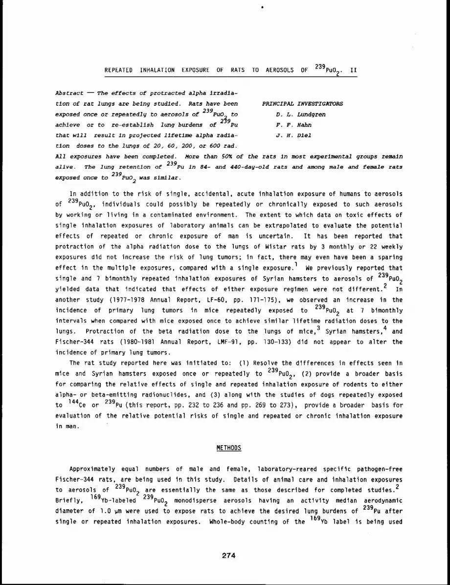
REPEATED INHALATION EXPOSURE OF RATS TO AEROSOLS OF 239pu02. II
Abstract --The effects of protracted alpha irradla-
tion of rat lungs are being studied. Rats have been PRINCIPAL INVESTIGATORS
exposed once or repeated1~ to aerosols of 239pu0_ to D.L. Lundgren
achieve or to re-establlsh lung burdens of 2~9pu F.F. Hahn
thac w111 result in projected llfetlme alpha radla- J.H. Diel
tlon doses to the lungs of 20, 60, 200, or 600 tad.
All exposures have been completed. More than 50~ of the rats In most experimental groups remain
alive. The lung retentlon of 239pu in 84- and 440-day-old rats and among male and female rats
exposed once to 239pu02 was slmilar.
In addition to the risk of single, accidental, acute inhalation exposure of humans to aerosols
of 239pu02, individuals could possibly be repeatedly or chronically exposed to such aerosols
by working or living in a contaminated environment. The extent to which data on toxic effects of
single inhalation exposures of laboratory animals can be extrapolated to evaluate the potential
effects of repeated or chronic exposure of man is uncertain. It has been reported that
protraction of the alpha radiation dose to the lungs of Wistar rats by 3 monthly or 22 weekly
exposures did not increase the risk of lung tumors; in fact, there may even have been a sparing
effect in the multiple exposures, compared wlth a single exposure, l We previously reported that
single and 7 bimonthly repeated inhalation exposures of Syrian hamsters to aerosols of 23gPu02
yielded data that indicated that effects of either exposure regimen were not different. 2 In
another study (1977-1978 Annual Report, LF-60, pp. 171-175), we observed an increase in the
incidence of primary lung tumors in mice repeatedly exposed to 239pu02 at 7 bimonthly
intervals when compared with mice exposed once to achieve similar lifetime radiation doses to the
lungs. Protraction of the beta radiation dose to the lungs of mice, 3 Syrian hamsters,4 and
Fischer-344 rats (lgBO-IgSl Annual Report, LMF-gl, pp. 130-133) did not appear to alter the
incidence of primary lung tumors.
The rat study reported here was initiated to: (l) Resolve the differences in effects seen
Syrian hamsters exposed once or repeatedly to 23gPu02, (2) provide a broader basismice and
for comparing the relatlve effects of single and repeated inhalation exposure of rodents to either
alpha- or beta-emitting radionuclides, and (3) along with the studies of dogs repeatedly exposed
to 144Ce or 239pu (this report, pp. 232 to 236 and pp. 269 to 273), provide a broader basis for
evaluation of the relative potential risks of single and repeated or chronic inhalation exposure
in man.
METHODS
Approximately equal numbers of male and female, laboratory-reared specific pathogen-free
Fischer-344 rats, are being used in this study. Details of animal care and inhalation exposures239to aerosols of PuO_ are essentially the same as those described for completed studies.2
Briefly, 16gYb-labeled z 239pu02 monodisperse aerosols having an activity median aerodynamic
diameter of l.O ~m were used to expose rats to achieve the desired lung burdens of 23gPu after
single or repeated inhalation exposures. Whole-body counting of the 169yb label is being used
274
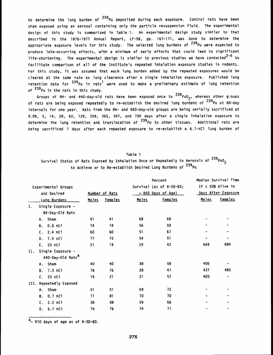
to determine the lung burden of 239pu deposited during each exposure. Control rats have been
sham exposed using an aerosol containing only the particle resuspension fluid. The experimental
design of this study is summarized in Table I. An experimental design study similar to that
described in the 1976-1977 Annual Report, LF-58, pp. 167-171, was done to determine the
appropriate exposure levels for this study. The selected lung burdens of 239pu were expected to
produce late-occurrlng effects, with a minimum of early effects that could lead to significant
life-shortening. The experimental deslgn is similar to previous studies we have conducted 2-5 to
facilitate comparison of all of the Institute’s repeated inhalation exposure studies in rodents.
For thls study, it was assumed that each lung burden added by the repeated exposures would be
cleared at the same rate as lung clearance after a single inhalation exposure. Published lung
retention data for 239pu in rats I were used to make a preliminary estimate of lung retention
of 239pu in the rats in this study.
Groups of 84- and 440-day-old rats have been exposed once to 239pu02, whereas other groups
of rats are being exposed repeatedly to re-establlsh the desired lung burdens of 239pu at 60-day
intervals for one year. Rats from the 84- and 440-day-old groups are being serially sacrificed at
0.08, 5, 14, 28, 63, 12g, 259, 365, 547, and 730 days after a single inhalation exposure to
determine the lung retention and translocation of 239pu to other tissues. Additional rats are
being sacrificed 7 days after each repeated exposure to re-establish a 6.7-nCi lung burden of
Table l
Survival Status of Rats Exposed by Inhalation Once or Repeatedly to Aerosols of 239pu02
to Achieve or to Re-establlsh Desired Lung Burdens of ~ ¢39pu
Experimental Groups
and Oesired Number of Rats
Lung Burdens Males Females
I. Single Exposure -
B4-Day-Old Rats
A. Sham 41 41
B. 0.8 nCi 74 74
C. 2.4 nCi 60 60
D. 7.5 nCi 77 75
E. 23 nCi 21 19
II. Single Exposure -
440=Day-Old Ratsa
A. Sham 40 40
B. 7.5 nCi 76 76
C. 23 nCi 19 21
III. Repeatedly Exposed
A. Sham 51 51
B. 0.7 nCi 71 81
C. 2.2 nCi 38 38
D. 6.7 nCi 76 76
Percent
Survival (as of 9-30-83;
....... ~ B50 DaYS of Aqe)
Males Females
68 68
56 59
51 67
54 51
29 42
38 58
28 47
21 57
69 72
70 70
59 6674 71
Median Survival Time
If < 50% Alive in
DaYS After Exposure
Males Females
669 684
406
437 485
403
a glO days of age as of 9~30-83.
275
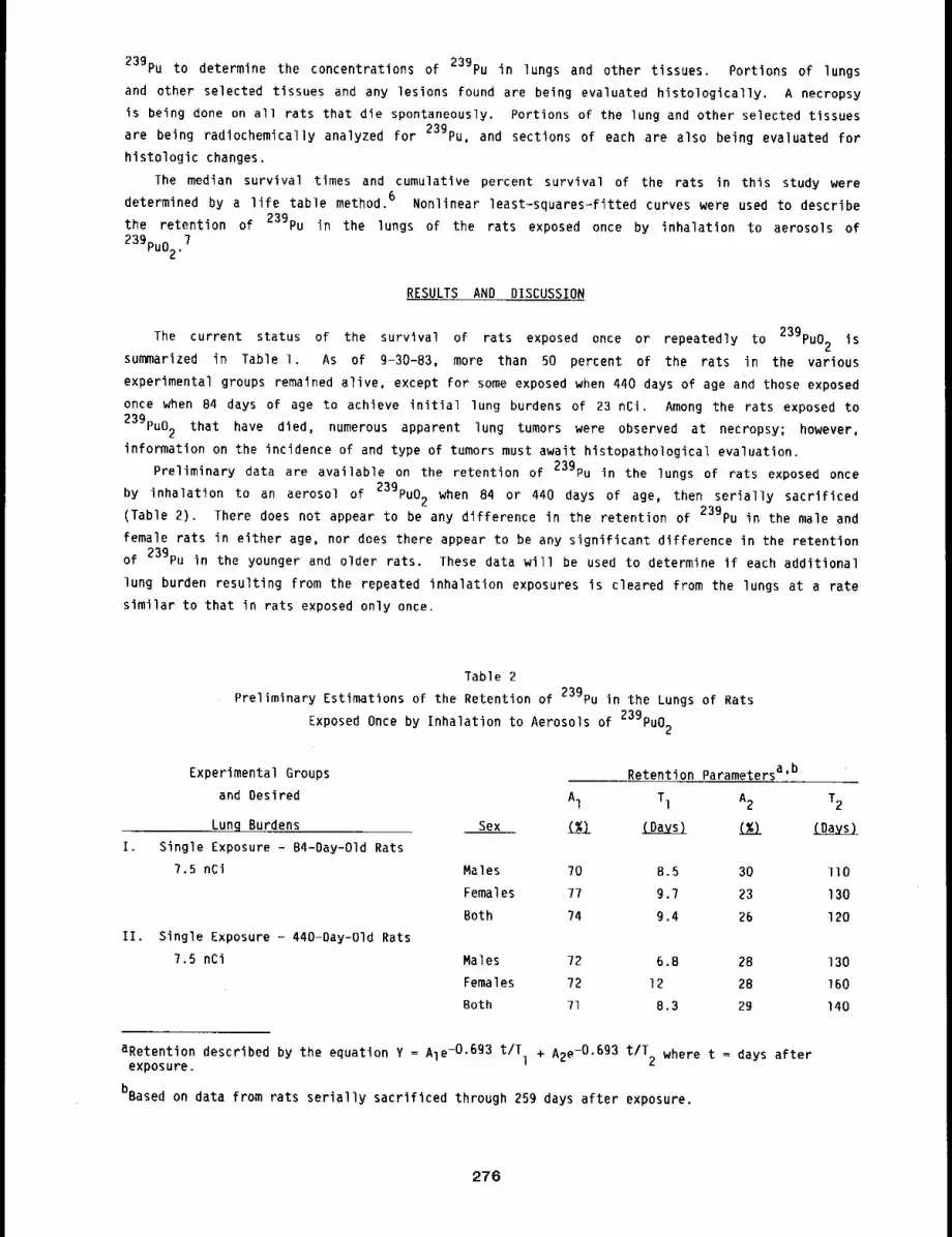
239pu to determine the concentrations of 239pu in lungs and other tissues. Portions of lungs
and other selected tissues and any lesions found are being evaluated histologically. A necropsy
is being done on all rats that die spontaneously. Portions of the lung and other selected tissues
are being radlochemically analyzed for 239pu, and sections of each are also being evaluated for
histologic changes.
The median survival times and cumulative percent survival of the rats in this study were
determined by a life table method. 6 Nonlinear least-squares-fitted curves were used to describe
the retention of 239pu in the lungs of the rats exposed once by inhalation to aerosols of
239pu02.7
RESULTS AND DISCUSSION
The current status of the survival of rats exposed once or repeatedly to 239pu02 is
summarized in Table I. As of 9-30-83, more than 50 percent of the rats in the various
experimental groups remained alive, except for some exposed when 440 days of age and those exposed
once when 84 days of age to achieve initial lung burdens of 23 nCi. Among the rats exposed to
239pu02 that have died, numerous apparent lung tumors were observed at necropsy; however,
information on the incidence of and type of tumors must await histopathological evaluation.
Preliminary data are available on the retention of 239pu in the lungs of rats exposed once
an aerosol of 239pu02 when 84 or 440 days of age, then serially sacrificedby inhalation to
(Table 2). There does not appear to be any difference in the retention of 239pu in the male and
female rats in either age, nor does there appear to be any significant difference in the retention
of 239pu in the younger and older rats. These data will be used to determine if each additional
lung burden resulting from the repeated inhalation exposures is cleared from the lungs at a rate
similar to that in rats exposed only once.
Table 2
Preliminary Estimations of the Retention of 239pu in the Lungs of Rats
Exposed Once by Inhalation to Aeroso]s of 239pu02
I.
Experimental Groups
and Desired
Lung Burdens
Single Exposure - 84-Day-Old Rats
7.5 nCi
II. Single Exposure - 440-Day-Old Rats
7.5 nCi
Sex
Retention Parametersa’b
A1 T1 A2 T2
(%) ~ (%) {Days)
Males 70 8.5 30 II0
Females 77 9.7 23 130
Both 74 9.4 26 120
Males 72 6.8 28 130
Females 72 12 28 160
Both 71 8.3 29 140
aRetention described by the equation Y = Ale-O.6g3 t/T + A2e-O.693 t/T where t = days afterexposure, l 2
bBased on data from rats serially sacrificed through 259 days after exposure.
276
REFERENCES
I. Sanders. C. L. and 5. A. Mahaffey, Inhalation Carcinogenesis of Repeated Exposures to High-Fired 239pu02 in Rats, Health Phys. 41: 629-644,lg81
2. Lundgren, D. L., F. F. Hahn, A. H. Rebar, and R. O. McClellan, Effects of Single and RepeatedInhalation Exposure of Syrian Hamsters to Aerosols of 239Pu02, Intl. 5. Radiat. Biol. 43:1-18, 1983.
3. Hahn, F. F., D. L. Lundgren, and R. O. McClellan, Repeated Inhalation Exposure of Mice to144Ce02. II. Biologic Effects, Radiat. Res. 82: 123-137, 19BO.
4. Lundgren, D. L., F. F. Hahn, and R. O. McClellan, Effects of Single and Repeated InhalationExposure of Syrian Hamsters to Aerosols of 144Ce02, Radiat. Res. 90: 3?4-394, 1982.
5. Lundgren, D. L., F. F. Hahn, and R. O. McClellan, Effects of Repeated Inhalation Exposure ofRats to Aerosols of 144Ce02: A Preliminary Report, in Current Concepts in Lunq Dosimetry(D. R. Fisher, ed.), pp. 83-89, U.S. Department of Energy, Washington, DC, 1983.
6. Benedetti, 5., K. Yuen, and L. Young, PIL, Life Tables and Survival Functions, in BMDPStatistical Software (W. J. Dixon, Chief ed.), pp. 557-575, University of California Press,Berkeley, 1983.
7. Ralston, M., Derivative-Free Nonlinear Regression, in BMDP Statistical Software (W. 5. Dixon,Chief ed.), pp. 305-314, University of California Press, Berkeley, 1983.
277
TOXIC EFFECTSOF INHALED 244Cm203IN RATS. III
Abstract -- The toxlolty of 244cm203_ is belng stud-
led in rats exposed by inhalatlon to monodlsperse PRINCIPAL INVESTIGATORS
aerosols to achieve desired ~n~t~al lung burdens D. L. Lundgren
ranging from 1 to 17,000 nCl/kg body welght. F. F. Hahn
Curium-243 was used to label the 244Cm203 so that R.A. Gu~imette
the lnltlal lung burden of each rat could be deter-
m~ned by gamma countlng. Median survival t~mes and cumulative survlval dlstrlbutlons of rats wlth
InJtlal lung burdens of > 450 nci/kg were slgnlflcantly decreased. Prellmlnarg estimates of the
retention of 244Cm in the lung, llver , and skeletx>n through 650 days after exposure are
presented.
Radioisotopes of curium constitute a significant portion of the alpha activity in the spent
fuel from conventional power reactors. These radionuclides present hazards to man by various
routes of exposure, including inhalation. Compared with plutonium, relatively few studies of the
toxicity of inhaled curium compounds, including curium oxides, have been conducted in laboratory
animals. 1’2 Although the oxides of curium are markedly more soluble than those of plutonium,3
it appears that the incidences of lung tumors in rats exposed to curium or plutonium are similar
in the dose range of 20 to 200 rad to lungs.l In previous studies of the toxic effects of
inhaled Cm compounds, a direct measurement of the initial lung burden for each animal was not
available. The purpose of the study reported here was to obtain information on the dose-response
relationships of 244Cm in rats exposed by inhalation to a well characterized aerosol of
244Cm203 in which the initial lung burden of each animal was determined, thus permitting
more accurate dosimetry for each rat.
METHODS
The curium used in this study contained 243Cm as a gamma label, and is hereafter referred to
as Cm203. The details of the methods used and previous progress in this study were presented
(lgBO-lgB1 Annual Report, LMF-91, pp. 186-189 and 19BI-1982 Annual Report, LMF-I02, pp. 357-360).
Briefly, after removal of the Pu daughters by solvent extraction, a monodisperse aerosol having an
activity median aerodynamic diameter of l.O ~m was prepared by heat-treatment of curium
hydroxide at llSO°C. The resulting Cm203 aerosol was used for the inhalation exposure of
rats. All rats were exposed to the aerosol within 24 h of the preparation of the monodisperse
particles. Sham-exposed rats were exposed to heat-treated aerosols of pH i0.2 aqueous ammonia,
the medium in which the Cm203 particles were resuspended for the rat exposures. All rats were
12 ± l week of age at the time of exposure.
After inhalation exposure to Cm203, all rats were whole-body counted at time intervals aslong as significant data could be obtained to determine the whole-body retention of 244Cm. A
complementary study is being conducted in two groups of rats that are being serially sacrificed
after exposure to 244Cm203 to determine the retention of 244Cm in various tissues. The
278
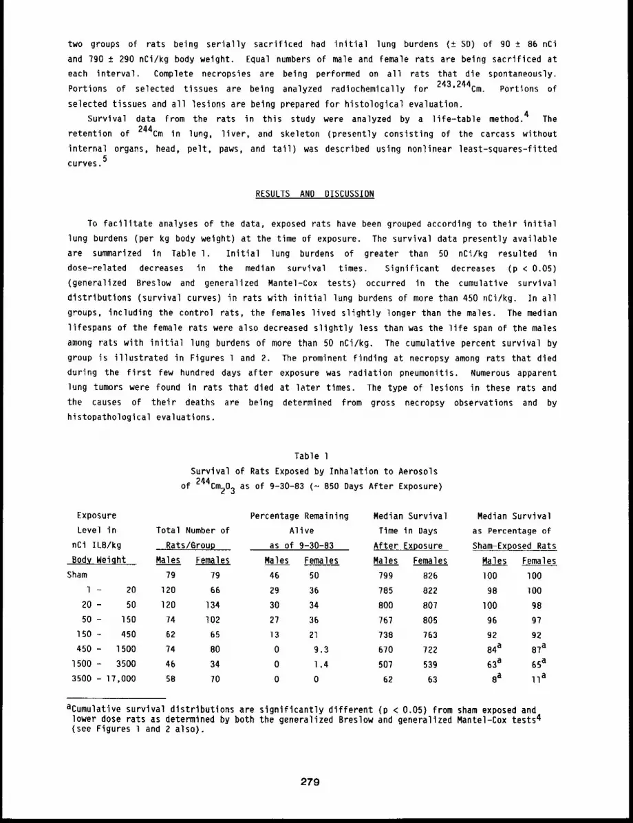
two groups of rats being serially sacrificed had initial lung burdens (± SD) of go ± 86 nCi
and ?go ± 290 nCi/kg body weight. Equal numbers of male and female rats are being sacrificed at
each interval. Complete necropsies are being performed on all rats that die spontaneously.
Portions of selected tissues are being analyzed radiochemically for 243’244Cm. Portions of
selected tissues and all lesions are being prepared for histological evaluation.
Survival data from the rats in this study were analyzed by a life-table method. 4 The
retention of 244Cm in lung, liver, and skeleton (presently consisting of the carcass without
internal organs, head, pelt, paws, and tail) was described using nonlinear least-squares-fltted5
curves.
RESULTS AND DISCUSSION
To facilitate analyses of the data, exposed rats have been grouped according to their initial
lung burdens (per kg body weight) at the time of exposure. The survival data presently available
are summarized in Table I. Initial lung burdens of greater than 50 nCi/kg resulted in
dose-related decreases in the median survival times. Significant decreases (p < 0.05)
(generalized Breslow and generalized Mantel-Cox tests) occurred in the cumulative survival
distributions (survival curves) in rats with initial lung burdens of more than 450 nCi/kg. In all
groups, including the control rats, the females lived slightly longer than the males. The median
lifespans of the female rats were also decreased slightly less than was the life span of the males
among rats with initial lung burdens of more than 50 nCi/kg. The cumulative percent survival by
group is illustrated in Figures 1 and 2. The prominent finding at necropsy among rats that died
during the first few hundred days after exposure was radiation pneumonitis. Numerous apparent
lung tumors were found in rats that died at later times. The type of lesions in these rats and
the causes of their deaths are being determined from gross necropsy observations and by
histopathologlcal evaluations.
Table l
Survival of Rats Exposed by Inhalation to Aerosols
244Cm203 as of 9-30-83 (~ 850 Days After Exposure)of
Exposure
Level in Total Number of
nCi ILB/kg Rats/Group
Body Weight Males Females Males Females
Sham 7g 79 46 50
l - 20 120 66 29 36
20 - 50 120 134 30 34
50 - 150 74 I02 27 36
150 - 450 62 65 13 21
450 - 1500 74 80 0 9.3
1500 - 3500 46 34 0 1,4
3500 - 17,000 58 70 0 0
Percentage Remaining
Alive
as of 9-30-83
Median Survival Median Survival
Time in Days as Percentage of
After E~posure Sham-Exposed Rats
Males Females Males Females
79g 826 I00 I00
?85 822 98 lO0
800 807 I00 98
?67 805 96 g?
738 763 92 92
6?0 722 84a 87a
507 539 63a 65a
62 63 8a IIa
acumulative survival distributions are significantly different (p < 0.05) from sham exposed andlower dose rats as determined by both the generalized Breslow and generalized Mantel-Cox tests4
(see Figures l and 2 also).
279
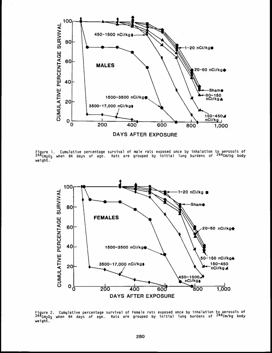
1oo --i,,i
:>:>o: 80:3
W0< 60I--ZLUO
w 40n
LU
1--20
_I
:3o oO
450-1500 nCiIk
MALES
1500-3500 nCi/k,
3500-17,000 nCIIkg0
-20 nCi/kg¯
,20-50 nCi/kgO
50nCilkg&
150-450&,, I nCi/k0 I
200 400 600 800 1,000
DAYS AFTER EXPOSURE
Figure I. Cumulative percentage survival of male rats exposed once by inhalation to aerosols of244Cm203 when 84 days of age. Rats are grouped by initial lung burdens of 244Cm/kg bodyweight.
DAYS AFTER EXPOSURE
Figure 2. Cum41ative percentage survival of female rats exposed once by inhalation to aerosols of244Cm203 when 84 days of age. Rats are grouped by initial lung burdens of 244Cm/kg bodyweight.
280
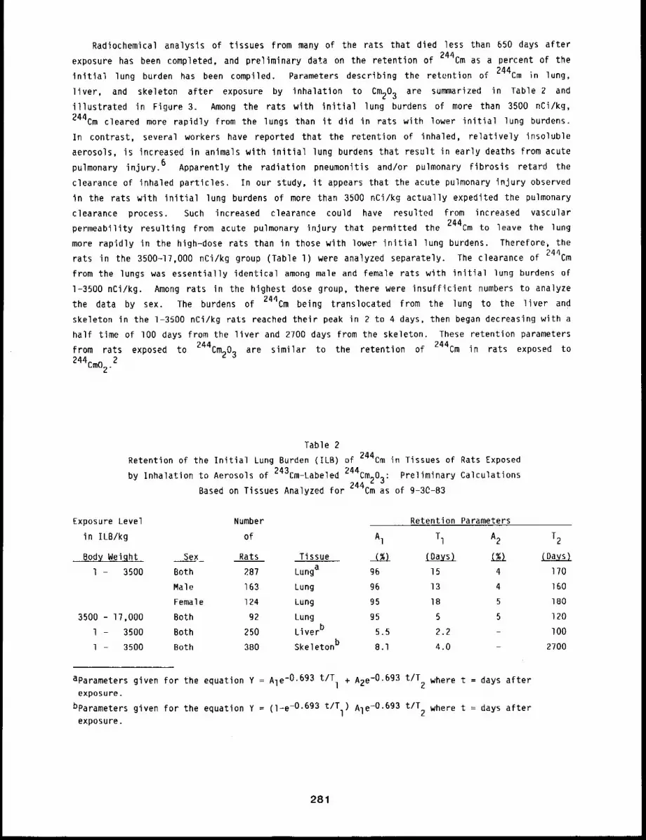
Radiochemical analysis of tissues from many of the rats that died less than 650 days after
exposure has been completed, and preliminary data on the retention of 244Cm as a percent of the
initial lung burden has been compiled. Parameters describing the retention of 244Cm in lung,
liver, and skeleton after exposure by inhalation to Cm203 are summarized in Table 2 and
illustrated in Figure 3. Among the rats with initial lung burdens of more than 3500 nCi/kg,
244Cm cleared more rapidly from the lungs than it did in rats with lower initial lung burdens.
In contrast, several workers have reported that the retention of inhaled, relatively insoluble
aerosols, is increased in animals with initial lung burdens that result in early deaths from acute
pulmonary injury. 6 Apparently the radiation pneumonitis and/or pulmonary Fibrosis retard the
clearance of inhaled particles. In our study, it appears that the acute pulmonary injury observed
in the rats with initial lung burdens of more than 3500 nCi/kg actually expedited the pulmonary
clearance process. Such increased clearance could have resulted from increased vascular
permeability resulting from acute pulmonary injury that permitted the 244Cm to leave the lung
more rapidly in the high-dose rats than in those with lower initial lung burdens. Therefore, the
rats in the 3500-17,000 nCi/kg group (Table l) were analyzed separately. The clearance of 244Cm
from the lungs was essentially identical among male and female rats with initial lung burdens of
1-3500 nCi/kg. Among rats in the highest dose group, there were insufficient numbers to analyze
the data by sex. The burdens of 244Cm being translocated from the lung to the liver and
skeleton in the 1-3500 nCi/kg rats reached their peak in 2 to 4 days, then began decreasing with a
half time of lO0 days from the liver and 2700 days from the skeleton. These retention parameters
from rats exposed to 244Cm203 are similar to the retention of 244Cm in rats exposed to
244Cm022
Table 2Retention of the Initial Lung Burden (ILB) of 244Cm in Tissues of Rats Exposed
243 Z44by Inhalation to Aerosols of Cm-Labeled " Cm^O~: Preliminary Calculations
L JBased on Tissues Analyzed for 244Cm as of g-30-83
Exposure Level
in ILB/kg
Bod Wei ht
1 - 3500
3500 - 17,000
1 - 3500
1 - 3500
Number Retention Parameters
of Al Tl A2 T2
Sex Rats Tissue (%) (Days) (%) (Davs)
Both 287 Lunga 96 15 4 170
Male 163 Lung 96 13 4 160
Female 124 Lung 95 18 5 180
Both g2 Lung 95 5 5 120
Both 250 Liverb 5.5 2.2 - lO0
Both 3BO Skeletonb 8.1 4.0 - 2700
aparameters given for the equation Y = Ale-0.693 t/T + A2e-O.693 t/T where t = days after1 2exposure.
bparameters given for the equation Y = (l-e -0.693 t/T ) Ale-O.6g3 t/T where t = days afterl 2exposure.
281
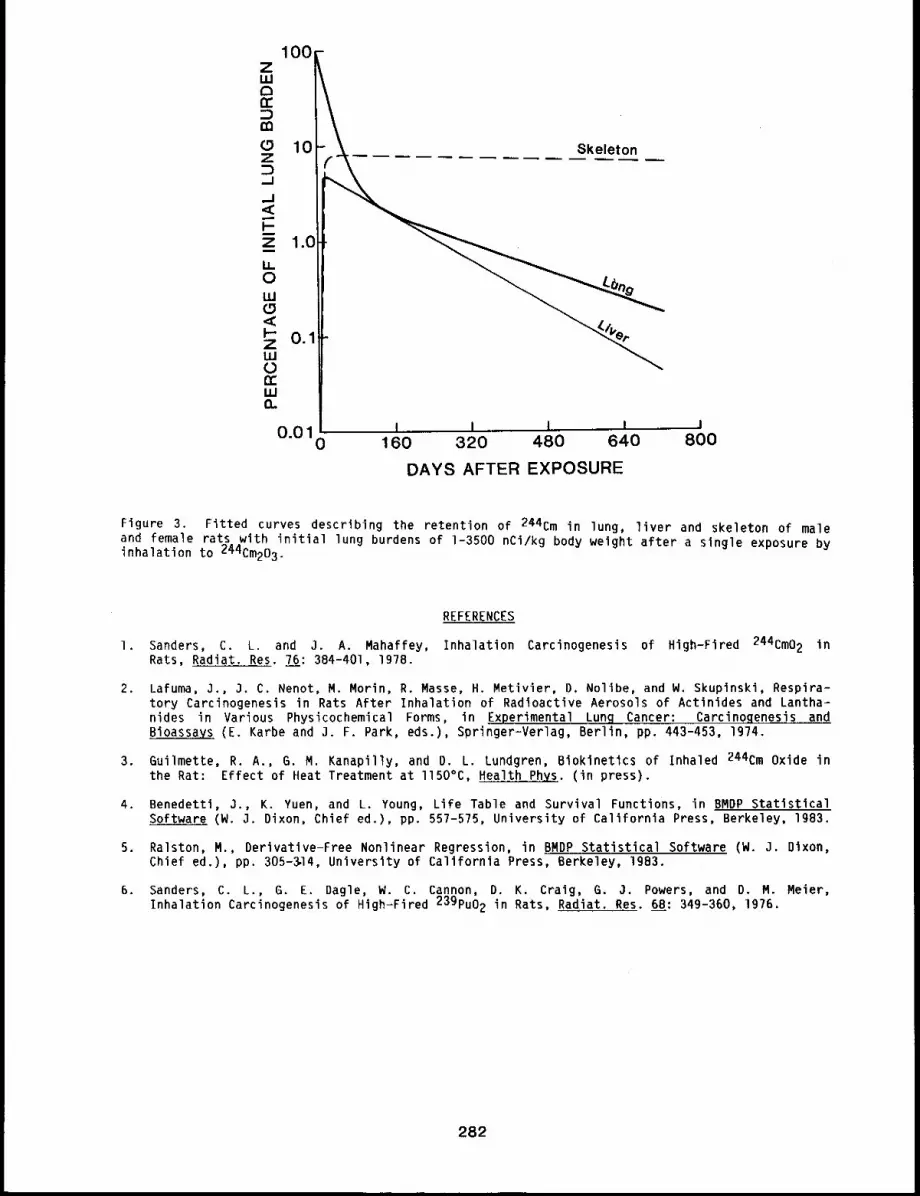
Skeleton
I i L t J160 320 480 640 800
DAYS AFTER EXPOSURE
Figure 3. Fitted curves describing the retention of 244Cm in lung, liver and skeleton of maleand female rats with initial lung burdens of 1-3500 nCi/kg body weight after a single exposure byinhalation to 244Cm203.
REFERENCES
l. Sanders, C. L. and O. A. Mahaffey, Inhalation Carcinogenesis of High-Fired 244Cm02 inRats, Radiat. Res. 76: 384-401, 197B.
2. Lafuma, 3., 3. C. Nenot, M. Morin, R. Masse, H. Metivier, D, Nolibe, and W. Skuplnski, Respira-tory Carcinogenesis in Rats After Inhalation of Radioactive Aerosols of Actinides and Lantha-hides in Various Physicochemical Forms, in Experimental Lung Cancer: Carcinogenesis andBioassaYs (E. Karbe and J. F. Park, eds.), Springer-Verlag, Berlin, pp. 443-453, 1974.
3. Guilmette, R. A., G. M. Kanapilly, and D. L. Lundgren, Biokinetics of Inhaled 244Cm Oxide inthe Rat: Effect of Heat Treatment at ll50°C, Health ~. (in press).
4. Benedettl, 3., K. Yuen, and L. Young, Life Table and Survival Functions, in BMDP StatisticalSoftware (W. 3. Dixon, Chief ed.), pp. 557-5?5, University of California Press, Berkeley, lg83.
5. Ralston, M., Derivatlve-Free Nonlinear Regression, in BMDP Statistical Software (W. 3. Dixon,Chief ed.), pp. 305-~14, University of California Press, Berkeley, 1983.
6. Sanders, C. L., G. E. Dagle, W. C. Cannon, D. K. Craig, G. 3. Powers, and D M~ Meier,
Inhalation Carcinogenesis of High-Fired 239pu02 in Rats, Radiat Res. 68: 349-360, i9 .
282

THE RETENTION, DISTRIBUTION, AND CYTOGENETIC EFFECTS OF INHALED 23gPu(N03)4
IN THE CYNOMOLGUS MONKEY
Abstract -- Inhalatlon of plutonium nitrate by
cgnomolgus monkeys resulted in activity being de-
poslted in The iungs, followed by translocatlon
to liver and bone. Actlvltg in the testes was
0.0001 of the initial lung burden and was concen-
trated in the interstitial tissue. No increase in
chromosome aberration frequency was observed in
blood lymphocytes as a function of time after
plutonlum inhalatlon. When all chromosome data
PRINCIPAL INVESTIGATORS
A. L. Brooks
H. C. Redman
F. F. Hahn
J. A. Mewhlnneg
J. M. Smith
R. O. McClellan
were combined, there was a small exposure-level-dependent increase In The frequency of chromosome
aberrations in blood lymphocytes after inhalatlon of large quantitles of plutonium. In cgnomolgus
monkegs, the frequency of chromosome aberrations fn blood igmphocgtes is not a good indicator of
radiation dose or damage from inhaled plutonium.
Occupational exposures to plutonium have been reported to produce chromosome aberrations in
human blood lymphocytes, l However, many pollutants present in occupational environments can
also produce chromosome aberrations. This makes it difficult to establish the relationship
between cytogenetic damage and radiation dose for a single occupational contaminant. This
research was conducted in nonhuman primates to measure the influence of inhalation of
239pu(N03) 4 on the production of chromosome aberrations in blood lymphocytes and to isolate
the radiation exposure from exposure to other clastogenic agents in the environment. The research
also provides data on the retention and distribution pattern of a relatively soluble form of
plutonium after an inhalation exposure. These data make it possible to calculate radiation dose
to various organs and to relate dose to potential genetic and somatic effects.
METHODS
The ages of the monkeys, exposure methods, characteristics of the aerosol, and data on the
initial deposition and cytogenetic effects have been reported (Ig79-BO Annual Report, LMF-84;
1980-81 Annual Report, LMF-gl). Briefly, 16 male cynomolgus monkeys were exposed by inhalation to
polydisperse aerosols of 23gPU(N03)4. The initial lung burden was determined by sacrifice
of some animals at 4 days after exposure. Other animals were sacrificed at 4, 40, 365, and ll7B
days after exposure, and the retention and distribution pattern of plutonium was determined by
radiochemical analyses of tissue and excreta samples. Tissues with low levels of activity, such
as the testes and blood, were analyzed by low-level alpha radiochemistry techniques. Urine and
feces were collected at regular intervals throughout the study and the 23gPu excreted from the
body was determined.
Samples of blood were taken at 6, 12, and 37 months after inhalation exposure, and the
frequency of chromosome aberrations was determined in the blood lymphocytes by previously
published methods. 2 The lymphocytes were cultured for 48 h and the frequencies of chromosome
aberrations were measured on coded slides. An attempt was made to relate aberration frequency to
inhaled activity, time of exposure, and the radiation dose to the lung.
283
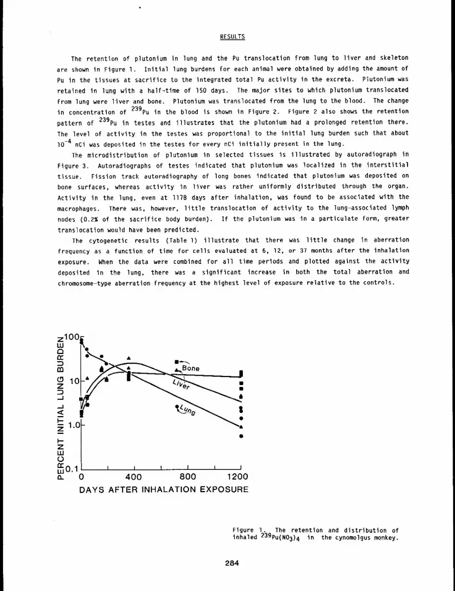
RESULTS
The retention of plutonium in lung and the Pu translocation from lung to liver and skeleton
are shown in Figure I. Initial lung burdens for each animal were obtained by adding the amount of
Pu in the tissues at sacrifice to the integrated total Pu activity in the excreta. Plutonium was
retained in lung with a half-time of 150 days. The major sites to which plutonium translocated
from lung were liver and bone. Plutonium was translocated from the lung to the blood. The change
in concentration of 239pu in the blood is shown in Figure 2. Figure 2 also shows the retention
pattern of 239pu in testes and illustrates that the plutonium had a prolonged retention there.
The level of activity in the testes was proportional to the initial lung burden such that about
lO-4 nCi was deposited in the testes for every nCi initially present in the lung.
The microdistribution of plutonium in selected tissues is illustrated by autoradiograph in
Figure 3. Autoradiographs of testes indicated that plutonium was localized in the interstitial
tissue. Fission track autoradiography of long bones indicated that plutonium was deposited on
bone surfaces, whereas activity in liver was rather uniformly distributed through the organ.
Activity in the lung, even at I178 days after inhalation, was found to be associated with the
macrophages. There was, however, little translocation of activity to the lung-associated lymph
nodes (0.2% of the sacrifice body burden). If the plutonium was in a particulate form, greater
translocation would have been predicted.
The cytogenetic results (Table l) illustrate that there was little change in aberration
frequency as a function of time for cells evaluated at 6, 12, or 37 months after the inhalation
exposure. When the data were combined for all time periods and plotted against the activity
deposited in the lung, there was a significant increase in both the total aberration and
chromosome-type aberration frequency at the highest level of exposure relative to the controls.
m
ZW0~N.1 i , i
0 400 800 1200
DAYS AFTER INHALATION EXPOSURE
Figure I. The retention and distribution ofinhaled 239pu(N03)4 in the cynomolgus monkey.
284
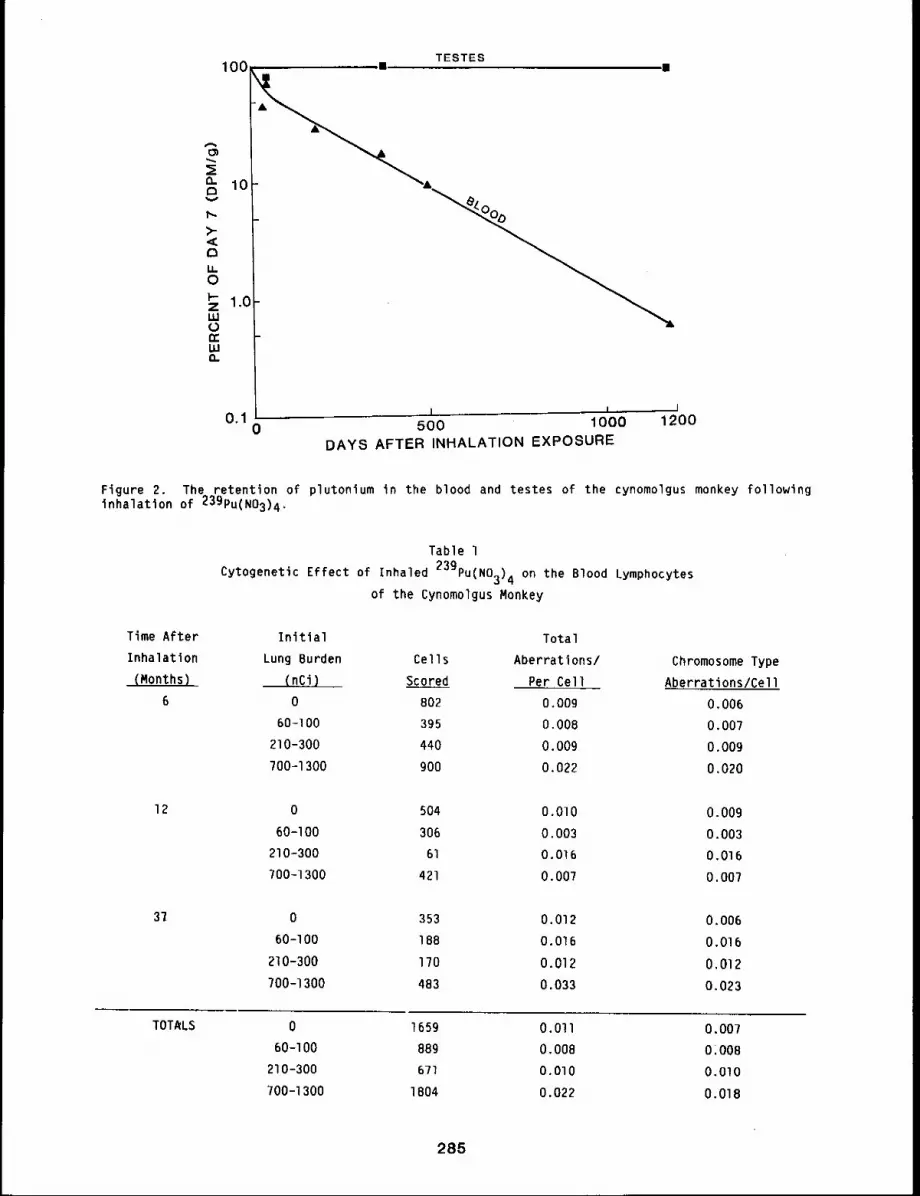
TESTES100 ~l m
I i I0"1 0 500 1000 1200
DAYS AFTER INHALATION EXPOSURE
Figure 2. The retention of plutonium in the blood and testes of the cynomolgus monkey followingInhalatlon of 23gPU(N03)4.
Time After
Inhalation
(Months)
6
Table 1
239pu(N03)4 on the Blood LymphocytesCytogenetic Effect of Inhaled
of the Cynomolgus Monkey
Initial
Lung Burden Cells
(nCi) Scored
0 802
60-100 395
210-300 440
700-1300 900
Total
Aberrations/
Per Cell
0.009
O.OOB
O.OOg
0.022
Chromosome Type
Aberrations/Cell
0.006
0.007
O.OOg
0.020
12 0 504 O.OlO 0.009
60-I00 306 0.003 0.003
210-300 61 0.016 0.016
700-1300 421 0.007 0.007
0 353 0.012 0.006
60-I00 18B 0.016 0.016
210-300 170 0.012 0.012
700-1300 4B3 0.033 0.023
TOTALS 0 1659 0.011 0.007
60-I00 889 0.008 01008
210-300 671 0.010 0.010
700-1300 1804 0.022 0.018
285
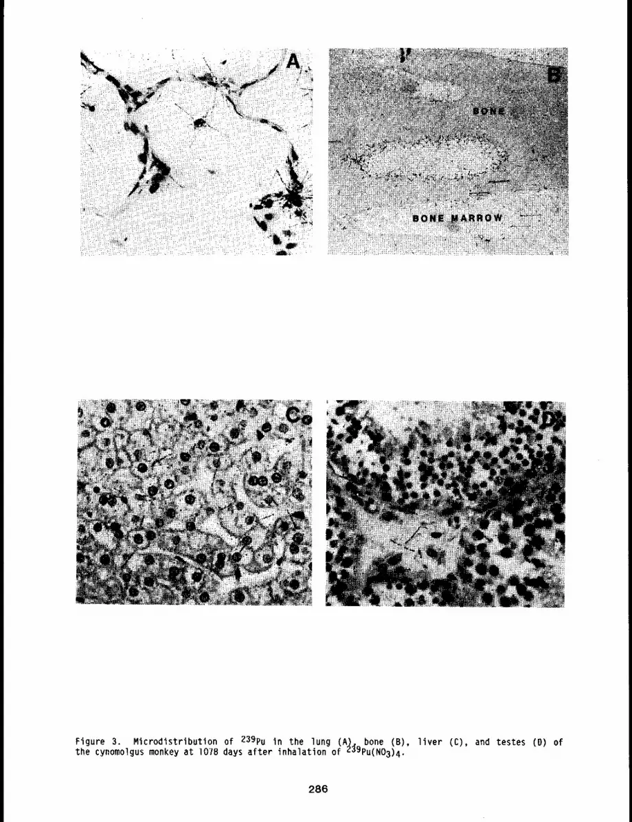
Figure 3. Microdistribution of 239pu in the lung (A). bone (B), liver (C), and testes the cynomolgus monkey at 107B days after inhalation of 239pu(N03)4.
286
DISCUSSION
The retention and distribution of plutonium nitrate in cynomolgus monkeys was similar to that
reported after intravenous injection of rhesus monkeys with plutonium citrate. 3 The retention
half-life in lung of 150 days was similar to that for the blood, 170 days. Because of the conti-
nuing translocation of activity from the lung to other organs, it has been impossible to estimate
retention times in bone and liver. Additional scheduled sacrifices combined with modeling studies
will make it possible to determine retention times of plutonium in liver and bone. However, the
apparent clearance of activity from the liver during approximately the last 800 days of the study
seems to be fairly close to that for lung, whereas activity in bone is still increasing or at
least staying constant over this same interval. Such data indicate that the retention time for
bone is much longer than that for liver. That much of the activity in the lung was associated
with the lung macrophages at I178 days after inhalation exposure indicates that this activity
might be removed by lung lavage. It also suggests that a fraction of the activity in lung exists
as insoluble material that has accumulated in the macrophages. The failure of the material to
accumulate in lung-associated lymph nodes suggests that it is either leaving the nodes at the same
rate as it is entering or that it is not particulate in nature. The nonuniform distribution of
activity in the testes resulted in a lower dose to the genetically important spermatogonial cells
than would have occurred with uniform distribution of dose. 4 Because of the low activity level,
5.9 pCi/g, and the nonuniform distribution of the activity, the genetic dose after the inhalation
of l.O uCi was calculated to be only about 1.8 rad at i178 days. Detection of genetic changes in
these animals would thus not be possible even at activity levels that caused some rather extensive
lung damage.
The lack of increase in chromosome aberration frequency in blood lymphocytes as a function of
time after inhalation exposure may be related to several factors. First, the life span of lympho-
cytes in cynomologus monkeys may be short. At the low radiation dose rates delivered to the blood
in this study, individual cells would accumulate low total doses. Second, the total dose to blood
lymphocytes in this study even with long-lived lymphocytes may be so low that changes in aberra-
tion frequency are not detectable. Third, cell killing by the high-LET radiation may be removing
damaged cells from the blood. With combined chromosome data there is an increase in aberration
frequency at the highest level of exposure. The use of chromosome aberrations in the blood of pri-
mates may be of little use in predicting dose or health effects from inhaled plutonium. These
results emphasize the importance of carefully considering occupation, environment, and lifestyle
in evaluating the meaning of chromosome aberrations observed in humans exposed to plutonium before
the aberrations can be attributed to the dose received from the isotope.
REFERENCES
I. Brandom, W. F., A. D. Bloom, P. G. Archer, V. E. Archer, R. W. Bistline, and G. Saccomanno,Somatic Cell Genetics of Uranium Miners and Plutonium Workers. A Biological Dose-ResponseIndicator, in Late Biological Effects of Ionizing Radiation, Vol. I, pp. 507-51B.International Atomic Energy Agency, Vienna, 1978 (STI/PUB/4B9).
2, LaBauve, R. 3., A. L. Brooks, J. L. Mauderly, F. F. Hahn, H. C. Redman, C. Macken, D. O.Slauson, 3. A. Mewhinney, and R. O. McClellan, Cytogenetic and Other Biological Effects of23g-Plutonium Oxide Inhaled by the Rhesus Monkey, Radiat. Res. 82: 310-335, Ig80.
3. Durbin, P. W., Plutonium in Mammals: Influence of Plutonium Chemistry Route ofAdministration, Initial Distribution, and Long-Term Metabolism, Health Phys. 29: 295-510, 1975.
4, Brooks, A. L., J. H. Diel, and R. O. McClellan, The Influence of Testicular Microanatomy onthe Potential Genetic Dose from Internally Deposited Plutonium-239 Citrate in Chinese Hamster,Mouse, and Man, Radiat, Res, 77: 292-302, 197g.
287
THE INDUCTION OF CHROMOSOME ABERRATIONS IN THE LIVER
OF THE CHINESE HAMSTER BY IN3ECTED THOROTRAST
Abstract -- After Chinese hamsters were injected with
colloldal thorium dioxide (Thorotrast) and serially PRINCIPAL IWVESTIGATORS
sacrificed, the frequency of chromosome aberrations A.L. Brooks
in their liver cells was determined. The chromosome R.A. Gullmette
aberration frequency increased l~nearly wlth time.
The radlatlon dose to the l~ver was estimated at each time interval where aberrations were
recorded, and the slope of the dose response curve was compared to that observed for anlmals
injected with either alpha- (239pu) or beta (144Ce-144pr) emitting radlonuclldes. The slope
for the Thorotrast-induced aberrations (2.1 x 10⁻³ aberratlons/cell/rad) was lower than that for
239pu (4.8 x 10⁻³), but greater than that for 144ce (0.31 x 10⁻³). Thls suggests either
that the doses were overestimated ~n the case of Thorotrast or that the non-unlform ~-dose
dlstrlbutlon from the ThoroCrast resulted In more wasted radiation than wlth the more uniformly
deposited 239pu. Both of these factors would decrease the slope of the dose response curve for
Thorotrast.
Colloidal thorium dioxide (Thorotrast) was used at one time as an x-ray contrast medium.
is retained for a long time in the liver and other organs rich in reticuloendothelial elements.
Because 232Th and its daughters decay partly through alpha emissions, substantial exposure of
the liver results. A high incidence of liver cancer, after a long latent period, has been
observed in patients injected with this material, l Epidemiological data from this human
population have been used in hazard evaluation by a number of national and international
committees to set the risk for the induction of liver cancer by other alpha~emitting radionuclides
for which no data are available in man. There are two major questions on how well these data can
be applied generically to estimating the risk of liver cancer. First, what is the role of
chemical toxicity from the heavy metal and the large mass loading from gram quantities of injected
thorium on the carcinogenic response of the liver? Second, how does the non-uniform alpha dose
distribution from Thorotrast influence the frequency of radiation-induced liver cancer? Research
in animal models can help to answer these two questions. This project was designed to evaluate
the effectiveness of Thorotrast in the induction of primary cellular damage, measured as
chromosome aberrations in the liver, relative to that reported for other alpha-emitting
radionuclides.
METHODS
Equal numbers of male and female young adult (150 to 200=day-old) Chinese hamsters (48 total)
were used in this study. They were maintained one per cage and fed Mouse Breeder Blok and water
ad libitum.
Thorotrast was provided by Dr. Werner Riedel, Free University of Berlin. It contained 25.5
± 0.4% by weight of ThO2 and 20 weight percent yellow dextrin. The particle size distributionwas 9.3 ± 4.1 nm. 2 The ratio of 228Th to 232Th, an indicator of parent/daughter
equilibrium, was 0.3 as of October 2, IgBl. The preparation contained 0.12 ~Ci/g of 232Th.
This material, similar to that used in human studies, has been used in experimental rodent studies
288
in Berlin and at the University of Utah. In humans, a typical injected volume of Thorotrast,
25 ml, contained about 5 g thorium and resulted in a body burden of 0.6 ~Ci of 232Th. For
daughter products, the energy and the fractional decay can be combined to calculate the amount of
energy released for each decay of a 228Th atom (31.8 MeV). For these cytogenetic studies, the
activity injected per g tissue was about lO times higher than used in the human patients.
Twenty-four animals were injected in%raveneously with either O.1 ml (males) or 0.08
(females) of Thorotrast, and 24 control animals were injected with the yellow dextrin carrier.
Twelve additional animals were injected with Thorotrast and sacrificed at lO or 50 days and the
distribution of the Thorotrast determined. Animals in the cytogenetic study, 12 per group, were
subjected to partial-hepatectomy at 50, lO0, 200, or 400 days after Thorotrast injection to
stimulate liver cell division, and the frequency of chromosome aberrations was determined.3
RESULTS
The Thorotrast distribution animals were sacrificed at lO or 50 days. Radiochemical analysis
of tissues indicated that about 60% of the injected activity was deposited and retained in liver.
With this value, the maximum radiation dose to the liver could be calculated assuming that:
I. The ratio of 22Blh/232Th did not change and was equal to that of the original
injection solution, 0.3.
2. The amount of energy absorbed in the Thorotrast particles was negligible.
3. The daughters (224Ra, 220Rn, 216po, 212Bi, and 212po) remained in the liver and
were in equilibrium with 22BTh.
4. For each decay of a 22BTh atom, there was 31.8 MeV of alpha energy deposited in the
liver, and for every decay of 232Th there was 4.0 MeV deposited.
With these assumptions, the dose rate to the liver was calculated to be 0.65 rad/day. This
was then multiplied by the time of exposure, assuming that retention was constant. This dose
estimate is considered high because it assumes no clearance of the Th or its daughters from the
liver and no absorption of energy in the Thorotrast particles.
The aberration types and response of the chromosomes to the exposure as a function of time are
shown in Table 1 for both exposed and control animals. There is a linear increase in the
frequency of chromosome aberrations as a function of time in animals injected with Thorotrast. In
a total of 1321 cells scored, there were only B chromatid-type aberrations and 160 chromosome-type
aberrations. Figure l shows the dose response relationship derived for the Thorotrast compared to
Table 1
Chromosome Aberrations Produced in Liver of Chinese Hamsters by Thorotrast
Treatment (days)
Controls
Total Cells CSDa CSExcb CTAc Aberrations/cell
633 9 8 2 0.03
Thorotrast 50 1B5 7 6 0 0.07
lO0 206 lO 15 2 0.13
200 203 17 42 0 0.29
400 94 ll 35 4 0.55
aCSD = Chromosome Deletions
bcsExc = Chromosome Exchanges & Rings & Dicentrics
CCTA = Chromatid Type Aberrations
289
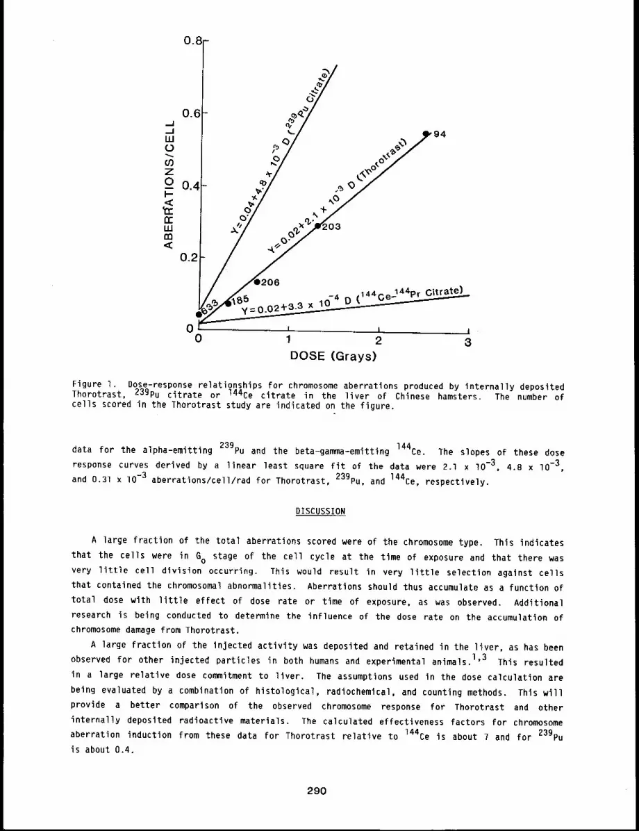
y=O.O-4 D (144ce’144Pr Lte)
0 i i i ¯0 1 2 3
DOSE (Grays)
Figure I. Dose-response relationships for chromosome aberrations produced by internally depositedThorotrast, 23gPu citrate or 144Ce citrate in the liver of Chinese hamsters. The number ofcells scored in the Thorotrast study are indicated on the figure.
data for the alpha-emitting 239pu and the beta-gamma-emitting 144Ce. The slopes of these dose
response curves derived by a linear least square fit of the data were 2.1 x lO-3, 4.8 x lO-3,
and 0.31 x lO-3 aberrations/cell/rad for Thorotrast, 239pu, and 144Ce, respectively.
DISCUSSION
A large fraction of the total aberrations scored were of the chromosome type. This indicates
that the cells were in GO stage of the cell cycle at the time of exposure and that there was
very little cell division occurring. This would result in very little selection against cells
that contained the chromosomal abnormalities. Aberrations should thus accumulate as a function of
total dose with little effect of dose rate or time of exposure, as was observed. Additional
research is being conducted to determine the influence of the dose rate on the accumulation of
chromosome damage from Thorotrast.
A large fraction of the injected activity was deposited and retained in the liver, as has been
observed for other injected particles in both humans and experimental animals.l’3 This resulted
in a large relative dose commitment to liver. The assumptions used in the dose calculation are
being evaluated by a combination of histological, radiochemical, and counting methods. This will
provide a better comparison of the observed chromosome response for Thorotrast and other
internally deposited radioactive materials. The calculated effectiveness factors for chromosome
aberration induction from these data for Thorotrast relative to 144Ce is about 7 and for 239pu
is about 0.4.
290
These preliminary results suggest that dose distribution, chemical effects, or particle
loading do not increase the frequency of chromosome aberrations induced by Thorotrast exposures.
This provides some evidence that the data from Thorotrast patients should not overestimate the
risk for liver damage from other internally deposited alpha-emittlng radionuclides.
REFERENCES
I. Van Kaick, G., D. Lorenz, H. Muth, and A. Kaul, Malignancies in German Thorotrast Patients andEstlmated Tissue Dose, Health Phys. 35: 167-I75, 1978.
Riedel, w., A. Dalheimer, M. Said, U. Walter, and A. Kaul, Recent Results of the GermanThorotrast Study - Dose Relevant Physical and Biological Properties of Thorotrast EquivalentColloids, Health Phys. 44: Suppl. I, 293-298, 1983.
3. Brooks, A. L. and D. K. Mead, The Metaphase Chromosomes of Chinese Hamster Liver CellsFollowing Partial Hepatectomy, Can. 3. Gen. C~tol. ll: 794-798, lg69.
291
THE INFLUENCE OF AGE AND 239pu02 EXPOSURE ON THE PULMONARY IMMUNE RESPONSE OF DOGS
Abstract --Thls study evaluated the influence of
age and 239pu02 inhalatlon on rhe humoral ~mmune re- PRINCIPAL INVESTIGATORS
sponse of the lungs of Beagle dogs after local depo- J. Galvln
sltlon of antlgen. Four dogs had received a slngle D.E. Blce
brlef exposure (5 to 6 years earl~er) by pernasal B.A. Muggenburg
inhalat~on to one of three monodlsperse PuO2 aero-sols of 0.72 ~m, 0.75 ~m, or 1.4 ~ actlvlt 9 medlan aerodgnamlc dlameter. Thls resulted in
in~tlal lung burdens of 0.52 to 1.10 pCI and 19mph node doses of 72-144 krad. Four non-exposed
dogs served as age-matched controls. The humoral immune response was measured b9 the
enzyme-llnked ~unosorbent assag, whlch showed that even though some dogs had a hlgh IgG
antlgen-speclflc antlbod 9 response in the serum, the same dogs dld not recrult immune effector
cells and products Into the lung. Age influenced the recrultment of serum Jmmunoglobullns Into
the lung.
After inhalation, the fractional rate of translocation of relatively insoluble 239pu02
particles to regional tracheobronchial lymph nodes is approximately 0.0002 day-l. After trans-
239pu02 remains in the lymph nodes for long times, providing a continuous sourcelocation, the
of alpha irradiation of the resident and migratory T and B lymphocytes. It has long been known
that lymphocytes are one of the most radiosensitive cell types in the mammalian body.
After challenge of the canine lung with particulate antigen, the immune response begins in the
tracheobronchial lymph nodes; immune effector cells or products are released into the blood and
accumulate in the lung, providing immune protection, l Clearly, the translocation of plutonium
to the lymph nodes results in large radiation doses to these cells that may be detrimental to the
immune system.
MATERIALS AND METHODS
Eight Beagle dogs, 6 to 7 years of age - three from a study of the dose patterns for inhaled
23gPu02 in Beagle dogs, one from a dose-response study of inhaled 239pu02 in dogs, and
four age-matched control dogs from the Institute’s colony - were used in this study. Before inmlu-
nization or lavage, the dogs were moved to indoor temperature-controlled cages, and food and water
were withheld for 18 h. The dogs were anesthetized with 5% halothane gas in oxygen, and an endo-
tracheal tube was positioned in the trachea. Specific locations in the lung were selected for
instillation of antigen or saline through a fiberoptic bronchoscope. The left cardiac lung lobe
was immunized with lOlO sheep red blood cells in l ml saline, as previously described, l The
right cardiac lung lobe served as a control lobe and received l ml of physiological saline. On 5,
7, lO, 12, 14, 17, and 20 days after immunization, blood was drawn, and each lung lobe was re-enter-
ed with the bronchoscope and washed with 50 m] of sterile saline. The wash fluid was 85 to 95%
recovered and was stored on ice to await laboratory procedures. The enzyme-linked immunosorbent
assay was performed to examine the antigen-specific antibody recovered in lavage fluids and in the
serum, using a modified method of Voller. 2 Cytocentrifuge slides of lung cells were prepared
from the recovered bronchoalveolar lavage fluid. The percent of alveolar macrophages, lympho-
cytes, and polymorphonuclear leukocytes (PMN) was determined by counting 300 total cells/slide.
The total number of cells/ml of lavage fluid was determined using a Coulter counter. Clinical
records were evaluated from birth to time of immunization to determine if the dogs had any history
of lymphopenia.
292
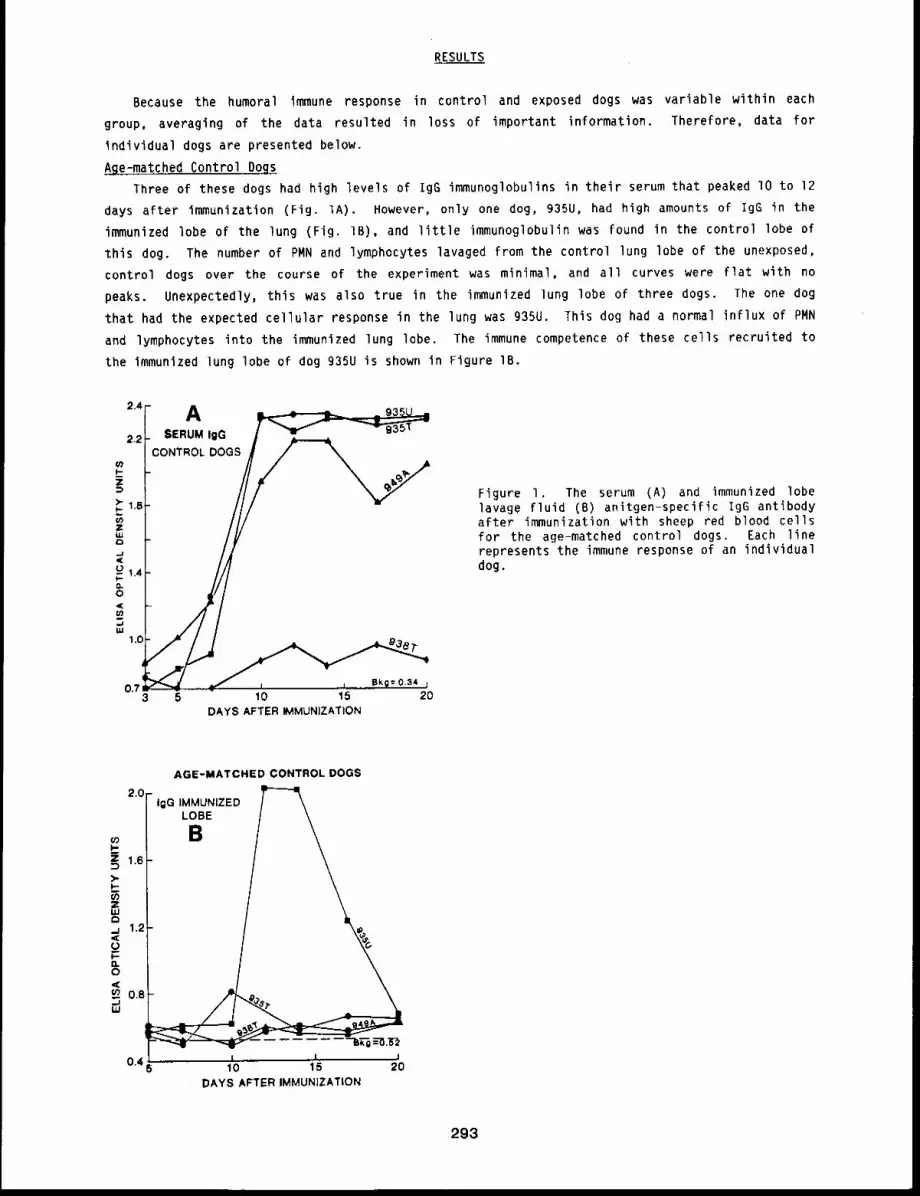
RESULTS
Because the humoral immune response in control and exposed dogs was variable within each
group, averaging of the data resulted in loss of important information. Therefore, data for
individual dogs are presented below.
Age-matched Control Dogs
Three of these dogs had high levels of IgG immunoglobulins in their serum that peaked lO to 12
days after immunization (Fig. IA). However, only one dog, 935U, had high amounts of IgG in the
immunized lobe of the lung (Fig, IB), and little immunoglobulin was found in the control lobe
this dog. The number of PMN and lymphocytes lavaged from the control lung lobe of the unexposed,
control dogs over the course of the experiment was minimal, and all curves were flat with no
peaks. Unexpectedly, this was also true in the immunized lung lobe of three dogs. The one dog
that had the expected cellular response in the lung was 935U. This dog had a normal influx of PMN
and lymphocytes into the immunized lung lobe. The immune competence of these cells recruited to
the immunized lung lobe of dog g35U is shown in Figure IB.
2.4
2.2
0.73
SERUM igGCONTROL DOGS
~ ~L Bkg= 0.34 1
5 10 15 20DAYS AFTER IMMUNIZATION
Figure I. The serum (A) and immunized lobelavage fluid (B) anitgen-specific IgG antibodyafter immunization with sheep red blood cellsfor the age-matched control dogs. Each linerepresents the immune response of an individualdog.
2.0
1.6
1.2
O.8
0.4
AGE-MATCHED CONTROL DOGS
" IgG IMMUNIZED
L BE
10 15DAYSAFTER IMMUNIZATION
2O
293
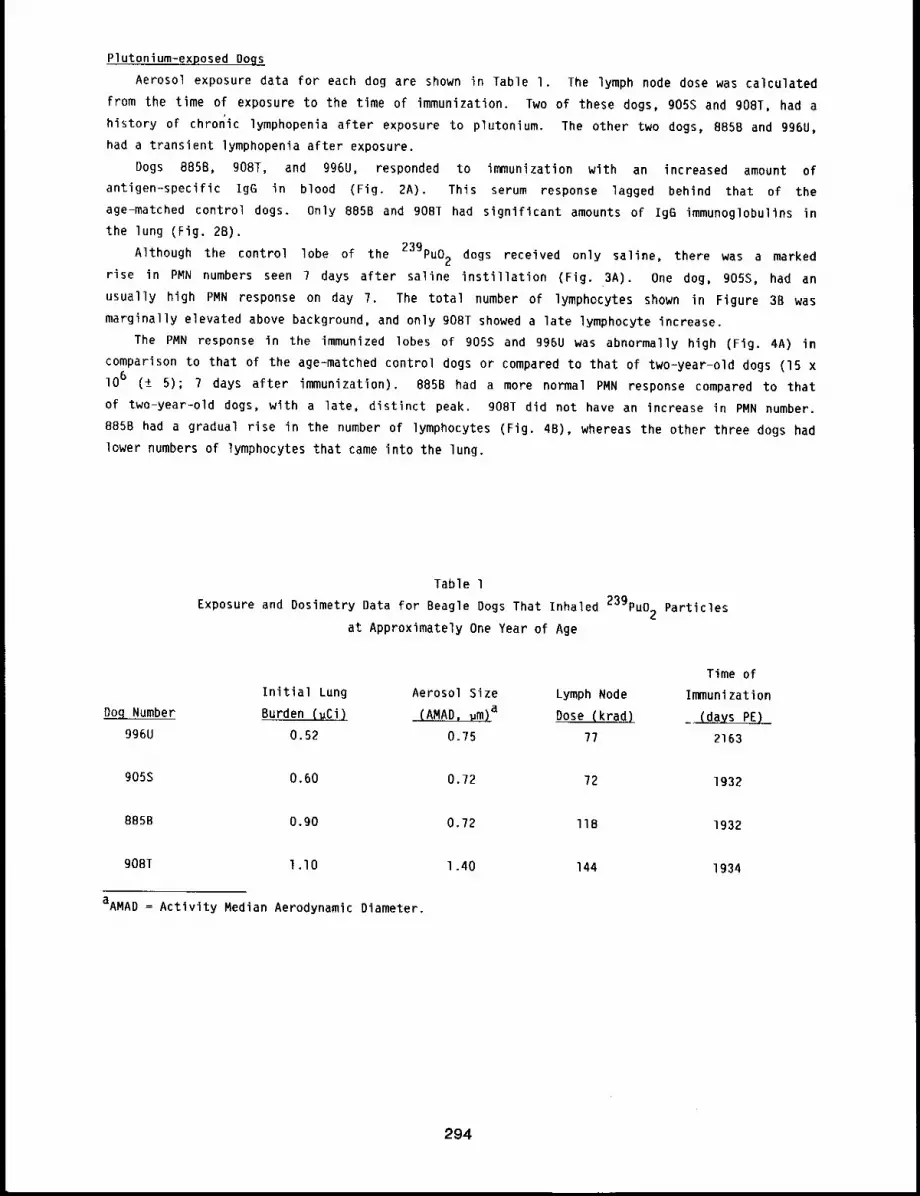
Plutonium-exposed Doqs
Aerosol exposure data for each dog are shown in Table I. The lymph node dose was calculated
from the time of exposure to the time of immunization. Two of these dogs, g05S and 908T, had a
history of chronic lymphopenia after exposure to plutonium. The other two dogs, 8B5B and g96U,
had a transient lymphopenia after exposure.
Dogs 885B, 908T, and 996U, responded to immunization with an increased amount of
antigen-specific Ig6 in blood (Fig. 2A). This serum response lagged behind that of the
age-matched control dogs. Only 885B and 90BT had significant amounts of Ig6 immunoglobulins in
the lung (Fig. 2B).
239pu02 dogs received only saline, there was a markedAlthough the control lobe of the
rise in PMN numbers seen 7 days after saline instillation (Fig. 3A). One dog, 905S, had
usually high PMN response on day 7. The total number of lymphocytes shown in Figure 3B was
marginally elevated above background, and only 908T showed a late lymphocyte increase.
The PMN response in the immunized lobes of 905S and 996U was abnormally high (Fig. 4A)
comparison to that of the age-matched control dogs or compared to that of two-year-old dogs (15
lO6 (± 5); 7 days after immunization). B85B had a more normal PMN response compared to that
of two-year-old dogs, with a late, distinct peak. 908T did not have an increase in PMN number.
885B had a gradual rise in the number of lymphocytes (Fig. 4B), whereas the other three dogs had
lower numbers of lymphocytes that came into the lung.
Table 1
Exposure and Dosimetry Data for Beagle Dogs That Inhaled 239pu02 Particles
at Approximately One Year of Age
Time ofInitial Lung Aerosol Size Lymph Node Immunization
Do.qNumber Burden (u~) ~a Dose (krad.) _~
996U 0.52 0.75 77 2163
905S 0.60 0.72 72 1932
885B o.go 0.72 lIB 1932
908~ l.lO
aAMAD = Activity Median Aerodynamic Diameter.
1.40 144 1934
294

2.4
F-
)-F-
ZuJ 1.6
,.J,¢
E 1,2O
~ 0,8
0,6 5 10 15 20DAYS AFTER IMMUNIZATION
Figure 2. The serum (A) and immunized lobelavage fluid (B) antigen-specific IgG antibodyafter immunization with sheep red blood cellsfor dogs that inhaled 239pu02. Each linerepresents the immune response of an individualdog.
IgG IN IMMUNIZED LOBE2.2
B~~2.0
’~ 1.2
99eu0~0.
¯ 5 10 15 20
DAYS AFTER IMMUNIZATION
Figure 3. The control lung lobe cell popula-tion: total number of PMN (A) and lymphocytes(B) in the lung wash fluid from dogs exposed
to 239pu02. Each point represents thetotal number of cells (lO 6) from an individ-ual dog.
,oF30 A
%X
~ sen
z
-I,WO
I--OI.- 0 ~
10[- Total Lymphocytes~ ~ m
| in Control Lobe ~o~~ ~
oT ~"--T-----~T 65 10 15 20
DAYS AFTER IMMUNIZATION
295
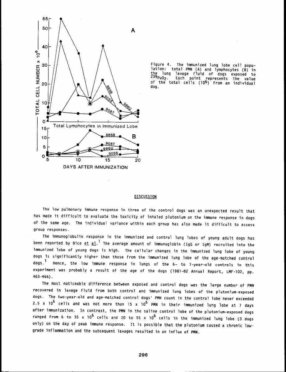
A
0 I I I
15Total Lymphocytes in immunized Lobe
5 10 15 20
DAYS AFTER IMMUNIZATION
Figure 4. The immunized lung lobe cell popu-lation: total PMN (A) and lymphocytes (B) the lung lavage fluid of dogs exposed to23gPu02. Each point represents the valueof the total cells (lO 6) from an individualdog.
DISCUSSION
The low pulmonary immune response in three of the control dogs was an unexpected result that
has made it difficult to evaluate the toxicity of inhaled plutonium on the immune response in dogs
of the same age. The individual variance within each group has also made it difficult to assess
group responses.
The immunoglobulin response in the immunized and control lung lobes of young adult dogs has
been reported by Bice et al. 1 The average amount of immunoglobin (IgG or igM) recruited into the
immunized lobe of young dogs is high. The cellular changes in the immunized lung lobe of young
dogs is significantly higher than those from the immunized lung lobe of the age-matched control
dogs.lHence, the low immune response in lungs of the 6- to 7-year=old controls in this
experiment was probably a result of the age of the dogs (1981-82 Annual Report, LMF-I02, pp.
463-466).
The most noticeable difference between exposed and control dogs was the large number of PMN
recovered in lavage fluid from both control and immunized lung lobes of the plutonium-exposed
dogs. The two-year-old and age-matched control dogs’ PMN count in the control lobe never exceeded
2.5 x 10⁶ cells and was not more than 15 x lO6 PMN in their immunized lung lobe at 7 days
after immunization. In contrast, the PMN in the saline control lobe of the plutonium-exposed dogs
ranged from 6 to 35 x lO 6 cells and 20 to 55 x lO6 cells in the immunized lung lobe (3 dogs
only) on the day of peak immune response. It is possible that the plutonium caused a chronic low-
grade inflammation and the subsequent lavages resulted in an influx of PMN.
296
Dogs B85B, 908T, and 996U received a large radiation dose to their lung-associated lymph
nodes, but significant levels of antibody were present in the serum of these animals after lung
immunization. It was postulated that exposure to plutonium would obliterate the function of the
lymph nodes that drain the lung. If true, then immune cells and immunoglobulins produced in these
dogs after immunization were probably produced in lymphoid tissues that were not exposed to
239pu02 particles but located elsewhere in these dogs. It is possible that antigen drained
from the lung may pass through damaged lymph nodes and be filtered in other lymphoid tissues, such
as the mediastinals or even the spleen. A production of immune cells and antibody in these
tissues could resume the immune function for the lung-associated lymph nodes for the protection of
the lung from antigenic material and other infectious agents.
REFERENCES
I. Bice, D. E., D. L. Harris, J. O. Hill, B. A. Muggenburg, and R. K. Wolff, Immune ResponsesAfter Localized Lung Immunization in the Dog, Am. Rev. Respir. Dis. ~: 755-760, 1980.
2. Voller, A., D. E. Bidwell, and A. Bartlett, Microplate Enzyme-immunoassays for theImmunodiagnosis of Virus Infections, in Manual of Clinical Immunology (N. R. Rose andH. Friedman, eds.), Washington, D.C.: American Society for Microbiology, pp. 506-512, 1976.
297
PULMONARY PROCOAGULANT ACTIVITY OF DOGS WITH LUNG TUMORS
Abstract -- This stud9 evaluated the procoagulant
actlv~t9 and spontaneous macrophage mlgration of PRINCIPAL INVESTIGATORS
lung lavage cells from: a) dog lung lobes that bad J. Galvinradlographlcallg proven lung tumors, b) lobes wlth- D.E. Bice
out tumors In the same dogs, and c) control dog B.A. Muggenburg
lungs with no detectable lung masses. These dog
lungs were lavaged, the cell fraction and wash fluid were assayed for procoagulant act~vltg, and
the spontaneous migration of bronchial wash cells was measured. The procoagulant activity of the
cells and fluid washed from the lung lobe with a tumor was slgnlflcantl 9 increased over that of
both sets of the control lung lobes. The spontaneous migration area of cells from the tumor lobe
was slgnlficantl 9 greater than that of cells from both sets of control lung lobes.
It has been reported that the supernatants of various cultured neoplasms contain procoagulant
actlvity, l It is thought that the neoplasm produces a factor that acts upon macrophages to
increase their production of thromboplastin; hence, the macrophage can be involved in the blood
clotting process after initiators have been presented either through the blood stream or by
leaking into the lung because of the neoplastic process. We evaluated the procoagulant activity
of canine segmental lavage cells and fluid from animals with radiographically proven lung tumors
and compared results to the procoagulant activity of control lung lobes from either the same dog
or dogs without lung tumors. This was an effort to evaluate procoagulant activity produced in
yivo in lung.
MATERIALS AND METHODS
Animals
l’wenty Beagle dogs, 2 to 15 years old, from the Institute’s colony were used in this study.
Seven dogs (2 years old) without tumors served as controls. The other 13 dogs (6 to 15 years old)
were diagnosed by radiography as having lung tumors at various stages of progression. Different
types of neoplasm were identified by cell types obtained by lung washings. TWO of the neoplasms
were mammary tumors that had metastasized to the lung. Eight primary lung tumors were probably
caused by the radiation from inhaled radioactive compounds (239pu, 144Ce, 137Cs, or gly).
Three other primary lung tumors were found in control dogs. The dogs were brought in from
indoor-outdoor kennels 3 days before lavage to allow them to acclimate to indoor cages. They were
housed one to a cage, fed once daily, and given water ad libitum.
The tumors were located by radiography, and a lung lobe in that specific area of the lung was
lavaged. During the same procedure, a lung lobe in the same dog’s lung without radiographically
visible tumors was also lavaged. Two lung lobes in the control dogs were lavaged. The lobes
selected in each dog were entered and washed Five times with lO ml of sterile saline through the
biopsy channel of a bronchoscope. The amount of Fluid recovered ranged from 50 to 90%. The
airways of lobes with tumors were sometimes nearly occluded, and the instilled wash fluid was
difficult to recover. This also presented a problem for recovery of sufficient cells from lung
lobes of dogs with tumors to perform the assays.
298
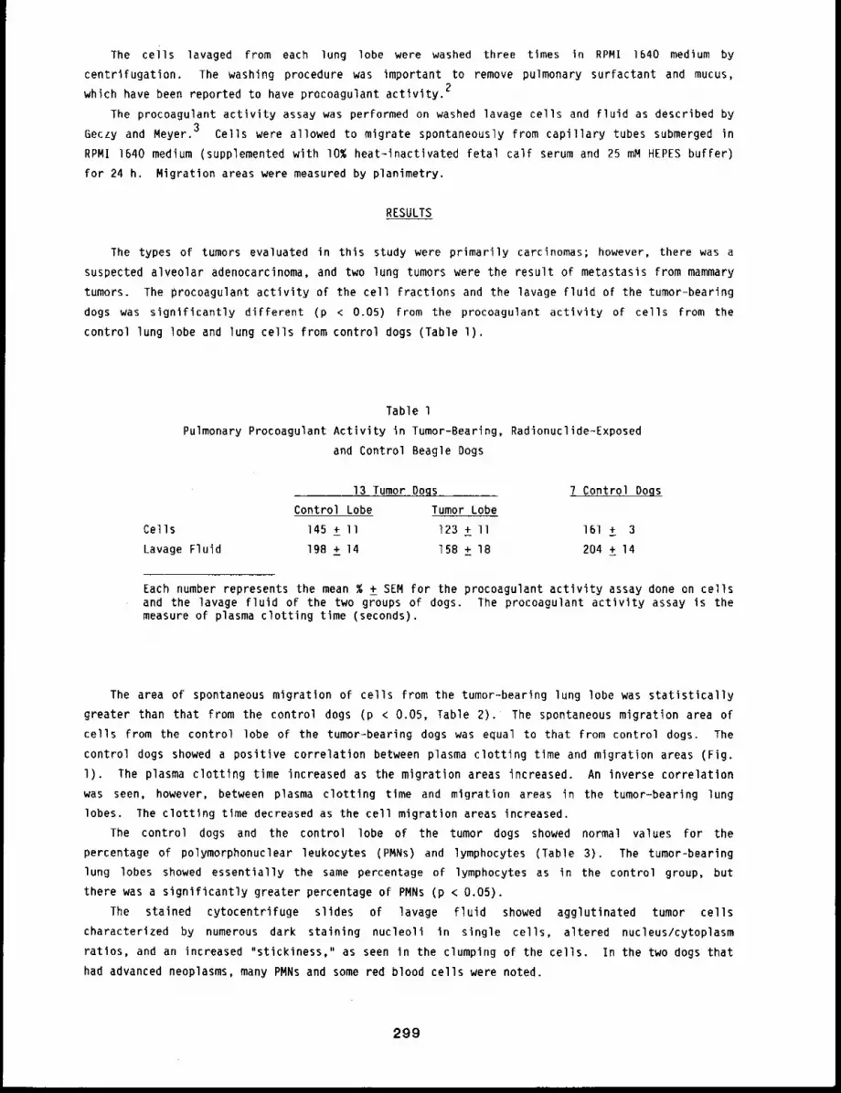
The cells lavaged from each lung lobe were washed three times in RPMI 1640 medium by
centrifugation. The washing procedure was important to remove pulmonary surfactant and mucus,
which have been reported to have procoagulant activity.2
The procoagulant activity assay was performed on washed lavage cells and fluid as described by
Geczy and Meyer. 3 Cells were allowed to migrate spontaneously from capillary tubes submerged in
RPMI 1640 medium (supplemented with I0% heat-inactlvated fetal calf serum and 25 mM HEPES buffer)
for 24 h. Migration areas were measured by planimetry.
RESULTS
The types of tumors evaluated in this study were primarily carcinomas; however, there was a
suspected alveolar adenocarcinoma, and two lung tumors were the result of metastasis From mammary
tumors. The procoagulant activity of the cell fractions and the lavage fluid of the tumor-bearing
dogs was significantly different (p < 0.05) from the procoagulant activity of cells from the
control lung lobe and lung cells from control dogs (Table l).
Table 1
Pulmonary Procoagulant Activity in Tumor-Bearing, Radionuclide-Exposed
and Control Beagle Dogs
13 Tumor OQgs 7 Control Dogs
Control Lobe Tumor Lobe
Cells 145 + II 123 + II 161 + 3
Lavage Fluid 198 ± 14 158 ± 18 204 ± 14
Each number represents the mean % ± SEM for the procoagulant activity assay done on cellsand the lavage fluid of the two groups of dogs. The procoagulant activity assay is themeasure of plasma clotting time (seconds).
The area of spontaneous migratlon of cells from the tumor-bearing lung lobe was statistically
greater than that from the control dogs (p < 0.05, Table 2). The spontaneous migration area
cells from the control lobe of the tumor-bearing dogs was equal to that from control dogs. The
control dogs showed a positive correlation between plasma clotting tlme and migration areas (Fig.
l). The plasma clotting time increased as the migration areas increased. An inverse correlation
was seen, however, between plasma clotting time and migration areas in the tumor-bearing lung
lobes. The clotting time decreased as the cell migration areas increased.
The control dogs and the control lobe of the tumor dogs showed normal values for the
percentage of polymorphonuclear leukocytes (PMNs) and lymphocytes (Table 3). The tumor-bearing
lung lobes showed essentially the same percentage of lymphocytes as in the control group, but
there was a significantly greater percentage of PMNs (p < 0.05).
The stained cytocentrifuge slides of lavage fluid showed agglutinated tumor cells
characterized by numerous dark staining nucleoli in single cells, altered nucleus/cytoplasm
ratios, and an increased "stickiness," as seen in the clumping of the cells. In the two dogs that
had advanced neoplasms, many PMNs and some red blood cells were noted.
299
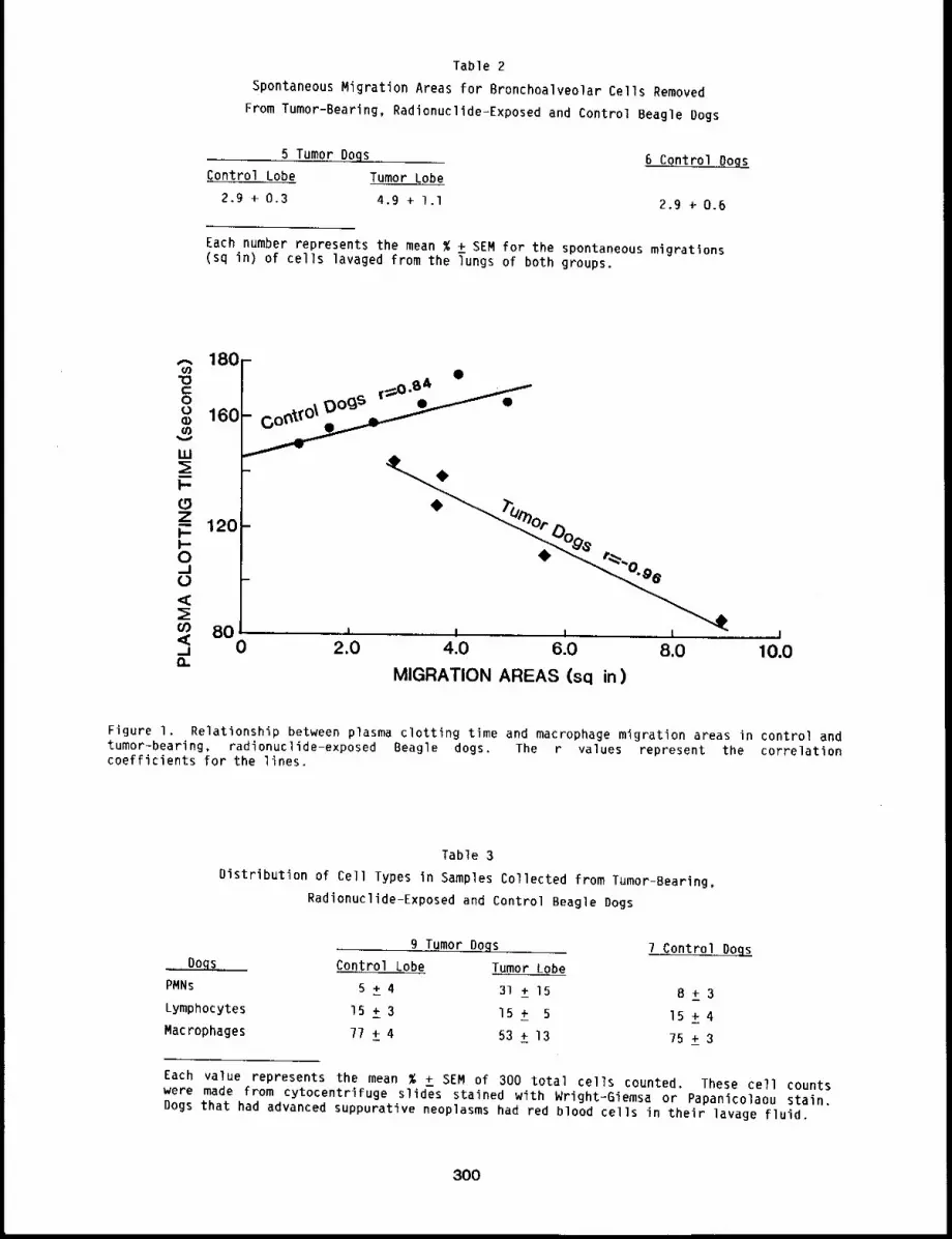
Table 2Spontaneous Migration Areas for Bronchoalveolar Cells Removed
From Tumor-Bearing, Radionuclide-Exposed and Control Beagle Dogs
5 Tumor Dogs 6 Control BogsControl Lobe Tumor Lobe
2.9 + 0.3 4.9 + I.I 2.9 ÷ 0.6
Each number represents the mean % + SEM for the spontaneous migrations(sq in) of cells lavaged from the Tungs of both groups.
LU
I--0Z~. 120i--0_10<c
8O<C,_1n
J ! i I i0 2.0 4.0 6.0 8.0 10.0
MIGRATION AREAS (sq in)
Figure I. Relationship between plasma clotting time and macrophage migration areas in control andtumor-bearing, radionuclide-exposed Beagle dogs. The r values represent the correlationcoefficients for the lines.
Table 3
Distribution of Cell Types in Samples Collected from Tumor-Bearing,
Radionuclide-Exposed and Control Beagle Dogs
9 Tumor Dogs 7 Control DogsDOL Control Lobe Tumor Lobe
PMNs 5 ± 4 31 ± 15 8 + 3Lymphocytes IS ± 3 15 ± 5 15 + 4Macrophages 77 ± 4 53 ± 13 75 + 3
Each value represents the mean % + SEM of 300 total cells counted. These cell countswere made from cytocentrifuge slides stained with Wright-Giemsa or Papanicolaou stain.Dogs that had advanced suppurative neoplasms had red blood cells in their lavage fluid,
300
DISCUSSION
This study showed the plasma clotting activity to be greater for cells and lavage fluid from
lung lobes that contained tumors than that for lung lobes without tumors. Most studies of
procoagulant activity point to a cell-associated tissue thromboplastin as the source of
procoagulant activity. This study indicated extracellular release of the procoagulant activity by
showing a significant decrease in plasma clotting time after addition of lavage fluid to the assay
from the tumor-bearing lung lobe. Dvorak et al., 1 however, said that this is caused by presence
of membrane vesicles with procoagulant activity that pelleted after ultracentrifugation of tumor
cell culture supernatants. This indicated that the procoagulant activity was still cell-associated
in bits of membrane containing the procoagulant activity and that these bits of membrane were
released into the surrounding medium.
The migration areas from the tumor-bearing lung lobes were significantly larger than those
from either of the control lung lobes. This could represent an activation of the macrophage by
the neoplasm, but the macrophages were probably phagocytizing the debris resultant of the tumor,
which could increase their mobility. The percentage of PMNs in the tumor lung lobe was
significantly elevated, indicating an inflammatory response in that lung lobe.
The correlation data that showed clotting time decreased as migration area increased was
contrary to published immunological data. 3 The procoagulant activity from tumors has been found
to be primarily a factor in addition to thromboplastin; 4 therefore, the mechanism by which
tumors trigger the coagulation cascade may be different from the pathway followed by immunological
stimulation.
Researchers report other pathological pulmonary conditions that have increased fibrin content
in the lung; however, little is known about the reason or consequence of its being there.
REFERENCES
I. Dvorak, H. R., S. C. Quay, N. S. Orenstein, A. M. Dvorak, P. Hahn, A. M. Bitzer, and A. C.Carvalho, Tumor Shedding and Coagulation, Science 212: 923-g24, IgBl.
2. PineD, G. F., E. Regoeczi, M. W. C. Hatton, and M. D. Brain, The Activation of Coagulation byExtracts of Mucus: A Possible Pathway of Intravascular Coagulation AccompanyingAdenocarcinomas, J. Lab. Clin. Med. 82: 255-260, __
3. Geczy, C. L. and P. A. Meyer, Leukocyte Procoagulant Activity in Man:of Delayed-type Hypersensitivity, J. Immunol. 128(I): 331-336,
4. Gordon, S. G., 3. 3. Franks, and B. Lewis, Cancer Procoagulant A:Procoagulant From Malignant Tissue, Thromb. Res. 6: 127-137, I.g75.
An In Vitro Correlate
A Factor X Activating
301/302

DOSE-RESPONSE RELATIONSHIPS FOR INHALED CHEMICAL TOXICANTS
Short- and long-term studies are being conducted to determine dose-response relationships for
inhaled chemical toxicants. The first paper presents a status report of a major study on the
long-term effects of inhaled diesel exhaust. In these studies, rats and mice are being exposed to
dilutions of exhaust from 5.7-1iter displacement diesel engines operated on the Federal Test
Procedure cycle simulating an urban driving pattern. Animals are being exposed for 7 h/day, 5
days/week to diluted exhaust at levels of 7000, 3500, and 350 ~g of partlcles/m 3 of air. The
original groups were exposed for 24 months for the mice and 30 months for the rats.
Analysis of the data and histopathological examination of tissues is incomplete at this time.
However, particle clearance from the lung was slowed, and lung~burdens of diesel particles
continued to increase at the two higher exposure levels. At those levels there were pulmonary
inflammation, cell proliferation, epithelial metaplasia, and fibrosis, which was also reflected by
pulmonary function impairment. No adverse health effects were observed at the lowest exposure
level.
The next paper reports on cytokinetic effects of fluidized bed combustion fly ash. Although
fly ash has a relatively low toxicity when inhaled, proliferation of some respiratory cell types
was induced. This proliferation was particularly notable and persistent in lung=associated
lymphoid tissues and could affect defense mechanisms of the lung.
The final paper in the section reports the results of inhalation exposure of dogs to either
lO0 or 400 mg/m3 of trifluoroethanol (TFE), which is a heat exchange fluid and a commercial
solvent, as well as a known testlcular toxicant. The exposures were 6 h/day, 5 days/week for 8
weeks. These studies provide further indication that TFE could be a testicular toxin when inhaled
by man.
303/304

LIFE-SPAN STUDY OF RODENTS INHALING DIESEL EXHAUST: RESULTS THROUGH 30 MONTHS
Abstract -- A llfe-span study of rodents exposed
by ~nhalation 7 h/da~, 5 days~week to dl~uted dlesel
exhaust aC particle concentrations of 7000, 3500,
and 350 pg/m 3 continued to 24 months of expo-
sure for mlce and to 30 months of exposure for rats.
Partlcle clearance was slowed and lung burdens of
dlesel particles contlnued to increase at the two
hlgher levels. The partlele accumulatlon was as-
sociated wlth lung ~nflam~atlon~ cell prollfera-
tlon, eplthellal metaplas~a~ and flbrosls, which
was reflected by impairment of resplratory func-
tlon. Immunologlcal functlon ~n pulmonary lymph
nodes was altered. CyCochrome P-450 levels in lung,
altered at 12 months, were normal at later tlmes.
No adverse health effects were observed at the low-
PRINCIPAL INVESTIGATORS
J. L. Mauderl9
J. M, Benson
D. E. B~ce
R. L. CarpenLer
M. J. Evans
R. F. Henderson
R. K. Jones
R. O. McClellan
J. A. Plckrell
H. C. Redman
S. G. Shaml
R. K, Wolff
est exposure level. Examlnatlon of lungs for tumors is contlnu~ng.
A life-span study of rats and mice is being conducted to examine potential adverse health
effects of chronic inhalation of diesel exhaust. The exposures were initiated on February lg,
1981. The background, experimental design, methods, and results through 18 months have been
reported (1980-81 Annual Report, LMF-gl, pp. 312-330; 1981-82 Annual Report, LMF-I02, pp.
379-389). This report summarizes results through 30 months of exposure of the original groups of
animals.
METHODS
Fischer-344 rats and CD-I mice, specific pathogen-free, from the Instltute’s colony are housed
continuously in whole-body exposure chambers and exposed 7 h/day, 5 days/week to clean air as
controls (C) or to concentrations of diluted exhaust containing nominally 7000 (high = H),
(medium = M), or 350 (low = L) ,g partlcles/m 3. Additional animals are in a barrier housing
facility as colony controls (CC). Exposures were initiated when the animals were 17 weeks old.
Each experimental group initially contained 288 rats and 360 mice (equal numbers of males and
females). Exposures of an additional 40 rats and 120 mice per group were begun at approximately
the l year point, and a new CC group of rats was entered to replace the original group lost
through heat stress.
Exhaust is generated by two 1980, 5.7 liter Oldsmobile engines operating alternately on the
Federal Test Procedure urban driving cycle and burning a standardized certification fuel. Exhaust
is diluted serially with clean air to provide the desired concentrations. Particle concentrations
were initially measured by filter samples in each chamber daily, and gases were sampled several
times daily in each chamber. After 34 weeks of exposure, particle samples were taken from one
chamber per level daily; bag samples for gases were taken from one chamber per level weekly.
Animals are checked twice daily for morbidity and mortality and are weighed monthly.
Biological endpoints measured at 6-month intervals in the original exposure groups included
305
respiratory function, tracheal mucociliary particle clearance, deep lung particle clearance,
immunological responses in pulmonary lymph nodes, enzyme and cytological profiles of airway
fluids, lung tissue proteases and connective tissue, xenobiotlc enzyme levels in selected tissues,
lung burdens of exhaust particles, lung cytokinetics, and histopathology. These endpoints were
measured in all rat groups through 24 months of exposure. Few mice remained alive at 24 months,
and the number of rats alive at 30 months precluded measurement of particle clearance, immunology,
and lung burdens of particles.
Twelve of the r’ats and mice sacrificed for morphological evaluations after 18 months of
exposure were also used to examine rates of cell proliferation in the lung. Two males and two
females of each species from the C, I, and H exposure groups were injected with tritiated
thymidine 2 h before sacrifice, and autoradiographs of 1-~m plastic sections of lung tissue were
prepared, l Labeling indices were calculated for the following lung cell types: large airway
epithelium, terminal bronchiolar epithelium, alveolar lype II epithelial cells, alveolar
interstitial cells (which may include Type I epithelial cells, endothelial cells, and
fibroblasts), and alveolar macrophages. For cytokinetic analysis, the alveolar regions of animals
exposed to the H level were subdivided histologically into relatively normal and abnormal areas.
Rats (6-9 per exposure group) and mice (12-15 per exposure group) were sacrificed, and
lungs were analyzed by selected measurements of connective tissue metabolism after 6, 12, IB,
(rats and mice) and 24 (rats only) months of exposure. Airway hydroxyproline peptides,
proteinase, and total lung collagen were measured at each sacrifice period in rats and mice. At
12 and 18 months, radioactive incorporation of proline into hydroxyproline was measured to
determine the percent of newly synthesized protein that was devoted to collagen. In addition,
ultrafilterable collagenous peptides were measured. Elastin quantity, production, and
hydroxyproline content were also measured.
At 24 months, lO rats per experimental group were given brief, nose-only exposures to two
radiolabeled particles to evaluate clearance by serial scintillation counting. Exhaust exposures
continued during the counting period. Long-term clearance was measured using 2 ~m mass median
aerodynamic diameter fused aluminosilicate particles containing cesium-134 (half life = 2.05
years), and intermediate-term clearance was measured using O.l ~m volume median diameter
gallium-67 oxide particles (half life ~ 3.25 days). Rats were whole-body counted at O, 3, 6, lO,
13, 18, 26, 54, llO, 137, 164, and 194 days after exposure to tracer particles,
RESULTS AND DISCUSSION
The exposure system has continued to operate very well, with less than 14 h of unexpected down
thne through 30 months of exposure. A summary of the exposure atmospheres through 30 months (905
exposure days) is given in Table I. The mean particle concentrations were within one percent of
target values and the coefficients of variation of weekly mean particle concentrations for the H,
M, and L levels were II, 13, and 20%, respectively. Data for the size and uniformity of
distribution of particles within the chambers were previously reported (1981-82 Annual Report,
LMF-I02, pp. 373-318). The mass median diameters in ~m (geometric standard deviations
parentheses) were 0.23 (4.4), 0.25 (4.5), and 0.26 (4.2) for H, M, and L levels, respectively.
Survival and body weight data from the original exposure groups are still being evaluated. An
insufficient number of mice survived to 24 months of exposure to allow measurements of endpoints.
Because of mortality and sacrifices, only 20, 18, 15, and 21 rats in the original H, M, L, and C
groups, respectively, survived to 30 months of exposure (34 months of age). The age at 50%
survival was 829, 848, 831, and 865 days (27.3, 27.9, 27.3, and 28.4 months), for H, M, L. and
rats, respectively. Body weight data collected during measurement of respiratory function and
306
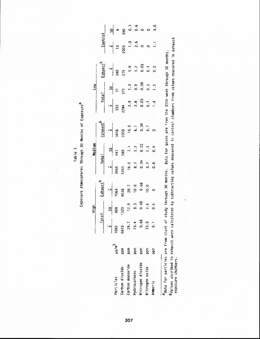
Ta
ble
1
Exp
osu
re A
tmo
sph
ere
s T
hro
ug
h 3
0 M
on
ths
of
Exp
osu
rea
Hig
hM
ed
ium
Lo
w
To
tal
Exh
au
stb
To
tal
Exh
au
stb
To
tal
Exh
au
stb
Co
ntr
ol
SD
~~
SD
~~
SD
~~
SD
Pa
rtic
les
pg
/m3
?0
82
80
87
06
93
46
94
47
34
56
35
3)l
34
01
36
Ca
rbo
n d
ioxi
de
pp
m6
64
31
32
04
63
84
35
55
90
23
50
22
84
37
12
79
20
05
39
0C
arb
on
mo
no
xid
ep
pm
29
.71
2.9
28
.71
6.5
7.I
15
.52
.9l.O
1.9
l.O0
.7H
ydro
carb
on
sp
pm
13
.4B
.3lO
.B8
.75
.26
.13
.80
.91
.22
.60
.6N
itro
ge
n
dio
xid
ep
pm
0.6
80
.48
0.6
80
.34
0.2
20
.34
0.0
50
.09
0.0
50
0N
itro
ge
n
oxi
de
pp
mlO
.O2
.6lO
.O5
.71
.55
.70
,70
.30
.70
0A
mfn
on
iap
pm
0.7
0.6
0o
.g0
.90
1.4
1.3
0.3
l.l3
.0
aD
ata
fo
r p
art
icle
s a
re fr
om
st
art
o
f st
ud
y th
rou
gh
3
0 m
on
ths.
Da
ta fo
r g
ase
s a
re
fro
m th
e
35
th
we
ek
thro
ug
h
30
mo
nth
s.b
Va
lue
s a
scrib
ed
to
e
xha
ust
w
ere
ca
lcu
late
d
by
sub
tra
ctin
g
valu
es
me
asu
red
in
con
tro
l ch
am
be
rs fro
m
valu
es
me
asu
red
in
exh
au
ste
xpo
sure
ch
am
be
rs.
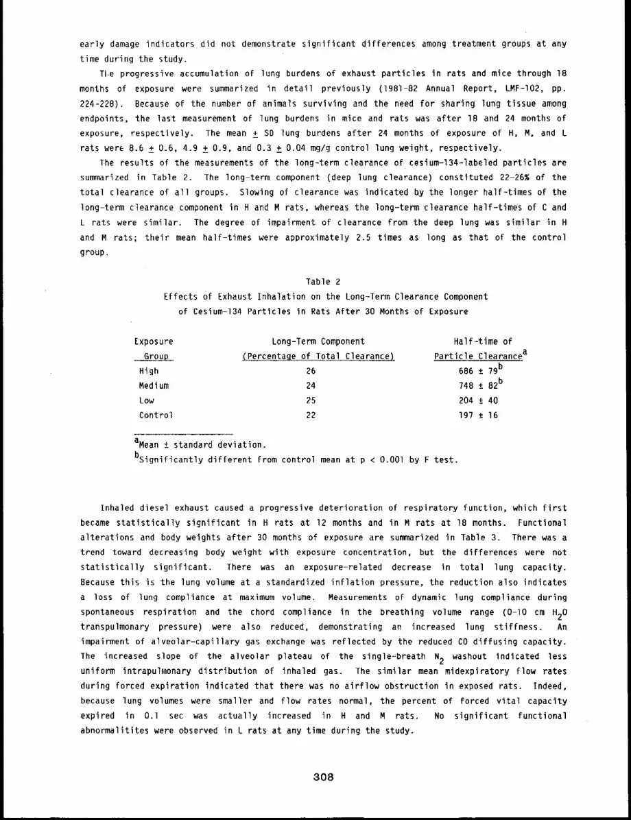
early damage indicators did not demonstrate significant differences among treatment groups at any
time during the study.
Tl,e progressive accumulation of lung burdens of exhaust particles in rats and mice through 18
months of exposure were summarized in detail previously (lgBI-82 Annual Report, LMF-I02, pp.
224-228). Because of the number of animals surviving and the need for sharing lung tissue among
endpoints, the last measurement of lung burdens in mice and rats was after IB and 24 months of
exposure, respectivelyo The mean ± SD lung burdens after 24 months of exposure of H, M, and L
rats wer~ 8.6 ± 0.6, 4.9 ± 0.9, and 0.3 ± 0.04 mg/g control lung weight, respectively.
The results of the measurements of the long-term clearance of cesium-134-1abeled particles are
summarized in Table 2. The long-term component (deep lung clearance) constituted 22-26% of the
total clearance of all groups. Slowing of clearance was indicated by the longer half-times of the
long-term clearance component in H and M rats, whereas the long-term clearance half-times of C and
L rats were similar. The degree of impairment of clearance from the deep lung was similar in H
and M rats; their mean half-times were approximately 2.5 times as long as that of the control
group.
Table 2
Effects of Exhaust Inhalation on the Long-Term Clearance Component
of Cesium-134 Particles in Rats After 30 Months of Exposure
Exposure Long-Term Component Half-time of
Group (Percentage of Total Clearance) Particle Clearancea
High 26 686 ± 7gb
Medium 24 748 ± B2b
Low 25 204 ± 40
Control 22 Ig7 ± 16
aMean ± standard deviation.
bsignificantly different from control mean at p < O.OOl by F test.
Inhaled diesel exhaust caused a progressive deterioration of respiratory function, which first
became statistically significant in H rats at 12 months and in M rats at 18 months. Functional
alterations and body weights after 30 months of exposure are summarized in Table 3. There was a
trend toward decreasing body weight with exposure concentration, but the differences were not
statistically significant. There was an exposure-related decrease in total lung capacity.
Because this is the lung volume at a standardized inflation pressure, the reduction also indicates
a loss of lung compliance at maximum volume. Measurements of dynamic lung compliance during
spontaneous respiration and the chord compliance in the breathing volume range (O-lO cm H20
transpulmonary pressure) were also reduced, demonstrating an increased lung stiffness. An
impairment of alveolar-capillary gas exchange was reflected by the reduced CO diffusing capacity.
The increased slope of the alveolar plateau of the single-breath N2 washout indicated less
uniform intrapulmonary distribution of inhaled gas. The similar mean midexpiratory flow rates
during forced expiration indicated that there was no airflow obstruction in exposed rats. Indeed,
because lung volumes were smaller and flow rates normal, the percent of forced vital capacity
expired in O.1 sec was actually increased in H and M rats. No significant functional
abnormalitites were observed in L rats at any time during the study.
3O8
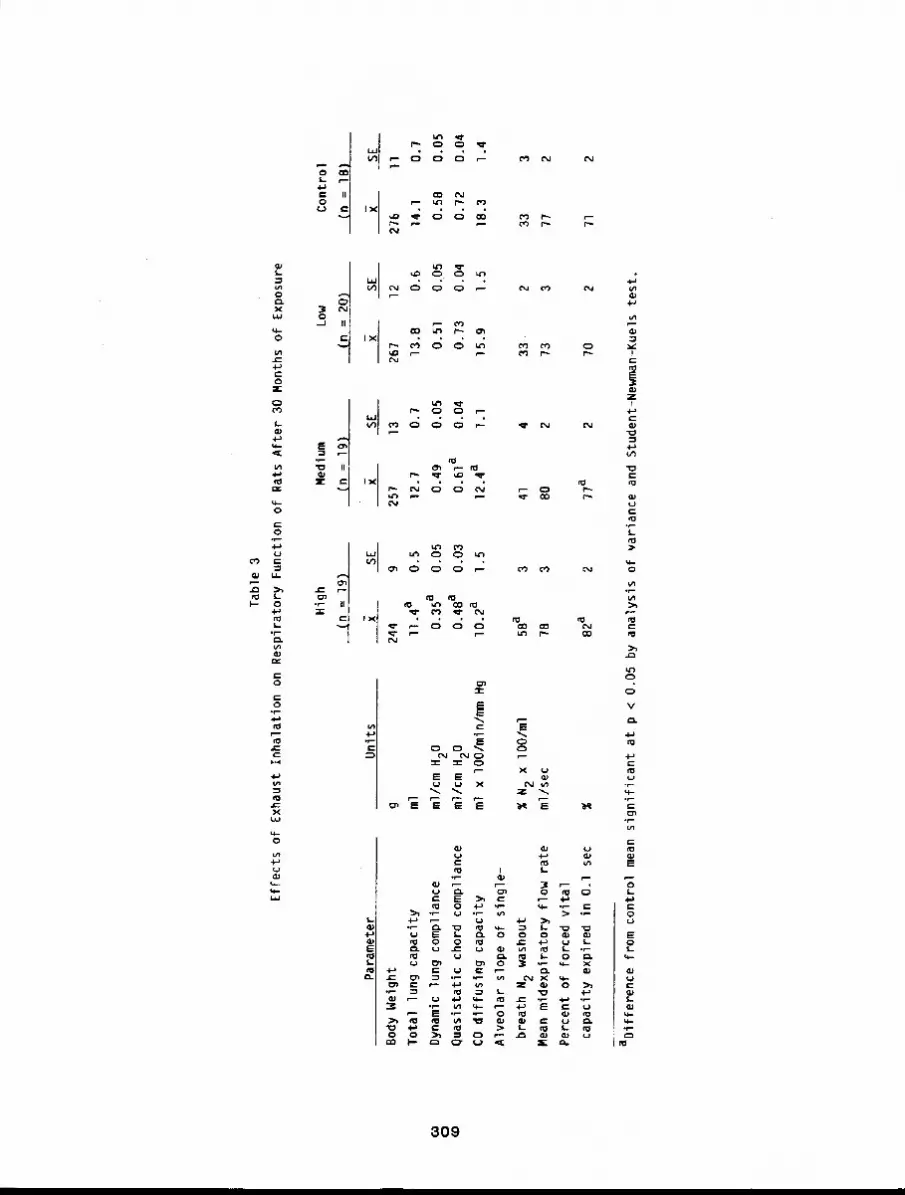
Ta
ble
3
Effe
cts
of
Exh
au
st
Inh
ala
tion
o
n R
esp
ira
tory
F
un
ctio
n
of
Ra
ts A
fte
r 3
0 M
on
ths
of
Exp
osu
re
f.o o (o
Pa
ram
ete
r
Bo
dy
We
igh
t
To
tal
lun
g
cap
aci
ty
Dyn
am
ic lu
ng
co
mp
lian
ce
qu
asi
sta
tic
cho
rd
com
plia
nce
CO
diff
usi
ng
ca
pa
city
Alv
eo
lar
slo
pe
o
f si
ng
le-
bre
ath
N2
wa
sho
ut
Me
an
mid
exp
ira
tory
flo
w
rate
Pe
rce
nt
of
forc
ed
vi
tal
cap
aci
ty
exp
ire
d
in
O.l
sec
Hig
hM
ed
ium
Lo
w
(n
= 1
9)
(n =
Ig
)(n
=
20
)
Un
its~
SE
~S
E~
SE
g2
44
g2
57
13
26
?1
2
ml
II.4
a0
.51
2.7
0.7
13
.80
.6
ml/c
m H
20
0.3
5a
0.0
50
.49
0.0
50
.51
0.0
5
ml/c
m H
20
0.4
8a
0.0
30
.61
a0
.04
0.7
30
.04
ml
x lO
0/m
in/m
m H
g1
0.2
a1
.51
2.4
al.l
15
.91
.5
Co
ntr
ol
(n =
IB) S
E
276
II
14.1
0.7
0.5
80
.05
0.72
0.04
18.3
1.4
% N
2 x
lO
0/m
l5
8a
34
14
33
23
33
ml/s
ec
7B
38
02
73
3?
72
%8
2a
27
7a
27
02
aD
iffe
ren
ce
fro
m co
ntr
ol
me
an
sig
nifi
can
t a
t p
<
0.0
5
by
an
aly
sis
of
varia
nce
a
nd
Stu
de
nt-
Ne
wm
an
-Ku
els
te
st.
?l
2
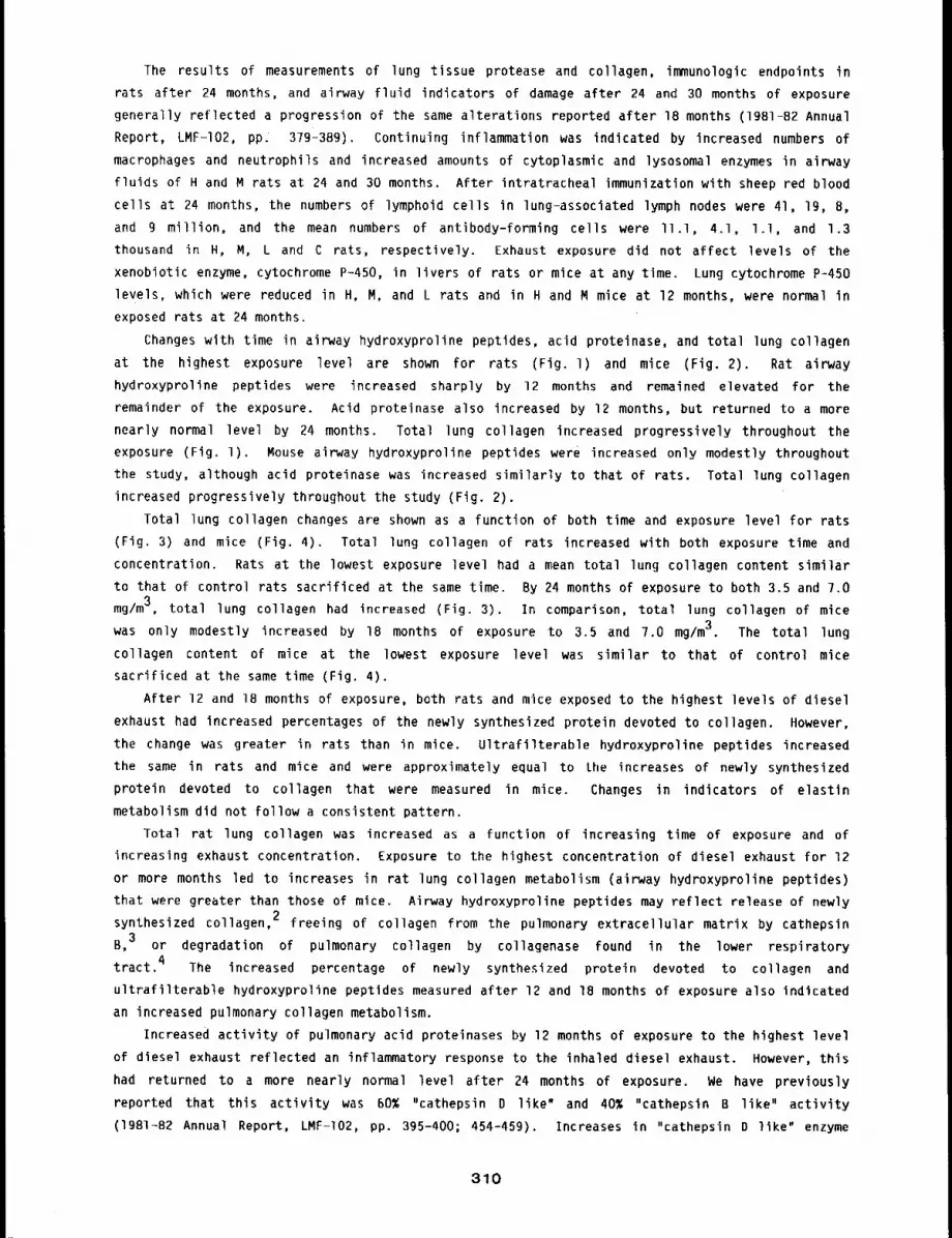
The results of measurements of lung tissue protease and collagen, immunologic endpoints in
rats after 24 months, and airway fluid indicators of damage after 24 and 30 months of exposure
generally reflected a progression of the same alterations reported after 18 months (igB1-B2 Annual
Report, LMF-I02, pp. 379-389). Continuing inflammation was indicated by increased numbers of
macrophages and neutrophils and increased amounts of cytoplasmic and lysosomal enzymes in airway
fluids of H and M rats at 24 and 30 months. After intratracheal immunization with sheep red blood
cells at 24 months, the numbers of lymphoid cells in lung-associated lymph nodes were 41, 19, 8,
and 9 million, and the mean numbers of antibody-forming cells were ll.l, 4.1, l.l, and 1.3
thousand in H, M, L and C rats, respectively. Exhaust exposure did not affect levels of the
xenobiotic enzyme, cytochrome P-450, in livers of rats or mice at any time. Lung cytochrome P-450
levels, which were reduced in H, M, and L rats and in H and M mice at 12 months, were normal in
exposed rats at 24 months.
Changes with time in airway hydroxyproline peptides, acid proteinase, and total lung collagen
at the highest exposure level are shown for rats (Fig. l) and mice (Fig. 2). Rat airway
hydroxyproline peptldes were increased sharply by 12 months and remained elevated for the
remainder of the exposure. Acid proteinase also increased by 12 months, but returned to a more
nearly normal level by 24 months. Total lung collagen increased progressively throughout the
exposure (Fig. l). Mouse airway hydroxyproline peptides were increased only modestly throughout
the study, although acid proteinase was increased similarly to that of rats. Total lung collagen
increased progressively throughout the study (Fig. 2).
Total lung collagen changes are shown as a function of both time and exposure level for rats
(Fig. 3) and mice (Fig. 4). Total lung collagen of rats increased with both exposure time
concentration. Rats at the lowest exposure level had a mean total lung collagen content similar
to that of control rats sacrificed at the same time. By 24 months of exposure to both 3.5 and 7.0
mg/m3, total lung collagen had increased (Fig. 3). In comparison, total lung collagen of mice
was only modestly increased by 18 months of exposure to 3.5 and 7.0 mg/m3. The total lung
collagen content of mice at the lowest exposure level was similar to that of control mice
sacrificed at the same time (Fig. 4).
After 12 and 18 months of exposure, both rats and mice exposed to the highest levels of diesel
exhaust had increased percentages of the newly synthesized protein devoted to collagen. However,
the change was greater in rats than in mice. Ultrafilterable hydroxyproline peptides increased
the same in rats and mice and were approximately equal to the increases of newly synthesized
protein devoted to collagen that were measured in mice. Changes in indicators of elastin
metabolism did not follow a consistent pattern.
Total rat lung collagen was increased as a function of increasing time of exposure and of
increasing exhaust concentration. Exposure to the highest concentration of diesel exhaust for 12
or more months led to increases in rat lung collagen metabolism (airway hydroxyproline peptides)
that were greater than those of mice. Airway hydroxyproline peptides may reflect release of newly
synthesized collagen, 2 freeing of collagen from the pulmonary extracellular matrix by cathepsin3B, or degradation of pulmonary collagen by collagenase found in the lower respiratory
tract. 4 The increased percentage of newly synthesized protein devoted to collagen and
ultrafilterable hydroxyproline peptldes measured after 12 and 18 months of exposure also indicated
an increased pulmonary collagen metabolism.
Increased activity of pulmonary acid proteinases by 12 months of exposure to the highest level
of diesel exhaust reflected an inflammatory response to the inhaled diesel exhaust. However, this
had returned to a more nearly normal level after 24 months of exposure. We have previously
reported that this activity was 60% "cathepsin D like" and 40% "cathepsin B like" activity
(1981-82 Annual Report, LMF-I02, pp. 395-400; 454-459). Increases in "cathepsin D llke" enzyme
310
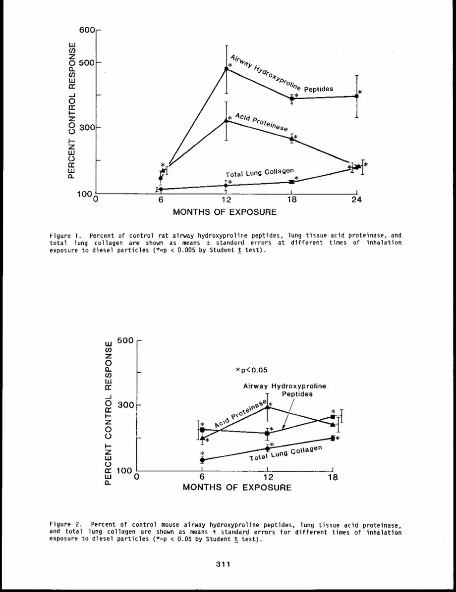
600 -
LUCOZ0 500-Q.O0wn-_j0cc
Z0 30(3-0I-ZUJ0trI.Ut"L
1000
MONTHS OF EXPOSURE
Figure I. Percent of control rat airway hydroxyproline peptldes, lung tissue acid proteinase, andtotal lung collagen are shown as means ± standard errors at different times of inhalationexposure to diesel particles (*=p < 0.005 by Student t test).
w 500-ZOnCOLLItr._J0 300-tri-z00I-ZUJ0rc 100w 0n
*p<O.05
Airway HydroxyprolineT Peptides
I~ ~ ¯
I ,, l I6 12 18
MONTHS OF EXPOSURE
Figure 2. Percent of control mouse airway hydroxyproline peptldes, lung tissue acid proteinase,and total lung collagen are shown as means ± standard errors for different times of inhalationexposure to diesel particles (*=p < 0.05 by Student t test).
311
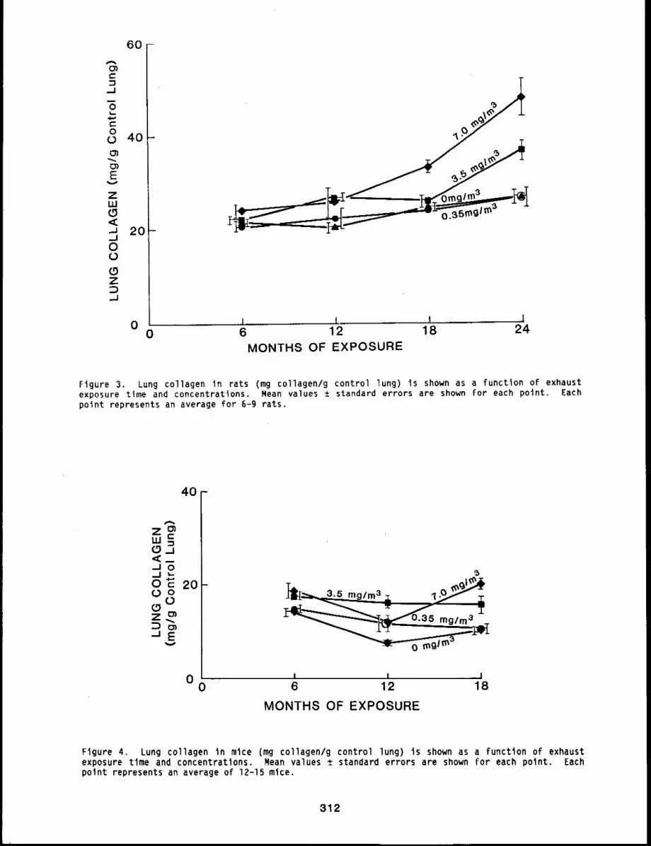
60-
Ao3t-
O
Oo 40-
c~E
v
zLU(.9<¯ ~ 20-_JOO(5zo,,_1
,l l I 10 0
6 12 18 24
MONTHS OF EXPOSURE
Figure 3. Lung collagen in rats (mg collagen/g control lung) is shown as a function of exhaustexposure time and concentrations. Mean values ± standard errors are shown for each point. Eachpoint represents an average for 6-9 rats.
40-
m~ D/ 0/O~ 2008
"EV
~’I ~3"5 mg/m3 Z, I 9 ~/~
0 i I J0 6 12 18
MONTHS OF EXPOSURE
Figure 4. Lung collagen in mice (mg collagen/g control lung) is shown as a function of exhaustexposure tlme and concentrations. Mean values ± standard errors are shown for each point. Eachpoint represents an average of 12-15 mice.
312
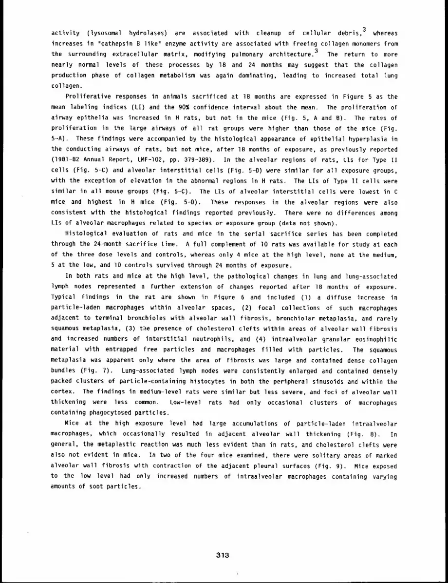
activity (lysosomal hydrolases) are associated with cleanup of cellular debris, 3 whereas
increases in "cathepsin B like" enzyme activity are associated with freeing collagen monomers from
the surrounding extracellular matrix, modifying pulmonary architecture. 3 The return to more
nearly normal levels of these processes by 18 and 24 months may suggest that the collagen
production phase of collagen metabolism was again dominating, leading to increased total lung
collagen.
Proliferative responses in animals sacrificed at 18 months are expressed in Figure 5 as the
mean labeling indices (LI) and the 90~ confidence interval about the mean. The proliferation
airway epithelia was increased in H rats, but not in the mice (Fig. 5, A and B). The rates
proliferation in the large airways of all rat groups were higher than those of the mice (Fig.
5-A). These findings were accompanied by the histological appearance of epithelial hyperplasia in
the conducting airways of rats, but not mice, after IB months of exposure, as previously reported
(1981-82 Annual Report, LMF-I02, pp. 379-389). In the alveolar regions of rats, LIs for Type
cells (Fig. 5-C) and alveolar interstitial cells (Fig. 5-D) were similar for all exposure groups,
with the exception of elevation in the abnormal regions in H rats. The LIs of Type II cells were
similar in all mouse groups (Fig. 5-C). The LIs of alveolar interstitial cells were lowest in
mice and highest in H mice (Fig. 5=D). These responses in the alveolar regions were also
consistent with the histological findings reported previously. There were no differences among
LIs of alveolar macrophages related to species or exposure group (data not shown).
Histological evaluation of rats and mice in the serial sacrifice series has been completed
through the 24-month sacrifice time. A full complement of 10 rats was available For study at each
of the three dose levels and controls, whereas only 4 mice at the high level, none at the medium,
5 at the low, and lO controls survived through 24 months of exposure.
In both rats and mice at the high level, the pathological changes in lung and lung-associated
lymph nodes represented a further extension of changes reported after 18 months of exposure.
Typical findings in the rat are shown in Figure 6 and included (1) a diffuse increase
particle-laden macrophages within alveolar spaces, (2) focal collections of such macrophages
adjacent to terminal bronchioles with alveolar wall fibrosis, bronchiolar metaplasia, and rarely
squamous metaplasia, (3) t;le presence of cholesterol clefts within areas of alveolar wall fibrosis
and increased numbers of interstitial neutrophils, and (4) intraalveolar granular eosinophilic
material with entrapped free particles and macrophages filled with particles. The squamous
metaplasia was apparent only where the area of Fibrosis was large and contained dense collagen
bundles (Fig. ?). Lung-associated lymph nodes were consistently enlarged and contained densely
packed clusters of particle-containing histocytes in both the peripheral sinusoids and within the
cortex. The findings in medium-level rats were similar but less severe, and foci of alveolar wall
thickening were less common. Low-level rats had only occasional clusters of macrophages
containing phagocytosed particles.
Mice at the high exposure level had large accumulations of particle-laden intraalveolar
macrophages, which occasionally resulted in adjacent alveolar wall thickening (Fig. 8).
general, the metaplastic reaction was much less evident than in rats, and cholesterol clefts were
also not evident in mice. In two of the four mice examined, there were solitary areas of marked
alveolar wall fibrosis with contraction of the adjacent pleural surfaces (Fig. 9). Mice exposed
to the low level had only increased numbers of intraalveolar macrophages containing varying
amounts of soot particles.
313
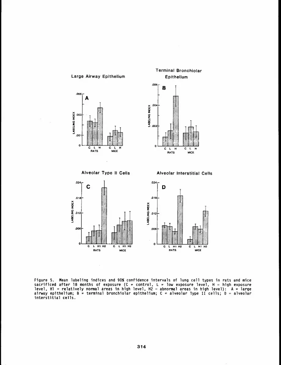
Large Airway Epithelium
.005
C L HMICE
Terminal BronchiolarEpithelium
,006
.004 - B
C L H C L H
RATS MICE
Alveolar Type II Cells
.024 ,
.00~
C,024
C / H1 H2 C L H1 H2
RATS MICE
Alveolar Interstitial Cells
.01B
z~ ,012,,=,.J
,00B
0 0
D
o/
C L H1 H2 C L H1 H2RATS MICE
Figure 5. Mean labeling indices and 90% confidence intervals of lung cell types in rats and micesacrificed after 18 months of exposure (C = control, L = low exposure level, H : high exposurelevel, HI ~ relatively normal areas in high level, H2 = abnormal areas in high level): A = largeairway epithelium; B = terminal bronchiolar epithelium; C = alveolar Type II cells; D = alveolarinterstitial cells.
314
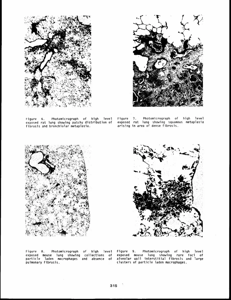
Figure 6. Photomicrograph of high level Figure 7. Photomicrograph of high levelexposed rat lung showing patchy distribution of exposed rat lung showing squamous metaplasiafibrosis and bronchiolar metaplasia, arising in area of dense fibrosis.
Figure 8. Photomicrograph of high level Figure 9. Photomicrograph of high levelexposed mouse lung showing collections of exposed mouse lung showing rare foci ofparticle laden macrophages and absence of alveolar wall interstitial fibrosis and largepulmonary fibrosis, clusters of particle laden macrophages.
315
CONCLUSIONS
The results to date continue to support the hypothesis that no serious noncarcinogenic health
effects occur in rodents exposed chronically to levels of diesel exhaust that do not result in
progressive accumulations of particle burdens in their lungs. In this study, lung burdens of L
animals were nearly conszant after 6 months of exposure, whereas those of M and H animals increas-
ed progressively throughout the exposure. Measurements of the clearance of both gallium-67 oxide
and cesium-134 fused aluminosilicate particles indicated that clearance was normal in L animals
and retarded in M and H animals. These changes in clearance offer an explanation for the conti-
nued increase in lung burdens of soot in the lungs of the M- and H-level animals previously
reported (1981-82 Annual Report, LMF-I02, pp. 224-228). The progressive accumulation of exhaust
particles in lungs of animals has been associated with progressive inflammatory and fibrotic lung
disease in this and other studies.5-8 These results suggest that the most pressing need for
additional research on noncarcinogenic health effects of diesel exhaust emissions is the
development of better predictions for the exhaust exposure patterns likely to cause progressive
accumulations of particles in human lungs. Because all studies to date have started with young,
healthy animals, another need is to compare effects in animal models of populations that might
have different sensitivities because of age or health status.
The question of carcinogenicity of inhaled diesel exhaust remains unanswered. Epidemiological
studies have not demonstrated a clear relationship between diesel exhaust exposure and lung tumors
in man. Results of animal studies to date indicate that at worst, diesel exhaust may be a weak
carcinogen. Malignant lung tumors have been observed in exhaust-exposed animals (personal commu-
nication, Dr. J. Vostal, General Motors Research), although not at statistically significant inci-
dences. These tumors were very small and required examination of serial sections of lung for de-
tection; thus, it is possible that some tumors might have been missed in earlier studies. The
lungs of animals in the present study will be sectioned and examined in detail, starting with
those exposed for the longest times to the highest levels; however, results are not yet available.
REFERENCES
I. Shami, S. G., L. A. Thibodeau, A. R. Kennedy, and J. B. Little, Recovery from Ozone-InducedInjury in the Lungs of Syrian Golden Hamsters, Exp. MoT. Pathol. 36: 57-71, 1982.
2. Low, R. B., K. R. Cutroneo, G. S. Davis, and M. S. Giancola, Lavage Type III Procollagen N--Ter-minal Peptides in Human Pulmonary Fibrosis and Sarcoidosis, Lab. Invest. 48: ?55-759, 1983.
3. Barrett, A. J., Chapter 5, Cathepsin D and Other Carboxyl Proteinases; Chapter 4, Cathepsin Band Other Thiol Proteinases; in Proteinases in Mammalian Cells and Tissues (A. J. Barrett,ed.), Elsevier/North Holland Publishing Co., Amsterdam, Holland, pp. 181-247, 1977.
4. Gadek, J. E., J. A. Kelman, G. Fells, S. E. Weinberger, A. L. Horwitz, H. Y. Reynolds, J. D.Fulmer, and R. G. Crystal, Collagenase in the Lower Respiratory Tract of Patients withIdiopathic Pulmonary Fibrosis, N. En I. J. Med. 3_01: 737-742, 1979.
5. Gross, K. B., Pulmonary Function Testing of Animals Chronically Exposed to Diluted DieselExhaust, J. A~I. Toxicol. l: 116-123, 1981.
6. McClellan, R. 0., A. L. Brooks, R. K. Jones, J. L. Mauderly, and R. K. Wolff, InhalationToxicology of Diesel Exhaust Particles, in Toxicological Effects of Emissions From DieselEngines (J. Lewtas, ed.), Elsevier, New York, pp. 99-120, 1982o
7. Pepelko, W. E., EPA Studies on the Toxicological Effects of Inhaled Diesel Engine Emissions,in Toxicological Effects of Emissions From Diesel Engines (J. Lewtas, ed.), Elsevier, NewYork, pp. 121-142, 1982.
8. Kaplan, H. L., K. J. Springer, and W. F. MacKenzie, Studies of Potential Health Effects ofLong-Term Exposure to Diesel Exhaust.Em~ssion~, Final Report, SwRI Project No. 01-0750-i03,Southwest Research Institute, San Antonio, TX, June, 1983.
316
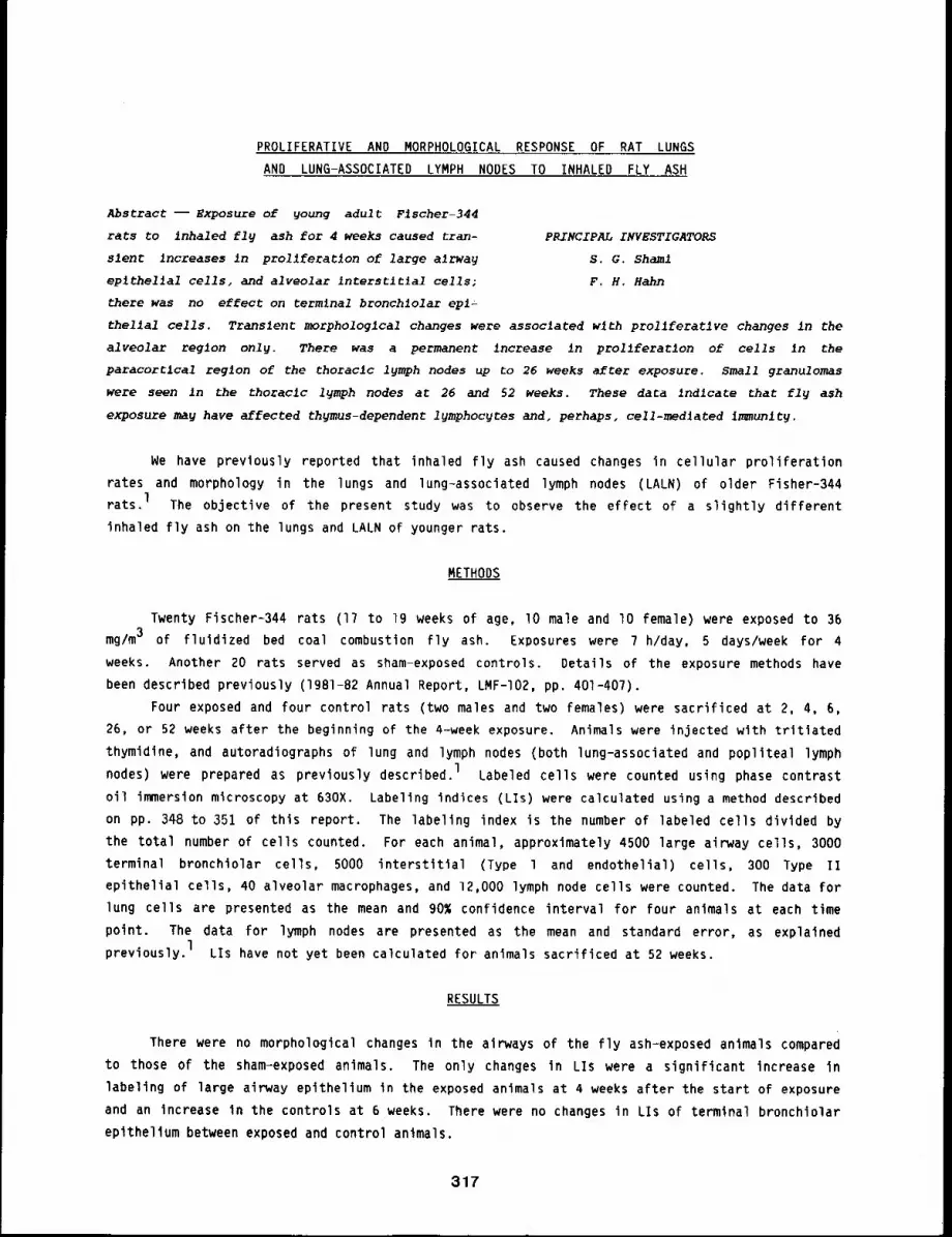
PROLIFERATIVE AND MORPHOLOGICAL RESPONSE OF RAT LUNGS
AND LUNG-ASSOCIATED LYMPH NODES TO INHALED FLY ASH
Abstract -- Exposure of young adult Fischer-344
rats to inhaled fly ash for 4 weeks caused tran- PRINCIPAL INVESTIGATORS
slent Increases in prollferatlon of large alrway s.G. Sham1
eplthellal cells, and alveolar interstltlal cells; F.H. Hahn
there was no effect on terminal bronchlolar epl=
thellal cells. Transient morphologlcal changes were associated with prollferatlve changes in the
alveolar reglon only. There was a permanent increase in prollferatlon of cells ~n the
paracorClcal region of the thoracic lymph nodes up to 26 weeks after exposure. Small granulomas
were seen In the thoracic lymph nodes at 26 and 52 weeks. These data Indlcate that fly ash
exposure may have affected thymus-dependent lymphoc~es and, perhaps, ce11-med~ated immunltg.
We have previously reported that inhaled fly ash caused changes in cellular proliferation
rates and morphology in the lungs and lung-associated lymph nodes (LALN) of older Fisher-344
rats. 1 The objective of the present study was to observe the effect of a slightly different
inhaled fly ash on the lungs and LALN of younger rats.
METHODS
Twenty Fischer-344 rats (17 to 19 weeks of age, lO male and lO female) were exposed to
mg/m3 of fluidized bed coal combustion fly ash. Exposures were 7 h/day, 5 days/week for 4
weeks. Another 20 rats served as sham-exposed controls. Details of the exposure methods have
been described previously (1981-82 Annual Report, LMF-I02, pp. 401-407).
Four exposed and four control rats (two males and two females) were sacrificed at 2, 4,
26, or 52 weeks after the beginning of the 4-week exposure. Animals were injected with tritlated
thymidine, and autoradiographs of lung and lymph nodes (both lung-associated and popliteal lymph
nodes) were prepared as previously described, l Labeled cells were counted using phase contrast
oil immersion microscopy at 630X. Labeling indices (LIs) were calculated using a method described
on pp. 348 to 351 of this report. The labeling index is the number of labeled cells divided by
the total number of cells counted. For each animal, approximately 4500 large airway cells, 3000
terminal bronchiolar cells, 5000 interstitial (Type l and endothelial) cells, 300 Type
epithelial cells, 40 alveolar macrophages, and 12,000 lymph node cells were counted. The data for
lung cells are presented as the mean and 90% confidence interval for four animals at each time
point. The data for lymph nodes are presented as the mean and standard error, as explained
previously, l LIs have not yet been calculated for animals sacrificed at 52 weeks.
RESULTS
There were no morphological changes in the airways of the fly ash-exposed animals compared
to those of the sham-exposed animals. The only changes in LIs were a significant increase in
labeling of large airway epithelium in the exposed animals at 4 weeks after the start of exposure
and an increase in the controls at 6 weeks. There were no changes in LIs of terminal bronchiolar
epithelium between exposed and control animals.
317
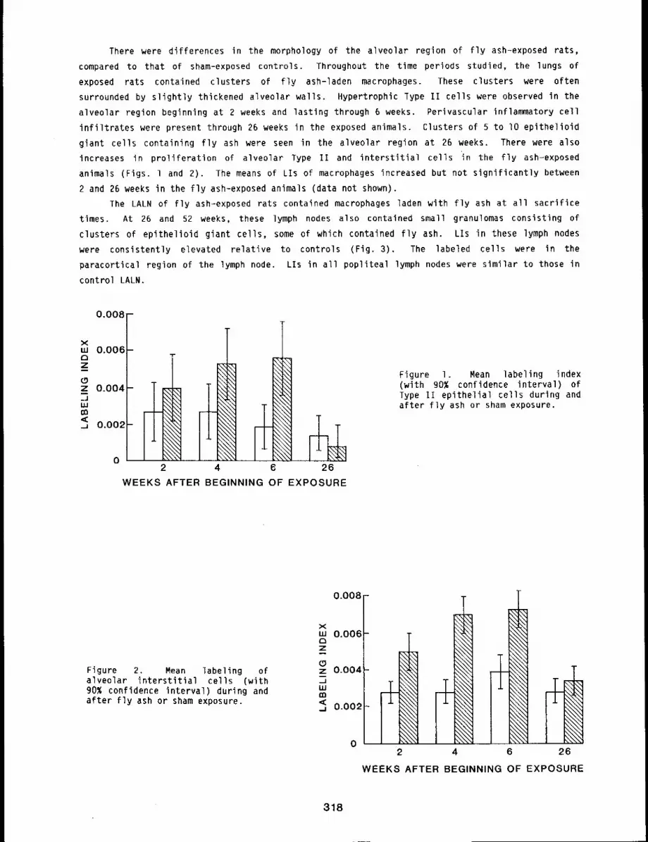
There were differences in the morphology of the alveolar region of fly ash-exposed rats,
compared to that of sham-exposed controls. Throughout the time periods studied, the lungs of
exposed rats contained clusters of fly ash-laden macrophages. These clusters were often
surrounded by slightly thickened alveolar walls. Hypertrophic Type II cells were observed in the
alveolar region beginning at 2 weeks and lasting through 6 weeks. Perivascular inflammatory cell
infiltrates were present through 26 weeks in the exposed animals. Clusters of 5 to I0 epithelioid
giant cells containing fly ash were seen in the alveolar region at 26 weeks. There were alsoincreases in proliferation of alveolar Type II and interstitial cells in the fly ash-exposed
animals (Figs¯ 1 and 2). The means of Lls of macrophages increased but not significantly between
2 and 26 weeks in the fly ash-exposed animals (data not shown).
The LALN of fly ash-exposed rats contained macrophages laden with fly ash at all sacrifice
times. At 26 and 52 weeks, these lymph nodes also contained small granulomas consisting of
clusters of epithelioid giant cells, some of which contained fly ash. Lls in these lymph nodes
were consistently elevated relative to controls (Fig. 3). The labeled cells were in the
paracortical region of the lymph node. Lls in all popliteal lymph nodes were similar to those in
control LALN.
0.00(
xLU 0.00(C3Zi(5z 0.004i=JLLIrm
_J 0.002
T
WEEKS AFTER BEGINNING OF EXPOSURE
Figure I. Mean labeling index(with 90% confidence interval) Type II epithelial cells during andafter fly ash or sham exposure.
Figure 2. Mean labeling ofalveolar interstitial cells (with90% confidence interval) during andafter fly ash or sham exposure.
0.008
xLU 0.006-c}zM(5z 0.004-.JLUm<. 0.002~
2
\\,1\\,,i
26
WEEKS AFTER BEGINNING OF EXPOSURE
318
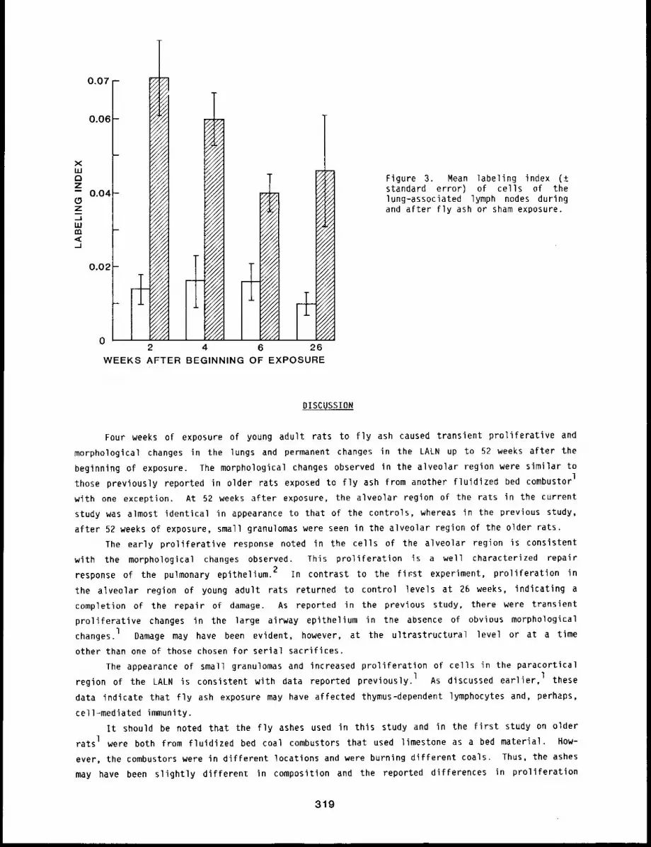
0.07 -
0.06 -
xu.IC3Z-- 0.04z._1LU,,,-,,<._1
0.02
i I rr2 4 6 26
WEEKS AFTER BEGINNING OF EXPOSURE
Figure 3. Mean labeling index (±standard error) of cells of thelung-associated lymph nodes duringand after fly ash or sham exposure.
DISCUSSION
Four weeks of exposure of young adult rats to fly ash caused transient proliferative and
morphological changes in the lungs and permanent changes in the LALN up to 52 weeks after the
beginning of exposure. The morphological changes observed in the alveolar region were similar to
those previously reported in older rats exposed to fly ash from another fluidized bed combustorl
with one exception. At 52 weeks after exposure, the alveolar region of the rats in the current
study was almost identical in appearance to that of the controls, whereas in the previous study,
after 52 weeks of exposure, small granulomas were seen in the alveolar region of the older rats.
The early proliferative response noted in the cells of the alveolar region is consistent
with the morphological changes observed. This proliferation is a well characterized repair
response of the pulmonary epithelium. 2 In contrast to the first experiment, proliferation in
the alveolar region of young adult rats returned to control levels at 26 weeks, indicating a
completion of the repair of damage. As reported in the previous study, there were transient
proliferative changes in the large airway epithelium in the absence of obvious morphological
changes. 1 Damage may have been evident, however, at the ultrastructural level or at a time
other than one of those chosen for serial sacrifices.
The appearance of small granulomas and increased proliferation of cells in the paracortical
region of the LALN is consistent with data reported previously. 1 As discussed earlier, 1 these
data indicate that fly ash exposure may have affected thymus-dependent lymphocytes and, perhaps,
cell-mediated immunity.
It should be noted that the fly ashes used in this study and in the first study on older
rats I were both from fluidized bed coal combustors that used limestone as a bed material. How-
ever, the combustors were in different locations and were burning different coals. Thus, the ashes
may have been slightly different in composition and the reported differences in proliferation
319
and small granuloma formation could be due to this factor. However, current evidence indicates
that there is little difference in the toxicity of the various fly ashes. 3 It is more probable
that the differences observed were due to the age of the rats, because it has been clearly
established that older animals are more sensitive to irritant-induced lung injury than younger
animals.4 This, however, does not rule out the possibility that the differences were due to
relatively small variations in the composition of the two fly ash samples.
REFERENCES
1 ¯
3.
4.
Shami, S. G., S. A. Silbaugh, F. F. Hahn, W. C. Griffith, and C. H. Hobbs, Cytokinetic andMorphological Changes in the Lungs and Lung-Associated Lymph Nodes of Rats After Inhalationof Fly Ash, Environ. Res. (in press).
Evans, M. J., Cell Death and Cell Renewal in Small Airways and Alveoli, in Mechanisms inRespiratory Toxicology (H. Wltschi, P. Nettesheim, eds.), 189-218, 1982.
Hobbs, C. H., Status of Research on Physical, Chemical and Biological Characterization ofParticulate and Organic Emissions from Conventional and Fluidized Bed Combustion of Coal:1976 to the present. DOE/ER-O162, 1983.
Evans, M. J., L. J. Cabral-Anderson, and G. Freeman, Effects of NO2 on the Lungs of AgingRats. II. Cell Proliferation, Exp, Mol. Pathol. 27: 366-376, 1977.
320
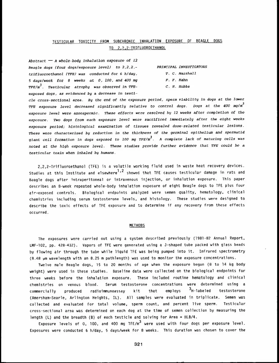
TESTICULAR TOXICITY FROM SUBCHRONIC INHALATION EXPOSURE OF BEAGLE DOGS
TO 2.2,2-TRIFLUOROETHANOL
Abstract --A whole-body inhalatlon exposure of 12
Beagle dogs (four dogs~exposure level) to 2,2,2,- pRINCIPAL INVESTIGATORS
trlfluoroethanol (TFE) was conducted for 6 h/day~ T.C. Marshall
5 days~week for 8 weeks at 0, 100, and 400 mg F.F. Hahn
TFE/m3. Testlcular atrophy was observed in TFE-. C.H. Hobbs
exposed dogs, as evidenced by a decrease in testl-
cle cross.-sectlonal area. By the end of the exposure period, sperm vlab~l~ty in dogs at the lower
TFE exposure level decreased slgnlflcantly relative to control dogs. Dogs at the 400 mg/m3
exposure level were azoospermlc. These effects were resolved by 12 weeks after completion of the
exposure. TWO dogs from each exposure level were sacrlflced lmmedlately after the elght weeks
exposure perlod; hlsrologlcal examlnatlon of tlssues revealed dose-related testlcular les~ons.
These were characterlzed by reduction in the thickness of the germlnal eplthellum and spermatid
glant cell formation in dogs exposed to 100 mg TFE/m3. A complete lack of maturlng cells was
noted at The hlgh exposure level. These studles provide further evldence that TFE could be a
test~cular toxin when inhaled by humans.
2,2,2-Trifluoroethanol (TFE) is a volatile working fluid used in waste heat recovery devices.
Studies at this Institute and elsewhere 1’2 showed that TFE causes testicular damage in rats and
Beagle dogs after intraperitoneal or intravenous injection, or inhalation exposure. This paper
describes an 8-week repeated whole-body inhalation exposure of eight Beagle dogs to TFE plus four
air-exposed controls. Biological endpoints analyzed were semen quality, hematology, clinical
chemistries including serum testosterone levels, and histology. These studies were designed to
describe the toxic effects of TFE exposure and to determine if any recovery from these effects
occurred.
METHODS
The exposures were carried out using a system described previously (1981-82 Annual Report,
LMF-I02, pp. 428-432). Vapors of TFE were generated using a O-shaped tube packed with glass beads
by flowing air through the tube while liquid TFE was being pumped into it. Infrared spectrometry
(8.48 ,m wavelength with an 8.25 m pathlength) was used to monitor the exposure concentrations.
Twelve male Beagle dogs, 15 to 20 months of age when the exposure began (B to ]4 kg body
weight) were used in these studies. Baseline data were collected on the biological endpoints for
three weeks before the inhalation exposure. These included routine hematology and clinical
chemistries on venous blood. Serum testosterone concentrations were determined using a
commercially produced radioimmunoassay kit that employs 3H-labeled testosterone
(Amersham--Searle, Arlington Heights, IL). All samples were evaluated in triplicate. Semen was
collected and evaluated for total volume, sperm count, and percent live sperm. Testicular
cross-sectional area was determined on each dog at the time of semen collection by measuring the
length (L) and the breadth (B) of each testicle and solving for Area = ~LB/4.
Exposure levels of O, lO0. and 400 mg TFE/m3 were used with four dogs per exposure level.
Exposures were conducted 6 h/day, 5 days/week for B weeks. This duration was chosen to cover the
321
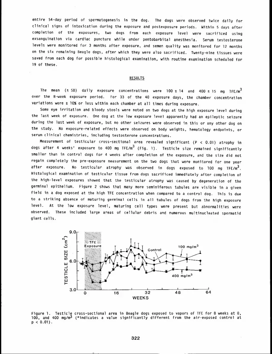
entire 54-day period of spermatogenesis in the dog. The dogs were observed twice daily for
clinical signs of intoxication during the exposure and postexposure periods. Within 5 days after"
completion of the exposures, two dogs from each exposure level were sacrificed using
exsanguination via cardiac puncture while under pentobarbital anesthesia. Serum testosterone
levels were monitored for 3 months after exposure, and semen quality was monitored for 12 months
on the six remaining Beagle dogs, after which they were also sacrificed. Twenty-nine tissues were
saved from each dog for possible histological examination, with routine examination scheduled for
19 of these.
RESULTS
The mean (± SD) daily exposure concentrations were lO0 ± 14 and 400 ± 15 mg IFE/m3
over the 8-week exposure period. For 33 of the 40 exposure days, the chamber concentration
variations were ± IO% or less within each chamber at all times during exposure.
Some eye irritation and bloody stools were noted on two dogs at the high exposure level during
the last week of exposure. One dog at the low exposure level apparently had an epileptic seizure
during the last week of exposure, but no other seizures were observed in this or any other dog on
the study. No exposure-related effects were observed on body weights, hematology endpoints, or
serum clinical chemistries, including testosterone concentrations.
Measurement of testicular cross-sectional area revealed significant (P < O.Ol) atrophy
dogs after 4 weeks’ exposure to 400 mg TFE/m3 (Fig. l). Testicle size remained significantly
smaller than in control dogs for 4 weeks after completion of the exposure, and the size did not
regain completely the pre-exposure measurement on the two dogs that were monitored for one year
after exposure. No testicular atrophy was observed in dogs exposed to IO0 mg TFE/m3.
Histological examination of testicular tissue from dogs sacrificed immediately after completion of
the high-level exposures showed that the testicular atrophy was caused by degeneration of the
germinal epithelium. Figure 2 shows that many more seminiferous tubules are visible in a given
field in a dog exposed at the high TFE concentration when compared to a control dog. This is due
to a striking absence of maturing germinal cells in all tubules of dogs from the high exposure
level. At the low exposure level, maturing cell types were present but abnormalities were
observed. These included large areas of cellular debris and numerous multinucleated spermatid
giant ceils.
9.0
E0
UJN
6.0LUJC)Dl-uJ
3.0~/ ......
~- I i I I I
0 16 32 48WEEKS
Figure I. Testicle cross-sectional area in Beagle dogs exposed to vapors of TFE for 8 weeks at O,lOO, and 400 mg/m3 (*indicates a value significantly different from the air-exposed control atp < O.Ol).
322
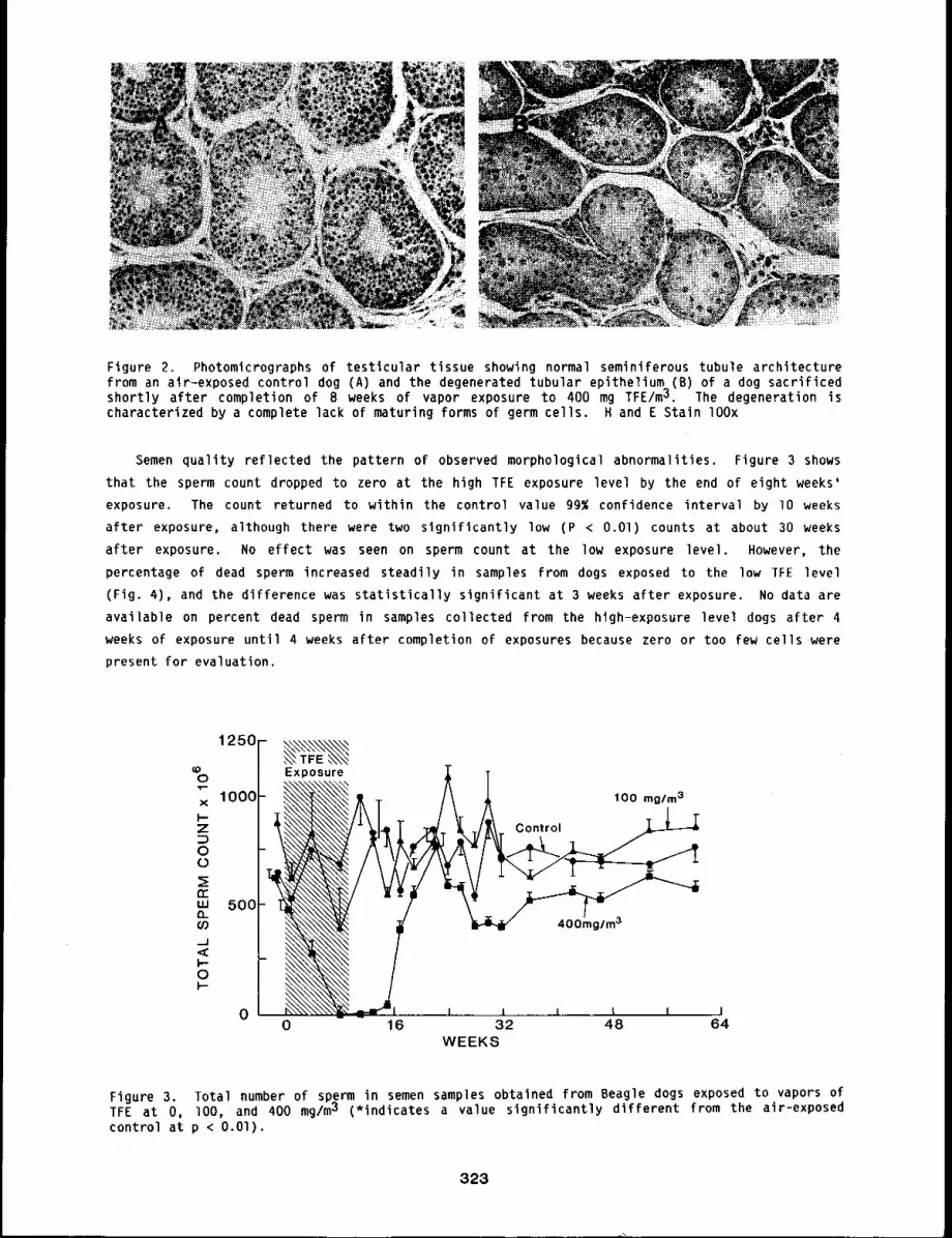
Figure 2. Photomicrographs of testlcular tissue showing normal seminiferous tubule architecturefrom an air-exposed control dog (A) and the degenerated tubular epithelium (B) of a dog sacrificedshortly after completion of 8 weeks of vapor exposure to 400 mg TFE/m~. The degeneration ischaracterized by a complete lack of maturing forms of germ cells. H and E Stain lOOx
Semen quality reflected the pattern of observed morphological abnormalities. Figure 3 shows
that the sperm count dropped to zero at the high TFE exposure level by the end of eight weeks’
exposure. The count returned to within the control value 9g~ confidence interval by lO weeks
after exposure, although there were two significantly low (P < O.Ol) counts at about 30 weeks
after exposure. No effect was seen on sperm count at the low exposure level. However, the
percentage of dead sperm increased steadily in samples from dogs exposed to the low TFE level
(Fig. 4), and the difference was statistically significant at 3 weeks after exposure. No data are
available on percent dead sperm in samples collected from the high-exposure level dogs after 4
weeks of exposure until 4 weeks after completion of exposures because zero or too few ceils were
present for evaluation.
~0 I Exposure T
× 1000 ~&"~ 31 O0 rng/mI--Z Contr
erW 500o..CO
p.0p-
00 16 32 48 64
WEEKS
Figure 3. Total number of sperm in semen samples obtained from Beagle dogs exposed to vapors ofTFE at O, lO0, and 400 mg/m3 (*indicates a value significantly different from the air-exposedcontrol at p < O.O1).
323
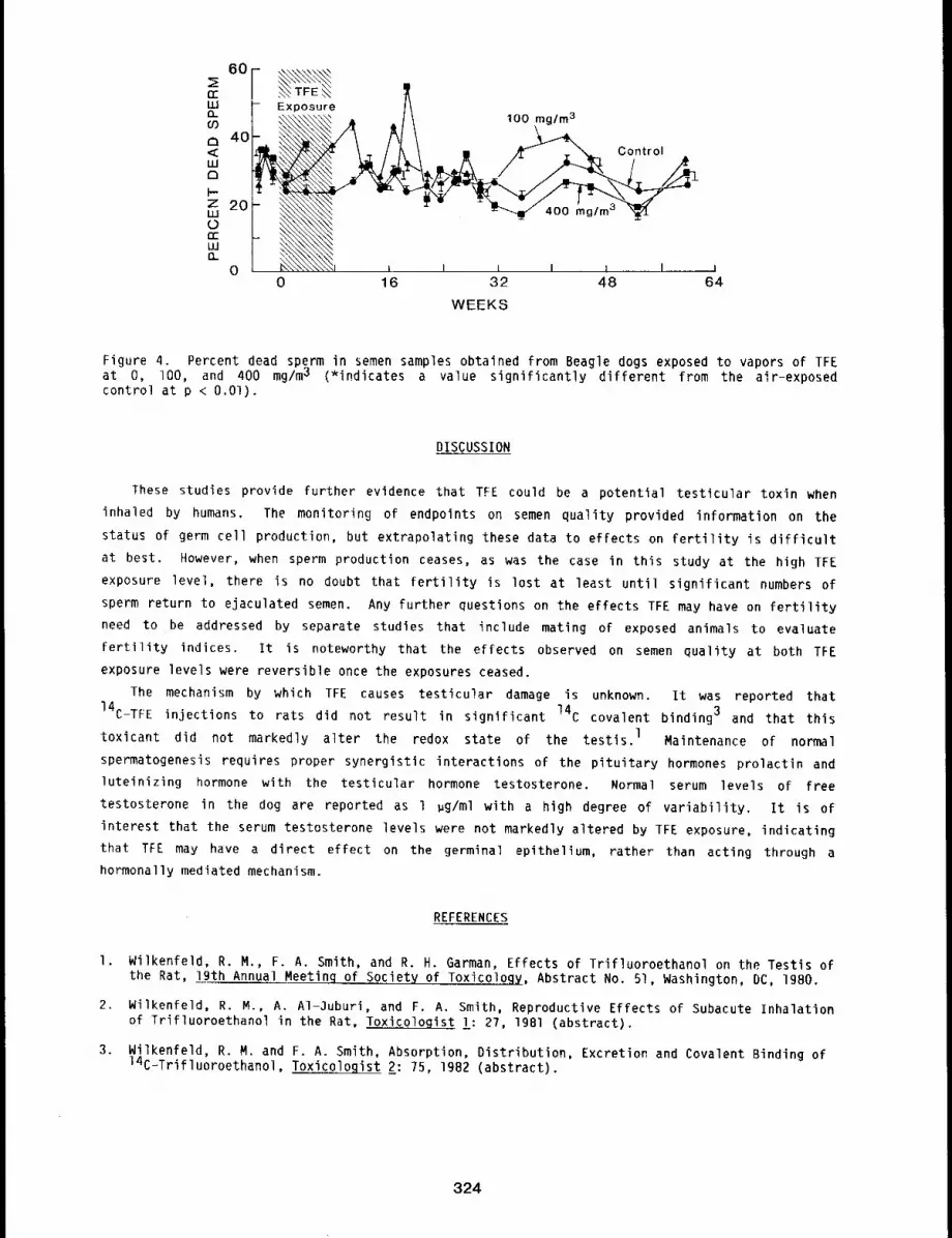
60
rrLUn09a 40<ELUa
Z 20I.U
rrLU0_
0
~ TFE ~Exposure
100 mg/m3
. co , o,
i i i i i I0 16 32 48
WEEKS
i64
Figure 4. Percent dead sperm in semen samples obtained from Beagle dogs exposed to vapors of TFEat O, lO0, and 400 mg/m3 (*indicates a value significantly different from the air-exposedcontrol at p < O.Ol).
DISCUSSION
These studies provide further evidence that TFE could be a potential testicular toxin when
inhaled by humans. The monitoring of endpoints on semen quality provided information on the
status of germ cell production, but extrapolating these data to effects on fertility is difficult
at best. However, when sperm production ceases, as was the case in this study at the high TFE
exposure level, there is no doubt that fertility is lost at least until significant numbers of
sperm return to ejaculated semen. Any further questions on the effects TFE may have on fertility
need to be addressed by separate studies that include mating of exposed animals to evaluate
fertility indices. It is noteworthy that the effects observed on semen quality at both TFE
exposure levels were reversible once the exposures ceased.
The mechanism by which TFE causes testicular damage is unknown. It was reported that
14C-TFE injections to rats did not result in significant 14C covalent binding 3 and that this
toxicant did not markedly alter the redox state of the testis, l Maintenance of normal
spermatogenesis requires proper synergistic interactions of the pituitary hormones prolactin and
luteinizing hormone with the testicular hormone testosterone. Normal serum levels of free
testosterone in the dog are reported as l ~g/ml with a high degree of variability. It is of
interest that the serum testosterone levels were not markedly altered by TFE exposure, indicating
that TFE may have a direct effect on the germinal epithelium, rather than acting through a
hormonally mediated mechanism.
REFERENCES
I. Wilkenfeld, R. M., F. A. Smith, and R. H. Garman, Effects of Trifluoroethanol on the Testis ofthe Rat, 19th Annual Meetinq of Society of Toxicology, Abstract No. 51, Washington, DC, 1980.
2. Wilkenfeld, R. M., A. AI-3uburi, and F. A. Smith, Reproductive Effects of Subacute Inhalationof Trifluoroethanol in the Rat, Toxicologist l: 27, 19Bl (abstract).
3. Wilkenfeld, R. M. and F. A. Smith, Absorption, Distribution, Excretion and Covalent Binding of14C-Trifluoroethanol, Toxicologist 2: 75, 1982 (abstract).
324
BIOLOGICAL FACTORS THAT INFLUENCE DOSE-RESPONSE RELATIONSHIPS
Any study of the relationship between toxicant dose and the resulting biological effect must
take into account biological factors that may influence the resulting relationship as much as or
more than the dose-related factors. Some of these factors are the species used, the age of the
anlmals, and the health status of the animals. The eight papers in this section present
information relative to how factors such as the age or the species used may influence the results
of toxicity studies.
The first five papers in this section present the results of studies designed to better
understand the role of the immune system as it relates to the pulmonary defense system and the
immune responses of the lung after inhalation of toxicants. The first paper reports that local
accumulation of antlbody-secreting cells in the lungs of dogs mediate immune phagocytosis and,
thus, may play a critical role in pulmonary defense mechanisms. The second paper reports on the
use of a newly developed assay to evaluate cell-mediated immunity of the lung.
The third paper reports on the effect of age on immune response after localized lung
immunization. Dogs older than seven years of age appeared to have reduced capability to recruit
immune cells and antibody from blood into the lung. The fourth and Fifth papers report
continuation of studies to define immune responses after localized lung immunization in species of
animals commonly used in toxicity studies. The studies of monkeys and rabbits extend the
information available on differences in species response. Rabbits appear to have a similar
response to that of rats, whereas dogs and monkeys appear to have a response similar to that of
man.
The sixth paper reports on an improved technique for calculating labeling indices of lung
epithelial cells. The next paper reports on the respiration of rats while confined in exposure
tubes, which are commonly used to expose rats to toxicants. These studies provide information
that extend our capabilities in measuring endpoints in toxicity studies.
The final paper in the section reports on a technique for depleting reduced glutathione (GSH)
in the tissues of mice using L-buthlonlne-S,R-sulfoxlmine in the drinking water. Reduced
glutathione is important in metabolic pathways for detoxificatlon and excretion of various
xenobiotic compounds. Thus, this method for depleting GSH may be useful in toxological studies
requiring animals with decreased in vivo levels of GSH.
3251326

IMMUNE PHAGOCYTOSIS BY CANINE PULMONARY ALVEOLAR MACROPHAGES
Abstract --The deposition of a particulate antigen
in the lungs of dogs causes the local accumulation PRINCIPAL INVESTIGATORS
of cytoph~llc antibody and opsonlzlng antibody. A.G. Harmsen
These antlbodles greaCly enhanced phagocytosls by D.E. B~ce
pulmonary alveolar macrophages as shown b9 in vltro B.A. Muggenburg
and in vlvo experlments. It appears that these
affects were due to local anrlbody-secretlng cells. The local accumulation of antibody-secreting
cells in the lung medlated immune phagocytosls and, thus, may pla9 a critical role In pulmDnar9
defense mechanisms.
Immune responses to infectious agents greatly enhance nonspecific defense mechanisms in the
lung. In particular, humoral responses can produce specific cytophilic and opsonizing antibodies
that enhance phagocytosis. The source of antibodies in the alveolus can be either the blood or,
as this laboratory has shown, antibody-secreting cells in the alveolus,l
The purpose of this investigation was to determine if antibody-dependent phagocytosis (immune
phagocytosis) by pulmonary alveolar macrophages (PAM) is influenced by the local production
specific antibody in the alveolus. The model developed involved the immunization of dogs by
depositing antigen into specific lung lobes and documenting immune phagocytosis in vitro and in
vivo. The results indicated that immune phagocytosis by PAM is most intense in lung lobes
previously exposed to antigen and is probably due to the local production of specific antibody.
METHODS
Eight Beagle dogs between 2 and 3 years of age were used in this study. Physical examinations
were performed to determine the health of each dog before immunization, and all dogs were healthy
throughout this study. Specific airways were selected for instillation of lOlO sheep red blood
cells (SRBC) in l ml saline, or l ml saline with a fiberoptic bronchoscope, as in previous
studies, l Four dogs were immunized by the instillation of SRBC into both the left cardiac and
left diaphragmatic lobes. These served as immunized lobes. The same dogs were also instilled
with saline into the right cardiac and right diaphragmatic lobes. These served as control lobes.
Another four dogs were instilled with l ml of saline into the right cardiac, and right
diaphragmatic lobes. These served as unimmunized dog control lobes.
At 2, 6, 9, 13, and 16 days after immunization, the immunized and control airways of immunized
dogs and the control airways of unimmunized dogs were re-entered with the fiberoptic bronchoscope
while the dogs were under halothane anesthesia. The lung lobes were lavaged with saline (five
washes with lO ml/wash). The lavage fluid and cells were separated, and the cells were
resuspended in medium RPMI 1640 (Grand Island Biologicals, Grand Island, NY). The ability of the
PAM from the various lobes to phagocytize SRBC was used to indicate the presence of cytophilic
antibody to SRBC on the PAM. The ability of the lavage fluids from the various lobes to enhance
phagocytosis of SRBC by PAM from unimmunized dogs was used to determine the presence of opsonizing
antibody in the alveolous. The phagocytosis assay was done by incubating PAM with the variously
treated SRBC, removing the SRBC not phagocytized by hypotonic lysis, and counting the phagocytized
SRBC in cytocentrifuge preparations. In vivo phagocytosis was evaluated at 13 days after
327
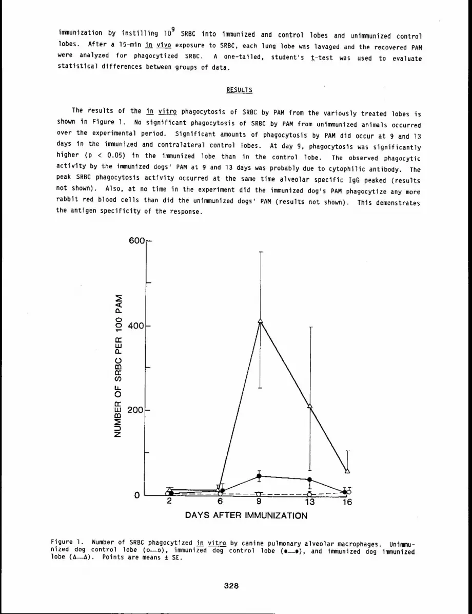
immunization by instilling I09 SRBC into immunized and control lobes and unimmunized control
lobes. After a 15-min in vivo exposure to SRBC, each lung lobe was ]avaged and the recovered PAM
were analyzed for phagocytized SRBC. A one-tailed, student’s t-test was used to evaluate
statistical differences between groups of data.
RESULTS
The results of the in vitro phagocytosis of SRBC by PAM from the variously treated lobes is
shown in Figure I. No significant phagocytosis of SRBC by PAM from unimmunized animals occurred
over the experimental period. Significant amounts of phagocytosis by PAM did occur at 9 and 13
days in the immunized and contralateral control lobes. At day 9, phagocytosis was significantly
higher (p < 0.05) in the immunized lobe than in the control lobe. The observed phagocytic
activity by the immunized dogs’ PAM at g and 13 days was probably due to cytophilic antibody. The
peak SRBC phagocytosis activity occurred at the same time alveolar specific IgG peaked (results
not shown). Also, at no time in the experiment did the immunized dog’s PAM phagocytize any more
rabbit red blood cells than did the unimmunized dogs’ PAM (results not shown). This demonstrates
the antigen specificity of the response.
600 -
0
l
-’t’T--2 6 9 13 16
DAYS AFTER IMMUNIZATION
Figure I. Number of SRBC phagocytized in vitro by canine pulmonary alveolar macrophages. Unimmu-nized dog control lobe (o--o), immunized dog control lobe (o--o), and immunized dog immunizedlobe (A~). Points are means ± SE.
328
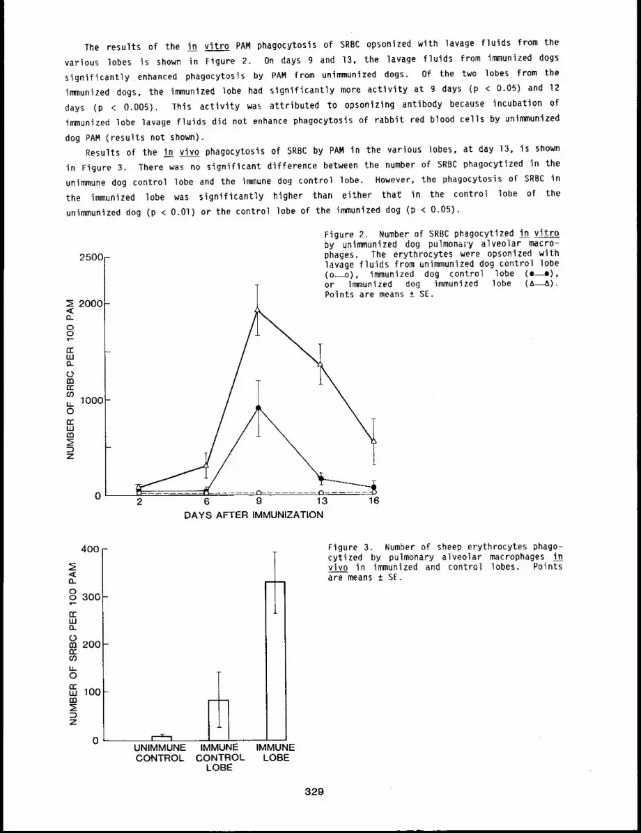
The results of the in vitro PAM phagocytosis of SRBC opsonized with lavage fluids from the
various lobes is shown in Figure 2. On days 9 and 13, the lavage fluids from immunized dogs
significantly enhanced phagocytosis by PAM from unimmunized dogs. Of the two lobes from the
immunized dogs, the immunized lobe had significantly more activity at 9 days (p < 0.05) and
days (p < 0.005). This activity was attributed to opsonizing antibody because incubation
immunized lobe lavage fluids did not enhance phagocytosis of rabbit red blood cells by unimmunized
dog PAM (results not shown).
Results of the in vivo phagocytosis of SRBC by PAM in the various lobes, at day 13, is shown
in Figure 3. There was no significant difference between the number of SRBC phagocytized in the
unimmune dog control lobe and the immune dog control lobe. However, the phagocytosis of SRBC in
the immunized lobe was significantly higher than either tha~ in the control lobe of the
unimmunized dog (p < O.O1) or the control lobe of the immunized dog (p < 0.05).
25OO
2000
oo
w
m
1000O
w
z
0
\
........ {")
2 6 9 13 16
Figure 2. Number of SRBC phagocytized in vitroby unimmunized dog pulmonary alveolar macro-phages. The erythrocytes were opsonized withlavage fluids from unimmunized dog control lobe(o~o), immunized dog control lobe (e--o),or immunized dog immunized lobe (A~).Points are means ± SE.
DAYS AFTER IMMUNIZATION
400
oo 300
w
0200
°Iw 100
z
0 ’ I ~- ] ....
UNIMMUNECONTROL CONTROL
LOBE
IMMUNE IMMUNELOBE
Figure 3. Number of sheep erythrocytes phago-cytized by pulmonary alveolar macrophages invivo in immunized and control lobes. Pointsare means ± SE.
329
DISCUSSION
Studies from this laboratory have shown that antibody-secreting cells accumulate in the lungs
of dogs immunized to a particulate antigen. 1’2 The results of the present study indicate that
the accumulation of antibody-secreting cells affects the local phagocytosis of particulate antigen
by PAM. Increased levels of both cytophilic antibody and opsonizing antibody were found in the
lobes of immunized animals. However, the activity of both these types of antibody was higher in
the immunized lobe than in the contralateral control lobes (Figs. I and 2). The same situation
also found with antibody-secreting cells. Immunized lobes contain significantly more antibody
secreting cells than do the contralateral control lobes. 1’2 The in vitro phagocytosis data,
indicating an enhancement of cytophilic and opsonizing antibody levels, was confirmed by in vivo
phagocytosis experiments (Fig. 3).
These data on immune phagocytosis and previous data on antibody-forming cells together suggest
that antibody secreted locally in the lung plays a critical role in pulmonary defense mechanisms.
REFERENCES
l ¯ Bice, D. E., D. L. Harris, and B. A. Muggenburg, Regional Immunologic Responses FollowingLocalized Deposition of Antigen in the Lung, Exp. Lun~ R~S. l: 33-41, IgSO.
Bice, D. E., O. L. Harris, J. O. Hill, B. A. Muggenburg, and R. K. Wolff, Immune ResponsesAfter Localized Lung Immunization in the Dog, Am. Rev. Respir. Dis. 122: 755-?60, IgBO.
330
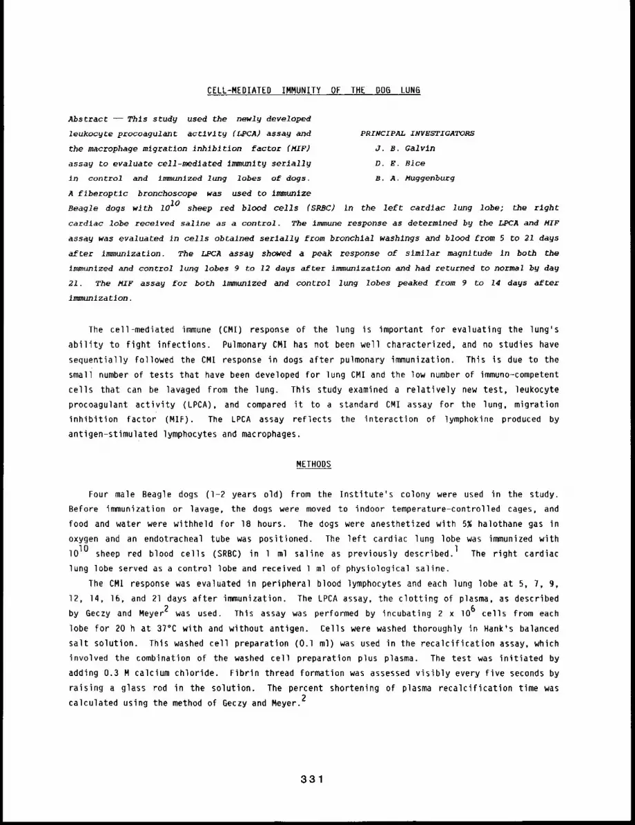
CELL-MEDIATED IMMUNITY OF THE DO6 LUNG
Abstract -- Thls study used the newly developed
leukocyte procoagula~t acCIvlty (LPCA) assay a~d PRINCIPAL INVESTIGATORS
the macrophage m~grat~on inh~bltlon factor (MIF) J.B. Galvln
assay to evaluate cell-medlated Immunlty serlally D.E. Bice
~n control and immunlzed lung lobes of dogs. B.A. Muggenburg
A flberoptlc bronchoscope was used co lmmunlze
Beagle dogs with 1010 sheep red blood cells (SRBC) In the left cardlac lung lobe~ the right
cardlac lobe received sallne as a control. The immune response as determined by the LPCA and MIF
assay was evaluated ~n cells obtained serially from bronchial washings and blood from 5 to 21 days
after immunlzation. The LPCA assay showed a peak response of slmllar magnitude in both the
immunized and control lung lobes 9 to 12 days after immunlzatlon and had returned to normal by day
21. The MIF assay for both immunlzed and control lung lobes peaked from 9 to 14 days afteE
immunization.
The cell-mediated immune (CMI) response of the lung is important For evaluating the lung’s
ability to fight infections. Pulmonary CMI has not been well characterized, and no studies have
sequentially Followed the CMI response in dogs after pulmonary immunization. This is due to the
small number of tests that have been developed for lung CMI and the low number of immunoo-competent
cells that can be lavaged from the lung. This study examined a relatively new test, leukocyte
procoagulant activity (LPCA), and compared it to a standard CMI assay for the lung, migration
inhibition factor (MIF). The LPCA assay reflects the interaction of lymphokine produced
antigen-stimulated Iymphocytes and macrophages.
METHODS
Four male Beagle dogs (I-2 years old) from the Institute’s colony were used in the study.
Before immunization or lavage, the dogs were moved to indoor temperature-controlled cages, and
food and water were withheld for 18 hours. The dogs were anesthetized with 5% halothane gas in
oxygen and an endotracheal tube was positioned. The left cardiac lung lobe was immunized with
lOlO sheep red blood cells (SRBC) in l ml saline as previously described.l The right cardiac
lung lobe served as a control lobe and received l ml of physiological saline.
lhe CMI response was evaluated in peripheral blood lymphocytes and each lung lobe at 5, 7, 9,
12, 14, 16, and 21 days after immunization. The LPCA assay, the clotting of plasma, as described
by Geczy and Meyer2 was used. This assay was performed by incubating 2 x lO6 cells from each
lobe for 20 h at 37°C with and without antigen. Cells were washed thoroughly in Hank’s balanced
salt solution. This washed cell preparation (O.l ml) was used in the recalcification assay, which
involved the combination of the washed cell preparation plus plasma. The test was initiated by
adding 0.3 M calcium chloride. Fibrin thread formation was assessed visibly every five seconds by
raising a glass rod in the solution. The percent shortening of plasma recaIcification time was2calculated using the method of Geczy and Meyer.
331
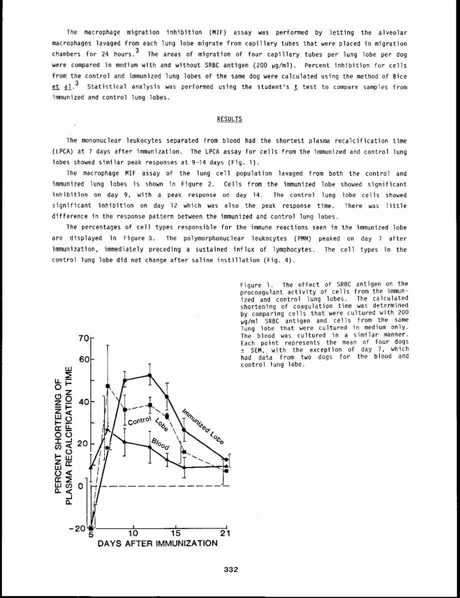
The macrophage migration inhibition (MIF) assay was performed by letting the alveolar
macrophages lavaged from each lung lobe migrate from capillary tubes that were placed in migration
chambers for 24 hours. 3 The areas of migration of four capillary tubes per lung lobe per dog
were compared in medium with and without SRBC antigen (200 #g/ml). Percent inhibition for cells
from the control and immunized lung lobes of the same dog were calculated using the method of Bite
et al. 3 Statistical analysis was performed using the student’s t test to compare samples from
immunized and control lung lobes.
RESULTS
The mononuclear leukocytes separated from blood had the shortest plasma recalcification time
(LPCA) at 7 days after immunization. The LPCA assay for cells from the immunized and control lung
lobes showed similar peak responses at 9-14 days (Fig. l).
The macrophage MIF assay of the lung cell population lavaged from both the control and
immunized lung lobes is shown in Figure 2. Cells from the immunized lobe showed significant
inhibition on day 9, with a peak response on day 14. The control lung lobe cells showed
significant inhibition on day 12 which was also the peak response time. There was little
difference in the response pattern between the immunized and control lung lobes.
lhe percentages of cell types responsible for the immune reactions seen in the immunized lobe
are displayed in Figure 3. The polymorphonuclear leukocytes (PMN) peaked on day 7 after
immunization, immediately preceding a sustained influx of lymphocytes. The cell types in the
control lung lobe did not change after saline instillation (Fig. 4).
Figure I. The effect of SRBC antigen on theprocoagulant activity of cells from the immun-ized and control lung lobes. The calculatedshortening of coagulation time was determinedby comparing cells that were cultured with 200#g/ml SRBC antigen and cells from the samelung lobe that were cultured in medium only.
70 The blood was cultured in a similar manner.Each point represents the mean of four dogs
I\ ~
+_ SEM, with the exception of day 7, which
60 had data from two dogs for the blood andcontrol lung lobe.
40°E
Oo~t~olo9 iYl\l ,, ’%
-205 10 15 21
DAYS AFTER IMMUNIZATION
332
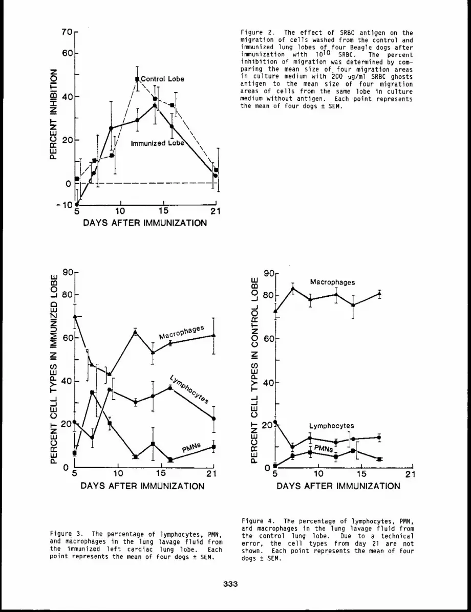
70
6O
Z0l
ca 40"l-
Z
I--ZLUC,) 20rrLUn
0
-105
~Control Lobe
/l’JT.
//~/’
Immunized Lobe\ \k\
7-1--i-I
10 15 21DAYS AFTER IMMUNIZATION
Figure 2. The effect of SRBC antigen on themigration of cells washed from the control andimmunized lung lobes of four Beagle dogs afterimmunization with lO lO SRBC. The percentinhibition of migration was determined by com-paring the mean size of four migration areasin culture medium with 200 ~g/ml SRBC ghostsantigen to the mean size of four migrationareas of cells from the same lobe in culturemedium without antigen. Each point representsthe mean of four dogs ± SEM.
°°°I0
80auJNZ
;E 6C
Z
(/)LUQ- 40>-I----I,.,ILUOI-- 20’ZLUOtrLUn
05
1 I 110 15 21
DAYS AFTER IMMUNIZATION
9O’" F T Macrophages
J-JO 80 ~~~~,~,~0rrI-Z0 6O0Z
O0LU
~- 40-
..1
.../ -W0~- 20k LymphocyteszW
Wn
05 10 15DAYS AFTER IMMUNIZATION
!
21
Figure 3. The percentage of lymphocytes, PMN,and macrophages in the lung lavage fluid fromthe immunized left cardiac lung lobe. Eachpoint represents the mean of four dogs ± SEM.
Figure 4. The percentage of lymphocytes, PMN,and macrophages in the lung lavage fluid fromthe control lung lobe. Due to a technicalerror, the cell types from day 21 are notshown. Each point represents the mean of fourdogs ± SEM.
333
DISCUSSION
The blood LPCA peaked before the immunized lung lobe LPCA, which would be expected if the
lymphocyte recirculation theory in the lung were true. l The lung LPCA followed a response time
similar to that of the MIF assay. This indicated that sensitized lymphocytes were recruited to
the lung resulting in not only macrophage migration inhibition, but also an increased shortening
of plasma clotting time. The increased shortening of plasma clotting time is thought to be due to
increased conversion of fibrinogen to fibrin on the surface of the macrophage. This would account4lot the increased stickiness (lack of migration) seen in the MIF assay.
The inflammatory response in the immunized lung lobe as indicated by the influx of PMN has
possibly altered the macrophage population used as the indicator cell in the MIF assay. The
control lobe in both assays showed a good response with no inflammation. This was due to the
presence of sensitized lymphocyte trafficking through the control lung lobe.5
The inflammatory response and the immune reaction are thought to occur in concert. The cell
types coming into the immunized lung lobe showed this is a likely hypothesis. The preliminary PMN
influx seemed to pave the way for the diapedsis of lymphocytes. This was probably due to the
release of chemotactic factors during the inflammatory process. The immune response showed
significant increases on the same day as the influx of lymphocytes.
In conclusion, the LPCA assay correlated well with the standard CMI assay, MIF. The LPCA
assay was easy to perform and was reproducible. The CMI response in the lung peaked 9-14 days
after immunization, and the LPCA assay can be used as a measure of lung CMI.
REFERENCES
l ¯
2.
3.
4.
Bite, D. E., D. L. Harris, 3. O. Hill, B. A. Muggenburg, and R. K. Wolff, Immune ResponsesAfter Localized Lung Immunization in the Dog, Am. Rev. Respir. Dis. 122: 755-760, Ig80.
Geczy, C. L. and P. A. Meyer, Leukocyte Procoagulant Activity in Man: An In Vitro Correlateof Delayed-Type Hypersensitivity, 3. Immunol. ~: 331-336, 1982.
Bice, D. E., G. Heins, D. Gruwell, and J. Salvaggio, Rabbit Alveolar Macrophage as IndicatorCells in Migration Inhibitory Factor Assays, 3. Reticuloendoth. Soc. 22: 427-436, 1977.
Geczy, C. L. and D. L. Hopper, A Mechanism of Migration Inhibition in Delayed-TypeHypersensitivity Reactions. II. Lymphokines Promote Procoagulant Activity of Macrophages InVitro, J. Immunol. 126: 1059-1063, IgSl.
5. Sprent, J., Migration and Life Span of Lymphocytes, in B and T Cells in Immune Recognition,John Wiley and Sons, New York, NY, pp. 59-82, 1977.
334
EFFECTS OF AGE ON IMMUNE RESPONSES AFTER LOCALIZED LUNG IMMUNIZATION
Abstract --Dogs 5 through 16 years of age were im-
munlzed b9 deposition of antigen ~n a s~ngle lung PRINCIPAL INVESTIGATORS
lobe. The number of lymphoid cells producing spe- D.E. Bice
clflc IgM antibody and the level of speclflc IgG B.A. Muggenburg
ant~bod9 were measured in blood, as well as in the
~min~nlzed and control ltmg lobes. Even though antlbody-formlng cells and speclflc antibody were
found In the blood of older animals, most dogs over 7 years of age had slgnlflcantly fewer
antlbody-formlng cells and less speclflc IgG antlbodg In the Immunlzed lung lobe than did younger
anlmals. Changes necessary to recrult Immune cells and antlbody from the blood Into the lung
appeared to be reduced In dogs older than 7 years of age.
The production of immune cells and antibody in response to antigens is gradually reduced with
age. This age-dependent reduction of immunity is considered to be one reason for increased
infections, and possibly for the increased tumor incidence, in aged individuals. Although a
common cause of death in aged individuals is pneumonia, no studies on the effects of age on lung
immunity have been published. This study evaluated the effects of age on immune responses in dogs
from 5 through 16 years of age that were immunized by localized deposition of antigen in the
lung. The immune response was measured in the blood, in an immunized lung lobe, and in a control
lung lobe. Data presented in this report indicate that aged dogs have a significantly lower
immune response in the lung. Antigen deposited in the lungs of older dogs did not induce changes
in wscular permeability necessary to allow a normal accumulation of immune cells and antibody in
the immunized lung.
METHODS
Twenty Beagle dogs between 5 and 16 years of age from the Institute’s colony were used in this
study. Groups of dogs were used that were 5, 7 to g, lO to 12, and 14 to 16 years of age.
Physical examinations were performed to determine the health of each dog before immunization, and
all dogs were healthy throughout this study. Specific airways in the left cardiac lung lobes were
selected for immunization with a fiberoptic bronchoscope while the dogs were under halothane
anesthesia, as in previous studies.l The left cardiac lung lobes were immunized by the
instillation of lOlO sheep red blood cells (SRBC) in 1 ml of physiological saline. The right
cardiac lung lobe served as the control and received l ml of saline.
The control and immunized airways were re-entered with the fiberoptic bronchoscope at 5, 7,
l 0, 12, and 14 days after immunization. The airways were lavaged with saline (5 washes with l
ml/wash). Blood samples were also taken at these times. The number of lymphoid cells in the
lavage fluid and in blood samples that were producing anti-SRBC IgM antibody was determined by the
Cunningham modification of the 3erne plaque assay. 2 The level of anti-SRBC IgG antibody in the
lavage fluids and in sera was evaluated with an enzyme-linked immunoassay. All sera were diluted
1:200, and lavage fluid samples were diluted 1:50 for evaluation with the enzyme-linked
immunoassay. The data obtained from different age groups were analyzed for statistical
differences by pairwise t-tests with Bonferroni probabilities. A multiple linear regression test
335
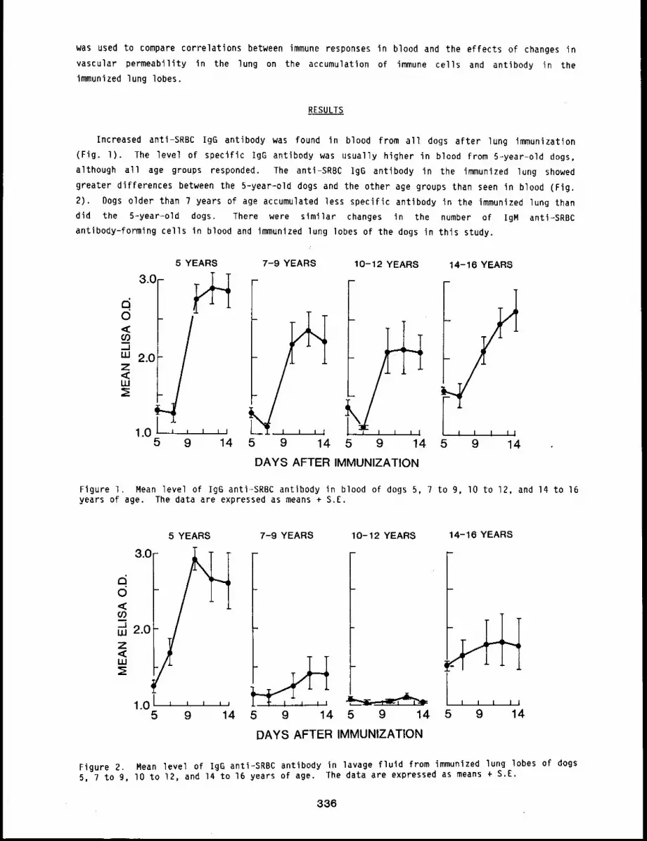
was used to compare correlations between immune responses in blood and the effects of changes in
vascular permeability in the lung on the accumulation of immune cells and antibody in the
immunized lung lobes.
RESULTS
Increased anti-SRBC IgG antibody was found in blood from all dogs after lung immunization
(Fig. I). The level of specific igG antibody was usually higher in blood from 5-year-old dogs,
although all age groups responded. The anti-SRBC igG antibody in the immunized lung showed
greater differences between the 5-year=old dogs and the other age groups than seen in blood (Fig.
2). Dogs older than 7 years of age accumulated less specific antibody in the immunized lung than
did the 5=year-old dogs. There were similar changes in the number of IgM anti=SRBC
antibody-forming cells in blood and immunized lung lobes of the dogs in this study.
5 YEARS
3.o-
0
<=
1.05 9 14
7-9 YEARS 10-12 YEARS
5 9 14 5 9 14
DAYS AFTER IMMUNIZATION
14-16 YEARS
5 9 14
Figure I. Mean level of IgG anti-SRBC antibody in blood of dogs 5, 7 to 9, I0 to 12, and 14 to 16years of age. The data are expressed as means + S.E.
3.0
5 YEARS 7-9 YEARS 10-12 YEARS 14-16 YEARS
1o0’ 1 I 1 I J I I l l I
5 9 14 5 9 14 5 9 14 5 9 14
DAYS AFTER IMMUNIZATION
Figure 2. Mean level of IgG anti-SRBC antibody in lavage fluid from immunized lung lobes of dogs5, 7 to g, lO to 12, and 14 to 16 years of age. The data are expressed as means + S.E.
336
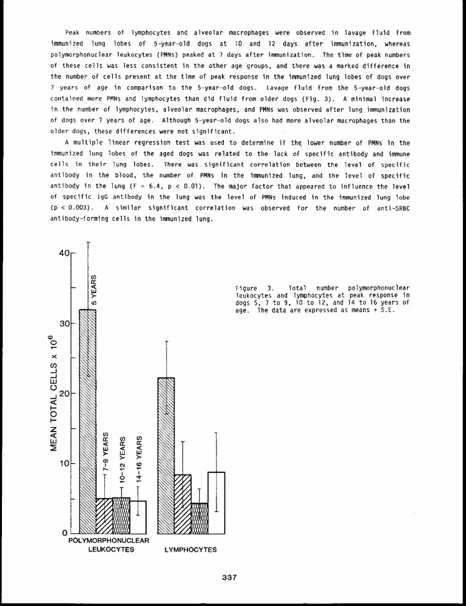
Peak numbers of lymphocytes and alveolar macrophages were observed in lavage fluid from
immunized lung lobes of 5-year-old dogs at lO and 12 days after immunization, whereas
polymorphonuclear leukocytes (PMNs) peaked at 7 days after immunization. The time of peak numbers
of these cells was less consistent in the other age groups, and there was a marked difference in
the number of cells present at the time of peak response in the immunized lung lobes of dogs over
7 years of age in comparison to the 5-year-old dogs. Lavage fluid from the 5-year-old dogs
contained more PMNs and lymphocytes than did fluid from older dogs (Fig. 3). A minimal increase
in the number of lymphocytes, alveolar macrophages, and PMNs was observed after lung immunization
of dogs over 7 years of age. Although 5-year-old dogs also had more alveolar macrophages than the
older dogs, these differences were not significant.
A multiple linear regression test was used to determine if th~ lower number of PMNs in the
immunized lung lobes of the aged dogs was related to the lack of specific antibody and immune
cells in their lung lobes. There was significant correlation between the level of specific
antibody in the blood, the number of PMNs in the immunized lung, and the level of specific
antibody in the lung (F = 6.4, p < O.Ol). The major factor that appeared to influence the level
of specific IgG antibody in the lung was the level of PMNs induced in the immunized lung lobe
(p < 0.003). A similar significant correlation was observed for the number of anti-SRBC
antibody-forming cells in the immunized lung.
3O
oPOLYMORPHONUCLEAR
LEUKOCYTES LYMPHOCYTES
Figure 3. Total number polymorphonuclearleukocytes and lymphocytes at peak response indogs 5, ? to 9, lO to 12, and 14 to 16 years ofage. The data are expressed as means + S.E.
337

DISCUSSION
Large numbers of antibody-forming cells and levels of specific antibody are found in the blood
of dogs after lung immunization.l Antibody-forming cells accumulate in the immunized lung lobes
because of changes in vascular permeability, and a significant increase in specific antibody is
found in lavage fluid from the immunized lung. 1’3 The accumulation of immune cells and antibody
in the lung would offer immune defense against pathogens; loss of this response could result in an
increased susceptibility to pulmonary infectlons.
In this study, dogs over 7 years of age had some reduction of anti-SRBC IgG antibody in blood,
although all dogs did respond. In contrast, minimal immune responses were observed in the
immunized lungs of the older dogs. Although aged dogs had some specific antibody and anti-SRBC
antibody-forming cells in their blood, it is possible that immune cells and antibody could not
enter the immunized lung because changes in vascular permeability were not induced by the exposure
of the lung to antigen.
Statistical evaluation of the correlation between peak immune responses in the blood, the
number of PMNs in the immunized lung lobe, and the level of specific antibody in the immunized
lung lobe suggested that the response indicated by increased PMNs was important in controlling
entrance of immune cells and antibody into the immunized lung. However, the exact mechanisms
responsible for recruitment of immune cells and antibody to the lung are not understood. The
increased number of PMNs in immunized lobes may not be directly responsible for recruitment of
immune cells, but only an indication that a change in vascular permeability has occurred.
Whatever the mechanisms that control the accumulation of antibody and immune cells in the
immunized lung, our data indicate that in lungs of aged dogs changes are not produced that allow a
normal accumulation of immune cells and antibody in the immunized lung.
These results indicate that changes can occur both in the development of immunity and in the
recruitment of immune cells to the lung in aged dogs that could compromise the immune protection
of the lung. These changes could be partly responsible for increased pulmonary infections seen in
aged individuals.
REFERENCES
I. Bice, D. E., M. A. Degen, D. L. Harris, and B. A. Muggenburg, Recruitment of Antibody-FormlngCells in the Lung After Local Immunization is Nonspecific, Am. Rev. ResPir. Dis. 126: 635-639,1982.
2. Cunningham, A. J. and A. Szenberg, Further Improvements in the Plaque Technique for DetectingSingle Antibody-Forming Cells, 3. Immunol. 14: 599-601, 1968.
3. Brownstein, D. G., A. H. Rebar, D. E. Bice, B. A. Muggenburg, and J. O. Hill, Immunology ofthe Lower Respiratory Tract: Serial Morphologic Changes in the Lungs and TracheobronchialLymph Nodes of Dogs After Intrapulmonary Immunization with Sheep Erythrocytes, Am. 3. Pathol.98: 499-514, 1980.
338
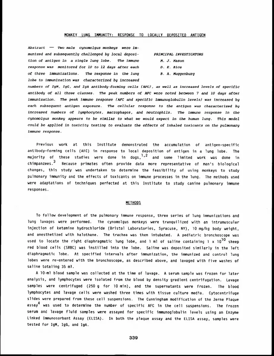
MONKEY LUNG IMMUNITY: RESPONSE TO LOCALLY DEPOSITED ANTIGEN
Abstract -- Two male cgnomolgus monkegs were im-
munlzed and subsequentl9 challenged by local deposl- PRINCIPAL INVESTIGATORS
tlon of antigen in a slngle lung lobe. The ~mmune M.J. Mason
response was monltored for i0 to 12 dags after each D.E. B1ce
of three immunlzatlons. The response in the lung B.A. Muggenburg
lobe to immun~zatlon was characterized by Increased
numbers of IgM, IgG, and IgA a~tlhody-formlng cells (AFC), as well as increased levels of speclfic
an~ibod9 of all three classes. The peak numbers of AFt were noted between 7 and 10 days after
Immunlzation. The peak immune response (AFt and speclfIc Immunoglobulln levels) was increased 9
each subsequent antigen exposure. The cellular response to the antlgen was characterized by
increased numbers of l~mphocgtes, macrophages, and neutrophlls. The immune response In the
cynomolgus monkey appears to he similar ~o what we would expect in the human lung. This model
could be applied in toxJclt~ testlng to evaluate the effects of inhaled toxlcants on the pulmonar9
immune response.
Previous work at this Institute demonstrated the accumulation of antigen-specific
antibody-forming cells (AFC) in response to local deposition of antigen in a lung lobe. The
majority of these studies were done in dogs, 1’2 and some limited work was done in
chimpanzees. 3 Because primates often provide data more representative of man’s biological
changes, this study was undertaken to determine the feasibility of using monkeys to study
pulmonary immunity and the effects of toxicants on immune processes in the lung. The methods used
were adaptations of techniques perfected at this Institute to study canine pulmonary immune
responses.
METHODS
To follow development of the pulmonary immune response, three series of lung immunizations and
lung lavages were performed. The cynomolgus monkeys were tranquilized with an intramuscular
injection of ketamine hydrochloride (Bristol Laboratories, Syracuse, NY), lO mg/kg body weight,
and anesthetized with halothane. The trachea was then intubated. A pediatric bronchoscope was
used to locate the right diaphragmatic lung lobe, and 1 ml of saline containing l x lOlO sheep
red blood cells (SRBC) was instilled into the lobe. Saline was deposited similarly in the left
diaphragmatic lobe. At specified intervals after immunization, the immunized and control lung
lobes were re-entered with the bronchoscope, as described above, and lavaged with five washes of
saline totaling 35 ml.
A lO-ml blood sample was collected at the time of ]avage. A serum sample was frozen for later
analysis, and lymphocytes were isolated from the blood by density gradient centrifugation. Lavage
samples were centrifuged (250 g for lO min), and the supernatants were frozen. The blood
lymphocytes and lavage cells were washed three times with tissue culture media. Cytocentrifuge
slides were prepared from these cell suspensions. The Cunningham modification of the 3erne Plaque
assay 4 was used to determine the number of specific AFC in the cell suspensions. The frozen
serum and lavage fluid samples were assayed for specific immunoglobulin levels using an Enzyme
Linked Immunosorbant Assay (ELISA). In both the plaque assay and the ELISA assay, samples were
tested for IgM, IgG, and IgA.
339
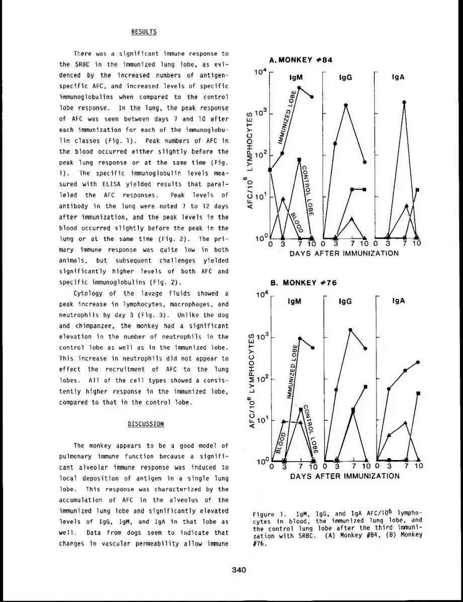
RESULTS
TKere was a significant immune response to
the SRBC in the immunized lung lobe, as evi-
denced by the increased numbers of antigen-
specific AFC, and increased levels of specific
immunoglobulins when compared to the control
lobe response. In the lung, the peak response
of AFC was seen between days 7 and I0 after
each immunization for each of the immunoglobu-
lin classes (Fig. l). Peak numbers of AFC
the blood occurred either slightly before the
peak lung response or at the same time (Fig.
l). The specific immunoglobulin levels mea-
sured with ELISA yielded results that paral-
leled the AFC responses. Peak levels of
antibody in the lung were noted 7 to 12 days
after immunization, and the peak levels in the
blood occurred slightly before the peak in the
lung or at the same time (Fig. 2). The pri-
mary immune response was quite low in both
animals, but subsequent challenges yielded
significantly higher levels of both AFC and
specific immunoglobulins (Fig. 2).
Cytology of the lavage fluids showed a
peak increase in lymphocytes, macrophages, and
neutrophils by day 3 (Fig. 3). Unlike the dog
and chimpanzee, the monkey had a significant
elevation in the number of neutrophils in the
control lobe as well as in the immunized lobe.
This increase in neutrophils did not appear to
effect the recruitment of AFC to the lung
lobes. All of the cell types showed a consis-
tently higher response in the immunized lobe,
compared to that in the control lobe.
DISCUSSION
The monkey appears to be a good model of
pulmonary immune function because a signifi-
cant alveolar immune response was induced to
local deposition of antigen in a single lung
lobe. This response was characterized by the
accumulation of AFC in the alveolus of the
immunized lung lobe and significantly elevated
levels of IgG, IgM, and IgA in that lobe as
well. Data from dogs seem to indicate that
changes in vascular permeability allow immune
A. MONKEY 4.8410 4 F IgM
- IgG IgA
G) 103I~
t-->-OO-rEL 102>-.,..,J
0,t,...."- 0101
100 0~30 3 7100DAYS AFTER iMMUNIZATION
1 04
103/I.UI-->-OO"i-n 02
...1
0
~ 0~
10°
B. MONKEY +76
IgM IgG IgA
yi-,
7 10 0 3 7 10 0 3DAYS AFTER IMMUNIZATION
7 10
Figure i. IgM, IgG, and IgA AFC/IO6 lympho-cytes in blood, the immunized lung lobe, andthe control lung lobe after the third immuni-zation with SRBC. (A) Monkey #84, (B) Monkey#76.
340
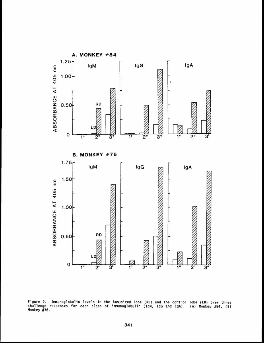
A. MONKEY ÷841.25 -
Ec-
1.000
LUL)Z 0.50°
tn-Oif?m
0 ’
IgM
/xi ~ 2° 3°
fIgG
0 1°
IgA
B. MONKEY ,~,76
1.75 -IgM
E 1.50t-
U30
F-< 1.00LU0z
03Dc0 0.50
0
RD
1° 2° 3° I° 1°
IgG
,i.
2o 13°
IgA
./.
//.
,’////.//.//.
1//
//Ji/.
2°
r~h’F~
3°
Ftgure 2. Immunoglobulin levels In the immunized lobe (RD) and the control lobe (LD) over threechal]enge responses for each c]ass of immunoglobulln (IgM, IgG and IgA). (A) Monkey #84, Monkey #76,
341
108
107
(B,_.1,_1LUL~j 106
0
105
Lobe
1040 3 7 100
Control Lobe
B=
.ill
I3 7 10 0 3 7 10 0 3 7 10
DAYS AFTER IMMUNIZATION
Figure 3. Cellular responses in the lavage fluid after the third immunization with SRBC. (A)Monkey #84, (B) Monkey #76.
cells and antibody to enter the lung. 2 One such indication that vascular permeability is
changed is the increased number of neutrophils seen only in the immunized lobe of the dog and
chimpanzee. However, the cellular response seen in the lavage fluid from the monkeys was somewhat
different from that previously reported in the dog1’2 and chimpanzee. 3 In the dog and
chimpanzee, a prominent neutrophilic response is not seen in the control lobe as was observed with
the monkeys in this study. These high numbers of neutrophils, coupled with the low levels of
antibody and the low numbers of plaquelforming cells in the control lobe, suggest that neutrophils
are not solely responsible for the changes in vascular permeability that allow accumulation of
immune cells in the alveolus.
Based on the data from humans5 and chimpanzees, 3 the immune response in the cynomolgus
monkey appears to be similar to what we would expect in the human lung. This model could be
applied in toxicity testing to evaluate the effects of inhaled toxicants on the pulmonary immune
response.
REFERENCES
I. Bice, D. E., D. L. Harris, J. O. Hill, B. A. Muggenburg, and R. K. Wolff, Immune ResponsesAfter Localized Lung Immunization in the Dog, Am. Rev. Respir. Dis. 122: 755-760, 1980.
2. Bice, D. E., O. L. Harris, and B. A. Muggenburg, Regional Immunologic Responses FollowingLocalized Deposition of Antigen in the Lung, Exp. Lung~ Res. l: 33-41, IgSO.
3. Bite, D. E., D. L. Harris, B. A. Muggenburg, and J. A. Bowen, The Evaluation of Lung Immunityin Chimpanzees, Am. Rev. Respir. Dis. 126: 35B-359, 1982.
4. Cunningham, A. J. and A. Szenberg, Further Improvements in the Plaque Technique for DetectingSingle Antibody-Forming Cells, 3. Immunol. 14: 599, 1968.
5. Lawrence, E. C., R. M. Blaese, R. R. Martin, and P. M. Stevens, Immunoglobulin Secreting Cellsin Normal Human Bronchial Lavage Fluids, J. Clin. Invest. 62: 832-835, 197B.
342
IMMUNE RESPONSES IN RABBITS AFTER LOCALIZED I.UNG IMMUNIZATION
Abstract --The locallzed deposltlon of partlcu-
late antlgen in the lung of rabb~ts induced a peak PRINCIPAL INVESTIGATORS
number of spec~flc IgMantlbody-formlng cells in D.E. Blce
the lung-assoclaTed 19mph nodes at 7 days after B.A. Muggenburg
~mmunlzatlon. Lymphoid tissues That dld not re-
ce~ve Iymphatlc drainage from the lung (spleen and popIJteal lymph nodes) had background numbers
of antlbody-formlng cells. A low number of antlbody..formlng cells was found in the blood.
However, lung immun~zatlon dld not increase the accumulatlon of immune cells In the immunized lung
lobes of rabbits. This response was slmilar to that repoEted prevlouslg for rats and different
from that for dogs, subhuman prlmates, and man.
There are basic differences in the development of immune responses in the lungs after
pulmonary immunization of dogs and rats. 1’2 After deposition of antigen in the dog lung, a
large number of antibody-forming cells are found in the blood and lavage fluid From immunized lung
lobes, l Published data indicate that immune cells are produced in the lung-associated lymph
nodes after lung immunization of the dog and that immune cells recovered in the lavage fluid from
the immunized lungs are recruited from the blood. 3 Mature plasma cells are present in the
alveoli of immunized lung lobes of the dog, and immune cells in the alveoli are actively producing
antibody. The presence of immune cells in the alveoli of dog, chimpanzee, cynomolgus monkey (this
report, pp. 339 to 342), and human probably serve an important role in lung defense.1’4’5
Immune cells in the alveoli may be at risk for cell killing or damage because they are exposed
directly to inhaled pollutants.
Although lung immunization of the rat also induces immune responses in the lung-associated
lymph nodes, few or no antibody-forming cells are found in the blood or in lung lavage fluid from
immunized rat lungs. Therefore, rats could not be used to evaluate effects of inhaled pollutants
on immune cells in the alveoli, and a need exists for small animal species that can be used in
these toxicity tests. This study evaluated immune responses in the rabbit after localized
deposition of particulate antigen in a single lung lobe to determine if immune cells accumulate in
the immunized rabbit lung. The number of specific immune cells in the lung-associated and
popliteal lymph nodes, spleen, blood and lavage fluid, and tissues from immunized and control lung
lobes was evaluated at intervals after immunization. The resultant data indicate that few
specific antibody-forming cells were present in the blood of immunized rabbits and that these
immune cells did not accumulate in immunized lung lobes.
METHODS
A total of 25 young adult male rabbits, Lov:(NZW), was used in this study. Each rabbit was
given a pre-anesthetic (0.2 ml/kg Innovar-vet) and anesthetized by i.v. injection of l
Pentobarbital. While the rabbits were anesthetized, a pediatric fiberoptic bronchoscope (Olympus
Corp. America, New Hyde Park, NY) was used to locate an airway in the left diaphragmatic lung
lobe. A 1.27-mm O.D. catheter was passed through the bronchoscope biopsy channel into the
selected airway, and IOg sheep red blood cells (SRBC) in 0.5 ml saline were instilled. Groups
343
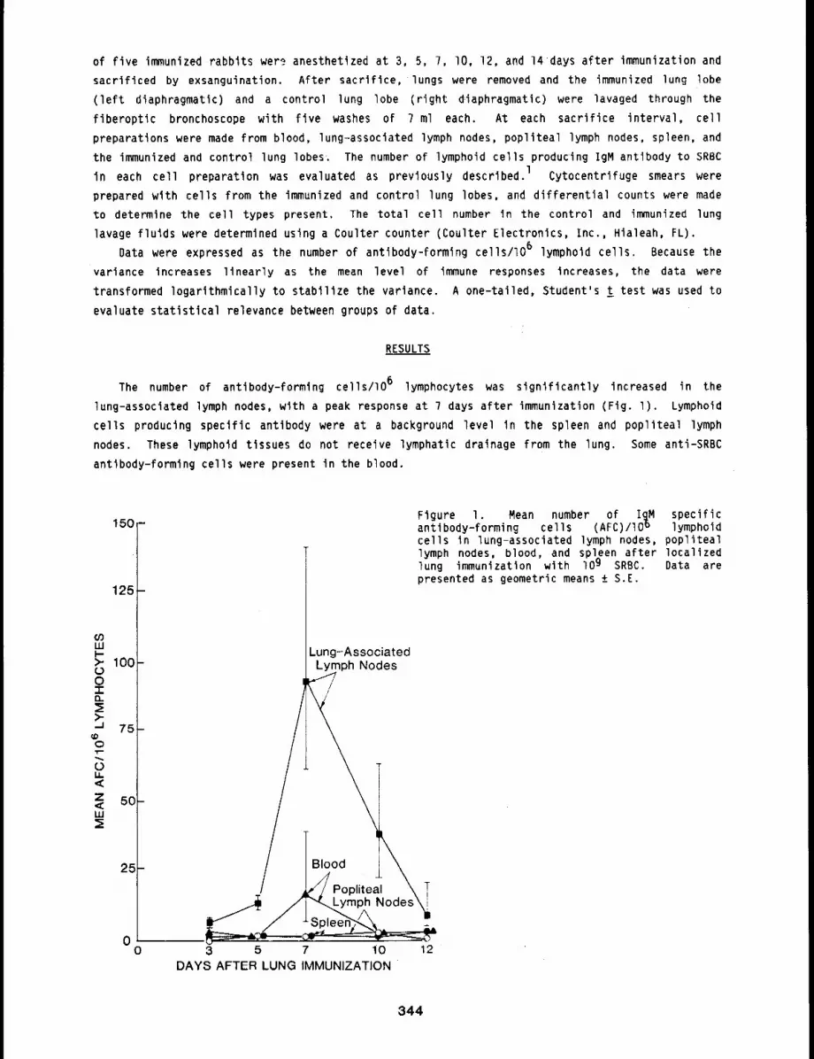
of five immunized rabbits wer~ anesthetized at 3, 5, ?, lO, 12, and 14days after immunization and
sacrificed by exsanguination. After sacrifice, lungs were removed and the immunized lung lobe
(left diaphragmatic) and a control lung lobe (right diaphragmatic) were lavaged through
fiberoptic bronchoscope with five washes of 7 ml each. At each sacrifice interval, cell
preparations were made from blood, lung-associated lymph nodes, popliteal lymph nodes, spleen, and
the immunized and control lung lobes.. The number of lymphoid cells producing IgM antibody to SRBC
in each cell preparation was evaluated as previously described, l Cytocentrifuge smears were
prepared with cells from the immunized and control lung lobes, and differential counts were made
to determine the cell types present. The total cell number in the control and immunized lung
lavage fluids were determined using a Coulter counter (Coulter Electronics, Inc., Hialeah, FL).
Data were expressed as the number of antibody-formlng cells/lO 6 lymphoid cells. Because the
variance increases linearly as the mean level of immune responses increases, the data were
transformed logarithmically to stabilize the variance. A one-tailed, Student’s t test was used to
evaluate statistical relevance between groups of data.
RESULTS
The number of antibody-forming cells/lO 6 lymphocytes was significantly increased in the
lung-associated lymph nodes, with a peak response at 7 days after immunization (Fig. l). Lymphoid
cells producing specific antibody were at a background level in the spleen and popliteal lymph
nodes. These lymphoid tissues do not receive lymphatic drainage from the lung. Some anti-SRBC
antlbody-forming cells were present In the blood.
150 -
12E
25
00
Figure I. Mean number of I~M specificantlbody-forming cells (AFC)/IO ° lymphoidcells in lung-associated lymph nodes, popliteallymph nodes, blood, and spleen after localizedlung immunization with 109 SRBC. Data arepresented as geometric means ± S.E.
Lung-AssociatedLymph Nodes
\/ Ik///Popliteal~ T
3 5 7 10 12DAYS AFTER LUNG IMMUNIZATION
344
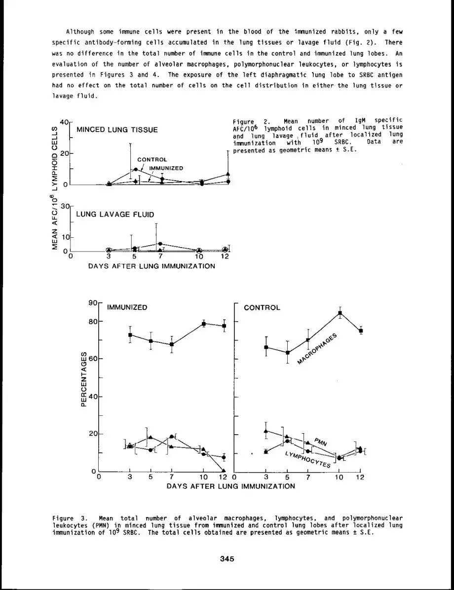
Although some immune cells were present in the blood of the immunized rabbits, only a few
specific antibody-forming cells accumulated in the lung tissues or lavage fluid (Fig. 2). There
was no difference in the total number of immune cells in the control and immunized lung lobes. An
evaluation of the number of alveolar macrophages, polymorphonuclear leukocytes, or lymphocytes is
presented in Figures 3 and 4. The exposure of the left diaphragmatic lung lobe to SRBC antigen
had no effect on the total number of cells on the cell distribution in either the lung t~ssue or
lavage fluid.
MINCED LUNG TISSUE
ICONTROL TL,J ,MMUNIZED /
LUNG LAVAGE FLUID
3 5 7 10 12DAYS AFTER LUNG IMMUNIZATION
Figure 2. Mean number of igM specificAFC/IO6 lymphoid cells in minced lung tissueand lung lavage ,fluid after localized lungimmunization with I0 9 SRBC. Data arepresented as geometric means +_ S.E.
9O
8O
03w 60<EI--ZLU
n’40m
IMMUNIZED
0 .... L I I -j -,’~
0 3 5 7 10 12 0
CONTROL
DAYS AFTER LUNG IMMUNIZATION
Figure 3. Mean total number of alveolar macrophages, lymphocytes, and polymorphonuclearleukocytes (PMN) in minced lung tissue from immunized and control lung lobes after localized lungimmunization of lO9 SRBC. The total cells obtained are presented as geometric means ± S.E.
345
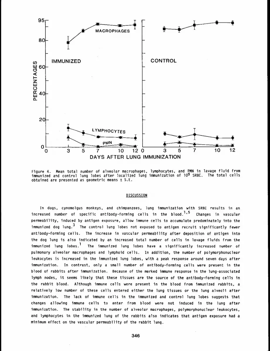
95-
80-
COw 6O
I-Z -W
rr 4C-Wn
2C-
00 3
IMMUNIZED
__ ~ LYMPHOCYTES
LpMN
CONTROL
5 7 10 12 0DAYS AFTER LUNG IMMUNIZATION
3 5 7 10 12
Figure 4. Mean total number of alveolar macrophages, lymphocytes, and PMN in lavage fluid fromimmunized and control lung lobes after localized lung immunization of lO9 SRBC. The total cellsobtained are presented as geometric means ± S.E.
DISCUSSION
In dogs, cynomolgus monkeys, and chimpanzees, lung immunization with SRBC results in an
increased number of specific antibody-forming cells in the blood.l’5 Changes in vascular
permeability, induced by antigen exposure, allow immune cells to accumulate predominately into the
immunized dog lung. 3 The control lung lobes not exposed to antigen recruit significantly fewer
antibody-forming cells. The increase in vascular permeability after deposition of antigen into
the dog lung is also indicated by an increased total number of cells in lavage fluids from the
immunized lung lobes. 1 The immunized lung lobes have a significantly increased number of
pulmonary alveolar macrophages and lymphoid cells. In addition, the number of polymorphonuclear
leukocytes is increased in the immunized lung lobes, with a peak response around seven days after
immunization. In contrast, only a small number of antibody-forming cells were present in the
blood of rabbits after immunization. Because of the marked immune response in the lung-associated
lymph nodes, it seems likely that these tissues are the source of the antibody-forming cells in
the rabbit blood. Although immune cells were present in the blood from immunized rabbits, a
relatively low number of these cells entered either the lung tissues or the lung alveoli after
immunization. The lack of immune cells in the immunized and control lung lobes suggests that
changes allowing immune cells to enter from blood were not induced in the lung after
immunization. The stability in the number of alveolar macrophages, polymorphonuclear leukocytes,
and lymphocytes in the immunized lung of the rabbits also indicates that antigen exposure had a
minimum effect on the vascular permeability of the rabbit lung.
346
lhe lung-associated lymph nodes in the rabbit, like those In all other specles tested to date,
are the primary site for the production of antfbody-fornllng cells after lung immunization with
SRBC. However, why only a small number of antibody-forming cells were observed in the blood, in
contrast to responses in dogs, is not known. It is possible that antigen dose is important and
that higher doses of SRBC in the rabbit lung mlght result in a higher number of antibody-formingcells in the blood. Nevertheless, if no change in vascular permeability is induced in the rabbit
lung by immunlzatfon, it seems unltkely that immune cells could accumulate in the immunized lung.
REFERENCES
I. Bite, D. E., M. A. Degen, D. L. Harris, and B. A. Muggenburg, Recruitment of Antibody-FormingCells in the Lung After Local Immunization is Nonspecific, Am. Rev. Resplr. Dis. 126: 635-639,lg82.
2. Schnlzlein, C. T., D. E. Bice, C. E. Mitchell, and F. F. Hahn, Effects on Rat Lung Immunity byAcute Lung Exposure to Benzo(a)pyrene, Arch. Environ. Health 3__77: 201-206, 1982.
3. Brownstein, D. G., A. H. Rebar, D. E. Bice, B. A. Muggenburg, and 3. O. Hill, Immunology ofthe Lower Respiratory Tract: Serial Morphologlc Changes in the Lungs and TracheobronchialLymph Nodes of Dogs After Intrapulmonary Immunization With Sheep Erythrocytes, Am. 3. Pathol.9_88: 499-514, 1980.
4. Lawrence, E. C., R. M. Blaese, and R. R. Martin, Immunoglobulin Secreting Cells in NormalHuman Bronchial Lavage Fluids, 3. Clin. Invest. 6_22: 832-835, 1978.
5. Blce, D. E., D. L. Harris, B. A. Muggenburg, and 5. A. Bowen, The Evaluation of Lung Immunityin Chimpanzees, ~m. Rev, Respir. Dis. 126: 358-359, 1982.
347
AN IMPROVED METHOD FOR CALCULATING LABELING INDICES OF LUNG EPITHELIAL CELLS
Abstract --An improved method has been deveZoped
to calculate labellng ~nd~ces (LIs) of eplChelial PRINCIPAL INVESTIGATORS
an4 Interstltlal cells in the alrways and alveolar s.G. Shaml
reglon of the lungs of rats. Thls method is more T.M. Caton
accurate and requires less tlme than the tradl- W.C. Grlfflth
t~onal method, whlch involves countlng all labeled M.J. Evans
and unlabele4 cells. In the new method, all la-
beled cells are counted~ but the total number of cells is estlmated, based on a representatlve
sample count of alrwag and alweolar cells in each anlmal. The LIs as calculated b9 ~he two
methods correlated very well. Most of the error ~n the new method was due to sampllng error~ vet9
llttle was attrlbuted to estimatlng the total number of cells.
Proliferation is measured by calculating the labeling index (LI), which is the number of cells
labeled with tritiated thymidine divided by the total number of cells in a given population.
Under most conditions, proliferation rates are very low in the lung epithelium compared to other
renewing cell populations in the body. Therefore, it is necessary to count a very large number of
cells to reduce sampling error and obtain a reasonably accurate estimate of the LI. Time
considerations often make counting of large sample sizes impractical. The purpose of this study
was to develop a method to increase the sample size without increasing time necessary to obtain
this sample size to calculate the Lls of lung epithelial cells. The rationale behind this new
method is that the sample size can be increased greatly by estimating the total number of cells
rather than counting each cell. The error introduced by the estimation of cell numbers would be
more than compensated for by the decreased sampling error because of increased sample size. We
used both control animals and animals exposed to fly ash. We thus evaluated both normal lungs and
lungs with increased proliferation in all cell types to test the usefulness of this new method.
METHODS
A total of 15 animals were used to test the new method against the old. Seven of these
animals had been exposed to fly ash aerosols for 2 or 4 weeks, and 8 were sham-exposed controls.
Details of the exposure are reported elsewhere (this report, pp. 317 to 320). Autoradiographs
lung tissue were prepared as reported elsewhere (this report, pp. 317 to 320). The lungs were
divided into airways and alveolar regions for analysis. Lls were first measured for each animal
by the traditional method. They were then calculated by the new method and the two results
compared. The number of cells counted or estimated by each method was the same in each animal.
About 2000 airway cells, 3000 interstitial cells, 450 Type II cells, and 75 macrophages were
counted in each animal.
The new method to estimate the number of cells in the airways was to measure the length of the
airways with an ocular reticule and then multiply the number of reticule lengths (RLs) by the
number of cells per RL at a magnification of 430x. The number of cells per RL was determined by
348
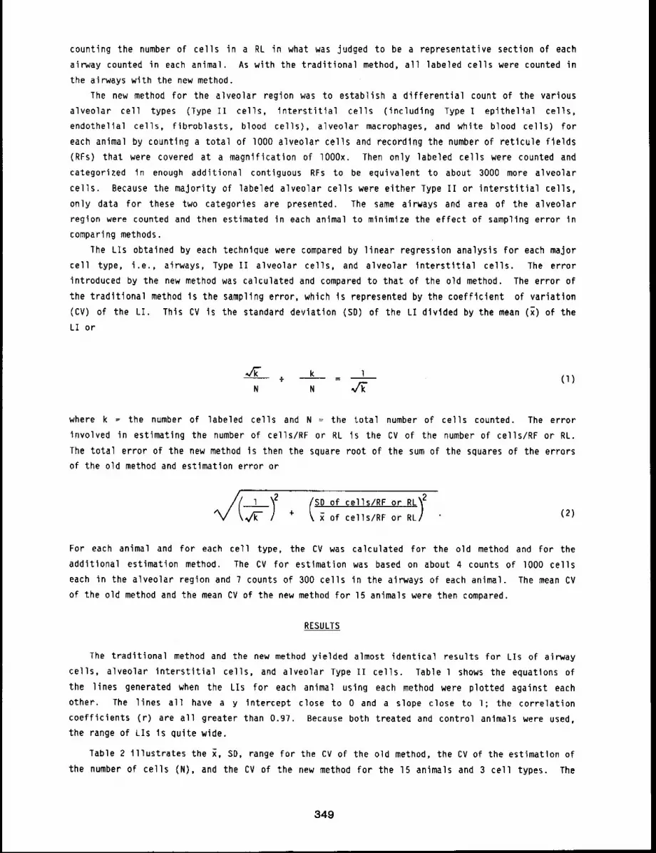
counting the number of cells in a RL in what was judged to be a representative section of each
airway counted in each animal. As with the traditional method, all labeled cells were counted in
the airways with the new method.
The new method for the alveolar region was to establish a differential count of the various
alveolar cell types (Type Ii cells, interstitial cells (including Type I epithelial cells,
endothelial cells, fibroblasts, blood cells), alveolar macrophages, and white blood cells) for
each animal by counting a total of I000 alveolar cells and recording the number of reticule fields
(RFs) that were covered at a magnification of lO00x. Then only labeled cells were counted and
categorized in enough additional contiguous RFs to be equivalent to about 3000 more alveolar
cells. Because the majority of labeled alveolar cells were either Type II or interstitial cells,
only data for these two categories are presented, The same airways and area of the alveolar
region were counted and then estimated in each animal to minimize the effect of sampling error in
comparing methods.
lhe LIs obtained by each technique were compared by linear regression analysis for each major
cell type, i.e., airways, Type II alveolar cells, and alveolar interstitial cells. The error
introduced by the new method was calculated and compared to that of the old method. The error of
the traditional method is the sampling error, which is represented by the coefficient of variation
(CV) of the LI. This CV is the standard deviation (SD) of the LI divided by the mean (~)
LI or
k l÷
N N(I)
where k = the number of labeled cells and N ~ the total number of cells counted. The error
involved in estimating the number of cells/RF or RL is the CV of the number of cells/RF or RL.
The total error of the new method is then the square root of the sum of the squares of the errors
of the old method and estimation error or
~I~---) ? IS~°f cells/RF °r RL~2+ of cells/RF or RL/ (2)
For each animal and for each cell type, the CV was calculated for the old method and for the
additional estimation method. The CV for estimation was based on about 4 counts of lO00 cells
each in the alveolar region and 7 counts of 300 cells in the airways of each animal. The mean CV
of the old method and the mean CV of the new method for 15 animals were then compared.
RESULTS
The traditional method and the new method yielded almost identical results for LIs of airway
cells, alveolar interstitial cells, and alveolar Type II cells. Table l shows the equations of
the lines generated when the LIs for each animal using each method were plotted against each
other. The lines all have a y intercept close to 0 and a slope close to l; the correlation
coefficients (r) are all greater than 0.97. Because both treated and control animals were used,
the range of LIs Is quite wide.
Table 2 illustrates the x, SD, range for the CV of the old method, the CV of the estimation of
the number of cells (N), and the CV of the new method for the 15 animals and 3 cell types. The
349
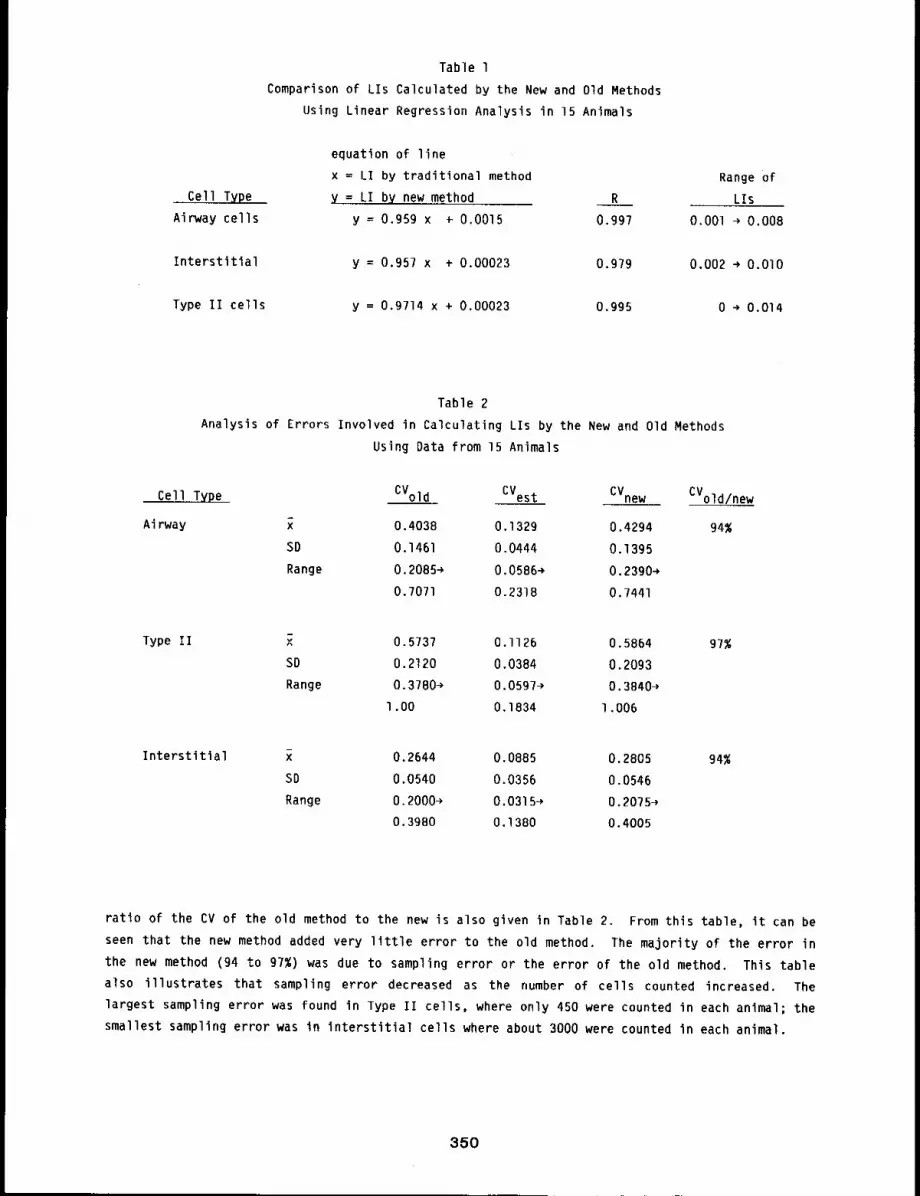
Cell Type
Airway cells
Table l
Comparison of LIs Calculated by the New and Old Methods
Using Linear Regression Analysis in 15 Animals
equation of line
x = LI by traditional method
y = LI by new method R
y = 0.959 x ÷ 0.0015 0.997
Interstitial y = 0,957 x + 0.00023 0.979
Type II cells y = 0.9714 x + 0.00023 0.995
Range of
LIs
O.OOl ~ 0.008
0.002 ~ O.OlO
0 ~ 0.014
Table 2
Analysis of Errors Involved in Calculating LIs by the New and Old Methods
Using Data from 15 Animals
Cell Type CVold CVest CVnew CVold/new
Airway ~ 0.4038 0.1329 0.4294 94%
SD 0.1461 0.0444 0.1395
Range 0.2085~ 0.0586~ 0.2390~
0.7071 0.2318 0,7441
Type II x 0.573? 0.I126 0.5864 97%
SD 0.2120 0.0384 0.2093
Range 0.3780~ 0.0597~ 0.3840~
l.O0 0.1834 1.006
Interstitial x 0.2644 0.0885 0.2805 94%
SD 0.0540 0.0356 0.0546
Range 0.2000~ 0.0315~ 0.2075~
0.3980 0.1380 0.4005
ratio of the CV of the old method to the new is also given in Table 2. From this table, it can be
seen that the new method added very little error to the old method. The majority of the error in
the new method (94 to 97%) was due to sampling error or the error of the old method. This table
also illustrates that sampling error decreased as the number of cells counted increased. The
largest sampling error was found in Type II cells, where only 450 were counted in each animal; the
smallest sampling error was in interstitial cells where about 3000 were counted in each animal.
350
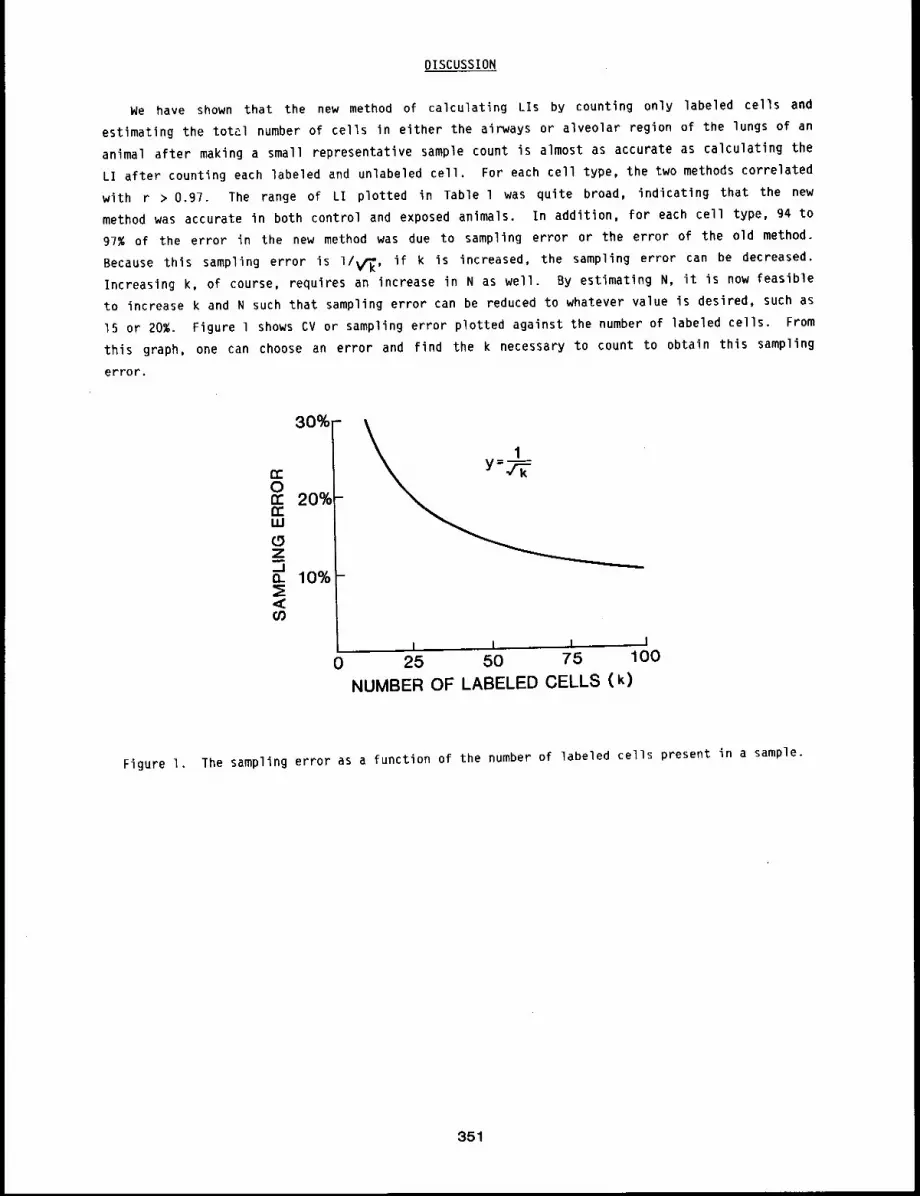
DISCUSSION
We have shown that the new method of calculating LIs by counting only labeled cells and
estimating the total number of cells in either the airways or alveolar region of the lungs of an
animal after making a small representative sample count is almost as accurate as calculating the
LI after counting each labeled and unlabeled cell. For each cell type, the two methods correlated
with r > 0.97. The range of LI plotted in Table l was quite broad, indicating that the new
method was accurate in both control and exposed animals. In addition, for each cell type, g4 to
97% of the error in the new method was due to sampling error or the error of the old method.
Because thls sampling error is I/K, if k is increased, the sampling error can be decreased.
Increasing k, of course, requires an increase in N as well. By estimating N, it is now feasible
to increase k and N such that sampling error can be reduced to whatever value is desired, such as
15 or 20%. Figure l shows CV or sampling error plotted against the number of labeled cells. From
this graph, one can choose an error and find the k necessary to count to obtain this sampling
error.
30%
n-On’- 20%n"uJ(.9Z._1o_ 10%<~
0I I I I
25 50 75 100NUMBER OF LABELED CELLS (k)
Figure 1. The sampling error as a function of the number of labeled cells present in a sample.
351
RESPIRATION OF RATS IN NOSE-ONLY EXPOSURE TUBES
Abstract -- Resplrarlon and hod9 surface tempera-
ture of 3-, 6-, Z2-, and 24-mOnth-oid rats were mea- PRINCIPAL INVESTIGATOR
sured during conflnement in two d~fferent styles of J.L. Mauderl9nose-onl9 exposure tubes. No dlfference in resplra--
rlon was found between rats in older and newer stgle tubes. Awerage mlnute volumes after 60 m~n
of conflnement were Z.40, 0.81, 0.67 and 0.70 ml/mln/g body welght for 3-, 6-., 12-, and
24-month-old rats, respectlwely. The higher m~nute volume of the youngest rats was ascribed to
excitement and a higher ~tabollc rare.
There is frequent need for estimates of the respiration of rats confined in nose-only
inhalation exposure tubes. Various estimates are used; however, one of the most frequently
referenced is the minute volume of 0.65 ml/min/g body weight reported by Guyton in 19471 from
measurements of 35, ll3-g rats of unreported strain or sex. A minute volume of 0.56 ml/min/g for
free-standing Long-Evans rats was measured at this Institute using nonrebreathing valves.2
However, a mean minute volume of 1.28 ml/min/g was later obtained from 3-month--old Fischer-344
rats after l h of exposure tube confinement (1978-1979 Annual Report, LF-69, pp. 475-47B).
Because this value seemed high and because it was obtained by nonrebreathing valve rather than
under conditions representative of exposure, its utility was uncertain. The present study was
conducted to provide better estimates of rat respiration in exposure tubes under typical exposure
conditions.
METHODS
Ten male and lO female laboratory-reared, specific pathogen-free Fischer-344 rats, at each of
four ages, 3, 6, 12, and 24 months, were used in the experiment outlined in Table I. Two types of
plastic exposure tubes were used, an older type with a metal nose cone and long-used at this
Institute for acute exposures,3 and a newer type with a plastic nose cone and now being used for
repeated, chronic exposures. Rats were placed in the various sized tubes according to the body
weight criteria in Table I.
The tubes were modified to serve as volume displacement plet~ysmographs. A diaphragm of latex
dental dam clamped between the nose cone and the body of the tube formed a seal around the rat’s
nose. The tube was perforated in the area of the rat’s body and a larger tube was sealed around
this area to form a jacket. A Fleisch 4-0 pneumotachograph attached to a hole in the jacket
measured displaced air as the animal breathed. Respiratory frequency, tidal volume, and minute
volume were measured using a Buxco Model 4 Pulmonary Mechanics Analyzer. Body surface temperature
was measured by a thermistor on the inner surface of the tube at the level of the rat’s abdomen.
Calibration for volume and frequency response revealed less than 5% maximum error within the
required ranges.
Tests were conducted using an 80-port, nose-only exposure plenum 3 operated at 19 I/min
compressed air and balanced to ambient pressure at the rat’s noses. Rats were held in the tubes
for ]5 min before beginning the "exposure" to simulate loading and transportation time encountered
3,52
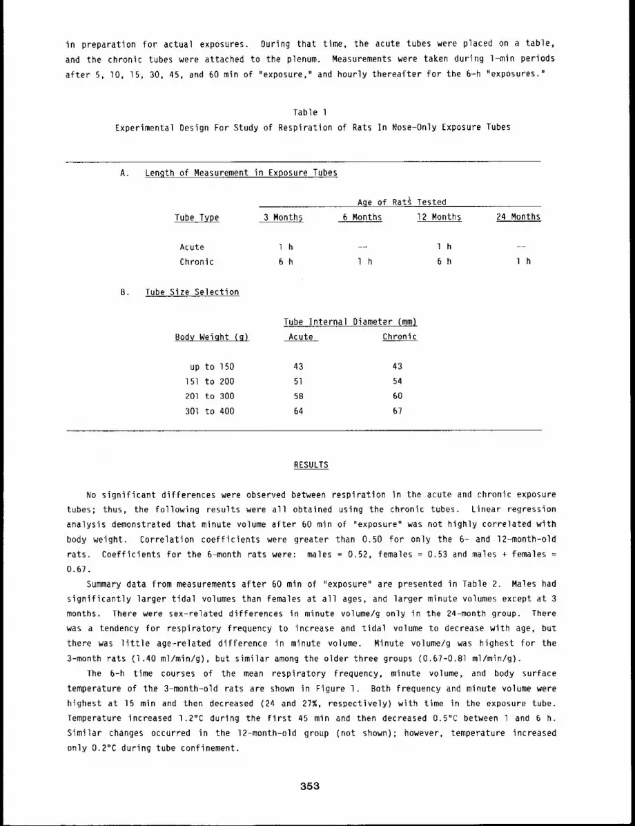
in preparation for actual exposures. During that time, the acute tubes were placed on a table,
and the chronic tubes were attached to the plenum. Measurements were taken during 1-min periods
after 5, lO, 15, 30, 45, and 60 min of "exposure," and hourly thereafter for the 6=h "exposures."
Table l
Experimental Design For Study of Respiration of Rats In Nose-Only Exposure Tubes
A. Length of Measurement in Exposure Tubes
Age of Rat~ Tested
Tube Type 3 Months 6 Months 12 Months 24 Months
Acute 1 h -- 1 h --
Chronic 6 h 1 h 6 h I h
B. Tube Size Selection
Tube Internal Diameter (mm).
Body Weight (g) Acute Chronic
up to 150 43 43
151 to 200 51 54
201 to 300 58 60
301 to 400 64 67
RESULTS
No significant differences were observed between respiration in the acute and chronic exposure
tubes; thus, the following results were all obtained using the chronic tubes. Linear regression
analysis demonstrated that minute volume after 60 min of "exposure" was not highly correlated with
body weight. Correlation coefficients were greater than 0.50 for only the 6- and 12-month-old
rats. Coefficients for the 6-month rats were: males = 0.52, females = 0.53 and males + females =
0.67.
Summary data from measurements after 60 min of "exposure" are presented in Table 2. Males had
significantly larger tidal volumes than females at all ages, and larger minute volumes except at 3
months. There were sex-related differences in minute volume/g only in the 24-month group. There
was a tendency for respiratory frequency to increase and tidal volume to decrease with age, but
there was little age=related difference in minute volume. Minute volume/g was highest for the
3-month rats (I.40 ml/min/g), but similar among the older three groups (0.67-0.81 ml/min/g).
The 6-h time courses of the mean respiratory frequency, minute volume, and body surface
temperature of the 3-month-old rats are shown in Figure l. Both frequency and minute volume were
highest at 15 min and then decreased (24 and 27%, respectively) with time in the exposure tube.
Temperature increased 1.2°C during the first 45 min and then decreased 0.5°C between l and 6 h.
Similar changes occurred in the 12-month-old group (not shown); however, temperature increased
only 0.2°C during tube confinement.
353
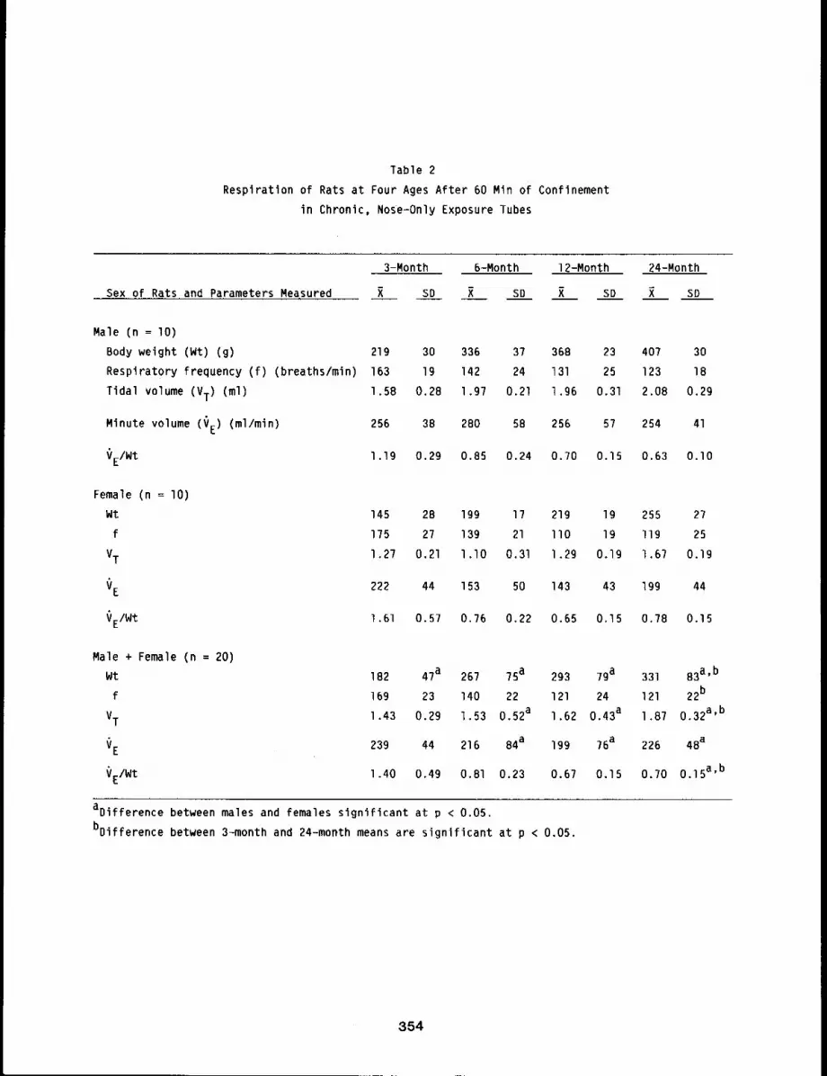
Table 2
Respiration of Rats at Four Ages After 60 Min of Confinement
in Chronic, Nose=Only Exposure Tubes
Sex of Rats and Parameters Measured
Male (n ~ lO)
Body weight (Wt) (g)
Respiratory frequency (f) (breaths/min)
Tidal volume (VT) (ml)
Minute volume (VE) (ml/min)
VEIWt
3-Month
R SO
6-Month 12-Month 24-Month
X SD R SO X SD
219 30 336 37 368 23 407 30
163 19 142 24 131 25 123 18
1.5B 0.28 1.97 0.21 1.96 0.31 2.08 0.29
256 38 280 58 256 57 254 41
l .19 0.29 0.85 0.24 0.70 0.15 0.63 O.lO
Female (n = lO)
Wt 145 2B 199 17 219 19 255 27
f 175 27 139 21 llO 19 ll9 25
VT1.27 0.21 l.lO 0.31 1.29 0.19 1.67 0.19
VE 222 44 153 50 143 43 199 44
VE/Wt 1.61 0.57 0.76 0.22 0.65 0.15 0.78 0.15
Male + Female (n = 20)
Wt 182 47a 267 75 a 293 79 a 331 B3a’b
f 169 23 140 22 121 24 121 22b
VT1.43 0.29 1.53 0.52a 1.62 0.43a 1.87 0.32a’b
VE 239 44 216 84a 199 76a 226 4Ba
VE/Wt 1.40 0,49 0.81 0.23 0.67 0.15 0.70 0.15a’b
aoifference between males and females significant at p < 0.05.
bDifference between 3-month and 24-month means are significant at p < 0.05.
354
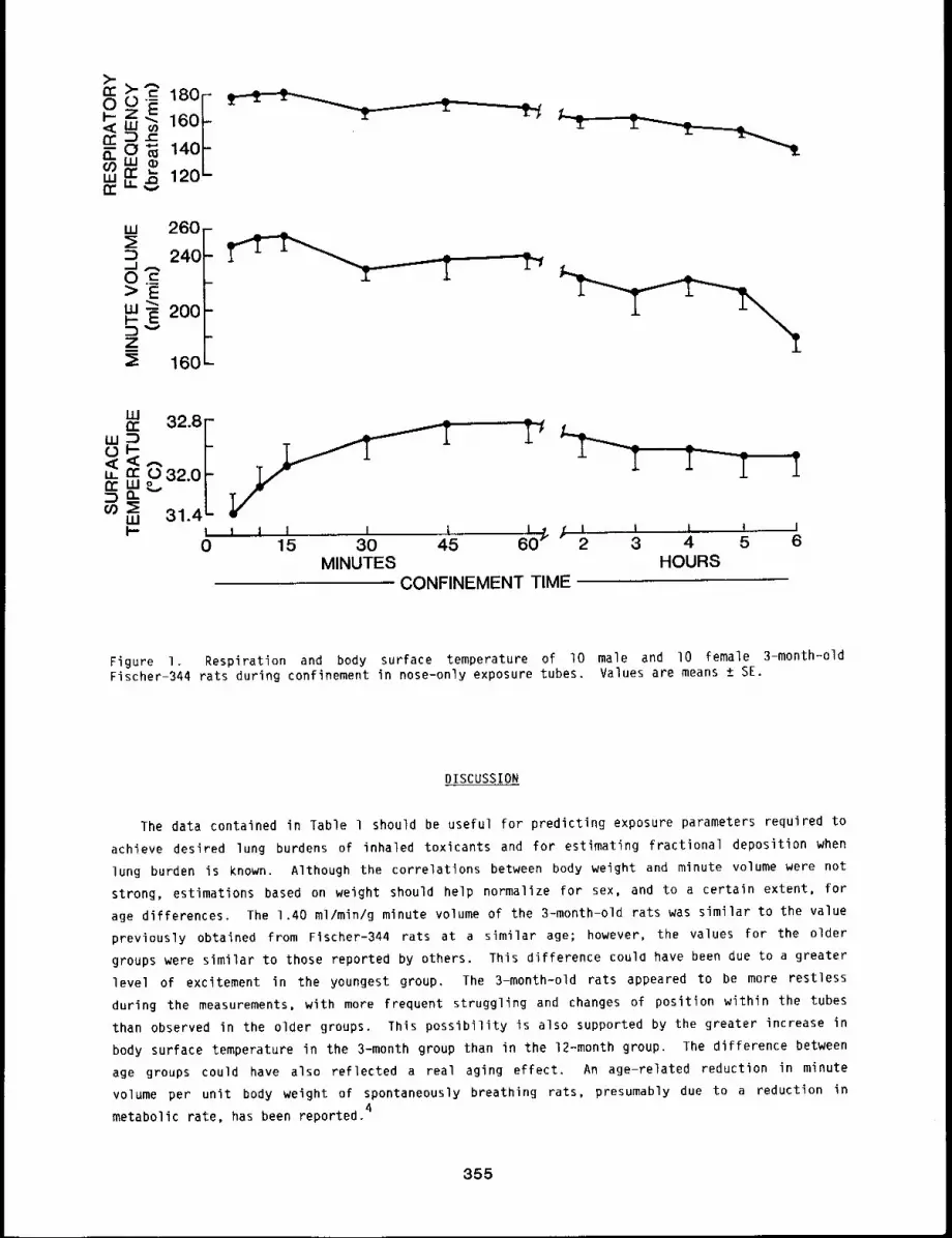
W
~D 240
200
260,-
:~ 160
32.8
o 32.0I== ,,=, 314,-l i I I I I L, j I I I I I0 15 30 45 60r 2 3 4 5 6
MINUTES HOURSCONFINEMENT TIME
Figure I. Respiration and body surface temperature of I0 male and I0 female 3-month-oldFischer-344 rats during confinement in nose-only exposure tubes. Values are means ± SE.
DISCUSSION
The data contained in Table 1 should be useful for predicting exposure parameters required to
achieve desired lung burdens of inhaled toxicants and for estimating fractional deposition when
lung burden is known. Although the correlations between body weight and minute volume were not
strong, estimations based on weight should help normalize for sex, and to a certain extent, for
age differences. The 1.40 ml/min/g minute volume of the 3-month-old rats was similar to the value
previously obtained from Fischer-344 rats at a similar age; however, the values for the older
groups were similar to those reported by others. This difference could have been due to a greater
level of excitement in the youngest group. The 3-month-old rats appeared to be more restless
during the measurements, with more frequent struggling and changes of position within the tubes
than observed in the older groups. This possibility is also supported by the greater increase in
body surface temperature in the 3-month group than in the 12-month group. The difference between
age groups could have also reflected a real aging effect. An age-related reduction in minute
volume per unit body weight of spontaneously breathing rats, presumably due to a reduction in4
metabolic rate, has been reported.
355
REFERENCES
I. Guyton, A. C., Measurement of the Respiratory Volumes of Laboratory Animals, Am. J. Physiol.150: 70=77, 1947.
2. Mauderly, J. L,, J. E. Tesarek, Linda 3. Sifford, and Lorna 3. Sifford, RespiratoryMeasurements of Unsedated Small Laboratory Mammals Using Nonrebreathing Valves, Lab. Anim.Sci. 29: 323-329, 1979.
3. Raabe, O. G., 3. E. Bennick, M. E. Light, C. H. Hobbs, R. L. Thomas, and M. I. Tillery, An’Improved Apparatus for Acute Inhalation Exposure of Rodents to Radioactive Aerosols, Toxicol.A_p_pl. Pharmacol. 2_66: 264-273, 1973.
4. Leong, K. 3., G. F. Dowd, and H. N. MacFarland, A New Technique for Tidal Volume Measurementin Unanesthetized Small Animals, Can. J. Physiol. Pharmacol. 42: 189-194, 1964.
356
TOXICOLOGICAL ASPECTS OF THE LONG-TERM DEPLETION OF REDUCED
GLUTATHIONE IN MICE GIVEN L-BUTHIONINE-S,R-SULFOXIMINE
Abstract -- L-Buth~onlne-S,R-sulfoxlmlne (BSO)
added to the drlnklng water of mice was found to PRINCIPAL INVESTIGATORS
deplete reduced glutathlone (GSH) in lungs, pul- J.D. Sun
monary lavage fluld, liver, kldnegs, and bloo~ ~n S.S. Ragsdale
both a dose~ and tlme-dependent manner. It was J.M. Benson
also found that BSO treatments did not show any R.F. HenderSOn
blochem~cal indications of pulmonary or hepatlc
toxlcltles, or affect levels of lung or llver cgtochrome P-450. Thus, this method for depleting
GSH may be useful in toxicological studies requ~rlng animals wlth decreased In vlvo levels of GSH.
In higher animals, reduced glutathione (GSH) is important in metabolic pathways for
detoxification and excretion of various xenobiotic compounds. One useful approach in studies that
investigate the biochemical mechanisms of chemically induced toxicities has been to deliberately
lower tissue levels of GSH by administering different GSH depleting agents. However, such
treatments are unable to lower GSH levels for extended periods without repeated injections, can be
toxic, and often affect the metabolic fate of the xenobiotic compound being studied. This report
describes a possible alternative for in vivo depletion of GSH by using L-buthionine-S,R-
sulfoximine (BSO). This study characterized BSO depletion of GSH in various tissues by the
addition of this agent in the drinking water of mice for 2B days. Further, a number of
biochemical parameters were also measured to assess the toxicological aspects of such treatments.
METHODS
Male, laboratory-raised, CD-I mice (specific pathogen-free; 13 to 15 weeks of age) from the
Institute colony were used for this study. These mice were allowed free access to food (Wayne Lab
Blox) and untreated water containing O, lO, 20, or 30 mM BSO for 28 days. During this time,
periodic measurements of body weight and water consumption were made. At various times, animals
were sacrificed, and lungs, liver, kidneys, and blood samples were taken. Each lung sample was
lavaged twice with a total of 2.B ml of 0.15 M saline and then perfused intravascularly with
0.15 M saline to remove blood. Lavage fluid samples were then centrifuged at 300 x g for lO min
and the resulting cellular pellets were resuspended in l.O ml of 0.15 M saline for the
determination of total and differential cell counts. GSH was measured in aliquots of lung, liver,
and kidney homogenates (50 mM Tris-Cl, pH ?.4), lavage fluid supernatants, and whole blood by the
methods of Cohn and Lyle. l Portions of lung homogenates and lavage fluid supernatants were
assayed for alkaline phosphatase (AP), lactate dehydrogenase (LDH), glucose-6-phosphate
dehydrogenase (G6PDH), glutathione reductase (GR), glutathione peroxidase (GP) and total soluble
protein. 2 Cytochrome P-450 levels were measured in microsomes prepared from lung and liver
homogenates. 3 The remaining whole blood samples were centrifuged and the serum assayed for
glutamate-pyruvate transaminase (SGPT) and ==glutamyl transpeptidase (~GTP). In addition,
357
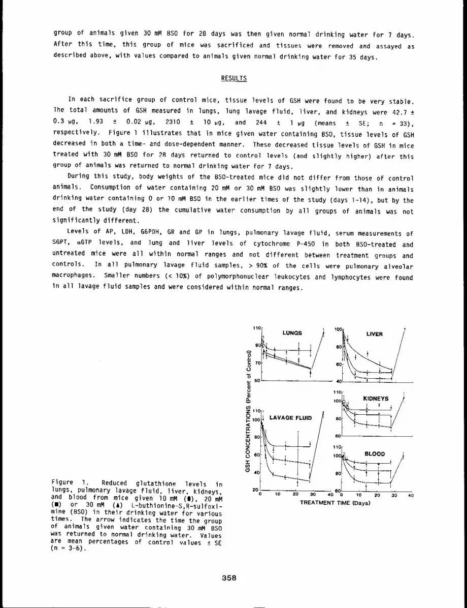
group of animals given 30 mM BSO for 28 days was then given normal drinking water for 7 days.
After this time, this group of mice was sacrificed and tissues were removed and assayed as
described above, with values compared to animals given normal drinking water for 35 days.
RESULTS
In each sacrifice group of control mice, tissue levels of GSH were found to be very stable.
The total amounts of GSH measured in lungs, lung lavage fluid, liver, and kidneys were 42.7 ±
0.3 ~g, 1.93 ± 0.02 ~g, 2310 ± lO ~g, and 244 ± 1 ~g (means ± SE; n = 33),
respectively. Figure l illustrates that in mice given water containing BSO, tissue levels of GSH
decreased in both a time- and dose-dependent manner. These decreased tissue levels of GSH in mice
treated with 30 mM BSO for 2B days returned to control levels (and slightly higher) after this
group of animals was returned to normal drinking water for 7 days.
During this study, body weights of the BSO-treated mice did not differ from those of control
animals. Consumption of water containing 20 mM or 30 mM BSO was slightly lower than in animals
drinking water containing 0 or lO mM BSO in the earlier times of the study (days 1-14), but by the
end of the study (day 28) the cumulative water consumption by all groups of animals was not
significantly different.
Levels of AP, LDH, G6PDH, GR and GP in lungs, pulmonary lavage fluid, serum measurements of
S6PT, :GTP levels, and lung and liver levels of cytochrome P-450 in both BSO-treated and
untreated mice were all within normal ranges and not different between treatment groups and
controls. In all pulmonary lavage fluid samples, > 90% of the cells were pulmonary alveolar
macrophages. Smaller numbers (< I0%) of polymorphonuclear leukocytes and lymphocytes were found
in all lavage fluid samples and were considered within normal ranges.
Figure I. Reduced glutathione levels inlungs, pulmonary lavage fluid, liver, kidneys,and blood from mice given lO mM (I), 20 (m) or 30 mM (A) L-buthionine-S,R-sulfoxi-mine (BSO) in their drinking water for varioustimes. The arrow indicates the time the groupof animals given water containing 30 mM BSOwas returned to normal drinking water. Valuesare mean percentages of control values ± SE(n = 3-6).
1~o~ LUNGS//} loo~ LIVER /
/
P
O3Z 110oC- loot~t-Z B0LUC)Z
,,r
~ 40
2o
11°r TlooLI, KIDNEYS /
~60~
i ~ or100~ BLOOD .~
10 20 30 40 0 10 20 30
TREATMENT TIME (Days)4O
358
DISCUSSION
BSO is a potent and apparently highly specific inhibitor of GSH biosynthesis that results in
GSH depletion in various tissues.4 If one assumes complete inhibition of the
~-glutamylcysteine synthetase enzyme by this substrate analog, the rate of GSH depletion should
be indicative of that tissue’s rate of GSH utilization or release into the circulatory system. In
this study, GSH depletion by BSO was seen to be somewhat less rapid and/or less effective in
lungs, blood, and kidneys, as compared with pulmonary lavage fluid or liver (Fig. l). This slower
response by lung GSH may simply reflect the slower rate of GSH turnover in lungs than in lavage
fluid or liver. The lesser response in blood and kidneys may have occurred because of the normal
release of GSH by other tissues into blood and absorption by the kidneys, the primary organ for
GSH degradation and the reuse of its composite amino acids.
Use of BSO to deplete in vivo concentrations of GSH in studies that investigate the
biochemical mechanisms of chemically induced toxicities would not be of value if this substrate
analog also caused toxic and/or undesirable biochemical effects. This study showed that BSO
treatments did not significantly affect total body weights or total water consumption of mice
during the experiments. More sensitive biochemical parameters also showed no signs of BSO-induced
toxicities, Measurements of specific parameters as indicators of biochemical damage in lungs and
lavage fluid showed no indications of pulmonary cell or tissue damage, or lung inflammatory
response. Serum enzyme levels also showed normal liver functions in BSO-treated mice. In
addition, the levels of cytochrome P-450 showed (by this criteria) that xenobiotic metabolism
would not be affected by BSD treatment. Therefore, BSO administered in drinking water to
laboratory animals appears to be an effective and non-toxic method for depleting in vivo levels of
GSH without adversely affecting other metabolic parameters important in toxicological studies.
REFERENCES
I. Cohn, V. H. and J. Lyle, A Fluorometric Assay for Glutathione, Anal. Biochem. 14: 434-440,1966.
2. DeNicola, D. B., A. H. Rebar, and R. F. Henderson, Early Damage Indicators in the Lung V.Biochemical and Cytological Response to NO2 Inhalation, T oxicq.l. Appl. Pharmacol. 60:301-312, 1981.
3. Omura, T. and R. Sato, The Carbon Monoxide-Binding Pigment of Liver Microsomes I. Evidencefor its Hemoprotein Nature, 3. Biol. Chem. 239: 23?0-2378, 1964.
4. Griffith, O. W. and A. Meister, Potent and Specific Inhibition of GIutathione Synthesis byButhionine Sulfoximine (S-n-Butyl Homocysteine Sulfoximine), J. Biol. Chem. 254: 7558-7560,1979.
359/360

RISK ASSESSMENT
The papers in this section report on studies designed to better predict the actual risk to man
based both on the toxicity of materials associated with a technology or process and the degree of
exposure of man to the material in question. Basic elements required for health risk assessment
are definition of source terms, models of environmental distribution, dosimetry, and dose-response
relationships.
The first paper reports on the use of a toxicology matrix approach to incorporate all of the
toxicity and epidemiological information available on a substance or agent as well as on
surrogate substances - into the subsequent risk assessment for man. This approach addresses an
important need within risk assessment because it is often necessary to use toxicity studies in
laboratory animals to predict health effects in man.
The second paper in the section summarizes work that has been completed on the assessment of
the potential health and environmental effects of the developing technology of fluidized bed
combustion of coal. Few human health effects were predicted from development of the technology,
but occupational and public risks from accidents would be significant, just as they would be from
expansion of the current technology of pulverized coal combustion. The next paper presents a more
detailed examination of one aspect of fluidized bed combustion, that is, the disposal of solid
wastes.
The final paper in the section reports on the use of fractal mathematics to estimate
environmental dilution factors. These types of techniques are essential to an improved
understanding of man’s potential exposure to pollutants from a technology.
361/362

HUMANRISK RELATIONSHIPS DERIVED FROM EPIDEMIOLOGY AND LABORATORY STUDIES
Abstract --Proven technlques are needed for ~n-
corporatlng the results of laborator9 toxlcology PRrNCIPAL INVESTIGATORS
studles into human r~sk assessments. Two sample R.G. Cuddlhy
calculatlons of lung cancer r~sk factors for In- B, B~ Boecker
haled rad~oactlve particles and dlesel englne F.F. Hahn
exhaust are glven here to 111ustrate a Coxlcolog9 R.O. McClellan
informat~on matrlx approach. Th~s approach com-
blnes the results of ep~demlology and laboratory anlmal studles of the substance or agent of
prlncipal concern, along w~th slm~lar Informatlon on other surrogate substances. Beyond the
estlmates of lung cancer rlsk factors derlved by uslng thls approach, an additional advantage is
galned by havlng estlmates of uncertainty that can be obtalned b9 Incorporating all avallahle
toxlcology Informatlon Into the analys~s. Thls approach is recommended for both rlsk assessment
and In deslgnlng follow-on toxlcology studies to improve prellmlnary assessments for new
potentlally harmful agents enterlng our envlronment.
INTRODUCTION
The experimental designs of many toxicology studies can usefully be expanded to make their
results more applicable to human health risk assessment. Laboratory studies of single toxic sub=
stances performed in isolation do not provide a means for applying the results to humans in a
quantitative manner. Thus, they are seldom used in critical analyses to resolve adversary posi=
tions in legal proceedings or in formulating human exposure standards for environmental pollu-
tants. Part of this difficulty relates to differences between animals and people in (a) life
span, (b) physiological characterisitcs that influence exposure patterns and dosimetry, (c) meta-
bolism, and (d) their relative sensitivities to toxic and carcinogenic agents. Another difficulty
may arise from the experimental conditions and levels of exposure used in laboratory studies,
which often differ substantially from those involving people. For these reasons, studies in labo-
ratory animals may be used only to provide descriptive information about the toxicity or carcino-
genicity of physical or chemical agents, whereas the quantitative assessment of risks may still be
calculated from poorly related studies in human populations. Many toxicologic evaluations are
begun with in vitro cell toxicity tests or transformation assays. These have the advantages of
relatively low cost and short duration. However, the mechanisms of exposure and injury to cells
in culture are even more remote from human exposure conditions than those of whole-animal
laboratory studies.
New mathematical modeling techniques are being evaluated to improve the usefulness of labora-
tory studies in human risk assessments, but they have not yet received sufficient attention to
build the necessary experience wlth which to judge their applicability. I=3 Some involve complex
mathematical calculations that tend to obscure the basic approaches and to discourage many toxico-
logists from applying them to new information. For the more complex calculations to attain wider
use, it may be useful to gain experience with more simple mathematical forms. At the same time,
we may also learn more about designing new toxicology studies that apply better to human risk eva-
luations.
An important goal of toxicology is to evaluate risks from potentially harmful new substances0
entering the environment and thereby aid in preventing injurious exposures to people. It would be
363
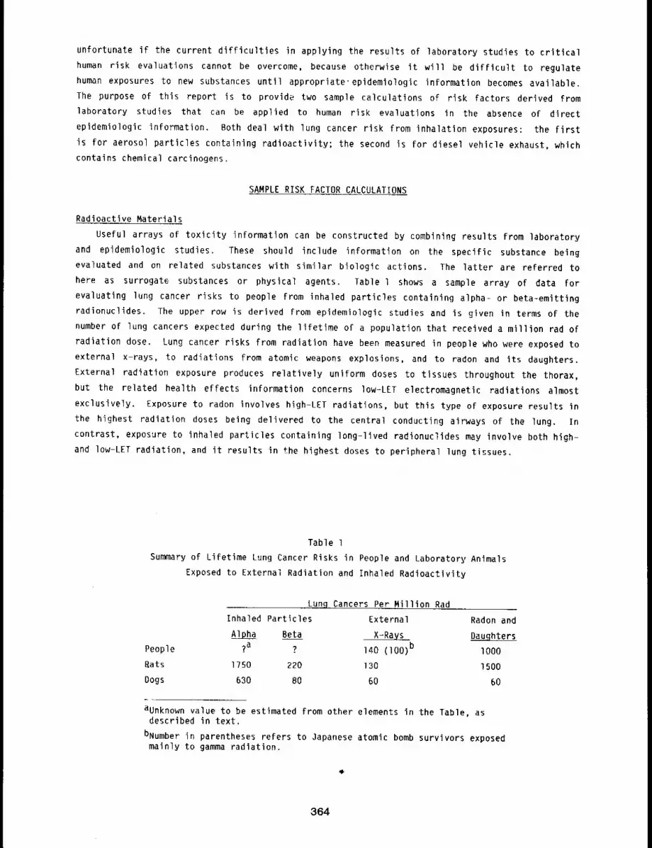
unfortunate if the current difficulties in applying the results of laboratory studies to critical
human risk evaluations cannot be overcome, because otherwise it will be difficult to regulate
human exposures to new substances until appropriate’epidemiologic information becomes available.
The purpose of this report is to provide two sample calculations of risk factors derived from
laboratory studies that can be applied to human risk evaluations in the absence of direct
epidemiologic information. Both deal with lung cancer risk from inhalation exposures: the first
is for aerosol particles containing radioactivity; the second is for diesel vehicle exhaust, which
contains chemical carcinogens.
SAMPLERISK FACTOR CALCULATIONS
Radioactive Materials
Useful arrays of toxicity information can be constructed by combining results from laboratory
and epidemiologic studies. These should include information on the specific substance being
evaluated and on related substances with similar biologic actions. The latter are referred to
here as surrogate substances or physical agents. Table l shows a sample array of data for
evaluating lung cancer risks to people from inhaled particles containing alpha- or beta-emitting
radionuclides. The upper row is derived from epidemiologic studies and is given in terms of the
number of lung cancers expected during the lifetime of a population that received a million rad of
radiation dose. Lung cancer risks from radiation have been measured in people who were exposed to
external x-rays, to radiations from atomic weapons explosions, and to radon and its daughters.
External radiation exposure produces relatively uniform doses to tissues throughout the thorax,
but the related health effects information concerns low-LET electromagnetic radiations almost
exclusively. Exposure to radon involves hlgh-LET radiations, but this type of exposure results in
the highest radiation doses being delivered to the central conducting airways of the lung. In
contrast, exposure to inhaled particles containing long-lived radionuclides may involve both high-
and low-LET radiation, and it results in the highest doses to peripheral lung tissues.
Table l
Summary of Lifetime Lung Cancer Risks in People and Laboratory Animals
Exposed to External Radiation and Inhaled Radioactivity
Lunq Cancers Per Million Rad
People
~ats
Dogs
Inhaled Particles External Radon and
Alpha Beta X-Rays_ Daughters?a ~ 140 (lO0)b lO00
1750 220 130 1500
630 80 60 60
aUnknown value to be estimated From other elements in the Table, asdescribed in text.
bNumber in parentheses refers to Japanese atomic bomb survivors exposedmainly to gamma radiation.
364
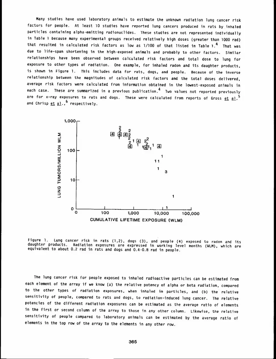
Many studies have used laboratory animals to estimate the unknown radiation lung cancer risk
factors for people. At least lO studies have reported lung cancers produced in rats by inhaled
particles containing alpha-emitting radionuclides. These studies are not represented individually
in Table l because many experimental groups received relatively high doses (greater than lO00 rad)
that resulted in calculated risk factors as low as I/lO0 of that listed in Table I. 4 That was
due to life-span shortening in the high-exposed animals and probably to other factors. Similar
relationships have been observed between calculated risk factors and total dose to lung for
exposure to other types of radiation. One example, for inhaled radon and its daughter products,
is shown in Figure I. This includes data for rats, dogs, and people. Because of the inverse
relationship between the magnitudes of calculated risk factors and the total doses delivered,
average risk factors were calculated from information obtained in the lowest-exposed animals in
each case. These are summarized in a previous publication. 4 Two values not reported previously
are for x-ray exposures to rats and dogs. These were calculated from reports of Gross et al.5
and Chrisp e__tt a l. 6 respectively
z0.J
n-O
I-r~Z,_1
1,000 -
100 -
10-
00
22 2
211[] 22[] 1[~111 []
111
3
1
,,, I I I1 !100 1,000 10,000 100,000
CUMULATIVE LIFETIME EXPOSURE (WLM)
Figure 1. Lung cancer risk in rats (1,2), dogs (3), and people (4) exposed to radon and daughter products. Radiation exposures are expressed in working level months (WLM), which areequivalent to about 0.2 rad in rats and dogs and 0.4-0.8 rad in people.
The lung cancer risk for people exposed to inhaled radioactive particles can be estimated from
each element of the array if we know (a) the relative potency of alpha or beta radiation, compared
to the other types of radiation exposures, when inhaled in particles, and (b) the relative
sensitivity of people, compared to rats and dogs, to radiation-induced lung cancer. The relative
potencies of the different radiation exposures can be estimated as the average ratio of elements
in the first or second column of the array to those in any other column. Likewise, the relative
sensitivity of people compared to laboratory animals can be estimated by the average ratio of
elements in the top row of the array to the elements in any other row.
365
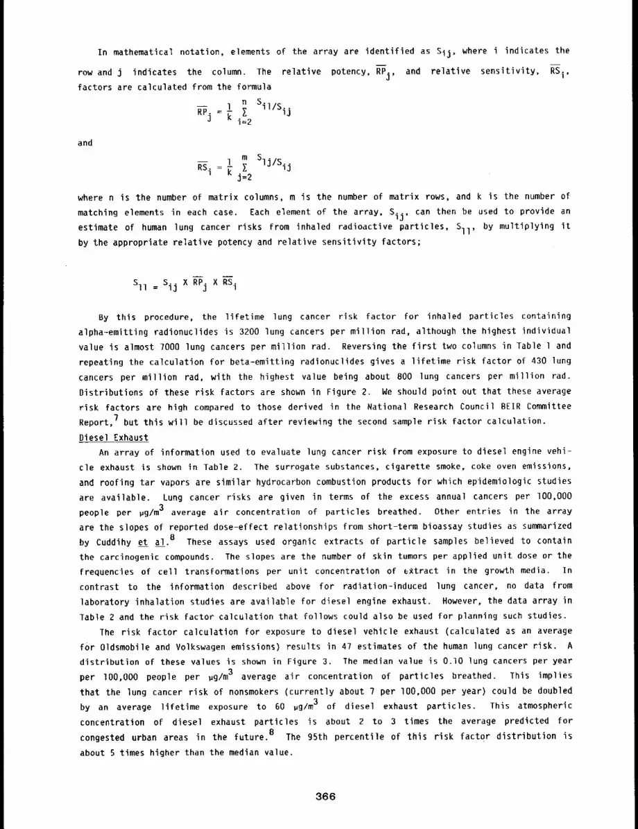
In mathematical notation, elements of the array are identified as Sij, where i indicates the
row and j indicates the column. The relative potency, RPj, and relative sensitivity, RSi,factors are calculated from the formula
nZ Sil/Sij-- 1
RPj = [ i=2
and
1 m SljlSij~i=~ I
j=2
where n is the number of matrix columns, m is the number of matrix rows, and k is the number of
matching elements in each case. Each element of the array, Sij, can then be used to provide an
estimate of human lung cancer risks from inhaled radioactive particles, Sll, by multiplying it
by the appropriate relative potency and relative sensitivity factors;
-- m
SII : Sij X RPj X RSi
By this procedure, the lifetime lung cancer risk factor for inhaled particles containing
alpha-emitting radionuclides is 3200 lung cancers per million rad, although the highest individual
value is almost 7000 lung cancers per million rad. Reversing the first two columns in Table l and
repeating the calculation for beta-emitting radionuclides gives a lifetime risk factor of 430 lung
cancers per million rad, with the highest value being about 800 lung cancers per million rad.
Distributions of these risk factors are shown in Figure 2. We should point out that these average
risk factors are high compared to those derived in the Rational Research Council BEIR Committee
Report, 7 but this will be discussed after reviewing the second sample risk factor calculation.
Diesel Exhaust
An array of information used to evaluate lung cancer risk from exposure to diesel engine vehi-
cle exhaust is shown in Table 2. The surrogate substances, cigarette smoke, coke oven emissions,
and roofing tar vapors are similar hydrocarbon combustion products for which epidemiologic studies
are available. Lung cancer risks are given in terms of the excess annual cancers per lO0,O00
people per ~g/m3 average air concentration of particles breathed. Other entries in the array
are the slopes of reported dose-effect relationships from short-term bioassay studies as summarized
by Cuddihy et al. 8 These assays used organic extracts of particle samples believed to contain
the carcinogenic compounds. The slopes are the number of skin tumors per applied unit dose or the
frequencies of cell transformations per unit concentration of extract in the growth media. In
contrast to the information described above for radiation-induced lung cancer, no data from
laboratory inhalation studies are available for diesel engine exhaust. However, the data array in
Table 2 and the risk factor calculation that follows could also be used for planning such studies.
The risk factor calculation for exposure to diesel vehicle exhaust (calculated as an average
for Oldsmobile and Volkswagen emissions) results in 47 estimates of the human lung cancer risk.
distribution of these values is shown in Figure 3. The median value is O.lO lung cancers per year
per lO0,O00 people per ~g/m3 average air concentration of particles breathed. This implies
that the lung cancer risk of nonsmokers (currently about 7 per lO0,O00 per year) could be doubled
by an average lifetime exposure to 60 ~g/m3 of diesel exhaust particles. This atmospheric
concentration of diesel exhaust particles is about 2 to 3 times the average predicted for
congested urban areas in the future. 8 The 95th percentile of this risk factor distribution is
about 5 times higher than the median value.
366
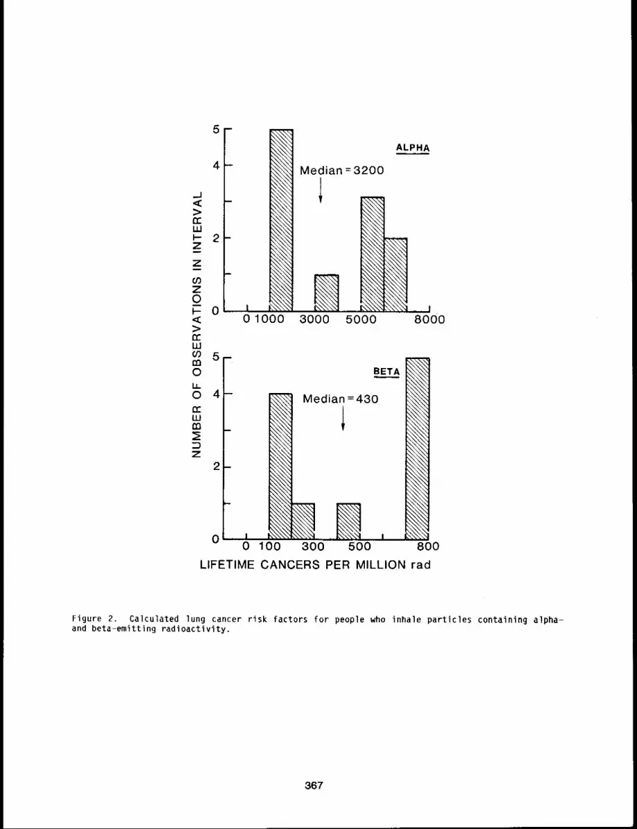
_J< ->¢cWI- 2-ZZ
coz0~- 0,,¢:>n,..w
5-m0LL0 4-
ILlt’n
z 2
ot
.\\\’,~\\\\\\\\\~\\\\\~\\\\\\\\\
,\\\’~
~\\\\
\\\\~
~\\\\\\\\~~
01000
ALPHA
Median =3200
3OOO 50OOI
8OOO
B ET_,,AA
Median =430,,\\\\~
\\\\’,~
.\\\\\
\\\\\
300 500 800
LIFETIME CANCERS PER MILLION rad
Figure 2. Calculated lung cancer risk factors for people who inhale particles containing alpha-and beta-emitting radioactivity.
367
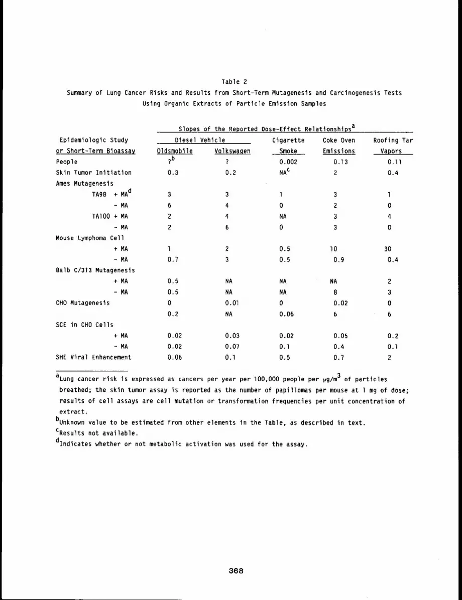
Table 2
Summary of Lung Cancer Risks and Results from Short-Term Mutagenesis and Carcinogenesis Tests
Using Organic Extracts of Particle Emission Samples
Epldemiologic Study
or Short-Term Bioassay
People
Skin Tumor Initiation
Ames Mutagenesis
TAg8 ÷ MAd
- NA
TAIO0 ÷ MA
- MA
Mouse Lymphoma Cell
MA
- MA
Balb C/3T3 Mutagenesis
+ MA
- MA
CHO Mutagenesis
Slopes of the Reported Dose-Effect Relationshipsa
Diesel Vehicle Cigarette Coke Oven Roofing Tar
Oldsmobile Volkswagen Smoke Emissions Vapors?b ? 0,002 0,13 O.ll
0.3 0.2 NAc 2 0.4
3 3 1 3 1
6 4 0 2 0
2 4 NA 3 4
2 6 0 3 0
1 2 0.5 I0 30
0.7 3 0.5 0.9 0.4
0.5 NA NA NA 2
O. 5 NA NA 8 3
0 O.Ol 0 0.02 0
O. 2 NA O. 06 6 6
SCE in CHO Cells
÷ MA 0.02 0,03 0.02 0.05 0.2
- MA 0.02 0.07 O.l 0.4 O.l
SHE Viral Enhancement 0.06 O.l 0.5 0.? 2
aLung cancer risk is expressed as cancers per year per lO0,O00 people per ~g/m3 of particles
breathed; the skin tumor assay is reported as the number of papillomas per mouse at l mg of dose;
results of cell assays are cell mutation or transformation frequencies per unit concentration of
extract,
bunknown value to be estimated from other elements in the Table, as described in text.
CResults not available.
dlndicates whether or not metabolic activation was used for the assay.
368
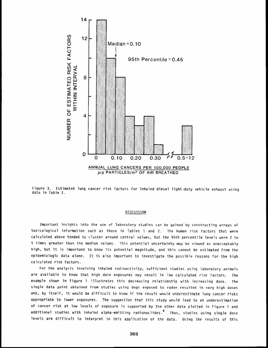
rr>
uJ
<z
,,50n--wm
z
14-
12-
0
\\\\\
\\\\\
\\\\\
\\\\\\\\\\
\\\\\
Median =0.10
95th Percentile =0.45
ANNUAL LUNG CANCERS PER 100,000 PEOPLE~g PARTICLES/m3 OF AIR BREATHED
Figure 3. Estimated lung cancer risk factors for inhaled diesel light-duty vehicle exhaust usingdata in Table 2.
DISCUSSION
Important insights into the use of laboratory studies can be gained by constructing arrays of
toxicological information such as those in Tables l and 2. The human risk factors that were
calculated above tended to cluster around central values, but the 95th percentile levels were 2 to
5 times greater than the median values. This potential uncertainty may be viewed as unacceptably
high, but it is important to know its potential magnitude, and this cannot be estimated from the
epidemiologic data alone. It is also important to investigate the possible reasons for ti~e high
calculated risk factors.For the analysis involving inhaled radioactivity, sufficient studies using laboratory animals
are available to know that high dose exposures may result in low calculated risk factors. The
example shown in Figure 1 illustrates this decreasing relationship with increasing dose. The
single data point obtained from studies using dogs exposed to radon resulted in very high doses
and, by itself, it would be difficult to know if the result would underestimate lung cancer risks
appropriate to lower exposures. The suggestion that this study would lead to an underestimation
of cancer risk at low levels of exposure is supported by the other data plotted in Figure l and
additional studies with inhaled alpha-emitting radionuclides. 4 Thus, studies using single dose
levels are difficult to interpret in this application of the data. Using the results of this
369

single dose level study in dogs in the lung cancer risk factor array in Table 1 leads to the
conclusion that people may be several times more sensitive than dogs to radiation-induced lung
cancer. This probably caused us to substantially overestimate the radiation lung cancer risk
factors for people. To improve this risk factor calculation, it would be useful to do additional
studies of dogs exposed to inhaled radon and of rats exposed to inhaled beta=emitting
radionuclides at doses less than lO00 rad.
The problem of selecting appropriate dose levels for studies aimed at human risk evaluations
and a similar problem related to the potential influence of dose rate as reported by Hahn et
~!. g are transparent in the analysis used for evaluating diesel exhaust particles (Table 2).
Most of the laboratory studies of diesel exhaust particles used in vitro cell culture assays
wherein the exposures are unlike those occurring in people or animals. The quantitative impact of
this difference on calculations of human risk factors is unknown. Albert e t ~!.3 reported that
there was good correspondence between results of the skin tumor initiation assay and measured lung
cancer risks in people exposed to cigarette smoke, coke oven emissions, and roofing tar vapors.
However, all of the skin tumor assays were not conducted in parallel as would be desired to avoid
variability that can result from studies being conducted in different laboratories and at
different times. It is important to continue these comparisons to strengthen our confidence in
the usefulness of short-term bioassay tests for risk assessment purposes.
Finally, from the data in Table 2, we cannot demonstrate that all of the short-term assays
accurately reflect the known differences in lung cancer risks for people exposed to cigarette
smoke, coke oven emissions, and roofing tar vapors. Epidemiologic studies indicate that cigarette
smoke is only l to 2~ as effective in causing lung cancer per unit exposure as the other
emissions, but results of the short term bioassays indicate more similar relative potencies. Part
of this difficulty is due to the often undetectable mutagenic activity in cigarette smoke as
reported for many of the assays. Thus, the short term assays should be conducted to provide a
wider range of sensitivity to the surrogate substances, and appropriate mathematical techniques
must be used to better incorporate zero values into the analysis.
CONCLUSIONS
Constructing arrays of toxicity information on substances with known and potential biological
effects provides a means for evaluating risks from new substances entering our environment.
Laboratory studies using animals and in vitro cell transformation assays can be made a vital part
of risk assessment in the absence of direct epidemiologic information on the exposure-effect
relationships. Laboratory studies are most appropriate for this purpose when the test systems are
applied in parallel studies of both known and suspected harmful agents. Careful attention must
also be given to using dose levels and exposure conditions appropriate to the human exposure
situation.
It may be useful for scientists designing new toxicologic studies aimed at risk evaluation to
develop their initial research plans around toxicity data arrays so that there is a clear
understanding of all of the studies that may be useful to conduct simultaneously and in parallel.
In many cases, as much study or more may be needed with the surrogate substances as with the
primary substance of cancers so that the information gained with new suspected toxic agents can be
related to known human risks.
370
REFERENCES
l ¯ DuMouchel, W. H. and 3. E. Harris, Bayes and Empirical Bayes Methods for Combining CancerExperiments ’in Man and Other Species. Department of Economics, Massachusetts Institute ofTechnology, Technical Report No. 24, pp. 1-51, 1981.
2. Lindley, O. V. and A. F. M. Smith, Bayes Estimates for the Linear Model, J, Royal star. Soc,Series B 34, pp. 1-41, 1972.
3, Albert, R. E., 3. Lewtas, S. Nesnow, I. W. Thorslund, and E. Anderson, Comparative PotencyMethod for Cancer Risk Assessment: Application to Diesel Particulate Emissions, Risk Analysis3: lOl-llT, 1983.
4. Cuddihy, R. G., Risks of Radiation Induced Lung Cancer, in Proceedings of the 17th AnnualMeeting of the National Council Radiation Protection and Measurements, Washington, D. C., pp.133-152, 1982.
5. Gross, P., E. M. Pfitzer, 3. Watson, R. T. P. OeTreville, M. Kaschak, E. B. Tolker, and ~, A.Babyak, Bronchial Intramural Adenocarcinomas in Rats from X-Ray Irradiation of the Chest,Cancer 23: i046-I060, 1969.
6. Chrisp, C. E., R. D. Phemister, A. C. Andersen, L. S. Rosenblatt, and M. Goldman, Pathology inLifespan Study of X=Irradiated Female Beagles, Radiat, Res. 67: 557, Ig76.
7. National Research Council Committee on the Biological Effects of Ionizing Radiations,The Effects on Populations of Exposure to Low Levels of Ionizing Radiation: 1980,National Academy Press, Washington, O. C., Ig8O.
8. Cuddihy, R. G., W. C. Grifflth, and R. O. McClellan, Health Risks from Light Duty OieselVehicles, Environ. Sci. Technol. 18: 14A=21A, 1984.
9. Hahn, F. F., B. B. Boecker, R. G. Cuddihy, C. H. Hobbs, R. O. McClellan, and M. B. Snipes,Influence of Radiation Dose Patterns on Lung Tumor Incidence in Dogs that Inhaled BetaEmitters: A Preliminary Report, Radiat. Res. 96: 505=517, 1983.
371
POTENTIAL HEALTH AND ENVIRONMENTAL EFFECTS OF THE FLUIDIZED BED COMBUSTION OF COAL
FINAL REPORT
Abstract --The potential health and envlronmental
risks for a coal combustion technology based on PRINCIPAL INVESTIGATORS
fluldlzed bed combustlon are sunTmarlzed. The F.A. Seller
larges~ Impacts are projected in the environment, C.H. Hobbs
because of emlsslons of CO2 and acid percursors. R.G. Cudd~h9A major 5eneflt would be the reduction in the
em~sslon of acid precursors because fluldlzed bed combustors (FBC) produce slgnlflcancl9 less than
conventlonal combustors. Few human health effects are projected from effluents of an FBC
industry, but slgnlf~cant occupatlonal and publlc risks are related to its operatlon.
This is a summary of a Health and Environmental EfFects DOcument] on the burning of coal by
fluidized bed combustion. An increase in the use of coal, an increasing fraction of which may be
burned at the low temperature of fluidized bed combustors (FBC), is causing concern about
potential health and environmental effects. The report addresses these concerns, in particular:
...... the use of coal and the FBC market penetration projected for the year 2000;
-- the use of raw materials and the production of effluents and wastes by the industry;
...... the health risks for both workers and the public of operating the power plants and of
mining, transporting, and preparing the raw materials and disposing of the wastes;
.... the environmental levels of effluents and waste products;
-- the resulting damages to human health, the environment, and materials.
The report does not cover special exposure situations resulting in increased risks for a few
individuals or a few localized ecosystems.
Projected Coal and FBC Industries
A projection of l billion tonnes* per year in the year 2000 is chosen, affording relatively
easy scaling to other levels of coal use. The ratio of surface to underground mining is assumed
to increase to roughly 2:1, and the contribution of western coal fields to total production to
about 50%.
The market share for the future FBC industry is projected for the year 2000 by making use of
market penetration models for the energy sector. Assuming further restrictions on SO2 and NOxemissions, a share of 5% of the coal combustion market may be attained. This results in an annual
coal use of 43 million tonnes and a production of lO0 billion kWh of useful energy.
The annual use of raw materials and the production of wastes are estimated mainly on the basis
of the performance of a single low-power FBC plant at Georgetown University. The values for the
annual input and output of the projected FBC and an equivalent pulverized coal combustion industry
with flue--gas desulfurization (PCC/FGD) are listed in Table
Notable differences between the two methods of coal combustion are the FBC’s larger need for
limestone and thus the larger production of solid wastes, as well as its projected higher emission
of gaseous hydrocarbons. On the other hand, the equivalent PCC/FGD industry emits more SO2because of the lower reliability of the FGD system, about five times more nitrogen oxides, and
nine times more CO. Both methods cause the production of the same quantitity of CO2 and the
mobilization of similar amounts of trace elements through the mining of raw materials,
*I tonne = 1 metric ton ~ 1,000 kg.
372
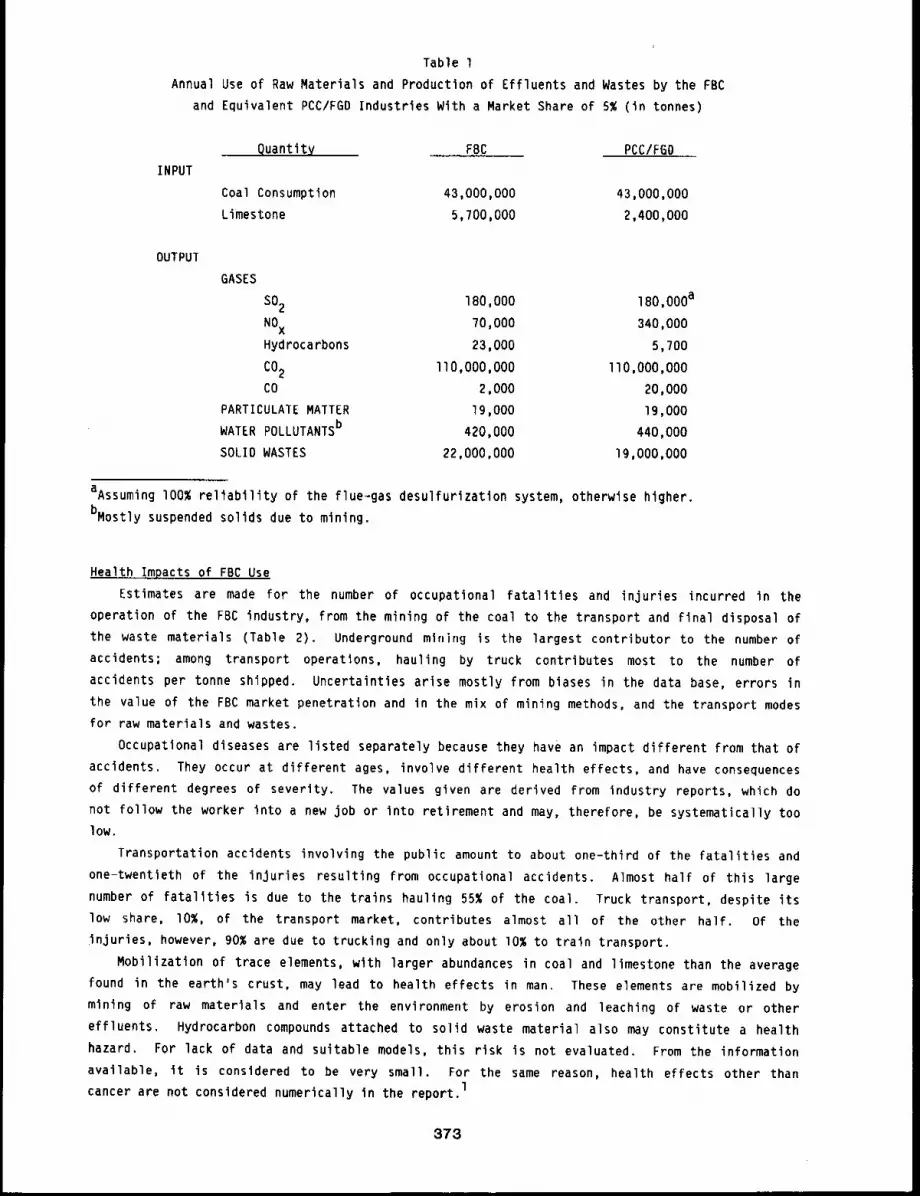
Table l
Annual Use of Raw Materials and Production of Effluents and Wastes by the FBC
and Equivalent PCC/FGD Industries With a Market Share of 5% (in tonnes)
INPUT
Quantity FBC PCC/FBO
Coal Consumption 43,000,000 43,000,000
Limestone 5,?00,000 2,400,000
OUTPUT
GASES
SO2 IBO,O00 180,000a
NOX70,000 340,000
Hydrocarbons 23,000 5,700
CO2 llO,O00,O00 llO,O00,O00
CO 2,000 20,000
PARTICULATE MATTER 19,000 Ig,O00
WATER POLLUTANTSb 420,000 440,000
SOLID WASTES 22,000,000 19,000,000
aAssuming I00% reliability of the flue~gas desulfurization system, otherwise higher.
bMostly suspended solids due to mining.
Health Impacts of FBC Use
Estimates are made for the number of occupational fatalities and injuries incurred in the
operation of the FBC industry, from the mining of the coal to the transport and final disposal of
the waste materials (Table 2). Underground m|nlrlg is the largest contributor to the number
accidents; among transport operations, hauling by truck contributes most to the number of
accidents per tonne shipped. Uncertainties arise mostly from biases in the data base, errors in
the value of the FBC market penetration and in the mix of mining methods, and the transport modes
for raw materials and wastes.
Occupational diseases are listed separately because they have an impact different from that of
accidents. They occur at different ages, involve different health effects, and have consequences
of different degrees of severity. The values given are derived from industry reports, which do
not follow the worker into a new job or into retirement and may, therefore, be systematically too
low.
Transportation accidents involving the public amount to about one-third of the fatalities and
one-twentieth of the injuries resulting from occupational accidents. Almost half of this large
number of fatalities is due to the trains hauling 55% of the coal. Truck transport, despite its
low share, I0%, of the transport market, contributes almost all of the other half. Of the
injuries, however, 90% are due to trucking and only about I0% to train transport.
Mobilization of trace elements, with larger abundances in coal and limestone than the average
found in the earth’s crust, may lead to health effects in man. These elements are mobilized by
mining of raw materials and enter the environment by erosion and leaching of waste or other
effluents, Hydrocarbon compounds attached to solid waste material also may constitute a health
hazard. For lack of data and suitable models, this risk is not evaluated. From the information
available, it is considered to be very small. For the same reason, health effects other than
cancer are not considered numerically in the report,l
373
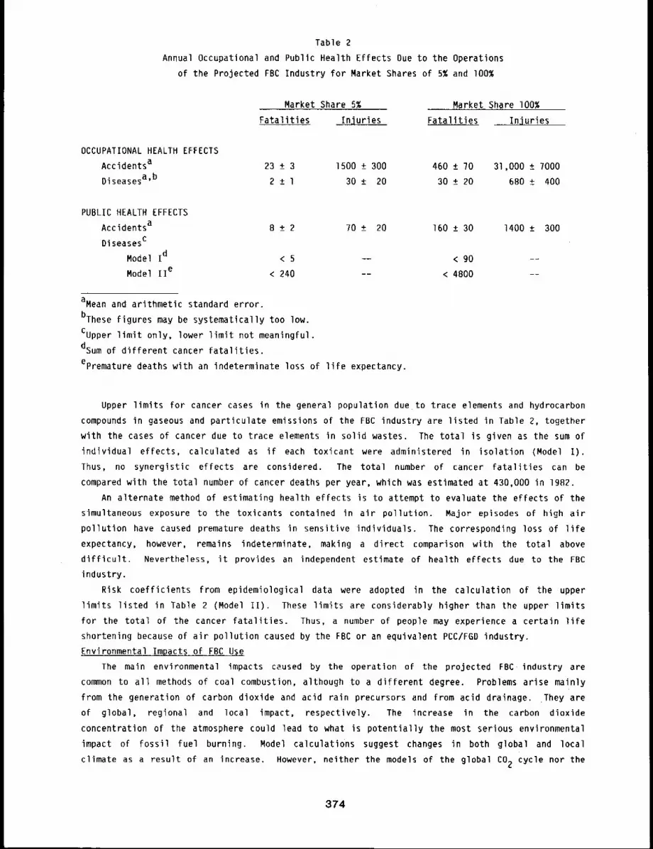
Table 2
Annual Occupational and Public Health Effects Due to the Operations
of the Projected FBC Industry for Market Shares of 5% and 100%
Market Share 5% Market Share I00%
Fatalities Injuries Fatalities Injuries
OCCUPATIONAL HEALTH EFFECTS
Accidentsa 23 ± 3 1500 ± 300 460 ± ?0 31,000 ± 7000
Diseasesa’b 2 ± l 30 ± 20 30 ± 20 680 ± 400
PUBLIC HEALTH EFFECTS
Accidentsa 8 ± 2 70 ± 20 160 ± 30
Diseasesc
Model Id < 5 -- < go
Model IIe < 240 -- < 4800
1400 ± 300
aMean and arithmetic standard error.
bThese figures may be systematically too low.
Cupper limit only, lower limit not meaningful.
dsum of different cancer fatalities.
epremature deaths with an indeterminate loss of life expectancy.
Upper limits for cancer cases in the general population due to trace elements and hydrocarbon
compounds in gaseous and particulate emissions of the FBC industry are listed in Table 2, together
with the cases of cancer due to trace elements in solid wastes. The total is given as the sum of
individual effects, calculated as if each toxicant were administered in isolation (Model I).
Thus, no synergistic effects are considered. The total number of cancer fatalities can be
compared with the total number of cancer deaths per year, which was estimated at 430,000 in 1982.
An alternate method of estimating health effects is to attempt to evaluate the effects of the
simultaneous exposure to the toxicants contained in air pollution. Major episodes of high air
pollution have caused premature deaths in sensitive individuals. The corresponding loss of life
expectancy, however, remains indeterminate, making a direct comparison with the total above
difficult. Nevertheless, it provides an independent estimate of health effects due to the FBC
industry.
Risk coefficients from epidemiological data were adopted in the calculation of the upper
limits listed in Table 2 (Model If). These limits are considerably higher than the upper limits
for the total of the cancer fatalities. Thus, a number of people may experience a certain life
shortening because of air pollution caused by the FBC or an equivalent PCC/FGD industry.
Environmental Impacts of FBC Use
The main environmental impacts caused by the operation of the projected FBC, industry are
common to all methods of coal combustion, although to a different degree. Problems arise mainly
from the generation of carbon dioxide and acid rain precursors and from acid drainage. They are
of global, regional and local impact, respectively. The increase in the carbon dioxide
concentration of the atmosphere could lead to what is potentially the most serious environmental
impact of fossil fuel burning. Model calculations suggest changes in both global and local
climate as a result of an increase. However, neither the models of the global CO2 cycle nor the
374
climate models are founded well enough to make valid predictions at this time. Acid depositions
are a more regional problem, affecting northeastern North America, Central Europe, and
Scandinavia. They cause damage to fauna and flora of aquatic ecosystems, may lead to increasedacidity in terrestrial ecosystems and damage forests, other plants, and materials exposed to
them. With increasing acidification, lakes and rivers can no longer support a fish population,
while forests first stagnate, then decline and die. Losses caused by corrosion alone are
estimated to amount from billions of dollars to tens of billions of dollars annually. Losses to
agriculture, forests, and buildings are mo,’e difficult to estimate and are not evaluated in the
report.
Acid drainage arises from water that percolates through coal mines, mine tailings piles, coal
storage piles, and waste deposits. It leaches sulfuric acid, salts, metals, and other trace
elements. The damages are local and restricted essentially to the aquatic system into which the
leachwaters drain. The effects are practically the same as those caused by acid depositions. In
addition, the high trace element content may cause some local problems by biological enrichment,
but data are insufficient for an analysis.Emissions of SO2 and NOx can lead to direct damage to foliage downwind of the power
plants. Nitrogen oxides also contribute to the chemical reactions in the polluted atmosphere in
which oxidants such as ozone and Peroxyacetyl nitrate (PAN) are formed. Whereas PAN should be problem only in the Southwest, 1 ozone will cause damages nationwide. Crop losses caused by
ozone alone are estimated to cost on the order of $I0 billion annually.
Environmental impacts of coal transport are small, except near transfer and terminal
stations. There, appreciable local damages are possible because of the thick cover of coal dust.
Normally the trace elements in the coal dust are not considered to he an appreciable hazard.
Serious impacts on local environments may occur near burning mines and mine railings piles. Over
longer periods, severe acidification may set in, even in well-buffered soils.
The environmental impacts mentioned above are essentially the same for the two coal combustion
methods considered for the year 2000. The notable exceptions are the effects of the emisslons of
SO2 and NOx. Here the use of FBC over PCC/FGD represents a reduction by an indeterminatefactor for SO2, a factor )f about five for NOx, and an unknown factor for ozone (Table ~).This will be reflected in a decrease of acid deposition and of damages by ozone.
CONCLUSIONS
Analysis of the health and environmental risks from operations of the projected FBC industry
yields an evaluation of thF main areas of concern.
-- Possible climatic changes due to the buildup of CO2 in the atmosphere are potentially
the most serious and far-reaching of all environmental impacts. The CO2 production isindependent of the combustion method, but burning other fossil fuels generates less, and
some alternate energy carriers produce none.
. - Acid deposition in the environment can seriously damage aquatic and terrestrial
ecosystems. Although coal combustion’s share in these damages is not clear at present,
selecting FBC over PCC/FGD will reduce that share.
-- Occupational fatalities and injuries are sizeable and directly reflect the hazards of
mining and transporting coal and limestone. The transport of these raw materials also
involves the public to a considerable extent.
--- Occupational diseases due to the inhalation of coal dust are projected to decrease with
the lower dust levels in current underground mines and with the shift of production to
surface mines.
375
-- On the average, trace elements mobilized by the mining of coal and limestone will have
few health and environmental effects.
Public health effects from industrial effluents are projected to be small, particularly
in comparison with those occurring at present.
REFERENCE
I. Seller, F. A., C. H. Hobbs, and R. G. Cuddihy, Potential Health and Environmental Effects ofthe Fluidized Bed Combustion of Coal - Final Report, Inhalation Toxicology Research Institute,Lovelace Biomedical and Environmental Research Institute, LMF-I06, UC-48, 1983.
376
HEALTH RISKS FROM THE DISPOSAL OF SOLID FBC WASTES IN THE ENVIRONMENT
Abstract --Estimates are made for potential health
hazards from deposlClon of solid flu141zed bed com- PRINCIPAL INVESTIGATOR
bustor wastes in the environment. Environmental F.A. Seller
d11utlon factors are estimated on ~he basis of mod-
el assumptlons and measuremonts of equillhrlum leachwater concentrations, Upper llmlts for annual
cancer faCalltles from drinking water contamination wlth trace elemonEs are given. No estlmates
for endpolnts other than cancer or for leached hydrocarbon compounds are made.
Solid wastes originate from mining preparation and combustion of coal. In coal mining, large
quantities of overburden and waste materials are deposited temporarily or permanently in waste
piles. They are exposed to erosion and leaching by rainwater and sometimes by streams. In
addition, about half the coal mined in the United States is treated to remove rocks and mineral
constituents. These twosources generate increasing piles of carbonaceous mineral wastes, which
have been estimated to amount to nearly 3 billion tonnes.*
In regions with a high content of pyritic sulfur, the main problem is drainage of sulfuric
acid, which originates not only in the tailing piles but also in the mine itself, wherever
surfaces are exposed to air and water. From 1960 to 1970, inactive underground mines were
responsible for more than half of the sulfuric acid effluents, with active underground mines
contributing almost another Quarter.l
In western U. S., where both coal and overburden are low in pyritic sulfur and have a high
content of alkaline compounds, acid or alkaline drainage is not a severe problem. In these
regions, the leaching of trace elements is not prevalent because of the low acidity or even
alkalinity of waters percolating through mines and waste piles. In eastern and mid-western coal
fields, however, the high acidity of the leachwater (pH values from 2.8-5) and the resulting high
trace metal content may cause additional problems.
The solid combustion wastes generated by fluidized bed combustor (FBC) power plants (fly ash,
bottom ash and bed residue) are slightly corrosive and reactive because of their lime content.
These wastes are mostly disposed of in sanitary landfills, but a small amount will be used as soll
conditioner or converted into construction materials. Landfills and reclaimed mining areas
properly covered with a layer of soil will present no inhalation risk. Water, however, will
percolate through soil cover and wastes, and will leach toxic trace elements and compounds
according to its pH value. If these migrate through the environment and reach man, health effectslmay result. In a more extensive report, these effects are estimated for an FBC industry with
shares of 5~ and I00% of the coal combustion market in the United States.
Leaching Characteristics of Solid Wastes
A thorough in vitro study of leaching from a large number of FBC waste samples was conducted
by Sun and Peterson, 2 Equilibrium worst-case leachwater samples are found to have no odor or
color, and do not contain any foaming agents. In addition, they meet the drinking water standards
for the metals Ag, As, Ba, Cd, Cr, Hg, Pb, and Se, as well as those for fluorides and nitrates.
l tonne = l metric ton = lO00 kg.
377
Concentrations of calcium, sulfates, total dissolved solids, and hydrogen ions (pH value) exceed
drinking water standards. Dilution by factors between 2 and 40 is needed to bring the runoff
water to compliance, l Although health hazards could still arise from some enrichment mechanisms
in the environment, no such mechanism of significance has been found.
Leachwater from mine tailings and coal preparation wastes can have quite different
compositions and acidities from FBC leachate, particularly in high sulfur coal mines. With the
shift toward surface mining and western coal fields and with drainage control technologies
proposed for Future mining operations, these differences should become less important on a
national level. Indeed, with co-disposal of alkaline agents such as lime, the leachwater
composition is comparable to that of FBC leachate, and the different forms of solid waste will,
therefore, be treated the same as FBC wastes.
Generation and Transport of Leachwater
Some simplifying assumptions are made for the calculation of the source term of toxic
compounds leached from solid wastes nationwide. First, all waste deposits are assumed to be
covered with soil and plants. Total evapotranspiration losses from the soil surface and from
plants will reduce the rain water available for leaching by an average factor of about two.1
Second, water reaching the deposit is allowed to reach equilibrium concentrations of toxicants
while percolating through the deposit. 2 Also, for groundwater runoff, an equilibrium flow is
assumed to exist for transport of trace elements to a point of exfiltration into surface waters or
to a groundwater well. Third, for surface runoff of the leachate, it is assumed that not only
rivers but also lakes reach equilibrium concentrations. Fourth, it is assumed that suppliers
whose water is derived from ground water (40%) or surface water (60%) will deliver their water
without further processing.
In the operation of a reference power plant rated at lO00 MW(e), 1.9 million tonnes of solid
waste are produced annually. Over its lifetime of 30 years, 57 million tonnes are accumulated.
At an average density of 2.5 tonnes/m 3 this amounts to 23 million m3 of waste, and assuming a
low average height of I0 m, the deposit will cover an area of 2.3 million m2. The average
rainfall of 0.73 m will produce 830,000 tonnes of leachwater annually (0.5 x 0.73 x 2.3 million
m3 at l tonne/m3). For the projected FBC industry with a 5% market ~hare, the amount is 9.5
million tonnes; for a market share of I00% it is 190 million tonnes.
Environmental Dilution
On a nationwide average, it is assumed that dilution in surface and in underground waters is
the same. An upper limit for the dilution factor is the ratio between the water falling on the
area of the U. S. and the water falling on the waste deposits, i.e., the ratio of the areas. Itsvalue, 300,000 ("1,870,000 km2/(ll.4 x 2.3 km2)), is very high, reflecting the Fact
complete, unimpeded mixing of all the rainwater in the U. S. is assumed. For an FBC market share
of I00%, this dilution factor would be 20 times lower, or’ 15,000.A more realistic value is obtained by estimating the probability of rainwater that remains on
or in the greund, becoming part of the drinking water. The annual rainfall in the U. S. is 5./5
trillion tonnes (7,870,000 km2 x 0.73 m at 1 tonne/m3), of which 38% - or 2.2 trillion tonnes1-- remain on or in the ground. The anmLal water consumption of 37 billion tonnes is derived
from this amount of rain water. Less than 0.5% of this quantity, about 180 million tonnes (2liters/day x 365 days/year x 250 million), will be used for drinking, cooking, and food
1preparation. The dilution factor is, therefore, 12,000 (2.18 trillion tonnes/180 million
tonnes). Here unimpeded mixing is assumed only over an area comprising the source of the
pollutant and the drinking water supply.Both of the estimates above, however, ignore the geometry of watersheds and rivers. For small
watersheds, Hack3 derived an empirical relationship between the area of the watershed and the
378
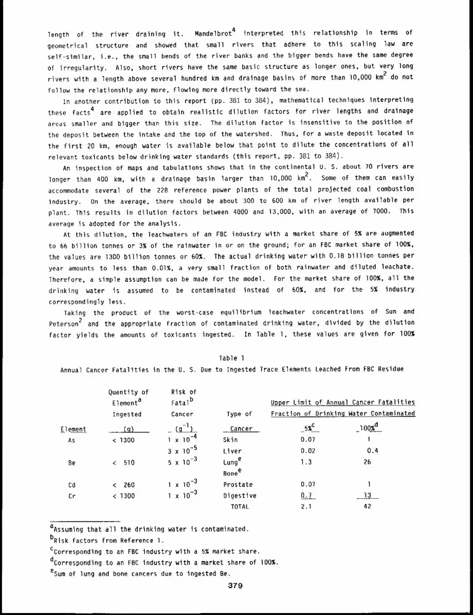
length of the river draining it. Mandelbrot 4 interpreted this relationship in terms of
geometrical structure and showed that small rivers that adhere to this scaling law are
self-similar, i.e., the small bends of the river banks and the bigger bends have the same degree
of irregularity. Also, short rivers have the same basic structure as longer ones, but very long
rivers with a length above several hundred km and drainage basins of more than lO,O00 km2 do not
follow the relationship any more, flowing more directly toward the sea.
In another contribution to this report (pp. 381 to 384), mathematical techniques interpreting
these facts 4 are applied to obtain realistic dilution factors for river lengths and drainage
areas smaller and bigger than this size. The dilution factor is insensitive to the position of
the deposit between the intake and the top of the watershed. Thus, for a waste deposit located in
the first 20 km, enough water is available below that point to dilute the concentrations of all
relevant toxicants below drinking water standards (this report, pp. 381 to 384).
An inspection of maps and tabulations shows that in the continental U. S. about 70 rivers are
longer than 400 km, with a drainage basin larger than lO,O00 km2. Some of them can easily
accommodate several of the 228 reference power plants of the total projected coal combustion
industry. On the average, there should be about 300 to 600 km of river length available per
plant. This results in dilution factors between 4000 and 13,000, with an average of 7000. This
average is adopted for the analysis.
At this dilution, the leachwaters of an FBC industry with a market share of 5% are augmented
to 66 billion tonnes or 3% of the rainwater in or on the ground; for an FBC market share of I00%,
the values are 1300 billion tonnes or 60%. The actual drinking water with O.IB billion tonnes per
year amounts to less than 0.01%, a very small fraction of both rainwater and diluted leachate.
Therefore, a simple assumption can be made for the model. For the market share of I00%, all the
drinking water is assumed to be contaminated instead of 60%, and for the 5% industry
correspondingly less.
Taking the product of the worst-case equilibrium leachwater concentrations of Sun and
Peterson 2 and the appropriate fraction of contaminated drinking water, divided by the dilution
factor yields the amounts of toxicants ingested. In Table l, these values are given for I00%
Table l
Annual Cancer Fatalities in the U. S. Due to Ingested Trace Elements Leached From FBC Residue
Element
As
Be
Cd < 260 l x 10-3 0.07 l
Cr < 1300 l x lO-3 0.7 13
2 .l 42
Quantity of Risk of
Elementa Fatalb Upper Limit of Annual Cancer Fatalities
Ingested Cancer Type of Fraction of Drinkinq Water Contaminated
(q) _ (g-l) Cancer 5%c I00%d
< 1300 l x lO-4 Skin 0.07 l
3 x lO"5 Liver 0.02 0.4
< 510 5 x 10⁻³ Lunge 1.3 26
Bonee
Prostate
Digestive
TOTAL
aAssuming that all the drinking water is contaminated.
bRisk factors from Reference I.
Ccorresponding to an FBC industry with a 5% market share.
dcorresponding to an FBC industry with a market share of i00%.
esum of lung and bone cancers due to ingested Be.
879
contaminated drinking water. With the risk factors discussed in the Health and Environmental
Effects document, l the number of annual cancer fatalities can be calculated for each trace
element. The standard geometric error of these values can be approximated from the two main
sources of error, the risk coefficients and the dilution factor, resulting in a total geometric
standard error of 3.2. Upper limits are, therefore, a factor of lO higher than the mean. In
Table l, the upper limits are given for 5% and 100% of the drinking water contaminated with the
diluted concentrations. For the assumptions made above, this corresponds also to the upper limits
for the FBC industries with 5% and I00% market share.
Not taken into account in this analysis are health effects other than cancer and those caused
by the ingestion of organic compounds. For health effects other than cancer, the data are
insufficient to allow the determination of numerical risk factors. For organic compounds, there
is not only a dearth of risk factors, but also very little information on the actual
concentrations of particular compounds in the solid wastes. An assessment is therefore not made
in either case.
REFERENCES
I. Seiler, F. A., C. H. Hobbs, and R. G. Cuddihy, Potential Health and Environmental Effects ofthe Fluidized Bed Combustion of Coal - Final Report, Inhalation Toxicology Research Institute,Lovelace Biomedical and Environmental Research Institute, LMF-I06, UC-48, lgB3.
2. Sun, C, C. and C. H. Peterson, Fluidized Bed Combustion Residue Disposal: EnvironmentalImpact and Utilization, pp. 900-912 of U. S. Department of Energy Proceedings of the SixthInternational Conference on Fluidized Bed Combustion, CONF-BO0428, IgBO.
3. Hack, 3. T., Studies of Longitudinal Stream Profiles in Virginia and in Maryland, U. S.Geological Survey, Prof. Paper 294-B, 1957.
4. Mandelbrot, B. B., !he Fractal Geometry. of Nature, Freeman, San Francisco, 1982.
380
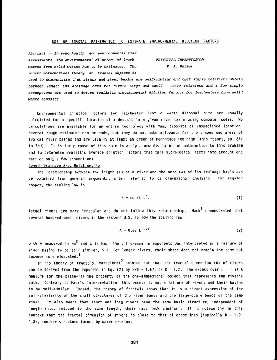
USE OF FRACTAL MATHEMATICS TO ESTIMATE ENVIRONMENTAL DILUTION FACTORS
Abstract -- In some health and envlronmental risk
assessments, the environmental dllurlon of leach- PRINCIPAL INVESTIGATOR
waters from solid wastes has to be estlmated. The F.A. Seller
recent mathematlcal theory of fractal objects is
used to demonstrate that rivers and flyer baslns are self-slmllac and that s~mple relatlons obCaln
between length and drainage area for rivers large and small. These relations and a few s]mple
assumptions are used to derive reallstlc environmental d11utlon factors for leachwaters from solld
waste deposits.
Environmental dilution factors for leachwater from a waste disposal site are usually
calculated for a specific location of a deposit in a given river basin using computer codes. No
calculations are available for an entire technology with many deposits of unspecified location.
Several rough estimates can be made, but they do not make allowance for the shapes and areas of
typical river basins and are usually at least an order of magnitude too high (this report, pp. 377
to 380)° It is the purpose of this note to apply a new discipline of mathematics to this problem
and to determine realistic average dilution factors that take hydrological facts into account and
rest on only a few assumptions.
Lenqth-Drainage Area Relationship
The relationship between the length (L) of a river and the area (A) of its drainage basin
be obtained from general arguments, often referred to as dimensional analysis. For regular
shapes, the scaling law is
A = const L2. (I)
Actual rivers are more irregular and do not follow this relationship. HackI demonstrated that
several hundred small rivers in the eastern U.S. follow the scaling law
A = 0.67 L1"67, (2)
with A measured in km2 and L in km. The difference in exponents was interpreted as a failure of
river basins to be self-similar, i.e. for longer rivers, their shape does not remain the same but1becomes more elongated.
In his theory of fractals, Mandelbrot 2 pointed out that the fractal dimension (D) of rivers
can be derived from the exponent in Eq. (2) by 2/D = 1.67, or D = 1.2. The excess over D = 1 is
measure for the plane-filling property of the one-dimensional object that represents the river’s
path. Contrary to Hack’s interpretation, this excess is not a failure of rivers and their basins
to be self-similar. Indeed, the theory of fractals shows that it is a direct expression of the
self-similarity of the small structures of the river banks and the large-scale bends of the same
river. It also means that short and long rivers have the same basic structure, independent of
length (i.e. reduced to the same length, their maps look similar). It is noteworthy in this
context that the fractal dimension of rivers is close to that of coastlines (typically D = 1.2-
1.3), another structure formed by water erosion.
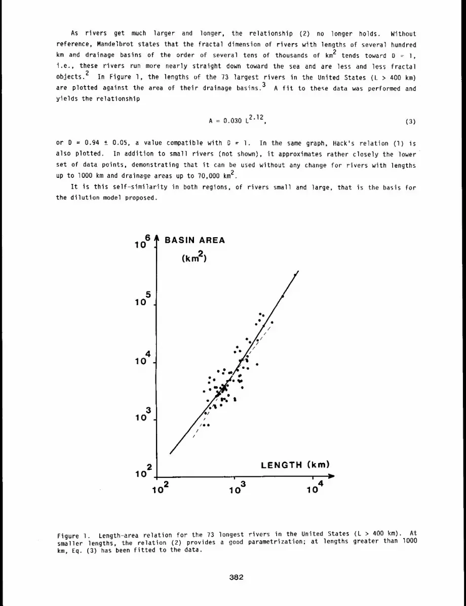
As rivers get much larger and longer, the relationship (2) no longer holds. Without
reference, Mandelbrot states that the fractal dimension of rivers with lengths of several hundred
km and drainage basins of the order of several tens of thousands of km2 tends toward D = l,
i.e., these rivers run more nearly straight down toward the sea and are less and less fractal
objects.2 In Figure l, the lengths of the 73 largest rivers in the United States (L > 400 km)
are plotted against the area of their drainage basins.3 A fit to the~e data was performed and
yields the relationship
A = 0.030 L2"12, (3)
or D = 0.94 ± 0.05, a value compatible with D = I. In the same graph, Hack’s relation (I)
also plotted. In addition to small rivers (not shown), it approximates rather closely the lower
set of data points, demonstrating that it can be used without any change for rivers with lengths
up to I000 km and drainage areas up to 70,000 km2.
It is this self-similarity in both regions, of rivers small and large, that is the basis for
the dilution model proposed.
610
510
410
310
210
1 02
BASIN AREA
¯ /i ¯ VQI/
eZe
, e" ~l ¯
~ //iI
LENGTH (km)
3 410 10
Figure 1. Length-area relation for the 73 longest rivers in the United States (L > 400 km). smaller lengths, the relation (2) provides a good parametrization; at lengths greater than lO00km, Eq. (3) has been fitted to the data.
382
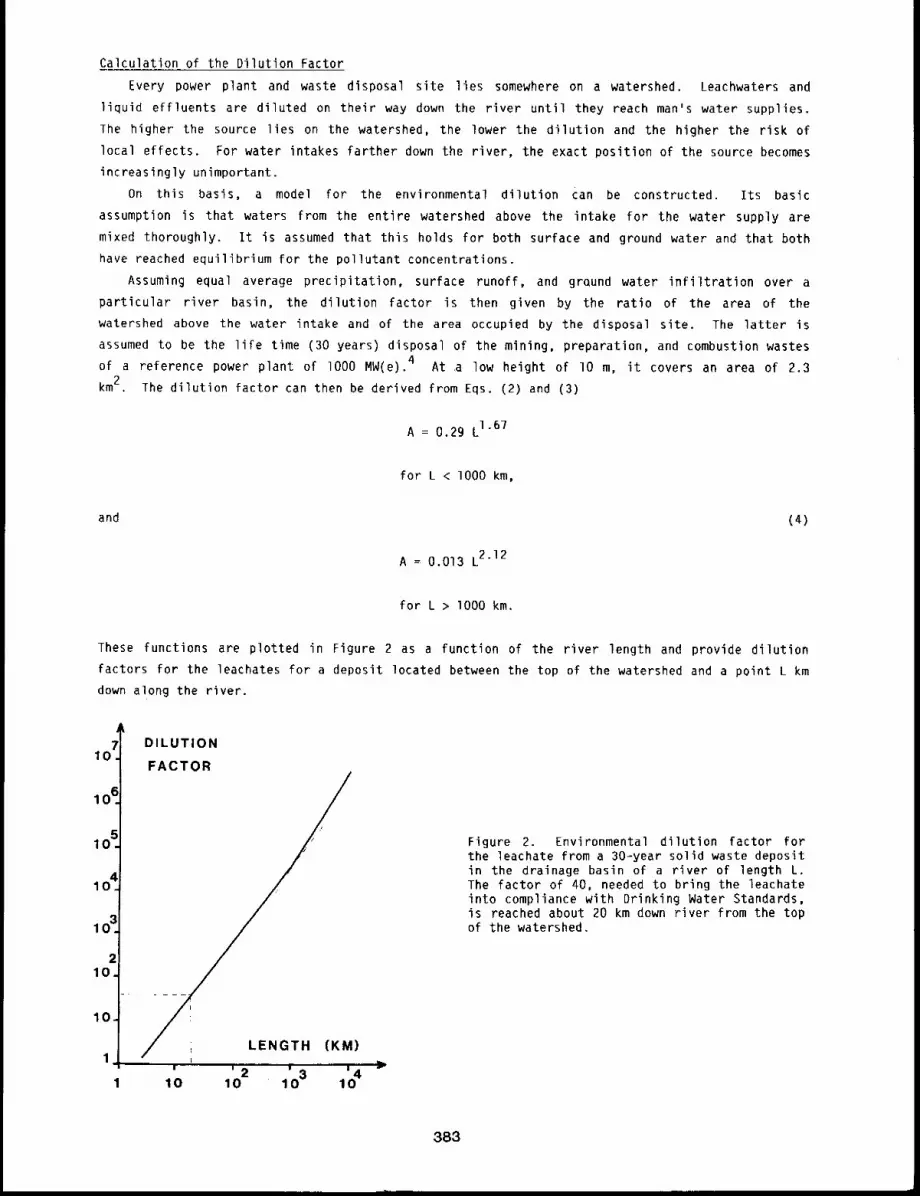
Calculation of the Dilution Factor
Every power plant and waste disposal site lies somewhere on a watershed. Leachwaters and
liquid effluents are diluted on their way down the river until they reach man’s water supplies.
lhe higher the source lies on the watershed, the lower the dilution and the higher the risk of
local effects. For water intakes farther down the river, the exact position of the source becomes
increasingly unimportant.
On this basis, a model for the environmental dilution Can be constructed. Its basic
assumption is that waters from the entire watershed above the intake for the water supply are
mixed thoroughly. It is assumed that this holds for both surface and ground water and that both
have reached equilibrium for the pollutant concentrations.
Assuming equal average precipitation, surface runoff, and grQund water infiltration over a
particular river basin, the dilution factor is then given by the ratio of the area of the
watershed above the water intake and of the area occupied by the disposal site. The latter is
assumed to be the life time (30 years) disposal of the mining, preparation, and combustion wastes
of a reference power plant of lO00 MW(e).4 At .a low height of lO m, it covers an area of 2.3
km2. The dilution factor can then be derived from Eqs. (2) and (3)
A = 0.29 L167
for L < I000 km,
and (4)
A = 0.013 L212
for L > lO00 km.
These functions are plotted in Figure 2 as a function of the river length and provide dilution
factors for the leachates for a deposit located between the top of the watershed and a point L km
down along the river.
DILUTION
FACTOR
;//
}
¯ " ’410 10 2 1 ’0 3 10
Figure 2. Environmental dilution factor forthe leachate from a 30-year solid waste depositin the drainage basin of a river of length L.The factor of 40, needed to bring the leachateinto compliance with Drinking Water Standards,is reached about 20 km down river from the topof the watershed.
383

REFERENCES
I. Back, J. ~., Studies of Longitudinal Stream Profiles in Virginia and in Maryland, U. S.Geological Survey, Prof. Paper 294-B, 1957.
2. Mandelbrot, B. B., The Fractal Geometry of Nature, Freeman, San Francisco, 1982.
3. Showers, V., The World in Figures, J. Wiley & Sons, New York, 1973.
4. Seiler, F. A., C. H. Hobbs, and R. G. Cuddihy, Potential Health and Environmental Effects ofthe Fluidized Bed Combustion of Coal - Final Report, Inhalation Toxicology Research Institute,Lovelace Biomedical and Environmental Research institute, LMF-I06, UC-48, 1983.
384
APPENDIX A
STATUS OF LONGEVITY AND SACRIFICE EXPERIMENTS IN BEAGLE DOGS
Data in this appendix relate to (a) total body and lung content of radionuclides and (b)
resultant radiation dose received by individual dogs that have been assigned to longevity or
sacrifice studies. The data are presented as a reference source for scientists in this laboratory
and for scientists in other laboratories who desire to follow in detail the progress of these
studies. The data represent the best information available, and it must be noted that, with time,
certain values and diagnoses will be modified and updated as new and better information becomes
available.
RADIOACTIVITY CONTENT
Initial body burden (IBB) is defined as the best current estimate of the total radionuclide
content within the body immediately after an inhalation exposure or intravenous injection.
Long-term retained burden (LTRB) is defined as the best current estimate of the amount
radionuclide remaining in the body after early clearance of the nasopharyngeal and tracheobronchial
regions via the gastrointestinal tract. The term is used in these tables to describe the type of
body burden resulting from inhalation of a radionuclide in a relatively soluble form. Because
absorption could occur from the gastrointestinal tract as well as the lung, the magnitude of the
long=term retained burden after inhalation of a radionuclide in a relatively soluble form bears a
relationship to the amount of radionuclide deposited in the entire respiratory tract and not just
the fraction deposited in the pulmonary region.
Initial lung burden (ILB) is defined as the long-term retained burden associated with the
inhalation of relatively insoluble particles. In this case, essentially all of the body burden
remaining after early clearance of the nasopharyngeal and tracheobronchial regions is in the
pulmonary region.
CLINICOPATHOLOGICAL FEATURES
Comments are tabulated for the current interpretation of the most prominent clinicopathologi-
cal features associated with the death of animals. It should be recognized that many animals have
multiple tumors or other lesions, not all of which can be listed in a summary table. They are
discussed in greater detail in the text of this and preceding reports and in open literature
publications.
RADIATION DOSE CALCULATIONS
The methods used in establishing the radiation dose parameters presented have been described
in the text of the report or referenced to previous reports. A key consideration in these
calculations is the tissue weight because the absorbed dose is inversely proportional to tissue
weight. Tissue weights used for the dose values reported in Appendix A have changed over the
years; it is important that the reader be aware of these changes and the rationale behind them.
Lung weights used in the earliest reported dose calculations (1966-1967 Annual Report, LF-38,
pp. 19-64 and 1967-1968 Annual Report, LF-39, pp. 14=75) were based on a (lung weight)/(body
weight) ratio of 0.0075 determined from tissue weights from exsanguinated dogs. This ratio was
changed to 0.014 in the 1968=1969 Annual Report (LF-41, pp. 27-28), based on calculations of the
estimated weight of lung with its normal complement of blood in the living dog. Subsequent ex-
perimental evidence reported in the 1971-1972 Annual Report (LF-45, pp. I19=128) indicated that
this value was too high. Based on these results, our best estimate of the (lung weight (with
blood))/(body weight) ratio O.Oll. Thi s val ue has beenused for a ll d ose calcu lations for d og
lungs in all Annual Report Appendices, beginning with those in the 1972-1973 Annual Report, LF-46.
385
Liver weights used in early reports were calculated using a (liver weight)/(body weight) ratio
of 0.027, which was based on tissue weights From exsanguinated dogs. It was used For dose calcu-
lations in all reports through the 197i-1972 Annual Report, LF-45. Based on experimental data
presented in LF-45, the best estimate for the (liver weight (with blood))/(body weight) ratio
0.050. This value has been used For all dose calculations for dog liver beginning with the 1972-
1973 Annual Report, LF-46.
Skeleton weights have always been calculated on the basis of a (skeleton weight)/(body weight)
ratio of O.lO.
Tracheobronchial lymph nodes weights are based on a (tracheobronchial lymph node weight)/(body
weight) ratio of 0.00005.
I. gOSrCl 2, Longevity Dogs ...............................3B7
2. 90SrCl2, Sacrifice Dogs ................. , ............. 3893. 144CeCl 3, Longevity Dogs .............................. 3904 91yCl3, Longevity Dogs¯ ¯ .............................. 3925. 91yCl~, Sacrifice Dogs
137 ~ ................. ..............393
6 CsCI, Longevity Dogs" ¯ .............................. 3947. gOy in Fused Aluminosilicate Particles, Longevity Dogs ...............
3968. gIy in Fused Aluminosilicate Particles, Longevity Dogs ................
3989. 144Ce in Fused Aluminosilicate Particle~ Longevity Dogs (Series I) .........
400I0. 144Ce in Fused Aluminosilicate Particles
II. 144Ce in Fused Aluminosilicate Particles
12. 144Ce in Fused Aluminosilicate Particles
13. 144Ce in Fused Aluminosilicate Particles
Longevity Dogs (Series II) ........ 401
Sacrifice Dogs (Series II, Ill and IV) . 403
Immature Longevity Dogs .......... 405
Immature Sacrifice Dogs .......... 40614. 144Ce in Fused Aluminosilicate Particles Aged Longevity Dogs ............
40715. 90Sr in Fused Aluminisilicate Particles, Longevity Dogs ................
40816. 144Ce in Fused Aluminosilicate Particles, Repeatedly Exposed Dogs ..........
41117. 238pu0 Monodisperse
18. 238pu02 Monodisperse
239~ ^219. vuu2 Monodisperse20. 239pu0 Monodisperse
239~ 221. vuu2 Monodisperse
22. 239pu02 Monodisperse
23. 239pu02 Monodisperse
24. 239pu02 Monodisperse
Aerosol (I.5 #m AMAD), Longevity Dogs ............. 412Aerosol (3.0 #m AMAD), Longevity Dogs ............. 414Aerosol (0.75 ~m AMAD), Longevity Dogs ............. 416Aerosol (1.5 #m AMAD), Longevity Dogs ............. 417Aerosol (3.0 #m AMAD), Longevity Dogs ............. 419Aerosol (I.5 ~m AMAD), Immature Longevity Dogs ......... 421Aerosol (1.5 ~m AMAD), Aged Longevity Dogs ........... 423Aerosol (0.?5 #m AMAD), Repeatedly Exposed Dogs ........ 424
386
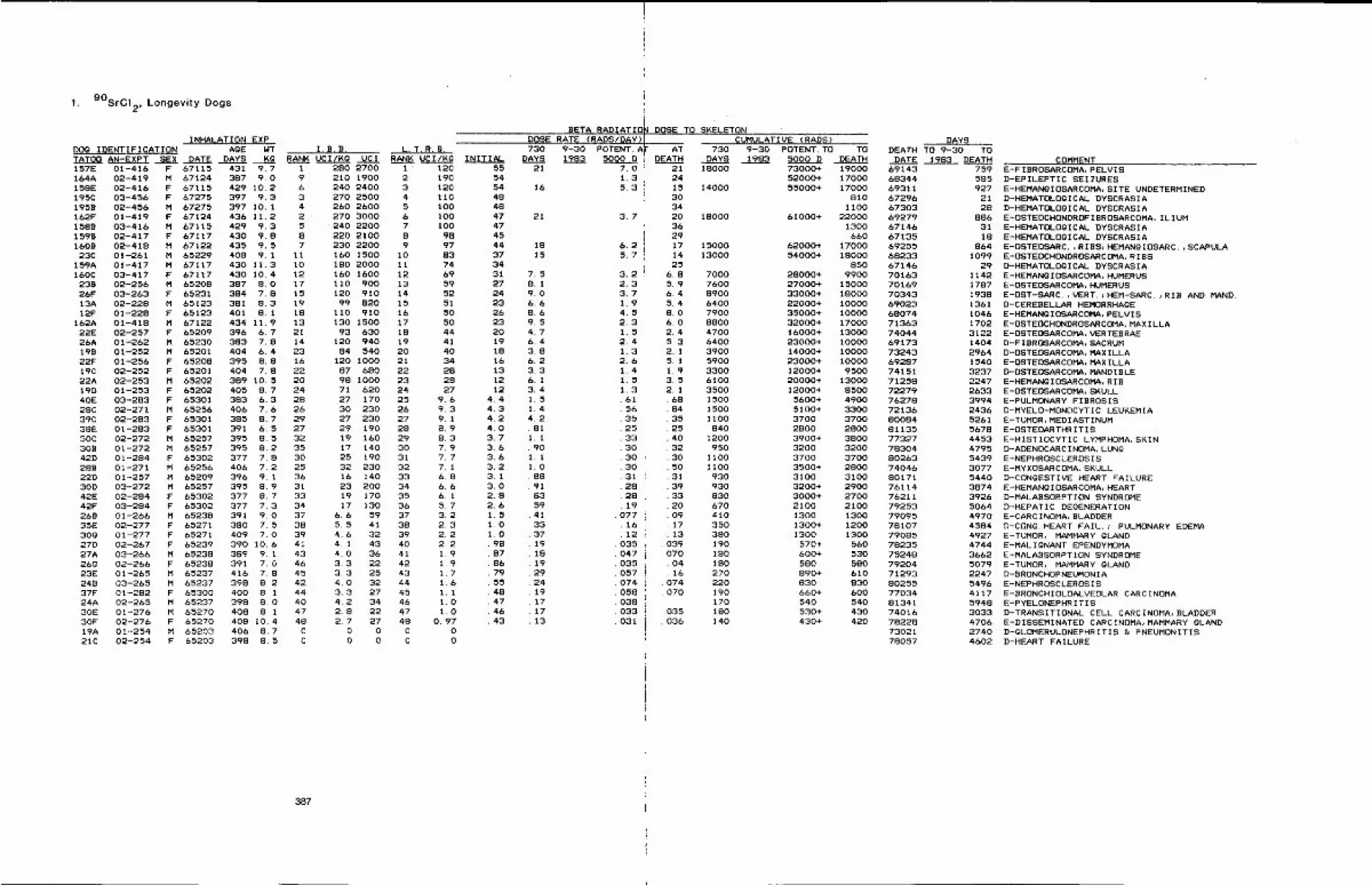
1. 90SrCl 2, Longevity Dogs
DOg IDENTIFICATIONTATO0 AN-EXPT ~’EX157E 01-416 F164A 02-419 M158E 02-416 F195C 03-456 F195B 02-456 M162F 0%-419 F1588 03-416 M1598 02-417 Ft60B 02-418 M23C 01-261 M
159A 01-417 M160C 03-417 F238 02-256 M26F 03-263 F13A 02-228 M12F 01-228 F
lb2A 01-418 M22E 02-257 F26A 01-262 M198 01-252 M22F 01-256 F19C 02-252 F22A 02-253 M19D 01-253 F40E 03-283 F28C 02-271 M39C 02-283 F38E 01-283 F30C 02-272 M308 01-272 M425 01-284 F28B 01-271 M22D 01-257 M30D 03-272 M42E 02-284 F42F 03-284 F268 01-266 M35E 02-277 F300 01-277 F27D 02-267 F27A 03-266 M26G 02-266 F23E 01-265 M248 03-265 M37F 01-282 F24A 02-265 M30E 01-276 M30F 02-27& F19A 01-254 M21C 02-254 F
INHALATION EXPAGE NT
DATE DAYS KG67115 431 9.767124 387 9.067115 429 10.267275 397 9.367275 397 10.167124 436 11.267115 429 9.367117 430 9.867122 435 9,565229 408 9.167117 430 11.367117 430 10.465208 387 8.065231 384 7.865123 381 8.365123 401 8.167122 434 11.965209 396 6.765230 383 7.865201 404 6.465208 395 8.865201 404 7.865202 389 10.565202 405 8.765301 383 6.365256 406 7.665301 385 8.765301 391 6.565257 395 8.565257 395 8.265302 377 7.865256 406 7.265209 396 9.165257 395 8.965302 377 8.765302 377 7.365238 391 9.065271 380 7.565271 409 7.065239 390 10.665238 389 9.165238 391 7.065237 416 7.865237 398 8.265300 400 8,165237 398 S.O65270 408 8.165270 408 10.465203 406 8.765203 398 8.5
I. B.B.RANK UCI/KG UCI
1 280 27009 210 19006 240 24003 270 25004 260 26002 ’ 270 30005 240 22008 220 21007 230 2200
11 160 1500I0 180 200012 160 160017 110 90015 120 91019 99 82018 110 91013 130 150021 93 63014 120 94023 84 54016 120 100022 87 68020 98 100024 71 62028 27 17026 30 23029 27 23027 29 19032 19 16035 17 14030 25 19025 32 23036 16 14031 23 20033 19 17034 17 13037 6.6 5938 5.5 4139 4.6 3241 4.1 4343 4.0 3646 3.3 2245 3.3 2542 4.0 3244 3.3 2740 4,2 3447 2.8 2248 2,7 27C 0 0C 0 0
L T.R.B.RAN~ UCI/½G
1 1202 1903 1204 1105 1006 1007 1008 989 97
10 8311 7412 b913 5914 5215 5116 5017 5018 4419 4120 4021 3422 2823 2824 2725 9.626 9.327 9.128 8.929 8.330 7.931 7.732 7.133 6.834 6.635 6.136 5.737 3.238 2.339 2,240 2.241 1.942 1.943 1.744 1.645 1.146 1.047 1.048 0.97C 0C 0
INITIAL555454484847474544373431272423262320191816131212
4.44.34.24.03.73.63.63.23.13.02.82.61.51.01.0. 98
8786795548474643
387
LBETA RADIATION DOSE TO SKELETON
DOSE RATE (RADS/pAY) / CUMULATIVE (RADS)I
730 9-30 POTENT. A[ AT 730 9-30 POTENT. TO TODAYS 1983 5000 D ! DEATH DAYS 1983 5000 D DEATH
21 18000
16
7.01.35.3
21 3.7
18 6.215 5.7
7.58.19.06.68.69.54.76.43.86.23.36.13.41.51.44.2.811.1. 901.11.088918359413337191819292419171713
3.22.33.71.94.52.31.52.41.32.61.41.51.3615635253330303031282819
0771612
035047O35O57074058038033031
2124153034203629171425
6.85.96.45.48.06.02.45.32.15.11.93.52.1
688435254O32305031393320091713
O3907O. 04.16O74070
035036
DAYSDEATH TO 9-30 TO
DATE 1983 DEATH73000+ 19000 69143 75952000+ 17000 68344 585
14000 55000+ 17000 69311 927810 67296 21
1100 67303 2818000 61000* 22000 69279 886
1300 67146 31660 67135 18
15000 62000÷ 17000 69255 86413000 54000+ 18000 68233 1099
850 67146 297000 28000+ 9900 70163 11427600 27000+ 15000 70169 17878900 33000+ 18000 70343 19386400 2"2000+ 10000 69023 13617900 35000+ i0000 68074 10468800 32000+ 17000 71363 17024700 16000+ 13000 74044 31226400 23000+ 10000 69173 14043900 14000+ 10000 73243 29645900 23000+ 10000 69287 15403300 12000* 9500 74151 32376100 20000+ 13000 71258 22473500 12000* 8500 72279 26331500 5600+ 4900 76278 39941500 5100+ 3300 72136 243&1100 3700 3700 80084 5261
840 2800 2800 81135 56781200 3900+ 3800 77327 4453
950 3200 3200 78304 47951100 3700 3700 80263 54391100 3500+ 2800 74046 3077
930 3100 3100 80171 5440930 3200* 2900 76114 3874830 3000+ 2700 76211 392a670 2100 2100 79253 5064410 1300 1300 79095 4970350 1300+ 1200 78107 4584380 1300 1300 79085 4927190 570+ 560 78235 4744180 600+ 530 75248 3662180 580 580 79204 5079270 890+ 610 71293 2247220 830 830 80255 5496190 660+ 600 77034 4117170 540 540 81341 5948180 530+ 430 74016 3033140 430+ 420 78228 4706
73021 274078057 4602
COMMENTE-FIBROSARCOMA, PELVtSD-EPILEPTIC SEIZURESE-HEMANgIOSARCOMA, SITE UNDETERMINEDD-HEMATOLOgICAL DYSCRASIAD-HEMATOLOgICAL DYSCRASIAE-OSTEOCHONDROFIBROSARCOMA, ILIUME-HEMATOLOGICAL DYSCRASIAE-HEMATOLOGICAL DYSCRASIAE-OSTEOSARC.,RIBS~HEMANGIOSARC.,SCAPULAE-OSTEOCHONDROSARCOMA, RIBSD-HEMATOLOgICAL DYSCRASIAE-HEMANGIOSARCOMA, HUMERUSE-OSTEOSARCOMA, HUMERUSE-OST-SARC.,VERT.~HEM-SARC.,RIB AND MAND.D-CEREBELLAR HEMORRHAGEE-HEMANGIOSARCOMA, PELVISE-OSTEOCHONDROSARCOMA, MAXILLAE-OSTEOSARCOMA, VERTEBRAED-FIBROSARCOMA, SACRUMD-OSTEOSARCOMA, MAXILLAE-OSTEOSARCOMA, MAXILLAD-OSTEOSARCOMA, MANDIBLEE-HEMANGIOSARCOMA, RI8E-OSTEOSARCOMA, SKUt.LE-PULMONARY FIBROSISD-MYELO-MONOCYTIC LEUKEMIAE-TUMOR, MEDIASTINUME-OSTEOARTHRITISE-HISTIOCYTIC LYMPHOMA, SKIND-ADENOCARCINOMA, LUNgE-NEPHRQSCLEROSISE-MYXOSARCOMA, SKULLD-CONGESTIVE HEART FAILUREE-HEMANgIOSARCOMA, HEARTD-MALABSORPTION SYNDROMED-HEPATIC DEGENERATIONE-CARCINOMA, BLADDERD-CONG. HEART FAIL.~ PULMONARY EDEMAE-TUMOR, MAMMARY GLANDE-MALIGNANT EPENDYMOMAE-MALABSORPTIQN SYNDROMEE-TUMOR, MAMMARY GLANDD-BRONCHOPNEUMONIAE-NEPHRQSCLEROSISE-BRONCHIOLOALVEOLAR CARCINOMAE-PYELONEPHRITISD-TRANSITIONAL CELL CARCINOMA, BLADDERE-DISSEMINATED CARCINOMA, MAMMARY GLANDD-GLOMERULONEPHRITIS & PNEUMONITISD-HEART FAILURE
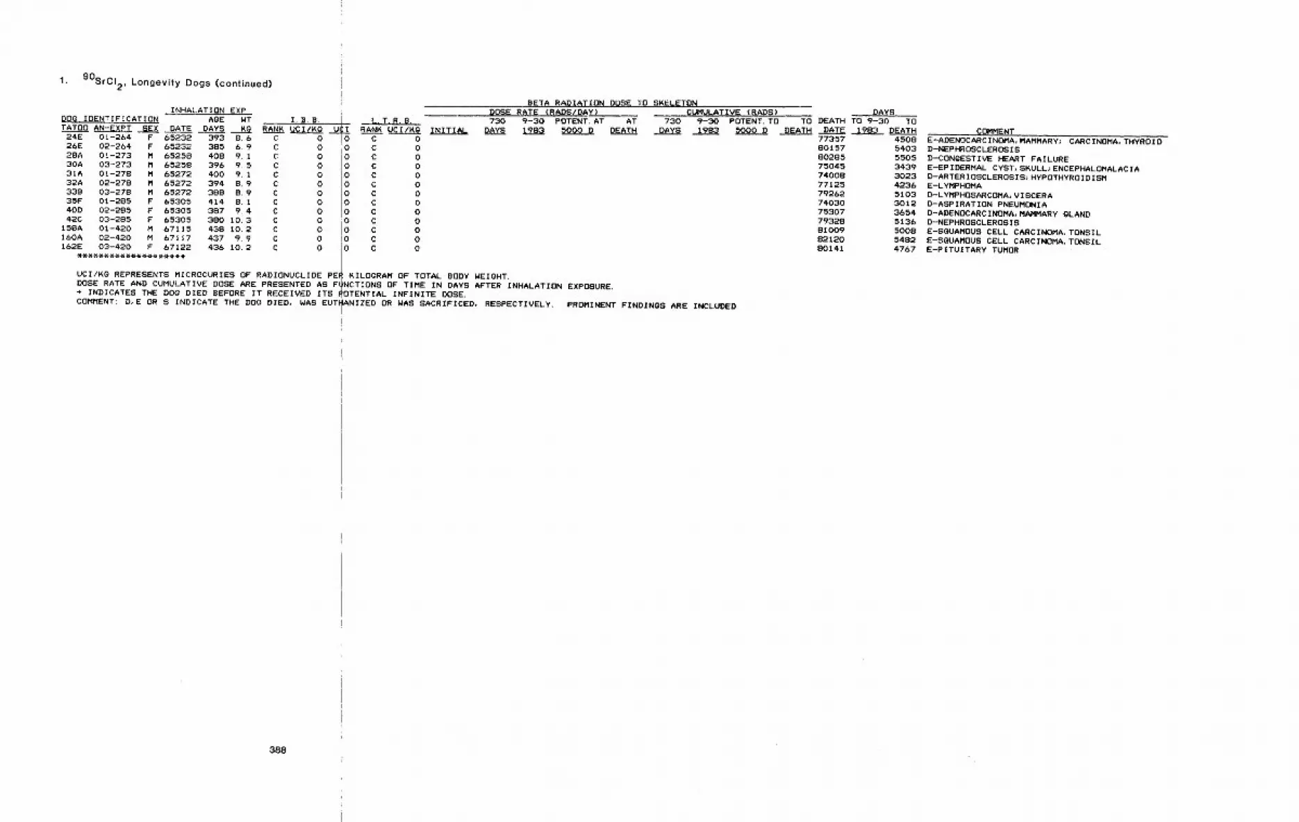
1. 90SrCI 2, Longevity Dogs (continued)
INHALATION EXPBETA RADIATION DOSE TO SKELETON
DOSE RATE (RADS/DAY)DOG IDENTIFICATION AgE WT I.B~B. | L.T.R.B. 730 9-30 POTENT. AT AT 730TATOO AN-EXPT SEX DATE DAYS KG RANK UCI/KG
~U IRANK UCI(Kg ~NIT~A~ DAYS 1983 5000 D DEATH DAYS
24E 01-264 F 65232 393 8.6 C 0 C 026E 02-264 F 65232 385 6.9 C 0 iO C 028A 01-273 M &5258 408 9.1 C 0 I0 C 030A 03-273 M 65258 396 9.5 C 0 ! 0 C 031A 01-278 M &5272 400 9.1 C 0 1.0 C 032A 02-278 M 65272 394 8.9 C 0 i~O C 033B 03-278 M 65272 388 8.9 C 0 0 C 035F 01-285 F 65305 414 8.1 C 0 0 C 040D 02-285 F 65305 387 9.4 C 0 i0 C 042C 03-285 F 65305 380 10.3 C 0 0 C 0
15SA 01-420 M 67115 438 10.2 C 0 0 C 0I&OA 02-420 M 67117 437 9.9 C 0
,. ~UMULATIVE (RADS)9-30 POTENT. TO TO1983 5000 D DEATH
I0 C 0162E 03-420 F 67122 436 10. 2 C 0 I0 C 0
UCI/KG REPRESENTS MICROCURIES OF RADIONUCLIDE PE~ KILOGRAM OF TOTAL BODY NEIQHT.DOSE RATE AND CUMULATIVE DOSE ARE PRESENTED AS F~NCTIONS OF TIME IN DAYS AFTER INHALATION EXPOSURE.+ INDICATES THE DOG DIED BEFORE IT RECEIVED ITS ~OTENTIAL INFINITE DOSE.COMMENT: D, E OR S INDICATE THE DOg DIED, WAS EUTIIANIZED OR WAS SACRIFICED, RESPECTIVELY. PROMINENT FINDINGS ARE INCLUDED.
DEATH.DATE77357801578028575045740087712579262740307530779328810098212080141
DAYSTO 9-30 TO
1983 DEATH45085403550534393023423&5103301236545136500854824767
COMMENTE-ADENOCARCINOMA, MAMMARY; CARCINOMA, THYROIDD-NEPHROSCLEROSISD-CONgESTIVE HEART FAILUREE-EPIDERMAL CYST, SKULL~ENCEPHALOMALACIAD-ARTERIOSCLEROSIS~HYPOTHYROIDISME-LYMPHOMAD-LYMPHOSARCOMA~VISCERAD-ASPIRATION PNEUMONIAD-ADENOCARCINOMA, MAMMARY gLANDD-NEPHROSCLEROSISE-SGUAMOUS CELL CARCINOMA, TONSILE-SGUAMOUS CELL CARCINOMA, TONSILE-PITUITARY TUMOR
388
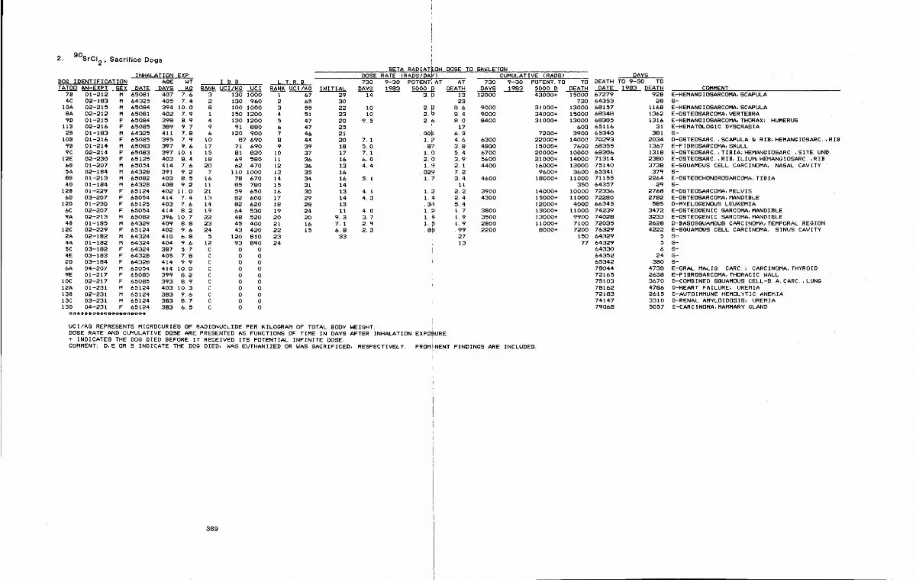
2. 90SrCI 2, Sacrifice Dogs
BETA RADIATION DOSE TO SKELETONINHALATION EXP DOSE RATE (RADS/DA~) CUMULATIVE (RADS) DAYS
i
DOg IDENTIFICATION AgE WT I.B.B. L.T.R.B. 730 9-30 POTENTIAT AT 730 9-30 POTENT. TO TO DEATH TO 9-30 TOTATO0 AN-EXPT .SEX DATE DAYS ~ RANK UCI/Kg UCI RANK UCI/Kg INITIAL DAYS 1983 5000 p DEATH DAYS 1983 5000 D DEATH DATE 1983 DEATH
7B 01-212 M 65081 407 7.6 3 130 1000 1 67 29 14 3.0 13 12000 43000+ 15000 67279 9284C 02-183 M 64325 405 7.4 2 130 960 2 65 30 23 730 64353 28
IOA 02-215 M 65084 394 10.0 8 100 1000 3 55 22 10 2.2 8.6 9000 31000+ 13000 68157 11688A 02-212 M 65081 402 7.9 1 150 1200 4 51 23 10 2.~ 814 9000 34000+ 15000 68348 13629D 01-215 F 65084 398 8.9 4 130 1200 5 47 20 9.5 2.6 810 8400 31000+ 13000 68305 1316
118 02-216 F 65085 389 9.7 9 91 880 6 47 25 i 17 600 65116 3128 01-183 M 64325 411 7.8 6 120 900 7 46 21 .006 6.3 7200+ 3900 65340 381
108 01-216 F 65085 395 7.9 I0 87 690 8 44 20 7.1 1.7 4.6 6300 22000+ 14000 70293 203498 01-214 M 65083 397 9.6 17 71 690 9 39 18 5.0 .8~ 3.8 4800 15000+ 7600 68355 13679C 02-214 F 65083 397 10.1 15 81 820 I0 37 17 7.1 1. b 5.4 6700 20000+ i0000 68306 1318
12E 02-230 F 65125 403 8.4 18 69 580 11 36 16 6.0 2. b 3.9 5600 21000+ 14000 71314 238068 01-207 M 65054 414 7.6 20 62 470 12 36 13 4.4 i_~ 2.1 4400 16000+ 13000 75140 37385A 02-184 M 64328 391 9.2 7 110 1000 13 35 16 .02~ 7.2 9600+ 3600 65341 3798B 01-213 M 65082 403 8.5 16 78 670 14 34 16 5. I 1.7 3.4 4600 18000+ 11000 71155 22644D 01-184 M 64328 408 9.2 ii 85 780 15 31 14 i II 350 64357 29
12B 01-229 F 65124 402 II.O 21 59 650 16 30 13 4. I 1.~ 2.2 3900 14000+ 10000 72336 27686D 03-207 F 65054 414 7.4 13 82 600 17 29 14 4.3 1. ~ 2.4 4300 15000+ 11000 72280 2782
12D 01-230 F 65125 403 7.6 14 82 620 18 28 13 .3~ 5.4 12000+ 4000 66345 5856C 02-207 F 65054 414 8.2 19 64 530 19 24 Ii 4.0 1.2 1.7 3800 13000+ 11000 74239 34729A 02-213 M 65082 396 10.7 22 48 520 20 20 9.3 3,7 1. ~ 1.9 3500 13000+ 9900 74028 323348 01-185 M 64329 409 8.8 23 45 400 21 16 7.1 2.9 I. ~ 1.9 2800 11000+ 7100 72035 2628
12C 02-229 F 55124 402 9.6 24 43 420 22 15 6.8 2,3 .8~ .99 2200 8000+ 7200 76329 42222A 02-182 M 64324 410 6.8 5 120 810 23 33 27 150 64329 54A 01-182 M 64324 404 9.6 12 93 890 24 13 77 64329 55C 03-182 F 64324 387 5.7 C 0 0 i 64330 64E 03-183 F 64328 405 7.8 C 0 0 64352 242D 08-184 F 64328 414 9.9 C 0 0 i 65342 3806A 04-207 M 65054 414 10.0 C 0 0 78044 47389E 01-217 F 65083 399 8~2 C 0 0 72165 2638
lOC 02-217 F 65085 393 8.9 C 0 0 75103 367012A 01-231 M 65124 403 10.3 C 0 0 78162 4786138 02-231 M 65124 383 9.6 C 0 0 72183 261513C 03-231 M 65124 383 8.7 C 0 0 74147 331013D 04-231 F 65124 383 6. 5 C 0 0 79068 5057
UCII~g REPRESENTS MICROCURIES OF R~DIONUCLIDE PER KILOGRAM OF TOTAL BODY WEIGHT.DOSE RATE AND CUMULATIVE DOSE ARE PRESENTED AS FUNCTIONS OF TIME IN DAYS AFTER INHALATION EXPO~;URE.+ INDICATES THE DO0 DIED BEFORE IT RECEIVED ITS POTENTIAL INFINITE DOSE.COMMENT: D,E OR S INDICATE THE DOg DIED, WAS EUTHANIZED OR WAS SACRIFICED, RESPECTIVELY. PROMINENT FINDINGS ARE INCLUDED.
COMMENTE-HEMANgIOSARCOMA, SCAPULAS-E-HEMANgIOSARCOMA, SCAPULAE-OSTEOSARCOMA, VERTEBRAE-HEMANgIOSARCOMA, THORAX~ HUMERUSE-HEMATOLOgIC DYSCRASIAS-D-OSTEOSARCo,SCAPULA & RIB;HEMANgIOSARC. RIBE-FIBROSARCOMA, SKULLE-OSTEOSARC.,TIBIA~HEMANgIOSARC.,SITE UND.E-OSTEOSARC.,RIB, ILIUM~HEMANgIOSARC.,RIBE-SGUAMOUS CELL CARCINOMA, NASAL CAVITYS-E-OSTEOCHONDROSARCOMA, TZ B IAS-E-OSTEOSARCOMA, PELVISE-OSTEOSARCOMA, MANDIBLED-MYELOgENOUS LEUKEMIAE-OSTEOgENIC SARCOMA, MANDIBLEE-OSTEOgENIC SARCOMA, MANDIBLED-BASOSGUAMOUS CARCINOMA, TEMPORAL REGIONE-SOUAMOUS CELL CARCINOMA, SINUS CAVITYS-S-S-S-S-E-ORAL MALIg. CARC.~ CARCINOMA, THYROIDE-FIBROSARCOMA, THORACIC W~LLD-COMBINED SQUAMOUS CELL-B.A. CARC.,LUNgD-HEART FAILURE~ UREMIAD-AUTOIMMUNE HEMOLYTIC ANEMIAD-RENAL AMYLOIDOSIS~ UREMIAE-CARCINOMA, MAMMARY gLAND
389
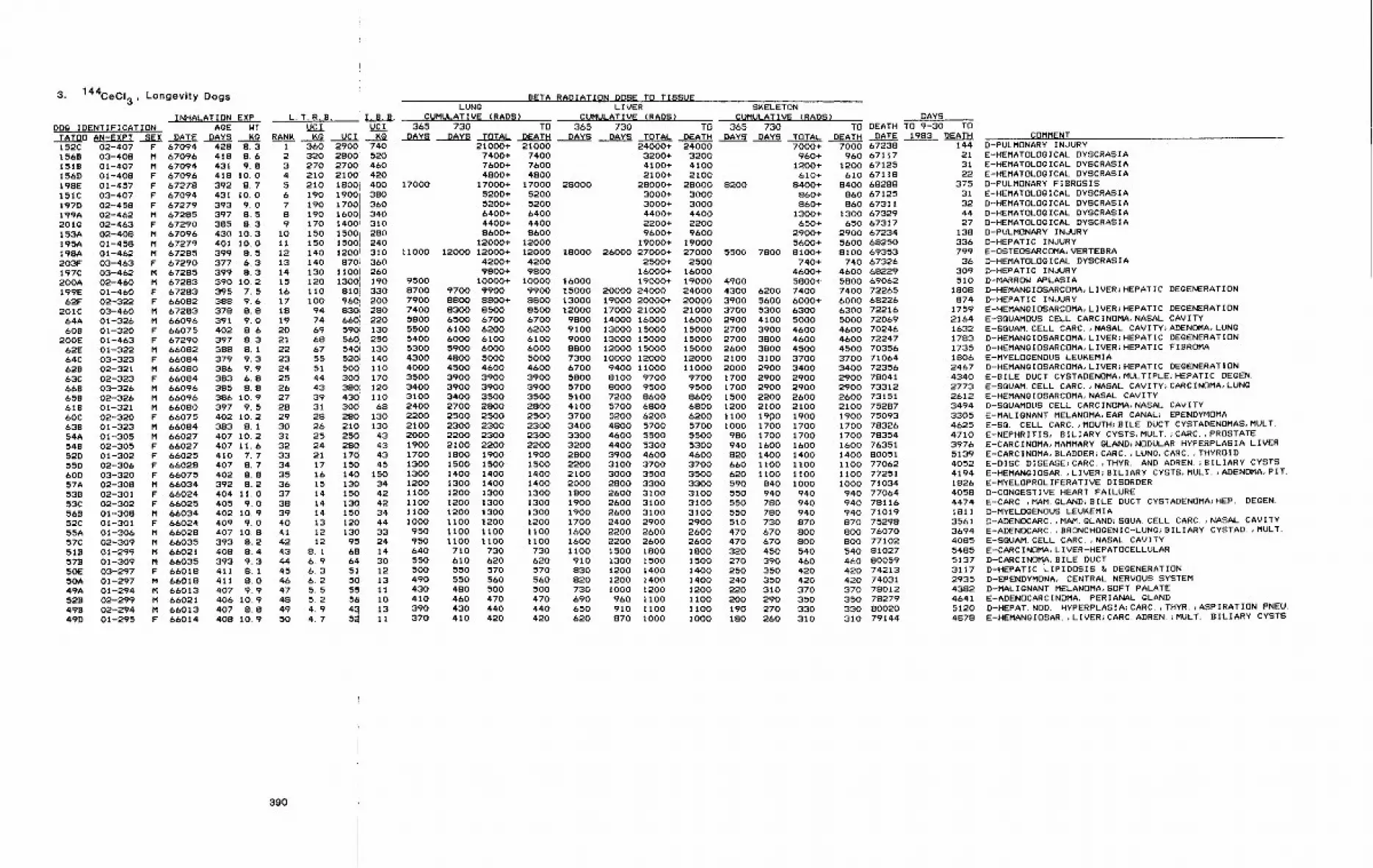
144CeCl3, Longevity Dogs3.
INHALATION EXP L.T.R.B.DOg IDENTIFICATION AgE NT UCITATO0 AN-EXPT SEX DATE DAYS KO RANK Kg UCI
F 67094 428 8.3M 67096 418 8,6M 67094 431 9.8F 67096 418 10.0F 67278 392 8.7F 67094 431 I0.0F 67279 393 9.0M 67285 397 8.5F 67290 385 8.3
152C 02-407156B 03-4081519 01-407156D 01-408198E 01-457151C 03-407197D 02-458199A 02-462201g 02-463153A 02-408195A 01-458198A 01-462203F 03-463197C 03-462200A 02-460199E 01-46062F 02-322
201C 03-46064A 01-326
M 67096M 67279M 67285F 67290M 67285M 67283F 67283F 66082M 67283M 66096
609 01-320 F 66075200E 01-463 F 67290
62E 01-322 M 6608264C 03-323 F 66084628 02-321 M 6608063C 02-323 F 66084669 03-326 M 66096658 02-326 M 66096618 01-321 M 6608060C 02-320 F 66075638 01-323 M 6608454A 01-305 M 66027548 02-305 F 6602752D 01-302 F 6602555D 02-306 F 6602860D 03-320 F 6607557A 02-308 M 6603453B 02-301 F 66024
430 10.3401 i0.0399 8.5 12377 6.3 13399 8.3 14390 I0.2 15395 7.5 16388 9.6 17378 8.8 18391 9.0 19402 8 6 20397 8.3 21388 8,1 22379 9.3 23386 9.9 24383 6.8 25385 8.8 26386 10.9 27397 9.5 28402 10~2 29383 8.1 30407 10~2 31407 11.6 32410 7.7 33407 8.7 34402 8.8 35392 8.2 36404 11.0 37
53C 02-302 F 66025 405 9.0 38568 01-308 M 66034 402 10.9 3952C 01-301 F 66024 409 9.0 4055A 01-306 M 66028 407 10.8 4157C 02-309 M 66035 393 8.2 42518 01-299 M 66021 408 8.4 43579 01-309 M 66035 393 9.3 4450E 03-297 F 66018 411 8.1 4550A 01-297 M 66018 411 8,0 4649A 01-294 M 66013 407 9.9 47528 02-299 M 66021 406 10.9 48498 02-294 M 66013 407 8.8 4949D 01-295 F 66014 408 10.9 50
l 1.9. B,UC I
KO1 360 2900 7402 320 2800 5203 270 2700: 4604 210 2100 4205 210 1800i 400 170006 190 19001 3807 190 17001 3608 190 1600j 3409 170 1400= 310
10 150 28011
LUNg
CUMULATIVE (PADS)365
DAYS
BETA RADIATION DOSE TO TISSUELIVER SKELETON
CUMULATIVE (RADS) CUMULATIVE (RADS) DAYS730 TO 365 730 TO 365 730 TO DEATH TO 9-30 TO
DAYS TOTAL DEATH DAYS DAYS TOTAL DEATH DAYS DAYS TOTAL DEATH DATE 1983 OEATH
1500150 1500 240140 1200! 310 11000 12000 12000+ 12000 18000 26000 27000+ 27000 5500 7800 8100+
21000+ 21000 24000+ 24000 7000+ 7000 672387400+ 7400 3200+ 3200 960+ 960 671177600+ 7600 4100+ 4100 1200+ 1200 671254800+ 4800 2100+ 2100 610+ 610 67118
17000+ 17000 28000 28000+ 28000 8200 8400+ 8400 682885200+ 5200 3000+ 3000 860+ 860 671255200+ 5200 3000+ 3000 860+ 860 673116400+ 6400 4400+ 4400 1300+ 1300 673294400+ 4400 2200+ 2200 &50+ 650 673178600+ 8600 9600+ 9600 2900+ 2900 67234
12000+ 12000 19000+ 19000 5600+ 5600 682508100 69353
14o 87oi 360130 1100! 260120 13001 190ii0 810i 330i00 96~ 20094 830; 28074 660,’ 22069 59(> 13068 56~ 25067 5401 13055 52@ 14051 500 11044 30~ 17043 3801 12039 430’ 1103I 300’ 6828 280 13o26 210 13025 25~ 4324 28Q 4321 174 4317 150 4516 140 15015 130 3414 150 4214 13~ 4214 150 3413 120 4412 130 3312 95 24
8.1 68 146.9 64 306.3 511 126. 2 5d 135.5 55 115. 2 56 i04, 9 43 134. 7 5~ 11
95008700 9700 99007900 8800 8800+7400 8300 85005800 6500 6700 67005500 6100 6200 62005400 6000 6100 61005300 5900 6000 60004300 4800 5000 50004000 4500 4600 46003500 3900 3900 39003400 3900 3900 39003100 3400 3500 35002400 2700 2800 28002200 2500 2500 25002100 2300 2300 23002000 2200 2300 23001900 2100 2200 22001700 1800 1900 19001300 1500 1500 15001300 1400 1400 14001200 1300 14001100 1200 1300 13001100 1200 1300 13001100 1200 1300 13001000 1100 1200 1200950 1100 1100 1100950 1100 1100 1100640 710 730 730550 610 620 620500 550 570 570490 550 560 560430 480 500 500410 460 470 470390 430 440 440370 410 420 420
4200+ 4200 2500+ 2500 740+9800+ 9800 16000+ 16000 4600+
10000+ 10000 16000 19000+ 19000 4900 5800+9900 15000 20000 24000 24000 4300 6200 74008800 13000 19000 20000+ 20000 3900 5600 6000+8500 12000 17000 21000 21000 3700 5300 6300
9800 14000 160009100 13000 150009000 13000 150008800 12000 150007300 10000 120006700 9400 110005800 8100 97005700 8000 95005100 7200 86004100 5700 68003700 5200 62003400 4800 57003300 4600 55003200 4400 53002800 3900 46002200 3100 37002100 3000 3500
1400 2000 2800 33001800 2600 31001900 2600 31001900 2600 31001700 2400 29001600 2200 26001600 2200 26001100 1500 1800910 1300 1500830 1200 1400820 1200 1400730 1000 1200690 960 Ii00650 910 Ii00620 870 I000
16000 2900 4100 500015000 2700 3900 460015000 2700 3800 460015000 2600 3800 450012000 2100 3100 370011000 2000 2900 34009700 1700 2900 29009500 1700 2900 29008600 1500 2200 26006800 1200 2100 21006200 1100 19p0 19005700 1000 1700 1700550053004600370035003300310031003100290026002600
980 1700 1700940 1600 1600820 1400 1400660 1100 1100620 1100 II00590 840 1000550 940 940550 78O 94O550 780 940510 730 870470 670 800470 670 800
1800 320 450 5401500 270 390 4601400 250 350 4201400 240 350 4201200 220 310 3701100 200 290 3501100 190 270 3301000 180 260 310
740 673264500 682295800 690627400 722656000 682266300 722165000 720694600 702464600 722474500 703563700 710643400 723562900 780412900 733122600 731512100 752B71900 750931700 783261700 783541600 763511400 800511100 770621100 772511000 71034940 77064940 78116940 71019870 75298800 76070SO0 77102540 81027460 80059420 74213420 74031370 78O12350 78279330 80020310 79144
COMMENT144 D-PULMONARY INJURY21 E-HEMATOLOgICAL DYSCRASIA31 E-HEMATOLOgICAL DYSCRASIA22 E-HEMATOLOgICAL DYSCRASIA
375 D-PULMONARY FIBROSIS31 E-HEMATOLOgICAL DYSCRASIA32 D-HEMATOLOgICAL DYSCRASIA44 D-HEMATOLOgICAL DYSCRASIA27 D-HEMATOLOgICAL DYSCRASIA
138 D-PULMONARY INJURY336 D-HEPATIC INJURY799 E-OSTEOSARCOMA, VERTEBRA
36 D-HEMATOLOgICAL DYSCRASIA309 D-HEPATIC INJURY510 D-MARROW APLASIA
1808 D-HEMANGIOSARCOMA, LIVER;HEPATIC DEGENERATION874 D-HEPATIC INJURY
1759 E-HEMANGIOSARCOMA, LIVER~HEPATIC DEGENERATION2164 E-SGUAMOUS CELL CARCINOMA, NASAL CAVITY1632 E-SQUAM. CELL CARC.,NASAL CAVITY;ADENOMA, LUNg1783 D-HEMANgIOSARCOMA~LIVER;HEPATIC DEGENERATION1735 D-HEMANgIOSARCOMA~LIVER~HEPATIC FIBROMA1806 E-MYELOgENOUS LEUKEMIA2467 D-HEMANgIOSARCOMA, LIVER}HEPATIC DEGENERATION4340 E-BILE DUCT CYSTADENOMA, MULTIPLE, HEPATIC DEGEN.2773 E-SQUAH. CELL CARC. , NASAL CAVITY; CARCINOMA, LUNg2612 E-HEMANGIOSARCOMA~ NASAL CAVITY3494 D-SOUAMOUS CELL CARCINOMA, NASAL CAVITY3305 E-MALIgNANT MELANOMA, EAR CANAL~ EPENDYMOMA4625 E-SO. CELL CARC.,MOUTH~BILE DUCT CYSTADENOMAS, MULT.4710 E-NEPHRITIS, 9ILIARY CYSTS, MULT.}CARC.,PROSTATE3976 E-CARCINOMA, MAMMARY gLAND;NODULAR HYPERPLASIA LIVER5139 E-CARCINOMA, BLADDER;CARC.,LUNg~CARC.,THYROID4052 E-DISC DISEASE;CARC.,THYR. AND ADREN.;BILIARY CYSTS4194 E-HEMANgIOSAR.,LIVER;BILIARY CYSTS, MULT.;ADENOMA, PIT.1826 E-MYELOPROLIFERATIVE DISORDER4058 D-CONgESTIVE HEART FAILURE4474 E-CARC ,MAM. gLAND~BILE DUCT CYSTADENOMA~HEP. DEGEN.1811 D-MYELOgENOUS LEUKEMIA
3561 D-ADENOCARC.,MAM. GLANDJSQUA. CELL CARC.,NASAL CAVITY3694 E-ADENOCARC. ,BRONCHOGENIC-LUNg~BILIARY CYSTAD. ,MULT.4085 E-SGUAM. CELL CARC.,NASAL CAVITY5485 E-CARCINOMA, LIVER-HEPATOCELLULAR5137 D-CARCINOMA, BILE DUCT3117 D-HEPATIC LIPIDOSIS & DEGENERATION2935 D-EPENDYMONA, CENTRAL NERVOUS SYSTEM4382 D-MALIgNANT MELANOMA, SOFT PALATE4641 E-ADENOCARCINOMA, PERIANAL gLAND5120 D-HEPAT. NOD. HYPERPLASIA;CARC.,THYR.;ASPIRATION PNEU.4878 E-HEMANgIOSAR.,LIVER;CARC. ADREN.~MULT. 8ILIARY CYSTS
390
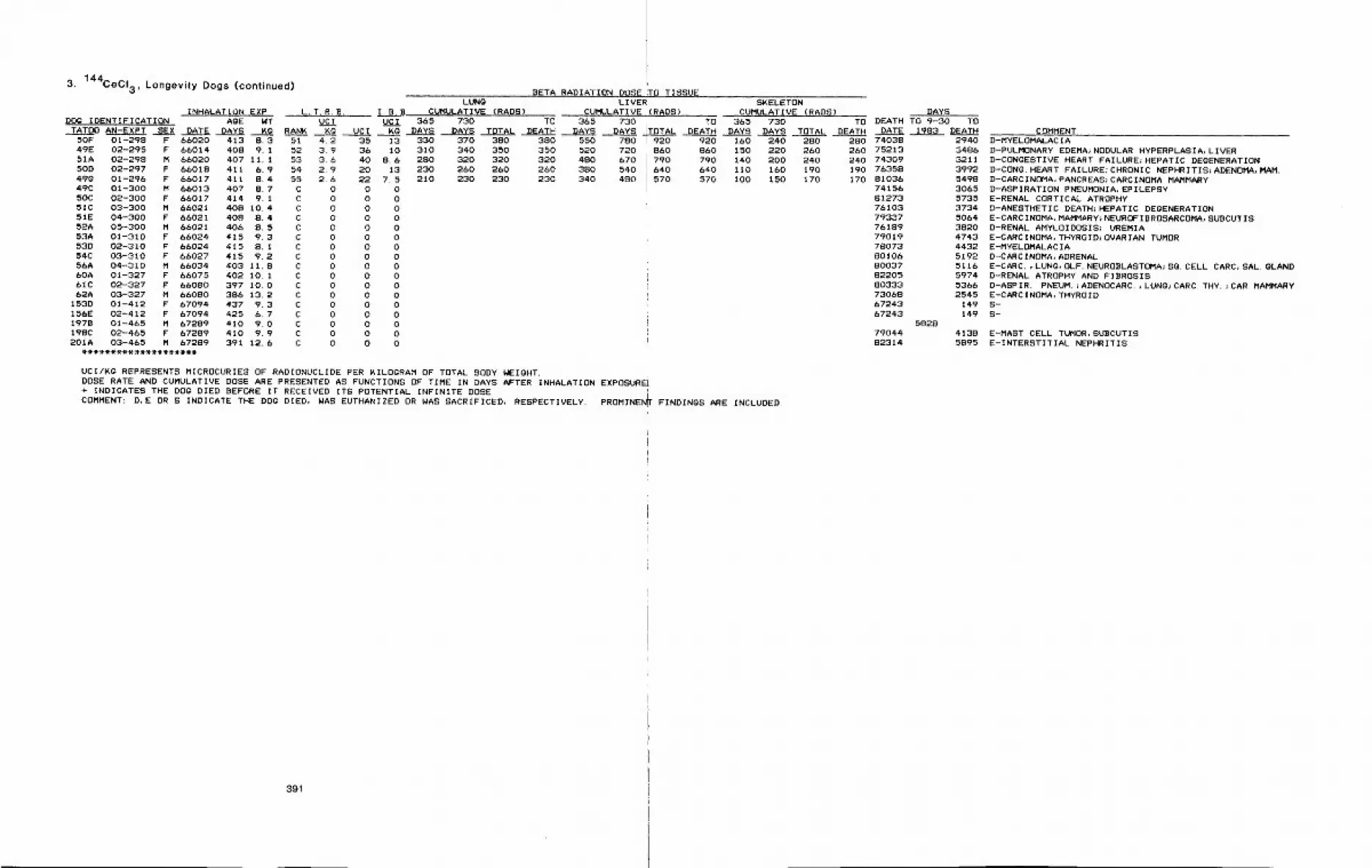
3. 144CECI3, Longevity Dogs (continued)
INHALATION EXP L.T.R.B. I.B.BDOG IDENTIFICATION AGE WT UCI UCI
TATOO AN-EXPT SEX DATE DAYS KG RANK KG UCI KG50F 01-298 F 66020 413 8. 3 51 4.2 35 1349E 02-295 F 66014 408 9.1 52 3.9 36 1051A 02-298 M 66020 407 11.1 53 3.6 40 8.650D 02-297 F 66018 411 6.9 54 2.9 20 1349g 01-296 F 66017 411 8.4 55 2.6 22 7. 549C 01-300 M 66013 407 8.7 C 0 O 050C 02-300 F 66017 414 9.1 C 0 0 051C 03-300 M 66021 408 10.4 C 0 0 051E 04-300 F 66021 408 8.4 C 0 0 052A 05-300 M (>6021 406 8. 5 C 0 0 053A 01-310 F 66024 415 9.3 C 0 0 053D 02-310 F 66024 415 8. I C 0 0 054C 03-310 F 66027 415 9.2 C 0 0 056A 04-310 M 66034 403 11.8 C 0 0 060A 01-327 F 66075 402 10. 1 C 0 0 061C 02-327 F 66080 397 10.0 C 0 0 062A 03-327 H 660B0 386 13.2 C 0 0 0
153D 01-412 F 67094 437 9.3 C 0 0 0156E 02-412 F 67094 425 6.7 C 0 0 0197B 01-465 M 67289 410 9.0 C 0 0 0198C 02-465 F 67289 410 9.9 C 0 0 0201A 03-465 M 67289 391 12.6 C 0 0 0
LUNGCUMULATIVE (RADS)
BETA RADIATION DOSE ITO TISSUELIVER
CUMULATIVE I(RADS)365 730 TO 365
DAYS DAYS TOTAL DEATH DAYS330 370 380 380 550310 340 350 350 520280 320 320 320 480230 260 260 260 3S0210 230 230 230 340
730 TODAYS TOTAL DEATH
780 920 920720 860 860670 790 790540 640 640480 570 570
SKELETONCUMULATIVE (RADS~
365 730 TODAYS DAYS TOTAL DEATH
160 240 280150 220 260140 200 240110 160 190100 150 170
DAYSDEATH TO 9-30
DATE i~83280 74038260 75213240 74309190 76358170 81036
7415681273761037933776189790197~073BOIO6800378220580333730886724367243
58287904482314
UCI/Kg REPRESENTS MICROCURIES OF RADIONUCLIDE PER KILOGRAM OF TOTAL BODY WEIGHT.DOSE RATE AND CUMULATIVE DOSE ARE PRESENTED AS FUNCTIONS OF TIME IN DAYS AFTER INHALATION EXPOSUREL+ INDICATES THE DOG DIED BEFORE IT RECEIVED ITS POTENTIAL INFINITE DOSE.COMMENT: D,E OR S INDICATE THE DOG DIED, WAS EUTHANIZED OR WAS SACRIFICED, RESPECTIVELY. PROMINEN~ FINDINGS ARE INCLUDED.
TODEATH COMMENT
2940348632113992549830655735373450&438204743443251925116597453662545
149I49
41385895
D-MYELOMALACIAD-PULMONARY EDEMA~NODULAR HYPERPLASIA, LIVERD-CONGESTIVE HEART FAILURE~HEPATIC DEGENERATIOND-CONG. HEART FAILURE;CHRONIC NEPHRITIS~ADENOMA, MAM.D-CARCINOMA, PANCREAS;CARCINOMA MAMMARYD-ASPIRATION PNEUMONIA, EPILEPSYE-RENAL CORTICAL ATROPHYD-ANeSTHETIC DEATH~HEPATIC DEGENERATIONE-CARCINOMA, MAMMARY~NEUROFIBROSARCOMA, SUBCUTISD-RENAL AMYLOIDOSIS; UREMIAE-CARCINOMA, THYROIDJOVARIAN TUMORE-MYELOMALACIAD-CARCINOMA, ADRENALE-CARC.,LUNG, OLF. NEUROBLASTOMA;SG. CELL CARC, SAL. gLANDD-RENAL ATROPHY AND FIBROSISD-ASPIR. PNEUM.~ADENOCARC.,LUNG~CARC. THY.~CAR. MAMMARYE-CARCINOMA, THYROIDS-S-
E-MAST CELL TUMOR, SUBCUTISE-INTERSTITIAL NEPHRITIS
391
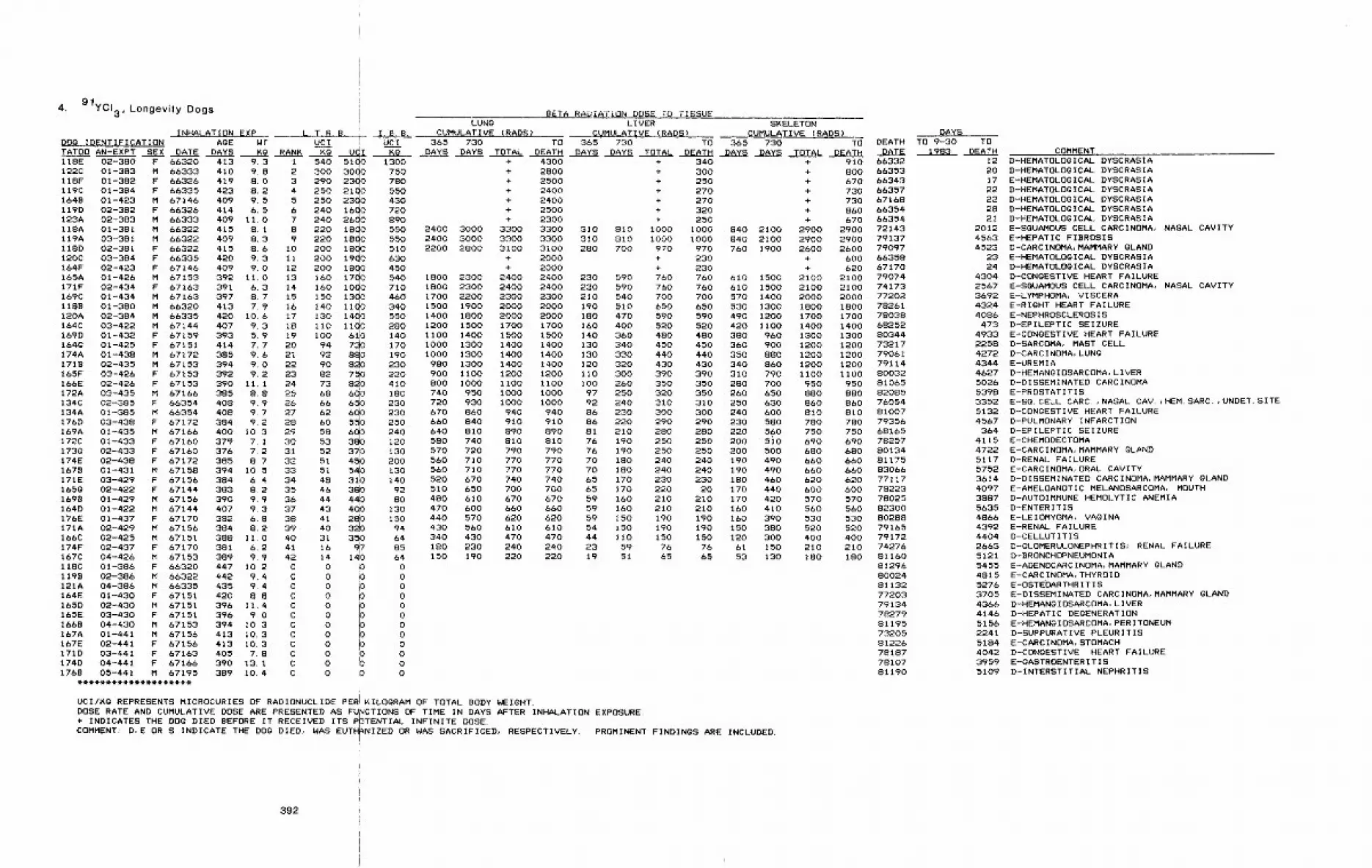
4. 9/ycI3 , Longevity Dogs
INHALAT ION EXP L.T.R.B.
BETA RADIATION DOSE TO TISSUELUNg LIVER SKELETON
I.B.B. CUMULATIVE (RADS~ CUMULATIVE (RADS) CUMULATIVE (RADS)DOg IDENTIFICATION AgE Wr UCI i UCI 365 730 TO 365 730 TO 365 730TATO0 AN-EXPT SEX DATE DAYS Kg RANK Kg UU___~ Kg DAYS DAYS TOTAL DEATH DAYS DAYS TOTAL DEATH DAYS DAYS TOTAL118E 02-380 F 66320 413 9.3 1 540510~ 1300 + 4300 + 340 +122C 01-383 M 66333 410 9.8 2 300 30qO 750 + 2800 + 300 +118F 01-382 F 66326 419 8.0 3 290 2300 780 + 2500 + 250 +119C 01-384 F 66335 423 8.2 4 250 21~0 550 + 2400 + 270 +1648 01-423 M 67146 409 9.5 5 250 23q0 430 + 2400 + 270 +119D 02-382 F 66326 414 6.5 6 240 16qO 720 + 2500 + 320 +123A 02-383 M 66333 409 11.0 7 240 26Q0 890 + 2300 + 250 +lISA 01-381 M 66322 415 S.l 8 220 18~0 550 2400 3000 3300 3300 310 810 lOOO I000 840 2100 2900II9A 03-381 M 66322 409 8.3 9 220 1SqO 550 2400 3000 3300 3300 310 810 I000 I000 840 2100 2900
I
118D 02-381 F 66322 415 8.6 i0 200 18C~0 510 2200 2800 3100 3100 280 700 970 970 760 1900 260012OC 03-384 F 66335 420 9.3 ii 200 1900 630 + 2000 + 230 +164F 02-423 F 67146 409 9.0 12 200 IBqO 450 + 2000 + 230 +165A 01-426 M 67153 392 11.0 13 160 17C~ 540 1800 2300 2400 2400 230 590 760 760 610 1500 2100171F 02-434 F 67163 391 6.3 14 160 10q0 710 1800 2300 2400 2400 230 590 760 760 610 1500 2100169C 01-434 M 67163 397 8.7 15 150 1300 460 1700 2200 2300 2300 210 540 700 700 570 1400 20001188 01-380 M 66320 413 7.9 16 140
ii~340 1500 1900 2000 2000 190 510 650 650 530 1300 1800
120A 02-384 M 66335 420 10.6 17 130 14 550 1400 1800 2000 2000 180 470 590 590 490 1200 1700164C 03-422 M 67144 407 9.3 18 ii0 Ii 280 1200 1500 1700 1700 160 400 520 520 420 1100 1400169D 01-432 F 67159 393 5.9 19 100 61~ 140 1100 1400 1500 1500 140 360 480 480 380 960 1300164g 01-425 F 67151 414 7.7 20 94 730 170 i000 1300 1400 1400 130 340 450 450 360 900 1200174A 01-438 M 67172 385 9.6 21 92 840 190 1000 1300 1400 1400 130 330 440 440 350 880 12001718 02-435 M 67153 394 9.0 22 90 82P3 230 980 1300 1400 1400 120 320 430 430 340 860 1200
i
165F 03-426 F 67153 392 9.2 23 82 750 220 900 1100 1200 1200 ii0 300 390 390 310 790 1100166E 02-426 F 67153 390 11. I 24 73 820 410 800 1000 II00 1100 I00 260 350 350 280 700 950172A 03-435 M 67166 385 8.8 25 68 6~’3 180 740 950 leO0 1000 97 250 320 350 260 650 880134C 02-385 F 66354 408 9.9 26 66 6,.5~ 230 720 930 1000 1000 92 240 310 310 250 630 860134A 01-385 M 66354 408 9.7 27 62 6C~,} 230 670 860 940 940 86 230 300 300 240 600 810176D 03-438 F 67172 384 9.2 28 60 5,~0 250 660 840 910 910 86 220 290 290 230 580 780169A 01-435 M 67166 400 103 29 58 6~3 240 640 810 890 890 81 210 280 280 220 560 750172C 01-433 F 67160 379 7.1 30 53 38~ 120 580 740 810 810 76 190 250 250 200 510 690173g 02-433 F 67160 376 7.2 31 52 376 130 570 720 790 790 76 190 250 250 200 500 680174E 02-438 F 67172 385 8.7 32 51 45~ 200 560 710 770 770 70 180 240 240 190 490 6601678 01-431 M 67158 394 10. 5 33 51 54~ 130 560 710 770 770 70 180 240 240 190 490 660171E 03-429 F 67156 384 6.4 34 48 31~ 140 520 670 740 740 65 170 230 230 180 460 6201650 02-422 F 67144 383 8.2 35 46 3~{} 92 510 650 700 700 65 170 220 20 170 440 6001698 01-429 M 67156 390 9.9 36 44 446 80 480 610 670 670 59 160 210 210 170 420 570164D 01-422 M 67144 407 9.3 37 43 4OK) 130 470 600 660 660 59 160 210 210 160 410 560176E 01-437 F 67170 382 6.8 38 41 28~0 150 440 570 620 620 59 150 190 190 160 390 530171A 02-429 M 67156 384 s. 2 39 40 3~ 94 430 560 610 610 54 15o 190 190 150 3s0 520166C 02-425 M 67151 388 11.0 40 31 350 64 340 430 470 470 44 110 150 150 120 300 400174F 02-437 F 67170 381 6.2 41 16 ~7 85 180 230 240 240 23 59 76 76 61 150 210167C 04-426 M 67153 389 9.9 42 14 140 64 150 190 220 220 19 51 &5 65 53 130 180118C 01-386 F 66320 447 10.2 C 0 ~ 01198 02-386 M 66322 442 9.4 C 0 ~ O121A 04-386 M 66335 435 9.4 C 0 OI&4E 01-430 F 67151 420 8.8 C 0 # 0165D 02-430 M 67151 396 11.4 C 0 ~ 0165E 03-430 F 67151 396 9.0 C 0
~
0166B 04-430 M 67153 394 10.3 C 0 0Ib7A 01-441 M 67156 413 10.3 C 0 0167E 02-441 F 67156 413 10.3 C 0 ~ 0171D 03-441 F 67163 405 7.8 C 0 0 0174D 04-441 F 67166 390 13.1 C 0 ~ 01768 05-441 M 67195 389 10.4 C 0 0 0
UCI/~g REPRESENTS MICROCURIES OF RADIONUCLIDE PERi KILOGRAM OF TOTAL BODY WEIGHT.
DOSE RATE AND CUMULATIVE DOSE ARE PRESENTED AS FUNCTIONS OF TIME IN DAYS AFTER INHALATION EXPOSURE.+ INDICATES THE DOg DIED BEFORE IT RECEIVED ITS PpTENTIAL INFINITE DOSE.COMMENT: D,E OR S INDICATE THE DOg DIED, WAS EUTH~NIZED OR WAS SACRIFICED, RESPECTIVELY. PROMINENT FINDINGS ARE INCLUDED.
DAYSTO DEATH TO 9-30 TO
DEATH DATE 1983 DEATH910 66332 12800 66353 20670 66343 17730 66357 22730 67168 22860 66354 28670 66354 21
2900 72143 20122900 79137 45632600 79097 4523
600 66358 23620 67170 24
2100 79074 43042100 74173 25672000 77202 36921800 78261 43241700 78038 40861400 68252 4731300 80344 49331200 73217 22581200 79061 42721200 79114 43441100 80032 4627
950 81065 5026880 82085 5398860 76054 3352810 81007 5132780 79356 4567750 68165 364690 78257 4115680 80134 4722660 81175 5117660 83066 5752620 77117 3614600 78223 4097570 78025 3887560 82300 5635530 80288 4866520 79165 4392400 79172 4404210 74276 2663180 81160 5121
81296 545580024 481581132 527677203 370579134 436678279 414681195 515673205 224181226 518478187 404278107 395981190 5109
COMMENTD-HEMATOLOgICAL DYSCRASIAD-HEMATOLOgICAL DYSCRASIAE-HEMATOLOgICAL DYSCRASIAD-HEMATOLOgICAL DYSCRASIAD-HEMATOLOgICAL DYSCRASIAD-HEMATOLOgICAL DYSCRASIAD-HEMATOLOgICAL DYSCRASIAE-SGUAMOUS CELL CARCINOMA, NASAL CAVITYE-HEPATIC FIBROSISD-CARCINOMA, MAMMARY gLANDE-HEMATOLOgICAL DYSCRASIAD-HEMATOLOgICAL DYSCRASIAD-CONgESTIVE HEART FAILUREE-SQUAMOUS CELL CARCINOMA, NASAL CAVITYE-LYMPHOMA, VISCERAE-RIgHT HEART FAILUREE-NEPHROSCLEROSISD-EPILEPTIC SEIZUREE-CONgESTIVE HEART FAILURED-SARCOMA, MAST CELLD-CARCINOMA, LUNgE-UREMIAD-HEMANgIOSARCOMA, LIVERD-DISSEMINATED CARCINOMAE-PROSTATITISE-SO. CELL CARC. ,NASAL CAV. ~HEMSARC. ,UNDET. SITED-CONgESTIVE HEART FAILURED-PULMONARY INFARCTIOND-EPILEPTIC SEIZUREE-CHEMODECTOMAE-CARCINOMA, MAMMARY ~LANDD-RENAL FAILUREE-CARCINOMA, ORAL CAVITYD-DISSEMINATED CARCINOMA, MAMMARY gLANDE-AMELOANOTIC MELANOSARCQMA, MOUTHD-AUTOIMMUNE HEMOLYTIC ANEMIAD-ENTERITISE-LEIOMYOMA, VAGINAE-RENAL FAILURED-CELLUTITISD-OLOMERULONEPHRITIS; RENAL FAILURED-BRONCHOPNEUMONIAE-ADENDCARCINOMA, MAMMARY gLANDE-CARCINOMA, THYROIDE-OSTEOARTHRITISE-DISSEMINATED CARCINOMA, MAMMARY gLANDD-HEMANgIOSARCOMA, LIVERD-HEPATIC DEGENERATIONE-HEMANGIOSARCOMA, PERITONEUMD-SUPPURATIVE PLEURITISE-CARCINOMA, STOMACHD-CONgESTIVE HEART FAILUREE-gASTROENTERITISD-INTERSTITIAL NEPHRITIS
392
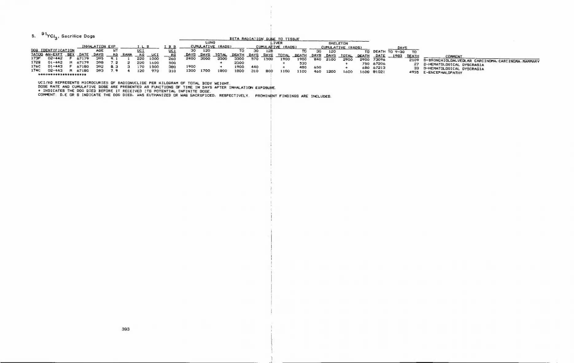
5.91yct3 ’ Sacrifice Dogs
INHALATION EXPDOG IDENTIFICATION AGE WTTATO0 AN-EXPT SEX DATE DAYS KQ RAN~173F 02-442 F 67179 395 9.1 i1728 01-442 M 67179 398 7.2 2176C 01-443 F 67180 392 8.3 3174C 02-443 M 67180 393 7.9 4
I. L.B. I.B.B.UCI UCI
IRG UC I KG220 1000 260220 1600 500170 1500 380120 970 310
BETA RADIATION DO~E TO TISSUELUNG _IVER SKELETON
CUMULATIVE {RADS) CUMUL~FIVE (RADS> CUMULATIVE (RADS)30 120 TO 30 I~D TO 30 120
DAYS .DAYS TOTAL DEATH DAYS DAY~ TOTAL DEATH DAYS DAYS ,ITOTAL DEATH2400 3000 3300 3300 570 t500 1900 1900 840 2100
+ 2300 + 5301900 + 1900 440 i + 480 6501300 1700 1800 1800 310 80~ 1100 1100 460 1200
UCI/KG REPRESENTS MICROCURIES OF RADIONUCLIOE PER KILOGRAM OF TOTAL BODY WEIGHT. !DOSE RATE AND CUMULATIVE DOSE ARE PRESENTED AS FUNCTIONS OF TIME IN DAYS AFTER INHALATION EXPOSURE,+ INDICATES THE DOG DIED BEFORE IT RECEIVED ITS POTENTIAL INFINITE DOSE. !COMMENT: D,E OR S INDICATE THE DOG DIED, WAS EUTHANIZED OR WAS SACRIFICED, RESPECTIVELY1 PROMINENT FINDINGS ARE INCLUDED.
DAYSTO DEATH TO 9-30 TO
DATE2900 2900 73096+ 750 6720&+ 680 67213
1600 1600 81021
1983 ,DEATH COMMENT2109 D-BRONCHIOLOALVEOLAR CARCINDMA~CARCINOMA, MAMMARY
27 D-HEMATOLOGICAL DYSCRASIA33 D-HEMATOLOgICAL DYSCRASIA
4955 E-ENCEPHALOPATHY
393
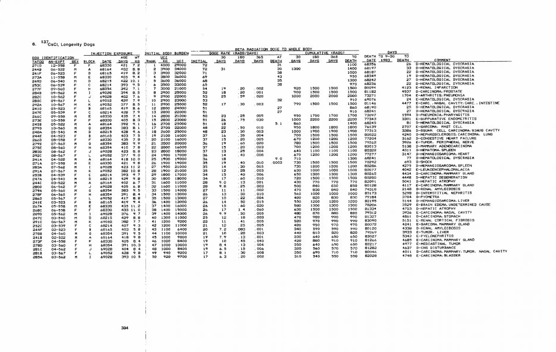
6. 137CSCI, Longevity Dogs
INJECTION EXPOSUREDOG IDENTIFICATION AGETATO0 AN-EXPT SEX BLOCK DATE DAYS271D 12-558 F F 69330 421 7.22448 06-522 M A 69164 402 8.8241F 06-523 F B 69165 419 8.2273A 11-558 M E 69330 405 9.4249D 06-540 M D 69215 422 10. I253C 06-539 F C 69214 393 9. 5277F 09-560 F H 68354 392 7.12848 09-562 M I 69028 394 8.5282C 10-562 F d 69028 402 7.6280C 09-567 F L 69052 429 7.9292A 10-567 M K 69052 377 9.52418 05-523 F B 68165 419 8.6247E 05-539 F C 69214 428 7.9266C 09-558 M E 69330 435 7.4273E 10-558 F F 68330 405 8.32459 05-522 M A 68164 392 9.1279D 10-560 M g 68354 383 8.1248A 05-540 M D 68215 428 9.6244E 04-523 F B 69165 403 7.5266D 08-558 F F 68330 435 7.82798 07-560 M g 68354 383 9.9275E 08-560 F H 6S354 410 7.82830 08-562 F d 69028 423 8.8292C 08-567 F L 69052 377 9.0241A 04-522 M A 68164 418 10.0271A 07-558 M E 69330 421 9.8283A 07-562 M I 69028 423 11.2291A 07-567 M K 69052 382 I0~8253B 04-539 F C 68214 393 9~7247A 04-540 M D 68215 429 9.8244C 03-522 M A 68164 402 6.7280D 06-562 F d 69028 405 6.8279A 05-560 M g 68354 383 9. 5278F 06-560 F H 68354 391 8.4286D 05-567 F L 69052 417 8.8241E 03-523 F B 68165 419 9.4267A 05-558 M E 68330 435 11.2268C 06-558 F F 68330 433 11.0289D 05-562 M I 69028 376 9.7247C 03-540 M D 68215 429 8.8291C 06-567 M K 69052 382 7.7252C 03-539 F C 68214 407 9.3244F 02-523 F B 68165 403 5.8278B 04-560 M g 69354 391 9. 52418 02-522 M A 69164 418 9.8273F 04-558 F F 68330 405 S. 42780 03-560 F H 68354 391 10.3281C 04-562 F J 69028 404 8.42818 03-567 F L 69052 428 9.8285A 03-562 M I 69028 393 10.5
~NITIAL BODY BURDENWT UClKQ iRANK KO VCI
I 4000 290002 3900 340003 3900 320004 3800 360005 3600 360006 3500 330007 3000 210008 2900 250009 2900 22000
10 2900 2300011 2900 2500012 2800 2400013 2800 2200014 2800 2100015 2800 2300016 2700 2500017 2700 2200018 2600 2500019 2100 1600020 2100 1600021 2000 2000022 2000 1600023 2000 1800024 1900 1700025 1900 19O0O26 1900 1900027 1900 2100028 1900 2100029 1800 1700030 1800 1800031 1600 11000
DOSE RATE (RADS/DAYS)
32 1600 1100033 1500 1400034 1500 1300035 1500 1300036 1400 1300037 1400 1600038 1400 1500039 1400 140004O 1300 1100041 1200 920042 1200 11OOO43 1100 640044 1100 1000045 1000 980046 1000 840047 1000 1000048 1000 840049 940 920050 920 9700
30INITIAL DAYS
72727169686554525353525151505151484837373637373636353535343428
31
19 . 2018 .2025 . 59
28272627262626262522212021191919191717
17 . 30
2326182223161519151314181914121517
9.09.8111310141517
9.9121112
7.210
7.910
8.46.58.16.3
BETA RADIATION DOSE TO WHOLE BODYCUMULATIVE (RADS)
180 30 180 365 TODAYS DAYS DAYS DAYS DEATH
¯ 25¯ 75
¯ 5030352060202540
403028406013251130225060
1.430184125
08320134513153020
365 ATDAYS DEATH
313038433538
¯ 002¯ 001¯ 020
32.003
2727
005030
5.1010003004005020003004OOS
9~0010 . 0003005OO3OO6010
.0010030020100O40150200600050O30O500500100300104O00400bO08002
DAYSDEATH TO 9-30 TO
DATE 1993 DEATH1100 68356
1300 1400 681971000 68187950 68349
1300 68242970 68236
920 1500 1500 1500 80094~900 1500 1500 1500 91182
~000 2000 2000 2000 73271910 69076
790 1500 1500 1500 91146860 69190910 69241
950 1700 1700 1700 730971000 2200 2200 2200 77343860 1400 68245950 1800 1800 1800 76139
1000 1900 1900 1900 77313700 1500 1500 1500 80022.670 1200 1200 1200 77204780 1500 1500 1500 79262700 1200 1200 1200 83013640 1100 1100 1100 80322670 1200 1200 1200 77277710 1300 68241730 1500 1500 1500 70292730 1200 1200 1200 80286630 1000 1000 1000 80077650 1300 1300 1300 90265720 1500 1500 1500 80280430 770 770 770 82091500 840 950 850 80128470 830 840 840 74-310560 1000 1000 1000 83173480 820 830 930 79184550 1200 1200 1200 82195
580 1300 1300 1300 78206630 1500 1500 1500 81334480 870 880 880 79312470 980 990 990 81327520 970 990 990 83090
480 950 970 970 90072340 590 590 590 80120440 810 820 820 79269330 640 650 650 93027420 880 910 910 81266350 640 650 650 82217320 560 570 570 81282350 690 710 710 80046310 540 550 550 82028
COMMENT26 D-HEMATOLOGICAL DYSCRASIA33 D-HEMATOLOGICAL DYSCRASIA22 D-HEMATOLOglCAL DYSCRASIA19 D-HEMATOLOGICAL DYSCRASIA27 D-HEMATOLOgICAL DYSCRASIA22 D-HEMATOLOGICAL DYSCRASIA
4123 E-RENAL INFARCTION4537 D-CARCINOMA, PROSTATE1704 E-ARTHRITIS~PNEUMONIA
24 E-HEMATOLOgICAL DYSCRASIA4477 E-CARC.,NASAL CAVITY;CARC.,INTESTINE
25 D-HEMATOLOgICAL DYSCRASIA27 D-HEMATOLOgICAL DYSCRASIA
1594 D-PNEUMONIA, PHARYNgITIS3301 E-SUPPURATIVE ENDOMETRITIS
81 D-HEMATOLOgICAL DYSCRASIA2707 E-SARCOMA, MAST CELL3386 D-SGUAM. CELL CARCINOMA, SINUS CAVITY4240 D-NEPHROSCLEROSIS;CARCINOMA, LUNG3162 D-CONgESTIVE HEART FAILURE392& E-TUMOR, PERIPHERAL NERVE5138 D-MAMMARY ADENOCARCINOMA4311 D-HEMATOMA, SPLEEN3147 D-HEHANGIOSARCOMA, HEART
77 D-HEMATOLOgICAL DYSCRASIA693 D-SHOCK
4275 D-HEMANOIOSARCOMA, SPLEEN4042 E-LEUKOENCEPHALOMALACIA4434 D-CARCINOMA, MAMMARY gLAND4448 D-HEPATIC DEGENERATION5041 D-HEPATIC ATROPHY4117 E-CARCINOMA, MAMMARY gLAND2148 D-RENAL AMYLOIDOSIS5298 D-INTERSTITIAL NEPHRITIS3784 D-PYOMETRA5144 D-HEMANgIOSARCOMA, LIVER3529 E-BRAIN EDEMA, UNDETERMINED CAUSE4753 D-HEPATIC ATROPHY3936 E-CARCINOMA, NASAL CAVITY4861 D-CARCINOMA, STOMACH5151 E-RENAL CORTICAL FIBROSIS4241 E-SARCOMA, MAMMARY gLAND
4338 D-RENAL AMYLOIDOSIS3933 E-TUMOR, LIVER5342 E-PYELONEPHRITIS4685 E-CARCINOMA, MAMMARY gLAND4977 E-MEDIASTINAL TUMOR4637 D-CNS DISTURBANCE4011 D-CARCINOMA, MAMMARY~TUMOR, NASAL CAVITY4748 E-CARCINOMA, BLADDER
394

6. 137CSCI, Longevity Dogs (continued)
INdECTION EXPOSURE INITIAL BODY BURDENDOg IDENTIFICATION AP-.-~ WT UCITATO0 AN-EXPT SEX BLOCK DATE DAYS ~g RANK KO287A 04-567 M ~ 69052 410 10.2 51 900 9200249C 02-540 M D 68215 422 S.S 52 900 79002&&A 03-558 M E 68330 435 9.1 53 890 8100248C 02-539 F C 68214 427 8.3 54 880 7300241C 01-522 M A &81b4 418 9.7 C 0 0244D 01-523 F B 68165 403 7.2 C 0 0251D 01-539 F C 68214 408 6.8 C 0 02478 01-540 M D 68215 429 9.4 C O 02708 01-558 M E 68330 423 8.4 C 0 0267D 02-558 F F 68330 435 7.4 C 0 0277A 02-560 H g 68354 392 9.4 C 0 0274E 01-560 F H 68354 419 7~ 1 C 0 0282A 01-562 M I 69028 402 8.& C 0 0283C 02-562 F d 69028 395 8.8 C 0 0286C 01-567 M K 69052 417 8.4 C 0 0282D 02-567 F L 69052 426 6.9 C 0 0
UCI INITIAL
BETA RADIATIO~DOSE RATE {RADS/DAYS)
30 180 365DAYS DAYS DAYS
17 8.0 .28 .00717 S.& .22 .002lb 7.9 .15 .004lb 7.6 .11 .002
UCI/KO REPRESENTS MICROCURIES OF RADIONUCLIDE PER KILOGRAM OF TOTAL BODY WEIGHT.DOSE RATE AND CUMULATIVE DOSE ARE PRESENTED AS FUNCTIONS OF TIME IN DAYS AFTER INJECTION OF 137-CS.+ INDICATES THE DOg DIED BEFORE IT RECEIVED ITS POTENTIAL INFINITE DOSE.COMMENT: D,E OR S INDICATE THE DOg DIED, WAS EUTHANIZED OR WAS SACRIFICED, RESPECTIVELY. PROMINENT
DOSE TO WHOLE BODY
ATDEATH
CUMULATIVE (RADS)30 180 365 TO
DAYS DAYS DAYS DEATH360 670 690 690340 700 710 710330 630 640 640330 blO 610 610
FINDINGS ARE INCLUDED.
DAYSDEATH TO 9-30 TO
DATE 1983 DEATH75332 247181015 454981056 447580318 448782313 526370081 64783054 5319
55375422
79225 391375239 244277154 3088
53585358
82011 470778030 3265
COMMENTE-NEUROFIBROSARCOMA, LIVERE-CARCINOMA, BLADDERE-HEMANOIOSARCOMA, SPLEENE-LIVER DEGEN.~CARC.,LIVER~CARC.,LUNgE-INTERSTITIAL NEPHRITISD-HEMOLYTIC ANEMIAJENDOCARDITISE-INTERSTITIAL NEPHRITIS
D-ENDOMETRITIS; PERITONITISD-RENAL AMYLOIDOSISD-CARCINOMA, MAMMARY gLAND
E-PYELONEPHRITISE-RENAL FAILURE~UREMIA
395
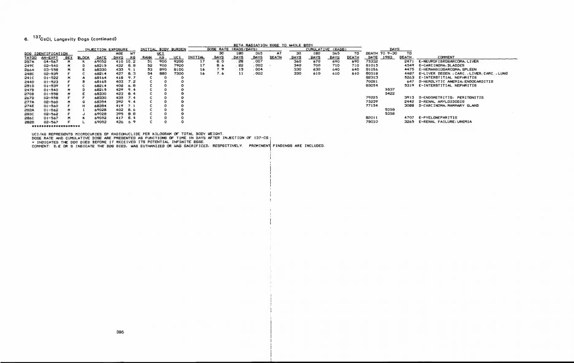
6. 137CSCI, Longevity Dogs (continued)
INJECTION EXPOSUREDOg IDENTIFICATION AgETATO0 AN-EXPT SEX BLOC~ DATE DAYS
INITIAL BODY BURDENWT UC IK(~ RAN~ KG UC I
287A 04-567 M K 69052 410 10.2 51 900 9200249C 02-540 M D 6S215 422 8.8 52 900 7900266A 03-558 M E 68330 435 9.1 53 890 8100248C 02-539 F C 68214 427 8.3 54 880 7300241C 01-522 M A 68164 418 9. 7 C 0 0244D 01-523 F B 68165 403 7.2 C 0 0251D 01-539 F C 68214 408 6.8 C 0 02478 01-540 M D 68215 429 9.4 C 0 02708 01-558 M E 68330 423 8.4 C 0 0267D 02-558 F F 68330 435 7.4 C 0 0277A 02-560 M g 68354 392 9.4 C 0 0274E 01-560 F H 68354 419 7. I C 0 0282A 01-562 M I 69028 402 8.6 C 0 0283C 02-562 F J 69028 395 8.8 C 0 0286C 01-567 M R 69052 417 8.4 C 0 0282D 02-567 F L 69052 426 6.9 C 0 0
BETA RADIAT10NDOSE RATE (RADS/DAYS)
30 180 365INITIAL DAYS DAYS DAYS
17 8.0 .28 .00717 8.6 .22 .00216 7.9 ~15 .00416 7.6 .11 .002
)OSE TO WHOLE BODYCUMULATIVE (RADS)
AT 30 180 365 TO)EATH DAYS DAYS DAYS DEATH
360 670 690 690340 700 710 710330 630 640 640330 610 610 610
UCI/~g REPRESENTS MICROCURIES OF RADIONUCLIDE PER KILOGRAM OF TOTAL BODY WEIGHT.DOSE RATE AND CUMULATIVE DOSE ARE PRESENTED AS FUNCTIONS OF TIME IN DAYS AFTER INJECTION OF 137-CS.+ INDICATES THE DOG DIED BEFORE IT RECEIVED ITS POTENTIAL INFINITE DOSE. ICOMMENT: D,E OR S INDICATE THE DOg DIED, WAS EUTHANIZED DR WAS SACRIFICED, RESPECTIVELY. PROMINEN FINDINGS ARE INCLUDED.
DAYSDEATH TO 9-30 TO
DATE 1983 DEATH75332 247181015 454981056 447580318 448782313 526370081 64783054 5319
55375422
79225 391375239 244277154 3088
53585358
82011 470778030 3265
COMMENTE-NEUROFIBROSARCOMA, LIVERE-CARCINOMA, BLADDERE-HEMANOIOSARCOMA, SPLEENE-LIVER DEOEN. JCARC.,LIVER~CARC.,LUNQE-INTERSTITIAL NEPHRITISD-HEMOLYTIC ANEMIA~ENDOCARDITISE-INTERSTITIAL NEPHRITIS
D-ENDOMETRITIS; PERITONITISD-RENAL AMYLOIDOSISD-CARCINOMA, MAMMARY GLAND
E-PYELONEPHRITISE-RENAL FAILURE;UREMIA
395
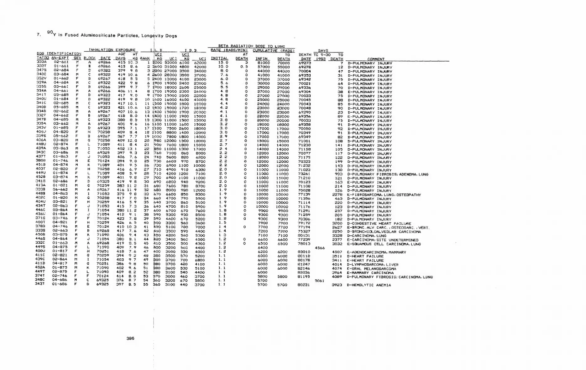
7. 90y in Fused Aluminosilicate Particles, Longevity Dogs
INHALATION EXPOSURE I.L.BDOg IDENTIFICATION AGE WT UCITATO0 AN-EXPT SEX BLOC~. DATE DAYS KG RANR !’KG UCI
A 69266 415 10.3B 69266 415 8.6D 69322 379 9.8C 69322 419 10.6B 69267 418 5.5C 69322 422 9.8B 69266 399 9.7A 69266 406 11.4D 69323 417 9.0D 69322 419 9.8C 69323 417 10.1C 69323 421 10.6A 69267 407 10.6B 69267 418 8.0C 69323 380 8.5A 69267 400 9.6D 69323 395 7.1H 70258 409 8.4B 69267 367 7.70 70258 409 12.0L 71089 411 8.4I 71053 402 13. IC 69325 397 9.3J 71053 406 7.6E 70124 394 9.0K 71089 401 9.5H 70258 416 6.9L 71089 408 5.9K 71089 401 9.8D 69325 419 9.8G 70259 383 11.2A 69267 416 11,9I 71053G 70258H 70259d 71053I 71054J 71054F 70124H 70259E 70124B 69268
71090d 71054A 69268L 71090H 70251G 70259I 71054 403 9.7G 70251 386 9.8K 71090 402 9.6L 71090 409 8.2F 70124 414 8.0C 69325 376 8.7D 69325 397 85
333A 02-661 M333T 01-661 F347S 02-684 F340C 03-684 M332V 01-662 F339A 04-684 M3358 03-661 F334A 04-661 M341T 03-685 F340U 01-684 F341C 02-685 M3408 05-685 M3348 02-662 M332T 04-662 F3478 04-685 M335A 03-662 M343V 01-685 F406U 04-820 F339S 05-662 F406A 03-820 M448U 02-874 F439A 03-863 M343C 03-686 M437T 01-863 F3808 01-746 M451B 04-874 M403T 02-820 F449U 01-874 F452B 03-874 M3418 02-686 F413A 01-821 M333B 06-662 M4488 04-863 M402C 01--820 M404U 03-821 F434T 02-863 F446C 03-864 M436U 01-864 F371S 03-746 F400T 04-821 F3788 04-746 M333S O2-663 F450B 03-875 M446S 04-864 F332C 01-663 M449S 04-875 F400U 01-817 F411C 02-821 M439C 02-864 M411D 04-817 M452A 01-875 M449T 02-875 F374T 02-746 F348C 04-686 M343T 01-686 F
I, B. B.BETA RADIATION DOSE TO LUNG
RATE (RADS/MIN) CUMULATIVE (RADS)AT TO
UCI INITIAL DEATH INFIN. DEATH
DAYSDEATH TO 9-30 TO
DATE 1983 DEATHi 5200 53000 6100 620002 ~&O0 31000 4800 420003 ~800 27000 3500 340004 ~600 28000 3500 370005 ~400 13000 4100 230006 ~900 19000 2400 230007 ~900 18000 2600 250008 ~700 19000 2300 260009 1!700 15000 2500 22000
10 i1600 16000 4600 4500011 1500 15000 1800 1800012 1400 15000 1700 1800013 Ii400 15000 1900 2000014 11400 Ii000 1900 1500015 1700 11000 1500 1300016 ~100 11000 1600 1500017 11100 7500 2600 1800018 I!I00 8800 1400 1200019 1000 7800 1800 1400020 ~i80 12000 1~0 1800021
~7600 1800 15000
22 11000 1300 1700023 ~60 7100 860 800024 ?40 5600 820 620025 T30 6600 970 870026 ~30 6900 11.00 1000027 ~I0 4900 910 630028 ~I0 4200 1200 710029 ~00 6900 II00 1100030 690 6800 940 920031 ~80 7600 780 870032 680 8000 980 12000
375 9~8 33 670 6600 850 8300417 7.0 34 660 4700 790 5500416 5~9 35 ~40 3700 860 5100415 7.3 36 ~40 4700 810 5900380 11.2 37 600 6700 670 7600412 9.1 38 590 5300 930 8500423 7.8 39 590 4600 670 5200426 6.5 40 #00 3300 560 3600410 10.3 41 490 5100 700 7000417 7.6 42 460 3500 590 4400406 9.4 43 ~50 4200 560 5300380 8~I 44 ~20 3400 560 4500419 8.5 45 410 3500 500 4300409 7.9 46 ~00 3200 560 4400418 7.6 47 #00 3000 530 4000394 9.2 48 380 3500 570 5200
49 480 3700 700 680050
~803700 420 4100
51 80 3600 530 510052 380 3100 540 440053 370 3000 460 370054 360 3200 670 580055 ~60 3100 440 3700
15.0I0.08.07.66.05.65.54.84.84.84.44.24.14.13.83.23.03.02.92.72.72.42.22.22.22.22.02.02.02.02.01.91.91.91.91.91.81.81.8I 5I 41 41 31 21 21 21.21.11.11.11.1I.I1.11.1lJI
3 81000 70000 692730.5 57000 55000 69278
0 44000 44000 700040 41000 41000 693530 37000 37000 693420 30000 30000 700210 29000 29000 693360 27000 27000 693040 27000 27000 700330 25000 25000 700450 24000 24000 700430 23000 23000 700480 23000 23000 692900 22000 22000 693560 20000 20000 700330 18000 18000 693580 17000 17000 700500 17000 17000 703490 17000 17000 693490 15000 15000 710010 14000 14000 712300 14000 14000 711580 12000 12000 700770 12000 12000 711750 12000 12000 703230 12000 12000 712320 11000 11000 710230 11000 11000 732610 11000 11000 712100 11000 II000 701230 11000 11000 711080 11000 ii000 700280 10000 10000 771390 10000 10000 713560 10000 10000 711140 10000 10000 711760 9500 9500 712910 9300 9300 712590 9300 9300 70306
7900 7900 791720 7700 7700 77194
7200 7200 753277100 7100 80131
0 6600 6600 772390 6500 6500 78013
64006200 6200 830106000 6000 801186000 6000 801786000 6000 812476000 6000 821466000 820365800 5800 8119557005700 5700 80231
4566
5061
COMMENT7 D-PULMONARY INJURY
12 D-PULMONARY INJURY47 D-PULMONARY INJURY31 D-PULMONARY INJURY75 D-PULMONARY INJURY64 D-PULMONARY INJURY70 D-PULMONARY INJURY38 E--PULMONARY INVURY75 D-PULMONARY INJURY88 D-PULMONARY INJURY85 D-PULMONARY INJURY90 D-PULMONARY INJURY23 D-PULMONARY INJURY89 E-PULMONARY INJURY75 D-PULMONARY INJURY91 D-PULMONARY INdURY92 D-PULMONARY INJURY91 D-PULMONARY INdURY82 D-PULMONARY INJURY
108 D-PULMONARY INJURY141 D-PULMONARY INJURY105 D-PULMONARY INJURY117 D-PULMONARY INJURY122 D-PULMONARY INJURY199 D-PULMONARY INJURY143 D-PULMONARY INJURY130 D-PULMONARY INJURY903 D-PULMONARY FIBROSIS~ADENOMA, LUNG121 D-PULMONARY INJURY163 E-PULMONARY INJURY214 D-PULMONARY INJURY126 D-PULMONARY INJURY
2278 E-FIBRDSARCOMA, LUNG~OSTEOPATHY463 D-PULMONARY INJURY220 D-PULMONARY INJURY123 D-PULMONARY INJURY237 D-PULMONARY INJURY205 D-PULMONARY INdURY182 D-PULMONARY INJURY
3200 D-CONGESTIVE HEART FAILURE2627 E-BRONC. ALV. CARC. ~OSTEOSARC~,VERT.2250 D-BRONCHIOLOALVEOLAR CARCINOMA3328 D-CARCINOMA, LUNg2377 E-CARCINOMA~SITE UNDETERMINED3032 E-SQUAMOUS CELL CARCINOMA, LUNG
4507 E-ADENOCARCINOMA, MAMMARY3511 D-HEART FAILURE3411 E-HEART FAILURE4014 D-LYMPHOSARCOMA, LIVER4074 E-ORAL MELANOSARCOMA3964 E-MAMMARY CARCINOMA4089 D-PULMONARY FIBROSIS~CARCINOMA, LUNG
3923 D-HEMOLYTIC ANEMIA
396
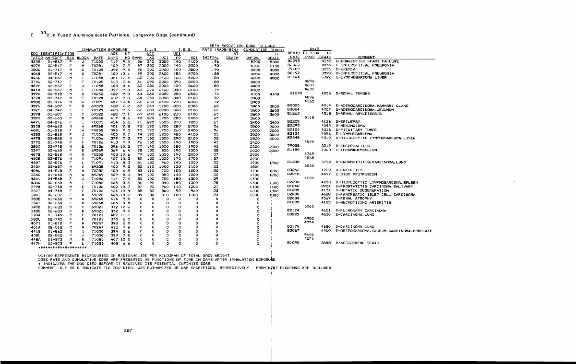
7. 90y in Fused Aluminosilicate Particles, Longevity Dogs (continued)
INHALATION EXPOSURE I.L.B. I. B. B.DOg IDENTIFICATION AgE WT UCI U¢ITATO0 AN-EXPT SEX BLOCK DATE DAYS Kg RANK Kg UCI Kg UCI4348 01-867 F d 71055 417 9.4 56 340 3200 440 41004078 02-817 F H 70251 402 7.2 57 320 2300 440 3200380D 01-747 M E 70125 395 9.4 58 300 2900 400 38004068 03-817 M g 70251 402 12.1 59 300 3600 480 5700446D 04-867 M I 71055 381 11.4 60 300 3400 460 5200375U 02-747 F F 70125 415 7.6 61 290 2200 390 30004378 03-867 F d 71055 408 8.4 62 280 2300 430 3600441A 02-867 M I 71055 399 9.0 63 270 2400 340 3100399A 02-818 M g 70252 422 9.0 64 260 2300 280 2500377B 03-747 M E 70125 412 9.0 65 250 2300 340 3100450C 01-876 M K 71091 407 10.4 66 250 2600 270 2800339U 04-687 F D 69328 428 7.2 67 240 1700 320 23003728 04-747 F F 70125 423 9.6 68 230 2200 320 31003398 01-687 M C 69328 428 9.1 69 230 2100 230 21003328 03-663 F B 69268 419 8.6 70 220 1900 280 2400447U 04-876 F L 71091 414 6.6 71 220 1500 270 18003358 04-663 M A 69268 401 9.8 72 190 1900 280 2700408U 01-818 F H 70252 395 9.0 73 190 1700 260 24004388 01-868 F J 71056 405 9.7 74 190 1800 420 41004478 03-868 M I 71056 379 7.3 75 180 1300 290 21003778 01-748 F F 70126 413 9.9 76 150 1500 190 1900380C 03-748 M E 70126 396 10.2 77 140 1500 180 1900339T 02-665 F B 69269 369 6.4 78 130 830 190 12004078 03-818 M g 70252 403 10.6 79 130 1300 190 2000450E 03-876 M K 71091 407 10.2 80 130 1300 170 1700448T 02-876 F L 71091 413 8.3 81 120 960 140 1200343A 03-687 M C 69328 400 9.3 82 110 1000 120 li00405U 04-818 ? H 70252 403 6.8 83 110 720 150 1000334C 01-665 M A 69269 409 8.3 84 100 850 140 1200436V 04-868 F d 71056 414 7.4 85 100 750 180 13004388 02-868 M I 71056 405 8.6 86 98 840 150 13003798 02-748 M E 70126 402 10.7 87 90 960 110 1200372T 04-748 F F 70126 424 10.4 88 83 860 92 960340T 02-687 F D 69328 425 10.2 89 80 810 100 1100333E 01-660 M A 69265 414 9.5 C 0 0 0 0334T 02-660 F B 69265 405 8.5 C 0 0 0 03498 01-683 M C 69321 372 12.2 C 0 O 0 03488 02-683 F D 69321 372 9.0 C 0 0 0 0378A 01-745 M E 70121 407 11.6 C O 0 0 0383U 02-745 F F 70121 375 6.0 C 0 0 0 0407T 01-812 F H 70247 398 8.0 C 0 0 0 0401A 02-812 M g 70247 413 9.2 C 0 0 0 04418 01-862 M I 71050 394 8.6 C 0 0 0 04380 02-862 F J 71050 399 7,8 C 0 0 0 0448A 01-873 M K 71085 407 10.0 C 0 0 0 0447W 02-873 F L 71085 408 6.6 C 0 O 0 0
BETA RADIATION DOSE TO LUnGRATE {RADS/MIN)
ATINITIAL DEATH
9693908888888079757272696965656556565552434338373733333g302928272323000000000000
CUMULATIVE IRADS)I TO
INFIN. "DA_D_D_D_D_D_D_D_D~5300 53005100 51004800 48004800 48004800 48004800 i4400 i
43004100 4100390039003800 38003600 36003600
~60036003400 34003000 ~ooo3000 30003000 30002800 280024002300 23002000 20002000200019oo %90018oo17oo ~7001700 1iTgO15001500 ~5001400 1~4001300 I~3001300 1~00
000000000000
UCI/Kg REPRESENTS MICROCURIES OF RADIONUCLIDE PER KILOGRAM OF TOTAL BODY WEIGHT.DOSE RATE AND CUMULATIVE DOSE ARE PRESENTED AS FUNCTIONS OF TIME IN DAYS AFTER INHALATIONEXPOSURE.+ INDICATES THE DOg DIED BEFORE IT RECEIVED ITS POTENTIAL INFINITE DOSE.COMMENT: D~E OR S INDICATE THE DOG DIED, WAS EUTHANIZED OR WAS SACRIFICED, RESPECTIVELY. PROMIN
DAYSDEATH TO 9-30 TO
DATE 1983 DEATH COMMENT82295 425883062 455979189 335180197 359881124 3722
489646014601
81290 405648964565
80325 401483084 470781263 4318
511882209 413680293 404282105 423682134 409682348 4310
489579058 321981189 4303
47694565
81230 37925058
83266 476282018 4497
460082288 425081042 393481285 417782208 462882084 456781005 4123
506582174 460183223 4850
49004774
83179 468083067 4400
46064571
81090 3658
D-CONgESTIVE HEART FAILUREE-INTERSTITIAL PNEUMONIAD-UREMIAD-INTERSTITIAL PNEUMONIAE-LYMPHOSARCOMA, LIVER
D-RENAL TUMORS
E-ADENOCARCINOMA, MAMMARY GLANDD-ADENOCARCINOMA, JEJUNUMD-RENAL AMYLOIDOSIS
D-EPILEPSYD-MENINgIOMAE-PITUITARY TUMORE-LYMPHOSARCOMAD-HISTEOCYTIC LYMPHOSARCOMA, LIVER
D-ENCEPHALITISE-THROMBOEMBOLISM
D-ENDOMETRITIS;CARCINOMA, LUNg
D-ENTERITISE-DISC PROTRUSION
D-HISTEOCYTIC LYMPHOSARCOMA, SPLEEND-PROSTATITIS~CARCINOMA, SALIVARYD-HEPATIC DEGENERATIOND-PANCREATIC ISLET CELL CARCINOMAE-RENAL ATROPHYE-NECROTIZINg ARTERITIS
E-PULMONARY CARCINOMAE-CARCINOMA, LUNg
D-CARCINOMA, LUNgE-OSTEOSARCOMA, SACRUM;CARCINOMA, PROSTATE
D-ACCIDENTAL DEATH
{NT FINDINGS ARE INCLUDED.
397
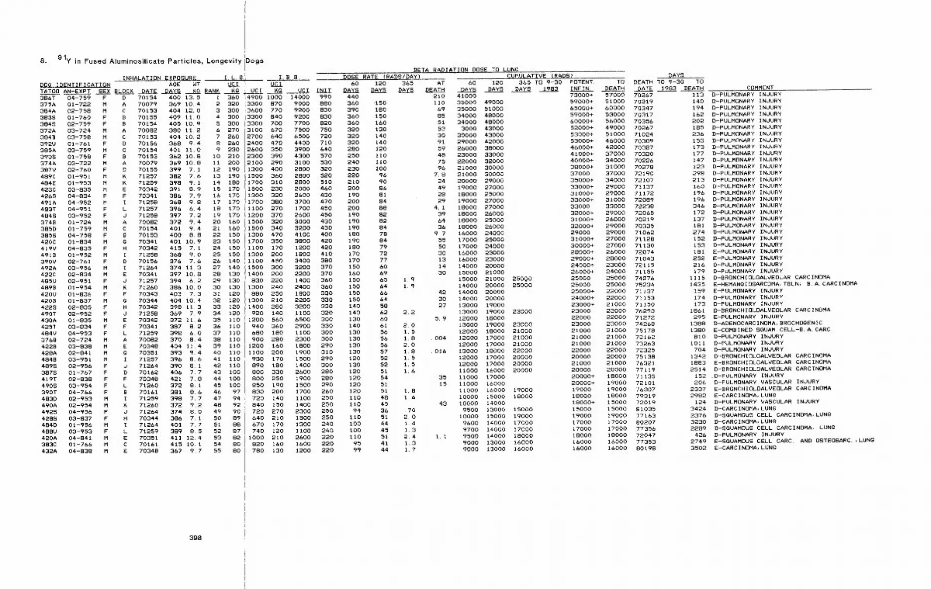
8. 91y in Fused Aluminosilicate Particles, Longevity Dogs
INHALATION EXPOSURE I.L.B,jDOg IDENTIFICATION AQE WT UCITATO0 AN-EXPT SEX BLOC~ DATE DAYS K~ RANK KQ ’ UCI386T 04-759 F D 70154375A 01-722 M A 70079384A 02-758 M C 701533838 01-760 F B 701553848 02-759 F B 70154372A 03-724 M A 700823848 03-758 M C 70153392U 01-761 F D 70156385A 03-759 M C 701543938 01-758 F B 70153374A 03-722 M A 70079387V 02-760 F D 70155489C 01-951 M K 71257484E 01-953 M K 71259423C 03-835 M E 703424268 04-834 F F 70341491A 04-952 M I 71258483T 04-951 F L 712574848 03-952 F J 712583748 01-724 M A 700823850 01-759 M C 701543858 04-758 F B 70153420C 01-834 M G 70341419V 04-835 F H 703424918 01-952 M I 71258390V 02-761 F 0 70156492A 03-956 M I 71264422C 02-834 M E 70341485U 02-951 F d 712574898 01-954 M R 71260420U 01-836 F F 703434208 01-837 M g 703444228 02-835 P H 70342490T 02-952 F d 71258430A 01-835 M425T 03-834 F484V 04-953 F3768 02-724 M4228 03-838 M428A 02-841 M4848 03-951 M4898 02-956 F3878 01-767 F419T 02-838 F4908 03-954 F390T 04-766 F483D O2-953 M
400 13.5369 10.4404 12.0409 11.0405 10.9 5 300380 11.2 6 270404 10.2 7 260368 9.4 8 260401 11.0 9 230362 10.8 I0 210369 10.8 11 200399 7.1 12 190382 7.6 13 190398 9.1 14 180391 8,9 15 170386 7.9 16 170368 9.8 17 170396 6,4 18 170397 7,2 19 170372 9,4 20 160401 9.4 21 160400 8.8 22 150401 10.9 23 150415 7.1 24 150368 9,0 25 150376 7.6 26 140374 11,3 27 140397 10.8 28 130394 6.2 29 130386 10.0 30 130403 7.3 31 120404 10.4 32 120398 11.3 33 120369 7,9 34 120
35 II036 Ii037 11038 11039 11040 11041 11042 11043 i0044 i0045 i0046 9747 94
E 70342 372 11.6F 70341 387 8,2L 71259 398 6.0A 70082 370 8.4E 70348 404 11.4Q 70351 393 9.4I 71257 396 8.6J 71264 390 8.1D 70162 406 7.7F 70348 421 7.8L 71260 372 8, IB 70161 381 8.6I 71259 398 7.7
I. B B.UCI
X~ UCI INITd1 360 14900 I000 14000 9902 320 13300 870 9000 8803 300 i 3600 770 9200 8304 300 3300 840 9200 830
3300 700 7700 8203100 670 7500 7502700 640 6500 7202400 470 4400 7102600 350 3900 6402300 390 4300 5702100 290 3100 5301300 400 2800 5201500 360 2800 5201700 310 2800 5101500 230 2000 4601300 320 2600 4301700 380 3700 470ii00 270 1700 4501200 370 2600 4501500 320 3000 4301500 340 3200 4301300 470 4100 4001700 350 3800 4201100 170 1200 4201300 200 1800 4101100 450 3400 3801500 300 3200 3701400 200 2200 370
830 220 1400 3601300 240 2400 360
880 250 1800 3301300 210 2200 3301400 280 3200 330
920 140 1100 3201200 560 6500 300940 360 2900 330680 180 1100 300900 280 2300 300
1200 160 1800 2901100 200 1900 310930 170 1500 290890 180 1400 300800 330 2600 280800 250 1900 280850 190 1500 290830 200 1700 260720 140 1100 250
490A 02-954 M4928 04-956 F4288 03-837 F484D 01-956 M488U 03-953 F420A 04-841 M383C 01-766 M432A 04-838 M
71260 372 9.2 48 92d 71264 374 8.0 49 90H 70344 386 7.1 50 89I 71264 401 7.7 51 88L 71259 389 8.5 52 87E 70351 411 12~4 53 82C 70161 415 I0. 1 54 80E 70348 367 9.7 55 80
398
840 150 1400 250720 270 2300 250640 210 1500 250670 170 1300 240740 120 1100 240
I000 210 2600 220820 160 1600 220780 t30 1200 220
BETA RADIATION DOSE TO LUN~DOSE RATE (RADS/DAY)
60 120 365 ATDAYS DAYS DAYS DEATH
440 210360 ’150 Ii0390 180 &9360 150 85360 160 51320 130 53320 140 30320 140 91280 120 59250 110 48240 llO 75230 i00 96220 96210 90200 86190 81200 84200 88190 82190 82190 84180 78190 84180 79170 72170 77150 60160 69150 65 1.9150 64 I~9150 66150 64140 58140 62 2.2130 60140 61 2.0130 56 1.5130 56 1.8130 56 2.0130 57 1.8120 52 1.5130 52 1,5120 51 l,b120 54120 51120 51 1.8II0 48 1.6110 4594 36 .70
ii0 51 2.0i00 44 1.4i00 45 1.3ii0 51 2.4
95 41 1.399 44 1.7
CUMULATIVE (RADS)365 TO 9-30 POTENT.
1983 INFIN.73000+ 57000 7026759000+ 51000 7021965000+ 60000 70347
60 120DAYS DAYS DAYS
4100035000 4900035000 5100034000 4800034000 48000
3000 4300030000 4300029000 4200026000 3800023000 3300022000 3200021000 30000
7.8 21000 3000024 20000 2900049 19000 2700028 18000 2500029 19000 27000
4.1 18000 2700039 18000 2600064 18000 2500036 18000 26000
9.7 16000 2400055 17000 2500050 17000 2400030 16000 2300013 16000 2300014 14000 2000030 15000 21000
15000 21000 2500014000 20000 25000
42 14000 2000030 14000 2000027 13000 19000
13000 19000 230005.9 12000 18000
13000 19000 2300012000 18000 21000
.004 12000 17000 2100012000 17000 21000
.016 13000 18000 2200012000 17000 2000012000 17000 20000II000 16000 20000
35 11000 1700015 Ii000 16000
I1000 16000 19000I0000 15000 18000
43 i0000 140009500 13000 15000
10000 15000 190009600 14000 170009700 14000 17000
1.1 9500 14000 180009000 13000 160009000 13000 16000
DAYSTO DEATH TO 9-30 TO
DEATH DATE 1983 DEATH COMMENT
59000+ 53000 7031760000+ 56000 7035652000+ 49000 7026753000+ 51000 7102453000+ 46000 7030946000+ 42000 7032741000+ 37000 7033040000+ 34000 7022638000+ 31000 7027837000 37000 7219035000+ 34000 7210733000+ 29000 7113731000+ 29000 7117233000+ 31000 7208933000 33000 7223832000+ 29000 7206531000+ 26000 7021932000+ 29000 7033529000 29000 7106231000+ 27000 7112830000+ 27000 7113028000+ 26000 7207429000+ 28000 7104324000+ 23000 7211526000+ 24000 7115525000 25000 7427625000 25000 7523425000+ 22000 7113724000+ 22000 7115323000+ 21000 7115023000 23000 7629322000 22000 7127223000 23000 7426821000 21000 7517821000 21000 7216221000 21000 7326322000 22000 7232520000 20000 7513821000 21000 7632120000 20000 7711920000+ 18000 7113520000+ 19000 7210119000 19000 7630718000 18000 7931918000+ 15000 7201915000 15000 8103519000 19000 7716317000 17000 8020717000 17000 7735618000 18000 7204716000 16000 7735316000 16000 80198
113 D-PULMONARY INJURY140 D-PULMONARY INJURY194 D-PULMONARY INJURY162 D-PULMONARY INJURY202 D-PULMONARY INJURY185 D-PULMONARY INJURY236 D-PULMONARY INJURY153 D-PULMONARY INJURY173 D-PULMONARY INJURY177 D-PULMONARY INJURY147 D-PULMONARY INJURY123 D-PULMONARY INJURY298 D-PULMONARY INJURY213 D-PULMONARY INJURY160 D-PULMONARY INJURY196 D-PULMONARY INJURY196 D-PULMONARY INJURY346 D-PULMONARY INJURY172 D-PULMONARY INJURY137 D-PULMONARY INJURY181 D-PULMONARY INJURY274 D-PULMONARY INJURY152 D-PULMONARY INJURY153 D-PULMONARY INJURY181 E-PULMONARY INJURY252 E-PULMONARY INJURY216 D-PULMONARY INJURY179 D-PULMONARY INJURY
1115 D-BRONCHIQLOALVEOLAR CARCINOMA1435 E-HEMANGIOSARCOMA, TBLN~ B.A. CARCINOMA
159 E-PULMONARY INJURY174 D-PULMONARY INJURY173 D-PULMONARY INJURY
1861 D-8RONCHIOLOALVEOLAR CARCINOMA295 E-PULMONARY INJURY
1388 D-ADENOCARCINOMA, BROCHOgENIC
1380 E-COMBINED SOUAM. CELL-B.A. CARC.810 D-PULMONARY INJURY
I011 D-PULMONARY INJURY704 D-PULMONARY INJURY
1342 D-8RONCHIOLOALVEOLAR CARCINOMA1883 E-BRONCHIOLOALVEOLAR CARCINOMA2514 D-8RONCHIOLOALVEOLAR CARCINOMA
152 .D-PULMONARY INJURY206 D-PULMONARY VASCULAR INJURY
2337 E-BRQNCHIOLOALVEOLAR CARCINOMA2982 E-CARCINOMA, LUN~
124 D-PULMONARY VASCULAR INJURY3424 D-CARCINOMA, LUNO2376 D-SQUAMOUS CELL CARCINOMA, LUNO3230 D-CARCINOMA, LUNg2289 D-SQUAMOUS CELL CARCINOMA, LUNg
426 D-PULMONARY INJURY2749 E-SQUAMOUS CELL CARC. AND OSTEOSARC.,LUNg3502 E-CARCINOMA, LUNQ
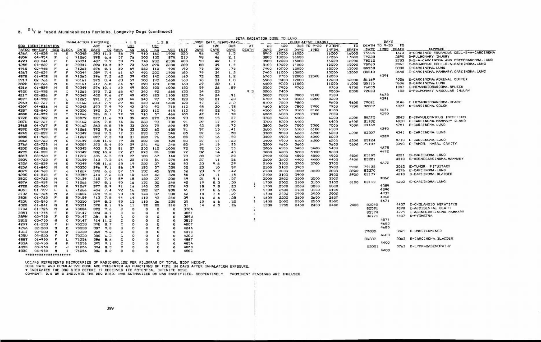
8. 91y in Fused Aluminosilicate Particles, Longevity Dogs (continued)
INHALATION EXPOSURE I.L.B. 1. B.B.DOg IDENTIFICATION AgE WT UCI UCI 60 120TATOO AN-EXPT ~SEX BLOCK DATE DAYS KQ RANK Kg UCI Kg UCI INIT DAYS DAYS426A 01-838 M g 70348 393 11.5 56 79 910 160 1900 220 96 42485W 04-954 F d 71260 398 6.6 57 76 500 130 870 210 9S 46422T 03-841 F F 70351 407 9.9 58 75 740 230 2300 200 93 424258 04-837 F H 70344 390 10.5 59 73 760 270 2800 200 88 394918 02-958 F d 71265 376 8.1 60 69 560 110 900 190 75 30426T 02-837 F F 70344 389 7.4 61 67 490 200 1500 180 79 344878 01-958 M ~ 71265 396 7.2 62 59 430 140 1000 160 72 32391T 02-766 F D 70161 375 8.4 63 59 500 190 1600 160 70 313828 03-766 M C 70161 417 6.8 64 57 390 120 850 160 69 30431A 01-839 M E 70349 376 10.1 65 49 500 100 1000 130 59 26492C 03-958 M I 71265 375 7.2 66 47 340 92 &60 130 54 23421T 02-836 F F 70343 402 9.6 67 45 430 120 1100 120 54 24489T 04-958 F L 71265 391 7.7 68 44 340 97 750 120 50 21396X 03-767 F B 70162 363 7.9 69 44 340 200 1&O0 120 57 27430C 04-836 M g 70343 373 7.9 70 42 340 90 710 110 48 20428T 02-840 F H 70350 392 5.7 71 41 230 110 610 110 49 224888 04-959 M I 71266 396 8.1 72 39 310 57 460 100 47 213728 02-722 M A 70079 377 11.6 73 35 400 270 3100 93 38 15387U 02-767 F B 70162 406 7.8 74 34 260 93 730 91 39 173968 04-767 F D 70162 363 8.8 75 33 300 78 &90 93 42 194890 02-959 M K 71266 392 9.6 76 33 320 65 630 91 37 154248 03-839 F H 70349 398 9.3 77 31 290 37 340 85 37 164888 01-960 F d 71267 397 7.3 78 31 230 76 560 85 37 16386A 03-763 M C 70159 405 11.0 79 30 330 38 420 83 37 17376A 03-725 M A 70084 372 8.4 SO 29 240 40 340 80 34 154200 03-836 M E 70343 403 9.3 81 27 250 110 1000 72 32 154298 04-839 F F 70349 382 10.2 82 27 270 86 SSO 72 32 14484T 03-960 F L 71267 406 6.3 83 27 18g 67 440 73 30 12383V 04-763 F D 70159 413 7.3 84 23 170 51 370 64 27 11422A 02-839 M g 70349 405 11.6 85 19 230 37 430 53 23 9.6425A 01-840 M g 70350 396 9.1 86 19 180 57 520 52 23 104878 04-960 F d 71267 398 6.6 87 19 130 45 290 52 23 9.94208 04-840 F H 70350 410 7.6 88 18 140 42 320 50 23 11382C 02-763 M C 70159 415 7.4 89 18 130 27 200 49 21 9.1487A 03-959 M I 71266 397 8.1 90 1& 130 30 240 42 19 8.34928 02-960 M K 71267 377 8.9 91 16 140 30 270 43 18 7.8485T 01-959 F L 71266 404 7.4 92 16 120 27 200 41 19 8.6373A 02-725 M A 70084 378 9.0 93 15 140 37 330 42 18 7.4383W 01-763 F B 70159 413 7.8 94 14 110 43 340 39 16 6.6423U 03-840 F F 70350 399 8.3 95 13 110 36 220 35 15 6.64328 01-841 M E 70351 370 8.1 96 11 92 25 210 31 14 6.5370A 01-725 M A 70084 393 9.6 C 0 0 0 0 370A385T 01-755 F B 70147 394 8. i C 0 0 0 0 385T389W 02-755 F D 70147 381 8.4 C 0 0 0 0 389W3818 03-755 M C 70147 414 11.2 C O 0 0 0 3818420T 01-833 F H 70338 398 8.7 C O O O 0 420T424A 02-833 M E 70338 387 9.8 C 0 0 0 0 424A4318 03-833 M g 70338 365 9.2 C 0 0 0 0 4318428U 04-833 F F 70338 380 6.3 C 0 0 0 0 428U488T 01-950 F L 71256 386 8.6 C 0 0 0 0 488T483A 02-950 M K 71256 395 9.1 C O 0 0 0 483A4858 03-950 F J 71256 394 8.3 C 0 0 O 0 4858488C 04-950 M I 71256 386 8.2 C 0 0 0 0 488C
BETA RADIATION DOSE TO LUNgDOSE RATE (RADS/DAY)
365DAYS
1.52.11.71.4.751.21.21.0I.I.89
.91
. 601.2
58767837597541585063555548303629394245373O23352728222&
ATiDEATH
9.3
CUMULATIVE (RADS)60 120 365 TO 9-30 POTENT. TO
DAYS DAYS DAYS 1983 INFIN. DEATH
DAYSDEATH TO 9-30 TO
DATE 1983 DEATH8900 13000 16000 16000 16000 751358800 13000 16000 17000 17000 792288500 12000 15000 16000 16000 782128100 12000 14000 15000 15000 782637400 10000 12000 12000 12000 803587400 11000 13000 13000 13000 803406700 9700 12000 12000 12000 43916500 9400 12000 12000 12000 811696400 9200 11000 11000 11000 801155500 7900 9700 9700 9700 760055200 7400 9000+ 8300 720835000 7200 9000 9100 9100 46784700 6700 8100 8200 8200 43915100 7500 9500 9600 9600 790214600 6500 7800 7900 7900 7900 823374500 6500 8100 8100 8100 46714300 6200 7700 7800 7800 43903700 5200 6100 6200 6200 802703700 5300 6400 6400 6400 811823800 5600 7000 7000 7000 7000 831653600 5100 6100 6100 6100 43903500 5000 6200 6200 6200 6200 823073400 4900 6000 6000 6000 43893400 4900 6100 6200 6200 6200 831243200 4~00 5600 5600 5600 791873000 4300 5400 5400 5400 4678
3000 4200 5200 5300 5300 46722900 4000 4800 4800 4800 4800 831052600 3600 4400 4400 4400 4400 832212100 3100 3700 3700 3700 46722100 3100 3900 3900 3900 791252100 3000 3800 3800 3800 3800 832702100 3100 3900 3900 3900 821772000 2800 3500 3500 3500 48621700 2500 3100 3100 3100 3100 831151700 2500 3000 3000 3000 43891700 2500 3100 3100 3100 43901700 2400 2900 2900 2900 49371600 2200 2600 2600 2600 4862
1400 2000 2500 2500 2500 467I1300 1900 2400 2400 2400 2400 83040
820918317882171
UCI/Kg REPRESENTS MICROCURIES OF RADIONUCLIDE PER KILOGRAM OF TOTAL BODY WEIGHT.DOSE RATE AND CUMULATIVE DOSE ARE PRESENTED AS FUNCTIONS OF TIME IN DAYS AFTER INHALATION EXPOSURE.+ INDICATES THE DOg DIED BEFORE IT RECEIVED ITS POTENTIAL INFINITE DOSE.COMMENT: D,E OR S INDICATE THE DOg DIED, WAS EUTHANIZED OR WAS SACRIFICED, RESPECTIVELY. PROMINENT F!INDINgS ARE INCLUDED.
79080
80332
82001
487446834683
4683
4400
4400
161328902783284133803648
402636061847
183
31464377
384340384751
COMMENTD-COMBINED SGUAMOUS CELL-B-A-CARCINOMAD-PULMONARY INJURYD-B-A-CARCINOMA AND OSTEOSARCOMA, LUNgE-SQUAMOUS CELL-B-A-CARCINOMA, LUNgE-CARCINOMA, LUNgE-CARCINOMA, MAMMARY~CARCINOMA, LUNg
E-CARCINOMA, ADRENAL CORTEXD-CARCINOMA, LUNgE-HEMANgIOSARCOMA, SPLEEND-PULMONARY VASCULAR INJURY
E-HEMANgIOSARCOMA, HEARTD-CARCINOMA, COLON
D-gRANULOMATOUS INFECTIONE-CARCINOMA, MAMMARY gLANDD-CARCINOMA, LUNg
4341 E-CARCINOMA, LUNg
4713 E-CARCINOMA, LUNg3390 E-TUMOR, NASAL CAVITY
42214810
D-CARCINOMA, LUNgE-ADENOCARCINOMA, MAMMARY
3062 E-TUMOR, pITUITARY4376 E-CARCINOMA, LUNg4210 D-CARCINOMA, BLADDER
4232 E-CARCINOMA, LUNg
4437439047794407
30~9
3363
3763
E-CHOLANQIO HEPATITISE-ACCIDENTAL DEATHE-ADENDCARCINOMA~MAMMARYD-PYOMETRA
D-UNDETERMINED
E-CARCINOMA, BLADDER
D-LYMPHOADENOPATHY
399
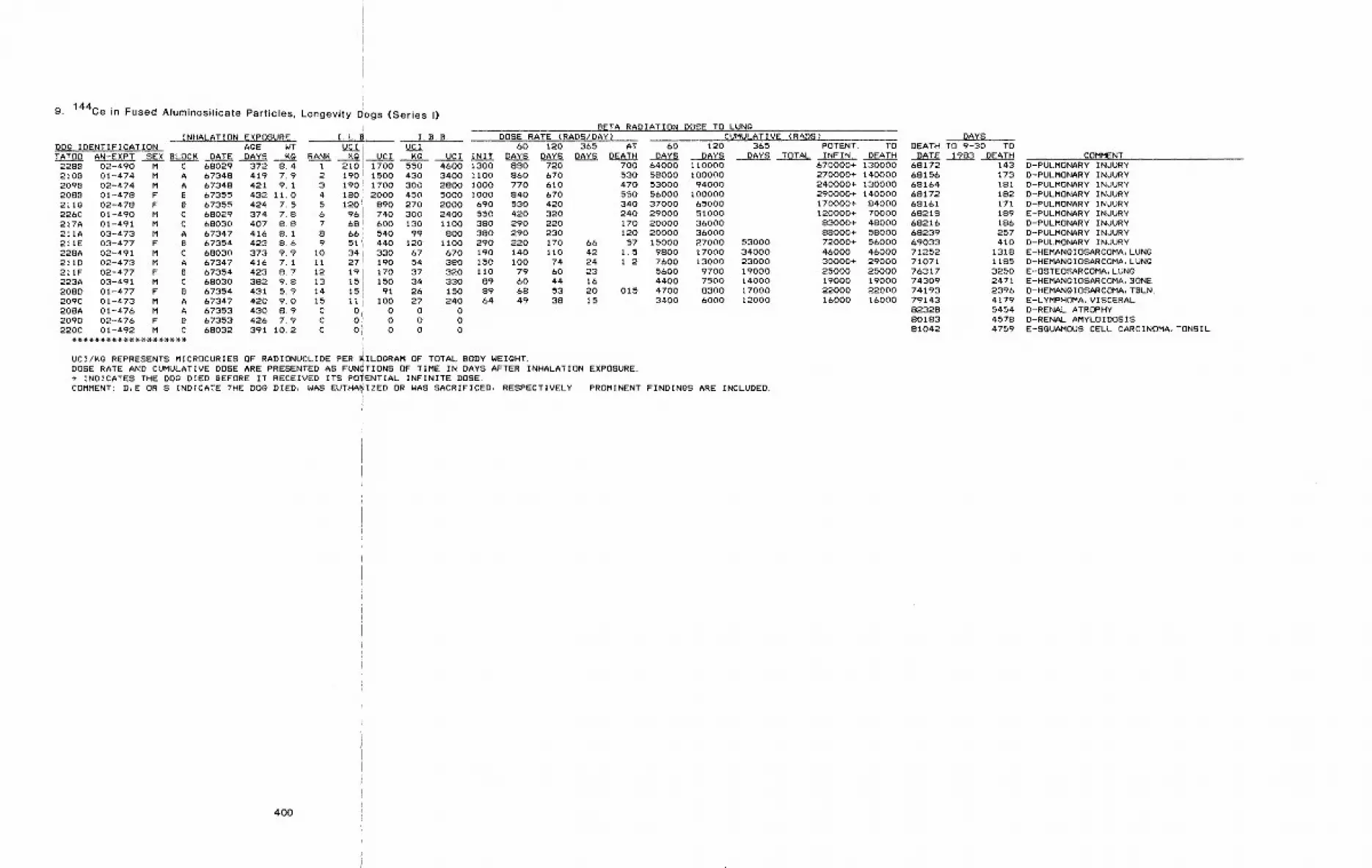
9. 144Ce in Fused Afuminosilicate Particles, Longevity ~ogs (Series E)
INHALATION EXPOSURE I.L. .DOg IDENTIFICATION A~E WT UCITATO0 AN-EXPT SEX BLOCK _DATE DAYS KO RANK Kg UCI
228B 02-490 M C 68029 372 8.4 1 210210B 01-474 M A 67348 419 7.9 2 1902098 02-474 M A 67348 421 9.1 3 1902088 01-478 F B 67355 432 II,0 4 1802110 02-478 F B 67355 424 7 5 5 120226C 01-490 M C 68029 374 7 8 6 96217A 01-491 M C 68030 407 8.8 7 68211A 03-473 M A 67347 416 8.1 8 66211E 03-477 F B 67354 423 8.6 9 51228A 02-491 M C 68030 373 9.9 I0 34211D 02-473 M A 67347 416 7. 1 Ii 27211F 02-477 F B 67354 423 8 7 12 19223A 03-49l M C 68030 382 9.8 13 15208D 01-477 F B 67354 431 5.9 14 15209C 01-473 M A 67347 420 9.0 15 II208A 01-476 M A 67353 430 8.9 C 0209D 02-476 F B 67353 426 7.9 C 0220C 01-492 M C 68032 391 10,2 C 0
BETA RADIATION DOSE TO LUNGI.B.B. DOSE RATE (RADS/DAY)
UCI 60 120 365 AT 60Kg UCI INIT DAYS DAYS DAYS DEATH DAYS
1700 550 4600 1300 8801500 430 3400 II00 8601700 300 2800 i000 7702000 450 5000 I000 840
890 270 2000 690 530740 300 2400 550 420
CUMULATIVE (RADS)120
DAYS
600 130 1100 380 290540 99 800 380 290440 120 ii00 290 220330 67 670 190 140190 54 380 150 i00170 37 320 110 79150 34 330 89 6091 26 150 89 68
100 27 240 64 490 0 00 0 00 0 0
7206706106704203202202301701107460445338
DAYS365 POTENT. TO DEATH TO 9-30 TO
DAYS TOTAL INFIN. DEATH DATE 1983 DEATH700 64000 i10000530 58000 I00000470 53000 94000550 56000 i00000340 37000 65000240 29000 51000170 20000 36000120 20000 36000
66 57 15000 27000 5300042 1.5 9800 17000 3400024 1.2 7600 13000 2300023 5600 9700 1900016 4400 7500 1400020 015 4700 8300 1700015 3400 6000 12000
IUCI/KG REPRESENTS MICROCURIES OF RADIONUCLIDE PER ~ILOQRAM OF TOTAL BODY WEIGHT.DOSE RATE AND CUMULATIVE DOSE ARE PRESENTED AS FUN(TIONS OF TIME IN DAYS AFTER INHALATION EXPOSURE.+ INDICATES THE DOg DIED BEFORE IT RECEIVED ITS PO’ENTIAL INFINITE DOSE.COMMENT: D,E OR S INDICATE THE DO~ DIED, WAS EUTHA! !IZED OR WAS SACRIFICED, RESPECTIVELY. PROMINENT FINDINGS ARE INCLUDED.
400
&70000+ 130000 68172270000+ 140000 68156240000+ 130000 68164290000+ 140000 68172170000+ 84000 68161120000+ 70000 6821883000+ 48000 6821688000+ 58000 6823972000+ 56000 6903346000 46000 7125230000+ 29000 7107125000 25000 7631719000 19000 7430922000 22000 7419316000 16000 79143
823288018381042
COMMENT143 D-PULMONARY INJURY173 D-PULMONARY INJURY181 D-PULMONARY INJURY182 D-PULMONARY INJURY171 D-PULMONARY INJURY189 E-PULMONARY INJURY186 D-PULMONARY INJURY257 D-PULMONARY INJURY410 D-PULMONARY INJURY
1318 E-HEMANgIOSARCOMA, LUNQ1185 D-HEMANQIOSARCOMA, LUNG3250 E-OSTEOSARCOMA, LUNg2471 E-HEMANgIOSARCOMA, BQNE2396 D-HEMANgIOSARCOMA, TBLN.4179 E-LYMPHOMA, VISCERAL5454 D-RENAL ATROPHY4578 D-RENAL AMYLOIDOSIS4759 E-SQUAMOUS CELL CARCINOMA, TONSIL

10. 144Ce in Fused Aluminosilicate Particles, Longevity Dogs (Series tl)
INHALATION EXPOSURE [email protected] IDENTIFICATION AgE WT UCI UCITATO8 AN-EXPT SEX BLOCK DATE DAYS KO RANK Kg UCI K~
315V 02-595 F D 69149 398 7.2298B 02-586 M A 69121 399 9.1327A 01-642 M E 69213 387 9.4479U 04-947 F L 71225 379 6.83308 02-642 F F 69213 374 8.32978 03-586 F B 69121 402 10.4470A 03-947 M K 71225 397 II.04658 03-918 F d 71176 382 7.9465A 04-918 M I 71176 382 11.4330U 03-641 F F 69212 373 6.0315A 01-595 M C 69149 398 10.93308 04-641 M E 69212 373 6.3303A 01-586 M A 69121 422 9. 5454A 03-883 M g 71106 402 8.84538 04-883 F H 71106 408 8.04648 01-918 M I 71176 385 9.4310T 02-594 F D 69148 421 8.94608 02-918 F O 71176 419 7.94805 02-947 F L 71225 373 8.33128 03-594 M C 69148 399 9.02988 03-585 F B 69120 398 i0. 44558 01-883 M Q 71106 402 11.7471A 01-947 M K 71225 397 7.5453T 02-883 F H 71106 408 6.4315U 01-594 F D 69148 397 8.33048 01-585 F B 69120 386 7.4311B 03-593 M C 69147 400 9.3328T 02-641 F F 69212 385 10.6467A 03-916 M I 71175 373 12.2467T 04-946 F L 71224 422 6.4297B 02-585 M A 69120 401 9.6326C 01-641 M E 69212 391 9.4463A 02-916 M I 71175 411 10.94808 03-946 M K 71224 372 8.24548 04-882 F H 71105 401 9.6454E 03-882 M g 71105 401 8.9305V 02-584 F B 69119 382 6.9460T 04-916 F J 71175 418 7.43278 01-640 M E 69211 385 9.0323V 02-640 F F 69211 408 7.83039 03-584 M A 69119 389 6.73198 02-593 F D 69147 420 9.14698 02-946 F L 71224 397 7.24788 01-946 M ~ 71224 379 9.63088 01-593 M C 69147 402 10.3454C 01-882 M ~ 71105 401 9.5464T 01-916 F J 71175 384 7.4455T 02-882 F H 71105 401 10.43138 01-598 F D 69160 411 7.929&B 01-592 M A 69135 418 10.0466V 04-915 F O 71174 380 7.9313C 02-59S M C 69160 411 9.6304T 02-592 F B 69135 401. 7.8324V 03-638 F F 69210 402 7.233~A 04-638 M E 69210 370 9.0
I. B.B.BETA RAD~
DOSE RATE (RADS/DAY)60 120 365 AT
UCI INIT ~AYS pAYS DAYS DEATH1 66 470 170 1200 370 270 200 1302 65 590 130 1200 370 290 240 1503 56 520 120 1100 320 240 180 994 54 360 180 1200 320 190 140 57 125 53 440 110 920 300 230 180 916 46 470 170 1800 270 200 150 60 157 44 480 67 730 270 200 150 69B 41 330 100 820 240 180 130 919 41 460 61 700 230 180 140 &6
I0 37 220 85 510 220 150 110 43 3.211 35 380 130 400 200 130 100 43 6.012 34 220 72 460 200 140 110 3813 33 320 76 720 190 140 110 7414 32 280 68 600 190 140 97 31 1.215 29 230 38 300 170 130 100 41 1.816 27 250 45 420 160 110 SO 29 .3017 26 230 130 1100 150 110 86 33 7.918 24 190 80 630 150 97 71 2719 24 200 51 430 150 100 77 29 .9120 24 210 49 440 140 100 79 31 . 1521 23 240 62 650 130 97 73 2922 19 220 60 700 110 83 &3 2323 19 150 34 260 120 SO 59 2224 18 110 46 290 100 75 57 2025 18 150 51 420 I00 77 58 2126 17 120 35 260 98 72 55 21 .01827 14 130 20 190 79 57 43 16 .06028 13 140 26 270 76 55 41 1529 13 160 26 310 77 55 41 1430 13 Sl 18 120 78 55 43 1731 12 110 50 480 68 45 34 1432 12 ii0 18 170 70 51 38 i433 12 130 20 220 74 50 37 1434 11 91 20 170 68 46 35 1435 I0 95 40 390 60 44 33 12 . 1336 9.8 87 21 190 60 42 32 1237 9.8 67 18 120 57 37 27 10 280938 9.5 70 30 330 56 42 32 1239 8.0 72 16 140 46 32 24 9.740 7.8 60 12 91 45 33 25 9.2 .01041 7.6 51 17 120 44 34 27 1242 6.3 57 9. 4 86 36 26 20 7.743 5,8 42 13 91 35 26 20 8.044 5.7 54 10 99 33 26 21 8.345 5.4 55 7.4 76 31 23 18 .02846 5.4 51 14 130 32 24 18 6. 747 5.0 37 12 86 29 21 15 5.348 4.9 51 16 170 30 21 15 5.049 2.4 19 8.2 64 14 9.8 7.4 2.950 2li 21 3,8 38 12 8,9 6.9 2.851 2.0 15 4.5 36 II 8.6 6,6 2.552 1. 8 17 3. 8 37 10 7. 9 b. 2 2. 553 I. 6 12 2. 5 19 9, 2 6. 8 5. 3 2, 254 1.5 11 3.9 28 8.3 6.7 5.3 2.155 I. 3 11 2. 2 20 7, 3 51 9 4 . 7 l i 9
ATION DOSE TO LUNOCUMULATIVE (RADS)
60 120 365 POTENT. TO.1DAY5 DAYS DAYS TOTAL INFIN. DEATH19000 33000 89000+ 53000 70030
;20000 36000 100000+ 57000 69355117000 29000 84000+ 50000 70121114000 24000 47000 62000+ 59000 73284!15000 28000 71000+ 50000 70127i14000 24000 50000 66000+ 61000 71141~14000 24000 53000+ 41000 72135112000 22000 46000+ 28000 71361112000 22000 57000+ 41000 72122!11000 18000 36000 47000+ 46000 72194
9500 17000 33000 46000+ 43000 7133510000 18000 34000 44000 44000 75334
9800 17000 39000+ 24000 693149800 17000 31000 38000 38000 742388900 16000 33000 44000+ 43000 742368000 14000 26000 33000 33000 752387700 14000 27000 36000+ 34000 711837200 12000 23000 30000 30000 761607300 13000 25000 32000 32000 750177200 13000 25000 54000 34000 742176800 12000 23000 32000 32000 771995800 10000 20000 26000 26000 770935700 9900 19000 25000 25000 772165300 9200 18000 23000 23000 782775400 9400 18000 24000 24000 800925000 8800 17000 23000 23000 752564000 7000 14000 18000 18990 7429539~3 6800 13000 17090 17000 793653900 6800 1300 16000 16000 761123900 6800 14000 18000 18000 761473300 5600 11000 15000 15000 760653600 6200 12000 16000 16000 782053600 6200 12000 15900 15000 791023300 5700 11000 15000 15000 821253100 5400 10000 13000 13000 751713000 5200 10000 13000 13000 772784600 8700 12000 12000 120002900 5100 10000 13000 13900 801892300 4000 7800 11000 11000 823162300 4000 7900 10000 10000 751272300 4100 8600 12000 12000 761331800 3200 6300 8500 8500 810491800 3200 6400 8600 8600 832351800 3100 6400 8700 8700 781691600 2800 5700 7700 7700 823421700 2900 5700 7800 7800 783011500 2500 4800 6200 6200 821121500 2500 4700 6000 6000 76072709 1200 2400 3200 3200 79257620 1100 2200 3000 3000 81162590 1009 2100 2700 2700540 960 1900 2600 2600 76083470 840 1700 2300 2300 79324450 810 1700 2200 2200 82160390 710 1500 2000 2000 2000 79132
DAYSDEATH TO 9-30 TO
DATE 1983 DEATH
5267
4482
246234273790279750275185311
1077916
2313193
1228I226i523
7651810125318953001217921832728396123271974380517631749250132802849391915272365
COMMENTD-PULMONARY INJURYD-PULMONARY INJURYD-PULMONARY INJURYE-HEMANOIOSARCOMA, LUN¢D-PULMONARY INJURYE-HEMANOIOSARC. AND FIBROSARC.,LUNOD-PULMONARY INJURYD-PULMONARY INJURYD-PULMONARY INJURYE-HEMANOIOSARCOMA, LUNOD-HEMANgIOSARC. AND B-A-CARCINOMA, LUNgD-HEM-SARC.,SITE UND.~B-A-CARC.,LUNgD-PULMONARY VASCULAR INJURYD--PULMONARY THROMBOSIS;AMYLOIDOSISD-HEM-SARC.-B-A-CARC.-BRONCHO. CA.,LUNOD-BRONCHIOLOALVEOLAR CARCINOMAE-HEMANOIOSARCOMA, LUNOD-MIXED TUMOR,LUNO~B-A-CARCINOMAE-SGUAMOUS CELL CARCINOMA, NASAL CAVITYD-HEMANOIOSARCOMA, SPLEENE-MIXED TUMOR,LUNgJ OSTEOSARCOMA, LUNOD-EPILEPSYE-HEMANgIOSARCOMA, BONEE-HEMANgIOSARCOMA, SPLEEND-ADENOCARCINOMA, LUNOD-HEMANgIOSARCOMA, LIVERE-HEMANOIOSARCOMA, BOTH HUMERIE-gASTROENTEROPATHYD-HEMANgIOSARCOMA, TBLND-ACCIDENTAL DEATHD-PLEURITIS (NOCARDIA SP. E-HEMANgIOSARCOMA, TBLND-HEMANOIOSARCOMA, HEARTE-HEMANgIOSARC.,TBLN;CARCINOMA, LUNQD-HEMANOIOSARCOMA, HEARTE-HEMANOIOSARCOMA, DERMIS
3301485321072570428543942502494327533955179337494410
EICHRONIC TRACHITISE-CARCINOMA, LUNgD-HEMANgIOSARCOMA, TBLNE-HEHANgIOSARCOMA, LIVERE-CARCINOMA, LUNgE-INTERSTITIAL NEPHRITIS~LUNO CARC.D-HEMANgIOSARCOMA, TBLND-MYOCARDIAL DEgENERATION}LUNO TUMORE-HEMANgIOSARCOMA, DISSEMINATEDD-PYOMETRA AND HEMANgIOMA:TBLNE-HEMANOIOSARCOMA, SITE UNDETERMINEDD-HEMANOIOSARCOMA, TBLND-CONGEST. HEART FAIL.~CARCINOMA, LUNO
2479384146983574
D-PERITONITIS (NOCARDIA SP. )E-ADENOCARCINOMA, BLADDERE-CARCINOMA, LUNgD-UNDETERMINED
401
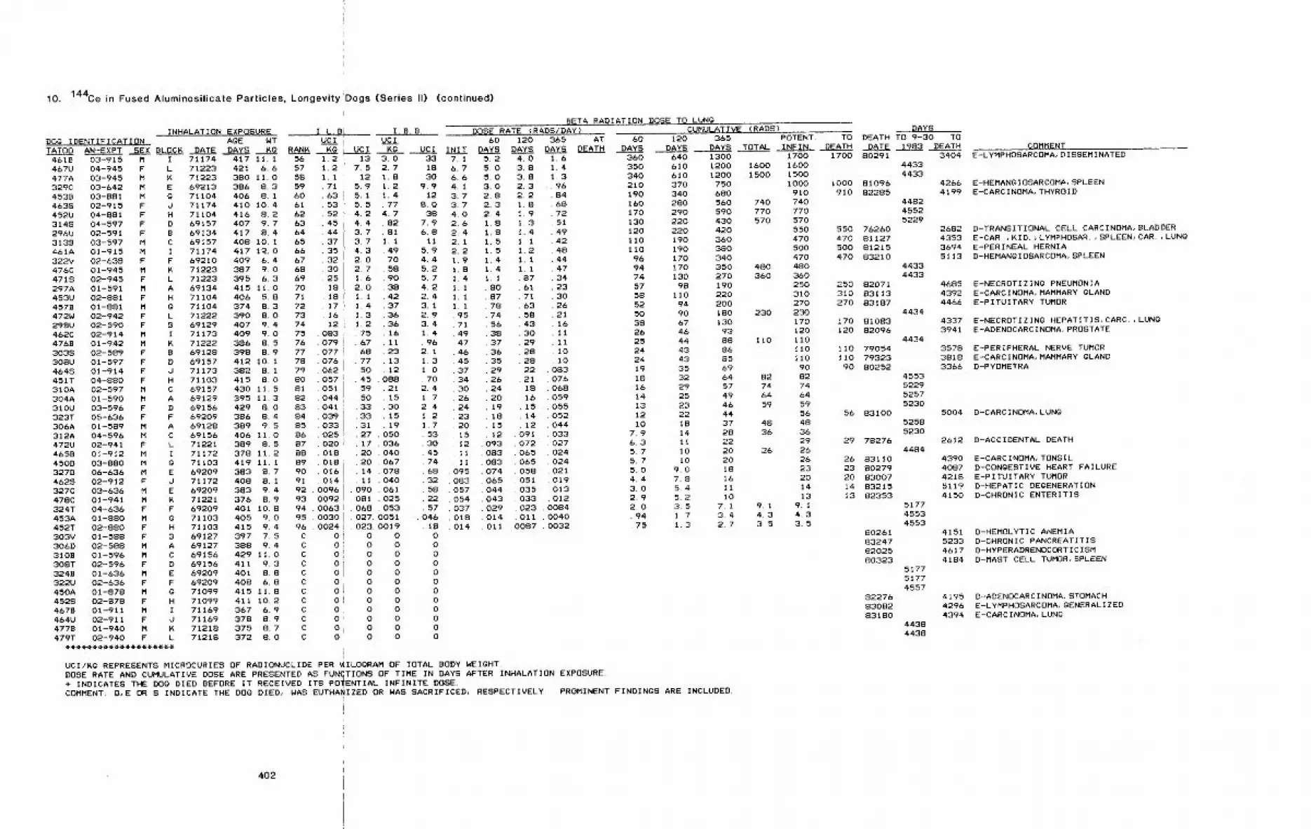
10. 144Ce in Fused Aluminosilicate Particles, Longevity
INHALATION EXPOSURE I.L.BDOG IDENTIFICATION AgE WT UCITATO0 AN-EXPT SEX BLOC~ DATE DAYS Kg RANK KQ
’Dogs (Series II)
I. B.B.UCI
UCI ~g UCI
(continued)
INIT
BETA RADIATION DOSE TO LUNg
DOSE RATE {RADS/DAY)60 120 365 AT 60
DAYS DAYS DAYS DEATH DAYS1.61.41.39684687251494248444734233026211611II1010
. 083076068059O55O52O44033027024024021019013012
008400400032
4618 03-915 M I 71174 417 11.1467U 04-945 F L 71223 421 6.6477A 03-945 M ~ 71223 380 11.0329C 03-642 M E 69213 386 8.34538 03-881 M G 71104 406 8. i4635 02-915 F d 71174 410 10.4452U 04-881 F H 71104 416 8.23148 04-597 F D 69157 407 9.7296U 02-591 F B 69134 417 8.43139 03-597 M C 69157 408 i0. I461A 01-915 M I 71174 417 12.0322V 02-638 F F 69210 409 6.4476C 01-945 M ~ 71223 387 9.04718 02-945 F L 71223 395 6.3297A 01-591 M A 69134 415 11.0453U 02-881 F H 71104 406 5.84578 01-881 M g 71104 374 8.3472W 02-942 F L 71222 390 8,0298U 02-590 F B 69129 407 9.4462C 02--914 M I 71173 409 9.04768 01-942 M ~ 71222 386 8.53038 02-589 F B 69128 398 8.9308U 01-597 F D 69157 412 10.14648 01-914 F d 71173 382 81451T 04-880 F H 71103 415 S.O310A 02-597 M C 69157 430 11.5304A 01-590 M A 69129 395 11.3310U 03-596 F D 69156 429 S.O323T 05-636 F F 69209 386 8.4306A 01-589 M A 69128 389 9.5312A 04-596 M C 69156 406 11.0472U 02-941 F L 71221 389 8.5465B 01-912 M I 71172 378 11.2450D 03-880 M 0 71103 419 ii. I327D 06-636 M E 69209 383 8.74628 02-912 F J 71172 408 S. 1327C 03-636 M E 69209 383 9.4478C 01-941 M ~ 71221 376 8.9324T 04-636 F F 69209 401 10.8453A 01-880 M g 71103 405 9.0452T 02-880 F H 71103 415 9.4303V 01-588 F B 69127 397 7.5306D 02-588 M A 69127 388 9.4310B 01-596 M C 69156 429 11.030ST 02-596 F D 69156 411 9.3324B 01-636 M E 69209 401 8.8322U 02-636 F F 69209 408 6.8450A 01-878 M g 71099 415 11.84528 02-878 F H 71099 411 10.24678 01-911 M I 71169 367 6.9464U 02-911 F d 71169 378 8.94778 01-940 M K 71218 375 8.7479T 02-940 F L 71218 372 8.0
120DAYS
360 640350 610340 610210 370190 340160 280170 290130 220120 220110 190110 19096 17094 17074 13057 9858 11052 9450 9038 6726 4625 4424 4324 4319 3518 3216 2914 2513 2312 22i0 18
7,9 146.3 ii5.7 105.7 I05.0 9.04.4 7.83.0 5.42.9 5.22.0 3.5.94 1 7.75 1.3
CUMULATIVE (RADS) DAYS365 POTENT. TO DEATH TO 9-30 TO
DAYS TOTAL INFIN. DEATH DATE 1983 DEATH1300 1700 1700 80291 34041200 1600 1600 44331200 1500 1500 4433
750 1000 I000 81096 4266680 910 910 82285 4199560 740 740 4482590 770 770 4552430 570 570 5229420 550 550 76260 2682360 470 470 81127 4353380 500 500 81215 3694340 470 470 83210 5113350 480 480 4433270 360 360 4433190 250 250 82071 4685220 310 310 83113 4392200 270 270 83187 4466ISO 230 230 4434130 170 170 81083 433793 120 120 82096 394188 II0 Ii0 443486 110 II0 79054 357885 Ii0 110 79323 381869 90 90 80252 336664 82 82 455357 74 74 522949 64 64 525746 59 59 523044 56 56 83100 500437 48 48 525828 36 36 523022 29 29 78276 261220 26 26 448420 26 26 83110 439018 23 23 80279 408716 20 20 83007 421811 14 14 83215 511910 13 13 82353 4150
7. 1 9. 1 9. I 51773. 4 4. 3 4. 3 45532. 7 3. 5 3. 5 4553
PROMINENT FINDINGS ARE INCLUDED¯
56 I. 257 I. 258 1. 159 7160 6361 5362 5263 4564 4465 3766 3567 3268 3069 2570 1871 1872 1773 I 674 1275 08376 07977 07778 07679 06280 057Sl 05182 04483 04184 03985 03386 02587 02088 01889 01890 01691 . 01492 . 009693 . 009294 . 006395 . 003096 . 0024C 0c 0C 0C 0C 0C 0C 0C oc O.C O’C 0iC 0
13 3.07.5 2.7
12 1.85.9 1.25.1 1.45.5 .774.2 4.74.4 .823.7 .813.7 1.14.3 .492. 0 702.7 58t. 6 902.0 381. 1 421. 4 371. 3 361. 2 3675 1667 1168 2377 1350 1245 . 08859 . 2150 . 1533 . 3033 . 1531 . 1927 . 05017 03620 04020 06714 07811 040
. 090 061
. OSl 025
. 068 053¯ 027. 0051, 023. 0019
0 00 00 00 00 00 00 00 00 00 00 00 0
331830
9.912
S.O38
7.96.8ii
5.94.45.25.74.22.43.12.93.41.4. 962.11.31.0. 702.41.72.41.21.7533045746832582257
O46IS000000000000
7,16.76.64.13.73.74,02,62.42.12.21.91,81,41.11.11.195714947464537343026242320
,15.12,II.11
. 095¯ 083¯ 057. 054. 037¯ 018¯ 014
5,25,05.03.02.82.3;2.41,8l,S1.51.51.41.41.1
SO87787456383736352926242019iS1512
¯ 093083083074065O44043029014011
4.03.83.82.32.2l.S1,9I 3I 4I 11 21 11 1
87bl71635843302928282221iS16151412
091072g65065058051035033023011
0087
UCI/KG REPRESENTS MICROCURIES OF RADIONUCLIDE PER ~IILOgRAM OF TOTAL BODY WEIGHT.
DOSE RATE AND CUMULATIVE DOSE ARE PRESENTED AS FUNCTIONS OF TIME IN DAYS AFTER INHALATION EXPOSURE.+ INDICATES THE DOG DIED BEFORE IT RECEIVED ITS POTENTIAL INFINITE DOSE.COMMENT: D,E OR S INDICATE THE DOG DIED, WAS EUTHA~IZED OR WAS SACRIFICED, RESPECTIVELY.
4O2
80261 415183247 523382025 461780323 4184
517751774557
82276 419583082 429683180 4394
44384438
COMMENTE-LYMPHOSARCOMA, DISSEMINATED
E-HEMANgIOSARCOMA, SPLEENE-CARCINOMA, THYROID
D-TRANSITIONAL CELL CARCINOMA, BLADDERE-CAR ,KID.>LYMPHOSAR. ,SPLEEN~CAR.,LUNGE-PERINEAL HERNIAD-HEMANGIOSARCOMA, SPLEEN
E-NECROTIZINg PNEUMONIAE-CARCINOMA, MAMMARY GLANDE-PITUITARY TUMOR
E-NECROTIZINg HEPATITIS;CARC.,LUNGE-ADENOCARCINOMA, PROSTATE
E-PERIPHERAL NERVE TUMORE-CARCINOMA, MAMMARY gLANDD-PYOMETRA
D-CARCINOMA. LUNG
D-ACCIDENTAL DEATH
E-CARCINOMA, TONSILD-CONgESTIVE HEART FAILUREE-PITUITARY TUMORD-HEPATIC DEGENERATIOND-CHRONIC ENTERITIS
D-HEMOLYTIC ANEMIAD-CHRONIC PANCREATITISD-HYPERADRENOCORTICISMD-MAST CELL TUMOR, SPLEEN
D-ADENOCARCINOMA, STOMACHE-LYMPHOSARCOMA, gENERALIZEDE-CARCINOMA, LUNG

11. 144Ce in Fused Atuminosilicate ParticJes, Sacrifice Dogs (Series II, HI and IV)
INHALATION EXPOSUREDOg IDENTIFICATIQNTATOD AN-EXPT ~EX SER
5418 01-999 F520A 02-998 M5300 04-1007 M5308 01-1002 M525W 01-1004 F527A 03-1007 M521T 03-998 F526A 01-1001 M5268 02-1000 M5268 01-1007 F525T 02-I003 F522T 03-I003 F525U 02-1004 F539A 03-997 M530A 04-998 M541U 01-1000 F5350 03-1000 M539D 04-1000 M522U 01-998 F526C 02-997 M5198 04-1004 F5248 02-1001 F5228 04-997 F5278 02-1002 M532U 02-1007 F521B 03-999 M536T 03-1001 F519T 03-1004 F527D 01-997 M519A 03-1002 M520B 04-1003 M523T 02-1008 F5430 04-1004 M5208 01-1003 F526D 02-999 M541A 05-1000 M5388 04-1001 F533T 01-1008 F
I.L.B. I.B.B.AgE WT UCI UCI
KO UCI Kg UCI INIT DAYS DAYS DAYSDATE DAYS KG RANK395 7.9 1 71 560 140 1100 410 310 230427 11.0 2 66 720 130 1400 370 280 210417 9.2 3 64 590 140 1300 370 260 210411 9.5 4 60 560 91 860 340 220428 7.3 5 58 420 110 810 340 250 190418 9.2 6 54 500 78 720 310 210 160426 8.7 7 52 450 93 810 310 240 180416 7.5 8 52 390 97 730 300 200412 5.8 9 52 300 120 680 310 220 160423 6.6 i0 51 330 100 670 290 210427 8.8 11 48 420 110 990 290 210 160432 8.0 12 48 380 140 1100 290 210 160428 9.1 13 46 420 89 810 280 190 140394 9.3 14 41 380 120 1100 240 180 130404 11.5 15 39 450 83 950 230 180 130396 7.9 16 35 280 62 490 210 140 110399 7.7 17 34 260 70 540 200 140 110397 7.9 18 33 260 52 410 190 140 100424 7.8 19 33 250 92 720 190 140 99409 6.9 20 33 230 68 470 190 130 96439 8.3 21 32 270 73 610 190 140 100425 6.7 22 32 210 55 370 190 130 100423 8.7 23 31 270 78 680 180 140 110412 9.3 24 31 290 56 520 180 130 95413 7.8 25 31 240 42 330 180 130 98427 7.6 2& 30 230 84 640 170 130 100403 7.6 27 29 220 66 500 170 120 88439 8.7 28 28 240 91 790 170 120404 7.9 29 27 220 47 370 160 120 90437 8.6 30 27 230 110 940 160 120 87435 11.9 31 26 310 180 2100 160 120 85435 6.0 32 26 150 47 280 150 99 73399 7. b 33 26 190 42 320 150 110 84435 6.7 34 24 160 67 450 150 110 82411 5.5 35 16 86 51 280 94 68 51396 8.3 36 16 140 28 230 92 67 51402 6,7 37 14 95 24 160 83 57 42413 5.9 38 14 81 20 120 79 56 41416 8.8 C 0 0 0 0
C 0 0 0 0C 0 0 0 0C 0 0 0 0C 0 0 0 0C 0 0 0 0C 0 0 0 0C 0 0 0 oC 0 0 o 0C 0 0 0 0C o 0 o 0C 0 0 0 0
BETA!RADIATiON DOSE TO LUNgDOSE RATE (RADS/D~Y) CUMULATIVE DOSE (RADS) DAYS
&O 120 365 9-30 AT 60 120 365 POTENT. TO DEATH TO 9-30 TO~983 DEATH DAYS DAYS DAYS TOTAL INFIN. DEATH DATE ,19S3 ,,~EATH
II 72103II 72102II 72115II 72109II 72111II 72115II 72102II 72108II 72104II 72115II 72110II 72110II 72111II 72101II 72102II 72104II 72104II 72104II 72102II 72101II 72111II 72108II 72101II 72109II 72115II 72103II 72108II 72111II 72101II 72109II 72110II 72116II 72109II 72110II 72103II 72104II 72108tI 72116II 720975238 01-995 F
5388 02-995 M533A 03-995 M5428 04-995 F540T 05-995 F542A 06-995 M522V 01-996 F5478 02-996 M522A 03-996 M5408 04-996 M530S 05-996 F5218 06-996 F
II 72097 391 9.3II 72097 394 8.3II 72097 388 8.211 72097 389 5.7II 72097 388 9. 1II 72098 420 7.8II 72098 378 10.4II 72098 420 8-7II 72098 390 8.0II 72098 400 8.6II 72098 422 8.6
403
5348
404439
353842
333639
36332929343120191616
220 21000 37000150 19000 34000130 19000 33000190 17000140 18000 31000
99 15000 26000170 16000 29000170 15000110 16000 27000190 15000150 15000 2600099 15000 2600027 14000 24000 4500028 12000 22000 4100062 12000 22000
2.6 10000 18000 3400014 9900 17000 34000
9.9 9600 17000 3300094 9600 1700019 9400 16000 31000
2.5 9800 17000 330005.9 9300 16000 33000
57 9500 17000¯ 46 9200 16000 30000
9100 16000 310002.8 9200 16000 32000
50 8600 1500086 860022 8400 15000 29000
4.2 8100 14000 280005. 4 8200 14000 27000
17 7300 12000 240007800 14000 27000
7,3 7600 13000 260005,2 4800 8300 160005.2 4700 8200 16000
4100 7100 14000 18000 180004000 6900 13000 18000
COMMENT85000+ 39000 7223177000+ 45000 7228399000+ 55000 7300587000+ 21000 7218970000+ 40000 7229075000+ 42000 7300368000+ 30000 7223075000+ 19000 7218955000+ 35000 7228474000+ 19000 7219658000+ 28000 7224176000+ 43000 7300261000+ 53000 7331656000+ 47000 7324951000+ 37000 7302545000 45000 7510147000+ 43000 7405643000 41000 7411335000+ 17000 7223040000+ 35000 7325043000 43000 7510645000+ 43000 7429440000+ 28000 7235739000 39000 7615341000 41000 7814543000 43000 7510639000+ 24000 7300233000+ 16000 7224141000+ 33000 7325737000+ 35000 7429536000+ 34000 7426732000+ 27000 7326436000 36000 8014434000+ 32000 7411422000+ 20000 7411422000+ 20000 74113
18000 8230372354
72224
7410875104
791047410875014
4183
4194
41944194
4193
4193
128 S-PULMONARY INJURY181 S-PULMONARY INJURY256 S-PULMONARY INJURY
80 S-PULMONARY INJURY179 S-PULMONARY INJURY254 S-PULMONARY INJURY128 S-PULMONARY INJURY
81 S-PULMONARY INJURY180 S-PULMONARY INJURY
81 S-PULMONARY INJURY131 S-PULMONARY INJURY258 S-PULMONARY INJURY571 E-PULMONARY INJURY514 S-PULMONARY INJURY289 S-PULMONARY INJURY
1093 S-PULMONARY INJURY683 D-PULMONARY INJURY740 S-PULMONARY INJURY128 S-PULMONARY INJURY515 S-PULMONARY INJURY
1091 S-PULMONARY INJURY917 S-PULMONARY INJURY256 S-PULMONARY INJURY
1505 D-HEMANGIOSARCOMA, LUNg2222 E-HEMANgIOSARCQMA, HEART1099 S-HEMANgIOSARCOMA, LUNQ260 S-PULMONARY INJURY130 S-PULMONARY INJURY522 D-PULMONARY INJURY917 S-PULMONARY INJURY888 D-PULMONARY INJURY514 S-PULMONARY INJURY
2957 E-HEMANQIOSARCOMA, LUNg735 S-PULMONARY INJURY742 S-PULMONARY INJURY740 S-PULMONARY INJURY
3840 D-INTERSTITIAL PNEUMONIA257 S-NORMAL
127 S-NORMAL
742 S-NORMAL1102 S-NORMAL
2563 D-MYOCARDIAL INFARCT741 S-NORMAL
1012 S-NORMAL
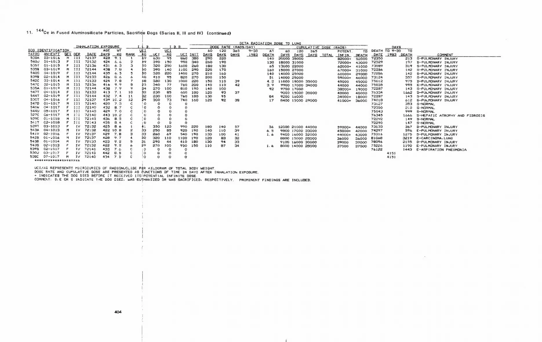
¯ , I11. 144Ce in Fused Aluminosilicate Particles, Sacrlh~e Dogs (Series II, Itl and IV) (continued)
I BETA RADIATION DOSE TO LUNgINHALATION EXPOSURE I.~.B. I.B.B. DOSE RATE (RADS/DAY)
DOQ IDENTIFICATION AGE WT ~ UCI 60 120 365TATO0 AN-EXPT SEX SER DATE DAYS KG RANK ~ UCI Kg UCI INIT DAYS DAYS DAYS
538A 03-1016 M iII 72137 428 9.1 1 69 630 95 860 380 290 220540U 01-1013 F Eli 72132 424 6.6 2 ~9 390 150 990 340 260 190535T 01-1015 F iIl 72136 43t 6,3 3 ~0 320 250 1600 260 1S0 130535B 03-1019 M III 72144 438 7.8 4 #0 390 140 1i00 290 220 170540S 04-1019 F Ill 72144 435 6,5 5 ~0 320 220 1400 270 210 1605398 02-1014 M III 72133 426 8.6 6 48 410 95 820 270 200 150542C 03-1014 M III 72133 424 7.8 7 ~8 3aO 130 1000 220 I50 110 39547C 02-1015 M IlI 72136 416 9,9 S ~5i 340 77 760 200 140 110 42535A 01-1019 M III 72144 438 7.9 9 34 270 100 810 190 140 I00547T 04-1014 F ill 72133 413 7, I i0 ~2 230 85 600 180 120 93 37544T 02-i019 F III 72144 432 7.4 11
~230 I00 760 iSO 130 95
530T 04-I016 F III 72137 439 10.2 12 300 73 740 160 120 92 38547D 01-1017 M III 72140 420 7.3 C !0 0 0 0541W 04-I017 F III 72140 432 8.7 C !0 0 0 0544U 05-1017 F IIt 72140 429 7.0 C iO 0 0 0527C 06-1017 M III 72140 443 10.2 C 0 0 0 0539C 01-i018 M III 72143 436 8.5 C I0 0 0 0541T 02-1018 F III 72143 435 8~4 C I0 0 0 0539T 03-1013 F IV 72132 425 8.6 1 ~i 350 120 990 220 180 140 57543A 04-1013 M IV 72132 422 10.8 2 ~3 250 85 920 190 140 II0 39541V 02-1016 F IV 72137 429 7.8 3 33 260 69 540 190 130 I00 415428 01-1016 M IV 72137 428 9.7 4 ~2 320 ii0 tlO0 190 120 83 325438 01-1014 M IV 72133 423 9.2 5 ~1 290 44 410 180 130 94 33543S 02-1013 F IV 72132 422 9.2 6 29 270 100 950 150 110 87 34539S 02-1017 F IV 72140 433 7.6 C ~0 0 0 0530U 03-1017 F IV 72140 442 8.5 C !0 0 0 0538C 07-1017 M IV 72140 434 7.5 C !0 0 0 0
(UCI/X.g REPRESENTS MICROCURiES OF RADIONUCLIDE P~R KILOGRAM OF TOTAL BODY WEIGHT.DOSE RATE AND CUMULATIVE DOSE ARE PRESENTED AS ~UNCTIONS OF TIME IN DAYS AFTER INHALATION EXPOSURE.+ INDICATES THE DO~ DIED BEFORE IT RECEIVED ITS~POTENTIAL INFINITE DOSE.COMMENT: D,E OR S INDICATE THE DOQ DIED, WAS EU-HANIZED OR WAS SACRIFICED, RESPECTIVELY. PROMINENT FINDINQS ARE INCLUDED.
CUMULATIVE DOSE (RADS) DAYS9-30 AT 60 120 365 POTENT. TO DEATH TO 9-30 TO1983 DEATH DAYS DAYS DAYS TOTAL INFIN. DEATH DATE 1983 DEATH
140 20000 35000130 18000 31000
65 13000 22000160 15000 27000140 14000 25000
51 14000 250004.2 11000 18000 350003.9 9900 17000 34000
92 9700 170009000 15000 30000
84 9200 1600017 8400 15000 29000
56 12000 21000 440006.5 9800 17000 330001.6 9400 16000 32000
SSO0 15000 280009100 16000 30000
1.6 8000 14000 28000
COMMENT82000+ 52000 7235072000+ 43000 7232962000+ 41000 7308967000+ 31000 7228660000+ 29000 7228659000+ 46000 7312445000 45000 7501244000 44000 7503538000+ 19000 722a740000 40000 76334
34000+ 18000 7228741000+ 36000 74018
731277235075043763457229272290
59000+ 44000 7313345000+ 42000 7429744000+ 43000 7531636000 36000 8106839000 39000 7809637000 37000 75226
7612241514151
213 S-PULMONARY INJURY197 D-PULMONARY INJURY319 S-PULMONARY INJURY142 S-PULMONARY INJURY142 S-PULMONARY INJURY357 S-PULMONARY INJURY975 D-PULMONARY INJURY995 E-PULMONARY INJURY143 S-PULMONARY INJURY
1662 D-PULMONARY INJURY143 S-PULMONARY INJURY612 S-PULMONARY INJURY353 S-NORMAL210 S-NORMAL999 S-NORMAL
1666 S-HEPATIC ATROPHY AND FIBROSIS149 S-NORMAL147 S-NORMAL367 D-PULMONARY INOURY896 E-PULMONARY INJURY
1275 D-PULMONARY INJURY3219 E-CARCINOMA, LUNG2155 D-PULMONARY INJURY1190 E-PULMONARY INJURY1443 E-ASPIRATION PNEUMONIA
404
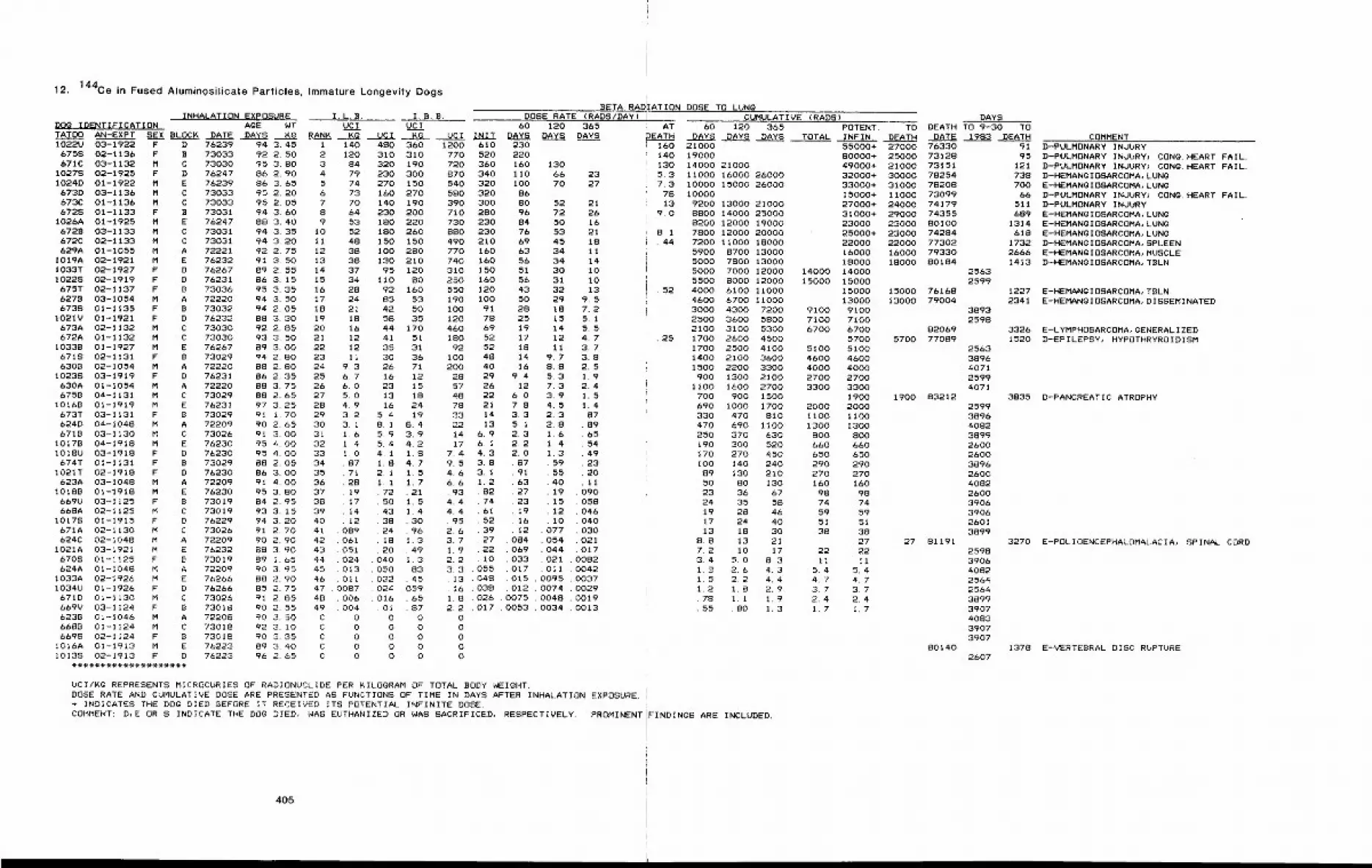
12. 144Ce in Fused Aluminosilicate Particles, Immature Longevity Dogs
INHALATION EXPOSURE I.L.B. I.B.B.DOg IDENTIFICATION AGE NT UCI UCITATO0 AN-EXPT SEX BLOCK DATE DAYS Kg RANK. ~g UCI KC1022U 03-1922 F D 76239 94 3.45
6758 02-1136 F B 73033 92 2.50671C 03-1132 M C 73030 95 3.80
10278 02-1925 F D 76247 86 2.901024D 01-1922 M E 76239 86 3.65
673D 03-1136 M C 73033 95 2.206730 01-1136 M C 73033 95 2.056728 01-1133 F B 73031 94 3.60
1026A 01-1925 M E 76247 88 3.406728 03-1133 M C 73031 94 3.356720 02-1133 M C 73031 94 3.20629A 01-1055 M A 72221 92 2.75
I019A 02-1921 M E 76232 91 3.50I033T 02-1927 F D 76267 89 2.5510228 02-1919 F D 76231 86 3. 15675T 02-1137 F B 73036 95 3.35627B 03-1054 M A 72220 94 3. 506738 01-1135 F B 73032 94 2.05
1021V 01-1921 F D 76232 88 3.30673A 02-1132 M C 73030 92 2.85672A 01-1132 M C 73030 93 3.50
I033B 01-1927 M E 76267 89 3.006718 02-1131 F B 73029 94 2.806308 02-1054 M A 72220 88 2.80
10238 03-1919 F D 76231 86 2.35630A 01-1054 M A 72220 88 3.756758 04-1131 M C 73029 88 2.65
I0168 01-1919 M E 76231 97 3.25673T 03-1131 F B 73029 91 1.70624D 04-1048 M A 72209 90 2.656719 03-1130 M C 73026 91 3.00
1017B 04-1918 M E 76230 95 4.00IOI8U 03-1918 F D 76230 95 4.00674T 01-1131 F B 73029 88 2.05
I021T 02-1918 F D 76230 86 3.00623A 03-1048 M A 72209 91 4.00
I0188 01-1918 M E 76230 95 3.80669U 03-1125 F B 73019 84 2.95668A 02-1125 M C 73019 93 3. 15
I0178 01-1915 F D 76229 94 3.20671A 02-1130 M C 73026 91 2.70024C 02-i048 M A 72209 90 2.90
I021A 03-1921 M E 76232 88 3.906708 01-1125 F B 73019 89 1.65624A 01-1048 M A 72209 90 3.95
1033A 02-1926 M E 76266 88 2.90I034U 01-1926 F D 76266 85 2.75671D 01-1130 M C 73026 91 2.85669V 03-1124 F B 73018 90 2.55623B 01-1046 M A 72208 90 3.506688 01-1124 M C 73018 92 3. 106698 02-1124 F B 73018 90 3.35
IOI6A 01-1913 M E 76223 89 3.40I0138 02-1913 F D 76223 96 2,65
I 1402 1203 844 795 746 737 708 649 53
10 52Ii 4812 3813 3814 3715 3416 2817 2418 2119 1820 1621 1222 1223 1124 9.325 6.726 6.027 5.028 4.929 3.230 3.131 1,632 1.433 1.034 ,8735 7136 2837 1938 1739 1440 1241 08942 06143 05144 02445 01346 01147 .Joe7~8 .00649 , 004C 0C 0C 0C 0C 0
BETA RADDOSE RATE (RADS/DAY)
60 120 365UCI INIT DAYS DAYS DAYS
480 360 1200310 310 770320 190 720230 300 870270 150 540160 270 580140 190 390230 200 710180 220 730180 260 880150 150 490I O0 280 770130 210 740
95 120 310I I0 80 250
92 160 55085 53 19042 50 10058 35 12044 170 46041 51 18035 31 9230 36 1 O026 71 20016 12 2823 15 5713 18 4816 24 78
5. 4 19 338. 1 8.4 225.9 3.9 145.4 4.2 174~1 1.8 7.41.8 4.7 9.52,1 1.5 4.6I,i 1.7 6.672 , 21 . 9350 1.5 4.443 1.4 4.438 . 30 . 9524 . 96 2. 618 1.3 3.720 .49 1. 9
040 I. 3 2. 2050 . 83032 . 45024 , 059016 . 65
Ol . 870 00 00 00 00 0
610520360340320320300280230230210160160150160120100
9178695252484O292622211413
6¯96.14.33.83.11.28274615239272210
3. 3 . 055, 13 . 048. 16 . 038
230220160 130ii0 66i00 70
86SO 5296 7284 5076 5369 4563 3456 3451 3056 3143 3250 2928 1825 1519 1417 1218 ii14 9.716 8.8
9.4 5.312 7.3
6.0 3.97.8 4.53.3 2.35.1 2.82.3 1.62.2 1.42.0 1.3
87 5991 5563 4027 1923 1519 1216 I012 .077
084 .054069 .044033 .021017 .011015 .0095012 .0074
1.8 .026 .0075 .00482.2 .017 .0053 .0034
00000
2327
2126162118ii14I0I013
9.57.25.15.54.73.73.82.51.92.41.51.48789655449232011
09O058O46040030021017
008200420037002900190013
:ATION DOSE TO LUNGCUMULATIVE (RADS)
AT 60 120 365 POTENT. TODAYS DAYS DAYS TOTAL INFIN. DEATH
1601401305.37.3
7813
9.0
B.l44
, . 52
¯ 25
21000 55000+ 2700019000 80000+ 2500014000 21000 49000+ 21000ii000 16000 26000 32000+ 3000010000 15000 26000 33000+ 31000i0000 15000+ 11000
9200 13000 21000 27000+ 240008800 14000 25000 31000+ 290008200 12000 19000 23000 230007800 12000 20000 25000+ 230007200 11000 18000 22000 220005900 8700 13000 16000 160005000 7800 13000 18000 180005000 7000 12000 14000 140005500 8000 12000 15000 150004000 6100 11000 15000 150004600 6700 11000 13000 130003000 4300 7200 9100 91002500 3600 5800 7100 71002100 3100 5300 6700 67001700 2800 4500 5700 57001700 2500 4100 5100 51001400 2100 3600 4600 46001500 2200 3300 4000 4000900 1300 2100 2700 2700
1100 1600 2700 3300 3300700 900 1500 1900 1900690 1000 1700 2000 2000330 470 810 llO0 II00470 690 llOO 1300 1300250 370 630 800 800190 300 520 660 660170 270 450 650 650100 140 240 290 29089 130 210 270 27050 80 130 160 16023 36 67 98 9824 35 58 74 7419 28 46 59 5917 24 40 51 5113 18 30 38 38
8.8 13 21 27 277.2 I0 17 22 223.4 5.0 8.3 II Ii1.8 2.6 4.3 5, 4 5,41.5 2.2 4,4 4.7 4.71.2 1.8 2.9 3.7 3.7.78 1,1 1,9 2.4 2,4.55 .80 1.3 1.7 1,7
UCI/KG REPRESENTS MICROCURIES OF RADIONUCLIDE PER KILOGRAM OF TOTAL BODY WEIGI4T.DOSE RATE AND CUMULATIVE DOSE ARE PRESENTED AS FUNCTIONS OF TIME IN DAYS AFTER INHALATION EXPOSURE.+ INDICATES THE DOg DIED BEFORE IT RECEIVED ITS POTENTIAL INFINITE DOSE.COMMENT: D,E OR S INDICATE THE DOG DIED, WAS EUTHANIZED OR WAS SACRIFICED, RESPECTIVELY, PROMINENT
405
FINDINGS ARE INCLUDED¯
DAYSDEATH TO 9-30 TO
DATE 1983 DEATH76330 9173128 9573151 12178254 73878208 70073099 6674179 51174355 68980100 131474284 61877302 173279330 266680184 1413
25632599
76168 122779004 2341
38932598
82069 332677089 1520
25633896407125994071
83212 383525993896408238992600260038962600408226003906390626013899
81191 32702598390640822564256438993907408339073907
80140 13782607
COMMENTD-PULMONARY INJURYD-PULMONARY INVURY~ CONG. HEART FAIL.D-PULMONARY INJURY~ CONg. HEART FAIL.D-HEMAN~IOSARCOMA, LUN~E-HEMANgIOSARCOMA, LUN~D-PULMONARY INJURY~ CONg. HEART FAIL.D-PULMONARY INJURYE-HEMANgIOSARCOMA, LUNgE-HEMANglOSARCOMA, LUNgE-HEMANglOSARCOMA, LUNgD-HEMANgIOSARCOMA, SPLEENE-HEMANgIOSARCOMA, MUSCLED-HEMANGIOSARCOMA, TBLN
E-HEMANGIOSARCOMA, TBLN.E-HEMANGIOSARCOMA, DISSEMINATED
E-LYMPHOSARCOMA, gENERALIZEDD-EPILEPSY~ HYPOTHRYROIDISM
D-PANCREATIC ATROPHY
E-POLIOENCEPHALOMALACIA, SPINAL CORD
E-VERTEBRAL DISC RUPTURE
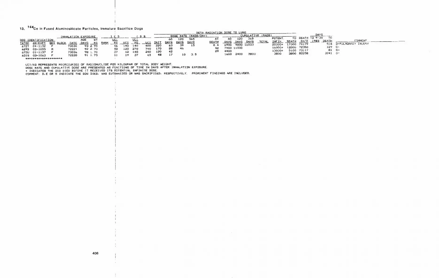
13. 144Ce in Fused Atuminosilicate Particles, Immature Sacrifice Dogs
INHALATION EXPOSURE I.~.B.DOg IDENTIFICATION AgE WT V~ITATO0 AN-EXPT SEX BLOCK DATE DAYS KG RANK ~g
73030 93 2.95 4672221 92 2.70 437303b 98 1.70 3772228 91 1.75 II
672T 04-1132 F6298 03-1055 M673U 01-1137 F631S 03-1063 F
BETA RADIATION DOSE TO LUNg
I.B.B. DOSE RATE (RADS/DAY) CUMULATIVE (RADS) DAYS
UCI 60 120 365 AT 60 120 365 POTENT. TO DEATH TO 9-30 TO
UCI Kg UCI INIT DAYS DAYS DAYS DEATH DAYS DAYS DAYS TOTAL INFIN. DEAT~ _DATE 1983 DEATH COMMENT
140 140 400 220 63 39 15 8.4 6900 9800 16000 20000+ 17000 74179 514 D-PULMONARY INJURY
120 270 740 170 88 46 42 7500 11000 16000+ 12000 72350 129 S-
62 140 240 120 42 29 4400 13000+ 5100 73117 81 S-
19 37 65 48 17 i0 3.5 1600 2400 3800 3800 3800 82258 3241 S-
UCI/~g REPRESENTS MICROCURIES OF RADIONUCLIDE PER KILOGRAM OF TOTAL BODY WEIGHT.DOSE RATE AND CUMULATIVE DOSE ARE PRESENTED AS Fi~NCTIONS OF TIME IN DAYS AFTER INHALATION EXPOSURE.+ INDICATES THE DOG DIED BEFORE IT RECEIVED ITS fOTENTIAL INFINITE DOSE.COMMENT: D,E OR S INDICATE THE DOg DIED, WAS EUTI[ANIZED OR WAS SACRIFICED, RESPECTIVELY. PROMINENT FINDINGS ARE INCLUDED.
406
J
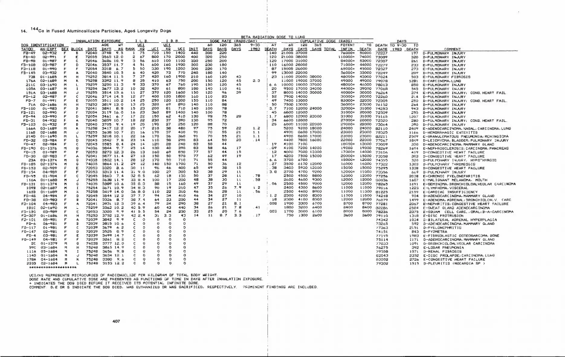
14. 144Ce in Fused Aluminosilicate Particles, Aged Longevity Dogs
BETA RADIATION DOSE TO LUNgINHALATION EXPOSURE I.L.B. I.B.B. DOSE RATE {RADS/DAY) CUMULATIVE DOSE (RADS) DAYS
DOg IDENTIFICATION AgE NT UCl UCl 60 120 365 9-30 . AT 60 120 365 POTENT. TO DEATH TO 9-30 TOTATO0 AN-EXPT SEX BLOCK DATE DAYS Kg RANK Kg UCI Kg UCI INIT DAYS DAYS DAYS 1983 DEATH DAYS DAYS DAYS TOTAL INFIN. DEATH DATE 1983 DEATHIFD-49 02-932 FFD-40 02-991 FFD-98 01-987 F
FD-108 03-987 FFD-I18 01-990 FFD-145 03-932 F
738 01-1685 M176A 02-1689 M211C 03-1690 MI05A 03-1687 M151A 03-1688 M
FD-12 02-987 FFD-7 01-991 F
71A 02-1686 MFD-IO0 01-983 FFD-121 02-990 F
FD-94 03-990 FFD-31 04-932 F
FD-I03 01-932 F166A 03-I689 M116B 02-1688 M214D 01-1690 M
FD-32 03-984 FFD-47 02-984 F
FD-190 01-1376 MFD-15 01-989 FFD-30 02-983 F
23A 03-1374 MFD-185 02-1374 MFD-153 02-989 FFD-154 04-989 F
FD-95 01-984 F116A 01-1688 M
FD-131 01-1374 M1098 02-1687 M1658 01-1689 M
FD-4B 04-984 FFD-3B 03-983 F
FD-104 04-983 FISIC 02-1690 M
FD-150 03-989 FFD-307 01-1686 MFD-101 05-981 F
FD-6 06-981 FFD-I17 01-981 FFD-147 02-981 F
FD-4 03-981 FFD-149 04-981 F
2C 01-1379 M59C 03-1684 M
IlIA 05-1684 M114D 01-1684 M17SA 04-1684 M2258 02-1684 M
B 72040 3748 9.5E 72055 3565 12.0C 72046 3696 10.9D 72046 3537 11.7F 72054 3318 6.7A 72040 3840 10.5H 75252 3814 11. 5K 75258 3392 11.9L 75259 3~50 11.3I 75254 3677 132d 75255 3514 13.6C 72046 3714 14.5E 72055 3511 10.2H 75253 3819 13.0B 72041 3841 8.8F 72054 3119 16.0D 72054 3461 6.7A 72040 3859 10.7B 72040 3705 9.4K 7525S 3417 12,2d 75255 363S 10.7L 75259 3218 10.1
I 75 710 150 1400 440 3002 67 800 170 2000 400 3203 56 610 100 1100 330 2504 51 600 160 1900 300 2305 50 330 190 1200 300 2206 40 420 73 770 240 1807 37 420 160 1900 210 160S 35 410 63 750 200 1509 33 370 67 760 190 150
10 32 420 61 800 180 14011 27 370 120 1600 150 12012 27 400 120 1800 160 II014 25 250 120 1300 150 11013 25 320 69 890 140 II015 23 200 81 710 140 10016 23 360 89 1400 140 10017 22 150 62 410 130 9818 22 230 37 390 130 9519 20 190 27 250 120 8820 17 210 38 480 97 7521 16 170 37 400 91 7022 16 170 36 360 91 70
72045 3542" 7.8 23 1472045 3585 8.4 24 1474036 3844 9.7 25 1472053 3273 12.3 26 1372041 3877 11.4 27 1374035 3502 14. 1 28 1274035 3864 11.2 29 1272053 3320 8.6 30 1172053 3313 11.4 31 9.072045 3563 7~4 32 8.575255 3638 11.9 33 8.474035 3889 10.6 35 8.375254 3671 10.9 34 8.375258 3419 14.0 36 8.0
D 72045 3544 12.2 37 7.7B 72041 3326 8.7 38 7. 4A 72041 3931 12.3 39 6,4L 75259 3362 10.2 40 5.9E 72053 3320 9.9 41 5.5H 75253 3752 12.9 42 2.4A 72039 3842 9.9 C 0B 72039 3815 10.6 C 0C 72039 3679 6.2 C 0D 72039 3525 8.9 C 0E 72039 3499 14.7 C 0F 72039 3261 8.2 C 0@ 74038 3777 12.0 C 0H 75248 3865 14.9 C 0I 75248 3656 9.8 C OJ 75248 3634 12. i C 0K 75248 3380 9.6 C OL 75248 3153 12.2 C 0
110 38 290 83 64120 28 240 83 58130 40 390 83 58160 69 850 77 57150 50 570 77 62170 50 710 71 55140 150 1700 71 50
96 46 400 65 49100 27 300 53 39
62 18 130 50 37100 17 200 48 3888 15 160 49 3790 19 210 47 35
II0 22 310 46 3694 34 410 46 3464 23 200 44 3479 24 290 38 2760 43 440 34 2654 24 230 33 2531 3.3 43 14 110 0 00 0 00 0 00 0 00 0 00 0 00 0 00 0 00 0 00 0 00 0 00 0 0
220250200IBO170140120 43120 45 2.3120 45110 4196 39838488787875 28727O59 22 1,255 21 1. I55 2t 1,150 20 .144446 1744 1549 194436 1238 1529 1128 11 .7830 II .5829 1126 7.9 t.228 ii .5627 IO27 II21 8.121 7.8 .4120 7.6
8.7 3,3 .17
140 21000 37000I20 21000 38000120 17000 31000110 16000 2800087 15000 2600099 13000 2200023 11000 20000 38000
11000 19000 370006.6 10000 18000 37000
20 9500 17000 3400037 8000 14000 3000052 7900 1400049 7400 1300050 7500 13000
3.7 7100 12000 2400045 6800 12000
1.7 6800 12000 2300034 6600 1200025 6000 I1000 22000
5200 9200 IBO004900 8600 170004900 8600 170004400 7800 16000
19 4100 7100.09 4100 7200 14000
12 4000 7000 1300017 4200 7500 15000
6,6 3700 6700.27 3500 6100 12000.38 3400 5900 120003.8 2700 4700 9200
2500 4500 88002600 4600 9300
.06 2500 4500 90002400 4300 86002500 4400 8900
1.2 2300 4200 8300.18 2300 4100 8500008 1900 3300 6700
1800 3200 6400003 1700 3000 6100
750 1300 2600
UCI/KQ REPRESENTS MICROCURIES OF RADIONUCLIDE PER KILOGRAM OF TOTAL BODY WEIGHT.DOSE RATE AND CUMULATIVE DOSE ARE PRESENTED AS FUNCTIONS OF TIME IN DAYS AFTER INHALATION EXPOSURE.+ INDICATES THE DOg DIED BEFORE IT RECEIVED ITS POTENTIAL INFINITE DOSE.COMMENT: D,E OR S INDICATE THE DOg DIED, WAS EUTHANIZED OR WAS SACRIFICED, RESPECTIVELY. PROMINENT FINDINGS ARE INCLUDED.
76000+ 50000 72237Ii0000+ 74000 73009
84000+ 53000 7230771000+ 44000 7227965000+ 45000 7232756000+ 33000 7224948000+ 43000 7702449000 49000 7907848000+ 46000 7801344000+ 39000 7706840000+ 30000 7626430000+ 20000 7226083000+ 22000 7230536000+ 23000 7615232000+ 31000 7425331000+ 20000 7230931000 31000 7516527000+ 20000 7232029000+ 22000 7306424000 24000 8211023000 23000 7832523000 23000 8221122000 22000 7706718000+ 13000 7300919000 19000 7826917000+ 14000 7310620000+ 15000. 7305818000+ 12000 7435516000 16000 7732215000 15000 7529512000+ 11000 7335612000 12000 7725612000 12000 8003612000 12000 78265ii000 11000 7901611000 ii000 81225ii000 IlO00 7421812000 12000 760798700 8700 772818400 8400 822868000 8000 782363600 3600 79110
743427326577363741517719575114770337627579358820438305279302
COMMENT197 D-PULMONARY INJURY320 D-PULMONARY INJURY261 D-PULMONARY INJURY233 E-PULMONARY INJURY273 E-PULMONARY INJURY209 D-PULMONARY INJURY503 E-PULMONARY FIBROSIS
1281 D-CARCINOMA, LUNg850 S-PULMONARY INJURY545 D-PULMONARY INJURY374 E-PULMONARY INJURYJ CONg. HEART FAIL.214 D-PULMONARY INJURY250 D-PULMONARY INJURY~ CONg. HEART FAIL.264 D-PULMONARY INJURY943 D-PULMONARY INJURY255 D-PULMONARY INJURY
1207 D-PULMONARY INJURY280 D-PULMONARY INJURY} CONQ. HEART FAIL.390 D-PULMONARY INJURY
2409 E-ADENDCARCINOMA, NASAL~CARCINOMA, LUNQ1166 D-HEMORRHAgIC ENTERITIS2509 E-QRANULOMATOUS PNEUMONIA;MENINgIOMA1849 D-LEIOMYOMA, BLADDER~PULMONARY INJURY330 D-ADENOCARCINOMA, MAMMARY gLAND
1694 E-NEPHROSCLEROSIS}CARCINOMA, PANCREAS419 D-CONgESTIVE HEART FAILURE383 D-CONgESTIVE 14EART FAILURE320 D-PULMONARY INJURY} HYPOTHYROID
1383 D-PULMONARY THROMBOSIS1338 D-CONgESTIVE HEART FAILURE669 D-PULMONARY INJURY
2038 E-CHRONIC PYELONEPHRITIS1607 E-MALIgNANT MELANOMA, MOUTH1691 E-SEMINOMA~BRONCHIOLOALVEOLAR CARCINOMA1223 E-LYMPHOMA, VISCERAL2159 E-CARDIAC INSUFFICIENCY
904 D-ADENOCARCINOMA, MAMMARY gLAND1499 E-ADENOMA, ADRENALIBRONCHIOLOALV. CARC.2067 E-NEPHRiTIS~CONgESTIVE HEART FAILURE2584 E-SWEAT gLAND ADENOCARCINOMA2375 E-SGUAM. CELL CARC.,ORAL}B-A-CARCINOMA1318 E-DISC PROTRUSION1034 D-BILATERAL ADRENAL HYPERPLASIA
592 D-ADENOCARCINOMA, MAMMARY gLAND2151 D-PYELONEPHRITIS843 D-PYOMETRA
1983 E-FIBROBLASTIC OSTEOSARCOMA, BONE1171 D-ADENOCARCINOMA, MAMMARY gLAND1091 D-BRONCHIOLOALVEOL~R CARCINOMA
392 E-LOBAR PNEUMONIA1571 D-RENAL FIBROSIS2352 E-DISC PROLAPSE;CARCINOMA, LUNg2726 E-CONgESTIVE HEART FAILURE1515 D-PLEURITIS (NOCARDIA SP.
407
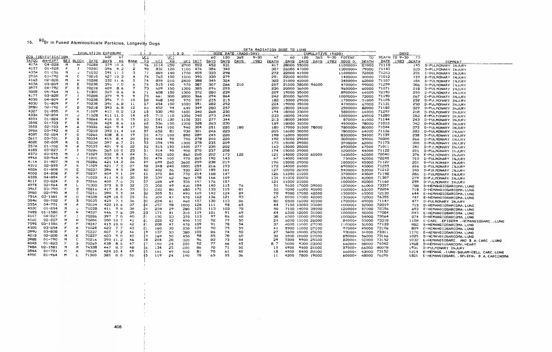
15. 90Sr in Fused Afuminosilicate Particles, Longevit!y Dogs
INHALATION EXPOSURE I.L.B.DOg IDENTIFICATION AgE WT U~ITATO0 AN-EXPT SEX BLOC~ DATE DAN’S K@ RANK ~. UCi
H 70288 379 10.6I 70288 396 9.2d 71032 391 11.5C 70218 427 10.3H 70238 332 11.6E 70238 396 7.0D 70218 409 8.6L 71300 369 8.6I 70288 379 9.5F 70238 396 7.0F 70238 396 6.8 IiD 70218 393 6.8 12K 71029 413 8.2 13d 71028 411 II.0 14g 70266 416 8.6 15B 70036 424 8.6 16A 70036 424 9.4 17C 70218 393 11.4 18
70266 408 8.6 19B 70034 415 8.4 20E 70238 397 6.7 21A 70035 421 9.8 22I 70286 365 10.0 23O 71029 332 8.3 24L 71300 404 9.4 25H 70286 421 14.2 26K 71029 421 7.2 27E 70237 396 9.5 28F 70237 404 9. 1 29K 71028 411 9.0 30g 70266 400 7.6 31L 71300 375 8.8 32D 70216 417 8.6 33C 70216 390 9.5 340 74338 429 I0 0 35B 70035 425 7.5 36A 70034 422 10.6 37d 71028 411 9.8 38N 74337 446 7.6 39I 70286 397 7.0 40H 70286 380 10,7 41P 74347 415 10.0 42K 71028 423 7.7 43F 70237 407 7.2 44E 70237 403 9.0 45C 70216 390 11.2 46Q 70265 438 8.6 47M 74338 447 8.2 48A 70034 424 10.4 49L 71300 385 8.0 50
417A 04-828 M415T 01-828 F435A 01-856 M393A 01-792 M4168 02-828 M403A 03-809 M397T 04-792 F5008 04-964 M417T 03-828 F4038 04-809 P403U 01-809 F398U 02-792 F432T 01-855 F433A 02-854 M405X 01-824 F3558 01-703 F3558 02-703 M398A 03-792 M408T 02-824 F361T 03-701 F402D 02-809 M357A 01-702 M4188 03-827 F437D 03-855 M494A 03-964 M411A 01-827 M431U 02-855 F402A 01-808 M4008 04-808 F4338 04-854 F411T 03-824 F4978 02-964 M396T 03-790 F398D 02-790 M751A 03-1581 M354W 02-702 F355A 04-701 M433C 01-854 M7488 01-1580 F414T 04-827 F416C 02-827 M759S 03-1586 F4308 03-854 F399U 03-808 F4018 02-808 M3988 01-790 M4048 01-823 F74BA 02--1581 M354A 01-701 M495C 01-964 M
BETA RADIATION DOSE TO LUNgI.B.B. DOSE RATE (RADSIDAY) CUMULATIVE (RAD8) DAYS
UCI 60 120 365 9-30 AT 60 120 365 9-30 POTENT. TO DEATH TO 9-30 TO~g UCI INIT DAYS DAYS DAYS 1983 DEATH DAYS DAYS DAYS 1983 5000 D. DEATH DATE 1983 DEATH
1 96 1014 250 2700 5032 90 832 120 1100 4763 77 885 140 1700 4054 F4 763 I00 I000 3905 ~4 855 210 2400 3886 V4 515 140 970 3877 ~3 629 150 1300 3858 91 608 150 1300 3729 ~0 661 300 2800 366
10 68 468 140 970 357~7 454 150 I000 351&6 452 94 640 349
~5 530 190 1600 340~5 710 II0 1300 340~3 ,,oo=,~2 536 150 1300 32848 544 I00 940 304
7 652 81 930 301’LSp 473 i00 890 289~3 444 90 750 278~3 354 190 1300 278~3 515150 1400277~I 514 90 900 270~i. 423100 860268~0 474 I00 970 26549 699 260 3600 259~8 348 240 1800 25442 402 56 530 22341 370 84 770 214
~8 339 &2 560 198~7 284 69 530 19775 308 49 430 184~3 282 80 680 172~2 305 51 490 16931 309 47 470 162
I! 224 61 460 157257 98 1000 128
¯ 234 29 280 12523 171 41 310 119~i 150 53 370 113~i 223 42 450 II0~i 208 47 470 10921 160 30 230 109
ii 137 53 380 I00169 50 450 98204 28 320 96
~7 150 24 200 92~6. 134 25 200 86
~161 49 510 81
I 119 24 190 78
452 431386 340333 298330 279345 324307 266 210296 255280 239294 264235 215282 252280 257268 230273 248277 344263 230251 220 180246 223245 220240 220235 2O9230 202237 208202 173 122192 143238 219216 193185 161168 147166 149176 157140 115 76133 115 81142 124 89136 121 93130 113 86iii 98 65113 103 78101 91 69
97 86 6092 79 5091 78 5490 79 5986 74 5085 78 6082 73 5477 66 4578 71 5070 61 4065 55 36
COMMENT417 28000 55000307 26000 47000272 220¢0 41000251 22000 40000302 21000 42000207 21000 38000 94000230 20000 36000209 19000 35000242 20000 36000182 15000 29000224 19000 35000200 18000 34000194 18000 33000233 18000 34000213 18000 34000189 18000 32000180 17000 31000 78000205 16000 30000198 16000 30000192 15000 29000173 15000 29000163 15000 28000174 15000 29000121 14000 25000 6000067 14000 24000
196 15000 29000172 14000 26000127 12000 22000126 riO00 21000131 I1000 20000121 11000 21000
51 9600 17000 3900052 9000 16000 4000070 9300 17000 4200067 8900 17000 4200080 8500 16000 4000044 7100 13000 3300058 7100 14000 3500044 6500 12000 3100038 6300 12000 2900024 6000 11000 2600037 6000 II000 2700041 5900 i1000 2700027 5600 I0000 2500030 5500 I0000 2700029 5300 9900 25000
8.7 5000 9300 2300015 4900 9400 2400018 4500 8400 20000It 4300 7800 19000
ii00000+ 87000 711181300000+ 79000 711431100000+ 72000 71263
140000+ 50000 71012340000+ 62000 71107470000+ 99000 71259960000+ 60000 71071890000+ 64000 72190
1000000+ 73000 71190170000+ 51000 71105470000+ 67000 71131250000+ 82000 71182580000+ 64000 71297
1000000+ 65000 7128087000+ 61000 71144
450000+ 78000 71013780000+ 80000 71044780000+ 64000 71136830000+ 54000 71139300000+ 59000 70300370000+ 62000 71173690000+ 67000 71011120000+ 44000 71122160000+ 62000 7204075000+ 42000 72245
180000+ 45000 71122690000+ 45000 71255540000+ 53000 71212370000+ 43000 71158350000+ 42000 71307I00000+ 40000 71160120000+ 66000 73357100000+ 63000 72204150000+ 65000 72130150000+ 72000 76355170000+ 49000 71147100000+ 52000 72019120000+ 57000 72356I00000+ 58000 77084100000+ 54000 73064
84000+ 53000 73318110000+ 50000 77133
97000+ 49000 7310693000+ 54000 7331185000+ 56000 7316682000+ 52000 7315266000+ 58000 7604287000+ 66000 8007866000+ 43000 7315260000+ 48000 76295
195 D-PULMONARY INJURY220 D-PULMONARY INJURY231 E-PULMONARY INJURY159 D-PULMONARY INJURY184 D-PULMONARY INJURY386 D-PULMONARY INJURY218 D-PULMONARY INJURY255 D-PULMONARY INJURY267 E-PULMONARY INJURY232 D-PULMONARY INJURY258 D-PULMONARY INJURY329 D-PULMONARY INJURY268 D-PULMONARY INJURY252 E--PULMONARY INJURY243 D-PULMONARY INJURY342 D-PULMONARY INJURY373 D-PULMONARY INJURY283 D-PULMONARY INJURY238 D-PULMONARY INJURY266 D-PULMONARY INJURY300 D-PULMONARY INJURY341 D-PULMONARY INJURY201 D-PULMONARY INJURY376 E-PULMONARY INJURY310 D-PULMONARY INJURY201 D-PULMONARY INJURY226 D-PULMONARY INJURY340 D-PULMONARY INJURY286 D-PULMONARY INJURY279 D-PULMONARY INJURY259 D-PULMONARY INJURY788 D-HEMANgIOBARCOMA, LUNg718 D-HEMANgIOSARCOMA, LUNg644 E-HEMANglOSARCOMA, LUNg747 E-HEMANgIOSARCOMA, LUNg477 D-PULMONARY INJURY715 D-HEMANgIOSARCOMA, LUNQ693 E-HEMANglOSARCOMA, LUNg843 E-HEMANQIOSARCOMA, LUNg874 D-HEMANgIOSARCOMA, LUNQ
1128 E-CARC. ,EPIDERM. IHEMANQIOSARC.,LUNg882 E-HEMAN~IOSARCOMA, LUN~809 E-HEMANgIOSARCOMA, LUNg
1170 E-HEMANgIOSARCOMA, LUNg1025 D-HEMANgIOSARCOMA, LUNQ1032 E-HEMANglOSARC. AND B.A. CARC.,LUNQ1968 D-HEMANgIOSARCOMA, HEART1931 D-PULMONARY INJURY1214 E-HEMANg.,LUNg~SQUAM-CELL CARC, LUNg1821 E-HEMANgIOSARC.,SPLEEN~ B.A. CARCINOMA
408
[
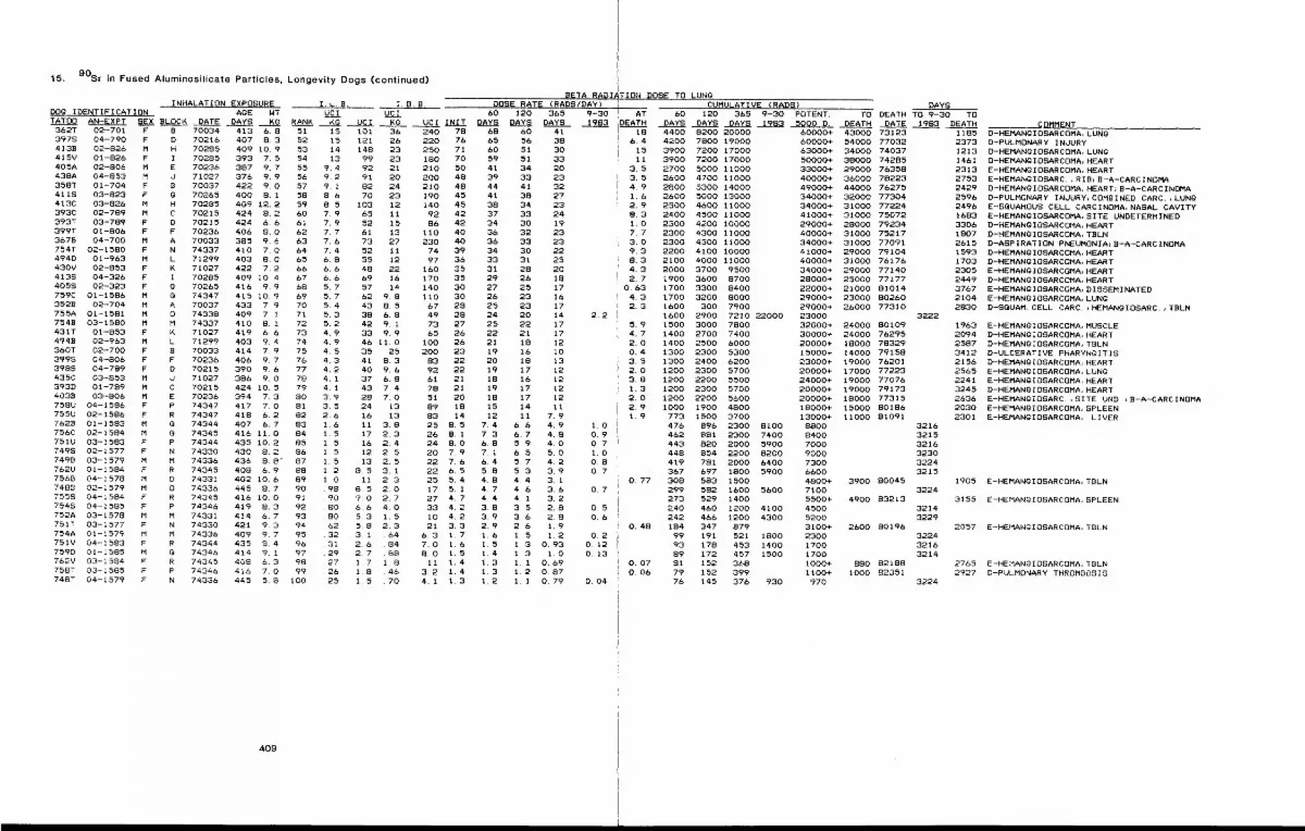
15. 90St in Fused Aluminosilicate Particles, Longevity Dogs (continued)
INHALATION EXPOSUREDOg IDENTIFICATION AOE WTTATO0 AN-EXPT SEX BLOCK DATE D.AYS KG RANK
362T 02-701 F3978 04-790 F4138 02-826 M415V 01-826 F405A 02-806 M438A 04-853 M358T 01-704 F4118 03-823 F413C 03-826 M393C 02-789 M393T 03-789 F399T 01-806 F3678 04-700 M754T 02-1580 F494D 01-963 M430V 02-853 F4138 04-326 F4055 02-323 F759C 01-1586 M3528 02-704 M755A 01-1581 M7548 03-1580 M431T 01-853 F4948 02-963 M360T 02-700 F3995 04-806 F3988 04-789 F435C 03-853 M393D 01-789 M4038 03-806 M758U 04-1586 F755U 02-1586 F7628 01-1583 M756C 02-1584 M751U 03-1583 F7498 02-1577 F7498 03-1579 M762U 01-1584 F7568 04-1578 M7488 02-1579 M755S 04-1584 F7548 04-1585 F752A 03-1578 M751T 03-1577 F754A 01-1579 M751V 04-1583 F759D 01-1585 M76~V 03-1594 F758T 03-1585 F748T 04-1579 F
B 70034 413 6.8D 70216 407 8.3H 70285 409 10.9I 70285 393 7.5E 70236 387 9.7J 71027 376 9.9B 70037 422 9.0g 70265 400 8.1H 70285 409 12.2C 70215 424 8.2D 70215 424 6.6F 70236 406 8.0A 70033 385 9.6N 74337 410 7,0L 71299 403 8.0K 71027 422 7.2I 70285 409 10.4g 70265 416 9.9Q 74347 415 I0.9A 70037 433 7.90 74338 409 7.1M 74337 410 8.1K 71027 419 6.6L 71299 403 9.4B 70033 414 7.9F 70236D 702150 71027C 70215E 70236P 74347R 74347
66676869707172737475
406 9.7 76390 9.6 77386 9.0 78424 10.5 79394 7.3 80417 7.0 81418 6.2 82
Q 74344 407 6.7 838 74345 416 11.0 84P 74344 435 I0.2 85N 74330 430 8.2 86M 74336 436 8.8" 87R 74345 408 6.9 880 74331 402 10.6 890 74336 445 8.7 90R 74345 416 10.0 91P 74346 419 8.3 92M 74331 414 6.7 93N 74330 421 9.3 94M 74336 409 9.7 95R 74344 435 8.4 960 74346 414 9. 1 97R 74345 408 6.3 98P 74346 416 7.0 99N 74336 445 5.8 100
409
51 15 101 3652 15 121 2653 14 148 2354 13 99 2355 9.4 92 2156 9.2 91 2057 9.1 82 2458 8.6 70 2359 8,5 103 1260 7.9 65 II61 7.9 52 1562 7.7 61 1363 7.6 73 2764 7.4 52 1165 6.8 55 12
6.6 48 226.6 69 165.7 57 145.7 62 9. 85.4 43 S. 55.3 38 6.85.2 42 9. i4.9 33 9.94.9 46 11.04. 5 35 254.3 41 8.34.2 40 9.64.1 37 6.84.1 43 7 43.9 28 7.03.5 24 132.6 16 131.6 Ii 3.81.5 17 2.31.5 16 2.41.5 12 2.51.5 13 2.51.2 8,5 3.11.0 11 2 3.98 8.5 2.0
90 9.0 2.780 6.6 4.080 5.3 1.562 5.8 2.332 3.1 .6431 2,6 .8429 2.7 .8827 1.7 1.826 1.8 .4625 1.5 .70
I. B. 8.BETA RADI~
D0SE RATE (RADS/DAY)60 120 365 9-30
UCI INIT DAYS DAYS DAYS 1983240 78 68 60 41220 76 65 56 38250 71 60 51 30180 70 59 51 33210 50 41 34 20200 48 39 33 23210 48 44 41 32190 45 41 38 27140 45 38 34 23
92 42 37 33 2486 42 34 30 19
110 40 36 32 23230 40 36 33 23
74 39 34 30 2297 36 33 31 25
160 35 31 28 20170 35 29 26 18140 30 27 25 17IiO 30 26 23 1667 28 25 23 1749 28 24 20 1473 27 25 22 1765 26 22 21 17
100 26 21 18 12200 23 19 16 I 0
83 22 20 18 1392 22 19 17 1261 21 18 16 1278 21 19 17 1251 20 18 17 1289 18 15 14 1183 14 12 11 7. 925 8.5 7.4 6.6 4.926 8. 1 7.3 6.7 4.824 8.0 6.8 5.9 4.020 7.9 7,1 6.5 5.022 7.6 6.4 5.7 4.222 6.5 5.8 5.3 3.925 5.4 4.8 4.4 3.117 5.1 4,7 4.6 3.627 4.7 4.4 4, 1 3.233 4.2 3,8 3,5 2.810 4,2 3.9 3.6 2.821 3.3 2~9 2,6 1.9
6.3 1.7 1.6 1.5 1.27.0 1.6 1. 5 1.3 0.938.0 1.5 1.4 1.3 1.0Ii 1.4 1.3 1. 1 0.69
3.2 1.4 1.3 1.2 0.874.1 1.3 1.2 1. 1 0.79
2.2
1.00.90.71.00.80.7
0.7
TION DOSE TO LUNgCUMULATIVE (RADS) DAYS
AT 60 120 365 9-30 POTENT. TO DEATH TO 9-30 TODEATH DAYS DAYS DAYS 1983 5000 D, DEATH DATE 1983 DEATH
186.4
1511
3.53.54.91.62.98.31.07.73.09.38.34.32.7
O. 634.32.3
5.94.72.00.43.52.03.81.32.02.91.9
O. 77
0.50.6
O. 480.2
0.120.13
O. 07O. 06
O. 04
4400 8200 200004200 7800 190003900 7200 170003900 7200 170002700 5000 II0002600 4700 II0002800 5300 140002600 5000 130002500 4600 110002400 4500 110002300 4200 I00002300 4300 110002300 4300 110002200 4100 100002100 4000 110002000 3700 95001900 3600 87001700 3300 84001700 3200 80001600 300 79001600 2900 7210 220001500 3000 78001400 2700 74001400 2500 60001300 2300 53001300 2400 62001200 2300 57001200 2200 55001200 2300 57001200 2200 5600I000 1900 4800
773 1500 3700476 896 2300 8100462 881 2300 7400443 820 2000 5900448 854 2200 8200419 781 2000 6400367 697 1800 5900308 583 1500299 582 1600 5600273 529 1400240 460 1200 4100242 466 1200 4300184 347 87999 191 521 180093 178 453 140089 172 457 150081 152 36879 152 39976 145 376 930
COMMENT60000+ 43000 7312360000+ 54000 7703263000+ 34000 7403750000+ 38000 7428533000+ 29000 7635840000+ 36000 7822349000+ 44000 7627534000+ 32000 7730434000+ 31000 7722441000+ 31000 7507229000+ 28000 7923440000+ 31000 7521734000+ 31000 7709141000+ 29000 7910440000+ 31000 7617634000+ 29000 7714028000+ 25000 7717722000+ 21000 8101429000+ 23000 8026029000+ 26000 7731023000 322232000+ 24000 8010930000+ 24000 7629520000+ 18000 7832915000+ 14000 7915823000+ 19000 7620120000+ 17000 7722324000+ 19000 7707620000+ 19000 7917320000+ 18000 7731518000+ 15000 8018613000+ 11000 81091
8800 32168400 32157000 32169000 3230730O 32246600 32154800+ 3900 800457100 32245500+ 4900 832134500 32145200 32293100+ 2600 801962300 32241700 32161700 32141000+ 880 821881100+ I000 82351970 3224
1185 D-HEMANQIOSARCOMA, LUNQ2373 D-PULMONARY INdURY1213 D-HEMANQIOSARCOMA, LUNG1461 D-HEMANQIOSARCOMA, HEART2313 E-HEMANQIOSARCOMA, HEART2753 E-HEMANgIOSARC.~RIB;B-A-CARCINOMA2429 D-HEMAN~IOSARCOMA, HEART;B-A-CARCINOMA2596 D-PULMONARY INJURY~COMBINED CARC.,LUNQ2496 E-SQUAMOUS CELL CARCINOMA, NASAL CAVITY1683 E-HEMANgIOSARCOMA, SITE UNDETERMINED3306 D-HEMANgIOSARCOMA, HEART1807 D-HEMANgIOSARCOMA, TBLN2615 D-ASPiRATION PNEUMONIA~B-A-CARCINOMA1593 D-HEMANgIOSARCOMA, HEART1703 D-HEMANgIOSARCOMA, HEART2305 E-HEMANgIOSARCOMA, HEART2449 D-HEMANgIOSARCOMA, HEART3767 E-HEMANgIOSARCOMA, DISSEMINATED2104 E-HEMANgIOSARCOMA, LUNg2830 D-SGUAM. CELL CARC. ;HEMANQIOSARC.,TBLN
1963 E-HEMANQIOSARCOMA, MUSCLE2094 D-HEMANGIOSARCOMA, HEART2587 D-HEMANGIOSARCOMA, TBLN3412 D-ULCERATIVE PHARYNGITIS2156 D-HEMANQIOSARCOMA,HEART2565 E-HEMANGIOSARCOMA, LUNQ2241 E-HEMANgiOSARCOMA, HEART3245 D-HEMANgIOSARCOMA, HEART2636 E-HEMANQIOSARC. ,SITE UND. ;B-A-CARCINOMA2030 E-HEMANgIOSARCOMA, SPLEEN2301 E-HEMANglOSARCOMA, LIVER
1905 E-HEMANQIOSARCOMA, TBLN
3155 E-HEMANQIOSARCOMA, SPLEEN
2057 E-HEMANglOSARCOMA, TBLN
2765 E-HEMANglOSARCOMA, TBLN2927 D-PULMONARY THROMBOSIS
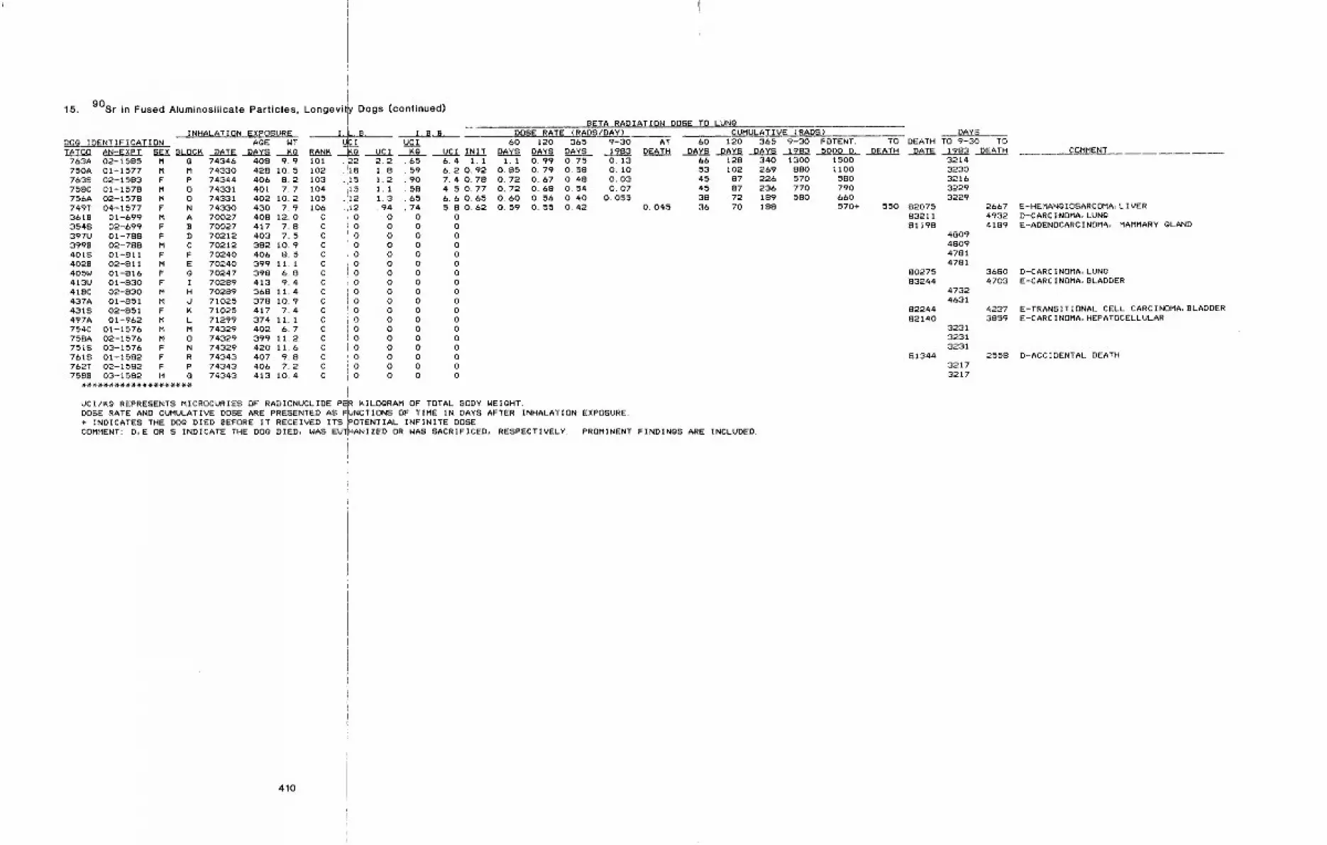
15. 90Sr in Fused Aluminosilicate Particles, Longevily Dogs (continued)
INHALATION EXPOSUREDOg IDENTIFICATION
I.L.B.AgE WT
TATO0 AN-EXPT S~X BLOCK DATE DAYS Kg RANK .763A 02-1585 M G 74346 408 9.9 101750A 01-1577 M M 74330 428 10.5 102763S 02-1583 F P 74344 406 8.2 103758C 01-1578 M 0 74331 401 7.7 104756A 02-1578 M 0 74331 402 10.2 105749T 04-1577 F N 74330 430 7.9 1063618 01-699 M A 70027 408 12.0 C354S 02-699 F B 70027 417 7.8 C397U 01-788 F D 70212 403 7.5 C3998 02-788 M C 70212 382 10.9 C401S 01-811 F F 70240 406 8.5 C402B 02-811 M E 70240 399 11. 1 C405W 01-816 F g 70247 398 6.8 C413U 01-830 F I 70289 413 9.4 C418C 02-830 M H 70289 368 11.4 C437A 01-851 M d 71025 378 10.9 C431S 02-851 F K 71025 417 7.4 C497A 01-962 M L 71299 374 ii. 1 C754C 01-1576 M M 74329 402 6.7 C758A 02-1576 M 0 74329 399 11.2 C751S 03-1576 F N 74329 420 11,6 C761S 01-1582 F R 74343 407 9.8 C762T 02-1582 F P 74343 406 7.2 C7588 03-1582 M G 74343 413 10,4 C
L~31~o.22.!is
15.15112,12
00000000000000000
io
uc I2.21.81.21,11.3. 94
000000000000000000
I. B, B.UC I 60
Kg UCI INIT DAYS
BETA R,ADIATION DOSE TO LUNgDOSE RATE (RADS/DAY) CUMULATIVE (RADS)
120 365 9-30 AT 60 120 365 9-30 POTENT. TODAYS DAYS 1983 D~ATH DAYS DAYS DAYS 1983 5000 D. DEATH0.99 0,75 0.13 6& 128 340 1300 15000,79 0.58 0.10 53 102 269 880 11000.67 0,48 0.03 45 87 226 570 5800.68 0.54 0.07 45 87 236 770 7900.56 0.40 0.055 38 72 189 580 6600.55 0.42 0.045 36 70 188 570+
65 6.4 i.i59 6.2 0.9290 7.4 0.7858 4.5 0.7765 6. 60. 6574 5. 80. 620 00 00 00 00 oo 0o 00 00 o0 00 O0 00 00 00 00 o0 00 o
1.1O. 85O. 72O. 72O. 60O. 59
DAYSDEATH TO 9-30 TO
DATE 1983 DEATH
550 820758321181198
8027583244
8224482140
81344
32t43230321632293229
4809480947814781
47324631
323132313231
32173217
UCI/K~ REPRESENTS MICROCURIES OF RADIONUCLIDE P~R KILOORAM OF TOTAL BODY WEIGHT.DOSE RATE AND CUMULATIVE DOSE ARE PRESENTED AS ~UNCTIONS OF TIME IN DAYS AFTER INHALATION EXPOSURE.+ INDICATES THE DO~ DIED BEFORE IT RECEIVED ITSiPOTENTIAL INFINITE DOSE.COMMENT: D,E OR S INDICATE THE DO~ DIED, WAS EU~HANIZED OR WAS SACRIFICED, RESPECTIVELY. PROMINENT FINDINGS ARE INCLUDED.
266749324189
36804703
42373859
2558
COMMENT
E-HEMANgIOSARCOMA, LIVERD-CARCINOMA, LUNgE-ADENOCARCINOMA, MAMMARY gLAND
D-CARCINOMA, LUNQE-CARCINOMA, BLADDER
E-TRANSITIONAL CELL CARCINOMA, BLADDERE-CARCINOMA, HEPATOCELLULAR
D-ACCIDENTAL DEATH
410

16. 144Ce in Fused Aluminosilicate Particles, Repeatedly Exposed Dogs
BETA RADIATION DOSE TO LUNgDOSE RATE (RADS/DAYS)
INITIAL FINALEXPOSURE EXPOSURE
AFTER AFTERAgE WT AgE WT INITIAL FINALDOg IDENTIFICATION
TATO0 AN-EXPT SEX gROUP DATE DAYS KG DATE DAYS ~g EXP.645C 01-1294 M648U 02-1294 F664C 03-1294 M641T 04-1294 F644T 05-1294 F6468 01-1295 F654T 02-1295 F6458 03-1295 F641C 04-1295 M662U 01-1292 F654B 02-1292 M645A 03-1292 M6518 04-1292 F6658 05-1292 M654A 01-1293 M641B 02-1293 M6488 03-1293 M6488 04-1293 F649U 01-1290 F650U 02-1290 F649V 03-1290 F650£ 04-1290 M641A 05-1290 M6628 01-1291 F655U 02-1291 F6448 03-1291 F665A 04-1291 M6648 01-1288 M664A 02-1288 M648T 03-1288 F6638 04-1288 F6468 05-1288 M648A 01-1289 M6498 02-1289 M662T 03-1289 F657A 04-1289 M
EXP.756465606448725571465455575353
ATDEATH
.28
¯ 38.17&.5
.11044.12
085
052.22
46 05658 . 5465 2.028 .363229 0563424 07219 01925 .282325 17.4
I 73340 518 8.3 75288 1195 9.5 18I 73340 513 6.5 75288 1191 7.1 18I 73340 452 9.6 75288 1130 11.7 20I 73340 526 8.9 75288 1204 11.5 19I 73340 518 7.1 75288 1195 8.3 14I 73341 518 7. t 75289 1196 9.0 15I 73341 495 7.6 75289 1173 8.5 17I 73341 519 9.0 75289 1197 12.5 24I 73341 527 9.3 75289 1205 11.3 23
II 73338 458 6.2 75286 1136 7.2 58II 73338 492 6.3 75286 1170 7.2 54II 73338 516 10.7 75286 1194 12.1 74II 73338 500 8. 4 75286 1178 9.4 62II 73338 434 8.8 75286 1112 10.2 67II 73339 493 10.6 75287 1171 11.6 56II 73339 525 10.9 75287 1203 12.5 55II 73339 512 8.6 75287 1220 9~0 55II 73339 512 7.5 75287 1220 8.2 66
Ill 73333 502 9,2 75282 1180 9.2 30Ill 73333 501 8.5 75282 1178 10.0 26Ill 73333 502 7.8 75282 1180 8.9 33Ill 73333 501 9.8 75282 1178 11.3 30Ill 73333 519 13.5 752S2 1178 13.2 34Ill 73334 454 10.8 75283 1133 11.5 31III 73334 486 8.2 75283 1165 10.4 28Ill 73334 512 10. 5 75283 1191 11.6 33Ill 73334 430 10.4 75283 1109 10.9 39
C 73331 443 11.3 75273 1115 11.8C 73331 443 11.9 75273 1115 11.9C 73331 504 7.9 75273 1076 8.9C 73331 451 9.5 75273 1123 11.2C 73331 508 8.2 75050 957 9.8C 73332 505 8.4 75274 1178 9.1C 73332 501 10.9 75274 1174 11.4C 73332 452 9.8 75274 1124 9.7C 73332 477 9.6 75274 1150 10.6
365DAYS
1300013000130001100012000i0000130001100015000170001600018000170001800018000170001700019000870091009300930087008500780084009400
CUMULATIVE DOSE (RADS)
730:DAY~
37000350003700031000350002600Q350003oooq38000340003400~3600~360003500d3800c3400c3700¢39o0(1800c2000C.1900C2000C1800C2100(1600(1700C19000
EXPOSURE GROUPS:gROUP I- LUN~ BURDEN INCREASED BY 2.5 UCI 144-CE/Kg BODY WEIGHT EVERY 56 DAYS FOR 13 EXPOSURES.gROUP If- LUNg BURDEN RE-ESTABLISHED AT 9¯0 UCI 144-CE/KQ BODY WEIGHT EVERY 56 DAYS FOR 13 EXPOSURES.gROUP III- LUNg BURDEN RE-ESTABLISHED AT 4.5 UCI 144-CE/Kg BODY WEIGHT EVERY 56 DAYS FOR 13 EXPOSURES.
UC!/Kg REPRESENTS MICROCURIES OF RADIONUCLIDE PER KILOGRAM OF TOTAL BODY WEIGHT.DOSE RATE AND CUMULATIVE DOSE ARE PRESENTED AS FUNCTIONS OF TIME IN DAYS AFTER INHALATION EXPOSURE.+ INDICATES THE DOg DIED BEFORE IT RECEIVED ITS POTENTIAL INFINITE DOSE.COMMENT: D,E OR S INDICATE THE DOG DIED, WAS EUTHANIZED OR WAS SACRIFICED, RESPECTIVELY¯ PROMINENT
TOTA~POTENTIAL
INFIN.5200048000500004400048000+
36000 36000500004100052000430004500047000470004600049000430004800052000230002600025000
27000 270002300025000210002100024000+
FINDINGS ARE INCLUDED.
DAYSFROM FIRSTINHALATION
TO DEATH TO 9-30 TODEATH DATE 1983 DEATH52000 7924048000 8027550000 7912444000 7931546000 77135
50000 8009441000 8026352000 8007443000 8125445000 8301947000 8009247000 8229246000 8021149000 7921543OO0 8O15148000 7900852000 7807123OOO 7827926000 8120225000 80032
23000 7929225000 8019921000 7831221000 8313919000 76010
76288
75067
83166
COMMENT
3584
3592
35943594
3594
3593
35933593
2091 D-MYELOMALACIA;HEHANgIOSARCOMA, LUNg2491 D-FIBRINOUS PNEUMONIA1975 D-HEMANgIOSARC.,SPLEENJSGUAM. CELL CARC.,LUNg
2166 D-PULMONARY INJURY1256 D-PULMONARY INJURY
2309 D-HEMANgIOSARCOMA, HEART2478 E-CARCINOMA, THYROID2289 D-CARCINOMA, LUNg2838 E-HEMANgIOSARCOMA, TBLN~CARCINOMA, LUNg
3333 E-HEMANgIOSARCOMA, SPLEEN2310 D-CARCINOMA, LUNg
3241 E-CARCINOMA, LUNg;HEMANgIOSARCOMA, TBLN2429 D-ADENOCARCINOMA, LUNg2067 D-PNEUMONITIS AND FIBROSIS~B-A-CARCINOMA2368 D-CARCINOMA, LUNg1860 D-PNEUM. AND FIBROSIS~B-A-CARC.~HEMANgIOSARC.,TBLN1558 D-HEMOLYTIC ANEMIA1772 E-PARVOVIRUS INFECTION2791 E-HEHANgIOSARCOMA, SPLEEN2255 E-TUMOR, BRAIN
2150 E-HEMANgIOSARCOMA, LIVER2421 D-HEMANglOSARCOMA, TBLN1804 E-HEMANgIOSARCOMA, TBLN
3457 D-RADIATION PNEUMONITIS AND PULMONARY FIBROSIS771 D-BONE MARROW APLASIA
1052 D-AUTOHEMOLYTIC ANEMIA
466 D-ACCIDENTAL DEATH AFTER NINTH EXPOSURE
3486 D-TRANSITIONAL CELL CARCINOMA, BLADDER
411
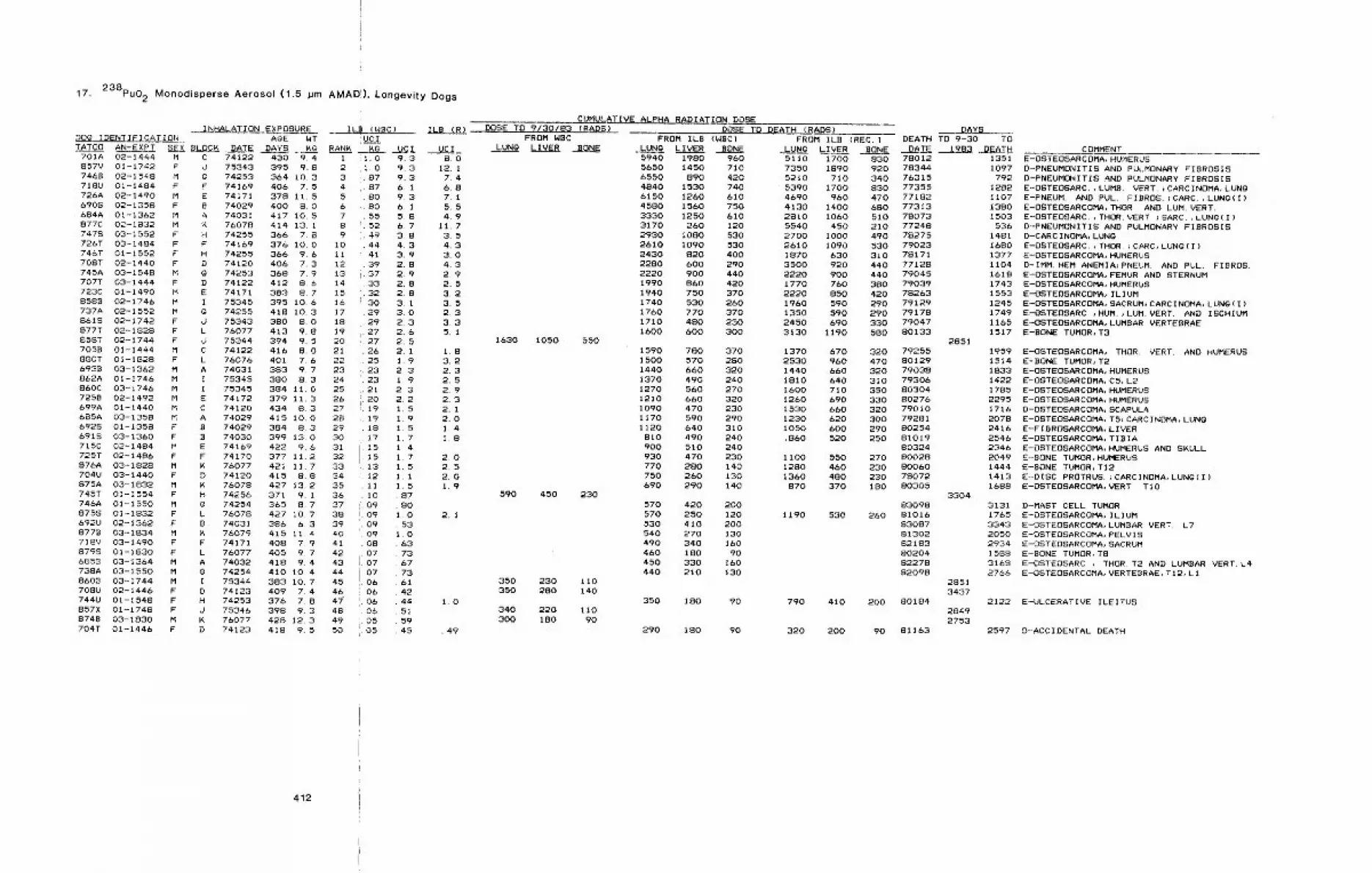
17. 238pu02 Monodisperse Aerosol (1.5 ~m AMAD’:), Longevity Dogs
INHALATION EXPOSURE IDO@ IDENTIFICATION AOE WTTATO0 AN-EXPT 8E~ BLOCK DATE ~A’~’S. KG RANK __
701A 02-1444 M C 74122 430 9.4 1857V 01-1742 F d 75343 395 9.8 27468 02-1548 M g 74253 364 10.3 3718U 01-1484 F F 74169 406 7~ 5 4726A 02-1490 M E 74171 378 11.5 56908 02-1358 F B 74029 400 8.0 6684A 01-1362 M A 74031 417 10.5 7877C 02-1832 M K 76078 414 13. 1 87478 03-1552 F H 74255 366 7.8 9726T 03-1484 F F 74169 376 10.0 10746T 01-1552 F H 74255 366 9.6 Ii708T 02-1440 F O 74120 406 7.3 12745A 03-1548 M ~ 74253 368 7.9 13707T 03-1444 F D 74122 412 8.6 14723C 01-1490 M E 74171 383 8.7 158588 02-1746 M I 75345 395 10.6 16737A 02-1552 M g 74255 418 10.3 178618 02-1742 F O 75343 380 8.0 18877T 02-1828 F L 76077 413 9.8 19858T 02-1744 F J 75344 394 9.5 20705B 01-1444 M C 74122 416 8.0 21880T 01-1828 F L 76076 401 7.6 226938 03-1362 M A 74031 383 9.7 23862A 01-1746 M I 75345 380 8.3 24860C 03-1746 M I 75345 384 11.0 257258 02-1492 M E 74172 379 11.3 26699A 01-!440 M C 74120 434 8.3 27685A 03-1358 M A 74029 415 I0.0 286925 01-1358 F B 74029 384 8.3 296918 03-1360 F B 74030 399 13.0 30715C 02-1484 M E 74169 422 9.6 31725T 02-1486 F F 74170 377 11.2 32876A 03-1828 M M 76077 421 11.7 33704U 03-1440 F O 74120 415 8.8 34875A 03-1832 M K 76078 427 13.2 35745T 01-1554 F H 74256 371 9.1 36746A 01-1550 M 0 74254 365 8.7 378758 01-1832 F L 76078 427 10.7 38692U 02-1362 F B 74031 386 6.3 39877B 03-1834 M ~ 76079 415 11.4 40718V 03-1490 F F 74171 408 7.9 418798 01-1830 F L 76077 405 9.7 426858 03-1364 M A 74032 418 9.4 43738A 03-1550 M 0 74254 410 10.4 448608 03-1744 M I 75344 383 10.7 45708U 02-1446 F D 74123 409 7.4 46744U 01-1548 F H 74253 376 7.8 4i857X 01-1748 F d 75346 398 9.3 488748 03-1830 M K 76077 428 123 49704T 01-1446 F D 74123 418 9.5 50
412
(W~C) ILB (R)UC__2
KG1.01.08787808055524944413937333230292927
,. 27¯ 26.25
,. 2323
.2120I"
LI919181715151312II100909090908070707060606060505
UC I UC I9.3 8.09.3 12.19.3 7.46.1 6.89.3 7.16.1 5.55.8 4.96.7 11.73.8 3.54.3 4.33.9 3.02.8 4.32.9 2.92.8 2.52,8 3.23.1 3.53.0 2.32.3 3.32.6 5,12¯52.1 1.81.9 3.22.3 2.31.9 2,52.3 2.92.2 2.31.5 2.11.9 2.01.5 1.417 1.81 417 2.015 2.51 1 2.0I 5 1.9. 87¯ 801.0 2.1. 531.0¯ 63
736773614244 1. 0515945 . 49
CUMULATIVE ALPHA RADIATION DOSEDOSE TO 9/30/83 (RADS)
FROM WBCLUNg LIVER BONE
1630 1050 550
590 450 230
350 230 II0350 280 140
340 220 II0300 180 90
DOSE TO DEATH (RADS)FROM ILB (WBC)
LUN~ LIVER BONE5940 1980 9605650 1450 7106550 890 4204840 1530 7406150 1260 6104580 1560 7503330 1250 6103170 260 1202930 1080 5302610 1090 5302430 820 4002280 600 2902220 900 4401990 860 4201940 750 3701740 530 2601760 770 3701710 480 2301600 600 300
1590 780 3701500 570 2801440 660 3201370 490 2401270 560 2701210 660 3201090 470 2301170 590 2901120 640 310810 490 240900 510 240930 470 230770 280 140750 260 130690 290 140
570 420 200570 250 120530 410 200540 270 130490 340 160460 180 90450 330 160440 210 130
350 180 90
290 180 90
FROM ILB (REC.)LUNg LIVER BONE
DAYSDEATH TO 9-30 TO
DATE 1983 DEATH5110 1700 830 780127350 1890 920 783445210 710 340 763155390 1700 830 773554690 960 470 771824130 1400 680 773132810 1060 510 780735540 450 210 772482700 1000 490 782752610 1090 530 790231870 630 310 781713500 920 440 771282220 900 440 790451770 760 380 790392220 850 420 782631960 590 290 791291350 590 290 791782450 690 330 790473130 1190 580 80133
1370 670 320 792552530 960 470 801291440 660 320 790381810 640 310 793061600 710 350 803041260 690 330 802761530 660 320 790101230 620 300 792811050 600 290 80254,860 520 250 81019
803241100 550 270 800281280 460 230 800601360 480 230 78072
870 370 180 80305
1190 530 2608309881016830878130282183802048227882098
790 410 200 80184
320 200 90 81163
2851
3304
28513437
28492753
13511097792
t282110713801503536
14811680137711041618174315531245174911651517
195915141833142217852295171620782416254623462049144414131688
31311765334320502934158831682766
2122
2597
COMMENTE-OSTEOSARCOMA, HUMERUSD-PNEUMONITiS AND PULMONARY FIBROSISD-PNEUMONITIS AND PULMONARY FIBROSISE’-OSTEOSARC.,LUMB. VERT. ;CARCINOMA, LUNgE-PNEUM. AND PUL. FIBROS.~CARC.,LUNg(1)E-OSTEOSARCOMA, THOR. AND LUM. VERT.E-OSTEOSARC¯,THOR. VERT. JBARC.,LUNG(1)D-PNEUMONITI8 AND PULMONARY FIBROSISD-CARCINOMA, LUNgE-OSTEOSARC.,THOR.~CARC, LUNO(1)E-OSTEOSARCOMA, HUMERUSD-IMM. HEM. ANEMIA~PNEUM. AND PUL. FIBROS.E-OSTEOSARCOMA, FEMUR AND STERNUME-OSTEOSARCOMA, HUMERUSE-OSTEOSARCOMA, ILIUME-OSTEOSARCOMA, SACRUM~CARCINOMA, LUNO(I)E.-OSTEOSARC.,HUM.,LUMVERT. AND ISCHIUME-OSTEOSARCOMA, LUMBAR VERTEBRAEE-BONE TUMOR, T3
E-OSTEOSARCOMA, THOR. VERT. AND HUMERUSE-BONE TUMOR, T2E-OSTEOSARCOMA, HUMERUSE-OSTEOSARCOMA, C5, L2E-OSTEOSARCOMA, HUMERUSE-OSTEOSARCOMA, HUMERUSD-OSTEOSARCOMA, SCAPULAE-OSTEOSARCOMA, TS~CARCINOMA, LUNOE-FIBROSARCOMA, LIVERE-OSTEOSARCOMA, TIBIAE--OSTEOSARCOMA, HUMERUS AND S~ULLE-BONE TUMOR,HUMERUSE-BONE TUMOR,T12E--DISC PROTRUS. ~CARCINOMA, LUNg(I)E-OSTEOSARCOMA, VERT. TIO
D--MAST CELL TUMORE-OSTEOSARCOMA, ILIUME-OSTEOSARCOMA, LUMBAR VERT. L7E-OSTEOSARCOMA, PELVISE-OSTEOSARCOMA, SACRUME-BONE TUMOR, TOE-OSTEOSARC. , THOR. T2 AND LUMBAR VERT. L4E-OSTEOSARCOMA, VERTEBRAE, TI2, L1
E-ULCERATIVE ILEITUS
O-ACCIDENTAL DEATH
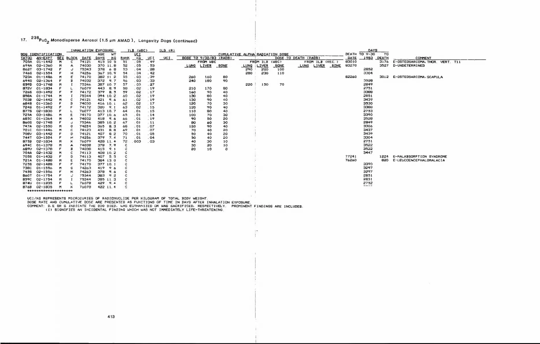
17. 238pu02 Monodisperse Aerosol (1.5 ~m AMAD), Longevity Dogs (continued)
INHALATION EXPOSURE ILB (WBC) ILB (R)DOO IDENTIFICATION AgE NT UCITATO0 AN-EXPT SEX BLOC~ DATE DAYS Kg RANK ~g UCI UCI
705A 01-1442 M C 74121 415 10.5694A 02-1360 M A 74030 370 11.8862T 03-1742 F d 75343 378 6.87468 02-1554 F H 74255 367 10.9723A 01-1486 M E 74170 382 11.26948 02-1364 F B 74032 372 9.7859D 03-1748 M I 75346 387 10.7872V 01-1834 F L 76079 443 8.97268 03-1492 F F 74172 379 8.5858A 01-1744 M I 75344 394 10.2703B 02-1442 M C 74121 421 9.66848 01-1360 F B 74030 416 10. I7248 01-1492 F F 74172 3B0 9. I8778 02-1830 F L 76077 413 10.7725A 03-1486 M E 74170 377 10.6685C 01-1364 M A 74032 418 9.68608 02-1748 F d 75346 385 10.2747A 02-1550 M g 74254 365 8.3701C 03-1446 M C 74123 43t 8.8708V 03-1442 F D 74121 407 8.2744T 03-1554 F H 74256 379 7.48758 02-1834 M ~ 76079 428 11.4694C 01-1378 M A 74038 378 7.9689U 02-1378 F B 74038 415 9. I704A 02-1432 M C 74113 408 10.27058 01-1432 F D 74113 407 5.5721A 01-1488 M E 74170 384 1307258 02-I488 F F 74170 377 10.1738C 01-1556 M g 74263 419 9.67458 02-1556 F H 74263 378 9.6860T 01-1754 F J 75344 383 9.2859C 02-1754 M I 75344 385 11.3874U 01-1835 F L 76078 429 9.4876B 02-1835 M ~ 76078 422 11.4
51 . 0552 . 0553 .0454 .0455 .0356 . 0357 .0358 0259 0260 0261 0262 0263 0264 0165 Ol66 0167 0168 0169 0170 0171 0172 003
CCCCCCCCCCCC
CUMULATIVE ALPHA RADIATION DOSEDOSE TO 9/30/8~ (RADS)
49 FROM WBC53 LUNg LIVER BONE284239 260 160 8033 240 180 902719 210 170 SO17 160 90 4019 130 80 4019 130 90 4017 120 70 3015 120 90 4015 110 SO 4014 100 70 3019 90 50 2011 SO 60 3007 120 90 4007 70 40 2005 50 40 2004 50 40 2003 40 30 10
30 20 1020 10 0
DOSE TO DEATH (RADS)FROM ILB (WBC)
LUNG LIVER BONE290 220 100280 230 110
220 150 70
UCI/~g REPRESENTS MICROCURIES OF RADIONUCLIDE PER ~ILOgRAM OF TOTAL BODY WEIGHT.DOSE RATE AND CUMULATIVE DOSE ARE PRESENTED AS FUNCTIONS OF TIME IN DAYS AFTER INHALATION EXPOSURE.COMMENT: D,E OR S INDICATE THE DOG DIED, WAS EUTHANIZED OR WAS SACRIFICED, RESPECTIVELY. PROMINENT FINDINGS ARE INCLUDED.
(I) SIQNIFIES AN INCIDENTAL FINDINg WHICH WAS NOT IMMEDIATELY LIFE-THREATENING.
FROM ILB (REC.)LUNG LIVER BONE
DAYSDEATH TO 9-30 TO
DATE 1983 DEATH83010 317683270 3527
28523304
301235282849275133882851343935303388275333903528284933063437343933042751352235223447
1224820
339032973297285128512752
82260
7724176260
COMMENTE-OSTEOSARCOMA, THOR. VERT. T11D-UNDETERMINED
E-0STEOSARCOMA, SCAPULA
E-MALABSORPTION SYNDROMEE-LEUCOENCEPHALOMALACIA
413
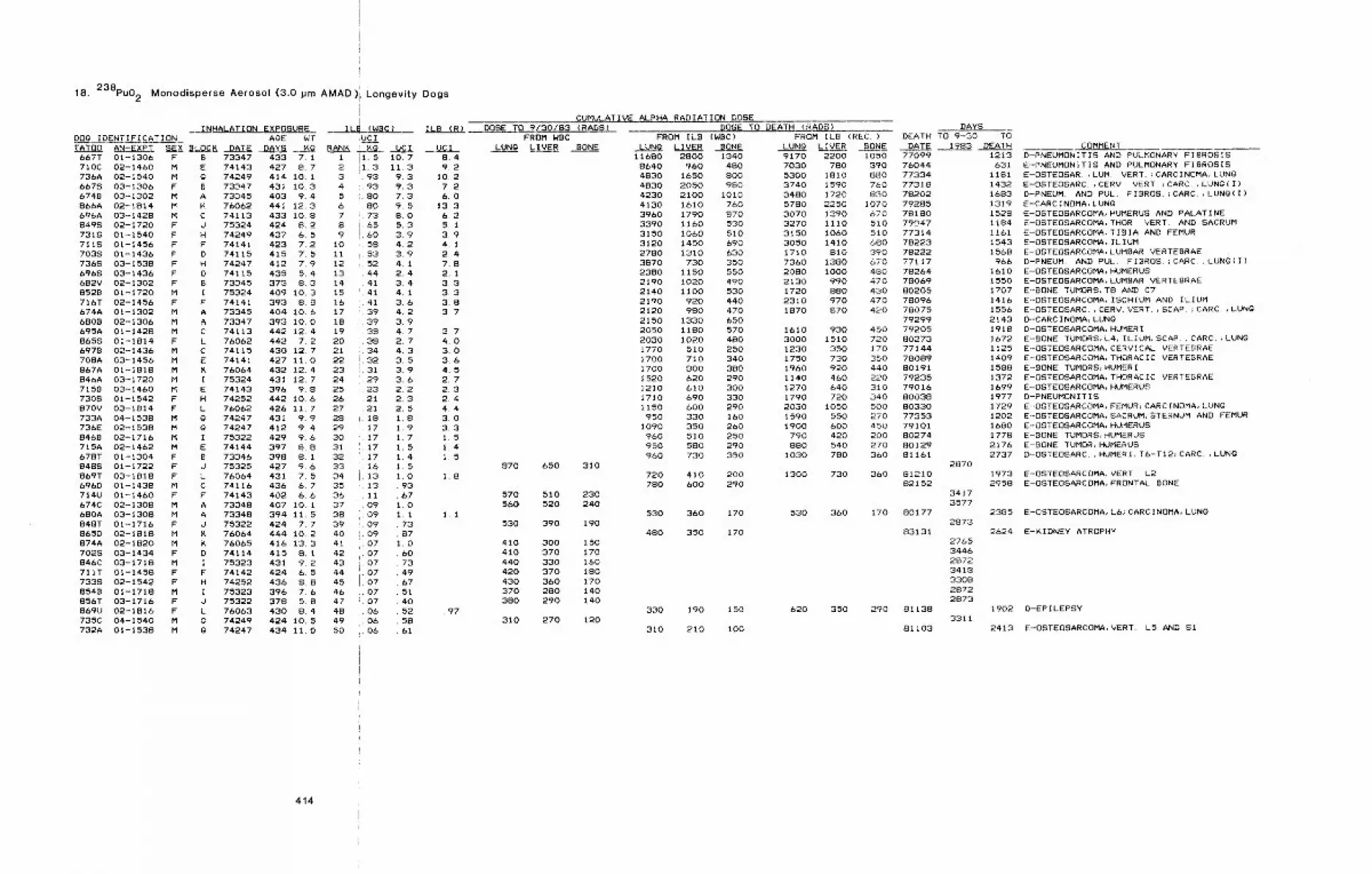
18. 238pu02
l!
Monodisperse Aerosol (3.0 pm AMAD)I Longevity Dogs
INHALATION EXPOSUREDOg IDENTIFICATION AGE WTTATO0 AN-EXPT SEX BLOCK DATE DAYS ~g
667T 01-1306 F B 73347 433 7. I710C 02-1460 M E 74143 427 8.7736A 02-1540 M G 74249 414 10. i6678 03-1306 F B 73347 431 10.36748 03-1302 M A 73345 403 9.4866A 02-1814 M ~ 76062 441 12.3696A 03-1428 M C 74113 433 10.88498 02-1720 F d 75324 424 8.27318 01-1540 F H 74249 437 6.57118 01-1456 F F 74141 423 7.27035 01-1436 F D 74115 415 7.57368 03-1538 F H 74247 412 7.96968 03-1436 F D 74115 435 5.4682V 02-1302 F B 73345 373 8.3852B 01-1720 M I 75324 409 10.3716T 02-1456 F F 74141 393 8.8674A 01-1302 M A 73345 404 10.66808 02-1306 M A 73347 393 I0.0695A 01-1428 M C 74113 442 12.48658 01-!814 F L 76062 442 7.26978 02-1436 M C 74115 430 12.7708A 03-1456 M E 74141 427 II.0867A 01-I818 M K 76064 432 12.4846A 03-1720 M I 75324 431 12.77158 03-1460 M E 74143 396 9.87308 01-1542 F H 74252 442 10.6870V 03-!814 F L 76062 426 11.7733A 04-1538 M g 74247 431 9.9736E 02-1538 M Q 74247 412 9.48468 02-1716 M I 75322 429 9.6715A 02-1462 M E 74144 397 8.8678T 01-1304 F B 73346 398 8.18488 01-1722 F J 75325 427 9~6869T 03-1818 F L 76064 431 7.56968 01-1438 M C 74116 436 6.7714U 01-1460 F F 74143 402 6.6674C 02-1308 M A 73348 407 10. 1680A 03-1308 M A 73348 394 11.5848T 01-1716 F J 75322 424 7.7865D 02-1818 M K 76064 444 10.2874A 02-1820 M K 76065 416 13.37028 03-1434 F D 74114 415 8.1846C 03-1718 M I 75323 431 9.2711T 01-1458 F F 74142 424 6.57338 02-1542 F H 74252 436 8.88548 01-1718 M I 75323 396 7.6856T 03-1716 F J 75322 378 5.8869U 02-1816 F L 76063 430 8.4735C 04-1540 M g 74249 424 10.5732A 01-1538 M g 74247 434 11.0
414
,UClRANK
123456789
i011121314151617181920212223242526272829303132333435363738394O41424344454647484950
IL~ (WBC) ILB (R)
=. Xg UC I UC Iil. 5 10.7 8.4il.3 Ii.3 9,2
93 9.3 10.293 9.3 7.280 7.3 6.080 9. 5 13.373 BO 6.265 5.3 5.160 3.9 3.958 4.2 4.153 3.9 2.452 4. 1 7.844 2.4 2.141 3.4 3,34I 41 3341 3.6 3.839 4.2 3. 739 3. 938 4.7 3.738 2.7 4.034 4.3 3. 032 3.5 3.631 3.9 4.529 3.6 2.723 2.2 2.321 2.3 2.421 2.5 4.418 1.8 3.017 I 9 3.317 I 7 1.517 1 5 1.417 1 4 1.516 I 513 1 0 1.813 .93Ii .6709 I. 009 1. i I. 109 . 7309 . 8707 I. 007 6007 7307 4907 6707 5107 40
¯ 06 52 . 9706 5806 61
CUMULATIVE ALPHA RADIATION DOSEDOSE TO 9/30/83 (RADS)
FROM WBCLUNg LIVER BONE
870 650 310
570 510 230560 520 240
530 390 190
410 300 150410 370 170440 330 160420 370 180430 360 170370 28O 140380 290 140
310 270 120
DOSE TO DEATH (RADS)FROM ILB (WBC)
LUNG LIVER BONE11680 2800 1340
8640 960 4804830 1650 8004830 2050 9804230 2100 10104130 1610 7603960 1790 8703390 1160 5303150 !060 5103120 1450 6902780 1310 6303870 730 3502380 1150 5502190 !020 4902140 1100 5302190 920 4402120 980 4702150 1330 6502050 1180 5702030 1020 4801770 510 2501700 710 3401700 800 3801520 620 2901210 610 3001710 690 3301150 600 290950 330 160
1090 350 260960 510 250950 580 290960 730 350
720 410 200780 bOO 290
530 360 170
480 350 170
330 190 150
310 210 100
FROM ILB (REC.)LUNG LIVER BONE
DAYSDEATH TO 9-30 TO
DATE 1983 DEATH9170 2200 10507030 780 3905300 1810 8803740 1590 7603480 1720 8305780 2250 10703070 1390 6703270 1110 5103150 1060 5103050 1410 6801710 810 3907360 1380 6702080 1000 4802130 990 4701720 880 4302310 970 4701870 870 420
1610 930 4503000 1510 7201230 350 1701750 730 3501960 920 4401140 460 2201270 640 3101790 720 3402030 1050 5001590 550 2701900 600 450
790 420 200880 540 270
1030 780 360
1300 730 360
530 360 170
620 350 290
77099 121376044 63177334 118177318 143278202 168379285 131978180 152879047 118477314 116178223 154378222 156877117 96678264 161078069 155080205 170778096 141678075 155679299 214379205 191880273 167277144 1!2578089 140980191 158879235 137279016 169980038 197780330 172977353 120279101 168080274 177880129 217681161 2737
287081210 197382152 2958
34173577
80177 23852873
83131 2624
81138
81103
2765344628723418330828722873
3311
COMMENTD--~NEUMONITIS AND PULMONARY FIBROSISE-PNEUMONITiS AND PULMONARY FIBROSISE-OSTEOSAR. ,LUM VERT. ;CARCINOMA, L.UNQE-OSTEOSARC. ,CERV. VERT ~CARC. ,LUNG(I)D-PNEUM. AND PUL. FIBROS.~CARC.,LUNg(1)E-CARCINOMA, LUNGE-OSTEOSARCOMA, HUMERUS AND PALATINEE-DSTEOSARCOMA, THOR. VERT. AND SACRUME’-OSTEOSARCOMA, TIBIA AND FEMURE-OSTEOSARCOMA, ILIUME-OSTEOSARCOMA, LUMBAR VERTEBRAED-PNEUM. AND PUL. FIBROS.~CARC ,LUNG(I)E-OSTEOSARCOMA, HUMERUSE-OSTEOSARCOMA, LUMBAR VERTEBRAEE-BONE TUMORS, T8 AND C7E--OSTEOSARCOMA, ISCHIUM AND ILIUME-OSTEOSARC.,CERV. VERT.,SCAP. ;CARC.,LUNg~-CARCINOMA, LUN~D-OSTEOSARCOMA, HUMERIE-BONE TUMORS, L4, ILIUM, SCAP.~CARC.,LUNGE-OSTEOSARCOMA, CERVICAL VERTEBRAEE-OSTEOSARCOMA, THORACIC VERTEBRAEE-BONE TUMORS, HUMERIE-OSTEDSARCOMA, THORACIC VERTEBRAEE-OSTEOSARCOMA, HUMERUSD-PNEUMONITISE--OSTEOSARCOMA, FEMUR~CARCINOMA, LUNgE-OSTEOSARCOMA, SACRUM, STERNUM AND FEMURE-OSTEOSARCOMA, HUMERUSE--BONE TUMORS, HUMERUSE-BONE TUMOR, HUMERUSD-OSTEOSARC. ,HUMERI,T6-TI2;CARC. ,LUNG
E-OSTEOSARCOMA, VERT L2E-OSTEOSARCOMA, FRONTAL BONE
E-OSTEOSARCOMA, L6; CARCINOMA, LUNg
E-KIDNEY ATROPHY
1902 D-EPILEPSY
2413 E-OSTEOSARCOMA, VERT. L5 AND SI
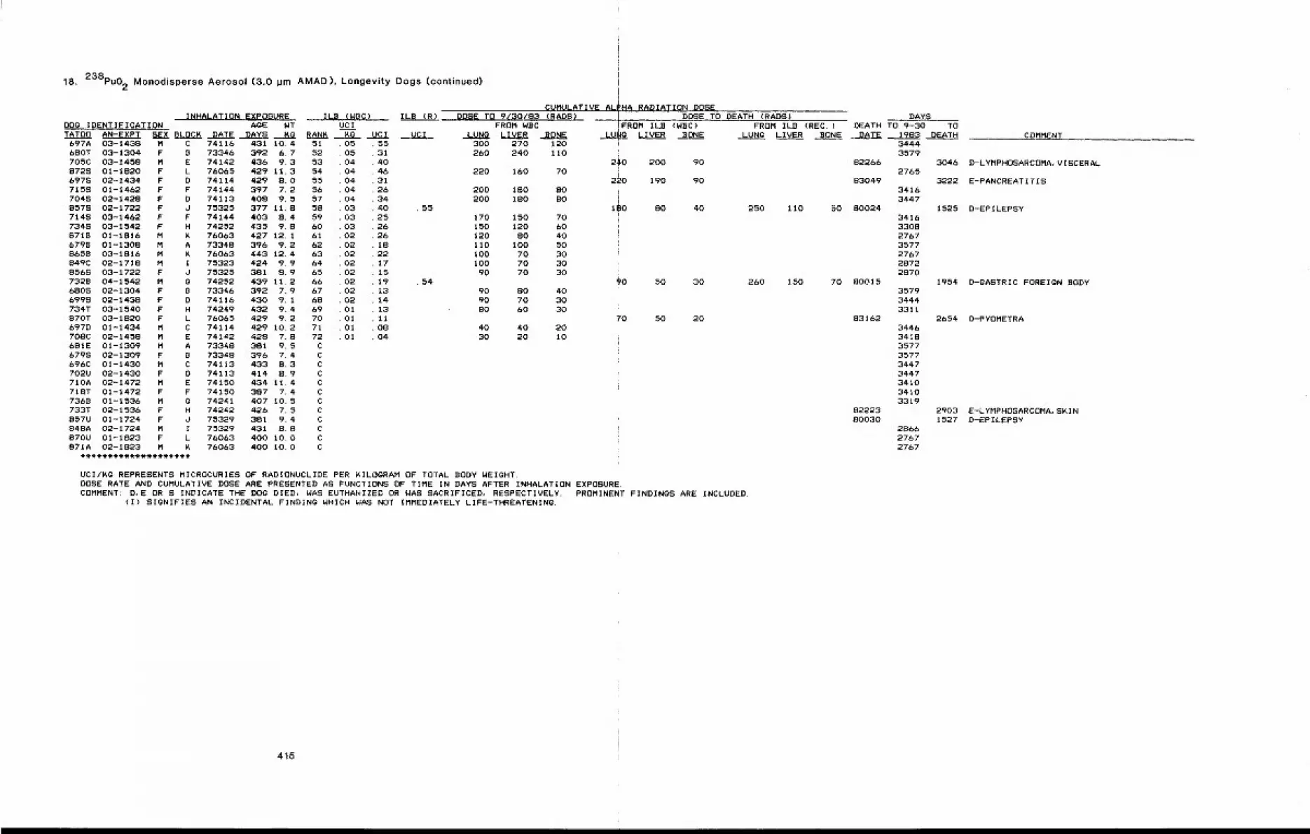
18. 238pu02 Monodisperse Aerosol (3.0 ~m AMAD), Longevity Dogs (continued)
INHALATION EXPOSUREDOg IDENTIFICATION AgETATO0 AN-EXPT SE~XBLOC~ DATE DAYS
697A 03-1438 M C 74116 431 10.4&80T 03-1304 F B 73346 392 6.7705C 03-1458 M E 74142 436 9.38725 01-1820 F L 76065 429 11.36978 02-1434 F D 74114 429 S.O7155 01-1462 F F 74144 397 7.27045 02-1428 F D 74113 408 9.58575 02-1722 F d 75325 377 11.87145 03-1462 F F 74144 403 8.47345 03-1542 F H 74252 435 9.88719 01-1816 M K 76063 427 12. I6798 01-1308 M A 73348 396 9.28658 03-1816 M K 76063 443 12.4849C 02-1718 M I 75323 424 9.98565 03-1722 F d 75325 381 S. 97328 04-1542 M G 74252 439 11.26SOS 02-1304 F B 73346 392 7.96995 02-1438 F D 74116 430 9.1734T 03-1540 F H 74249 432 9.4870T 03-1820 F L 76065 429 912697D 01-1434 M C 74114 429 I0~2708C 02-1458 M E 74142 429 7.8681E 01-1309 M A 73348 381 9.56795 02-1309 F B 73348 396 7.4696C 01-1430 M C 74113 433 8.3702U 02-1430 F D 74113 414 8.9710A 02-1472 M E 74150 434 11.4718T 01-1472 F F 74150 387 7.47368 01-1536 M g 74241 407 10.5733T 02-1536 F H 74242 426 7.5857U 01-1724 F d 75329 381 9.4848A 02-1724 M I 75329 431 8.8870U 01-1823 F L 76063 400 I0.0871A 02-1823 M K 76063 400 IO.O
ILB (WBC)NT UCI~g RAN~ KG UCI
51 0552 0553 0454 0455 0456 0457 0458 0359 0360 0361 0262 0263 0264 0265 026b 0267 0268 0269 0170 0171 0172 01CCCCCCCCCCCC
55314O463126344O2526261822171519131413II0804
ILB (R)
UCI
CUMULATIVE ALIDOSE TO 9/30/83 (RADS~
FROM WBCLUNg LIVER BONE
300 270 120260 240 110
’HA RADIATION DOSE
170 150 70150 120 60120 80 40110 100 50100 70 30I00 70 3090 70 30
DOSE TO DEATH (RADS)FROM ILB (WBC) FROM ILB (REC.)I
LUng LIVER BONE LUNG LIVER BONE
DAYSDEATH TO 9-30 TO
DATE 1983 DEATH34443579
2~0 200 90 82266220 160 70 !
220 190 90 83049200 180 80 i200 180 80 I
.55 1~0 SO 40 250 110 50 80024I
.i
.54 90 50 30 260 150 70 8001590 80 4090 70 3080 60 30
70 50 20 8316240 40 2030 20 I 0
8222380030
UCI/Kg REPRESENTS MICROCURIES OF RADIONUCLIDE PER ~ILOgRAM OF TOTAL BODY WEIGHT.DOSE RATE AND CUMULATIVE DOSE ARE PRESENTED AS FUNCTIONS OF TIME IN DAYS AFTER INHALATION EXPOSURE.COMMENT: D,E OR S INDICATE THE DOG DIED, WAS EUTHANIZED OR WAS SACRIFICED, RESPECTIVELY. PROMINENT FINDINGS ARE INCLUDED.
(1) SIGNIFIES AN INCIDENTAL FINDINg WHICH WAS NOT IMMEDIATELY LIFE-THREATENINg.
2765
34163447
3416330827673577276728722870
357934443311
344634183577357734473447341034103319
286627672767
3046
3222
1525
1954
2654
29031527
COMMENT
D-LYMPHOSARCOMA, VISCERAL
E-PANCREATITIS
D-EPILEPSY
D-gASTRIC FOREIGN BODY
D-PYOMETRA
E-LYMPHOSARCOMA, SKIND-EPILEPSY
415
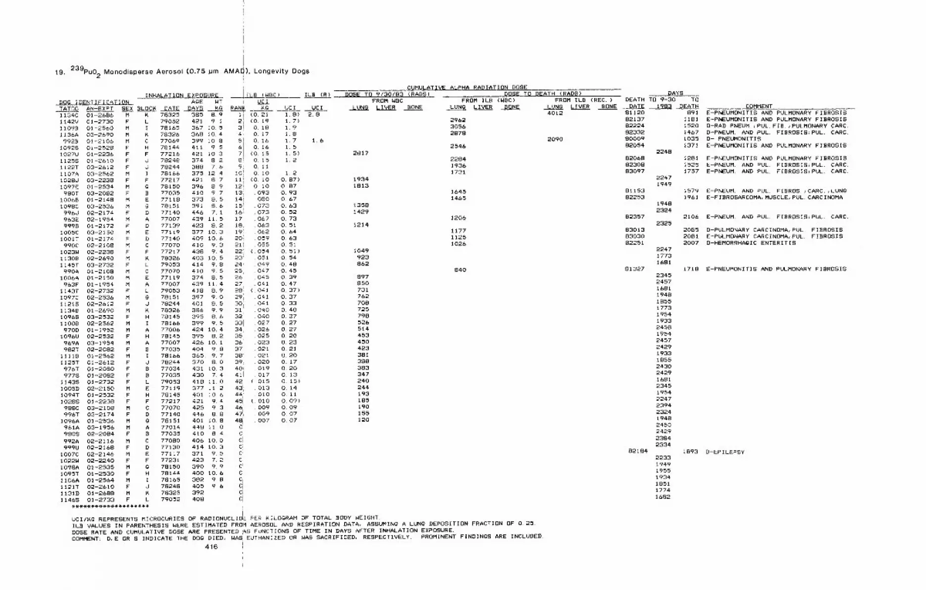
19. 239pu02 Monodisperse Aerosol (0.75 pm AMAOi), Longevity Dogs
INHALATION EXPOSUREDOG IDENTIFICATION A@E WTTATO0 AN-EXPT SEX BLOCK DATE DAYS KQI134C 01-2686 M K 78325 385 8.9I142V 01-2730 F L 79052 421 9. iII098 01-2550 M I 78165 367 10.5I136A 03’-2690 M K 78326 368 10.4
9928 0!-2106 M C 77069 399 10.8I0928 01-2528 F H 78144 411 9.5I027U 01-2236 F F 77216 421 10.3I1258 01-2610 F J 78248 374 8.2I122T 03-2612 F d 78244 388 7.6!I07A 03-2562 M I 78166 375 12.4I028U 03-2238 F F 77217 421 8.7I097E 01-2534 M g 78150 396 8.9980T 03-2082 F B 77035 410 9 7
I0068 01-2148 M E 77118 373 8,5I098C 03-2536 M ~ 78151 391 8.6996U 02-2174 F D 77140 446 7. i963E 02-1954 M A 77007 439 11.59998 01-2172 F D 77139 423 8.2
I005C 03-2150 M E 77119 377 10.3!O01T 01-2174 F D 77140 409 i0.6990C 02-2108 M C 77070 410 9.3
I023W 02-2238 F F 77217 438 9.4I1308 02-2690 M K 78326 403 i0~ 5I145T 03-2732 F L 79053 414 9.8990A 01-2108 M C 77070 410 9.5
I006A 01-2150 M E 77119 374 8.5963F 01-1954 M A 77007 439 11.4
I143T 02-2732 F L 79053 418 8.9I097C 02-2536 M G 78151 397 9.011218 02-2612 F J 78244 401 8.5I134B 01-2690 M K 78326 386 9.9I0968 03-2532 F H 78145 395 8~611008 02-2562 M I 78166 399 9.5970D 01-1952 M A 77006 424 10,4
I096U 02-2532 F H 78145 395 8.2969A 03-1954 M A 77007 426 10. I982T 02-2082 F B 77035 404 9.8
IIIIB 01-2562 M I 78166 365, 9.7I125T 01-2612 F O 78244 370 8. G976T 01-2080 F B 77034 431 I0.39778 01-2082 F B 77035 430 7.4
I!438 01-2732 F L 79053 418 11.0I0050 02-2150 M E 77119 377 11.2I094T 01-2532 F H 78145 401 i0.610288 01-2238 F F 77217 421 9.4988C 03-2108 M C 77070 425 9.3996T 03-2174 F D 77140 446 88
I096A 01-2536 M G 78151 401 10.8961A 03-1956 M A 77014 448 ii.09808 02-2084 F B 77035 410 8.4992A 02-2116 M C 77080 406 I0.0999U 02-2168 F D 77130 414 10.3
I007C 02-2146 M E 77117 371 9.51022W 02-2240 F F 77231 423 7.2I098A 01-2535 M g 78150 390 9.9I095T 01-2530 F H 78144 400 10.6I106A 01-2564 M I 78165 382 9.8II21T 02-2610 F J 78248 405 9.6I1310 01-2688 M K 78325 392I1468 01-2733 F L 79052 408
ILB (WBC)
UCI1.8)1.7)1.9I~81.71.51.5)1.2
CUMULATIVE ALPHA RADIATION DOSEILB (R) DOSE TO 9/30/83 (RADS) DOSE TO DEATH (RADS)
FROM WBC FROM IL.B (WBC)UCI LUNg LIVER BONE LUNg LIVER BONE
2.8296230562878
2546
228419361731
16451465
1206
117711251026
840
i UClRA~ ~G
i I (0.2121 (0. 193! o. 184~ O. i75i O. 16
0.16(0. 15
8! O. 1591 O. II
1011 O. 1oii’, (0. i012! o. i013’, 093i4i o8o15 ! 07316: 07317 067181 06319i. 062201 05921=, 055221 ( 05423’ 05124’ 04925, 04726 04527 04128: 04129’! 04130; 04131 ~ 04032: 04033! 02734 02635 02536= 02337 02138’ 02139,. 02040, 01941i 01742, (. 015431 . 013441’ i 0 i 0
45i (. o1046: . 00947!~ . 00948i .007C.C=
ciC’,c~cci
CciClc;cl,..
FROM ILB (REC.)LUNg LIVER BONE4012
2090
DAYSDEATH TO 9-30 TO
DATE 1983 DEATH
1.6
2~17
1.2O. 87) 1934O. 87 1813O. 93O. 67O. 63 1358O. 52 1429O. 73O. 51 1214O. 64O. 630.51O. 51 ) 1049O. 54 923o. 48 8620.45O. 39 89701 47 850O. 37) 731O. 37 762O. 33 708O. 40 725O. 37 798O. 27 526O. 27 514O. 20 453O. 23 450O. 21 4230. 20 38iO. 17 388O. 20 383O. 13 347o. 15) 240O. 14 244O. Ii 193o. 09) 185O. 09 190o. 07 155O. 07 120
UCl/~g REPRESENTS MICROCURIES OF RADIONUCLIDE PER KILOGRAM OF TOTAL BODY WEIGHT.ILB VALUES IN PARENTHESIS WERE ESTIMATED FROM AEROSOL AND RESPIRATION DATA, ASSUMINg A LUNG DEPOSITION FRACTION OF 0.25.
DOSE RATE AND CUMULATIVE DOSE ARE PRESENTED ;AS FUNCTIONS OF TIME IN DAYS AFTER INHALATION EXPOSURE.COMMENT: D,E OR S INDICATE THE DOg DIED~ WASEUTHANIZED OR WAS SACRIFICED, RESPECTIVELY. PROMINENT FINDINGS ARE INCLUDED.
416 i
81120 89182137 118182224 152082332 146780009 103582054 1371
224882068 128182308 152583097 1757
22471949
81153 157982253 1961
19482324
82357 21062325
83013 208583030 208182251 2007
224717731681
81327 1718234524571681194818551773195419332458195424572429193318552430242916812345195422472394232419482450242923842334
82184 18932233194919551934185117741682
COMMENTE-PNEUMONITIS AND PULMONARY FIBROSISE-PNEUMONITIS AND PULMONARY FIBROSISD-RAD. PNEUM.;PUL. FIB.;PULMONARY CARC.D-PNEUM. AND PUL. FIBROSIS~PUL. CARC.D- PNEUMONITISE--PNEUMONITIS AND PULMONARY FIBROSIS
E-PNEUMDNITIS AND PULMONARY FIBROSISE-PNEUM. AND PUL. FIBROSIS}PUL. CARC.E-PNEUM. AND PUL. FIBROSIS;PUL. CARC.
E-PNEUM. AND PUL. FIBROS. }CARC. ,LUNGE--FIBROSARCOMA, MUSCLE~PUL. CARCINOMA
E-PNEUM. AND PUL FIBROSISIPUL. CARC.
D-PULMONARY CARCINOMA~PUL FIBROSISE-PULMONARY CARCINOMAiPUL. FIBROSISD-HEMORRHAgIC ENTERITIS
E-PNEUMONITIS AND PULMONARY FIBROSIS
D-EPILEPSY
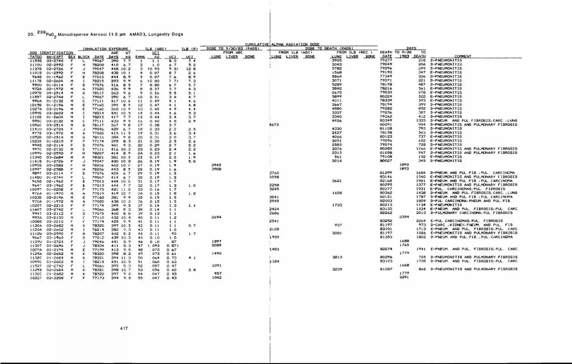
20. 239pu02 Monodisperse Aerosol (1.5 pm AMAD), Longevity Dogs
INHALATION EXPOSURE ILB (WBC)DOg IDENTIFICATION A~E WT UCITATO0 AN-EXPT SEX BLOCK DATE DAYS Mg RANK Kg
ILB (R)
UCI UCI11558 03-2744 F L 79067 390 7.71110U 02-2592 F H 78208 410 6.711378 02-2726 F J 79047 448 10.211018 01-2592 F H 78208 438 I0, 1
9648 01-1962 F B 77013 444 8.91117B 02-2604 M I 78215 393 9.9
990U 01-2114 F D 77076 416 8, 5972A 02-1972 M A 77020 436 9.9
10978 04-2514 M g 78117 363 9.61155T 02-2744 F L 79067 390 6. 7
996A 01-2132 M C 77111 417 10.6 1110158 01-2196 M E 77160 394 8.9 121027A 03-2196 M E 77160 365 10.9 1310998 03-2602 M I 78214 451 105 1411108 01-2604 M I 78215 417 7, 7 15
995C 03-2132 M C 77111 433 9,9 161096C 03-2514 M @ 78117 367 9.8 171141U 03-2724 F d 79046 429 6.7 18
9778 03-1972 M A 77020 415 11.5 1910928 02-2514 M g 78116 384 9.8 20I023X 01-2210 F F 77174 395 8.5 21
9948 02-2114 F D 77076 401 9.3 22997C 02-2132 M C 77111 416 10.3 23
I099V 02-2590 F H 78207 414 8.9 241134D 03-2684 M K 78321 381 10.5 2511418 01-2726 F d 79047 430 10.0 25I0958 03-2588 F H 78206 462 10.0 27I099T 02-2588 F H 78206 443 8.5 28
989T 03-2114 F D 77076 425 6,7 291148U 01-2744 F L 79067 414 6,7 30
9655 02-1962 F B 77013 444 10.0 31964T 03-1962 F B 77013 444 7,7 32
IO09T 01-2208 F F 77173 421 II.0 33976A 01-1970 M A 77019 419 12.7 34
1023B 02-2196 M E 77160 381 9.9 35970A 01-1972 M A 77020 438 10.3 36
I020T 02-2210 F F 77174 399 9.5 371160T 03-2742 F L 79066 368 8.3 38
994T 03-2112 F D 77075 400 8.8 39995A 03-2130 M C 77110 432 10.4 40
I0088 03-2210 F F 77174 425 9.9 41II30A 03-2682 M K 78320 397 10.5 42II20A 02-2602 M I 78214 382 9.3 431112W 03-2590 F H 78207 402 8,2 44966T 03-1960 F B 77012 439 10.3 45
I139U 01-2724 F J 79046 441 8.9 46II30T 01-2696 F d 78334 411 8,3 47!O07A 01-2194 M E 77159 413 9,5 481129A 02-2682 M K 78320 398 8,2 49I132C 01-2684 M K 78321 394 11.3 50I099C 01-2602 M I 78214 451 i0. 5 51I153T 02-2742 F L 79066 395 8.3 5211298 02-2684 M ~ 78321 398 10.7 53I130C 01-2682 M K 78320 397 9.0 54I022T 02-2208 F F 77173 394 9.5 55
I 1.12 1.03 (0. 934 O. 875 O. 876 (0. 807 O. SO8 O, 579 O, 56
10 O. 51O. 49O. 470,4,5O. 44O. 44O. 40O. 38O. 330.310.31O. 30O. 29O. 23O. 23O. 190.19O. 190.190.190.190.170,170,160.150,150,150,140.140.130.110.110.110. II0.110.100. I0
(, 09307307306406005705604,7047
8.0 7.46.7 5.29, 3) 12. 88,7 2.67.6 8.97.7) 7.36.7 5.75.7 4.35.5 5.13.4 4.75.1 4.64.1 4.84.9 4.44.6 5.33.4 3.74,0 2.93,72,2 2.53.6 3.43.0 3.72.5 2.62.7 2.22.4 2.22.1 1.62.0 1.91.9 5.01,91,61.31,31.71.3 1.01.71.8 1.21.51.51.3 1.11.11.11.21.11.1 0,71,0.93 1,71.0. 87
O, 87)O, 670,610.73 4.1O, 63O. 470.60 2.8O. 43O. 43
417
CUMULATIVEDO8~ TO 9/30/83 (RAD8)
FROM WBCLUNg LIVER BONE
39453908
2694
18972089
1490
1091
9571042
ALPHA RADIATION DOSEDOSE TO DEATH (RADS)
FROM IL8 (NBC)LUNg LIVER BONE
~675
37603558
22583245
3041~945
~4242686
2541
2105
1959
~1485
1184
FROM ILB (REC.)LUNg LIVER BONE DATE3905304357821568586430715289384226755899401136674880333733404926
4330242740663051258533762315
5613014
3621
1658
1733
937
3081
38=13
3239
DAYSDEATH TO 9-30 TO
1983 DEATH79277 21079049 20679296 24979190 34777349 33679071 22178198 48778216 56179030 27880224 52278339 59378194 39979282 85279236 38779262 41280349 133380291 90481108 79378158 50380123 73779096 65279074 72880282 126681058 94779108 15280027 345
18931893
81299 168483146 154082168 198180295 137782277 193180362 143882121 178782003 180980213 113483133 153482262 2013
235483252 226981197 97383101 171381197 108681353 1802
82274
8029683123
81087
16881765
1779
1668
17792291
COMMENTD-PNEUMONITISD-PNEUMONITISD-PNEUMONITISE-PNEUMONITISD-PNEUMONITISD-PNEUMONITISD-PNEUMONITISE-PNEUMONITISE-PNEUMONITISE-PNEUMONITISE-PNEUMONITISD-PNEUMONITISD-PNEUMONITISD-PNEUMONITISD-PNEUMONITISD-PNEUM. AND PUL. FIBROSIS~CARC.,LUNgD-PNEUMONITIS AND PULMONARY FIBROSISD-PNEUMONITI8D-PNEUMONITISE-PNEUMONITISE-PNEUMONITISD-PNEUMONITISE-PNEUMONITIS AND PULMONARY FIBROSISD-PNEUMONITIS AND PULMONARY FIBROSISE-PNEUMONITISD-PNEUMONITIS
D-PNEUM. AND PUL, FIB.~PUL, CARCINOMAE-PNEUMONITIS AND PULMONARY FIBROSISE--PNEUM. AND PUL FIB.~PUL. CARCINDMAE-PNEUMONITIS AND PULMONARY FIBROSISE-PUL. CARCINOMAS>PUL. FIBROSISD-PNEUM. AND PUL. FIBROSIS~CARC.,LUNgD-PNEUM. AND PUL. FIB.}PUL. CARCINOMAD-PUL. CARCINOMA~PNEUM. AND PUL. FIB.E-PNEUMONITISE-PNEUM AND PUL, FIBROSIS~PUL, CARC.D-PULMONARY CARCINOMA~PUL, FIBROSIS
E-PUL. CARCINOMAS;PUL FIBROSISD-CARC.,KIDNEY~PNEUM. AND PUL, FIB.E-PNEUM, AND PUL. FIBROSIS~PUL. CARC,E-PNEUMONITI8 AND PULMONARY FIBROSISD-PNEUM, AND PUL. FIB. iPUL. CARCINDMA
1941 E-PNEUM, AND PUL. FIBROSIS}PUL. CARC.
705 D-PNEUMONITIS AND PULMONARY FIBROSIS1735 D-PNEUM. AND PUL. FIBROSIS}PUL, CARC.
862 D-PNEUMONITIS AND PULMONARY FIBROSIS
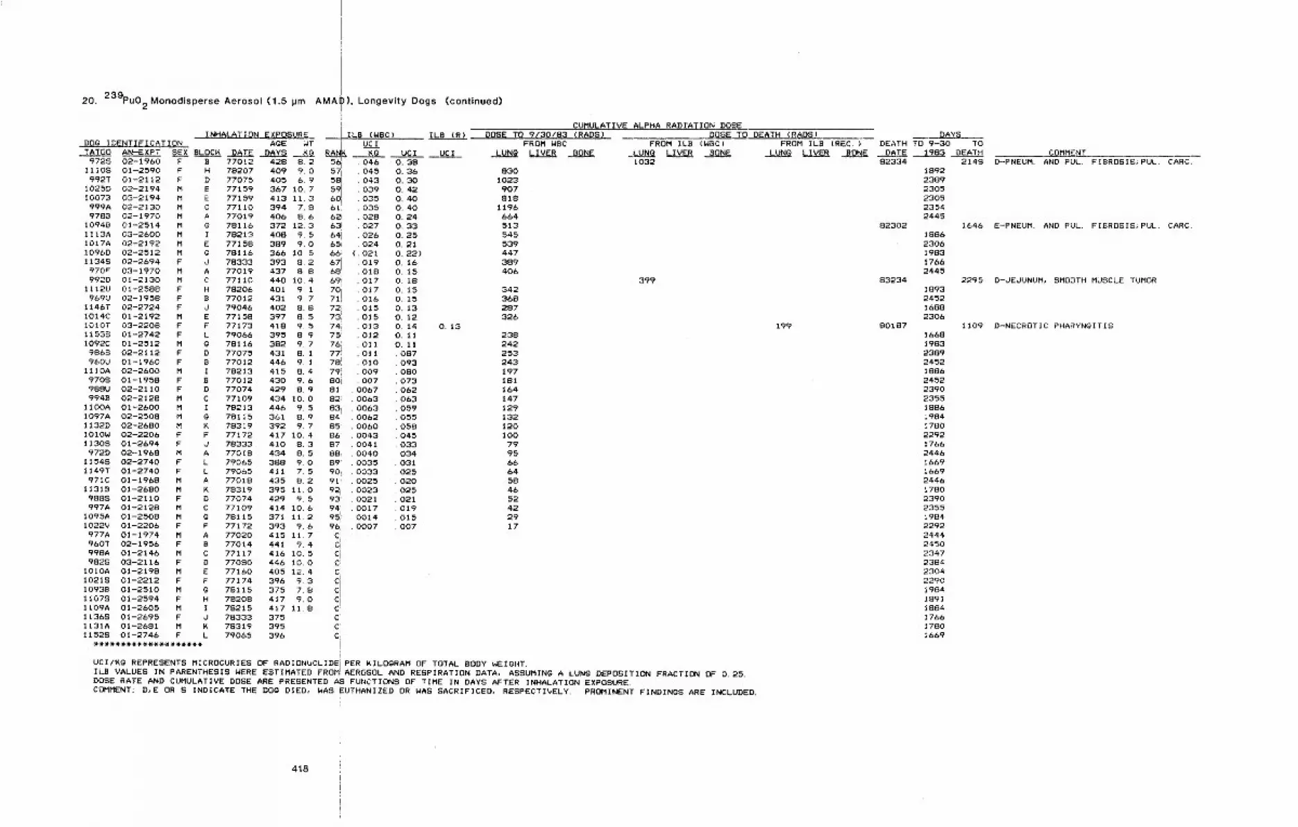
20. 239pu0 2 Monodisperse Aerosol (1.5 ~m AMAI
!
I ILB (WBC)
)), Longevity Dogs (continued)
UC I0.380.3~0.30O. 420.40O, 400,240.330.250.210.22)0.160.150.180.150~150.130,120.140,110. ii
087O93080O73062O630590550580450330340310250200250210190150O7
ILB (R)
,U,C I
CUMULATIVE ALPHA RADrATION DOSEDOSE TO 9C30/83 (RADS)
FROM WBCLUNg LIVER BONE
8301023907818
1196664513545539447389406
DOSE TO DEATH (RADS)FROM ILB (WBC)
LUNg LIVER BONE1032
INHALATION EXPOSUREDO0 IDENTIFICATION AgE WT |TATGO AN-EXPT SEX BLOC~ DATE DAYS ~.G RA,
9728 02-1960 F B 77012 42B 8.211108 01-2590 F H 78207 409 9.0 5~992T 01-2112 F D 77075 4056.9 5~
I025D 02-2194 M E 77159 367 10.7 5~I0078 03-2194 M E 77159 413 11,3 6d999A 02-2130 M C 77110 394 7.8 6119788 02-1970 M A 77019 406 8.6 6~
I0948 01-2514 M @ 78116 372 12.3 631113A 03-2600 M I 78213 408 9.5 6~I017A 02-2192 M E 77158 389 9.0 651096D 02-2512 M G 78116 366 10.5 66;11348 02-2694 F J 78333 393 8.2 671970F 03-1970 M A 77019 437 8.8 68992D 01-2130 M C 77110 440 10.4 69i
I112U 01-2588 F H 78206 401 9. I 70969U 02-1958 F B 77012 431 9.7 711
1146T 02-2724 F J 79046 402 8.8 72!I014C 01-2192 M E 77158 397 8. 5 731I010T 03-2208 F F 77173 418 9.5 74iI1538 01-2742 F L 79066 395 8.9 75I092C 01-2512 M @ 78116 382 9. 7 7619868 02-2112 F D 77075 431 B. 1 771960U 01-1960 F B 77012 446 9. I 781
1110A 02-2600 M I 78213 415 8.4 7919708 01-1958 F B 77012 430 9.6 801988U 02-2110 F D 77074 429 8.9 81994B 02-2128 M C 77109 434 10.0 82~
IIOOA 01-2600 M I 78213 446 9.5 83iI097A 02-2508 M g 78115 361 8.9 84I132D 02-2680 M ~ 78319 392 9.7 85I010W 02-2206 F F 77172 417 10.4 86~11308 01-2694 F J 78333 410 8.3 87
972D 02-1968 M A 77018 434 8, 5 88111548 02-2740 F L 79065 388 9.0 891149T 01-2740 F L 79065 411 7.5 901971C 01-1968 M A 77018 435 8.2 91:
11318 01-2680 M K 78319 395 II.0 92i9888 01-2110 F D 77074 429 9. 5 93=997A 01-2128 M C 77109 414 10.6 94’
1095A 01-2508 M g 78115 371 11.2 951:I022V 01-2206 F F 77172 393 9.6 96,977A 01-1974 M A 77020 415 11.7 C960T 02-1956 F B 77014 441 9.4 CI998A 01-2146 M C 77117 416 i0. 5 Ci9828 03-2116 F D 77080 446 I0.0 C;
tOIOA 01-2198 M E 77160 405 12.4 C10218 01-2212 F F 77174 396 9.3 C!
1093B 01-2510 M G 78115 375 7.8 C11078 01-2594 F H 78208 417 9.0 CIII09A 01-2605 M I 78215 417 11.8 CI1368 01-2695 F J 78333 375 C’I131A 01-2681 M K 78319 395 C~11528 01-2746 F L 79065 396 CI
UC I
046045043039035035028027026024
( o21019o18017017016015015013012011011010009OO7
006700630063006200600043004100400035O033002500230021001700140007
342368287326
O. 132382422532431971811641471291321201 O0
79956664584652422917
399
UCI/Kg REPRESENTS MICROCURIES OF RADIONUCLIDEjPER ~ILOGRAM OF TOTAL BODY WEIGHT.ILB VALUES IN PARENTHESIS WERE ESTIMATED FROM:AEROSOLI AND RESPIRATION DATA, ASSUMINg A LUNG DEPOSITION FRACTION OF 0.25.DOSE RATE AND CUMULATIVE DOSE ARE PRESENTED AS FUNCTIONS OF TIME IN DAYS AFTER INHALATION EXPOSURE.COMMENT: D,E OR S INDICATE THE DOg DIED, WAS EUTHANIZED O.R WAS SACRIFICED, RESPECTIVELY. PROMINENT FINDINGS ARE INCLUDED.
FROM ILB (REC.)LUNG LIVER BONE
DAYSDEATH TO 9-30 TO
DATE 1983 DEATH
199
82334 2148
82302
83234
80187
189223892305230523542445
18862306198317662445
1893245216882306
1668198323892452188624522390235518861984178022921766244616691669244617802390235519842292244424502347238423042290198418911884176617801669
COMMENTD-PNEUM. AND FUL, FIBROSIS~PUL. CARC.
1646 E-PNEUM. AND PUL. FIBROSIS}PUL. CARC.
2295 D-JEJUNUM, SMOOTH MUSCLE TUMOR
1109 D-NECROTIC PHARYNGITIS
418
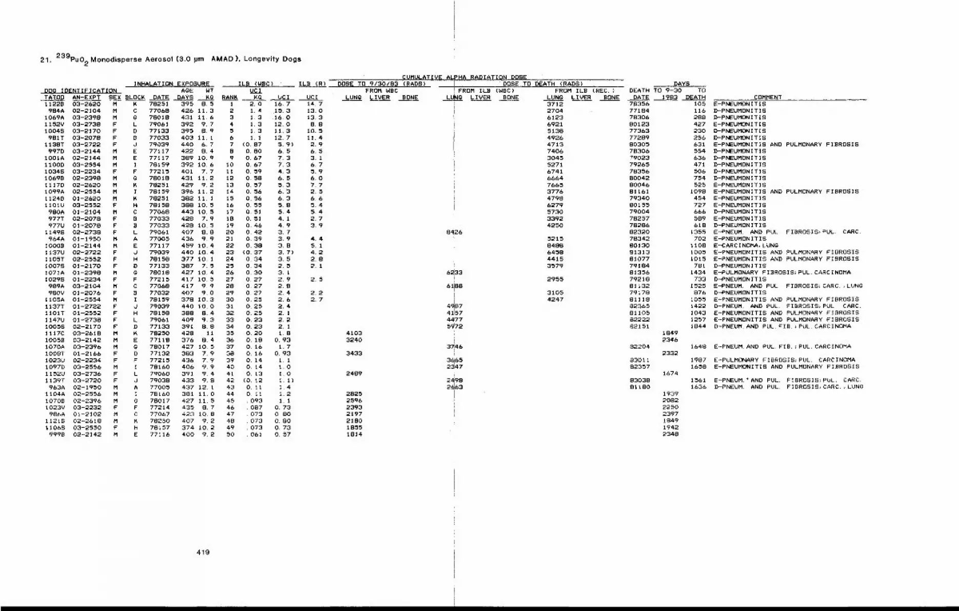
21. 239pu02Monodisperse Aerosot (3.0 pm AMAD), Longevity Dogs
INHALATION EXPOSUREDOg IDENTIFICATION AgE WTTATO0 AN-EXPT SEX BLOCK DATE DAYS KO11228 03-2620 M K 78251 395 8,5
984A 02-2104 M C 77068 426 11.31069A 03-2398 M 0 78018 431 11.61152V 03-2738 F L 79061 392 9. 7I0048 03-2170 F D 77133 395 8.9981T 03-2078 F B 77033 403 11.1
1138T 03-2722 F d 79039 440 6. 7997D 03-2144 M E 77117 422 8,4
1001A 02-2144 M E 77117 389 10,91100D 03-2554 M I 78159 392 10,610348 03-2234 F F 77215 401 7,710698 02-2398 M 0 78018 431 11,21117D 02-2620 M K 78251 429 9.2I099A 02-2554 M I 78159 396 11,211248 01-2620 M K 78251 382 11.11101U 03-2552 F H 78158 388 10.5980A 01-2104 M C 77068 443 10. 5977T 02-2078 F B 77033 428 7.9977U 01-2078 F B 77033 428 10.5
11498 02-2738 F L 79061 407 8.8964A 01-1950 M A 77005 436 9,9
10008 01-2144 M E 77117 459 10.4I137U 02-2722 F d 79039 440 10.41105T 02-2552 F H 78158 377 10.1iO07S 01-2170 F D 77133 387 7.5I071A 01-2398 M g 78018 427 10.4I0298 01-2234 F F 77215 417 10.5989A 03-2104 M C 77068 417 9,9980V 01-2076 F B 77032 407 9.0
1105A 01-2554 M I 78159 378 10.31137T 01-2722 F d 79039 440 10.01101T 01-2552 F H 78158 388 8.41147U 01-2738 F L 79061 409 9,310058 02-2170 F D 77133 391 8.81117C 03-2618 M K 78250 428 111005B 03-2142 M E 77118 376 8,41070A 03-2396 M G 78017 427 10.51008T 01-2166 F D 77132 383 7.91023U 02-2234 F F 77215 436 7.9I097D 03-2556 M I 78160 406 9.91152U 03-2736 F L 79060 391 9.4I139T 03-2720 F d 79038 433 9.8963A 02-1950 M A 77005 437 12. 1
1104A 02-2556 M I 78160 381 11.0I070B 02-2396 M 0 78017 427 11.51023V 03-2232 F F 77214 435 8.7986A 01-2102 M C 77067 423 i0,8
11218 02-26!8 M K 78250 407 9.2II068 03-2550 F H 78157 374 10.29998 02-2142 M E 77116 400 9.2
ILB (WBC) ILB (R)UCI
RAN~ KO UCI UCII23456789
I0II1213141516171819202122232425262728293O3132333435363738394041424344454647484950
2.01,41.31.31.31.1
(0. 87O, 80O, 67O. 67O. 59O. 58O. 57O. 56O. 56O, 550.510,51O. 46O. 42O. 39O. 38
(0. 37O. 34O. 34O. 300 r 27O, 27O, 27O. 250.250.25O, 23O. 23O. 200.180.16O. 160.140.14O. 13
(0. 120. ii0. I1
093087073073073061
16.715,316.012.011,312.7
5.9)6,57.37. 3:4.36.55.36.36.35.85.44.14,93.73.93.83.73.52.53.12.92.82.42.62,42.12,22.11.8
O, 931.7
O, 931.11.01.0I.I)1.41.21.1
O, 73O. 80O. 80O, 73O. 57
14.713.013.38,8
10.511.42.96.53.16.75.96.07.72.56.65.45.42.73,9
4.45.14.22.82.1
2.5
2.22.7
CUMULATIVE ALDO8~ TO 9/30/83 (RADS)
FROM WBCLUN@ ~IVER BONE
41033240
3433
2489
2825259623932197218018551814
=HA RADIATION DOSEDOSE TO DEATH (RADS)
¯ FROM ILB (NBC) FROM ILB (REC,)DAYS
8q26
6~33
44775472
3746
34652347
2~982~63
DEATH TO 9-30LIVER BONE LUNG LIVER BONE DATE 1983
7835677184783068012377363772898030578306790237926578356800428004681161793408015579O047825778286823207834280130813138107779184813567921881132791788111882365811058222282151
82204
8301182357
8303881180
18492346
2332
1674
1939208222502397184919422348
3712270461236921513849264713740630455271674166647665377647986279573033924250
52158488645844153579
2955
31054247
TODEATH
105116288427230256631554636471506754525
1098454727666589618
1355702
110810051015781
1434733
1525876
10551422104312571844
1648
19871658
COMMENTE-PNEUMONITISD-PNEUMONITISD-PNEUMONITISE-PNEUMONITISD-PNEUMONITISD-PNEUMONITISE-PNEUMONITIS AND PULMONARY FIBROSISD-PNEUMONITISD-PNEUMONITISD-PNEUMONITISD-PNEUMONITISD-PNEUMONITISE-PNEUMONITISE-PNEUMONITIS AND PULMONARY FIBROSISE-PNEUMONITISE-PNEUMONITISD-PNEUMONITISE-PNEUMONITISD-PNEUMONITISE-PNEUM. AND PUL. FIBROSIS~PUL. CARC.E-PNEUMONITISE-CARCINOMA, LUNGE-PNEUMONITIS AND PULMONARY FIBROSISE-PNEUMONITIS AND PULMONARY FIBROSISD-PNEUMONITISE-PULMONARY FIBROSISJPUL. CARCINOMAD-PNEUMONITISE-PNEUM, AND PUL, FIBROSIS;CARC.,LUNOD-PNEUMONITISE-PNEUMONITIS AND PULMONARY FIBROSISD-PNEUM. AND PUL. FIBROSIS}PUL. CARC.E-PNEUMONITIS AND PULMONARY FIBROSISE-PNEUMONITIS AND PULMONARY FIBROSISD-PNEUM. AND PUL. FIB.~PUL. CARCINOMA
E-PNEUM. AND PUL. FIB, ;PUL. CARCINOMA
E-PULMONARY FIBROSISIPUL. CARCINOMAE-PNEUMONITIS AND PULMONARY FIBROSIS
1561 E--PNEUM.’AND PUL. FIBROSIS}PUL, CARC.1636 D--PNEUM. AND PUL. FIBROSIS>CARC.,LUNO
419
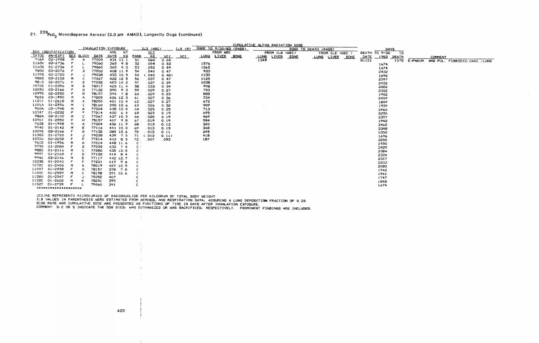
21. 239pu02 Monodisperse Aerosol (3.0 pm AMAD), Longevity Dogs (continued)
INHALATION EXPOSURE
,DATE966A 02-1948 M A 77004 431 11. I
1160V 02-2736 F L 79060 365 9.811608 01-2736 F L 79060 365 9.3980U 03-2076 F B 77032 408 11.9
11398 02-2720 F d 79038 433 10.59888 03-2102 M C 77067 422 12. 59818 02-2076 F B 77032 403 10.2
I0728 01-2396 M G 78017 425 11. 4I005U 03-2166 F D 77132 390 9.3I0998 02-2550 F H 78157 394 7.8965A 03-1950 M A 77005 436 12.3
1121C 01-2618 M K 78250 401 10.41101A 01--2556 M I 78160 390 10.6
960A 03-1948 M A 77004 438 I0.01034T 01-2232 F F 77214 400 6.4
982A 02-2102 M C 77067 437 10.51096T 01-2550 F H 78157 407 9.8
9638 01-1948 M A 77004 436 11.9994D 01-2142 M E 77116 441 10.3
10098 02-2166 F D 77132 380 10.6113~S 01-2720 F d 79038 439 7.51033U 02-2232 F F 77214 403 8.59610 01-1956 M A 77014 448 11.69758 01-20B4 F B 77035 433 7.4988D 01-2116 M C 77080 435 I0.0999T 01-2168 F D 77130 414 8.4994C 03-2146 M E 77117 442 12.7
10338 01-2240 F F 77231 419 9.6I072C 01-2400 M G 78019 427 10.51104T 01-2558 F H 78157 378 7.01100C 01-2559 M I 7815S 391 106II28U 01-2547 F d 78352 4071122C 01-2622 M K 78251 3951152T 01-2739 F L 79060 391
ILB (WBC) ILB (R)DOG IDENTIFICATI0N AGE WT UCITATO0 AN-EXPT SEX BLOCK DAYS KG RANK ~g UCI UCI
51.52 i53’545556575859606162636465666768697O7172CCCCCCCCCCCC
060 0.64054 0.53053 0.49040 0.47
( 040 0.40)037 0.47037 0.39033 0.39029 0.27029 0.23027 0.36027 0.27026 0.32025 0.25025 0.15020 0.19019 0.19013 0.13013 0.13013 0.11
(.013 0.117.007 .053
CUHULATIVE ALPHA RADIATION DOSEDOSE TO 9/30/83 (PADS)
FROM WBCLUNG LIVER BONE
15761265
933113011251038
998753888734672909713695469584320368245418187
DOSE TO DEATH (PADS)FROM ILB (NBC) FROM ILB (REC.)
LUNg LIVER BONE LUNG ~IVER BONE1349
UCI/KQ REPRESENTS MICROCURIES OF RADIONUCLIDE PER KILOGRAM OF TOTAL BODY WEIGHT.ILB VALUES IN PARENTHESIS WERE ESTIMATED FROM AEROSOL AND RESPIRATION DATA, ASSUMING A LUNG DEPOSITION FRACTION OF 0.25.DOSE RATE AND CUMULATIVE DOSE ARE PRESENTED AS, FUNCTIONS OF TIME IN DAYS AFTER INHALATION EXPOSURE.COMMENT: D,E OR S INDICATE THE DOG DIED, NAB EUTHANIZED OR WAS SACRIFICED, RESPECTIVELY. PROMINENT FINDINGS ARE INCLUDED.
DAYSDEATH TO 9-30 TO
DATE 1983 DEATH81121 1578
167416742432169623972432208223321942245918491939246022502397194224602348233216962250245024292384233423472233~08019421941174718481674
E-PNEUM.COMMENT
AND PUL. FIBROSIS}CARC.,LUNG
420
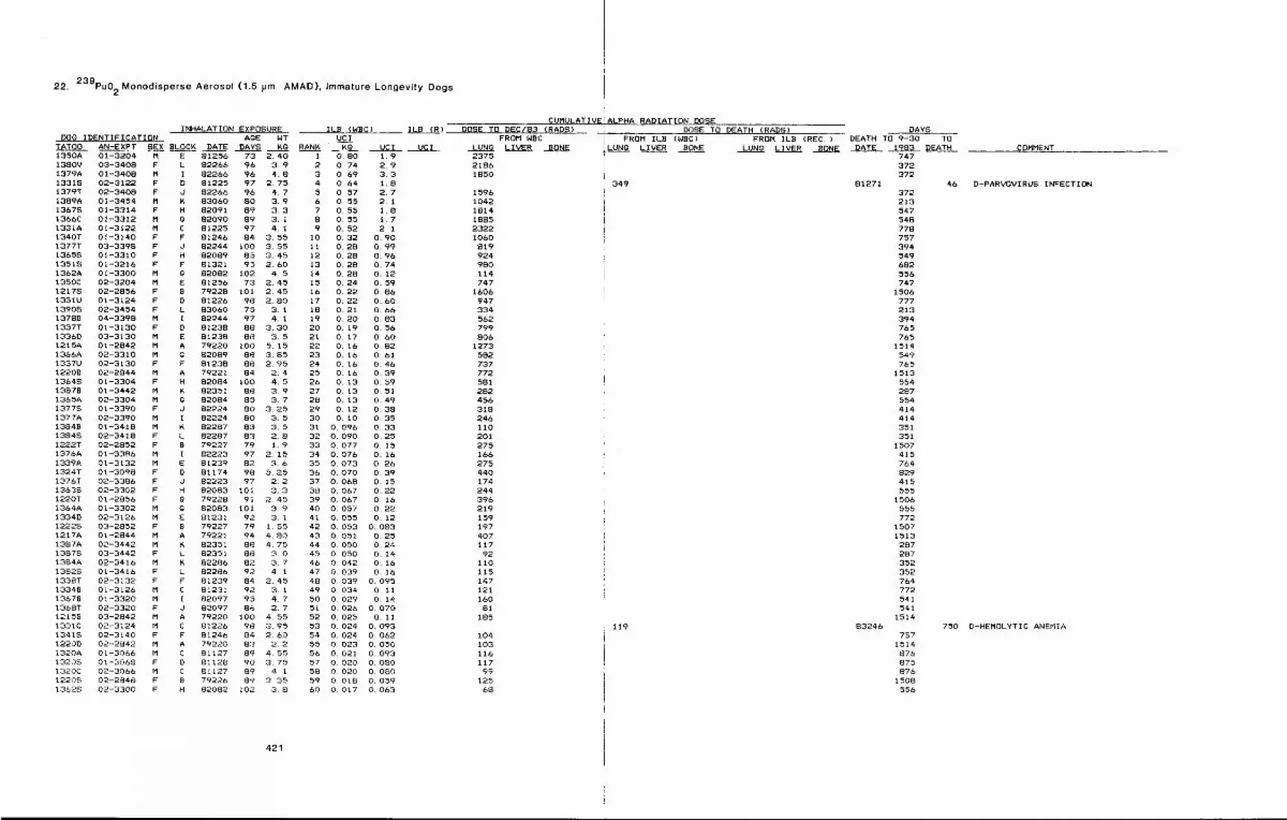
22. 239pu02Monodisperse Aerosol (1.5 pm AMAD), Immature Longevity Dogs
INHALATION EXPOSURE ILB (WBC)DOG IDENTIFICATION AgE WT UCI
TATS0 AN-EXPT SEX BLOCK DATE DAYS KO RANK Kg1350A 01-3204 M E 81256 73 2.40 1 0~80 1.91380V 03-3408 F L 82266 96 3.9 2 0.74 2.91379A 01-3408 M I 82266 96 4.8 3 0,69 3,313318 02-3122 F D 81225 97 2.75 4 O,&4 1.81379T 02-3408 F J 82266 96 4,7 5 0,57 2.71389A 01-3454 M K 83060 80 3.9 6 0.55 2.113678 01-3314 F H 82091 89 3.3 7 0.55 1.81366C 01-3312 M ~ 82090 89 3. I 8 0.55 1.71331A 01-3122 M C 81225 97 4, I 9 0.52 2, 11340T 01-3140 F F 81246 84 3.55 I0 0.32 0.901377T 03-3398 F d 82244 i00 3.55 11 0.28 0.9913658 01-3310 F H 82089 85 3.45 12 0.28 0.9613518 01-3216 F F 81321 95 2.60 13 0.28 0.741362A 0t-3300 M ~ 82082 102 4.5 14 0.28 0.121350C 02-3204 M E 81256 73 2.45 15 0,24 0.5912178 02-2856 F 9 79228 101 2.45 16 0.22 0.861331U 01-3t24 F D 81226 98 2.80 17 0,22 O. bO13908 02-3454 F L 83060 75 3.1 le 0.21 0.6613788 04-3398 M I 82244 97 4, i 19 0.20 0,831337T 01-3130 F D 81238 88 3.30 20 O. 19 0,561336D 03-3130 M E 81238 88 3.5 21 0.17 0,601215A 01-2842 M A 79220 100 5.15 22 O. l& 0,82136AA 02-3310 M G 82089 88 3.85 23 0.16 0,611337U 02-3i30 F F 81238 88 2.95 24 O~ 16 0,4612208 02-2844 M A 79221 84 2.4 25 O. t& 0.3913648 01-3304 F H 82084 I00 4,5 26 0. 13 0.5913878 01-3442 M K 82351 88 3.9 27 0.13 0,511365A 02-3304 M g 82084 85 3.7 28 0, 13 0,49I377S 01-3390 F d 82~24 80 3,25 29 0. 12 0,381377A 02-3390 M I 82224 80 3.5 30 O. i0 0.3513849 01-3418 M K 82287 83 3.5 31 0.096 0.3313848 02-3418 F L 82287 83 2.8 32 0.090 0,251222T 02-2852 F 9 79227 79 1.9 33 0.077 0,151376A 01-3386 M I 82223 97 2 15 34 0,076 0. 161339A 01-3132 M E 81239 82 3.6 35 0.073 0,261324T 01-3098 F D 81174 98 5.25 36 0.070 0,391376T 02-3.386 F O 82223 97 2.2 37 0.068 0.1513638 02-3302 F H 82083 10I 3.3 38 0.067 0.221220T 01-2856 F 9 79228 91 2.45 39 0.067 O. 161364A 01-3302 M G 82083 101 3.9 40 0,057 0,221334D 02-3126 M E 81231 92 3 1 41 0.055 0,1212225 03-2852 F 9 79227 79 1.55 42 0.053 0.0831217A 01-2844 M A 79221 94 4.80 43 0~051 0.251387A 02-3442 M ~ 82351 88 4.75 44 0~050 0,2413878 03-3442 F L 82351 88 3.0 45 0.050 0.14I384A 02-3416 M K 82286 82 3,7 46 0.042 0,1613828 01-3416 F L 82286 92 4 1 47 0,039 0, 161338T 02-3132 F F 81239 84 2.45 48 0,039 0.09513349 01-3126 M C 81231 92 3. 1 49 0,034 O, 1113679 01-3320 M I 82097 95 4~ 7 50 0.029 0,141368T 02-3320 F d 82097 86 2.7 51 0.026 0.07012159 03-2842 M A 79220 I00 4.55 52 0.025 0,111331C 02-3124 M C 81226 98 3.95 53 0.024 0.09313418 02-314.0 F F 81246 84 2.60 54 0.024 0.06~I220D 02-2842 M A 79220 83 2.2 55 0.023 0.0501320A 0i-3066 M C 81127 89 4.55 56 0.021 0.09313208 01-3068 F D 81128 90 3.75 57 0.020 0.0801320C 02-3066 M C 81127 89 4 1 58 0.020 0,08012208 02-2848 F 9 79226 89 3 35 59 0.018 0.05913628 02-3300 F H 82082 102 3.8 60 0.017 0.063
ILB (R)
UCI UCI
421
CUMULATIVEDOSE TO DEC/B3 (RADS)
FROM NBCLUNQ LIVER BONE237521861850
159610421814188523221060819924980114747
1606947334562799806
12735827377725812824563182461102012751662754401742443962191591974O711792
11011514712116081
185
10410311611799
12568
ALPHA RADIATION DOSEDOSE TO DEATH (RADS)
FROM IL9 (NBC) FROM ILB {REC. LUN~ LIVER BONE LUN9 LIVER BONE
349
119
DAYSDEATH TO 9-30 TO
DATE 1983 DEATH747372372
81271372213547548778757394549682556747
1506777213394765765
1514549765
1513554287554414414351351
1507415764829415555
1506555772
15071513287287352352764772541541
151483246
7571514876875876
1508556
COMMENT
46 D-PARVOVIRUS INFECTION
750 D-HEMOLYTIC ANEMIA
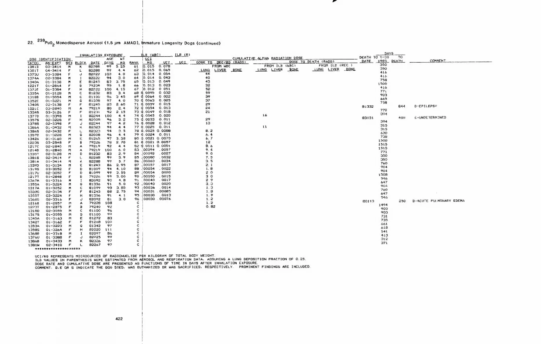
22. 239pu02 Monodisperse Aerosol (1.5 pm AMAD),
INHALATION EXPOSUREDOg IDENTIFICATION AgE WT
TATO0 AN-EXPT SEX BLOCK DATE D;;YS 14"013S1B 03-3414 M 14" 82288 99 5.25138IT 04-3414 F L 82288 99 4.41373U 03-3384 F d 82#22 100 4.01374A 02-3384 M I 82222 94 3.01340A 01-3138 M E 81245 83 3.751221T 01-2864 F B 79234 95 1.81373T 01-3384 F H 82222 100 4.151335A 01-3128 M C 81232 83 3,413188 01-3054 M C 81100 96 3,451352C 01-3221 M g 81338 97 4.013408 02-3138 F F 81245 83 2.60I221C 03-2840 M A 79219 80 2.413348 03-3126 F F 81231 92 2.1513778 01-3398 M I 82244 100 4.413578 02-3228 F H 82008 96 3213788 02-3398 F d 82244 97 4.21386A 01-3432 M 14. 82323 94 4.413868 02-3432 F L 82323 94 3,513578 01-3228 M g 82008 96 4.41342A 01-3160 M E 81265 97 3.3812238 03-2848 F B 79226 78 2.701217C 02-2840 M A 792i9 92 4.412148 01-2840 M A 79219 tO0 6.01335T 02-3128 M D 81232 83 2.913818 02-3414 F L 82288 99 3.91381A 01-3414 M K 82288 99 5.713398 01-3134 M E 81243 86 2.9513198 03-3052 F D 81009 94 4. iO1317U 02-3052 F D 81099 99 3.551217T 01-2848 F B 79226 99 3.001367A 01-3316 M I 82092 90 4~81355A 01-3224 M 0 81356 91 5.01317A 01-3052 M C 81099 98 3.851338S 02-3134 F F 81243 88 2.751355T 02-3224 F H 81356 91 4..113688 02-3316 F J 82092 81 3.012168 01-2857 M A 79228 10812~3T 01-2875 F B 79240 921318D 02-3055 M C 81100 9613178 01-3055 M D 81100 991345A 01-3163 M E 81272 831342T 01-3162 F F 81268 I001353A 01-3223 M g 81342 9713588 01-3264 F H 82020 iii13688 01-3318 M I 82097 861376U 01-3388 F J 82~25 9913868 01-3433 M 14. 82326 971380W 02-3410 F L 82267 97
RANK
nmature Longevity Dogs (continued)
LB (WBC) ILB (R)
UC Ij UC,I.
14.0 UC IIEo15o 078 DOSE TO DEC/B3 (RADS)
CUMULATIVE ALPHA RADIATION DOSEDOSE TO DEATH (RAD8)
FROM WBCLUN~ LIVER, BONE
4440433252593937292421
2913
62 i0.0t5 0.06563 iO. 014 O. 05464 I=0- 014 O. 04365 !0. 013 O. 04966 !0. 013 O. 023
Io. o12 o. o516768 O. (}095 O. 03269dd1°°64 o. o2270 0063 o. 02571(~(~i 0059 O. 01572 0054 O. 01373 ~t. 0049 O. 01874 q. oo4so. 02075d. 0033 O. 01176 Q. 0028 O. 01277 O, 0025 O. 01178 O. 0025 O. 008879 O. 0024 O. 01180 O. 0021 O. 007081 Q. 0021 O~ 005782 (~.0011 O. 005183 ,100094 005784 "I 00092 002785 .i 00080 003286 .I 00060 003487., 00057 001788 .I 00054 002289 .’i 00054 002090 .i 00050 001591 . 00040 001792 .i 00040 002093 .i 00036 001494 . 00031 . 0008595 . 00030 . 001396 .100030 . 00076CCccCCcCc ,CCC i
8.26.46.712869,49,07.53.52.11.82.03i 02.93.31.31.81.91.21.2O. 82
FROM ILB ~WBC)LUNO LIVER BONE
16
FROM ILB (REC.)LUNg LIVER BONE
11
UCI/KQ REPRESENTS MICROCURIES OF" RADIONUCLIDE PER KILOGRAM OF TOTAL BODY WEIGHT.ILB VALUES IN PARENTHESIS WERE ESTIMATED FROM A~ROSOL AND RESPIRATION DATA, ASSUMINg A LUNg DEPOSITION FRACTION OF 0.25.DOSE RATE AND CUMULATIVE DOSE ARE PRESENTED AS FUNCTIONS OF TIME IN DAYS AFTER INHALATION EXPOSURE.COMMENT: D,E OR S INDICATE THE DOg DIED, WAS EUTHANIZED OR WAS SACRIFICED, RESPECTIVELY. PROMINENT FINDINGS ARE INCLUDED.
DAYSDEATH TO 9-30 TO.DATE 1983 DEATH
81332
83131
80113
350350416416758
1500416771903665758
772394
394315315630738
150815151515
771350350760904904
1508546647904760&4754.6
1494903903731735661618541413312371
COMMENT
844 D-EPILEPSY
488 E-UNDETERMINED
250 D-ACUTE PULMONARY EDEMA
422
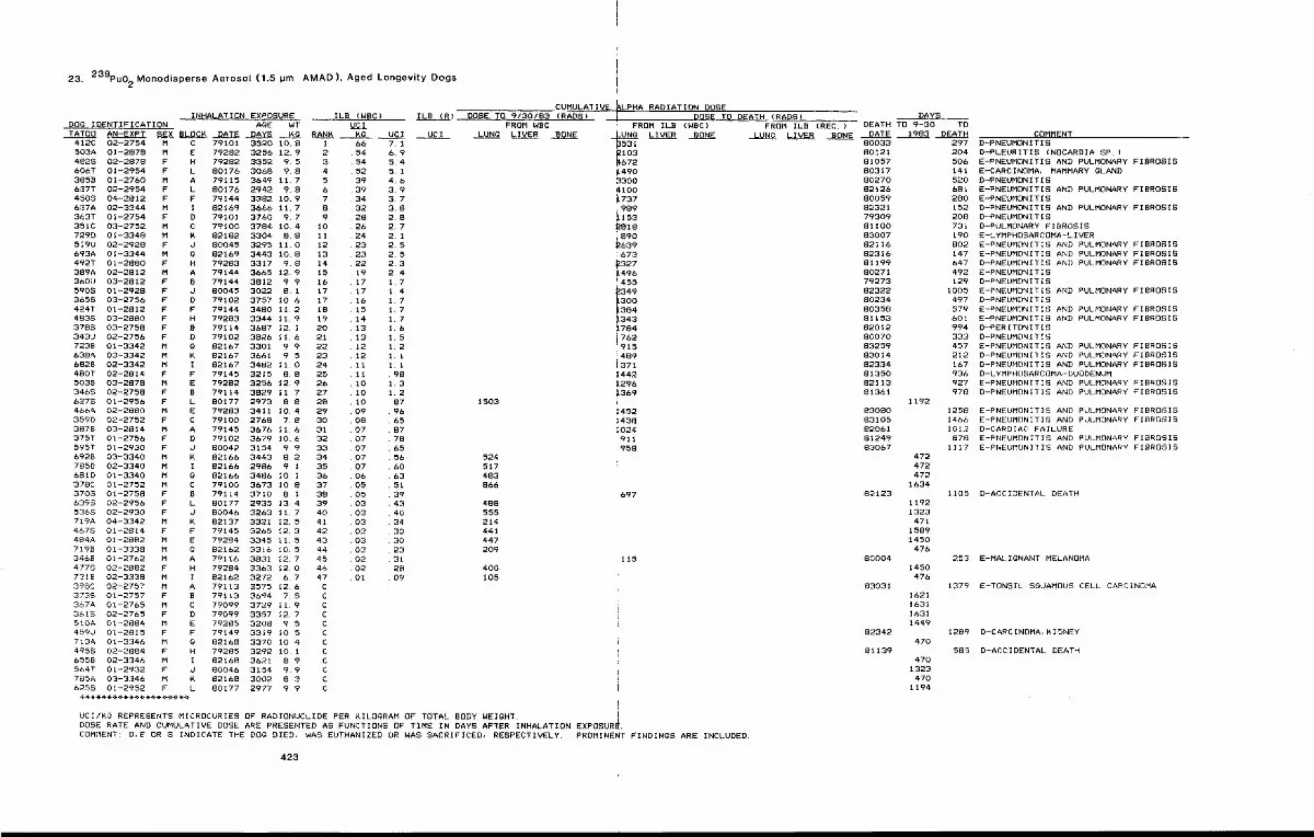
23. 239pu02Monodisperse Aerosol (1.5 jJm AMAD), Aged Longevity Dogs
INHALATION EXPOSURE ILB (WBC) ILB (R)DENTIFICATION AgE WT
TATO0 AN-EXPT SEX BLOCK DATE DAYS Kg412C 02-2754 M C 79101 3520 I0,8503A 01-2878 M E 79282 3256 12.94828 02-2878 F H 79282 3352 9.5606T 01-2954 F L 80176 3068 9.83858 01-2760 M A 79115 3649 11.7637T 02-2954 F L 80176 2942 9.84508 04-2812 F F 79144 3382 10.9637A 02-3344 M I 82169 3666 II.7363T 01-2754 F D 79101 3760 9.7351C 03-2752 M C 79100 3784 10.4729D 01-3348 M K 82182 3304 8.8519U 02-2928 F d 80045 3295 11. 0693A 01-3344 M ~ 82169 3443 10.8492T 01-2880 F H 79283 3317 9.8389A 02--2812 M A 79144 3665 12.9 153bOU 03-2812 F B 79144 3812 9.9 165908 01-2928 F J 80045 3022 8.1 173658 03-2756 F B 79102 3757 10.6 174~4T 01-2812 F F 79144 3480 11.2 18483S 03-2880 F H 79283 3344 11.9 193788 03-2758 F B 79114 3687 12. I 20343U 02-2755 F D 79102 3826 11.6 217239 01-3342 M g 82167 3301 9.9 22638A 03-3342 M K 82167 3661 9.5 236828 02-3342 M I 82167 3482 I1.0 24480T 02-2814 F F 79145 3215 8.8 255038 03-2878 M E 79282 8255 12.9 263468 02-2758 F B 79114 3829 11.7 276278 01-2956 F L 80177 2973 8.8 28466A 02-2880 M E 79283 3411 10.4 293590 02-2752 F C 79100 2768 7.8 303878 03-2814 M A 79145 3676 11.6 31375T 01-2756 F D 79102 3679 10.6 32595T 01-2930 F d 80042 3154 9.9 336928 03-3340 M K 82166 3443 8.2 347858 02-3340 M I 82166 2986 911 356810 01-3340 M g 82166 3486 10. I 36378C 01-2752 M C 79100 3673 10.8 373708 01-2758 F S 79114. 3710 B. I 386398 02-2955 F L 80177 2935 13. 4 395365 02-2930 F J 80046 3263 11. 7 40719A 04-3342 M K 82137 3321 12. 5 414678 01-2814 F F 79145 3265 12.3 42484A 01-2882 M E 79284 3345 11.5 437198 01-3338 M ~ 82162 3316 10.5 443469 01-2762 M A 79116 3831 12.7 454775 02-2882 F H 79284 3363 12.0 467318 02-3338 M I 82162 3272 6.7 47398C 02-2757 M A 79113 3575 12.6 C3739 01-2757 F B 79113 3694 7. 5 C367A 01-2765 M C 79099 3729 11.9 C3618 02-2765 F D 79099 3357 12.7 C510A 01-2884 M E 79285 3208 9.5 C459U 01-2815 F F 79149 3319 10,5 C713A 01-3346 M g 82168 3370 10. 4 C4958 02-2884 F H 79285 3292 i0. i C6558 02-3346 M I 82168 3621 8.9 C5647 01-2932 F J 80046 3154 9 9 C785A 03-3346 M K 82168 3002 8,3 C6258 01-2952 F L 80177 2977 9.9 C
DOg I gClRANK K¢ UCI UGI
1 662 543 544 525 396 397 348 329 28
10 2611 2412 2313 23I 4 22
1917171615141313i2121I1tI01010090807070707070605050303030303030202O1
7.16.95.45.14.63,93.73.82.82.72.12.52.52.32.41,71,41.71,71.71,61.51.21.11.1¯ 981.31,2. 87
96658778655660635139434034.303023312809
CUMULATIVE ~LPHA RADIATION DOSED,OSETO 9/30/83 (RAD5)
FROM WBCLUNO LIVER BONE
1503
524517483866
488555214441447209
400105
DOSE TO DEATH {RADS)FROM ILB {WBC)
’UN~) BONELIVER
~103~672~49033004100~737i989~153~818i8902639;673~327i496!455
i349300
~384~3431784i762’915~489i371~4421296i369
14521438!024!911i958
FROM ILB (REC.)LUNO LIVER BONE
697
115
UCI/KQ REPRESENTS MICROCURIES OF RADIONUCLIDE PER KIL.OgRAM OF TOTAL BODY WEIGHT.DOSE RATE AND CUMULATIVE DOSE ARE PRESENTED AS FUNCTIONS OF TIME IN DAYS AFTER INHALATION EXPOSURe,COMMENT: D,~ OR S INDICATE THE DOg DIED, WAS EUTHANIZED OR WAS SACRIFICED, RESPECTIVELY. PROMINENT FINDINGS ARE INCLUDED.
DAYSDEATH TO 9-30 TO
DATE 1983 DEATH80033801218105780317802708212680059823217930981100830078211682316811998027179273823228023480358811538201280070832598301482334813508211381361
8308083105820618124983067
82123
80004
83031
82342
81139
1192
472472472
1634
11921323
47115891450
476
1450476
1621163116311449
470
4701323470
1194
COMMENT297 D-PNEUMONITIS204 D-PLEURITIS (NOCARDIA SP. )506 E-PNEUMONITIS AND PULMONARY FIBROSIS141 E-CARCINOMA, MAMMARY gLAND520 D-PNEUMONITIS681 E-PNEUMONITIS AND PULMONARY FIBROSIS280 E-PNEUMONITIS152 D-PNEUMONITIS AND PULMONARYFIBROSIS208 D-PNEUMONITIS731 D-PULMONARY FIBROSIS190 E-LYMPHOSARCOMA-LIVER802 E-PNEUMONITIS AND PULMONARYFIBROSIS147 E-PNEUMONITIS AND PULMONARYFIBROSIS647 D-PNEUMONITIS AND PULMONARYFIBROSIS492 E-PNEUMONITIS129 D-PNEUMONITIS
1005 E-PNEUMONITIS AND PULMONARYFIBROSIS497 D-PNEUMONITIS579 E-PNEUMONITIS AND PULMONARYFIBROSIS601 E-PNEUMONITIS AND PULMONARYFIBROSIS994 D-PERITONITIS333 D-PNEUMONITIS457 E-PNEUMONITIS AND PULMONARYFIBROSIS212 D-PNEUMONITIS AND PULMONARYFIBROSISl&7 D-PNEUMONITIS AND PULMONARYFIBROSIS936 D-LYMPHOSARCOMA-DUODENUM927 E-PNEUMONITIS AND PULMONARYFIBROSIS978 D-PNEUMONITIS AND PULMONARYFIBROSIS
1258146&1012878
1117
1105
253
1379
1289
585
E-PNEUMONITIS AND PULMONARY FIBROSISE-PNEUMONITIS AND PULMONARYFIBROSISD-CARDIAC FAILUREE-PNEUMONITIS AND PULMONARY FIBROSISE-PNEUMEINITIS AND PULMONARYFIBROSIS
D-ACCIDENTAL DEATH
E-MAL IgNANT MELANOMA
E-TONSIL SOUAMOUS CELL CARCINOMA
D-CARCINOMA, KIDNEY
D-ACCIDENTAL DEATH
423
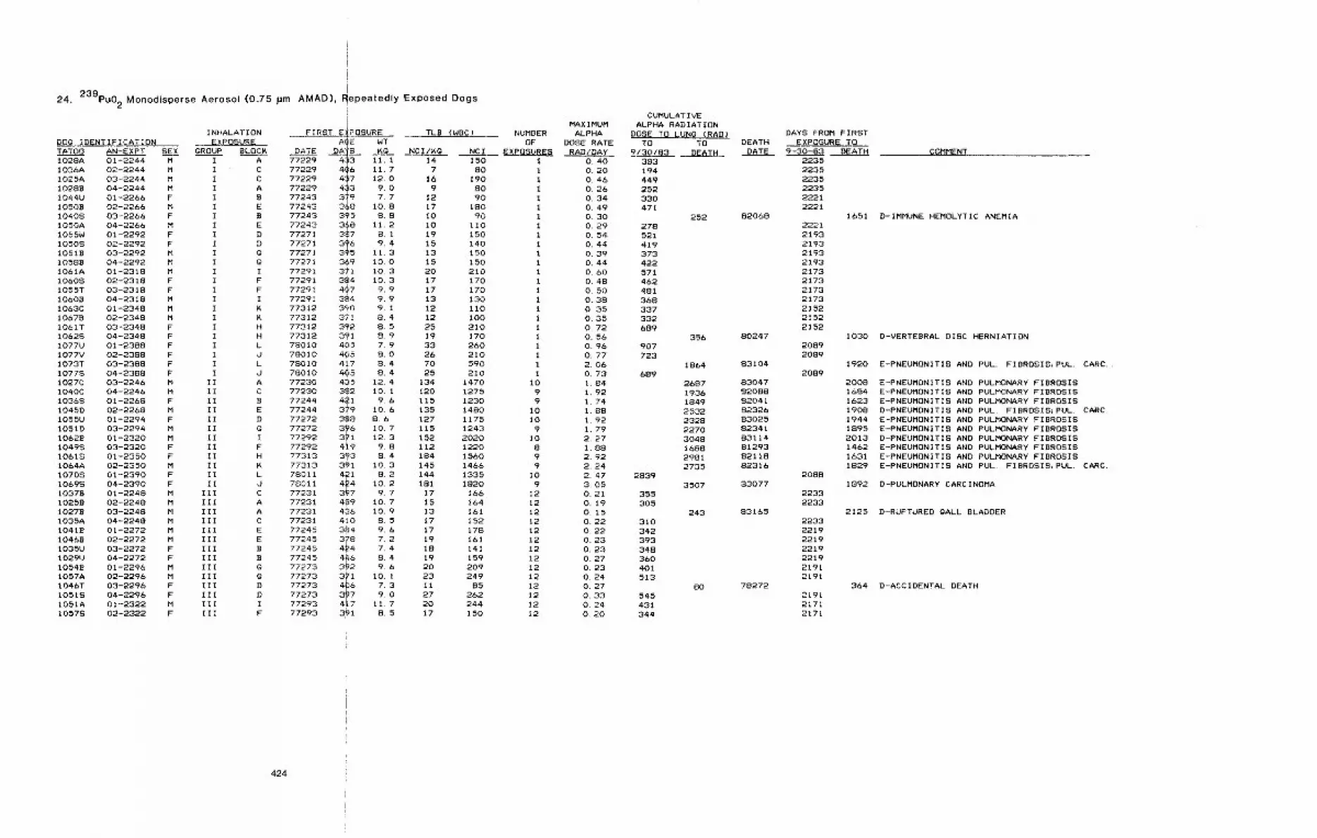
i
24. 239pu02 Monodisperse Aerosol (0.75 pm AMAD), Repeatedly Exposed Dogs
(f
INHALATION FIRST E~POSURE TLB (WBC)DOg IDENTIFICATION EXPOSURE A~E WT
D#TE DA_~ ~g4-~3 ii. I
TATOO AN-EXPT1028A 01-2244I036A 02-2244I025A 03-2244I0288 04-2244I044U 01-2266I0508 02-2266I0408 03-2266I050A 04-22661055W 01-2292I0508 02-2292I0518 03-2292I0588 04-2292I061A 01-2318I0608 02-2318I055T 03-2318I0608 04-23!8I063C 01-2348I0678 02-2348I061T 03-2348I0628 04-2348I077U 01-2388I077V 02-2388I073T 03-2388I0778 04-23881027C 03-22461040C 04-224610368 01-2268I045D 02-22681055U 01-229410510 03-2294I0628 01-232010498 03-232010618 01-2350I064A 02-235010708 01-239010698 04-239010378 01-2248I0258 02-224810278 03-22481035A 04-2248I0418 01-2272I0468 02-22721035U 03-22721029U 04-2272I0548 01-22961057A 02-2296I046T 03-2296I0518 04-2296I051A 01-232210578 02-2322
SE__X &ROUP BLOCMM I AM I CM I CM I AF I BM I EF I BM I EF I DF I DM I g
M I IF I FF I FM I IM IM IF I HF I HF I LF I JF I LF I JM II AM II CF II BM II EF II DM IIM II IF II FF II HM II KF II LF II JM III CM Ill AM ill AM III CM III EM III EF III BF III BM IIIM III gF III DF III DM Ill IF Ill F
7722977229 406 11.777229
417
12.077229 4 3 9.077243 3 9 7.777243 3 8 10.877243 395 8.877243
3-~811.2
77271 3 7 Si177271
3~69.4
77271 3 5 11.377271 369 10.077291 371 10.377291 384 10,377291 4~7 9,977291 384 9,977312 390 9.177312 371 8.477312 392 8.577312 391 8.978010 405 7.978010 405 8,078010 417 8.478010 4~5 8,477230 435 12.477230 382 10.177244 4~I 9.677244 379 101677272 388 8.677272
3~610.7
77292 3 1 12.377292 4~9 9.877313 3~3 8.477313 391 10.378011 421 8.278011
4~410.2
77231 3 7 9.777231 4~9 10.777231 436 10.977231 410 8.577245 384 9.677245 378 7.277245 4~4 7.477245
4168.4
77273 3 2 9.677273
311
10. 177273 4 6 7.377273 3 7 9.077293 417 11.777293 3~I 8.5
NUMBEROF
NCI/K@ NCI EXPOSURES14 150 17 80 i
16 190 i9 80 1
12 90 117 180 1I0 90 110 II0 I19 150 I15 140 I13 150 115 150 120 210 117 170 117 170 113 130 112 110 112 i00 125 210 I19 170 133 260 126 210 170 590 125 210 1
134 1470 10120 1275 9115 1230 9135 1480 I0127 1175 10115 1243 9152 2020 I0112 1220 8184 1560 9145 1466 9144 1335 I0181 1820 9
17 16b 1215 164 1213 161 1217 152 1217 178 1219 161 1218 141 1219 159 1220 209 I223 249 12Ii 85 1227 262 1220 244 1217 150 12
MAXIMUMALPHA
DOSE RATERAD/DAY
O. 40O. 20O. 46O. 26O. 34O. 49O. 30O. 29O, 540.44O, 39O. 44O. 60O. 48O. 50O. 38O. 35O. 35O, 72O. 56O. 96O. 772, 06O, 731 841 921 741 881 921 792. 27I. 882, 922~ 242.473.050.210.190.15O. 22O. 22O. 23O. 23O. 27O, 23O. 24O, 27O. 33O. 24O. 20
CUMULATIVEALPHA RADIATION
DOSE TO LUNG CRAD)TO TO DEATH
9/30/83 DEATH DATE383194449252330471
252278521419373422571462481368337332689
3569O7723
1864689
2687193618492532232822703048168829812735
28393507
355305
243310342393348360401513
80545431344
DAYS FROM FIRSTEXPOSURE TO
9-30-83 DEATH
82068
80247
83104
83047820888204182326830258234183114812938211882316
83077
83165
78272
22352235223522352221222t
222121932193219321932173217321732173215221522152
20892089
2089
2088
22332233
2233221922192219221921912191
219121712171
COMMENT
1651 D-IMMUNE HEMOLYTIC ANEMIA
1030 D-VERTEBRAL DISC HERNIATION
1920 E-PNEUMONITIS AND PULl FIBROSIS~PUL. CARC,,
2008168416231908194418952013146216311829
1892
2125
E-PNEUMONITIS AND PULMONARYFIBROSISE-PNEUMONITIS AND PULMONARYFIBROSISE-PNEUMQNITIS AND PULMONARYFIBROSISD-PNEUMDNITIS AND PUL. FIBRDSIS~PUL. CARC.E’-PNEUMONITIS AND PULMONARY FIBROSISE-PNEL~ONITIS AND PULMONARYFIBROSISD-PNEUMONITIS AND PULMONARYFIBROSISE-PNEUMONITIS AND PULMONARYFIBROSISE-PNEUMONITIS AND PULMONARYFIBROSISE-PNEUMONITIS AND PUG FIBROSIS~PUL. CARC.
D-PULMONARY CARCINOMA
D-RUPTURED gALL BLADDER
364 D-ACCIDENTAL DEATH
424
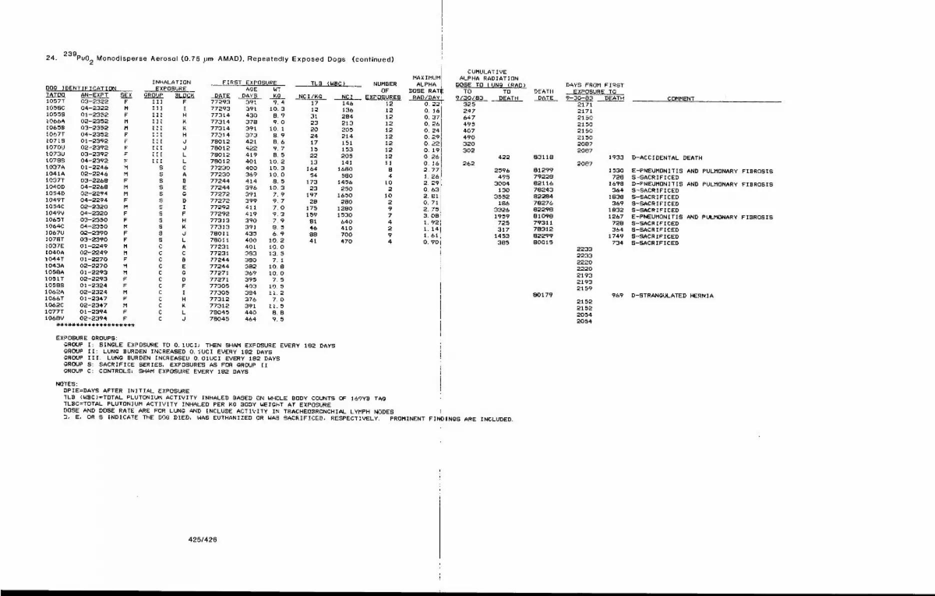
24. 239pu02 Monodisperse Aerosol (0.75 ,urn AMAD), Repeatedly Exposed Dogs (continued)
INHALATION FIRST EXPOSURE TLB (WBC) NUMBERDOg IDENTIFICATION EXPOSURE AGE WT OFTATO0 AN-EXPT SEX gROUP BLOCK DATE DAYS KQ NCI/KG NCI EXPOSURES1057T 03-2322 F III F 77293 391 9.4 17 146 121058C 04-2322 M III I 77293 391 10.3 12 136 121055S 01-2352 F III H 77314 430 8.9 31 284 12lOb6A 02-2352 M III K 77314 378 9.0 23 213 1210658 03-2352 M Ill K 77314 391 10. 1 20 205 12I067T 04-2352 F III H 77314 373 8.9 24 214 12I071S 01-2392 F Ill d 78012 421 8.6 17 151 12I070U 02-2392 F III d 78012 422 9.7 15 153 121073U 03-2392 F III L 78012 419 8.5 22 205 121078S 04-2392 F IIi L 78012 401 10.2 13 141 111037A 01-2246 M S C 77230 40G t0.3 164 1680 S1041A 02-2246 M S A 77230 369 10.0 54 580 4I037T 03-2268 F S B 77244 414 8.5 173 1456 101040D 04-2268 M S E 77244 396 10.3 23 250 21054D 02-2294 M S @ 77272 391 7.9 197 1650 10I049T 04-2294 F S D 77272 399 9.7 28 280 2I054C 02-2320 M S I 77292 411 7.0 175 1280 91049V 04-2320 F S F 77292 419 9.3 159 1530 7IO&5T 03-2350 F S H 77313 390 7.9 81 640 41064C 04-2350 M S K 77313 391 8.5 46 410 2IO&7U 02-2390 F S d 78011 435 6.9 88 700 9I078T 03-2390 F S L 78011 400 10.2 41 470 4I037E 01-2249 M C A 77231 401 10.01040A 02-2249 M C C 77231 333 13.51044T 01-2270 F C B 77244 380 7.11043A 02-2270 M C E 77244 382 10.8105SA 01-2293 M C @ 77271 369 10.01051T 02-2293 F C D 77271 395 7.51058S 01-2324 F C F 77305 403 10.51062A 02-2324 M C I 77305 384 11.21066T 01-2347 F C H 77312 376 7.01062C 02-2347 M C K 77312 391 11.51077T 01-2394 F C L 78045 440 8.81068V 02-2394 F C d 78045 464 9.5
MAXIMUMALPHA
DOSE RATIRAD/DAY
O. 220.16O. 37,O. 26O. 24O. 29,O. 22!0. I9’O. 26’0.162. 77iI. 26i2. 29!O. 632.81’0.712. 753. 08t1.921,141.61O. 90
EXPOSURE gROUPS:GROUP I: SINGLE EXPOSURE TO O. IUCI~ THEN SHAM EXPOSURE EVERY 182 DAYSGROUP II: LUNG BURDEN INCREASED O. IUCI EVERY 182 DAYSGROUP III: LUNG BURDEN INCREASED O. 01UCI EVERY 182 DAYSGROUP S: SACRIFICE SERIES~ EXPOSURES AS FOR gROUP IIGROUP C: CONTROLS; SH~M EXPOSURE EVERY 182 DAYS
NOTES:DPIE=DAYS AFTER INITIAL EXPOSURETLB (WSC)=TOTAL PLUTONIUM ACTIVITY INHALED BASED ON WHOLE BODY COUNTS OF 169YB TAgTLBC=TOTAL PLUTONIUM ACTIVITY INHALED PER ~@ BODY WEIGHT AT EXPOSUREDOSE AND DOSE RATE ARE FOR LUNg AND INCLUDE ACTIVITY IN TRACHEOBRONCHIAL LYMPH NODESD, E, OR S INDICATE THE DOG DIED, WAS EUTHANIZED OR WAS SACRIFICED,
CUMULATIVEALPHA RADIATION
~OSE TO LUNG (RAD)TO TO
9/30/83 DEATH325247647495407490320302
422262
2596495
3004130
3552186
33261959725317
14533S5
RESPECTIVELY. PROMINENT FInDINgS ARE INCLUDED,
DEATI4DATE
83118
812997922882116782438228478276822988109879311783128229980015
80179
DAYS FROM FIRSTEXPOSURE TO
9-30-83 DEATH21712171215021502150215020872087
19332087
1530728
1698364
1838369
18321267
728364
1749734
2233223322202220219321932159
9692152215220542054
COMMENT
D-ACCIDENTAL DEATH
E-PNEUMONITIS AND PULMONARY FIBROSISS-SACRIFICEDD-PNEUMONITIS AND PULMONARY FIBROSISS-SACRIFICEDS-SACRIFICEDS-SACRIFICEDS-SACRIFICEDE-PNEUMONITIS AND PULMONARY FIBROSISS-SACRIFICEDS-SACRIFICEDS-SACRIFICEDS-SACRIFICED
D-STRANGULATED HERNIA
425t426

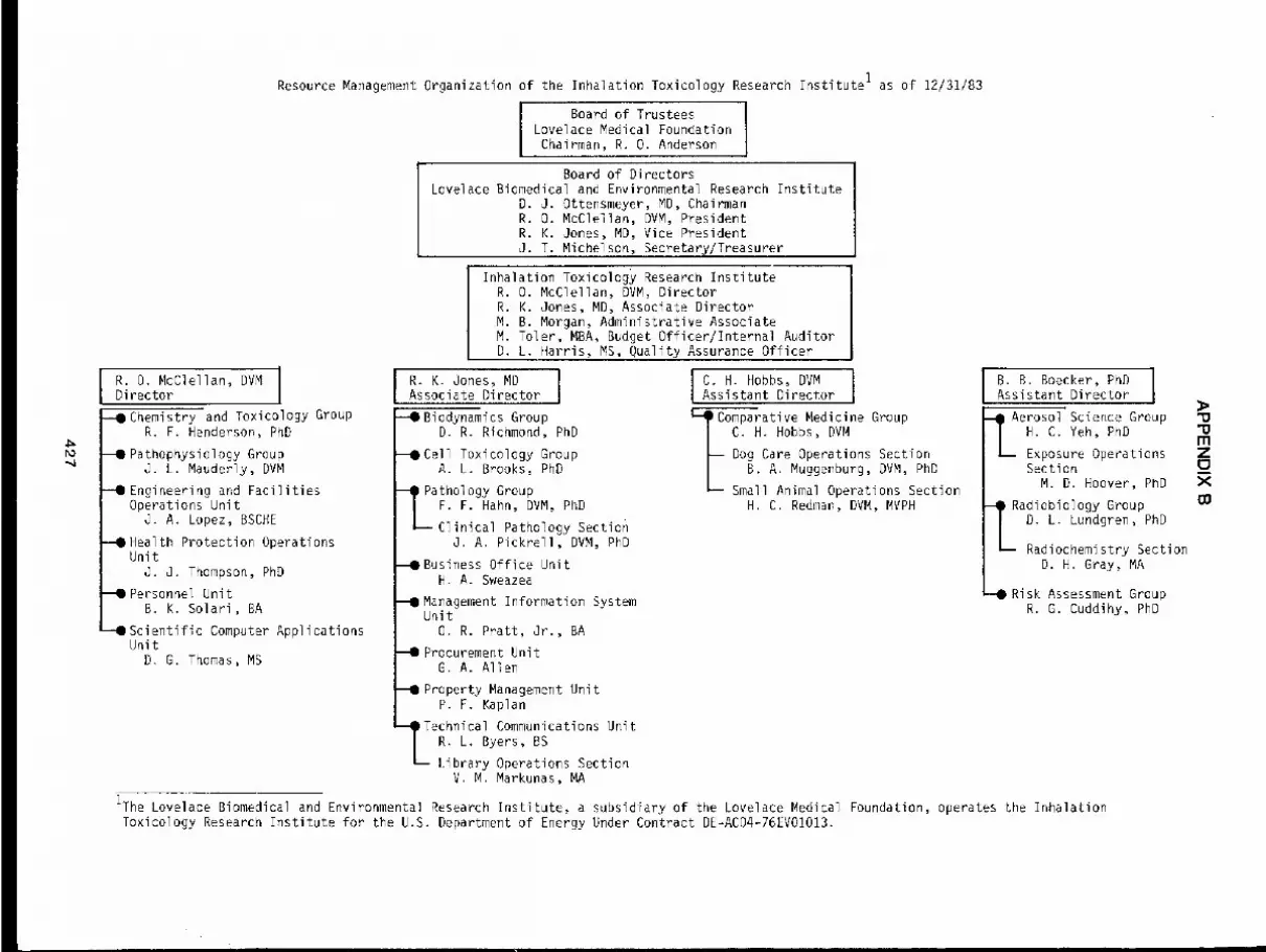
R. O. McClellan, DVMDi rector
--IChemistry and Toxicology GroupR. F. Henderson, PhD
--II Pathophysiology GroupJ. L. Mauderly, DVM
--llEngineering and FacilitiesOperations Unit
J. A. Lopez, BSCHE
--0 Health Protection OperationsUnit
J. J. Thompson, PhD
Personnel UnitB. K. Sol ari, BA
Scientific Computer ApplicationsUnit
D. G. Thomas, MS
Resource Management Organization of the Inhalation Toxicology Research Institute I as of 12/31/83
Board of TrusteesLovelace Medical FoundationChairman, R. O. Anderson
Board of DirectorsLovelace Biomedical and Environmental Research Institute
D. J. Ottensmeyer, MD, ChairmanR. O. McClellan, DVM, PresidentR. K. Jones, MD, Vice PresidentJ. T. Michelson, Secretary~Treasurer
Inhalation Toxicology Research InstituteR. O. McClellan, DVM, DirectorR. K. Jones, MD, Associate DirectorM. B. Morgan, Administrative AssociateM. Toler, MBA, Budget Officer/Internal AuditorD. L. Harris, MS, Quality Assurance Officer
R. K. Jones, MD I C. H. Hobbs, DVMAssociate Director I Assistant Director
--OBiodynamics Group 1--~Comparative Medicine GroupD. R. Richmond, PhD I C. H. Hobbs, DVMI
~Cell Toxicology Group ~ Dog Care Operations SectionA. L. Brooks, PhD I B.A. Muggenburg, DVM, PhD
IL- Small Animal Operations Section
T_Pathology GroupF. F. Hahn, DVM, PhD H.C. Redman, DVM, MVPHClinical Pathology Section
J. A. Pickrell, DVM, PhD
~Business Office UnitH. A. Sweazea
--1|Management Information SystemUnit
O. R. Pratt, Jr., BA
~Procurement UnitG. A. Allen
~Property Management UnitP. F. Kaplan
I Technical Communications UnitR. L. Byers, BS
Library Operations SectionV. M. Markunas, MA
1The Lovelace Biomedical and Environmental Research Institute, a subsidiary of the Lovelace Medical Foundation, operates the InhalationToxicology Research Institute for the U.S. Department of Energy Under Contract DE-ACO4-76EV01013.
B. B. Boecker, PhDAssistant Director
~Aerosoi’Science Group "Q
LH" C’ Yeh’ PhDExposure Operations ZSection ~__
M. D. Hoover, PhD X
I Radiobiology GroupD. L. L~ndgren, PhD
Radiochemistry SectionD. H. Gray, MA
-ORisk Assessment GroupR. G. Cuddihy, PhD
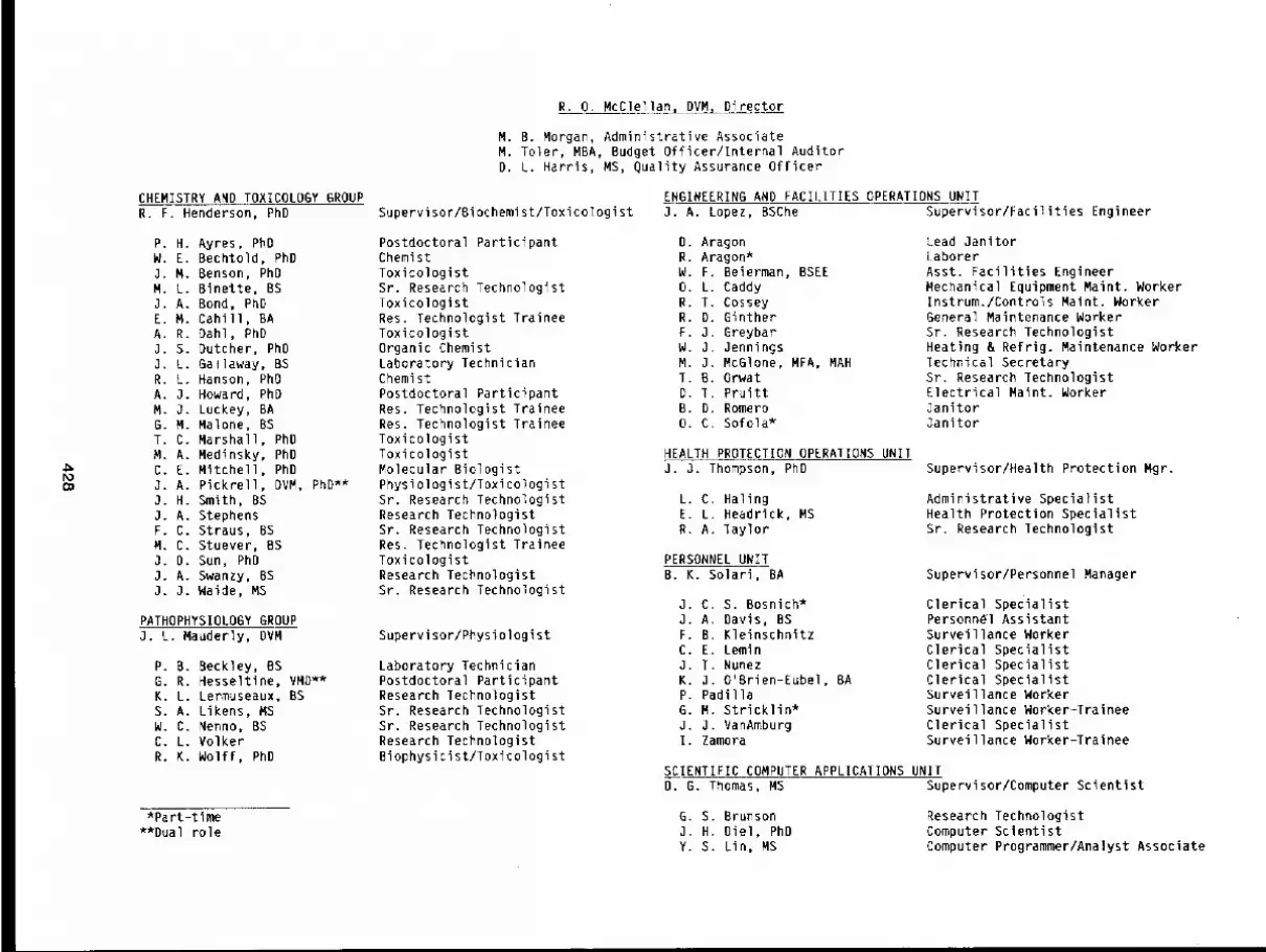
CHEMISTRY AND TOXICOLOGY GROUPR. F. Henderson, PhD
P. H. Ayres, PhOW. E. Bechtold, PhD3. M. Benson, PhDM. L. Binette, BSJ. A. Bond, PhDE. M. Cahill, BAA. R. Dahl, PhDJ. S. Dutcher, PhDJ. L. Gallaway, BSR. L. Hanson, PhOA. J. Howard, PhDM. J. Luckey, BAG. M. Malone, BST. C. Marshall, PhDM. A. Medinsky, PhDC. E. Mitchell, PhDJ. A. Pickrell, DVM, PhD**J. H. Smith, BSJ. A. StephensF. C. Straus, BSM. C. Stuever, BSJ. D. Sun, PhDJ. A. Swanzy, BSJ. J. Waide, MS
PATHOPHYSIOLDGY GROUPJ. L. Mauderly, DVM
P. B. Beckley, BSG. R. Hesseltine, VMD**K. L. Lermuseaux, BSS. A. Likens, MSW. C. Nenno, BSC. L. VolkerR. K. Wolff, PhD
*Part-time**Dual role
R. O. McClellan, DVM, DireEtor
M. B. Morgan, Administrative AssociateM. Toler, MBA, Budget Officer/Internal AuditorD. L. Harris, MS, Quality Assurance Officer
Supervisor/Biochemist/ToxicologistENGINEERING AND FACILITIES OPERATIONS UNITJ. A. Lopez, BSChe Supervisor/Facilities Engineer
Postdoctoral ParticipantChemistToxicologistSr. Research TechnologistToxicologistRes. Technologist TraineeToxicologistOrganic ChemistLaboratory TechnicianChemistPostdoctoral ParticipantRes. Technologist TraineeRes. Technologist TraineeToxicologistToxicologistMolecular BiologistPhysiologist/ToxicologistSt. Research TechnologistResearch TechnologistSr. Research TechnologistRes. Technologist TraineeToxicologistResearch TechnologistSr. Research Technologist
Supervisor/Physiologist
Laboratory TechnicianPostdoctoral ParticipantResearch TechnologistSr. Research TechnologistSr. Research TechnologistResearch TechnologistBiophysicist/Toxicologist
D. AragonR. Aragon*W. F Beierman, BSEEO. LR. TR. DF. JW. 3M. 3T. BD. TB. DO. C
CaddyCosseyGintherGreybar3enningsMcGlone, MFA, MAHOrwatPruittRomeroSofola*
HEALTHPROTECTION OPERATIONS UNIT3. J. Thompson, PhD
L. C. HalingE. L. Headrick, MSR. A. Taylor
PERSONNEL UNITB. K. Solari, BA
J. C S. Bosnich*O. A Davis, BSF. B KleinschnitzC. E LeminJ. T NunezK. J O’Brien-Eubel, BAP. PadillaG. M. Stricklin*J. J. VanAmburgI. Zamora
Lead JanitorLaborerAsst. Facilities EngineerMechanical Equipment Maint. WorkerInstrum./Controls Maint. WorkerGeneral Maintenance WorkerSr. Research TechnologistHeating & Refrig. Maintenance WorkerTechnical SecretarySr. Research TechnologistElectrical Maint. WorkerJanitorJanitor
Supervisor/Health Protection Mgr.
Administrative SpecialistHealth Protection SpecialistSr. Research Technologist
Supervisor/Personnel Manager
Clerical SpecialistPersonn~l AssistantSurveillance WorkerClerical SpecialistClerical SpecialistClerical SpecialistSurveillance WorkerSurveillance Worker-TraineeClerical SpecialistSurveillance Worker-Trainee
SCIENTIFIC COMPUTER APPLICATIONS UNITD. G. Thomas, MS Supervisor/Computer Scientist
G. S. Brunson3. H. Oiel, PhDY. S. Lin, MS
Research TechnologistComputer ScientistComputer Programmer/Analyst Associate
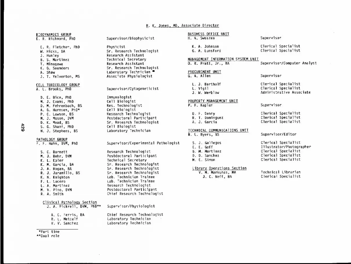
R. K. Jones, MD, Associate Director
BIODYNAMICS GROUPD. R. Richmond, PhD Supervisor/Biophysicist
BUSINESS OFFICE UNITH. A. Sweazea Supervisor
E. R. Fletcher, PhDW. Hicks, BAJ. HunleyB. S. MartinezT. MinagawaK. G. SaundersA. ShawO. T. YeIverton, MS
PhysicistSr. Research TechnologistResearch AssistantTechnical SecretaryResearch AssistantSr. Research TechnologistLaboratory Technician oAssociate Physiologist
K. A. JohnsonG. A. Lunsford
MANAGEMENT INFORMATION SYSTEM UNITO. R. Pratt, Jr., BA
PROCUREMENT UNITG. A. Allen
Clerical SpecialistClerical Specialist
Supervisor/Computer Analyst
Supervisor
(o
CELL TOXICOLOGY GROUPA. L. Brooks, PhD
D. E. Bice, PhDM. J. Evans, PhDD. M. Fehrenbach, BSA. G. Harmsen, ghD*P. C. Lawson, BSM. J. Mason, DVMD. K. Mead, BSS. G. Shami, PhDN. J. Stephens, BS
PATHOLOGY GROUPF. F. Hahn, DVM, PhD
S. C. BarnettM J. Behr, OVMK L. EplerK M. Garcia, BAJ A. Hogan, BAR J. Jaramillo, BSK N. KnightonF L. LuceroL A. MartinezM V. Pino, DVMR A. Smith
Supervisor/Cytogeneticist
ImmunologistCell BiologistRes. TechnologistCell BiologistResearch TechnologistPostdoctoral ParticipantSr. Research TechnologistCell BiologistLaboratory Technician
Supervisor/Experimental Pathologist
Research TechnologistPostdoctoral ParticipantTechnical SecretarySr. Research TechnologistSr. Research TechnologistSr. Research TechnologistLab. Technician TraineeLab. Technician TraineeResearch TechnologistPostdoctoral ParticipantChief Research Technologist
L. J. BertholfL. VigilJ. W. Werblow
PROPERTY MANAGEMENT UNITP. F. Kaplan
D. F. DennyR. T. DominguezA. J. Garcia
TECHNICAL COMMUNICATIONS UNITR. L. Byers, BS
S. J. GallegosE. E. GoffB. M. MartinezD. D. SanchezM. E. Simon
Library Operations SectionV. M. Markunas, MA
J. C. Neff, BA
Clerical SpecialistClerical SpecialistAdministrative Associate
Supervisor
Clerical SpecialistClerical SpecialistClerical Specialist
Supervisor/Editor
Clerical SpecialistIllustrator/PhotographerClerical SpecialistClerical SpecialistClerical Specialist
Technical LibrarianClerical Specialist
Clinical Pathology SectionJ. A. Pickrell, DVM, PhD** Supervisor/Physiologist
A. C. Ferris, BAR. L. MetcalfV. V. Sanchez
Chief Research TechnologistLaboratory TechnicianLaboratory Technician
*Part time**Dual role
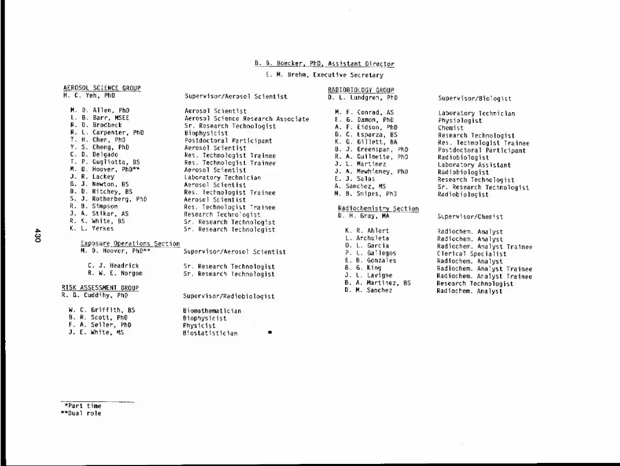
(,,0o
AEROSOL SCIENCE GROUPH. C. Yeh, PhD
M. D. Allen, PhDE. B. Barr, MSEER. D. BrodbeckR. L. Carpenter, PhDT. H. Chen, PhDY S. Cheng, PhDC D. DelgadoT P. Gugliotta, BSM O. Hoover, PhD**J R. LackeyG 3. Newton, BSB D. Ritchey, BSS. 3. Rothenberg, PhDR. B. SimpsonJ. A. Stikar, ASR. K. White, BSK. L. Yerkes
E__~posure Operations SectionM. D. Hoover, PhD**
C. J. HeadrickR. W. E. Norgon
RISK ASSESSMENT GROUPR. G. Cuddihy, PhD
W. C. Griffith, BSB. R. Scott, PhDF. A. Seller, PhD3. E. White, MS
B. B. Boecker, PhO, Assistant Director
E. M. Brehm, Executive Secretary
Supervisor/Aerosol Scientist
Aerosol ScientistAerosol Science Research AssociateSr. Research TechnologistBiophysicistPostdoctoral ParticipantAerosol ScientistRes. Technologist TraineeRes. Technologist TraineeAerosol ScientistLaboratory TechnicianAerosol ScientistRes. Technologist TraineeAerosol ScientistRes. Technologist TraineeResearch TechnologistSr. Research TechnologistSt. Research Technologist
Supervisor/Aerosol Scientist
Sr. Research TechnologistSr, Research Technologist
Supervisor/Radiobiologist
BiomathematicianBiophysicistPhysicistBiostatistician
RADIOBIOLOGY GROUPD. L. Lundgren, PhD
M. F. Conrad, ASE. G. Damon, PhOA. F. Eidson, PhDD. C. Esparza, BSK. G. Gillett, BAB. J. Greenspan, PhDR. A. Guilmette, PhDJ. L. MartinezJ. A. Mewhinney, PhOE. J. SalasA. Sanchez, MSM. B. Snipes, PhD
Radiochemistry SectionD. H. Gray, MA
K. R. AhlertL. Archu!eta0. L. GarciaP. L. GallegosE. B. GonzalesB. G. KingJ. L. LavigneB. A. Martinez, BSD. M. Sanchez
Supervisor/Biologist
Laboratory TechnicianPhysiologistChemistResearch TechnologistRes. Technologist TraineePostdoctoral ParticipantRadiobiologistLaboratory AssistantRadiobiologistResearch TechnologistSr. Research TechnologistRadiobiologist
Supervisor/Chemist
Radiochem. AnalystRadiochem. AnalystRadiochem. Analyst TraineeClerical SpecialistRadiochem. AnalystRadiochem. Analyst TraineeRadiochem. Analyst TraineeResearch TechnologistRadiochem. Analyst
*Part time**Dual role
COMPARATIVE MEDICINE GROUPC. H. Hobbs, DVM**
T. R. Henderson, PhDG. R. Hesseltine, VMD**B. A. Muggenburg, DVM, PhD**H. C. Redman, DVM, MPVN**
Dog Care Operations SectionB. A. Muggenburg, DVM, PhD**
C. W. Burns, BAF. L. DollahiteJ. M. DuranK. J. Everhart*E. GarciaT. M. Grein-PeaseD. A. Hicks*M. HoganJ. P. Johnson*R. LindsayA. D. MurrinL. M. Pistorio*C. G. RomeroC. RoqueR. 8. SedilloL. C. SwansonL. R. R. TaplinV. A. WhiteS. L. WilsonM. L. Wright
C. H. Hobbs, DVM, Assistant Director
Supervisor/ToxicologistSmall Animal Operations SectionH. C. Redman, DVM, MPVM**
BiochemistPostdoctoral ParticipantPhysiologistResearch Veterinarian
Supervisor/Physiologist
Research TechnologistSr. Animal TechnicianSr. Animal TechnicianAnimal CaretakerAnimal TechnicianAnimal CaretakerAnimal CaretakerChief Animal TechnologistAnimal CaretakerAnimal TechnicianClerical SpecialistAnimal CaretakerAnimal TechnicianAnimal TechnicianAnimal lechniclanAnimal TechnicianSr. Animal TechnicianLaboratory TechnicianAnimal CaretakerLaboratory Technician
A. J. AlbertS. L. AllenR. L. BonchoskyM. A. CasausD. M. ChavezC. L. DavisC. A. ElliottP. R. GenthnerP. A. GentiliniJ. R. JewellR. A. LewisS. McAnallyM. H. MarshallA. G. MoraJ. RenardR. M. RuettenJ. S. Salazar, ASM. A. Sanchez*J. L. SargeantJ. A. SwansonS. WalkerC. C. Ynostroza, AS
Supervisor/Research Veterinarian
Animal CaretakerAnimal CaretakerAnimal CaretakerAnimal CaretakerAnimal TechnicianAnimal CaretakerAnimal TechnicianAnimal CaretakerAnimal CaretakerAnimal CaretakerAnimal TechnicianAnimal CaretakerAnimal TechnicianAnimal CaretakerSr. Animal TechicianAnimal CaretakerChief Animal TechnologistAnimal CaretakerAnimal CaretakerAnimal TechnicianAnimal TechnicianAnimal Technician
*Part time**Dual role

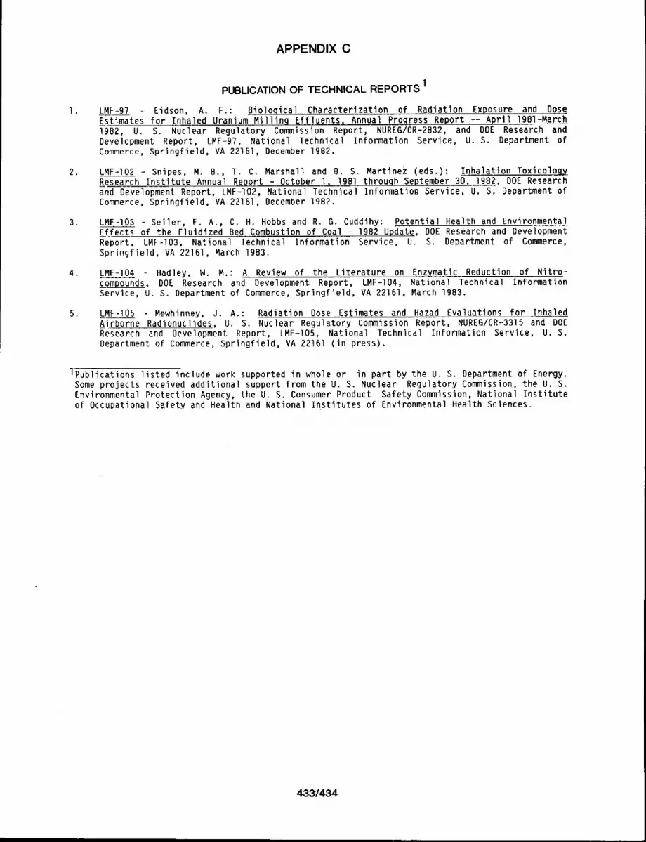
APPENDIX C
PUBLICATION OF TECHNICAL REPORTS 1
1 ¯ LMF-97 Eidson, A. F.: BioloQical Characterization of Radiation Exposure and DoseEstimates for Inhaled Uranium Milling Effluents~ Annual Progre.ss Report -- April Ig81-March].982, U. S. Nuclear Regulatory Commission Report, NUREG/CR-2B32, and DOE Research andDevelopment Report, LMF-97, National Technical Information Service, U. S. Department ofCommerce, Springfield, VA 22161, December lgB2.
2. LMF-I02 - Snipes, M. B., T. C. Marshall and B. S, Martinez (eds.): Inhalation Toxicolog~Research Institute Annual ~.~.9.£t - October l~1981 throuQh..September 30, 1982, DOE Researchand Development Report, LMF-102, National Technical Informatio~ Service, U. S. Department ofCommerce, Springfield, VA 22161, December 1982.
3. LMF-I03 - Seiler, F. A., C. H. Hobbs and R. G. Cuddihy: Potential Health and EnvironmentalEffects of the Fluidized Bed Combustion of Coal - 1982 Update, DOE Research and DevelopmentReport, LMF.-I03, National Technical Information Service, U. S. Department of Commerce,Springfield, VA 22161, March 1983.
LMF-104 Hadley, W. M.: A Review of the Literature on Enzymatic Reduction of Nitro-compounds, DOE Research and Development Report, LMF-I04, National Technical InformationService, U. S. Department of Commerce, Springfield, VA 22161, March 1983.
5, LMF-I05 Mewhinney, 3. A.: Radiation Dose Estimates and Hazad Evaluations for InhaledAirborne Radionuclides, U. S. Nuclear Regulatory Commission Report, NUREG/CR-3315 and DOEResearch and Development Report, LMF-I05, National Technical Information Service, U. S.Department of Commerce, Springfield, VA 22161 (in press).
lPublications listed include work supported in whole or in part by the U. S. Department of Energy.Some projects received additional support from the U. S. Nuclear Regulatory Commission, the U. S.Environmental Protection Agency, the U. S. Consumer Product Safety Commission, National Instituteof Occupational Safety and Health and National Institutes of Environmental Health Sciences.
433/434

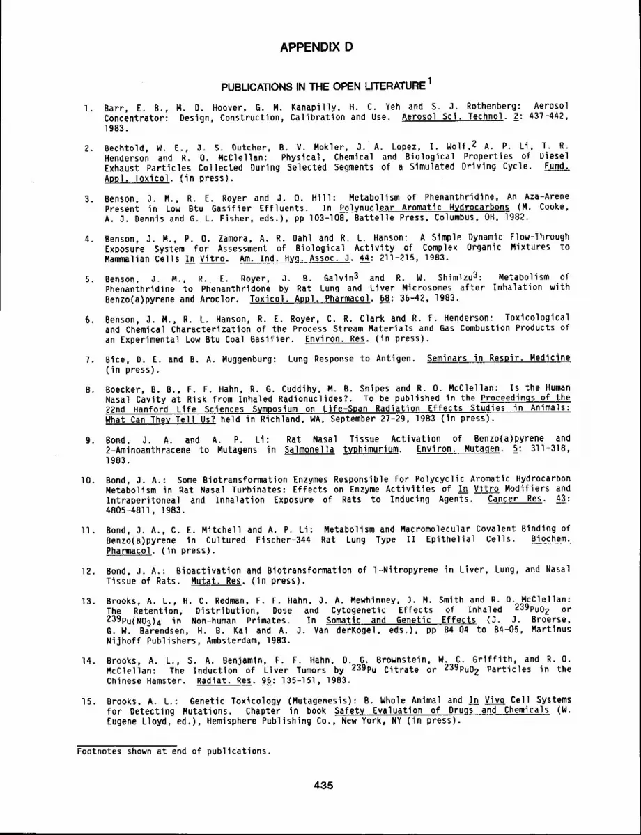
APPENDIX D
PUBLICATIONS IN THE OPEN LITERATURE1
i. Barr, E. B,, M. O. Hoover, 6, M. Kanapilly, H. C. Yeh and S. J. Rothenberg: AerosolConcentrator: Design, Construction, Calibration and Use. Aerosol Sci. Technol. 2: 437-442,1983.
2. Bechtold, W. E., 3. S. Dutcher, B. V. Mokler, 3. A. Lopez, I. Wolf, 2 A. P. Li, I. R.Henderson and R. O. McClellan: Physical, Chemical and Biological Properties of DieselExhaust Particles Collected During Selected Segments of a Simulated Driving Cycle. Fund.Appl. Toxicol. (in press),
3. Benson, 3. M., R. E. Royer and J. O. Hill: Metabolism of Phenanthridine, An Aza-ArenePresent in Low Btu 6asifier Effluents. In Polynuclear Aromatic H~drocarbons (M. Cooke,A. J. Dennis and 6. L. Fisher, eds,), pp 103-10B, Battelle Press, Columbus, OH, 1982,
4. Benson, J. M., P. O. Zamora, A. R. Dahl and R. L. Hanson: A Simple Dynamic Flow-lhroughExposure System for Assessment of Biological Activity of Complex Organic Mixtures toMammalian Cells In Vitro. Am. Ind. Hyg~ Assoc. J. 44: 211-215, 1983.
5. Benson, 3. M., R. E. Royer, J. B. Galvin 3 and R. W. Shimlzu3: Metabolism ofPhenanthridine to Phenanthridone by Rat Lung and Liver Microsomes after Inhalation withBenzo(a)pyrene and Aroclor, Toxicol~Appl. Pharmacol. 68: 36-42, 1983.
6, Benson, J. M., R. L. Hanson, R. E. Royer, C. R. Clark and R. F. Henderson: Toxicologicaland Chemical Characterization of the Process Stream Materials and Gas Combustion Products ofan Experimental Low Btu Coal Basifier. Environ. Res. (in press).
7. Bice, D. E. and B. A. Muggenburg: Lung Response to Antigen. Seminars in Respir, Medicine(in press).
8. Boecker, B. B., F. F, Hahn, R. G. Cuddihy, M. B. Snipes and R. O. McClellan: Is the HumanNasal Cavity at Risk from Inhaled Radionuclides?. To be published in the Proceedings of the22rid Hanford Life Sciences Symposium on Life-Span Radiation Effects Studies in Animals:What Can They Tell Us? held in Richland, WA, September 27-29, 1983 (in press).
9. Bond, J. A. and A. P. Li: Rat Nasal Tissue Activation of Benzo(a)pyrene and2-Aminoanthracene to Mutagens in Salmonella typhimurium. Environ. Mutagen. 5: 311-318,1983.
lO. Bond, J. A.: Some Biotransformation Enzymes Responsible for Polycyclic Aromatic HydrocarbonMetabolism in Rat Nasal Turhinates: Effects on Enzyme Activities of In Vitro Modifiers andIntraperitoneal and Inhalation Exposure of Rats to Inducing Agents. Cancer Res. 43:4805-4811, 1983.
II. Bond, J. A,, C. E. Mitchell and A. P. Li: Metabolism and Macromolecular Covalent Binding ofBenzo(a)pyrene in Cultured Fischer-344 Rat Lung Type Ii Epithelial Cells. Biochem.Pharmacol. (in press).
12. Bond, 3. A.: Bioactivation and Biotransformation of l-Nitropyrene in Liver, Lung, and NasalTissue of Rats. Mutat. Res. (in press).
13. Brooks, A. L., H. C. Redman, F. F. Hahn, 3. A. Mewhinney, 3. M. Smith and R. O. McClellan:The Retention, Distribution, Dose and Cytogenetic Effects of Inhaled 239pu02 or239PU(N03)4 in Non=human Primates. In Somatic and Genetic Effects (J. J. Broerse,G.W. Barendsen, H. B. Kal and A. 3. Van derKogel, eds.), pp B4-04 to B4-05, MartinusNijhoff Publishers, Ambsterdam, 1983.
14, Brooks, A. L., S. A. Benjamin, F. F. Hahn, D. G, Brownstein, W. C. Griffith, and R. O.McClellan: The Induction of Liver Tumors by 239pu Citrate or 239pu02 Particles in theChinese Hamster, Radiat. Res. 95: 135-151, 1983.
15. Brooks, A. L,: Genetic Toxicology (Mutagenesis): B. Whole Animal and In Vivo Cell Systemsfor Detecting Mutations. Chapter in book Safety Evaluation of Drugs and Chemicals (W.Eugene Lloyd, ed.), Hemisphere Publishing Co., New York, NY (in press).
Footnotes shown at end of publications.
435
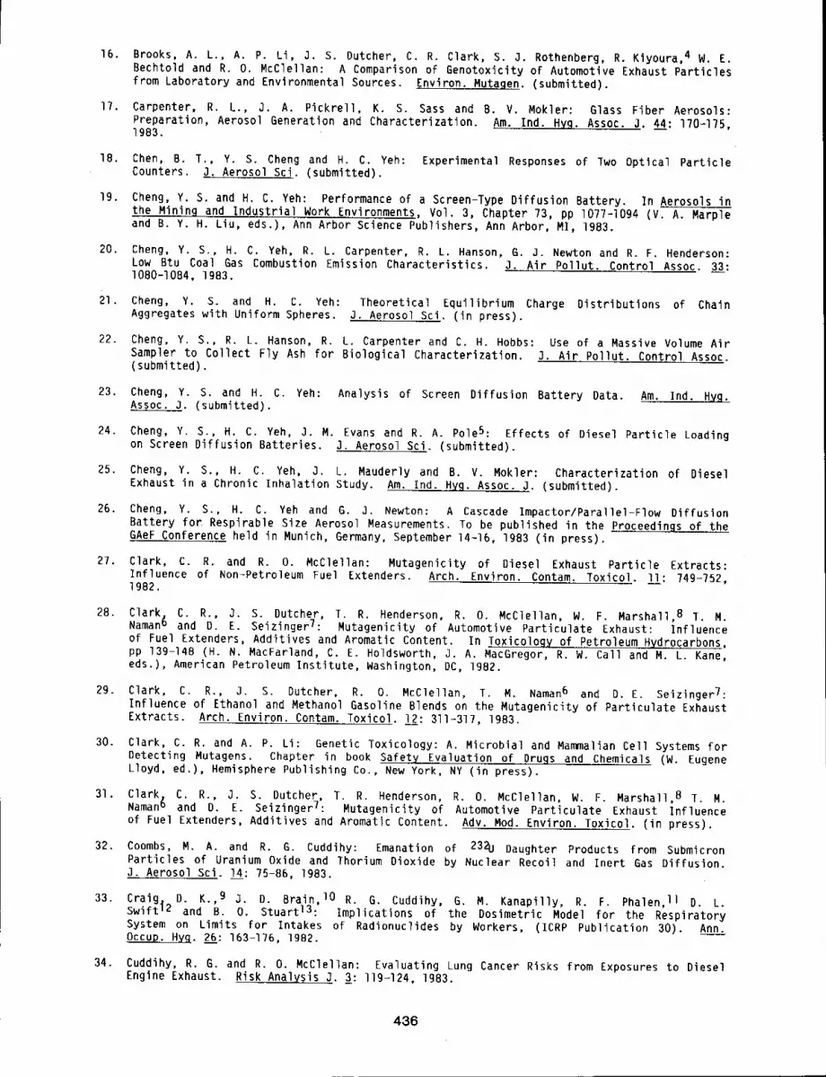
16. Brooks, A. L., A, P. Li, J. S. Dutcher, C. R. Clark, S. J. Rothenberg, R. Kiyoura, 4 W. E.Bechtold and R. O. McClellan: A Comparison of Genotoxicity of Automotive Exhaust Particlesfrom Laboratory and Environmental Sources. Environ. Mutagen. (submitted).
17. Carpenter, R. L., J. A. Pickrell, K. S. Sass and B. V. Mokler: Glass Fiber Aerosols:Preparation, Aerosol Generation and Characterization. Am. Ind. H~g. Assoc. j. 44: 170=175,1983.
18. Chen, B. T., Y. S. Cheng and H. C. Yeh: Experimental Responses of Two Optical ParticleCounters, 3. Aerosol Sci. (submitted).
19. Cheng, Y. S. and H. C. Yeh: Performance of a Screen-Type Diffusion Battery. In Aerosols inthe Mining and Industrial Work Environments, Vol. 3, Chapter 73, pp I077-I094 (V. A. Marpleand B. Y. H. Liu, eds.), Ann Arbor Science Publishers, Ann Arbor, MI, 1983.
20. Cheng, Y. S., H. C. Yeh, R. L. Carpenter, R. L. Hanson, G. 3. Newton and R. F. Henderson:Low Btu Coal Gas Combustion Emission Characteristics. J. Air Pollut. Control ASSOC. 33:1080-10B4, 1983.
21. Cheng, Y. S. and H. C. Yeh: Theoretical Equilibrium Charge Distributions of ChainAggregates with Uniform Spheres. J. Aerosol Sci. (in press).
22. Cheng, Y. S., R. L. Hanson, R. L. Carpenter and C. H. Hobbs: Use of a Massive Volume AirSampler to Collect Fly Ash for Biological Characterization. J. Air Pollut. Control Assoc.(submitted).
23, Cheng, Y. S. and H. C. Yeh: Analysis of Screen Diffusion Battery Data. Am. Ind. Hyg.Assoc. J. (submitted).
24. Cheng, Y. S., H. C. Yeh, J. M. Evans and R. A. Pole5: Effects of Diesel Particle Loadingon Screen Diffusion Batteries. J. Aerosol Sci. (submitted).
25. Cheng, Y. S., H. C. Yeh, J. L. Mauderly and B. V. Mokler: Characterization of DieselExhaust in a Chronic Inhalation Study. Am. ind. Hyg. Assoc. J. (submitted).
26. Cheng, Y. S., H. C. Yeh and G. J. Newton: A Cascade Impactor/Parallel-Flow DiffusionBattery for Respirable Size Aerosol Measurements. To be published in the Proceedings of theGAeF Conference held in Munich, Germany, September 14-16, 1983 (in press).
27. Clark, C. R. and R. O. McClellan: Mutagenicity of Diesel Exhaust Particle Extracts:Influence of Non-Petroleum Fuel Extenders. Arch. Environ. Contam. Toxicol. l_=ll: 749-752,1982.
28. Clark. C. R., J. S. Dutcher, T. R. Henderson, R. O. McClellan, W. F. Marshall 8 I. M.Naman6 and D. E. SeizingerT: Mutagenicity of Automotive Particulate Exhaust: Influenceof Fuel Extenders, Additives and Aromatic Content. In Toxicology of Petroleum Hydrocarbons,pp 139-14B (H. N. MacFarland, C. E. Holdsworth, J. A. MacGregor, R. W. Call and M. L. Kane,eds.), American Petroleum Institute, Washington, DC, 1982.
29. Clark, C. R., J. S. Dutcher, R. O. McClellan, T. M. Naman6 and D.E. SeizingerT:Influence of Ethanol and Methanol Gasoline Blends on the Mutagenicity of Particulate ExhaustExtracts. Arch. Environ. Contam. Toxicol. L2: 311-317, 1983.
30. Clark, C. R. and A. P. Li: Genetic Toxicology: A. Microbial and Mammalian Cell Systems forDetecting Mutagens. Chapter in book Safety Evaluation of Drugs and Chemicals (W. EugeneLloyd, ed.), Hemisphere Publishing Co., New York, NY (in press).
31. Clark. C. R., J. S. Dutcher, T. R. Henderson, R. O. McClellan, W. F. Marshall 8 ~. M.Naman6 and D. E. SeizingerT: Mutagenicity of Automotive Particulate Exhaust Influenceof Fuel Extenders, Additives and Aromatic Content. Adv. Mod. Environ. Toxicol. (in press).
32. Coombs, M. A. and R. G. Cuddihy: Emanation of 232U Daughter Products from SubmicronParticles of Uranium Oxide and Thorium Dioxide by Nuclear Recoil and Inert Gas Diffusion.J. Aerosol Sci. lj: 75-86, 1983.
33. Craig, D. K. 9 j. D. Brain, lO R. G. Cuddihy, G. M. Kanapilly, R. F. Phalen, II D. L.Swift 12 and B. O. Stuartl3: Implications of the Dosimetric Model for the RespiratorySystem on Limits for Intakes of Radionuclides by Workers, (ICRP Publication 30). Ann.Occup. Hyq. 26: 163-176, 1982.
34. Cuddihy, R. G. and R. O. McClellan: Evaluating Lung Cancer Risks from Exposures to DieselEngine Exhaust. Risk Analysis J. 3: I19-124, 1983.
436
35. Cuddihy, R. G.: Risk Assessment Relationships for Evaluating Effluents from CoalIndustries. The Science of the Total Environ. 2_B_B: 479-492, 1983.
36. Cuddihy, R. G., W. C. Griffith and R. O. McClellan: Health Risks from Light Duty DieselVehicles. Environ. Sci. Technol. (in press).
37. Dahl, A: R., S. A. Felicetti and B. A. Muggenburg: Clearance of Sulfuric Acid-lntroducedS 3b
from the Respiratory Tracts of Rats, Guinea Pigs and Dogs Following Inhalation orInstillation. J. Toxicol. Environ. Health 3: 293-297, 1983.
38. Dahl, A. R., M. B. Snipes, B. A. Muggenburg and 1. C. Young5: Deposition of Sulfuric AcidMists in the Respiratory Tract of Beagle Dogs. J. Toxicol. Environ. Health I I: 141-150,1983.
39. Dahl, A. R. and W. M. Hadley: Formaldehyde Production Promoted by Rat Nasal CytochromeP-450 Dependent Monooxygenases with Nasal Decongestants, Essences, Solvents, Air Pollutants,Nicotine and Cocaine as Substrates. Toxicol. Appl. Pharmacol. 67: 200-205, 1983.
40. Dahl, A. R. and W. C. Griffith: Deposition of Sulfuric Acid Mist in the Respiratory Tractsof Guinea Pigs and Rats. J. Toxicol. Environ. Health 12: 371-384, 1983.
41. Dahl, A. R., J. M. Benson, R. L. Hanson and S. J. Rothenberg: The Fractionation ofEnvironmental Samples According to Volatility by Vacuum Line-Cryogenic Distillation. Am.Ind. H~g. Assn. J. (in press).
42. Damon, E. G., B. V. Mokler and R. K. Jones: Infuence of Elastase-lnduced Emphysema and theInhalation of an Irritant Aerosol on Deposition and Retention of an Inhaled InsolubleAerosol in Fischer-344 Rats. Toxicol. Appl. Pharmacol. 67: 322-330, 1983.
43. Damon, E. G., A. F. Eidson, F. F. Hahn, W. C. Griffith and R. A. Guilmette: Comparison ofEarly Lung Clearance of Yellowcake Aerosols in Rats with In Vitro Dissolution and InfraredAnalysis. Health Phys. (in press).
44. Damon, E. G., B. V. Mokler, T. R. Henderson and R. K. Jones: Lung Retention of InhaledAluminum Chlorhydrate in Syrian Hamsters. Arch. Environ. Health (submitted).
45. Diel, J. H.: Microdosimetry of Internally Deposited Radionuclides. Int. J. Appl. Radiat.Isot. 33: 967-979, 1982.
46. Diel, J. H. and D. L. Lundgren: Repeated Inhalation Exposure of Beagle Dogs to 239pu02:Retention and Translocation. Health Ph__hy_~. 43: 655-662, 1982.
47. Diel, J. H. and J. A. Mewhinney: Fragmentation of Inhaled 238pu02 Particles in Lung.Health Phys. 44: 135-143, Ig83.
48. Diel, J. H., R. A. Guilmette, F. F. Hahn, D. L. Lundgren, J. A. Mewhinney, B. A. Muggenburg,M. B. Snipes, B. B. Boecker and R. O. McClellan: Dosimetry of Internally DepositedRadionuclides in Lung and Its Usefulness in Predicting Biological Effects. In CurrentConcepts in Lun9 Dosimetry (D. R. Fisher, ed.), pp 18-28, CONF-820492-Pt. l, PNL-SA-l12049,Technical Information Center, Springfield, VA 22161, February 1983.
49. Diel, J. H. and J. A. Mewhinney: Lung lumor Induction in Syrian Hamsters withInternally-Deposited Particulate Pu: A Synthesis Based on Microscopic Dose Distribution.Radiat. Environ. Biophys. (in press).
50. Diel, J. H., J. A. Mewhinney and R. A. Guilmette: Microscopic Dose Distribution Around PuO2Particles in Lungs of Hamsters, Rats and Dogs. Radiat. Environ. Biophy_~. (submitted).
51. Outcher, J. S. and C. E. Mitchell: Distribution and Elimination of (14C)--Phenanthridonein Rats After Inhalation. In Polynuclear Aromatic Hydrocarbons, (M. W. Cooke and A. J.Dennis, eds), pp 427-435, Battelle Press, Columbus, OH, 1982.
52. Dutcher, J. S., R. E. Rower, C. E. Mitchell and A. R. Dahl: Fractionation of Coal lar fromLow Btu Gasification. In Advanced Techniques in Synthetic Fuels Analysis, pp 133-144,(Wright, Weimer, Felix, eds.), Department of Energy Report, lechnical Information Center,Springfield, VA 22161, 1983.
53. Dutcher, J. S., R. E. Royer, J. O. Hill, C. E. Mitchell and R. L. Hanson: Identification ofComponents of Biologically Active Fractions of Low Btu Gasifier Coal Tars. In AdvancedTechniques in Synthetic Fuels Analysis, pp 12-23 (Wright, Weimer, Felix, edd.), Departmentof Energy Report. Technical Information Center, Springfield, VA 22161, 1983.
437
54. Dutcher, J. S. and C. E. Mitchell: Distribution and Elimination of Inhaled Phenanthridonein Fischer-344 Rats. J. Toxicol. Environ. Health (in press).
55. Dutcher, 3. S., J. D. Sun, J. A. Lopez, I. Wolf, 2 R. K. Wolff and R. O. McClellan:Generation’and Characterization of Radiolabeled Diesel Exhaust. Am. Indus. Hyg. Assoc. 3.(submitted).
56. Eidson, A. F. and J. A. Mewhinney: In Vitro Dissolution of Respirable Aeosols of IndustrialUranium and Plutonium Mixed Oxide Nuclear Fuels. Health Phys. 45: I023-I038, 1983.
57. Eidson, A. F. and W. C. Griffith: Techniques for Yellowcake Dissolution Studies In Vitroand Their Use in Bioassay Interpretation. Health Phys. (in press).
58. Eidson, A. F. and E. G. Damon: Predicted Deposition Rates of Uranium Yellowcake AerosolsSampled in Uranium Mills. Health Phys. (in press).
59. Greene, S. A., R. K. Wolff, F. F. Hahn, R. F. Henderson, 3. LC Mauderly and D. L. Lundgren:Sulfur Dioxide-lnduced Chronic Bronchitis in Beagle Dogs. 3. Toxicol. Environ. Health (inpress).
60. Greene, S. A., R. K. Wolff, D. L. Lundgren and W. C. Griffith: A Comparison of Acepromazineand Xylazine Iranquilization Prior to Halothane Anesthesia for Measurement of IrachealMucous Clearance. 3. Appl. Physiol. (submitted).
61. Gregory, R. A., J. A. Pickrell, F. F. Hahn and C. H. Hobbs: Pulmonary Effects ofIntermittent Subacute Exposure to Low-Level Nitrogen Dioxide. 3. Toxicol. Environ. Healthl_l_: 405-414, 1983.
62. Gregory, R. E., L. R. Wilson and M. C. Godwin: A Computer Interfaced On-Line Serial DataAcquisition and Control System: Its Application to an Automated Inhalation ExposureSystem. Am. Ind. Hyg. Assoc. J. (in press).
63. Griffis, L. C., 3. A. Pickrell, R. L. Carpenter, R. K. Wolff, S. 3. McAllen 5 and K. L.Yerkes: Deposition of Crocidolite Asbestos and Glass Microfibers Inhaled by the BeagleDog. Am. Ind. Hyg. Assoc. 3. 44: 216-222, 1983.
64. Griffis, L. C., R. K. Wolff, R. F. Henderson, W. C. Griffith, B. V. Mokler and R. O.McClellan: Clearance of Diesel Soot Particles from Rat Lung After a Subchronic DieselExhaust Exposure. Fund. Appl. Toxicol. 3: 99-I03, 1983.
65. Griffis, L. C. and J. A. Pickrell: Effect of Sample Conditioning and Chamber Loading onRate of Formaldehyde Release from Wood Products in a Desiccator Test. Environ. Int. 9: 3-7,1983.
66. Griffith, W. C., 3. A. Mewhinney, B. A. Muggenburg, B. B. Boecker and R. G. Cuddihy:Bioassay Model for Estimating Body Burdens of 241Am from Excretion Analyses. Health Phys.44: 545-554, 1983.
67. Griffith, W. C., D. L. Lundgren, F. F. Hahn, B. B. Boecker and R. O. McClellan: AnInterspecies Comparison of the Biological Effects of an Inhaled, Relatively Insoluble BetaEmitter. To be published in the Proceedings of the 22nd Hanford Life Sciences Symposium onLife-Span Radiation Effects Studies in Animals: What Can They Tell Us?, held in Richland,WA, September 27-29, 1983 (in press).
68. Guilmette, R. A., G. M. Kanapilly, D. L. Lundgren and A. F. Eidson: Biokinetics of Inhaled244Curium Oxide in the Rat: Effect of Heat Treatment at ll50°C. Health Phys. (in press).
69. Guilmette, R. A. and F. A. Archibeque: A Standalone Multidetector Alpha SpectrometricCounting and Analysis System. Health Phys. (in press).
70. Guilmette, R. A., 3. H. Oiel, B. A. Muggenburg, 3. A. Mewhinney, B. B. Boecker and R. O.McClellan: Biokinetics of Inhaled 239pu02 in the Beagle Dog: Effect of Aerosol ParticleSize. int. 3. Radiat. Biol. (submitted).
71. Hackett, N. A.: Cell Proliferation in Lung Following Acute Fly Ash Exposure. Toxicol. 27:273-286, 1983.
72. Hadley, W. M. and A. R. Dahl: Cytochrome P-450-Dependent Monooxygenase Activity in NasalMembranes of Six Species. Drug Metab. Dispos. ll: 275-276, 1983.
438
73. Hadley, W. M., A. R. Dahl, J. M. Benson, F. F. Hahn and R. O. McClellan: Cytochro,,e P-450Dependent Monooxygenases in Nasal Epithelial Membranes: Effect of Phenobarbital andBenzo(a)pyrene. To be published in the Proceedings of the Western Pharmaceutial SocietyMeetin~ held in Santa Fe, NM, January If-15, 1982 (in press).
74. Hahn, F. F., B. B. Boecker, R. G. Cuddihy, C. H. Hobbs, R. O. McClellan and M. B. Snipes:Influence of Radiation Dose Patterns on Lung Tumor Incidence in Dogs that Inhaled BetaEmitters. In Somatic and Genetic Effects (J. J, Broerse, G. W. Barendsen, H. B. Kal, A. J.Van derKogel, eds.), pp C7-03 to C7-04, Martinus Nijhoff Publishers, Amsterdam, 1983.
75. Hahn, F. F., B. B. Boecker, R. G. Cuddihy, C. H. Hobbs, R. O. McClellan and M. B. Snipes:Influence of Radiation Dose Patterns on Lung Tumor Incidence in Dogs that Inhaled BetaEmitters: A Preliminary Report. Radiat. Res. (in press).
76. Hahn, F. F., B. A. Muggenburg, B. B. Boecker, R. G. Cuddihy, W. C. Griffith, R.A.Guilmette, R. O. McClellan and J. A. Mewhinney: Insights into Radionuclide-lnduced LungCancer in People from Life Span Studies in Beagle Dogs. To be published in the Proceedingsof the 22nd Hanford Life Sciences Symposium on Life-Span Radiation Effects Studies inAnimals: What Can They Tell Us? held in Richland, WA, September 27-29, 1983 (in press).
77. Hanson, R. L., I. R. Henderson, C. H. Hobbs, C. R. Clark, R. L. Carpenter, J. S. Dutcher,T.M. Harvey 14 and D. F. Huntl4: Detection of Nitroaromatic Compounds on CoalCombustion Particles. J. Toxicol. Environ. Health l_!_l: 971-980, 1983.
78. Hanson, R. L., C. R. Clark, R. L. Carpenter and C. H. Hobbs: Comparison of Tenax-GC andXAD-2 as Polymer Adsorbents for Sampling Combustion Exhaust Gases. To be published in theProceedings of the ACS Symposium on Identification and Analysis of Organic Pollutants inAir, Ann Arbor Science Publishers (in press).
79. Hanson, R. L., J. M. Benson, G. J. Newton and R. F. Henderson: Characterization,of SelectedEPA Priority Pollutant Elements in Process and Potential Waste Streams of an ExperimentalLow Btu Gasifier. Fuel Processing Technol. (in press).
80. Harkema, J. R., J. L. Mauderly and F. F. Hahn: The Effects of Emphysema on Oxygen Toxicityin Rats. Am. Rev. Respir. Dis. 126: I058-I065, 19B2.
81. Harkema, J. R., M. J. Mason, D. F. Kusewitt and J. A. Pickrell: Cholecystotomy as Treatmentfor Obstructive Jaundice in a Dog. J. Am. Vet. Med. Assoc. 181: B15=816, 1982.
82. Harkema, 3. R., R. R. King and F. F. Hahn: Carcinoma of lhyroglossal Duct Cysts: A CaseReport and Review of the Literature. J. Am. Anim. Hosp. Assocc. (in press).
83. Harkema, J. R., J. L. Mauderly, R. E. Gregory and J. A. Pickrell: A Comparison ofStarvation and Elastase Models of Emphysema in the Rat. Am. Rev. Respir. Dis. (submitted).
84. Henderson, R. F. and J. S. LowreyS: Effect of Anesthetic Agents on Lavage FluidParameters Used as Indicators of Pulmonary Injury. Lab. Anim. Sci. 33: 60-62, 1983.
85. Henderson, R. F. and C. H. Hobbs: Short-Term Nongenetic Toxicological Testing of InhalableCombustion Particles from Coal-Fired Power Plants. To be published in the Electric PowerResearch Institute Symposium ProceedingLS~ (in press).
86. Henderson, R. F.: The Use of Pulmonary Washings to Detect Lung Damage. Environ. HealthPerspect. (in press).
87. Henderson, T. R. J. D. Sun, R. E. Rover, C. R. Clark, I. M. Harvey, 14 D. F. Fulford 15A. M. Lovette 15’ and W. R. Davidson 15: Triple-Quadrupole Mass Spectrometry Studies ’ofNitroaromatic Emissions from Different Diesel Engines. Environ. Sci. Technol. 17: 443-448,1983.
88. Henderson, ~. R., J. D. Sun, A. P. Li, R. L. Hanson, T. M. Harvey, 14 J. Shabanowitz14
and D. F. Huntl4: GC/MS and MS/MS Studies of Diesel Exhaust Mutagenicity and Emissionsfrom Chemically Defined Fuels. Environ. Sci. Technol. (submitted).
B9. Hill, J. 0., D. E. Bice, D. L. Harris and B. A. Muggenburg: Evaluation of the PulmonaryImmune Response by Analysis of Bronchoalveolar Fluids Obtained by Serial Lung LavageImmunization. Int. Arch. Allergy Appl. Immunol. 71: 173-177, 1983.
90. Hill, J. 0., R. E. Rower, C. E. Mitchell and J. S. Dutcher: In Vitro Cytotoxicity toAlveolar Macrophages of Tar from a Low Btu Coal Gasifier. Environ. Res. 31: 484-492, lgB3.
439
91. Hillam, R. p.,16 D. E. Bice, F. F. Hahn and C. T. Schnizlein:Dioxide Exposure on Cellular Immunity After Lung Immunizatiohn.1983.
Effects of Acute NitrogenEnviron. Res. 31: 201-211,
92. Hobbs, C. H.: Status of Research on Physical, Chemical and Biological Characterization ofParticulate and Organic Emissions from Conventional and Fluidized Bed Combustion of Coal:1976 to the Present. DOE Research and Development Report, DOE/ER-O162, U. S. Department ofEnergy, Office of Health and Environmental Research, Office of Energy Research, Washington,DC, April 1983.
93. Hobbs, C. H. and R. O. McClellan: Inhalation Toxicology: B. Deposition, Retention andResponse of the Lung to Inhaled Materials. Chapter in book Safety Evaluation of Drugs andChemicals (W. Eugene Lloyd, ed.), Hemisphere Publishing Co., New York, NY (in press).
94. Hobbs, C. H., D. E. Bice, R. L. Carpenter, C. R. Clark, F. F. Hahn, R. L. Hanson, R. F.Henderson, J. L. Mauderly, G. J. Newton, J. A. Pickrell, S. 3. Rothenberg, F. A. Seiler,R. K. Wolff and S. H. Weissman: Potential Health Effects of Fluidized Bed Combustion. Tobe published in The First International Fluidized Bed and Applied Technology Symposium,Beijing, China, August 2Z-26, 1983 (in press).
95. Hoover, M. D., G. Morawietz 17 and W. StoeberlT: Optimizing Resolution and Sampling Ratein Spinning Duct Aerosol Centrifuges. Am. Ind. Hyg. Assoc. J. 44: 131-134, 1983.
96. Hoover, M. D., G. 3. Newton, H. C. Yeh and A. F. Eidson: Characterization of Aerosols fromIndustrial Fabrication of Mixed-Oxide Nuclear Reactor Fuels. In Aerosols in the Mining andIndustrial Work Environments, Vol. 2, Chap. 39, pp 533-548 (V. A. Marple and B. Y. H. Liu,eds.), Ann Arbor Science Publishers, Ann Arbor, MI, 1983.
97. Hoover, M. D., G. J. Newton, E. B. Barr and B. A. Wong: Aerosols from Metal CuttingTechniques Typical of Decommissioning Nuclear Facilities Inhalation Hazards and WorkerProtection. In the Supplement to the Proceedings of the International Decommissioning~, pp 131-145, held in Seattle, WA, October lO-14, 1982, 1983.
9B. Hoover, M. D., W. Stoeber 17 and G. Morawietzl7: Experiment on Laminar Flow in aRotating, Curved Duct of Rectangular Cross Section. J. Fluids Engr. (in press).
99. Johnson, W. K.: State Space Forced Oscillation Measurements of Histamine Induced SmallAirway Constriction. IEEE Frontiers of Enqineering...~n Health Care Proceedinqs, pp I-4,1982.
100. Kanapilly, G. M., R. K. Wolff, P. B. DeNee and R. O. McClellan: Generation,Characterization and Deposition of Inhaled Ultrafine Aggregate Aerosols. Ann. Occup. HyQ.J. 26: 77--91, 1982.
I01 . Kanapilly, G. M., J. A. Stanley, G. J. Newton, B. A. Wong and P. B. DeNee: Characterizationof an Aerosol Sample from Three Mile Island Reactor Auxiliary Building. Health Phys. 45:981-990, 1983.
102.
103.
Li, A. P. and J. S. Dutcher: Mutagenicity of Mono-, Di-, and Iri-Nitropyrenes in ChineseHamster Ovary Cells. Mutat. Res. Lett. ll9: 387-392, 1983.
Li, A. P., C. R. Clark, R. L. Hanson, T. R. Henderson and C. H. Hobbs: ComparativeMutagenicity of a Coal Combustion Fly Ash Extract in Salmonella typhimurium and ChineseHamster Ovary Cells. Environ. Mutagen. 5: 263-272, 1983.
104. Li, A. P., A. L. Brooks, C. R. Clark, R. W. Shimizu, 3 R. L. Hanson and J. S. Dutcher:Mutagenicity Testing of Complex Environmental Mixtures with Chinese Hamster Ovary Cells. InShort-Term Bioassays in the Analysis of Complex Environmental Mixtures Ill (Waters, Sandhu,Lewtas, Claxton, Chernoff and Nesnow, eds.), pp 183-196, Plenum Publishing Corporation,1983.
105. Li, A. P. and R. W. Shimizu3: A Modified Agar Assay for the Quantitation of Mutation atthe Hypoxanthine Guanine Phosphoribosyl Transferase Gene Locus in Chinese Hamster OvaryCells. Mutation Res. ll: 365-370, 1983.
106. Li, A. P., F. F. Hahn, P. O. Zamora, R. W. Shimizu, 3 R. F. Henderson, A. L. Brooks andR. Richards5: Characterization of a Lung Epithelial Cell Strain with PotentialApplications in Toxicological Studies. Toxicol. (in press).
440
I07. Lundgren, O. L~, F. F. Hahn and R. O. McClellan: Effects of Repeated Inhalation Exposure ofRats to Aerosols of 144Ce02: A Preliminary Report. In Current Concepts in LungDosimetr~ (D. R. Fisher, ed.), pp 83-89, CONF-B20492-Pt. l, PNL-SA-l1049, lechnicalInformation Center, Springfield, VA 22161, February 1983.
108. Lundgren, D. L., F. F. Hahn, A. H. Rebar and R. O. McClellan: Effects of Single or RepeatedInhalation Exposure of Syrian Hamsters to Aerosols of 239pu02. Int. J. Radiat. Biol.
4__33: 1-18, 1983.
109. Marshall, T. C. and Y. S. Cheng: Deposition and Fate of Inhaled Ethylene Glycol Vapor andCondensation Aerosol in the Rat. Fund. A~P!. Toxicol. 3: 175-181, 1983.
llO. Marshall, T. C., F. F. Hahn, A. L. Brooks and C. H. Hobbs: Acute Testicular Toxicity of2,2,2-lrifluoroethanol in Rats, Chinese Hamsters and Beagle Dogs. Toxicol. Appl. Pharmacol.(submitted).
Ill. Mauderly, J. L., S. A. Silbaugh, S. A. Likens, J. R. Harkema and W. K. Johnson: Comparisonsof the Respiratory Functional Responses of Animals and Man. In Proceedings of the 13thConference on Environmental Toxicology held in Dayton, OH, November 168-18, 1982,AFAMRL-TR-82-101, pp 147-170, 1983.
IT2. Mauderly, 3. L. and R. O. McClellan: Toxicity of Inhaled Coal Combustion Fly Ash: ALiterature Review. To be published in the Electric Power Research Institute symposiumProceedings (in press).
IT3. Mauderly, J. L.: Respiratory Function Responses of Animals and Men to Oxidant Gases and toPulmonary Emphysema. J. Toxicol. Environ. Health (in press).
I14. McClellan, R. 0.: Role of Inhalation Studies with Animals in Defining Human Health Risksfor Vehicle and Power Plant Emissions. Environ. Health Perspect. 47: 283-292, IgB3.
iTS. McClellan, R. 0., B. B. Boecker, F. F. Hahn, R. K. Jones, B. A. Muggenburg, H. C. Redman andM. B. Snipes: Toxicity of Inhaled 90SrCl2. In Somatic and Genetic Effects (J. J.Broerse, G. W. Barendsen, H. B. Kal, A. 3. Van derKogen, eds.), pp C7-05 to C?-06, MartinusNijhoff Publishers, Amsterdam, 1983.
IT6. McClellan, R. O. and C. H. Hobbs: Inhalation Toxicology: A. Generation, Characterizationand Exposure Systems for Test Atmospheres. Chapter in book Safety Evaluation of Drugs andChemicals (W. Eugene Lloyd, ed.), Hemisphere Publishing Co., New York, NY (in press).
liT. McClellan, R. 0., B. B. Boecker and 3. A. Lopez: Inhalation loxicology: Considerations inthe Design and Operation of Laboratories. To be published in the Proceedin~ of theNational Conference on Toxicology Laboratory Design and Management for the 80’s and Beyondheld in Washington, DC, September 28-29, 1982 (in press).
IT8. McClellan, R. 0.: Health Effects of Exposure to Diesel Exhaust. In Progress in PulmonaryToxicology, Elsevier Biomedical Press (in press).
llg. McClellan, R. 0., B. B. Boecker, F. F. Hahn and B. A. Muggenburg: Lovelace ITRI Studies onthe Toxicity of Inhaled Radionuclides in Beagle Dogs. To be published in the ProceedingF ofthe 22nd Hanford Life Sciences Symposium on Life-Span Radiation Effects Studies in Animals:What Can They Tell Us?. held in Richland, WA, September 27-29, 1983 (in press).
120. Medinsky, M. A., R. G. Cuddihy, W. C. Griffith, S. H. Weissman and R. O. McClellan:Projected Uptake and Toxicity of Selenium Compounds from the Environment. Environ. Res.(submitted).
121. Medinsky, M. A., A. R. Dahl, S. J. Rothenberg and R. G. Cuddihy: A Double Isotope Methodfor Characterizing Hygroscopic Selenious Acid Aerosols Used in Inhalation Exposures. Am.Ind. Hyg. Assoc. J. (submitted).
122. Mewhinney. 3. A. and J. H. DieT: Retention of inhaled 238pu02 in Beagles: AMechanistic Approach to Description. Health Phys. 45: 39-60, 1983.
123. Mewhinney, 3. A. and W. C. Griffith: A Tissue Distribution Model for Assessment of HumanInhalation Exposure to 241Am02. Health Phys. 44: 53?-544, 1983.
441
124. Mewhinney, J. A., F. F. Hahn, M. B. Snipes, W. C. Griffith, B. B. Boecker and R. O.McClellan: Incidence of Bone Cancer in Beagles Following Inhalation of 90SrCl 2 or238pu02: Implications for Estimation of Risk in Humans. To be published in theProceedings of the 22nd Hanford Life Sciences Symposium on Life-Span Radiation EffectsStudies in’ Animals: What Can They Tell Us:, held in Richland, WA, September 2?-29, 1983 (inpress).
125. Mitchell, C. E.: The Metabolic Fate of Benzo(a)pyrene in Rats After Inhalation. Toxicol.2_8_8: 65-?3, 1983.
126. Mitchell, C. E., R. F. Henderson and R. O. McClellan: Distribution, Retention, and Fate of2-Aminoanthracene in Rats after Inhalation. Toxicol. Appl. Pharmacol. (submitted).
127. Mokler, B. V. and R. K. White: Quantitative Standard for Exposure Chamber Integrity. Am__~.Ind. Hyg. Assoc. 3. 44: 292-295, 1983.
128. Mokler, B. V., F. A. Archibeque, R. L. Beethe, C. P. J. Kelly, J. A. Lopez, 3. L. Mauderlyand D. L. Stafford: Diesel Exhaust Exposure System for Animal Studies. Fund. Appl.Toxicol. (in press).
12g. Muggenburg, B. A., J. A. Mewhinney, W. C. Griffith, F. F. Hahn, R. O. McClellan, B. B.Boecker and B. R. Scott: Dose-Response Relationships for Bone Cancers from Plutonium inDogs and People. Health Phys. 4_4: 529-536, 1983.
130. Muggenburg, B. A., B. B. Boecker, F. F. Hahn and R. O. McClellan: The Risk of Liver Tumorsin Dogs and Man from Radioactive Aerosols. To be published in the Proceedings of the 22ndHartford Life Sciences Symposium on Life-Span Radiation Effects Studies in Animals: What CanThey Tell Us?, held in Richland, WA, September 2?-29, 1983 (in press).
131. Newton, G. J., M. D. Hoover, E. B. Barr, B. A. Wong and P. D. RitterIB: Aerosols fromMetal Cutting TechniQues Typical of Decommissioning Nuclear Facilities - Experimental Systemfor Collection and Characterization. In Supplement of the Proceedings of the InternationalDecommissioning Symposium held in Seattle, WA, October TO-14, 1982, pp 116-130, 1983.
132. Newton, G. J., Y. S. Cheng, E. B. Barr and H. C. Yeh: Effects of Collection Substrates onPerformance and Wall Losses in Cascade impactors. Am. Ind. H~q. Assoc. J. (submitted).
133. Pickrell, J. A., 3. H. Diel, D. O. Slauson, W. H. Halliwell and 3. L. Mauderly:Radiation-lnduced Pulmonary Fibrosis Resolves Spontaneously if Dense Scars Are Not Formed.Exp. Mole. Pathol. 3__88: 22-32, 1983.
134. Pickrell, J. A., a. O. Hill, R. L. Carpenter, F. F. Hahn and A. H. Rebar: In Vitro and LnVivo Response After Exposure to Man-Made Mineral and Asbestos Insulation Fibers. Am. Ind.~g. Assoc. 3. 44: 557-561, 1983.
135. Pickrell, J. A., B. V~ Mokler, L. C. Griffis and C. H. Hobbs: Formaldehyde Release RateCoefficients from Selected Consumer Products. Environ. Sci. Technol. (in press).
136. Pickrell, J. A., L. C. Griffls, B. V. Mokler, G. M. Kanapilly and C. H. Hobbs: FormaldehydeRelease from Selected Consumer Products: Influence of Chamber Loading, Multiple Products,Relative Humidity, and Temperature. Environ. Sci. Technol. (submitted).
137. Rothenberg, S. J. and D. L. Swiftl2: Aerosol Deposition in the Human Lung at VariableTidal Volumes: Calculation of Fractional Deposition. Aerosol Sci. Technol. (in press).
138. Rothenberg, S. 3., C. R. Brundle, Ig P. B. DeNee, R. L. Carpenter, G. J. Newton and R. F.Henderson: Analysis of Gasifier Samples Collected with a High Temperature-High PressureCascade Impactor: Electron Spectroscopy for Chemical Analysis, Energy Dispersive X-rayAnalysis and Scanning Electron Microscopy. Appl. Spectroscop~ (in press).
139. Rothenberg, S. J., D. B. Kittelson, 20 Y. S. Cheng and R. O. McClellan: Adsorption ofNitrogen and Xylene by Light Duty Diesel Exhaust Samples. Aerosol Sci. Technol.(submitted).
140. Royer, R. E., C. E. Mitchell, R. L. Hanson, J. S. Dutcher and W.E. Bechtold:
Fractionation, Chemical Analysis and Mutagenicity Testing of Low Btu Coal Gasifier Tar.Environ. Res. 31: 460-471, 1983.
141. Rowatt, 3. D., 3. O. Hill and D. L. Lundgren: RespiratoryInfection of
Cyclophosphamide-Treated Mice with Pseudomonas aeru~qjnosa. ~. (in press).
442
142. Sandoval, R. p.,21 G. J. Newton and B. A. Wong: Characterization of Uranium OxideAerosols Produced by High Enegy Environments. To be published in the Proceedinqs of theParticle Sampling and Measurement Symposium, U.S. Environmental Protection Agency,Galnesville, FL, October Ig81 (in press).
143. Scott, B. R.: Theoretical Models for Estimating Dose-Effect Relationships after CombinedExposure to Cytotoxicants. Bull. Math. Biol. 45: 323-345, lgB3.
144. Scott, B. R.: Methodologies for Predicting the Expected Combined Stochastic RadiobiologicalEffects of Different Ionizing Radiations and Some Applications. Radlat. Res. (in press).
145. Scott, B. R., F. F. Hahn, R. A. Guilmette, B. A. Muggenburg, M. B. Snipes, B. B. Boecker andR. O. McClellan: Use of Studies with Laboratory Animals to Assess the Potential EarlyHealth Effects of Combined Internal Alpha and Beta Irradiation. To be published in theProceedings of the 22nd Hanford Life Sciences Symposium on Life-Span Radiation EffectsStudies in Animals: What Can They Tell Us? held in Richland, WA, September 27-29, 1983 (inpress).
146. Seiler, F. A,, C. H. Hobbs and R. G. Cuddihy: Potential Health and Environmental Effects of
the Fluidized Bed Combustion of Coal. Environ. Sci. Techno].. (submitted).
147. Seller, F. A.: Error Propagation for Large Errors: Application of a General Formalism.
Risk Analysis (submitted).
148. Seizinger, D. E., 7 T, M. Naman,6 W. F. Marshall, B C, R. Clark and R.O. McClellan:Diesel Particulates and Bioassay Effect of Fuels, Vehicles, and Ambient Temperature.Society of Automotive Engineers, No. 820813, pp l-lO, Pittsburgh, PA, lg82.
149, Shami, S. G., S. A, Silbaugh, F. F. Hahn, W. C. Griffith and C. H. Hobbs: Cytokinetic andMorphological Changes in the Lungs and Lung-Associated Lymph Nodes of Rats after Inhalationof Fly Ash. Environ. Res. (submitted).
150. Silbaugh, S. A. and J. L. Mauderly: Noninvasive Detection of Airway Constriction in theAwake Guinea Pig. 3. Appl. physiol. (submitted).
151. Snipes, M. B. and G. M. Kanapilly: Retention and Dosimetry of 106Ru Inhaled Along withInert Particles by Fischer-344 Rats. Health Phys. 44: 335-348, 1983.
152. Snipes, M. B.: Retention of Relatively Insoluble Particles Inhaled by Dogs, Rats and Mice.In Current Concepts in Lunq Dosimetry (D. R. Fisher, ed.), pp 73-79, CONF-820492-Pt. PNL-SA-11049, Technical Information Center, Springfield, VA 22161, February 1983.
153. Snipes, M. B., B. A. Muggenburg and D. E. Bice: Translocation of Particles from Lung Lobesor the Peritoneal Cavity to Regional Lymph Nodes in Beagle Dogs. J. Toxicol. Environ.Health II: 703-712, lgB3.
154. Snipes, M. B., B. B, Boecker and R. O. McClellan: Retention of Monodisperse or PolydisperseAluminosilicate Particles Inhaled by Dogs, Rats and Mice. Toxico!., Appl. Pharmacol. 69:345-362, lgB3.
155. Snipes, M. B., G. T. Chavez 5 and B. A. Muggenburg: Disposition of 3, 7 and 13 ~mMicrospheres Instilled into Lungs of Dogs, Environ. Res. (in press).
156. Stanley, J. A., A. F. Eidson and J. A. Mewhinney: Distribution, Retention and Dosimetry ofPlutonium and Americium in the Rat, Dog and Monkey After Inhalation of an Industrial MixedUranium and Plutonium Oxide Aerosol. Health Ph~ys, 43: 521-530, lgB2.
157. Sun, O. D., R, K. Wolff, G. M. Kanapilly and R. O, McClellan: Effect of ParticleAssociation on the Biological Fate of Inhaled Organic Pollutants. In Polynuclear AromaticHydrocarbons (M. W. Cooke and A. a. Dennis, eds,), pp l137-1151, Battelle Press, Columbus,OH 1982.
158. Sun, 3, D., R. K, Wolff, H. M. Aberman5 and R. O. McClellan: Inhalation of l-NitropyreneAssociated with Ultrafine Insoluble Particles or as a Pure Aerosol: A Comparison ofDeposition and Biological Fate. Toxicol. Appl. Pharmacol. 69: I85-19B, lgB3.
159. Sun, J. D., R. K. Wolff, G. M. Kanapilly and R. O. McClellan: Lung Retention and MetabolicFate of Inhaled Benzo(a)pyrene Associated with Diesel Exhaust Particles. Toxicol. Appl.Pharmacol. (in press).
443
160. Sun, J. O. and R. O. McClellan: Respiratory Tract Clearance of 14C-Diesel ExhaustCompounds Associated with Diesel Particles or as a Particle-Free Extract. Fund Ap~l.Toxicol. (in press).
161. Sun, J. D., C. L. David 5 and A. P. Li: Involvement of Reduced Glutathione in theMutagenicity Induced by Diesel Exhaust Compounds. Environ. Mutagen. (submitted).
162. Weissman, S. H., R. L. Carpenter and G. J. Newton: Respirable Aerosols from Fluidized BedCoal Combustion. 3. Elemental Composition of Fly Ash. Environ. Sci Technol l?: 65-71,1983. " --
163. Weissman, S. H., R, G. Cuddihy and M, A. Medinsky: Absorption, Distribution and ~etentionof Inhaled Selenious Acid and Selenium Metal Aerosols in Beagle Dogs. Toxicol ApplPharmacol. 6__77: 331-337, 1983. ’ - "
164. Wolff, R. K,: Effects of Airborne Pollutants on Mucociliary Clearance. Chapter in Progressin Pulmonary ToxicolQgy, Elsevier Biomedical Press (in press). ’"
165. Wolff, R. K., G. M. Kanapilly, R. H. Gray and R. O. McClellan: Deposition and Retention ofInhaled Aggregate 67Ga203 Particles in Beagle Dogs, Fischer-344 Rats, and CD-I Mice.Am. Indus. Hyg. Assoc. ,l. (submitted).
166. Yeh, H. C. and Y. S. Cheng: Theoretical Study of Equilibrium Bipolar Charge Distribution onNonuniform Primary Straight Chain Aggregate Aerosols. Aerosol Sci. Technol 2: 383-388,1983. ’ -
167. Yeh, H. C., Y. S. Cheng and G. M. Kanapilly: Use of the Electrical Aerosol Analyzer atReduced Pressure. In Aerosols in the Mining and Industrial Work Environments, Vol. 3, Chap.73, pp lllT-l134 (V. A. Marple and B. Y. H. Liu, eds.), Ann Arbor Science Publishers AnnArbor, MI, 1983.
168. Yeh, H. C., Y. S. Cheng and R. L. Carpenter: Evaluation of an In-Line Dilutor for SubmicronAerosols. Am. Ind. Hyg. Assoc. J. 44: 358-360, 19B3.
169. Yelverton, J. T., D. R. Richmond, E. R. Fletcher, Y. y. Phillips, 22 J. Jaeger 22 and A.Young22: Bioeffects of Simulated Muzzle Blasts. To be published in the Proceedings ofthe 8th International Symposium on Military Application of Blast Simulation held in Spiez’,Switzerland, June 20-24, 1983 (in press).
170. Zamora, P. 0., R. E. Gregory and A. L. Brooks: In Vitro Evaluation of the lumor PromotingPotential of Diesel Exhaust Particle Extracts. J. Toxicol Environ. Health ll: 187-197,1983. " --
171. Zamora, P. 0., R. E. Gregory, A. P. Li and A. L. Brooks: An In Vitro Model for the Exposureof Lung Alveolar Epithelial Cells to Toxic Gases. J. Environ. Pathol. Toxicol. (in press).
172. Zamora, P. 0., J. M. Benson, T. C. Marshall, B. V. Mokler, A. P. Li, A. R. Dahl, A. L.Brooks and R. O. McClellan: Cytotoxicity and Mutagenicity of Vapor Phase EnvironmentalPollutants in Rat Lung Epithelial Cells and Chinese Hamster Ovary Cells. J. Toxicol.Environ. Health (in press).
173. Zamora, P. 0., J. M. Benson, A. P. Li and A. L. Brooks: Evaluation of an Exposure SystemUsing Cells Grown on Collagen Gels for Detecting Highly Volatile Mutagens in the CHO/HGPRTMutation Assay. Environ. Mutaqenesis (in press).
Ipublications and presentations listed include work supported in whole or in part by the U. S.Department of Energy. Some projects received additional support from the U. S. Nuclear RegulatoryCommission, the U. S. Environmental Protection Agency, the U. S. Consumer Product Safety Commis-sion, National Institute of Occupational Safety and Health, National Institutes of EnvironmentalHealth Sciences and National Toxicology Program under interagency agreements with the Departmentof Energy.
21srael Institute for Biological Research, Ness=Ziona, Israel.
3Associated Western Universities Laboratory Graduate Participant.
4Research Institute of Environmental Science, Tokyo, Japan
5Associated Western Universities Summer Student Participant
6Conoco Oil Company, Ponca City, OK
?Bartlesville Energy Technology Center, Bartlesville, OK
444
8phillips Petroleum, Bartlesville, OK
gLitton Bionetics, Rockville, MD
lOHarvard University, Cambridge, MA
liuniversity of California-lrvine, CA
12johns Hopkins University, Baltimore, MD
13Wright Patterson AFB, OH
14University of Virginia
15Sciex, Inc., Toronto, Canada
16Associated Western Universities Faculty Participant
lTFraunhofer Institute for Toxicology and Aerosol Science, Muenster, West Germany
18EG&G, Idaho, Inc.
19IBM Research Centre, San Jose, CA.
20University of Minnesota
21Sandia National Laboratories, Albuquerque, NM
22Walter Reed Army Institute of Research
445/446

APPENDIX E
PRESENTATIONS BEFORE
REGIONAL OR NATIONAL SCIENTIFIC MEETINGS
AND
1EDUCATIONAL AND SCIENTIFIC SEMINARS
l. Barr, E. B.: Effects of Various Collection Substrates on Cascade Impactor Performance.American Industrial Hygiene Conference, Philadelphia, PA, May 2~-27, 1983.
2. Bechtold, W. E.: Fractionation of Diesel Particle Extracts by Sephadex LH20 and ThinLayer Chromatography. American Chemical Society Meeting, Seattle, WA, March 21-25, 1983.
3. Benson, J. M., P. O. Zamora, A. R. Dahl and R. L. Hanson: A Simple Dynamic Flow-ThroughExposure System. Society of Toxicology Meeting, Las Vegas, NV, March 7-II, 1983.
4. Bite, D. E.: Species Comparisons in Development of Lung Immunity. Rocky MountainImmunology Meeting, Taos, NM, September 9-12, 1983.
5. Bice, D. E. and B. A. Muggenburg: Effects of Age on the Development of Immunity after LungImmunization. American Thoracic Society Meeting, Kansas City, MO, May 7-II, 1983.
6. Boecker, B. B., F. F. Hahn, R. G. Cuddihy, M. B. Snipes and R. O. McClellan: Is the HumanNasal Cavity at Risk from Inhaled Radionuclides?. 22nd Hanford Symposium on Life-SpanRadiation Effects Studies in Animals: What Can They Tell Us?, Richland, WA, September 27-29,1983.
7. Bond, J. A. and A. P. Li: Activation of Benzo(a)pyrene and 2-Aminoanthracene to Mutagens Salmonella typhimurium by Rat Nasal Turbinates. Environmental Mutagen Society Meeting, SanAntonio, TX, March 3-6, 19B3.
8. Bond, J. A., C. E. Mitchell and A. P. Li: Metabolism of Benzo(a)pyrene in Fischer-344 RatLung Type II Cells, Effects of Organic Extracts of Pollutants on Metabolism and CovalentBinding. Society of Toxicology Meeting, Las Vegas, NV, March 7-11, 1983.
g. Bond, J. A.: Metabolism of 14C-Nitropyrene in Isolated Perfused Rat Lung and Liver.American Society for Pharmacology and Experimental Therapeutics Meeting, Philadelphia, PA,August 7-11, 1983.
I0. Brooks, A. L., S. A. Benjamin and R. O. McClellan: Induction of Cancer and ChromosomeAberrations in the Liver of Chinese Hamsters by 239pu02 Particles or 239pu Citrate.Seventh International Congress of Radiation Research, Amsterdam, The Netherlands, July 3-B,19B3.
II. Carpenter, R. L.: Powder Dispersing Properties of a Fluidized Bed Aerosol Generator.American Association for Aerosol Research Annual Meeting, College Park, MD, April 18-22,19B3.
12. Chen, T. H. B.: Number Concentration Measurement of Submicrometer Particles and the Designof an Absolute Condensation Nuclei Counter. American Association for Aerosol ResearchAnnual Meeting, College Park, MD, April 18-22, IgB3.
13. Chen, B. T., Y. S. Cheng and H. C. Yeh: Experimental Responses of Two Optical ParticleCounters. 16th Aerosol Technology Meeting, Albuquerque, NM, September 12-14, 1983.
14. Cheng, Y. S., H. C. Yeh, B. V. Mokler and R. O. McClellan: Aerosol MonitoringCharacterization During Animal Exposures to Diesel Exhaust Generated with a SimulatedDriving Cycle. American Association for Aerosol Research Annual Meeting, College Park, MD,April IB-22, 1983.
15. Cheng, Y. S.: Modification and Evaluation of a Lovelace Nebulizer. American IndustrialHygiene Association Conference, Philadelphia, PA, May 22-27, 1983.
447
16. Cheng, Y. S., H. C. Yeh and G. J. Newton:Device for Respirable Aerosol Measurements.NM, September 12-14, 1983.
An Impactor/Parallel Flow Diffusion Battery16th Aerosol Technology Meeting, Albuquerque,
17. Cheng, Y. S. and H. C. Yeh:Respirable Aerosol Measurements.1983.
An Impactor/Parallel Flow Diffusion Battery Device forEleventh GAeF Meeting, Munich, Germany, September" 14-16,
18. Cuddihy, R. G., W. C. Griffith, B. B. Boecker, F. F. Hahn and R. O. McClellan: EstimatingRadiation Cancer Risks Using Studies in Laboratory Animals and Human Epidemiology.IAEA/World Health Organization International Symposium on Biological Effects of Low-LevelRadiation with Special Regard to the Stochastic and Non-stocahastic Effects, Venice, Italy,April llq5, 1983.
19. Dahl, A. R., L. Hall 2 and W. M. Hadley: Characterization and Partial Purification ofRabbit Nasal Cytochrome P-450. Society of -Toxicology Meeting, Las Vegas, NV, March 7-11,1983.
20. Dahl, A. R. and W. M. Hadley: The Relationship Between Nasal Cancer and FormaldehydeProduction by the Action of Nasal Cytochrome P-450-Dependent Monooxygenase. InternationalCongress of Toxicology III, San Diego, CA, August 2B-31, 1983.
21. Damon, E. G. and A. F. Eidson: Translocation and Retention of Uranium from YellowcakeSubcutaneously Implanted in Rats. Health Physics Society Meeting, Baltimore, MD,June 19-24, 1983.
22. Diel, J. H., B. A. Muggenburg, R. A. Guilmette, D. L. Lundgren and F. F. Hahn: RepeatedInhalation Exposure of Beagle Dogs to Aerosols of 239pu02: Early Effects. RadiationResearch Society Meeting, San Antonio, TX, February 27-March 3, 1983.
23. Dutcher, J. S. and C. E. Mitchell: Distribution and Elimination of Phenanthridone in Ratsafter Inhalation. Seventh International Symposium on PAH’s, Columbus, OH, October 26-28,1982.
24. Dutcher, 3. S. and 3. D. Sun: Metabolism and Excretion of l-Nitropyrene in Fischer-344Rats. 67th Annual Federation of American Societies for Experimental Biology Meeting,Chicago, IL, April lO-15, 1983.
25. Eidson, A. F.: Infrared Analysis of Refined Uranium Ore. American Chemical SocietyMeeting, Washington, DC, August 28-September 2, 1983.
26. Eidson, A. F., H. C. Yeh and G. M. Kanapilly: Aerosols Released from Controlled Combustionof Plutonium Metal. 16th Aerosol Technology Meeting, Albuquerque, NM, September 12-14,lg83.
2?. Galvin, J. B., 3 D. E. Bice and B. A. Muggenburg: Pulmonary Procoagulant Activity of Dogswith Lung Tumors. Rocky Mountain Immunology Meeting, Taos, NM, September 9-12, 1983.
28. Greene, S. A. and R. K. WolfF: Sulfur Dioxide-lnduced Chronic Bronchitis in Beagle Dogs.Society of Toxicology Meeting, Las Vegas, NV, March 7-11, 1983.
29. 6riffith, W. C., R. G. Cuddihy, B. B. Boecker, R. A. Guilmette, M. A. Medinsky and 3. A.Mewhinney: Comparison of Aerosols in Lungs of Laboratory Animals. Health Physics SocietyMeeting, Baltimore, MD, June 19-22, 1983.
30. Griffith, W. C., D. L. Lundgren, F. F. Hahn, B. B. Boecker and R. O. McClellan: AnInterspecies Comparison of the Biological Effects of an Inhaled, Relatively Insoluble BetaEmitter. 22rid Annual Hartford Symposium on Life-Span Radiation Effects Studies in Animals:What Can They Tell Us?, Richland, WA, September 27-29, 1983.
31. Guilmette, R. A. and B. A. Muggenburg: Reduction of Curium Translocation from Lung byConstant Infusion of DTPA After Inhalation of Curium Dxide by Rats. Health Physics SocietyMeeting, Baltimore, MD, June 19-23, 19B3.
32. Hahn, F. F., C. H. Hobbs, S. A. Silbaugh and D. E. Bice: Toxicity of Inhaled Fly Ash inRats. American College of Veterinary Pathologists, Atlanta, GA, November 2-5, 1982.
33. Hahn, F. F.: Inhalation Toxicology and Human Health, Inhalation Pathology andHistopathology. Department of Pathology, University of Alabama, Birmingham, AL, November 18-19, 1982.
448
34. Hahn, F. F.: Pulmonary Effects of Inhaled Radioactive Particulates. Radiation ResearchSociety Meeting, San Antonio, TX, February 27-March 3, 19B3.
35. Hahn, F. F.:Toxicology.1983.
Effects of Beta-Emitting Radioactive Materials, An Example of InhalationDepartment of Pathology, Colorado State University, Ft. Collins, CO, May 4,
36. Hahn, F. F., B. B. Boecker, R. G. Cuddihy, C. H. Hobbs, R. O. McClellan and M. B. Snipes:Influence of Radiation Dose Patterns on Lung lumor Incidence in Dogs that Inhaled BetaEmitters. Seventh International Congress of Radiation Research, Amsterdam, The Netherlands,July 3-8, 1983.
37. Hahn, F. F., B. A. Muggenburg, B. B. Boecker, R. G. Cuddihy, W. C. Griffith, R. A.Guilmette, R. O. McClellan and J. A. Mewhinney: Insights into Radionuclide-lnduced LungCancer in People from Life--Span Studies in Beagle Dogs. 22nd Annual Hartford Symposium onLife-Span Radiation Effects Studies in Animals: What Can lhey Tell Us?, Richland, WA,September 27-29, lg83.
38. Hanson, R. L., A. R. Dahl, J. M. Benson, S. 3. Rothenberg and J. S. Dutcher: Identificationof Volatile Components of Environmental Samples after Fractionation by Vacuum Line CryogenicDistillation. 186th National American Chemical Society Meeting, Washington, DC,August 28-September 2, 1983.
39. Harmsen, A. G., 4 Immune Phagocytosis by Canine Alveolar Macrophages.Immunologists Meeting, Taos, NM, September 9-12, 1983.
Rocky Mountain
40. Harmsen, A. G., 4 M. D. Hoover and F. A. Seiler: Health Risk Implications of UsingBeryllium in Fusion Reactors. Third Topical Meeting on Fusion Reactor Materials,Albuquerque, NM, September 19-22, 1983.
Henderson, R. F., W. M. Hadley, J. L. Mauderly and R. O. McClellan: The Accumulation ofDiesel Soot in Lungs of Rodents Exposed in Life Span Studies to Diluted Diesel Exhaust.Society of Toxicology Meeting, Las Vegas, NV, March 7-11, lg83.
42. Henderson, R. F., 3. D. Sun, R. K. Jones, J. L. Mauderly and R. O. McClellan: Biochemicaland Cytological Response in Airways of Rodents Exposed in Life Span Studies to DieselExhaust. American Thoracic Society Meeting, Kansas City, MO, May 8-11, 1983.
43. Henderson, R. F.: Inhalation loxicology of Combustion Products from Diesel Engines.National Bureau of Standards, Washington, DC, June 14, 19B3.
44. Henderson, R. F., S. A. Silbaugh, C. H. Hobbs, J. L. Mauderly and R. O. McClellan:Comparison of the Pulmonary Toxicities of Three Types of Inhaled Environmental Particles.International Congress of Toxicology III, San Diego, CA, August 28-September 2, 1983.
45. Henderson, T. R. and 3. D. Sun: El/Direct Exposure Probe Analysis for High Molecular WeightPAHs in Diesel Soot Extracts. American Society for Mass Spectrometry, Boston, MA, May 8-13,1983.
46. Hobbs, C. H., D. E. Bite, R. L. Carpenter, C. R. Clark, F. F. Hahn, R. L. Hanson, R. F.Henderson, J. L. Mauderly, G. 3. Newton, J. A. Pickrell, S. 3. Rothenberg, F. A. Seller,R. K. Wolff and S. H. Weissman: Inhalation Toxicity of Fly Ash. Society of ToxicologyMeeting, Las Vegas, NV, March 7-11, 1983.
47. Hobbs, C. H., R. L. Carpenter, G. J. Newton, 3. 3. Kovach, 5 R. L. Hanson, D. E. Bice andF. F. Hahn: Potential Health Effects of Fluidized Bed Combustion. First InternationalFluidized Bed Combustion and Applied lechnology Symposium, Beijing, China, August 22-26,1983.
48. Hoover, M. D., G. J. Newton, E. B. Barr and B. A. Wong: Aerosols from Metal CuttingTechniques Typical of Decommissioning Nuclear Facilities Inhalation Hazards and WorkerProtection. International Decommissioning Symposium, Seattle, WA, October lO-14, 1982.
49. Hoover, M. D., R. L. Carpenter and H. C. Yeh (presented by Y. S. Chenq): Evaluation of aPortable Continuous Aerosol Monitor for Characterizing Exposure Atmospheres. AmericanIndustrial Hygiene Association Conference, Philadelphia, PA, May 22-27, 1983.
50. Hoover, M. D.: Studies of Potential Aerosols from Fusion Energy System. American NuclearSociety Meeting, Detroit, MI, June 12-17, 1983.
449
51. Hoover, M. D., G. J. Newton, F. A. Seiler and S. J. Rothenberg: Studies of Aerosols fromHigh,~Temperature Processes. 16th Aerosol Technology Meeting, Albuquerque, NM, September 12-14, 1983.
52. Hoover, M. D., S. J. Rothenberg and F. A. Seiler: Generation and Characterization ofPotential Aerosols from Fusion Energy Systems. Third lopical Meeting on Fusion ReactorMaterials, Albuquerque, NM, September 19-22, 1983.
53. King, R. R., R. F. Henderson, R. K. Wolff and J. L. Mauderly: Pulmonary Effects ofIntravenously Injected Chloramine-T in the Dog. Society of Toxicology Meeting, Las Vegas,NV, March 7-11, 1983.
54. Li, A. P. and J. S. Dutcher: Mutagenicity of Nitrated Pyrenes in Cultured Mammalian Cells.Environmental Mutagen Society Meeting, San Antonio, TX, March 3-6, 1983.
55. McClellan, R. 0., A. L. Brooks, A. P. Li, C. R. Clark, J. S. Dutcher and W. E. Bechtold:Comparative Genetic Toxicity of Particles from a Diesel or Spark Ignition Car and from anAutomotive Tunnel. Society of Toxicology Meeting, Las Vegas, NV, March 3-7, 1983.
56. McClellan, R. 0.:Exhaust Particles.1983.
Reconciling the Results of Short- and Long-Term Bioassays of DieselJapan/U. S. Medical Sciences Exchange, Honolulu, Hawaii, March 21-23,
57. McClellan, R. 0.: Potential Toxicity of Inhaled Diesel Particulates. Purdue University,Lafayette, IN, April 4, 1983.
58. McClellan, R. O.: Potential Health Effects of Diesel Exhaust Emissions. Gulf Coast Chapterof the Society of Toxicology, Houston, TX, May 12, 1983.
59. McClellan, R. 0.: Approaches to Inhalation Toxicity Studies. Shell Toxicology Laboratory,Houston, TX, May 13, 1983.
60. McClellan, R. 0., B. B. Boecker, W. C. Griffith, F. F. Hahn, R. K. Jones, B. A. Muggenburg,H. C. Redman and M. B. Snipes: Toxicity of Inhaled 90SrCl 2 in Beagle Dogs. SeventhInternational Congress of Radiation Research, Amsterdam, The Netherlands, July 3-8, 1983.
61. McClellan, R. 0., B. B. Boecker, F. F. Hahn and B. A. Muggenburg: Lovelace IIRI Studies onthe Toxicity of Inhaled Radionuclides in Beagle Dogs. 22rid Hanford Life Sciences Symposiumon Life-Span Radiation Effects Studies in Animals: What Can lhey Tell Us?, Richland, WA,September 27-29, 1983.
62. Marshall, T. C., F. F. Hahn, R. F. Henderson, S. A. Silbaugh and R. K. WolfF: SubchronicInhalation Exposure of Guinea Pigs to Formaldehyde CCH20). Society of Toxicology Meeting,Las Vegas, NV, March 7-II, 1983.
63. Marshall, T. C.: Release of Formaldehyde into the Indoor Environment and Its PotentialHealth Effects When Inhaled. Environmental and Biological Transformation of XenobioticsSymposium, Lexington, KY, June 23-24, 1983.
64. Marshall, T. C., F. F. Hahn and C. B. Hobbs: Subchronic Inhalation Exposure of Beagle Dogsto 2, 2, 2-Trifluoroethanol (TFE). International Congress of Toxicology Ill, San Diego, CA,August 28-September 2, 1983.
65. Mauderly, J. L.: Sensitivity of Forced Expiratory Tests to Emphysema in Rats. AmericanPhysiological Society Meeting, San Diego, CA, October TO-15, 1982.
66. Mauderly, J. L.: Comparisons of the Respiratory Functional Responses of Animals and Man.13th Annual Conference on Environmental Toxicology, Dayton, OH, November 15-18, 1982.
67. Mauderly, J. L.: Functional Manifestations of Lung Injury in Animals.Veterinary Medicine, Kansas State University, Manhattan, KS, April 6, 1983.
CoIlege of
68. Mauderly, J. L.: Comparisons of the Respiratory Functional Responses of Animals and Man toLung Injury. Department of Anatomy and Physiology, College of Veterinary Medicine, KansasState University, Manhattan, KS, April 7, 1983.
69. Mauderly, J. L.: Measurement of Respiratory Function of Animals, Department of Surgery andMedicine. College of Veterinary Medicine, Kansas State University, Manhattan, KS, April 7,1983.
450
70. Mauderly, J. L., J. R. Harkema, R. E. Gregory and J. A. Pickrell: Comparison of Starvationand Elastase Emphysema Models in Rats. Federation of American Societies for ExperimentalBiology Meeting, Chicago, IL, April I0-15, 1983.
?I. Mauderly, ’J. L., R. F. Henderson, R. K. Jones, R. O. McClellan and J. A. Pickrell: Effectsof Chronic Diesel Exhaust Inhalation on Lung Function and Structure of Rats. AmericanThoracic Society Meeting, Kansas City, MO, May 8-11, 1983.
72. Mauderly, J. L.: Changes in Lung Function in Animal Models of Pollutant-lnduced Lung Injuryand Their Similarities to Human Responses. Workshop on Fundamental Approaches toExtrapolation Modeling of Inhaled Toxicants: Ozone and Nitrogen Dioxide, Washington, DC,June 28, 1983.
73. Medinsky, M. A. and H. Shelton2: Biliary Excretion of 3H Nitropyrene. Society ofToxicology Meeting, Las Vegas, NV, March 7-11, lgB3.
74. Medinsky, M. A., J. S. Dutcher and J. A. Bond: Disposition of 14C-Dichloropropene inFischer-344 Rats. American Society of Pharmacology and Experimental lherapeutic Meeting,Philadelphia, PA, August 7-11, 1983.
75. Mewhinney, J. A., A. F. Eidson and V. E. Powers2: The Effect of Wet-Dry Cycles inDissolution of Particles and Mixed Uranium and Plutonium Oxides. Health Physics Society,Baltimore, MD, June 19-23, 1983.
76. Mewhinney, 3. A.: Inhalation Toxicology: Perspectives for the Health Physicist. ContinuingEducation Lecture, Health Physics Society Meeting, Baltimore, MD, June 19-23, 1983.
77. Mewhinney, 3. A., W. C. Griffith, F. F. Hahn, M. B. Snipes, B. B. Boecker and R. O.McClellan: Incidence of Bone Cancer in Beagles after Inhalation of 90SRC12 or238pu02: Implications for Estimation of Risk to Humans. 22nd Annual Hanford Symposiumon Life-Span Radiation Effects Studies in Animals: What Can lhey Tell Us?, Richland, WA,September 27-29, 1983.
78. Mitchell, C. E.: Effects of Aryl Hydrocarbon Hydroxylase Induction on the In Vivo CovalentBinding of l-Nitropyrene, Benzo(a)pyrene, 2-Aminoanthracene and Phenanthridone to Mouse LungDNA. Gordon Research Conference on Genetic Toxicology, New London, NH, June 27-July l,1983.
79. Muggenburg, B. A., M. B. Snipes and D. E. Bice: Translocation of Particles from Lung Lobesor the Peritoneal Cavity to Regional Lymph Nodes in Beagle Dogs. Federation of AmericanSocieties for Experimental Biology Meeting, Chicago, IL, April I0-15, 1983.
80. Muggenburg, B. A., B. B. Boecker, F. F. Hahn and R. O. McClellan: The Risk of Liver Cancerin Dogs and Man from Inhaled Radionuclides. 22nd Annual Hanford Symposium on Life-SpanRadiation Effects Studies in Animals: What Can They Tell Us?, Richland, WA, September 27-29,1983.
81. Newton, G. J., M. D. Hoover, E. B. Barr, B. A. Wong and P. D. Ritter6: Aerosols fromMetal Cutting Techniques Typical of Decommissioning Nuclear Facilities - Experimental Systemfor Collection and Characterization. International Decommissioning Symposium, Seattle, WA,October I0-14, 1982.
82. Newton, G. J.: Explosive Fragmentations of LWR Spent Fuel and UO2. Society forExperimental Stress Analysis Meeting, Cleveland, OH, May 19, 1983.
83. Newton, G. J., Y. S. Cheng, B. A. Wong and B. B. Boecker: Aerosol Measurements in thePartially Completed Underground Waste Isolation Pilot Plant. 16th Aerosol TechnologyMeeting, Albuquerque, NM, September 12-14, 1983.
84. Pickrell, 5. A.: Formaldehyde Releases from Consumer Products. Rio Grande Section,American Industrial Hygiene Association Conference, Albuqueque, NM, October 28-29, 1982.
85. Pickrell, 3. A., R. E. Gregory, D. J. Cole 2 and F. F. Hahn: Effect of Acute OzoneExposure on the Proteinase-Antiproteinase Balance in the Rat Lung. American ThoracicSociety Meeting, Kansas City, MO, May 7-11, 1983.
86. Pickrell, J. A.: Effect of Acute Ozone Exposure on the Proteinase-Antiproteinase Balance inthe Rat. Gordon Conference on Elastin, Boston, MA, August 15-1g, 1983.
451
8?. Rothenberg, S. J., C. R. Brundle, 7 P. B. DeNee, R. L. Carpenter, G. J. Newton and R. F.Henderson: Electron Spectroscopy for Chemical Analysis, Energy Dispersive X-Ray Analysisand Scanning Electron Microscopy of Gasifier Process Stream Samples Collected on a HighTemperature-High Pressure Impactor. 19th Annual American Vacuum Society New Mexico ChapterMeeting, Albuquerque, NM, April 26-28, 1983.
88. Rothenberg, S. J. and G. Metzler2: Adsorption of Nitrogene and M-Xylene by CoalCombustion Fly Ash. 186th National American Chemical Society Meeting, Washington, DC,August 28-September 4, 19B3.
89. Rothenberg, S. J., R. L. Hanson, A. R. Dahl, J. A. Pickrell, P. A. Nagy2 and C. Rowhl2:Surface Area, Adsorption and Desorption Studies on Indoor Dust Samples. 16th AerosolTechnology Meeting, Albuqueque, NM, September 12-14, 1983.
90. Scott, B. R.: Evidence of Errors Associated with the RBE Method of Predicting the CombinedRadiological Effects of Different Ionizing Radiation and Proposal of the Use of AlternativeAdditive-Damage Models. Radiation Research Society Meeting, San Antonio, TX,February 27-March 3, 1983.
91. Scott, B. R., F. F. Hahn, R. A. Guilmette, B. A. Muggenburg, M. B. Snipes, B. B. Boecker andR. O. McClellan: Use of Studies with Laboratory Animals to Assess the Potential EarlyHealth Effects of Combined Internal Alpha and Beta Irradiation. 22nd Annual HanfordSymposium on Life-Span Radiation EfFects Studies in Animals: What Can lhey Tell Us?,Richland, WA, September 27-29, 1983.
92. Seller, F. A.: Potential Health and Environmental Effects of the Fluidized Bed Combustionof Coal. American Association for the Advancement of Science Meeting, Detroit, MI,May 26-30, 1983.
93. Shami, S. G., S. A. Silbaugh and F. F. Hahn: Cytokinetic Changes in the Lungs and ThoracicLymph Nodes of the Rat after Inhalation of Fly Ash. Society of Toxicology Meeting, LasVegas, NV, March 7-I0, 1983.
94. Shimizu, R. W., 3 A. L. Brooks, J. M. Benson and A. P. Li: Induction of Sister ChromatidExchanges (SCEs) by Promutagens and Complex Environmental Pollutants in Chinese HamsterPrimary Lung Cultures. Environmental Mutagen Society, San Antonio, TX, March 3-6, 19B3.
95. Snipes, M. B., B. B. Boecker and R. O. McClellan: Retention of Monodisperse or PolydisperseAluminosilicate Particles Inhaled by Dogs, Rats and Mice. American Association for AerosolResearch, College Park, MD, April 20, 1983.
96. Snipes, M. B., S. L. Whaley2 and B. A. Muggenburg: Availability of Inhaled Particles forRemoval by Lavage in Rats and Dogs. Health Physics Society Meeting, Baltimore, MD,June 19-23, 1983.
97. Sun, J. D., R. K. Wolff, G. M. Kanapilly and H. M. Aberman2:Association on the Biological Fate of Inhaled Organic Pollutants.Symposium on PAHs, Columbus, OH, October 26-28, 1982.
Effect of ParticleSeventh International
98. Sun, J. D., R. K. Wolff, G. M. Kanapilly and R. O. McClellan: Lung Retention and MetabolicFate of Benzo(a)pyrene Associated with Diesel Particles. Society of Toxicology Meeting, LasVegas, NV, March ?-ll, 1983.
99. Sun, J. D., R. K. Wolff, C. E. Mitchell, J. A. Bond, J. S. Dutcher, G. M. Kanapilly andR.O. McClellan: Biological Fate of Inhaled Combustion Products from Diesel Engines.Gordon Conference on Combustion Products, New London, NH, August 14-19, 1983.
lO0. Valberg, P. A., B R. K. Wolff and J. L. Mauderly: Effect of Post-Exposure Hyperpnea onRedistribution and Clearance of Submicron Particles. American Thoracic Society Meeting,Kansas City, MO, May 7-II, 1983.
lOl. Wolff, R. K.: Sulfuric Acid and Fly Ash Inhalation: Effects on Tracheal Mucous Clearance,The Mucous Blanket, and Airway Fluids. American Physiological Society Meeting, San Diego,CA, October I0-15, 1982.
I02. Wolff, R. K., G. M. Kanapilly, Y. S. Cheng and R. O. McClellan: Comparison of Deposition ofO.l ~m Aggregate and Spherical 67Ga203 Particles Inhaled by Beagle Dogs. Society ofToxicology Meeting, Las Vegas, NV, March 7-11, 1983.
103. Yeh, H. C. and Y. S. Cheng: Effects of Large Particles and Screen Loading on thePerformance of a Screen-Type Diffusion Battery. American Association for Aerosol ResearchAnnual Meeting, College Park, MD, April 18-22, 1983.
452
I04. Yeh, H. C.: Aerosol Size Measurements for Inhalation Toxicity Studies. Chemistry SystemsLaboratory Scientific Conference on Obscuration and Aerosol Research, Edgewood, MD, June 20-24, 1983.
I05. Yelverton, J. T., D. R. Richmond and E. R. Fletcher, Y. Y. Phillips, 9, J. Jaeger 9 and A.Young9: Bioeffects of Simulated Muzzle Blasts. Eighth International Symposium onMilitary Applications of Blast Simulation, Spiez, Switzerland, 3une 20-24, 1983.
I06. Zamora, P. 0., J. M. Benson, B. V. Mokler, A. P. Li, A. R. Dahl, A. L. Brooks and R. O.McClellan: In Vitro Cytotoxicity of Vapor Phase Environmental Pollutants in Lung EpithelialCells and Fibroblast Cells. Society of Toxicology Meeting, Las Vegas, NV, March ?-ll,1983.
llf multiple-authored papers are presented by other than the senior author, the name of the indi-vidual who presented the paper is underlined. Presentations listed include work supported inwhole or in part by the U. S. Department of Energy. Some projects received additional supportfrom the U. S. Nuclear Regulatory Commission, the U. S. Environmental Protection Agency, the U. S.Consumer Product Safety Commission, National Institute of Occupational Safety and Health, NationalToxicology Program and National Institutes of Environmental Health Sciences under interagencyagreements with the Department of Energy.
2Associated Western Universities Summer Student Participant
3Associated Western Universities Laboratory Graduate Participant
4Associated Western Universities Faculty Participant
5Morgantown Energy Technology Center, Morgantown, WVA
6EG&G, Idaho, Inc.
71BM Research Centre, San Jose, CA
8Harvard School of Public Health
9Walter Reed Army Institute of Research
453/454

APPENDIX F
SEMINARS PRESENTED BY VISITING SCIENTISTS
WADDELL, DR. WILLIAM J., Department of Pharmacology and Toxicology, University of LouisvilleMedical School, Louisville, KY: Mammalian Fate of Some Specific Organic Compounds Identified inCi arette Smoke, October 5, 1982.
KONIG, DR. JOHANN, Fraunhofer Institute for Toxicology and Aerosol Research, Muenster, WestGermany: Analysis of the Fate of PAHs in the Atmosphere, November 2, 1982.
RICKERI, DR. DOUGLAS, Chemical Industry Institute of Toxicology, Research Triangle Park, NC:Disposition of Nitrotoluene Carcinogens in Rats and Humans, November 16, 1982.
SWIFI, DR. DAVID L., School of Hygiene and Public Health, Johns Hopkins University,Baltimore, MD:Aerosol Behavior in the Respiratory Tract, February 17, 1983.
RICHERSON, DR. HAL B., University of Iowa Hospital, Allergy-Immunology Section, Iowa City,. IA:Muramyldipeptide as Adjuvant in Chronic Hypersensitivity Pneumonitis, February 24, 1983.
ROUYER, DR. J. L., French Atomic Energy Commission, Fontenay-aux-Roses, France: Studies at theFrench Atomic Energy Commission Laboratories, April 25, 1983.
SCHMIDI, DR. HANS E., European Institute for lransuranium Elements, Karlsruhe, West Germany:Aerosol Research Activities at the Karlsruhe Establishment, May 2, lg83.
CAPEN, DR. CHARLES C., Department of Veterinary Pathobiology, Ohio State University, Columbus,OH: Mechanisms of Endocrine Disease, May 16, 19B3.
KNIEF, DR. RONALD A., GPU Nuclear, Three Mile Island, PA: Update on Three Mile Island, May 23,lg83.
MACDONALD, DR. HUGH, Central Electricity Generating Board, Bristol, UK: Radiation Research Needsfrom the Perspective of Nuclear Power Producers_, May 23, 1983.
BELAND, DR. FREDERICK, National Center for Toxicological Research, Jefferson, AR: Nitropyrenes:Metabolic Activation and Biological Properties, May 24, 1983.
LEONARD, DR. TOM, Smith, Klein and French, Philadelphia, PA: Initiation and Promotion ofDinitrotoluenes, May 24, 1983.
JUNITILA, MS. MARJA-LEENA, Institute of Occupational Health, Helsinki, Finland: In VivoMeasurements of Magnetic Metals in the Lungs of Welders,. Metal Grinders and Other IndustrialWorkers, June l, 1983.
SHAW, DR. DAVID T., Laboratory for Power and Environmental Studies, State University of New York,Buffalo, NY: New Directions in Aerosol Science, June 13, 1983.
OZKAYNAK, DR. HALUK, Energy and Environmental Policy Center, Harvard University, Cambridge, MA:The Harvard Study on Health Effects of Exposure to Airborne Particles_s, June 2?, 1983.
EICEMAN, DR. GARY, Chemistry Department, New Mexico State University, Las Cruces, NM: ToxicOrganic Compounds in Emission Streams from Incineration of Municipal Refuse: Studies in AnalyticalChemistry, June 30, 1983.
HALLIWELL, DR. WILLIAM H., Toxigenics, Decatur, IL: The Critical Effects of Inhaled UnleadedGasoline in Rats and Mice, June 30, 1983.
PRYOR, DR. RICHARD J., Los Alamos National Laboratory, Los Alamos, NM: Update on the SpaceNuclear Reactor Safety Research Project, June 30, 1983.
FERROND, DR. GEORGE A., Gesellschaft fuer Strahlen- und Umweltforschung, Munich, West Germany:Growth of Aerosol Particles in the Human Airway, July 18, 1983.
BRIGHTWELL, DR. JOHN, Battelle-Geneva Laboratory, Geneva, Switzerland: Current Status of BattelleGeneva Studies on the Toxicity of Automotive Emissions, August 4, 1983.
455
ROMBOUI, DR. PETER , National Institute of Public Health, The Netherlands: The Influence ofExposure Regiments on NO2 Effects, August 18, 1983.
SAMEI, DR. JONAIHAN, Department of Medicine, University of New Mexico, Albuquerque, NM:Epidemiolo~ical Investigations of Lun~ Cancer in New Mexico, August 24, IgB3.
IJALVE, DR. HANS, Department of Veterinary Toxicology, University of Uppsala, Sweden:Distribution and Metabolism of Some N-nitroso Compounds in the Resgirator Tract of Rodents,August 26, 1983.
KJELLSIRAND, DR. PER, Department of Zoophysiology, University of Lund, Sweden: Grou~ Effects inInhalation Research and Species and Strain Differences in Inhalation Experiments with HaloqenatedSolvents, September 6, 19B3.
RUSSELL, DR. JAMES, Simonsens Labs, Gilroy, CA: Techniques Used for Producinq Animals atSimonsens Labs, September 12, 1983.
456
INDEX OF PRINCIPAL AUTHORS
Ayres, P. H., 109
Bechtold, W. E., 91
Benson, O. M., 94
Bice, D. E., 335, 343
Boecker, B. B., 194, 213, 220, 224, 232
Bond, J. A,, 98, lOl
Brooks, A. L., Ill, ll4, 283, 288
Carpenter, R. L., 37
Chen, T. B., 58, 63, 6?
Cheng, Y. S., 3, 53, 72
Cuddihy, R. G., 363
Dahl, A. Ro, I06
Diel, O. H., 137, 269
Galvin, O. B., 292, 298, 331
Guilmette, Ro A., 144, 156, 159, 260
Hahn, F. F., 177, 190, 203, 208
Hanson, R. L., 20
Harmsen, A. G., 327
Henderson, T. R., 81, 86
Hesseltine, G. R., 133
Hoover, M. D., 24, 46, 49
Lundgren, D. L., 274, 278
McClellan, R. 0., 183
Marshall, T. C., 321
Mason, M. J., 339
Mauderly, 3. L., 305, 352
Medinsky, M. A., 171
Mewhinney, J. A., 149, 153, 243
Mitchell, C. E., 163, 167
Muggenburg, B. Ao, 198, 237, 252. 264
Rothenberg, S. 3., lO, 16
Seller, F. A., 372, 377, 381
Shaml, S. G., 317, 348
Shimizu, R. W., lit
Snipes, M. B., 123, 128, 228
Sun, 3. D., 42, 357
Yeh, H. C., 31