introduction to toxicology
TRANSCRIPT

Tahar AbdAlaziz Suliman MD, PhD Clinical Toxicology & Neurology (Pediatric)

Toxicology
The study of the adverse effects of
chemical, physical or biological
agents on living organisms and
the ecosystem, including the
prevention and amelioration of
such adverse effects.
Toxicology
The science of poisons that studies toxic substances with respect to their:
• Sources
• Properties
• Mechanism of toxicity
• Toxic effects
• Detection
• Clinical manifestations
• Management
Poison
• Any agent capable of producing a harmful response in a biological system, seriously
injuring function or producing death.
• Any substance can be toxic if it was
introduced in a dose capable of disturbing the normal physiological homeostasis of the exposed body.
OTHER TERMS
Toxicare toxic substances from chemicalsants:
• Toxins: are poisonous substance produced within living cells or organisms – Venom
Sources
•Chemical Source: the commonest source e.g. drugs, corrosives
•Plant Source: e.g. hashish, cocaine
•Animal Source: the least but most serious source. Venomous animals such as scorpions, spiders, snakes, wasps.
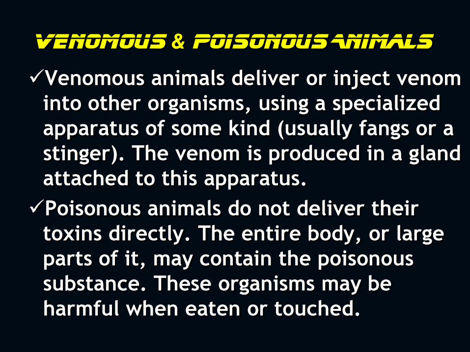
Venomous & Poisonous Animals
Venomous animals deliver or inject venom
into other organisms, using a specialized
apparatus of some kind (usually fangs or a
stinger). The venom is produced in a gland
attached to this apparatus.
Poisonous animals do not deliver their
toxins directly. The entire body, or large
parts of it, may contain the poisonous
substance. These organisms may be
harmful when eaten or touched.

Wasp sting (VENOM)

Androctonus bicolor

Cerastes vipera

Tetradotoxin in Puffer Fish
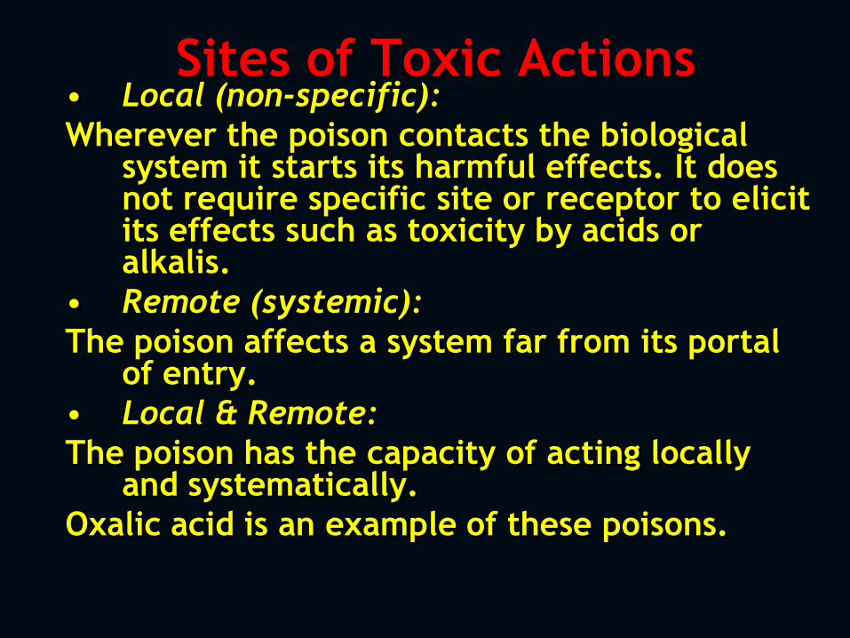
Sites of Toxic Actions • Local (non-specific):
Wherever the poison contacts the biological system it starts its harmful effects. It does not require specific site or receptor to elicit its effects such as toxicity by acids or alkalis.
• Remote (systemic):
The poison affects a system far from its portal of entry.
• Local & Remote:
The poison has the capacity of acting locally and systematically.
Oxalic acid is an example of these poisons.
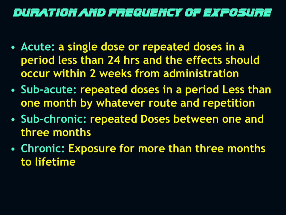
Duration and frequency of exposure
• Acute: a single dose or repeated doses in a
period less than 24 hrs and the effects should
occur within 2 weeks from administration
• Sub-acute: repeated doses in a period Less than
one month by whatever route and repetition
• Sub-chronic: repeated Doses between one and
three months
• Chronic: Exposure for more than three months
to lifetime
Chronicity Index
•The ratio of the acute to
chronic LD50 dosage
•Compounds with strong
cumulative properties have
larger chronicity index.
Types of Toxic Mechanisms • Direct: the poison itself can cause toxic
effects as in corrosives.
• Indirect: toxicity results from the interaction of the poison with the biological activity within biological system as in CO. This type can take many forms:
– Binding to cell membrane making changes in their function or structure thus affecting their normality.
– Interference with enzymatic actions.
– Formation of metabolites which are more toxic than the parent poison.
– Effects on DNA
Classification of toxic agents
1) According to the Target organ they are
acting on it (hepatotoxic, Nephrotoxic)
2) According to their Use (food additive,
drug, pesticide)
3) According to their Source (animal or
plant)
4) According to their Effects (carcinogen,
mutagen)
5) According to their Physical state
(gas, liquid)
6) According to their Chemistry
(Amine, hydrocarbon)
7) According to their Poisoning
potentiality (extremely toxic, slight
toxic, etc)
8) According to their Biochemical
mechanism of action (alkylating
agent, AchE inhibitor)
Factors Affecting Action of Poison A) Factors related to the poison:
• Dose: a basic principle in toxicology. – Dose is the amount of chemical that comes into
contact with the body or gets inside the body.
– The increase of dose will increase the severity of toxicity.
• Physical status: gaseous state is more toxic than liquid state than the solid state.
• Purity: this depends on the impurity of the poison; if the impurities are more toxic than the poison, the toxicity will be more and vice versa.
Spectrum of undesired effects Each drug produces a number of effects, but
only one is associated with the primary
objective of the therapy; all other effects
are considered undesirable or side effects.
Side effects may be beneficial (antihistamine))
The effects that are always undesirable are
referred to as adverse, deleterious or toxic
effects of the drug.
B) Factors related to the individual:
– Age
– Health
– Sensitivity
– Sex
C) Factors related to mode of exposure:
– Inhalation > IM > ingestion > Skin contact
D) Factors related to environment: – Temperature, pressure, humidity, radiation can cause
alterations on poisons status.
Forensic Toxicology
• The forensic toxicologist is
concerned with the detection and
estimation of poisons for legal
purposes:
– Tissues and body fluids obtained at
autopsy
– Blood, urine, or gastric material
obtained from a living person
•Poisoning as a cause of death can be proven
only with toxicologic analyses that
demonstrate the presence of the poison in
the tissues or body fluids of the deceased.
•Presence of poisons can be demonstrated
only by chemical methods of isolation and
identification.
•If toxicological analyses are avoided, death
may be ascribed to poisoning without definite
proof.
•Analytical toxicology deals with the
detection, identification, and
quantification of poisons.
•This role is important in forensic
and clinical toxicology where the
process deals with poisons in
human tissues.
SAMPLES REQUIRED FOR TOXICOLOGICAL
ANALYSIS FROM AUTOPSIES
•Blood
– The best place at autopsy is from femoral & iliac veins,
then axillary veins in consequence
• NO SAMPLES FROM:
1. Jugular veins: may be contaminated by reflux
from upper thorax
2. General body cavity: highly contaminated by
intestinal contents
3. Heart or great vessels in chest: postmortem
diffusion of drugs & alcohol from the stomach
or aspirated vomit contaminate these sites
•Urine
20-30 ml urine in sterile
container without preservatives
•Faeces
Used in heavy metals as arsenic,
lead, mercury
•Vomit and Stomach contents
•Organs – The most common organ saved for
analysis is liver
– Bile can be helpful in morphine and chlorpromazine
– Lungs in some cases of solvents
•Hair & Nails – Heavy metal poisonings
– Recently, prolonged use of opiates
METHODS OF ANALYSIS • QUALITATIVE METHODS
A) COLOR TEST
This is a rapid, easily performed,
qualitative, screening test, but not
specific method
• Can be used as bed side rapid test
•Examples:
1- Ferric chloride test for salicylates (pink-purple)
2- Zwikker test for barbiturates (purple color)
3- Formaldehyde-Sulfuric acid test for BZD (orange)
4- Mandalin Test for opioid (brown color)
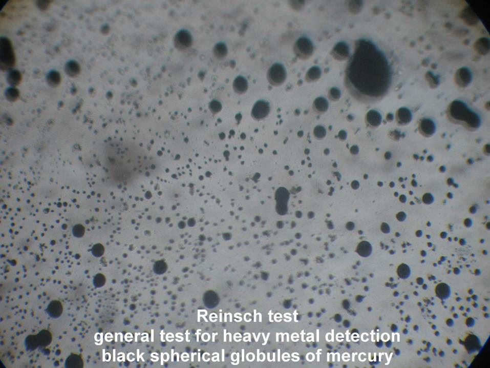
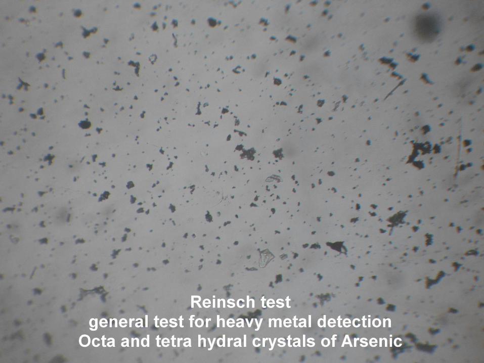
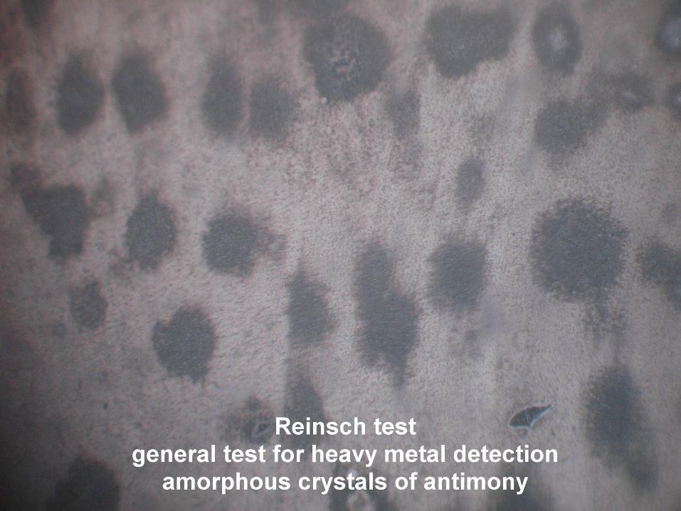
B) CHEMICAL TEST
• Reinsch Test is an initial indicator to detect the presence of one or more of the following Heavy Metals in a biological sample – Antimony
– Arsenic
– Bismuth
– Selenium
– Thallium
– Mercury
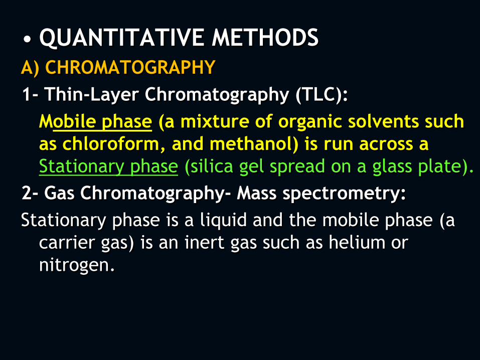
• QUANTITATIVE METHODS A) CHROMATOGRAPHY
1- Thin-Layer Chromatography (TLC):
Mobile phase (a mixture of organic solvents such
as chloroform, and methanol) is run across a
Stationary phase (silica gel spread on a glass plate).
2- Gas Chromatography- Mass spectrometry:
Stationary phase is a liquid and the mobile phase (a
carrier gas) is an inert gas such as helium or
nitrogen.
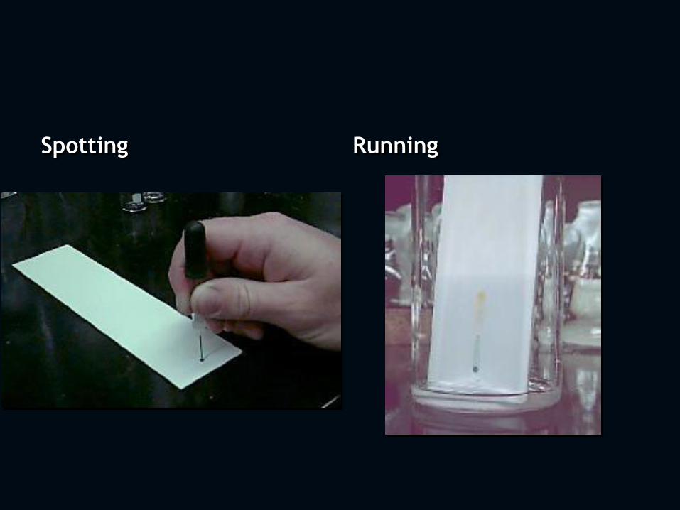
Spotting Running
3- High-Performance Liquid Chromatography (HPLC):
In HPLC the stationary phase is a column packed with
solid particles and the mobile phase is a liquid solvent.
B) IMMUNOASSAYS
• Enzyme-multiplied immunoassay technique (EMIT).
frequently used to detect the presence of certain drugs
in urine.
• Polarization immunoassay (FPIA)
• Radioimmunoassay (RIA)
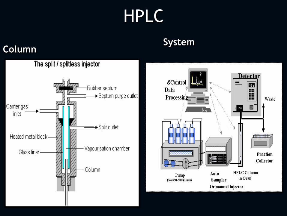
HPLC
Column System
Thin-Layer Chromatography (TLC)
This is a separation technique .
It was described in details by Ergon Stahl
(1969).
In TLC a mobile phase (a mixture of
organic solvents such as chloroform,
and methanol) is run across a
stationary phase (silica gel spread on a
glass plate).
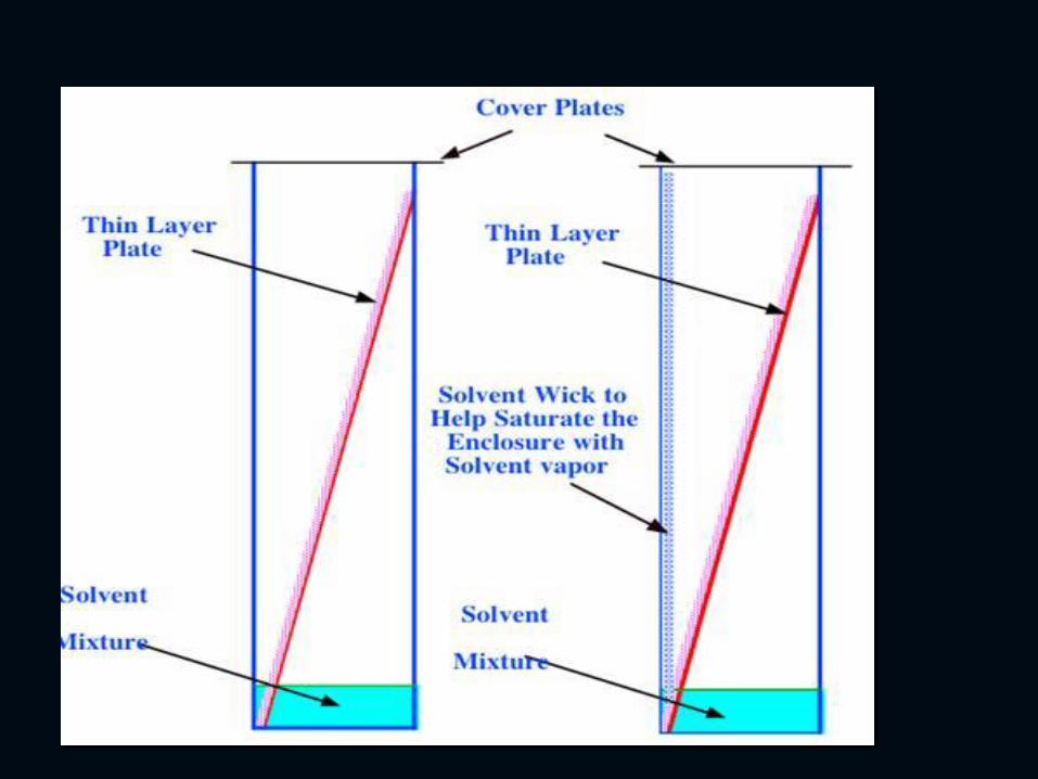
TLC The samples to be analyzed are spotted
near the bottom portion of the plate
and allowed to dry. Then the plate is
placed upright into a chamber, with the
bottom of the plate (where the sample
has been spotted) in contact with the
mobile phase. The mobile phase will
then draw up across the plate by
capillary action.
As the solvent moves past the samples, the
components of the samples will migrate,
with the speed of migration dependent
upon the relative affinity of the
components for the mobile phase
compared to the stationary phase.
When the leading edge of the solvent
reaches the top of the plate, it is removed
from the solvent and allowed to dry. The
location of the sample components can
then be visualized.
Stahl provided methods for 264 stains
or dyes that can be applied for the
required component such as
ninhydrin will react with
amphetamine to give pink color.
Alternatively, a fluorescent dye can be
incorporated in the solid phase, so
that ultraviolet light can reveal the
sample components as dark spots
against the a bright background.
The results of TLC can be quantified by
using the retention factor (Rf) which is the ratio of the distance that
a sample component moves to the
distance that the leading edge of the
solvent moves.
Rf = Sample distance movement
Solvent distance movement
Sample components can be identified
by comparing their Rf to the Rf of
known substances or by using a table
of Rfs given according to the mobile
and stationary phases used.
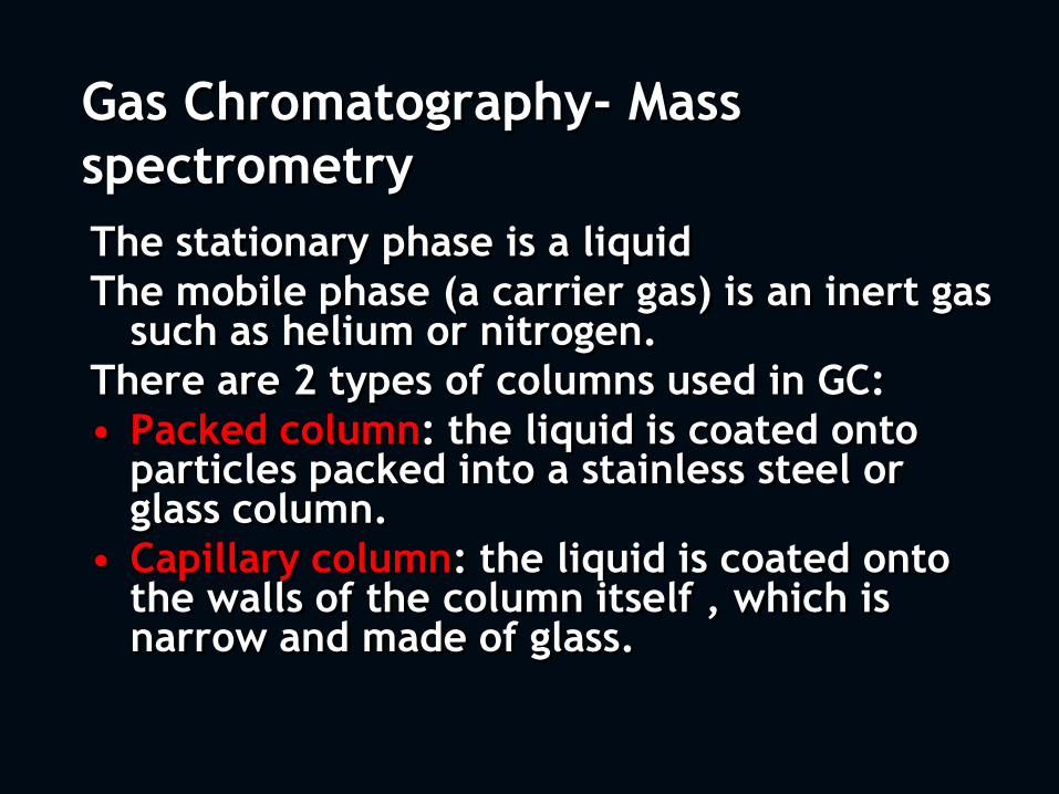
Gas Chromatography- Mass
spectrometry
The stationary phase is a liquid
The mobile phase (a carrier gas) is an inert gas such as helium or nitrogen.
There are 2 types of columns used in GC:
• Packed column: the liquid is coated onto particles packed into a stainless steel or glass column.
• Capillary column: the liquid is coated onto the walls of the column itself , which is narrow and made of glass.

• Samples are injected into a heated port, where they are vaporized and carried into the column along with the carrier gas. A detector then produces a signal as sample components exit the column.
• When the detector is hooked up to a recorder, a gas chromatogram (recording) can be produced. This is a plot of electronic signal versus time, which shows series of peaks that corresponds to the components of the sample.
• The time it takes for a substance to pass thorough a column (retention time Rt) can be compared to standards to identify that substance. Also, since the area under each peak is proportional to the concentration of that substance, comparison with a standard of known concentration allows estimation of the concentrations.
• Flame photometric detectors increase the sensitivity over flame ionization.
• Electron capture detector uses radioactive source and it can detect picograms of DDT due to presence of 5 chlorine atoms in the molecule.
Mass Spectrometry (MS)
Although chromatography allows identification of substances based on Rf and Rt with standards, definitive identification requires additional analysis. Mass spectrometry is one of these methods; it is used in combination with gas chromatography. As the sample components exit the GC column, they are routed into a vacuum chamber in the mass spectrometer, where they are hit with a beam of electrons.

This knocks electrons off the sample
molecules, creating positive electrons
and breaking them into fragments.
These fragments are then passed
through an electromagnetic field,
which separates them by their
mass/charge ratio. The resulting
spectrum plotting the abundance and
mass/ charge ration of each fragment
is specific for a given substance.
High-Performance Liquid Chromatography (HPLC)
In HPLC the stationary phase is a column packed with solid particles and the mobile phase is a liquid solvent.
As the mobile phase is pumped through the column, the sample is injected. A detector then identifies the components as they exit the column. Components are identified by their Rt, the length of time they take to pass through the column. And the results are compared with standards.