a novel electrode for pasteless ecg monitoring
TRANSCRIPT
Paper Ref: S0212_P0338 3rd International Conference on Integrity, Reliability and Failure, Porto/Portugal, 20-24 July 2009
-1-
A NOVEL ELECTRODE FOR PASTELESS ECG MONITORING D. Vasconcelos*, **, A.C. Alves*, G. Barreto*, P. Pedrosa**,***, D. Freitas*, F. Vaz*** and C. Fonseca*,** *FEUP – Faculdade de Engenharia, Universidade do Porto, R. Roberto Frias 4200-465, Porto, Portugal
** INEB – Instituto de Engenharia Biomédica, Divisão de Biomateriais; Universidade do Porto, Rua do Campo
Alegre, 823, 4150-180 Porto, Portugal
*** Dept. de Física, Universidade do Minho, Campus de Azurém, 4800-058 Guimarães, Portugal Emails: bio06007 fe.up.pt, bio06024 fe.up.pt, bio06031 fe.up.pt, [email protected],
[email protected], [email protected], [email protected]
ABSTRACT
A dry bracelet electrode for electrocardiographic (ECG) signal monitoring was developed and
successfully tested in human volunteers. The new electrode dispenses with the usual gel
application and the previous skin preparation to monitor the signal. It was fabricated from a
polyethylene tereftalate (PET) polymer sheet that was coated with a thin conductive titanium
nitride (TiN) layer. As the direct sputtering of the TiN layer on the polymeric surface results
in a poor adherence of the layer, a specific polymer activation treatment was developed,
consisting of the polymer surface bombardment with alternate beams of argon and titanium
ions. The adhesion tests and the SEM analysis of the coatings proved the success of the
treatment. The TiN coating displays the fcc crystalline structure of the δ-TiN bulk material
with a rough, pyramidal-like morphology. The electrochemical analysis that was carried out
in a saline solution (to mimic the skin sweat contact) showed delamination of the film after
prolonged contact with the saline solution..
The new electrode was tested against the classic gelled silver/silver chloride electrode
(Ag/AgCl) and the ECG records proved to be very similar with both devices. The new
bracelet electrode is ideal for monitoring in ambulatory conditions, as the usual abrasive skin
preparation and the gel application may be skipped. It may be particularly suitable for long-
term monitoring, where the prolonged contact with the gel paste often induces strong allergic
reactions.
-2-
1. INTRODUCTION
The life expectancy in the industrialized countries has risen in the last 20 years from 72 to 80
years old, bringing a strong increase of the health associated costs. One of the strategies that
are being adopted to reduce such costs is the adoption of preventive medicine, involving the
monitoring of important health parameters in ambulatory conditions. The ECG is definitively
one of such parameters, as the cardiovascular diseases are the main cause of death and
morbidity in the developed countries. However, the ambulatory long-term ECG monitoring is
still difficult, due to the characteristics of the actual electrodes, which either demand the
presence of a gel (that dehydrates with air exposure) or, in the case of the disposable
electrodes, have a limited shelf-life and loose their performance after a few days in service
[McAdams, 2006]. Finally, the classic silver/silver chloride (Ag/AgCl) electrodes, either
reusable or disposable, may cause strong allergic reaction, especially if they have to be in
contact with skin for a long time (McAdams, 2006).
Dry electrodes allow skipping the previous skin preparation and gel application, what makes
them “plug-and-play” devices, ideal for ECG monitoring in ambulatory conditions. Presently,
there are no marketed ECG dry electrodes even if this is a very active research topic.
Gruetzmann (Gruetzmann, 2007) tested a conductive foam as a candidate to an ECG sensor
and Baek (Baek, 2008) fabricated and tested a bracelet ECG electrode from a PDMS flexible
polymer. Both authors explored the possibility of using deformable sensors in order to
achieve an improved skin/electrode coupling. Even if the principle was demonstrated,
Gruetzmann stated that his device still had to be improved and Baek’s electrode follows a
complex fabrication process. Yu (Yu, 2009) proposed an electrode based in a micro-needles
array that was deposited on silicon. Besides the infection risk and discomfort due to the
invasive nature of the device, there is the possibility that the needles will become
encapsulated by scar tissue at the long-term, with a dramatic increase of the contact
impedance.
We developed a PET (polyethylene tereftalate) based bracelet device, where the polymeric
substrate was coated with a TiN conductive layer, involving a simple procedure and cheap
materials. TiN films are well known for their excellent chemical and mechanical properties,
which led to a very broad range of applications, as diverse as the mechanical protection of
machine parts and cutting tools (Malik, 2004), coatings for orthopedic and dental prosthesis
(Piscanec, 2004), or even as diffusion barriers for electronic devices (Gao, 2004).
-3-
Despite the rigid nature of the electrode a good skin contact could be achieved due to
electrode design and its small size. This new electrode proved to be able to withstand daily
handling and cleaning.
2. EXPERIMENTAL PROCEDURES
2.1. Surface activation and sputtering of the TiN layer
The electrodes were prepared by depositing a TiN thin film on a 3 mm thick PET sheet
surface (Goodfellow Metals Inc.), by reactive DC magnetron sputtering. The deposition
system is a “home-made” laboratory-sized deposition system, composed of two vertically
opposed rectangular magnetrons (unbalanced of type II) in a closed field configuration. The
film was prepared with the substrate holder positioned at 70 mm from the Ti target (99.6 at.
%), coupled with a DC current density of 100 A·m-2 during 1200 s. A gas atmosphere
composed of argon (60 sccm) and nitrogen (5.5 sccm, corresponding to a partial pressure of
4.1×10⁻² Pa) was used. The working pressure was approximately 0.4 Pa. The substrates were
grounded, and no external heating was used. In order to improve the adhesion of the TiN film
to the PET substrate, usually very weak, a three-fold procedure was used. The procedure
started with a first plasma treatment in an Ar atmosphere (80 sccm - 4.8×10⁻¹ Pa), using a
pulsed DC power supply (200 kHz, Ton = 1536 ns) with 0.5 A for 600 s. This Ar
flow/pressure was selected from a series of three different treatments (40 sccm - 2.7×10⁻¹ Pa,
60 sccm - 3.9×10⁻¹ Pa and; 80 sccm - 4.8×10⁻¹ Pa), in which the last revealed to be the one
that promoted the most favourable changes in the polymer substrates surface, both in terms of
roughness and contact angle (CA) variations. These optimization studies were carried out in
the group by Pedrosa et al. and A. Ferreira et al. (articles in preparation). After this plasma
activation/treatment, a very thin layer of TiN was deposited in a plasma atmosphere
composed of Ar (flow of 60 sccm) and nitrogen (5.5 sccm), using a DC current density of 100
A m-2, during 120 s. This set of parameters corresponds to those that would be used for the
deposition of the film itself. A third step comprised a second plasma treatment, similar to the
first one. This second plasma treatment was carried out in order to promote some collisions of
ions with the thin layer deposited in the previous step. In this way, some atoms of the thin
layer were “pushed” deeper inside the polymer’s surface, which was expected to act as
-4-
bonding centres and thus improving even further the adhesion of the main layer to be
deposited after.
2.2. Characterization of the prepared film
The surfaces of the TiN films were observed with a Jeol JSM 6301F microscope operating at
10 KeV. The crystalline structure of the film was scanned by X-ray diffraction in a Philips
PW1710 equipment, operating with the Cu Kα radiation in a Bragg-Brentano configuration.
The adhesion tests in the deposited layer were carried out following the ASTM D3359-08 X-
cut tape test standard. It consists on doing 2 cuts on the surface of the TiN films with a 45º
angle between them, in order to form the shape of an X. The surface is controlled before and
after this X-cut by optical microscopy. Then, a pressure sensitive tape is placed on top of the
cut and pulled-out, leading to a certain degree of delamination of the interface between the X-
cut and the film itself. Ten, the effect of the test is assessed by optical microscopy, comparing
with the images of the cut before application of the tape. The adhesion will then be rated
according with the scale present in the above-referred standard.
Electrochemical impedance spectra were acquired daily during one week, using a Solartron
1250 frequency response analyzer connected to a EG&G PAR 273A potentiostat, driven by
the Zplot software from Solartron. The testing frequencies ranged from 20 kHz to 2 mHz, and
the amplitude of the AC signal was 7 mV (rms). The Zview software was used for the
simulation of the experimental spectra. The saturated calomel electrode (SCE) was used as the
reference electrode and a graphite rod as the counter electrode. The electrolyte was a 0.1M
NaCl solution. The AFM and SEM microscopies were performed with a Pico Scan controller
atomic force microscope using the tapping mode, and a Jeol JSM 6301F microscope operating
at 20 KeV respectively. The ECG was acquired with the Biopac@ system (hardware and
software) in a three electrode configuration, with two electrodes placed in the left and right
wrists and the reference electrode in the right leg. The Ag/AgCl electrodes used as controls
were hydrogel electrodes of the disposable type, also from Biopac.
3. RESULTS AND DISCUSSION
3.1. Surface activation treatment – Roughness and Contact angle
In the study developed by Pedrosa et al. and A. Ferreira et al., a set of polymeric substrates
(polypropylene, PP, polycarbonate, PC and PET) were plasma treated with three different Ar
-5-
plasma conditions: 40 sccm (2.7×10⁻¹ Pa), 60 sccm (3.9×10⁻¹ Pa) and 80 sccm (4.8×10⁻¹ Pa).
The conditions that were found to give the best adhesion in the case of PET were those
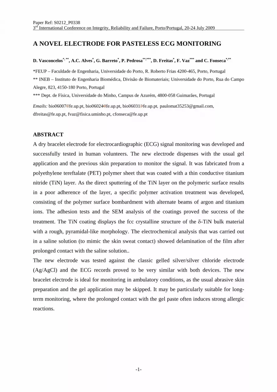
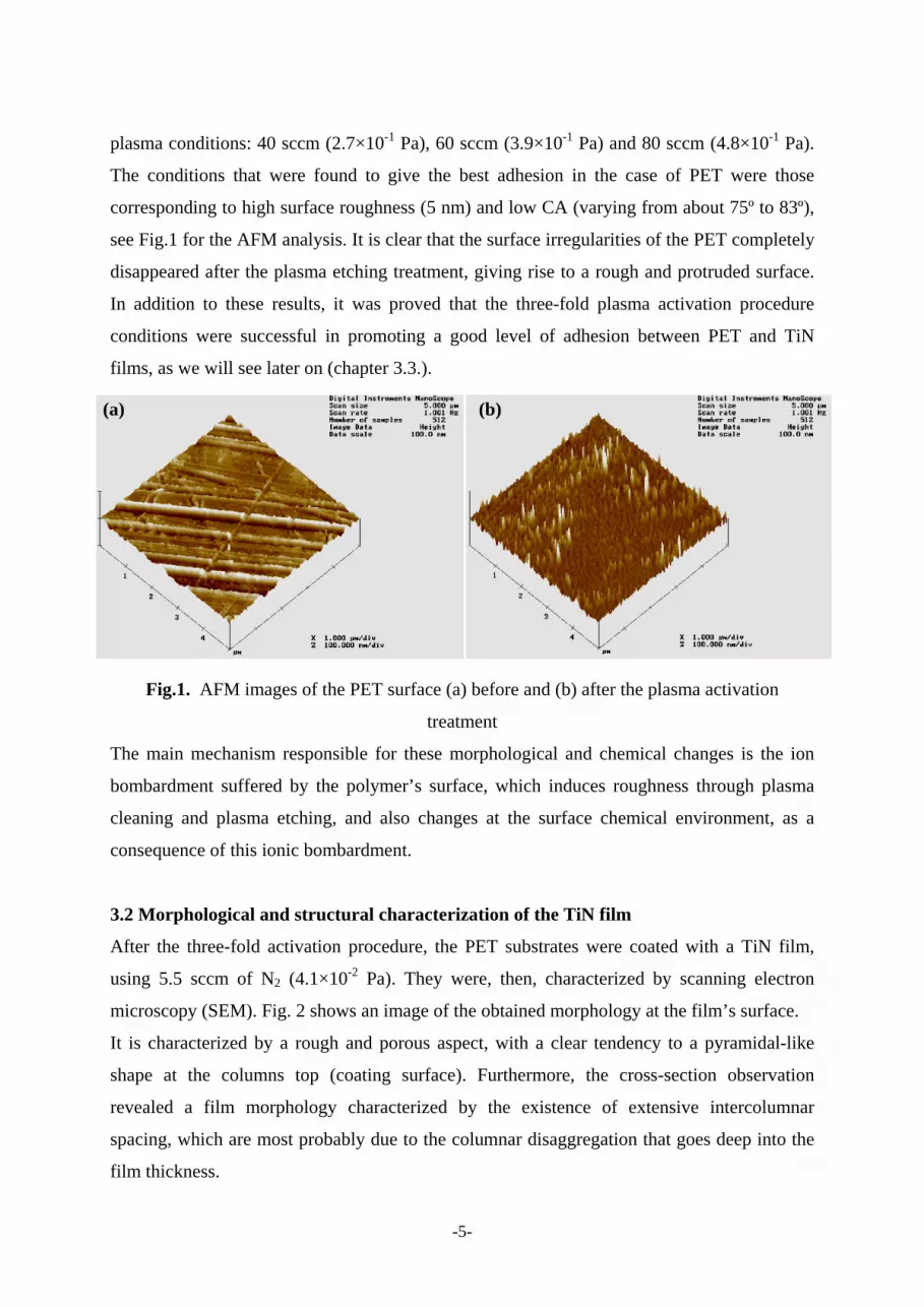
corresponding to high surface roughness (5 nm) and low CA (varying from about 75º to 83º),
see Fig.1 for the AFM analysis. It is clear that the surface irregularities of the PET completely
disappeared after the plasma etching treatment, giving rise to a rough and protruded surface.
In addition to these results, it was proved that the three-fold plasma activation procedure
conditions were successful in promoting a good level of adhesion between PET and TiN
films, as we will see later on (chapter 3.3.).
Fig.1. AFM images of the PET surface (a) before and (b) after the plasma activation
treatment
The main mechanism responsible for these morphological and chemical changes is the ion
bombardment suffered by the polymer’s surface, which induces roughness through plasma
cleaning and plasma etching, and also changes at the surface chemical environment, as a
consequence of this ionic bombardment.
3.2 Morphological and structural characterization of the TiN film
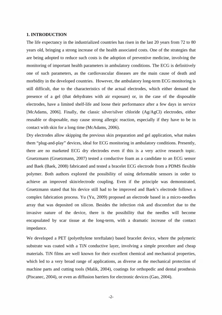
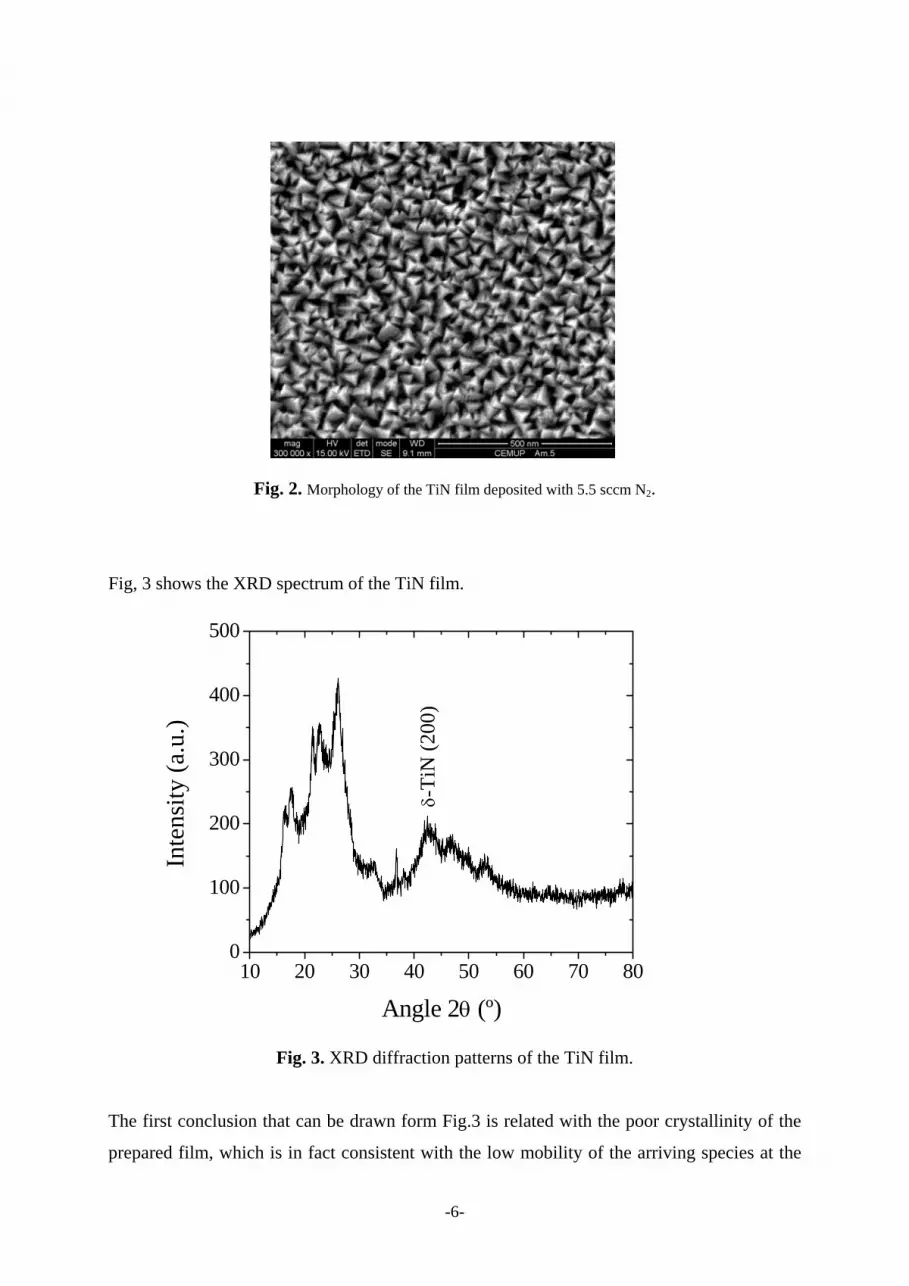
After the three-fold activation procedure, the PET substrates were coated with a TiN film,
using 5.5 sccm of N2 (4.1×10⁻² Pa). They were, then, characterized by scanning electron
microscopy (SEM). Fig. 2 shows an image of the obtained morphology at the film’s surface.
It is characterized by a rough and porous aspect, with a clear tendency to a pyramidal-like
shape at the columns top (coating surface). Furthermore, the cross-section observation
revealed a film morphology characterized by the existence of extensive intercolumnar
spacing, which are most probably due to the columnar disaggregation that goes deep into the
film thickness.
(a) (b)
-6-
Fig. 2. Morphology of the TiN film deposited with 5.5 sccm N2.
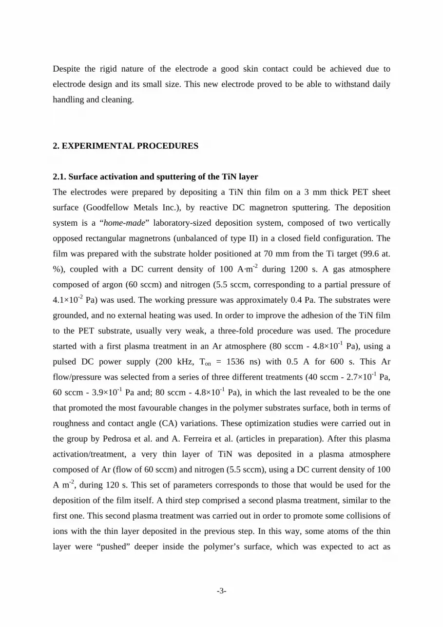
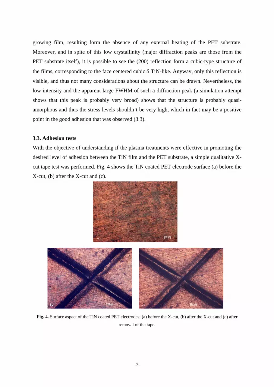
Fig, 3 shows the XRD spectrum of the TiN film.
Fig. 3. XRD diffraction patterns of the TiN film.
The first conclusion that can be drawn form Fig.3 is related with the poor crystallinity of the
prepared film, which is in fact consistent with the low mobility of the arriving species at the
10 20 30 40 50 60 70 800
100
200
300
400
500
Inte
nsity
(a.u
.)
Angle 2θ (º)
δ-Ti
N (2
00)
-7-
growing film, resulting form the absence of any external heating of the PET substrate.
Moreover, and in spite of this low crystallinity (major diffraction peaks are those from the
PET substrate itself), it is possible to see the (200) reflection form a cubic-type structure of
the films, corresponding to the face centered cubic δ TiN-like. Anyway, only this reflection is
visible, and thus not many considerations about the structure can be drawn. Nevertheless, the
low intensity and the apparent large FWHM of such a diffraction peak (a simulation attempt
shows that this peak is probably very broad) shows that the structure is probably quasi-
amorphous and thus the stress levels shouldn’t be very high, which in fact may be a positive
point in the good adhesion that was observed (3.3).
3.3. Adhesion tests
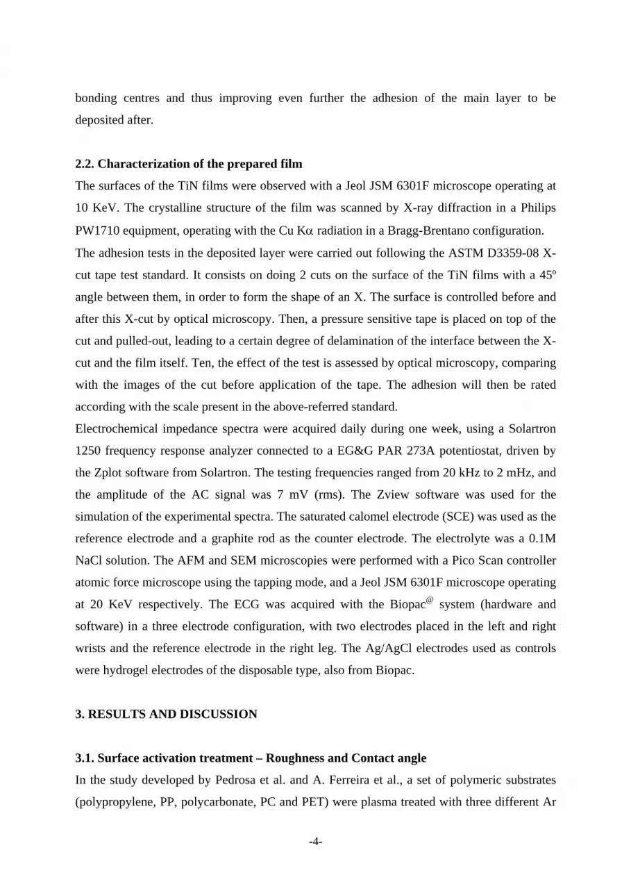
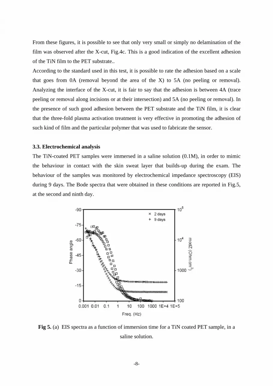
With the objective of understanding if the plasma treatments were effective in promoting the
desired level of adhesion between the TiN film and the PET substrate, a simple qualitative X-
cut tape test was performed. Fig. 4 shows the TiN coated PET electrode surface (a) before the
X-cut, (b) after the X-cut and (c).
Fig. 4. Surface aspect of the TiN coated PET electrodes; (a) before the X-cut, (b) after the X-cut and (c) after
removal of the tape.
a
b c
-8-
From these figures, it is possible to see that only very small or simply no delamination of the
film was observed after the X-cut, Fig.4c. This is a good indication of the excellent adhesion
of the TiN film to the PET substrate..
According to the standard used in this test, it is possible to rate the adhesion based on a scale
that goes from 0A (removal beyond the area of the X) to 5A (no peeling or removal).
Analyzing the interface of the X-cut, it is fair to say that the adhesion is between 4A (trace
peeling or removal along incisions or at their intersection) and 5A (no peeling or removal). In
the presence of such good adhesion between the PET substrate and the TiN film, it is clear
that the three-fold plasma activation treatment is very effective in promoting the adhesion of
such kind of film and the particular polymer that was used to fabricate the sensor.
3.3. Electrochemical analysis
The TiN-coated PET samples were immersed in a saline solution (0.1M), in order to mimic
the behaviour in contact with the skin sweat layer that builds-up during the exam. The
behaviour of the samples was monitored by electrochemical impedance spectroscopy (EIS)
during 9 days. The Bode spectra that were obtained in these conditions are reported in Fig.5,
at the second and ninth day.
Fig 5. (a) EIS spectra as a function of immersion time for a TiN coated PET sample, in a
saline solution.

-9-
The most remarking feature corresponds to the constant impedance part of the spectra
(10.000-100 Hz), related with the pure resistive behaviour of the electrolyte and the film (null
phase angle), that increases with the immersion time. This is an indication of the progressive
degradation of the film, first with the formation of cracks and then with delamination from the
substrate, as it was confirmed by microscopic and visual analysis.
3.4. Testing of the device for ECG acquisition in human volunteers A photo of the new electrode is reported in Fig. 6. The central part is coated with a TiN thin
film. The electric contact was made with conductive epoxidic glue on the side of the electrode
and then coated with an insulating varnish.
Fig. 6. Image of the dry PET ECG electrode (left) and ECG records with the dry and
Ag/AgCl electrodes
The ECG records displayed in the right part of the figure were obtained in rest conditions,
both with the classic Ag/AgCl and the new dry electrodes. It is apparent that the records show
very similar signals, that allowed to obtain the same cardiac rate. The same test was
performed in exercise conditions but neither of the electrodes allowed monitoring reliable
signals in such conditions.
The electrodes were also intensively used in a science fair, for four days, and no degradation
of the recording performance was noticed.
Dry Ag/AgCl

-10-
4.CONCLUSIONS A dry ECG electrode with the shape of a bracelet was developed and tested for ECG signal
acquisition. This electrode dispenses with the use of any previous skin preparation or gel
application, making it ideal for ambulatory utilization. The electrode substrate was a 3 mm
thick PET polymer, whose surface was activated through an argon plasma treatment, in order
to promote the adhesion of the TiN conductive layer that was afterwards deposited by reactive
magnetron sputtering.
A simple X-cut adhesion test proved the excellent adhesion of the TiN film to the polymeric
substrate. The X-rays analysis showed the poor cristallinity of the TiN layer and the SEM
analysis showed a rough, porous and highly textured surface.
On the other hand, the electrochemical tests performed in saline solution showed that the film
progressively degradates with prolonged immersion in saline solution (9 days), what may be a
drawback of the developed coating if long-term exams are envisaged. At present, TiN films
with lower amounts of nitrogen are being produced, in order to achieve compact films that are
expected to display a higher corrosion resistance than the porous layers of the present work.
Concerning the performance of the new electrode, the ECG data showed that the signals are
very similar to the signal obtained with the classic Ag/AgCl electrodes, opening the
possibility for the future utilization of these devices in ambulatory conditions.
ACKNOWLEDGEMENTS D. Vasconcelos, A.C. Alves and G. Barreto are grateful to the MIB (Mestrado Integrado em Bioengenharia) teachers for having given them the opportunity to perform this work in the framework of the Laboratorios Integrados de Engenharia Biomédica I discipline.
C. Fonseca and F. Vaz would like to thank to the CRUP Germany-Portugal Bilateral program, through the A12/08 fund.
F. Vaz acknowledges the funding from the Portuguese Science Foundation “Fundação para a Ciência e Tecnologia”, project PTDC/CTM/69362/2006.
-11-
REFERENCES McAdams in “Encyclopedia of Medical Devices and Instrumentation”, Second Edition, edited
by John G. Webster (2006).
- Gruetzmann, A., Stefan Hansen, S., Muller, J. Novel dry electrodes for ECG monitoring,
Physiol. Measur. 28; 2007; p.1375-90.
- Baek, J., An, J., Choi, J. Flexible polymeric dry electrodes for the long-term monitoring of
ECG, Sensors and Actuators, A143; 2008; 423-429.
- Vaz, F., Cerqueira P., Rebouta L. et al Structural, Optical and Mechanical Properties of
Coloured TiNxOy Thin Films”, Thin Solid Films, 447-448; 2004; p.449-454.
- H. Malik, R.Mgaloblishvili, B.Mills, J. Mat. Science Letters 19 (19) (2004) 1779.
- S. Piscanec, L. Ciacchi, E. Vesselli, G. Comelli, O. Sbaizero, S. Meriani, A. De Vita, Acta
Materialia 52 (2004) 1237.
- L. Gao, J. Gstöttner, R. Emling, Ch. Linsmeier, A. Wiltner, W. Hansch, D.Schmitt-
Landsiedel, Microelectronic Engineering 76 (1-4) (2004) 76.