a novel antagonist of the prostaglandin e2 ep4 receptor inhibits th1 differentiation and th17...
TRANSCRIPT
RESEARCH PAPER
A novel antagonist of the prostaglandin E2 EP4
receptor inhibits Th1 differentiation and Th17expansion and is orally active in arthritis modelsbph_647 292..310
Q Chen1*, K Muramoto2*, N Masaaki2, Y Ding1, H Yang1, M Mackey1, W Li1, Y Inoue2,K Ackermann1, H Shirota1, I Matsumoto3, M Spyvee1, S Schiller1, T Sumida3, F Gusovsky1 andM Lamphier1
1Eisai Research Institute of Boston, Andover, MA, USA, 2Tsukuba Research Laboratories, Ibaraki, Japan, and 3Department ofClinical Immunology, Graduate School of Comprehensive Human Science, University of Tsukuba, Tsukuba, Japan
Background and purpose: Rheumatoid arthritis (RA) is an autoimmune disorder involving subsets of activated T cells, inparticular T helper (Th) 1 and Th17 cells, which infiltrate and damage tissues and induce inflammation. Prostaglandin E2 (PGE2)enhances the Th17 response, exacerbates collagen-induced arthritis (CIA) and promotes inflammatory pain. The current studyinvestigated whether selective antagonism of the PGE2 EP4 receptor would suppress Th1/Th17 cell development and inflam-matory arthritis in animal models of RA.Experimental approach: Effects of PGE2 and a novel EP4 receptor antagonist ER-819762 on Th1 differentiation, interleukin-23(IL-23) production by dendritic cells (DCs), and Th17 development were assessed in vitro. The effect of ER-819762 wasevaluated in CIA and glucose-6-phosphate isomerase (GPI)-induced arthritis models. In addition, the effects of ER-819762 onpain were evaluated in a model of chronic inflammatory pain induced by complete Freund’s adjuvant (CFA) in the rat.Key results: Stimulation of the EP4 receptor enhanced Th1 differentiation via phosphatidylinositol 3 kinase signalling,selectively promoted Th17 cell expansion, and induced IL-23 secretion by activated DCs, effects suppressed by ER-819762 oranti-PGE2 antibody. Oral administration of ER-19762 suppressed Th1 and Th17 cytokine production, suppressed disease incollagen- and GPI-induced arthritis in mice, and suppressed CFA-induced inflammatory pain in rats.Conclusion and implications: PGE2 stimulates EP4 receptors to promote Th1 differentiation and Th17 expansion and iscritically involved in development of arthritis in two animal models. Selective suppression of EP4 receptor signalling may havetherapeutic value in RA both by modifying inflammatory arthritis and by relieving pain.British Journal of Pharmacology (2010) 160, 292–310; doi:10.1111/j.1476-5381.2010.00647.x
Keywords: PGE2; EP4 receptor; EP4 antagonist; Th1; Th17; IFN-g; IL-17; IL-23; CIA; GPI-induced arthritis model
Abbreviations: BSA, bovine serum albumin; CIA, collagen-induced arthritis; CFA, complete Freund’s adjuvant; CRE, cAMPresponse elements; ER-819762, (S)-1’-(3,5-dimethylbenzyl)-2-ethyl-7,9-dimethoxy-10-methyl-5,10-dihydrospiro[benzo[e]imidazo[1,5-a]azepine-1,4’-piperidin]-3(2H)-one; FBS, fetal bovine serum; GPCR, Gprotein-coupled receptors; GPI, glucose-6-phosphate isomerase; IBMX, isobutylmethylxanthine; imDC, imma-ture human dendritic cell; PGE2, prostaglandin E2; PI3K, phosphatidylinositol 3-kinase; PLAP, placental-likealkaline phosphatase; RA, rheumatoid arthritis; Th1, T helper 1; Th17, T helper 17; TLR, Toll-like receptor
Introduction
Rheumatoid arthritis (RA) is a chronic autoimmune disorderthat is estimated to affect up to 1% of the population world-wide (Williams, 2006). Although not life-threatening, RA is a
painful and debilitating disease that progressively limits theability of patients to carry on normal lives. The factors thattrigger this disease are not well understood but are believed toinclude both genetic and environmental components. Overthe last decade, novel discoveries into the regulation of theimmune system have permitted a better understanding of thedevelopment of autoimmunity. Upon encountering antigen,T helper (Th) cells differentiate into several subtypes, depend-ing on various factors such as the cytokines produced, theconsequent activation of intracellular signalling pathways,
Correspondence: Qian Chen, Eisai Research Institute of Boston Inc., 4 Corpo-rate Drive, Andover, MA 01810 USA. E-mail: [email protected]*These authors contributed equally to this work.Received 30 June 2009; revised 2 November 2009; accepted 9 November 2009
British Journal of Pharmacology (2010), 160, 292–310© 2010 Eisai Inc.Journal compilation © 2010 The British Pharmacological Society All rights reserved 0007-1188/10www.brjpharmacol.org

and the expression of transcription factors. Among these Tcell subtypes, Th1 and Th17 cells have been shown to becritically involved in the development of autoimmunity(Schulze-Koops and Kalden, 2001; Fouser et al., 2008). Whilethe molecular mechanisms controlling the differentiationand expansion of these T cell subsets have begun to be eluci-dated, specific agents to suppress the function of these criticalT cell subtypes are only in early-stage development.
Prostaglandin E2 (PGE2) is an arachidonic acid metabolitethat acts as a potent biological mediator, exerting its effectsvia activation of membrane G protein-coupled receptors(GPCRs). There are four receptor subtypes (EP1, 2, 3 and 4;nomenclature follows Alexander et al., 2009) which selec-tively bind PGE2 and mediate its effects: Activation of EP1
receptors leads to the influx of calcium. Activation of EP3
receptors can induce a variety of signalling events dependingon the particular EP3 splicing variant being expressed, withinhibition of adenylate cyclase activity via Gi being the mostcommon effect. EP2 and EP4 receptors induce Gs-mediatedactivation of adenylate cyclase and a subsequent increase inintracellular cyclic AMP (cAMP). In addition, EP4 receptorsactivate the phosphatidylinositol 3-kinase (PI3K) signallingpathway (Fujino et al., 2003). PGE2 is known to play impor-tant roles in mediating many inflammatory responses and isoften found at increased concentrations under a variety ofinflammatory conditions (Hata and Breyer, 2004). Manyreports suggest that PGE2, via the induction of intracellularcAMP, can suppress pro-inflammatory T cell function, includ-ing T cell receptor signalling and consequent production ofinterleukin (IL)-2 (Mustelin and Tasken, 2003; Chemnitzet al., 2006). PGE2 has also been implicated in T-cell differen-tiation and is reported to inhibit Th1 but not Th2 cytokinesvia the induction of intracellular cAMP (Betz and Fox, 1991;Gold et al., 1994; Hilkens et al., 1995; Okano et al., 2006).However, other reports indicate a pro-inflammatory role forPGE2. PGE2 can induce production of IL-23 from dendriticcells (DCs), which promotes the differentiation of pro-inflammatory Th17 cells (Sheibanie et al., 2004; Khayrullinaet al., 2008). Recent reports also suggest that PGE2 can syner-gize with IL-23 to promote expansion of human Th17 cellsand enhance IL-17 production (Chizzolini et al., 2008; Boni-face et al., 2009; Napolitani et al., 2009). Furthermore, PGE2
has been shown to exacerbate symptoms in mouse models ofarthritis (Sheibanie et al., 2007a) and inflammatory boweldisease (Sheibanie et al., 2007b), and the blockade of EP2 andEP4 receptor signalling in a mouse model of arthritis canalleviate the severity of the disease (Mccoy et al., 2002; Hondaet al., 2006).
Here, we show that PGE2 stimulation of the EP4 receptorcan promote Th1 differentiation, IL-23 production in DCs,and Th17 cell expansion. These effects can be suppressedby a novel EP4 receptor antagonist ER-819762 ((S)-1’-(3,5-dimethylbenzyl)-2-ethyl-7,9-dimethoxy-10-methyl-5,10-dihydrospiro[benzo[e]imidazo[1,5-a]azepine-1,4’-piperidin]-3(2H)-one) (Spyvee et al., 2009) or an anti-PGE2 antibody. Wealso show that oral administration of ER-819762 to DBA/1mice can effectively suppress disease in collagen-inducedarthritis (CIA) or glucose-6-phosphate isomerase (GPI)-induced arthritis models. ER-819762 was also effective intreating chronic inflammatory pain in a rat model. These
results suggest that PGE2 signalling via the EP4 receptor exertsa pro-inflammatory effect in vivo and is physiologically rel-evant to the pathology of inflammatory arthritis. EP4 recep-tors might therefore be an attractive drug target for thetreatment of RA, with the potential not only to relieve painand symptoms but also to modify the underlying aetiology ofthe disease
Methods
AnimalsAll animal studies were performed with the approval of theAnimal Ethics Committee at Eisai according to LaboratoryAnimal Welfare guidelines. BALB/c and DO11.10 mice werepurchased from Jackson Laboratory. C57BL/6 and DBA/1 miceand F344 rats were purchased from Charles River Laborato-ries. Mice and rats for each strain were group-housed undercontrolled conditions with a constant temperature (23 � 3°C)and humidity (55 � 5%), a 12-h light/dark cycle and adlibitum access to water and standard pelleted food.
Radioligand EP4 receptor binding assayThe radioligand EP4 receptor binding assay was performedusing ChemiScreen recombinant human EP4 receptor mem-brane preparations from Millipore, according to the manufac-turer’s instructions. Briefly, membranes prepared fromChem-1 cells overexpressing human EP4 receptor cDNA (Mil-lipore) were mixed with 1.8 nmol·L-1 [3H]-PGE2 and5 mmol·L-1 unlabelled PGE2 in the presence or absence ofvarious concentrations of ER-819762 in binding buffer[50 mmol·L-1 HEPES, pH 7.4, 5 mmol·L-1 MgCl2, 1 mmol·L-1
CaCl2, 0.2% bovine serum albumin (BSA)] in a nonbinding96-well plate, and incubated for 1–2 h at room temperature.Prior to filtration, a GF/C 96-well filter plate was coated with0.33% polyethyleneimine for 30 min, then washed with50 mmol·L-1 HEPES, pH 7.4, 0.5% BSA. Binding reactions weretransferred to the filter plate, and washed three times withwash buffer (1 mL per well per wash). The plate was dried andradioactivity counted. Binding of ER-819762 to other relatedprostanoid receptors was performed by MDS Pharma Services(Bothell, WA, USA) using a similar radiolabelled ligand dis-placement method.
Cell-based GPCR assaysSE302 is a clone of the human embryonic kidney 293 (HEK/293) cell line containing a reporter driven by cAMP responseelements (CRE) in its promoter, and producing secretedplacental-like alkaline phosphatase (PLAP). HEK/293 cellsexpress endogenous EP4 receptors and show induction ofPLAP in response to PGE2 and EP4 receptor agonists, but notEP1, 2 or 3 receptor agonists (Supplementary Fig. 2). Cells weremaintained in DMEM/F12 (50:50) (MediaTech, Inc., Manas-sas, VA, USA) supplemented with 10% fetal bovine serum(FBS; Tissue Culture Biologicals) plus penicillin/streptomycin.When used for assays, cells were plated in a 96-well plate at 2¥ 104 cells/100 mL per well in serum-free assay medium(DMEM/F12 supplemented with 0.1% BSA plus penicillin/
EP4 receptor antagonist for inflammatory arthritisQ Chen et al 293
British Journal of Pharmacology (2010) 160 292–310
streptomycin) and incubated for 4–6 h. Cells were then stimu-lated with 3 ng mL-1 PGE2 in the presence or absence ofvarious concentrations of ER-819762 overnight, and PLAPactivity was measured by mixing 15 mL of culture superna-tants with 75 mL of Lumi-phos (Lumigen, Inc.) and 60 mL ofassay buffer containing 8 mmol·L-1 MgSO4 in 0.1 mol·L-1
carbonate-bicarbonate buffer pH11 in a new 96-well blackplate and incubated for 2 h at room temperature. Lumines-cence was read with an Envision 2102 Multilabel reader. Char-acterization of compound selectivity was performed byMillipore GPCR Profiler Service, which assays intracellularcalcium mobilization in cells expressing individual GPCRsand the promiscuous Ga15 protein. Endogenous EP2 receptoractivity in U2-OS cells was assayed using the EPIC ResonantWaveguide Biosensor system (Corning).
In vitro T-cell assaysNaive CD4+ T cells were purified from spleens of either BALB/cor DO11.10 mice by antibody-coated magnetic beads asdescribed by the manufacturer (Robosep; StemCell Technolo-gies). For BALB/c mice, 1 ¥ 105 CD4+ T cells were cultured for3–6 days in a 96-well plate in 100 mL complete RPMI medium(CellGro) containing 10% regular FBS under: (i) neutral con-ditions (1 mg mL-1 plate-bound anti-CD3 + 1 mg mL-1 solubleanti-CD28 + 10 ng mL-1 mouse IL-2), (ii) Th1-promoting con-ditions (neutral + 5 ng mL-1 mouse IL-12 + 10 mg mL-1 anti-IL-4 antibody) or (iii) Th2-promoting conditions [neutral +10 ng mL-1 of mouse IL-4 + 10 mg mL-1 anti-interferon (IFN)-gantibody]. In experiments where exogenous PGE2 or EP4
receptor agonists were added to the culture, charcoal-strippedFBS (Hyclone) was used, which has reduced amounts of lipids.IFN-g or IL-4 in culture supernatants were quantified byenzyme-linked immunosorbent assay (ELISA). Cell prolifera-tion was assayed with either Alamar Blue or CellTiter-Gloreagents according to the manufacturers’ instructions. ForDO11.10 mice, mitomycin C-treated splenocytes from BALB/cmice were used as antigen-presenting cells and co-culturedwith naive CD4+ T cells in a 5:1 ratio (5 ¥ 105 mitomycinC-treated splenocytes in 100 mL medium + 1 ¥ 105 CD4 T cellsin 100 mL medium). These cultures were stimulated with anovalbumin peptide 323-339 (OVA peptide; 0.3 ng mL-1) underneutral, Th1- or Th2-promoting conditions, as describedabove. EP4 receptor agonists and antagonists, other cAMP-inducing agents, inhibitors of PI3K or protein kinase A (PKA)or anti-PGE2 antibody were added during Th celldifferentiation.
In order to study the effect of EP4 receptor agonists, antago-nists and cAMP-inducing agents on IL-17 production, totalCD4+ T cells isolated from spleens of C57BL/6 mice wereactivated with plate-bound anti-CD3 (2 mg mL-1) plus solubleanti-CD28 (2 mg mL-1) in the presence or absence of IL-23(10 ng mL-1) and presence or absence of EP4 receptor agonist/antagonists or other agents at the indicated concentrationsfor 3–5 days. Culture supernatants were analysed by IL-17ELISA, and cell proliferation was measured with CellTiter-Glo.
IL-23-induced Th17 expansionCD4+ T cells were isolated from C57BL/6 mice and activatedwith antibody against T cell receptor b chain (1 mg mL-1 plate-
bound) and anti-CD28 (2 mg mL-1 soluble) with or withoutIL-23 (30 ng mL-1) for 5 days in complete RPMI medium con-taining 10% normal FBS. IL-17-producing cells were analysedby IL-17 intracellular staining. Briefly, cells were stimulatedfor 5 h with phorbol 12-myristate 13-acetate (50 ng mL-1),ionomycin (500 ng mL-1) and Golgistop (1 mL mL-1), stainedwith anti-CD4 antibody, fixed and permeabilized (Cytofix/Cytoperm) and stained with anti-IL-17 antibody (all from BDBiosciences) and then analysed by flow cytometry.
In vitro human monocytes-derived DC assayHuman peripheral blood monocytes (PBMC) were isolated byFicoll gradient from heparinized venous blood of healthy,drug-free volunteers, following written informed consent.CD14+ cells were purified from human PBMC using MiltenyiCD14 microbeads according to the manufacturer’s instruc-tions, and differentiated with human GM-CSF (500 U mL-1) +human IL-4 (500 U mL-1) in complete RPMI medium contain-ing 10% charcoal-stripped FBS for 8 days. Detached immatureDCs (imDCs) were stimulated with lipopolysaccharide (LPS)(Escherichia coli 0111:B4; 10 ng mL-1) and the Toll-like recep-tor (TLR)7 ligand, R-848 (2.5 mg mL-1) with or without theaddition of EP4 receptor agonist/antagonists or anti-PGE2 atthe concentrations indicated for 24 h. Concentrations ofIL-23 in culture supernatants were measured by ELISA
(eBioscience).
Collagen-induced arthritis modelMale DBA/1 mice were immunized by injection at the base ofthe tail with 0.1 mL emulsion containing 150 mg bovine typeII collagen (bCII) emulsified in complete Freund’s adjuvant(CFA). Three weeks after the first immunization, all mice wereboosted with bCII emulsified in Freund’s incomplete adju-vant. ER-819762 was given orally daily at a dose of 10, 30 or100 mg·kg-1 from day 20 after primary immunization butbefore disease onset (prophylactic evaluation) or after thedisease induction (therapeutic evaluation). The severity ofarthritic symptoms in the paws of each mouse was graded, ina double-blind manner, according to Williams (2004). At theend of the experiments, knee and ankle joints were used forradiography analysis. X-ray score was defined as the totalscore of a combination of osteopenia, bone erosion and newbone formation as follows: 0 – no change; 1 – slight change,2 – moderate change; 3 – severe change (Kop et al., 2006).Each treatment group consists of six to eight mice.
GPI-induced arthritis modelMale DBA/1 mice were immunized by injecting at the base ofthe tail 0.15 mL of emulsion containing 300 mg recombinanthuman GPI-glutathione-S-transferase fusion protein (hGPI) inCFA. ER-819762 was given orally daily at a dose of 10 or30 mg·kg-1 from day 6 after primary immunization but beforedisease onset (prophylactic evaluation) or after the diseaseinduction (therapeutic evaluation). Each treatment groupconsisted of six to eight mice. Arthritic animals were clinicallyassessed by an arthritis scoring system as follow (Iwanamiet al., 2008): 0 = no evidence of inflammation, 1 = subtle
EP4 receptor antagonist for inflammatory arthritis294 Q Chen et al
British Journal of Pharmacology (2010) 160 292–310

inflammation or localized oedema, 2 = easily identified swell-ing but localized to dorsal or ventral surface of paws, andscore 3 = swelling on all aspects of paws. Serum samples werecollected at the end of the study and analysed for cytokinelevels by ELISA. To analyse popliteal lymph node cells, emul-sified GPI was injected into the foot pad of DBA/I mice andER-819762 was orally administered once daily at 30 mg·kg-1
from the day of immunization. Lymph nodes were removed 6days later, and cells were stimulated with 10 mg mL-1 recom-binant GPI and GolgiStop (BD) for 12 h (Iwanami et al.,2008). IL-17- and IFN-g-producing cells were analysed asdescribed above.
CFA-induced hyperalgesiaComplete Freund’s adjuvant, consisting of 100 mg of Mycobac-terium tuberculosis H37 RA (Difco, Detroit, MI, USA) in 100 mLof liquid paraffin (Wako Pure Chemicals), was injected intothe right hind footpad of 7-week-old male F344 rats. Threedays after CFA injection, rats exhibited a lame walking reac-tion consisting of a three-legged gait. ER-819762 orindomethacin was orally administered at day 3 and theinhibitory effect on the lame walking reaction was evaluated,without knowledge of treatment, after 2–3 h of dosing(Higuchi et al., 1986). Each treatment group consisted ofseven rats.
Ex vivo lymph node studiesMale DBA/1 mice were immunized with bCII emulsified inCFA, as described above. ER-819762 was orally administereddaily at a dose of 30 mg·kg-1 from the day of immunization.Suspensions of single cells were prepared from draininglymph nodes from mice, 15 days after immunization. Cellswere plated in a 96-well plate at 4 ¥ 105 cells per 200 mL perwell in complete RPMI medium and stimulated with eitherbCII (50 mg mL-1) or phosphate-buffered saline for 72 h.Cytokine production in culture supernatants was analysed byELISA, and cell proliferation was measured by CellTiter-Glo.
Data analysesAll values shown in the text and figures are mean � SD.Nonlinear regression analysis of the data and calculation ofIC50 values were performed using Prism 4 (GraphPad Software,San Diego, CA, USA). Statistical analysis was performed byDunnett-type multiple comparison test or paired t-test; avalue of P < 0.05 (two-sided) was considered statisticallysignificant.
MaterialsER-819762 ((S)-1’-(3,5-dimethylbenzyl)-2-ethyl-7,9-dimethoxy -10-methyl-5,10-dihydrospiro [benzo[e]imidazo[1,5-a]azepine-1,4’-piperidin]-3(2H)-one) was synthesized atEisai Research Institute of Boston Inc. Indomethacin was pur-chased from Cayman Chemicals. For in vitro testing,ER-819762 was dissolved in 100% dimethyl sulphoxide(DMSO) and the final concentration of DMSO in the assaywas 0.1%. For in vivo evaluation, ER-819762 or indomethacin
was suspended in 0.5% (w/v) methylcellulose and orallyadministered at 10 mL·kg-1 per mouse or 5 mL·kg-1 per rat.
Mouse IL-2, IL-12, IL-23 and human GM-CSF were pur-chased from R&D Systems. Human IL-4 and GM-CSF werefrom Peprotec. Anti-CD3 (clone 145-2C11), anti-CD28 (clone37.51), anti-IL-4 (clone 11B11), anti-IFN-g (clone XMG12) andPE-anti-mouse IL-17 (clone TC11-18H10) were purchasedfrom BD Pharmingen. Anti-TCR (clone H57-597) was pur-chased from eBioscience. OVA peptide, mitomycin C, H-89,KT5721, LY-294002 and isobutylmethylxanthine (IBMX) werepurchased from Sigma. PGE2, PGE1-alcohol (PGE1–OH), butap-rost, forskolin and anti-PGE2 IgG1 monoclonal antibody(clone 2B5) were purchased from Cayman Chemicals. LPS andR-848 were from InVivoGen. CD14+ cell isolation kits werefrom MiltenyiBiotec. CD4+ T cell isolation kits were fromMiltenyiBiotec or StemCell Technologies. IFN-g ELISA kits werefrom Pierce; IL-4 ELISA kits are from R&D Systems; IL-23 ELISA
kits were from eBioscience. Intracellular IL-17 stainingreagents were from BD Biosciences. Alamar Blue reagents werefrom Biosource International. CellTiter-Glo reagents werefrom Promega. CFA for mouse RA models was an emulsionform prepared by Difco (Michigan) and CFA for the lamewalking model was desiccated powder from Difco, which wesuspended in liquid paraffin (Wako Chemicals).
Results
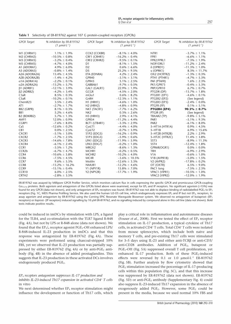
Identification of selective EP4 receptor antagonistsIn the course of screening for drugs unrelated to the prostanoidreceptors, we discovered a series of compounds that couldsuppress the expression of a stably transfected cytomegalovirus(CMV) promoter in HEK/293 cells. The CMV promoter isknown to be modulated by cAMP signalling (Hunninghakeet al., 1989), and we found that these compounds inhibited theinduction of CMV promoter activity by a factor present in FBS.The inducing factor in FBS was identified as PGE2 and induc-tion of cAMP was found to be mediated solely by the endog-enous EP4 receptor in HEK/293 cells (Supplementary Figs 1 and2). A representative of this series of compounds, ER-819762(structure shown in Fig. 1D), displaced PGE2 binding to humanEP4 receptors (IC50 value of 70 � 11 nmol·L-1; Fig. 1A), but didnot displace ligand binding to several related human pros-tanoid GPCRs, including EP2, DP, CRTH2 and TP receptors, andthe leukotriene GPCRs LTB4, CysLT1 and CysLT2 receptors(Fig. 1C). ER-819762 also suppressed human EP4 receptor-mediated cell signalling as measured in a cAMP-dependentreporter assay (IC50 value of 59 � 6 nmol·L-1) (Fig. 1B). In alarger cell signalling panel of 107 GPCRs, ER-819762(1 mmol·L-1) was highly selective for EP4 receptors, exhibitingno agonism or antagonism for any other receptor, includingthe related PGE2 EP1, EP2 and EP3 receptors (Table 1).
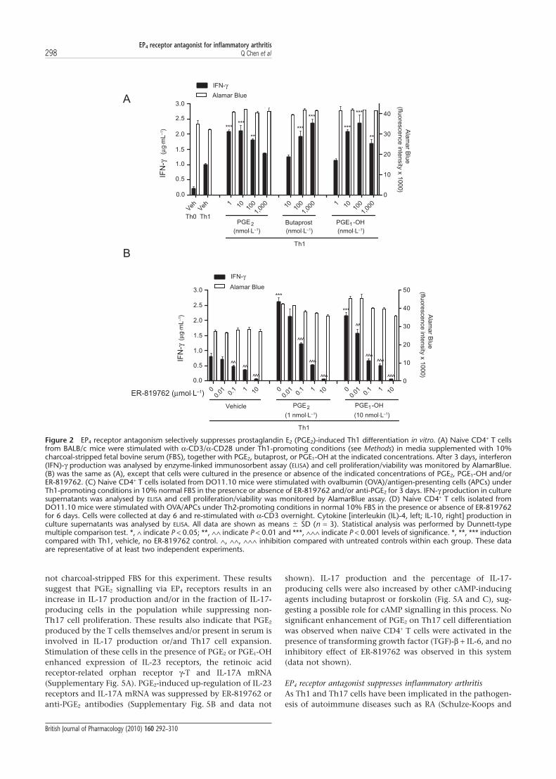
EP4 receptor antagonism suppresses in vitro Th1 differentiationAs PGE2 has been reported to modulate T cell differentiationand function, we tested the effect of ER-819762 in Th1 andTh2 differentiation assays. Th1 differentiation was induced byactivating naïve CD4+ T cells with anti-CD3 and anti-CD28antibodies in 10% charcoal-stripped FBS in the presence of
EP4 receptor antagonist for inflammatory arthritisQ Chen et al 295
British Journal of Pharmacology (2010) 160 292–310

IL-2, IL-12 and anti-IL-4 antibody. Th2 differentiation wasinduced by IL-4 and anti-IFN-g antibody. Addition of PGE2,butaprost (an EP2 receptor agonist) and prostaglandin E1
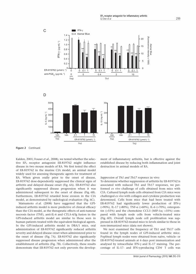
alcohol (PGE1-OH; an EP3/EP4 receptor agonist) significantlyenhanced the differentiation of naïve CD4+ T cells into Th1cells (Fig. 2A). ER-819762 suppressed PGE2- and PGE1-OH-induced IFN-g production by Th1-differentiating cells in aconcentration-dependent manner (Fig. 2B), but had no effecton cellular ATP levels (CellTiter-Glo, Promega), an indicatorof cell metabolic activity. Figure 2B also shows that ER-819762inhibited IFN-g in the absence of added prostaglandins, sug-gesting that the PGE2 produced by the T cells themselves actsin an autocrine manner to promote Th1 differentiation.ER-819762 had no effect on butaprost-stimulated IFN-g pro-duction at up to 1 mmol·L-1 (Supplementary Fig. 3). Th1 andTh2 differentiation were also induced by co-culturing naïveCD4+ T cells isolated from DO11.10 mice with mitomycinC-treated splenocytes and activating with the OVA peptideunder neutral, Th1- or Th2-polarizing conditions in normal10% FBS as described in Methods. In this experiment, IFN-gproduction was suppressed by either ER-819762 or a neutral-izing monoclonal anti-PGE2 antibody (clone 2B5; Fig. 2C),and these effects were non-additive. We also observed noeffect of ER-819762 on Th2 differentiation at up to10 mmol·L-1 (Fig. 2D).
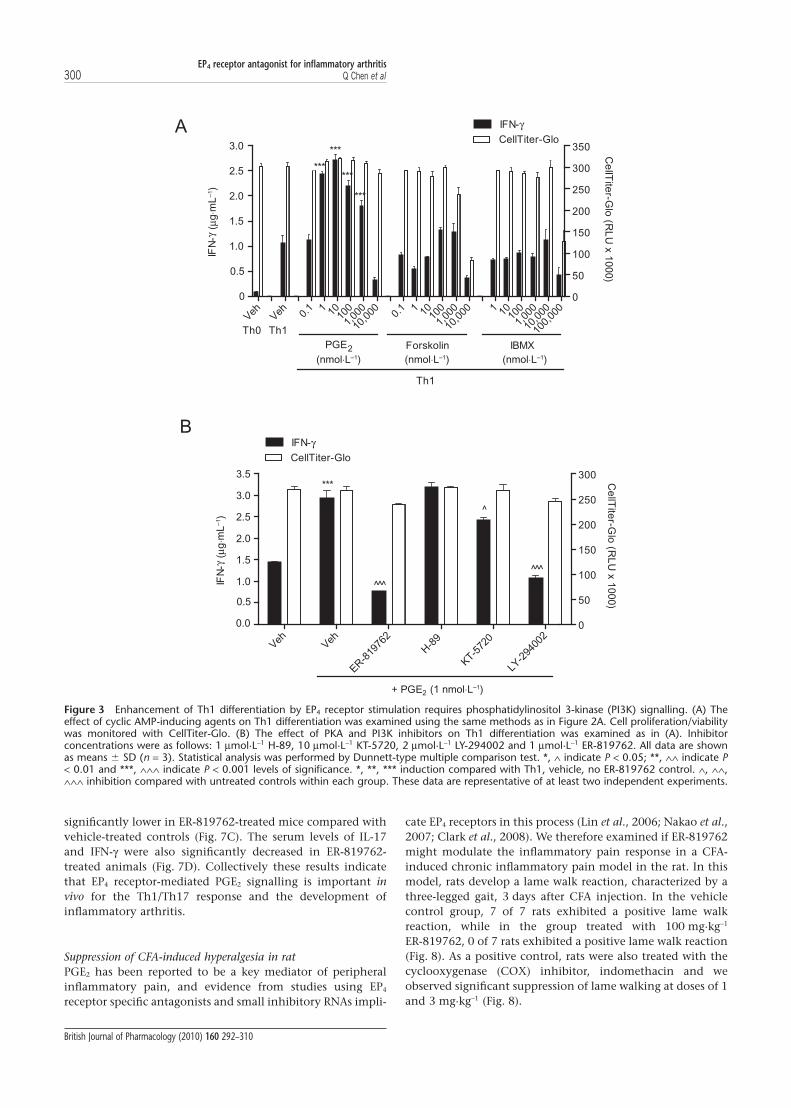
Although Th1 differentiation was enhanced by PGE2, asmeasured by increased IFN-g production (Fig. 2), neither for-skolin, an activator of adenylate cyclase, nor IBMX, a phos-phodiesterase inhibitor, caused a statistically significant
enhancement in IFN-g production (Fig. 3A), suggesting thatthe promotion of Th1 differentiation by PGE2 was not due tocAMP signalling. Moreover, PGE2-stimulated Th1 differentia-tion was not suppressed by the PKA inhibitors H-89(1 mmol·L-1) or was only weakly suppressed by a structurallyunrelated PKA inhibitor KT-5720 (10 mmol·L-1), but wasstrongly suppressed by the PI3K inhibitor LY294002(2 mmol·L-1), as well as by ER-819762 (Fig. 3B). Higher con-centrations of H-89 (10 mmol·L-1) were toxic (data notshown). These results suggest that the PI3K pathway, but notthe PKA-cAMP signalling pathway functioning downstreamof EP4 receptors is primarily responsible for PGE2-enhancedTh1 differentiation. Butaprost also induced Th1 differentia-tion (Fig. 2A), raising the possibility that EP2 receptors maysignal via PI3K in addition to PKA-cAMP.
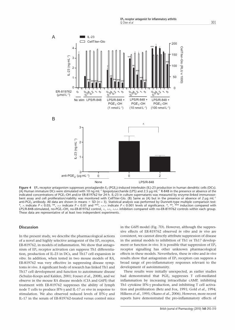
EP4 receptor antagonism suppresses IL-23 secretion in humanmonocyte-derived DCsIt was recently reported that PGE2 can promote Th17 celldifferentiation in mice by inducing DCs to preferentiallyproduce IL-23 (Sheibanie et al., 2007a; Khayrullina et al.,2008). Similarly, receptors that mobilize cAMP have beenreported to enhance IL-23 secretion by human DCs (Schnurret al., 2005). We therefore examined the role of EP4 receptorsignalling in immature human dendritic cells (imDCs).imDCs were generated from CD14+ monocytes by differentia-tion with GM-CSF plus IL-4 and assayed for IL-23 productionin media containing charcoal-stripped FBS. IL-23 production
N
NO
N
O
O
B
–10 –9 –8 –7 –6 –5 –10 –9 –8 –7 –6 –50
20
40
60
80
100
120
Log [ER-819762] (mol⋅L–1)
0.1 μmol⋅L–1
1 μmol⋅L–1
(mol⋅L–1)
0
20
40
60
80
100
120
% C
RE
B r
ep
ort
er
activity
C
Log [ER-819762]
A
% d
isp
lace
me
nt
of
[ H
] P
GE
32
–50 0 50 100 150
Leukotriene BLT
Leukotriene CysLT
Leukotriene CysLT
CRTH2
DP
EP
EP
TP
% inhibition of radioligand binding
ER-819762
4
2
2
1
D
Figure 1 Activity and structure of ER-819762. (A) Competitive displacement of radiolabelled prostaglandin E2 (PGE2) from cell membranesoverexpressing EP4 receptors (Millipore ChemiScreen). (B) Inhibition of PGE2-induced cAMP response element–placental-like alkaline phos-phatase reporter activity in human embryonic kidney cells, which express endogenous EP4 receptors (Supplementary Figure 2). Data arerepresentative of mean � SD derived from three independent experiments. (C) Competitive displacement of radiolabelled ligands from cellmembranes overexpressing various prostanoid and leukotriene receptors by 0.1 and 1 mmol·L-1 ER-819762 (data from MDS Pharma,Bothel, WA, USA). (D) Chemical structure of ER-819762: (S)-1’-(3,5-dimethylbenzyl)-2-ethyl-7,9-dimethoxy-10-methyl-5,10dihydrospiro[benzo[e]imidazo[1,5-a]azepine-1,4’-piperidin]-3(2H)-one.
EP4 receptor antagonist for inflammatory arthritis296 Q Chen et al
British Journal of Pharmacology (2010) 160 292–310
could be induced in imDCs by stimulation with LPS, a ligandfor the TLR4, and co-stimulation with the TLR7 ligand R-848(Fig. 4A), but not by LPS or R-848 alone (data not shown). Wefound that the EP3/4 receptor agonist PGE1-OH enhanced LPS/R-848-induced IL-23 production in imDCs and that thisresponse was antagonized by ER-819762 (Fig. 4A). Theseexperiments were performed using charcoal-stripped 10%FBS, yet we observed that IL-23 production was partially sup-pressed by either ER-819762 (Fig. 4A) or by anti-PGE2 anti-body (Fig. 4B) in the absence of added prostaglandins. Thissuggests that IL-23 production in these activated DCs involvesendogenously produced PGE2.
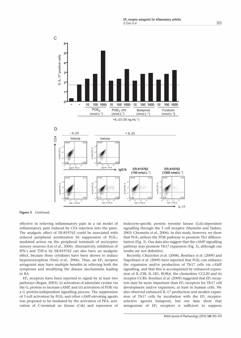
EP4 receptors antagonism suppresses IL-17 production andinhibits IL-23-induced Th17 expansion in activated CD4+ T cellsin vitroWe next determined whether EP4 receptor stimulation mightinfluence the development or function of Th17 cells, which
play a critical role in inflammation and autoimmune diseases(Fouser et al., 2008). First we tested the effect of EP4 receptorstimulation on IL-17 production, a typical cytokine of Th17cells, in activated CD4+ T cells. Total CD4+ T cells were isolatedfrom mouse splenocytes, which include both naïve andmemory T cells, and pre-existing Th17 cells were stimulatedfor 3–5 days using IL-23 and either anti-TCRb or anti-CD3/anti-CD28 antibodies. Addition of PGE2, butaprost orPGE1-OH (Fig. 5A) suppressed overall T cell proliferation, yetenhanced IL-17 production. Both of these PGE2-inducedeffects were reversed by 0.1 or 1.0 mmol·L-1 ER-819762(Fig. 5B). Further analysis by flow cytometry showed thatPGE2 stimulation increased the percentage of IL-17-producingcells within this population (Fig. 5C), and that this increasewas suppressed by ER-819762 (data not shown). ER-819762(Fig. 5D) or anti-PGE2 antibody (Supplementary Fig. 4) couldalso suppress IL-23-induced Th17 expansion in the absence ofexogenously added PGE2. However, some PGE2 could bepresent in the media, because we used normal 10% FBS and
Table 1 Selectivity of ER-819762 against 107 G protein-coupled receptors (GPCRs)
GPCR Target % inhibition by ER-819762(1 mmol·L-1)
GPCR Target % inhibition by ER-819762(1 mmol·L-1)
GPCR Target % inhibition by ER-819762(1 mmol·L-1)
M1 (CHRM1) 1.1% � 1.9% CCK2 (CCKBR) -8.1% � 4.0% NTR1 -3.7% � 1.1%M2 (CHRM2) -10.5% � 0.8% CRF1 (CRHR1) -16.2% � 0.4% FPR1 -6.6% � 1.1%M3 (CHRM3) -3.2% � 0.4% CRF2 (CRHR2) -9.5% � 0.1% FPR2/FPRL1 -7.3% � 1.9%M5 (CHRM5) -4.7% � 4.0% D1 -8.1% � 1.5% NOP/ORL1 -11.2% � 2.4%A1 (ADORA1) 4.9% � 6.0% D2 0.6% � 6.6% d (OPRD1) -11.3% � 1.0%A3 (ADORA3) -0.8% � 1.4% D5 25.2% � 9.5% k (OPRK1) 8.3% � 11.7%A2A (ADORA2A) 13.4% � 4.5% ETA (EDNRA) 4.2% � 2.4% OX2 (HCRTR2) -1.3% � 0.3%A2B (ADORA2B) -1.4% � 4.2% GPR40 -3.1% � 3.1% PTH1 (PTHR1) -4.7% � 5.3%a1A (ADRA1A) -2.2% � 0.1% GPR43 5.1% � 2.5% PAF (PTAFR) 1.6% � 2.3%a2A (ADRA2A) -13.2% � 5.7% GABBAb1 -9.7% � 0.5% PK1/GPR73 -0.4% � 5.3%b1 (ADRB1) -12.1% � 3.9% GAL1 (GALR1) 22.9% � 1.9% PRP/GPR10 6.7% � 0.7%b2 (ADRB2) -4.2% � 3.4% GCGR -4.5% � 2.0% PTGDR (DP) -13.7% � 1.8%C3aR 8.5% � 0.3% mGlu1 0.6% � 8.2% PTGER1 (EP1) -4.6% � 0.1%C5aR -10.2% � 0.1% GnRH -12.3% � 1.1% PTGER2 (EP2) (See legend)ChemR23 3.5% � 2.4% H1 (HRH1) -4.6% � 1.0% PTGER3 (EP3) -2.4% � 0.0%AT1 -2.7% � 1.7% H2 (HRH2) -4.8% � 0.9% PTGFR (FP) 0.1% � 5.1%BB2 (GRPR) 8.1% � 0.3% NK1 (TACR1) -7.7% � 6.2% PTGER4 (EP4) 90.3% � 0.7%BB1 5.2% � 4.6% NK2 0.3% � 2.6% PTGIR (IP) (See legend)B2 (BDKRB2) 5.7% � 1.3% H3 (HRH3) 2.9% � 4.1% TBXAR2 (TP) -9.8% � 5.1%AMY1 12.0% � 0.9% GPR54 -11.2% � 4.4% PAR1 -5.1% � 9.3%CGRP1 -7.6% � 8.0% BLT1 (LTB4R1) -3.3% � 2.9% PAR2 -6.1% � 8.0%CaS -12.6% � 0.2% CysLT1 -4.4% � 4.0% 5-HT1A (HTR1A) 0.9% � 4.5%CB1 0.0% � 2.5% CysLT2 -0.7% � 3.9% 5-HT1B 6.9% � 15.6%CXCR1 -5.1% � 3.0% S1P2 (EDG5) -16.2% � 0.9% 5-HT2B (HTR2B) 2.2% � 2.9%CXCR2 -1.7% � 2.5% S1P3 (EDG3) -5.9% � 0.6% 5–HT2C (HTR2C) 11.4% � 3.8%CXCR3 -14.6% � 7.8% LPA1 (EDG2) -11.9% � 5.9% SST3 -8.9% � 11.3%CXCR4 -6.1% � 2.4% LPA3 (EDG7) -0.2% � 1.0% SST4 -12.4% � 1.8%CCR1 -3.5% � 1.2% MRGX2 -8.6% � 1.3% GPR68(OGR1) 0.0% � 0.0%CCR2b -6.7% � 4.7% MCHR1 -0.2% � 0.5% TRH -6.9% � 2.9%CCR4 -10.6% � 5.8% MCHR2 -10.2% � 4.0% V1A -0.6% � 1.0%CCR6 -7.5% � 4.5% MC4R -3.6% � 10.5% V1B (AVPR1B) -5.0% � 1.5%CCR7 9.6% � 5.5% Motilin -12.6% � 3.3% V2 (AVPR2) -17.8% � 0.2%CCR8 -13.2% � 16.2% NMUR1 -12.2% � 4.6% OT (OXTR) 6.5% � 9.0%CCR9 -15.1% � 0.4% Y1 (NPY1R) 15.8% � 7.0% PAC1 long -7.5% � 2.3%CCR10 6.0% � 2.5% Y2 (NPY2R) -17.7% � 1.9% VPAC1 (VIPR1) -10.5% � 1.0%CRTH2 -2.8% � 3.5% VPAC2 (VIPR2) -12.0% � 1.9%
ER-819762 was assayed by Millipore GPCR Profiler Service, which monitors calcium flux in cells expressing the specific GPCR and promiscuous GPCR-couplingGa15/16 proteins. Both agonism and antagonism of the GPCRs listed above were examined, except for EP2 and IP receptors. No significant agonism (>15%) wasfound for any GPCR (data not shown), and only antagonism of EP4 receptors was found. ER-819762 was not able to displace binding of radiolabelled PGE2 to EP2
receptors (Fig. 1C, MDS Pharma Profiling Service. We also used the human U2-OS cell line, which endogenously expresses EP2 and IP but not EP4, receptors, toassay agonism and antagonism by ER-819762 using the Corning EPIC Resonate Waveguide Biosensor system. We observed no antagonism of butaprost- (EP2
receptors) or iloprost- (IP receptors) induced signalling by 10 mM ER-819762, and no signalling induced by compound alone in this cell line (data not shown). Boldtexts indicate positive results.
EP4 receptor antagonist for inflammatory arthritisQ Chen et al 297
British Journal of Pharmacology (2010) 160 292–310
not charcoal-stripped FBS for this experiment. These resultssuggest that PGE2 signalling via EP4 receptors results in anincrease in IL-17 production and/or in the fraction of IL-17-producing cells in the population while suppressing non-Th17 cell proliferation. These results also indicate that PGE2
produced by the T cells themselves and/or present in serum isinvolved in IL-17 production or/and Th17 cell expansion.Stimulation of these cells in the presence of PGE2 or PGE1-OHenhanced expression of IL-23 receptors, the retinoic acidreceptor-related orphan receptor g-T and IL-17A mRNA(Supplementary Fig. 5A). PGE2-induced up-regulation of IL-23receptors and IL-17A mRNA was suppressed by ER-819762 oranti-PGE2 antibodies (Supplementary Fig. 5B and data not
shown). IL-17 production and the percentage of IL-17-producing cells were also increased by other cAMP-inducingagents including butaprost or forskolin (Fig. 5A and C), sug-gesting a possible role for cAMP signalling in this process. Nosignificant enhancement of PGE2 on Th17 cell differentiationwas observed when naïve CD4+ T cells were activated in thepresence of transforming growth factor (TGF)-b + IL-6, and noinhibitory effect of ER-819762 was observed in this system(data not shown).
EP4 receptor antagonist suppresses inflammatory arthritisAs Th1 and Th17 cells have been implicated in the pathogen-esis of autoimmune diseases such as RA (Schulze-Koops and
Veh Veh 1 10 100
1,00
0 10 100
1,00
0 1 10 100
1,00
0
0.0
0.5
1.0
1.5
2.0
2.5
3.0
0
10
20
30
40
Th1
******
**
***
***
***
***
**
IF
N-γ
IFN-γ
Butaprost
Ala
mar B
lue
(fluore
sce
nce in
tensity
x 1
00
0)
Alamar Blue
Th0 Th1
A
00.
01 0.1 1 10 0
0.01 0.
1 1 10 00.
01 0.1 1 10
1.0
1.5
2.0
2.5
3.0
0
10
20
30
40
50
Vehicle
Th1
^̂^̂
^̂ ^
^̂ ^
^̂ ^
^̂ ^
^̂
^̂ ^^̂ ^
^̂ ^
***
***
IF
N- γ
Alamar Blue
IFN-γ
ER-819762
B
PGE2 PGE -OH1
PGE2 PGE -OH1
0.5
0.0
Ala
mar B
lue
( fluore
scen
ce in
tensity
x 1
000
)
(μg
⋅mL
–1)
(μg
⋅mL
–1)
(nmol⋅L–1)
(μmol⋅L–1)
(nmol⋅L–1)
(1 nmol⋅L–1) (10 nmol⋅L–1)
(nmol⋅L–1)
Figure 2 EP4 receptor antagonism selectively suppresses prostaglandin E2 (PGE2)-induced Th1 differentiation in vitro. (A) Naive CD4+ T cellsfrom BALB/c mice were stimulated with a-CD3/a-CD28 under Th1-promoting conditions (see Methods) in media supplemented with 10%charcoal-stripped fetal bovine serum (FBS), together with PGE2, butaprost, or PGE1-OH at the indicated concentrations. After 3 days, interferon(IFN)-g production was analysed by enzyme-linked immunosorbent assay (ELISA) and cell proliferation/viability was monitored by AlamarBlue.(B) was the same as (A), except that cells were cultured in the presence or absence of the indicated concentrations of PGE2, PGE1-OH and/orER-819762. (C) Naive CD4+ T cells isolated from DO11.10 mice were stimulated with ovalbumin (OVA)/antigen-presenting cells (APCs) underTh1-promoting conditions in 10% normal FBS in the presence or absence of ER-819762 and/or anti-PGE2 for 3 days. IFN-g production in culturesupernatants was analysed by ELISA and cell proliferation/viability was monitored by AlamarBlue assay. (D) Naive CD4+ T cells isolated fromDO11.10 mice were stimulated with OVA/APCs under Th2-promoting conditions in normal 10% FBS in the presence or absence of ER-819762for 6 days. Cells were collected at day 6 and re-stimulated with a-CD3 overnight. Cytokine [interleukin (IL)-4, left; IL-10, right] production inculture supernatants was analysed by ELISA. All data are shown as means � SD (n = 3). Statistical analysis was performed by Dunnett-typemultiple comparison test. *, ∧ indicate P < 0.05; **, ∧∧ indicate P < 0.01 and ***, ∧∧∧ indicate P < 0.001 levels of significance. *, **, *** inductioncompared with Th1, vehicle, no ER-819762 control. ∧, ∧∧, ∧∧∧ inhibition compared with untreated controls within each group. These dataare representative of at least two independent experiments.
EP4 receptor antagonist for inflammatory arthritis298 Q Chen et al
British Journal of Pharmacology (2010) 160 292–310
Kalden, 2001; Fouser et al., 2008), we tested whether the selec-tive EP4 receptor antagonist ER-819762 might influencedisease in two mouse models of RA. We first tested the effectof ER-819762 in the murine CIA model, an animal modelwidely used for assessing therapeutic agents for treatment ofRA. When given orally prior to the onset of disease,ER-819762 dose-dependently suppressed the clinical signs ofarthritis and delayed disease onset (Fig. 6A). ER-819762 alsosignificantly suppressed disease progression when it wasadministered subsequent to the onset of disease (Fig. 6B).Furthermore, ER-819762 retarded bone erosion in the CIAmodel, as demonstrated by radiological evaluation (Fig. 6C).
Matsumoto et al. (2008) have suggested that the GPI-induced arthritis model is more predictive of clinical efficacythan the CIA model, as the therapeutic effects of anti-tumournecrosis factor (TNF), anti-IL-6 and CTLA-4/Ig fusion in theGPI-induced arthritis model are similar to those seen inhuman patients treated with the equivalent biological agents.In the GPI-induced arthritis model in DBA/1 mice, oraladministration of ER-819762 significantly reduced arthritisseverity and delayed disease onset when administered prior tothe onset of disease (Fig. 7A). ER-819762 also significantlysuppressed disease progression when administered after theestablishment of arthritis (Fig. 7B). Collectively, these resultsdemonstrate that ER-819762 not only prevents the develop-
ment of inflammatory arthritis, but is effective against theestablished disease by reducing both inflammation and jointdestruction in animal models of RA.
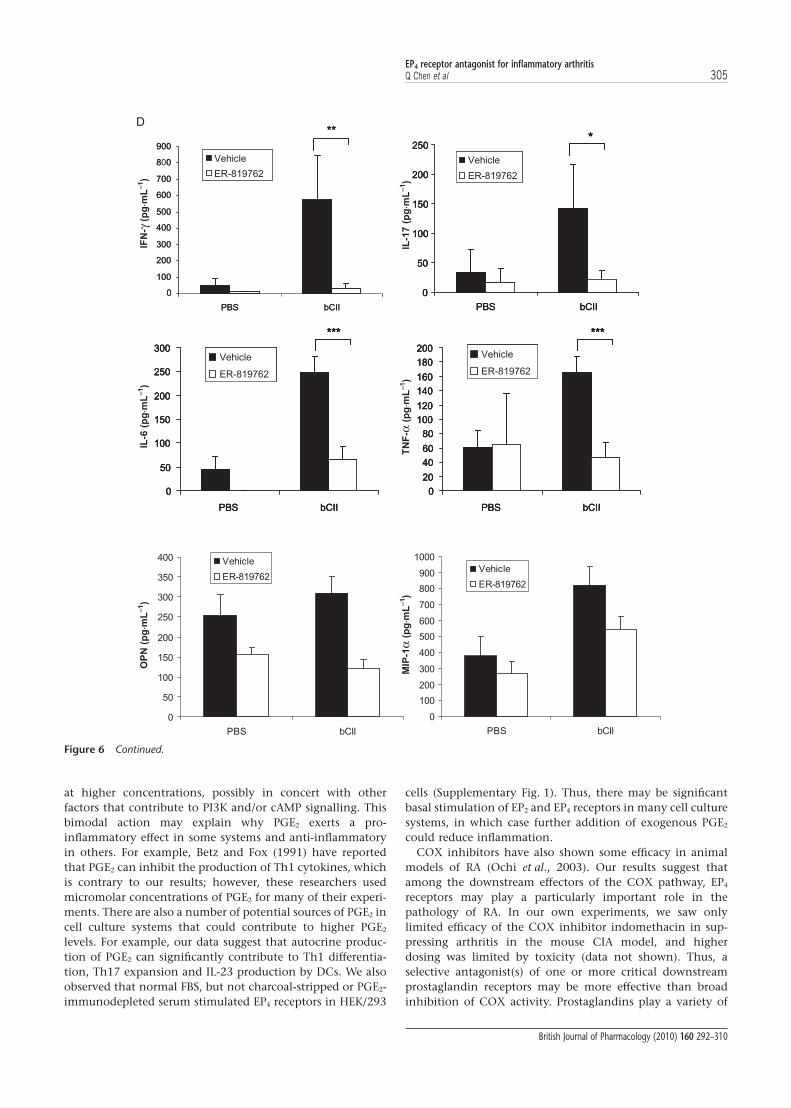
Suppression of Th1 and Th17 responses in vivoTo determine whether suppression of arthritis by ER-819762 isassociated with reduced Th1 and Th17 responses, we per-formed ex vivo challenge of cells obtained from mice withCIA. Cultured lymph node cells obtained from CIA mice werechallenged ex vivo with collagen and cytokine production wasdetermined. Cells from mice that had been treated withER-819762 had significantly lower production of IFN-g(>90%), IL-17 (>80%), TNF-a (>60%), IL-6 (>70%), osteopon-tin (>55%) and the chemokine CCL3 (MIP-1a; >35%) com-pared with lymph node cells from vehicle-treated mice(Fig. 6D). Overall lymph node cell proliferation was sup-pressed in ER-819762-treated mice to levels similar to those innon-immunized mice (data not shown).
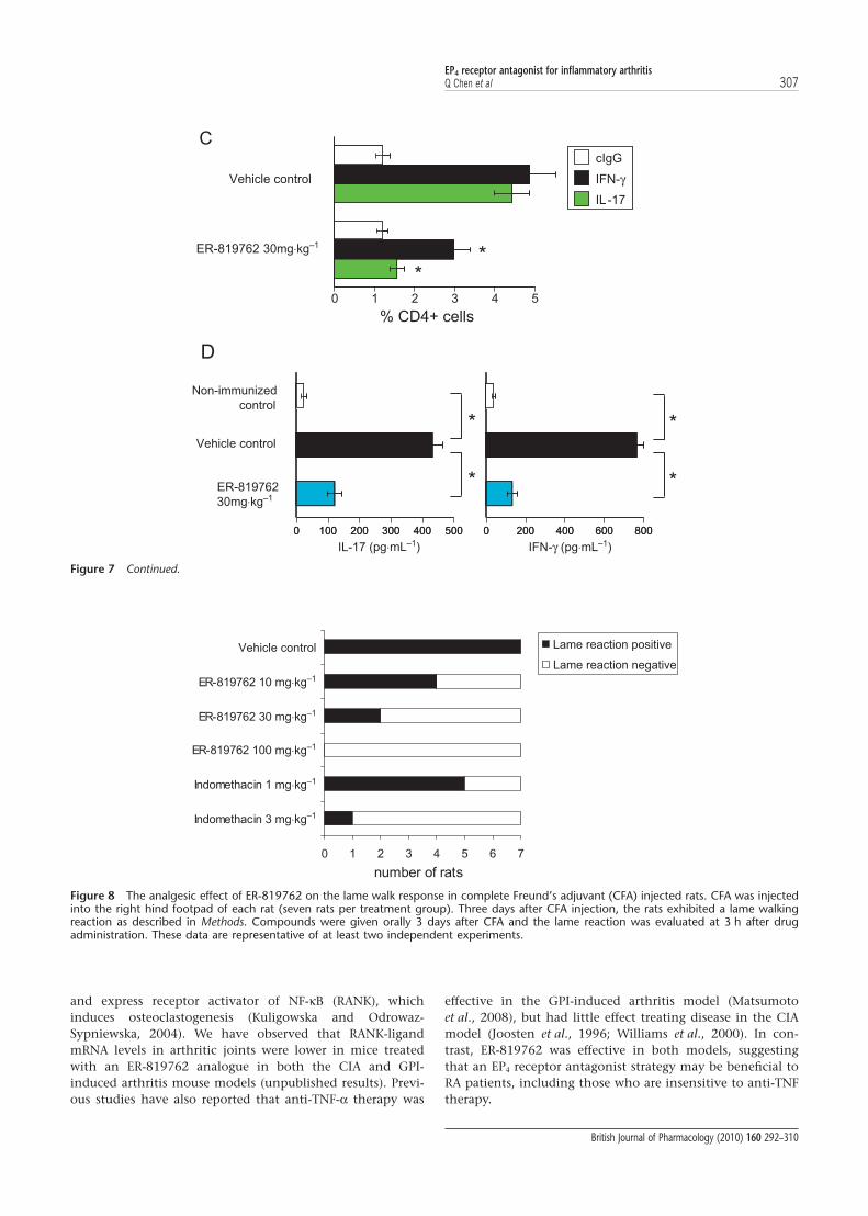
We next examined the frequency of Th1 and Th17 cellsfound in the lymph nodes of GPI-induced arthritic mice.Popliteal lymph nodes were obtained from naïve, vehicle- orER-819762-treated animals at 6 days post immunization andanalysed by intracellular IFN-g and IL-17 staining. The per-centage of IL-17- and IFN-g-producing CD4+ T cells was
C
D
0 0 0.1 1 0 0.1 1 0 0.1 1 0 0.1 10
0.2
0.4
0.6
0.8
1.0
1.2
1.4
0
5
10
15
20
0 0.4
Th1
2 100
Th0
^̂ ^
^̂ ^
^̂
^̂ ^
^̂ ^
^̂
^̂ ^
^̂ ^
^̂ ^
^̂ ^ ^̂ ^
IFN-γ Alamar Blue
IFN
-γ
ER-819762
anti-PGE
Veh Veh 0.1 1 100
100
200
300
400
500
ER-819762
Th2
Th0
IL-4
Veh Veh 0.1 1 100
10
20
30
ER-819762
Th2
Th0
IL-1
0
2
Ala
mar B
lue
(fluore
scence in
tensity
x 1
000)
(μg
⋅mL
–1)
(ng
⋅mL
–1)
(ng
⋅mL
–1)
(μg⋅mL–1)
(μmol⋅L–1) (μmol⋅L–1)
(μmol⋅L–1)
Figure 2 Continued.
EP4 receptor antagonist for inflammatory arthritisQ Chen et al 299
British Journal of Pharmacology (2010) 160 292–310
significantly lower in ER-819762-treated mice compared withvehicle-treated controls (Fig. 7C). The serum levels of IL-17and IFN-g were also significantly decreased in ER-819762-treated animals (Fig. 7D). Collectively these results indicatethat EP4 receptor-mediated PGE2 signalling is important invivo for the Th1/Th17 response and the development ofinflammatory arthritis.
Suppression of CFA-induced hyperalgesia in ratPGE2 has been reported to be a key mediator of peripheralinflammatory pain, and evidence from studies using EP4
receptor specific antagonists and small inhibitory RNAs impli-
cate EP4 receptors in this process (Lin et al., 2006; Nakao et al.,2007; Clark et al., 2008). We therefore examined if ER-819762might modulate the inflammatory pain response in a CFA-induced chronic inflammatory pain model in the rat. In thismodel, rats develop a lame walk reaction, characterized by athree-legged gait, 3 days after CFA injection. In the vehiclecontrol group, 7 of 7 rats exhibited a positive lame walkreaction, while in the group treated with 100 mg·kg-1
ER-819762, 0 of 7 rats exhibited a positive lame walk reaction(Fig. 8). As a positive control, rats were also treated with thecyclooxygenase (COX) inhibitor, indomethacin and weobserved significant suppression of lame walking at doses of 1and 3 mg·kg-1 (Fig. 8).
A
B
Veh Veh 0.1 1 1010
0
1,00
0
10,0
00 0.1 1 1010
0
1,00
0
10,0
00 1 10100
1,00
0
10,0
00
100,
000
0
0.5
1.0
1.5
2.0
2.5
3.0
0
50
100
150
200
250
300
350
IFN-γCellTiter-Glo
Th0 Th1
PGE Forskolin IBMX
Th1
***
***
***
***
IFN
-γ
Ce
l lTit e
r- Glo
(RL
U x
1000)
2
Veh Veh
ER-8
1976
2
H-8
9
KT-572
0
LY-2
9400
2
0.0
0.5
1.0
1.5
2.0
2.5
3.0
3.5
0
50
100
150
200
250
300
IFN-γCellTiter-Glo
+ PGE2
^̂ ^
^̂ ^
^
***
IFN
-γ
Ce
llTite
r -Glo
(RL
U x
1000)
(μg
⋅mL
–1)
(μg
⋅mL
–1)
(nmol⋅L–1) (nmol⋅L–1)
(1 nmol⋅L–1)
(nmol⋅L–1)
Figure 3 Enhancement of Th1 differentiation by EP4 receptor stimulation requires phosphatidylinositol 3-kinase (PI3K) signalling. (A) Theeffect of cyclic AMP-inducing agents on Th1 differentiation was examined using the same methods as in Figure 2A. Cell proliferation/viabilitywas monitored with CellTiter-Glo. (B) The effect of PKA and PI3K inhibitors on Th1 differentiation was examined as in (A). Inhibitorconcentrations were as follows: 1 mmol·L-1 H-89, 10 mmol·L-1 KT-5720, 2 mmol·L-1 LY-294002 and 1 mmol·L-1 ER-819762. All data are shownas means � SD (n = 3). Statistical analysis was performed by Dunnett-type multiple comparison test. *, ∧ indicate P < 0.05; **, ∧∧ indicate P< 0.01 and ***, ∧∧∧ indicate P < 0.001 levels of significance. *, **, *** induction compared with Th1, vehicle, no ER-819762 control. ∧, ∧∧,∧∧∧ inhibition compared with untreated controls within each group. These data are representative of at least two independent experiments.
EP4 receptor antagonist for inflammatory arthritis300 Q Chen et al
British Journal of Pharmacology (2010) 160 292–310
Discussion
In the present study, we describe the pharmacological actionsof a novel and highly selective antagonist of the EP4 receptor,ER-819762, in models of inflammation. We show that antago-nism of EP4 receptor activation can suppress Th1 differentia-tion, production of IL-23 in DCs, and Th17 cell expansion invitro. In addition, when tested in two mouse models of RA,ER-819762 was very effective in suppressing disease symp-toms in vivo. A significant body of research has linked Th1 andTh17 cell development and function to autoimmune disease(Schulze-Koops and Kalden, 2001; Fouser et al., 2008), and weobserve in the mouse RA disease models (CIA and G6PI) thattreatment with ER-819762 suppresses the ability of lymphnode T cells to produce IFN-g and IL-17 ex vivo in response tostimulation. We also observed reduced levels of IFN-g andIL-17 in the serum of ER-819762-treated versus control mice
in the G6PI model (Fig. 7D). However, although the suppres-sive effects of ER-819762 observed in vitro and in vivo areconsistent, we cannot directly attribute suppression of diseasein the animal models to inhibition of Th1 or Th17 develop-ment or function in vivo. It is possible that suppression of EP4
receptor signalling has other unknown pharmacologicaleffects in these models. Nevertheless, these in vitro and in vivoresults show that antagonism of EP4 receptors can suppress abroad range of pro-inflammatory responses relevant to thedevelopment of autoimmunity.
These results were initially unexpected, as earlier studieshad demonstrated that PGE2 suppresses T cell-mediatedinflammation by increasing intracellular cAMP, inhibitingTh1 cytokine IFN-g production, and inhibiting T cell activa-tion and proliferation (Betz and Fox, 1991; Gold et al., 1994;Hilkens et al., 1995; Okano et al., 2006). However, more recentreports have demonstrated the pro-inflammatory effects of
0 00.
04 0.2 1 5 0
0.04 0.
2 1 5 00.
04 0.2 1 5 0
0.04 0.
2 1 5
0
1
2
3
4
0
50
100
150
200
IL-23
CellTiter-Glo
ER-819762
LPS/R-848 LPS/R-848 + LPS/R-848 + LPS/R-848 +No stim
^̂ ^ ^̂ ^^̂ ^^̂ ^
*
***
***
^̂^̂ ^ ^̂ ^ ^̂ ^
^̂
^̂ ^
^̂ ^^̂ ^
^̂ ^
IL-2
3
Ce
ll Tite
r- Glo
(RL
U x
100
0)
A
B
0
1
2
3
None
anti-PGE
^
IL-2
3
LPS/R-848
0 0 2
PGE -OH1 PGE -OH1 PGE -OH1
2
(ng
⋅mL
–1)
(ng
⋅mL
–1)
(μmol⋅L–1)
(1 nmol⋅L–1) (10 nmol⋅L–1) (100 nmol⋅L–1)
(μg⋅mL–1)
Figure 4 EP4 receptor antagonism suppresses prostaglandin E2 (PGE2)-induced interleukin (IL)-23 production in human dendritic cells (DCs).(A) Human immature DCs were stimulated with 10 ng mL-1 lipopolysaccharide (LPS) and 2.5 mg mL-1 R-848 in the presence or absence of theindicated concentrations of PGE1-OH and/or ER-819762 for 24 h. IL-23 in culture supernatants was measured by enzyme-linked immunosor-bent assay and cell proliferation/viability was monitored with CellTiter-Glo. (B) Same as (A) but in the presence or absence of 2 mg mL-1
anti-PGE2 antibody. All data are shown in means � SD (n = 3). Statistical analysis was performed by Dunnett-type multiple comparison test:*, ∧ indicate P < 0.05; **, ∧∧ indicate P < 0.01 and ***, ∧∧∧ indicate P < 0.001 levels of significance. *, **, *** induction compared withLPS/R-848-stimulated, no-PGE1-OH, no-ER-819762 control, ∧, ∧∧, ∧∧∧ inhibition compared with no-ER-819762 controls within each group.These data are representative of at least two independent experiments.
EP4 receptor antagonist for inflammatory arthritisQ Chen et al 301
British Journal of Pharmacology (2010) 160 292–310

PGE2 in Th17 development (Chizzolini et al., 2008; Bonifaceet al., 2009; Napolitani et al., 2009) and DC activation(Sheibanie et al., 2004; Khayrullina et al., 2008). As antago-nism of EP4 receptor signalling suppressed Th1 differentia-tion, Th17 cell expansion, and the development ofpathologies in mouse CIA- and GPI-induced arthritis, wepropose that the immune stimulatory activities of PGE2 arerelevant to these diseases.
Another debilitating aspect of RA is the pain associated withjoint inflammation. This inflammatory pain is mediated, at
least in part, by PGE2 stimulation of EP4 receptors (Lin et al.,2006; Nakao et al., 2007). Selective inhibition of EP4 receptorsignalling by several different EP4 receptor antagonists hasbeen shown to cause a marked reduction in joint pain,mechanical and thermal hyperalgesia and oedema in rat andin guinea pig models of pain and inflammation, often withsimilar efficacy to that observed with selective COX-2 inhibi-tors such as rofecoxib (Lin et al., 2006; Nakao et al., 2007;Clark et al., 2008; Murase et al., 2008; Jones et al., 2009). Con-sistent with these findings, we observed that ER-819762 was
Veh Veh 1 10 100
1,00
0 1 10 100
1,00
0 1 10 100
1,00
0 1 10 100
1,00
0
0
1
2
3
4
5
6
0
50
100
150
200
250
300
IL-17
CellTiter-Glo
Butaprost
+ IL-23
Forskolin
***
***
***
**
******
***
***
*IL
-17
Ce
llTite
r -Glo
( RL
U x
10
00
)
A
B
Veh Veh 0.1 1
0
1
2
3
4
5
6
0
50
100
150
200
250
300
350
400
IL-17
CellTiter-Glo
ER-819762
+ IL-23
***
^
^̂
IL-1
7
Ce
llTite
r-Glo
( RL
U x
1000)
PGE PGE -OH2 1
+ PGE 2
(ng
⋅mL
–1)
(ng
⋅mL
–1)
(10ng⋅mL–1)
(10 nmol⋅L–1)
(nmol⋅L–1) (nmol⋅L–1)
(10ng⋅mL–1)
(nmol⋅L–1) (nmol⋅L–1)
(μmol⋅L–1)
Figure 5 Prostaglandin E2 (PGE2)-EP4 receptor signalling regulates Th17 cell development. (A) Total CD4+ T cells isolated from mousesplenocytes were stimulated with anti-CD3/anti-CD28 plus interleukin (IL)-23 in the presence or absence of exogenous PGE2, butaprost,PGE1-OH, or forskolin for 3 days. IL-17 in culture supernatants was measured by enzyme-linked immunosorbent assay. (B) Same methods asin (A), except that ER-819762 was added at the indicated concentrations. (C) Total CD4+ T cells were stimulated with a-TCRb/a-CD28 � IL-23in the presence or absence of exogenously added PGE2, butaprost, PGE1-OH, or forskolin for 5 days and the percentage of Th17 cells wasanalysed by IL-17 intracellular staining. The horizontal broken line represents the level of IL-17 positive cells in the presence of IL-23 only. (D)Same methods as in (C), except that no PGs were added, and ER-819762 was added at the indicated concentrations. The number of Th17 cellswas analysed by IL-17 intracellular staining. Upper plots show staining with control isotype-matched staining antibody, bottom plots showstaining with anti-IL-17 antibody. First two columns show unstimulated and IL-23-stimulated cells. Right-hand lower two plots showIL-23-stimulated cells treated with different concentrations of ER-819762. All data are shown in means � SD (n = 3). Statistical analysis wasperformed by Dunnett-type multiple comparison test: *, ∧ indicate P < 0.05; **, ∧∧ indicate P < 0.01 and ***, ∧∧∧ indicate P < 0.001 levelsof significance. *, **, *** induction compared with lipopolysaccharide/R-848-stimulated, no-PGE1-OH, no-ER-819762 control, ∧, ∧∧, ∧∧∧inhibition compared with no-ER-819762 controls within each group. These data are representative of at least two independent experiments.
EP4 receptor antagonist for inflammatory arthritis302 Q Chen et al
British Journal of Pharmacology (2010) 160 292–310
effective in relieving inflammatory pain in a rat model ofinflammatory pain induced by CFA injection into the paws.The analgesic effect of ER-819762 could be associated withreduced peripheral sensitization by suppression of PGE2-mediated action on the peripheral terminals of nociceptorsensory neurons (Lin et al., 2006). Alternatively, inhibition ofIFN-g and TNF-a by ER-819762 can also have an analgesiceffect, because these cytokines have been shown to inducehypernociception (Verri et al., 2006). Thus, an EP4 receptorantagonist may have multiple benefits in relieving both thesymptoms and modifying the disease mechanisms leadingto RA.
EP4 receptors have been reported to signal by at least twopathways (Regan, 2003): (i) activation of adenylate cyclase viathe Gs protein to increase cAMP, and (ii) activation of PI3K viaa G protein-independent signalling process. The suppressionof T-cell activation by PGE2 and other cAMP-elevating agentswas proposed to be mediated by the activation of PKA, acti-vation of C-terminal src kinase (Csk) and repression of
leukocyte-specific protein tyrosine kinase (Lck)-dependentsignalling through the T cell receptor (Mustelin and Tasken,2003; Chemnitz et al., 2006). In this study, however, we showthat PGE2 utilizes the PI3K pathway to promote Th1 differen-tiation (Fig. 3). Our data also suggest that the cAMP signallingpathway may promote Th17 expansion (Fig. 5), although ourresults are not definitive.
Recently, Chizzolini et al. (2008), Boniface et al. (2009) andNapolitani et al. (2009) have reported that PGE2 can enhancethe expansion and/or production of Th17 cells via cAMPsignalling, and that this is accompanied by enhanced expres-sion of IL-23R, IL-1R1, RORgt, the chemokine CCL20 and itsreceptor CCR6. Boniface et al. (2009) suggested that EP2 recep-tors may be more important than EP4 receptors for Th17 celldevelopment and/or expansion, at least in human cells. Wealso observed enhanced IL-17 production and modest expan-sion of Th17 cells by incubation with the EP2 receptor-selective agonist butaprost, but our data show thatantagonism of EP4 receptors is sufficient to suppress
- - 10 100 1000 10 100 1000 10 100 1000 10 100 10000
1
2
3
4
5
6
(nmol.L –1) (nmol.L ) (nmol.L–1) (nmol.L–1)
+IL-23 (30 ng.mL –1)
10 100 1000 10 100 1000 10 100 1000 10 100 10000
1
2
3
4
5
6
–1 )
C
IgG1k
+ IL-23
CD
4
IgG1k
- IL-23
Vehicle Vehicle
D
% IL-1
7 p
ositiv
e c
ells
PGE2 PGE1 -OH Butaprost Forskolin
mIL-17 PE-A10
010
110
210
310
0
101
102
103
mC
D4
FITC
-A
93.05 0.17
6.50 0.28
mIL-17 PE-A10
010
110
210
3
0
101
102
103
mC
D4
FITC
-A
10
91.87 3.72
4.13 0.28
mIL-17 PE-A10
010
110
210
310
0
101
102
103
mC
D4
FITC
-A
94.44 2.35
2.96 0.25
mIL-17 PE-A10
010
110
210
3
0
101
102
103
mC
D4
FITC
-A
10
94.02 1.38
4.38 0.23
IgG1k PE-A10
010
110
210
310
0
101
102
103
mC
D4
FITC
-A
94.89 0.03
5.08 0.00
IgG1k PE-A10
010
110
210
3
0
101
102
103
mC
D4
FITC
-A
10
96.40 0.06
3.54 0.00
IL-17
ER-819762
(100 nmol.L–1)
ER-819762
(1000 nmol.L–1)
ER-819762
(100 nmol.L
ER-819762
(1000 nmol.L
Figure 5 Continued.
EP4 receptor antagonist for inflammatory arthritisQ Chen et al 303
British Journal of Pharmacology (2010) 160 292–310

PGE2-mediated Th17 expansion and IL-17 production inmouse cells. Our data also showed that PGE2 did not promoteTh17 differentiation per se, as we did not see an increase inTh17 cell frequency following PGE2 stimulation of purifiednaïve CD4+ T cells in the presence of TGF-b and IL-6. Rather,we observed an increase in Th17 cells when total CD4+ T cellswere stimulated with PGE2 in the presence of IL-23, indicatingthe expansion of pre-differentiated Th17 cells. Napolitaniet al. (2009) suggested that PGE2 acts by inhibiting expansionof CCR6- T cells rather than increasing the proliferation ofCCR6+ Th17 cells, independent of IL-23. In agreement withthis report, we also observed enhanced IL-17 production byPGE2 in the absence of IL-23 co-stimulation (data not shown).In addition, we showed that EP4 receptor stimulation canenhance IL-23 production by activated human DCs and thatthis activity can be inhibited by a selective EP4 receptorantagonist or anti-PGE2 antibody in the presence or absenceof exogenously added PGE2. Sheibanie et al. (2007a) have also
recently reported that PGE2 exacerbates disease in the CIAmouse by enhancing DC IL-6 and IL-23 production, the latterof which maintains Th17 cell survival and proliferation andconsequently promotes IL-17 production. Collectively, theseresults support the idea that PGE2 stimulation of EP4 receptorspromotes Th17 cell expansion at two stages by: (i) enhancingIL-23 production by DCs, and (ii) directly acting on memoryT cells to promote IL-17 production and Th17 cell expansion(Sheibanie et al., 2004; Chizzolini et al., 2008; Khayrullinaet al., 2008; Boniface et al., 2009).
We observed that while low concentrations of PGE2 pro-moted IFN-g production under Th1-differentiation condi-tions, production started to decrease at higher concentrationsof PGE2 or PGE1-OH without loss of cell viability (Figs 2A and3A). Similar results were seen with higher concentrations ofbutaprost (data not shown). Thus, PGE2 appears to have abimodal effect on immune stimulation; promoting inflamma-tion at lower concentrations while attenuating inflammation
0
1
2
3
4
5
6
0 1 2 10
Days after 2nd immunization
*
Art
hritis s
core
Art
hritis s
core
***
***
***
A B
Vehicle control
ER-819762 10
ER-819762 30
ER-819762 100
3 4 5 6 7 8 9
*
0
1
2
3
4
5
6
7
8
6 7 8 9 10 11 12 14 15 16 17 1813
***
**
Days after 2nd immunization
Vehicle control
ER-819762 10 mg⋅kg–1
mg⋅kg–1
mg⋅kg–1
mg⋅kg–1
mg⋅kg–1
mg⋅kg–1
ER-819762 30
ER-819762 100
prophylactic dosing
therapeutic dosing
C
*
Vehiclecontrol
10 30 1000
2
4
6
8
10
12
X-r
ay s
co
re
ER-819762
* *
Figure 6 EP4 receptor antagonism suppresses disease and Th1/Th17 cytokines in collagen-induced arthritis in mice. (A) DBA/1 mice wereimmunized with bovine type II collagen (bCII)/complete Freund’s adjuvant (CFA) (primary immunization) and boosted with bCII in incompleteFreund’s adjuvant at day 21 (second immunization) to induce arthritis as described in Methods. ER-819762 was orally administered daily fromday 20 after primary immunization but before the onset of disease, and arthritis scores were monitored over the course of the study asdescribed in Methods. (B) Same methods as in (A), but ER-819762 was administered after induction of disease on day 7 after secondimmunization. (C) Radiological analysis of inflamed paws at the end of the therapeutic collagen-induced arthritis study shown in 6B. The X-rayscore is defined in Methods. (D) Ex vivo cytokine analysis. Mice were immunized with bCII/CFA or vehicle, similar to (A), except that ER-819762was administered from the day of primary immunization (day 0). Lymph node cells were purified at day 15 and cultured in the presence ofbCII (50 mg mL-1) or phosphate-buffered saline for 72 h, and cytokine production was analysed. Statistical analysis was performed byDunnett-type multiple comparison test compared with vehicle control (A–C) or paired t-test (D). Levels of significance: *P < 0.05; **P < 0.01;***P < 0.001. These data are representative of at least two independent experiments.
EP4 receptor antagonist for inflammatory arthritis304 Q Chen et al
British Journal of Pharmacology (2010) 160 292–310
at higher concentrations, possibly in concert with otherfactors that contribute to PI3K and/or cAMP signalling. Thisbimodal action may explain why PGE2 exerts a pro-inflammatory effect in some systems and anti-inflammatoryin others. For example, Betz and Fox (1991) have reportedthat PGE2 can inhibit the production of Th1 cytokines, whichis contrary to our results; however, these researchers usedmicromolar concentrations of PGE2 for many of their experi-ments. There are also a number of potential sources of PGE2 incell culture systems that could contribute to higher PGE2
levels. For example, our data suggest that autocrine produc-tion of PGE2 can significantly contribute to Th1 differentia-tion, Th17 expansion and IL-23 production by DCs. We alsoobserved that normal FBS, but not charcoal-stripped or PGE2-immunodepleted serum stimulated EP4 receptors in HEK/293
cells (Supplementary Fig. 1). Thus, there may be significantbasal stimulation of EP2 and EP4 receptors in many cell culturesystems, in which case further addition of exogenous PGE2
could reduce inflammation.COX inhibitors have also shown some efficacy in animal
models of RA (Ochi et al., 2003). Our results suggest thatamong the downstream effectors of the COX pathway, EP4
receptors may play a particularly important role in thepathology of RA. In our own experiments, we saw onlylimited efficacy of the COX inhibitor indomethacin in sup-pressing arthritis in the mouse CIA model, and higherdosing was limited by toxicity (data not shown). Thus, aselective antagonist(s) of one or more critical downstreamprostaglandin receptors may be more effective than broadinhibition of COX activity. Prostaglandins play a variety of
**
0
100
200
300
400
500
600
700
800
900
PBS bCII
Vehicle
ER-819762
*
0
50
100
150
200
250
PBS bCII
Vehicle
ER-819762
**
0
100
200
300
400
500
600
700
8 0
900
PBS bCII
IFN
-γ (p
g◊m
L–1)
TN
F-α
(pg◊m
L–1)
MIP
-1α
(pg◊m
L–1)
IL-1
7 (p
g◊m
L–1)
IL-6
(pg◊m
L–1)
OP
N (p
g◊m
L–1)
Vehicle
ER-819762
*
0
50
100
150
200
250
PBS bCII
Vehicle
ER-819762
*
0
50
100
150
200
250
PBS bCII
Vehicle
ER-819762
D
***
0
50
100
150
200
250
300
PBS bCII
Vehicle
ER-819762
***
0
20
40
60
80
100
120
140
160
180
200
PBS bCII
Vehicle
ER-819762
***
0
50
100
150
200
250
300
PBS bCII
Vehicle
ER-819762
***
0
50
100
150
200
250
300
PBS bCII
Vehicle
ER-819762
***
0
20
40
60
80
100
120
140
160
180
200
PBS bCII
Vehicle
ER-819762
***
0
20
40
60
80
100
120
140
160
180
200
PBS bCII
Vehicle
ER-819762
0
50
100
150
200
250
300
350
400
PBS bCII
Vehicle
ER-819762
0
100
200
300
400
500
600
700
800
900
1000
PBS bCII
Vehicle
ER-819762
Figure 6 Continued.
EP4 receptor antagonist for inflammatory arthritisQ Chen et al 305
British Journal of Pharmacology (2010) 160 292–310

roles in modulating inflammation and can exert both anti-and pro-inflammatory effects. For example, one proposedexplanation for why aspirin and other COX inhibitors areineffective in treating allergic inflammation is that PGD2
produced downstream of the COX enzymes stimulates theDP receptor, which promotes allergic inflammation, whilePGE2 stimulates the EP3 receptor, which suppresses allergicinflammation (Kunikata et al., 2005). In addition, the moretargeted approach of antagonizing EP4 receptors mightsuppress inflammation without the side-effects associatedwith some non-steroidal anti-inflammatory drugs andCOX inhibitors, including increased gastrointestinal and
cardiovascular risks. Consistent with this, Takeuchi et al.(2007) showed that the EP4 receptor antagonist CJ-042794did not produce any damaging effects in the gastrointestinalmucosa of control or adjuvant-induced arthritic rats,whereas indomethacin caused gross lesions. More impor-tantly, we found that ER-819762 not only could prevent, butcould suppress established disease in the CIA and GPI-induced arthritis models. Bone destruction in CIA was alsosignificantly reduced by ER-819762. The effects ofER-819762 in suppressing bone destruction may be due inpart to suppression of osteoclastogenesis promoted by IL-17and PGE2. IL-17 stimulates osteoblasts to synthesize PGE2
1
2
3
4
5
6
7
8
9
Days after immunization
Arth
ritis
scor
e
* * ****
*
*
** *
** *
0123456789
10
8 10 12 14 16 18 20
4
Days after immunization
*****A
rthrit
issc
ore
A
B
7 8 9 10 11 12 13 14
**
*
*
*
Vehicle control
ER-819762 10
ER-819762 30
Vehicle control
ER-819762 10
ER-819762 30
Compound administered from day 6 (prophylactic)
Compound administered from day 9 (therapeutic)
mg⋅kg–1
mg⋅kg–1
mg⋅kg–1
mg⋅kg–1
Figure 7 EP4 receptor antagonism suppresses disease and Th1/Th17 cytokines in glucose-6-phosphate isomerase (GPI)-induced arthritis inmice. (A) DBA/1 mice were immunized with GPI/complete Freund’s adjuvant to induce arthritis as described in Methods. ER-819762 was orallyadministered daily from day 6 after immunization, but before the onset of disease. Clinical scores were monitored over the course of the study.(B) Same methods as in (A), but ER-819762 was administered after disease induction (day 9). (C) Same methods as in (A), but ER-819762 wasadministered from the day of immunization. Popliteal lymph node cells were removed from mice at day 6 and re-stimulated with GPI in culture.interleukin (IL)-17- and interferon (IFN)-g-producing cells were quantified by intracellular staining and flow cytometry. Experiments with isotypecontrol IgG are shown as cIgG. (D) Serum was collected at the end of the GPI study shown in (A), and analysed by IL-17 and IFN-genzyme-linked immunosorbent assay. Statistical analysis was performed by Dunnett-type multiple comparison test compared with vehiclecontrol (A and B) or paired t-test (C and D). Levels of significance: *P < 0.05; **P < 0.01; ***P < 0.001. These data are representative of at leasttwo independent experiments.
EP4 receptor antagonist for inflammatory arthritis306 Q Chen et al
British Journal of Pharmacology (2010) 160 292–310
and express receptor activator of NF-kB (RANK), whichinduces osteoclastogenesis (Kuligowska and Odrowaz-Sypniewska, 2004). We have observed that RANK-ligandmRNA levels in arthritic joints were lower in mice treatedwith an ER-819762 analogue in both the CIA and GPI-induced arthritis mouse models (unpublished results). Previ-ous studies have also reported that anti-TNF-a therapy was
effective in the GPI-induced arthritis model (Matsumotoet al., 2008), but had little effect treating disease in the CIAmodel (Joosten et al., 1996; Williams et al., 2000). In con-trast, ER-819762 was effective in both models, suggestingthat an EP4 receptor antagonist strategy may be beneficial toRA patients, including those who are insensitive to anti-TNFtherapy.
IL -17IFN-
cIgG
0 1 2 3 4 5
IL-17IFN-
cIgG
IL-17IFN-γ
cIgG
**
C
D
0 100 200 300 400 500 0 200 400 600 800
*
* *
0 100 200 300 400 500 0 200 400 600 800
*
% CD4+ cells
IL-17 IFN-γ
Vehicle control
ER-819762
Non-immunized control
Vehicle control
ER-819762
(pg⋅mL–1) (pg⋅mL–1)
30mg⋅kg–1
30mg⋅kg–1
Figure 7 Continued.
0 1 2 3 4 5 6 7
Vehicle control
ER-819762 10
ER-819762 30
ER-819762 100
Indomethacin 1
Indomethacin 3
Lame reaction positive
Lame reaction negative
number of rats
mg⋅kg–1
mg⋅kg–1
mg⋅kg–1
mg⋅kg–1
mg⋅kg–1
Figure 8 The analgesic effect of ER-819762 on the lame walk response in complete Freund’s adjuvant (CFA) injected rats. CFA was injectedinto the right hind footpad of each rat (seven rats per treatment group). Three days after CFA injection, the rats exhibited a lame walkingreaction as described in Methods. Compounds were given orally 3 days after CFA and the lame reaction was evaluated at 3 h after drugadministration. These data are representative of at least two independent experiments.
EP4 receptor antagonist for inflammatory arthritisQ Chen et al 307
British Journal of Pharmacology (2010) 160 292–310

These results and methodologies have been shared earlierwith colleagues in another laboratory, and they have recentlyconfirmed that PGE2-EP4 receptor signalling promotes Th1cell differentiation, IL-23 production by DCs and Th17 cellexpansion (Yao et al., 2009). This group also tested an EP4
receptor antagonist with a very different molecular structurefrom ER-819762, supporting the idea that the anti-inflammatory effects of ER-819762 are indeed due to EP4
receptor antagonism and not due to action on another, uni-dentified target of the compound.
In summary, we show that an antagonist of EP4 receptors,ER-819762, can suppress inflammation at multiple stages, assummarized in Fig. 9, as well as moderating inflammatorypain. Our results suggest that selective antagonism of EP4
receptors could have therapeutic benefit in modifying boththe underlying pathology of RA and alleviating pain, thusproviding potential total management for RA patients.
Acknowledgements
We thank Tohru Arai (Eisai, Japan) for the HEK/293 cell linecontaining the CREB reporter, and Jesse Chow, Brian Gal-lagher, Ieharu Hishinuma, Sally Ishizaka, Tim Chamberlain,Minlung Wang, Stephan Perron, Andrew Bender, Jeffrey Rose,Joanne Marsh, Kara Loiacono, Natalie Twine, Donna Young,Jane Daun, Frank Fang, and Jia Liu of Eisai Research Instituteof Boston for important contributions to the experiments anddiscussion. We would like to particularly thank Professor
Shuh Narumiya (Kyoto University), who was instrumental inproviding confirmation of our early confidential results. Theresults in this manuscript were previously disclosed in PCTpatent application publication WO 2009/064431 filed on 13November 2008.
Conflict of Interest
All authors were employed by Eisai Inc. (USA) or Eisai Co.,Ltd. (Japan) at the time of these studies. The authors have nofurther conflicting financial interests.
References
Alexander SPH, Mathie A, Peters JA (2009). Guide to Receptors andChannels (GRAC), 4th edn. Br J Pharmacol 158 (Suppl. 1): S1–S254.
Betz M, Fox BS (1991). Prostaglandin E2 inhibits production of Th1lymphokines but not of Th2 lymphokines. J Immunol 146: 108–113.
Boniface K, Bak-Jensen KS, Li Y, Blumenschein WM, McGeachy MJ,McClanahan TK et al. (2009). Prostaglandin E2 regulates Th17 celldifferentiation and function through cyclic AMP and EP2/EP4receptor signaling. J Exp Med 206: 533–548.
Chemnitz JM, Driesen J, Classen S, Riley JL, Debey S, Beyer M et al.(2006). Prostaglandin E2 impairs CD4+ T cell activation by inhibi-tion of lck: implications in Hodgkin’s lymphoma. Cancer Res 66:1114–1122.
Chizzolini C, Chicheportiche R, Alvarez M, de Rham C,Roux-Lombard P, Ferrari-Lacraz S et al. (2008). Prostaglandin E2
DC
TLRs, etc.
TNaiveTh1Th1
Th1 Differentiation:via PI3K pathway
Th17 Expansion:via cAMP pathway?
DAMPs
PAMPs
antigen
DC
TLRs, etc.
TNaive
DAMPs
PAMPs
antigen
Th17
ER-819762
IL-23
Th1Th1
Th1Th1
Th1Th1
Th17
Th17
Th17
EP2EP2
EP2EP2
EP4EP4
EP4EP4 EP4EP4
EP4EP4 EP4EP4
EP4EP4
N
NO
N
O
O
Figure 9 Multiple effects of ER-819762 on pro-inflammatory responses. Blue lines indicate the multiple steps at which ER-819762 wasobserved to exert an immunosuppressive effect in our studies. During infection or under conditions of chronic autoimmune inflammation,exogenous pathogen-associated molecular pattern stimuli (PAMPs) and/or endogenous danger-associated molecular pattern stimuli (DAMPs)drive immune cell activation in conjunction with antigen. In the case of Toll-like receptors, this signalling synergizes with the prostaglandin E2
(PGE2)-activated EP4 receptor signalling pathway to enhance IL-23 production by dendritic cells (DCs). EP4 receptor signalling in naïve T cellspromotes their differentiation into Th1 effector cells via the phosphatidylinositol 3-kinase (PI3K) pathway, whereas EP4 receptor signallingpromotes the expansion of Th17 effector cells via the cyclic AMP pathway. ER-819762 blocks EP4 receptor-enhanced Th1 differentiation andsuppresses Th17 function both indirectly, by reducing DC IL-23 production and, as a consequence, Th17 survival, and directly by suppressingEP4 receptor-enhanced Th17 expansion and/or IL-17 production. However, it is unknown if these actions of the EP4 receptor antagonist cancompletely account for suppression of disease in the animal models, and other mechanisms are possible in addition.
EP4 receptor antagonist for inflammatory arthritis308 Q Chen et al
British Journal of Pharmacology (2010) 160 292–310
synergistically with interleukin-23 favors human Th17 expansion.Blood 112: 3696–3703.
Clark P, Rowland SE, Denis D, Mathieu MC, Stocco R et al. (2008).MF498, a selective E prostaglandin receptor 4 antagonist, relievesjoint inflammation and pain in rodent models of rheumatoid andosteoarthritis. J Pharmacol Exp Ther 325: 425–434.
Fouser LA, Wright JF, Dunussi-Joannopoulos K, Collins M (2008).Th17 cytokines and their emerging roles in inflammation andautoimmunity. Immunol Rev 226: 87–102.
Fujino H, Xu W, Regan JW (2003). Prostaglandin E2 induced func-tional expression of early growth response factor-1 by EP4, but notEP2, prostanoid receptors via the phosphatidylinositol 3-kinase andextracellular signal-regulated kinases. J Biol Chem 278: 12151–12156.
Gold KN, Weyand CM, Goronzy JJ (1994). Modulation of helper T cellfunction by prostaglandins. Arthritis Rheum 37: 925–933.
Hata AN, Breyer RM (2004). Pharmacology and signalling of prostag-landin receptors: multiple roles in inflammation and immunemodulation. Pharmacol Ther 103: 147–166.
Higuchi S, Tanaka N, Shioiri Y, Otomo S, Aihara H (1986). Two modesof analgesic action of aspirin, and the site of analgesic action ofsalicylic acid. Int J Tissue React 8: 327–331.
Hilkens CM, Vermeulen H, Joost van Neerven RJ, Snijdewint FGM,Wierenga EA, Kapsenber ML (1995). Differential modulation of Thelper type 1 (Th1) and T helper type 2 (Th2) cytokine secretion byprostaglandin E2 critically depends on interleukin-2. Eur J Immunol25: 59–63.
Honda T, Segi-Nishida E, Miyachi Y, Narumiya S (2006).Prostacyclin-IP signaling and prostaglandin E2-EP2/EP4 signalingboth mediate joint inflammation in mouse collagen-induced arthri-tis. J Exp Med 203: 325–335.
Hunninghake GW, Monick MM, Liu B, Stinski MF (1989). Thepromoter-regulatory region of the major immediate-early gene ofhuman cytomegalovirus responds to T-lymphocyte stimulation andcontains functional cyclic AMP-response elements. J Virol 63: 3026–3033.
Iwanami K, Matsumoto I, Tanaka-Watanabe Y, Mihira M, Ohsugi Y,Mamura M et al. (2008). Crucial role of IL-6/IL-17 axis in the induc-tion of arthritis by glucose-6-phosphate isomerase. Arthritis Rheum58: 754–763.
Jones RL, Giembycz MA, Woodward DF (2009). Prostanoid receptorantagonists: development strategies and therapeutic applications.Brit J Pharm 158: 104–145.
Joosten LA, Helsen MM, van de Loo FA, van den Berg WB (1996).Anti-cytokine treatment of established type II collagen-inducedarthritis in DBA/1 mice. A comparative study using anti-TNF-a,anti-IL-1 a/b, and IL-1Ra. Arthritis Rheum 39: 797–809.
Khayrullina T, Yen JH, Jing H, Ganea D (2008). In vitro differentiationof dendritic cells in the presence of prostaglandin E2 alters theIL-12/IL-23 balance and promotes differentiation of Th17 cells. JImmunol 181: 721–735.
Kop EN, Adriaansen J, Smeets TJM, Ver voordeldonk M, Lier RA,Hamann J et al. (2006). CD97 neutralisation increases resistance tocollagen-induced arthritis in mice. Arthritis Res Ther 8: R155.
Kuligowska M, Odrowaz-Sypniewska G (2004). Role of interleukin-17in cartilage and bone destruction in rheumatoid arthritis. OrtopTraumatol Rehabil 6: 235–241.
Kunikata T, Yamane H, Segi E, Matsuoka T, Sugimoto Y et al. (2005).Suppression of allergic inflammation by the prostaglandin E recep-tor subtype EP3. Nature Immunol 6: 524–531.
Lin CR, Amaya F, Barrett L, Wang H, Takada J, Samad TA et al. (2006).Prostaglandin E2 receptor EP4 contributes to inflammatory painhypersensitivity. J Pharmacol Exp Ther 319: 1096–1103.
McCoy JM, Wicks JR, Audoly LP (2002). The role of prostaglandin E2
receptors in the pathogenesis of rheumatoid arthritis. J Clin Invest110: 651–658.
Matsumoto I, Zhang H, Yasukochi T, Iwanami K, Tanaka Y et al.
(2008). Therapeutic effects of antibodies to tumor necrosis factor-a,interleukin-6 and cytotoxic T-lymphocyte antigen 4 immunoglobu-lin in mice with glucose-6-phosphate isomerase induced arthritis.Arthritis Res Ther 10: R66.
Murase A, Okumura T, Sakakibara A, Tonai-Kachi H, Nakao K,Takada J (2008). Effect of prostanoid EP4 antagonist, CJ-042,794,in rat models of pain and inflammation. Eur J Pharm 580: 116–121.
Mustelin M, Tasken K (2003). Positive and negative regulation of Tcell activation through kinases and phosphatases. Biochem J 371:15–27.
Nakao K, Murase A, Ohshiro H, Okumura T, Taniguchi K, Murata Yet al. (2007). CJ-023,423 a novel, potent and selective prostaglandinEP4 receptor antagonist with antihyperalgesic properties. J Pharma-col Exp Ther 322: 686–694.
Napolitani G, Acosta-Rodriguez EV, Lanzavecchia A, Sallusto F (2009).Prostaglandin E2 enhances Th17 responses via modulation of IL-17and IFN-gamma production by memory CD4+ T cells. Eur J Immunol39: 1301–1312.
Ochi T, Ohkubo O, Mutoh S (2003). Role of cyclooxygenase-2, but notcyclooxygenase-1, on type II collagen-induced arthritis in DBA/1Jmice. Biochem Pharmacol 66: 1055–1060.
Okano M, Sugata Y, Fujiwara T, Matsumoto R, Nishibori M, Shimizu Ket al. (2006). Prostanoid 2 (EP2)/EP4-mediated suppression ofantigen-specific human T cell responses by prostaglandin E2.Immunol 118: 343–352.
Regan J (2003). EP2 and EP4 prostanoid receptor signalling. Life Sci 74:143–153.
Schnurr M, Toy T, Shin A, Wagner M, Cebon J, Maraskovsky E (2005).Extracellular nucleotide signaling by P2 receptors inhibits IL-12 andenhances IL-23 expression in human dendritic cells: a novel role forthe cAMP pathway. Blood 105: 1582–1589.
Schulze-Koops H, Kalden JR (2001). The balance of Th1/Th2 cytokinesin rheumatoid arthritis. Best Pract Res Clin Rheumatol 15: 677–691.
Sheibanie AF, Tadmori I, Jing H, Vassiliou E, Ganea D (2004). Prostag-landin E2 induces IL-23 production in bone marrow-derived den-dritic cells. FASEB J 18: 1318–1320.
Sheibanie AF, Khayrullina T, Safadi FF, Ganea D (2007a). Prostaglan-din E2 exacerbates collagen-induced arthritis in mice through theinflammatory interleukin-23/interleukin-17 axis. Arthritis Rheum56: 2608–2619.
Sheibanie AF, Yen JH, Khayrullina T, Emig F, Zhang M, Tuma R et al.(2007b). The proinflammatory effect of prostaglandin E2 in experi-mental inflammatory bowel disease is mediated through the IL-23 ->IL-17 axis. J Immunol 178: 8138–8147.
Spyvee M, Shaffer CI, Seletsky BM, Schiller S, Liu J, Li X-YL et al.(2009). WO 2009/064274 A1.
Takeuchi K, Tanaka A, Kato S, Aihara, Amagase K. (2007). Effect of(S)-4-(1-(5-chloro-2-(4-fluoropheyoxy)benzamido)ethyl) benzoidacid (CJ-42794), a selective antagonist of prostaglandin E receptorsubtype 4, on ulcerogenic and healing responses in rat gastrointes-tinol mucosa. J Pharmacol Exp Ther 322: 903–912.
Verri WA Jr, Cunha TM, Parada CA, Poole S, Cunha FQ et al. (2006).Hypernociceptive role of cytokines and chemokines: targets foranalgesic drug development? Pharmacol Ther 112: 116–138.
Williams RO (2004). Collagen-induced arthritis as a model for rheu-matoid arthritis. Methods Mol Med 98: 207–216.
Williams RO (2006). Pathogenesis and therapy of rheumatoid arthri-tis. Ernst Schering Found Symp Proc 1: 107–130.
Williams RO, Marinova-Mutafchieva L, Feldmann M, Maini RN(2000). Evaluation of TNF-a and IL-1 blockade in collagen-inducedarthritis and comparison with combined anti-TNF-a/anti-CD4therapy. J Immunol 165: 7240–7245.
Yao C, Sakata D, Esaki Y, Li Y, Matsuoka T, Kuroiwa K et al. (2009).Prostaglandin E(2)-EP4 signaling promotes immune inflammationthrough Th1 cell differentiation and Th17 cell expansion. Nat Med2009 May 24. [Epub ahead of print].
EP4 receptor antagonist for inflammatory arthritisQ Chen et al 309
British Journal of Pharmacology (2010) 160 292–310
Supporting information
Additional Supporting Information may be found in theonline version of this article:
Figure S1 Modulation of CMV promoter activity byER-819762. Our initial drug screen utilized the cytomegalovi-rus immediate-early (CMV) promoter driving expression of aprotein unrelated to the prostanoid receptors in HEK/293cells. After we observed down-regulation of protein expres-sion by ER-819762, we examined activity of the CMV pro-moter by stably transfecting a plasmid containing the Renillaluciferase reporter driven by the CMV promoter (pRL-CMV;Promega) into HEK/293 cells. Cells were cultured overnight inDMEM media supplemented with 0.1% fatty-acid free bovineserum albumin (Sigma A0281), and Renilla luciferase activitywas assayed the next day (Promega Renilla Luciferase assaykit). Addition of 10% fetal bovine serum (FBS) to the culturesup-regulated CMV activity, and this induction was suppressedby 1 mmol·L-1 ER-819762. Pre-treatment of FBS by incubationwith activated charcoal (CSFBS), which removes a variety oflipids, abolished induction of the CMV promoter. Activitycould be restored by addition of prostaglandin E2 (PGE2) toCS-FBS, and this activity was inhibited by ER-819762. Addi-tion of the cAMP-inducing agent forskolin (FSK) could alsoinduce CMV activity, but this induction was not suppressedby ER-819762. FBS that had been immunodepleted using anti-PGE2 antibodies (DP-FBS: Cayman Chemicals, clone 2B5) wasnot able to induce CMV activity, indicating that the CMV-inducing activity present in FBS is PGE2 or a PGE2-relatedmolecule.Figure S2 PGE2 induction of cAMP signalling in HEK/293cells is mediated by EP4. HEK/293 cells were stably transfectedwith a vector containing response elements for the CREBtranscription factor driving expression of a secreted alkalinephosphatase reporter (CRE-PLAP). This reporter construct canbe up-regulated by stimuli that induce intracellular cAMP, asshown here for forskolin. We also stimulated these cells with
PGE2 (EP1, 2, 3, and 4 agonist), butaprost (EP2 agonist) orPGE1-OH (EP3 and 4 agonist). We observed induction of PLAPactivity in response to forskolin, PGE2 or PGE1-OH, but not tobutaprost. ER-819762 could suppress induction by PGE2 orPGE1-OH (data not shown). We also saw no induction ofCRE-PLAP by up to 100 nmol·L-1 sulprostone, an agonist ofEP3 and EP1 (data not shown). These data indicate that of thefour PGE2 receptors, only EP4 is able to induce cAMP signal-ling in HEK/293 cells.Figure S3 Effect of ER-819762 on Th1 cell differentiation.Naive CD4+ T cells from BALB/c mice were stimulated witha-CD3/a-CD28 under Th1-promoting conditions in the pres-ence or absence of exogenous PGE2, butaprost, PGE1-OH plusincreasing amount of ER-819762 for 3 days. IFN-g production(solid bars) was analyzed by ELISA and cell proliferation/viability (open bars) was monitored by AlamarBlue assay.Figure S4 Effect of anti-PGE2 antibody on IL-23 mediatedTh17 cell expansion. Total CD4+ T cells were stimulated witha-TCRb/a-CD28 � 30 ng·mL-1 IL-23 in the presence orabsence of a-PGE2 antibody for 5 days. No exogenousPGE2 was added in these experiments. The number of IL-17cells was analyzed by IL-17 intracellular staining and showedthat treatment with a-PGE2 antibody results in a strikingdecrease in the proportion of IL-17-producing cells inducedby IL-23.Figure S5 PGE2 treatment induces mRNA expression ofIL-23R, RORgt and IL-17A during Th17 cell development.Total CD4+ T cells were stimulated with a-TCRb/a-CD28 �
30 ng·mL-1 IL-23 in the presence or absence of 10 nmol·L-1
PGE2 or 100 nmol·L-1 PGE1-OH (a) or in the presence orabsence of 1 mmol·L-1 ER-819762 or 10 mg·mL-1 a-PGE2 Ab (b)for 5 days. Total RNA was isolated and analysed by real-timePCR for the expression of IL-23R, RORgt and IL-17A mRNA.
Please note: Wiley-Blackwell are not responsible for thecontent or functionality of any supporting materials suppliedby the authors. Any queries (other than missing material)should be directed to the corresponding author for the article.
EP4 receptor antagonist for inflammatory arthritis310 Q Chen et al
British Journal of Pharmacology (2010) 160 292–310