orally active fusion inhibitor of respiratory syncytial virus
TRANSCRIPT

10.1128/AAC.48.2.413-422.2004.
2004, 48(2):413. DOI:Antimicrob. Agents Chemother. Meanwell and Mark KrystalLisa Zadjura, Richard Colonno, Junius Clark, Nicholas Lucinda Lamb, Ivette Medina, Julia Roach, Zheng Yang,Venables, Hatice Gulgeze, Ashok Trehan, Jennifer James, Guangxiang Luo, Kathy Kadow, Eugene V. Genovesi, BrianBradley Pearce, Alan Wang, Rita Civiello, Stacey Voss, Christopher Cianci, Kuo-Long Yu, Keith Combrink, Ny Sin, Respiratory Syncytial VirusOrally Active Fusion Inhibitor of
http://aac.asm.org/content/48/2/413Updated information and services can be found at:
These include:
REFERENCEShttp://aac.asm.org/content/48/2/413#ref-list-1at:
This article cites 65 articles, 24 of which can be accessed free
CONTENT ALERTS more»articles cite this article),
Receive: RSS Feeds, eTOCs, free email alerts (when new
http://journals.asm.org/site/misc/reprints.xhtmlInformation about commercial reprint orders: http://journals.asm.org/site/subscriptions/To subscribe to to another ASM Journal go to:
on June 8, 2013 by guesthttp://aac.asm
.org/D
ownloaded from

ANTIMICROBIAL AGENTS AND CHEMOTHERAPY, Feb. 2004, p. 413–422 Vol. 48, No. 20066-4804/04/$08.00�0 DOI: 10.1128/AAC.48.2.413–422.2004Copyright © 2004, American Society for Microbiology. All Rights Reserved.
Orally Active Fusion Inhibitor of Respiratory Syncytial VirusChristopher Cianci, Kuo-Long Yu,† Keith Combrink,‡ Ny Sin, Bradley Pearce, Alan Wang,
Rita Civiello, Stacey Voss, Guangxiang Luo, Kathy Kadow, Eugene V. Genovesi,Brian Venables, Hatice Gulgeze, Ashok Trehan, Jennifer James,
Lucinda Lamb, Ivette Medina, Julia Roach, Zheng Yang,Lisa Zadjura, Richard Colonno, Junius Clark,
Nicholas Meanwell, and Mark Krystal*The Bristol-Myers Squibb Pharmaceutical Research Institute, Wallingford, Connecticut 06492
Received 7 July 2003/Returned for modification 9 October 2003/Accepted 4 November 2003
BMS-433771 was found to be a potent inhibitor of respiratory syncytial virus (RSV) replication in vitro. Itexhibited excellent potency against multiple laboratory and clinical isolates of both group A and B viruses, withan average 50% effective concentration of 20 nM. Mechanism-of-action studies demonstrated that BMS-433771inhibits the fusion of lipid membranes during both the early virus entry stage and late-stage syncytiumformation. After isolation of resistant viruses, resistance was mapped to a series of single amino acid mutationsin the F1 subunit of the fusion protein. Upon oral administration, BMS-433771 was able to reduce viral titersin the lungs of mice infected with RSV. This new class of orally active RSV fusion inhibitors offers potential forclinical development.
Respiratory syncytial virus (RSV) belongs to the Pneumovi-rus genus of the Paramyxovirus virus family. RSV is the leadingcause of virus-induced lower respiratory tract disease amonginfants and children (6, 21, 26) and is the most common patho-gen found in children under 5 years of age admitted to thehospital. Essentially every child develops an RSV infectionduring the first 2 years of life, and recurrent infections arecommon (20, 24, 42, 44). RSV is especially serious in prema-ture infants and children with bronchopulmonary dysplasia orcongenital heart disease. Additionally, in recent studies, RSVwas the most common virus identified in the middle-ear fluid ofchildren suffering from acute otitis media (25, 43). RSV infec-tion has also been implicated in the development of childhoodasthma and other long-term conditions involving pulmonarydysfunction (54–56).
RSV is also a significant etiologic agent in the elderly (17, 18,42, 65). In this population, RSV infection manifests as a flu-like illness that can be misdiagnosed as influenza (39, 46). Arecent epidemiology study illustrates the impact of RSV onmortality. In that study it was estimated that in the UnitedStates �17,000 deaths/year are attributable to RSV infection,with �78% of these deaths occurring in persons over 65 yearsof age (62). Patients in nursing homes are at greatest risk, withoutbreak rates measured as high as 40% (39, 58). For adults,RSV infection is most dangerous in immunosuppressed pa-tients, as it is life threatening. For instance, in bone marrowtransplant patients, there can be progression to severe lowerrespiratory pneumonia, leading to high mortality rates (16, 20,38). Recent data also suggest that RSV is a significant patho-
gen in healthy adults, with one study showing that 43% of theadults with confirmed RSV infections missed work for periodsof up to 2 weeks (12, 19, 44).
At present, the only clinically approved therapeutic agentagainst RSV is aerosolized ribavirin (Virazole). However, thisdrug is of limited use due to its mode of administration, amongother issues (22, 52). In addition, a humanized monoclonalantibody, Synagis, has been approved for prophylactic use (52).However, its use is restricted to high-risk children up to 2 yearsof age.
Recently, a number of small-molecule inhibitors of RSVinfection in cell culture have been described (27, 33, 41, 60; K.Andries et al., Abstr. 40th Intersci. Conf. Antimicrob. AgentsChemother., abstr. 130, 2000). Interestingly, although thesesmall molecules show little structural homology with eachother, three of these compounds have been reported to inhibitRSV in a similar fashion, by interfering with virus-cell mem-brane fusion (27, 41; Andries et al., 40th ICAAC). However,none of these compounds exhibited pharmacokinetic proper-ties that would allow for oral dosing. We have identified aseries of small-molecule RSV inhibitors from the proprietarycompound library at Bristol-Myers Squibb. Following a syn-thetic chemistry effort, BMS-433771 was generated as a poten-tial drug candidate. Unlike other small-molecule RSV inhibi-tors, BMS-433771 has pharmacokinetic properties that allowfor oral efficacy in an in vivo animal model of infection.
MATERIALS AND METHODS
Compounds. BMS-233675 (47, 48) was the original lead compound identifiedfrom the screen of the Bristol-Myers Squibb proprietary chemical deck. BMS-433771 and BMS-243458 were prepared by the Medicinal Chemistry group atBristol-Myers Squibb. Bis(5-amidino-2-benzimidazolyl)methane (BABIM), aknown inhibitor of RSV, was synthesized by a previously published procedure(64) (Fig. 1). For in vitro experiments, all compounds were dissolved in dimethylsulfoxide to a concentration of 20 mM.
RSV growth. All tissue culture reagents were obtained from GIBCO/BRL(Grand Island, N.Y.). HEp-2 cells and the Long, A2, and B Wash/18537/62
* Corresponding author. Mailing address: The Bristol-Myers SquibbPharmaceutical Research Institute, 5 Research Parkway, Wallingford,CT 06492. Phone: (203) 677-7974. Fax: (203) 677-6088. E-mail: [email protected].
† Present address: Lilly Research Laboratories, Eli Lilly & Com-pany, Indianapolis, IN 46285-0438.
‡ Present address: Cumbre, Inc., Dallas, TX 75235.
413
on June 8, 2013 by guesthttp://aac.asm
.org/D
ownloaded from

(BWash) RSV strains were obtained from the American Type Culture Collection(Manassas, Va.). HEp-2 cells were propagated at 37°C in Dulbecco’s modifiedEagle’s medium (DMEM) supplemented with heat-inactivated 10% fetal bovineserum (FBS), 200 U of penicillin G per ml and 200 �g of streptomycin per ml(PEN-STR), and L-glutamine (Glu). Human RSV laboratory stains and clinicalisolates were propagated on 85% confluent HEp-2 monolayers in DMEM sup-plemented with 2% FBS, PEN-STR, and Glu. Cells were infected at a multiplic-ity of infection (MOI) of 0.002 to 0.01 PFU/cell. Infectivity was monitored byexamining the cell monolayers for cytopathic effect (CPE) and the appearance ofsyncytia. RSV was harvested when sufficient cytopathology was observed, usuallyat 4 to 6 days postinfection. Clinical isolates of RSV were obtained from AnnFalsey (University of Rochester) or Peter Wright (Vanderbilt University). Afteramplification the viral titers ranged from 1.0 � 106 to 2.5 � 107 PFU/ml, asdetermined by plaque assays with methylcellulose overlays.
Plaque titration of RSV. Virus titrations were performed on �85% confluentHEp-2 monolayers in 35-mm2 tissue culture dishes. Monolayers were rinsed withphosphate-buffered saline (PBS) and infected with 100 �l of diluted virus stock.Following 1 h of adsorption, the dishes were carefully overlaid with 2 ml ofminimal essential medium (MEM) supplemented with 2% FBS, PEN-STR, andGlu containing 0.75% methylcellulose (4,000 cP) and an additional 0.1%NaHCO3. At 6 days post infection, the overlay was removed and the infectedmonolayers were stained with 0.2% crystal violet in 25% methanol for 2 h,followed by gentle rinsing. During examination of compound efficacy, the com-pound was present in both the inoculum and the methylcellulose overlay.
Assay for cell protection against RSV. An assay for cell protection againstRSV was used to screen for compounds that inhibit RSV-induced CPE in tissueculture. The RSV Long strain was used to infect HEp-2 cells that were seeded inflat-bottom 96-well plates at 1.5 � 104 cells/well, and cell viability and cytotoxicitywere examined by determination of the ability of cell mitochondria to metabolize3-(4,5-dimethylthiazole-2-yl)-2,5-diphenyltetrazolium bromide (MTT), as de-scribed previously (49). All assays were run in quadruplicate, and a series ofuninfected HEp-2 cells containing test compounds were run in parallel to assessthe cytoxicities of the test compounds. MTT was added to the cells following a6-day incubation, at which time complete cell death was observed for the cells incontrol wells with virus-infected cells.
The cell protection assays with other viruses used a similar protocol, withspecific modifications based on virus type. For influenza virus, Madin-Darbybovine kidney (MDBK) or Madin-Darby canine kidney (MDCK) cells were used.Influenza virus A/WSN/33 was added to wells in 100 �l of medium at an MOI of0.002, and MTT incorporation assays were conducted after a 3-day incubation at37°C (36). Rhinovirus (human rhinovirus type 16), vesicular stomatitis virus(VSV; Indiana strain), and poliovirus were assayed with H1-HeLa cells platedinto black, clear-bottom 96-well plates (nontreated) at MOIs of 0.1, 0.01, and 0.1,respectively. Cell viabilities in assays with these three viruses were measured byAlamar Blue (Biosource International, Camarillo, Calif.) reduction. Followingappropriate incubation periods, each well of the plates was incubated with 20 �lof Alamar Blue for 5 h at 34°C, and the results were read on an LJL microplatereader equipped with a dichroic 561-rhodamine filter (Molecular Devices,
Sunnyvale, Calif.). The plates were processed at 3 days postinfection for rhino-virus and at 2 days postinfection for VSV and poliovirus. For human immuno-deficiency virus (HIV), MT-2 cells were used along with the T-cell-tropic LAIstrain of HIV at an MOI of 0.001 in DMEM supplemented with 10% FBS,PEN-STR, and Glu. Following a 5-day incubation at 37°C, antiviral activity wasmeasured by harvesting cell media and quantitating HIV by determination ofreverse transcriptase activity (51). Cytotoxicity was assessed by the standardMTT assay (49).
Viral protein expression assay. The abilities of the test compounds to inhibitRSV replication was measured by the expression of virus-specific proteins ininfected cells by a [35S]methionine incorporation assay. RSV was used to infect80% confluent HEp-2 cells in 35-mm2 dishes in the presence of the test com-pounds. For single-cycle replication studies, cells were infected at MOIs of 2 to10 in the presence or absence of test compound in DMEM supplemented with2% FBS, PEN-STR, and Glu. At 16 h postinfection, the medium was removedand the infected cells were starved in 1 ml of DMEM without methionine for 30min. The medium was removed and replaced with 200 �l of DMEM that lackedmethionine and that was supplemented with 20 �Ci of [35S]-methionine([35S]Protein Labeling Mix; NEN Life Science, Boston, Mass.). After 90 min at37°C, the labeling medium was removed and the monolayers were carefullywashed with 2 ml of PBS. The cells were lysed in 200 �l of radioimmunopre-cipitation assay buffer (50 mM Tris [pH 8.0], 150 mM NaCl, 1% Nonidet P-40,0.5% sodium dexoycholate, 0.1% sodium dodecyl sulfate), and RSV proteinswere precipitated with 5 �g of goat anti-RSV polyclonal immunoglobulin G(Fitzgerald Industries, Concord, Mass.) and a 25-�l suspension of protein G-Sepharose (Amersham Pharmacia Biotech, Piscataway, N.J.) (1). Samples wereanalyzed on 12% acrylamide Tris-Glycine Ready Gels (Bio-Rad, Hercules,Calif.), and the gels were fixed and treated with En3Hance (NEN Life Science)according to the instructions of the manufacturer. The radiolabeled proteins indried gels were quantitated by scanning the autoradiographs with a MolecularDynamics SI Personal Densitometer with ImageQuaNT software (MolecularDevices, Sunnyvale, Calif.). The intensity of the RSV-specific matrix band wasused for EC50pro determinations. EC50pro represents the concentration of inhib-itor yielding 50% of the level of protein synthesis obtained for the untreatedvirus-infected control. For multiple-cycle protein expression studies, infectionswere carried out at MOIs of 0.08 to 0.4, and the cells were incubated for 64 hbefore radiolabeling, as described above.
Virus-specific protein expression studies were also conducted for parainflu-enza virus type 3 and Sendai virus type 52 on �85% confluent MDBK cellmonolayers in 35-mm2 dishes. Cells infected with Sendai virus were labeled with[35S]methionine at 16 h postinfection, viral protein bands were identified directlyby sodium dodecyl sulfate-polyacrylamide gel electrophoresis, and dried gelswere used for quantitation, as described above. Parainfluenza virus type 3-in-fected cells were radiolabeled with [35S]methionine at 32 h postinfection, andproteins were identified after immunoprecipitation with goat anti-parainfluenzavirus type 1 polyclonal immunoglobulin G (which is cross-reactive with parain-fluenza virus type 3; Biodesign International, Saco, Maine) and protein G-Sepharose.
Syncytium formation assay. HEp-2 cells in 35-mm2 dishes were infected withRSV at MOIs of 5 to 10 PFU/cell in 1 ml of DMEM supplemented with 2% FBS,PEN-STR, and Glu. At 16 h postinfection, 25 or 250 nM compound was addedto the medium. Monolayers were fixed 12 h later in 100% methanol for 5 min,stained with Accustain (Sigma Diagnostics, St. Louis, Mo.), washed with water,and air dried. Syncytia were photographed with a Polaroid camera mounted onan Olympus inverted microscope.
Generation of resistant viruses. Resistant RSVs were generated through serialpassages (MOIs, 0.01 to 0.1) of the wild-type Long strain in liquid mediumsuccessively containing 1, 2, 5, 10, 20, and 40 �g of test compound per ml.Resistant viruses were plaque purified on HEp-2 monolayers overlaid with 0.75%agarose containing 5 �g of compound per ml. Individual plugs containing singleplaques were removed with a pipette tip and amplified in T-150 flasks with orwithout 1 to 2 �g of test compound per ml. Viruses resistant to BMS-233675,BMS-243458, or BABIM were generated (Fig. 1).
Genotypic characterization of resistant viruses. Genomic RNAs from wild-type and resistant viruses were obtained from infected HEp-2 monolayersthrough cell lysis, phenol extraction, and ethanol precipitation (1). Viral RNAswere reverse transcribed and amplified by PCR with primers specific for thefusion (F) protein, short hydrophobic (SH) protein, and the glycoprotein (Gprotein). Multiple individual clones of each gene were isolated and sequenced. Inaddition, the reverse transcription-PCR gene product was sequenced directly toobtain a consensus sequence.
Reverse genetics. The infectious cDNA clone derived from the A2 strain (D53)was obtained from Peter Collins (National Institutes of Health). The avian
FIG. 1. Structures of key fusion inhibitors used in this study.
414 CIANCI ET AL. ANTIMICROB. AGENTS CHEMOTHER.
on June 8, 2013 by guesthttp://aac.asm
.org/D
ownloaded from

poxvirus MVA, which expresses the bacteriophage T7 polymerase, was obtainedfrom Bernard Moss (National Institutes of Health). Isolation of a rescued virusby use of transfected full-length RSV DNA, various T7 polymerase-based RSVprotein expression plasmids, and the MVA vaccinia virus was performed exactlyas described previously (8). The infectious clone was modified to contain thesingle lysine-to-arginine change at amino acid 394 of the F protein that was foundin a resistant virus. In order to do this, a unique BssHII sequence was inserted atnucleotide 7317 of the infectious clone. This was accomplished by altering thenucleotides at positions 7317 (C to G), 7318 (A to C), and 7320 (A to C) to createthe BssHII site without changing the amino acid coding sequence. The identicalBssHII site was also inserted into the cloned 458R7 F-protein gene, whichcontains a lysine-to-arginine mutation at amino acid 394. A 1,706-nucleotidefragment created through a unique StuI restriction site (present at nucleotide5611 of the full-length clone) and the BssHII site was recovered from themodified 458R7 F-protein gene and used to replace the homologous BssHII-StuIfragment in the modified D53 full-length clone. This created an infectious full-length clone with only one amino acid difference (K394R) from the sequence ofthe wild-type BMS-433771-sensitive virus, designated the RSV A2 K394R trans-fectant.
Mouse model of RSV infection. The inbred BALB/c mouse host model of RSVinfection (2, 61) was used to examine BMS-433771 for in vivo efficacy. Thecompound was tested by oral administration to female BALB/c mice (age, 6 to10 weeks; weight, between 18 and 22 g). BMS-433771 was dissolved in a solutionof 50% polyethylene glycol 400 (Sigma, St. Louis, Mo.) in water. The mice weredosed by oral gavage with 0.2 ml of solution in water 1 h before virus inoculation.To initiate RSV infection, the mice were anesthetized by intraperitoneal injec-tion of ketamine (70 mg/kg of body weight) and xylazine (20 mg/kg) and wereinoculated via the intranasal route with 105 50% tissue culture infectious doses(TCID50s) of the Long strain of RSV in 50 �l of cell culture medium.
At 4 days after RSV infection, the mice were euthanized by CO2 asphyxiation.Excised lungs were prepared as homogenates (10%; wt/vol) in Hanks balancedsalt solution containing 0.21 M sucrose, 25 mM HEPES, and 5 mM sodiumL-glutamate supplemented with 20 U of penicillin G per ml, 20 �g of strepto-mycin per ml, and 0.05 �g of amphotericin B per ml (GIBCO/BRL). Lunghomogenates were frozen on dry ice and thawed to release cell-associated virusand were then held on ice until clarification by centrifugation at 300 � g for 10min at 4°C. For TCID50 determination, lung homogenate samples were titratedfor RSV infectivity in HEp-2 cells. Each test sample was assayed in quadruplicatesets of serial threefold dilutions in serum-free MEM supplemented with 10 mMHEPES buffer, 2 mM sodium L-glutamine, 10 U of penicillin G per ml, 10 �g ofstreptomycin per ml, and 0.025 �g of amphotericin B per ml. Each 100-�l sampledilution was plated onto HEp-2 cells in flat-bottom 96-well polystyrene plates(Corning, Acton, Mass.). An additional 100 �l of supplemented MEM contain-ing 5% (vol/vol) fetal calf serum was added to each well. After 6 days ofincubation, 65 �l of supernatant from each well was transferred, in a replica-plate format, to fresh 96-well microculture plates of HEp-2 cell monolayerscontaining 50 �l of serum-free supplemented MEM. Following 90 min of incu-bation at 37°C, 100 �l of supplemented MEM containing 5% (vol/vol) fetal calfserum was added to each well and the cultures were incubated for an additional6 days. This transfer provided an additional amplification of virus and allowed formassive syncytium formation after 4 days of incubation, with cell death evidentby day 6. The replica plates were stained and fixed with a solution of crystal violetdye and formalin (0.05% crystal violet, 7.4% formalin, 20% ethanol). The dye-fixative mixture was left on the cells for at least 20 min and was removed by gentlerinsing with water. The dye remaining in the wells was dissolved in 200 �l of a33% acetic acid solution and quantitated by measuring the absorbance at 590 nmwith an automated spectrophotometer (SpectraMax 250). From the comparisonof the absorbance readings of these wells with those of control wells, the end-point dilution of each test sample titration that achieved the TCID50 was deter-mined by using the SOFTmax PRO Macintosh Microplate Analysis Softwareapplications system (Molecular Devices). The final RSV titers in the lung werecalculated as the log10 TCID50 per gram of lung. The lower limit of detection forthis assay was �2.5 log10 TCID50 per g of lung.
Nucleotide sequence accession numbers. The RSV Long strain fusion proteinnucleotide sequences for BMS-433771-resistant and wild-type viruses were sub-mitted to GenBank and have been given accession numbers AY330611,AY330612, AY330613, A330614, AY330615, and AY330616.
RESULTS
Identification and in vitro activities of RSV inhibitor com-pounds. The ability to protect HEp-2 cells from a virus-in-
duced CPE was used as a means to screen the Bristol-MyersSquibb proprietary compound deck for inhibitors of RSV.Agents that inhibit the growth of RSV are able to protectHEp-2 cells from the CPE generated by replicating RSV in this6-day assay. From this screen, BMS-233675 (Fig. 1) was iden-tified as an inhibitor of RSV-induced cytopathology. The ef-fective concentration of compound yielding 50% protection(EC50) in the assay was calculated to be 0.34 �M, whereas the50% cytotoxic concentration (CC50) was 84 �M (Fig. 2). Riba-virin, the only agent approved for use for the treatment of RSVinfection, had an EC50 and a CC50 in this assay of 2.7 and 34�M, respectively (data not shown).
By using BMS-233675 as a starting point, a chemistry initia-tive was undertaken in an attempt to increase the potency ofthis series against RSV and to optimize various parametersrequired for development of the compound as an antiviralagent for use by humans. The initial structure-activity relation-ship of the benzotriazole series exemplified by BMS-233675has been reported previously (67), and additional reports arein preparation. These studies resulted in the identification ofBMS-433771 (Fig. 1) as a potential clinical candidate. In dosetitration experiments, BMS-433771 was able to protect HEp-2cell cultures from RSV Long strain-induced CPE with an EC50
of 12 nM (Fig. 3A). The relative CC50 required for reductionof cellular MTT metabolism in the absence of virus was �218�M, the highest concentration tested (data not shown).
The cell protection assay for RSV is an indirect assay, sinceit examines the health of the infected cell. In order to directlyexamine viral replication, a virus-specific protein-radiolabelingassay was used. In this assay, the extent of viral protein expres-sion was measured after single or multiple cycles of replicationthrough metabolic labeling, radioimmunoprecipitation, andquantitation of the RSV matrix protein. This assay was foundto be highly reproducible, and the results correlated well withthose of the cell protection assay. By this assay, BMS-433771inhibited RSV Long strain protein expression in infected cellsin a multiple-cycle assay, with an EC50pro of 13 nM (Fig. 3B).
BMS-433771 was also examined for its activity against RSVin plaque reduction assays. Compounds were added to cellsinfected with RSV (�50 PFU) and overlaid with methylcellu-lose for 5 days before crystal violet staining and analysis. The
FIG. 2. Levels of cell protection from RSV by BMS-233675 (EC50s;filled circles) and cytotoxicity of BMS-233675 (CC50s; open circles).
VOL. 48, 2004 ORALLY ACTIVE FUSION INHIBITOR OF RSV 415
on June 8, 2013 by guesthttp://aac.asm
.org/D
ownloaded from

EC50s of BMS-433771 for the Long strain RSV in HEp-2 cellswere found to range from 2 to 40 nM (data not shown). There-fore, BMS-433771 inhibited multiple-cycle RSV replication intissue culture, as measured by cell protection, viral proteinexpression, and plaque reduction assays.
Activities of BMS-433771 against laboratory and clinicalisolates. All of the experiments described above were per-formed with the Long strain (subgroup A). In order to examinethe spectrum of activity of BMS-433771 against multiple RSVs,representatives of both the A and the B subgroups were ob-tained and analyzed in the viral protein expression assay. BMS-433771 was efficacious against laboratory strains A2 and BWashington, with EC50pros of 10 and 18 nM, respectively (Ta-ble 1). In addition, BMS-433771 showed appreciable activitiesagainst eight clinical isolates (six subgroup A isolates and twosubgroup B isolates), with EC50pros ranging from 9 to 50 nM
(Table 1). The average EC50pro for the 11 viral strains was 20.4nM.
Selectivity of BMS-433771 against various viruses. In orderto examine the specificity of BMS-4337771, efficacy experi-ments were performed with a number of related and unrelatedviruses. These included parainfluenza virus type 3, Sendai vi-rus, VSV, influenza A virus (WSN strain), human rhinovirus,HIV, and poliovirus. When the activity of BMS-433771 wasexamined in either cell protection or viral protein expressionassays with these viruses, BMS-433771 did not exhibit anyinhibitory activity at the highest concentrations examined (Ta-ble 2). The highest concentrations used range from 2,000- to16,000-fold above the EC50 of BMS-433771 for the Long strainof RSV. The results demonstrate that BMS-433771 is a specificinhibitor of RSV. Additionally, since tests with these viruseswere conducted in four separate cell lines, cytotoxicity valuesfor BMS-433771 in these cells were also measured.BMS-433771 did not exhibit significant cytotoxicity in any ofthe cell lines in which it was evaluated, with the CC50s beinggreater than the maximum concentrations examined (Table 2).
BMS-433771 is a reversible inhibitor of viral fusion. Exper-iments were carried out with BMS-433771 to determine themechanism of action of the compound in cell culture. Time-of-addition experiments with high viral MOIs (2 to 10 PFU/cell) illustrated that BMS-433771 is an inhibitor of an earlystep of viral infection (Fig. 4). The readout in these experi-ments was the expression of virus-specific proteins at 16 hpostinfection. The EC50pro obtained when BMS-433771 wasadded at the onset of Long virus infection in single-cycle rep-lication studies was �20 nM (data not shown). However, at aconcentration of 5 �M (250 times the EC50pro), BMS-433771was efficient at inhibiting viral protein synthesis only when it
FIG. 3. BMS-433771 inhibition of multiple-cycle RSV replication.(A) Measurement of BMS-433771-induced protection of HEp-2 cellsfrom RSV-induced CPE. Data are plotted as the percent protectioncompared to the CPE for untreated, infected controls. (B) BMS-433771 inhibition of RSV multiple-cycle viral replication as measuredby viral protein expression assay. Data are plotted as the percentreplication compared to that of an untreated, infected control byquantitating the virus-expressed matrix protein.
TABLE 1. Inhibition of RSV strains by BMS-433771
Virus Group EC50pro (nM)a
Long A 13 � 1.6A2 A 10 � 2.1B Washington B 18 � 4.0V8612–22b A 14 � 2.7V911–73b A 26 � 4.6HOU-0915b A 22 � 1.8RUG-0420b A 24 � 2.2JEN-1133b A 50 � 15.6LEO-0713b A 23 � 3.2MUL-0721b B 16 � 1.8BEN-0819b B 9 � 2.5
a The results are averages of two experiments.b Clinical isolate.
TABLE 2. Activity of BMS-433771 against other RNA viruses
Virus Cell line EC50(�M)
CC50(�M)
Influenza virus MDCK/MDBK �200 �200Human rhinovirus H1 HeLa �200 �200Poliovirus H1 HeLa �200 �200VSV H1 HeLa �200 �200HIV MT-2 �200 �200Parainfluenza virus type 3 MDBK �25 �25Sendai virus MDBK �25 �25
416 CIANCI ET AL. ANTIMICROB. AGENTS CHEMOTHER.
on June 8, 2013 by guesthttp://aac.asm
.org/D
ownloaded from

was added at the time of infection, reducing the level of viralprotein expression by �95% (Fig. 4A, lane B). When 5 �Mcompound was added at 2 or 4 h postinfection at 37°C, poorinhibition of viral protein expression was observed (Fig. 4A,lanes C and D, respectively). Ribavirin addition resulted inequivalent inhibition whether the compound was added at thetime of infection or at 2 or 4 h postinfection at 37°C (Fig. 4A,lanes E to G). This outcome would be expected for ribavirin,which ultimately exerts its inhibitory effects at the stage of viraltranscription, a postentry event (10).
When an enveloped virus infects cells at 4°C, the virus canstill bind to cellular receptors, but the low temperature pre-vents viral fusion (28, 45). This allows discrimination of themechanism of action of BMS-433771 between a mechanism ofvirus adsorption and one of membrane fusion. Reversibilityexperiments were performed by adding BMS-433771 along
with RSV to HEp-2 cells and incubation for 3 h at 37°C. Thecells were then placed at 4°C and washed five times with 2 mlof cold PBS to remove the BMS-433771. Finally, the disheswere incubated with 2 ml of either fresh medium or mediumwith anti-RSV antibody (20 �g) for 30 min at 4°C before thedishes were returned to 37°C. The cells were radiolabeled with[35S]methionine 16 h later to determine the extent of viralinfection. As expected, treatment with BMS-433771 withoutwashing or with anti-RSV antibody was able to significantlyinhibit viral protein expression (Fig. 4B, lanes B and C, respec-tively). However, viral replication is essentially reversible whenBMS-433771 is washed out after 3 h (Fig. 4B, lane E). Also, ifBMS-433771 was washed out after 3 h and anti-RSV antibodywas added at that time, viral protein expression was inhibitedto an extent similar to that detected in control samples (Fig.4B, lane G). This strongly suggests that BMS-433771 does notinhibit virus adsorption, because if it had, the input RSV wouldhave been removed during the washing step, making the re-versal of viral inhibition impossible. On the other hand, afusion inhibitor would permit viral binding, and if the bindingwere reversible, the compound would be washed away, therebyallowing the infection to proceed (Fig. 4B, lane E). In addition,since RSV infection can still be blocked by the addition ofanti-RSV antibody once BMS-433771 is removed (Fig. 4B,
FIG. 4. BMS-433771 mechanism-of-action studies. All sampleswere processed for evaluation as described in the text for the viralprotection assay. (A) Effect of time of addition of BMS-433771 andribavirin on RSV replication at 37°C. Samples were kept at 37°C, 5 �Mcompound was added postinfection at the times indicated below eachlane, and incubation was continued for another 16 h at 37°C. (B) Re-versible inhibition of RSV by BMS-433771. HEp-2 cells were infectedwith RSV at 37°C in the presence of 5 �M BMS-433771 (771) for 3 hand then placed at 4°C and treated as described below each lane beforethe samples were shifted to 37°C for 16 h. Five micrograms of anti-RSV antibody (�-RSV) was added to the specified samples. (C) Effectof time of addition of BMS-433771 on RSV replication at 4°C. Fivemicromolar BMS-433771 was added at the times postinfection indi-cated below each lane at 4°C before the samples were shifted to 37°Cfor 16 h.
VOL. 48, 2004 ORALLY ACTIVE FUSION INHIBITOR OF RSV 417
on June 8, 2013 by guesthttp://aac.asm
.org/D
ownloaded from

lane G), the compound is inhibiting the virus at a stage atwhich it is still susceptible to antibody neutralization. Thisresult indicates that the inhibited virus has not yet entered thecells, in concordance with the hypothesis that BMS-433771inhibits fusion of the virion envelope with the cellular surfaceplasma membrane.
Other experiments were performed to further elucidate themechanism of inhibition of BMS-433771. HEp-2 cells wereinfected with virus and incubated at 4°C for specified times.BMS-433771 was added at various times postinfection, andcells were shifted to 37°C for 16 h before being processed.BMS-433771 can inhibit RSV replication during single-cycleinfection if it is added at 0, 2, or 4 h postinfection at 4°C (Fig.4C, lanes B, C, and D, respectively). This is in contrast to theresults of the experiment performed at 37°C (Fig. 4A), in whichinhibition was observed only at the zero time point. This resultindicates that BMS-433771 can still inhibit fusion after virionshave bound to cellular receptors. Ribavirin, on the other hand,exhibits inhibition when it is added at the time of infection orat 2 or 4 h infection at either 4°C (Fig. 4C, lanes E to G) or37°C (Fig. 4A, lanes E to G).
BMS-433771 inhibits RSV-induced syncytium formation. Acharacteristic of RSV infection in vitro is that infected cellsfuse with adjacent infected or uninfected cells to form giantsyncytia (Fig. 5B). If BMS-433771 is an inhibitor of virus-cellfusion during early infection, it should also be an inhibitor oflate-stage cellular fusion, which results in syncytium formation.In order to properly examine the effects of BMS-433771 onsyncytium formation, BMS-433771 was added at 16 h postin-fection to ensure that it had no effects on viral entry or otherearly steps in replication. When 25 or 250 nM BMS-433771 wasadded to infected cells at 16 h, complete inhibition of RSV-induced syncytium formation was observed (Fig. 5C and D,respectively), showing that it inhibits F-induced membrane fu-sion.
Isolation and genotype mapping of BMS-433771-resistantvirus. RSV expresses three identified envelope proteins (the Fprotein, the G [attachment] protein, and the SH protein) thatmay be involved in viral entry. Resistant viruses were gener-ated in an effort to determine the molecular target of ourchemotype. Viruses were isolated through multiple passages inHEp-2 cells in the presence of increasing concentrations ofcompound (BMS-233675, BMS-243458, or BABIM) (Fig. 1).BMS-243458 is an early synthetic analog within the series.BABIM is a previously reported fusion inhibitor that sharesseveral common structural elements with this particular series(13, 14, 64). Five viruses in which resistance was generated withthese three inhibitors were examined for cross-resistance toBMS-433771. All viruses exhibited significant resistance toBMS-433771 (Table 3).
In order to further characterize these resistant viruses, thegenes for the F, G, and SH proteins from independent drug-resistant viruses were amplified by a reverse transcription-PCR. Both amplified cDNA and independent clones from eachresistant virus were sequenced. No amino acid changes weredetected in any of the genes for the G protein or the SHprotein of the resistant virus sequenced compared to the se-quence of the wild-type Long strain. Sequencing of the F-protein genes from the resistant and wild-type Long virusesrevealed that all resistant viruses contained single amino acid
changes in the F1 subunit of the F protein compared to thesequence of our wild-type Long strain (Table 3). It should benoted that our wild-type Long F-protein gene contained twoamino acid changes compared with the sequence of the LongF-protein gene entered in GenBank. One change was from Proto Ser at amino acid 101, and the other was from Ala to Val atamino acid 442.
Among the resistant viruses, the Lys-to-Arg change at aminoacid position 394 (K394R) was found within the cysteine-richdomain of the F1 subunit. The amino acid changes found in theF-protein genes of other resistant viruses included a F140I orV144A changes, which were located in the fusogenic peptide ofthe F1 protein, or a D489Y change, which was located in theC-terminal heptad repeat (Table 3). Although these resistantviruses were initially selected by using either the early inhibitorcompounds BMS-233675 and BMS-243458 or the structurallyrelated compound BABIM, they were all shown to possessresistance to BMS-433771, with increases in EC50s rangingfrom 35-fold to �1,250-fold compared to that for the wild-typeLong strain (Table 3).
In additional studies conducted to prove that a single aminoacid change in the F1 gene by itself can induce resistance, theK394R mutation was inserted into an infectious clone of theA2 strain of RSV (8). This was accomplished through mu-tagenesis of the infectious A2 clone generated by Collins et al.(8). The A2 K394R transfectant was rescued and analyzed forsusceptibility to BMS-433771. The A2 K394R transfectant vi-
FIG. 5. Effect of BMS-433771 on RSV-induced syncytium forma-tion. HEp-2 cells were infected with RSV, and BMS-433771 was addedat 16 h postinfection. The samples were evaluated for syncytium for-mation after an additional 12 h of incubation. (A) Uninfected HEp-2cells; (B) infected cells with no drug treatment; large multinucleatedcells (syncytia) are clearly seen and are the result of RSV-induced cellfusion; (C) infected HEp-2 cells to which 25 nM BMS-433771 wasadded; (D) infected HEp-2 cells to which 250 nM BMS-433771 wasadded.
418 CIANCI ET AL. ANTIMICROB. AGENTS CHEMOTHER.
on June 8, 2013 by guesthttp://aac.asm
.org/D
ownloaded from
rus exhibits significant resistance to BMS-433771 (Table 3).Thus, these experiments provide strong evidence that a singleamino acid change in the F1 subunit can impart resistance toBMS-433771, thereby confirming the importance of the F pro-tein in the mechanism of action of the inhibitor.
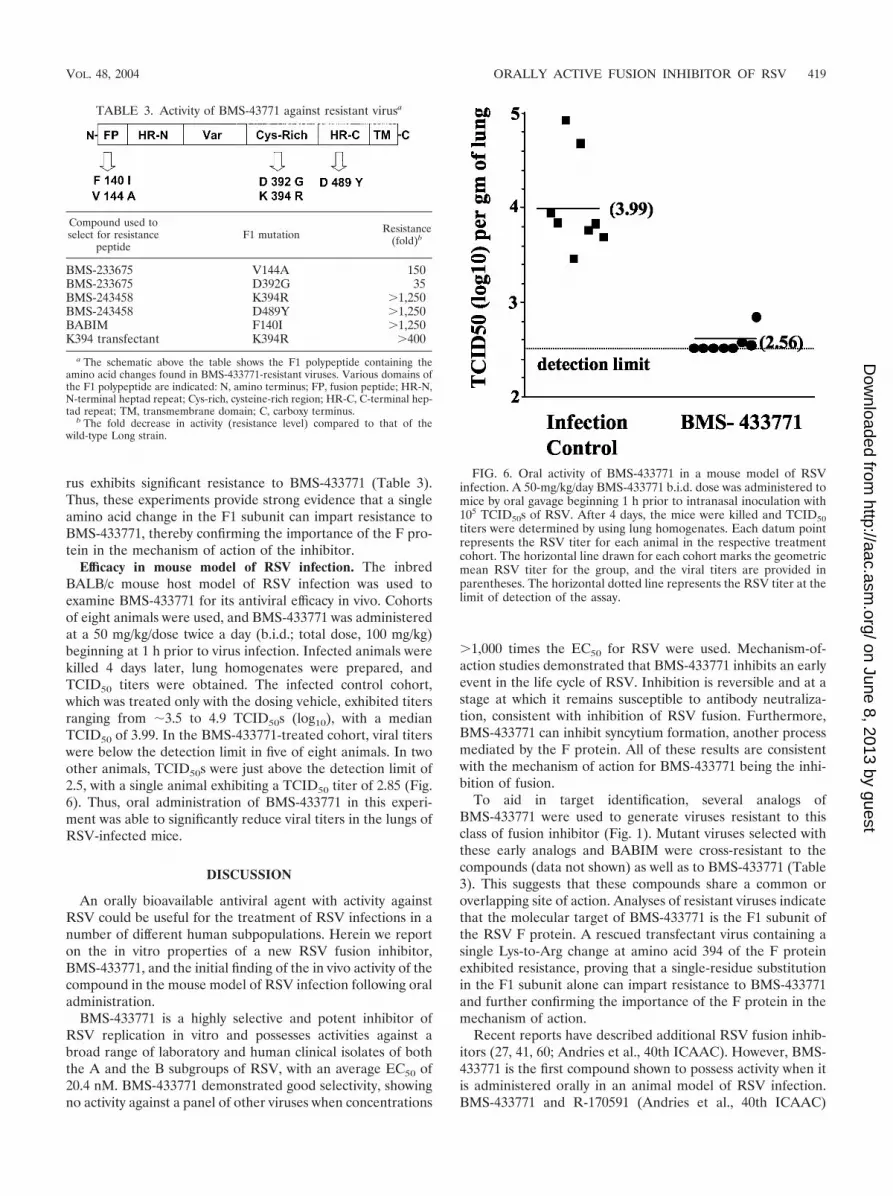
Efficacy in mouse model of RSV infection. The inbredBALB/c mouse host model of RSV infection was used toexamine BMS-433771 for its antiviral efficacy in vivo. Cohortsof eight animals were used, and BMS-433771 was administeredat a 50 mg/kg/dose twice a day (b.i.d.; total dose, 100 mg/kg)beginning at 1 h prior to virus infection. Infected animals werekilled 4 days later, lung homogenates were prepared, andTCID50 titers were obtained. The infected control cohort,which was treated only with the dosing vehicle, exhibited titersranging from �3.5 to 4.9 TCID50s (log10), with a medianTCID50 of 3.99. In the BMS-433771-treated cohort, viral titerswere below the detection limit in five of eight animals. In twoother animals, TCID50s were just above the detection limit of2.5, with a single animal exhibiting a TCID50 titer of 2.85 (Fig.6). Thus, oral administration of BMS-433771 in this experi-ment was able to significantly reduce viral titers in the lungs ofRSV-infected mice.
DISCUSSION
An orally bioavailable antiviral agent with activity againstRSV could be useful for the treatment of RSV infections in anumber of different human subpopulations. Herein we reporton the in vitro properties of a new RSV fusion inhibitor,BMS-433771, and the initial finding of the in vivo activity of thecompound in the mouse model of RSV infection following oraladministration.
BMS-433771 is a highly selective and potent inhibitor ofRSV replication in vitro and possesses activities against abroad range of laboratory and human clinical isolates of boththe A and the B subgroups of RSV, with an average EC50 of20.4 nM. BMS-433771 demonstrated good selectivity, showingno activity against a panel of other viruses when concentrations
�1,000 times the EC50 for RSV were used. Mechanism-of-action studies demonstrated that BMS-433771 inhibits an earlyevent in the life cycle of RSV. Inhibition is reversible and at astage at which it remains susceptible to antibody neutraliza-tion, consistent with inhibition of RSV fusion. Furthermore,BMS-433771 can inhibit syncytium formation, another processmediated by the F protein. All of these results are consistentwith the mechanism of action for BMS-433771 being the inhi-bition of fusion.
To aid in target identification, several analogs ofBMS-433771 were used to generate viruses resistant to thisclass of fusion inhibitor (Fig. 1). Mutant viruses selected withthese early analogs and BABIM were cross-resistant to thecompounds (data not shown) as well as to BMS-433771 (Table3). This suggests that these compounds share a common oroverlapping site of action. Analyses of resistant viruses indicatethat the molecular target of BMS-433771 is the F1 subunit ofthe RSV F protein. A rescued transfectant virus containing asingle Lys-to-Arg change at amino acid 394 of the F proteinexhibited resistance, proving that a single-residue substitutionin the F1 subunit alone can impart resistance to BMS-433771and further confirming the importance of the F protein in themechanism of action.
Recent reports have described additional RSV fusion inhib-itors (27, 41, 60; Andries et al., 40th ICAAC). However, BMS-433771 is the first compound shown to possess activity when itis administered orally in an animal model of RSV infection.BMS-433771 and R-170591 (Andries et al., 40th ICAAC)
FIG. 6. Oral activity of BMS-433771 in a mouse model of RSVinfection. A 50-mg/kg/day BMS-433771 b.i.d. dose was administered tomice by oral gavage beginning 1 h prior to intranasal inoculation with105 TCID50s of RSV. After 4 days, the mice were killed and TCID50titers were determined by using lung homogenates. Each datum pointrepresents the RSV titer for each animal in the respective treatmentcohort. The horizontal line drawn for each cohort marks the geometricmean RSV titer for the group, and the viral titers are provided inparentheses. The horizontal dotted line represents the RSV titer at thelimit of detection of the assay.
TABLE 3. Activity of BMS-43771 against resistant virusa
Compound used toselect for resistance
peptideF1 mutation Resistance
(fold)b
BMS-233675 V144A 150BMS-233675 D392G 35BMS-243458 K394R �1,250BMS-243458 D489Y �1,250BABIM F140I �1,250K394 transfectant K394R �400
a The schematic above the table shows the F1 polypeptide containing theamino acid changes found in BMS-433771-resistant viruses. Various domains ofthe F1 polypeptide are indicated: N, amino terminus; FP, fusion peptide; HR-N,N-terminal heptad repeat; Cys-rich, cysteine-rich region; HR-C, C-terminal hep-tad repeat; TM, transmembrane domain; C, carboxy terminus.
b The fold decrease in activity (resistance level) compared to that of thewild-type Long strain.
VOL. 48, 2004 ORALLY ACTIVE FUSION INHIBITOR OF RSV 419
on June 8, 2013 by guesthttp://aac.asm
.org/D
ownloaded from

share some structural homology, while the other two com-pounds, VP-14637 (41) and RFI-647 (27), have no obviousstructural similarity to any of the other compounds. Interest-ingly, all four RSV fusion inhibitors were identified by usingcell culture screens. Cell culture screens for antiviral activitytheoretically target multiple pathways throughout the virusreplicative cycle. The fact that RSV fusion seems to be specif-ically targeted in this assay may suggest that fusion inhibitorsare significantly easier to detect in this type of screen thaninhibitors of other stages of the virus life cycle. One explana-tion may be that the F protein is more promiscuous than otherviral proteins in its ability to bind to small molecules and thatthe fusion process, in which numerous interactions and con-formational changes occur, provides multiple target sites thatmolecules can functionally inhibit. The variation in the struc-tures of the multiple RSV fusion inhibitors tends to supportthis view. In addition, with entry inhibitors, compounds neednot pass through cell membranes in order to demonstrateantiviral activity in a cell culture screen. It is interesting thatreports of cell culture-based screens for influenza virus inhib-itors also identified three distinct inhibitors of the hemagglu-tinin-catalyzed membrane fusion reaction (7, 37, 50, 59).
Single amino acid changes were found in the F-protein genesof all the resistant viruses. These viruses were generated afterseveral passages in the presence of increasing concentrationsof compound. All mutations were the result of a single nucle-otide change, so it is possible that they were generated as earlyas the first passage. Interestingly, all of the amino acid changesin the resistant viruses occurred within the F1 subunit. TheRSV F protein is translated as a single polypeptide that isenzymatically cleaved into two subunits (subunits F1 and F2)that remain connected via a disulfide bridge (9). By analogywith the influenza virus HA2 and HIV gp41 proteins, the RSVF1 subunit is responsible for the mediation of membrane fu-sion (3, 15, 30, 68). The F1 subunit contains the hydrophobicfusion peptide, which is buried within the molecule in its nativestate (57). After F is bound to a receptor, a conformationalrearrangement in the F protein exposes the fusion peptide,which inserts into the opposing cell membrane and, through aseries of steps that remain undefined, promotes fusion of theviral and host cell membranes (4, 68). The finding that allresistant viruses possess amino acid changes in the F1 polypep-tide complements the mechanism-of-action studies that dem-onstrate that BMS-433771 inhibits RSV-induced membranefusion. Additionally, since all of the amino acid changes in theresistant viruses mapped to the F1 subunit, it strongly suggeststhat the F1 polypeptide is the specific molecular target ofBMS-433771.
The F1 subunit contains the amino-terminal fusion peptidethat is believed to insert in the host cell membrane duringfusion. The F1 subunit also contains the two heptad repeatregions, located at the N and C termini, that are hypothesizedto associate in an antiparallel manner, bringing about apposi-tion of viral and cellular membranes during fusion (35, 40, 68).A potential binding pocket for small inhibitor molecules hasbeen described within a structure of the RSV fusion coreobtained through cocrystallization of peptides representing theN-terminal and C-terminal heptad repeats (68). This pocket ispresent in the trimer of N-terminal repeats and is occupied bytwo phenylalanine residues (F483 and F488) from the C-ter-
minal heptad repeat peptides. One of the resistant virusesselected in our study had an amino acid change (D489Y) nearthis region in the C-terminal heptad repeat. The RSV fusioninhibitors R-170591 and VP-14637 also produced resistant vi-ruses with mutations in the same region (D486N and F488Yfor R-170591 and VP-14637, respectively) of the C-terminalheptad repeat region of the F1 subunit (11; Andries et al., 40thICAAC). Other mutations that resulted in resistance to BMS-433771 map to alternate areas of the F1 subunit, suggestingthat changes throughout the F1 subunit can abrogate the in-hibition by BMS-433771. Two distinct mutations (F140I andV144A) were found in the hydrophobic fusion peptide, whichis the series of amino acids within the N terminus of the F1subunit involved in host membrane insertion. Also, two addi-tional BMS-433771-resistant viruses have amino acid changesthat occur in the cysteine-rich domain (D392G or K394R)found between the heptad repeat regions. The precise functionof this region is unknown at present (5). However, other fusioninhibitors induce mutations that map to this area. Resistanceto R-170591 can result from an S398L change (Andries et al.,40th ICAAC), while VP-14637 can generate resistant viruswith a T400A substitution (11). The similar substitution pat-terns in resistant viruses may suggest that these three inhibi-tors, even though they are quite different in structure, mayshare similar modes of binding to the F1 subunit. It would beof interest to examine these resistant viruses for cross-resis-tance to the different RSV fusion inhibitors.
The availability of the mouse model of RSV infection en-ables examination of potential inhibitors of RSV in vivo. How-ever, the value of animal models of RSV infection as harbin-gers of human RSV infection remains questionable (18).Nevertheless, for a new chemical entity with inhibitory activityagainst RSV in vitro, the mouse model of infection can provideproof of principle for efficacy in vivo and spur interest infurther clinical development. In mice, BMS-433771 adminis-tered orally at 50 mg/kg b.i.d. beginning 1 h prior to infectionsignificantly reduced the titers of RSV in the lungs of mice.This is the first report of a small-molecule RSV fusion inhibitorwith activity in an animal model following oral administration.BMS-433771 has also been shown to have inhibitory activity inthe cotton rat model of RSV infection following oral admin-istration (C. Cianci et al., submitted). As for the other RSVinhibitors, BABIM was reported to have antiviral activity incotton rats after intraperitoneal administration (63), andR-170591 demonstrated efficacy in vivo after aerosol deliveryin rodent models of RSV infection (Andries et al., 40thICAAC). The efficacy of RFI-641 was shown in three modelsof RSV infection, but only when it was administered via theintranasal or aerosol route (27, 66).
A clear demonstration of the clinical significance of inhibi-tion of the RSV F-protein function has been established byusing Synagis, the humanized monoclonal antibody directedagainst the F protein (23, 29, 53). BMS-433771 targets thesame RSV protein and viral function. Biochemical studiesdemonstrated that BMS-433771 is a specific inhibitor of mem-brane fusion induced by the viral F protein. Inhibition of mem-brane fusion is a mechanism of antiviral activity that is beingexplored for a number of different viruses, including HIV, withthe first fusion inhibitor of HIV recently being approved by theFood and Drug Administration (31, 32, 34). Accordingly,
420 CIANCI ET AL. ANTIMICROB. AGENTS CHEMOTHER.
on June 8, 2013 by guesthttp://aac.asm
.org/D
ownloaded from

BMS-433771 is a novel small-molecule antiviral agent that issuitable for clinical evaluation due to its mechanism of action,in vitro potency, selectivity, and, most significantly, oral efficacyin vivo.
REFERENCES
1. Ausubel, F. M., R. Brent, R. E. Kingston, D. D. Moore, J. G. Seidman, andJ. A. Smith. 1989. Current protocols in molecular biology. John Wiley &Sons, Inc., New York, N.Y.
2. Byrd, L. G., and G. A. Prince. 1997. Animal models of respiratory syncytialvirus infection. Clin. Infect. Dis. 25:1363–1368.
3. Calder, L. J., L. Gonzalez-Reyes, B. Garcia-Barreno, S. A. Wharton, J. J.Skehel, D. C. Wiley, and J. A. Melero. 2000. Electron microscopy of thehuman respiratory syncytial virus fusion protein and complexes that it formswith monoclonal antibodies. Virology 271:122–131.
4. Carr, C. M., and P. S. Kim. 1993. A spring-loaded mechanism for theconformational change of influenza hemagglutinin. Cell 73:823–832.
5. Chambers, P., C. R. Pringle, and A. J. Easton. 1992. Sequence analysis of thegene encoding the fusion glycoprotein of pneumonia virus of mice suggestspossible conserved secondary structure elements in paramyxovirus fusionglycoproteins. J. Gen. Virol. 73:1717–1724.
6. Chanock, R. M., and R. H. Parrott. 1965. Acute respiratory disease ininfancy and childhood: present understanding and prospects for prevention.Pediatrics 36:21–39.
7. Cianci, C., K. L. Yu, D. D. Dischino, W. Harte, M. Deshpande, G. Luo, R. J.Colonno, N. A. Meanwell, and M. Krystal. 1999. pH-dependent changes inphotoaffinity labeling patterns of the H1 influenza virus hemagglutinin byusing an inhibitor of viral fusion. J. Virol. 73:1785–1794.
8. Collins, P. L., M. G. Hill, E. Camargo, H. Grosfeld, R. M. Chanock, andB. R. Murphy. 1995. Production of infectious human respiratory syncytialvirus from cloned cDNA confirms an essential role for the transcriptionelongation factor from the 5� proximal open reading frame of the M2 mRNAin gene expression and provides a capability for vaccine development. Proc.Natl. Acad. Sci. USA 92:11563–11567.
9. Collins, P. L., K. McIntosh, and R. M. Chanock. 2001. Respiratory syncytialvirus, p. 1443–1486. In D. M. Knipe and P. M. Howley (ed.), Virology, 3rded. Raven Press, New York, N.Y.
10. De Clercq, E. 2001. Antiviral drugs: current state of the art. J. Clin. Virol.22:73–89.
11. Douglas, J. L., M. L. Panis, E. Ho, K. Y. Lin, S. H. Krawczyk, D. M. Grant,R. Cai, S. Swaminathan, and T. Cihlar. 2003. Inhibition of respiratorysyncytial virus fusion by the small molecule VP-14637 via specific interactionswith F protein. J. Virol. 77:5054–5064.
12. Dowell, S. F., L. J. Anderson, H. E. J. Gary, D. D. Erdman, J. F. Plouffe,T. M. J. File, B. J. Marston, and R. F. Breiman. 1996. Respiratory syncytialvirus is an important cause of community-acquired lower respiratory infec-tion among hospitalized adults. J. Infect. Dis. 174:456–462.
13. DuBovi, E. J., J. D. Geratz, S. R. Shaver, and R. R. Tidwell. 1981. Inhibitionof respiratory syncytial virus-host cell interactions by mono- and diamidines.Antimicrob. Agents Chemother. 19:649–656.
14. DuBovi, E. J., J. D. Geratz, and R. R. Tidwell. 1980. Inhibition of respiratorysyncytial virus by bis(5-amidino-2-benzimidazolyl)methane. Virology 103:502–504.
15. Dutch, R. E., T. S. Jardetzky, and R. A. Lamb. 2000. Virus membrane fusionproteins: biological machines that undergo a metamorphosis. Biosci. Rep.20:597–612.
16. Englund, J. A., C. J. Sullivan, M. C. Jordan, L. P. Dehner, G. M. Vercellotti,and H. H. Balfour. 1988. Respiratory syncytial virus infection in immuno-compromised adults. Ann. Intern. Med. 109:203–208.
17. Falsey, A. R., C. K. Cunningham, W. H. Barker, R. W. Kouides, J. B. Yuen,M. Menegus, L. B. Weiner, C. A. Bonville, and R. F. Betts. 1995. Respiratorysyncytial virus and influenza A infections in the hospitalized elderly. J. Infect.Dis. 172:389–394.
18. Falsey, A. R., J. J. Treanor, R. F. Betts, and E. E. Walsh. 1992. Viralrespiratory infections in the institutionalized elderly: clinical and epidemio-logic findings. J. Am. Geriatr. Soc. 40:115–119.
19. Falsey, A. R., and E. E. Walsh. 2000. Respiratory syncytial virus infection inadults. Clin. Microbiol. Rev. 13:371–384.
20. Garcia, R., I. Raad, D. Abi-Said, G. Bodey, R. Champlin, J. Tarrand, L. A.Hill, J. Umphrey, J. Neumann, J. Englund, and E. Whimbey. 1997. Noso-comial respiratory syncytial virus infections: prevention and control in bonemarrow transplant patients. Infect. Control Hosp. Epidemiol. 18:412–416.
21. Glezen, W. P., L. H. Taber, A. L. Frank, and J. A. Kasel. 1986. Risk ofprimary infection and reinfection with respiratory syncytial virus. Am. J. Dis.Child. 140:543–546.
22. Greenough, A. 2001. Recent advances in the management and prophylaxis ofrespiratory syncytial virus infection. Acta Paediatr. Suppl. 90:11–14.
23. Greenough, A., and M. Thomas. 2000. Respiratory syncytial virus prevention:past and present strategies. Expert Opin. Pharmacother. 1:1195–1201.
24. Hacking, D., and J. Hull. 2002. Respiratory syncytial virus-viral biology andthe host response. J. Infect. 45:18–24.
25. Heikkinen, T., M. Thint, and T. Chonmaitree. 1999. Prevalence of variousrespiratory viruses in the middle ear during acute otitis media. N. Engl.J. Med. 340:260–264.
26. Holberg, C. J., A. L. Wright, F. D. Martinez, C. G. Ray, L. M. Taussig, andM. D. Lebowitz. 1991. Risk factors for respiratory syncytial virus-associatedlower respiratory illnesses in the first year of life. Am. J. Epidemiol. 133:1135–1151.
27. Huntley, C. C., W. J. Weiss, A. Gazumyan, A. Buklan, B. Feld, W. Hu, T. R.Jones, T. Murphy, A. A. Nikitenko, B. O’Hara, G. Prince, S. Quartuccio,Y. E. Raifeld, P. Wyde, and J. F. O’Connell. 2002. RFI-641, a potent respi-ratory syncytial virus inhibitor. Antimicrob. Agents Chemother. 46:841–847.
28. Ikeda, S., J. Neyts, S. Verma, A. Wickramasinghe, P. Mohan, and E. DeClercq. 1994. In vitro and in vivo inhibition of ortho- and paramyxovirusinfections by a new class of sulfonic acid polymers interacting with virus-cellbinding and/or fusion. Antimicrob. Agents Chemother. 38:256–259.
29. Johnson, S., C. Oliver, G. A. Prince, V. G. Hemming, D. S. Pfarr, S.-C. Wang,M. Dormitzer, J. O’Grady, S. Koenig, J. K. Tamura, R. Woods, G. Bansal, D.Couchenour, E. Tsao, W. C. Hall, and J. F. Young. 1997. Development of ahumanized monoclonal antibody (MEDI-493) with potent in vitro and invivo activity against respiratory syncytial virus. J. Infect. Dis. 176:1215–1224.
30. Kahn, J. S., M. J. Schnell, L. Buonocore, and J. K. Rose. 1999. Recombinantvesicular stomatitis virus expressing respiratory syncytial virus (RSV) glyco-proteins: RSV fusion protein can mediate infection and cell fusion. Virology254:81–91.
31. Kilby, J. M., S. Hopkins, T. M. Venetta, B. DiMassimo, G. A. Cloud, J. Y.Lee, L. Alldredge, E. Hunter, D. Lambert, D. Bolognesi, T. Matthews, M. R.Johnson, M. A. Nowak, G. M. Shaw, and M. S. Saag. 1998. Potent suppres-sion of HIV-1 replication in humans by T-20, a peptide inhibitor of gp41-mediated virus entry. Nat. Med. 4:1302–1307.
32. Kilby, J. M., J. P. Lalezari, J. J. Eron, M. Carlson, C. Cohen, R. C. Arduino,J. C. Goodgame, J. E. Gallant, P. Volberding, R. L. Murphy, F. Valentine,M. S. Saag, E. L. Nelson, P. R. Sista, and A. Dusek. 2003. The safety, plasmapharmacokinetics, and antiviral activity of subcutaneous enfuvirtide (T-20),a peptide inhibitor of gp41-mediated virus fusion, in HIV-infected adults.AIDS Res. Hum. Retrovir. 19:685–693.
33. Kimura, K., S. Mori, K. Tomita, K. Ohno, K. Takahashi, S. Shigeta, and M.Terada. 2000. Antiviral activity of NMSO3 against respiratory syncytial virusinfection in vitro and in vivo. Antivir. Res. 47:41–51.
34. Lalezari, J. P., K. Henry, M. O’Hearn, J. S. Montaner, P. J. Piliero, B.Trottier, S. Walmsley, C. Cohen, D. R. Kuritzkes, J. J. Eron, Jr., J. Chung,R. DeMasi, L. Donatacci, C. Drobnes, J. Delehanty, and M. Salgo. 2003.Enfuvirtide, an HIV-1 fusion inhibitor, for drug-tesistant HIV infection inNorth and South America. N. Engl. J. Med. 348:2175–2185.
35. Lawless-Delmedico, M. K., P. Sista, R. Sen, N. C. Moore, J. B. Antczak, J. M.White, R. J. Greene, K. C. Leanza, T. J. Matthews, and D. M. Lambert. 2000.Heptad-repeat regions of respiratory syncytial virus F1 protein form a six-membered coiled-coil complex. Biochemistry 39:11684–11695.
36. Luo, G., R. Colonno, and M. Krystal. 1996. Characterization of a hemag-glutinin-specific inhibitor of influenza A virus. Virology 226:66–76.
37. Luo, G. X., A. Torri, W. E. Harte, S. Danetz, C. Cianci, L. Tiley, S. Day, D.Mullaney, K. L. Yu, C. Ouellet, P. Dextraze, N. Meanwell, R. Colonno, andM. Krystal. 1997. Molecular mechanism underlying the action of a novelfusion inhibitor of influenza A virus. J. Virol. 71:4062–4070.
38. Martin, M. A., M. J. Bock, M. A. Pfaller, and R. P. Wenzel. 1988. Respiratorysyncytial virus infections in adult bone marrow transplant recipients. Lanceti:1396–1397.
39. Mathur, U., D. W. Bentley, and C. B. Hall. 1980. Concurrent respiratorysyncytial virus and influenza A infections in the institutionalized elderly andchronically ill. Ann. Intern. Med. 93:49–52.
40. Matthews, J. M., T. F. Young, S. P. Tucker, and J. P. Mackay. 2000. The coreof the respiratory syncytial virus fusion protein is a trimeric coiled coil.J. Virol. 74:5911–5920.
41. McKimm-Breschkin, J. 2000. VP-14637 ViroPharma. Curr. Opin. Investig.Drugs 1:425–427.
42. Mills, J. 1996. Management of respiratory syncytial virus infections. PlenumPress, New York, N.Y.
43. Moyse, E., M. Lyon, G. Cordier, J. F. Mornex, L. Collet, and P. Froehlich.2000. Viral RNA in middle ear mucosa and exudates in patients with chronicotitis media with effusion. Arch. Otolaryngol. Head Neck Surg. 126:1105–1110.
44. Murry, A. R., and S. F. Dowell. 1997. Respiratory syncytial virus: not just forkids. Hosp. Pract. 15:87–88.
45. Osiowy, C., and R. Anderson. 1995. Neutralization of respiratory syncytialvirus after cell attachment. J. Virol. 69:1271–1274.
46. Osterweil, D., and D. Norman. 1990. An outbreak of an influenza-like illnessin a nursing home. J. Am. Geriatr. Soc. 38:659–662.
47. Pagani, F., and F. Sparatore. 1965. Benzotriazolyl-alkyl-benzimidazoles andtheir dialkyl-aminoalkyl derivatives. Boll. Chim. Farm. 104:427–431.
48. Paglietti, G., V. Boido, and F. Sparatore. 1975. Dialkylaminoalkylbenzimi-dazoles of pharmacological importance. IV. Il Farmaco 30:505–511.
VOL. 48, 2004 ORALLY ACTIVE FUSION INHIBITOR OF RSV 421
on June 8, 2013 by guesthttp://aac.asm
.org/D
ownloaded from

49. Pauwels, R., J. Balzarini, M. Baba, R. Snoeck, D. Schols, P. Herdewijn, J.Desmyter, and E. De Clercq. 1988. Rapid and automated tetrazolium-basedcolorimetric assay for the detection of anti-HIV compounds. J. Virol. Meth-ods 20:309–321.
50. Plotch, S. J., B. O’Hara, J. Morin, O. Palant, J. LaRocque, J. D. Bloom, S. A.Lang, Jr., M. J. DiGrandi, M. Bradley, R. Nilakantan, and Y. Gluzman.1999. Inhibition of influenza A virus replication by compounds interferingwith the fusogenic function of the viral hemagglutinin. J. Virol. 73:140–151.
51. Popovic, M., M. G. Sarngadharan, E. Read, and R. C. Gallo. 1984. Detec-tion, isolation, and continuous production of cytopathic retroviruses (HTLV-III) from patients with AIDS and pre-AIDS. Science 224:497–500.
52. Prince, G. A. 2001. An update on respiratory syncytial virus antiviral agents.Expert Opin. Investig. Drugs 10:297–308.
53. Schmidt, A. C., R. B. Couch, G. J. Galasso, F. G. Hayden, J. Mills, B. R.Murphy, and R. M. Chanock. 2001. Current research on respiratory viralinfections: Third International Symposium. Antivir. Res. 50:157–196.
54. Sigurs, N. 2001. Epidemiologic and clinical evidence of a respiratory syncy-tial virus-reactive airway disease link. Am. J. Respir. Crit. Care Med. 163:S2–S6.
55. Sigurs, N., R. Bjarnason, F. Sigurbergsson, B. Kjellman, and B. Bjorksten.1995. Asthma and immunoglobulin E antibodies after respiratory syncytialvirus bronchiolitis: a prospective cohort study with matched controls. Pedi-atrics 95:500–505.
56. Simoes, E. A. 2001. Treatment and prevention of respiratory syncytial viruslower respiratory tract infection. Long-term effects on respiratory outcomes.Am. J. Respir. Crit. Care Med. 163:S14–S17.
57. Smith, B. J., M. C. Lawrence, and P. M. Colman. 2002. Modeling thestructure of the fusion protein from human respiratory syncytial virus. Pro-tein Eng. 15:365–371.
58. Sorvillo, F. J., S. F. Huie, M. A. Strassburg, A. Butsumyo, W. X. Shandera,and S. L. Fannin. 1984. An outbreak of respiratory syncytial virus pneumoniain a nursing home for the elderly. J. Infect. 9:252–256.
59. Staschke, K. A., S. D. Hatch, J. C. Tang, W. J. Hornback, J. E. Munroe, J. M.
Colacino, and M. A. Muesing. 1998. Inhibition of influenza virus hemagglu-tinin-mediated membrane fusion by a compound related to podocarpic acid.Virology 248:264–274.
60. Sudo, K., K. Konno, W. Watanabe, S. Shigeta, and T. Yokota. 2001. Mech-anism of selective inhibition of respiratory syncytial virus by a benzodithiincompound (RD3–0028). Microbiol. Immunol. 45:531–537.
61. Taylor, G., E. J. Stott, M. Hughes, and A. P. Collins. 1984. Respiratorysyncytial virus infection in mice. Infect. Immun. 43:649–655.
62. Thompson, W. W., D. K. Shay, E. Weintraub, L. Brammer, N. Cox, L. J.Anderson, and K. Fukuda. 2003. Mortality associated with influenza andrespiratory syncytial virus in the United States. JAMA 289:179–186.
63. Tidwell, R. R., J. D. Geratz, W. A. Clyde, K. U. Rosenthal, and E. J. Dubovi.1984. Suppression of respiratory syncytial virus infection in cotton rats bybis(5-amidino-2-benzimidazolyl)methane. Antimicrob. Agents Chemother.26:591–593.
64. Tidwell, R. R., J. D. Geratz, and E. J. Dubovi. 1983. Aromatic amidines:comparison of their ability to block respiratory syncytial virus induced cellfusion and to inhibit plasmin, urokinase, thrombin, and trypsin. J. Med.Chem. 26:294–298.
65. Treanor, J., and A. Falsey. 1999. Respiratory viral infections in the elderly.Antivir. Res. 44:79–102.
66. Weiss, W. J., T. Murphy, M. E. Lynch, J. Frye, A. Buklan, B. Gray, E. Lenoy,S. Mitelman, J. O’Connell, S. Quartuccio, and C. Huntley. 2003. Inhalationefficacy of RFI-641 in an African green monkey model of RSV infection.J. Med. Primatol. 32:82–88.
67. Yu, K. L., Y. Zhang, R. L. Civiello, K. Kadow, C. Cianci, M. Krystal, andN. A. Meanwell. 2003. Fundamental structure-activity relationships associ-ated with a new structural class of respiratory syncytial virus inhibitor.Bioorg. Med. Chem. Lett. 13:2141–2144.
68. Zhao, X., M. Singh, V. N. Malashkevich, and P. S. Kim. 2000. Structuralcharacterization of the human respiratory syncytial virus fusion protein core.Proc. Natl. Acad. Sci. USA 97:14172–14177.
422 CIANCI ET AL. ANTIMICROB. AGENTS CHEMOTHER.
on June 8, 2013 by guesthttp://aac.asm
.org/D
ownloaded from