an orally delivered small-molecule formulation with antiangiogenic and anticancer activity
TRANSCRIPT
An orally delivered small-molecule formulation withantiangiogenic and anticancer activity
Ofra Benny1, Ofer Fainaru1, Avner Adini1, Flavia Cassiola1, Lauren Bazinet1, Irit Adini1, ElkePravda1, Yaakov Nahmias2, Samir Koirala3, Gabriel Corfas3, Robert J D’Amato1,4, andJudah Folkman11 Vascular Biology Program and Department of Surgery, Children’s Hospital Boston, HarvardMedical School, 1 Blackfan Circle, St. Karp Research Building, Boston, Massachusetts 02215, USA2 Center for Engineering in Medicine, Massachusetts General Hospital, Harvard Medical School,114 16th Street, Charlestown, Massachusetts 02129, USA3 Department of Neurology, F.M. Kirby Neurobiology Center, Children’s Hospital, Harvard MedicalSchool, 300 Longwood Avenue, Boston, Massachusetts 02115, USA4 Department of Ophthalmology, Harvard Medical School, Children’s Hospital Boston, 1 BlackfanCircle, St. Karp Research Building, Boston, Massachusetts 02215, USA
AbstractTargeting angiogenesis, the formation of blood vessels, is an important modality for cancer therapy.TNP-470, a fumagillin analog, is among the most potent and broad-spectrum angiogenesis inhibitors.However, a major clinical limitation is its poor oral availability and short half-life, necessitatingfrequent, continuous parenteral administration. We have addressed these issues and report an oralformulation of TNP-470, named Lodamin. TNP-470 was conjugated to monomethoxy-polyethyleneglycol–polylactic acid to form nanopolymeric micelles. This conjugate can be absorbed by theintestine and selectively accumulates in tumors. Lodamin significantly inhibits tumor growth,without causing neurological impairment in tumor-bearing mice. Using the oral route ofadministration, it first reaches the liver, making it especially efficient in preventing the developmentof liver metastasis in mice. We show that Lodamin is an oral nontoxic antiangiogenic drug that canbe chronically administered for cancer therapy or metastasis prevention.
Angiogenesis inhibition has become an important treatment modality for the suppression oftumor growth and metastasis progression1,2. TNP-470 is an analog of fumagillin, which wasisolated from the fungus Aspergillus fumigatus fresenius3 and is among the most potentinhibitors of angiogenesis. In animal models, TNP-470 showed a broad anticancer spectrum
Correspondence should be addressed to O.B. ([email protected]).AUTHOR CONTRIBUTIONSO.B. designed and developed Lodamin formulation, designed experiments, performed tissue culture, in vivo studies and histology,analyzed data and wrote the manuscript; O.F. conducted intrasplenic injections, assisted with editing the manuscript; A.A. and I.A.assisted with cell assays and histology; F.C. conducted TEM imaging and analysis; L.B. assisted with animal studies and conductedcorneal assays; E.P. performed confocal microscope imaging; Y.N. performed liver toxicity assays; S.K. and G.C. designed, conductedand analyzed neurotoxicity tests in mice; R.J.D. advised, edited the revisited manuscript and supervised corneal assays. J.F. supervisedthe entire study, designed experiments, analyzed data and edited the manuscript.COMPETING INTERESTS STATEMENTThe authors declare competing financial interests: details accompany the full-text HTML version of the paper athttp://www.nature.com/naturebiotechnology/.Published online at http://www.nature.com/naturebiotechnology/Reprints and permissions information is available online at http://npg.nature.com/reprintsandpermissions/
NIH Public AccessAuthor ManuscriptNat Biotechnol. Author manuscript; available in PMC 2010 January 7.
Published in final edited form as:Nat Biotechnol. 2008 July ; 26(7): 799–807. doi:10.1038/nbt1415.
NIH
-PA Author Manuscript
NIH
-PA Author Manuscript
NIH
-PA Author Manuscript
of activity4. TNP-470 inhibited the growth of primary and metastatic murine tumors and humanxenografts such as breast cancer, neuroblastoma, ovarian cancer, prostate cancer, glioblastoma,neurofibrosarcoma and uterine sarcoma and led to a reduction in their vascularization5–12. Themechanism of the antiangiogenic activity of TNP-470 is still not completely clear, thoughproposed molecular mechanisms have suggested targeting methionine aminopeptidase(MetAP-2), affecting cell cycle through p53 activation, induction of p21(CIP/WAF) orpreventing Rac1 activation11,13–15.
TNP-470 was one of the first antiangiogenic drugs to undergo clinical trials16. It demonstratedits ability to slow tumor progression or cause durable complete regression17 either given as asingle agent, or in combination with other conventional chemotherapeutic drugs such aspaclitaxel and carboplatin17–23. The dose-limiting toxicity was neural side-effects such asdizziness, decreased concentration, short-term memory loss, confusion and depression23.Successful elimination of these neurologic symptoms was achieved by the development ofCaplostatin (formulated for injection)24, in which TNP-470 was conjugated to a N-(2-hydroxypropyl)methacrylamide (HPLA) polymer to block penetration of the blood-brainbarrier.
Nevertheless, a major clinical limitation of TNP-470 remained unresolved. The poor oralavailability of TNP-470 coupled with its extremely short plasma half-life imposes a strictregime of prolonged parenteral administration23,25. TNP-470 needs to be administeredfrequently as continuous intravenous infusions (usually over 1 h, multiple times a week) in theclinic. Therefore, developing an oral formulation may considerably improve patientcompliance and provide means for a long-term antiangiogenic therapy for cancer and otherangiogenesis-dependent diseases.
In this work we report an oral antiangiogenic polymeric drug with potent antitumor andantimetastatic efficacy named Lodamin. We characterize the physicochemical properties andshow that this formulation of TNP-470 overcomes the drug’s limitations while retaining itsantiangiogenic activity. Lodamin is produced by conjugation of TNP-470 to a di-blockcopolymer, monomethoxy-polyethyleneglycol–polylactic acid (mPEG-PLA). Theamphiphilic nature of this polymeric drug enables self-assembly of micelles in an aqueousmedium26. In this structure, the TNP-470 is located in the core, where it is protected from theacidic environment of the stomach, thus enabling oral availability. Furthermore, we takeadvantage of using biocompatible, commonly used and well-characterized polymers27,28.
Our results indicate that Lodamin, administered orally, is effectively absorbed in the intestineand accumulates in tumor tissue. The drug significantly inhibits angiogenesis, as demonstratedby inhibition of human umbilical vein endothelial cell (HUVEC) proliferation, by the cornealmicropocket assay and in mouse tumor models. Lodamin significantly inhibited primary tumorgrowth as demonstrated in models of melanoma and lung cancer. Notably, oral Lodaminsuccessfully prevented liver metastasis of melanoma tumor cells without causing liver toxicityor other side effects and prolonged mouse survival.
Unlike free TNP-470, Lodamin does not penetrate the blood-brain barrier and accordingly didnot cause neurotoxicity in mice. These results suggest that Lodamin may be a good candidatefor a safe maintenance drug with effective antitumor and antimetastatic properties.
RESULTSChemical and physical characterization of Lodamin
To predict the oral availability of TNP-470, we measured its hydrophobicity using the log-Dparameter. The measured log-D values were 2.39 at pH = 2 and 2.57 at pH = 7.4. The high log-
Benny et al. Page 2
Nat Biotechnol. Author manuscript; available in PMC 2010 January 7.
NIH
-PA Author Manuscript
NIH
-PA Author Manuscript
NIH
-PA Author Manuscript
D values (>2) indicate very low solubility in water. This property led us to design a formulationwith improved solubility and oral availability.
We characterized the chemical and physical properties of Lodamin. We confirmed the bindingof TNP-470 to mPEG-PLA by 3H NMR (data not shown) and mass spectrometry showing anaverage m/z of 3,687 for mPEG-PLA-TNP-470. Figure 1a illustrates the preparation ofLodamin. Using an amine detection reagent, we determined the incorporation efficiency ofethylenediamine to mPEG-PLA as 65%, and in the second step TNP-470 was shown to bebound with an efficiency of >90%. Lodamin contained 0.8–1% (wt/wt) free TNP-470 asdetermined by high-performance liquid chromatography (HPLC). We determined the averagesize and size distribution of Lodamin by dynamic light scattering (DLS) spectroscopy (Fig.1b) on the day of preparation and after 10 d of incubation in aqueous medium, to evaluateLodamin stability (n = 4). The majority of micelles (90%) on the day of preparation were 7.8–8 nm in diameter, with a small population of larger particles (200–400 nm). The size remainedalmost unchanged after 10 d.
We characterized the morphology of Lodamin by transmission electron microscopy (TEM)(Fig. 1c). The images showed that the polymeric micelles had acquired a uniform sphericalstructure, which remained stable after 2 weeks of incubation in water at 37 °C. Because thedrug is located in the PLA core of the micelle structure and PLA is spontaneously hydrolyzedin an aqueous environment, we studied the release kinetics of TNP-470 from Lodamin. A slow-release kinetic of TNP-470 was obtained after incubation in PBS (pH = 7.4) or in gastric liquid(pH = 1.2). The TNP-470 was released over a period of 28 d with an early peak burst of ~30%after the first day of incubation (Fig. 1d).
Endothelial cells take up Lodamin by endocytosisWe evaluated the uptake of polymeric micelles by HUVECs and the kinetics of their uptake.HUVECs were incubated with 6-coumarin labeled mPEG-PLA micelles for 20 min, 2, 4 and7 h and were imaged by confocal microscopy (Fig. 2a). In as little as 20 min after incubation,micelles were taken up by the cells and located in their cytoplasm. After 2 h the uptake wasmaximal and after 4–7 h micelles were detected as defined aggregates inside the cytoplasm.Fluorescence-activated cell sorting (FACS) of HUVECs incubated with rhodamine-labeledpolymeric micelles for the same incubation times confirmed a maximal uptake after 2 h (Fig.2b), whereas no difference was observed between 2 and 24 h of incubation. In live-cell analysis,co-localization of Lyso-tracker staining with the micelles suggests endocytosis as themechanism of uptake. Incubation of the micelles with HUVECs in cold conditions reducedmicelle uptake by up to 55%, confirming that endocytosis is the most likely mechanism ofuptake (data not shown).
Lodamin inhibits proliferation of endothelial cellsNext, we evaluated the effect of Lodamin on the proliferation of endothelial cells. After 48 h,Lodamin (62.5 nM–1,000 nM TNP-470 equivalent) inhibited HUVEC proliferation by 88–95% respectively (Fig. 2c). The growth of HUVECs treated with Lodamin (60 nM TNP-470equivalent) was completely inhibited compared to untreated cells or cells treated by vehicleonly. We observed no substantial cytotoxic effect (Fig. 2c).
Lodamin inhibits bFGF and VEGF-induced angiogenesis in vivoThe antiangiogenic properties of Lodamin were evaluated in vivo by the corneal micropocketangiogenesis assay29. Mice were treated with daily oral Lodamin (15 mg/kg per day) or vehiclefor 6 d. Figure 2d shows the inhibition of basic fibroblast growth factor (bFGF)-inducedangiogenesis in representative eyes of treated or untreated mice. Quantification of theangiogenesis area (Fig. 2d, lower panel) showed 31% inhibition of angiogenesis, compared to
Benny et al. Page 3
Nat Biotechnol. Author manuscript; available in PMC 2010 January 7.
NIH
-PA Author Manuscript
NIH
-PA Author Manuscript
NIH
-PA Author Manuscript
vehicle (P = 0.00016, n = 10). Similar results were obtained with 160 ng vascular endothelialgrowth factor (VEGF)165-induced angiogenesis in the cornea (data not shown); in this caseLodamin treatment resulted in 40% inhibition of vessel area.
Polymeric micelles: absorption by the intestineTo study the intestinal absorption of the polymeric-micelles, we orally administered mPEG-PLA-rhodamine micelles to mice. After 2 h mice were euthanized and isolated segments ofthe small intestine were fixed and imaged using confocal microscopy. The polymeric micelles(detected in the red channel) were intensively taken up by columnar epithelium lining theluminal side of the small intestine (Fig. 3a). In a high magnification of small intestine villi,micelles are clearly detected in the lamina propria and in the vicinity of blood vessels, indicatingtransepithelial absorption. In high-resolution TEM images of single gut-epithelial cells,endosomes loaded with drug were readily detected (Fig. 3a, arrows). These endosomes differedin contrast and number when compared to those in the intestine of an untreated mouse. Thesedata indicate that Lodamin is most likely taken up by intestinal villi through endocytosis.
Biodistribution, tumor accumulation and toxicity studiesThe biodistribution and tissue uptake of orally administered labeled Lodamin was studied bytreating mice with fluorescently labeled mPEG-PLA micelles for 3 d. After harvesting tissue,we quantified tissue drug concentration by dye extraction or by FACS. The results of thefluorescent dye extraction method (Fig. 3b) showed that a high concentration of fluorescentsignal was present in the stomach and the small intestine, and the highest levels were presentin the liver. Notably, the brain lacked fluorescent signal. In the serum, labeled micelles werealready detected 1 h after oral administration, peaking after 2 h and were still detected at 72 h.In tumor-bearing mice, FACS analysis of enzymatically digested tissues (Fig. 3c,d)demonstrated a large uptake of labeled micelles by the liver, and no uptake by the brain.Notably, the highest uptake of micelles was detected in tumor cells (Fig. 3c). Taken together,these results indicate that the drug was concentrated mostly in the tumor and to a lesser extentin the gastrointestinal organs, and absent in the brain.
No tissue abnormalities were detected by histological analyses (H&E) of liver, intestine, lungand kidney in Lodamin-treated mice (15 mg/kg TNP-470 equivalent per day, for 20 d)compared to untreated mice (data not shown). In addition, no substantial differences were foundbetween the mouse serum liver-enzyme profiles of Lodamin-treated and untreated mice. In theLodamin-treated group the aspartate aminotransferase (AST) and alanine aminotransferase(ALT) concentrations were 41 ± 9 u/l and 120 ± 39 u/l, respectively, whereas in the untreatedgroup they reached 37.5 ± 4 u/l and 152 ± 131 u/l, respectively.
Lodamin inhibits primary tumor growthWe evaluated the biological efficacy of Lodamin as an antiangiogenic anticancer agent intumor-bearing mice. When mice received an oral dose of free TNP-470 (30 mg/kg every otherday), we did not observe tumor growth inhibition in subcutaneous Lewis lung carcinomas(LLC) (Fig. 4a). The equivalent dose of Lodamin, however, resulted in substantial tumorgrowth inhibition (Fig. 4a). This inhibition was observed after 12 d of Lodamin treatment, andat day 18 tumor growth was inhibited by 83%. Different dosing regimens of Lodamin, 15 mg/kg every day, 30 mg/kg every other day and 15 mg/kg every other day, resulted in 87%, 77%and 74% inhibition of tumor volume, respectively (Fig. 4b). The vehicle (mPEG-PLA) showedno effect on tumor growth and was similar to untreated control mice (Fig. 4c).
In another tumor model, murine melanoma (B16/F10) subcutaneous tumor growth was alsoinhibited by oral Lodamin (15 mg/kg per day). This treatment was effective after 4 d oftreatment, and after 13 d, 77% volume inhibition was obtained (Fig. 4d). No apparent side
Benny et al. Page 4
Nat Biotechnol. Author manuscript; available in PMC 2010 January 7.
NIH
-PA Author Manuscript
NIH
-PA Author Manuscript
NIH
-PA Author Manuscript
effects, including weight loss, were detected in either tumor model. Higher doses of Lodamin—30 mg/kg per day and 60 mg/kg every other day—showed substantial tumor inhibition;however, these higher doses were accompanied by weight loss (data not shown). Figure 4eshows representative tumors of treated or untreated LLC or B16/F10 tumors.
Lodamin does not cause neurotoxicity in miceBecause our biodistribution study indicated that Lodamin does not cross the blood-brainbarrier, we tested whether the possible leaching of free drug into the brain might result inneurotoxicity and cerebellar dysfunction. We subjected mice to a sensitive test of motorcoordination—crossing a narrow (4 mm) balance beam30. The performance of Lodamin-treated mice (30 mg/kg TNP-470 equivalent every other day for 14 d) was similar to that ofcontrol (water-treated) mice, whereas mice injected with the same dose of free TNP-470committed over twice as many errors (P < 0.0001) (Fig. 4f). These results indicate that Lodamintreatment avoids the cerebellar neurotoxicity observed with unconjugated TNP-470 treatment.
Lodamin inhibits tumor angiogenesis and proliferationWe tested the effect of Lodamin on the histological structure of LLC tumors. Both treated anduntreated tumors showed a dense cellular structure (Fig. 5). The tumors of untreated mice hada net organization of small and large vessels with an apparent lumen structure as demonstratedby CD-31 immunostaining. In contrast, Lodamin-treated tumors formed very smallundeveloped vessels (Fig. 5). Lodamin-treated tumors showed less cellular proliferation thanuntreated tumors, as detected by the nuclear marker Ki-67. TdT-mediated dUTP nick endlabelling (TUNEL) staining for the detection of apoptosis suggested enhanced apoptosis inLodamin-treated tumors. Lodamin-treated tumors had fewer vessels (green) but high levels ofapoptosis, predominantly in tumor cells (red). In the control tumor tissue, apoptotic cells weremostly found in the capsule and less in the center.
Lodamin prevents development of liver metastasisThe effect of oral Lodamin administration on liver metastases was tested after injecting B16/F10 tumor cells into the spleen. Oral Lodamin treatment dramatically affected development ofB16/F10 liver metastasis. After 18 d of treatment mice were autopsied. All untreated mice hadascites and their enlarged livers had macroscopic malignant nodules and extensive cirrhosis(Fig. 6a,b). In contrast, Lodamin-treated mice had no macroscopic metastases in the abdomenor in the liver (Fig. 6a,b). Their organs had a normal morphology and no weight loss or otherapparent side effects were found. Immunohistology showed only a few sporadic B16/F10 cellsin the liver that had not developed into lesions (Fig. 6b). Only one treated mouse out of sevenhad malignant nodules in its liver, but the liver was smaller than in the untreated control andhad less cirrhosis. The spleens of all Lodamin-treated mice had a normal morphology comparedto the congestion found in the enlarged spleens of control mice (Fig. 6c). Twenty days afterB16/F10 cell injection into the spleen, four out of seven control mice had died whereas alltreated mice survived (Fig. 6d).
DISCUSSIONIn cancer patients, tumor- and micrometastasis can remain for prolonged periods of time in adormant asymptotic state before diagnosis and development of disease1,31. Recent advancesin identifying biomarkers of the “angiogenic switch”2,32–34 may open new possibilities forearly detection of dormant tumors. Dormancy highlights the need for chronic long-term therapywith nontoxic anticancer drugs preferentially administered orally.
In this study, we describe the development of a nontoxic oral formulation of TNP-470, namedLodamin, as an antitumor and antimetastatic drug. We chose this drug because TNP-470, a
Benny et al. Page 5
Nat Biotechnol. Author manuscript; available in PMC 2010 January 7.
NIH
-PA Author Manuscript
NIH
-PA Author Manuscript
NIH
-PA Author Manuscript
highly potent angiogenesis inhibitor, is a reasonable candidate for cancer maintenance therapyand metastasis prevention.
In the current study we devised a soluble formulation for TNP-470, a molecule with very poororal availability as illustrated by its high log-D values, indicating low water solubility35. Wemade an oral formulation by conjugating TNP-470 with mPEG-PLA to form mPEG-PLA-TNP-470 polymeric micelles. Unlike TNP-470, which is only dissolvable in organic solvents,Lodamin can be suspended in water to form polymeric micelles thanks to the amphiphilicnature of PEG-PLA26. In this structure the drug is located in the core of the micelle, protectingit from the harsh gastrointestinal environment36. Polymer micelles have previously been usedfor the delivery of hydrophobic drugs37,38 and gene delivery39.
TEM indicated that Lodamin acquired a stable spherical morphology of nanomicelles. Inaddition, PLA, a biodegradable and biocompatible polymer, hydrolyzes in an aqueousenvironment and allows slow release of TNP-470. In vitro studies showed a continuous releaseof TNP-470 from Lodamin over almost a period of 1 month, with the majority of the drugreleasing within the first 4 d (in both gastric and plasma pH conditions). Although an acidicenvironment is known to accelerate degradation of PLA, we observed only a minor effect onday 15. One possible explanation may be the masking effect of the PEG shell, which delayswater penetration to the PLA core and slows diffusion-mediated release of the drug throughthis layer. In culture, Lodamin was rapidly taken up by endothelial cells via endocytosis andretained the original antiangiogenic activity of the free TNP-470 as demonstrated by theinhibition of HUVEC proliferation.
mPEG-PLA micelles penetrated the gut epithelial layer into the submucosa as shown by usingfluorescent markers. The mechanism of Lodamin penetration to gut epithelial cells seems tobe by endocytosis, as detected by high-resolution TEM. In serum, labeled micelles had a longblood circulation time of at least 72 h after administration, a significant increase compared tofree drug, which was detected in mice sera up to 2 h after treatment24. Biodistribution showedrelatively high concentrations in the liver, as oral administration directly delivers the drug fromthe intestine to the liver. However, no liver toxicity was observed by histology and by liverenzyme profiling after 20 d of daily Lodamin treatment.
Orally delivered Lodamin showed substantial antitumor effects (83% reduction), whereasorally administered free TNP-470 had no effect. The effect on LLC growth was dose dependent,as 30 mg/kg every other day was more effective than 15 mg/kg every other day, and a dose of15 mg/kg daily was more effective than 30 mg/kg every other day (a double dose given every2 d). A similar antitumor effect of Lodamin was observed with melanoma B16/F10 tumors,confirming the broad biological effect of Lodamin, very much like the original free TNP-470.Immunohistochemical studies carried out on LLC tumor tissues showed Lodamin-inducedreduction of proliferation and angiogenesis. TUNEL staining indicated a high level of tumorcell apoptosis after treatment. These results suggest that the prevention of angiogenesis byLodamin leads to tumor cell apoptosis.
One of the most important effects of oral Lodamin is the prevention of liver metastasis in mice.Liver metastasis is very common in many tumor types and is often associated with a poorprognosis and survival rate. We chose the intrasplenic model for induction of liver metastasis,in which the transition time of B16/F10 cells from the spleen to the liver microvasculature wasfound to be very fast (20% of the injected cells are located in the liver after 15 min)40. Miceinjected in the spleen with B16/F10 cells had a low survival rate. They developed ascites,macroscopic malignant nodules and extensive cirrhosis in their livers 20 d after injection.However, all oral Lodamin–treated mice survived up to this point and had a normal liver andspleen morphology without any apparent side effects. This dramatic antitumor effect in the
Benny et al. Page 6
Nat Biotechnol. Author manuscript; available in PMC 2010 January 7.
NIH
-PA Author Manuscript
NIH
-PA Author Manuscript
NIH
-PA Author Manuscript
liver may be due to the oral route of administration in which Lodamin is absorbed in thegastrointestinal tract and concentrated in large quantities in the liver via the portal vein. Theseresults suggest that Lodamin can prevent the development of metastasis in the liver.Importantly, this property of the polymeric micelles might be used for the development of otherantiangiogenic or anticancer drugs that target liver metastasis.
In tumor-bearing mice, Lodamin showed preferential accumulation in the tumor rather than inother tissues. This accumulation may be due to the enhanced permeability and retention effect(EPR) caused by the ‘leaky’ discontinuous endothelium of the tumor vasculature41. The EPReffect plays a central role in nanoparticle targeting to tumor beds, as in the case ofCaplostatin24 and other conjugated drugs42. Furthermore, biodistribution studies showed thatpolymeric micelles do not penetrate the brain through the blood-brain barrier. BecauseTNP-470 was shown to cause neurotoxic side-effects in humans in clinical trials23, weexamined whether Lodamin treatment caused neurotoxicity and cerebellar dysfunction intreated mice. In the narrow beam test, mice injected with free TNP-470 showed a substantialincrease in foot-slip errors, whereas Lodamin-treated mice performed like untreated controlmice.
In summary, oral Lodamin shows promising therapeutic properties in the treatment of solidtumors and metastasis in mice. It retains the antiangiogenic properties of free TNP-470 whileadding important advantages: oral availability, tumor accumulation, continued slow releaseand fewer side effects. It may be useful for cancer patients as a long-term maintenance drugto prevent tumor recurrence. Furthermore, it may be used as a maintenance therapy for chronicangiogenic diseases such as age-related macular degeneration, endometriosis and rheumatoidarthritis.
METHODSAll animal procedures were performed in compliance with Boston Children’s Hospitalguidelines, and protocols were approved by the Institutional Animal Care and Use committee.
Log-D measurement for TNP-470Aqueous solubility is one of the important chemical properties affecting oral absorption of adrug. In order to predict the intestinal absorption of TNP-470, we measured its log-D valuewhich is a parameter of hydrophobicity determined by the ratio of drug concentration in octanolto that in water at 25 °C (Analiza). High log-D values (>2) indicate low water solubility andhence a poor oral availability of a drug. For this study log-D values of TNP-470 (30 mM) weremeasured at plasma and stomach pHs: pH = 7.4 and pH = 2, respectively.
Preparation of Lodamin: mPEG-PLA-TNP-470 polymeric micellesTNP-470 (D. Figg) was conjugated to a diblock co-polymer using a two-step reaction (Fig. 1).In the first step succinated mPEG2000-PLA1000 with free carboxic acid end-groups (AdvancedPolymers Materials) was reacted with ethylenediamine (Sigma-Aldrich). Succinated mPEG-b-PLA-OOCCH2CH2COOH (500 mg) was dissolved in DMSO and reacted with ethyl(diethylaminopropyl) carbodiimide (EDC) and a catalyst N-hydroxysuccinimide (NHS) in amolar ratio of 1:10:20 (Polymer:EDC:NHS, respectively). A fivefold molar excess ofethylendiamine was added and reacted for 4 h at 25 °C. The polymer solution was then dialyzed(MWCO 1000, Spectra/Por Biotech Regenerated Cellulose, VWR) against DMSO leading toa 65% reaction efficiency. In the second step, the amine-containing polymer was mixed withTNP-470 (350 g), dissolved in DMSO and the solution was stirred for 4 h at 25 °C. Thepolymeric micelles were formed by dialyzing the DMSO solution of the conjugate againstdouble distilled water (d.d.w.) using a regenerated cellulose dialysis bag (MWCO 1000,
Benny et al. Page 7
Nat Biotechnol. Author manuscript; available in PMC 2010 January 7.
NIH
-PA Author Manuscript
NIH
-PA Author Manuscript
NIH
-PA Author Manuscript
Spectra/Por Biotech Regenerated Cellulose, VWR) to obtain micelles with high incorporationefficiency (>90%) and 0.8–1% free drug (wt/wt). The micelles were then lyophilized and storedat −20 °C in a dry environment until use.
Prepartion of fluorescently labeled mPEG-PLA micelleRhodamine-labeled polymeric-micelles were formed using a similar protocol. The mPEG-PLAwas conjugated via the N-terminal amine group with lissamine rhodamine B sulfonyl chloride(Molecular Probes) in DMSO. For green fluorescent polymer micelles, a commonly usedhydrophobic marker 6-coumarin (Sigma-Aldrich) at 0.1% wt/wt was added to the polymericsolution before the final dialysis step.
Characterization of conjugationNMR spectrometer analysis (NERCE/BEID, Harvard Medical School) was conducted for eachreaction step and mass spectrometry (Proteomic core, Harvard Medical School) was performedon the conjugate. To evaluate the efficiency of ethylendiamine binding to mPEG-PLA (firstreaction step, Fig. 1a) and TNP-470 binding to the polymer by the amine (second step, Fig.1a), we used the colorimetric amine detection reagent: 2,4, 6-trinitrobenozene sulfonic acid(TNBSA) (Pierce). TNBSA reacts with primary amines to produce a yellow product whoseintensity was measured at 450 nm. To calculate amine concentration in the polymer a linearcalibration curve of amino acid was used. To measure TNP-470 loading into polymericmicelles, we incubated 10 mg/ml Lodamin in 500 μl NaOH (0.1 N) to accelerate PLAdegradation. After an overnight incubation with shaking (100 r.p.m) at 37 °C, we addedacetonitrile to the samples (1:1 NaOH:acetonitrile) and analyzed for TNP-470 concentration.TNP-470 concentration was measured using HPLC (System Gold Microbore, BeckmanCoulter). A 20-μl portion of each sample was injected into a Nova-pak C18 column (3.9 mm× 150 mm i.d.; Waters) and analyzed using a calibration curve of TNP-470. TNP-470 bindingto amine was also measured using TNBSA reagent and was confirmed by subtracting thenonbound drug from the total drug added to the reaction.
Lodamin size, morphology and in vitro TNP-470 releaseThe particle size distribution of Lodamin was measured by Dynamic Light Scattering (DLS,DynaPro, Wyatt Technology). The measurements were done at 25 °C using Dynamic V6software. Lodamin (1.5 mg/ml) dispersed in d.d.w. was measured in 20 successive readings ofthe DLS.
To study the morphology of Lodamin, TEM images were taken on the day of preparation and1 week after preparation. Polymer micelles dispersed in d.d.w. were imaged with cryo-TEM(JEOL 2100 TEM, Harvard University—CNS). To study the kinetic release of TNP-470(without its chlorine), Lodamin (20 mg) was incubated with either 1 ml PBS pH = 7.4 orsimulated gastric fluid (HCL:d.d.w. pH = 1.2). Every few days, supernatant was taken andanalyzed for TNP-470 concentration and a cumulative release graph of TNP-470 wasdetermined. TNP-470 concentration was measured using HPLC. TNP-470 was detected as apeak at 6 min with 50% acetonitrile in water at the mobile phase. The flow rate was 1 ml/min,and the detection was monitored at 210-nm wavelength.
Cell cultureMurine LLC and B16/F10 melanoma cells were obtained from American Type CultureCollection. HUVECs were purchased from Cambrex. The cells were grown and maintained inmedium as recommended by the manufacturers. Dulbecco’s Modified Eagle’s Medium with10% FBS was used for tumor cells and EMB-2 (Cambrex Bio Science) containing 2% FBSand EGM-2 supplements was used for HUVECs.
Benny et al. Page 8
Nat Biotechnol. Author manuscript; available in PMC 2010 January 7.
NIH
-PA Author Manuscript
NIH
-PA Author Manuscript
NIH
-PA Author Manuscript
Uptake of polymeric micelles by HUVECs and their localization in cellsTo evaluate the uptake of polymeric micelles by HUVEC, we used 6-coumarin-labeled mPEG-PLA micelles or rhodamine conjugated to mPEG-PLA. HUVECs were seeded in a 24-wellplate (2 × 10⁴ per well) in EGM-2 medium (Cambrex) and were allowed to attach overnight.Fluorescent-labeled micelles (10 mg/ml) were suspended over a bath sonicator for 5 min, and20 μl of the suspension was added to the cultured cells. After the designated time points (20min, 2, 4, 7 and 24 h), the cells were washed three times with PBS and analyzed by FACS oralternatively fixed with 4% parformaldehyde. For confocal microscopy, cells were mountedusing DAPI containing Vectashild (Vector Laboratories). Optical sections were scanned usingLeica TCS SP2 AOBS, a ×40 objective equipped with 488-nm argon, 543-nm HeNe and 405-nm diode lasers. To study Lodamin internalization into endothelial cells, we used confocalmicroscopy to co-localize 6-coumarin–labeled polymeric micelles with endolysozome. LiveHUVECs were imaged in different time points after addition of labeled micelles to cell medium(15 μg/ml) up to 1 h. At this point, LysoTracker Red (Molecular Probes) was added to themedium for the detection of acidic intracellular vesicles: endosomes and lysosomes. After 20min of incubation, cells were imaged by confocal microscopy using optical sections with 488-nm argon, 543-nm HeNe and 405-nm diode lasers.
To further verify that Lodamin internalization occurs through endocytosis, we measured celluptake in cold conditions (4 °C) in comparison to cell uptake at 37 °C. HUVECs were platedin a concentration of 15,000 cells/ml in two 24-well plates for 24 h. Fluorescence-labeledpolymeric micelles (15 μg/ml) were added and incubated at different time points: 20, 30, 40and 60 min (n = 3) in 4 °C and in 37 °C. After the designated time points, the cells were washedthree times with PBS and lysed with 100 μl lysis buffer (BD Biosciences). Cell extracts weremeasured for fluorescent signal in a Wallac 1420 VICTOR plate-reader (Perkin-Elmer LifeSciences) with excitation/emission at 488 nm/530 nm.
HUVEC growth and proliferationHUVECs were exposed to different concentrations of Lodamin equivalent to 50–1,000 nMfree TNP-470 (0.12–2.4 mg/ml micelles) and incubated in a low serum medium for 48 h at 37°C. To rule out a possible cytotoxic effect of the carrier, empty micelles were added to HUVECsat the same concentration as the control (4.8 mg/ml). A WST-1 proliferation assay (RocheDiagnostics) was used. Cell viability was calculated as the percentage of formazan absorbanceat 450 nm of treated versus untreated cells. Data were derived from quadruplicate samples intwo separate experiments. The effect of Lodamin (60 nM TNP-470 equivalent every other day)on HUVEC growth rate was evaluated by daily counting of HUVECs up to 5 d and comparingthis to the number of untreated cells or cells treated with vehicle (same concentration asLodamin).
Corneal micropocket assayTo evaluate the antiangiogenic properties of Lodamin, the corneal micropocket angiogenesisassay was performed as previously detailed29. Pellets containing 80 ng carrier-free recombinanthuman bFGF or 160 ng VEGF (R&D Systems) were implanted into micropockets created inthe cornea of anesthetized mice. Mice were treated daily with 15 mg/kg TNP-470 equivalentof Lodamin for 6 d, and then the vascular growth area was measured using a slit lamp. Thearea of neovascularization was calculated as vessel area by the product of vessel lengthmeasured from the limbus and clock hours around the cornea, using the following equation:vessel area (mm2) = (π × clock hours × vessel length (mm) × 0.2 mm).
Benny et al. Page 9
Nat Biotechnol. Author manuscript; available in PMC 2010 January 7.
NIH
-PA Author Manuscript
NIH
-PA Author Manuscript
NIH
-PA Author Manuscript
Body distribution, intestinal absorption and toxicity of LodaminFor all biodistribution studies we used a fluorescent marker for tracking Lodamin. Mice wereadministered 6-coumarin–labeled mPEG-PLA by oral gavage for 3 d (100 μl of 1.5 mg/ml).On the third day of treatment, after 8 h of fasting, animals were killed and spleen, kidney, brain,lungs, liver, intestine, stomach and bladder were collected. The fluorescent 6-coumarin wasextracted from the tissues by incubation with formamide for 48 h at 25 °C. Samples werecentrifuged and signal intensity of fluorescence of supernatants was detected with a Wallac1420 VICTOR plate-reader (Perkin-Elmer Life Sciences) with excitation/emission at 488 nm/530 nm. The results were normalized to protein levels in the corresponding tissues. Tissueautofluoresence was corrected by subtracting the fluorescent signal of nontreated mouse organsfrom the respective readings in treated mice. Similarly, levels of fluorescent signal in mousesera were measured at different time points (1, 2, 4, 8, 24, 48 and 72 h) using excitation/emissionreadings at 488 nm/530 nm.
To analyze cell uptake in the different tissues in tumor-bearing mice, we administered orallymPEG-PLA-rhodamine micelles (100 μl of 1.5 mg/ml) or water to C57Bl/6J mice bearing LLCtumors (200 mm3) for 3 d. Organs were removed, incubated for 50 min in collagenase (LiberaseBlendzyme 3; Roche Diagnostics) in 37 °C to obtain a single-cell suspension. Thesesuspensions were analyzed by FACS to quantify the uptake of micelles into different tissuecells when compared to those in the untreated mouse.
To evaluate intestinal absorption, mPEG-PLA-rhodamine micelles were orally administeredto C57Bl/6J mice after 8 h of fasting. After 2 h mice were killed and 2.5-cm segments of thesmall intestine were removed, washed and analyzed by histology and confocal microscopy.The rhodamine-labeled polymeric micelles were detected by confocal microscopy (Leica TCSSP2 AOBS) with a 488-nm argon laser line. Actin filaments were stained with phalloidin-FITC(Sigma) and nuclei were stained by DAPI (Sigma). To further study the uptake of Lodamin inthe intestine, high-resolution images were made with cryo-TEM. Intestines from treated (asabove) and untreated mice were excised and immersed immediately in a freshly prepared 4%paraformaldehyde in PBS pH 7.4 for 2 h at 25 °C. The samples were washed in PBS, transferredto a 30% sucrose solution overnight at 4 °C and embedded in OCT and kept at −80 °C untilprocessing. Fifteen sections, each 10 μm thick, were prepared and processed for confocalmicroscopy or TEM. Four TEM samples were fixed for 30 min in freshly prepared 2%paraformaldehyde, 2.5% glutaraldehyde, 0.025% CaCl2 in 0.1 M sodium cacodylate buffer,pH 7.4 and subsequently postfixed for 30 min in 1% osmium tetroxide in 0.1 M sym collidinebuffer, pH 7.4 at 25 °C, stained en bloc in 2% uranyl acetate, dehydrated and embedded underinverted plastic capsules. Samples were snapped free of the glass cover-slips by a cycle of rapidfreezing and thawing. Thin sections were cut en face with diamond knives using a LEICA UCTUltramicrotome. Specimens were examined using a JEOL 2100 TEM.
To exclude tissue toxicity, histological analysis (H&E) of liver, intestine, lung and kidneyswas conducted (Beth-Israel pathology department). To further exclude liver toxicity, weanalyzed serum levels of the liver enzymes AST and ALT (done at Shriners Burns Hospital).These studies were performed on mice treated with Lodamin for 20 d (15 mg/kg TNP-470equivalent per day) and compared to untreated mice (n = 3–4).
Oral administration of Lodamin in vivo and primary tumor experimentsAnimal procedures were performed in the animal facility at Children’s Hospital Boston using8-week-old C57Bl/6J male mice (Jackson Laboratories).
For tumor experiments: LLC cells (1 × 10⁶) or B16/F10 melanoma cells (1 × 10⁶) wereimplanted subcutaneously in 8-week-old C57Bl/6J male mice. Oral availability of free
Benny et al. Page 10
Nat Biotechnol. Author manuscript; available in PMC 2010 January 7.
NIH
-PA Author Manuscript
NIH
-PA Author Manuscript
NIH
-PA Author Manuscript
TNP-470 was compared to that of Lodamin. A dose of 30 mg/kg every other day of freeTNP-470 and an equivalent dose of Lodamin (Lodamin, 6 mg in 100 μl/d per mouse) wereadministered to LLC tumor-bearing mice (~100 mm3) and tumor growth was followed for 18d. Free drug was given as a suspension in d.d.w. and freshly prepared before each dose.Additionally, we compared different doses and frequencies of Lodamin treatment: 15 mg/kgevery day, 15 mg/kg every other day and 30 mg/kg every other day. To eliminate any possibleeffect of the vehicle (polymer without drug), we gave one group of mice micelles without drugand tumor progression was compared to water-treated mice.
For the melanoma tumor experiment, a daily dose of 15 mg/kg Lodamin was administered toB16/F10 melanoma-bearing mice. In all experiments, tumor size and animal weight weremonitored every 2 d. Tumor volume was measured with calipers in two diameters as follows:(width)2 × (length) × 0.52. Note that all the above Lodamin doses are presented as TNP-470equivalent.
Oral administration of Lodamin and liver metastasis experimentsTo examine the effect of oral Lodamin treatment on metastasis development and prevention,liver metastases were generated by spleen injection. C57Bl/6J mice (n = 14) were anaesthetizedwith isoflurane and prepared for surgery. A small abdominal incision was made in the left flankand the spleen was isolated. B16/F10 tumor cells in suspension (50 μl, 5 × 10⁵ in DMEMmedium without serum) were injected into the spleen with a 30-gauge needle, and the spleenwas returned to the abdominal cavity. The wound was closed with stitches and metal clips.After 2 d mice were divided into two groups: one was treated daily with oral Lodamin (15 mg/kg) using gavage and the second group was administered water by gavage. After 20 d, weterminated the experiment. Mice were killed and autopsied, livers and spleens were removedby surgical dissection, imaged and histology was carried out. Liver and spleen tissues werestained with H&E to evaluate tissue morphology and detect metastasis. Immunohistochemistrywas carried out to specifically detect B16/F10 cells in the liver using anti-mouse melanomaantibody (HMB45, abCAM) and using DAB staining.
Evaluation of neurotoxicity with balance beam motor coordination testA slightly modified balance beam motor coordination test30 was performed on three groups ofmice: oral Lodamin–treated mice (30 mg/kg eq. every other day), free TNP-470 (30 mg/kgevery other day) subcutaneously injected mice and water-treated mice (administered bygavage). The mice were pretreated for 14 d (n = 4–5 per group) and then the mice were allowedto acclimate to the procedure room for 1 h, after which they were trained in three trials to crossa wide (20 mm width × 1 m length) balance beam. All the mice crossed the wide beam withoutmaking foot-slip errors. The mice were then trained on a narrow (4 mm width × 1 m length)beam for three trials. At the end of the training trials, no freezing behavior was observed, andthe mice would start to walk within 4 s of being placed on the beam. The mice were thenvideotaped as they performed in three test trials of three beam crossings each—a total of ninecrossings per mouse. The three trials were separated by at least 1 h to avoid fatigue of the mice.Videotaped crossings were scored for number of foot-slip errors and time to cross. Allexperiments and scoring of the different groups were performed by a blinded investigator.
Lodamin effect on angiogenesis, proliferation and apoptosis in tumor tissuesHistologic evaluation of tissue was performed on 8-μm thick frozen sections of LLC tumorsthat were removed from two random Lodamin-treated or untreated mice 14 d after treatment(15 mg/kg every day. TNP-470 equivalent). Tumors were sectioned and analyzed for cellmarkers, 20 microscope fields (×400) were imaged.
Benny et al. Page 11
Nat Biotechnol. Author manuscript; available in PMC 2010 January 7.
NIH
-PA Author Manuscript
NIH
-PA Author Manuscript
NIH
-PA Author Manuscript
Tissues were stained with H&E to detect tissue morphology. Immunohistochemistry wascarried out using Vectastain Elite ABC kit (Vector Laboratories). Primary antibodies includedCD31 (BD Biosciences) for microvessel staining and anti-Ki-67 (DAKO) for proliferating cell.Detection was carried out using a 3,3′-diaminobenzidine chromogen, which results in a positivebrown staining. Apoptotic cells were detected by reacting the tissues with TUNEL using a kit(Roche) following the company’s protocol. Vessels were detected in the same tissues by anti-CD31 and secondary FITC anti-mouse antibody (Jackson ImmunoResearch) conjugatedantibody (green) and nuclei were detected by DAPI (blue).
StatisticsIn-vitro data are presented as mean ± s.d., whereas in vivo data are presented as mean ± s.e.m.Differences between groups were assessed using unpaired two-tailed Student’s t-test, and P <0.05 was considered statistically significant.
AcknowledgmentsWe thank Donald Ingber for his support and encouragement of this work, Daniela Prox and Jenny Mu for theirassistance with animal studies and chemical analysis, Kristin Johnson for the graphic work, Chun Wang (Universityof Minnesota) for fruitful discussions. We thank D. Figg, National Cancer Institute, for TNP-470. This work issupported in part by a Department of Defense Congressional Award W81XWH-05-1-0115 (to J.F.). The submissionof this paper was completed before Folkman passed away in January and is dedicated to him for his support andmentorship.
References1. Holmgren L, O’Reilly M, Folkman J. Dormancy of micrometastases: balanced proliferation and
apoptosis in the presence of angiogenesis suppression. Nat Med 1995;1:149–153. [PubMed: 7585012]2. Naumov, G.; Folkman, J. Strategies to prolong the nonangiogenic dormant state of human cancer. In:
Darren, W.; Herbst, RS.; Abbruzzese, JL., editors. Antiangiogenic cancer therapy. Vol. 1. CRS Press;Boca Raton, FL, Taylor and Francis: 2007. p. 3-23.
3. Ingber D, et al. Synthetic analogue of fumagillin that inhibit angiogenesis and suppress tumour growth.Nature 1990;348:555–557. [PubMed: 1701033]
4. Folkman, J.; Kalluri, R. Tumor angiogenesis. In: Kufe, D., et al., editors. Cancer Medicine. Vol. 1.B.C. Decker Inc; Hamilton, Ontario: 2003. p. 161-194.
5. Yamaoka M, Yamamoto T, Ikeyama S, Sudo K, Fujita T. Angiogenesis inhibitor TNP-470(AGM-1470) potently inhibits the tumor growth of hormone-independent human breast and prostatecarcinoma cell lines. Cancer Res 1993;53:5233–5236. [PubMed: 7693335]
6. Shusterman S, et al. The angiogenesis inhibitor tnp-470 effectively inhibits human neuroblastomaxenograft growth, especially in the setting of subclinical disease. Clin Cancer Res 2001;7:977–984.[PubMed: 11309349]
7. Yanase T, Tamura M, Fujita K, Kodama S, Tanaka K. Inhibitory effect of angiogenesis inhibitorTNP-470 on tumor growth and metastasis of human cell lines in vitro and in vivo. Cancer Res1993;53:2566–2570. [PubMed: 7684319]
8. Takamiya Y, Brem H, Ojeifo J, Mineta T, Martuza R. AGM-1470 inhibits the growth of humanglioblastoma cells in vitro and in vivo. Neurosurgery 1994;34:869–875. [PubMed: 8052385]
9. Takamiya Y, Friedlander RM, Brem H, Malick A. Martuza, RL Inhibition of angiogenesis and growthof human nerve-sheath tumors by AGM-1470. J Neurosurg 1993;78:470–476. [PubMed: 8433151]
10. Emoto M, Tachibana K, Iwasaki H, Kawarabayashi T. Antitumor effect of TNP-470, an angiogenesisinhibitor, combined with ultrasound irradiation for human uterine sarcoma xenografts evaluated usingcontrast color Doppler ultrasound. Cancer Sci 2007;98:929–935. [PubMed: 17433035]
11. Nahari D, et al. Tumor cytotoxicity and endothelial Rac inhibition induced by TNP-470 in anaplasticthyroid cancer. Mol Cancer Ther 2007;6:1329–1337. [PubMed: 17431111]
Benny et al. Page 12
Nat Biotechnol. Author manuscript; available in PMC 2010 January 7.
NIH
-PA Author Manuscript
NIH
-PA Author Manuscript
NIH
-PA Author Manuscript
12. Kanamori M, Yasuda T, Ohmori K, Nogami S, Aoki M. Genetic analysis of high-metastatic clone ofRCT sarcoma in mice, and its growth regression in vivo in response to angiogenesis inhibitorTNP-470. J Exp Clin Cancer Res 2007;26:101–107. [PubMed: 17550138]
13. Sin N, et al. The anti-angiogenic agent fumagillin covalently binds and inhibits the methionineaminopeptidase, MetAP-2. Proc Natl Acad Sci USA 1997;94:6099–6103. [PubMed: 9177176]
14. Zhang Y, Griffith E, Sage J, Jacks T, Liu J. Cell cycle inhibition by the anti-angiogenic agent TNP-470is mediated by p53 and p21WAF1/CIP1. Proc Natl Acad Sci USA 2000;97:6427–6432. [PubMed:10841547]
15. Mauriz J, et al. Cell-cycle inhibition by TNP-470 in an in vivo model of hepatocarcinoma is mediatedby a p53 and p21WAF1/CIP1 mechanism. Transl Res 2007;149:46–53. [PubMed: 17196522]
16. Kruger E, Figg WD. TNP-470: an angiogenesis inhibitor in clinical development for cancer. ExpertOpin Investig Drugs 2000;9:1383–1396.
17. Kudelka A, Verschraegen C, Loyer E. Complete remission of metastatic cervical cancer with theangiogenesis inhibitor TNP-470. N Engl J Med 1998;338:991–992. [PubMed: 9527612]
18. Tran H, et al. Clinical and pharmacokinetic study of TNP-470, an angiogenesis inhibitor, incombination with paclitaxel and carboplatin in patients with solid tumors. Cancer ChemotherPharmacol 2004;54:308–314. [PubMed: 15184994]
19. Kudelka A, et al. A phase I study of TNP-470 administered to patients with advanced squamous cellcancer of the cervix. Clin Cancer Res 1997;3:1501–1505. [PubMed: 9815836]
20. Herbst R, et al. Safety and pharmacokinetic effects of TNP-470, an angiogenesis inhibitor, combinedwith paclitaxel in patients with solid tumors: evidence for activity in non-small-cell lung cancer. JClin Oncol 2002;20:4440–4447. [PubMed: 12431966]
21. Stadler W, et al. Multi-institutional study of the angiogenesis inhibitor TNP-470 in metastatic renalcarcinoma. J Clin Oncol 1999;17:2541–2545. [PubMed: 10561320]
22. Logothetis C, et al. Phase I trial of the angiogenesis inhibitor TNP-470 for progressive androgen-independent prostate cancer. Clin Cancer Res 2001;7:1198–1203. [PubMed: 11350884]
23. Bhargava P, et al. A phase I and pharmacokinetic study of TNP-470 administered weekly to patientswith advanced cancer. Clin Cancer Res 1999;5:1989–1995. [PubMed: 10473076]
24. Satchi-Fainaro R, et al. Targeting angiogenesis with a conjugate of HPMA copolymer and TNP-470.Nat Med 2004;10:255–261. [PubMed: 14981512]
25. Cretton-Scott E, Placidi L, McClure H, Anderson D, Sommadossi J. Pharmacokinetics andmetabolism of O-(chloroacetyl-carbamoyl) fumagillol (TNP-470, AGM-1470) in rhesus monkeys.Cancer Chemother Pharmacol 1996;38:117–122. [PubMed: 8616900]
26. Kataoka K, Harada A, Nagasaki Y. Block copolymer micelles for drug delivery: design,characterization and biological significance. Adv Drug Deliv Rev 2001;47:113–131. [PubMed:11251249]
27. Harris J, Chess R. Effect of pegylation on pharmaceuticals. Nat Rev Drug Discov 2003;2:214–221.[PubMed: 12612647]
28. Edlund U, Albertsson A. Degradable polymer microspheres for controlled drug delivery. Adv PolymSci 2001;157:67–112.
29. Rogers M, Birsner A, D’Amato R. The mouse cornea micropocket angiogenesis assay. Nat Protoc2007;2:2545–2550. [PubMed: 17947997]
30. Carter, R.; Morton, A.; Dunnett, S. Motor coordination and balance in rodents. In: Taylor, G., editor.Current Protocols in Neuroscience. Vol. 8.12. John Wiley & Sons, Inc; New York: 2001. p. 1-8.
31. Almog N, et al. Prolonged dormancy of human liposarcoma is associated with impaired tumorangiogenesis. FASEB J 2006;20:947–949. [PubMed: 16638967]
32. Marler J, et al. Increased expression of urinary matrix metalloproteinases parallels the extent andactivity of vascular anomalies. Pediatrics 2005;116:38–45. [PubMed: 15995028]
33. Folkman, J.; Klement, G. Platelet biomarkers for the detection of disease. US and International Patent.20060204951. 2006.
34. Cervi D, et al. Platelet-associated PF-4 as a biomarker of early tumor growth. Blood. 200735. Kwon, Y. Handbook of Essential Pharmacokinetics, Pharmacodynamics and Drug Metabolism for
Industrial Scientists. Plenum; New York: 2001.
Benny et al. Page 13
Nat Biotechnol. Author manuscript; available in PMC 2010 January 7.
NIH
-PA Author Manuscript
NIH
-PA Author Manuscript
NIH
-PA Author Manuscript
36. Pierri E, Avgoustakis K. Poly(lactide)-poly(ethylene glycol) micelles as a carrier for griseofulvin. JBiomed Mater Res A 2005;75:639–647. [PubMed: 16110497]
37. Kakizawa Y, Kataoka K. Block copolymer micelles for delivery of gene and related compounds. AdvDrug Deliv Rev 2002;54:203–222. [PubMed: 11897146]
38. Torchilin V. Targeted polymeric micelles for delivery of poorly soluble drugs. Cell Mol Life Sci2004;61:2549–2559. [PubMed: 15526161]
39. Nishiyama N, Kataoka K. Current state, achievements, and future prospects of polymeric micelles asnanocarriers for drug and gene delivery. Pharmacol Ther 2006;112:630–648. [PubMed: 16815554]
40. Barbera-Guillem E, Smith I, Weiss L. Cancer-cell traffic in the liver. I Growth kinetics of cancer cellsafter portal-vein delivery. Int J Cancer 1992;52:974–977. [PubMed: 1459739]
41. Greish K. Enhanced permeability and retention of macromolecular drugs in solid tumors: a royal gatefor targeted anticancer nanomedicines. J Drug Target 2007;15:457–464. [PubMed: 17671892]
42. Duncan R. Polymer conjugates as anticancer nanomedicines. Nat Rev Cancer 2006;6:688–701.[PubMed: 16900224]
Benny et al. Page 14
Nat Biotechnol. Author manuscript; available in PMC 2010 January 7.
NIH
-PA Author Manuscript
NIH
-PA Author Manuscript
NIH
-PA Author Manuscript
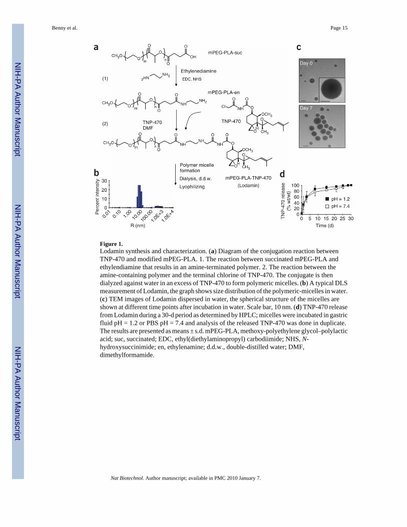
Figure 1.Lodamin synthesis and characterization. (a) Diagram of the conjugation reaction betweenTNP-470 and modified mPEG-PLA. 1. The reaction between succinated mPEG-PLA andethylendiamine that results in an amine-terminated polymer. 2. The reaction between theamine-containing polymer and the terminal chlorine of TNP-470. The conjugate is thendialyzed against water in an excess of TNP-470 to form polymeric micelles. (b) A typical DLSmeasurement of Lodamin, the graph shows size distribution of the polymeric-micelles in water.(c) TEM images of Lodamin dispersed in water, the spherical structure of the micelles areshown at different time points after incubation in water. Scale bar, 10 nm. (d) TNP-470 releasefrom Lodamin during a 30-d period as determined by HPLC; micelles were incubated in gastricfluid pH = 1.2 or PBS pH = 7.4 and analysis of the released TNP-470 was done in duplicate.The results are presented as means ± s.d. mPEG-PLA, methoxy-polyethylene glycol–polylacticacid; suc, succinated; EDC, ethyl(diethylaminopropyl) carbodiimide; NHS, N-hydroxysuccinimide; en, ethylenamine; d.d.w., double-distilled water; DMF,dimethylformamide.
Benny et al. Page 15
Nat Biotechnol. Author manuscript; available in PMC 2010 January 7.
NIH
-PA Author Manuscript
NIH
-PA Author Manuscript
NIH
-PA Author Manuscript
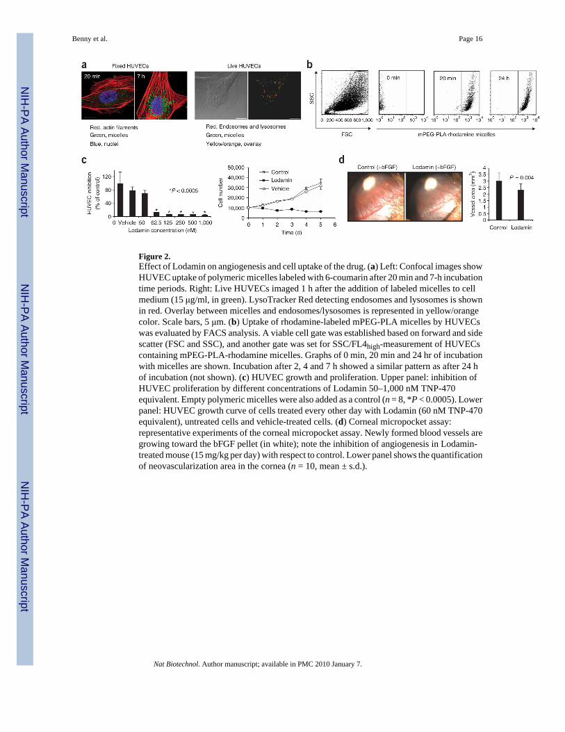
Figure 2.Effect of Lodamin on angiogenesis and cell uptake of the drug. (a) Left: Confocal images showHUVEC uptake of polymeric micelles labeled with 6-coumarin after 20 min and 7-h incubationtime periods. Right: Live HUVECs imaged 1 h after the addition of labeled micelles to cellmedium (15 μg/ml, in green). LysoTracker Red detecting endosomes and lysosomes is shownin red. Overlay between micelles and endosomes/lysosomes is represented in yellow/orangecolor. Scale bars, 5 μm. (b) Uptake of rhodamine-labeled mPEG-PLA micelles by HUVECswas evaluated by FACS analysis. A viable cell gate was established based on forward and sidescatter (FSC and SSC), and another gate was set for SSC/FL4high-measurement of HUVECscontaining mPEG-PLA-rhodamine micelles. Graphs of 0 min, 20 min and 24 hr of incubationwith micelles are shown. Incubation after 2, 4 and 7 h showed a similar pattern as after 24 hof incubation (not shown). (c) HUVEC growth and proliferation. Upper panel: inhibition ofHUVEC proliferation by different concentrations of Lodamin 50–1,000 nM TNP-470equivalent. Empty polymeric micelles were also added as a control (n = 8, *P < 0.0005). Lowerpanel: HUVEC growth curve of cells treated every other day with Lodamin (60 nM TNP-470equivalent), untreated cells and vehicle-treated cells. (d) Corneal micropocket assay:representative experiments of the corneal micropocket assay. Newly formed blood vessels aregrowing toward the bFGF pellet (in white); note the inhibition of angiogenesis in Lodamin-treated mouse (15 mg/kg per day) with respect to control. Lower panel shows the quantificationof neovascularization area in the cornea (n = 10, mean ± s.d.).
Benny et al. Page 16
Nat Biotechnol. Author manuscript; available in PMC 2010 January 7.
NIH
-PA Author Manuscript
NIH
-PA Author Manuscript
NIH
-PA Author Manuscript
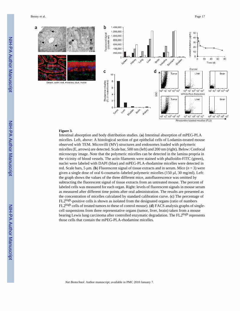
Figure 3.Intestinal absorption and body distribution studies. (a) Intestinal absorption of mPEG-PLAmicelles. Left, above: A histological section of gut epithelial cells of Lodamin-treated mouseobserved with TEM. Microvilli (MV) structures and endosomes loaded with polymericmicelles (E, arrows) are detected. Scale bar, 500 nm (left) and 200 nm (right). Below: Confocalmicroscopy image. Note that the polymeric micelles can be detected in the lamina propria inthe vicinity of blood vessels. The actin filaments were stained with phalloidin-FITC (green),nuclei were labeled with DAPI (blue) and mPEG-PLA-rhodamine micelles were detected inred. Scale bars, 5 μm. (b) Fluorescent signal of tissue extracts and in serum. Mice (n = 3) weregiven a single dose of oral 6-coumarin–labeled polymeric micelles (150 μl, 30 mg/ml). Left:the graph shows the values of the three different mice, autofluorescence was omitted bysubtracting the fluorescent signal of tissue extracts from an untreated mouse. The percent oflabeled cells was measured for each organ. Right: levels of fluorescent signals in mouse serumas measured after different time points after oral administration. The results are presented asthe concentration of micelles calculated by standard calibration curve. (c) The percentage ofFL2high-positive cells is shown as isolated from the designated organs (ratio of numbersFL2high cells of treated tumors to these of control mouse). (d) FACS analysis graphs of single-cell suspensions from three representative organs (tumor, liver, brain) taken from a mousebearing Lewis lung carcinoma after controlled enzymatic degradation. The FL2high representsthose cells that contain the mPEG-PLA-rhodamine micelles.
Benny et al. Page 17
Nat Biotechnol. Author manuscript; available in PMC 2010 January 7.
NIH
-PA Author Manuscript
NIH
-PA Author Manuscript
NIH
-PA Author Manuscript

Figure 4.Lodamin inhibits primary tumor growth without causing neurotoxicity. (a) Effect of free orconjugated TNP-470 on established Lewis lung carcinoma tumors: effect of 30 mg/kg everyother day of free TNP-470 given orally, compared to equivalent dose of Lodamin or water(n = 5 mice per group, *P < 0.05). (b) Lewis lung carcinoma volume during 18 d of differentfrequencies and doses of Lodamin: 30 mg/kg every other day (q.o.d.), 15 mg/kg every otherday, 15 mg/kg every day (q.d.) and water by gavage (n = 5 mice per group, *P < 0.05. (c)Evaluation of the effect of the vehicle, empty mPEG-PLA micelles, on Lewis lung carcinoma(n = 5 mice per group, *P < 0.05). (d) Effect of Lodamin given at a dose of 15 mg/kg everyday on B16/F10 murine melanoma tumor in C57Bl/6J, water was given as control (n = 5 miceper group, *P < 0.05). (e) Representative Lewis lung carcinoma and B16/F10 tumors removedfrom mice at day 18 after treatment with oral Lodamin at 30 mg/kg every other day and 15 mg/kg every day, respectively, and from control untreated mice. (f) Neurotoxicity evaluation ofLodamin-treated mice (10 d, 30 mg/kg every other day) compared to mice treated withsubcutaneous (30 mg/kg every other day) free TNP-470 or water given by gavage. Balancebeam test was quantified by foot-slip errors and the numbers of slips per meter are presented(n = 4–5 mice per group). *P < 0.05, **P < 0.01, ***P < 0.0001 (results are mean ± s.e.m.).
Benny et al. Page 18
Nat Biotechnol. Author manuscript; available in PMC 2010 January 7.
NIH
-PA Author Manuscript
NIH
-PA Author Manuscript
NIH
-PA Author Manuscript
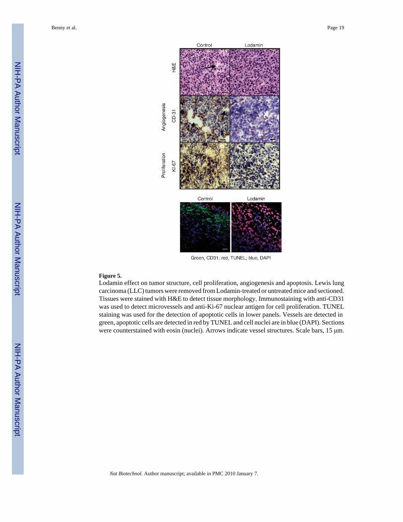
Figure 5.Lodamin effect on tumor structure, cell proliferation, angiogenesis and apoptosis. Lewis lungcarcinoma (LLC) tumors were removed from Lodamin-treated or untreated mice and sectioned.Tissues were stained with H&E to detect tissue morphology. Immunostaining with anti-CD31was used to detect microvessels and anti-Ki-67 nuclear antigen for cell proliferation. TUNELstaining was used for the detection of apoptotic cells in lower panels. Vessels are detected ingreen, apoptotic cells are detected in red by TUNEL and cell nuclei are in blue (DAPI). Sectionswere counterstained with eosin (nuclei). Arrows indicate vessel structures. Scale bars, 15 μm.
Benny et al. Page 19
Nat Biotechnol. Author manuscript; available in PMC 2010 January 7.
NIH
-PA Author Manuscript
NIH
-PA Author Manuscript
NIH
-PA Author Manuscript
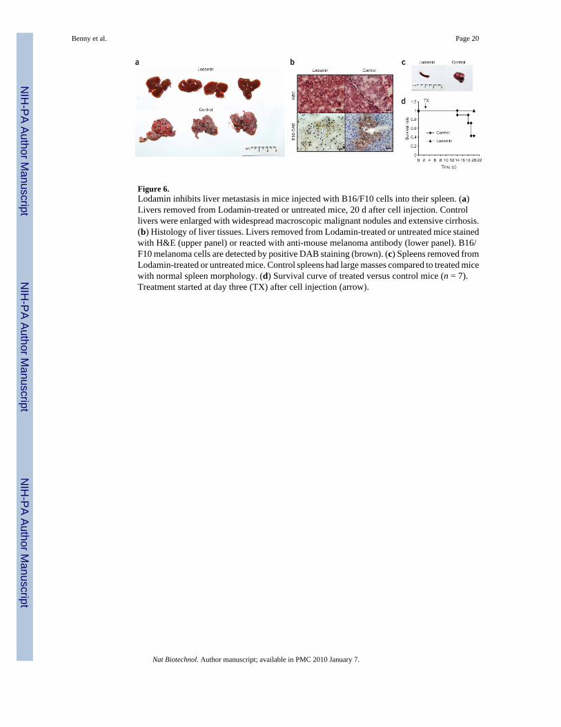
Figure 6.Lodamin inhibits liver metastasis in mice injected with B16/F10 cells into their spleen. (a)Livers removed from Lodamin-treated or untreated mice, 20 d after cell injection. Controllivers were enlarged with widespread macroscopic malignant nodules and extensive cirrhosis.(b) Histology of liver tissues. Livers removed from Lodamin-treated or untreated mice stainedwith H&E (upper panel) or reacted with anti-mouse melanoma antibody (lower panel). B16/F10 melanoma cells are detected by positive DAB staining (brown). (c) Spleens removed fromLodamin-treated or untreated mice. Control spleens had large masses compared to treated micewith normal spleen morphology. (d) Survival curve of treated versus control mice (n = 7).Treatment started at day three (TX) after cell injection (arrow).
Benny et al. Page 20
Nat Biotechnol. Author manuscript; available in PMC 2010 January 7.
NIH
-PA Author Manuscript
NIH
-PA Author Manuscript
NIH
-PA Author Manuscript