a bdnf infusion into the medial prefrontal cortex suppresses cocaine seeking in rats
TRANSCRIPT
A BDNF infusion into the medial prefrontal cortexsuppresses cocaine seeking in rats
William J. Berglind,1 Ronald E. See,1 Rita A. Fuchs,2 Shannon M. Ghee,1 Timothy W. Whitfield, Jr,1 Scott W. Miller3
and Jacqueline F. McGinty1
1Department of Neurosciences, Medical University of South Carolina, Charleston, SC 29425, USA2Department of Psychology, University of North Carolina, Chapel Hill, NC 27599, USA3Department of Biostatistics, Bioinformatics & Epidemiology, Medical University of South Carolina, Charleston, SC 29425, USA
Keywords: addiction, corticostriatal, nucleus accumbens, phospho-ERK, self-administration
Abstract
The medial prefrontal cortex (mPFC) is critical for reinstatement of cocaine seeking and is the main source of brain-derivedneurotrophic factor (BDNF) to striatal regions of the brain relapse circuitry. To test the hypothesis that BDNF in the mPFC regulatescocaine-seeking behavior, rats were trained to press a lever for cocaine infusions (0.2 mg ⁄ inf, 2 h ⁄ day) paired with light + toneconditioned stimulus (CS) presentations on 10 consecutive days. After the last self-administration session, rats received a singleinfusion of BDNF (0.75 lg ⁄ 0.5 lL ⁄ side) into the mPFC; this manipulation produced protracted effects on cocaine-seeking behavior(non-reinforced lever pressing). BDNF pretreatment administered after the last session attenuated cocaine seeking 22 h later and,remarkably, it also blocked cocaine-induced suppression of phospho-extracellular-regulated kinase and elevated BDNF immuno-reactivity in the nucleus accumbens. The same pretreatment also suppressed cocaine-seeking behavior elicited by response-contingent CS presentations after 6 days of forced abstinence or extinction training, as well as a cocaine challenge injection(10 mg ⁄ kg, i.p.) after extinction training. However, BDNF infused into the mPFC had no effect on food-seeking behavior.Furthermore, BDNF infused on the sixth day of abstinence failed to alter responding, suggesting that the regulatory influence ofBDNF is time limited. The suppressive effects of BDNF infused into the mPFC on cocaine seeking indicate that BDNF regulatescortical pathways implicated in relapse to drug seeking and that corticostriatal BDNF adaptations during early abstinence diminishcompulsive drug seeking.
Introduction
Exposure to contexts or cues previously associated with drug taking,or re-introduction of the drug itself, can elicit reinstatement of drugseeking in animal models of relapse (Spealman et al., 1999;Alleweireldt et al., 2001; Shaham et al., 2003; See, 2005). Thesusceptibility to drug relapse and other addictive behaviors is thoughtto depend on long-term neuroadaptations in mRNA, proteins andphospho-proteins (Nestler, 2004; Kalivas & Volkow, 2005). One of theproteins that has been implicated in the reinstatement of cocaineseeking in rats is brain-derived neurotrophic factor (BDNF; Lu et al.,2004; Liu et al., 2006). BDNF protein expression increases in theventral tegmental area (VTA), nucleus accumbens (NAc) and amy-gdala of rats following 30 or 90 days of withdrawal from cocaine self-administration (Grimm et al., 2003). Furthermore, BDNF infusionsinto the VTA or NAc enhance cocaine-induced behavioral sensitiza-tion (Horger et al., 1999), and increase cue- and cocaine-inducedreinstatement, a long-term effect that is present for at least 30 days ofwithdrawal (Lu et al., 2004). Conversely, repeated administration ofBDNF antiserum into the NAc during chronic cocaine self-adminis-tration attenuates cocaine-induced reinstatement (Graham & Self,
2004). Collectively, these studies implicate BDNF activity in the VTAand NAc in long-term modulation of cocaine-induced behavior.Cocaine-induced neuroadaptations are commonly manifested by
alterations in the postsynaptic plasticity of medium spiny neurons inthe NAc. These alterations are likely related to adaptations inprefrontal cortex (PFC) glutamatergic and VTA dopaminergic affer-ents, both of which are critical mediators of reinstatement to cocaineseeking triggered by conditioned stimuli (CS), context or cocaine itself(McFarland et al., 2003; McLaughlin & See, 2003; Fuchs et al., 2004,2005; Kalivas, 2004; Bachtell et al., 2005; Schmidt et al., 2005).Withdrawal from cocaine self-administration elicits long-term decrea-ses in NAc basal extracellular glutamate levels and associatedintracellular signaling (Pierce et al., 1996; Xi et al., 2002; Bakeret al., 2003). Thus, in addition to neuroplasticity in the mesoaccum-bens dopamine system, long-term adaptations in the PFC–NAccircuitry are critical for the reinstatement of cocaine seeking.Cortical pyramidal neurons are the predominant source of BDNF in
the striatum (Altar et al., 1997), including the NAc. However, thesignificance of BDNF in the PFC to cocaine-induced motivatedbehavior has not been investigated. Based on the importance of thecortico-accumbens pathway for reinstatement and the significant roleof BDNF in the activity of this pathway, in the current study weevaluated the effects of a BDNF infusion into the PFC on severalforms of cocaine-seeking behavior, on food-seeking behavior, as wellas on the expression of BDNF and phospho-extracellular-regulated
Correspondence: Dr J.F. McGinty, as above.E-mail: [email protected]
Received 7 April 2007, revised 23 May 2007, accepted 10 June 2007
European Journal of Neuroscience, Vol. 26, pp. 757–766, 2007 doi:10.1111/j.1460-9568.2007.05692.x
ª The Authors (2007). Journal Compilation ª Federation of European Neuroscience Societies and Blackwell Publishing Ltd
kinase (ERK), a major BDNF-TrkB signaling protein, in the NAc andcaudate putamen. We hypothesized that infusion of BDNF into themedial (m)PFC would enhance reinstatement of drug seeking, basedon findings that a bilateral BDNF infusion into the VTA immediatelyafter the final cocaine self-administration session augmented cocaineseeking (Lu et al., 2004). However, we found the opposite, that intra-PFC BDNF, when administered immediately after the final cocaineself-administration session but not 6 days later, suppressed cocaineseeking triggered by CS, context or cocaine itself. These data expandour understanding of the complex role of BDNF in the regulation ofaddictive behavior.
Materials and methods
Animals
Male Sprague–Dawley rats (n ¼ 84; Charles River Laboratories,Wilmington, MA, USA), weighing 275–325 g at the time of surgery,were housed individually on a reverse light : dark cycle. Rats weremaintained on 20–25 g of rat chow per day, with water available adlibitum. All protocols were approved by the Institutional Animal Careand Use Committee of the Medical University of South Carolina, andcarried out in accordance with the National Institutes of Health Guidefor the Care and Use of Laboratory Animals (NIH Publications no.80-23, revised 1996).
Lever response training
Rats were trained to lever press on a fixed ratio 1 schedule of foodreinforcement (45 mg pellets; Noyes, Lancaster, NH, USA) insound-attenuated operant conditioning chambers (30 · 20 · 24 cmhigh; Medical Associates, St Albans, VT, USA) during a 16 hovernight training session. The chambers were equipped with tworetractable levers, a stimulus light above each lever, a food pelletdispenser between the levers, a house light on the wall opposite thelevers, and a speaker connected to a tone generator (ANL-926,Medical Associates). During the session, each lever press on theactive lever resulted in delivery of a food pellet only. Lever presseson the inactive lever had no programmed consequences. Followingfood training, food pellet dispensers were removed from thechambers.
Surgery
At 48 h after food training, rats were anesthetized using a mixture ofketamine hydrochloride (66 mg ⁄ kg i.p.; Fort Dodge Animal Health,Fort Dodge, IA, USA) and xylazine (1.33 mg ⁄ kg i.p.; Bayer,Shawnee Mission, KS, USA) followed by Equithesin (0.5 mL ⁄ kg).Catheters constructed of Silastic laboratory tubing (0.64 mm i.d.,1.19 mm o.d.; Dow Corning, Midland, MI, USA) were implantedinto the right jugular vein of rats, and were anchored to the vein withsilk suture and a hardened silicon gel ball (silicon rubber sealant;General Electric, Waterford, NY, USA) placed 33 mm from theanterior end of the catheter. The Silastic tubing ran directly under theskin to an exit point located in the mid-scapular region. The openend of the catheter cannula was enclosed with a short piece ofpolyethylene tubing that had been heat sealed, and the threadedregion of the cannula was covered with a threaded plastic cap whennot in use. Immediately following catheter implantation, rats weremounted onto a stereotaxic device (Stoelting, Wood Dale, IL, USA),and bilateral stainless steel cannulae (26 gage, Plastics One,
Roanoke, VA, USA) were implanted 1 mm above the PFC targetregion (AP ¼ +3.0; ML ¼ +0.6; DV ¼ )1.6, relative to bregma,Paxinos & Watson, 1998). Following implantation, guide cannulaewere secured to the skull with cranioplastic cement and three steelmachine screws. Stylets (Plastics One) were placed into the guidecannulae to prevent blockage. Following surgery, rats were infusedi.v. with 0.1 mL each of cefazolin (100 mg ⁄ mL) and heparinizedsaline (70 U ⁄ mL) once daily during a 5 day recovery period.Catheter patency was verified by infusing 0.1 mL of methohexitalsodium (20 mg ⁄ mL, i.v.; Eli Lilly, Indianapolis, IN, USA), whichproduces a rapid loss of muscle tone only when administeredintravenously.
Cocaine self-administration
Cocaine self-administration was conducted in standard self-adminis-tration operant chambers (30 · 20 · 24 cm; Medical Associates).Each chamber contained two retractable levers (7 cm above thechamber floor) on each side of the front wall. Awhite circular stimuluslight (2.5 W, 24 V bulb) was located on the panel 7 cm above theactive (right) lever, and a red house light (2.5 W, 24 V bulb) waslocated on the wall on the opposite end of the chamber. The infusionline was tethered to a liquid swivel (Instech, Plymouth Meeting, PA,USA) mounted on a suspended counterbalance. The self-administra-tion apparatus was enclosed in a sound-attenuating chamber (MedicalAssociates). Cocaine hydrochloride (4 mg ⁄ mL; National Institute onDrug Abuse ⁄ RTI International, Research Triangle Park, NC, USA)was delivered using a computer-controlled infusion pump locatedoutside the chamber. The entire system was computer controlled usingMedical PC for Windows.Rats self-administered cocaine on 10 consecutive days during 2 h
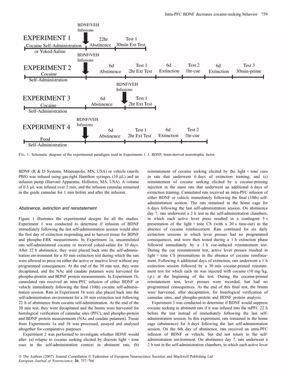
daily sessions. Figure 1 illustrates the experimental design of each ofthe experiments. Rats received 0.1 mL of heparinized saline(10 U ⁄ mL, i.v.) prior to each self-administration session. Animalswere then connected to the infusion tether and each session beganimmediately, signaled by the movement of both levers into thechamber. With the exception of the yoked-saline rats from Experi-ment 1, cocaine reinforcement was available on a fixed ratio 1schedule of reinforcement explicitly paired with a compound CSconsisting of illumination of a white stimulus light located above theactive lever and a tone (2 kHz, 15 dB above ambient noise).Responses on the active (right) lever resulted in the delivery ofcocaine (0.2 mg ⁄ 0.05 mL bolus) over 2 s, and each infusion wasfollowed by a 20 s ‘time-out’ period. During the time-out, activelever presses were recorded, but had no programmed consequences.Responses on the inactive (left) lever were also recorded, but had noprogrammed consequences. Yoked-saline rats (Experiment 1 ) seebelow) received an infusion of saline (0.05 mL bolus) over 2 s,contingent upon the cocaine infusion received by the self-adminis-tering rat in the adjacent self-administration chamber. Rats underwent10 days of cocaine self-administration prior to a single intracranialinfusion.
Intracranial infusions
We adapted the single infusion protocol and chose the dose of BDNF(0.75 lg ⁄ side) that Lu et al. (2004) previously used in vivo. ForBDNF or vehicle [10 mm phosphate-buffered saline (PBS)] infusions,infusion cannulae (33 gage; Plastics One) were inserted bilaterallyinto the guide cannulae such that 1 mm of the infusion cannulaeextended past the end of the guide cannulae. Human recombinant
758 W. J. Berglind et al.
ª The Authors (2007). Journal Compilation ª Federation of European Neuroscience Societies and Blackwell Publishing LtdEuropean Journal of Neuroscience, 26, 757–766
BDNF (R & D Systems, Minneapolis, MN, USA) or vehicle (sterilePBS) was infused using gas-tight Hamilton syringes (10 lL) and aninfusion pump (Harvard Apparatus, Holliston, MA, USA). A volumeof 0.5 lL was infused over 2 min, and the infusion cannulae remainedin the guide cannulae for 1 min before and after the infusion.
Abstinence, extinction and reinstatement
Figure 1 illustrates the experimental designs for all the studies.Experiment 1 was conducted to determine if infusion of BDNFimmediately following the last self-administration session would alterthe first day of extinction responding and to harvest tissue for BDNFand phospho-ERK measurements. In Experiment 1a, uncannulatedrats self-administered cocaine or received yoked-saline for 10 days.After 22 h abstinence, they were placed back into the self-adminis-tration environment for a 30 min extinction test during which the ratswere allowed to press on either the active or inactive lever without anyprogrammed consequences. At the end of the 30 min test, they weredecapitated, and the NAc and caudate putamen were harvested forphospho-protein and BDNF protein measurements. In Experiment 1b,cannulated rats received an intra-PFC infusion of either BDNF orvehicle immediately following the final (10th) cocaine self-adminis-tration session. Rats in Experiment 1b were also placed back into theself-administration environment for a 30 min extinction test following22 h of abstinence from cocaine self-administration. At the end of the30 min test, they were decapitated and the brains were harvested forhistological verification of cannulae sites (PFC), and phospho-proteinand BDNF protein measurements (NAc and caudate putamen). Tissuefrom Experiments 1a and 1b was processed, assayed and analysedaltogether for comparative purposes.
Experiment 2 was performed to investigate whether BDNF wouldalter: (a) relapse to cocaine seeking elicited by discrete light + tonecues in the self-administration context in abstinent rats; (b)
reinstatement of cocaine seeking elicited by the light + tone cuesin rats that underwent 6 days of extinction training; and (c)reinstatement of cocaine seeking elicited by a cocaine-priminginjection in the same rats that underwent an additional 6 days ofextinction training, Cannulated rats received an intra-PFC infusion ofeither BDNF or vehicle immediately following the final (10th) self-administration session. The rats remained in the home cage for6 days following the last self-administration session. On abstinenceday 7, rats underwent a 2 h test in the self-administration chambers,in which each active lever press resulted in a contingent 5 spresentation of the light + tone CS (with a 20 s time-out) in theabsence of cocaine reinforcement. Rats continued for six dailyextinction sessions in which lever presses had no programmedconsequences, and were then tested during a 1 h extinction phasefollowed immediately by a 1 h cue-induced reinstatement test.During the cue reinstatement test, active lever presses resulted inlight + tone CS presentations in the absence of cocaine reinforce-ment. Following 6 additional days of extinction, rats underwent a 1 hextinction session followed by a 30 min cocaine-primed reinstate-ment test for which each rat was injected with cocaine (10 mg ⁄ kg,i.p.) at the beginning of the test. During the cocaine-primedreinstatement test, lever presses were recorded, but had noprogrammed consequences. At the end of this final test, the brainswere harvested, after decapitation, for histological verification ofcannulae sites, and phospho-protein and BDNF protein analysis.Experiment 3 was conducted to determine if BDNF would suppress
cocaine seeking in abstinent rats if it was infused into the mPFC 22 hbefore the test instead of immediately following the last self-administration session. In this experiment, rats remained in the homecage (abstinence) for 6 days following the last self-administrationsession. On the 6th day of abstinence, rats received an intra-PFCinfusion of BDNF or vehicle, but did not return to the self-administration environment. On abstinence day 7, rats underwent a2 h test in the self-administration chambers, in which each active lever
Cocaine Self-Administration
BDNF/VEHInfusions
6dAbstinence
Test 12hr Ext Test
6dExtinction
6dExtinction
Test 21hr-cue
Test 330min-prime
6dAbstinence
Test 12hr Ext Test
22hrAbstinence
Test 130min Ext Test
EXPERIMENT 1
EXPERIMENT 3
EXPERIMENT 2
Cocaine Self-Administration or Yoked-Saline
BDNF/VEHInfusions
Cocaine Self-Administration
BDNF/VEHInfusions
Food Self-Administration
BDNF/VEHInfusions
6dAbstinence
Test 12hr Ext Test
6dExtinction
Test 21hr-cue
EXPERIMENT 4
Fig. 1. Schematic diagram of the experimental paradigms used in Experiments 1–3. BDNF, brain-derived neurotrophic factor.
Intra-PFC BDNF decreases cocaine-seeking behavior 759
ª The Authors (2007). Journal Compilation ª Federation of European Neuroscience Societies and Blackwell Publishing LtdEuropean Journal of Neuroscience, 26, 757–766
press resulted in a contingent 5 s presentation of the light + tone CS(with a 20 s time-out) in the absence of cocaine reinforcement. At theend of the experiment, the rats were decapitated and the brains wereharvested for histological verification of cannulae sites.
Food self-administration and reinstatement
Experiment 4 was designed to be as similar as possible to the design ofthe abstinence and cue-induced reinstatement phases of Experiment 2,except that the rats learned to self-administer food instead of cocaine.Rats initially lever-pressed for food reinforcement (45 mg Noyes foodpellets) on a fixed ratio 1 schedule of reinforcement, during daily 2 hsessions on 10 consecutive days. Subjects then remained in the homecage for a 6 day abstinence period, during which they were maintainedat 90% ad libitum weight. Rats then underwent a 2 h post-abstinencetest, during which active lever pressing resulted in the presentation ofthe previously paired light + tone but no food. Following 6 days ofextinction training, during which lever presses were recorded butnever resulted in food pellet or CS delivery, rats underwent a cue-induced reinstatement test for food-seeking behavior. During the cue-induced reinstatement test, active lever presses resulted in thepresentation of the CS previously paired with food during self-administration. The 1 h test was preceded by 1 h of extinctiontraining.
Tissue processing and histology
Rats in Experiments 1–4 were killed by rapid decapitation followingthe completion of the final extinction or reinstatement test. Brains wereimmediately removed and frozen in isopentane, then stored at )80 �Cuntil they were sectioned on a cryostat. Sections through the PFC weresectioned at 12 lm for cannulae placement verification. The remainingforebrain from rats in Experiments 1 and 2 was sectioned from rostralto caudal up to AP 1.7 from bregma. It was then removed from thechuck and sectioned caudal to rostral up to AP 0.7 from bregma. Theremaining slab of tissue contained the dorsal and ventral striatal targetareas that were extracted using a 13-gage tissue punch. Frozenpunches were stored at )80 �C prior to being processed forimmunoblotting.
Immunoblotting
The method described by Toda et al. (2003) was used. Protein wasextracted from both the NAc and caudate putamen, and the tissue wassonicated in Trizol (Invitrogen, Carlsbad, CA, USA). Samples wereanalysed for protein, diluted to equalize for the protein concentrations,and aliquoted. Samples were stored in the sample buffer [Tris–HCl,0.5 mm, pH 6.8; glycerol 10%; sodium dodecyl sulfate (SDS) 10%;b-mercaptoethanol, 5%; bromophenol blue, 0.05% w ⁄ v], boiled for2 min and loaded onto gels (12% SDS). Proteins were separated byapplication of 30 mA constant current for 25–30 min, transferred ontopolyvinylidene fluoride (PVDF) membrane strips (200 mA for60 min; Millipore, Billerica, MA, USA), and immunoblotted withanti-phospho-ERK 1 ⁄ 2 (1 : 500-phospho-p44 ⁄ 42 MAP kinase,Thr202 ⁄ Tyr204, Cell Signaling Technologies, Danvers, MA, USA)for 60 min at room temperature. PVDF strips were washed andincubated for 60 min at room temperature with secondary antibody[horseradish peroxidase (HRP)-linked anti-rabbit antibody 1 : 1000;Amersham Biosciences, Piscataway, NJ, USA] and protein complexeswere visualized by enhanced chemiluminescence ECL detection(Amersham Biosciences). Immunoblots were exposed to Hyperfilm
(Amersham Biosciences). Following imaging, membranes wereincubated for 10 min in stripping agent (re-blot plus Western blotmild antibody stripping solution; Chemicon International, Temecula,CA, USA) and then re-exposed to primary antibody specific for non-phosphorylated ERK 1 ⁄ 2 (1 : 1000) overnight. Protein bands werequantified by densitometry using Image J software. Analyses wereperformed by evaluating the ratio of phospho-ERK ⁄ total ERKexpression of each sample within a single membrane. The ratio wasnormalized across trials (a minimum of three comprehensive,individual trials was performed for each experiment) by expressingeach ratio value as a percentage of the control within each individualtrial.
BDNF enzyme-linked immunosorbent assay (ELISA)
BDNF levels were assessed using ELISA-Emax Immunoassay System(Promega, Madison, WI, USA) kits, according to the manufacturer’sprotocol. Flat-bottom plates were coated with an anti-BDNF mono-clonal antibody (1 : 1000) overnight. After blocking non-specificbinding, immobilized anti-BDNF monoclonal antibody was incubatedwith brain tissue samples containing BDNF protein or BDNFstandards serially diluted to prepare a standard curve, followed byanti-human BDNF polyclonal antibody (1 : 500). The complex wasbound using an IgY antibody conjugated to HRP as a tertiary reactant.After repeatedly washing unbound conjugate, plates were incubatedwith tetramethylbenzidine chromagenic substrate, and color changewas measured in an ELISA plate reader at 450 nm. Using this kit,BDNF can be quantified in the range 7.8–500 pg ⁄ mL. Measurementsgiven by the plate reader were calibrated against a standard curveprepared with human BDNF protein from Promega plotted in serialdilution with a correlation coefficient of r2 > 0.95 accepted. Adjustedconcentrations of BDNF expression were normalized to the averageconcentration of the control condition.
Statistical analysis
Responding on both the active and inactive levers across groupsduring relapse to cocaine seeking was compared with respondingduring the last 3 days of self-administration or responding during theextinction session that preceded the test as appropriate in each of thestudies. Due to the experimental design (multiple observations per rat)the data are correlated, so a statistical model capable of accounting forthis correlation was used for the behavioral data to avoid biasedP-values (Fitzmaurice et al., 2004). Further, the response variables(number of active and inactive lever presses) are ‘count’ data, so anapproach that does not assume normality was selected (Agresti, 2002).To address both of these issues in the statistical analysis, a generalizedestimating equation approach using a Poisson distribution (Litrellet al., 2002) was implemented using SAS v9.1 (SAS Institute, Cary,NC, USA), with treatment (e.g. BDNF or vehicle) and condition (e.g.extinction or cocaine challenge) as explanatory covariates (factors).Generalized estimating equation methods were first proposed by Liang& Zeger (1986), and details of their statistical properties are furtherdescribed by Fitzmaurice et al. (2004). These methods are similar tomixed model analyses of variance (anovas), as both explain acorrelated response variable with explanatory covariates ) anova isused for response data that are normally distributed, whereasgeneralized estimating equations are used for binary or count data.Generalized estimating equations use a chi-square because it iseffectively doing two analyses: one with no covariates and anotherwith all of the covariates of interest. It then does a goodness of fit test
760 W. J. Berglind et al.
ª The Authors (2007). Journal Compilation ª Federation of European Neuroscience Societies and Blackwell Publishing LtdEuropean Journal of Neuroscience, 26, 757–766
comparing the improvement in the model with covariates over themodel without them. For data that were approximately normallydistributed (immunoblotting and ELISA), a mixed model anova wasperformed and multiple comparisons were adjusted via the TukeyKramer test (for unequal n per group) when a significant F-value wasobtained. P-values < 0.05 were considered statistically significant.
Results
Histology
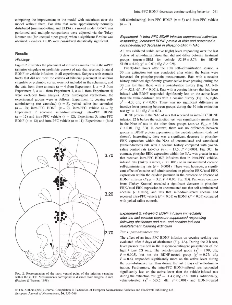
Figure 2 illustrates the placement of infusion cannula tips in the mPFC(anterior cingulate or prelimbic cortex) of rats that received bilateralBDNF or vehicle infusions in all experiments. Subjects with cannulatracts that did not meet the criteria of bilateral placement in anteriorcingulate or prelimbic cortex were not included in the schematic, andthe data from these animals (n ¼ 4 from Experiment 1, n ¼ 3 fromExperiment 2, n ¼ 1 from Experiment 3, n ¼ 1 from Experiment 4)were excluded from analysis. After histological verification, theexperimental groups were as follows: Experiment 1: cocaine self-administering (no cannulae) (n ¼ 8), yoked saline (no cannulae)(n ¼ 10); intra-PFC BDNF (n ¼ 9), intra-PFC vehicle (n ¼ 7);Experiment 2 (cocaine self-administering): intra-PFC BDNF(n ¼ 12) and intra-PFC vehicle (n ¼ 12); Experiment 3: intra-PFCBDNF (n ¼ 12) and intra-PFC vehicle (n ¼ 11); Experiment 4 (food
self-administering) intra-PFC BDNF (n ¼ 5) and intra-PFC vehicle(n ¼ 7).
Experiment 1: Intra-PFC BDNF infusion suppressed extinctionresponding, increased BDNF protein in NAc and prevented acocaine-induced decrease in phospho-ERK in NAc
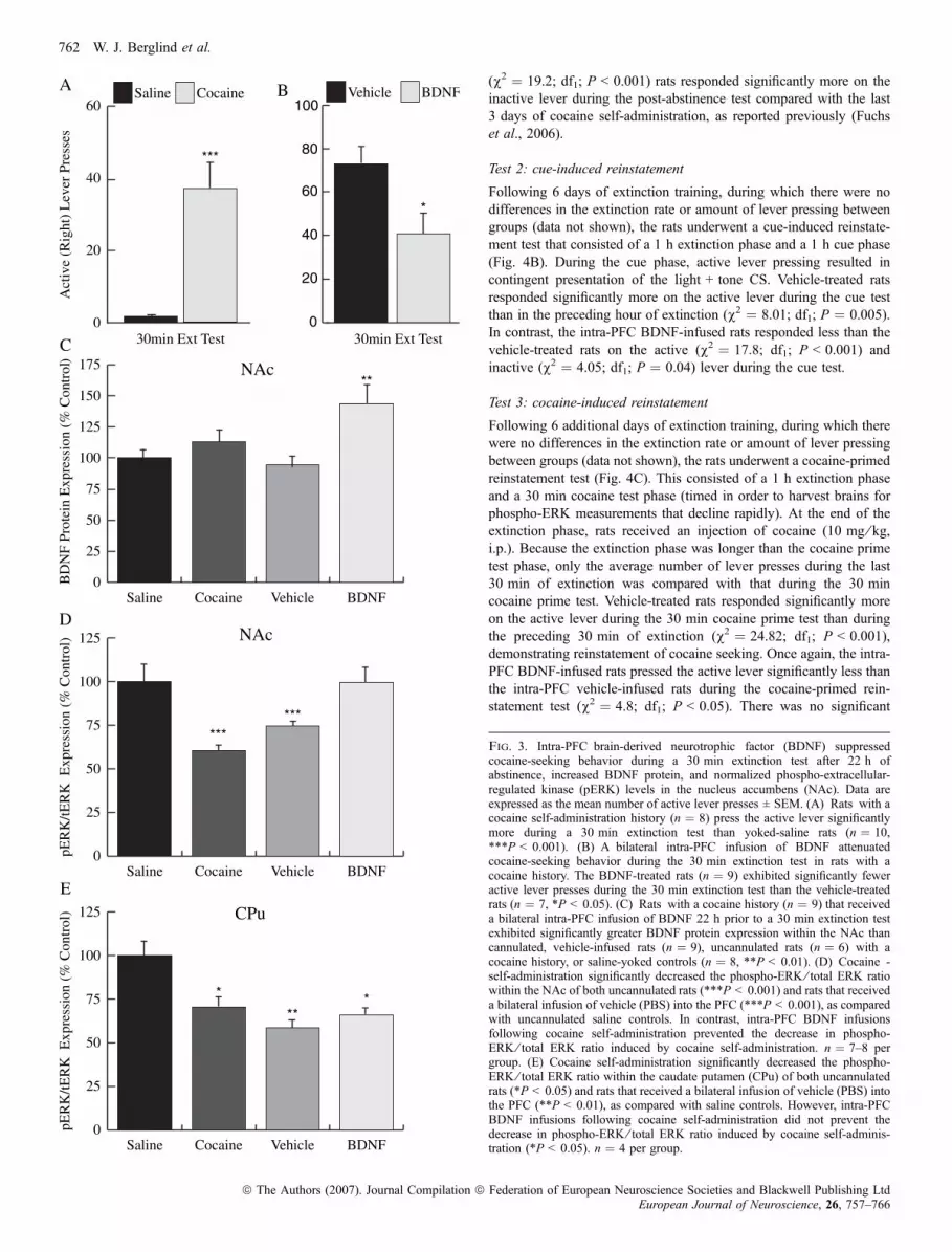
All rats exhibited stable active (right) lever responding over the last3 days of self-administration that did not differ between treatmentgroups (mean ± SEM for vehicle 52.19 ± 5.74; for BDNF51.60 ± 4.40; v2 ¼ 0.01; df1; P > 0.9).Twenty-two hours after the 10th self-administration session, a
30 min extinction test was conducted after which the brains wereharvested for phospho-protein measurements. Rats with a cocainehistory exhibited significantly greater active lever pressing during the30 min test than those with a yoked-saline history (Fig. 3A, left-v2 ¼ 52.3; df1; P < 0.001). Rats with a cocaine history that had beeninfused with BDNF responded significantly less on the active leverthan the vehicle-infused rats with a cocaine history (Fig. 3A, right-v2 ¼ 4.3; df1; P < 0.05). There was no significant difference ininactive lever pressing between groups during the 30 min extinctiontest (v2 ¼ 1.1; df1; P ¼ 0.3).BDNF protein in the NAc of rats that received an intra-PFC BDNF
infusion 22 h before the extinction test was significantly greater thanin the NAc of rats in the other three groups (anova F3,28 ¼ 6.0;P < 0.01, Fig. 3B). In contrast, there was no difference betweengroups in BDNF protein expression in the caudate putamen (data notshown). Interestingly, there was a significant decrease in phospho-ERK expression within the NAc of uncannulated and cannulated(vehicle-treated) rats with a cocaine history compared with yoked-saline control rats (anova F2,51 ¼ 15.5, P < 0.0001, Fig. 3C). Incontrast, phospho-ERK expression within the NAc was greater in ratsthat received intra-PFC BDNF infusions than in intra-PFC vehicle-infused rats (Tukey Kramer, P < 0.005) or in uncannulated cocaineself-administering rats (P < 0.0001). There was, however, a signifi-cant effect of cocaine self-administration on phospho-ERK ⁄ total ERKexpression within the caudate putamen in the presence or absence ofBDNF infusion (F3,15 ¼ 3.2, P < 0.05, Fig. 3D). Pair-wise compar-isons (Tukey Kramer) revealed a significant decrease in phospho-ERK ⁄ total ERK expression in uncannulated rats that self-administeredcocaine (P < 0.05), and rats that self-administered cocaine andreceived intra-PFC vehicle (P < 0.01) or BDNF (P < 0.05) comparedwith yoked-saline controls.
Experiment 2: intra-PFC BDNF infusion immediatelyafter the last cocaine exposure suppressed respondingfollowing abstinence and cue- and cocaine-inducedreinstatement following extinction
Test 1: post-abstinence test
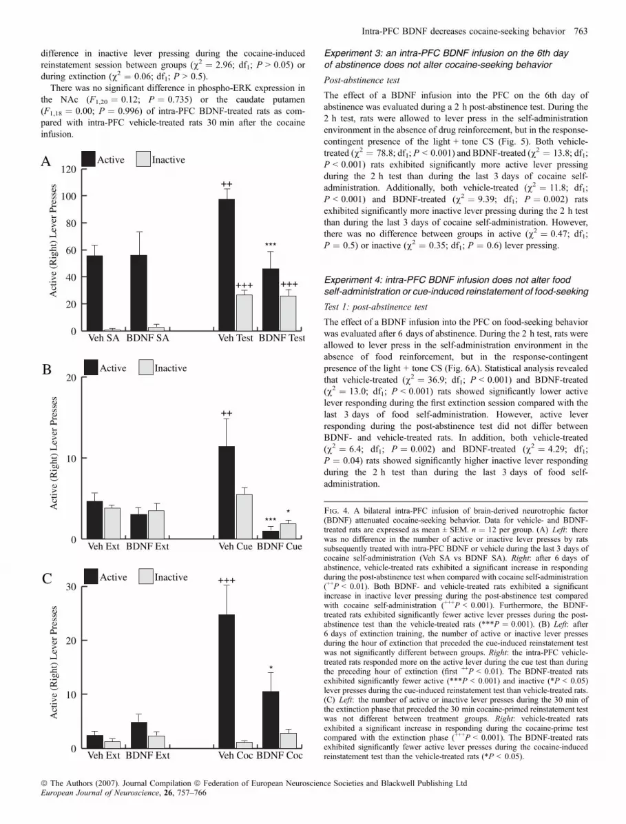
The effect of an intra-PFC BDNF infusion on cocaine seeking wasevaluated after 6 days of abstinence (Fig. 4A). During the 2 h test,lever presses resulted in the response-contingent presentation of thelight + tone CS only. The vehicle-treated group (v2 ¼ 7.99; df1;P ¼ 0.005), but not the BDNF-treated group (v2 ¼ 0.27; df1;P ¼ 0.6), responded significantly more on the active lever duringthe post-abstinence test than during the last 3 days of self-adminis-tration. Furthermore, the intra-PFC BDNF-infused rats respondedsignificantly less on the active lever than the vehicle-infused ratsduring the extinction test (v2 ¼ 11.43; df1; P < 0.001). Additionally,vehicle-treated (v2 ¼ 665.5; df1; P < 0.001) and BDNF-treated
3.70
3.20
2.70
Fig. 2. Representation of the most ventral point of the infusion cannulaewithin the mPFC. Measurements correspond to distance from bregma in mm(Paxinos & Watson, 1998).
Intra-PFC BDNF decreases cocaine-seeking behavior 761
ª The Authors (2007). Journal Compilation ª Federation of European Neuroscience Societies and Blackwell Publishing LtdEuropean Journal of Neuroscience, 26, 757–766
(v2 ¼ 19.2; df1; P < 0.001) rats responded significantly more on theinactive lever during the post-abstinence test compared with the last3 days of cocaine self-administration, as reported previously (Fuchset al., 2006).
Test 2: cue-induced reinstatement
Following 6 days of extinction training, during which there were nodifferences in the extinction rate or amount of lever pressing betweengroups (data not shown), the rats underwent a cue-induced reinstate-ment test that consisted of a 1 h extinction phase and a 1 h cue phase(Fig. 4B). During the cue phase, active lever pressing resulted incontingent presentation of the light + tone CS. Vehicle-treated ratsresponded significantly more on the active lever during the cue testthan in the preceding hour of extinction (v2 ¼ 8.01; df1; P ¼ 0.005).In contrast, the intra-PFC BDNF-infused rats responded less than thevehicle-treated rats on the active (v2 ¼ 17.8; df1; P < 0.001) andinactive (v2 ¼ 4.05; df1; P ¼ 0.04) lever during the cue test.
Test 3: cocaine-induced reinstatement
Following 6 additional days of extinction training, during which therewere no differences in the extinction rate or amount of lever pressingbetween groups (data not shown), the rats underwent a cocaine-primedreinstatement test (Fig. 4C). This consisted of a 1 h extinction phaseand a 30 min cocaine test phase (timed in order to harvest brains forphospho-ERK measurements that decline rapidly). At the end of theextinction phase, rats received an injection of cocaine (10 mg ⁄ kg,i.p.). Because the extinction phase was longer than the cocaine primetest phase, only the average number of lever presses during the last30 min of extinction was compared with that during the 30 mincocaine prime test. Vehicle-treated rats responded significantly moreon the active lever during the 30 min cocaine prime test than duringthe preceding 30 min of extinction (v2 ¼ 24.82; df1; P < 0.001),demonstrating reinstatement of cocaine seeking. Once again, the intra-PFC BDNF-infused rats pressed the active lever significantly less thanthe intra-PFC vehicle-infused rats during the cocaine-primed rein-statement test (v2 ¼ 4.8; df1; P < 0.05). There was no significant
Act
ive
(Rig
ht)
Lev
er P
ress
es
0
20
40
60
80
100BDNFVehicle
*
0
25
50
75
100
125
150
175
BDNFVehicleCocaineSaline
BD
NF
Prot
ein
Exp
ress
ion
(% C
ontr
ol)
NAc **
0
25
50
75
100
125
BDNFVehicleCocaineSaline
pER
K/tE
RK
Exp
ress
ion
(% C
ontr
ol) NAc
******
0
20
40
60CocaineSaline
30min Ext Test
***
0
25
50
75
100
125
BDNFVehicleCocaineSaline
pER
K/tE
RK
Exp
ress
ion
(% C
ontr
ol)
*
**
CPu
*
A
C
D
E
30min Ext Test
B
Fig. 3. Intra-PFC brain-derived neurotrophic factor (BDNF) suppressedcocaine-seeking behavior during a 30 min extinction test after 22 h ofabstinence, increased BDNF protein, and normalized phospho-extracellular-regulated kinase (pERK) levels in the nucleus accumbens (NAc). Data areexpressed as the mean number of active lever presses ± SEM. (A) Rats with acocaine self-administration history (n ¼ 8) press the active lever significantlymore during a 30 min extinction test than yoked-saline rats (n ¼ 10,***P < 0.001). (B) A bilateral intra-PFC infusion of BDNF attenuatedcocaine-seeking behavior during the 30 min extinction test in rats with acocaine history. The BDNF-treated rats (n ¼ 9) exhibited significantly feweractive lever presses during the 30 min extinction test than the vehicle-treatedrats (n ¼ 7, *P < 0.05). (C) Rats with a cocaine history (n ¼ 9) that receiveda bilateral intra-PFC infusion of BDNF 22 h prior to a 30 min extinction testexhibited significantly greater BDNF protein expression within the NAc thancannulated, vehicle-infused rats (n ¼ 9), uncannulated rats (n ¼ 6) with acocaine history, or saline-yoked controls (n ¼ 8, **P < 0.01). (D) Cocaine -self-administration significantly decreased the phospho-ERK ⁄ total ERK ratiowithin the NAc of both uncannulated rats (***P < 0.001) and rats that receiveda bilateral infusion of vehicle (PBS) into the PFC (***P < 0.001), as comparedwith uncannulated saline controls. In contrast, intra-PFC BDNF infusionsfollowing cocaine self-administration prevented the decrease in phospho-ERK ⁄ total ERK ratio induced by cocaine self-administration. n ¼ 7–8 pergroup. (E) Cocaine self-administration significantly decreased the phospho-ERK ⁄ total ERK ratio within the caudate putamen (CPu) of both uncannulatedrats (*P < 0.05) and rats that received a bilateral infusion of vehicle (PBS) intothe PFC (**P < 0.01), as compared with saline controls. However, intra-PFCBDNF infusions following cocaine self-administration did not prevent thedecrease in phospho-ERK ⁄ total ERK ratio induced by cocaine self-adminis-tration (*P < 0.05). n ¼ 4 per group.
762 W. J. Berglind et al.
ª The Authors (2007). Journal Compilation ª Federation of European Neuroscience Societies and Blackwell Publishing LtdEuropean Journal of Neuroscience, 26, 757–766
difference in inactive lever pressing during the cocaine-inducedreinstatement session between groups (v2 ¼ 2.96; df1; P > 0.05) orduring extinction (v2 ¼ 0.06; df1; P > 0.5).
There was no significant difference in phospho-ERK expression inthe NAc (F1,20 ¼ 0.12; P ¼ 0.735) or the caudate putamen(F1,18 ¼ 0.00; P ¼ 0.996) of intra-PFC BDNF-treated rats as com-pared with intra-PFC vehicle-treated rats 30 min after the cocaineinfusion.
Experiment 3: an intra-PFC BDNF infusion on the 6th dayof abstinence does not alter cocaine-seeking behavior
Post-abstinence test
The effect of a BDNF infusion into the PFC on the 6th day ofabstinence was evaluated during a 2 h post-abstinence test. During the2 h test, rats were allowed to lever press in the self-administrationenvironment in the absence of drug reinforcement, but in the response-contingent presence of the light + tone CS (Fig. 5). Both vehicle-treated (v2 ¼ 78.8; df1; P < 0.001) and BDNF-treated (v2 ¼ 13.8; df1;P < 0.001) rats exhibited significantly more active lever pressingduring the 2 h test than during the last 3 days of cocaine self-administration. Additionally, both vehicle-treated (v2 ¼ 11.8; df1;P < 0.001) and BDNF-treated (v2 ¼ 9.39; df1; P ¼ 0.002) ratsexhibited significantly more inactive lever pressing during the 2 h testthan during the last 3 days of cocaine self-administration. However,there was no difference between groups in active (v2 ¼ 0.47; df1;P ¼ 0.5) or inactive (v2 ¼ 0.35; df1; P ¼ 0.6) lever pressing.
Experiment 4: intra-PFC BDNF infusion does not alter foodself-administration or cue-induced reinstatement of food-seeking
Test 1: post-abstinence test
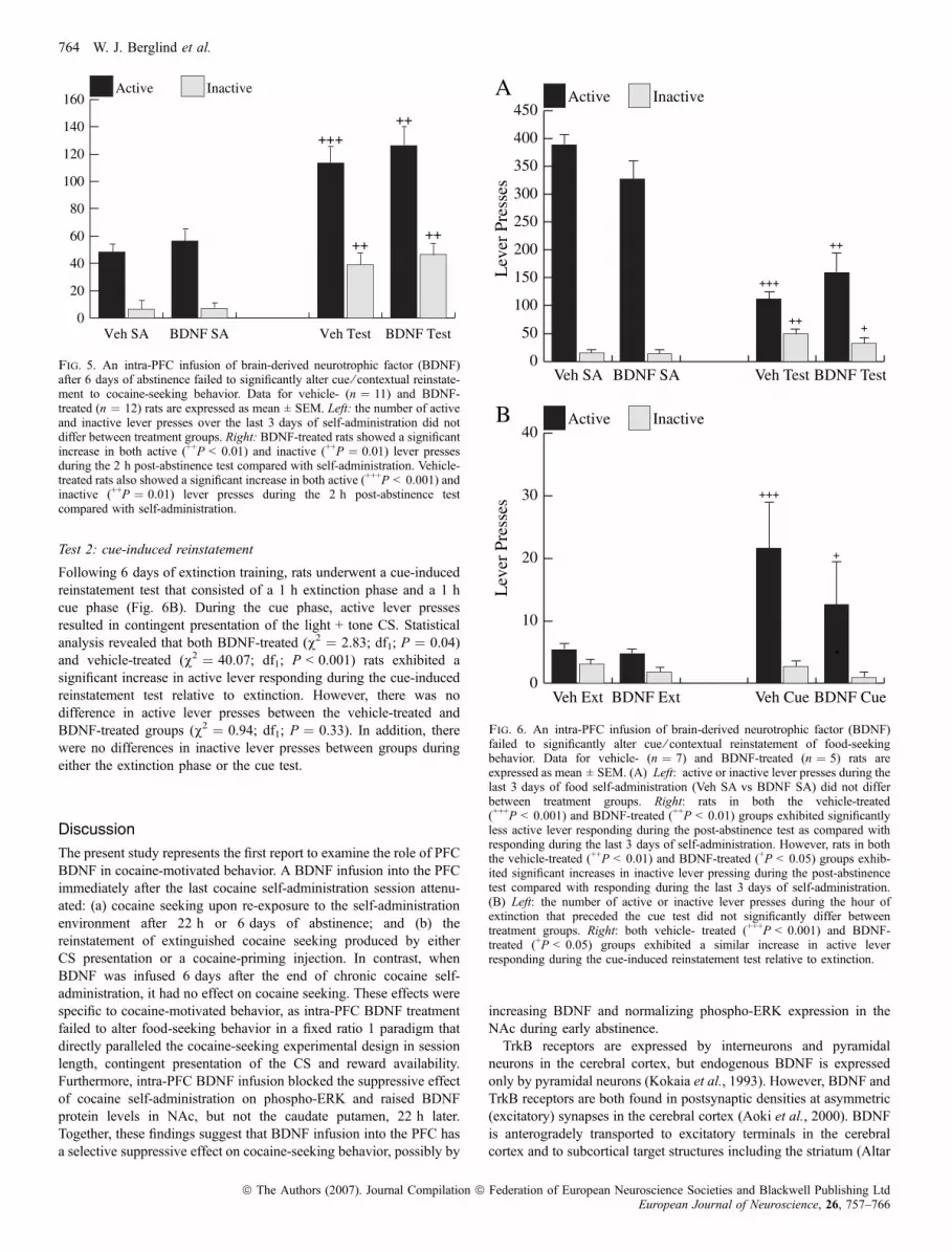
The effect of a BDNF infusion into the PFC on food-seeking behaviorwas evaluated after 6 days of abstinence. During the 2 h test, rats wereallowed to lever press in the self-administration environment in theabsence of food reinforcement, but in the response-contingentpresence of the light + tone CS (Fig. 6A). Statistical analysis revealedthat vehicle-treated (v2 ¼ 36.9; df1; P < 0.001) and BDNF-treated(v2 ¼ 13.0; df1; P < 0.001) rats showed significantly lower activelever responding during the first extinction session compared with thelast 3 days of food self-administration. However, active leverresponding during the post-abstinence test did not differ betweenBDNF- and vehicle-treated rats. In addition, both vehicle-treated(v2 ¼ 6.4; df1; P ¼ 0.002) and BDNF-treated (v2 ¼ 4.29; df1;P ¼ 0.04) rats showed significantly higher inactive lever respondingduring the 2 h test than during the last 3 days of food self-administration.
0
20
40
60
80
100
120InactiveActive
BDNF TestVeh TestBDNF SAVeh SA
Act
ive
(Rig
ht)
Lev
er P
ress
es
0
10
20InactiveActive
BDNF CueVeh CueBDNF ExtVeh Ext
Act
ive
(Rig
ht)
Lev
er P
ress
es
0
10
20
30InactiveActive
BDNF CocVeh CocBDNF ExtVeh Ext
Act
ive
(Rig
ht)
Lev
er P
ress
es
***
++
***
A
B
C
++
+++
*
+++ +++
* Fig. 4. A bilateral intra-PFC infusion of brain-derived neurotrophic factor(BDNF) attenuated cocaine-seeking behavior. Data for vehicle- and BDNF-treated rats are expressed as mean ± SEM. n ¼ 12 per group. (A) Left: therewas no difference in the number of active or inactive lever presses by ratssubsequently treated with intra-PFC BDNF or vehicle during the last 3 days ofcocaine self-administration (Veh SA vs BDNF SA). Right: after 6 days ofabstinence, vehicle-treated rats exhibited a significant increase in respondingduring the post-abstinence test when compared with cocaine self-administration(++P < 0.01). Both BDNF- and vehicle-treated rats exhibited a significantincrease in inactive lever pressing during the post-abstinence test comparedwith cocaine self-administration (+++P < 0.001). Furthermore, the BDNF-treated rats exhibited significantly fewer active lever presses during the post-abstinence test than the vehicle-treated rats (***P ¼ 0.001). (B) Left: after6 days of extinction training, the number of active or inactive lever pressesduring the hour of extinction that preceded the cue-induced reinstatement testwas not significantly different between groups. Right: the intra-PFC vehicle-treated rats responded more on the active lever during the cue test than duringthe preceding hour of extinction (first ++P < 0.01). The BDNF-treated ratsexhibited significantly fewer active (***P < 0.001) and inactive (*P < 0.05)lever presses during the cue-induced reinstatement test than vehicle-treated rats.(C) Left: the number of active or inactive lever presses during the 30 min ofthe extinction phase that preceded the 30 min cocaine-primed reinstatement testwas not different between treatment groups. Right: vehicle-treated ratsexhibited a significant increase in responding during the cocaine-prime testcompared with the extinction phase (+++P < 0.001). The BDNF-treated ratsexhibited significantly fewer active lever presses during the cocaine-inducedreinstatement test than the vehicle-treated rats (*P < 0.05).
Intra-PFC BDNF decreases cocaine-seeking behavior 763
ª The Authors (2007). Journal Compilation ª Federation of European Neuroscience Societies and Blackwell Publishing LtdEuropean Journal of Neuroscience, 26, 757–766
Test 2: cue-induced reinstatement
Following 6 days of extinction training, rats underwent a cue-inducedreinstatement test that consisted of a 1 h extinction phase and a 1 hcue phase (Fig. 6B). During the cue phase, active lever pressesresulted in contingent presentation of the light + tone CS. Statisticalanalysis revealed that both BDNF-treated (v2 ¼ 2.83; df1; P ¼ 0.04)and vehicle-treated (v2 ¼ 40.07; df1; P < 0.001) rats exhibited asignificant increase in active lever responding during the cue-inducedreinstatement test relative to extinction. However, there was nodifference in active lever presses between the vehicle-treated andBDNF-treated groups (v2 ¼ 0.94; df1; P ¼ 0.33). In addition, therewere no differences in inactive lever presses between groups duringeither the extinction phase or the cue test.
Discussion
The present study represents the first report to examine the role of PFCBDNF in cocaine-motivated behavior. A BDNF infusion into the PFCimmediately after the last cocaine self-administration session attenu-ated: (a) cocaine seeking upon re-exposure to the self-administrationenvironment after 22 h or 6 days of abstinence; and (b) thereinstatement of extinguished cocaine seeking produced by eitherCS presentation or a cocaine-priming injection. In contrast, whenBDNF was infused 6 days after the end of chronic cocaine self-administration, it had no effect on cocaine seeking. These effects werespecific to cocaine-motivated behavior, as intra-PFC BDNF treatmentfailed to alter food-seeking behavior in a fixed ratio 1 paradigm thatdirectly paralleled the cocaine-seeking experimental design in sessionlength, contingent presentation of the CS and reward availability.Furthermore, intra-PFC BDNF infusion blocked the suppressive effectof cocaine self-administration on phospho-ERK and raised BDNFprotein levels in NAc, but not the caudate putamen, 22 h later.Together, these findings suggest that BDNF infusion into the PFC hasa selective suppressive effect on cocaine-seeking behavior, possibly by
increasing BDNF and normalizing phospho-ERK expression in theNAc during early abstinence.TrkB receptors are expressed by interneurons and pyramidal
neurons in the cerebral cortex, but endogenous BDNF is expressedonly by pyramidal neurons (Kokaia et al., 1993). However, BDNF andTrkB receptors are both found in postsynaptic densities at asymmetric(excitatory) synapses in the cerebral cortex (Aoki et al., 2000). BDNFis anterogradely transported to excitatory terminals in the cerebralcortex and to subcortical target structures including the striatum (Altar
0
50
100
150
200
250
300
350
400
450InactiveActive
BDNF TestVeh TestBDNF SAVeh SA
Lev
er P
ress
es
0
10
20
30
40InactiveActive
BDNF CueVeh CueBDNF ExtVeh Ext
Lev
er P
ress
es+++
*
A
B
+++
++
++
+
+
Fig. 6. An intra-PFC infusion of brain-derived neurotrophic factor (BDNF)failed to significantly alter cue ⁄ contextual reinstatement of food-seekingbehavior. Data for vehicle- (n ¼ 7) and BDNF-treated (n ¼ 5) rats areexpressed as mean ± SEM. (A) Left: active or inactive lever presses during thelast 3 days of food self-administration (Veh SA vs BDNF SA) did not differbetween treatment groups. Right: rats in both the vehicle-treated(+++P < 0.001) and BDNF-treated (++P < 0.01) groups exhibited significantlyless active lever responding during the post-abstinence test as compared withresponding during the last 3 days of self-administration. However, rats in boththe vehicle-treated (++P < 0.01) and BDNF-treated (+P < 0.05) groups exhib-ited significant increases in inactive lever pressing during the post-abstinencetest compared with responding during the last 3 days of self-administration.(B) Left: the number of active or inactive lever presses during the hour ofextinction that preceded the cue test did not significantly differ betweentreatment groups. Right: both vehicle- treated (+++P < 0.001) and BDNF-treated (+P < 0.05) groups exhibited a similar increase in active leverresponding during the cue-induced reinstatement test relative to extinction.
0
20
40
60
80
100
120
140
160InactiveActive
BDNF TestVeh TestBDNF SAVeh SA
+++
++
++
++
Fig. 5. An intra-PFC infusion of brain-derived neurotrophic factor (BDNF)after 6 days of abstinence failed to significantly alter cue ⁄ contextual reinstate-ment to cocaine-seeking behavior. Data for vehicle- (n ¼ 11) and BDNF-treated (n ¼ 12) rats are expressed as mean ± SEM. Left: the number of activeand inactive lever presses over the last 3 days of self-administration did notdiffer between treatment groups. Right: BDNF-treated rats showed a significantincrease in both active (++P < 0.01) and inactive (++P ¼ 0.01) lever pressesduring the 2 h post-abstinence test compared with self-administration. Vehicle-treated rats also showed a significant increase in both active (+++P < 0.001) andinactive (++P ¼ 0.01) lever presses during the 2 h post-abstinence testcompared with self-administration.
764 W. J. Berglind et al.
ª The Authors (2007). Journal Compilation ª Federation of European Neuroscience Societies and Blackwell Publishing LtdEuropean Journal of Neuroscience, 26, 757–766

et al., 1997), where it is released in a calcium-dependent manner(Hartmann et al., 2001; Balkowiec & Katz, 2002; Pang et al., 2004).Thus, it is likely that after an intra-PFC infusion, targeted in the dorsalprelimbic cortex that has dense projections to the NAc (Vertes, 2004;Gabbott et al., 2005), exogenous BDNF binds to TrkB receptors,becomes internalized and is then transported to the NAc, leading toelevated levels at 22 h. In support of this possibility, exogenous BDNFis transported to remote sites in the CNS after intraventricular orintracerebral infusion in monkeys (Mufson et al., 1996). Additionalevidence that exogenous BDNF is internalized, transported andbecomes available for activity-dependent secretion has been demon-strated in primary hippocampal cultures (Santi et al., 2006). Further-more, 22 h after infusion of BDNF into the mPFC of naive rats,BDNF immunoreactivity is elevated in the NAc and amygdala, but notin the PFC or caudate putamen, as compared with vehicle-infusedrats (T.W. Whitfield, W.J. Berglind and J.F. McGinty, unpublishedresults). Thus, BDNF may have local effects at the site of infusion(PFC) and distal effects in PFC-target areas like NAc. The possibilitythat exogenous BDNF is transported to targets of PFC projectionneurons will be directly investigated in future studies by infusing afluorescently tagged BDNF (Stroh et al., 2004) into the PFC andsearching for its presence in remote targets at different time pointsafter infusion, including the lateral hypothalamus and mediodorsalnucleus of the thalamus, which receive the heaviest projections fromthe anterior cingulate and prelimbic cortex (Gabbott et al., 2005).BDNF has opposite effects on cortical pyramidal and interneuronexcitatory synapses in cortical cultures: it decreases the a-amino-3-hydroxy-5-methyl-4-isoxazolepropionic acid (AMPA) quantalamplitude of pyramidal neurons, whereas it increases the AMPAquantal amplitude of interneurons (Rutherford et al., 1998). Thisphenomenon requires several hours to develop in the presence ofsynaptic blockade and is known as homeostatic plasticity or synapticscaling (Turrigiano & Nelson, 2004). Thus, when cortical activity ishigh and BDNF release increases, synaptic strengths are scaled topromote decreased pyramidal neuronal activity and increased inhib-itory interneuron activity. Cocaine increases activity in cortico-accumbens projection neurons in animals with a history of cocaineself-administration, resulting in acute increased extracellular glutamatein NAc, even though animals with a cocaine history exhibit lowerbasal glutamate levels (McFarland et al., 2003). Thus, it is possiblethat elevated BDNF in the cortico-accumbens pathway attenuatescocaine seeking by preventing this cocaine-induced glutamate over-shoot or decreasing a cocaine withdrawal-induced increase in thesurface expression of AMPA receptors in the NAc (Boudreau & Wolf,2005). However, other mechanisms are possible, particularly becauseit is not known whether an increase in glutamatergic transmissionoccurs in response to re-exposure of animals to a cocaine-pairedenvironment or in response to discrete CS presentation, similar toexposure of animals to a cocaine challenge injection. Intra-PFC BDNFinfusion immediately after the last cocaine exposure, but not whendelayed for 6 days, was effective in decreasing cocaine seeking inabstinent rats and in rats that received explicit extinction training.These effects of exogenous BDNF are different than its effectsfollowing infusion into subcortical brain structures. For example,BDNF infused into the VTA or NAc augments cocaine-inducedsensitization (Horger et al., 1999) and cocaine seeking (Graham et al.,2004; Lu et al., 2004). Recent evidence indicates that synapticsensitization in midbrain dopamine neurons during protracted cocainewithdrawal is BDNF- and N-methyl-d-aspartate (NMDA) receptor-dependent (Pu et al., 2006), implying that elevated BDNF in the VTAmay contribute to synaptic potentiation that may trigger drug seeking.Furthermore, infusion of BDNF in the NAc may augment the effects
of cocaine by directly stimulating TrkB receptors on medium spinyneurons (Freeman et al., 2003). In contrast, potentiation of glutamatesignaling in the PFC, as discussed above, may contribute tosuppression of drug seeking by restoring homeostasis to glutamatergicsignaling, a hypothesis we propose to investigate. Interestingly, it isnot unprecedented that BDNF may have opposite effects in differentbrain regions: BDNF infusion in the hippocampus has an antidepres-sant-like effect (Shirayama et al., 2002), whereas its infusion in theVTA has a depression-inducing-like effect (Eisch, 2003). Therefore, asnoted by Berton & Nestler (2006) with regard to antidepressanttherapies, proposing BDNF ligands as novel therapeutics for drugaddiction may be dependent on developing BDNF signaling targetsthat differ in competing brain regions.These data suggest that during early abstinence, BDNF interferes
with cocaine-induced neuroadaptations that are critical to cocaineseeking. One adaptation reversed by BDNF is a suppression of NAcphospho-ERK expression in rats that were abstinent from cocaine self-administration for 22 h and underwent an extinction test. Thissuppression of phospho-ERK in the NAc of cocaine-seeking ratsabstinent from cocaine self-administration contrasts with recentevidence that cocaine-induced conditioned place preference increasesphospho-ERK in the NAc (Miller & Marshall, 2005). A majordifference between these cocaine-seeking paradigms that may underliedifferential phospho-ERK responses is that rats in the cocaine self-administration-paired environment press a lever previously associatedexplicitly with cocaine delivery, yet they do not receive the drugreward. They are actively learning via instrumental responding in thefirst extinction test that cocaine is no longer available. The withhold-ing of reward reduces the strength of the CS and the contingentactivation of midbrain dopamine and striatal neurons (Tobler et al.,2003; Schultz, 2007). As a consequence, extracellular dopamine andglutamate levels, that are required for ERK activation in the NAc(Girault et al., 2007), would be predicted to fall. In contrast, rats re-exposed to the conditioned place preference environment do notperform an instrumental task that teaches them that cocaine is nolonger available. Not surprisingly, extinction of conditioned placepreference is much slower to occur than extinction of self-adminis-tration behavior (Fuchs et al., 2002).In conclusion, the prolonged effects of BDNF infusions on cocaine-
seeking behavior suggest that BDNF-based treatments may represent anovel approach to preventing relapse. Further study of BDNFinteractions with glutamate in the cortico-accumbens pathway shouldshed light on the mechanisms underlying the suppressive effects ofintra-PFC BDNF infusion on relapse to cocaine seeking.
Acknowledgements
The authors thank Brian Wheeler for excellent technical assistance. Thisresearch was supported by National Institute on Drug Abuse grant P50DA15369 and NIH grant C06 RR015455.
Abbreviations
AMPA, a-amino-3-hydroxy-5-methyl-4-isoxazolepropionic; BDNF, brain-de-rived neurotrophic factor; CS, conditioned stimulus; ELISA, enzyme-linkedimmunosorbent assay; ERK, extracellular-regulated kinase; HRP, horseradishperoxidase; NAc, nucleus accumbens; PBS, phosphate-buffered saline; PFC,prefrontal cortex; PVDF, polyvinylidene fluoride; SDS, sodium dodecyl sulfate;VTA, ventral tegmental area.
References
Agresti, A. (2002) Categorical Data Analysis. Wiley, Hoboken, NJ, USA.Alleweireldt, A.T., Weber, S.M. & Neisewander, J.L. (2001) Passive exposure
to a contextual discriminative stimulus reinstates cocaine-seeking behavior inrats. Pharmacol. Biochem. Behav., 69, 555–560.
Intra-PFC BDNF decreases cocaine-seeking behavior 765
ª The Authors (2007). Journal Compilation ª Federation of European Neuroscience Societies and Blackwell Publishing LtdEuropean Journal of Neuroscience, 26, 757–766
Altar, C.A., Biven, T., Juhasz, M., Conner, J.M., Acheson, A.L., Lindsay, R.M.& Wiegand, S. (1997) Anterograde transport of brain-derived neurotrophicfactor and its role in the brain. Nature, 389, 856–860.
Aoki, C., Wu, K., Elste, A., Len, G.-W., Lin, S., Mauliffe, G. & Black, I.B.(2000) Localization of brain-derived growth factor and TrkB receptorsto postsynaptic densities of adult cerebral cortex. J. Neurosci., 59, 454–463.
Bachtell, R.K., Whisler, K., Karanian, D. & Self, D.W. (2005) Effects ofintranucleus accumbens shell administration of dopamine agonists andantagonists on cocaine-taking and cocaine-seeking behaviors in the rat.Psychopharmacology, 183, 41–53.
Baker, D.A., McFarland, K., Lake, R.W., Shen, H., Tang, X.C., Toda, S. &Kalivas, P.W. (2003) Neuroadaptations in cysteine-glutamate exchangeunderlie cocaine relapse. Nat. Neurosci., 6, 743–749.
Balkowiec, A. & Katz, D.M. (2002) Cellular mechanisms regulating activity-dependent release of native brain-derived neurotrophic factor from hippo-campal neurons. J. Neurosci., 22, 10399–10407.
Berton, O. & Nestler, E.J. (2006) New approaches to antidepressant drugdiscovery: beyond monoamines. Nat. Rev. Neurosci., 7, 137–151.
Boudreau, A.C. & Wolf, M.E. (2005) Behavioral sensitization to cocaine isassociated with increased AMPA receptor surface expression in the nucleusaccumbens. J. Neurosci., 25, 9144–9151.
Eisch, A.J. (2003) BDNF in the ventral midbrain-nucleus accumbens pathway:a role in depression. Bio. Psychiatry, 54, 994–1005.
Fitzmaurice, G.M., Laird, N.M. & Ware, J.H. (2004) Applied LongitudinalAnalysis. Wiley Interscience, Hoboken, NJ, USA.
Freeman, A.Y., Soghomonian, J.J. & Pierce, R.C. (2003) Tyrosine kinase B andC receptors in the neostriatum and nucleus accumbens are co-localized inenkephalin-positive and enkephalin-negative neuronal profiles and theirexpression is influenced by cocaine. Neuroscience, 117, 147–156.
Fuchs, R.A., Branham, R.K. & See, R.A. (2006) Different neural substratesmediate cocaine seeking after abstinence versus extinction training: a criticalrole for the dorsolateral caudate-putamen. J. Neurosci., 26, 3584–3588.
Fuchs, R.A., Evans, K.A., Parker, M.C. & See, R.E. (2004) Differentialinvolvement of the core and shell subregions of the nucleus accumbens inconditioned cue-induced reinstatement of cocaine seeking in rats. Psycho-pharmacology, 176, 459–465.
Fuchs, R.A., Ledford, C.C., Parker, M.P., Case, J.M., Mehta, R.H. & See, R.E.(2005) The role of the dorsomedial prefrontal cortex, basolateral amygdala,and dorsal hippocampus in contextual reinstatement of cocaine seeking inrats. Neuropsychopharmacology, 30, 296–309.
Fuchs, R.A., Weber, S.M., Rice, H.J. & Neisewander, J.L. (2002) Effects ofexcitotoxic lesions of the basolateral amygdala on cocaine-seekingbehavior and cocaine conditioned place preference in rats. Brain Res.,929, 15–25.
Gabbott, P.L., Warner, T.A., Jays, P.R., Salway, P. & Busby, S.J. (2005)Prefrontal cortex in the rat: projections to subcortical autonomic, motor, andlimbic centers. J. Comp. Neurol., 492, 145–177.
Girault, J.-A., Valjent, E., Caboche, J. & Herve, D. (2007) ERK2: a logicaland gate critical for drug-induced plasticity. Curr. Opin. Pharmacol., 7,77–85.
Graham, D.L. & Self, D.W. (2004) Intra-nucleus accumbens infusions of anti-BDNF produce long-term deficits in cocaine reinforcement and thepropensity to relapse. Program No. 465.2 2004 Abstract Viewer ⁄ ItineraryPlanner. Society for Neuroscience, Washington, DC, USA.
Grimm, J.W., Lu, L., Hayashi, T., Hope, B.T., Su, T.-P. & Shaham, Y. (2003)Time-dependent increases in brain-derived neurotrophic factor levels withinthe mesolimbic dopamine system after withdrawal from cocaine: implica-tions for incubation of cocaine craving. J. Neurosci., 3, 342–347.
Hartmann, M., Heumann, R. & Lessmann, V. (2001) Synaptic secretion ofBDNF after high frequency stimulation of glutamatergic synapses. J. EMBO,20, 5887–5897.
Horger, B.A., Iyasere, C.A., Berhow, M.T., Messer, C.J., Nestler, E.J. & Taylor,J.R. (1999) Enhancement of locomotor activity and conditioned reward tococaine by brain-derived neurotrophic factor. J. Neurosci., 19, 4110–4122.
Kalivas, P.W. (2004) Glutamate systems in cocaine addiction. Curr. Opin.Pharmacol., 4, 23–29.
Kalivas, P.W. & Volkow, N.D. (2005) The neural basis of addiction: apathology of motivation and choice. Am. J. Psychiatry, 162, 1403–1413.
Kokaia, Z., Bengzon, J., Metsis, M., Kokaia, M. & Lindvall, O. (1993)Coexpression of neurotrophins and their receptors in neurons of the centralnervous system. Proc. Natl Acad. Sci. USA, 90, 6711–6715.
Liang, K.Y. & Zeger, S.L. (1986) Longitudinal data analysis using generalizedlinear models. Biometrika, 73, 13–22.
Litrell, R.C., Stroup, W.W. & Freund, R.J. (2002) SAS for Linear Models. SASPress, Cary, NC, USA.
Liu, Q.-R., Lu, L., Zhu, X.-G., Gong, J.-P., Shaham, Y. & Uhl, G.R. (2006)Rodent BDNF genes, novel promoters, novel splice variants, and regulationby cocaine. Brain Res., 1067, 1–12.
Lu, L., Dempsey, J., Liu, S.Y., Bossert, J.M. & Shaham, Y. (2004) A singleinfusion of brain derived neurotrophic factor into the ventral tegmental areainduces long-lasting potentiation of cocaine seeking after withdrawal.J. Neurosci., 24, 1604–1611.
McFarland, K., Lapish, C.C. & Kalivas, P.W. (2003) Prefrontal glutamaterelease into the core of the nucleus accumbens mediates cocaine-inducedreinstatement of drug-seeking behavior. J. Neurosci., 23, 3531–3537.
McLaughlin, J. & See, R.E. (2003) Selective inactivation of the dorsomedialprefrontal cortex and the basolateral amygdala attenuates conditioned-cuedreinstatement of extinguished cocaine-seeking behavior in rats. Psychophar-macology, 168, 57–65.
Miller, C.A. & Marshall, J.F. (2005) Molecular substrates of retrieval andreconsolidation of cocaine-associated contextual memory. Neuron, 47, 1–12.
Mufson, E.J., Kroin, J.S., Liu, Y.-T., Sobreveila, T., Penn, R.D., Miller, J.A. &Kordower, J.H. (1996) Intrastriatal and intraventricular infusion of brain-derived neurotrophic factor in the cynamologous monkey: distribution,retrograde transport and co-localization with substantia nigra dopamine-containing neurons. Neuroscience, 71, 179–191.
Nestler, E.J. (2004) Molecular mechanisms of drug addiction. Neuropharma-cology, 47, 24–32.
Pang, P.T., Teng, H.K., Zeitsev, E., Woo, N.T., Sakalta, K., Zhen, S., Teng,K.K., Yung, W.-H., Hempstead, B.L. & Lu, B. (2004) Cleavage of proBDNFby tPA ⁄ plasmin is essential for long-term hippocampal plasticity. Science,306, 487–491.
Paxinos, G. & Watson, C. (1998) The Rat Brain in Stereotaxic Coordinates.Academic Press, San Diego, CA, USA.
Pierce, R.C., Bell, K., Duffy, P. & Kalivas, P.W. (1996) Repeated cocaineaugments excitatory amino acid transmission in the nucleus accumbens onlyin rats having developed behavioral sensitization. J. Neurosci., 16, 1550–1560.
Pu, L., Liu, Q. & Poo, M. (2006) Bdnf-dependent synaptic sensitization inmidbrain dopamine neurons after cocaine withdrawal. Nat. Neurosci., 9,605–607.
Rutherford, L.C., Nelson, S.B. & Turrigiano, G.G. (1998) BDNF has oppositeeffects on the quantal amplitude of pyramidal neuron and interneuronexcitatory synapses. Neuron, 21, 521–530.
Santi, S., Cappello, S., Riccio, M., Bergami, M., Aicardi, G., Schenk, U.,Matteoli, M. & Canossa, M. (2006) Hippocampal neurons recycle BDNF foractivity-dependent secretion and LTP maintenance. EMBO J., 25, 4372–4380.
Schmidt, H.D., Anderson, S.M., Famous, K.R., Kumareson, V. & Pierce, R.C.(2005) Anatomy and pharmacology of cocaine-priming-induced reinstate-ment of drug seeking. Eur. J. Pharmacol., 526, 65–76.
Schultz, W. (2007) Behavioral theories and the neurophysiology of reward.Annu. Rev. Psychol., 57, 87–115.
See, R.E. (2005) Neural substrates of cocaine–cue associations that triggerrelapse. Eur. J. Pharmacol., 526, 140–146.
Shaham. Y., Shalem, U., Lu, L., de Wit, H. & Stewart, J. (2003) Thereinstatement model of drug relapse: history, methodology and majorfindings. Psychopharmacology, 168, 3–20.
Shirayama, Y., Chen, A.C.-H., Nakagawa, S., Russell, D.S. & Duman, R.S.(2002) Brain derived neurotrophic factor produces anti-depressant effects inbehavioral models of depression. J. Neurosci., 22, 3251–3261.
Spealman, R.D., Barrett-Larimore, R.L., Rowlett, J.K., Platt, D.M. & Khroyan,T.V. (1999) Pharmacological and environmental determinants of relapse tococaine-seeking behavior. Pharmacol. Biochem. Behav., 6, 327–336.
Stroh, M., Zipfel, W.R., Williams, R.M., Ma, S.C., Webb, W.W. & Saltzman,W.M. (2004) Multiphoton microscopy guides neurotrophin modification withpoly (ethylene glycol) to enhance interstitial diffusion. Nat. Mat., 3, 489–494.
Tobler, T.N., Dickinson, A. & Schultz, W. (2003) Coding of predicted rewardomission by dopamine neurons in a conditioned inhibition paradigm.J. Neurosci., 23, 10402–10410.
Toda, S., Alguacil, L.F. & Kalivas, P.W. (2003) Repeated cocaine administra-tion changes the function and subcellular distribution of adenosine A1receptor in the rat nucleus accumbens. J. Neurochem., 87, 1478–1484.
Turrigiano, G.G. & Nelson, S.B. (2004) Homeostatic plasticity in thedeveloping nervous system. Nat. Rev. Neurosci., 5, 97–107.
Vertes, R.P. (2004) Differential projections of the infralimbic and prelimbiccortex in the rat. Synapse, 51, 32–58.
Xi, Z.X., Ramamoorthy, S., Baker, D.A., Shen, H., Samuvel, D.J. & Kalivas,P.W. (2002) Modulation of group II metabotropic glutamate receptorsignaling by chronic cocaine. J. Pharmacol. Exp. Ther., 303, 608–615.
766 W. J. Berglind et al.
ª The Authors (2007). Journal Compilation ª Federation of European Neuroscience Societies and Blackwell Publishing LtdEuropean Journal of Neuroscience, 26, 757–766