cytodifferentiation during spermiogenesis in lumbricus
TRANSCRIPT
C Y T O D I F F E R E N T I A T I O N D U R I N G S P E R M I O G E N E S I S
I N L U M B R I C U S T E R R E S T R I S
W. A. A N D E R S O N , A. W E I S S M A N , and R. A. E L L I S
From the Division of Biological and Medical Sciences, Brown University, Providence, Rhode Island
A B S T R A C T
The structural changes during spermiogenesis were studied on developing spcrmatids in seminal vcsicles and receptacles of Lumbricus terrestris fixed in glutaraldchydc-osmium tetrox- ide and cmbcddcd in Epon-Aralditc. The centriolc plays a prominent rolc in the morpho- genesis and organization of the microtubulcs of the manchettc and flagellum. Microtubules arising from the ccntriolc extend anteriorly to encase the developing middle piece, the nucleus, and the acrosome. The manchctte not only provides a supporting framework for the cell during elongation, but also may provide the motive force for the elimination of both nucleoplasm and cytoplasm. The manchettc participates in segregation and elimination of the nuclear vesicle that contains the nonchromatin nucleoplasm. Compartmentalization and conservation may also be a function of thc manchctte since those elements which re- main within the framework of microtubules are retained, while all the cytoplasm outside the manchette is discarded. At maturation, the endoplasmic rcticulum plays a key role in dismantling the manchettc and reducing the cytoplasm external to it. During the early stages of middle-piece formation, six ovoid mitochondria aggregate at thc posterior pole of the spermatid nuclcus. Concurrent with manchettc formation, the mitochondria arc com- pressed laterally into elongate wcdge-shaped components, and their outer limiting mem- branes fuse to form an hexagonal framework that surrounds the dense intramitochondrial matrices. Dense glycogen granulcs are arranged linearly between the peripheral flagellar tubules and the outer membrane of the mature spcrm tail.
I N T R O D U C T I O N
In the earthworm, spermatogenesis begins in the testis with the production of primary spermato- cytes, and continues in the seminal vesicles in which the spermatocytes proliferate synchronously to form morulae, each composed of 128 spermatids. By complex cytomorphogenetic processes, the spermatids which are attached to a central anucleate mass of cytoplasm differentiate into motile elongate spermatozoa (Chatton and Tuzet, 1941 ; Gatenby and Dalton, 1959). Cytodifferen- tiation of spermatids involves (I) cell elongation, (2) reduction of both nuclear and cytoplasmic volume, and (3) the formation of distinct cell compartments. The cytoplasmic alterations that
occur during spermatid differentiation provide a cell architecture adaptive for reproduction and eliminate nonessential cell components.
The role that various cell organelles play in directing spermatid differentiation has been the subject of numerous studies (Yasuzumi and Tanaka, 1958; Bawa, 1964; Bradke, 1963 a, b; Boisson and Mattei, 1965; Potswald, 1966; Tandler and Moriber, 1966). The Golgi apparatus, for example, is a key factor in acrosome formation in many species (Fawcett, 1966). The development of a system of microtubules that form the man- chette is correlated with cell elongation during spermiogenesis (Burgos and Fawcett, 1955; Sil-
l l
on January 5, 2019jcb.rupress.org Downloaded from http://doi.org/10.1083/jcb.32.1.11Published Online: 1 January, 1967 | Supp Info:
veira and Porter, 1964). According to these authors, the manche t t e provides a scaffold or support ing framework for the cell dur ing elonga- tion. T h e basic structural features of annel id spermiogenesis and acrosome format ion were presented by Ga tenby and Dal ton (1959) and Cameron and Fogel (1963). Involvement of the manche t t e in spermatid differentiat ion was demon- strated by Bradke (1963 a). Improved techniques for electron microscopy, especially the use
of g lu tara ldehyde-osmium tetroxidefixation, have
disclosed m a n y cytological details of spermatid differentiat ion tha t were not previously reported.
M A T E R I A L A N D M E T H O D S
Seminal vesicles and receptacles of Lumbricus terrestris were severed from the body wall, and small pieces of tissue, 1 to 2 mm 2, were placed for 1 hr in cold glu- taraldehyde fixative that contained 5.6 ml of biological
grade glutaraldehyde (36.4%), 1.5 g of sucrose, 50 ml of 0.1 M sodium eacodylate, and 44.4 ml of distilled water at pH 7.3. Tissue blocks were rinsed rapidly in a mixture containing 5 g of sucrose in 100 ml of 0.1 M sodium cacodylate and post- fixed in two changes of 2% osmium tetroxide for 30 rain each at 5°C. Following fixation, the blocks were dehydrated through 30, 50, 70, and 95% ace- tone-water solutions (10 rain each) and through two 30-rain changes of 100% acetone. The samples were agitated for 12 hr in a mixture of one part embedding medium with activator and one part acetone (1: 1). After 1 hr of agitation in pure embedding medium plus activator, the samples were finally embedded in an Epon-Araldite mixture (Voelz and Dworkin, 1962). Final polymerization was carried out in 100% resin in BEEM capsules by overnight incubation at 60°C. Sections were cut with glass knives on a Porter- Blum MT-1 ultramicrntome. Thick (1-/.~) sections were used for study with the light microscope. Th in sections (silver-gold) were mounted on uncoated cop-
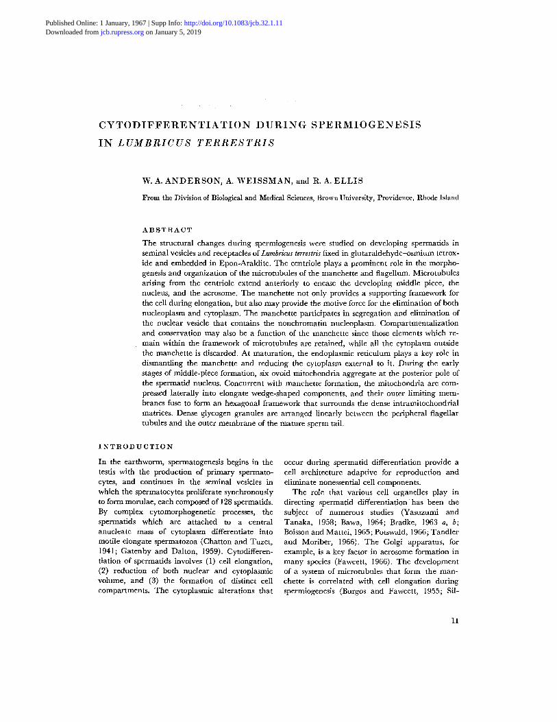
Fmmm 1 A centriole (C) and adjacent microtubules that extend randomly in the cytoplasm are located between the nucleus (N) and the Golgi nmmbrancs (G) of the early spermatid. X 36,000.
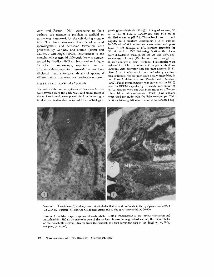
FIGURE ~ A later stage in spermatid maturation reveals a condensation of the nuclear chromatin and mitochondria (Mi) at the posterior pole of the nucleus. As seen in longitudinal section, the microtubules of tile manchette (arrows) diverge from the centriole (C) that forms the base of tile flagellum. G, Golgi complex. )< 36,000.
12 ThE JOURNAL OF CELL BIOLOGY • VOLUME 3~, 1967
per grids, stained first in a saturated solution of uranyl acetate in 40% ethanol for 3-10 min, rimed in dis- tilled water, and poststained for 5-10 min in an un- diluted solution of lead citrate (Reynolds, 1963). The material was examined and photographed with RCA- E M U 3D and 3F electron microscopes.
O B S E R V A T I O N S
Origin and Formation of the Manchette
In the early stages of spermatid development, a pair of centrioles is located at one pole of the nucleus where they are partially surrounded by the Golgi complex. One centriole migrates from this region to the peripheral cytoplasm of the spermatid where it is associated with the forma- tion of the acrosome. Numerous microtubules are distributed randomly in the juxtanuclear Golgi region, and several of these radiate from the second, stationary centriole (Fig. 1). Concurrent with the condensation ofchromat in and elongation
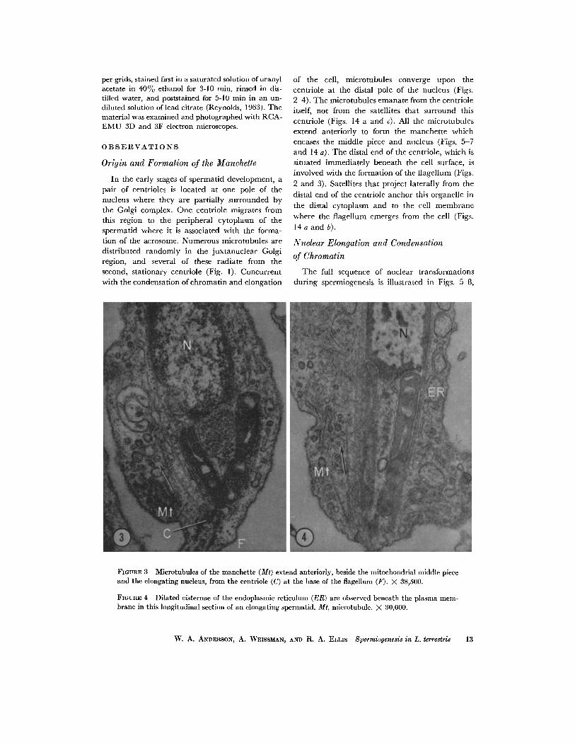
of the cell, microtubules converge upon the centriole at the distal pole of the nucleus (Figs. 2-4). The microtubules emanate from the centriole itself, not from the satellites that surround this centriole (Figs. 14 a and c). All the microtubules extend anteriorly to form the manchette which encases the middle piece and nucleus (Figs. 5 7 and 14 a). The distal end of the centriole, which is situated immediately beneath the cell surface, is involved with the formation of the flagellum (Figs.
2 and 3). Satellites that project laterally from the distal end of the centriole anchor this organelle in the distal cytoplasm and to the cell membrane where the flagellum emerges from the cell (Figs. 14 a and b).
Nuclear Elongation and Condensation
of Chromatin
The full sequence of nuclear transformations during spermiogenesis is illustrated in Figs. 5-8,
FmURE $ Microtubules of the manchette (Mr) extend anteriorly, beside the mitochondrial middle piece and the elongating nucleus, from the eentriole (C) at the base of the flagellum (F). X 38,500.
FIGURE 4 Dilated cisternae of the endoplasmic reticulum (ER) are observed beneath the plasma mem- brane in this longitudinal section of an elongating spermatid. Mt, nfierotubule. X 80,000.
W. A. ANDERSON, A. WEISSMAN, AND R. A. ELLIs Spermiogenesis in L. terrestris 13
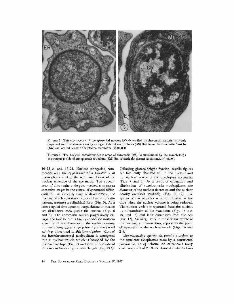
FIOURE 5 This cross-section of the spermatid nucleus (N) shows that its ehromatin material is evenly dispersed and that it is encased by a single circlet of microtubules (Mt) that form the manchette. Vesicles (ER) are located beneath the plasma membrane. X 80,000.
FIGURE 6 Tile nucleus, containing dense areas of chromatin (Ch), is surrounded by the manchette; a continuous profile of endoplasmic reticulum (ER) lies beneath the plasma membrane. X 48,000.
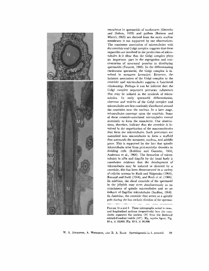
10-13 b, and 15-21. Nuclear elongation com- mences with the appearance of a framework of mierotubules next to the outer membrane of the nuclear envelope of the spermatid. The appear- ance of chromatin undergoes marked changes at successive stages in the course of spermatid differ- entiation. At an early stage of development, the nucleus, which contains a rather diffuse chromatin pattern, assumes a cylindrical form (Fig. 5). At a later stage of development, large chromatin masses are distributed throughout the nucleus (Figs. 6 and 8). The chromatin masses progressively en- large and fuse to form a highly condensed uniform structure. The differences in the nuclear density in these micrographs is due primarily to the varied staining times used in this investigation. Most of the interchromosomal nucleoplasm is segregated into a nuclear vesicle which is bounded by the nuclear envelope (Fig. 7) and runs at one side of the nucleus for nearly its entire length (Fig. 13 b).
Following glutaraldehyde fixation, myelin figures are frequently observed within the nucleus and the nuclear vesicle of the developing spermatid (Figs. 7 and 8). As a result of elongation and elimination of nonchromatin nucleoplasm, the diameter of the nucleus decreases and the nuclear density increases markedly (Figs. 10-12). The system of microtubules is most extensive at the time when the nuclear volume is being reduced. The nuclear vesicle is separated from the nucleus by microtubules of the manchette (Figs. 13 a-b, 15, and 16) and later eliminated from the cell (Fig. 17). An irregularity in the circular profile of the nucleus, in cross-section, represents the point of separation of the nuclear vesicle (Figs. 16 and 21).
The elongating spermatids remain attached to the anucleate cytoplasmic mass by a constricted portion of the cytoplasm. An extraneous fuzzy coat composed of 20-30-A filaments extends from
14 TRE JOURNAL OF CELL BIOLOGY " VOLtTME S~, 1967
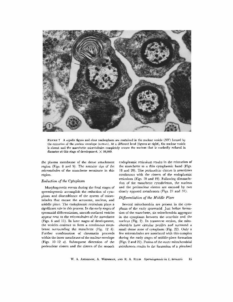
FIGURE 7 A myelin figure and clear nucleoplasm are contained in the nuclear vesicle (NV) formed by the extrusion of the nuclear envelope (arrows). At a different level (figures at right), the nuclear vesicle is absent and the manchette microtubules completely encase the nucleus that is markedly reduced in diameter at this stage of development. X 3~,000.
the plasma membrane of the dense attachment region (Figs. 8 and 9). The anterior tips of the microtubules of the manchette terminate in this region.
Reduction of the Cytoplasm
Morphogenetic events during the final stages of spermiogenesis accomplish the reduction of cyto- plasm and dissemblance of the system of micro- tubules that encase the acrosome, nucleus, and middle piece. The endoplasmic reticulum plays a significant role in this process. In the early stages of spermatid differentiation, smooth-surfaced vesicles appear next to the microtubules of the manchette (Figs. 6 and 11). In later stages of development, the vesicles coalesce to form a continuous mem- brane surrounding the manchette (Fig. 12 b). Further condensation of chromatin proceeds within the inner membrane of the nuclear envelope (Figs. 10-12 a). Subsequent distension of the perinuclear cistern and the cistern of the smooth
endoplasmic reticulum results in the relocation of the manchette to a thin cytoplasmic band (Figs. 18 and 20). The perinuclear cistern is sometimes continuous with the cistern of the endoplasmic reticulum (Figs. 18 and 19). Following disassocia- tion of the manchette cytoskeleton, the nucleus and the perinuclear cistern are encased by two closely apposed membranes (Figs. 21 and 31).
Differentiation of the Middle Piece
Several mitochondria are present in the cyto- plasm of the early spermatid. Just before forma- tion of the manchette, six mitochondria aggregate in the cytoplasm between the centriole and the nucleus (Fig. 2). In transverse section, the mito- chondria have circular profiles and surround a small dense zone of cytoplasm (Fig. 22). Only a few microtubules are associated with this complex during the early stages of middle-piece formation (Figs. 2 and 22). Fusion of the outer mitochondrial membranes results in the formation of a pinwheel
W. A. ANDERSON , A. WEISSMAN, AND R. A. ELLIS Spermiogenesls in L. terrestris 15
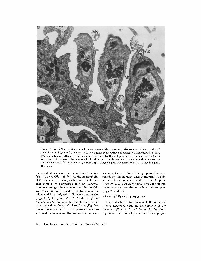
FIGURE 8 An oblique section through several spermatids in a stage of development similar to that of those shown in Figs. 6 and 7 demonstrates that nuclear condensation and elongation occur simultaneously. The spermatids are attached to a central nutrient mass by thin cytoplasmic bridges (short arrows) with an external "fuzzy coat." Numerous mltochondria and an elaborate endoplasmic reticulum are seen in the nutrient mass. AC, acrosome; Ch, chromatin; G, Golgi complex; Mt, microtubules; My, myelin figures. X 11,500.
framework that encases the dense intramitochon- drial matrices (Figs. 23-28). As the microtubules of the manchette develop, each unit of the hexag- onal complex is compressed into an elongate, tr iangular wedge; the cristae of the mitochondria are reduced in number and the central core of the mitochondria is reduced in diameter and density (Figs. 3, 4, 14 a, and 23-25). At the height of manchette development, the middle piece is en- cased by a thick sheath of microtubules (Fig. 24). Smooth membranes of the endoplasmic reticulum surround the manchette. Distension of the cisternae
accompanies reduction of the cytoplasm that sur- rounds the middle piece. Late in maturation, only a few microtubules surround the middle piece (Figs. 25-27 and 29 a), and finally only the plasma membrane encases the mitochondrial complex (Figs. 28 and 31).
The Basal Body and Flagellum
The centriole involved in manchet te formation is also concerned with the development of the flagellum (Figs. 2, 3, and 14 a). At the distal region of the centriole, satellite bodies project
16 TuE JOURNAL OF CELL BIOLOGY • VOLUME $~, 1967
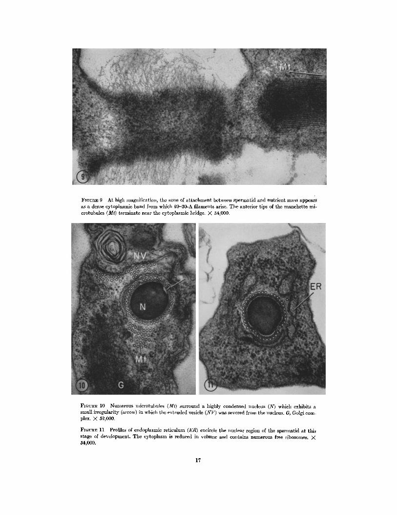
FIGVRE 9 At high magnification, the zone of attachment between spermatid and nutrient mass appears as a dense cytoplasmic band from which ~0-30-A filaments arise. The anterior tips of the manchette mi- crotuhules (Mr) terminate near the cytoplasmic bridge. X 54,000.
FiotrRs 10 Numerous microtubules (Mt) surround a highly condensed nucleus (N) which exhibits a small irregularity (arrow) in which the extruded vesicle (NV) was severed from the nucleus. G, Golgi com- plex. X 5~,000.
FIGtmE 11 Profiles of endoplasmic reticulum (ER) encircle the nuclear region of the spermatid at this stage of development. The cytoplasm is reduced in volume and contains numerous free ribosomes. × 54,000.
17
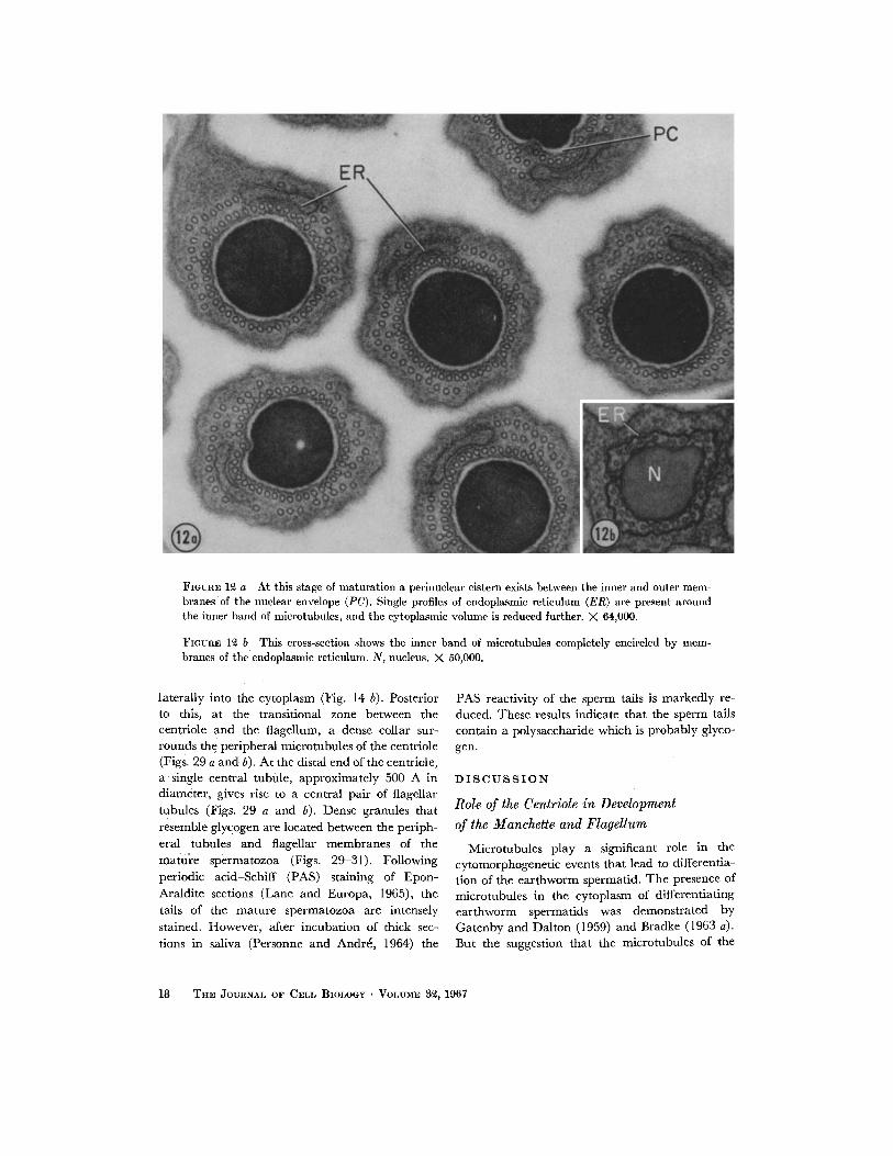
FIGUnE 12 a At this stage of maturation a perinuclear cistern exists between the inner and outer mem- branes of the nuclear envelope (PC). Single profiles of endoplasmic reticulmn (ER) are present around the inner band of microtubules, and the cytoplasmic volume is reduced further. X 64,000.
FIOUnE 1~ b This cross-section shows the inner band of microtubules completely encircled by mem- branes of the endoplasmic reticulum. N, nucleus. X 50,000.
laterally into the cytoplasm (Fig. 14 b). Posterior to this, at the transitional zone between the centriole and the flagellum, a dense collar sur- rounds the peripheral microtubules of the centriole (Figs. 29 a and b). At the distal end of the centriole, a single central tubule, approximately 500 A in diameter, gives rise to a central pair of flagellar tubules (Figs. 29 a and b). Dense granules that resemble glycogen are located between the periph- eral tubules and flagellar membranes of the mature spermatozoa (Figs. 29-31). Following periodic acid-Schiff (PAS) staining of Epon- Araldite sections (Lane and Europa, 1965), the tails of the mature spermatozoa are intensely stained. However, after incubation of thick sec- tions in saliva (Personne and Andr6, 1964) the
PAS reactivity of the sperm tails is markedly re- duced. These results indicate that the sperm tails contain a polysaccharide which is probably glyco- gen.
D I S C U S S I O N "
Role of the Centriole in Development of the ~lanchette and Flagellum
Microtubules play a significant role in the cytomorphogenetic events that lead to differentia- tion of the earthworm spermatid. The presence of microtubules in the cytoplasm of differentiating earthworm spermatids was demonstrated by Gatenby and Dalton (1959) and Bradke (1963 a). But the suggestion that the microtubules of the
18 THE JO~q~NAL OF CELL BIOLOGY • VOLUME 3~, 1967
manchette in spermatids of earthworm (Gatenby and Dalton, 1959) and python (Boisson and Mattei, 1965) are derived from the outer nuclear membrane is not supported by our observations. The consistent association of microtubules with the centriole and Golgi complex suggests that these organelles are involved in the production of micro- tubules. It is clear that the Golgi complex plays an important part in the segregation and con- centration of structural proteins in developing spermatids (Fawcett, 1966). In the differentiating earthworm spermatid, the Golgi complex is in- volved in acrosome formation. However, the intimate association of the Golgi complex to the centriole and microtubules suggests a functional relationship. Perhaps it can be inferred that the Golgi complex sequesters precursor substances that may be utilized in the synthesis of micro- tubules. In early spermatid differentiation, cisternae and vesicles of the Golgi complex and microtubules are first randomly distributed around the centrioles near the nucleus. In a later stage, microtubules converge upon the centriole. Some of these centriole-associated microtubules extend anteriorly to form the manchette. Our observa- tions, therefore, indicate that the centriole is in- volved in the organization of the macromolecules that form the microtubules. Such precursors are assembled into microtubules to form a scaffold that surrounds the acrosome, nucleus, and middle piece. This is supported by the fact that spindle microtubules arise from pericentriolar densities in dividing cells (Robbins and Gonatas, 1964; Anderson et al., 1966). The formation of micro- tubules in cilia and flagella by the basal body is correlative evidence that the development of microtubules may be induced or directed by a centriole; this has been demonstrated in a variety of cellular systems by Roth and Shigenaka (1964), Renaud and Swift (1964), and Roth et al. (1966). In addition, the distal centriole of the spermatid in the jellyfish may serve simultaneously as an attachment of spindle microtubules and as an inducer of flagellar microtubules (Szollosi, 1964). In Lumbricus, the centriole that serves as a spindle pole during the last meiotic division of the sperma-
FIGVRE 13 a and b These micrographs reveal in cross- and longitudinal sections (respectively) how the man- chette separates the nucleus (N) from the flattened extruded nuclear vesicle (NV). My, myelin figure. Fig. 13 a, X 55,000. Fig. 13 b, )< 36,000.
W. A. ANDERSON, A. WEISSMAN, AND R. A. ELLIS Spermiogenesis in L. terrestrla 19
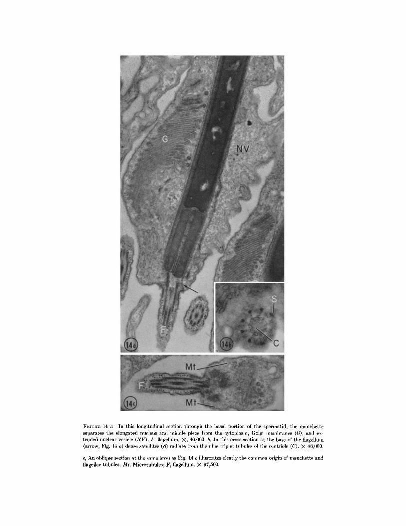
FmVRE 14 a In this longitudinal section through the basal portion of tim spermatid, the manchette separates the elongated nucleus and middle piece from the cytoplasm, Golgi membranes (G), and ex- truded nuclear vesicle (NV). F, flagellum. X. 40,000. b, In this cross-section at tile base of the flagellum (arrow, Fig. 14 a) dense satellites (S) radiate from the nine triplet tubules of the centriole (C). X 46,000.
c, An oblique section at the same level as Fig. 14 b illustrates clearly the common origin of manehette and flagellar tubules. Mr, Microtubules; F, flagellum. X 57j500.
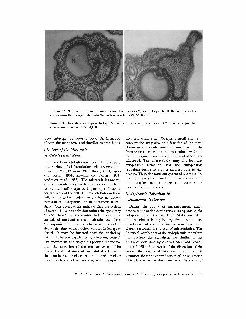
FIGURE 15 The sleeve of mierotubules around the nucleus (N) seems to pinch off the nonehromatin nueleoplasm that is segregated into the nuclear vesicle (NV). X 96,000.
FIGVnE 16 In a stage subsequent to Fig. 15, the nearly extruded nuclear visiele (NV) contains granular nonchromatin material. X 86,000.
tocyte subsequently seems to induce the formation of both the manchette and flagellar microtubules.
The Role of the Manchette
in Cytodifferentiation
Oriented microtubules have been demonstrated in a variety of differentiating cells (Burgos and Fawcett, 1955; Nagano, 1962; Bawa, 1964; Byers and Porter, 1964; Silveira and Porter, 1964; Anderson et al., 1966). The microtubules are re- garded as resilient cytoskeletal elements that help to maintain cell shape by imparting stiffness to certain areas of the cell. The microtubules in these cells may also be involved in the internal move- ments of the cytoplasm and in alterations in cell shape. Our observations indicate that the system of microtubules not only determines the symmetry of the elongating spermatids but represents a specialized mechanism that maintains cell form and organization. The manchette is most exten- sive at the time when nuclear volume is being re- duced. It may be inferred that the encircling microtubules are capable of synchronous centrif- ugal movement and may thus provide the motive force for extrusion of the nuclear vesicle. The directed redistribution of microtubules between the condensed nuclear material and nuclear vesicle leads to nuclear vesicle separation, segrega-
tion, and elimination. Compartmentalization and conservation may also be a function of the man- chette since those elements that remain within the framework of microtubules are retained while all the cell constituents outside the scaffolding are discarded. The microtubules may also facilitate cytoplasmic reduction, but the endoplasmic reticulum seems to play a primary role in this process. Thus, the transient system of microtubules that constitutes the manchette plays a key role in the complex cytomorphogenetic processes of spermatic differentiation.
Endoplasmic Reticulum in
Cytoplasmic Reduction
During the course of spermiogenesis, mem- branes of the endoplasmic reticulum appear in the cytoplasm outside the manchette. At the time when the manchette is highly organized, continuous membranes of the endoplasmic reticulum com- pletely surround the system of microtubules. The flattened membranes of the endoplasmic reticulum that encircle the manchette are similar to the "mantle" described by Andr~ (1963) and Brokel- mann (1961). As a result of the distension of the cistern, the peripheral thin layer of cytoplasm is separated from the central region of the spermatid which is encased by the manchette. Distension of
W. A. ANDERSON, A. WEISSMAN, AND R. A. ELLIS Spermiogenesis in L. terrestris 21
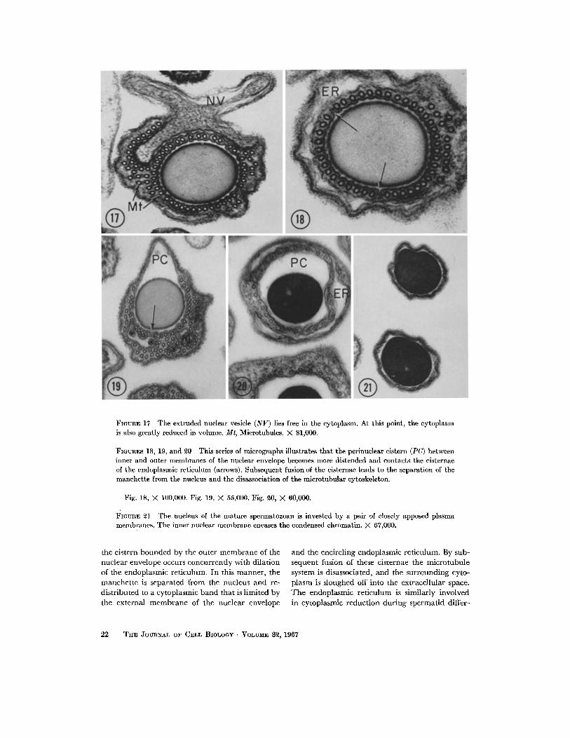
FIGURE 17 The extruded nuclear vesicle (NV) lies free in the cytoplasm. At this point, the cytoplasm is also greatly reduced in volume. Mr, Microtubules. X 81,000.
FIGURES 18, 19, and 20 This series of micrographs illustrates that the perinuclear cistern (PC) between inner and outer membranes of the nuclear envelope becomes more distended and contacts the cisternae of the endoplasmic rcticulum (arrows). Subsequent fusion of the cisternae leads to the separation of the manchette from the nucleus and the disassociation of the mlcrotubular cytoskeleton.
Fig. 18, X 10O,000. Fig. 19, M 55,000. Fig. ~0, M 60,000.
FlaunE 21 The nucleus of the mature spermatozoan is invested by a pair of closely apposed plasma membranes. The inner nuclear membrane encases the condensed chromatin. X 67,000.
the cistern bounded by the outer membrane of the nuclear envelope occurs concurrently with dilation of the endoplasmic reticulum. In this manner, the manchette is separated from the nucleus and re- distributed to a cytoplasmic band that is limited by the external membrane of the nuclear envelope
and the encircling endoplasmie reticulum. By sub- sequent fusion of these cisternae the microtubule system is disassociated, and the surrounding cyto- plasm is sloughed off into the extracellular space. The endoplasmic reticulum is similarly involved in cytoplasmic reduction during spermatid differ-
22 THE JOURNAL OF CELL BIOLOGY • VOLUME 32, 1967
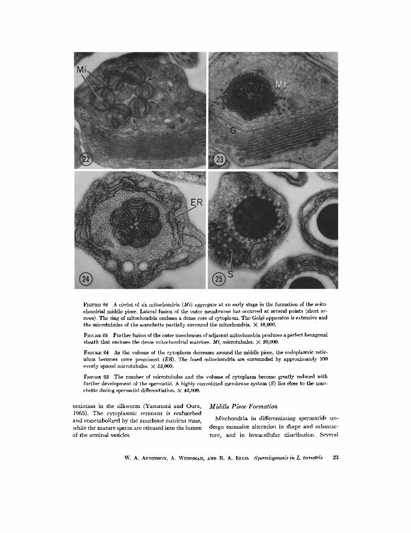
FIGUI~E ~ A circlet of six mitoehondria (Mi) aggregate at an early stage in the formation of the mito- ehondrial middle piece. Lateral fusion of the outer membranes has occurred at several points (short ar- rows). The ring of mitochondria encloses a dense core of cytoplasm. The Golgi apparatus is extensive and the microtubules of the manchette partially surround the mitoehondria. X 46,000.
FIGURE 23 Further fusion of the outer membranes of adjacent mitochondria produces a perfect hexagonal sheath that encloses the dense mitoehondrial matrices. Mt, microtubules. X 50,000.
FIGURE ~ As the volume of the cytoplasm decreases around the middle piece, the endoplasmic retie- ulum becomes more prominent (ER). The fused mitoehondria are surrounded by approximately 100 evenly spaced microtubules. >( 55,000.
FlOtmE 25 The number of microtubules and the volume of cytoplasm become greatly reduced with further development of the spermatid. A highly convoluted membrane system (S) lies close to the man- chette during spermatid differentiation. X 42,000.
ent ia t ion in the silkworm (Yasuzumi and Oura , 1965). The cytoplasmic r e m n a n t is reabsorbed and remetabol ized by the anucleate nu t r i en t mass, while the ma tu re sperm are released into the lumen of the seminal vesicles.
Middle Piece Formation
Mitchondr i a in differentiat ing spermatids un-
dergo extensive al terat ion in shape and substruc-
ture, and in intracel lular d i s t r ibu t ion . Several
W. A. ANDERSON, A. WFISSMAN, AI~D R. A. :ELLIS Spermiogenesis in L. terrestris 23

FmURES ~6, 27, and 28 This series of cross-sections of the middle piece illustrates the progressive loss of microtubules and cytoplasm, culminating in a mature middle piece surrounded solely by the plasma membrane (PM).
Fig. ~6, X 50,000. Fig. ~7, X 55,000. Fig. 28, X 73,000.
mitochondria are present in the cytoplasm of the
early earthworm spermatid, but only six mito-
chondria are mobilized to a region of cytoplasm
between the nucleus and centriole. The mitochon-
dria contain cristae that project into the matrix
(Fig. 22). The mitochondrial substructure is un-
like that of the atypical mitoehondria in spermatids
of other species where the cristae are folded or
flattened against the limiting membranes (Faw-
cett, 1957). During the course of spermiogenesis,
the spherical mitochondria are transformed into
a middle piece consisting of six pyramidal sub-
units bound within a hexagonal pinwheel frame-
work. The system of microtubules that encase the
middle piece probably provides the motive force
for lateral fusion and reorganization of the mito-
chondria. The compactness and position of the
middle piece contribute to the integrity of this
highly attenuated cell. In this capacity the middle
B I B L I O G R A P H Y
1. ANDERSON, W. A., A. WEISSMAN, and R. A. ELLIS. 1966. A comparative study of micro- tubules in vertebrate and invertebrate cells. Z. Zellforsch. Mikroskop. Anat. 71: 1.
2. ANDRI~, J. 1962. Contribution ~ la connaissancc
piece can serve effectively as an energy source for locomotion and fertilization.
Glycogen in the Flagellum
Dense granules between the peripheral tubules and the membrane of the flagellum are similar to the glycogen granules observed in spermatozoa by Personne and Andr~ (1964). The positive PAS reaction of the sperm tails demonstrates the pres- ence of a polysaccharide in this region of the cell, and the decrease in staining intensity of the tails subsequent to incubation in saliva indicates that the polysaccharide is probably glycogen. Glycogen granules in the tail are presumably available as stores to be utilized in A T P production by the mitochondria.
This investigation was supported by Research Grants CA-04046; GM-08380, and Training Grant GM- 00852 from the United States Public Health Service.
Received for publication 12 May 1966.
du chondriome. Etude de scs modifications ultrastructurales pendant la spermatog6n~se. J. Ultrastruct. Res. 6 (suppl. 3):1.
3. ANDRe, J. 1963. Some aspects of specialization in sperm. In General Physiology of Cell Special-
24 TIIE JOURNAL OF CELL BIOLOGY • VOLUME 3~, 1967
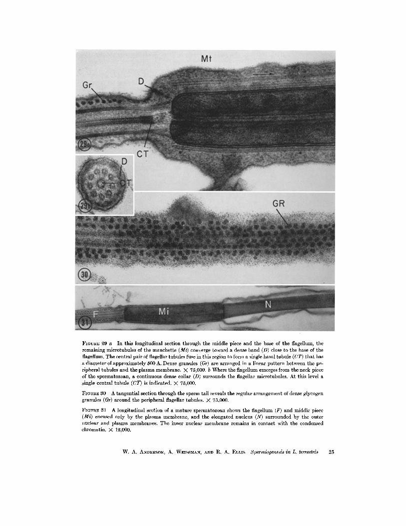
FIGURE ~9 a In this longitudinal section through the middle piece and the base of the flagellum, the remaining microtubules of the manchette (Mr) converge toward a dense band (D) close to the base of the flagellum. The central pair of flagellar tubules fuse in this region to form a single basal tubule (CT) that has a diameter of approximately 500 A. Dense granules (Gr) are arranged in a linear pattern between the pe- ripheral tubules and the plasma membrane. X 75,000. b Where the flagellum emerges from the neck piece of the spermatozoan, a continuous dense collar (D) surrounds the flagellar microtubules. At this level a single central tubule (CT) is indicated. >( 75,000.
FmvR~ 30 A tangential section through the sperm tail reveals the regular arrangement of dense glycogen granules (Gr) around the peripheral flagellar tubules. X 75,000.
FIOURE 31 A longitudinal section of a mature spermatozoan shows tile flagellum (F) and middle piece (Mi) encased only by the plasma membrane, and the elongated nucleus (N) surrounded by the outer nuclear and plasma membranes. The inner nuclear membrane remains in contact with the condensed chromatin. >( 1~,000.
W. A. ANDERSON, A. WEISSMAN, AND R. A. ELLIS Spermiogenesis in L. terrestri8 25
ization. D. Mazia, and A. Tyler, editors. Mc- Graw-Hill Co., N. Y. 91.
4. BAW*, S. R. 1964. An electron microscope study of spermiogenesis in a fire-brat insect. Thermobia domestica Pack. or. Cell Biol. 23: 431.
5. BomsoN, C., and X. MATTEI. 1965. Sur la sper- miog~nese de Python sebae (Gmelin) ~tudi~ au microscope ~lectronique, Compt. Rend. Soc. Biol. 159: 1192.
6. BRADKE, D. L. 1963 a. The origin and develop- ment of the manchette during spermatogenesis. Anat. Record. 145: 210.
7. BRADKE, D. L. 1963 b. Special features of sper- matogenesis in Lumb~icus terrestris. Anat. Record. 145: 360.
8. BROKELMANN, J. 1961. Surface modification of Sertoli cells at various stages of spermatogenesis in the rat. An electron microscope study. Anat Record. 139: 211.
9. BUROOS, M. H., and D. W. FAWCETT. 1955. Studies on the fine structure of the mammalian testis. I. Differentiation of the spermatids in the cat (Felis domestica). J. Biophys. Biochem.
Cytol. 1: 287. 10. BYERS, B., and K. R. PORTER. 1964. Oriented
microtubules in elongating cells of the develop- ing lens rudiment after induction. Proc. Natl. Acad. &i. U. S. 52: 1091.
11. CAMERON, M. L., and W. H. FOGEL. 1963. The development and structure of the acrosnme in the sperm of Lumbricus terrestris L. Can. 3". Zool. 41: 753.
12. CHATTON, E., and O. TUZET. 1941. Sur quelque faits nouveaux de la spermatogfin~se du Lum- bricus terrestris. Compt. Rend. Acad. Sci. 213: 373.
13. FAWCETT, D. W. 1957. Changes in the fine struc- ture of the cytoplasmic organelles during differ- entiation. In The 16th Symposium of the So- ciety for Development and Growth. D. Rund- nick, editor. Ronald Press Co., N. Y. 161.
14. FAWCETT, D. W. 1966. The Cell. W. B. Saunders Co., Philadelphia. 130.
15. GAa~NBY, J. B., and A. J. DALTON. 1959. Sper- miogenesis in Lumbricus herculeus. J . Biophys. Bin- chem. Cytol. 6: 45.
16. LANE, B. P., and D. L. EUROPA. 1965. Differential staining of uhrathin sections of epon-embedded tissues for light microscopy, o r. Histochem. Cyto- chem. 13; 579.
17. NAGANO, T. 1962. Observations on the fine strut-
ture of the developing spermatid in the do- mestic chicken, o r . Cell Biol. 14: 193.
18. PERSONNE, P., and J. ANDRe. 1964. Existence de glycog~ne mitochondrial dans le spermatozoide de la testacelle. J. Microscopic. 3: 643.
19. POTSWALD, H. E. 1966. Blebbing of the nuclear envelope during spermiogenesis in Spirorbis (Laeospira morchi) (Polychaeta). Anat. Record; 154: 403.
20. RENUAD, F., and H. SWIFT. 1964. The develop- ment of basal bodies and flagella in Allomyces arbusculus. J. Cell Biol. 23: 339.
21. REYNOLDS, E. S. 1963. The use of lead citrate as an electron-opaque stain in electron micros- copy. J. Cell Biol. 17: 208.
22. ROBBINS, E., and N. K. GONATAS. 1964. The ultrastructure of a mammalian cell during the mitotic cycle. J. Cell Biol. 21: 429.
23. ROTE, L. E., and Y. SHIG~NAKA. 1964. The struc- ture and formation of cilia and filaments in rumen protozoa. J. Cell Biol. 20: 249.
24. ROTH, L. E., H. J. WmsoN, andJ . CHAK•ABORTY. 1966. Anaphase structure in mitotic cells typified by spindle elongation. J. Ultrastruct. Res. 14: 460.
25. SmVEIR% M., and K. R. PORa~R. 1964. The spermatozoids of flatworms and their micro- tubular systems. Protoplasma. 59: 240.
26. SZOLLOSI, D. 1964. The structure and function of centrioles and their satellites in the jellyfish Phialidium gregarium. J. Cell Biol. 21: 465.
27. TANDLER, B., and L. G. MOR~BER. 1966. Micro- tubular structures associated with the acrosome during spermiogenesis in the water-strider, Gerris remigis (Say). J. Ultrastruct. Res. I4: 391.
28. VOELZ, H., and M. DWORKIN. 1962. Fine struc- ture of Myxococcus xanthus during morpho- genesis. 3". Bacteriol. 84: 943.
29. YASUZUMI, G., and H. TANAKA. 1958. Spermato- genesis in animals as revealed by electron microscopy. VI. Researches on the spermato- zoon-dimorphism in a pond snail, Cipango- paludina malleata. J . Biophys. Biochem. Cytol. 4 621.
30. YASUZUMI, G., and C. OURA. 1965. Spermato- genesis in animals as revealed by electron microscopy. XV. The fine structure of the mid- dle piece in the developing spermatid of the silkworm, Bombyx mori Linn& A. Zellforsch. Mikroskop. Anat. 67: 502.
26 THE JOURNAL OF CELL BIOLOGY " VOLUME 3~, 1967