on the nephridiostome of lumbricus
TRANSCRIPT
On the Nephridiostome of Lumbricus.By
Edwin S. Goodrich, F.R.S.,
Linacre Professor of Zoology and Comparative Anatomy in theUniversity of Oxford.
With Plates 11 and 12 and 2 Text-figures.
IT might be thought that little remains to be said about sucha familiar object as the nephridial funnel, or nephridiostome,1
of the earthworm, L u m b r i c u s . Its structure in the adulthas beenstudied by several eminent zoologist, samong whom maybe mentioned Gegenbaur and Benham, and its developmentdescribed by Vejdovsky, Wilson, and others. Yet opinions stilldiffer as to its. derivation and general morphology; and the latestdetailed study of the funnel by Eosen (1911) contains not onlysome misleading statements as to facts, but some theoreticalconclusions as to the homology of its parts which seem to mequite unjustified. For, basing his view chiefly on Ed. Meyer's(1886) observation that in the Polychaete P o l y m n i a thefunnel and canal of the excretory organ are of separate origin(the former developing from the coelomic epithelium), and oncertain doubtful observations made by Berg (1899) on thedevelopment of the nephridium of R h y n c h e l m i s , Rosenconcludes that the marginal cells of the lumbricid funnel arederived from the coelomic epithelium.
Already Benham (1904) had suggested that the nephridiumof the Oligochaete H a p l o t a x i s may be a nephromixium,owing apparently to a somewhat vague resemblance betweenthe nephridium and the sperm-duct. Similar suggestions have
1 Since the term nephrostome has been loosely used for many funnel-like structures, some of which are certainly not homologous with thefunnel of the nephridium of L u m b r i c u s , I have recently (1930) usedthe more definite term n e p h r i d i o s t o m e for the funnel belonging toand derived from the true nephridium.
166 EDWIN S. GOODRICH
been from time to time expressed by other authors, for instanceBoveri-Boner (1920).
In a general paper (1895) I endeavoured to show that twoquite different organs, the coelomoduct (derived from the wallof the coelom) and the true nephridium, may be distinguishedin all groups of Coelomata; and, further, that the coelomostomeor funnel of the coelomoduct, should not be confused with theopening into the coelom of the true nephridium (nephridiostome)found in Oligochaeta (and some Polychaeta). The termnephromixium was introduced by me (1900) to denote the com-pound organ found in certain families only of Polychaeta,formed by the grafting of a coelomostome on to a nephridium.I can see no justification for the application of this term to anyorgan in the Oligochaeta. Even if it could be proved that certaincells of the lumbricid funnel were derived from coelomicepithelium, this would be no good reason for using here theterm nephromixium, since in the Oligochaeta coelomostomesand nephridiostomes are quite independent and may coexistin the same segments.
A renewed study of the structure and development of thefunnel seemed, therefore, desirable, and indeed necessary, if acorrect interpretation of its general morphology is to be reached.
The following observations were made on living and preservednephridia of L u m b r i c u s t e r r e s t r i s L. The best fixativeis Bouin's fluid, but other familiar fixatives give good results.Sections were mostly stained in borax carmine followed by picro-nigrosin for general purposes; and in Mann's methyl-blue eosin,and iron-haematoxylin for special points and comparison. Ihave to thank Mr. G. R. de Beer for making the reconstruc-tion in wax used for text-fig. 1, p. 168.
NEPHRIDIOSTOME OF THE ADULT.
The early history of our knowledge of the structure of thenephridiostome of L u m b r i c u s and allied genera has beensufficiently dealt with by Benham (1891) and Rosen (1911), andneed not delay us here. Of all the descriptions hitherto givenof this organ that of Benham is the most correct and may beused as our starting-point. He describes the expanded upper or
NEPHRIDIOSTOME OF LUMBRICUS 167
dorsal lip provided with an even covering of cilia and an outercoat of coelomic epithelium. The narrow preseptal nephridialcanal formed of pierced 'drain-pipe' cells, with right and leftbands of cilia, on reaching the middle of the funnel opens intothe coelom, its walls bending outwards and then backwards oneach side. The drain-pipe cells here are continued into grooved' gutter-cells' along which extend the ciliated bands. The lattercells, he says, join the inturned ends of the right and left hornsof the crescentic row of marginal cells surrounding the dorsallip. Thus, if I understand him rightly, these 'centripetal'marginal cells are said to meet the ' centrifugal' gutter-cells onthe ventral side of the funnel, a statement which does not quitecorrectly represent the true state of affairs (see below and Text-fig. 2). Benham discovered that the marginal cells surrounda large central cell occupying the middle region of the dorsal lip.But, though he clearly distinguishes between the mass ofcoelomic corpuscles on the funnel from the funnel itself, he givesno very clear description or figure of the lower lip. Moreover, hisfigure of the whole funnel (PI. 23, fig. 4) does not correctlyindicate the disposition of the gutter-cells. Nor does the figuresince published by K. C. Schneider (1902) represent any betterthe true relations of gutter-cells, marginal cells, and lower lip.
In 1911 F. Eosen brought out a detailed work on the nephri-diostomeof L u m b r i c u s , based on the study of L u m b r i e u sa g r i c o 1 a Hoffm. On the whole he confirms Benham's descrip-tion, adding certain details, some but not all of which seem tobe correct. His diagram of the whole funnel (PI. 12, fig. 9)closely resembles my own reconstruction.
Eosen describes the marginal, central, and canal cells asprovided with a ' cuticula'; but I can find no such cuticle onany of these cells apart from the cell-wall. On the other hand,the closely set cilia are provided with distinct basal granuleswhich, when stained with iron-haematoxylin for instance, forma very conspicuous marginal layer (Text-fig. 2 and figs. 7, 8, and9, PL 11. The central cell is crescentic in shape, and Rosenpoints out correctly that where the dorsal wall of the canalmeets the concavity of the central cell at the base of the dorsallip there is a thin area with neither nucleus nor cilia. This
EDWIN S. GOODRICH
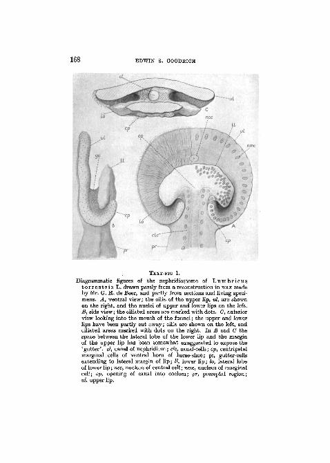
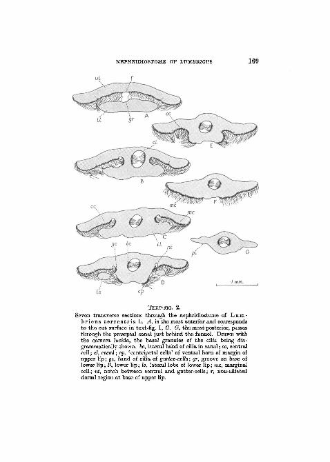
TEXT-FIG 1.
Diagrammatic figures of the nephridiostome of Lumbr icust e r r e s t r i s L. drawn partly from a reconstruction in wax madeby Mr. G. R. de Beer, and partly from sections and living speci-mens. A, ventral view; the cilia of the upper lip, ul, are shownon the right, and the nuclei of upper and lower lips on the left.B, side view; the ciliated areas are marked with dots. C, anteriorview looking into the mouth of the funnel; the upper and lowerlips have been partly cut away; cilia are shown on the left, andciliated areas marked with dots on the right. In B and C thespace between the lateral lobe of the lower lip and the marginof the upper lip has been somewhat exaggerated to expose the'gutter', el, canal of nephridium; clc, canal-cells; cp, centripetalmarginal. cells of ventral horn of horse-shoe; gc, gutter-cellsextending to lateral margin of lip; II, lower lip; lo, lateral lobeof lower lip; nee, nucleus of central cell; nine, nucleus of marginalcell; op, opening of canal into coelom; pr, preseptal region;id, upper lip.
NEPHEIDIOSTOMB OF LUMBRICUS 169
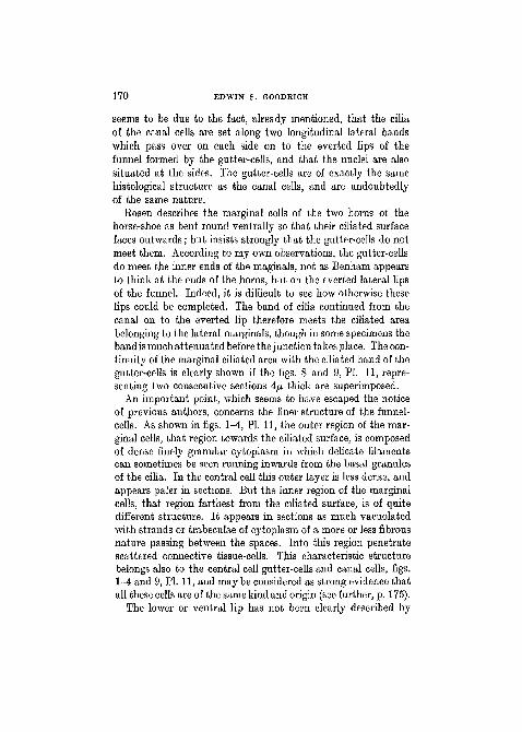
TEXT-FIG. 2.
Seven transverse sections through the nephridiostome of Lum-br icus t e r r e s t r i s L. A, is the most anterior and correspondsto the cut surface in text-fig. 1, C. 0, the most posterior, passesthrough the preseptal canal just behind the funnel. Drawn withthe camera lucida, the basal granules of the cilia being dia-grammatically shown. 6c, lateral band of cilia in canal; cc, centralcell; cl, canal; cp, 'centripetal cells' of ventral horn of margin ofupper lip; gc, band of cilia of gutter-cells; gr, groove on base oflower lip; II, lower lip; lo, lateral lobe of lower lip; me, marginalcell; nt, notch between central and gutter-cells; r, non-ciliateddorsal region at base of upper lip.
170 EDWIN S. GOODRICH
seems to be due to the fact, already mentioned, that the ciliaof the canal cells are set along two longitudinal lateral bandswhich pass over on each side on to the everted lips of thefunnel formed by the gutter-cells, and that the nuclei are alsosituated at the sides. The gutter-cells are of exactly the samehistological structure as the canal cells, and are undoubtedlyof the same nature.
Eosen describes the marginal cells of the two horns of thehorse-shoe as bent round ventrally so that their ciliated surfacefaces outwards; but insists strongly that the gutter-cells do notmeet them. According to my own observations, the gutter-cellsdo meet the inner ends of the maginals, not as Benham appearsto think at the ends of the horns, but on the everted lateral lipsof the funnel. Indeed, it is difficult to see how otherwise theselips could be completed. The band of cilia continued from thecanal on to the everted lip therefore meets the ciliated areabelonging to the lateral marginals, though in some specimens theband is much attenuated before the junction takes place. The con-tinuity of the marginal ciliated area with the ciliated band of thegutter-cells is clearly shown if the figs. 8 and 9, PI. 11, repre-senting two consecutive sections 4JA thick are superimposed.
An important point, which seems to have escaped the noticeof previous authors, concerns the finer structure of the funnel-cells. As shown in figs. 1-4, PI. 11, the outer region of the mar-ginal cells, that region towards the ciliated surface, is composedof dense finely granular cytoplasm in which delicate filamentscan sometimes be seen running inwards from the basal granulesof the cilia. In the central cell this outer layer is less dense, andappears paler in sections. But the inner region of the marginalcells, that region farthest from the ciliated surface, is of quitedifferent structure. It appears in sections as much vacuolatedwith strands or trabeculae of cytoplasm of a more or less fibrousnature passing between the spaces. Into this region penetratescattered connective tissue-cells. This characteristic structurebelongs also to the central cell gutter-cells and canal cells, figs.1-4 and 9, PI. 11, and may be considered as strong evidence thatall these cells are of the same kind and origin (see further, p. 175).
The lower or ventral lip has not been clearly described by
NEPHRIDIOSTOMB OF LUMBRICUS 171
previous authors. Rosen's account of it is the least satisfactorypart of his work. The general shape of the lower lip is seen inText-fig. 1. It is smaller than the upper lip, has a narrow baseand two backwardly directed lateral lobes which project intothe spaces bounded by the upper lip, everted lateral lips andventral horns of the marginal horse-shoe. It is not ciliated. Itmust be remembered that the whole of the exposed surfaceof the funnel and preseptal canal is covered with coelomicepithelium, as Benham showed. Along the edge of the upperlip and everted lateral lips this epithelium reaches to near themargin. The marginal cells, however, form the actual rim of thefunnel along upper and lateral lips, figs. 1 and 2, PL 11. Muchloose connective tissue lies between coelomic epithelium andcanal wall in the preseptal region, and some connective tissue-cells extend into the lips. Now, if I understand him rightly,Eosen believes that the bulk of the lower lip is not truly a partof the funnel, but is formed entirely of coelomic epithelium andconnective tissue. Influenced, apparently, by K. C. Schneider'sdescription of the nephridiostome of E i s e n i a rosea (1902),he repeatedly maintains that the lower lip is without nuclei,and figures it as such (p. 169, Text-fig. 7). According, then, toEosen's text and figures the true lower lip would be merely theshort projecting edge of the terminal canal cell. For this inter-pretation there appears to be no justification, and we shall seewhen studying the development that, just as in the case of theupper lip, the coelomic epithelium covers the outer surface onlyof the lower lip, while its inner surface and rim are formedof cells of nephridial origin. Since these cells are not ciliatedthey differ considerably in shape and structure from those ofthe upper lip. They form an irregular more or less columnarepithelium with oval nuclei, figs. 1-3, 5-7, PI. 11. The coelomicepithelium cells, on the other hand, consisting of flattened cellswith smaller more darkly staining nuclei, can generally easilybe distinguished from the nephridial epithelium, though thelimit between them is often not clearly marked. The lateralmargins of the lower lip are often drawn out into sharp irregularedges against which the cilia of the upper lip are closely applied,figs. 2 and 7, PI. 11.
172 EDWIN S. GOODEICH
Attached to the coelomic epithelium of the lower lip is thatpeculiar mass of coelomic corpuscles or leucocytes which has sooften been described. Since it forms no essential part of thefunnel, and has been dealt with thoroughly by Benham, Eosen,and others, it need not now detain us. At its point of attach-ment the coelomic epithelium is often considerably modifiedand disturbed, if not actually injured; but the limit betweenthe two can generally be made out. In well-preserved sectionsthe corpuscles are seen to differ in shape and composition fromthe coelomic epithelium cells, and to stain differently, especiallyin iron-haematoxylin.
DEVELOPMENT OF THE NEPHEIDIOSTOME.
It is generally agreed that, in Oligochaeta, part if not the wholeof the nephridium is derived from a large cell, the nephridio-blast. In this paper we are not concerned with the first originof this nephridioblast—whether it should be called ectodermal,mesectodermal, or mesodermal. Much controversy has takenplace on this point in the past, and every view has been advocated.For our present purpose we are concerned only with the originof the nephridiostome, and the point we wish to decide iswhether it is derived from the nephridial rudiment, from thecoelomic epithelium, or from both. Wilson (1899), one of thefirst to examine in detail the development of the nephridium inL u m b r i c u s , believed that the large anterior 'funnel cell',which gives rise to the funnel, is of separate origin from thenephridial canal. But, Vejdovsky, our greatest authority onthis question, who studied the development of nephridia from1884 to 1892, finally came to the conclusion that the nephridio-blast gives rise by division to both the canal cells and thefunnel-cell. The latter pushing its way through the septum onto its anterior face, according to him, forms the funnel (1892).Similarly Berg (1888, 1890) describes both canal and funnel asderived from a single rudiment. Lehmann (1887) had previouslycome to the same conclusion. Later Berg (1899) studied thedevelopment of the nephridium in R h y n c h e l m i s , aLumbriculid, where the funnel is smaller and simpler than in
NEPHRIDIOSTOME OF LUMBBICUS 178
L u m b r i c u s , having an upper lip of eight ciliated marginalcells and a lower lip of four non-ciliated cells. He concludedthat, whereas the canal cells and lower lip cells are all derivedfrom the nephridioblast, the upper lip marginal cells come fromthe coelomic epithelium. According to Staff (1910) on the con-trary, in C r i o d r i l u s , a Glossoscolecid, the funnel cell givesrise to the upper lip and canal cells to the lower lip.
Prom this brief review of the literature it may be said thatit is now generally agreed that both the canal and the funnel(or some part of it) are developed from a single large cell, thenephridioblast, a conclusion which is borne out by what weknow of the development of the nephridia in the lower Oli-gochaeta from the observations of Vejdovsky and the more recentwork of A. Meyer (1929). The only point which remains undecidedis whether the whole nephridiostome (excluding, of course, itsexternal covering of coelomic epithelium and the intrusive con-nective tissue) arises from the nephridioblast, or whether coelomicepithelium contributes any part of the upper lip proper.
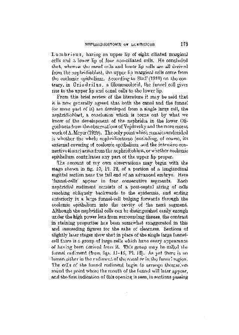
The account of my own observations may begin with thestage shown in fig. 10, PI. 12, of a portion of a longitudinalsagittal section near the tail end of an advanced embryo. Here'funnel-cells' appear in four consecutive segments. Bachnephridial rudiment consists of a post-septal string of cellsreaching obliquely backwards to the epidermis, and endinganteriorly in a large funnel-cell bulging forwards through thecoelomic epithelium into the cavity of the next segment.Although the nephridial cells can be distinguished easily enoughunder the high power lens from surrounding tissues, the contrastin staining properties has been somewhat exaggerated in thisand succeeding figures for the sake of clearness. Sections ofslightly later stages show that in place of the single large funnel-cell there is a group of large cells which have every appearanceof having been derived from it. This group may be called thefunnel rudiment (from figs. 11-15, PI. 12). As yet there is nolumen either in the rudiment of the canal or in the funnel region.The cells of the funnel rudiment begin to arrange themselvesround the point where the mouth of the funnel will later appear,and the first indication of this opening is seen, in sections passing
174 EDWIN S. GOODRICH
through the centre of the rudiment, as a small notch betweenthe developing upper and lower lips (figs. 16, 18, and 19, PL 12).This notch deepens and the lumen extends more and more intothe canal region (figs. 20-9, PL 12). Cilia soon appear on thefuture upper lip. By this time the cells of the whole nephridialrudiment have increased in number by repeated division andcome to surround the lumen. The canal region early becomesbent away from the surface and bulges into the coelom carryinga layer of coelomic epithelium with it.
In the development of the funnel rudiment dividing nucleifrequently are found, but it is difficult to trace the fate of indi-vidual cells. How many of these cells are derived from theoriginal 'funnel-cell' and how many from the foremost of thecanal cells I could not determine. But an unusually largenucleus is generally found in a cell at the anterior edge of thatpart of the rudiment which later gives rise to the lower lip, andthis large cell is undoubtedly a derivative of the 'funnel-cell'(figs. 13 and 14, PL 12).
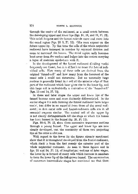
In these and later stages the upper and lower lips of thefunnel become more and more distinctly differentiated. In theearlier stages the cells forming the funnel rudiment have largernuclei, but differ in no essential from those of the canal rudi-ment; in fact canal cells and funnel-rudiment cells are in allessential respects similar. The central cell of the upper lipis not clearly distinguishable till the stage at which the lumenhas been formed in the funnel (fig. 29, PI. 12).
Figs. 30-2, PL 12, show three successive transverse sectionsthrough a young funnel. The upper and lower lips are nowclearly developed, and the continuity of these two projectinglips at the sides is obvious.
With regard to the lower lip the figures already mentionedshow that it is throughout developed from the funnel-rudiment,which itself is from the first merely the anterior part of thewhole nephridial rudiment. As seen in these figures and infigs. 33 and 84, PL 12, of longitudinal sections of later stages,the lower lip is formed of many cells which increase in numbersto form the lower lip of the full-grown funnel. The examinationof numerous intermediate stages has convinced me that these
NEPHRIDIOSTOME OF LUMBRICUS 175
cells do indeed form the epithelium at the edge of the lower lipof the adult worm described above, p. 171.
In the later stages the nuclei of the cells of the preseptal canalbecome arranged in two lateral rows, the dorsal and ventralwalls being deprived of nuclei. This disposition of the nucleiextends into the base of the lips and into the lateral evertedgutters; but the free edge of the lower lip and its lateral lobesare formed of an epithelium of many cells.
In the same way, my observations show that the upper lipis also formed from the funnel-rudiment, itself a portion of thewhole nephridial rudiment, and derived from the 'funnel-cell',possibly also from the most anterior canal cells. Strong evidencethat the marginal and central cells belong to the nephridialrudiment is afforded by the observation that the peculiarvacuolation of the basal region of the future central and mar-ginal cells (described above in the funnel of the adult, p. 170)appears quite early, indeed, even before the lumen has begun topierce the funnel-rudiment (figs. 16, 17, 19, 20-2, PI. 12). Thust h e e n t i r e n e p h r i d i o s t o m e (excluding, of course, theexternal covering of coelomic epithelium and intermediate layerof connective tissue) a p p e a r s to be an i n t e g r a l p a r t ofthe n e p h r i d i a l r u d i m e n t , and of any participation ofcoelomic epithelium cells in the formation of either upper orlower lip I can find no evidence.
This conclusion is borne out by a comparison with otherrelated forms. As we pass from such simple forms as theTubificiclae through the Lumbriculidae to primitive Lumbrico-morpha such as the Haplotaxidae (possibly ancestral to theLumbricidae), the nephridiostome is seen to become more andmore elaborated, as has been well described and figured byY. Boveri-Boner (1920). In Tubif ex it has only three or fournuclei, its rim has delicate cilia extending into the coelom whilethe upper lip is provided with a flame-like bunch of cilia workingin the lumen. This ' flame', as I long ago suggested (1895,1896),probably represents in the smaller and more primitive Oli-gochaeta the original ciliation of the protonephridial flame-cellfrom which the nephridiostome has doubtless been derived inphylogeny. In the more advanced L u m b r i c u l u s , larger
176 EDWIN S. GOODEICH
dorsal and smaller ventral lip are present. The former consistsof a central and two marginal cells somewhat vacuolated; yetthe inner flame-like bunch of cilia is still distinct from theperipheral cilia. H a p l o t a x i s g o r d i o i d e s , according toBoveri-Boner, has a nephridiostome very like that of a Lum-bricid. It is borne on an elongated preseptal canal, there isa large upper lip with one central and many marginal ciliatedcells, and a lower lip with many nuclei. The ' flame', however,may still be distinguished on the central cell. It is but a stepfrom this to the funnel of the earthworm where the marginalsare more numerous and the ciliation of the central cell has lost\ts flame-like character.
Moreover, since A. Meyer (1929)1 has recently given a detailedand careful account of the development of both canal cells andnephridiostome from the nephridioblast in Tub i f ex , thuscorroborating Vejdovsky, the evidence seems to be completethat the nephridiostome is an integral part of the nephridiumin Oligochaeta in general.
It follows that the attempts made by certain authors (seep. 165) to homologize the funnel of an Gligochaete with that ofthe nephromixium of a Polychaete must be rejected. They areperhaps due to the failure of these authors to appreciate thefact that the formation of a compound nephromixium has onlybeen proved to occur in the Order Polychaeta, and only incertain families of that Order; that it seems very unlikely thatthis peculiar combination should have come about in any other
1 With the first origin of the nephridioblast we are not concerned inthis paper; but I may point out that, although A. Meyer clearly traced thedevelopment of the whole nephridium from a nephridioblast lodged amongcoelomic epithelium cells on the anterior surface of the septum and con-cluded (unlike Vejdovsky) that this cell is derived from the coelomicepithelium, he does not seem to have proved his case. The first stage hedescribes is already a late stage in development when the coelomic cavityis large and the septum thin. At this stage the nephridioblast, whateverits origin may be, must perforce take up a position in the coelomic epithe-lium. From a personal inspection of Meyer's preparations I am convincedthat he is mistaken and failed to trace the origin of the nephridioblast inTubifex. Furthermore, there seems to be no justification for Meyer'sextraordinary theories concerning the general morphology of nephridiaand genital ducts in the Annelids.
NEPHRIDIOSTOME OF LUMBRICUS 177
Order; and that, therefore, the view that a nephromixium hasbeen formed in any other Order should only be accepted on thestrongest evidence. Since in Oligochaeta nephridia and coelo-moducts (genital funnels) admittedly exist separately and sideby side, of all the groups of Annelids it is the one in which weshould least expect to find such nephromixia.
SUMMARY.
The structure of the nephridiostome of L u m b r i c u st e r r e s t r i s L. is described, including the anatomical relationsof canal, gutter, central, and marginal cells and their cytologicalcharacters. The extent and relation of the lower lip to otherparts are also described.
An account of the development of the nephridium is givenfrom the stage when the rudiment still bears a single large' funnel-cell' bulging forwards through the septum into thecoelom. The whole nephridiostome (excluding the covering ofcoelomic epithelium and the connective tissue) is shown to arisefrom the nephridial rudiment, wholely or partly from that partof the funnel-rudiment which is derived from the 'funnel-cell'.Upper, lateral, and lower lips are all developed from the funnel-rudiment in which the lumen becomes pierced. There is noevidence that the coelomic epithelium contributes any part ofthe true nephridiostome.
The view sometimes put forward that the excretory organ ofL u m b r i c u s is a nephromixium is not founded on soundevidence, and is opposed to the simple straightforward inter-pretation of its morphology which follows most naturally fromthe facts and a comparison with lower forms.
EEFERENCES.Benham, W. B. (1891).—"The Nephridium of Lumbricus", 'Quart. Journ.
Micr. So.', vol. 32.(1904).—"On a New Species of Haplotaxis", ibid., vol. 48.
Bergh, R. S. (1888).—"Z. Bildungsgeschichte der Exkretionsorgane beiCriodrilus", 'Arb. Zool. Inst., Wiirzburg', vol. 8.
(1899).—"Entwicklung der Segmentalorgane", 'Zeitschr. f. Wiss.Zool.', vol. 66.
(1890).—"Neue Beitr. zur Embryol. der Anneliden. I. Zur Entw.u. Differ, des Ke;mstreifens von Lumbricus", ibid., vol. 50.
178 EDWIN S. GOODRICH
Gegenbaur, C. (1852).—"tJber d. sogen. Respirationsorgane der Regen-wurmer", 'Zeit. wiss. Zool.', vol. 4.
Goodrich, E. S. (1895).—"On the Coelom, Genital ducts, and Nephridia"Quart. Journ. Micr. Sci. v. 37.
(1900).—"On the Nephridia of the Polychaeta, Part III", 'Quart.Journ. Micr. Sc.', vol. 43.
Lehmann, 0. (1887).—"Homologie der Segmentalorgane u. Ausfuhrgiinged. Geschlechtsprodukte b. d. Oligochaeten", 'Jena, Zeitschr. Naturw.',vol. 21.
Meyer, A. (1929).—''Entwickl. d. Nephridien u. Gonoblasten bei Tubifex",Zeit. wiss. Zool., v. 133.
Meyer, Ed. (1887).—"Studien ii. d. Ko'rperbau der Anneliden", 'Mitth.Zool. Sta. Neapel', vol. 7.
Rosen, F. (1911).—"Der Wimpertrichter der Lumbrioiden", 'Zeitschr. f.wiss. Zool.', vol. 98.
Schneider, K. C. (1902).—"Lehrbuch d. vergl. Histologie der Tiere.'Jena.
Staff, F. (1910).—"Organogenetische Unters. uber Criodrilus lacuum",'Arb. Zool. Instit., Wien', vol. 18.
Vejdovsky, F. (1884).—'System u. Morphologie der Oligochaeten.' Prag.(1888-92).—' Entwicklungsgeschichtliche Untersuchungen.' Prag.
Wilson, E. B. (1889).—"Embryology of the Earthworm", 'J. of Morph.',vol. 3.
EXPLANATION OF PLATES.
REFERENCE LETTERS.
be, lateral band of cilia in nephridial canal; bv, blood-vessel; bw, body-wall; c, coelom; cc, central cell of upper lip; cep, coelomic epithelium;cl, canal of nephxidium; clc, canal-cell; dl, nucleus of cell of lower lip;cp, centripetal marginal cell; ct, connective tissue; ec, ectoderm; fc, ' funnel-cell'; fr, group of cells from which develops the funnel = funnel-rudiment;gc, gutter-cell; gr, groove continued from canal; Ic, leucocyte; II, lower lip;lo, lateral lobe of lower lip; me, marginal cell; n, nephridium; ncc, nucleusof central cell; nt, notch between central and gutter-cells; op, opening ofcanal into coelom; p, point at which gutter-cell meets marginal cells; pi,lateral edge of preseptal region; pr, preseptal region; pa, postseptal region;r, area without cilia continued from canal; s, septum; id, upper lip; v,vacuoles in cells of upper lip.
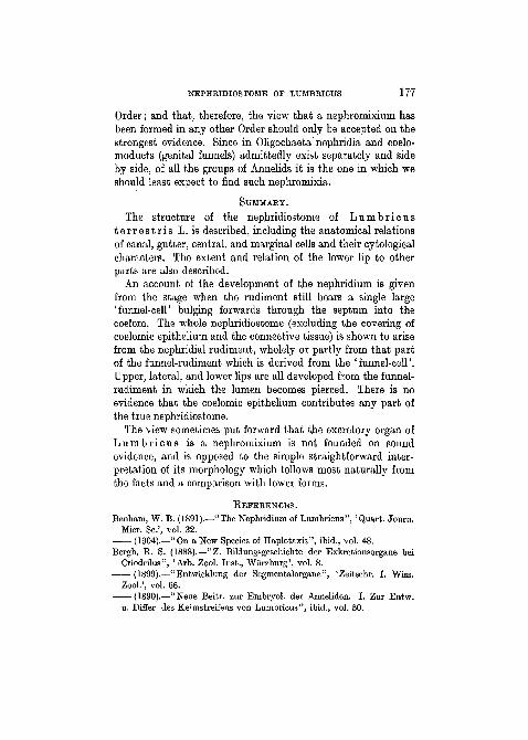
PLATE 11.
Magnification: the scale with fig. 1 applies to all figures except fig. 7.Fig. 1.—Longitudinal section through the middle of the nephridiostome,
and beginning of the nephridial preseptal canal. Only a portion of themass of leucocytes attached to the lower lip is shown.
NBPHEIDIOSTOME OF LUMBRICUS 179
Tig. 2.—Similar section through another funnel.Fig. 3.—Longitudinal section through the funnel shown in fig. 2 passing
through the lateral wall of the canal, showing the nuclei of two canal cells,and the ciliated band of the canal passing on to the gutter-cells over theedge of the opening. The arrows in this and the previous figure indicatecorresponding levels.
Fig. 4.—Transverse section of the upper lip cutting through marginalcells.
Figs. 5 and 6.—Transverse sections through the upper and lower lips;fig. 5 near the edge of the lower lip, fig. 6 near its base.
Fig. 7.—Portion of a transverse section of the upper and lower lips,showing the cilia of the marginal cell clinging to the edge of the lower lip.
Figs. 8 and 9.—Portions of two consecutive transverse sections of anephridiostome showing the continuity of the ciliated band of the gutter-cells with the ciliated area of the lateral marginal cells at point, p. Thecilia are not drawn (except in the canal) but their basal granules areindicated.
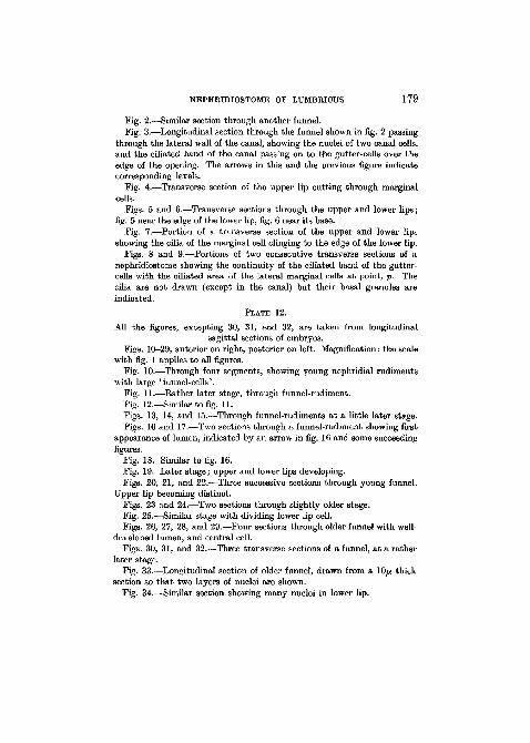
PLATE 12.
All the figures, excepting 30, 31, and 32, are taken from longitudinalsagittal sections of embryos.
Figs. 10-29, anterior on right, posterior on left. Magnification: the scalewith fig. 1 applies to all figures.
Fig. 10.—Through four segments, showing young nephridial rudimentswith large 'funnel-cells'.
Fig. 11.—Rather later stage, through funnel-rudiment.Fig. 12.—Similar to fig. 11.Figs. 13, 14, and 15.—Through funnel-rudiments at a little later stage.Figs. 16 and 17.—Two sections through a funnel-rudiment showing first
appearance of lumen, indicated by an arrow in fig. 16 and some succeedingfigures.
Fig. 18. Similar to fig. 16.Fig. 19. Later stage; upper and lower lips developing.Figs. 20, 21, and 22.—Three successive sections through young funnel.
Upper lip becoming distinct.Figs. 23 and 24.—Two sections through slightly older stage.Fig. 25.—Similar stage with dividing lower lip cell.Figs. 26, 27, 28, and 29.—Four sections through older funnel with well-
developed lumen, and central cell.Figs. 30, 31, and 32.—Three transverse sections of a funnel, at a rather
later stage.Fig. 33.—Longitudinal section of older funnel, drawn from a lOfj. thick
section so that two layers of nuclei are shown.Fig. 34.—Similar section showing many nuclei in lower lip.