arthrodesis of distal interphalangeal joints in the hand …€¦ · · 2014-11-10arthrodesis of...
TRANSCRIPT
Arthrodesis of Distal Interphalangeal Joints in the Hand with Interosseous Wiring and
Intramedullary K-wire Fixation Soo Hong Han, MD, Yoon Sik Cha, MD, Won Tae Song, MD
Department of Orthopaedic Surgery, CHA Bundang Medical Center, CHA University College of Medicine, Seongnam, Korea
Original Article Clinics in Orthopedic Surgery 2014;6:401-404 • http://dx.doi.org/10.4055/cios.2014.6.4.401
Received October 12, 2013; Accepted December 26, 2013Correspondence to: Soo Hong Han, MDDepartment of Orthopaedic Surgery, CHA Bundang Medical Center, CHA University College of Medicine, 59 Yatap-ro, Bundang-gu, Seongnam 463-712, Korea Tel: +82-31-780-5289, Fax: +82-31-780-3578E-mail: [email protected]
Arthrodesis of the distal interphalangeal (DIP) joint of the hand is commonly performed to treat a variety of condi-tions including pain, deformity, and instability. These problems usually derive from degenerative arthritis, in-flammatory diseases or traumatic problems such as irrepa-rable damage to the relevant flexor or extensor tendon, and joint destruction. Stable bony union is essential for successful arthrodesis, and discomfort caused by the hard-ware should be absent. Additionally, early joint movement should be allowed to prevent adjacent joint stiffness. Ar-throdesis of the DIP joints has a long history as evidenced by the variety of developed techniques, including crossed Kirschner wires (K-wires), screw or plate fixation and external fixation.1-4) Although results from multiple tech-
Background: To evaluate the efficacy of intramedullary K-wire fixation and interosseous wiring in the arthrodesis of the distal interphalangeal (DIP) joint with description of surgical procedure.Methods: We retrospectively analyzed 9 cases (7 women and 2 men) of DIP joint arthrodesis. The average age of patients was 44.2 years (range, 21 to 71 years) and the mean follow-up period was 19.6 months. Joint union was evaluated on the follow-up radiographs together with postoperative complications. Results: All cases achieved radiologic union of the arthrodesis site. There was no surgical complication except for one case of skin irritation by the interosseous wire knot which was removed during the follow-up period.Conclusions: Intramedullary K-wire fixation and interosseous wiring could be an alternative procedure of arthrodesis in the DIP joint. Keywords: Distal interphalangeal joint, Arthrodesis, Intramedullary K-wire, Interosseous wiring
niques of DIP joint arthrodesis are often very promising, each procedure has some degree of difficulty or requires specific instruments.1-3) We applied a simple procedure of intramedullary K-wire fixation and interosseous wiring. The purpose of this study was to introduce our technique of DIP joint arthrodesis using simple instruments and to assess its objective and subjective outcomes.
METHODS
We retrospectively reviewed a group of patients who had undergone arthrodesis of the DIP joints, using implantable intramedullay K-wire fixation and interosseous wiring, from October 2008 and March 2012. The study included 9 patients (7 women and 2 men) and the mean age of patients was 44.2 years (range, 21 to 71 years). Painful os-teoarthritis was observed in 4 patients, chronic irreparable mallet finger in 3 patients and post-traumatic arthritis in 2 patients. The dominant hand was affected in 6 patients, and the affected fingers were 4 ring fingers, 3 long fingers and 1 index and small finger each.
Surgical procedures were performed under local
Copyright © 2014 by The Korean Orthopaedic AssociationThis is an Open Access article distributed under the terms of the Creative Commons Attribution Non-Commercial License (http://creativecommons.org/licenses/by-nc/3.0)
which permits unrestricted non-commercial use, distribution, and reproduction in any medium, provided the original work is properly cited.Clinics in Orthopedic Surgery • pISSN 2005-291X eISSN 2005-4408
402
Han et al. Arthrodesis of Distal Interphalangeal Joints with Interosseous Wiring and Intramedullay K-wire Fixation Clinics in Orthopedic Surgery • Vol. 6, No. 4, 2014 • www.ecios.org
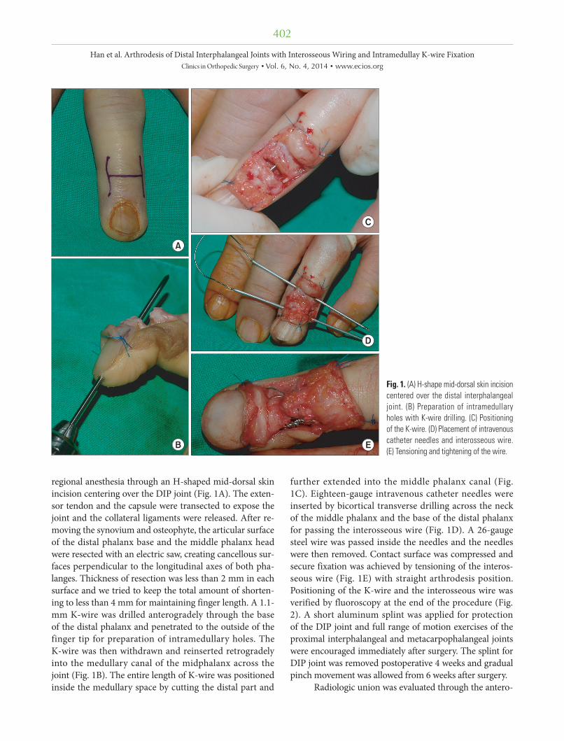
regional anesthesia through an H-shaped mid-dorsal skin incision centering over the DIP joint (Fig. 1A). The exten-sor tendon and the capsule were transected to expose the joint and the collateral ligaments were released. After re-moving the synovium and osteophyte, the articular surface of the distal phalanx base and the middle phalanx head were resected with an electric saw, creating cancellous sur-faces perpendicular to the longitudinal axes of both pha-langes. Thickness of resection was less than 2 mm in each surface and we tried to keep the total amount of shorten-ing to less than 4 mm for maintaining finger length. A 1.1-mm K-wire was drilled anterogradely through the base of the distal phalanx and penetrated to the outside of the finger tip for preparation of intramedullary holes. The K-wire was then withdrawn and reinserted retrogradely into the medullary canal of the midphalanx across the joint (Fig. 1B). The entire length of K-wire was positioned inside the medullary space by cutting the distal part and
further extended into the middle phalanx canal (Fig. 1C). Eighteen-gauge intravenous catheter needles were inserted by bicortical transverse drilling across the neck of the middle phalanx and the base of the distal phalanx for passing the interosseous wire (Fig. 1D). A 26-gauge steel wire was passed inside the needles and the needles were then removed. Contact surface was compressed and secure fixation was achieved by tensioning of the interos-seous wire (Fig. 1E) with straight arthrodesis position. Positioning of the K-wire and the interosseous wire was verified by fluoroscopy at the end of the procedure (Fig. 2). A short aluminum splint was applied for protection of the DIP joint and full range of motion exercises of the proximal interphalangeal and metacarpophalangeal joints were encouraged immediately after surgery. The splint for DIP joint was removed postoperative 4 weeks and gradual pinch movement was allowed from 6 weeks after surgery.
Radiologic union was evaluated through the antero-
Fig. 1. (A) H-shape mid-dorsal skin incision centered over the distal interphalangeal joint. (B) Preparation of intramedullary holes with K-wire drilling. (C) Positioning of the K-wire. (D) Placement of intravenous ca theter needles and interosseous wire. (E) Ten sioning and tightening of the wire.

403
Han et al. Arthrodesis of Distal Interphalangeal Joints with Interosseous Wiring and Intramedullay K-wire Fixation Clinics in Orthopedic Surgery • Vol. 6, No. 4, 2014 • www.ecios.org
posterior and lateral views of the radiographs which were checked every 2 weeks, until evidence of union. Union was defined by the presence of bony trabeculae crossing the ar-throdesis site. Visual analogue scale (VAS) of the DIP joint was recorded to evaluate the remaining pain postopera-tively. We also investigated the presence of any immediate of delayed surgical complications.
RESULTS
The mean follow-up period was 19.6 months (range, 12 to 36 months) and all cases had successful fusion of the joint. Patients showed union of the joint on radiographs taken from 6 to 10 weeks after surgery, and the mean time was 7.6 weeks. Average VAS score was improved from 6.5 points preoperatively to 1.2 points (range, 0 to 3 points) at the last follow-up in arthritis patients. Cases of chronic mallet finger were excluded from VAS score evaluation be-cause their main complaint was deformity without painful arthritic change. We noted no malunion, nail deformity, hardware migration or infection on physical examination or in follow-up radiographs. Wire removal was required in 1 patient, due to irritation by the knot of the interosseous wire at 4 months postoperatively and satisfactory relief of discomfort was achieved during the follow-up.
DISCUSSION
Arthrodesis is a generally accepted operative treatment for arthritis of the DIP joint to relieve pain and to correct de-formity and instability in patients who are unresponsive to conservative measures. Bunnell introduced a technique for
arthrodesis consisting of resection of the articular surfaces and securing of the joint with two K-wires. Since then, many similar techniques have been introduced.5,6) Because each technique has its own advantages and problems, no single technique has gained universal popularity.1)
K-wires have been used for many years, leading to insufficient osseous stabilization7) and making it neces-sary to remove the K-wire after the union. A correlation was found between infection and the use of K-wires in another study.8) We observed no postoperative complica-tion with suspected infection. Our opinion was that the intramedullary location of the entire length of K-wire low-ers the risk of infection from exposed K-wire tip outside the skin. More recently, headless compression screw or plate fixation for interphalangeal joint fusion has demon-strated consistent success rates.1,3) Villani et al.3) reported 102 cases of headless compression screw fixation for DIP joint arthrodesis, with 2 cases of prominent hardware, 1 complex regional pain syndrome and 1 symptomatic bony callus from 89 fused joints. Mantovani et al.2) also reported a 100% union rate of the DIP joint using the lateral approach with plate and screw fixation. However, these methods require special instruments or implants. There also have been several reports about complication related to incompatibility of implant size to the distal phalanx. Compared to these procedures, arthrodesis with an intramedullary K-wire and interosseous wiring is a simpler alternative that can provide early compression by wire tightening and enough stability by 2 kinds of fixation during fusion of the joint. Additionally, intramedullary hardware not only minimizes soft tissue irritation, which is especially important in these patients with a delicate soft tissue envelope, but also enables more comfortable early joint motion. Our procedure could provide good pain relief, high rate of bone fusion, and few complications for these reasons. Functional assessments were not carried out in this study because of various clinical features of other joints in most patients.
Even though slight flexion of the DIP joint is recom-mended in arthrodesis, some studies reported that extend-ed joint position did not cause any discomfort.1,3) We also made the arthrodesis position straight and the patients did not complain of any functional or aesthetic problems related to the fusion position.
Radiologically, all cases showed solid union at the arthrodesis site during the follow-up, and there was no difference between our radiologic union time and those of other published series, which have ranged from 6 to 12 weeks.2,4,9-11)
There was one case of interosseous wire knot irrita-
Fig. 2. Postoperative anteroposterior (A) and lateral (B) radiographs.
404
Han et al. Arthrodesis of Distal Interphalangeal Joints with Interosseous Wiring and Intramedullay K-wire Fixation Clinics in Orthopedic Surgery • Vol. 6, No. 4, 2014 • www.ecios.org
tion that required removal. We suggest avoiding a big knot and putting the knot inside the joint resection site, to pre-vent such problem. The results of this study indicated that DIP joint arthrodesis with an intramedullary K-wire with interosseous wiring is easily accomplished, has a low rate of complications, and is effective and reliable. This proce-dure has not been reported in the DIP joint, to the best of our knowledge.
Limitations of this study included its retrospective study design with a small sample size, and no direct com-parison with a group of patients randomly allocated to
another treatment.Intramedullary K-wire fixation and interosseous
wiring could be an alternative procedure of arthrodesis fixed with simple implants for the old DIP joint problems in the hand.
CONFLICT OF INTEREST
No potential conflict of interest relevant to this article was reported.
REFERENCES
1. Song JH, Lee JY, Chung YG, Park IJ. Distal interphalangeal joint arthrodesis with a headless compression screw: mor-phometric and functional analyses. Arch Orthop Trauma Surg. 2012;132(5):663-9.
2. Mantovani G, Fukushima WY, Cho AB, Aita MA, Lino W Jr, Faria FN. Alternative to the distal interphalangeal joint arthrodesis: lateral approach and plate fixation. J Hand Surg Am. 2008;33(1):31-4.
3. Villani F, Uribe-Echevarria B, Vaienti L. Distal interphalan-geal joint arthrodesis for degenerative osteoarthritis with compression screw: results in 102 digits. J Hand Surg Am. 2012;37(7):1330-4.
4. Wyrsch B, Dawson J, Aufranc S, Weikert D, Milek M. Distal interphalangeal joint arthrodesis comparing tension-band wire and Herbert screw: a biomechanical and dimensional analysis. J Hand Surg Am. 1996;21(3):438-43.
5. Mader K, Gausepohl T, Wolfgarten B, Pennig D. Percuta-neous arthrodesis of small joints in the hand: a minimum three-year follow-up. J Bone Joint Surg Br. 2003;85(7):1016-8.
6. Virreira M, Alonso-Vega C, Solano M, et al. Congenital Chagas disease in Bolivia is not associated with DNA poly-morphism of Trypanosoma cruzi. Am J Trop Med Hyg. 2006;75(5):871-9.
7. Mikolyzk DK, Stern PJ. Steinmann pin arthrodesis for salvage of failed small joint arthroplasty. J Hand Surg Am. 2011;36(8):1383-7.
8. Stern PJ, Fulton DB. Distal interphalangeal joint ar-throdesis: an analysis of complications. J Hand Surg Am. 1992;17(6):1139-45.
9. Brutus JP, Palmer AK, Mosher JF, Harley BJ, Loftus JB. Use of a headless compressive screw for distal interphalangeal joint arthrodesis in digits: clinical outcome and review of complications. J Hand Surg Am. 2006;31(1):85-9.
10. Carroll RE, Hill NA. Small joint arthrodesis in hand recon-struction. J Bone Joint Surg Am. 1969;51(6):1219-21.
11. Lister G. Intraosseous wiring of the digital skeleton. J Hand Surg Am. 1978;3(5):427-35.