treatment of distal radius fractures with a nonbridging ... · treatment of distal radius fractures...
TRANSCRIPT

Treatment of Distal Radius Fractures With a NonbridgingCross-Pin Fixator (The CPX System)
Ather Mirza, MD,*Þþ Mary Kate Reinhart, MS, CPNP, ANP,§ and Joseph John Bove, BA§
Abstract: Many treatment methods exist for patients presenting with afracture of the distal radius. With the evolution of innovative proceduresand devices, treatment of these particular fractures is shifting to morecontemporary approaches. The minimally invasive technique with thecross-pin fixator (CPX) system offers a new biomechanical concept forrelatively rigid fixation of distal radius fractures (DRF). The CPX systemuses percutaneous cross Kirschner wire fixation in combination with anonbridging external fixator. The stability of the model is derived fromthe external unilateral frame and the positioning of multiple 1.6 mmKirschner wires at various angles and planes to each other. This allowsfor maintenance of DRF reduction, early wrist mobilization, and aprompt return to the usual activities. Between September 2004 andSeptember 2008, there were 54 patients with 56 DRF who were treatedwith the CPX system. Excluded from the report are 2 patients who had abone graft and 1 patient who was not willing to adhere to thepostoperative protocol. Of the 51 patients with 53 DRF, no majorcomplications were reported. This article describes the CPX surgicaltechnique, the indications, the complications, and the postoperativemanagement.
Key Words: distal radius fracture, percutaneous, cross-pin fixatorsystem
(Tech Hand Surg 2009;13: 104Y109)
HISTORICAL PERSPECTIVEAlthough several techniques and instruments have evolved
in recent years, many of the current treatments of distal radiusfractures have not solved all the problems associated with suchfractures. Although cast immobilization is pertinent for usein certain instances, it does not consistently maintain reductionespecially in unstable intraarticular fractures.1 Likewise, span-ning external fixators, which were popular at one time, havebecome less prevalent because of their use of ligamentotaxis.2Y4
Ligamentotaxis carries inherent problems because it fails tomaintain reduction in all radiological parameters4Y6 and be-cause the viscoelastic behavior of soft tissues causes fixatorsto lose their distractive forces over time.7,8 More so, ligamento-taxis may foreshorten the extensor tendons, leading to stiffnessof the finger joints.4,6 In a study of bridging external fixationwith the Hoffman Fixator (Howmedica, Staines, United King-dom), McQueen et al and Michie9 reported poor hand functionand a high percentage of complications.
Nonbridging fixators are advantageous because they avoidthe use of ligamentotaxis and allow mobility of the wrist.Moreover, they permit direct control of the distal fragmentand maintain the radiological parameters, especially volar tilt.10
Good success has been reported when using nonbridging ex-ternal fixators.11Y13 In the early rehabilitation period, nonspan-ning fixators have shown greater improvements in grip strengthand wrist range of motion (ROM) when compared with bridg-ing external fixators.10 In a study of 52 patients with a fracturein the distal radius, Flinkkila et al13 found that a nonbridgingexternal fixator reestablished 87% to 98% of the ROM andgrip strength when measured up to the uninjured arm. In spiteof their success, nonbridging fixators have their problems.Most nonspanning fixators have pins inserted perpendicularto the shaft of the radius,11,14Y17 thereby failing to directly fixthe fracture. In addition, they bypass loading, which does notfacilitate fracture healing.
Direct fixation of the fracture certainly provides bettercontrol in fracture management. Biomechanically, a single wirethrough a fracture allows for translation and rotation alongthe axis of the wire.18Y20 A second wire, provided it is not par-allel to the first, offers greater stability, translation, and rota-tion.18,20,21 In a finite element model, Rogge et al18 demonstratedthat cross Kirschner wire (K-wire) fixation, when compared withparallel pinning, offered greater stability in maintaining frac-ture reduction. Graham and Louis,19 in a cadaver study, illus-trated that multiple pins up to 4 in a multiplanar direction,resulted in greater stability, especially if they passed throughthe ulnar shaft.
Among nonbridging fixators, the cross-pin fixator (CPX)device (A.M. Surgical, Smithtown, NY) is the only unilateralframe that has a cross-pin multiplanar configuration, providing3-dimensional stability. It is further differentiated from othernonbridging fixators11,15,17 because of its use of small 1.6 mmK-wires inserted in the mid-lateral plane. In a cadavericfracture model, Strauss et al22 compared the CPX system tovolar locking plate fixation. The authors concluded that nosignificant difference was present between the mechanicalstiffness of the CPX system and volar locked plate. The CPXsystem offers patients a reliable method for maintainingfracture reduction, a low risk of major complication, and aprompt return to usual activities.
INDICATIONS/CONTRAINDICATIONSThe CPX system is indicated for treatment of displaced
reducible extraarticular fractures, and non-displaced and dis-placed reducible intraarticular fractures. The various fractureswere documented according to the AO classification sys-tem.23Y25 In spite of osteoporotic bones or unstable fracturessuch as dorsal shear B2.2 and volar shear B3.3, the radiologicalparameters (radial height, radial inclination, and palmar tilt)were maintained from initial post-op to final evaluation.
Thus far, we have no experience with C2.3, C3.2, and C3.3fractures and therefore suggest that until clinical research is
From the *Hand and Microsurgery, St. Catherine of Siena Medical Center;†North Shore Surgi-Center, Smithown; ‡Stony Brook University, StonyBrook, NY; and §Smithtown, NY.Address correspondence and reprint requests to Ather Mirza, MD, 290 East
Main St, Suite 200, Smithtown, NY 11787. E-mail: [email protected].
Dr Mirza is a part owner of A.M. Surgical and thus, he has a financialinvolvement with the subject matter of this research. All authors did notreceive funding or grants in support of the research or the preparation ofthe manuscript.
Copyright * 2009 by Lippincott Williams & Wilkins
TECHNIQUE
104 Techniques in Hand & Upper Extremity Surgery & Volume 13, Number 2, June 2009
9Copyright @ 200 Lippincott Williams & Wilkins. Unauthorized reproduction of this article is prohibited.

available, these fractures be contraindicated. Other contraindi-cations include extensive soft tissue trauma, open fractures, aconsiderable skin compromise, noncompliance, dementia, oradvanced Parkinson disease.
TECHNIQUE
The DeviceThe CPX system takes advantage of closed reduction in-
ternal fixation with percutaneous cross-pin fixation and a non-bridging external fixator. Only 41 g (with the pins), thealuminum CPX device contains a 2-part moveable bar with2 screws to alter the length between 11.5 and 14.5 cm. Thesliding bar contains a head at each end, and each head has3 variable K-wire fixators (Fig. 1). The K-wire fixators have2 screws. One screw influences the insertion angle of theK-wire, and the second screw fastens the K-wire to the
FIGURE 2. A, Initial stab wound. B, Placement of clamp intothe stab wound.
FIGURE 3. Use of the tissue protector to minimize potential injuryand to determine placement of the first K-wire by FluoroScan.
FIGURE 4. The first distal K-wire insertion. A, Introductionof the K-wire through the tissue protector. B,FlouroScanVanteroposterior view. C, FlouroScanVlateral view.
FIGURE 1. The CPX device (A.M. Surgical).
Techniques in Hand & Upper Extremity Surgery & Volume 13, Number 2, June 2009 Distal Radius Fractures
* 2009 Lippincott Williams & Wilkins www.techhandsurg.com | 105
9Copyright @ 200 Lippincott Williams & Wilkins. Unauthorized reproduction of this article is prohibited.
fixator. The newer device offers 15 degrees of rotationaround the center of the guide hole. Before the fracture isreduced, all of the CPX system’s screws are loosened.
Surgical TechniqueThe operative procedure is done under regional intraven-
ous block, axillary block, or general anesthesia with fluoro-scopic imaging. Alternatively, one can use a Bier block with aforearm or an upper arm tourniquet. In patients with a shortforearm, an upper arm tourniquet is used because a forearmtourniquet makes proximal pin insertion challenging. Mostfractures were reduced with the classic maneuver of palmarflexion and ulnar deviation.5 Occasionally, we had to use longi-tudinal traction with finger traps to gain radial inclination andradial height. With longitudinal traction, one has to apply dorsalpressure on the distal fragment to maintain palmar tilt. Pres-sure in the volar direction may be discontinued after the firstK-wire is introduced. It is important to note that the first distaland proximal K-wires are inserted freehand before the CPXdevice is applied. An alternate technique is introducing all ofthe 4 K-wires freehand at 40 to 45- degree oblique anglesand then applying the CPX device afterward. Because of thefreedom of angulation around the K-wire guide holes (varia-bility of 30 degrees), the device can easily accommodate forfreehand insertion of all 4 K-wires. The advantage of thisalternative is it can expedite the surgery.
Fracture reduction is checked via FluoroScan for jointcongruency, palmar tilt, radial inclination, radial height, andulnar variance. When all these parameters are satisfied, oneshould proceed with the introduction of the first K-wire. Thefirst K-wire is inserted between the first and second dorsalcompartment by making a small stab wound and using a clampto spread the soft tissue down to the bone (Fig. 2A, B). A tissueprotector (A.M. Surgical, Smithtown, NY) is then used to avoidinjury to the radial sensory nerve (Fig. 3). The tissue protector isheld against the bone at a 40 to 45- degree angle, and aFlouroScan image is taken to ascertain the position of the tissueprotector against the bone. The smooth 1.6 mm K-wire is thendriven freehand through the tissue protector (Fig. 4A), acrossthe fracture site, penetrating the ulna cortex, and out the radial
FIGURE 5. The marked line ensuring the K-wire is properlyorientated toward the lunate fossa.
FIGURE 6. FlouroScan image of the first distal and proximalK-wires.
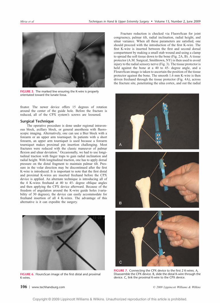
FIGURE 7. Connecting the CPX device to the first 2 K-wires. A,Disassemble the CPX device. B, slide the distal K-wire through thedevice. C, link the proximal K-wire to the CPX device.
Mirza et al Techniques in Hand & Upper Extremity Surgery & Volume 13, Number 2, June 2009
106 | www.techhandsurg.com * 2009 Lippincott Williams & Wilkins
9Copyright @ 200 Lippincott Williams & Wilkins. Unauthorized reproduction of this article is prohibited.

shaft in the mid lateral plane. To confirm proper placement, aFlouroScan is used for imaging in anteroposterior and lateralplanes (Fig. 4B, C).
To insert the proximal K-wire, place a K-wire on the dorsalside of the skin and take a FluoroScan image in the antero-posterior plane to ascertain that the proximal K-wire is aimingat the lunate fossa. Using a marking pen, trace a line onthe skin next to the K-wire (Fig. 5). On the radial side androughly 1 to 2 cm distal to the tracing, a proximal stab wound ismade in the mid-lateral plane. A clamp is used to spread thesoft tissues, exposing the bone. Then the tissue protector isagain introduced, and a 1.6-mm K-wire is driven freehand at a45-degree angle toward the lunate fossa, stopping at the sub-chondral bone. FluoroScan imaging is used to help confirmcorrect orientation of the K-wire (Fig. 6).
Unscrew the sliding bar and take the 2 halves apart(Fig. 7A). Slide the distal component first over the distalK-wire (Fig. 7B) and then the proximal component over theproximal K-wire (Fig. 7C). Place the 2 plastic spacers onto thedevice (Fig. 8) in order to keep the CPX device a distanceaway from the skin and allow mobilization of the wrist joint.Bring both components together and adjust the device to thedesired length. Tighten the screws controlling the length ofthe CPX device and the screws that attach the K-wires to theapparatus.
For insertion of the second proximal K-wire (only if allthe K-wires are not inserted freehand), pass the tissue protectorthrough the device and mark the point on the skin. Make anincision on the mark and use a clamp to spread the tissues to thebone. Drive the tissue protector toward the bone. Then insertthe K-wire from a proximal to distal direction and, in a similarmanner as the first proximal pin, FluoroScan, to determine itslocation. If unsatisfied with the K-wire position, then reinsertthe K-wire at a different angle and recheck its position withFlouroScan. The fourth K-wire is inserted in a distal to proxi-mal direction and FluoroScan is used to verify accurate pinplacement. In most instances, 2 K-wires distally and 2 proxi-mally will suffice (Fig. 9). Less frequently, more than 4 wiresare used in a given case. The device offers the use of 6 wires;3 proximally and 3 distally.
Tighten all of the screws of the device. Remove the bluespacers and cut the pins to a suitable length. Then, cap the pins toensure patient protection. More FlouroScan views are needed toconfirm that reduction is maintained. Injections with marcaineand epinephrine on all sides of the pins and into the fracturehematoma help to alleviate the patient’s pain postoperatively.The patient is placed in a postoperative dressing with a short armvolar splint and leaves the surgical setting, understanding tokeep the injured arm elevated and to exercise the fingers.
ComplicationsBecause of the positioning of the 1.6-mm K-wires in the
mid-lateral plane, the possibility of injuring an extensor ten-don is minimized. In addition, the mid-lateral approach leadsto less inflammatory reactions and thus risk of infections dueto the reduced mobility of the skin around the pin sites duringwrist ROM exercises. This was confirmed in our treatment of51 patients with no reported pin track infections or tendonruptures. There was no loss of reduction from initial postop-erative to final evaluation even in those patients with osteopo-rotic bones and comminuted fractures. No cases resorted toopen reduction internal fixation.
Major concern of inserting pins in the mid-lateral planeis injury to the radial sensory nerve. However, damage wasdiminished by soft tissue dissection and use of a tissue pro-tector. Although 2 patients had superficial radial nerve sen-sitivity, in both patients, it resolved to a transient form whentreated with desensitization and Gabapentin. Another patient,who had a number of injuries aside from the distal radius frac-tures, developed type I complex regional pain syndrome butthe symptoms resolved. A fourth patient had an extended
FIGURE 8. The spacers of the CPX device (courtesy of A.M.Surgical).
FIGURE 9. Anteroposterior FlouroScan image of 4 insertedK-wires.
FIGURE 10. Patient fitted with a volar splint.
Techniques in Hand & Upper Extremity Surgery & Volume 13, Number 2, June 2009 Distal Radius Fractures
* 2009 Lippincott Williams & Wilkins www.techhandsurg.com | 107
9Copyright @ 200 Lippincott Williams & Wilkins. Unauthorized reproduction of this article is prohibited.

recovery because of the development of carpal tunnel, butafter endoscopic release, a considerable improvement wasobserved.
RehabilitationPatients are seen for radiographic measurements on the
following postoperative visits: 5 days; 2, 4, and 6 weeks; and2, 3, 6, and 12 months. Each visit, Hibiclens is applied to all ofthe pin sites. At the initial postoperative visit, active finger ROMis assessed, patients are advised to attend therapy 3 times perweek, and a custom wrist/forearm orthosis is fitted by an Occu-pational Therapist (Fig. 10). During therapy, active finger, wrist,and forearm ROM commences (Fig. 11AYC). Six times a daypatients are directed to take off the orthosis and completetheir home exercise program. We found, in our study, thatmost people took their splints off at 4 weeks to performlight activities. Since then, we have been recommending thatpatients remove their splints for activities of daily living andonly wear their splints for high-risk activities and sleeping. Ra-diological confirmation of trabecular bridging and obliterationof distinct fracture lines signified that the corresponding fracturewas healed and the removal of the wires and device followed.Each patient decided whether the device was removed inan ambulatory center or in the office. Only 1 patient chose theoffice setting.
SUMMARYThe CPX system is a hybrid model that combines cross-
pin fixation with a nonbridging external fixator. The crossK-wire configuration with pins in multiplanar and multiangledirections creates a rigid fixation, which is enhanced by anexternal strut. The cross K-wires capture and stabilize thelarger fragments while buttressing the smaller fragments. TheCPX system significantly maintains fracture reduction, and itallows for early mobilization of the wrist and resumption ofusual activities. It is important to note that this technique iseasy to use because most orthopedic surgeons are familiarwith cross-pin fixation.
REFERENCES
1. Kapoor H, Agarwal A, Dhaon BK. Displaced intra-articular fracturesof distal radius: a comparative evaluation of results following closedreduction, external fixation. Injury. 2000;31:75Y79.
2. Vidal J, Buscayret C, Paran M, et al. Ligamentotaxis: In: L, MearsDC, ed. External skeletal fixation. Baltimore: Williams &Wilkins; 1983.
3. Bindra RR. Biomechanics and biology of external fixation of distalradius fractures. Hand Clin. 2005;21:363Y373.
4. Agee JM. External fixation: technical advances based uponmultiplanar ligamentotaxis. Orthop Clin North Am.1993;24:265Y274.
5. Bartosh RA, Saldana MJ. Intraarticular fractures of the distal radius:a cadaveric study to determine if ligamentotaxis restores radiopalmartilt. J Hand Surg. 1990;15(1):18Y21.
6. Agee JM. Distal radius fractures. Multiplanar ligamentotaxis. HandClin. 1993;9(4):577Y585.
7. Woo SL, Gomez MA, Akeson WH. The time and history-dependentviscoelastic properties of the canine medical collateral ligament.J Biomech Eng. 1981;103(4):293Y298.
8. Winemaker MJ, Chinchalker S, Richards RS, et al. Load relaxationand forces with activity in Hoffmann external fixators: a clinicalstudy in patients with Colles’ fractures. J Hand Surg.1988;23A:926Y932.
9. McQueen MM, Michie M, Court-Brown CM. Hand and wrist functionafter external fixation of unstable distal radial fractures. Clin Orthop.1992;285:200Y204.
10. McQueen MM. Redisplaced unstable fractures of the distal radius. Arandomized, prospective study of bridging versus non-bridging externalfixation. J Bone Joint Surg Br. 1998;80-B:665Y669.
11. Jenkins NH, Jones DG, Johnson SR, et al. External fixation ofColles’ fractures-an anatomical study. J Bone Joint Surg. 1987;69B:207Y211.
12. Krishnan J, Chipchase S, Slavotinek J. Intraarticular fractures of thedistal radius treated with metaphyseal external fixation. J Hand Surg.1998;23B:396Y399.
13. Flinkkila T, Ristiniemi J, Hyvonen P, et al. Nonbridging external
FIGURE 11. Wrist ROM exercises. A, Flexion. B, extension. C, radial deviation. D, ulnar deviation. E, pronation. F, supination.
Mirza et al Techniques in Hand & Upper Extremity Surgery & Volume 13, Number 2, June 2009
108 | www.techhandsurg.com * 2009 Lippincott Williams & Wilkins
9Copyright @ 200 Lippincott Williams & Wilkins. Unauthorized reproduction of this article is prohibited.

fixation in the treatment of unstable fractures of the distal forearm.Arch Orthop Trauma Surg. 2003;123:349Y352.
14. McQueen MM, Simpson D, Court-Brown CM. Use of the Hoffman2 Compact External Fixator in the Treatment of Redisplaced UnstableDistal Radial Fractures. J Orthop Trauma. 1999;13(7):501Y505.
15. Forgon M, Mammel E. The external fixateur in the management ofunstable Colles’ fracture. Int Orthop. 1981;5:9Y14.
16. Gradl G, Jupiter JB, Gierer P, et al. Fractures of the distal radiustreated with a nonbridging external fixation technique using multiplanarK-wires. J Hand Surg. 2005;30A:960Y968.
17. Melendez EM, Mehne DK, Posner MA. Treatment of unstable Colles’fractures with a new radius mini-fixator. J Hand Surg.1989;14A:807Y811.
18. Rogge R, Adams B, Goel VK. An analysis of bone stresses and fixationstability using a finite element model of simulated distal radiusfractures. J Hand Surg. 2002;27A:86Y92.
19. Graham TJ, Louis DS. Biomechanical aspects of percutaneouspinning for distal radius fractures. In: Saffar P, Cooney W, ed.Fractures of the distal radius. 1st ed. London: Martin Dunitz; 1995:31Y32.
20. Heatherly RD, Adams BD, Goel VK. An evaluation of distal radiusfracture pinning techniques using experimentally validated FE model.Montana: Summer Bioengineering conference, ASMEM, Big Sky;1999.
21. Stein AH Jr, Katz SF. Stabilization of comminuted fractures of thedistal inch of the radius: percutaneous pinning. Clin Orthop.1975;108:174Y181.
22. Strauss EJ, Banerjee D, Frederick KJ, et al. Evaluation of a novel,nonspanning external fixator for treatment of unstable extra-articularfractures of the distal radius: biomechanical comparison with a volarlocking plate. J Trauma. 2008;64:975Y981.
23. Muller ME. Distal Radius. In: Muller ME, Nazarian S, Koch P,Schatzker J, eds. AO Classification of Fractures. Berlin:Springer-Verlag; 1987:106Y115.
24. Kreder HJ, Hanel CP, McKee M, et al. Consistency of AO fractureclassification for the distal radius. J Bone Joint Surg. 1996;78-B:726Y731.
25. Flinkkila T, Annikka N, Kaarela O, et al. Poor interobserverreliability of AO classification of fractures of the distal radius.J Bone Joint Surg. 1998;80-B:670Y672.
Techniques in Hand & Upper Extremity Surgery & Volume 13, Number 2, June 2009 Distal Radius Fractures
* 2009 Lippincott Williams & Wilkins www.techhandsurg.com | 109
9Copyright @ 200 Lippincott Williams & Wilkins. Unauthorized reproduction of this article is prohibited.