distal radius fractures. outcome and new methods of...
TRANSCRIPT

LUND UNIVERSITY
PO Box 117221 00 Lund+46 46-222 00 00
Distal Radius Fractures. Outcome and New Methods of Surgical Treatment
Landgren, Marcus
2017
Document Version:Publisher's PDF, also known as Version of record
Link to publication
Citation for published version (APA):Landgren, M. (2017). Distal Radius Fractures. Outcome and New Methods of Surgical Treatment. Lund: LundUniversity, Faculty of Medicine.
General rightsCopyright and moral rights for the publications made accessible in the public portal are retained by the authorsand/or other copyright owners and it is a condition of accessing publications that users recognise and abide by thelegal requirements associated with these rights.
• Users may download and print one copy of any publication from the public portal for the purpose of private studyor research. • You may not further distribute the material or use it for any profit-making activity or commercial gain • You may freely distribute the URL identifying the publication in the public portalTake down policyIf you believe that this document breaches copyright please contact us providing details, and we will removeaccess to the work immediately and investigate your claim.

Distal Radius Fractures
Outcome and New Methods of Surgical Treatment
Marcus Landgren
DOCTORAL DISSERTATIONBy due permission of the Faculty of Medicine, Lund University, Sweden.
To be defended at lecture room F2, Skåne University Hospital, Lund. Date: Friday May 5, 2017 at 1 pm.
Faculty opponentProfessor Leiv M. Hove
Department of Surgical SciencesUniversity of Bergen
Norway

Organization
LUND UNIVERSITY
Dept of Orthopedics, Clinical Sciences, Lund
Document name
DOCTORAL DISSERTATION
Lund University Date of issue 2017-05-05
Author: Marcus Landgren Sponsoring organization
Title and subtitle: Distal radius fractures. Outcome and new surgical methods of treatment
Abstract
Stable distal radius fractures are treated non-surgically in a cast. Unstable distal radius fractures are treated with surgery. The majority of patients have good outcome, but up to one fifth are not satisfied. In this thesis the overall aim was to compare newer surgical techniques, evaluate our treatment over a longer time-period, and identify risk factors for poor outcome.
In a previous randomized study of unstable distal radius fractures, better grip strength and pronation-supination were found at one year in patients treated with open reduction and fragment-specific fixation compared to closed reduction and external fixation. In a follow up at five years, both grip strength as well as pronation-supination had normalized in both groups and no clinical or radiographic differences were found.
In a new randomized trial of unstable distal radius fractures, comparing the fragment-specific fixation and the newer volar locking plate, no difference was found in the primary outcome grip strength at one year. Both the fragment-specific fixation and the volar locking plate achieved good subjective, objective and radiological outcome, with more complications in the fragment-specific group.
In a prospective and consecutive distal radius fracture registry, a retrospective decade-long study analyzed the subjective outcome at one year, using the Disabilities of Arm Shoulder and Hand (DASH) questionnaire. At one year, good outcome was found for both non-surgically and surgically treated patients, but despite a shift of implant over the 10-year period, with volar locking plates replacing the external fixators and fragment-specific fixation, no change was found in subjective outcome.
Finally, patients with major disability at one year, identified in the distal radius fracture registry, were re-evaluated at 2 12 years. Half of the patients improved, but only a small proportion returned to normal scores and the rest improved to a moderate level of disability.
In conclusion, the surgical methods to treat distal radius fractures are good. But, a limited proportion of both and surgically treated patients has an inferior outcome. In the future, we need to focus on this
subgroup and find better ways to identify causes to inferior outcome and try to prevent these complications before they become irreversible.
Key words: Distal radius fracture, surgical treatment, non-surgical treatment, outcome, residual disability
Classification system and/or index terms (if any)
Supplementary bibliographical information Language English
Faculty of Medicine Doctoral Dissertation Series 2017:60
ISSN and key title 1652-8220 ISBN 978-91-7619-440-9
Recipient’s notes Number of pages 1 Price
Security classification
I, the undersigned, being the copyright owner of the abstract of the above-mentioned dissertation, hereby grant to all reference sources permission to publish and disseminate the abstract of the above-mentioned dissertation.
Signature Date 2017-03-29

Distal Radius Fractures
Outcome and New Methods of Surgical Treatment
Marcus Landgren

Cover illustrations by my mother Elisabeth Grönvall: Front – A sketch of a comminuted and intra-articular distal radius fracture. Back – An aquarelle illustrating pain after a wrist trauma.
© Copyright Marcus Landgren
Department of Clinical Sciences, Orthopedics, Faculty of Medicine, Lund University, Sweden
Doctoral Dissertations Series: 2017:60ISBN 978-91-7619-440-9ISSN 1652-8220
Printed in Sweden by Media-Tryck, Lund UniversityLund, 2017
Contact addressMarcus Landgren, MDDepartment of OrthopedicsSkåne University HospitalSE 221 85 Lund, SwedenTel: +46 46171000Mail: [email protected]

To my family
”…One consolation only remains, that the limb will at approximately 12 months again enjoy 90–95% in all its motions of the contralateral wrist, and with a DASH ≤10…”


Contents
List of papers, 3Abbreviations, 4Thesis at a glance, 5Introduction, 7Aims, 8Background, 9
History – treatment evolution or revolution? 9Epidemiology – size of the problem, 10Classification, 11Treatment, 11
Non-surgical treatment, 11Closed reduction and pinning/external fixation, 12Open reduction and internal fixation, 13Rehabilitation, 17
How to evaluate outcome, 18Radiographic assessment, 18Grip strength, 18Range of motion, 19Physician-based assessment instruments, 19Patient-reported outcome measures, 19
Complications, 22Malunion, 23
Patients and methods, 25Paper I and II – clinical studies, 26
Study design and patient populations, 26Recruitment and intervention, 27Outcome measurement, 28Radiographic assessment, 29
Paper III and IV – registry studies, 29The wrist fracture register , 29Paper III – the 1-year subjective outcome in 3,666 patients, 30Paper IV – long-term follow-up of patients with inferior outcome, 3
Statistical analyses, 3

Results, 33Paper I − external versus internal fixation? 33
Outcome, 33Complications and reoperations, 35
Paper II − fragment-specific versus volar locking plates? 36Outcomes, 36Complications and reoperations, 3
Paper III − the 1-year subjective outcome in 3,666 patients, 38Non-surgical versus surgical treatment and outcome, 39Surgeons experience and outcome, 39Outcome and residual symptoms at 1 year, 40Non-responder analysis, 41
Paper IV − long-term follow-up of patients with inferior outcome, 41Did the patients improve over time? 42Non-responder analysis, 42
Discussion, 43Overall perspective, 43Surgical treatment of unstable DRF, 4Fragment-specific versus external fixation, 44Internal or external fixation, 45The volar locking plate, 46Complications and reoperations, 47Evaluating outcome – a multifaceted aspect, 49The distal radius fracture – outcome over 10-years, 50The most dissatisfied patients – can they improve? 54Strengths and weaknesses, 55
Conclusions, 57Future perspectives, 58Summary, 59Sammanfattning på svenska, 61Acknowledgements, 63References, 65
Appendices

3
List of papers
I. External or internal fixation in the treatment of non-reducible distal ra-dial fractures? A 5-year follow-up of a randomized study involving 50 patients.
Landgren M, Jerrhag D, Tägil M, Kopylov P, Geijer M, and Abramo A. Acta Orthop 2011; 82 (5): 610−613.
II. Fragment-specific fixation versus volar locking plates in primary non-re-ducible or secondarily redisplaced distal radius fractures. A randomized controlled study.
Landgren M, Abramo A, Geijer M, Kopylov P, and Tägil M. J Hand Surg Am 2017; 42(3): 156−165.
III. Similar 1-year subjective outcome after a distal radius fracture during the 10-year-period 2003−2012. A longitudinal register-based study in-volving 3,666 patients.
Landgren M, Abramo A, Geijer M, Kopylov P, and Tägil M. Acta Orthop 2017; 88. Epub March 14, 2017 ahead of print.
IV. Do patients with an inferior subjective result 12 months after a distal radius fracture improve over time? A long-term 2−12 year register fol-low-up.
Landgren M, Teurneau V, Abramo A, Geijer M, and Tägil M. Submitted.

4
Abbreviations
ADL Activities of Daily Living
BMD Bone Mineral Density
CTS Carpal Tunnel Syndrome
CRPS Complex Regional Pain Syndrome
DASH Disabilities of the Arm, Shoulder and Hand, 30-item
DRF Distal Radius Fracture
MCID Minimal Clinical Important Difference
PRWE Patient-Rated Wrist Evaluation
PROM Patient Reported Outcome Measurement
RCT Randomized Controlled Trial
ROM Range Of Motion
QoL Quality-of-Life
SF-12 Short Form Survey, 12-item
SF-36 Short Form Survey, 36-item
QuickDASH Quick Disabilities of the Arm, Shoulder and Hand, 11-item
5
Thesis at a glance
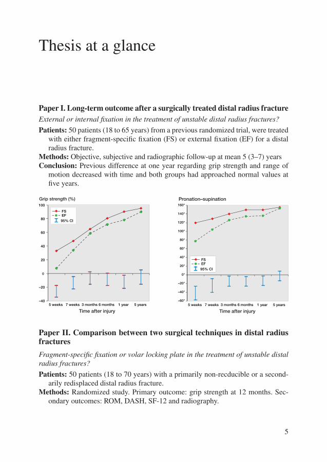
Paper I. Long-term outcome after a surgically treated distal radius fractureExternal or internal fixation in the treatment of unstable distal radius fractures?
Patients: 50 patients (18 to 65 years) from a previous randomized trial, were treated with either fragment-specific fixation (FS) or external fixation (EF) for a distal radius fracture.
Methods: Objective, subjective and radiographic follow-up at mean 5 (3–7) yearsConclusion: Previous difference at one year regarding grip strength and range of
motion decreased with time and both groups had approached normal values at five years.
Paper II. Comparison between two surgical techniques in distal radius fractures
Fragment-specific fixation or volar locking plate in the treatment of unstable distal radius fractures?
Patients: 50 patients (18 to 70 years) with a primarily non-recducible or a second-arily redisplaced distal radius fracture.
Methods: Randomized study. Primary outcome: grip strength at 12 months. Sec-ondary outcomes: ROM, DASH, SF-12 and radiography.
5 weeks 7 weeks 3 months 6 months 1 year 5 years
EF FS
95% CI
Pronation–supination
Time after injury
–60°
–40°
–20°
0°
20°
40°
60°
80°
100°
120°
140°
160°
–40
–20
0
20
40
60
80
100
5 weeks 7 weeks 3 months 6 months 1 year 5 years
Grip strength (%)
Time after injury
EF FS
95% CI
6
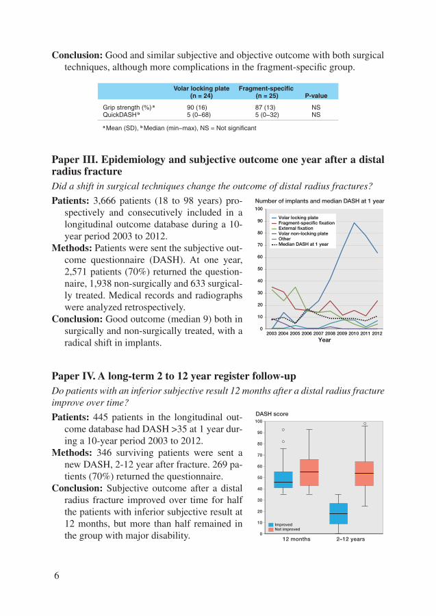
Conclusion: Good and similar subjective and objective outcome with both surgical techniques, although more complications in the fragment-specific group.
Paper III. Epidemiology and subjective outcome one year after a distal radius fractureDid a shift in surgical techniques change the outcome of distal radius fractures?
Patients: 3,666 patients (18 to 98 years) pro-spectively and consecutively included in a longitudinal outcome database during a 10-year period 2003 to 2012.
Methods: Patients were sent the subjective out-come questionnaire (DASH). At one year, 2,571 patients (70%) returned the question-naire, 1,938 non-surgically and 633 surgical-ly treated. Medical records and radiographs were analyzed retrospectively.
Conclusion: Good outcome (median 9) both in surgically and non-surgically treated, with a radical shift in implants.
Paper IV. A long-term 2 to 12 year register follow-upDo patients with an inferior subjective result 12 months after a distal radius fracture improve over time?
Patients: 445 patients in the longitudinal out-come database had DASH >35 at 1 year dur-ing a 10-year period 2003 to 2012.
Methods: 346 surviving patients were sent a new DASH, 2-12 year after fracture. 269 pa-tients (70%) returned the questionnaire.
Conclusion: Subjective outcome after a distal radius fracture improved over time for half the patients with inferior subjective result at 12 months, but more than half remained in the group with major disability.
Volar locking plate Fragment-specific (n = 24) (n = 25) P-value
Grip strength (%) a 90 (16) 87 (13) NSQuickDASH b 5 (0−68) 5 (0−32) NS
a Mean (SD), b Median (min−max), NS = Not significant
0
10
20
30
40
50
60
70
80
90
100
2003 2004 2005 2006 2007 2008 2009 2010 2011 2012Year
Volar locking plateFragment-specific fixationExternal fixationVolar non-locking plateOtherMedian DASH at 1 year
Number of implants and median DASH at 1 year
0
10
20
30
40
50
60
70
80
90
100
DASH score
12 months 2–12 years
Improved Not improved

7
Introduction
In 1814, when Abraham Colles wrote the article “On the Fracture of the Carpal Extremity of the Radius” (Colles 1814), he probably did not know that the best treatment of distal radius fractures would still be discussed today. Over the last 200 years, the treatment of distal radius has improved. For years, treatment of stable fractures included immobilization in cast or splints, with or without closed reduc-tion, and surgical treatment using pinning and external fixation was introduced first in the middle of the 20th century (Cooney et al. 1979; Diaz Garcia and Chung 2012). The definition of stability is not fully agreed upon (Walenkamp et al. 2015) and the challenge thus remains to identify which fractures are best treated surgical-ly and which are not (Chen and Jupiter 2007). In the last decades, open reduction and internal fixation has gradually become the preferred method for unstable distal radius fractures. The majority, 70% to 80% of adult patients, are still treated using closed reduction and splints (Chung et al. 2009; Mellstrand-Navarro et al. 2014).
An important factor in assessing successful outcome in any treatment is the rate of complications. Depending on the definition, the overall rate of complications in non-surgical and surgical treatment, spans from as low as 6% to as high as 80% (McKay et al. 2001). Treatment and research should aim at minimizing the rate of complications to achieve an optimal outcome. Early surgical methods have focused on optimal anatomical and radiographic outcome (Batra and Gupta 2002), to obtain a good objective and subjective patient outcome (Ritting and Wolf 2012), but the correlation is not evident (Leung et al. 2013).
The distal radius fracture is the most common fracture (Court-Brown and Cae-sar 2006). Today, the vast majority achieve a good subjective result (Abramo et al. 2008a), but as many as 21% still have remaining disability one year after the injury (MacDermid et al. 2003). Due to the high incidence of the fracture, an improved outcome in just a small percentage of patients would result in improved quality of life for a substantial number.
With this in mind, the intriguing challenge today is how to measure and interpret the outcome of current treatment and analyzing whether these lead to an acceptable outcome. The objective in the present thesis was to evaluate treatment over a longer time-period, identify risk factors for poor outcome, and compare the newer surgical techniques, in order to contribute to an improved outcome for this patient popula-tion.

8
Aims
The specific aims of the thesis were:
I. To investigate whether the superior short-term results in a previous study, comparing internal fixation to external fixation in unstable distal radius frac-tures persists over time.
II. To compare, in a randomized way, two methods of internal fixation, the frag-ment-specific fixation and the newer volar locking plate, in primarily non-re-ducible as well as in secondarily redisplaced distal radius fractures.
III. To evaluate the subjective outcome one year after a distal radius fracture, in both surgically and non-surgically treated patients, over a 10-year period us-ing the Disabilities of the Arm, Shoulder and Hand (DASH) score.
IV. To evaluate if the long-term subjective outcome would improve over time, in patients with major disability (DASH >35) one year after a distal radius fracture.

9
Background
History – treatment evolution or revolution?
Hippocrates (460–371 BC) described traumatic injuries of the wrist as being carpal dislocations. First in the 18th century, the French surgeon Jean-Louis Petit (1705) interpreted the carpal dislocation as an actual fracture. Another French surgeon Claude Pouteau (1783), was the first to describe the dorsally displaced distal radius fracture, still therefore bearing the eponym Pouteau-Colles fracture in some regions. Today, we are most familiar with the fracture bearing the name of Abraham Colles, the Irish surgeon and Professor of Anatomy in Dublin, who in 1814, wrote the clas-sic article, describing the fracture. Colles described, more than 80 years before the x-ray was invented by Wilhelm Conrad Röntgen, how the broken pieces should be brought back into an anatomic position and suggested the use of a splint, still today the basic treatment of a distal radius fractures (Fernandez and Jupiter 2002; Diaz Garcia and Chung 2012; Hove et al. 2014). Abraham Colles was succeeded in Dub-lin by another surgeon, Robert William Smith, accredited for the Smith’s fracture, the volarly displaced mirrored distal radius fracture of Colles (1847). A French sur-geon, Jean-Gaspard-Blaise Goyrand, described the volar displacement of the distal fragment already in 1832 (Fernandez and Jupiter 2002). Another French surgeon, Guillaume Dupuytren (1847), made important contributions to the description of the morphology of the different fracture patterns, based on postmortem examina-tions. The American surgeon John Rhea Barton (1838) described a dorsal or volar fracture-dislocation, due to an intra-articular fracture of the distal radiocarpal joint, which was given the eponym Barton’s fracture. After Röntgen’s discovery of x-rays in 1895, which quickly came to be used for fracture diagnosis, a greater understand-ing of the fracture was natural. Carl Beck (1898) presented the use of the x-ray technique in distal radius fractures (Fernandez and Jupiter 2002; Hove et al. 2014).
The distal radius fracture treatment has in many aspects remained unchanged for the majority of fractures. In the 1814 classic quote, Abraham Colles claims that “…the limb will at some remote period again enjoy perfect freedom in all its motions, and be completely exempt from pain…” (Colles 1814). For a majority of the patients with a distal radius fracture this may be true, but even after all these years and with hopefully improved treatment, still a substantial portion of patients have reduced motion and remaining discomfort or even pain as the end result.

10
Epidemiology – size of the problem
The distal radius fracture is the most common fracture and accounts in adults for 18% of the fractures in an orthopedic trauma unit (Court-Brown and Caesar 2006). There are three major subgroups to consider: 1) children/adolescents, 2) young adults, and 3) the elderly (Nellans et al. 2012). In children/adolescents, the most common fracture site is the distal forearm, which represents 24-26% of all fractures in this group (Tiderius et al. 1999; Hedström et al. 2010). In young adults, the bone quality is good. In this group, approximately 75% of the fractures are caused by high-energy trauma, more than 2/3 of the fractures are intra-articular, and more than 50% are initially displaced (Lindau et al. 1999). In the elderly the distal radius fracture is considered to be an early manifestation of osteoporosis, and the fracture a predictor of both later hip fracture (Mallmin et al. 1993) and increased mortality (Øyen et al. 2014). The incidence of the distal radius fracture represents a bimodal distribution with the peak incidence in children/adolescents (boys over-represented) and the elderly (women over-represented) (Court-Brown and Caesar 2006). The in-creasing incidence noted for adults above after the age of 50 years, is due to lower bone mineral density (BMD) (Van Staa et al. 2001). The independent predictors of a distal radius fracture for women over 65 years are reduced BMD, a previous frac-ture between age 50 and 65 years and a history of recurrent falls (Vogt et al. 2002). Interestingly, for a woman, regardless if younger or older than 50 years, the wrist is the most common fracture site. In contrast, the hand is the most common site for men under 50 years, and the hip in men older than 50 years (Rosengren et al. 2015).
The epidemiology of the distal radius fracture has been studied in the Scandina-vian countries with particular interest. Alffram and Bauer investigated the incidence of distal radius fractures in Malmö, Sweden in 1955, and found the annual incidence rate to be 19/10,000 person-years (Alffram and Bauer 1962). In several studies, an increasing incidence was reported in the last half of the 20th century, at least in an urban population in Malmö, and rising from 19 to 43 per 10,000 person-years be-tween 1950–1980 (Bengnér and Johnell 1985). More recent studies indicate that the increase of incidence rate has slowed down or even started to decrease after the turn of the century (Brogren et al. 2007; Flinkkilä et al. 2011; Sigurdardottir et al. 2011; De Putter et al. 2013; Wilcke et al. 2013; Dimai et al. 2014; Mellstrand-Navarro et al. 2014; Rosengren et al. 2015). The overall incidence was 26/10,000 person-years in a neighboring area in southern Sweden (Brogren et al. 2007), similar to the inci-dence in a large Swedish database study (Mellstrand-Navarro et al. 2014). Before the age of 50 years, the difference in incidence between genders is small. After the age of 50 years, women have a greater risk of sustaining a distal radius fracture with an incidence three to five times higher in women than in men (Melton III et al. 1998; Brogren et al. 2007; Lofthus et al. 2008; Flinkkilä et al. 2011; Sigurdardottir et al. 2011; Diamantopoulos et al. 2012; Wilcke et al. 2013).

11
Classification
Numerous classifications exist for distal radius fracture. Two of the most commonly used are the AO Classification with 9 main subgroups and the Frykman classifica-tion with 8 subgroups (Frykman 1967; Müller et al. 1990). The prognostic value of these classifications, to determine the outcome of a distal radius fracture is limit-ed (Flinkkila et al. 1998), especially regarding inter- and intra-observer agreement (Andersen et al. 1996; Kreder et al. 1996). The AO classification was used in Paper I–IV, but only using the three main groups (A, B and C), due to a better inter- and intra-observer agreement than when using all subgroups (Kreder et al. 1996).
Treatment
Non-surgical treatment
Non-surgical treatment is the preferred treatment for 3/4 of all adult distal radius fractures (Mellstrand-Navarro et al. 2014). Approximately 2/3 of all fractures will have an acceptable outcome with plaster treatment (Hove et al. 2014).
Stable non-displaced fractures
Primarily non-displaced distal radius fractures, defined as fractures with minimal displacement and minimal shortening are considered as stable and treated with cast immobilization or even elastic bandage (Abbaszadegan et al. 1989; Adolphson et al. 1993; Abramo et al. 2008a).
Unstable and displaced fractures
Instability has been based on the Lafontaine criteria and three or more risk factors need to be present: dorsal angulation >20° (perpendicular to a the long axis of the ra-dius); dorsal comminution; intra-articular radio-carpal fracture; an associated ulnar styloid fracture; and age ≥60 years (Lafontaine et al. 1989). In consensus protocols and clinical studies, instability is often defined as a fracture exceeding a defined ra-diographic displacement cut-off (Walenkamp et al. 2015), but obviously bone qual-ity, axial compression, metaphyseal comminution, concomitant ligament injuries and patient’s age are also important factors (Mackenney et al. 2006). According to a recent publication, volar comminution and displacement constitutes the greatest risk of instability (Wadsten et al. 2014) and the restoration of the volar cortex a strong predictor of both final volar tilt and carpal alignment at closed reduction (LaMartina et al. 2015).

12
Closed reduction with cast immobilization still remains the standard treatment for displaced distal radius fractures. The classic procedure is longitudinal traction, combining volar flexion, ulnar deviation and pronation of the wrist (Fernandez and Jupiter 2002). The longitudinal traction distracts the strong volar ligaments of the wrist (uniplanar ligamentotaxis) and when combined with a multiplanar technique, causing a volar translation of the distal fragments, the volar tilt will be restored (Agee 1994). After reduction, a forearm cast or above elbow cast is applied. Tra-ditionally, dorsal plaster slabs molded around the wrist are used in Scandinavian countries (Hove et al. 2014). No evidence exists, based on randomized trials, on which type of cast to use. A Cochrane review found no method of reduction or im-mobilization to be superior in the non-surgical treatment of a distal radius fracture (Handoll and Madhok 2003). The authors suggested the physician to use an accept-ed technique, cost-effective, and familiar to everyone at their health care unit. A cast provides comfort, but only relative stability and it appears to be the properties of the fracture that predict the stability (Mackenney et al. 2006). With cast treatment there is a significant risk of secondary displacement especially in volar comminution, and in severely comminuted and intra-articular fractures (Wadsten et al. 2014; Walen-kamp et al. 2015).
Closed reduction and pinning/external fixation
Surgical treatment is recommended for unstable and non-reducible distal radius frac-tures (Chen and Jupiter 2007; Downing and Karantana 2008). The method of choice for decades has been pinning and external fixation (Atroshi et al. 2006; Krukhaug et al. 2009). As in orthopedics in general, a technical evolution has taken place in the treatment for distal radius fractures in the last 30 years. New implants and tech-niques have been developed and marketed, especially evident in the last decade with a major shift towards more open reduction and internal fixation (Chung et al. 2009; Mattila et al. 2011; De Putter et al. 2013; Wilcke et al. 2013; Mellstrand-Navarro et al. 2014).
Percutaneous pinning
Many methods of percutaneous pinning have been described. In 1908, Lambotte suggested a concept of two pins inserted into the radial styloid for distal radius fractures (Rayhack 1993). In the 1950’s, DePalma described a technique inserting the pins from the distal ulna to the fractured radius transfixing the bones until heal-ing (DePalma 1952). In 1959 Willenegger and Guggenbuhl presented the first case report using only one pin for radial styloid fractures (Willenegger and Guggenbuhl 1959). In 1976, Kapandji advocated a technique with intrafocal pinning of unsta-ble extra-articular fractures in young patients (Kapandji 1976). In fractures with two articular fragments a two-pin technique is used with one pin inserted from the

13
radial styloid and one into the dorso-ulnar corner of the radius (Clancey 1984). A Cochrane review concluded that there is some evidence for the technique, but due to the high rates of complications, the precise role of the method is yet to be estab-lished (Handoll et al. 2007).
External fixation
The external fixator relies on indirect reduction of the fracture using ligamentotaxis to maintain the radial length (Cooney et al. 1979). Early versions of the external fixator were described in the beginning of the 20th century with Lambotte in 1908 as one of the pioneers. Anderson in 1932 and Hoffmann in 1938, both designed wrist-bridging frames for the distal radius (Hove et al. 2014). In the middle of the 1970s, the static external fixator was increasingly used for comminuted distal radius fractures (Jakob and Fernandez 1982). A new concept was introduced to reduce wrist stiffness. A wrist bridging fixator with a mobile hinge joint or using a non-bridging fixator with pins only inserted in the radius, both techniques allowing wrist motion (Clyburn 1987; Pennig 1993). Better range of motion, better grip strength and im-proved radiographic outcome were found in one study comparing non-bridging ver-sus static bridging external fixation (McQueen 1998), and no difference in clinical outcome was noted in two other studies (Krishnan et al. 2003; Atroshi et al. 2006). Further development included the dynamic bridging wrist fixator, which allowed early wrist motion and was compared to a static bridging external fixation. The ra-diographic and functional outcome was similar (Krukhaug et al. 2009). In another study, dynamic bridging fixation resulted in better radial length and wrist extension at one year, but with a higher rate of superficial pin-track infections in the dynamic (43%), compared to the static (11%) external fixation group (Hove et al. 2010). Oth-er studies comparing dynamic to static external fixation found similar rates (30%) of pin-track infections in both groups (Krishnan et al. 2003). The Cochrane institute found no robust evidence of an advantage of any external fixation method over the other (Handoll et al. 2008).
Open reduction and internal fixation
Although plaster cast, pinning, and external fixation were the standard treatments of distal radius fractures, open methods started to be discussed, based on findings of dissatisfactory results in 30% of the patients (Cooney et al. 1980). New treatment concepts were developed in the 1970−1990s, with open reduction and internal fix-ation and concepts like anatomical reduction, stable fixation and early mobilization were stressed (Augereau et al. 1983; Keating and McQueen 1994; Jupiter et al. 1996; Carter et al. 1998; Kambouroglou and Axelrod 1998; Fernandez and Jupiter 2002). The advantage of internal fixation over external fixation is maybe no longer controversial (Chen and Jupiter 2007) but it was first after the introduction of the

14
volar locking plate in 2002 (Orbay and Fernandez 2002), open reduction became the preferred surgical treatment option for the general orthopedic surgeon. Still today, the advantage of internal fixation is discussed. The Cochrane institute withdrew a previously published review regarding surgical intervention of distal radius fractures (Handoll and Madhok 2009) but is currently recruiting papers for a new review of Internal fixation versus other surgical methods for treating distal radius fractures, to be published in the future (Jariwala et al. 2014)
Non-locking volar plates
Fractures with a volar displacement, i.e. Smith and volar Barton fractures, have been treated with volar standard AO T-plates for years with acceptable functional results (Keating and McQueen 1994; Jupiter et al. 1996). Dorsal plates have been used with acceptable results (Carter et al. 1998; Kambouroglou and Axelrod 1998; Grewal et al. 2005), but the complication rates were high (47−72%) especially us-ing the Pi plate (Kambouroglou and Axelrod 1998; Rozental et al. 2003; Grewal et al. 2005) due to conflicts with the extensor tendon. In a meta-analysis of 46 studies (randomized controlled trials, prospective studies, and case series) between 1980 and 2004 (Margaliot et al. 2005), no evidence was found to support open reduction and internal fixation, with the older techniques, over closed reduction and external fixation.
Fragment-specific fixation
A new concept of internal fixation was introduced using the three-column theory, with two plating concepts launched almost at the same time. The three-column the-ory is based on the understanding of the distal radius and ulna as a biomechanical construct consisting of three units; the lateral, the intermediate, and the medial col-umns (Rikli and Regazzoni 1996). The lateral (radial) column is the lateral part of the radius including the styloid and the scaphoid fossa. The intermediate (middle) column is the medial part of the radius with the lunate fossa and the sigmoid notch. The medial (ulnar) column is the distal ulna, the triangular fibrocartilage, and the distal radio-ulnar joint. The fragment-specific fixation system, also based on the three column theory was developed by Medoff and introduced at our department in 1996. It uses low profile plates and wire forms to stabilize the individual col-umns (Medoff and Kopylov 1998; Konrath and Bahler 2002). The technique uses a minimally invasive anatomic approach to individually stabilize the radial styloid, the dorsal ulnar corner, the dorsal wall and volar rim. (Konrath and Bahler 2002; Benson et al. 2006). Biomechanical studies showed good mechanical stability by the small implants (Taylor et al. 2006). The fragment-specific fixation system was compared at our department to the external fixator in a randomized study, showing better range of motion and grip strength, not only at earlier time points, but as late as after one year (Abramo et al. 2009). The rationale for Paper I was to determine if the fragment-specific system, as an example of a method of open reduction and

15
internal fixation, was superior to the external fixation technique, in a longer time frame than one year.
Volar locking plates
The fragment specific fixation is technically demanding and it was first with the introduction of the volar locking plate, that surgery of the distal radius became pop-ular to the greater orthopedic community. Technically, the new idea was to treat also dorsally dislocated fractures by a volar approach (Orbay and Fernandez 2002). This was made possible by having the distal screws locked to the plate, lifting and securing the subchondral bone, thereby retaining the joint surface in an anatomical position during healing. With the technique, the locking screws are kept unicortical with no osteosynthesis material protruding dorsally, thereby avoiding the risk of interfering with the extensor tendons. Most volarly and dorsally dislocated fractures could be addressed, and the pronator quadratus muscle could be used as a protective layer between the plate and the tendons. The volar locking plates have since its in-troduction in 2002 become popular, and is now the most frequently used method of surgical treatment for distal radius fractures (Koval et al. 2008; Chung et al. 2009; Mattila et al. 2011; Wilcke et al. 2013; Mellstrand-Navarro et al. 2014). The method has proven to be biomechanically stable in studies using cadaver specimens and synthetic bone models (Koh et al. 2006; Willis et al. 2006). In clinical studies, good results have been found in patients of all ages (Orbay and Fernandez 2004; Chung et al. 2008; Jupiter and Marent-Huber 2009), but the method was spread in the ortho-pedic community without randomized comparisons to the old techniques. Several randomized studies have been published in the last decade comparing volar locking plates to external fixation, with and without adjuvant pins (Egol et al. 2008; Abramo et al. 2009; Wei et al. 2009; Wilcke et al. 2011; Jeudy et al. 2012; Karantana et al. 2013; Williksen et al. 2013; Roh et al. 2015; Navarro et al. 2016). In general, the volar locking plates yield quicker early recovery and a better anatomical reduction, but the conclusions regarding the functional and subjective outcome are not con-clusive (Table 1). In a randomized study comparing volar locking plates or dorsal plates to external fixation, similar outcomes were found (Xu et al. 2009). Further, in a randomized study of older patients, comparing non-bridging external fixation to volar locking plates, the outcome were found equal (Gradl et al. 2013), but a high number of extensor tendon injuries was found due to dorsal screw penetrations, a complication that should not occur in a series with experienced surgeons. In patients 65 years and older, similar outcomes were found in volar locking plate when com-pared with closed reduction and cast (Arora et al. 2011; Bartl et al. 2014). Recently, a large randomized multicenter trial found percutaneous pinning to be superior to volar plates. The 461 fractures were operated by 244 surgeons (Costa et al. 2014), implying that pinning might be better in low volume centers due to potential com-plications in the open techniques. In the two meta-analyses of the existing RCTs comparing volar locking plates with percutaneous pinning, a lower DASH score

16
Table 1. Literature review of recent randomized controlled trials of closed reduction and external fixation +/- pinning versus open reduction and internal fixation with newer techniques
Study Subjects Mean age AO Treatment FU Outcome Outcome p-value (year) (n) (range) (variables) (findings)
Egol 77 51 (18−78) A,B,C VLP vs. EF+P 12 Primary outcome not defined −(2008) Grip strengthb Similar NS Pronationb 95% vs. 100% 0.04 Radiographic Similar NS Less physiotherapy VLP in favor 0.02 Complications 21% vs. 18% NSAbramo 50 48 (20−65) A,C FS vs. EF±P 12 Grip strengtha 90% vs. 78% 0.03(2009) Forearm rotation 136° vs. 149° 0.03 DASH Similar NS Radiographic Similar NS Complications (major) 8 vs. 2 0.04Wei 46 59 (18−78) A,C VLP vs. RCP 12 DASHa VLP in favor 0.03(2009) vs. EF+P Lateral pinch strength VLP in favor 0.04 ROM Similar NS Radiographic (inclination) RCP in favor <0.05 Radiographic (length) RCP in favor <0.05 Complications (overall) 17% combined −Xu 30 44 (21−56) C VLP/DP 24 GOa Similar NS(2009) vs. EF±P GW Similar NS Grip strength Similar NS Pronation-supination Similar NS Radiographic Similar NS Complications (overall) Similar NSWilcke 63 56 (20−69) A,C1 VLP vs. EF±P 12 DASHa Similar NS(2011) Grip strength Similar NS Extension VLP in favor 0.04 Supination VLP in favor 0.02 Radiographic (height) VLP in favor <0.001 Overall complications 21% vs. 36% −Jeudy 75 65 (40−80) C VLP vs. EF+P 6 GOa VLP in favor <0.05(2012) Grip strengthb 84% vs. 76% 0.02 Flexion-extension Similar NS Radiographic Similar NS Complications Similar NS PRWE Similar NSKarantana 130 49 (18−73) A3, VLP vs. EF+P 12 PEMa Similar NS(2013) C2,C3 DASH Similar NS Grip strengthb 95% vs. 84% <0.001 ROM Similar NS Radiographic (volar tilt) VLP in favor <0.001 Radiographic (height) VLP in favor <0.004 Complications 24% vs. 42% NSWilliksen 102 54 (20−84) A,C VLP vs. EF 12 Mayo wrist scorea VLP in favor 0.02(2013) Supination VLP in favor 0.03 Grip strength Similar NS Ulnar sided pain VLP in favor 0.02 Radiographic (variance) VLP in favor 0.02 Operative time 88 vs. 77 min 0.02 Complications (overall) 29% vs. 30% NSGradl 102 63 (18−88) A3, C VLP vs. Nb-EF 12 Not defined a − −(2013) ROM Similar NS Grip strength Similar NS GW Similar NS Radiographic Similar NS Operative time 59 vs. 43 min <0.05 Complications 21% vs. 20% NSRoh 74 55 (18−70) C2,C3 VLP vs. EF+P 12 MHQa Similar NS(2015) ROM Similar NS Grip strength Similar NS Radiographic (variance) VLP in favor 0.02 Operative time 59 vs. 49 min <0.001 Complications 17% vs. 29% NS

17
and reduced total postoperative complications were found with volar locking plates (Chaudhry et al. 2015; Zong et al. 2015). Seven meta-analyses state some evidence of an improved subjective outcome, better anatomic restoration, and better range of motion with volar locking plates compared with external fixation (Cui et al. 2011; Wei et al. 2012; Esposito et al. 2013; Walenkamp et al. 2013; Wang et al. 2013; Xie et al. 2013; Li-hai et al. 2015).
Comparisons of the volar locking plate with the fragment specific technique are few. A prospective cohort study compared the volar locking plate with the frag-ment-specific fixation finding the volar locking plate to be a more stable fixation, with better objective and subjective outcomes in the early postoperative period, and with fewer complications (Sammer et al. 2008). To date, no randomized controlled trials comparing the fragment-specific fixation to the volar locking plate have been published, which was the rationale for Paper II.
Rehabilitation
In the immediate phase after a distal radius fracture, physiotherapy is started to reduce finger edema. Shoulder and elbow motion are endorsed to reduce stiffness (Oskarsson et al. 1997). After cast removal, early active wrist motion is initiated, with increasing load to the wrist. Rehabilitation after wrist fractures by means of a home exercise program can be effective in improvement of grip strength and range of motion (Krischak et al. 2009). Although, rehabilitation is a cornerstone of elimi-nating and preventing dysfunction, few randomized controlled trials exists (Hove et al. 2014; Handoll and Elliott 2015).
Legends to Table 1:a Primary outcome. b percent of uninjured side.FU = Follow-up (months)DASH = Disabilities of the Arm, Shoulder, and Hand score. GO = Green and O’Brian score, GW = Gartland and Werley score, PEM = Patient Evaluation Measure, PRWE = Patient-Rated Wrist Evaluation. DP = Dorsal plate, EF = External fixation, FS = Fragment-Specific fixation, Nb = Non-bridging, ±P = with or without pinning, +P = all patients received pins, RCP = Radial Column plate, VLP = Volar locking plate, VP = Volar non-locking plate. NS = Not significant, − = No statistic calculation.
Navarro 140 63 (50−74) A2, VLP vs. EF±P 12 DASHa Similar NS(2016) A3,C Radial deviation VLP in favor 0.02 Grip strength Similar NS Radiographic (variance) VLP in favor <0.001 Operative time 70 vs. 42 min <0.001 Complications (overall) 51% vs. 45% NS

18
How to evaluate outcome
Wrist fractures traditionally were evaluated using objective measurements, i.e. the view of the medical professionals of how the patient was influenced by an inju-ry. Traditionally, radiographic appearances, range of motion, and/or grip strength (MacDermid 1996) have been evaluated. At the end of the 20th century a more patient-centered view became common and several patient-reported outcome in-struments were developed for the upper extremity (Hudak et al. 1996; MacDermid 1996; Chung et al. 1998) to capture the experience of the patient’s own view on how the injury or disease influenced their daily life. An overview of the various outcome assessments is found in the following section.
Radiographic assessment
Posterior-anterior (PA) and lateral views are used for measuring radial height, radial inclination, ulnar variance, intra-articular joint step-off and dorsal angulation (Mann et al. 1992). Ulnar variance was measured according to the method of perpendic-ulars (Parker et al. 2014). Osteoarthritis of the wrist can be classified according to Kellgren and Lawrence (Kellgren and Lawrence 1957). It is generally agreed that the two surfaces of the distal radius, the scaphoid fossa and the lunate fossa, should be restored to avoid future posttraumatic arthritis (Knirk and Jupiter 1986; Karnezis et al. 2005). The correlation between objective and subjective outcomes and the degree of radiographic deformity is still controversial (McQueen and Caspers 1988; Batra and Gupta 2002; Fujii et al. 2002; Anzarut et al. 2004; Wilcke et al. 2007; Plant et al. 2017) as well as the results in the few exiting long term outcome-studies (McQueen and Caspers 1988; Kopylov et al. 1993; Földhazy et al. 2007; Brogren et al. 2011a; Finsen et al. 2012). Radiographic assessment can be helpful in predicting the stability, as described in the paragraph regarding unstable distal radius fractures (Lafontaine et al. 1989; Mackenney et al. 2006; Wadsten et al. 2014).
Grip strength
Grip strength captures pain besides strength and can be seen as an objective surro-gate variable of hand function. A common tool to measure grip strength is the Jamar hand dynamometer (Sammons Preston. Inc., Bolingbrook, IL, USA). Since grip strength varies between individuals, the patient often serves as his or her own con-trol and the strength is often given as a ratio of the uninjured hand. Grip strength was the primary outcome in Paper I and Paper II. Grip strength is a reliable outcome measurement, as neither time nor age appear to affect the test-retest reliability (Bo-hannon and Schaubert 2005). The minimal clinically important difference (MCID)

19
is considered the smallest change or difference in an outcome measure that is per-ceived to be clinically important (Wells et al. 2001). In a study attempting to estab-lish the MICD for grip strength, an absolute value of 6.5 kg or a grip ratio 19.5% of the uninjured side one year after a DRF has been suggested (Kim et al. 2014). Grip strength is sensitive in detecting a small clinical difference, and with good responsiveness (the ability to detect a change when a true change has occurred), in the evaluation of the objective outcome after distal radius fractures (MacDermid et al. 2000). Grip strength was one of the most significant factors related to patient satisfaction (Fujii et al. 2002).
Range of motion
The clinical examination includes evaluation of the three axes of rotation in the wrist. The range of motion (ROM) in the radio-carpal joint is extension-flexion and radial-ulnar deviation, while pronation-supination occurs in the distal and proximal radio-ulnar joints with the radius rotating around the head of ulna. A goniometer is used to measure the angle in degrees and estimates for good patient satisfaction after a distal radius fracture is 95% of the contralateral side (Ritting and Wolf 2012). ROM was used in Paper I and Paper II as a secondary outcome parameter.
Physician-based assessment instruments
Historically, different physician-based assessment instruments have been used. The Gartland and Werley point system using objective measures such as range of motion (Gartland and Werley 1951), further developments have included grip strength, but despite of the common use in hand and wrist surgery the score system no validation studies exits (Changulani et al. 2008). A clinical grading system, the Green and O’Brian, based on range of motion, grip strength and patient satisfaction is also at times used (Green and O’Brien 1978), further development is the Modified Mayo Wrist Score (Cooney et al. 1987).
Patient-reported outcome measures
A patient-reported outcome measure (PROM) is a tool used to measure the patient’s own view of function, pain and quality of life without the involvement of a research-er or clinician. Different types of PROMs have been available since the first were developed in the 1970s, either disease-specific or generic evaluating the general health (Weldring and Smith 2013). The most commonly used outcome instruments will be described below with an emphasis on the PROM used in this thesis.

20
Disabilities of Arm Shoulder and Hand (DASH)
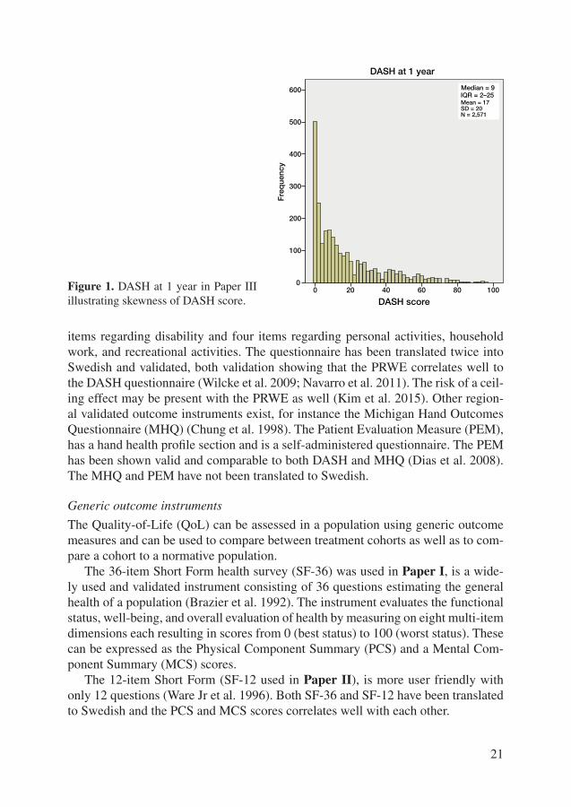
The DASH questionnaire, used in Papers I−IV, was initiated and developed in 1994 by the Institute for Work and Health in Ontario, Canada together with the American Academy of Orthopedic Surgeons (Hudak et al. 1996). It is a regional outcome measure for disorders in the upper limb evaluating the function of the arm, shoulder, and hand as one combined entity. Six domains are evaluated; daily activities, social function, work function, symptoms, sleep, and confidence. The DASH consists of 30 items (Appendix I). Each item has 5 response options available to calculate a total score ranging from 0 (no disability) to 100 (most severe disability). A DASH score cannot be calculated if more than 10 percent of the items are left blank by the patient. The DASH has been translated and validated (shown to measure the con-ditions according to its intended purpose) in Swedish (Atroshi et al. 2000). A short version, the QuickDASH (Appendix II), containing 11 of the 30 items was intro-duced (Beaton et al. 2005) and has also been translated into and validated in Swed-ish. A good correlation between the QuickDASH and the original 30 item DASH questionnaire was found (Gummesson et al. 2006). The DASH questionnaire was validated in patients with different chronic conditions in the upper extremity, before and after surgery (Gummesson et al. 2003; Sorensen et al. 2013). The initiators of the DASH instrument described two intentions for its use: 1) A descriptive meas-ure – to compare the impact of upper-limb disorders among individuals or groups. 2) An evaluative measure – to assess change over time related to natural history or the effect of treatment interventions (Kennedy 2011). For longitudinal changes, i.e. changes over time in patient health and function, an MCID was found to be between 8 and 19 points for DASH or QuickDASH in a variety of upper extremity conditions (Schmitt and Di Fabio 2004; Mintken et al. 2009; Polson et al. 2010; Sorensen et al. 2013; Franchignoni et al. 2014). In evaluating the MCID, the values before and after treatment are compared, with hopefully large clinical differences achieved due to the intervention. With the lack of standardized methods how to calculate the MCID, problems of interpretation arise (Slutsky 2013). DASH was not intended and is too blunt for evaluating small differences between two types of treatment. Yet another aspect to consider, comparing two treatments, is the risk of a ceiling effect of the chosen outcome instrument, when >15−20% of the cohort reaches the best outcome (Kim et al. 2015). The DASH has a risk of a ceiling effect and therefore possibly a lack of sensitivity. Since DASH is skewed and approximately 20% of patients with a distal radius fracture (Figure 1, Paper III) reach a score of 0 at 12 months, a difference between two groups may be interpreted falsely as absent when actually present, i.e. a type II error.
Other regional patient-reported outcome measures
The PRWE is a validated subjective outcome questionnaire, and was, in contrast to the DASH, designed to measure pain and disability specifically in the wrist (Mac-Dermid et al. 1998). This questionnaire consists of five items regarding pain; six
21
items regarding disability and four items regarding personal activities, household work, and recreational activities. The questionnaire has been translated twice into Swedish and validated, both validation showing that the PRWE correlates well to the DASH questionnaire (Wilcke et al. 2009; Navarro et al. 2011). The risk of a ceil-ing effect may be present with the PRWE as well (Kim et al. 2015). Other region-al validated outcome instruments exist, for instance the Michigan Hand Outcomes Questionnaire (MHQ) (Chung et al. 1998). The Patient Evaluation Measure (PEM), has a hand health profile section and is a self-administered questionnaire. The PEM has been shown valid and comparable to both DASH and MHQ (Dias et al. 2008). The MHQ and PEM have not been translated to Swedish.
Generic outcome instruments
The Quality-of-Life (QoL) can be assessed in a population using generic outcome measures and can be used to compare between treatment cohorts as well as to com-pare a cohort to a normative population.
The 36-item Short Form health survey (SF-36) was used in Paper I, is a wide-ly used and validated instrument consisting of 36 questions estimating the general health of a population (Brazier et al. 1992). The instrument evaluates the functional status, well-being, and overall evaluation of health by measuring on eight multi-item dimensions each resulting in scores from 0 (best status) to 100 (worst status). These can be expressed as the Physical Component Summary (PCS) and a Mental Com-ponent Summary (MCS) scores.
The 12-item Short Form (SF-12 used in Paper II), is more user friendly with only 12 questions (Ware Jr et al. 1996). Both SF-36 and SF-12 have been translated to Swedish and the PCS and MCS scores correlates well with each other.
DASH score100806040200
Freq
uenc
y
600
500
400
300
200
100
0
DASH at 1 year
Mean = 17SD = 20N = 2,571
Median = 9IQR = 2–25
Figure 1. DASH at 1 year in Paper III illustrating skewness of DASH score.

22
The EuroQol five-dimensional (EQ-5D) questionnaire is also a generic measure of health status and quality of life. The instrument is widely used in many health care interventions, but has been used in few studies of distal radius fractures (Bartl et al. 2014; Navarro et al. 2016).
Comparison DASH and Short Form instruments
The regional specific outcome measure DASH and the generic measure SF-36 has been found to correlate well regarding pain, but the correlations between DASH and physical and mental function is only moderate (Aktekin et al. 2011).
Complications
Complications are relatively frequent in the treatment of distal radius fractures and depend on type of treatment, whether non-surgical or surgical, as well as on how a complication is defined. The reported rate for complications vary widely from as low as 6% to as high as 80% (McKay et al. 2001).
Major complications cause significant morbidity. Complex Regional Pain Syn-drome (CRPS) is a condition with autonomic dysfunction, pain and impaired func-tion. CRPS can follow both non-surgical and surgical treatment with an incidence ranging from 1% to 37% (Mathews and Chung 2015). Treatment is complex, but in general early awareness of the risk, pain management, and anti-edema exercises are paramount as prevention and intensified physiotherapy the treatment once manifest. (Hove et al. 2014). Unplanned surgery can be regarded as a major complication. Carpal Tunnel Syndrome (CTS) can be either transient or manifest, the latter can be considered as a major complication and necessary to treat surgically. Tendon injuries, most often the long thumb extensor tendon (EPL) rupture can occur in non-surgically treated fractures but the incidence is low (0.3%) (Hove 1994). In sur-gically treated fractures tendon irritations are more frequent and synovitis could be considered as a minor complication while tendon rupture requiring tendon transfer as a major complication (Mathews and Chung 2015). With no uniform definition of complications, comparison between different reports on treatment techniques is difficult (McKay et al. 2001).
In closed reduction and external fixation of distal radius fractures, the overall complications rate ranges from 27 to 67% (Ahlborg and Josefsson 1999; Anderson et al. 2004). Superficial wound infection is the most frequent complication and var-ies from 11 to 43% (Ahlborg and Josefsson 1999; McKay et al. 2001; Krishnan et al. 2003; Margaliot et al. 2005; Hove et al. 2010) and in one study at a rate of 30% despite both perioperative and oral prophylactic antibiotics during the first postop-erative week (Krishnan et al. 2003). Other complications include pin loosening in 3 to 11% (Ahlborg and Josefsson 1999; Margaliot et al. 2005).

23
In open reduction and internal fixation, the overall complication rates span from 10% to 27% using volar locking plates (Rozental and Blazar 2006; Arora et al. 2007; Soong et al. 2011; Johnson et al. 2014). Tendon synovitis is a known consequence as a result of malpositioned plates protruding volarly or screws crossing the dorsal cortex. In a systematic review of 56 studies (6278 patients) the incidence of tendon synovitis was 5% after volar locking plate and 8% after dorsal plating (Johnson et al. 2014). Tendon rupture was rare with an incidence of 2% in volar plates and 2% in dorsal plates (Johnson et al. 2014), similar to earlier volar locking plate publications (Arora et al. 2007; Soong et al. 2011). The incidence of CRPS has been reported to be 4%, CTS 3%, screw loosening 2%, and intra-articular screw displacement to be 1% (Arora et al. 2007).
Malunion
Malunion is a general concept of a fracture that has healed in an anatomically sub-optimal position, which may lead to chronic pain and functional disability. The defi-nition of malunion of a distal radius fracture is not evident. Several definitions have been proposed, but none is universal for understanding all effects for the outcome. Fourrier et al. suggested correlation to the increased risk to develop disability, sug-gesting worse outcome if a fracture healed in more than 10–20° dorsal angulation, >1−2 mm radial shortening, and 20−30° radial inclination (Fourrier et al. 1981). MacKenny et al. defined malunion as dorsal angulation ≥0° and an ulnar variance of 1−2 mm (Mackenney et al. 2006). Brogren et al. defined malunion as dorsal angu-lation >10° or ulnar variance ≥1 mm (Brogren et al. 2011b). In this study, patients scored a significantly higher DASH score (mean 11) at one year after the fracture in patients with either dorsal angulation >10° or ulnar variance >1 mm, and a mean DASH score of 17 if both dorsal angulation >10° and ulnar variance >1 mm was present.
Due to the lack of a global definition, the prevalence of malunion after distal ra-dius fractures is thus not clear. Using the most strict definition of a dorsal angulation ≥0° and a ulnar variance of 1-2 mm, Mackenny et al. predicted malunion to occur in 27% of non-displaced fractures treated with cast, and 60% of displaced fractures treated with reduction and cast (Mackenney et al. 2006). Using the definition of malunion defined as a dorsal angulation >10° or ulnar variance ≥1 mm. Brogren et al. found 72% of their patients treated with closed reduction and cast, with closed reduction and external fixation, or percutaneous pinning to be malunited (Brogren et al. 2011b).
Malunion can turn out as either a minor or a major complication. When the mal-union renders pain, weakness or dysfunction for the patient and is a symptomatic malunion, it is considered as a major complication. Non-surgically treated fractures

24
tend to have a greater risk to malunite. In a randomized trial of closed reduction and cast versus open reduction and volar locking plate, a greater rate of symptomatic malunion was found in the closed reduction group at final (24 months) follow-up, 37% (DASH mean 14) and 0% (DASH mean 9) respectively (Sharma et al. 2014). Historically, the incidence of malunion was reported to be 24% after cast immobili-zation and 10% after surgical treatment (Haase and Chung 2012). In meta-analyses, malunion is reported at a rate of 2−4% after internal or external fixation (Margaliot et al. 2005; Zhang et al. 2016).
The most common symptoms of a malunion are related to an incongruency of the distal radio-ulnar joint (DRU-joint) and consist of pain at forearm rotation. In symptomatic patients with a malunion, a corrective osteotomy can be discussed with the patient (Haase and Chung 2012). Most often a radial osteotomy is performed, or an ulna osteotomy if the malunion is predominantly due to an axial shortening of the radius. In radius osteotomies, the fracture is cut and aligned at the previous fracture site and stabilized with screws and a plate. In a study of patients with symptomatic malunion (Abramo et al. 2008b), surgery resulted in a significant improvement in radiographic, functional, and subjective outome. The median DASH score improved from median 39 preoperatively to median 21 after one year.

25
Patients and methods
All studies were conducted at the Department of Orthopedics, in Lund, Sweden. The primary hospital catchment area has 256,000 inhabitants (Statistics Sweden) and approximately 450 adult patients (hospital reimbursement data) are treated for a distal radius fracture annually. Figure 2 shows the timeline and an overview of the patients in the 4 studies.
Paper I50 patients
Paper II50 patients
Paper IV445 patients
Paper III3,666 patients
2002 2003 2004 2005 2006 2007 2008 2009 2010 2011 2012 2013 2014
Figure 2. Timeline of patients and study years, Paper I−IV
Fracture type
Primary treatment
Final treatment
Follow-up
Minimally orundisplaced
High energy trauma/highly comminuted
Displaced a Volar Barton
Still displacedReduced
DisplacedStill in place
Closedreduction
Alwaysinternalfixation
Short armsplint
Clinical and radiographiccontrol after 7–10 days
OperationInternal or external
fixation
Conservative treatmentShort arm splint foranother four weeks
Patients with a distal radius fracture were treated according to a joint treatment protocol for southern Sweden (Figure 3, Abramo et al. 2008a). Non-dis-placed fractures were treated in a forearm cast for four to five weeks, and displaced fractures were reduced and casted. If the fracture was impossible to re-duce primarily or if a secondary loss of reduction occurred at the 7–10-day follow-up, surgical treatment was recommended.
Figure 3. Treatment protocol for distal radius fractures at the hospitals in the southern Swedish region (Södra sjukvårdsregionen). a Displaced = dorsal angulation >10° and/or ulnar variance >2 mm and/or articulation step >1 mm or volar angulation >25°.

26
Paper I and II – clinical studies
Study design and patient populations
Paper I was a long-term follow-up of a randomized study of 50 patients, included between May 2002 and December 2005, and treated with either fragment-specific or external fixation for a distal radius fracture. In that publication, the results 1 year after the fracture had been presented (Abramo et al. 2009) and in Paper I a mean 5 (3−7) years follow-up was performed. Paper I was approved by the local ethics committee (Lu 45/02). Inclusion and exclusion criteria are summarized in Table 2.
Paper II was a single-center, parallel group, prospective, randomized trial of 50 patients, included and surgically treated between December 2010 and December 2012 (Figure 4). The study was performed in accordance with the CONSORT crite-ria (Moher et al. 2010), registered in the ClinicalTrials.gov (ID No. NCT01311531), and monitored externally by a hospital-based independent organization (Clinical Study Support, R&D Centre, Lund, Skåne, Sweden). Paper II was approved by the local ethics committee (ETIK 2009/318). Inclusion and exclusion criteria are summarized in Table 3.
Table 2. Inclusion and exclusion criteria for Paper I
Inclusion criteria Exclusion criteria
Age 18−65 Previous ipsilateral wrist fractureInjury <10 days Open fractureFrykman I−VIII Volarly displacedPrimary unstable or non-reducible DRFs Fracture on the contralateral sideOne or more of the below: Concomitant fractures in need of treatment >20° dorsal angulation of the distal radius articular surface Ongoing radiotherapy or chemotherapy >2 mm shortening of ulnar variance Metabolic disease affecting the bone Incongruence of the radio-carpal joint Medication affecting the bone Incongruence of the distal radio-ulnar joint Dementia, psychiatric disorder, or alcohol abuseInformed consent
Table 3. Inclusion and exclusion criteria for Paper II
Inclusion criteria Exclusion criteria
Age 18−70 Previous ipsilateral wrist fractureInjury <14 days Open fractureAO A or C AO BPrimary non-reducible or a secondarily redisplaced DRF Fracture extending to the diaphysisOne or more of the below: Fracture on the contralateral side >20° dorsal angulation of the distal radius articular surface Concomitant fractures in need of treatment >2 mm shortening of ulnar variance Ongoing radiotherapy or chemotherapy > 1 mm incongruence of the radio-carpal Metabolic disease affecting the bone > 1 mm incongruence the distal radio-ulnar joint Medication affecting the boneInformed consent Dementia, psychiatric disorder, or alcohol abuse Difficulty understanding Swedish

27
Recruitment and intervention
In Paper I, in January 2009 participants in the previous randomized trial were in-vited to participate in a long-term follow-up study. Patients had been treated with either open reduction and internal fixation with fragment-specific wrist fixation (TriMed, Santa Clarita, CA, USA) or by closed reduction and bridging external fix-ation (Hoffmann, Stryker, Hopkinton, MA, USA/ Orthofix Srl, Bussolengo, Italy).
In Paper II, random numbers were generated (Research Randomizer, www.ran-domizer.org), blinded to examiners and placed in sealed envelopes by a secretary before commencing the study. Simple randomization was used with 25 patients in each group. The envelopes were opened in the operation theatre, immediately prior to the surgery. Patients were assigned to either fragment-specific wrist fixation or to
Assessed for eligibilityAll operated distal radius fractures
n = 342
Eligiblen = 113
Randomizedn = 50
Allocated to Fragment-Specific Fixation (n = 25)• Received allocated intervention (n = 24)• Did not receive allocated intervention (n = 1) (surgeon choice)
Allocated to Volar Locking Plate (n = 25)• Received allocated intervention (n = 24)• Did not receive allocated intervention (n = 1) (surgeon choice)
Lost to follow-up (n = 1) Lost to follow-up (n = 0)
Analyzed (n = 25)• Excluded from analysis (n = 0)
Analyzed (n = 24)• Excluded from analysis (n = 0)
ANALYSIS
FOLLOW-UP
ALLOCATION
ENROLLMENT
Not eligible (n = 229):– not meeting inclusion criteria • AO B fractures, 35 • age > 70 years, 94 • not meeting radiographic criteria, 48– meeting exclusion criteria, 52
Not included (n = 63):– declined to participate, 3– operated by non-study surgeon, 39– informed consent not possible, 21
Figure 4. Flowchart Paper II.

28
the volar locking plate group (TriMed, Santa Clarita, CA, USA). Surgical implants and instruments for both procedures were available in the common instrument tray. Three experienced hand surgeons performed all surgical procedures. Surgery was performed, in all patients using a tourniquet under brachial plexus block or general anesthesia. With the fragment-specific wrist fixation system (Konrath and Bahler 2002), an incision was made through the first extensor compartment. Reduction of the fracture was achieved with two pins and a pin-plate was applied onto the styloid pin and secured by screws. The fourth extensor compartment was opened and a buttress pin and/or ulnar pin-plate was applied for dorsal stability. If necessary, a volar buttress pin was introduced through a modified Henry’s incision. In the volar locking plate group, the modified Henry’s approach was used (Orbay and Fernandez 2002). A below-the-elbow plaster splint was applied for pain reduction. Patients postoperatively followed the standard rehabilitation for distal radius fractures. Fin-ger motion was started immediately to reduce edema. At 2 weeks the sutures and plaster was removed and wrist motion initiated.
Outcome measurement
Primary outcome in Paper I and Paper II was grip strength, which formed the basis for the calculation of the sample size.
Grip strength was examined in a standardized fashion, with the elbow flexed at 90 degrees, holding the wrist in neutral rotation and the wrist held between 0 and 30 degrees dorsal extension, as recommended by the American Society of Hand Therapists (ASHT) (Fess 1992). Three consecutive attempts were performed and the mean value calculated, expressed as an absolute value as well as a percentage of the contralateral uninjured side (Ashford et al. 1996). For correction of hand dominance, a 10% correction in grip strength in Paper II was made in right-handed subjects, with no correction performed in left-handed subjects (Petersen et al. 1989). Patients in Paper I were assessed once in the outpatient clinic 3 to 7 years after pri-mary surgery. Prior to the evaluation, an experienced physiotherapist had instructed the two orthopedic residents examining the patients in a standardized method to use the JAMAR, as well as the goniometer for range of motion (ROM). In Paper II, all patients were assessed clinically regarding grip strength and range of motion at 2 and 6 weeks, and at 3 and 12 months by one of two physiotherapists.
Pain at palpation, the presence of an obvious clinical wrist deformity in the frontal and/or lateral plane, and minor and major complications were recorded according to the predefined list in the protocol (Appendix III). In Paper I, patients were evaluated with the QuickDASH and SF-36 questionnaires. Patients not available for the clinical evaluation received a second letter and were asked to fill out the two questionnaires. Re-operations were identified using the medical records and radiographs. In Paper II, QuickDASH and the SF-12 were assessed at 6 weeks, 3 and 12 months.

29
Radiographic assessment
Radiographs in PA and lateral projections were obtained in the neutral position (Jed-linski et al. 1995). Measurements of radial inclination, ulnar variance and dorsal an-gulation (Mann et al. 1992) carried out using digital tools on the picture archiving and communication system (PACS) workstation, by one experienced radiologist. The radial inclination angle was measured on PA radiographs. Dorsal angulation was measured in the lateral projection and expressed as the angle of joint surface of the distal fragment relative to the long axis of the radius. Ulnar variance was measured from the distal radial joint surface to the distal ulnar surface, according to the method of perpendiculars on PA radiographs of the wrist. Apart from angles and shorten-ings, signs of secondary osteoarthritis were evaluated in the long-term follow-up, Paper I, evaluating the presence of reduced joint space width, subcortical sclerosis, subchondral cysts, and distal radio-ulnar joint (DRU-joint) incongruence (Kellgren and Lawrence 1957). In Paper II, radiographs were evaluated at inclusion (time of fracture), using perioperative fluoroscopy radiographs immediate after surgery, and radiographs at six weeks and twelve months. The presence of incongruence in the ra-dio-carpal joint (RC-joint) and the DRU-joint was recorded as well as fracture union defined as osseous bridging across the fracture site on both PA and lateral projections.
Paper III and IV – registry studies
The wrist fracture register
Both Paper III and Paper IV were approved by the ethics committee ETIK 2009/318 and were based on data from the Lund Wrist Fracture Register, Depart-ment of Orthopedics at Skåne University Hospital, Lund, Sweden. The register start-ed in 2001 with the intention to evaluate the subjective outcome after wrist fractures in a larger cohort. Since the start of the register, medical records from the emergency department were weekly scrutinized and patients 18 years or more with wrist frac-tures were prospectively and consecutively included. A patient reported outcome measurement (DASH) was distributed and collected three and twelve months after the fracture via regular mail. Non-responders received a reminder 2 weeks later. Results from the first 2 years, September 2001 to August 2003, have been reported (Abramo et al. 2008a). When established in 2001, the patients received the Swedish version of the 30-item DASH questionnaire (Atroshi et al. 2000). During the regis-try follow-up a shorter questionnaire, the 11-item QuickDASH questionnaire was developed, translated, and validated in Swedish (Gummesson et al. 2006). The orig-inal DASH questionnaire was replaced with the shorter QuickDASH in February 2008 to improve the reply rate.

30
Paper III – the 1-year subjective outcome in 3,666 patients
Between January 2003 and December 2012, 3,855 patients were coded as having a distal radius fractures at the Emergency Department at the Skåne University Hos-pital, Lund, Sweden (Figure 5). In total, 189 patients were excluded, of which 44 had a new distal radius fracture during the 10-year period. 105 had incorrect coding (distortions, fractures or dislocations of other parts of the arm), 5 patients came to the emergency department due to persisting pain from older fractures, 10 patients had recently been operated at other hospitals, and 25 patients had been incorrectly registered twice.
Thus, 3666 patients presented with a distal radius fracture during the 10-year pe-riod. All medical records were analyzed retrospectively to verify the diagnosis, the type of treatment, and the classification according to the International Classification of Disease (ICD) 10 system for distal radius fractures (S52.50, S52.51, S52.60, and S52.61). The operated patients were analyzed for the AO type, high or low energy injury, and time from fracture to surgery. The non-responders were analyzed regard-ing age, sex, treatment, AO-type, high vs. low energy.
Identified with a distal radius fracturein the 10-year period 2003–2012
(n = 3,855)
Excluded (n = 1,284):– new fracture during the 10-year period, 44– not having a distal radius fracture, 105– older distal radius fracture, 5– operated at other hospital, 10– registered twice, 25– not returning the 1-year DASH form, 1,095
1-year DASH(n = 2,571)
Non-surgically treatedwith 1-year DASH
(n = 1,938)
Surgically treatedwith 1-year DASH
(n = 633)
Volar lockingplate
(n = 332)
Volar non-locking plate
(n = 10)
Fragment-specificfixation
(n = 157)
Externalfixation
(n = 105)
Other(n = 29)
Figure 5. Flowchart Paper III.

31
Paper IV – long-term follow-up of patients with inferior outcome
Between January 2003 and December 2012, 445 patients (17%) with distal radius fractures had a DASH score >35 at one year implying major residual symptoms.
In December 2014, 346 patients were still alive and received a new DASH out-come from for long-term evaluation of their wrist (Figure 6). After three weeks a reminder was sent to the non-responders. The non-responders were analyzed re-garding age, sex, treatment, and re-operations.
Major residual symptoms at 12 months in the period 2003–2012 (n = 445)
Included (n = 346)
2–12-year DASH form (n = 269)
SURGICALLY TREATED PATIENTSDeceased after 12 months (n = 17)Intervention after initial treatment:– implant removal VLP (n = 1)– implant removal Fragment (n = 1)– implant removal VLP & K-wire ulna (n = 1)– re-osteosynthesis after VLP (n = 1)– local infection after Ex-fix (n = 1)
SURGICALLY TREATED PATIENTSNot returning 2–12-year DASH form (n = 15)Intervention after initial treatment:– implant removal VLP (n = 1)– implant removal Fragment (n = 1)– implant removal Volar non-locking (n = 1)– re-osteosynthesis after Ex-fix (n = 2)
SURGICALLY TREATED PATIENTSParticipants with 2–12-year DASH form (n = 73)Intervention within 12 months after surgery:– carpal tunnel release (n = 2)– implant removal Fragment (n = 1)– osteotomy after Ex-fix (n = 2)– re-osteosynthesis after Fragment (n = 1)
Intervention between 12 months and 2–12 years:– implant removal & fusion after Fragment (n = 1)– osteotomy after Fragment (n = 1)– implant removal Fragment (n = 1)– implant removal VLP (n = 3)
CONSERVATIVELY TREATED PATIENTSDeceased after 12 months (n = 82)Intervention after initial treatment:– carpal tunnel release (n = 1)
CONSERVATIVELY TREATED PATIENTSNot returning 2–12-year DASH form (n = 62)Intervention after initial treatment:– osteotomy (n = 1)
CONSERVATIVELY TREATED PATIENTSParticipants with 2–12-year DASH form (n = 196)Intervention within 12 months after surgery:– osteotomy (n = 5)– carpal tunnel release (n = 1)
Intervention between 12 months and 2–12 years:– osteotomy (n = 11)
Figure 6. Flowchart Paper IV, from injury to the long-term follow-up with information re-garding presence of an intervention after the initial treatment. VLP = volar locking plate. Fragment = Fragment-specific. Ex-fix = External fixation.

32
Statistical analyses
Paper I: Student’s t-test was used for continuous variables, such as range of motion (ROM) and grip strength. A two-sided p-value < 0.05 was considered statis-tically significant. The radiographic results regarding DRU-joint incongru-ence and the presence of osteoarthritis and reoperations were evaluated by Fischer’s exact test. The Wilcoxon rank sum test was used for QuickDASH score and SF-36.
Paper II: Data are presented as mean or median according to type of data and dis-tribution. The primary outcome was tested by Student’s t-test and a two-sided p-value < 0.05 was considered statistically significant. Statistical tests were made also for pre-determined secondary outcomes and post-hoc complication rate between the groups using the Wilcoxon rank sum test for the QuickDASH and Fischer’s exact test for complications. Our sample size calculation, based on a previous study (Kopylov et al. 1999), showed that 17 patients were need-ed two show a 20% difference in grip strength between the two groups with a power of 85% in a 2-sided test at the level of significance of 5%.
Paper III: DASH outcome data is ordinal with a skewed distribution, thus non-par-ametric tests were used with medians and interquartile range (IQR) to report the central tendency and variation. In the tables, also mean and standard devi-ations are presented to enable comparisons with previously published studies. For group comparisons, the Wilcoxon rank sum test was used. A two-sided p-value < 0.05 was considered statistically significant. For multiple group comparisons, the Kruskal-Wallis test was used. Cutoff values for the DASH score were arbitrarily set and the patients divided into 3 groups to reflect the severity of residual symptoms; minor (0−10), moderate (11−35) and ma-jor (36−100). The inter-observer reliability of AO type classification was as-sessed with unweighted Cohen’s kappa. A Spearman’s rank-order correlation was run to determine the relationship between age and DASH,
Paper IV: Demographic data were described using frequency and percentage. Con-tinuous variables with normal distribution were presented using mean and standard deviation (SD). Since DASH data are skewed and ordinal, non-par-ametric tests were used. Median and interquartile range (IQR) was used to describe the central tendency and variation. For paired data, the Wilcoxon signed rank test and for comparisons between groups the Mann-Whitney U test were used. The Chi-square method was used for between-group compari-sons and the Kruskal-Wallis test was used for multiple group comparisons. A two-sided p-value < 0.05 was considered statistically significant.

33
Results
Paper I − external versus internal fixation?
Outcome
45 of 50 patients attended the clinical examination. The mean follow-up time was 5 years (range 3–7), (radiographs illustrating the development from injury to 5-year follow-up, Figure 7 and 8). The mean grip strength was; 95% (SD 12) of the unin-jured side in the fragment-specific group and 90% (SD 13) in the external fixation group (p = 0.3), (Figure 9). Range of motion reached normal values in both groups and was similar between the groups (Figure 10).
Figure 7. 64-year-old woman in Paper I randomized to external fixation. From left to right; at injury, immediate postoperative, 1 year and 5 years postoperative.

34
Figure 8. 65-year-old woman in Paper I randomized to fragment specific fixation. From left to right; at injury, immediate postoperative, 1 year and 5 years postoperative.
–40
–20
0
20
40
60
80
100
5 weeks 7 weeks 3 months 6 months 1 year 5 years
Grip strength (%)
Time after injury
EF FS
95% CI
5 weeks 7 weeks 3 months 6 months 1 year 5 years
EF FS
95% CI
Pronation–supination
Time after injury
–60°
–40°
–20°
0°
20°
40°
60°
80°
100°
120°
140°
160°
Figure 9. Grip strength and the gradual im-provement from injury to 5 years. Error bars represent the 95% confidence interval (CI) of the difference between groups.
Figure 10. Pronation-supination and the gradual improvement from injury to 5 years. Error bars represent the 95% CI of the dif-ference between groups.

35
The median QuickDASH score was 9 (0–57) in the 45 patients participating in the clinical examination and 10 (0–34) in the 5 patients who did not attend the clini-cal examination. Median QuickDASH score was 11 (0–46) in the fragment-specific fixation group and 3 (0–57) in the external fixation group (Figure 11). Of the 50 patients, 27 (54%) patients had none or minor disability, i.e. DASH 0–10, 17 (34%) had moderate disability, and 6 (12%) hade major disability. In the fragment-specific group, 3 patients had major disability with DASH scores of 36, 45, and 45 respec-tively. In the external fixation group, 3 patients had major disability with DASH scores of 36, 48, and 57 respectively. SF-36 was similar between the 2 groups.
The groups were similar regarding osteoarthritis, radial inclination, ulnar vari-ance, dorsal angulation or radial compression. No patients in the fragment-specific group had distal radio-ulnar joint incongruency compared with 4 patients in the external fixation group (p = 0.05, DASH 0, 0, 0, and 2 respectively).
Complications and reoperations
In the fragment-specific group 1 patient was reoperated due to symptomatic mal-union and 12 patients had pins and/or plates removed after fracture healing, due to nerve irritation or tenosynovitis. In the external fixation group 5 patients were reop-erated due to symptomatic malunion, 3 had been operated for carpal tunnel release and 2 for pin tract skin adherence. Thus, no difference was found between groups regarding the total number of reoperations 10 compared with 14 (p = 0.4).
Fragment-specific External fixation
QuickDASH at 5 years
6 0
5 0
4 0
3 0
2 0
1 0
0
Figure 11. Similar median QuickDASH in both groups at 5 years. Fragment-specific (n = 24) and external fixation (n = 26), (p = 0.3). Lower whiskers represent min, upper whiskers represent Q3 + 1.5 × IQR.
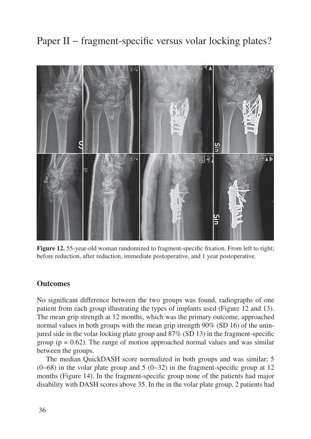
36
Paper II − fragment-specific versus volar locking plates?
Figure 12. 55-year-old woman randomized to fragment-specific fixation. From left to right; before reduction, after reduction, immediate postoperative, and 1 year postoperative.
Outcomes
No significant difference between the two groups was found, radiographs of one patient from each group illustrating the types of implants used (Figure 12 and 13). The mean grip strength at 12 months, which was the primary outcome, approached normal values in both groups with the mean grip strength 90% (SD 16) of the unin-jured side in the volar locking plate group and 87% (SD 13) in the fragment-specific group (p = 0.62). The range of motion approached normal values and was similar between the groups.
The median QuickDASH score normalized in both groups and was similar; 5 (0−68) in the volar plate group and 5 (0−32) in the fragment-specific group at 12 months (Figure 14). In the fragment-specific group none of the patients had major disability with DASH scores above 35. In the in the volar plate group, 2 patients had

37
major disability with DASH scores of 41 and 68 respectively. The generic SF-12 was similar between the 2 groups.
The dorsal angulation was –2° (SD 6) in the volar locking plate group compared with 5° (SD 5) in the frag-ment-specific group, measured on peri-operative fluoroscopic radiographs, but the difference was not found at later follow-ups. No other differences were found between groups regarding dislo-cation, shortening, joint incongruence or rate of healing.
Figure 13. 61-year-old woman randomized to volar locking plate. From left to right; before reduction, after reduction, immediate postoperative, and 1 year postoperative.
Figure 14. Similar median QuickDASH in both groups at 1 year (p = 0.4). Volar locking plate (n = 24) and fragment-specific (n = 25). Boxes represent IQR and whiskers min−max. In the boxplot with an outlier (circle), the upper whisker represents Q3 + 1.5 × IQR.
Fragment-specificVolar locking plate
QuickDASH at 1 year
60
70
40
20
0
5 0
3 0
1 0

38
Complications and reoperations
The overall complication rate was greater in the fragment-specific group (52%, n = 13) compared with the volar locking plate group (21%, n = 5) (p < 0.05). Minor complications were registered in both groups. In the fragment-specific group, 3 ma-jor complications were registered; 1 complex regional pain syndrome (CRPS), 1 hardware loosening, and 1 extensor pollicis longus (EPL) tendon rupture. The latter was related to plate loosening and was treated with hardware removal and tendon transfer. In the volar locking plate group, the patient that crossed over and was treat-ed with fragment-specific fixation had hardware loosening, which was considered as a major complication and the implant was extracted.
Paper III − the 1-year subjective outcome in 3,666 pa-tients
Totally 3,666 patients (2,833 women) with a distal radius fracture were included in the register in the period between January 2003 and December 2012. Patients were treated either non-surgically (78%) or surgically (22%). The overall mean age was 62 years (18−98). In the 2,571/3,666 (70%) patients who returned the DASH ques-tionnaire at 1 year, the overall median DASH score was 9 (IQR 2−25, mean 17). Higher median DASH scores were found for women than for men and for patients in the older age group 65−98 years than patients aged 18−64 years old (Table 4). A weak correlation was found between age and DASH (r
s = 0.24, p < 0.001).
Table 4.
Demographic data showing the DASH scores 1 year after fracture of the distal radius
1-yearDASH score n median (IQR) a mean (95% CI) p-value b
Sex Female c 2,051 11 (2–27) 18 (17–19) Male c 520 5 (0–18) 13 (11–14) < 0.001Age (years) 18–64 1,323 7 (1–20) 14 (13–15) ≥ 65 1,248 14 (3–32) 20 (19–21) < 0.001Treatment group Surgical 633 9 (3–25) 17 (15–18) Non-surgical 1,938 9 (2–27) 17 (16–18) 0.5a Interquartile rangeb Independent samples Wilcoxon rank sum test between group comparisonc Female mean age 64 and male mean age 53

39
Non-surgical versus surgical treatment and outcome
In the non-surgically treated group, the median DASH score was 9 (IQR 2−27), and was 9 (IQR 3−25) also in the surgically treated group (Table 4). Patients oper-ated with the volar locking plate had a better DASH (8 IQR 2−20) compared with fragment-specific fixation or external fixator (12, IQR 5−27 and 13, IQR 3−30; p < 0.004, Figure 15).
The severity of the fracture did influence the outcome and a worse median DASH score was found for patients with AO type C fractures (n = 188) (DASH 11, IQR 4−27), vs. AO type B (n = 36) (DASH 5, IQR 0−13), vs. AO type A (n = 405) (7, IQR 2−18) (p < 0.001). Examples of AO type A, B, and C are illustrated in Figures 16 to 18. No difference was found when dividing the trauma mechanism into high- vs. low-energy fractures.
In the patients with a distal forearm fracture, involving both the distal radius and the distal ulna (Figure 19), a substantially worse outcome of DASH, 23 (IQR 5−41) was found post-hoc.
Surgeons experience and outcome
According the local treatment tradition and organization, 4 hand surgeons operated the majority, 500/805 (62%) of the patients, who at one year had a lower median DASH score of 8 (IQR 2−23) compared to DASH 13 (IQR 5−34) in 164 patients operated by 25 general orthopedic surgeons or DASH 13 (IQR 3−32) in 66 patients operated by 17 orthopedic residents.
External fixationFragment-specificVolar locking plate
DASH at 1 year100
80
60
40
20
0
Figure 15. Boxplot showing median DASH and IQR in patients treated with the three main types of implants; Volar locking plate (n = 332), fragment-spe-cific (n = 157), and external fixation (n = 105). Upper whiskers represent Q3 + 1.5 × IQR. Circles represent outliers.

40
Outcome and residual symptoms at 1 year
The total cohort of 2,571 patients was divided into 3 groups reflecting the severity of residual symptoms at 1 year. 1,338/2,571 (52%) had no or minor residual symptoms (DASH 0–10), 788/2,571 (31%) moderate (DASH 11–35) and 445/2,571 (17%) major disabilities (DASH 36–100; Figure 20).
Also in the 633 surgically treated patients, a similar distribution of residual symptoms was found at 1 year with 334/633 (53%) having no or minor residual symptoms, 194/633 (30%) moderate, and 105/633 (17%) major disabilities (Figure 21).
Figure 16. 36-year-old woman in 2003 with an AO type A fracture.
Figure 17. 46-year-old man in 2008 with an AO type B fracture.
Figure 18. 45-year-old man in 2012 with an AO type C fracture.
Figure 19. 60-year-old woman with a dis-tal radius ulna fracture.

41
Non-responder analysis
In total, 1,095 (30%) patients did not return a correct DASH questionnaire 1 year after the fracture. A non-responder analysis was made and showed that the non-re-sponder group peaked both in the younger and the very old age groups, contained a higher proportion of men, and consisted of larger proportion of non-surgically treated patients. Similar response rates were found in the surgically treated patients regarding AO-type, however high-energy fractures had more non-responders than low-energy fractures.
Paper IV − long-term follow-up of patients with inferior outcome
The long-term follow-up questionnaire of patients scoring DASH >35 at 1 year was returned by 269 (77%) of the 346 invited patients. Patients received the ques-tionnaire 2−12 years after the fracture with a mean follow-up time of 5.5 years.
Figure 20. All patients in the cohort di-vided into 3 groups of severity based on the DASH score 1 year after fracture (n = 2,571). Minor residual symptoms (DASH 0−10), moderate (DASH 11−35) and major (DASH 36−100).
Figure 21. All surgically treated patients di-vided into 3 groups of severity based on the DASH score 1 year after fracture (n = 633). The volar locking plate was in routine use from 2006.
100
90
80
70
60
50
40
30
20
10
02003 2004 2005 2006 2007 2008 2009 2010 2011 2012
Year
Distribution (%) of all patients in the cohort
MajorModerateMinor
100
90
80
70
60
50
40
30
20
10
02003 2004 2005 2006 2007 2008 2009 2010 2011 2012
Year
Distribution (%) of surgically treated patients
MajorModerateMinor

42
73 patients (27%) had been treated surgically and 196 (73%) non-surgically. The mean age was 55 for men (range 18−83) and 66 for women (range 22−93).
Did the patients improve over time?
A significant improvement was found for the whole cohort, improving from a me-dian DASH score of 50 (IQR 43−61) to 36 (IQR 20−55), from 12 months to long-term follow-up (p < 0.001). Men improved more than women, from median 52 at 12 months to 25, compared with women who improved from median 50 to 39 (p = 0.002).
In 127 of 269 (47%) patients, the DASH score was better at the follow-up than the cut off limit of ≤35 used to indicate severe disability (Figure 22). Only 34 of these 127 patients reached a DASH score between 0 and 10 indicating minor disa-bility and more than half, 142/269 patients (53%), still had a DASH score exceeding 35. The mean age was similar for patients that improved or continued to have high DASH scores >35.
Non-responder analysis
77 of the 346 patients did not return the DASH questionnaire at the follow-up. 39 of these patients or their relatives declined participation due to dementia, severe illness or old age. 38 patients did not reply. The mean age at follow-up was similar for the responders and non-responders, 70 (range 26−97; SD 14) and 72 years (range 22−96; SD 18) respectively.
Figure 22. Median and IQR DASH at 12 months for the group that later improved and the group that did not improve. At the 2−12 year follow-up, 127 (47%) improved to DASH ≤35, while 142 (53%) continued to have high DASH scores >35. Whiskers represent min−max. In the boxplots with outliers (circles), upper whiskers represent Q3 + 1.5 × IQR.
0
10
20
30
40
50
60
70
80
90
100
DASH score
12 months 2–12 years
Improved Not improved

43
Discussion
Overall perspective
In the short perspective the outcome after a distal radius fracture (DRF) can be con-sidered to be good. The median DRF patient has a DASH score of 18 (Abramo et al. 2008a) at 3 months, indicating intermediate disability according to our definition. At 6 months, 63% of the patients have no or minimal pain (MacDermid et al. 2003). After 12 months, the DRF patients as a group is considered to have minimal pain and good function with minor restrictions in their activities of daily living (MacDer-mid et al. 2003), and a median DASH of 7.5 at 12 months (Abramo et al. 2008a). As a group, however, the patients still have not reached the level of an age- and sex matched group of uninjured patients (median DASH 2.5) (Abramo et al. 2008a) and when approximately 17−21% reported moderate to major disability as long as 12 months after the injury, it could be argued that the outcome of a DRF is not as fa-vorable as commonly anticipated (Paper III)(MacDermid et al. 2003). Especially, as shown in Paper IV, it appears that only a small proportion of the patients with major disability after one year will ever reach pre-fracture status even in the long-term.
The incidence of DRF in Scandinavia appears to have decreased slightly after the turn of the 20th century (Brogren et al. 2007; Sigurdardottir et al. 2011; Rosengren et al. 2015). The reason is unknown, but the trend is promising. With the life expectancy increasing in Sweden, for both women and men, (Statistics Sweden, Demographic reports, The future population of Sweden 2006-2050), the total number of fractures, however can be expected to increase and the burden on society to increase. The treat-ment of a DRF has changed in the last decades with new techniques, and with a larger proportion of patients being treated surgically (Chung et al. 2009; Mattila et al. 2011; Wilcke et al. 2013; Mellstrand-Navarro et al. 2014). One would have hoped that the outcome would have improved, at least for the surgically treated group, but the pro-portion of patients with an unfavorable outcome in the register study in Paper III remained unchanged. There may be several explanations to why the median DASH or the proportion of patients with high DASH scores remained unchanged over the decade. The instrument might not be sensitive enough or designed to minor changes, the DASH score for patients with poor outcome has already reached the ceiling, and finally these patients would maybe have ended up with poor outcome regardless of treatment due to comorbidities or psychological reasons. Or, the change in treatment simply has no effect on a population level.

44
Surgical treatment of unstable DRF
A fracture can be unstable depending on age, the initial displacement, metaphysal comminution, intra-articular involvement, angulation or axial compression (Ab-baszadegan et al. 1989; Lafontaine et al. 1989; Mackenney et al. 2006; Wadsten et al. 2014; Walenkamp et al. 2015). Due to the properties of the fractured bone, a cast only provides relative stability (Mackenney et al. 2006). In fractures reduced and treated with cast in acceptable anatomical position; (<10° and <5 mm radial shortening), only 50% will remain within an acceptable position at 1 week and not more than 30% at 5 weeks (Earnshaw et al. 2002). In a more recent publication, with acceptable position; (<10° dorsal tilt, <20° volar tilt, >10° radial tilt, <2 mm ulnar variance, and <2 mm intra-articular step off), 31% of fractures in acceptable alignment at 10 to 14 days continue to displace and 64% of all fractures will have displaced at the 3-month follow-up (Wadsten et al. 2014).
The anatomy can be restored with surgery but the surgical methods differ regard-ing the capacity to maintain the reduced fracture’s position during healing. With closed or limited open reduction plus pinning, a good position can be achieved, but secondary loss of position may occur (Cooney et al. 1980). The stability using external fixators with or without pinning is achieved only by indirect fixation, and the reduced position less likely to be maintained than in open fixation and internal fixation (Karantana et al. 2013). Using open reduction and plate fixation, an ana-tomic position can be achieved and maintained also in patients with poorer bone quality (FitzPatrick et al. 2012), but with an increased morbidity due to the open surgery and specific complications (Mathews and Chung 2015). In the last decades new theories and new technology have made it possible to design surgical implants allowing for stable durable anatomic fixation (Konrath and Bahler 2002; Orbay and Fernandez 2002). No evidence exists, however, that these improved techniques mat-ter for the subjective outcome for the patients.
Fragment-specific versus external fixation
Almost simultaneously two implants based on the three-column theory came to market (Medoff and Kopylov 1998; Rikli and Regazzoni 1996; Konrath and Bahler 2002; Rikli et al. 2003). The implants were intended for minimal invasive technique and made it possible to address the individual fragments of a comminuted fracture. Pins, wire-forms, and small plates held the distal fragments and were attached to the intact radius proximally. Implants were designed specifically for the volar and dorsal lip fragments as well as for the radial styloid, and allowed open surgery also for the shattered comminuted fracture. These fractures had previously been treated

45
by closed reduction and external fixation or pinning. In choosing an open technique the risk of surgical complications increased but, on the other hand, the chances of achieving better reduction, and in particular maintained fixation during healing, were better. To evaluate whether the new technique not only would improve the radiographic outcome, but also the subjective outcome, a randomized trial was de-signed comparing the open fragment-specific fixation and the traditional closed ex-ternal fixation technique (Abramo et al. 2009). The primary outcome variable, grip strength, was shown to be better in the fragment-specific group, as late as 12 months after the fracture (90% versus 78% of contralateral, p = 0.03). Also secondary var-iables as pronation-supination were found to be better using the fragment-specific fixation (149° versus 136°, p = 0.03), as was a tendency of more malunions in the external fixation group. The subjective outcome, as measured with median DASH was 5 for the fragment specific versus 6 in the external fixation group (p = 0.2). In Paper I, we investigated at long-term, mean 5 (3−7) years after the fracture, wheth-er internal fixation remained superior over time. At the long-term follow-up, grip strength and pronation-supination had improved in both groups, and a difference, if still existing, was not significant anymore. Other authors have found similar gradual improvement in grip strength after the first year (Brogren et al. 2011a). Seemingly, due to the prolonged immobilization and initial stiffness after an external fixation of the wrist, it takes long time for grip strength and forearm rotation to normalize. Further, even though 3−5 years had passed since the fracture, the median DASH score (fragment-specific group 11 (range 0−46); external fixation group 3 (range 0−57)) had not reached the median pre-injury measured level of DASH, which in the fragment-specific group was 0 (range 0−26) and the external fixation group 0 (range 0−44), respectively.
Internal or external fixation
Several studies imply that internal fixation of distal radius fractures results in better grip strength (Abramo et al. 2009; Jeudy et al. 2012; Karantana et al. 2013), better range of motion (Egol et al. 2008; Abramo et al. 2009; Wilcke et al. 2011; Williksen et al. 2013; Navarro et al. 2016), and better anatomical reduction (Wei et al. 2009; Wilcke et al. 2011; Karantana et al. 2013; Williksen et al. 2013; Roh et al. 2015; Navarro et al. 2016), but only few report better patient reported outcomes (Wei et al. 2009; Wilcke et al. 2011) when compared to external fixation or percutaneous pinning. Although no differences in patient reported outcome were shown in the individual series, an improvement is seen when the data is pooled as in recently published meta-analyses, in which internal fixation has been compared with exter-nal fixation. The improvement in patient reported outcome was significant up to a year after fracture, also better range of motion and better anatomic restoration were

46
found (Cui et al. 2011; Wei et al. 2012; Esposito et al. 2013; Walenkamp et al. 2013; Wang et al. 2013; Xie et al. 2013; Li-hai et al. 2015).
Only few randomized studies comparing external fixation to internal fixation have longer follow-up than one or two years, probably due to the difficulty in keep-ing patients in the trials for a longer time. In Paper I, 45/50 (90%) of the randomized patients attended the long-term follow-up and all 50 patients returned the outcome questionnaire. In an older randomized study on 90 patients with a displaced distal radius fracture, the mean follow-up was 4 years, comparing three treatment meth-ods; closed reduction and cast, external fixation, and open reduction and internal fix-ation with small non-locking T-plates and percutaneous K-wire. The authors’ main results were better radiological position, good functional outcome and better grip strength in the external and internal fixation groups (Kapoor et al. 2000). The group treated with reduction and cast scored lower functional scores, poorer radiographic outcome as well as lower grip strength. Only one randomized long-term follow-up has compared closed reduction and external fixation with adjuvant K-wires with open reduction and volar locking plates (Williksen et al. 2015). 104/114 (91%) pa-tients attended the 1 year and 91/113 (77%) the 5-year follow-up. The volar locking plate group had better range of motion (supination) at 1-year, but no difference was found at 5 years. Both treatment methods achieved good subjective, objective and radiographic outcome.
The volar locking plate
Towards the end of the recruitment period during the first randomized, prospec-tive trial between the fragment-specific fixation and external fixation (Abramo et al. 2009), a new type of fixation of wrist fractures appeared. The volar locking plate was introduced in 2002 (Orbay and Fernandez 2002) and quickly became popular both in Sweden and internationally. The concept was introduced without any evidence or comparing studies (Orbay and Fernandez 2004; Margaliot et al. 2005). The first study to compare the volar locking plate to the fragment-specific fixation was a biomechanical cadaver study, which found stability to be equivalent between the two (Taylor et al. 2006). In a prospective but non-randomized clinical study, the volar locking plate was compared to the fragment-specific fixation and the authors found the volar locking plate to be more stable (better dorsal angula-tion and retained radial length), provide better objective and subjective outcomes in the early postoperative period and having have less complications (Sammer et al. 2008). The volar locking plate has never been compared to fragment-spe-cific fixation in a randomized clinical trial (RCT). An RCT is considered to be best practice in evidence-based medicine, but is resource consuming (Moher et al. 2010).

47
In Paper II, an RCT was designed and performed with grip strength as primary outcome to evaluate if the results of the fragment-specific fixation could be further improved, which had been proven superior to external fixator in the previous series (Abramo et al. 2009). However, at the 1-year follow-up, no difference was found, but instead the results were very similar and both groups presented with excellent grip strength approaching normal values.
In the volar locking plate group the grip strength was 90% of the contralateral side versus 87% in the fragment-specific group. The grip strength ratios in both groups thus were similar to the 90% found in the fragment-specific group at 1 year, in the first randomized study (Abramo et al. 2009). And as comparison, the group of patients in the external fixator group had 78% grip strength after 1 year and reached 90% first at the late follow-up in Paper I 3–7 years after the fracture.
All other secondary measurements also showed similar results for the two groups, in Paper II. There were no significant differences in the objective, subjec-tive or radiographic outcome at 1 year. Both groups had similar and good subjective outcome, with median DASH scores of 5 in both groups, ranging from 0 to 68 in the volar locking plate group and from 0 to 32 in the fragment specific group.
In the fragment-specific group none of the patients had DASH scores above 35, the cutoff for major disability. In the volar locking plate group 2 patients had DASH exceeding the cutoff. One patient had a DASH score of 41 after 1 year and was the only patient involved in a true high-energy injury, a motorcycle accident. The other patient had a DASH score of 68 and was the only patient with multiple comorbidi-ties, known to correlate with higher DASH scores (FitzPatrick et al. 2012).
Although, DASH in both randomized studies in this thesis failed to show a dif-ference between the groups, the DASH still can be useful. With similar, almost identical results of two implants, like in Paper II, the rate of complications be-comes increasingly important. By using DASH we find the individuals in which our treatment fail. We can analyze the causes and identify measurement to avoid the complications causing the high score in our future patients. We can adjust the rehabilitation protocol or identify early problems like pain or swelling. The rate and type of complications found using a particular technique can also help us to judge which treatment method is the best for a certain fracture or patient group.
Complications and reoperations
In the report preceding Paper I (Abramo et al. 2009), the complications were classi-fied as minor (transient), moderate (transient after intervention) and major (chronic) complications. At 12 months more major complications with external fixation was noted (5 symptomatic malunion, 1 fractured metacarpal bone, 2 CRPS) compared with fragment-specific fixation (1 symptomatic malunion and 1 CRPS, p = 0.04).

48
While the total number of complications appeared higher in the external fixation group (n = 29 vs. n = 21, p = 0.3), no significant difference was found. In Paper I, reoperations were reevaluated beyond the 12 months reported in the original study. In the external fixation group, 8 reoperations occurred during the first year (5 os-teotomies and 3 carpal tunnel releases) and in addition, 2 operations were carried out after the first year for skin adherence caused by the external fixation pins. In the fragment-specific group 5 reoperations occurred during the first year (1 oste-otomy, 1 tenosynovectomy, 3 extractions of osteosynthesis materials) and another 9 hardware extractions after the first year. In total, during the entire period there were 14/26 (54%) reoperations in the fragment-specific group compared with 10/24 (42%) in the external fixation group, both rather high numbers. To our knowledge only one other long-term follow-up of a randomized study has reported reopera-tion rates for as long after primary surgery as in Paper I. In a randomized study comparing open reduction and volar locking plate to closed reduction and external fixation in unstable distal radius fractures the outcome from the 1-year follow-up (Williksen et al. 2013) was further studied at a 5-year follow-up (Williksen et al. 2015). Comparisons between studies are difficult, but at the 5-year follow-up, 16/52 (31%) reoperations had occurred in the internal fixation group and 10/59 (17%) in the external fixation group respectively. Hardware extraction was the most frequent reason for reoperation.
In the internal fixation group (n = 16) of Williksens report, 11 volar locking plates (VLP) were removed due to surgical technical complications (6 at 1-year fol-low-up and 5 at the long-term follow-up) and 4 volar locking plates were removed due to patient request (2 at one year and 2 at the long-term follow-up) and finally, 1 patient had early secondary surgery with reconstruction of the ulnar styloid process. Of the patients with hardware removal in the volar locking plate group, 2 also had EPL ruptures due to prominent screws. In the external fixation group (n = 10); 5 patients had minor surgery due to scar contractures, 2 had CTS release, 1 had ear-ly fixation-failure with external fixation and were reoperated with a volar locking plate. One patient was reoperated due to incomplete reduction and 1 patient in the external fixation group also had been treated with a dorsal plate and the dorsal plate removed due to loosening. Thus, results were similar compared to our long-term follow up regarding repeat surgery.
Patients treated with external fixation will need pin treatment, and are more prone to superficial wound infections, as found at 1-year (3/24 compared with 0/26 (Abramo et al. 2009). The high frequency of pin track infections in external fixation has also been shown in other studies (Ahlborg and Josefsson 1999; McKay et al. 2001; Anderson et al. 2004; Margaliot et al. 2005; Mathews and Chung 2015). All patients treated with external fixations need removal of pins, which can be painful. However, pin removal can be made without analgesia, correction of scars or adher-ence loosening in local anesthesia, whereas plate removals, have to be performed as a full operation with anesthesia or a block.

49
In Paper II, the study protocol included a predefined list of complications (Ap-pendix III) to be filled out at follow-up visits. Such a list should be part of the basic protocol before launching a RCT. Minor complications were defined as transient problems, considering not to affect the final outcome, while major were severe com-plications considered to influence the final outcome.
Minor complications, such as carpal tunnel syndrome, were diagnosed with two patients in each group and both resolved spontaneously without operative treatment. A transient radial neurapraxia was the most common minor complication in the fragment-specific group (6 patients) compared with the volar locking plate group (1 patients), and all cases resolved within the year. 1 patient had mild tendinitis and 1 patient had skin adherence in the fragment-specific group and all mentioned cases were considered as minor complications.
Major complications were few. One patient in the fragment-specific group devel-oped a CRPS, which resolved with intense exercise during the first year. In the volar locking plate group one plate loosened and was removed. This patient was actually treated with fragment-specific fixation instead of the allocated treatment, since it became clear intra-operatively that the fracture was distal to the watershed line and not able to fix with a volar locking plate. Alternatives to the techniques in Paper II, using newer treatment options addressing the volar rim fractures with volar hook plates might be a better method (O’Shaughnessy et al. 2015). In the fragment-spe-cific group, 2 patients had hardware loosening, but only 1 patient had the radial pin plate removed and at the same time as a concomitant EPL rupture was treated by tendon transfer. All 3 patients had good grip strength, (range 88% to 95%), similar to the other patients and a 1-year DASH score of 14–23.
The overall complication rate in Paper II, however, differed between 21% in the volar locking plate group and 52% in the fragment-specific group. Due to a lack of a unanimously accepted definition of minor, major or overall complications, com-parisons between studies are difficult, but similar 1-year complication rates have been reported in other studies. Karantana et al. found no statistically significant dif-ference; 24% overall complications in the volar locking plate group and 42% in the external fixation group (Karantana et al. 2013). Williksen et al. found no difference; 29% overall complications in the volar locking plate group and 31% in the external fixation group (Williksen et al. 2013). Navarro et al. found a 51% overall compli-cation rate in the volar locking plate group compared with to 45% in the external fixation group, without no statistical difference (Navarro et al. 2016).
Evaluating outcome – a multifaceted aspect
The patient outcome after a DRF is multifaceted. We still believe that the outcome is best evaluated using both objective and subjective outcome measures (Karnezis

50
et al. 2005; Földhazy et al. 2007; Wilcke et al. 2007; Changulani et al. 2008). How-ever, the patient reported outcome instruments of today may be able to discriminate between the outcome after a DRF after 3 versus 12 months or between before and after an osteotomy, but these instruments are too blunt to tell the difference be-tween two implants which already result in good but maybe not excellent results. Grip strength was used as the primary outcome parameter forming the basis for the sample size estimation in previous studies (Kopylov et al. 1999; Abramo et al. 2009) as well as in Paper I and Paper II. Grip strength was found to be a con-sistent and reliable outcome measure and seemingly a good surrogate variable for pain. But it appears that the result of the surgery has reached a level that is going to be difficult to surpass. If one has the intention to show another new method to be even better and alternative designs probably then has to be chosen. Using the data from Paper II resulting in a 3% difference in grip strength, a study on 420 patients in each treatment arm would be needed to show an incremental 3% improvement with 85% power. Maybe the limit has been reached for one small size single-center randomized study to show a significant difference in subjective or objective data, and meta-analyses are needed instead, or registers consisting of several thousand patients as in the hip and knee arthroplasty communities. Maybe we need to focus on minimizing the number of patients with poor results instead of trying to improve the mean and median values of subjective and objective measurements. Maybe other methods are needed, using wearable and automated measurements system to collect data and using artificial intelligence and mathematical algorithms to find the patterns.
The distal radius fracture – outcome over 10-years
In the clinical randomized studies, grip strength was used as primary outcome meas-ure, and found normalizing at 1 year. The DASH scores were low in the patients treated with internal fixation. Patients treated with external fixation also reached normal grip strength, but later. These conclusions are all based on the result of the median patient and in this next chapter we try to use our hospital-based distal radi-us fracture register to give a broader view of all the DRF patients during a 10-year period. The 1-year DASH score of patients included in the register between January 2003 and December 2012 was used. During the 10-year period a shift in the choice of surgical implant occurred, as reflected by the two previously reported randomized studies (Abramo et al. 2009; Paper II). No longitudinal registry study has evaluated if and how the subjective outcome of distal radius fractures during this shift.
In Paper III, 2571/3666 (70%) patients with a distal radius fracture returned a correctly filled out DASH questionnaire at 1 year. The median DASH was low with a score of 9 (IQR 2−25, mean 17). This is similar to the median DASH score of 7.5

51
(IQR 1.7−25) (mean 16) reported in the first report of the patients included between September 2001 and August 2003 (Abramo et al. 2008a), evaluating the treatment protocol used for distal radius fractures. Normative data exist for DASH in a popu-lation without a DRF. In a representative US population of 1800 patients, older than 18 years (Hunsaker et al. 2002), the normative values were median 4 (IQR 1−13) and mean 10.1 (SD 14.7) for DASH and median 4.5 (IQR 1−14) and mean 10.9 (SD 15.3) for QuickDASH (The DASH and QuickDASH - Outcome measures e-bulletin fall 2012). The developers of the DASH, the American Academy of Orthopedic Surgeons and the Institute for Work and Health in Toronto, Canada, suggested that a DASH score of less than 10 should be aimed for when evaluating outcome. In Paper III, 1,338 patients (52%) reported DASH-values between 0−10, which could be considered as a good result 1 year after a distal radius fracture and a return to pre-fracture status.
In Paper III, low and almost identical DASH scores were found for both non-surgically (median 9, IQR 3−25) and surgically (median 9, IQR 2−27) treated patients. The result supports that the treatment protocol (Abramo et al. 2008a) still is valid and that most patients reach an acceptable or good outcome at one year af-ter a distal radius fracture. The non-surgical treatment has largely been unchanged during the period. In contrast, the surgical technique and the choice of implants at the operating hospital has changed during this period, from external fixators (42%) and fragment-specific plates (45%) in 2003, to predominantly volar locking plates (65%) in 2012 (Figure 23). There was no radical improvement in the 1-year median DASH.
0
10
20
30
40
50
60
70
80
90
100
2003 2004 2005 2006 2007 2008 2009 2010 2011 2012Year
Volar locking plateFragment-specific fixationExternal fixationVolar non-locking plateOtherMedian DASH at 1 year
Number of implants and median DASH at 1 year
Figure 23. The 1 year DASH and types of implant used during the study period (n = 805). A randomized study between external fixator and fragment-specific fixation was conducted 2002–2005 and between fragment specific fixation and volar locking plate 2010–2012.

52
The change in choice of implants appears to be ubiquitous during this period and other centers have noted a similar shift from external to internal fixation, on a regional level (Stockholm 2004 to 2010; Wilcke 2013), on a national level (Sweden 2005 to 2010; Mellstrand-Navarro 2014), on a Scandinavian level (Finland 2003 to 2008; Mattila 2011) and internationally (USA 2000 to 2004; Chung 2009). The shifts occurred without any support from randomized studies, and although with somewhat conflicting results, the recent meta-analyses appear to provide some evidence for a better outcome of unstable distal radius fractures treated with internal fixation instead of external fixation. In a systematic review of the published meta-analyses of the vol-ar locking plate, it is even concluded that a significant lower DASH, a better volar tilt and radial inclination, and a lower infection rate is found at one year with the internal fixation compared with the external fixation (Zhang et al. 2016).
In Paper III, the mean rate of surgical treatment was 22%, which remained unchanged over the 10-year time period (Figure 24). In a Swedish national perspec-tive, the rate of surgical treatment of distal radius fractures increased from 16% in 2005 to 20% in 2010 (Mellstrand-Navarro et al. 2014)
Even if the change of implants did not change the outcome and even if surgical-ly and non-surgically treated patients achieved the same outcome, a difference in outcome was found, related to the experience of the surgeons. Thus, with a small group of dedicated surgeons with special interest in wrist fractures treating a large volume of patients a better subjective outcome in a cohort can be expected, and by speculation thereby reducing complications.
To better understand the outliers and not only analyze the median patient out-come, we divided the whole cohort of 2571 patients into three groups regarding re-sidual symptoms and outcome; “the good, the bad and the ugly”. We chose DASH 0-10 as a level of minor, 11 to 35 as moderate, and 36 to 100 as major residual
0
10
20
30
40
50
60
70
80
90
100
2003 2004 2005 2006 2007 2008 2009 2010 2011 2012Year
Annual distribution (%)
SurgicalNon-surgical
Figure 24. Relative proportions of non- surgical treatment (n = 2,861) and sur-gical treatme t (n = 805) in the period 2003 to 2012.

53
symptoms. The level 0 to 10 was based on the normative values for DASH and the level >35 was based on two studies of radial osteotomies due to malunion, in which the median DASH score was 39 (range 2−63) (Abramo et al. 2008b) and median 35 (range 22-61) (Abramo et al. 2010). The assumptions have some support from the founders of the DASH questionnaire who have not fully established cutoff lim-its for DASH, but suggested some tentative levels (DASH/QuickDASH e-Bulletin Summer 2013). Patients with a DASH score 10−29 are “aware of their upper-limb limitations but do not consider them a problem” and patients with a DASH score 40−69 are patients “having a lot of difficulty”.
Approximately half of the patients, both surgically (52%) and non-surgically (53%) treated patients, ended up with a DASH between 0-10, i.e. with minor dis-abilities and on par with the normative values of an unfractured population. The proportions of patients with moderate disability (DASH 11−35) were similar (30%). Major disabilities (DASH 36−100) were found in 17% in non-surgically or surgi-cally treated patients. In another cohort study of 129 patients (56% non-surgically and 44% surgically treated), 21% had moderate to severe disability, evaluated with PRWE, 1 year after a distal radius fracture (MacDermid et al. 2003). So even if the vast majority of the fractured wrist, regardless if surgically or non-surgically treated, at some remote period again will enjoy an almost perfect freedom in all its motions, and be exempt from pain, a rather large proportion (17−21%) still have major disa-bilities as late as 1 year after a distal radius fracture.
The selected cutoff levels of DASH into minor, moderate, and major residual disability after a distal radius fracture in Paper III, are presumably both valid and helpful to clinicians in evaluating patient related outcome and predicting patient outcome. The PRWE questionnaire has been shown to be more responsive than the DASH questionnaire in a study over a 6-month period in recovery after distal radius fractures (MacDermid et al. 2000) and slightly more responsive in another study over a six-month period in patients with hand and wrist problems (MacDer-mid and Tottenham 2004). However, both the DASH and PRWE can be used as a screening tool to find patients with poor outcomes in the months after a distal radius fracture. Especially if a patient continues to report a high DASH score over months the patient should be summoned for an outpatient visit to identify any complication of the non-surgical or surgical treatment, like CRPS, tendinitis, CTS or tendon rupture.
Limitations
A response rate of 70% in a patient-reported outcome after a distal radius fracture with no upper age limit was considered acceptable. The shift in 2008, replacing the 30-item DASH with the 11-item QuickDASH, increased the response rate from 66% to 73%. In comparison, 79% replied in a nationwide patient-reported outcome study of 4467 patients with displaced femoral neck fractures (Leonardsson et al. 2013). These patients were retrospectively identified and an outcome questionnaire was

54
mailed at mean 14 (7−22) months after the fracture. Patients included in the pro-spective wrist fracture register study (Paper III and Paper IV) received the DASH questionnaire at 3 and 12 months. In an attempt to analyze the non-responders, the patients were subdivided into three groups where an increased amount of both young (<40) and very old (>80) patients were found among the non-responders. Likely, some of these very old patients had died, although only 1 year had passed since their fracture. Further, more non-responders were found in the male cohort as well as in the non-surgically treated cohort.
The most dissatisfied patients – can they improve?
The overall outcome after a distal radius fracture is good, but still 17−21% will report a poor outcome at 1 year after the fracture. In Paper IV, the long-term over-all improvement in patients with major disability was found to be significant. The median DASH decreased from 50 (IQR 43−61) at 12 months to 36 (IQR 20−55) at the 2−12 year follow-up, p <0.001), a quite substantial decrease. Seemingly, time from injury to follow-up can be a sole factor for improvement of subjective outcome. In a long-term retrospective study of 260 patients with non-surgically treated distal radius fractures (Finsen et al. 2012), the DASH score was higher in the group in which 2.5−4 years had passed since the fracture (DASH mean 17), as compared to the group in which 10−13 years had passed (DASH mean 8). Others authors have found spontaneous improvement in DASH over time. In a prospec-tive population-based cohort study of 49 female patients aged 50−75 years with a non-surgically treated distal radius fracture (Brogren et al. 2011a), a trend was found of median DASH improving from 14 to 8, between 1 year and 2−4 years. Interestingly, a greater improvement was found in a subgroup of patients with mod-erate/severe malunion (defined as: radial shortening of ≥3 mm and/or dorsal angu-lation >15°), in which DASH was 20 at 1 year after fracture compared with 10 at 2−4 years after fracture.
Although, in Paper IV, 127/269 (47%) improved spontaneously after mean 5.5 years (2−12), to a level below the cutoff of major disability (DASH >35), more than half 142/269 (53%) of the patients continued to experience major disabilities. Of all the 269 patients with high DASH scores at 12 months only 13%, a surprisingly small proportion of the patients, returned to an estimated pre-injury level of DASH ≤10. Since the incidence of distal radius fractures is high, a considerable number of patients will suffer from pain and disability and not return to the level prior to the injury. The objective should be to prevent or minimize the number of patients with inferior outcome after a distal radius fracture as early as possible, thereby reducing the amount of avoiding post-operative complications such as swelling, pain, infec-tion, malunions or other reasons.

55
Limitations
All adult patient ages were included. Very old patients tend to score higher DASH scores (Klum et al. 2012) as well as patients with osteoporosis and comorbidities (FitzPatrick et al. 2012). We found a high mortality rate in the patients with major disability. 22% (346/445) of patients with major residual symptoms died between the 12-month follow-up and the 2−12-year follow-up. If this is higher in patients with a high DASH value or not has not been evaluated. A high mortality rate in the years after a distal radius fracture, has been reported and an increased Standardized Mortality Ratio in women >70 year at 5 years after a distal radius fracture, but not in men (Øyen et al. 2014).
Strengths and weaknesses
Paper I
Long-term follow-up comparative studies of external fixation and internal fixation are scarce, but are important to understand the outcome in the longer perspective. Improvement in the clinical evaluation regarding blinding of scars could have min-imized the risk of evaluation bias.
Paper II
Randomized controlled trials comprise the highest level of evidence. Despite this, only few randomized trials have compared two methods of internal fixation for treatment of DRF. By including both primarily non-reducible as well as second-ary redisplaced DRF the results can be generalized. The patients did not undergo blinding at the follow-up clinical visit, which might have biased the clinical eval-uation performed by four of the five authors. Little time to inform the first patient coming for surgery in the mornings resulted in eligible patients not being included in the study.
Paper III
The prospective register study is the largest and longest of its kind analyzing the subjective outcome in patients with distal radius fractures. In a decade of treatment shift, the treatment algorithm for wrist fractures is still valid; both non-surgically and surgically treated cohorts had similar low (good) median subjective outcome values over the period one year after fracture. Initially, almost one-third did not respond

56
to the 1-year DASH questionnaire, but by using the shorter form the non-response rate was reduced. Very old patients are likely to score higher due to co-morbidities.
Paper IV
Little is known regarding the subjective improvement over time in patients with a distal radius fracture, and even less in patients with major residual disability. Half of all patients improve after the first year, but still a large number of patients have persistent pain and disability after 2−12 years. More than one fifth of these patients were deceased at the long-term follow-up; another one fifth did not respond the questionnaire. Nevertheless, this group of patients with poor outcome needs to be drawn into focus.

57
Conclusions
Paper I: Grip strength and ROM continues to improve and approached normal values after mean 5 years, both in patients operated with closed reduc-tion and external fixation or open reduction and internal fixation.
Paper II: In 50 patients with a primarily non-reducible or secondarily redisplaced distal radius fractures the functional and subjective outcome after 1 year was good, regardless of being treated by volar locking plates or frag-ment-specific fixation both achieve.
Paper III: The subjective outcome of the whole adult distal radius fracture cohort remained unchanged over a 10-year-period 2003−2012, using the data from the Lund Wrist Fracture Register. About half of the patients re-turned to a pre-fracture state (DASH 0−10) but up to one sixth reported severe residual disability (DASH >35). There was a radical shift in the choice of implant but the proportion of surgically treated fractures re-mained the same over the 10-years time period.
Paper IV: In patients who reported severe residual disability (DASH >35) at 1 year, a modest improvement over time could be expected. Half of the patients continued to have major disability for the following 2−12 years and only 13% returned to pre-fracture status (DASH 0−10). Age at time of fracture and sex seemed to affect the outcome, with greater change of improvement if the patients were younger and male.

58
Future perspectives
The distal radius fracture continues to challenge physicians and cause morbidity in a large number of patients every year.
Lessons learned from the thesis was:
• The DASH outcome questionnaire is a good and useful instrument in screening and identifying patients with poor outcome. It is not a very sensitive tool for a randomized study comparing two similar methods of treatment with already good results. We need sharper instruments to evaluate outcome.
• 17% of patients having major residual symptoms after a distal radius fracture is not acceptable. The future aim must be to prevent complications early to improve long-term patient satisfaction.
• A complication rate between 20−50% is a too high and a future aim must be to re-duce the complications in both the surgically and non-surgically treated patients.
• 4% of the patients had a combined distal radius-ulna fracture. These patients had a significantly worse outcome, regardless if they were treated non-surgically or surgically. A future aim is to improve the treatment of this type fracture.
• A long-term follow-up is planned of the randomized trial in paper II comparing fragment-specific fixation with volar locking plates.

59
Summary
Background
Most distal radius fractures are treated in a similar way as 200 years ago with re-duction and cast splinting. To cite the “founding father” of the distal radius fracture treatment, Abraham Colles “…the limb will at some remote period again enjoy per-fect freedom in all its motions, and be completely exempt from pain…”. For most patients (in the register study at least half) this is true, but even after all these years, with hopefully improved treatment, there is a proportion of patients not satisfied with the end result. Since this fracture is one of the most common fractures, and with an ageing population, even a small percentage will add up to a substantial num-ber of patients with poor outcome, and improvements will result in improved quality of life for a substantial number of patients on a yearly basis.
The main objective in the research and treatment of this common fracture must be to identify risk factors for poor outcome and prevent these before developing into complications. The purpose of this thesis was to analyze the result of the treatment at our hospital; both at a population level using a DRF register, as well as to contrib-ute to the basis of evidence by evaluating the newest methods of surgical treatment.
Methods
In Paper I, a mean 5 years follow-up was performed of a randomized study be-tween an external fixator and the fragment-specific open fixation in the treatment of unstable distal radius fractures. The initial 1-year follow-up had shown better grip strength and range of motion. The aim was to analyze whether or not the difference would persist over time.
In Paper II, the best method at 1 year in paper I was compared with the pop-ular new volar plate in a randomized trial. Primarily non-reducible or secondary redisplaced distal radius fractures were included and outcome was measured using objective and subjective measures. The follow up time was 12 months. The aim was to find out if treatment could be further improved by the new method.
In Paper III, we conducted a retrospective epidemiological analysis of a 10-year period between 2003−2012 of our prospective and consecutive distal radius registry.

60
In the register, patients aged 18 years or more with a distal radius fracture, pre-senting at our hospital, were included in the database. The patients received a vali-dated subjective outcome form (Disabilities of Arm Shoulder and Hand/DASH) at the time of injury and at three and twelve months after the fracture. The patients’ records and radiographs were scrutinized, treatment recorded, and the fractures were classified according to the International Classification of Diseases (ICD-10), for type of surgery, AO fracture type, high or low energy, and time from fracture to surgery. The DASH scores at 12 months were analyzed over the 10-year-period. The aim was to find out if the subjective outcome changed over the 10-year-period.
In Paper IV, the group of patients with the highest (worst) DASH scores were followed up. Patients were sent a new DASH form 2−12 years after the fracture. The aim was to analyze if patients with elevated DASH-scores would normalize over time.
Results and Discussion
Paper I: Even if grip strength and pronation-supination was superior 1 year after surgical treatment in the cohort treated with fragment-specific fixation, the differ-ence was no longer statistically significant at the 5-year follow-up. Both treatment groups improved and had almost normal grip strength and pronation-supination. In the fragment-specific group half of the patients had had the implant removed. In the external fixation group there was a tendency for radiographic malunion.
Paper II: Patients with primarily non-reducible or secondarily redisplaced distal radius fractures, treated with either volar locking plate or fragment-specific fixa-tion both achieved good, almost identical subjective, objective, and radiographic outcome, although, more complications in the fragment-specific group was found.
Paper III: In the large prospective and consecutive cohort of patients with distal radius fracture, the 1-year outcome was good and basically unchanged over the 10-year period. About half the patients returned to a pre-fracture state (DASH 0-10) but up to one sixth reported severe residual disability (DASH >35). A marked shift in treatment was found in the 10-year period, but the overall and year-by-year subjec-tive outcome remained the same.
Paper IV: In the one sixth of the patients reporting severe residual disability (DASH >35) at 1-year, only 13% returned to pre-fracture status (DASH 0-10), 2-12 years after the fracture. Half of the patients with major disability still reported DASH >35, 2−12 years after the fracture. Thus, even if improvement in both function and patient-reported outcome will occur also after the first year, the injury will for many continue to cause problems and restrictions in life.

61
Sammanfattning på svenska
Handledsbrott är den vanligaste frakturen på en akutmottagning. De yngre vuxna patienterna är jämnt fördelade mellan könen men de äldre har en övervikt av kvin-nor. Hos de äldre uppstår frakturen företrädesvis i ett skelett med olika grad av ben-skörhet. Frakturen behandlas oftast med gips utan operation men drygt en femtedel opereras. Om felställningen är stor eller om frakturen inte bedöms kunna kvarstanna i ett gynnsamt läge rekommenderas en operation. Man väger in patientens funk-tionsnivå och ålder i den samlade bedömningen. Patienterna har under de första månaderna svårt att klara sina dagliga göromål och personliga hygien men de flesta patienter blir återställda ett år efter skadan. I vår studie har dock en femtedel av alla som bryter sin handled kvarvarande besvär ett år efter skadan såsom bestående svaghetskänsla och smärta vid belastning av handleden.
Syftet med denna avhandling är att analysera vår behandling under det senaste decenniet för att hitta sätt att förbättra behandlingen av hand ledsfrakturer. Avhand-lingen baseras på fyra arbeten, två kliniska studier och två registerstudier.
Det första delarbetet är en fem-års uppföljning av en jämförande studie mellan två kirurgiska behandlingar för felställda handledsfrakturer. Med den nya metoden, intern fixation, fixerade man frakturfragmenten med små plattor och stift och vi jämförde mot den traditionella metoden med yttre stag, en så kallad extern fixation. Vid ett år sågs bättre greppstyrka i handen och rotation av underarmen hos patienter opererade med den nyare metoden och en tendens till färre större komplikationer. Vid fem år förelåg det inte längre någon skillnad i greppstyrka eller rörelseomfång utan nu hade även patienter opererade med extern fixation uppnått ett normalvärde avseende greppstyrka och underarmsrotation.
I det andra delarbetet, jämfördes två olika typer av intern fixation för felställda handledsfrakturer. Vi jämförde en ny operationsmetod som snabbt blivit populär (volar platta) med den som föll bäst ut i den förra studien (fragment-specifik). Vid ett år hade bägge grupper ett bra utfall och hade uppnått ett normalvärde i grepp-styrka. Mellan grupperna fanns det ingen skillnad avseende rörelseomfång eller patientupplevd handfunktion men vi hittade fler komplikationer i gruppen som blivit opererade med fragment-specifik fixation än med volar platta.
Det tredje delarbetet är en genomgång av patienter i vår databas av patienter med handledsfraktur i vilken vi utvärderar den patientupplevda handfunktionen med en hälsoenkät (DASH). 3,666 patienter >18 år sökte akutmottagningen i Lund med en handledsfraktur 2003 till 2012 och ingick i studien. Patienterna blev behand-
62
lade enligt ett behandlingsprotokoll med ca 80 % gipsades och 20 % opererades. 2,571 patienter svarade på hälsoenkäten 12 månader efter frakturen. På gruppnivå var det patientupplevda resultatet bra och förblev oförändrad oavsett om man blev behandlad i början eller i slutet av 10-års perioden. Andelen patienter som blivit opererade var oförändrat, men en förändring av val av den kirurgiska behandlings-metoden noterades. Extern fixation ersattes med intern fixation och den volara plat-tan blev den vanligaste metoden mot slutet av perioden. En subgruppsanalys visade att patienter opererade med extern fixation skattade sin handfunktion som sämre än de som opererats med volar platta.
Även det fjärde delarbetet baserades på registret och 445 av 2,571 (17 %) pa-tienter med kraftiga kvarvarande besvär ett år efter sitt handledsbenbrott fick fylla i en ny hälsoenkät. 269 patienter svarade på enkäten två till 12 år efter fraktur. 127 av 269 (47 %) hade förbättrats till en nivå med minimala eller moderata besvär, men majori teten 142 av 269 (53 %) hade fortfarande kraftiga kvarvarande besvär.
Vi konkluderar att det behandlingsprotokoll vi använder för handledsfrakturer är användbart, och att de flesta patienter får ett bra funktionellt slutresultat både avseende patienter som behandlas utan operation och för de som opereras. De nyare operationsmetoderna med intern fixation gav snabbare rehabilitering, en tidigare normalisering av både greppstyrka och rörelseomfång efter ett år men skillnaden var försvunnen efter fem år. Under en 10-års period upplevde medianpatienten sin handled som bra men inte helt normaliserad efter ett år. Den sjättedel som upplevde sin handfunktion som kraftigt försämrad efter ett år förbättrades i hälften av fallen men blev sällan helt återställda även mätt efter många år.

63
Acknowledgements
I would like to express my sincere gratitude to:
All the patients participating in the studies. Without their participation, this research would not have been possible.
Magnus Tägil, my supervisor and mentor for inspiration, patience, support and en-couragement in research. I admire your endless energy and profound devotedness to this research field. For skillful guidance in the operation room sharing your exper-tise enabling improvement in my surgical technique in wrist fractures.
Tony Abramo, my co-supervisor for the talks and productive discussions, the spot-on feedback in the review process, sharing your solid understanding of fracture patterns and treatment in wrist surgery, and your advice and tutoring in the surgical field.
Mats Geijer, my co-supervisor for the fruitful discussions and inspiration in the writing process, hawkeyed suggestions in reviewing, sharing your thorough exper-tise and understanding of skeletal radiology and pep talks during the years.
Philippe Kopylov, my former boss and co-author for your energy, your precise sug-gestions in writing and reviewing, always there to support and listen to issues of both clinical, research and personal matters.
Daniel Jerrhag, my co-author for assistance in the project set-up and examination of patients in Paper I.
Vendela Teurneau, my co-author for assistance in collecting data and reviewing the manuscript in Paper IV.
Kaj Knutson, for adding the final touch with assistance in graphics and typesetting.
Ewa Persson and Monica Persson, for administrative assistance in the clinical stud-ies and assistance in handling the DASH questionnaires in the registry studies, re-spectively.

64
Kerstin Runnqvist and Sara Hellberg, for physiotherapy and assessment of the pa-tients in the second paper.
Ola Belfrage and Vasilis Zampelis, my great friends, colleagues and roommates for the constructive research and clinical discussions, and good times together.
Colleagues and friends of the Orthopedic Department in Lund and Malmö always prepared to discuss a clinical or research issue.
Elisabeth Grönvall, my mother and artist, for all the love, the patience, always ready to listen to my thoughts and looking after the kids.
Lennart Landgren, my father for raising me to the person I am, for giving advice and being a good listener.
The rest of my family, especially to Ludvig Landgren, Sarah Fager, Henrik Land-gren, Sofia Landgren for listening to my thoughts and giving support.
Kirsten Hald, my mother-in-law for helping out and looking after the boys.
Leif Thuesen, my father-in-law for instant feedback in research questions.
Lea Langhoff Thuesen, my lovely and wonderful wife for the day-to-day support, the discussions, impressive patience and tolerance letting me concentrate on this thesis. For your supermom skills in looking after our children, all the challenges and playing with them, while I have been absent.
Finally, our three boys for the patience and acceptance you have showed during these intense last months. Thank you Hampus my lovely and intelligent son, Casper my wonderful and smart son, Jonathan my happy and positive son, for being the ones you are.
This thesis has been supported by grants from Government Funding of Clinical Research within the National Health Service (ALF), Swedish Research Council (Vetenskapsrådet – project 2031), Regional Research Support (Region Skåne), the Alfred Österlund foundation, the Erik and Angelika Sparre foundation, the Greta and Johan Kock foundation, the Maggie Stephens foundation, Thure Carlsson foun-dation, and the Medical Faculty, Lund, Sweden.

65
References
Abbaszadegan H, Conradi P, Jonsson U. Fixation not needed for undisplaced Colles’ frac-ture. Acta Orthop 1989; 60: 60-2.
Abramo A, Geijer M, Kopylov P, Tägil M. Osteotomy of distal radius fracture malunion using a fast remodeling bone substitute consisting of calcium sulphate and calcium phosphate. J Biomed Mater Res B: Appl Biomater 2010; 92: 281-6.
Abramo A, Kopylov P, Geijer M, Tägil M. Open reduction and internal fixation compared to closed reduction and external fixation in distal radial fractures: a randomized study of 50 patients. Acta Orthop 2009; 80: 478-85.
Abramo A, Kopylov P, Tägil M. Evaluation of a treatment protocol in distal radius fractures. Acta Orthop 2008a; 79: 376-85.
Abramo A, Tägil M, Geijer M, Kopylov P. Osteotomy of dorsally displaced malunited frac-tures of the distal radius: no loss of radiographic correction during healing with a minimally invasive fixation technique and an injectable bone substitute. Acta Orthop 2008b; 79: 262-8.
Adolphson P, Abbaszadegan H, Jonsson U. Computer-assisted prediction of the instability of Colles’ fractures. Int Orthop 1993; 17: 13-5.
Agee JM. Application of multiplanar ligamentotaxis to external fixation of distal radius frac-tures. Iowa Orthop J 1994; 14: 31.
Ahlborg HG, Josefsson PO. Pin-tract complications in external fixation of fractures of the distal radius. Acta Orthop Scand 1999; 70: 116-8.
Aktekin LA, Eser F, Baskan BM, Sivas F, Malhan S, Oksuz E, et al. Disability of Arm Shoul-der and Hand Questionnaire in rheumatoid arthritis patients: relationship with disease activity, HAQ, SF-36. Rheumatol Int 2011; 31: 823-6.
Alffram P-A, Bauer GC. Epidemiology of fractures of the forearm. J Bone Joint Surg (Am) 1962; 44: 105-14.
Andersen, DJ, Blair, WF, Stevers, CM, Adams, BD, El-Khouri, GY, Brandser, EA. Classifi-cation of distal radius fractures: an analysis of interobserver reliability and intraobserv-er reproducibility. J Hand Surg 1996; 21: 574-82.
Anderson JT, Lucas GL, Buhr BR. Complications of treating distal radius fractures with external fixation: a community experience. Iowa Orthop J 2004; 24: 53-9.
Anzarut A, Johnson JA, Rowe BH, Lambert RG, Blitz S, Majumdar SR. Radiologic and patient-reported functional outcomes in an elderly cohort with conservatively treated distal radius fractures. J Hand Surg (Am) 2004; 29: 1121-7.

66
Arora R, Lutz M, Hennerbichler A, Krappinger D, Espen D, Gabl M. Complications follow-ing internal fixation of unstable distal radius fracture with a palmar locking-plate. J Orthop Trauma 2007; 21: 316-22.
Arora R, Lutz M, Deml C, Krappinger D, Haug L, Gabl M. A prospective randomized trial comparing nonoperative treatment with volar locking plate fixation for displaced and unstable distal radial fractures in patients sixty-five years of age and older. J Bone Joint Surg (Am) 2011; 93: 2146-53.
Ashford RF, Nagelburg S, Adkins R. Sensitivity of the Jamar dynamometer in detecting submaximal grip effort. J Hand Surg (Am) 1996; 21: 402-5.
Atroshi I, Gummesson C, Andersson B, Dahlgren E, Johansson A. The disabilities of the arm, shoulder and hand (DASH) outcome questionnaire: reliability and validity of the Swedish version evaluated in 176 patients. Acta Orthop 2000; 71: 613-8.
Atroshi I, Brogren E, Larsson G-U, Kloow J, Hofer M, Berggren A-M. Wrist-bridging ver-sus non-bridging external fixation for displaced distal radius fractures: a randomized assessor-blind clinical trial of 38 patients followed for 1 year. Acta Orthop 2006; 77: 445-53.
Augereau B, Lance D, Kerboul M. L’ostéosynthèse par plaque des fractures instables du poignet à déplacement antérieur. Int Orthop 1983; 7: 55-9.
Bartl C, Stengel D, Gebhard F, Bruckner T, ORCHID SG. The treatment of displaced in-tra-articular distal radius fractures in elderly patients: A randomized multi-center study (ORCHID) of open reduction and volar locking plate fixation versus closed reduction and cast immobilization. Deutsches Ärzteblatt International 2014; 111: 779.
Batra S, Gupta A. The effect of fracture-related factors on the functional outcome at 1 year in distal radius fractures. Injury 2002; 33: 499-502.
Beaton, DE, Wright, JG, Katz, JN. Development of the QuickDASH: comparison of three item-reduction approaches. J Bone Joint Surg (Am) 2005; 87: 1038-46.
Bengnér U, Johnell O. Increasing incidence of forearm fractures: a comparison of epidemi-ologic patterns 25 years apart. Acta Orthop 1985; 56: 158-60.
Benson LS, Minihane KP, Stern LD, Eller E, Seshadri R. The outcome of intra-articular distal radius fractures treated with fragment-specific fixation. J Hand Surg (Am) 2006; 31: 1333-9.
Bohannon RW, Schaubert KL. Test–retest reliability of grip-strength measures obtained over a 12-week interval from community-dwelling elders. J Hand Ther 2005; 18: 426-8.
Brazier JE, Harper R, Jones N, O’cathain A, Thomas K, Usherwood T, et al. Validating the SF-36 health survey questionnaire: new outcome measure for primary care. BMJ 1992; 305: 160-4.
Brogren E, Petranek M, Atroshi I. Incidence and characteristics of distal radius fractures in a southern Swedish region. BMC Musculoskelet Disord 2007; 8: 48.
Brogren E, Hofer M, Petranek M, Dahlin L, Atroshi I. Fractures of the distal radius in wom-en aged 50 to 75 years: natural course of patient-reported outcome, wrist motion and grip strength between 1 year and 2–4 years after fracture. J Hand Surg (Eur) 2011a; 36: 568-76.

67
Brogren E, Hofer M, Petranek M, Wagner P, Dahlin LB, Atroshi I. Relationship between distal radius fracture malunion and arm-related disability: a prospective popula-tion-based cohort study with 1-year follow-up. BMC Musculoskelet Disord 2011b; 12: 9.
Carter PR, Frederick HA, Laseter GF. Open reduction and internal fixation of unstable distal radius fractures with a low-profile plate: a multicenter study of 73 fractures. J Hand Surg (Am) 1998; 23: 300-7.
Changulani M, Okonkwo U, Keswani T, Kalairajah Y. Outcome evaluation measures for wrist and hand: which one to choose? Int Orthop 2008; 32: 1-6.
Chaudhry H, Kleinlugtenbelt YV, Mundi R, Ristevski B, Goslings J, Bhandari M. Are vol-ar locking plates superior to percutaneous K-wires for distal radius fractures? A me-ta-analysis. Clin Orthop Relat Res 2015; 473: 3017-27.
Chen NC, Jupiter JB. Management of distal radial fractures. J Bone Joint Surg (Am) 2007; 89: 2051-62.
Chung KC, Pillsbury MS, Walters MR, Hayward RA. Reliability and validity testing of the Michigan Hand Outcomes Questionnaire. J Hand Surg (Am) 1998; 23: 575-87.
Chung KC, Squitieri L, Kim HM. Comparative outcomes study using the volar locking plating system for distal radius fractures in both young adults and adults older than 60 years. J Hand Surg (Am) 2008; 33: 809-19.
Chung KC, Shauver MJ, Birkmeyer JD. Trends in the United States in the treatment of distal radial fractures in the elderly. J Bone Joint Surg (Am) 2009; 91: 1868-73.
Clancey GJ. Percutaneous Kirschner-wire fixation of Colles fractures. A prospective study of thirty cases. J Bone Joint Surg (Am)1984; 66: 1008-14.
Clyburn T. Dynamic external fixation for comminuted intra-articular fractures of the distal end of the radius. J Bone Joint Surg (Am)1987; 69: 248-54.
Colles A. On the fracture of the carpal extremity of the radius. The New England Journal of Medicine, Surgery and Collateral Branches of Science 1814; 3: 368-72.
Cooney W, Bussey R, Dobyns J, Linscheid R. Difficult wrist fractures. Perilunate frac-ture-dislocations of the wrist. Clin Orthop Relat Res 1987; 214: 136-47.
Cooney W, Linscheid RL, Dobyns JH. External pin fixation for unstable Colles’ fractures. J Bone Joint Surg (Am) 1979; 61: 840-5.
Cooney W, Dobyns J, Linscheid R. Complications of Colles’ fractures. J Bone Joint Surg (Am) 1980; 62: 613-9.
Costa ML, Achten J, Parsons NR, Rangan A, Griffin D, Tubeuf S, et al. Percutaneous fix-ation with Kirschner wires versus volar locking plate fixation in adults with dorsal-ly displaced fracture of distal radius: randomised controlled trial. BMJ 2014; 349: g4807.
Court-Brown CM, Caesar B. Epidemiology of adult fractures: a review. Injury 2006; 37: 691-7.
Cui Z, Pan J, Yu B, Zhang K, Xiong X. Internal versus external fixation for unstable distal radius fractures: an up-to-date meta-analysis. Int Orthop 2011; 35: 1333-41.

68
De Putter C, Selles R, Polinder S, Hartholt K, Looman C, Panneman M, et al. Epidemiol-ogy and health-care utilisation of wrist fractures in older adults in The Netherlands, 1997–2009. Injury 2013; 44: 421-6.
DePalma AF. Comminuted fractures of the distal end of the radius treated by ulnar pinning. J Bone Joint Surg (Am) 1952; 34: 651-62.
Diamantopoulos AP, Rohde G, Johnsrud I, Skoie IM, Hochberg M, Haugeberg G. The epide-miology of low-and high-energy distal radius fracture in middle-aged and elderly men and women in Southern Norway. PloS one 2012; 7: e43367.
Dias J, Rajan R, Thompson J. Which questionnaire is best? The reliability, validity and ease of use of the Patient Evaluation Measure, the Disabilities of the Arm, Shoulder and Hand and the Michigan Hand Outcome Measure. J Hand Surg (Eur) 2008; 33: 9-17.
Diaz-Garcia RJ, Chung KC. The evolution of distal radius fracture management: a historical treatise. Hand Clin 2012; 28: 105-11.
Dimai HP, Svedbom A, Fahrleitner-Pammer A, Resch H, Muschitz C, Thaler H, et al. Epi-demiology of distal forearm fractures in Austria between 1989 and 2010. Osteoporos Int 2014; 25: 2297-306.
Downing N, Karantana A. A revolution in the management of fractures of the distal radius? J Bone Joint Surg (Br) 2008; 90: 1271-5.
Earnshaw S, Aladin A, Surendran S, Moran C. Closed reduction of Colles fractures: com-parison of manual manipulation and finger-trap traction. J Bone Joint Surg (Am) 2002; 84: 354-8.
Egol K, Walsh M, Tejwani N, McLaurin T, Wynn C, Paksima N. Bridging external fixation and supplementary Kirschner-wire fixation versus volar locked plating for unstable fractures of the distal radius A Randomised, Prospective Trial. J Bone Joint Surg (Br) 2008; 90: 1214-21.
Esposito J, Schemitsch EH, Saccone M, Sternheim A, Kuzyk PR. External fixation versus open reduction with plate fixation for distal radius fractures: a meta-analysis of ran-domised controlled trials. Injury 2013; 44: 409-16.
Fess EE. Grip strength. 2nd ed. American Society of Hand Therapists, Chicago; 1992.
Fernandez DL, Jupiter JB. Fractures of the distal radius: a practical approach to manage-ment: Springer Science & Business Media; 2002.
Finsen V, Rod O, Rød K, Rajabi B, Alm-Paulsen P, Russwurm H. The relationship between displacement and clinical outcome after distal radius (Colles’) fracture. J Hand Surg (Eur) 2012: 1753193412445144.
FitzPatrick SK, Casemyr NE, Zurakowski D, Day CS, Rozental TD. The effect of osteo-porosis on outcomes of operatively treated distal radius fractures. J Hand Surg (Am) 2012; 37: 2027-34.
Flinkkila T, Raatikainen T, Hämäläinen M. AO and Frykman’s classifications of Colles’ frac-ture: No prognostic value in 652 patients evaluated after 5 years. Acta Orthop 1998; 69: 77-81.

69
Flinkkilä T, Sirniö K, Hippi M, Hartonen S, Ruuhela R, Ohtonen P, et al. Epidemiology and seasonal variation of distal radius fractures in Oulu, Finland. Osteoporos Int 2011; 22: 2307-12.
Fourrier P, Bardy A, Roche G, Cisterne J, Chambon A. Approach to a definition of mal-union callus after Pouteau-Colles fractures (author’s transl). Int Orthop 1981; 4: 299.
Franchignoni F, Vercelli S, Giordano A, Sartorio F, Bravini E, Ferriero G. Minimal clinically important difference of the disabilities of the arm, shoulder and hand outcome measure (DASH) and its shortened version (QuickDASH). J Orthop Sports Phys Ther 2014; 44: 30-9.
Frykman G. Fracture of the distal radius including sequelae-shoulder-handfinger syndrome, disturbance in the distal radio-ulnar joint and impairment of nerve function: a clinical and experimental study. Acta Orthop 1967; 38: 1-61.
Fujii K, Henmi T, Kanematsu Y, Mishiro T, Sakai T, Terai T. Fractures of the distal end of radius in elderly patients: a comparative study of anatomical and functional results. J Orthop Surg (Hong Kong) 2002; 10.
Földhazy Z, Törnkvist H, Elmstedt E, Andersson G, Hagsten B, Ahrengart L. Long-term outcome of nonsurgically treated distal radius fractures. J Hand Surg (Am) 2007; 32: 1374-84.
Gartland JJ, Werley CW. Evaluation of healed Colles’ fractures. J Bone Joint Surg (Am) 1951; 33: 895-907.
Gradl G, Gradl G, Wendt,M, Mittlmeier T, Kundt G, Jupiter JB. Non-bridging external fixa-tion employing multiplanar K-wires versus volar locked plating for dorsally displaced fractures of the distal radius. Arch Orthop Trauma Surg 2013; 133: 595-602.
Green DP, O’Brien ET. Open reduction of carpal dislocations: indications and operative techniques. J Hand Surg (Am) 1978; 3: 250-65.
Grewal R, Perey B, Wilmink M, Stothers K. A randomized prospective study on the treat-ment of intra-articular distal radius fractures: open reduction and internal fixation with dorsal plating versus mini open reduction, percutaneous fixation, and external fixation. J Hand Surg (Am) 2005; 30: 764-72.
Gummesson C, Atroshi I, Ekdahl C. The disabilities of the arm, shoulder and hand (DASH) outcome questionnaire: longitudinal construct validity and measuring self-rated health change after surgery. BMC Musculoskelet Disord 2003; 4: 11.
Gummesson C, Ward MM, Atroshi I. The shortened disabilities of the arm, shoulder and hand questionnaire (QuickDASH): validity and reliability based on responses within the full-length DASH. BMC Musculoskelet Disord 2006; 7: 44.
Haase SC, Chung KC. Management of malunions of the distal radius. Hand Clin 2012; 28: 207-16.
Handoll H, Madhok R. Conservative interventions for treating distal radial fractures in adults. Cochrane Database Syst Rev 2003; 2.
Handoll H, Vaghela MV, Madhok R. Percutaneous pinning for treating distal radial fractures in adults. Cochrane Database Syst Rev 2007; 3.

70
Handoll HH, Huntley JS, Madhok R. Different methods of external fixation for treating dis-tal radial fractures in adults. The Cochrane Library 2008.
Handoll HH, Madhok R. WITHDRAWN: Surgical interventions for treating distal radial fractures in adults. Cochrane Database Syst Rev 2009: CD003209.
Handoll HH, Elliott J. Rehabilitation for distal radial fractures in adults. The Cochrane Li-brary 2015.
Hedström EM, Svensson O, Bergström U, Michno P. Epidemiology of fractures in children and adolescents: Increased incidence over the past decade: a population-based study from northern Sweden. Acta Orthop 2010; 81: 148-53.
Hove LM. Delayed rupture of the thumb extensor tendon: a 5-year study of 18 consecutive cases. Acta Orthop Scand 1994; 65: 199-203.
Hove LM, Krukhaug Y, Revheim K, Helland P, Finsen V. Dynamic compared with static external fixation of unstable fractures of the distal part of the radius: a prospective, randomized multicenter study. J Bone Joint Surg (Am) 2010; 92: 1687-96.
Hove LM, Lindau T, Hølmer P. Distal radius fractures: current concepts: Springer; 2014.
Hudak PL, Amadio PC, Bombardier C. Development of an upper extremity outcome meas-ure: the DASH. Am J Ind Med 1996; 29: 602-8.
Hunsaker FG, Cioffi DA, Amadio PC, Wright JG, Caughlin B. The American academy of orthopaedic surgeons outcomes instruments. J Bone Joint Surg (Am)2002; 84: 208-15.
Jakob R, Fernandez D. The treatment of wrist fractures with the small AO external fixation device. Current concepts of external fixation of fractures: Springer; 1982. p. 307-14.
Jariwala AC, Phillips AR, Storey PA, Nuttall D, Watts AC. Internal fixation versus other sur-gical methods for treating distal radius fractures in adults. The Cochrane Library 2014.
Jedlinski A, Kauer JM, Jonsson K. X-ray evaluation of the true neutral position of the wrist: the groove for extensor carpi ulnaris as a landmark. J Hand Surg (Am) 1995; 20: 511-2.
Jeudy J, Steiger V, Boyer P, Cronier P, Bizot P, Massin P. Treatment of complex fractures of the distal radius: a prospective randomised comparison of external fixation ‘versus’ locked volar plating. Injury 2012; 43: 174-9.
Johnson N, Cutler L, Dias J, Ullah A, Wildin C, Bhowal B. Complications after volar locking plate fixation of distal radius fractures. Injury 2014; 45: 528-33.
Jupiter JB, Fernandez DL, Toh C-L, Fellman T, Ring D. Operative treatment of volar in-tra-articular fractures of the distal end of the radius. J Bone Joint Surg (Am) 1996; 78: 1817-28.
Jupiter JB, Marent-Huber M. Operative management of distal radial fractures with 2.4-mil-limeter locking plates. J Bone Joint Surg (Am) 2009; 91: 55-65.
Kambouroglou GK, Axelrod TS. Complications of the AO/ASIF titanium distal radius plate system (π plate) in internal fixation of the distal radius: a brief report. J Hand Surg (Am) 1998;23: 737-41.
Kapandji A. Internal fixation by double intrafocal plate. Functional treatment of non articu-lar fractures of the lower end of the radius (author’s transl). Ann Chir 1976; 30(11-12): 903-8.

71
Kapoor H, Agarwal A, Dhaon B. Displaced intra-articular fractures of distal radius: a com-parative evaluation of results following closed reduction, external fixation and open reduction with internal fixation. Injury 2000; 31: 75-9.
Karantana A, Downing ND, Forward DP, Hatton M, Taylor AM, Scammell BE, et al. Sur-gical Treatment of Distal Radial Fractures with a Volar Locking Plate Versus Conven-tional Percutaneous Methods. J Bone Joint Surg (Am) 2013; 95: 1737-44.
Karnezis I, Panagiotopoulos E, Tyllianakis M, Megas P, Lambiris E. Correlation between radiological parameters and patient-rated wrist dysfunction following fractures of the distal radius. Injury 2005; 36: 1435-9.
Keating J, McQueen M. Internal fixation of volar-displaced distal radial fractures. J Bone Joint Surg (Br) 1994; 76: 401-5.
Kellgren J, Lawrence J. Radiological assessment of osteo-arthrosis. Ann Rheum Dis 1957; 16: 494.
Kennedy CA. The DASH and QuickDASH outcome measure user’s manual 2011.
Kim JK, Park MG, Shin SJ. What is the minimum clinically important difference in grip strength? Clin Orthop Relat Res 2014; 472: 2536-41.
Kim S, Lee B, Lee C, Choi W, Kim J, Lee K. Comparison of ceiling effects between two patient-rating scores and a physician-rating score in the assessment of outcome after the surgical treatment of distal radial fractures. Bone Joint J 2015; 97: 1651-6.
Klum M, Wolf MB, Hahn P, Leclère FM, Bruckner T, Unglaub F. Normative data on wrist function. J Hand Surg (Am) 2012; 37: 2050-60.
Knirk JL, Jupiter JB. Intra-articular fractures of the distal end of the radius in young adults. J Bone Joint Surg (Am) 1986; 68: 647-59.
Koh S, Morris RP, Patterson RM, Kearney JP, Buford WL, Viegas SF. Volar fixation for dorsally angulated extra-articular fractures of the distal radius: a biomechanical study. J Hand Surg (Am) 2006; 31: 771-9.
Konrath GA, Bahler S. Open reduction and internal fixation of unstable distal radius frac-tures: results using the trimed fixation system. J Orthop Trauma 2002; 16: 578-85.
Kopylov P, Johnell O, Redlund-Johnell I, Bengner U. Fractures of the distal end of the radius in young adults: a 30-year follow-up. J Hand Surg (Br) 1993; 18: 45-9.
Kopylov P, Runnqvist K, Jonsson K, Aspenberg P. Norian SRS versus external fixation in re-displaced distal radial fractures: a randomized study in 40 patients. Acta Orthop Scand 1999; 70: 1-5.
Koval KJ, Harrast JJ, Anglen JO, Weinstein JN. Fractures of the distal part of the radius. J Bone Joint Surg (Am) 2008; 90: 1855-61.
Kreder HJ, Hanel DP, McKee M, Jupiter J, McGillivary G, Swiontkowski MF. Consistency of AO fracture classification for the distal radius. J Bone Joint Surg (Br) 1996; 78: 726-31.
Krischak GD, Krasteva A, Schneider F, Gulkin D, Gebhard F, Kramer M. Physiotherapy after volar plating of wrist fractures is effective using a home exercise program. Arch Phys Med Rehabil 2009; 90: 537-44.

72
Krishnan J, Wigg A, Walker R, Slavotinek J. Intra-articular fractures of the distal radius: a prospective randomised controlled trial comparing static bridging and dynamic non-bridging external fixation. J Hand Surg (Br) 2003; 28: 417-21.
Krukhaug Y, Ugland S, Lie SA, Hove LM. External fixation of fractures of the distal radius: a randomized comparison of the Hoffman compact II non-bridging fixator and the Dy-nawrist fixator in 75 patients followed for 1 year. Acta Orthop 2009; 80: 104-8.
Lafontaine M, Hardy D, Delince P. Stability assessment of distal radius fractures. Injury 1989; 20: 208-10.
LaMartina J, Jawa A, Stucken C, Merlin G, Tornetta P. Predicting alignment after closed reduction and casting of distal radius fractures. J Hand Surg (Am) 2015; 40: 934-9.
Leonardsson O, Rolfson O, Hommel A, Garellick G, Åkesson K, Rogmark C. Patient-re-ported outcome after displaced femoral neck fracture. J Bone Joint Surg (Am) 2013; 95: 1693-9.
Leung F, Kwan K, Fang C. Distal radius fracture; Current concepts and management. Bone Joint. Wrist and hand: Focus on..: 2013. http://www.boneandjoint.org.uk/content/fo-cus/distal-radius-fracture-current-concepts-and-management
Li-hai Z, Ya-nan W, Zhi M, Li-cheng Z, Hong-da L, Huan Y, et al. Volar locking plate versus external fixation for the treatment of unstable distal radial fractures: a meta-analysis of randomized controlled trials. J Surg Res 2015; 193: 324-33.
Lindau TR, Aspenberg P, Arner M, Redlundh-Johnell I, Hagberg L. Fractures of the distal forearm in young adults: an epidemiologic description of 341 patients. Acta Orthop 1999; 70: 124-8.
Lofthus C, Frihagen F, Meyer H, Nordsletten L, Melhuus K, Falch J. Epidemiology of distal forearm fractures in Oslo, Norway. Osteoporos Int 2008; 19: 781-6.
MacDermid JC. Development of a scale for patient rating of wrist pain and disability. J Hand Ther 1996; 9: 178-83.
MacDermid JC, Turgeon T, Richards RS, Beadle M, Roth JH. Patient rating of wrist pain and disability: a reliable and valid measurement tool. J Orthop Trauma 1998; 12: 577-86.
MacDermid JC, Richards RS, Donner A, Bellamy N, Roth JH. Responsiveness of the short form-36, disability of the arm, shoulder, and hand questionnaire, patient-rated wrist evaluation, and physical impairment measurements in evaluating recovery after a distal radius fracture. J Hand Surg (Am) 2000; 25: 330-40.
MacDermid JC, Roth JH, Richards RS. Pain and disability reported in the year following a distal radius fracture: a cohort study. BMC Musculoskelet Disord 2003; 4: 24.
MacDermid JC, Tottenham V. Responsiveness of the disability of the arm, shoulder, and hand (DASH) and patient-rated wrist/hand evaluation (PRWHE) in evaluating change after hand therapy. J Hand Ther 2004; 17: 18-23.
Mackenney P, McQueen M, Elton R. Prediction of instability in distal radial fractures. J Bone Joint Surg (Am) 2006; 88: 1944-51.
Mallmin H, Ljunghall S, Persson I, Naessén T, Krusemo U-B, Bergström R. Fracture of the distal forearm as a forecaster of subsequent hip fracture: a population-based cohort study with 24 years of follow-up. Calcif Tissue Int 1993; 52: 269-72.

73
Mann F, Wilson A, Gilula L. Radiographic evaluation of the wrist: what does the hand sur-geon want to know? Radiology 1992; 184: 15-24.
Margaliot Z, Haase SC, Kotsis SV, Kim HM, Chung KC. A meta-analysis of outcomes of external fixation versus plate osteosynthesis for unstable distal radius fractures. J Hand Surg (Am) 2005; 30: 1185-99.
Mathews AL, Chung KC. Management of complications of distal radius fractures. Hand Clin 2015; 31: 205-15.
Mattila VM, Huttunen TT, Sillanpää P, Niemi S, Pihlajamäki H, Kannus P. Significant change in the surgical treatment of distal radius fractures: a nationwide study between 1998 and 2008 in Finland. J Trauma Acute Care Surg 2011; 71: 939-43.
McKay SD, MacDermid JC, Roth JH, Richards RS. Assessment of complications of distal radius fractures and development of a complication checklist. J Hand Surg (Am) 2001; 26: 916-22.
McQueen M, Caspers J. Colles fracture: does the anatomical result affect the final function? J Bone Joint Surg (Br) 1988; 70: 649-51.
McQueen M. Redisplaced unstable fractures of the distal radius a randomised, prospective study of bridging versus non-bridging external fixation. J Bone Joint Surg (Br) 1998; 80: 665-9.
Medoff RJ, Kopylov P. Immediate internal fixation and motion of comminuted distal radius fractures using a new fragment specific fixation system. Orthop Transact 1998; 22: 165.
Mellstrand-Navarro C, Pettersson H, Tornqvist H, Ponzer S. The operative treatment of frac-tures of the distal radius is increasing Results from a nationwide Swedish study. Bone Joint J 2014; 96: 963-9.
Melton III LJ, Amadio PC, Crowson CS, O’Fallon WM. Long-term trends in the incidence of distal forearm fractures. Osteoporos Int 1998; 8: 341-8.
Mintken PE, Glynn P, Cleland JA. Psychometric properties of the shortened disabilities of the Arm, Shoulder, and Hand Questionnaire (QuickDASH) and Numeric Pain Rating Scale in patients with shoulder pain. J Shoulder Elbow Surg 2009; 18: 920-6.
Moher D, Hopewell S, Schulz KF, Montori V, Gøtzsche PC, Devereaux P, et al. CONSORT 2010 explanation and elaboration: updated guidelines for reporting parallel group ran-domised trials. J Clin Epidemiol 2010; 63: e1-e37.
Müller ME, Koch P, Nazarian S, Schatzker J. The comprehensive classification of fractures of long bones: Springer Science & Business Media; 1990.
Navarro CM, Ponzer S, Törnkvist H, Ahrengart L, Bergström G. Measuring outcome after wrist injury: translation and validation of the Swedish version of the patient-rated wrist evaluation (PRWE-Swe). BMC Musculoskelet Disord 2011; 12: 171.
Navarro CM, Ahrengart L, Törnqvist H, Ponzer S. Volar locking plate or external fixation with optional addition of K-wires for dorsally displaced distal radius fractures: a rand-omized controlled study. J Orthop Trauma 2016; 30: 217-24.
Nellans KW, Kowalski E, Chung KC. The epidemiology of distal radius fractures. Hand Clin 2012; 28: 113-25.

74
No authors listed. The Swedish National Council for Statistics (Statistical Database, Sta-tistics Sweden, Statistiska centralbyrån), www.scb.se (accessed 14 September 2016).
No authors listed. Institute for Work & Health 481 University Ave. ST, ON Toronto, Ontario, Canada M5G 2E9. The DASH and QuickDASH - Outcome measures e-bulletin fall 2012.
No authors listed. Institute for Work & Health 481 University Ave. ST, ON Toronto, Ontario, Canada M5G 2E9. The DASH and QuickDASH - Outcome measures e-bulletin sum-mer 2013.
O’Shaughnessy MA, Shin AY, Kakar S. Volar marginal rim fracture fixation with volar fragment-specific hook plate fixation. J Hand Surg (Am) 2015; 40: 1563-70.
Orbay JL, Fernandez DL. Volar fixation for dorsally displaced fractures of the distal radius: a preliminary report. J Hand Surg (Am) 2002; 27: 205-15.
Orbay JL, Fernandez DL. Volar fixed-angle plate fixation for unstable distal radius fractures in the elderly patient. J Hand Surg (Am) 2004; 29: 96-102.
Oskarsson G, Hjall A, Aaser P. Physiotherapy: an overestimated factor in after-treatment of fractures in the distal radius? Arch Orthop Trauma Surg 1997; 116: 373-5.
Parker AS, Nguyen,M, Minard CG, Guffey D, Willis MH, Reichel LM. Measurement of ulnar variance from the lateral radiograph: a comparison of techniques. J Hand Surg (Am) 2014; 39: 1114-21.
Pennig D. Dynamic external fixation of distal radius fractures. Hand Clin 1993; 9: 587-602.
Petersen P, Petrick M, Connor H, Conklin D. Grip strength and hand dominance: challeng-ing the 10% rule. Am J Occup Ther 1989; 43: 444-7.
Plant C, Parsons N, Costa M. Do radiological and functional outcomes correlate for fractures of the distal radius? Bone Joint J 2017; 99: 376-82.
Polson K, Reid D, McNair PJ, Larmer P. Responsiveness, minimal importance difference and minimal detectable change scores of the shortened disability arm shoulder hand (QuickDASH) questionnaire. Man Ther 2010; 15: 404-7.
Rayhack J. The history and evolution of percutaneous pinning of displaced distal radius fractures. Orthop Clin North Am 1993; 24: 287.
Rikli D, Regazzoni P, Babst R. Die dorsale Doppelplattenosteosynthese am distalen Ra-dius-ein biomechanisches Konzept und dessen klinische Realisation. Zentralblatt für Chirurgie 2003; 128: 1003-7.
Rikli DA, Regazzoni P. Fractures of the distal end of the radius treated by internal fixation and early function A preliminary report of 20 cases. J Bone Joint Surg (Br) 1996; 78: 588-92.
Ritting AW, Wolf JM. How to measure outcomes of distal radius fracture treatment. Hand Clin 2012; 28: 165-75.
Roh YH, Lee BK, Baek JR, Noh JH, Gong HS, Baek GH. A Randomized comparison of volar plate and external fixation for intra-articular distal radius fractures. J Hand Surg (Am) 2015; 40: 34-41.

75
Rosengren BE, Karlsson M, Petersson I, Englund M. The 21st century landscape of adult fractures: cohort study of a complete adult regional population. J Bone Miner Res 2015; 30: 535-42.
Rozental TD, Beredjiklian PK, Bozentka DJ. Functional outcome and complications follow-ing two types of dorsal plating for unstable fractures of the distal part of the radius. J Bone Joint Surg (Am) 2003; 85: 1956-60.
Rozental TD, Blazar PE. Functional outcome and complications after volar plating for dor-sally displaced, unstable fractures of the distal radius. J Hand Surg (Am) 2006; 31: 359-65.
Sammer DM, Fuller DS, Kim HM, Chung KC. A comparative study of fragment-specific versus volar plate fixation of distal radius fractures. Plast Reconstr Surg 2008; 122: 1441-50.
Schmitt JS, Di Fabio RP. Reliable change and minimum important difference (MID) propor-tions facilitated group responsiveness comparisons using individual threshold criteria. J Clin Epidemiol 2004; 57: 1008-18.
Sharma H, Khare GN, Singh S, Ramaswamy AG, Kumaraswamy V, Singh AK. Outcomes and complications of fractures of distal radius (AO type B and C): volar plating versus nonoperative treatment. J Orthop Sci 2014; 19: 537-44.
Sigurdardottir K, Halldorsson S, Robertsson J. Epidemiology and treatment of distal radius fractures in Reykjavik, Iceland, in 2004: comparison with an Icelandic study from 1985. Acta Orthop 2011; 82: 494-8.
Slutsky DJ. Outcomes assessment in wrist surgery. J Wrist Surg 2013; 2: 001-4.
Soong M, van Leerdam R, Guitton TG, Got C, Katarincic J, Ring D. Fracture of the distal radius: risk factors for complications after locked volar plate fixation. J Hand Surg (Am) 2011; 36: 3-9.
Sorensen AA, Howard D, Tan WH, Ketchersid J, Calfee RP. Minimal clinically important differences of 3 patient-rated outcomes instruments. J Hand Surg (Am) 2013; 38: 641-9.
Taylor KF, Parks BG, Segalman KA. Biomechanical stability of a fixed-angle volar plate versus fragment-specific fixation system: cyclic testing in a C2-type distal radius ca-daver fracture model. J Hand Surg (Am) 2006; 31: 373-81.
Tiderius CJ, Landin L, Düppe H. Decreasing incidence of fractures in children: an epide-miological analysis of 1,673 fractures in Malmö, Sweden, 1993–1994. Acta Orthop 1999; 70: 622-6.
Van Staa T, Dennison E, Leufkens H, Cooper C. Epidemiology of fractures in England and Wales. Bone 2001; 29: 517-22.
Vogt MT, Cauley JA, Tomaino MM, Stone K, Williams JR, Herndon JH. Distal radius frac-tures in older women: A 10-year follow-up study of descriptive characteristics and risk factors. The Study of Osteoporotic Fractures. J Am Geriatr Soc 2002; 50: 97-103.
Wadsten M, Sayed-Noor AS, Englund E, Buttazzoni G, Sjödén GO. Cortical comminution in distal radial fractures can predict the radiological outcome. Bone Joint J 2014; 96: 978-83.

76
Walenkamp M, Aydin S, Mulders M, Goslings J, Schep N. Predictors of unstable dis-tal radius fractures: a systematic review and meta-analysis. J Hand Ther Eur 2015: 1753193415604795.
Walenkamp MM, Bentohami A, Beerekamp MSH, Peters RW, van der Heiden R, Goslings JC, et al. Functional outcome in patients with unstable distal radius fractures, volar locking plate versus external fixation: a meta-analysis. Strategies Trauma Limb Re-constr 2013; 8: 67-75.
Wang J, Yang,Y, Ma J, Xing D, Zhu S, Ma B, et al. Open reduction and internal fixation versus external fixation for unstable distal radial fractures: A meta-analysis. Orthop Traumatol Surg Res 2013; 99: 321-31.
Ware Jr JE, Kosinski M, Keller SD. A 12-Item Short-Form Health Survey: construction of scales and preliminary tests of reliability and validity. Med Care 1996; 34: 220-33.
Wei DH, Raizman NM, Bottino CJ, Jobin CM, Strauch RJ, Rosenwasser MP. Unstable distal radial fractures treated with external fixation, a radial column plate, or a volar plate. J Bone Joint Surg (Am) 2009; 91: 1568-77.
Wei DH, Poolman RW, Bhandari M, Wolfe VM, Rosenwasser MP. External fixation versus internal fixation for unstable distal radius fractures: a systematic review and meta-anal-ysis of comparative clinical trials. J Orthop Trauma 2012; 26: 386-94.
Weldring T, Smith SM. Patient-Reported Outcomes (PROs) and Patient-Reported Outcome Measures (PROMs). Health Serv Insights 2013; 6: 61-8.
Wells G, Beaton D, Shea B, Boers M, Simon L, Strand V, et al. Minimal clinically important differences: review of methods. J Rheumatol 2001; 28: 406-12.
Wilcke MK, Abbaszadegan H, Adolphson PY. Patient-perceived outcome after displaced distal radius fractures: a comparison between radiological parameters, objective phys-ical variables, and the DASH score. J Hand Ther 2007; 20: 290-9.
Wilcke MK, Abbaszadegan H, Adolphson PY. Wrist function recovers more rapidly after volar locked plating than after external fixation but the outcomes are similar after 1 year: a randomized study of 63 patients with a dorsally displaced fracture of the distal radius. Acta Orthop 2011; 82: 76-81.
Wilcke MK, Hammarberg H, Adolphson PY. Epidemiology and changed surgical treatment methods for fractures of the distal radius: a registry analysis of 42,583 patients in Stockholm County, Sweden, 2004–2010. Acta Orthop 2013; 84: 292-6.
Wilcke MT, Abbaszadegan H, Adolphson PY. Evaluation of a Swedish version of the pa-tient-rated wrist evaluation outcome questionnaire: good responsiveness, validity, and reliability, in 99 patients recovering from a fracture of the distal radius. Scand J Plast Reconstr Surg Hand Surg 2009; 43: 94-101.
Willenegger H, Guggenbuhl, A. Operative treatment of certain cases of distal radius fracture. Helv Chir Acta 1959; 26: 81.
Williksen JH, Frihagen F, Hellund JC, Kvernmo HD, Husby T. Volar locking plates versus external fixation and adjuvant pin fixation in unstable distal radius fractures: a rand-omized, controlled study. J Hand Surg (Am) 2013; 38: 1469-76.

77
Williksen JH, Husby T, Hellund JC, Kvernmo HD, Rosales,C, Frihagen F. External fixation and adjuvant pins versus volar locking plate fixation in unstable distal radius fractures: a randomized, controlled study with a 5-year follow-up. J Hand Surg (Am) 2015; 40: 1333-40.
Willis AA, Kutsumi K, Zobitz ME, Cooney WP. Internal fixation of dorsally displaced frac-tures of the distal part of the radius. J Bone Joint Surg (Am) 2006; 88: 2411-7.
Xie X, Xie X, Qin,H, Shen L, Zhang C. Comparison of internal and external fixation of distal radius fractures: A meta-analysis of randomized controlled trials. Acta Orthop 2013; 84: 286-91.
Xu GG, Chan SP, Puhaindran ME, Chew WY. Prospective randomised study of intra-ar-ticular fractures of the distal radius: comparison between external fixation and plate fixation. Ann Acad Med Singapore 2009; 38: 600.
Zhang Q, Liu F, Xiao Z, Li Z, Wang B, Dong, J, et al. Internal versus external fixation for the treatment of distal radial fractures: a systematic review of overlapping meta-analyses. Medicine 2016; 95.
Zong S-L, Kan S-L, Su L-X, Wang B. Meta-analysis for dorsally displaced distal radius fracture fixation: volar locking plate versus percutaneous Kirschner wires. J Orthop Surg Res 2015; 10: 108.
Øyen J, Diamantopoulos AP, Haugeberg G. Mortality after distal radius fracture in men and women aged 50 years and older in southern Norway. PloS one 2014; 9: e112098.


Appendices I–III


INSTRUKTIONER
Denna enkät berör Dina symtom och Din förmåga attutföra vissa aktiviteter.
Svara på varje fråga, baserat på hur Du har måttden senaste veckan, genom att kryssa för ettsvarsalternativ för varje fråga.
Om det är någon aktivitet Du inte har utfört den senasteveckan får Du kryssa för det svar som Du bedömerstämmer bäst om Du hade utfört aktiviteten.
Det har ingen betydelse vilken arm eller hand Du använderför att utföra aktiviteten. Svara baserat på Din förmågaoavsett hur Du utför uppgiften.
Svenska DASH
DISABILITIES OF THE ARM, SHOULDER AND HAND
The DASH© Institute for Work & Health 2003. All rights reserved.
Svenska DASHIsam Atroshi, Christina GummessonHässleholms sjukhus och Lunds Universitet

Gradera Din förmåga att utföra följande aktiviteter under den senaste veckan genom att kryssa för ettsvarsalternativ för varje fråga
Storsvårighet
Visssvårighet
Måttligsvårighet
Omöjligt att göra
1. Öppna en ny burk, eller hårt sittande lock
2. Skriva
3. Vrida om en nyckel
4. Förbereda en måltid
5. Öppna en tung dörr
6. Lägga upp något på en hylla över Ditt huvud
7. Utföra tunga hushållssysslor (t ex tvätta golv och väggar, putsa fönster, hänga tvätt)
8. Trädgårdsarbete
9. Bädda sängen
10. Bära matkassar eller portfölj
11. Bära tunga saker (över fem kilo)
12. Byta en glödlampa ovanför Ditt huvud
13. Tvätta eller föna håret
14. Tvätta Din rygg
15. Ta på en tröja
16. Använda en kniv för att skära upp maten
17. Fritidsaktiviteter som kräver liten ansträngning (t ex spela kort, sticka, boule)
18. Fritidsaktiviteter som tar upp viss kraft eller stöt genom arm, axel eller hand (t ex spela golf, använda hammare, spela tennis, skytte, bowling)
19. Fritidsaktiviteter där Du rör på armen fritt (t ex spela badminton, simma, gympa)
20. Färdas från en plats till en annan
21. Sexuella aktiviteter
Ingensvårighet

Under de senaste sju dagarna, i vilken utsträckning har Dina arm-, axel- eller handproblem störtDitt vanliga umgänge med anhöriga, vänner, grannar eller andra?
Inte alls Lite Måttligt Mycket Väldigt mycket
Under de senaste sju dagarna, i vilken utsträckning har Dina arm-, axel- eller handproblem störtDitt vanliga arbete eller andra dagliga aktiviteter?
Jag känner mig mindre kapabel, har sämre självförtroende eller känner mig mindre behövd på grund av minaarm-, axel- eller handproblem.
Instämmer absolut inte Instämmer inte Vet inte Instämmer Instämmer absolut
Ange svårighetsgraden på Dina symtom de senaste sju dagarna:
24. Värk/smärta i arm, axel eller hand
25. Värk/smärta i arm, axel eller hand i samband med aktivitet
26. Stickningar (sockerdrickskänsla) i arm, axel eller hand
27. Svaghet i arm, axel eller hand
28. Stelhet i arm, axel eller hand
Ingen Lätt Måttlig Svår Mycket svår
Har Du haft svårt att sova, under de senaste sju dagarna, på grund av värk/smärta i arm, axel eller hand?
Inte alls Viss svårighet Måttlig svårighet Stor svårighet Mycket stor svårighet
23.
29.
30.
22.
Inte alls Lite Måttligt Mycket Väldigt mycket


Denna enkät berör Dina symtom och Din förmåga att utföra vissa aktiviteter.Svara på varje fråga, baserat på hur Du har mått den senaste veckan, genom att kryssa för ett svarsalternativför varje fråga.Om det är någon aktivitet Du inte har utfört den senaste veckan får Du kryssa för det svar som Du bedömerstämmer bäst om Du hade utfört aktiviteten.Det har ingen betydelse vilken arm eller hand Du använder för att utföra aktiviteten. Svara baserat på Din förmågaoavsett hur Du utför uppgiften.
Storsvårighet
Visssvårighet
Måttligsvårighet
Omöjligt att göra
1. Öppna en ny burk eller hårt sittande lock
2. Utföra tunga hushållssysslor (t ex tvätta golv, putsa fönster, hänga tvätt)
3. Bära matkassar eller portfölj
4. Tvätta Din rygg
5. Använda en kniv för att skära upp maten
6. Fritidsaktiviteter som tar upp viss kraft eller stöt genom arm, axel eller hand (t ex spela golf, använda hammare, spela tennis, skytte, bowling)
Ingensvårighet
QuickDASH Gummesson/Atroshi 2006
Hälsoenkät (arm/axel/hand)
7. Under den senaste veckan, i vilken utsträckning har Dina arm-, axel- eller handproblem störtDitt vanliga umgänge med anhöriga, vänner, grannar eller andra?
Inte alls Lite Måttligt Mycket Väldigt mycket
Under den senaste veckan, i vilken utsträckning har Dina arm-, axel- eller handproblem störtDitt vanliga arbete eller andra dagliga aktiviteter?
8.
Inte alls Lite Måttligt Mycket Väldigt mycket
Ange svårighetsgraden på Dina symtom den senaste veckan:
LättIngen Måttlig SvårMycket svår
9. Värk/smärta i arm, axel eller hand
10. Stickningar (sockerdrickskänsla) i arm, axel eller hand
11. Har Du haft svårt att sova, under den senaste veckan, på grund av värk/smärta i arm, axel eller hand?
Inte alls Viss svårighet Måttlig svårighet Stor svårighet Mycket stor svårighet


Fragment-specific fixation versus volar locking plates in primarily non-reducible or secondarily
redisplaced distal radius fractures: A randomized controlled study
Landgren et al. JHS Am 2017
Appendix III: Paper II
Complication list from the clinical research file assessed at the 6-week, 3- and 12-month follow-ups
Type of complication Yes No
Trigger finger
Pain
Superficial infection
Deep infection
Carpal tunnel syndrome
Radial neurapraxia
Ulnar neurapraxia
Tendinitis
De Quervain syndrome
Complex regional pain syndrome
Compartment syndrome
Skin adherence
Tendon adherence
Tendon rupture
Posttraumatic arthritis/osteoarthritis
Redisplacement
Other
