distal radius fracture - dr sameer desai radius fracture.pdfdisplaced distal radius physeal...
TRANSCRIPT

Distal Radius Fracture
Dr. Sameer DesaiPaediatric Orthopaedic Surgeon
KEM, Ruby, Sahyadri HospitalBaramati-Last Saturday of every month

Distal radius fracture
• Physeal injury
• Incomplete fracture
• Complete fracture
• Paediatric galeazzi fracture

Physeal injuries
Salter Harris Type 1 Salter Harris Type 2

Salter Harris Type 1 Treatment options
• Closed reduction
• Closed reduction and K wire
• Open reduction

Displaced Distal Radius Physeal Fractures-Treatment
• Closed reduction usually not difficult– Traction with finger traps
(reduce shear)– Gentle dorsal push
• Immobilize – Well molded cast / splint
above or below elbow – 3-4 weeks
immobilization

Physeal Injury Reduction Maneuver
Use finger trap for traction
Gentle push to complete reduction
Majority of correction achieved with traction

Closed reduction
• 3 point molding with slight wrist flexion
• Close followup is required because of risk of displacement
• Delayed presentation> 5 days- don’t reduce

Salter harris type 2: Closed reduction
• Distraction and flexion of distal epiphysis, carpus and hand over proximal metaphysis
• Intact dorsal periosteum is used as tension band to aid in reduction and stablilization.

Closed reduction and K wire
• Severely displaced physeal fractures
• Neurovascular compromise
• Volar soft tissue swelling

Technique
• Smooth pin
• 1.8mm K wire
• Hand drill

SH Type 2 Pre operative Post operative

Open reduction and fixation
• Irreducible fracture due to entrapped periosteum
or pronator quadratus
• Open fractures
• SH type 3,4
• Triplane equivalent fracture
• Surgical Approach - Volar

Complications
• Malunion• Physeal arrest• Ulnocarpal impaction syndrome• TFCC tears• Neuropathy

Metaphyseal fractures
• Torus
• Incomplete or greenstick
• Complete fractures- with or without ulna
fracture

Torus fracture
• Axial compression injuries
• Junction of metaphysis and diaphysis
• Stable fractures because of intact periosteum
• Treatment- splint/ cast
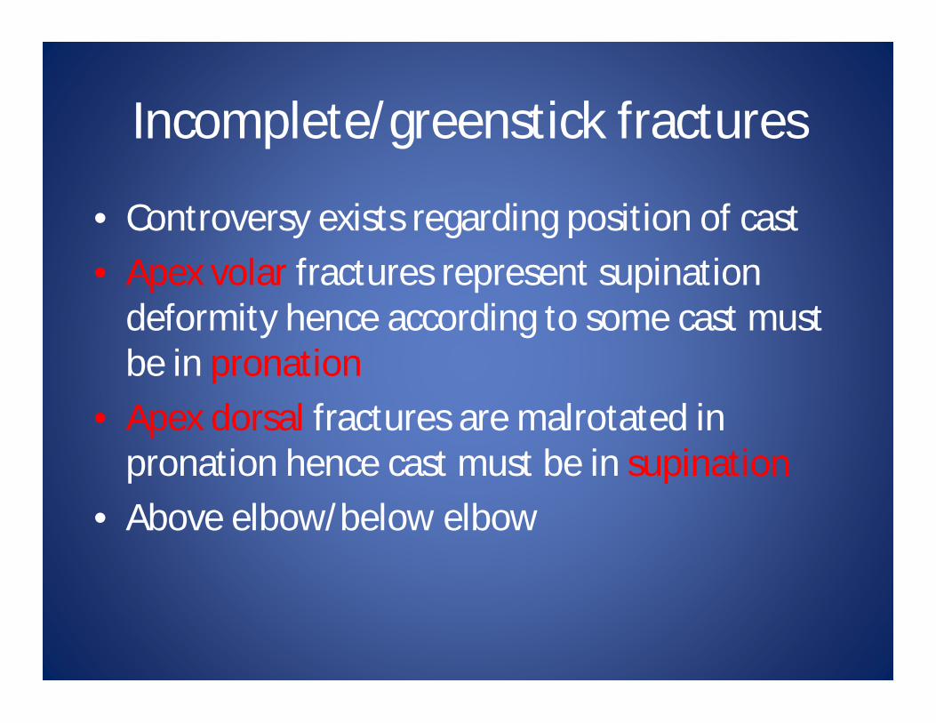
Incomplete/greenstick fractures
• Controversy exists regarding position of cast• Apex volar fractures represent supination
deformity hence according to some cast must be in pronation
• Apex dorsal fractures are malrotated in pronation hence cast must be in supination
• Above elbow/below elbow

Apex volar-plaster in pronation

Remodeling Potential Variables to Consider
• Age of child• Distance from fracture to physis
– Distal metaphyseal fractures most forgiving– Proximal forearm fractures: much less remodeling
• Angular deformities: – Physeal growth: correction of 0.8 - 1 degree per
month, or ~10 degrees per year• Rotational deformities will not remodel

Acceptable angular correctionAGE SAGITTAL PLANE FRONTAL PLANE
4-9 15-20 15
9-11 10-15 5
11-13 10 0
>13 0-5 0

Green stick fracture-radiusPre operative
Post operative

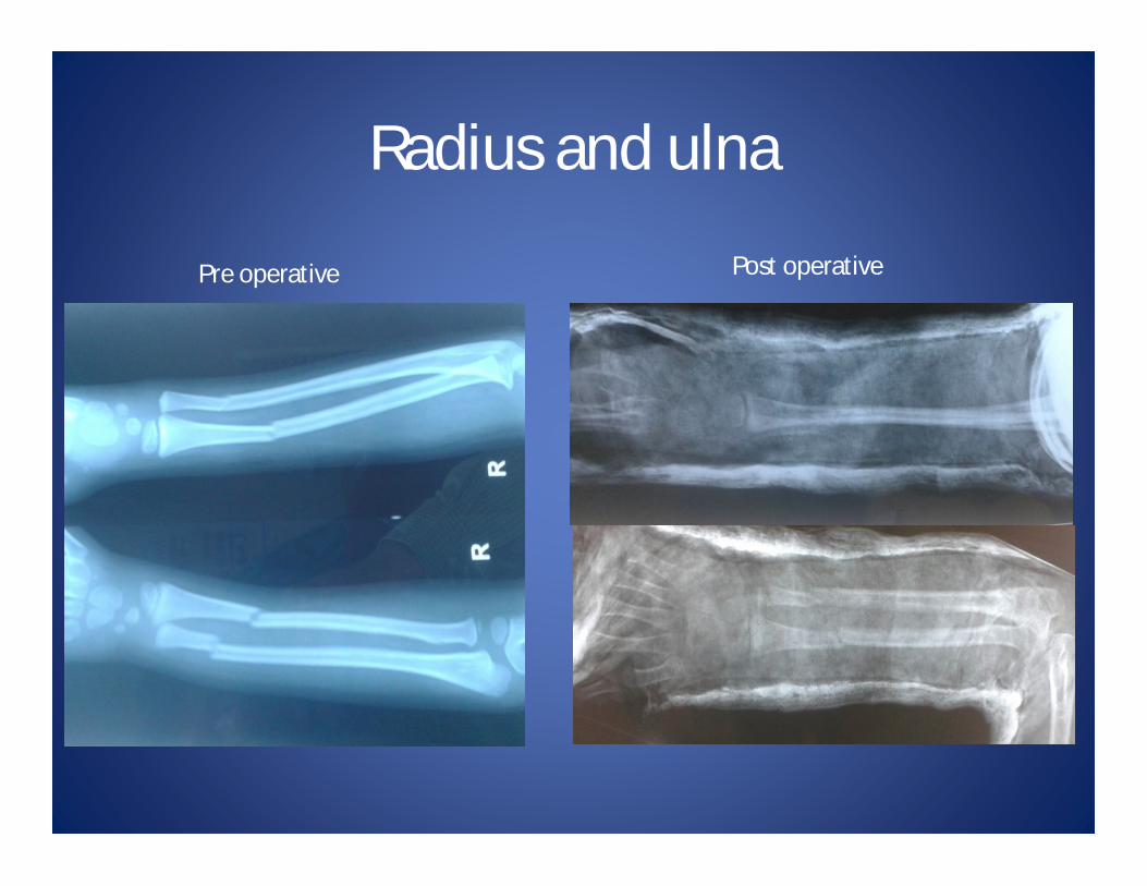
Radius and ulna
Pre operative Post operative

Risk factors for loss of reduction
• Poor casting• Bayoner apposition• Translation greater that 50% of diameter of
radius• Apex volar angulation greater that 30 deg• Isolated radius fractures• Radial and ulnar metaphyseal fractures at
same level

Distal end Radius fractureClinical

Reduction and fixationHyper dorsiflexion maneuver

Bayonet apposition

Reduction technique
Distraction Joystick
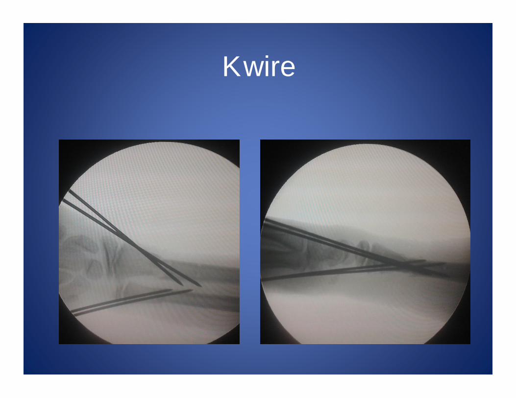
K wire

Compound fractureX ray Clinical
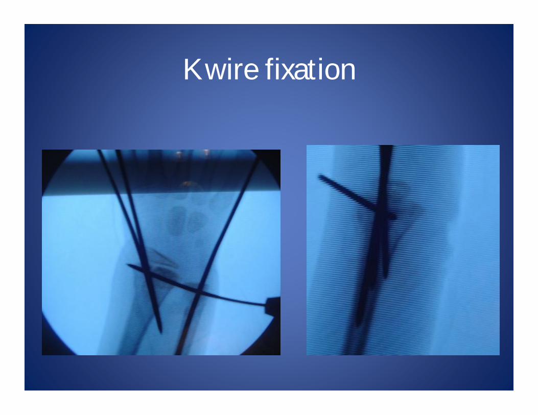
K wire fixation
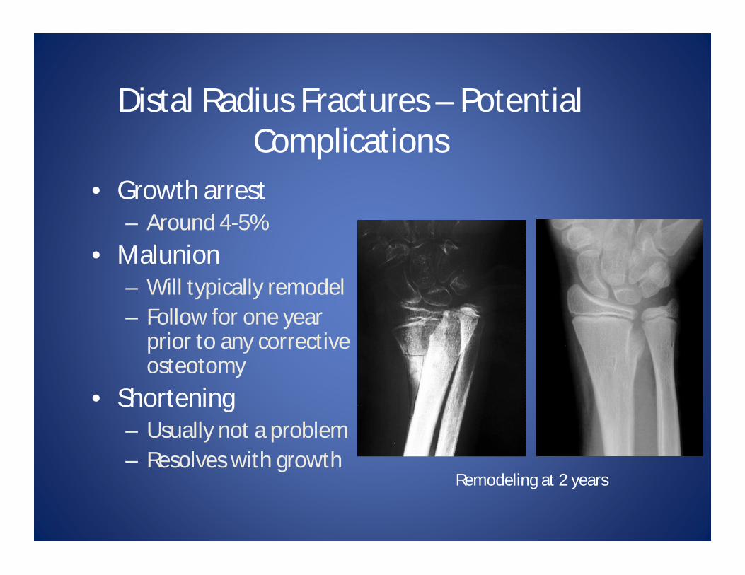
Distal Radius Fractures – Potential Complications
• Growth arrest – Around 4-5%
• Malunion– Will typically remodel– Follow for one year
prior to any corrective osteotomy
• Shortening – Usually not a problem – Resolves with growth
Remodeling at 2 years

Growth Arrest following Distal Radius Fracture
Injury films Injured and uninjured wrists after premature physeal closure

Distal Radius Growth Arrest
• Relatively rare (approx 4%)
• Related To:– Severity of trauma– Amount of
displacement– Repeated attempts at
reduction– Re-manipulation or
late manipulation

ComplicationPre operative Intra-op

After plaster removal
9 months followup
Clinical (at 9 mth)

Remodeling Potential - 12 yo Male
Presented 10 days after fracture – no reduction, splinted in ED and now with early healing – no additional reduction
At 6 months – extensive remodeling of deformity noted

Malunited distal end radius
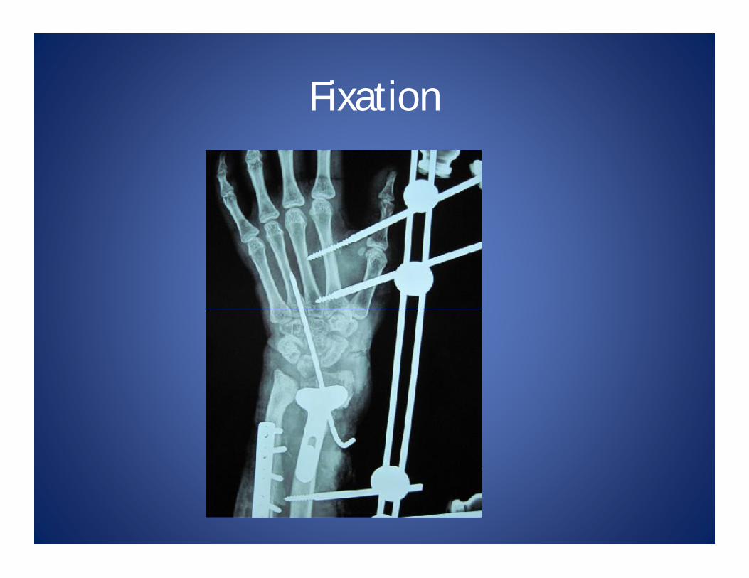
Fixation
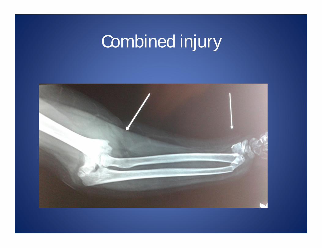
Combined injury
Pediatric Galeazzi fracture
• These fractures are often missed and may be
difficult to recognise.
• If there is an isolated radius fracture, always
examine the DRUJ on x-ray

Galeazzi Injury Complex
Dorsal Volar Equivalent
Fracture of distal radius associated with DRUJ disruption
Fracture of distal radius with distal ulnar physeal fracture

Treatment
• Most of these fractures can be managed with closed reduction. Fluoroscopy should be used to assess stability of the DRUJ after reduction.
• Adolescents are more likely to need open or percutaneous fixation to stabilise the DRUJ after reduction.
• Risk of ulna growth arrest (50%) in Galeazziequivalent
