distal radius
TRANSCRIPT
INTRODUCTION, SURGICALANATOMY/APPLIED ANATOMY, DEMOGRAPHICS OF DISTAL RADIUS FRACTURES, BIOMECHANICS OF FRACTURES AND IMPLANTS
DR.AKHILESWARI I I YR PG

HISTORY
• First surgeon to recognize these injuries was Pouteau in 1783.
• Later Abraham Colles in 1814 gave the classic description of fracture
• Dupuytren brought to the world attention that it is a fracture rather than a dislocation as it was previously assumed.
• Barton in1838 described wrist subluxation consequent to intraarticular fracture of radius which could be dorsal or volar.
• Smith described fracture of distal radius with ‘forward’ displacement.

INTRODUCTION:
• Distal radius fractures represent approximately one sixth(20%) of all fractures
• Distal radius fractures occur through the distal metaphysis of the radius
• May involve articular surface
• frequently involves the ulnar styloid

SURGICAL ANATOMY:
ARTICULAR SURFACE OF DISTAL RADIUS :
• Triangular in shape . The base of the triangle is formed by the sigmoid notch and apex by the radial styloid
• Has scaphoid and lunate fossae which articulate with the scaphoid and lunate bones respectively.
• The articular surface is inclined both in a volar and ulnar direction
• The sigmoid notch is semi cylindrical in shape allowing rotation of the radius around the ulnar head


PALMAR SURFACE OF RADIUS:
• Concave from proximal to distal• Smooth-easy contouring of plates• Provides attachment to radiocarpal ligaments-acts as
restraints to the normal tendency of carpus to slide in a volar and an ulnar direction
DORSAL SURFACE:• Convex and irregular• Most prominent area- LISTER’S tubercle• Grooves – form the floors of extensor compartments
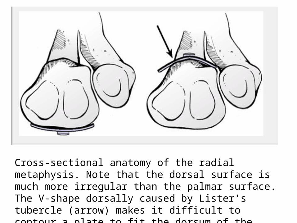
Cross-sectional anatomy of the radial metaphysis. Note that the dorsal surface is much more irregular than the palmar surface. The V-shape dorsally caused by Lister's tubercle (arrow) makes it difficult to contour a plate to fit the dorsum of the radius
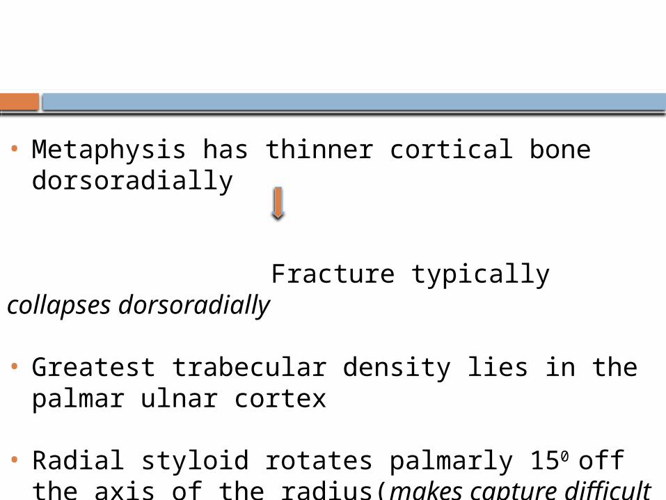
• Metaphysis has thinner cortical bone dorsoradially
Fracture typically collapses dorsoradially
• Greatest trabecular density lies in the palmar ulnar cortex
• Radial styloid rotates palmarly 150 off the axis of the radius(makes capture difficult from a dorsal approach)
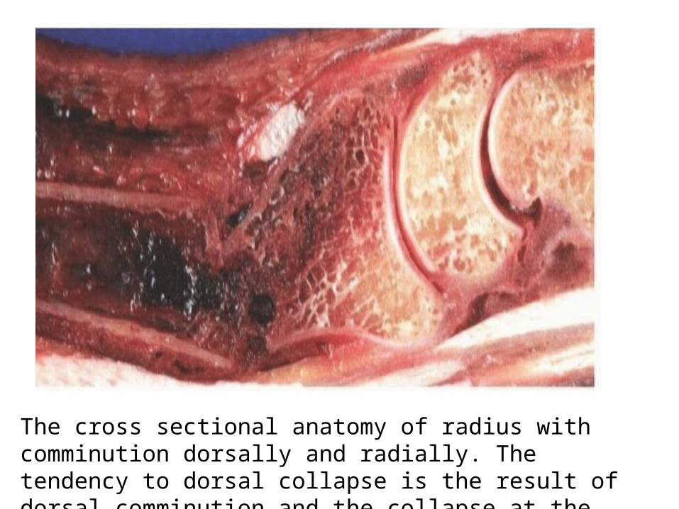
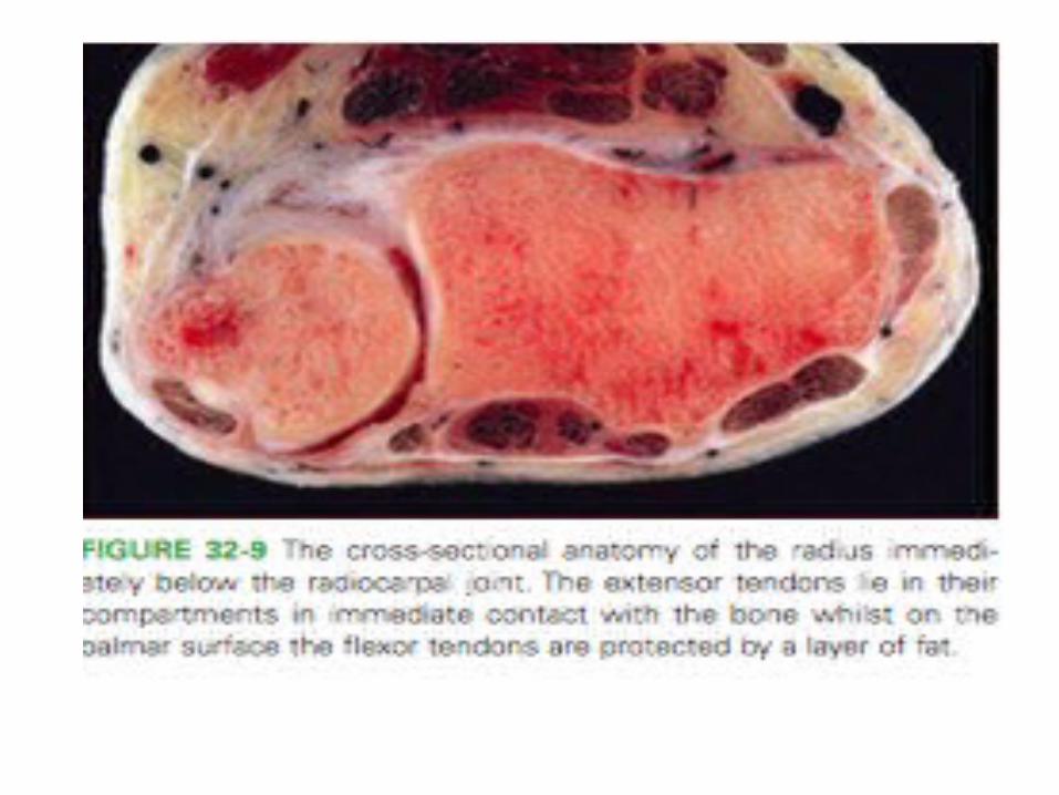
The cross sectional anatomy of radius with comminution dorsally and radially. The tendency to dorsal collapse is the result of dorsal comminution and the collapse at the midcarpal joint

LIGAMENTOUS ANATOMY:
• The extrinsic ligaments of the wrist play a major role in the use of indirect reduction techniques
• The palmar extrinsic ligaments are attached to the distal radius, and it is these ligaments that are relied on to reduce the components of a fracture using closed methods.

• There are two factors about these ligaments that make them significant for reduction
1. orientation of the extrinsic ligaments from the radial styloid is oblique relative to the more vertical orientation of the ligaments attached to the lunate facet.
2. thicker palmar ligaments when compared to the thinner dorsal ligaments

Dorsal ligaments are oriented in a relative “z” orientation, which allows them to lengthen with less force than the more vertically oriented palmar ligaments
Distraction will result in the palmar ligaments becoming taut before the dorsal ligaments.
Palmar cortex is brought out to length before the dorsal cortex
Difficult to achieve reduction of the normal 12 degrees of palmar tilt using distraction alone
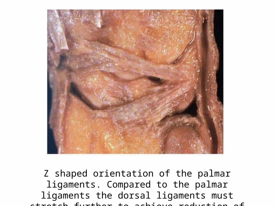
Z shaped orientation of the palmar ligaments. Compared to the palmar ligaments the dorsal ligaments must stretch further to
achieve reduction of the palmar tilt.

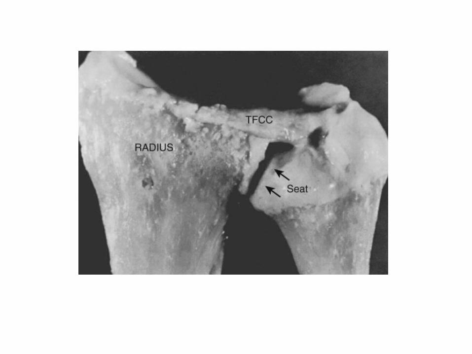
ROLE OF TFCC
• Also known as as ulnoligamentous complex• It consists of
The triangular fibrocartilage (TFC or articular disk), Meniscal homologue, Ulnocarpal [ulnolunate (UL) and lunotriquetral]
ligaments, The dorsal and volar radioulnar ligaments, Ulnar collateral ligament, and The extensor carpi ulnaris (ECU) subsheath
• TFCC is the main stabiliser of distal radioulnar joint in addition to contributing to ulnocarpal stability
• TFCC normally not only stabilises the ulnar head in sigmoid notch of radius but also acts as a buttress to support proximal carpal row
• During axial loading the radius carries the majority of load(82%),and the ulna a smaller load(18%)
• Increasing the ulnar variance to a positive 2.5mm increases the load transmission across the TFCC to 42%
• The TFCC excised the radial load increases to 94%
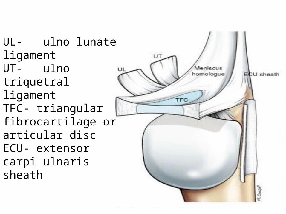
UL- ulno lunate ligamentUT- ulno triquetral ligamentTFC- triangular fibrocartilage or articular discECU- extensor carpi ulnaris sheath

APPLIED ANATOMY:
• Jakob and his co-authors interpreted the wrist as consisting of three distinct columns, each of which is subjected to different forces and thus must be addressed as discrete elements
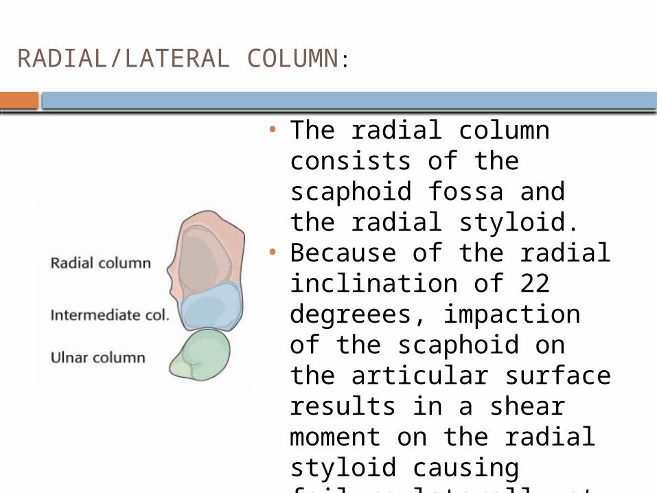
• The radial column consists of the scaphoid fossa and the radial styloid.
• Because of the radial inclination of 22 degreees, impaction of the scaphoid on the articular surface results in a shear moment on the radial styloid causing failure laterally at the radial cortex.
• The radial column, therefore, is best stabilized by buttressing the lateral cortex
RADIAL/LATERAL COLUMN:
• Consists of the lunate fossa and the sigmoid notch(DRUJ)
• Cornerstone of the radius because it is critical for both articular congruity and distal radioulnar function.
• Failure - occurs as a result of impaction of the lunate on the articular surface with dorsal comminution.
• The column is stabilized by a direct buttress of the dorsal ulnar aspect of the radius
INTERMEDIATE COLUMN:
MEDIAL COLUMN: The ulnar column consists of the distal ulna with the triangular fibrocartilage complex
DEMOGRAPHICS
• Incidence is both age and gender dependent
• There are three main peaks of fracture distribution children aged 5-14 years, male aged under 50 years and (athletic and high
energy injuries) females over the age of 40 years (fragility
fractures)• Majority of fractures in the elderly are extraarticular and
there is higher incidence of intra articular fractures in the younger patients
BIOMECHANICS
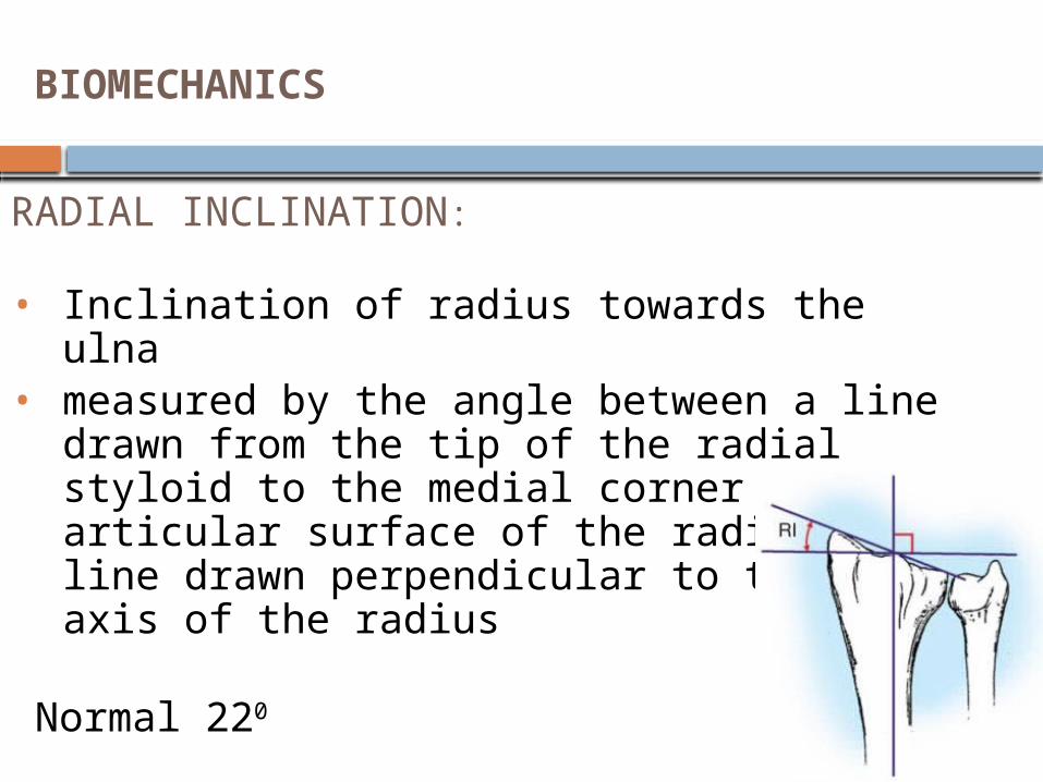
RADIAL INCLINATION: • Inclination of radius towards the ulna• measured by the angle between a line drawn from the
tip of the radial styloid to the medial corner of the articular surface of the radius and a line drawn perpendicular to the long axis of the radius
Normal 220 Acceptable range - <50 loss

• Measured by a line drawn perpendicular to the long axis of the radius and tangential to the most distal point of the ulnar head and a line drawn perpendicular to the long axis of the radius and at the level of the tip of the radial styloid
• Average : 11 mm (8-18) Acceptable <4mm loss compared to opp wrist >4mm increased load on lunate facet
RADIAL LENGTH:

DORSAL/PALMAR TILT:
A line is drawn connecting the most distal points of the volar and dorsal lips of the radius. The dorsal or palmar tilt is the angle created with a line drawn perpendicular along the longitudinal axis of the radius
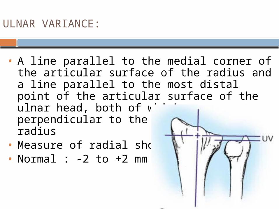
• A line parallel to the medial corner of the articular surface of the radius and a line parallel to the most distal point of the articular surface of the ulnar head, both of which are perpendicular to the long axis of the radius
• Measure of radial shortening• Normal : -2 to +2 mm
ULNAR VARIANCE:

• In lateral view, one line is drawn along the long axis of the capitate and one down the long axis of the radius. If the carpus is aligned, the lines will intersect within the carpus. If not, they will intersect outside the carpus.
CARPAL MALALLIGNMENT:
PATHOMECHANISM OF POSTERIORLY DISPLACED FRACTURE
• The usual cause is fall on the hyperextended wrist
• A)THE THEORY OF COMPRESSION IMPACTION
In hyperextension proximal carpal bones come and impact dorsal aspect of radius and body weight is transmitted through long axis of radius to distal end and compression occur at dorsal aspect of distal radius leading to fracture

B)THE AVULSION THEORY-
The indirect force presented by the body weight is transmitted through humerus, ulna, radius and then to volar wrist ligaments. Fracture occurs by avulsion mechanism applied by the tensile forces transmitted by the volar wrist ligaments

THE INCURVATION THEORY- Depends on position of the hand, the extent of the area of impact, the magnitude of the applied force
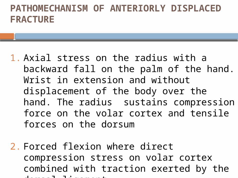
PATHOMECHANISM OF ANTERIORLY DISPLACED FRACTURE
1. Axial stress on the radius with a backward fall on the palm of the hand. Wrist in extension and without displacement of the body over the hand. The radius sustains compression force on the volar cortex and tensile forces on the dorsum
2. Forced flexion where direct compression stress on volar cortex combined with traction exerted by the dorsal ligament
BIOMECHANICS OF IMPLANTS
LIGAMENTOTAXIS:
It is the principle of molding fracture fragments into allignment as a result of tension applied across a fracture by the surrounding intact soft tissues.it neutralises the axial load placed on the distal radius by physiological activity of forearm musculature

OPEN REDUCTION AND INTERNAL FIXATION
Dorsal plating: The fixation is on the compression side of the fracture and provides a buttress against collapse.A. buttress plate applies a force to the bone which is perpendicular(normal) to the flat surface of the plate
Volar non locked plating—the primary indication is shear fracture of the volar lip. It may be unable to maintain fracture reduction in the presence of dorsal comminution
Volar locked plating---it has been shown to stabilize distal radius fracture with dorsal comminution
K-WIRE FIXATION:generally used to supplement short arm casting and for achieving palmar tilt in uniplanar ligamentotaxix

THANK YOU