van de graaff: human iv. support and movement 7. skeletal
TRANSCRIPT
Van De Graaff: Human Anatomy, Sixth Edition
IV. Support and Movement 7. Skeletal System: The Appendicular Skeleton
© The McGraw−Hill Companies, 2001
Skeletal System: The Appendicular Skeleton
Clinical Case StudyA 12-year-old boy was hit by a car while crossing a street. He was brought to the emergencyroom in stable condition, complaining of severe pain in his left leg. Radiographs revealed a 4-inch fracture extending superiorly from the distal articular surface of the tibia into the anteriorbody of the bone. The fragment of bone created by the fracture was moderately displaced. Withthe radiographs in hand, the orthopedic surgeon went into the waiting room and conferredwith the boy’s parents. He told them that this kind of injury was more serious in children andgrowing adolescents than in adults. He went on to say that future growth of the bone might bejeopardized and that surgery, although recommended, could not guarantee normal growth. Theparents asked, “What is it about this particular fracture that threatens future growth?”
If you were the surgeon, how would you respond?
Hints: Review the section on bone growth in chapter 6. Carefully examine figures 6.5 and 6.9in chapter 6 and figures 7.18 and 7.23 in this chapter.
Pectoral Girdle and Upper Extremity 173
Pelvic Girdle and Lower Extremity 178
CLINICAL CONSIDERATIONS 189
Clinical Case Study Answer 191
Developmental Exposition: The Appendicular Skeleton 192
Chapter Summary 194Review Activities 194
7
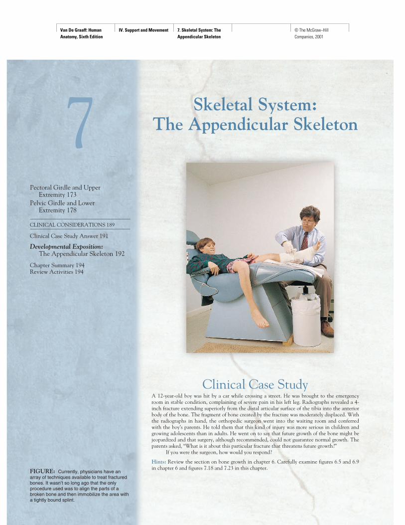
FIGURE: Currently, physicians have anarray of techniques available to treat fracturedbones. It wasn’t so long ago that the onlyprocedure used was to align the parts of abroken bone and then immobilize the area witha tightly bound splint.

Van De Graaff: Human Anatomy, Sixth Edition
IV. Support and Movement 7. Skeletal System: The Appendicular Skeleton
© The McGraw−Hill Companies, 2001
PECTORAL GIRDLE AND UPPER EXTREMITYThe structure of the pectoral girdle and upper extremities is adap-tive for freedom of movement and extensive muscle attachment.
Objective 1 Describe the bones of the pectoral girdle andthe articulations between them.
Objective 2 Identify the bones of the upper extremity andlist the distinguishing features of each.
Pectoral GirdleTwo scapulae and two clavicles make up the pectoral (shoulder)girdle (fig. 7.1). It is not a complete girdle, having only an ante-rior attachment to the axial skeleton, via the sternoclavicularjoint (see fig. 8.24) at the sternum. As an axial bone, the ster-num was described in chapter 6 (see fig. 6.38). Lacking a poste-rior attachment to the axial skeleton, the pectoral girdle has awide range of movement. Because it is not weight-bearing, it isstructurally more delicate than the pelvic girdle. The primaryfunction of the pectoral girdle is to provide attachment areas forthe numerous muscles that move the shoulder and elbow joints.
ClavicleThe slender S-shaped clavicle (klav'ı-kul; “collarbone”) connectsthe upper extremity to the axial skeleton and holds the shoulderjoint away from the trunk to permit freedom of movement. Thearticulation of the medial sternal extremity (fig. 7.2) of the clav-icle to the manubrium of the sternum is referred to as the stern-oclavicular joint. The lateral acromial (a-kro'me-al) extremity ofthe clavicle articulates with the acromion of the scapula (fig. 7.3). This articulation is referred to as the acromioclavicularjoint. A conoid tubercle is present on the acromial extremity ofthe clavicle, and a costal tuberosity is present on the inferiorsurface of the sternal extremity. Both processes serve as attach-ments for ligaments.
The long, delicate clavicle is the most commonly broken bonein the body. When a person receives a blow to the shoulder, or
attempts to break a fall with an outstretched hand, the force is trans-mitted to the clavicle, possibly causing it to fracture. The most vulner-able part of this bone is through its center, immediately proximal tothe conoid tubercle. Because the clavicle is directly beneath the skinand is not covered with muscle, a fracture can easily be palpated,and frequently seen.
ScapulaThe scapula (skap'you-la; “shoulder blade”) is a large, triangularflat bone on the posterior side of the rib cage, overlying ribs 2through 7. The spine of the scapula is a prominent diagonal bonyridge seen on the posterior surface (fig. 7.3). The spine strength-ens the scapula, making it more resistant to bending. Above thespine is the supraspinous fossa, and below the spine is the infra-spinous fossa. The spine broadens toward the shoulder as theacromion (figs. 7.3 and 7.4). This process serves for the attach-ment of several muscles, as well as for articulation with the clavi-cle. Inferior to the acromion is a shallow depression, the glenoid(gle'noid) cavity, into which the head of the humerus fits. Thecoracoid (kor'a-koid) process is a thick upward projection lyingsuperior and anterior to the glenoid cavity. On the anterior sur-face of the scapula is a slightly concave area known as the sub-scapular fossa.
The scapula has three borders delimited by three angles.The superior edge is called the superior border. The medial bor-der is nearest to the vertebral column, and the lateral border isdirected toward the arm. The superior angle is located betweenthe superior and medial borders; the inferior angle, at the junc-tion of the medial and lateral borders; and the lateral angle, atthe junction of the superior and lateral borders. It is at the lateralangle that the scapula articulates with the head of the humerus.Along the superior border, a distinct depression called the scapu-lar notch is a passageway for the suprascapular nerve.
The scapula has numerous surface features because 15 mus-cles attach to it. Clinically, the pectoral girdle is significant be-
cause the clavicle and acromion of the scapula are frequentlybroken in trying to break a fall. The acromion is used as a landmarkfor identifying the site for an injection in the arm. This site is chosenbecause the musculature of the shoulder is quite thick and containsfew nerves.
Brachium (Arm)The brachium (bra'ke-um) extends from the shoulder to the elbow.In strict anatomical usage, arm refers only to this portion of theupper limb. The brachium contains a single bone—the humerus.
HumerusThe humerus (fig. 7.5) is the longest bone of the upper extrem-ity. It consists of a proximal head, which articulates with the gle-noid cavity of the scapula; a body (“shaft”); and a distal end, which is modified to articulate with the two bones of the
Chapter 7 Skeletal System: The Appendicular Skeleton 173
CH
AP
TE
R 7
clavicle: L. clavicula, a small key
acromial: Gk. akros, peak; omos, shoulder
conoid tubercle: Gk. konus, cone; L. tuberculum, a small swelling
costal tuberosity: L. costa, rib; tuberous, a knob
scapula: L. scapula, shoulder
glenoid: Gk. glenoeides, shallow form
coracoid: Gk. korakodes, like a crow’s beak

Van De Graaff: Human Anatomy, Sixth Edition
IV. Support and Movement 7. Skeletal System: The Appendicular Skeleton
© The McGraw−Hill Companies, 2001
174 Unit 4 Support and Movement
CH
AP
TE
R 7
Skull
Ribcage
Oscoxae
Ilium
Pubis
Ischium
Sternum
Phalanges
Pelvicgirdle
Pectoralgirdle
Occipitalbone
Parietal boneTemporal bone
Maxilla
Mandible
Clavicle
ScapulaCostal cartilages
Humerus
Vertebral column
Ulna
Sacrum
Coccyx
Radius
Carpal bones
Metacarpal bones
Femur
Patella
Tibia
Fibula
Calcaneus
Tarsal bones
Metatarsal bones
Phalanges
(a) (b)
Ribs
Frontal bone
Zygomatic bone
Creek
FIGURE 7.1 The human skeleton. (a) An anterior view and (b) a posterior view. The axial portion is colored light blue.

Van De Graaff: Human Anatomy, Sixth Edition
IV. Support and Movement 7. Skeletal System: The Appendicular Skeleton
© The McGraw−Hill Companies, 2001
Chapter 7 Skeletal System: The Appendicular Skeleton 175
CH
AP
TE
R 7
Acromial extremity Sternal extremity
Creek
Body ofclavicle
Conoidtubercle
Conoidtubercle
Costaltuberosity
Acromial extremity
(a)
(b)
Sternal extremity
FIGURE 7.2 The right clavicle. (a) A superior view and (b) an infe-rior view.
Acromion ofscapula
Clavicle
Coracoid processof scapula
Head of humerus
Scapula
Greater tubercleof humerus
Body ofhumerus
FIGURE 7.3 This radiograph of the right shoulder shows the posi-tions of the clavicle, scapula, and humerus.
FIGURE 7.4 The right scapula. (a) An anterior view and (b) a posterior view.

Van De Graaff: Human Anatomy, Sixth Edition
IV. Support and Movement 7. Skeletal System: The Appendicular Skeleton
© The McGraw−Hill Companies, 2001
forearm. Surrounding the margin of the head is a slightly in-dented groove denoting the anatomical neck. The surgicalneck, the constriction just below the head, is a frequent fracturesite. The greater tubercle is a large knob on the lateral proximalportion of the humerus. The lesser tubercle is slightly anterior tothe greater tubercle and is separated from the greater by an inter-tubercular groove. The tendon of the long head of the bicepsbrachii muscle passes through this groove. Along the lateralmidregion of the body of the humerus is a roughened area, thedeltoid tuberosity, for the attachment of the deltoid muscle.Small openings in the body are called nutrient foramina.
The humeral condyle on the distal end of the humerushas two articular surfaces. The capitulum (ka-pit'yoo-lum) is
the lateral rounded part that articulates with the radius. Thetrochlea (trok'le-a) is the pulleylike medial part that articu-lates with the ulna. On either side above the condyle arethe lateral and medial epicondyles. The large medial epi-condyle protects the ulnar nerve that passes posteriorlythrough the ulnar sulcus. It is popularly known as the“funny bone” because striking the elbow on the edge of atable, for example, stimulates the ulnar nerve and producesa tingling sensation. The coronoid fossa is a depressionabove the trochlea on the anterior surface. The olecranon(o-lek'ra-non) fossa is a depression on the distal posteriorsurface. Both fossae are adapted to work with the ulna dur-ing movement of the forearm.
176 Unit 4 Support and Movement
CH
AP
TE
R 7
Greater tubercle
Lesser tubercle
Intertubercular groove
Deltoid tuberosity
Radial fossa
Lateral epicondyle
Capitulum
Lateral epicondyle
Nutrient foramen
Anatomical neck
Greater tubercleHead
Surgical neck
Body of humerus(posterior surface)
Body of humerus(anterior surface)
Olecranon fossa
Coronoid fossa
Medial epicondyle
Trochlea
Creek
Ulnar sulcus
FIGURE 7.5 The right humerus. (a) An anterior view and (b) a posterior view.
(a) (b)
deltoid: Gk. deltoeides, shaped like the letter ∆capitulum: L. caput, little head
trochlea: Gk. trochilia, a pulley
olecranon: Gk. olene, ulna; kranion, head

Van De Graaff: Human Anatomy, Sixth Edition
IV. Support and Movement 7. Skeletal System: The Appendicular Skeleton
© The McGraw−Hill Companies, 2001
The medical term for tennis elbow is lateral epicondylitis, whichmeans inflammation of the tissues surrounding the lateral epi-
condyle of the humerus. At least six muscles that control backward(extension) movement of the wrist and finger joints originate on thelateral epicondyle. Repeated strenuous contractions of these mus-cles, as in stroking with a tennis racket, may strain the periosteumand muscle attachments, resulting in swelling, tenderness, and painaround the epicondyle. Binding usually eases the pain, but only restcan eliminate the causative factor, and recovery generally follows.
Antebrachium (Forearm)The skeletal structures of the antebrachium are the ulna on themedial side and the radius on the lateral (thumb) side (figs. 7.6and 7.7). The ulna is more firmly connected to the humerus thanthe radius, and it is longer than the radius. The radius, however,contributes more significantly to the articulation at the wristjoint than does the ulna.
UlnaThe proximal end of the ulna articulates with the humerus andradius. A distinct depression, the trochlear notch, articulateswith the trochlea of the humerus. The coronoid process formsthe anterior lip of the trochlear notch, and the olecranon formsthe posterior portion. Lateral and inferior to the coronoidprocess is the radial notch, which accommodates the head ofthe radius.
On the tapered distal end of the ulna is a knobbed portion,the head, and a knoblike projection, the styloid process. Theulna articulates at both ends with the radius.
Chapter 7 Skeletal System: The Appendicular Skeleton 177
CH
AP
TE
R 7
Olecranon
Trochlear notch
Coronoid process
Tuberosity of ulna
Bodyof ulna
Interosseousborders
Ulnar notchof radius
Head of ulna
Styloid processof ulna
Radial notch of ulna
Head of radius
Neck of radius
Tuberosity of radius
Body ofradius
Styloid processof radius
Cre
ek
FIGURE 7.6 An anterior view of the right radius and ulna. FIGURE 7.7 A posterior view of the right radius and ulna.
styloid: Gk. stylos, pillar

Van De Graaff: Human Anatomy, Sixth Edition
IV. Support and Movement 7. Skeletal System: The Appendicular Skeleton
© The McGraw−Hill Companies, 2001
RadiusThe radius consists of a body with a small proximal end and alarge distal end. A proximal disc-shaped head articulates withthe capitulum of the humerus and the radial notch of the ulna.The prominent tuberosity of radius (radial tuberosity), for at-tachment of the biceps brachii muscle, is located on the medialside of the body, just below the head. On the distal end of the ra-dius is a double-faceted surface for articulation with the proximalcarpal bones. The distal end of the radius also has a styloidprocess on the lateral tip and an ulnar notch on the medial sidethat receives the distal end of the ulna. The styloid processes onthe ulna and radius provide lateral and medial stability for articu-lation at the wrist.
When a person falls, the natural tendency is to extend thehand to break the fall. This reflexive movement frequently re-
sults in fractured bones. Common fractures of the radius include afracture of the head, as it is driven forcefully against the capitulum; afracture of the neck; or a fracture of the distal end (Colles’ fracture),caused by landing on an outstretched hand.
When falling, it is less traumatic to the body to withdraw the ap-pendages, bend the knees, and let the entire body hit the surface.Athletes learn that this is the safe way to fall.
Manus (Hand)The hand contains 27 bones, grouped into the carpus, metacar-pus, and phalanges (figs. 7.8, 7.9, and 7.10).
CarpusThe carpus, or wrist, contains eight carpal bones arranged in twotransverse rows of four bones each. The proximal row, namingfrom the lateral (thumb) to the medial side, consists of thescaphoid (navicular), lunate, triquetrum (tri-kwé-trum) and pisi-form. The pisiform forms in a tendon as a sesamoid bone. Thedistal row, from lateral to medial, consists of the trapezium(greater multangular), trapezoid (lesser multangular), capitate,and hamate. The scaphoid and lunate of the proximal row artic-ulate with the distal end of the radius.
MetacarpusThe metacarpus, or palm of the hand, contains five metacarpalbones. Each metacarpal bone consists of a proximal base, a body,and a distal head that is rounded for articulation with the base ofeach proximal phalanx. The heads of the metacarpal bones aredistally located and form the knuckles of a clenched fist.
PhalangesThe 14 phalanges are the bones of the digits. A single fingerbone is called a phalanx (fa'langks). The phalanges of the fingersare arranged in a proximal row, a middle row, and a distal row.The thumb, or pollex (adjective, pollicis), lacks a middle phalanx.The digits are sequentially numbered I to V starting with thethumb—the lateral side, in reference to anatomical position.
A summary of the bones of the upper extremities is pre-sented in table 7.1.
The hand is a marvel of structural complexity that can withstandconsiderable abuse. Other than sprained ligaments of the fin-
gers and joint dislocations, the most common bone injury is a fractureto the scaphoid—a wrist bone that accounts for about 70% of carpalfractures. When immobilizing the wrist joint, the wrist is positioned inthe plane of relaxed function. This is the position in which the hand isabout to grasp an object between the thumb and index finger.
Knowledge Check1. Describe the structure of the pectoral girdle. Why is the
pectoral girdle considered an incomplete girdle?2. Identify the fossae and processes of the scapula.3. Describe each of the long bones of the upper extremity.4. Where are the styloid processes of the wrist area? What are
their functions?5. Name the bones in the proximal row of the carpus. Which
of these bones articulate(s) with the radius?
PELVIC GIRDLE AND LOWER EXTREMITYThe structure of the pelvic girdle and lower extremities is adaptivefor support and locomotion. Extensive processes and surface fea-tures on certain bones of the pelvic girdle and lower extremitiesaccommodate massive muscles used in body movement and inmaintaining posture.
Objective 3 Describe the structure of the pelvic girdle andlist its functions.
Objective 4 Describe the structural differences in the maleand female pelves.
Objective 5 Identify the bones of the lower extremity andlist the distinguishing features of each.
Objective 6 Describe the structural features and functionsof the arches of the foot.
178 Unit 4 Support and Movement
CH
AP
TE
R 7
carpus: Gk. karpos, wrist
navicular: L. navicula, small ship
lunate: L. lunare, crescent or moon-shaped
triquetrum: L. triquetrous, three-cornered
pisiform: Gk. pisos, pea
trapezium: Gk. trapesion, small table
capitate: L. capitatus, head
hamate: L. hamatus, hook phalanx: Gk. phalanx, finger bone or toe bone

Van De Graaff: Human Anatomy, Sixth Edition
IV. Support and Movement 7. Skeletal System: The Appendicular Skeleton
© The McGraw−Hill Companies, 2001
Chapter 7 Skeletal System: The Appendicular Skeleton 179
CH
AP
TE
R 7
Distal phalanx
Middle phalanx
Proximal phalanx
Distal phalanx
Proximal phalanx
First metacarpal bone
Trapezoid
Trapezium
Scaphoid
Lunate
Phalanges
Cre
ek
Metacarpalbones
CarpalbonesPisiform
Triquetral
HamateCapitate
Base
Body
Head
I
II
III
IV
V
Distalphalanx
Middlephalanx
Proximalphalanx
Hamate
Capitate
Trapezoid
Trapezium
Scaphoid
Lunate
Phalanges
Metacarpalbones
Carpalbones
Triquetral
III
IIIIV V
FIGURE 7.8 A posterior view of the bones of the right hand as shown in (a) a drawing and (b) a photograph. Each digit (finger) is indicated bya Roman numeral, the first digit, or thumb, being Roman numeral I.
(a)
(b)

Van De Graaff: Human Anatomy, Sixth Edition
IV. Support and Movement 7. Skeletal System: The Appendicular Skeleton
© The McGraw−Hill Companies, 2001
Pelvic GirdleThe pelvic girdle is formed by two ossa coxae (os'a kuk'se; “hip-bones”), united anteriorly at the symphysis pubis (figs. 7.11 and7.12). It is attached posteriorly to the sacrum of the vertebralcolumn. The sacrum, a bone of the axial skeleton, was describedin chapter 6 (see fig. 6.37). The deep, basinlike structure formedby the ossa coxae, together with the sacrum and coccyx, is calledthe pelvis (plural, pelves or pelvises). The pelvic girdle and its as-sociated ligaments support the weight of the body from the verte-bral column. The pelvic girdle also supports and protects thelower viscera, including the urinary bladder, the reproductive or-gans, and in a pregnant woman, the developing fetus.
The pelvis is divided into a greater (false) pelvis and alesser (true) pelvis (see fig. 7.15). These two components aredivided by the pelvic brim, a curved bony rim passing inferiorlyfrom the sacral promontory to the upper margin of the sym-physis pubis. The greater pelvis is the expanded portion of thepelvis, superior to the pelvic brim. The pelvic brim not only di-vides the two portions but surrounds the pelvic inlet of thelesser pelvis. The lower circumference of the lesser pelvisbounds the pelvic outlet.
180 Unit 4 Support and Movement
CH
AP
TE
R 7
Distal phalanx
Middle phalanx
Proximal phalanx
Distal phalanx
Proximal phalanx
First metacarpal bone
Trapezoid
Trapezium
Scaphoid
Capitate
Phalanges
Metacarpalbones
Carpalbones Pisiform
Triquetrum
Hamate
Lunate
I
II
III
IV
V
Head
Creek
BodyBase
FIGURE 7.9 An anterior view of the bones of the right hand.
Distal
Middle
Proximal
Pha
lang
es
Metacarpalbone
Hamate
Capitate
Triquetrum
Pisiform
LunateUlna
Distalphalanx
Proximalphalanx
Sesamoidbone
Trapezoid
Trapezium
Scaphoid
Radius
I
IIIIIIVV
FIGURE 7.10 A radiograph of the right hand shown in an antero-posterior projection. (Note the presence of a sesamoid bone at thethumb joint.) coxae: L. coxae, hips

Van De Graaff: Human Anatomy, Sixth Edition
IV. Support and Movement 7. Skeletal System: The Appendicular Skeleton
© The McGraw−Hill Companies, 2001
Each os coxae (“hipbone”) actually consists of three sepa-rate bones: the ilium, the ischium, and the pubis (figs. 7.13 and7.14). These bones are fused together in the adult. On the lateralsurface of the os coxae, where the three bones ossify, is a largecircular depression, the acetabulum (as''e-tab'yu-lum) which re-
ceives the head of the femur. Although both ossa coxae are sin-gle bones in the adult, the three components of each one areconsidered separately for descriptive purposes.
IliumThe ilium is the uppermost and largest of the three pelvic bones.It has a crest and four angles, or spines—important surface land-marks that serve for muscle attachment. The iliac crest forms theprominence of the hip. This crest terminates anteriorly as theanterior superior iliac spine. Just below this spine is the anterior
Chapter 7 Skeletal System: The Appendicular Skeleton 181
CH
AP
TE
R 7
TABLE 7.1 Bones of the Pectoral Girdle and Upper Extremities
Name and Number Location Major Distinguishing Features
Clavicle (2) Anterior base of neck, between sternum and scapula S-shaped; sternal and acromial extremities; conoid tubercle; costal tuberosity
Scapula (2) Upper back forming part of the shoulder Triangular; spine; subscapular, supraspinous, and infraspinous fossae; glenoidcavity; coracoid process; acromion
Humerus (2) Brachium, between scapula and elbow Longest bone of upper extremity; greater and lesser tubercles; intertuberculargroove; surgical neck, deltoid tuberosity; capitulum; trochlea; lateral andmedial epicondyles; coronoid and olecranon fossae
Ulna (2) Medial side of forearm Trochlear notch; olecranon; coronoid and styloid processes; radial notch
Radius (2) Lateral side of forearm Head; radial tuberosity; styloid process; ulnar notch
Carpal bone (16) Wrist Short bones arranged in two rows of four bones each
Metacarpal bone (10) Palm of hand Long bones, each aligned with a digit
Phalanx (28) Digits Three in each digit, except two in thumb
Base of sacrum Pelvic surfaceof sacrum
Superior articularprocess of sacrum
Iliac fossa
Anterior superioriliac spine
Anterior inferioriliac spine
Coccyx
Acetabular notch
Body of ischium
Inferior ramusof ischium
Pubis Symphysis pubis Inferior ramus of pubis
Pubic tubercle
Obturator foramen
Superior ramus of pubis
Acetabulum
Spine of ischium
Anterior sacral foramina
Sacroiliac articulation
Ilium
Cre
ek
Iliac crest
FIGURE 7.11 An anterior view of the pelvic girdle.
ilium: L. ilia, loin
ischium: Gk. ischion,hip joint
pubis: L. pubis, genital area
acetabulum: L. acetabulum, vinegar cup

Van De Graaff: Human Anatomy, Sixth Edition
IV. Support and Movement 7. Skeletal System: The Appendicular Skeleton
© The McGraw−Hill Companies, 2001
182 Unit 4 Support and Movement
CH
AP
TE
R 7
Fifth lumbarvertebra
Ilium
Sacrum
Coccyx
Pelvic inlet
Acetabulum
Pelvic brim
Pubis
Sacral promontory
Sacroiliac joint
Anterior inferioriliac spine
Head of femur
Neck of femur
Greater trochanterof femur
Obturator foramen
Lesser trochanter
Ischium
Symphysispubis
FIGURE 7.12 A radiograph of the pelvic girdle and the articulating femurs.
Ilium
Iliac crest
Anteriorsuperioriliac spine
Inferiorgluteal line
Anteriorinferioriliac spine
Superiorramus ofpubis
Inferior ramusof pubis
Obturatorforamen
Inferior ramusof ischium
Pubis
Creek
Anteriorgluteal line
Posteriorgluteal line
Posteriorsuperioriliac spine
Posteriorinferioriliac spine
Greatersciatic notch
Spine of ischium
Lesser sciaticnotchIschium
Ischialtuberosity
Acetabulum
FIGURE 7.13 The lateral aspect of the right os coxae. (The threebones comprising the os coxae are labeled in boldface type.)
Ilium
Iliac crest
Anteriorsuperioriliac spine
Anteriorinferioriliac spine
Arcuate line
Superiorramis of pubis
Pubictubercle
Symphysialsurface
Pubis
Inferior ramusof pubis
Inferior ramusof ischium
Ischial tuberosity
Obturator foramen
Lesser sciatic notchSpine of ischium
Greatersciatic notch
Posterior inferioriliac spine
Posterior superioriliac spine
Auricularsurface
Iliac tuberosity
Ischium
FIGURE 7.14 The medial aspect of the right os coxae. (The threebones comprising the os coxae are labeled in boldface type.)

Van De Graaff: Human Anatomy, Sixth Edition
IV. Support and Movement 7. Skeletal System: The Appendicular Skeleton
© The McGraw−Hill Companies, 2001
inferior iliac spine. The posterior termination of the iliac crest isthe posterior superior iliac spine, and just below this is the pos-terior inferior iliac spine.
Below the posterior inferior iliac spine is the greater sciaticnotch, through which the sciatic nerve passes. On the medialsurface of the ilium is the roughened auricular surface, whicharticulates with the sacrum. The iliac fossa is the smooth, con-cave surface on the anterior portion of the ilium. The iliacusmuscle originates from this fossa. The iliac tuberosity, for the at-tachment of the sacroiliac ligament, is positioned posterior to theiliac fossa. Three roughened ridges are present on the glutealsurface of the posterior aspect of the ilium. These ridges, whichserve to attach the gluteal muscles, are the inferior, anterior,and posterior gluteal lines (see fig. 7.13).
IschiumThe ischium (is'ke-um) is the posteroinferior bone of the oscoxae. This bone has several distinguishing features. The spine ofthe ischium is the projection immediately posterior and inferiorto the greater sciatic notch of the ilium. Inferior to this spine isthe lesser sciatic notch of the ischium. The ischial tuberosity isthe bony projection that supports the weight of the body in thesitting position. A deep acetabular (as''e-tab'yu-lar) notch is pre-sent on the inferior portion of the acetabulum. The large obtura-tor (ob'tu-ra''tor) foramen is formed by the inferior ramus of the
Chapter 7 Skeletal System: The Appendicular Skeleton 183
CH
AP
TE
R 7
FIGURE 7.15 A comparison of (a) the male and (b) the femalepelvic girdle.
TABLE 7.2 Comparison of the Male and Female Pelves
Characteristics Male Pelvis Female Pelvis
General structure More massive; More delicate; processes prominent processes not so prominent
Pelvic inlet Heart-shaped Round or oval
Pelvic outlet Narrower Wider
Anterior superior Not as wide apart Wider apartiliac spines
Obturator foramen Oval Triangular
Acetabulum Faces laterally Faces more anteriorly
Symphysis pubis Deeper, longer Shallower, shorter
Pubic arch Angle less than 90° Angle greater than 90°
ischium, together with the pubis. The obturator foramen is cov-ered by the obturator membrane, to which several muscles attach.
PubisThe pubis is the anterior bone of the os coxae. It consists of a su-perior ramus and an inferior ramus that support the body of thepubis. The body contributes to the formation of the symphysispubis—the joint between the two ossa coxae. At the lateral endof the anterior border of the body is the pubic tubercle, one ofthe attachments for the inguinal ligament.
The structure of the human pelvis, in its attachment to the ver-tebral column, permits an upright posture and locomotion on
two appendages (bipedal locomotion). An upright posture maycause problems, however. The sacroiliac joint may weaken with age,causing lower back pains. The weight of the viscera may weaken thewalls of the lower abdominal area and cause hernias. Some of theproblems of childbirth are related to the structure of the mother’spelvis. Finally, the hip joint tends to deteriorate with age, so thatmany elderly people suffer from degenerative arthritis(osteoarthrosis).
Sex-Related Differences in the PelvisStructural differences between the pelvis of an adult male andthat of an adult female (fig. 7.15 and table 7.2) reflect the fe-male’s role in pregnancy and parturition. In a vaginal delivery, ababy must pass through its mother’s lesser pelvis. Pelvimetry (pel-vim'e-tre) is the measurement of the dimensions of the pelvis—especially of the adult female pelvis—to determine whether acesarean section might be necessary. Diameters may be deter-mined by vaginal palpation or by sonographic images.
ThighThe femur is the only bone of the thigh. In the following discus-sion, however, the patella will also be discussed.

Van De Graaff: Human Anatomy, Sixth Edition
IV. Support and Movement 7. Skeletal System: The Appendicular Skeleton
© The McGraw−Hill Companies, 2001
FemurThe (fe'mur; “thighbone”) is the longest, heaviest, strongestbone in the body (fig. 7.16). The proximal rounded head of thefemur articulates with the acetabulum of the os coxae. Aroughened shallow pit, the fovea capitis femoris, is present inthe lower center of the head of the femur. The fovea capitisfemoris provides the point of attachment for the ligamentumcapitis femoris (see fig. 8.30), which helps to support the headof the femur against the acetabulum. It also provides the site forthe entry of an artery into the head of the femur. The con-stricted region supporting the head is called the neck and is acommon site for fractures in the elderly.
The body of the femur has a slight medial curve to bringthe knee joint in line with the body’s plane of gravity. The de-gree of curvature is greater in the female because of the widerpelvis. The body of the femur has several distinguishing featuresfor muscle attachment. On the proximolateral side of the body isthe greater trochanter, and on the medial side is the lessertrochanter. On the anterior side, between the trochanters, is theintertrochanteric (in''ter-tro''kan-ter'ik) line. On the posteriorside, between the trochanters, is the intertrochanteric crest.The linea aspera (lin'e-a as'per-a) is a roughened vertical ridge onthe posterior surface of the body of the femur.
The distal end of the femur is expanded for articulationwith the tibia. The medial and lateral condyles are the articular
184 Unit 4 Support and Movement
CH
AP
TE
R 7
Greater trochanter
Intertrochanteric line
Lateral epicondyle
Patellar surface
Head of femurGreater trochanter
Intertrochanteric crest
Gluteal tuberosity
Linea aspera
Lateral epicondyle
Cre
ek
Lateral condyleIntercondylar fossa
Fovea capitisfemoris
Neck of femur
Lesser trochanter
Body of femur
Medial epicondyle
Medial condyle
(a) (b)
FIGURE 7.16 The right femur. (a) An anterior view and (b) a posterior view.
femur: L. femur, thigh linea aspera: L. linea, line; asperare, rough

Van De Graaff: Human Anatomy, Sixth Edition
IV. Support and Movement 7. Skeletal System: The Appendicular Skeleton
© The McGraw−Hill Companies, 2001
processes for this joint. The shallow depression between thecondyles on the posterior aspect is called the intercondylarfossa. The patellar surface is located between the condyles onthe anterior side. Above the condyles on the lateral and medialsides are the epicondyles, which serve for ligament and tendonattachment.
PatellaThe patella (pa-tel'a; “kneecap”) is a large, triangular sesamoidbone positioned on the anterior side of the distal femur(figs. 7.17 and 7.18). It develops in response to strain in thepatellar tendon. It has a broad base and an inferiorly pointedapex. Articular facets on the articular surface of the patella ar-ticulate with the medial and lateral condyles of the femur.
The functions of the patella are to protect the knee jointand to strengthen the patellar tendon. It also increases the lever-age of the quadriceps femoris muscle as it extends (straightens)the knee joint.
The patella can be fractured by a direct blow. It usually doesnot fragment, however, because it is confined within the patel-
lar tendon. Dislocations of the patella may result from injury or fromunderdevelopment of the lateral condyle of the femur.
LegTechnically speaking, leg refers only to that portion of the lowerlimb between the knee and foot. The tibia and fibula are the bonesof the leg. The tibia is the larger and more medial of the two.
Chapter 7 Skeletal System: The Appendicular Skeleton 185
CH
AP
TE
R 7
Femur
Lateralepicondyleof femur
Patella
Head oftibia
Tibia
Fibula
FIGURE 7.17 A radiograph of the right knee.
TibiaThe tibia (tib'ea; “shinbone”) articulates proximally with thefemur at the knee joint and distally with the talus of the ankle. Italso articulates both proximally and distally with the fibula. Twoslightly concave surfaces on the proximal end of the tibia, themedial and lateral condyles (fig. 7.18) articulate with thecondyles of the femur. The condyles are separated by a slight up-ward projection called the intercondylar eminence, which pro-vides attachment for the cruciate ligaments of the knee joint (seefigs. 8.31 and 8.32). The tibial tuberosity, for attachment of thepatellar ligament, is located on the proximoanterior part of thebody of the tibia. The anterior crest, commonly called the“shin,” is a sharp ridge along the anterior surface of the body.
The medial malleolus (ma-le'o-lus) is a prominent medialknob of bone located on the distomedial end of the tibia. A fibu-lar notch, for articulation with the fibula, is located on the disto-lateral end. In that the tibia is the weight-bearing bone of theleg, it is much larger than the fibula.
FibulaThe fibula (fib'yu-la) is a long, slender bone that is more impor-tant for muscle attachment than for support. The head of thefibula articulates with the proximolateral end of the tibia. Thedistal end has a prominent knob called the lateral malleolus.
The lateral and medial malleoli are positioned on either side ofthe talus and help to stabilize the ankle joint. Both processes
can be seen as prominent surface features and are easily palpated.Fractures to the fibula above the lateral malleolus are common inskiers. Clinically referred to as Pott’s fracture, it is caused by a shear-ing force acting at a vulnerable spot on the leg.
Pes (Foot)The foot contains 26 bones, grouped into the tarsus, metatarsus,and phalanges (fig. 7.19). Although similar to the bones of thehand, the bones of the foot have distinct structural differences inorder to support the weight of the body and provide leverage andmobility during walking.
TarsusThere are seven tarsal bones. The most superior in position is thetalus, which articulates with the tibia and fibula to form theankle joint. The calcaneus (kal-ka'ne-us) is the largest of thetarsal bones and provides skeletal support for the heel of the foot.It has a large posterior extension, called the tuberosity of the
patella: L. patina, small plate
tibia: L. tibia, shinbone, pipe, flute
fibula: L. fibula, clasp or brooch
malleolus: L. malleolus, small hammer
tarsus: Gk. tarsos, flat of the foot
talus: L. talus, ankle

Van De Graaff: Human Anatomy, Sixth Edition
IV. Support and Movement 7. Skeletal System: The Appendicular Skeleton
© The McGraw−Hill Companies, 2001
calcaneus, for the attachment of the calf muscles. Anterior tothe talus is the block-shaped navicular bone. The remaining fourtarsal bones form a distal series that articulate with themetatarsal bones. They are, from the medial to the lateral side,the medial, intermediate, and lateral cuneiform (kyoo-ne' ı-form)bones and the cuboid bone.
MetatarsusThe metatarsal bones and phalanges are similar in name andnumber to the metacarpals and phalanges of the hand. They
differ in shape, however, because of their load-bearing role.The metatarsal bones are numbered I to V, starting with themedial (great toe) side of the foot. The first metatarsal bone islarger than the others because of its major role in supportingbody weight.
The metatarsal bones each have a base, body, and head.The proximal bases of the first, second, and third metatarsals ar-ticulate proximally with the cuneiform bones. The heads of themetatarsals articulate distally with the proximal phalanges. Theproximal joints are called tarsometatarsal joints, and the distaljoints are called metatarsophalangeal (met''a-tar''so-fa-lan'je-al)joints. The ball of the foot is formed by the heads of the first twometatarsal bones.
186 Unit 4 Support and Movement
CH
AP
TE
R 7
Base of patella
Medialcondyle
Tibial tuberosity
Anteriorborder
Body oftibia
Medial malleolus
Lateral malleolus
Body offibula
Fibular articularsurface
Head of fibula
Lateral condyle
Intercondylar eminence
Articular surface
Cre
ek
Anterior surface
Apex of patella
Intercondylar eminence
Articular surfaceof fibular head
Neck of fibula
Lateral malleolus
Patella
Tibia
Fibula
FIGURE 7.18 The right tibia, fibula, and patella. (a) An anterior view and (b) a posterior view.
(a) (b)
calcaneus: L. calcis, heel

Van De Graaff: Human Anatomy, Sixth Edition
IV. Support and Movement 7. Skeletal System: The Appendicular Skeleton
© The McGraw−Hill Companies, 2001
Chapter 7 Skeletal System: The Appendicular Skeleton 187
CH
AP
TE
R 7
Phalanges
Metatarsalbones
Tarsalbones
Distal phalanx
Middle phalanx
Proximal phalanx
Medialcuneiform bone
Intermediatecuneiform bone
Lateralcuneiform bone
Navicular bone
Cuboid bone
Talus
Calcaneus
I II IIIIV V
Distal phalanx
Proximalphalanx
Sesamoidbone
Metatarsal bones
Cuneiform bone
Navicular bone
Talus
Tibia
FibulaCalcaneus
I
Distal phalanx
Proximal phalanx
First metatarsalbone
Medial cuneiform bone
Intermediatecuneiform bone
Lateral cuneiformbone
Navicular bone
Talus
Tuberosity of calcaneus
Calcaneus
Cuboid bone
Fifth metatarsal bone
Proximal phalanx
Middle phalanx
Distal phalanx
Phalanges
Metatarsalbones
Head
Body
Base
Tarsalbones
Creek
II
III
IV
V
FIGURE 7.19 The bones of the right foot. (a) A photograph of a superior view, (b) a radiograph of a medial view, (c) a superior view, and (d) an inferior view. Each digit (toe) is indicated by a Roman numeral, the first digit, or great toe, being Roman numeral I.
(a) (b)
(c) (d)

Van De Graaff: Human Anatomy, Sixth Edition
IV. Support and Movement 7. Skeletal System: The Appendicular Skeleton
© The McGraw−Hill Companies, 2001
PhalangesThe 14 phalanges are the skeletal elements of the toes. As withthe fingers of the hand, the phalanges of the toes are arranged in aproximal row, a middle row, and a distal row. The great toe, or hal-lux (adjective, hallucis) has only a proximal and a distal phalanx.
Arches of the FootThe foot has two arches. They are formed by the structure andarrangement of the bones and maintained by ligaments and ten-dons (fig. 7.20). The arches are not rigid; they “give” whenweight is placed on the foot, and they spring back as the weightis lifted.
The longitudinal arch is divided into medial and lateralparts. The medial part is the more elevated of the two. The talusis keystone of the medial part, which originates at the calcaneus,rises at the talus, and descends to the first three metatarsal bones.The shallower lateral part consists of the calcaneus, cuboid, andfourth and fifth metatarsal bones. The cuboid is the keystonebone of this arch.
The transverse arch extends across the width of the footand is formed by the calcaneus, navicular, and cuboid bones pos-teriorly and the bases of all five metatarsal bones anteriorly.
A weakening of the ligaments and tendons of the foot maycause the arches to “fall”—a condition known as pes planus, or,more commonly, “flatfoot.”
The bones of the lower extremities are summarized intable 7.3.
188 Unit 4 Support and Movement
CH
AP
TE
R 7
Creek
Cuneiformbones
Cuboidbone
Talus
Calcaneus
NavicularboneTransverse arch
Longitudinal arch
First metatarsal bone
Phalanges ofbig toe
Bases ofmetatarsalbones
Transverse arch
(a)
(b)
FIGURE 7.20 The arches of the foot. (a) A medial view of theright foot showing both arches and (b) a transverse view throughthe bases of the metatarsal bones showing a portion of the trans-verse arch.
TABLE 7.3 Bones of the Pelvic Girdle and Lower Extremities
Name and Number Location Major Distinguishing Features
Os coxae (2) Hip, part of the pelvic girdle; composed of the Iliac crest; acetabulum; anterior superior iliac spine; greater sciatic notch of fused ilium, ischium, and pubis the ilium; ischial tuberosity; lesser sciatic notch of the ischium; obturator
foramen; pubic tubercle
Femur (2) Bone of the thigh, between hip and knee Head; fovea capitis femoris; neck; greater and lesser trochanters; linea aspera;lateral and medial condyles; lateral and medial epicondyles
Patella (2) Anterior surface of distal femur Triangular sesamoid bone
Tibia (2) Medial side of leg, between knee and ankle Medial and lateral condyles; intercondylar eminence; tibial tuberosity; anterior crest; medial malleolus; fibular notch
Fibula (2) Lateral side of leg, between knee and ankle Head; lateral malleolus
Tarsal bones (14) Ankle Large talus and calcaneus to receive weight of leg; five other wedge-shapedbones to help form arches of foot
Metatarsal bones (10) Sole of foot Long bones, each in line with a digit
Phalanx (28) Digits Three in each digit except two in great toe

Van De Graaff: Human Anatomy, Sixth Edition
IV. Support and Movement 7. Skeletal System: The Appendicular Skeleton
© The McGraw−Hill Companies, 2001
Knowledge Check6. Describe the structure and functions of the pelvic girdle.
How does its structure reflect its weight-bearing role?7. How can female and male pelves be distinguished? Why is
the lesser pelvis clinically significant in females?8. Describe the structure of each of the long bones of the lower
extremity and the position of each of the tarsal bones.9. Which bones of the foot contribute to the formation of the
arches? What are the functions of the arches?
CLINICAL CONSIDERATIONS
Developmental DisordersMinor defects of the extremities are relatively common malfor-mations. Extra digits, a condition called polydactyly (pol''e-dak't ı-le; fig. 7.21), is the most common limb deformity. Usuallyan extra digit is incompletely formed and does not function.Syndactyly (sin-dak't ı-le), or webbed digits, is also common.Polydactyly is inherited as a dominant trait, whereas syndactylyis a recessive trait.
Talipes (tal'ı-pez) or “clubfoot” (fig. 7.22), is a congenitalmalformation in which the sole of the foot is twisted medially. Itis not certain whether it is abnormal positioning or restrictedmovement in utero that causes this condition, but both geneticsand environmental conditions are involved in most cases.
Trauma and InjuryThe most common type of bone injury is a fracture—the crack-ing or breaking of a bone. Radiographs are often used to diagnosethe precise location and extent of a fracture. Fractures may beclassified in several ways, and the type and severity of the frac-ture is often related to the age and general health of the individ-ual. Pathologic fractures, for example, result from diseases thatweaken the bones. Most fractures, however, are called traumaticfractures because they are caused by injuries. The following aredescriptions of several kinds of traumatic fractures (fig. 7.23).
1. Simple, or closed. The fractured bone does not breakthrough the skin.
2. Compound, or open. The fractured bone is exposed to theoutside through an opening in the skin.
3. Partial (fissured). The bone is incompletely broken.
4. Complete. The fracture has separated the bone into twopieces.
5. Comminuted (kom'ı-noot'ed). The bone is splintered intoseveral fragments.
6. Spiral. The fracture line is twisted as it is broken.
7. Greenstick. An incomplete break (partial fracture), inwhich one side of the bone is broken, and the other sideis bowed.
8. Impacted. One end of a broken bone is driven into the other.
9. Transverse. The fracture occurs across the bone at a rightangle to the long axis.
10. Oblique. The fracture occurs across the bone at an obliqueangle to the long axis.
Chapter 7 Skeletal System: The Appendicular Skeleton 189
CH
AP
TE
R 7
FIGURE 7.21 Polydactyly is the condition in which there are extradigits. It is the most common congenital deformity of the foot, al-though it also occurs in the hand. Syndactyly is the condition inwhich two or more digits are webbed together. It is a common con-genital deformity of the hand, although it also occurs in the foot. Bothconditions can be surgically corrected.
FIGURE 7.22 Talipes, or clubfoot, is a congenital malformation ofa foot or both feet. The condition can be effectively treated surgicallyif the procedure is done at an early age.
polydactyly: Gk. polys, many; daktylos, finger
syndactyly: Gk. syn, together; daktylos, finger
talipes: L. talus, heel; pes, foot

Van De Graaff: Human Anatomy, Sixth Edition
IV. Support and Movement 7. Skeletal System: The Appendicular Skeleton
© The McGraw−Hill Companies, 2001
11. Colles’. A fracture of the distal portion of the radius.
12. Pott’s. A fracture of either or both of the distal ends of thetibia and fibula at the level of the malleoli.
13. Avulsion. A portion of a bone is torn off.
14. Depressed. The broken portion of the bone is driven in-ward, as in certain skull fractures.
15. Displaced. A fracture in which the bone fragments are notin anatomical alignment.
16. Nondisplaced. A fracture in which the bone fragments re-main in anatomical alignment.
When a bone fractures, medical treatment involves re-aligning the broken ends and then immobilizing them until newbone tissue has formed and the fracture has healed. The site andseverity of the fracture and the age of the patient determines thetype of immobilization. The methods of immobilization include
tape, splints, casts, straps, wires, screws, plates, and steel pins.Certain fractures seem to resist healing, however, even with thisarray of treatment options. New techniques for treating fracturesinclude applying weak electrical currents to fractured bones. Thismethod has shown promise in promoting healing and signifi-cantly reducing the time of immobilization.
Physicians can realign and immobilize a fracture, but theultimate repair of the bone occurs naturally within the bone it-self. Several steps are involved in this process (fig. 7.24).
1. When a bone is fractured, the surrounding periosteum isusually torn and blood vessels in both tissues are ruptured.A blood clot called a fracture hematoma (hem''a-to'ma)soon forms throughout the damaged area. A disruptedblood supply to osteocytes and periosteal cells at the frac-ture site causes localized cellular death. This is followed byswelling and inflammation.
190 Unit 4 Support and Movement
CH
AP
TE
R 7
A greenstick fracture is incomplete,and the break occurs on the convexsurface of the bend in the bone.
A partial (fissured) fractureinvolves an incomplete break.
A comminuted fracture iscomplete and results inseveral bony fragments.
A spiral fracture iscaused by twisting abone excessively.
An oblique fracture is complete,and the fracture line is at an angleto the long axis of the bone.
A transverse fracture is complete,and the fracture line is horizontal.
Cre
ek
FIGURE 7.23 Examples of fractures.
Colles’ fracture: from Abraham Colles, Irish surgeon, 1773–1843
Pott’s fracture: from Percivall Pott, British surgeon, 1713–88 hematoma: Gk. hema, blood; oma, tumor

Van De Graaff: Human Anatomy, Sixth Edition
IV. Support and Movement 7. Skeletal System: The Appendicular Skeleton
© The McGraw−Hill Companies, 2001
2. The traumatized area is “cleaned up” by the activity ofphagocytic cells within the blood and osteoclasts that re-sorb bone fragments. As the debris is removed, fibrocarti-lage fills the gap within the fragmented bone, and acartilaginous mass called a bony callus is formed. The bonycallus becomes the precursor of bone formation in muchthe same way that hyaline cartilage serves as the precursorof developing bone.
3. The remodeling of the bony callus is the final step in thehealing process. The cartilaginous callus is broken down, anew vascular supply is established, and compact bone de-
Chapter 7 Skeletal System: The Appendicular Skeleton 191
CH
AP
TE
R 7
(e)
FIGURE 7.24 Stages (a–d ) of the repair of a fracture. (e) A radiograph of a healing fracture.
Clinical Case Study AnswerThe injury involves the cartilaginous epiphyseal growth plate, which isthe site of linear growth in long bones. At cessation of growth, this platedisappears as the epiphysis and diaphysis fuse. Until this occurrence,however, disruption of the growth plate can adversely affect growth ofthe bone.
velops around the periphery of the fracture. A healed frac-ture line is frequently undetectable in a radiograph, exceptthat for a period of time the bone in this area may beslightly thicker.
callus: L. callosus, hard

Van De Graaff: Human Anatomy, Sixth Edition
IV. Support and Movement 7. Skeletal System: The Appendicular Skeleton
© The McGraw−Hill Companies, 2001
192
The Appendicular Skeleton
EXPLANATIONThe development of the upper and lower extremities is initiatedtoward the end of the fourth week with the appearance of foursmall elevations called limb buds (exhibit I). The superior pairare the arm buds, whose development precedes that of the infe-rior pair of leg buds by a few days. Each limb bud consists of amass of undifferentiated mesoderm partially covered with a layerof ectoderm. This apical (a'pı-kal) ectodermal ridge promotesbone and muscle development.
As the limb buds elongate, migrating mesenchymal tissuesdifferentiate into specific cartilaginous bones. Primary ossifica-tion centers soon form in each bone, and the hyaline cartilagetissue is gradually replaced by bony tissue in the process of endo-chondral ossification (see chapter 6).
Developmental Exposition
micromelia: Gk. mikros, small; melos, limb
amelia: Gk. a, without; melos, limb
Initially, the developing limbs are directed caudally, butlater there is a lateral rotation in the upper extremity and a me-dial rotation in the lower extremity. As a result, the elbows aredirected backward and the knees directed forward.
Digital rays that will form the hands and feet are apparentby the fifth week, and the individual digits separate by the end ofthe sixth week.
A large number of limb deformities occurred in childrenborn between 1957 and 1962. During this period, the seda-
tive thalidomide was used by large numbers of pregnant womento relieve “morning sickness.” It is estimated that 7,000 infants suf-fered severe limb malformations as a result of exposure to thisdrug in their early intrauterine life. The malformations ranged frommicromelia (short limbs) to amelia (absence of limbs).
Carpalbones
Radius
Ulna
Humerus
(d)
Digital rays
(a)
Elbow
Scapula
Humerus
UlnaMetacarpal bones
Phalanges
Radius
Carpalbones
(e)
Apical ectodermalridge
Limb buds
Ectoderm Mesenchymalprimordium oflimb bone
(c)
(b)
EXHIBIT I The development of the appendicular skeleton. (a) Limb buds are apparent in an embryo at 28 days and (b) an ectodermalridge is the precursor of the skeletal and muscular structures. (c) Mesenchymal primordial cells are present at 33 days. (d) Hyaline carti-laginous models of individual bones develop early in the sixth week. (e) Later in the sixth week, the cartilaginous skeleton of the upper ex-tremity is well formed.

Van De Graaff: Human Anatomy, Sixth Edition
IV. Support and Movement 7. Skeletal System: The Appendicular Skeleton
© The McGraw−Hill Companies, 2001
Chapter 7 Skeletal System: The Appendicular Skeleton 193
CH
AP
TE
R 7
CLINICAL PRACTICUM 7.1A 40-year-old male fell from a 10-foot lad-der while trimming a tree. He landed onan outstretched hand and heard a horriblecrack. He comes to your emergency roomfor evaluation. On examination, you notea markedly deformed forearm with an openwound. You note that the patient hasmildly weakened strength in the hand,normal sensation, as well as normal capil-lary refill and normal radial pulse. Youorder radiographs of the forearm for furtherevaluation.
Q U E S T I O N S1. Describe this fracture.2. What is the danger of an open fracture?3. Why is it important to evaluate
neuromuscular and vascular function inthe hand in this case?
CLINICAL PRACTICUM 7.2A 70-year-old female patient with knownthyroid cancer presents to you for a follow-up appointment several months after com-pleting her chemotherapy. At the currentappointment, she complains of a new painin her right hip. This pain began approxi-mately one month before and has beenslowly progressing. On physical exam, youfind nothing remarkable with the exceptionthat the patient is now walking with a no-ticeable limp. A conventional radiograph(left) and a CT scan (right) of the hip areshown here.
Q U E S T I O N S1. Why is the patient having pain in the
hip?2. What does this finding put the patient
at risk for?

Van De Graaff: Human Anatomy, Sixth Edition
IV. Support and Movement 7. Skeletal System: The Appendicular Skeleton
© The McGraw−Hill Companies, 2001
194 Unit 4 Support and Movement
CH
AP
TE
R 7
Chapter Summary
Pectoral Girdle and Upper Extremity(pp. 173–178)
1. The pectoral girdle consists of the pairedscapulae and clavicles. Anteriorly, eachclavicle articulates with the sternum atthe sternoclavicular joint.(a) Distinguishing features of the clavicle
include the acromial and sternalextremities, conoid tubercle, andcostal tuberosity.
(b) Distinguishing features of the scapulainclude the spine, acromion, andcoracoid process; the supraspinous,infraspinous, and subscapular fossae;the glenoid cavity; the coracoidprocess; superior, medial, and lateralborders; and superior, inferior, andlateral angles.
2. The brachium contains the humerus,which extends from the shoulder to theelbow.(a) Proximally, distinguishing features of
the humerus include a rounded head,greater and lesser tubercles, ananatomical neck, and anintertubercular groove. Distally, theyinclude medial and lateralepicondyles, coronoid and olecranonfossae, a capitulum, and a trochlea.
(b) The head of the humerus articulatesproximally with the glenoid cavity ofthe scapula; distally, the trochlea andcapitulum articulate with the ulnaand radius, respectively.
3. The antebrachium contains the ulna(medially) and the radius (laterally).(a) Proximally, distinguishing features of
the ulna include the olecranon andcoronoid processes, the trochlear
notch, and the radial notch. Distally,they include the styloid process andhead of ulna.
(b) Proximally, distinguishing features ofthe radius include the head and neckof radius and the tuberosity of radius.Distally, they include the styloidprocess and ulnar notch.
4. The hand contains 27 bones including 8 carpal bones, 5 metacarpal bones, and14 phalanges. The thumb lacks a middlephalanx.
Pelvic Girdle and Lower Extremity(pp. 178–189)
1. The pelvic girdle is formed by two ossacoxae, united anteriorly at the symphysispubis. It is attached posteriorly to thesacrum—a bone of the axial skeleton.
2. The pelvis is divided into a greater pelvis,which helps to support the pelvic viscera,and a lesser pelvis, which forms the wallsof the birth canal.
3. Each os coxae consists of an ilium,ischium, and pubis. Distinguishingfeatures of the os coxae include anobturator foramen and an acetabulum, thelatter of which is the socket forarticulation with the head of the femur.(a) Distinguishing features of the ilium
include an iliac crest, iliac fossa, anteriorsuperior iliac spine, anterior inferior iliacspine, and greater sciatic notch.
(b) Distinguishing features of the ischiuminclude the body, ramus, ischialtuberosity, and lesser sciatic notch.
(c) Distinguishing features of the pubisinclude the ramus and pubic tubercle.The two pubic bones articulate at thesymphysis pubis.
4. The thigh contains the femur, whichextends from the hip to the knee, where itarticulates with the tibia and the patella.(a) Proximally, distinguishing features of
the femur include the head, foveacapitus femoris, neck, and greater andlesser trochanters. Distally, theyinclude the lateral and medialepicondyles, the lateral and medialcondyles, and the patellar surface.The linea aspera is a roughened ridgepositioned vertically along theposterior aspect of the body of thefemur.
(b) The head of the femur articulatesproximally with the acetabulum ofthe os coxae and distally with thecondyles of the tibia and the articularfacets of the patella.
5. The leg contains the tibia medially andthe fibula laterally.(a) Proximally, distinguishing features of
the tibia include the medial andlateral condyles, intercondylareminence, and tibial tuberosity.Distally, they include the medialmalleolus and fibular notch. Theanterior crest is a sharp ridgeextending the anterior length of thetibia.
(b) Distinguishing features of the fibulainclude the head proximally and thelateral malleolus distally.
6. The foot contains 26 bones including 7 tarsal bones, 5 metatarsal bones, and 14 phalanges. The great toe lacks amiddle phalanx.
Review Activities
Objective Questions1. In anatomical position, the subscapular
fossa of the scapula faces(a) anteriorly. (c) posteriorly.(b) medially. (d) laterally.
2. The clavicle articulates with(a) the scapula and the humerus.(b) the humerus and the manubrium.(c) the manubrium and the scapula.(d) the manubrium, the scapula,
and the humerus.
3. Which of the following bones has aconoid tubercle?(a) the scapula(b) the humerus(c) the radius(d) the clavicle(e) the ulna
4. The proximal process of the ulna is(a) the lateral epicondyle.(b) the olecranon.(c) the coronoid process.
(d) the styloid process.(e) the medial epicondyle.
5. Which of the following statementsconcerning the carpus is false?(a) It consists of eight carpal bones
arranged in two transverse rows offour bones each.
(b) All of the carpal bones are consideredsesamoid bones.
(c) The scaphoid and the lunatearticulate with the radius.

Van De Graaff: Human Anatomy, Sixth Edition
IV. Support and Movement 7. Skeletal System: The Appendicular Skeleton
© The McGraw−Hill Companies, 2001
Chapter 7 Skeletal System: The Appendicular Skeleton 195
CH
AP
TE
R 7
Visit our Online Learning Center at http://www.mhhe.com/vdgfor chapter-by-chapter quizzing, additional study resources, and related web links.
(d) The trapezium, trapezoid, capitate,and hamate articulate with themetacarpals.
6. Pelvimetry is a measurement of(a) the os coxae.(b) the symphysis pubis.(c) the pelvic brim.(d) the lesser pelvis.
7. Which of the following is not a structuralfeature of the os coxae?(a) the obturator foramen(b) the acetabulum(c) the auricular surface(d) the greater sciatic notch(e) the linea aspera
8. A fracture across the intertrochantericline would involve(a) the ilium.(b) the femur.(c) the tibia.(d) the fibula.(e) the patella.
9. Relative to the male pelvis, the femalepelvis(a) is more massive.(b) is narrower at the pelvic outlet.(c) is tilted backward.(d) has a shallower symphysis pubis.
10. Clubfoot is a congenital foot deformity thatis clinically referred to as(a) talipes.(b) syndactyly.(c) pes planus.(d) polydactyly.
Essay Questions1. Contrast the structure of the pectoral and
pelvic girdles. How do the structuraldifferences relate to differences infunction?
2. Explain why the clavicle is more frequentlyfractured than the scapula.
3. List the processes of the bones of the upperand lower extremities that can be palpated.Why are these bony landmarks importantto know?
4. The bones of the hands are similar tothose of the feet, but there are someimportant differences in structure andarrangement. Compare and contrast theanatomy of these appendages, taking intoaccount their functional roles.
5. Define bipedal locomotion and discuss theadaptations of the pelvic girdle and lowerextremities that allow for this type ofmovement.
6. Explain how the female pelvis is adaptedto the needs of pregnancy and childbirth.
7. Explain the significance of the limb buds,apical ectodermal ridges, and digital raysin limb development. When does limbdevelopment begin and when is itcomplete?
8. What is meant by a congenital skeletalmalformation? Give two examples of suchabnormalities that occur within theappendicular skeleton.
9. What are the differences betweenpathological and traumatic fractures?Give some examples of traumaticfractures.
10. How does a fractured bone repair itself?Why is it important that the fracture beimmobilized?
Critical-Thinking Questions1. James Smithson, benefactor of the
Smithsonian Institution, died in 1829 atthe age of 64. Although his body had
been buried in Italy, it was reinterred in1904 near the front entry of theSmithsonian in Washington, D.C.Before the reburial, scientists at theSmithsonian carefully examinedSmithson’s skeleton to learn more abouthim. From the bones they concludedthat Smithson was rather slightly builtbut athletic—he had a large chest andpowerful arms and hands. His teeth wereworn on the left side from chewing apipe. The scientists also reported that“certain peculiarities of the right littlefinger suggest that he may have playedthe harpsichord, piano, or a stringedinstrument such as a violin.” Preservedbones can serve as a storehouse ofinformation. Considering currenttechnology, what other types ofinformation might be gleaned fromexamination of a preserved skeleton?
2. Which would you say has been moreimportant in human evolution—adaptation of the hand or adaptation ofthe foot? Explain your reasoning.
3. Speculate as to why a single bone ispresent in both the brachium and thethigh, whereas the antebrachium and legeach have two bones.
4. Compare the tibia and fibula with respectto structure and function. Which wouldbe more debilitating, a compound fractureof the tibia or a compound fracture of thefibula?