updates in mammography - university of...
TRANSCRIPT
Updates in Mammography
Updates in Mammography
Dr. Yang Faridah A. AzizDepartment of Biomedical ImagingUniversity Malaya Medical Centre

Updates in MammographyBreast Imaging
Updates in MammographyBreast Imaging
Dr. Yang Faridah A. AzizDepartment of Biomedical ImagingUniversity Malaya Medical Centre
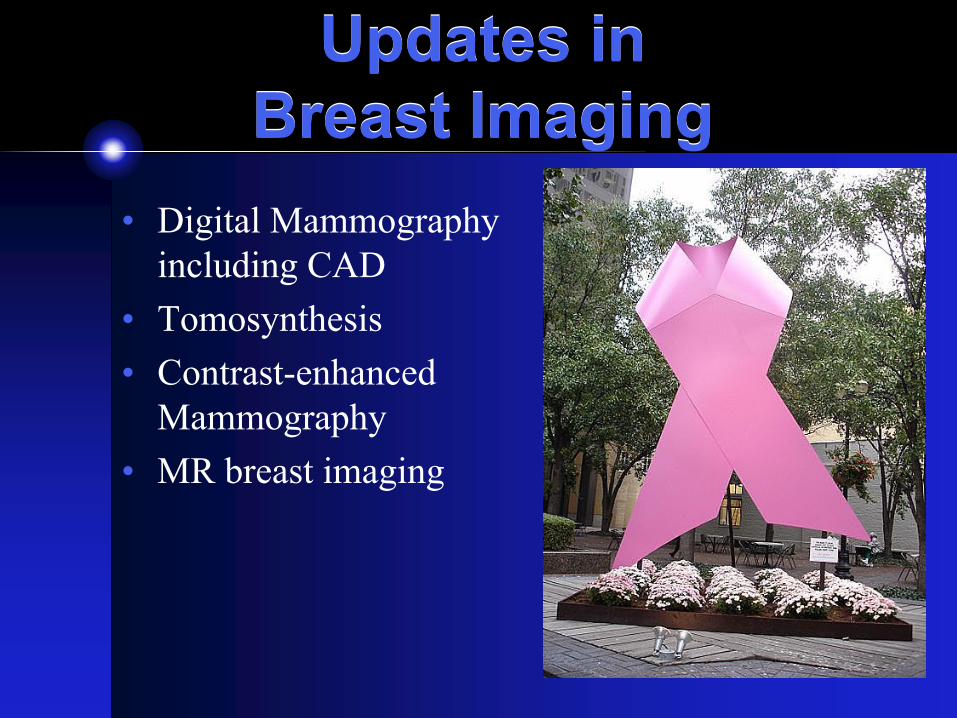
Updates in Breast Imaging
Updates in Breast Imaging
• Digital Mammography including CAD
• Tomosynthesis• Contrast-enhanced
Mammography• MR breast imaging

Breast DiseaseBreast Disease
• Breast carcinoma is the commonest cancer in Malaysian women in every race
• 3738 new cases every year• A woman in Malaysia has a 1 in 20
chance of developing breast cancer in her lifetime
National Cancer Registry Malaysia report 2003

Breast DiseaseBreast Disease
Statistics from National Cancer Registry, 2002 & 2003.Charts from http://www.radiologymalaysia.org/breasthealth/About/FactsNStats.htm
Mammography through the years
Mammography through the years
• Mammography is shown to be effective in reducing breast cancer mortality through early detection1
• Breast imaging started in 1913 by Albert Salomon• Mammography started in the 1960s2
• In 1969, the first x-ray units dedicated to breast imaging were available2
• By 1976, mammography as a screening device became standard practice and its value in the diagnosis of breast carcinoma was recognised
1. Tabar L et al. Beyond randomized controlled trials: organized mammography screening substantially reduces breast cancer mortality. Cancer 2001; 91: 1724-1731.2. http://www.emedicinehealth.com/mammogram/article_em.htm

Mammography through the years
Mammography through the years
• Screen-film mammography (SFM) is the technology of choice
• Personnel involved are well-trained in this method
• Low cost and high spatial resolution
Ongeval V et al. Current status of digital mammography for screening and diagnosis of breast cancer. Curr opinintern med 2007; 6(1): 77-84.
Mammography through the years
Mammography through the years
• In general radiology, transition to digital technology began two decades ago
• Digital mammography was first introduced in stereotactic biopsy
• Digital mammography was slow due to difficulty to produce full-field digital detectors
• In 2005, the first digital mammography was approved
Ongeval V et al. Current status of digital mammography for screening and diagnosis of breast cancer. Curr opin intern med 2007; 6(1): 77-84.

Digital vs SFMDigital vs SFM
• Detection of breast lesion• Workflow processes• Image quality including image post-
processing• Image archival and retrieval

Digital vs SFMDigital vs SFM
• Detection of breast lesion• Workflow processes• Image quality including image post-
processing• Image archival and retrieval

Detection of breast lesionDetection of breast lesion
• Breast is a difficult organ to imaged• Breast density ranges from dense (75% or
more of breast compose of glandular tissue) to fatty
• Sensitivity of detection of carcinoma is 62.9% in dense breast compared to 87% in fatty breast1
1. Carney PA et al. Individual and combined effects of age, breast density, and hormone replacement therapy use on the accuracy of screening mammography. Ann Intern Med 2003;138(3):168-175.

Image qualityImage quality
Fatty backgound

Image qualityImage quality
Moderately dense

Image qualityImage quality
Dense breast
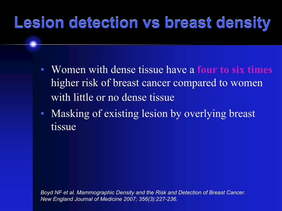
Lesion detection vs breast densityLesion detection vs breast density
• Women with dense tissue have a four to six timeshigher risk of breast cancer compared to women with little or no dense tissue
• Masking of existing lesion by overlying breast tissue
Boyd NF et al. Mammographic Density and the Risk and Detection of Breast Cancer. New England Journal of Medicine 2007; 356(3):227-236.

Digital vs SFMDigital vs SFM• Overall diagnostic accuracy of digital and film
mammography for breast cancer detection is similar
• Digital is more accurate in:• Women under 50• Women with dense breast• Premenopausal or perimenopausal women
• Due to wide dynamic range of digital, able to display contrasting regions without compromising resolution
Pisano ED et al. Diagnostic Performance of Digital versus Film Mammography for Breast-Cancer Screening. New England Journal of Medicine 2005; 353(17):1773-1783.

Detection of breast lesion: calcification
Detection of breast lesion: calcification
• SFM boasts a high spatial resolution – good for detection of microcalcification
• DM is limited by pixel size• However the high contrast resolution of DM
• shows more calcification compared to SFM• increases the ability to characterize calcification
better
Kim HH, Pisano ED, et al. Comparison of calcification specificity in digital mammography using soft-copy display versus screen-film mammography. AJR 2006; 187(1):47-50


Clustered microcalcificationBiopsy:Ductal carcinoma in situ
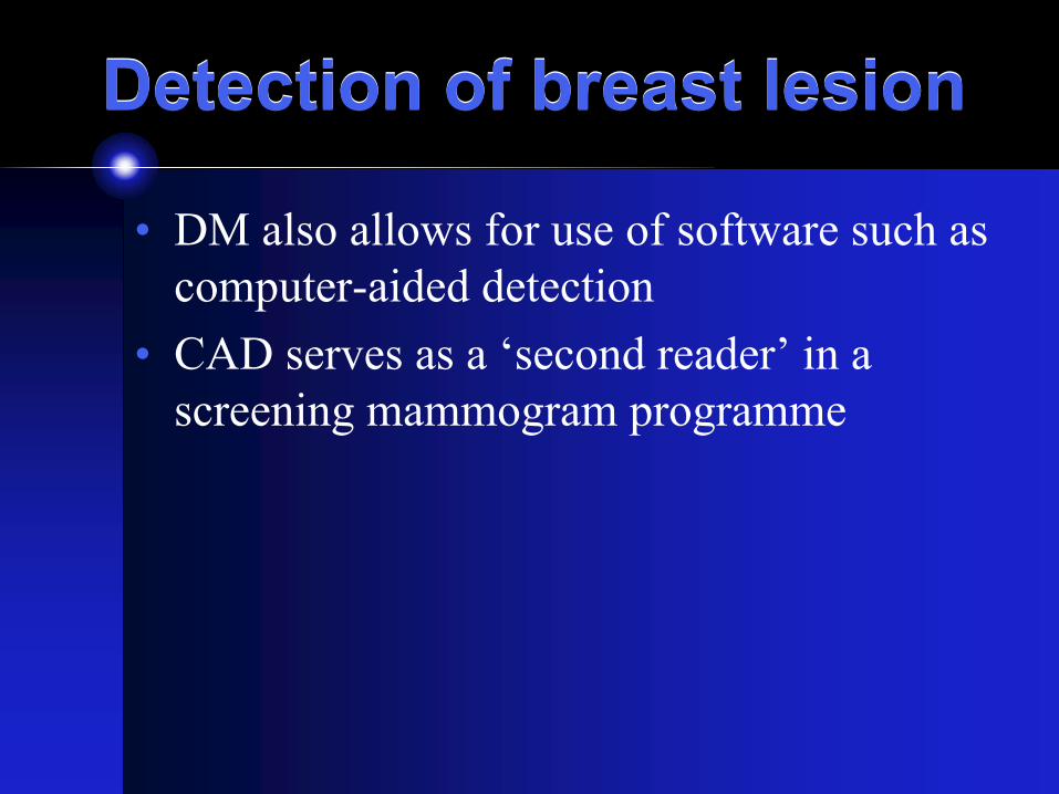
Detection of breast lesionDetection of breast lesion
• DM also allows for use of software such as computer-aided detection
• CAD serves as a ‘second reader’ in a screening mammogram programme
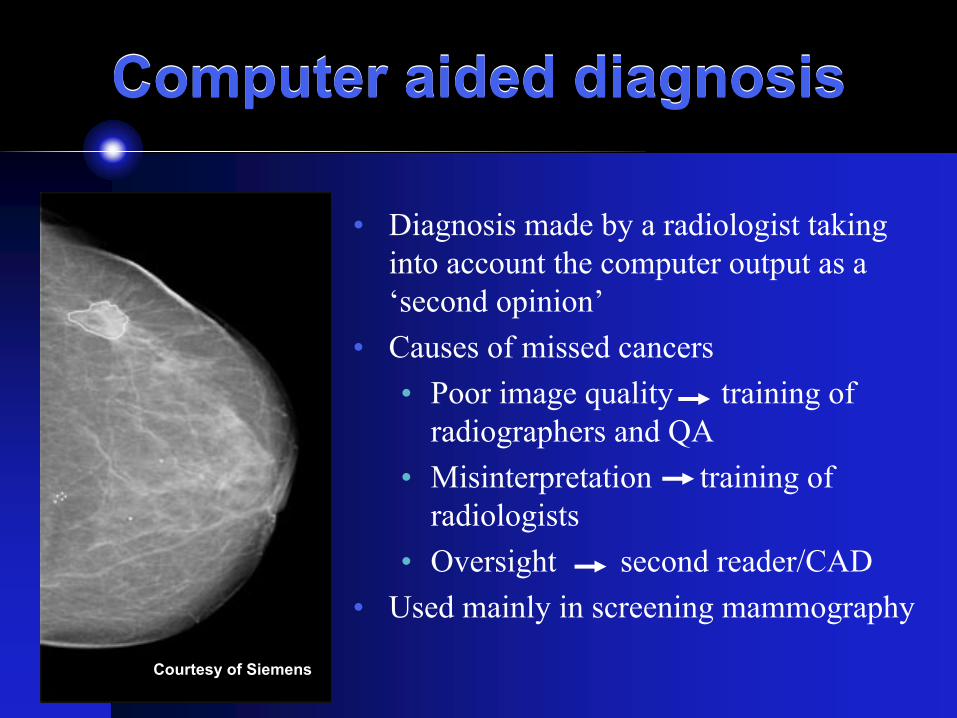
Computer aided diagnosisComputer aided diagnosis
• Diagnosis made by a radiologist taking into account the computer output as a ‘second opinion’
• Causes of missed cancers• Poor image quality training of
radiographers and QA• Misinterpretation training of
radiologists• Oversight second reader/CAD
• Used mainly in screening mammography
Courtesy of Siemens
Digital vs SFMDigital vs SFM
• Detection of breast lesion
• Workflow processes• Image quality including image post-
processing• Image archival and retrieval

Digital vs SFMDigital vs SFM
Reliance on personnel
Problems with film processing e.g. artifacts

Digital vs SFMDigital vs SFM

Digital ImagingDigital Imaging
Ranganathan S, Y Faridah, KH Ng. Moving into the digital era: a novel experience with the first full-field digital mammography system in Malaysia. Singapore Med J 2007; 48(9): 804-807.
Workflow of different modalities
10
7 7
10
42
5
5
5
0
5
10
15
20
25
30
1 2 3
Modalities
Tim
e (m
ins)
Interpretation and resultsProcessing timeExposure time
SFM CRM DM
Workflow processesWorkflow processes
• A 45% reduction in the time taken to perform and process images using DM compared to SFM
• DM is also useful in stereotactic biopsy and hookwire localisation
• Hard-copy images of DM is consistent without presence of artifacts
Ranganathan S, Y Faridah, KH Ng. Moving into the digital era: a novel experience with the first full-field digital mammography system in Malaysia. Singapore Med J 2007; 48(9): 804-807.

Workflow processes - biopsyWorkflow processes - biopsy
Use of digital system during biopsy decreases the overall time of procedure as images do not need to be processed for needle placement

Digital vs SFMDigital vs SFM
• Detection of breast lesion• Workflow processes
• Image quality including image post-processing
• Image archival and retrieval

Digital vs SFM – Image qualityDigital vs SFM – Image quality
• DM is able to manipulate digital information• Repeat rate with DM is low• DM can change image contrast, zoom and
magnify• Delineates subcutaneous skin better than
SFM• However no amount of image manipulation
could compensate for a badly taken mammogram!

Mammogram massMammogram mass
benign Sebaceous cyst


HPE – invasive ductalcarcinoma


Biopsy:Mucinous carcinoma

Mammogram - massMammogram - mass
Carcinoma

Digital vs SFMDigital vs SFM
• Detection of breast lesion• Workflow processes• Image quality including image post-
processing• Image archival and retrieval

Digital vs SFMDigital vs SFM
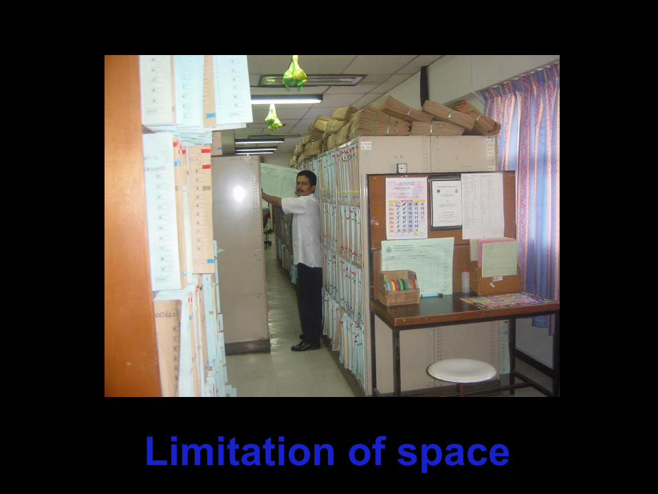
Limitation of space

MAMMOGRAM FILMS
FILMFILM
MORE FILM
RADIOLOGY DEPARTMENT, WARDS, CLINIC, OTHER
HOSPITALS
Limited storageMissing films
Labour intensiveDegradation of filmsWorkflow in a Radiology
department with SFM

MAMMOGRAPHY
Workflow in a Radiology department with Digital
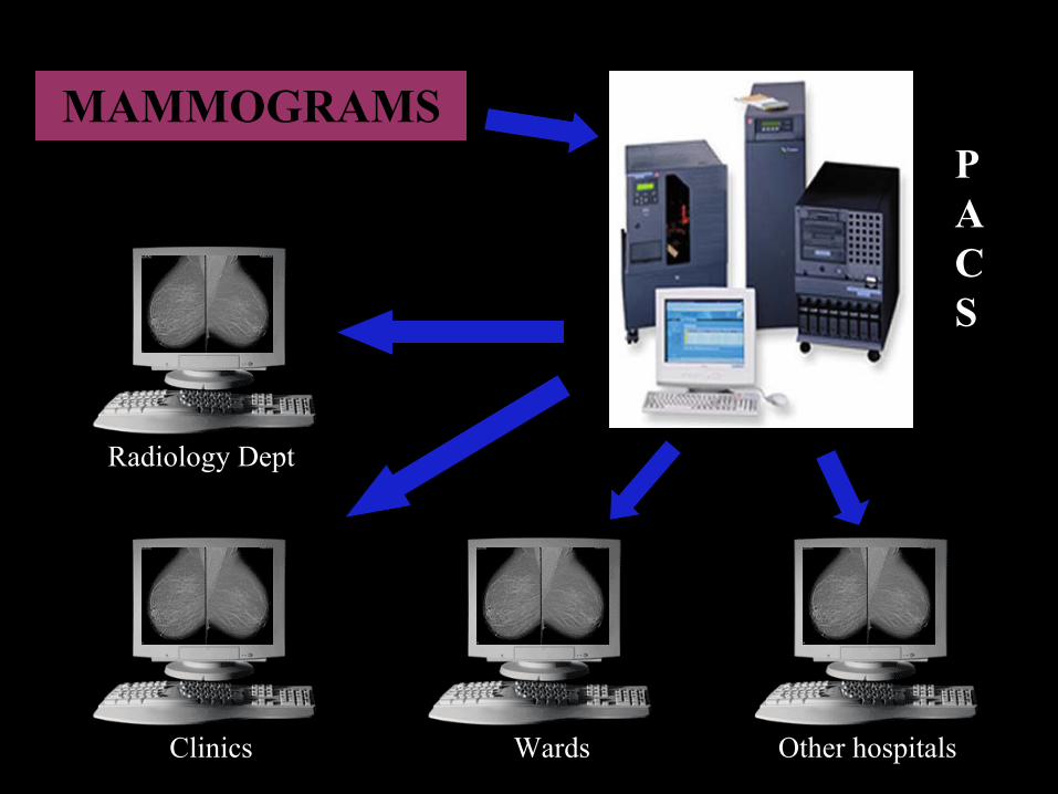
MAMMOGRAMS
Radiology Dept
Clinics Wards Other hospitals
PACS

Digital Mammography: in summaryDigital Mammography: in summary
DM >> SFMUse of software e.g. CADCAD
DM >> SFMImage archival and retrievalImage archival
DM >> SFMSubcutaneous tissue delineation
Cost
Image quality
Work-flow process
Detection of breast lesion
DM >>> SFMCost, cost, cost!
DM > SFMConsistency of hard-copy image
DM yes, SFM noAbility to manipulate image
DM < SFM initially if do soft-copy reporting
Reporting time
DM >> SFMProcessing time
DM > SFMDetection of microcalcification
DM > SFMAccuracy in dense breast
DM = SFMOverall sensitivity in screening for breast lesion

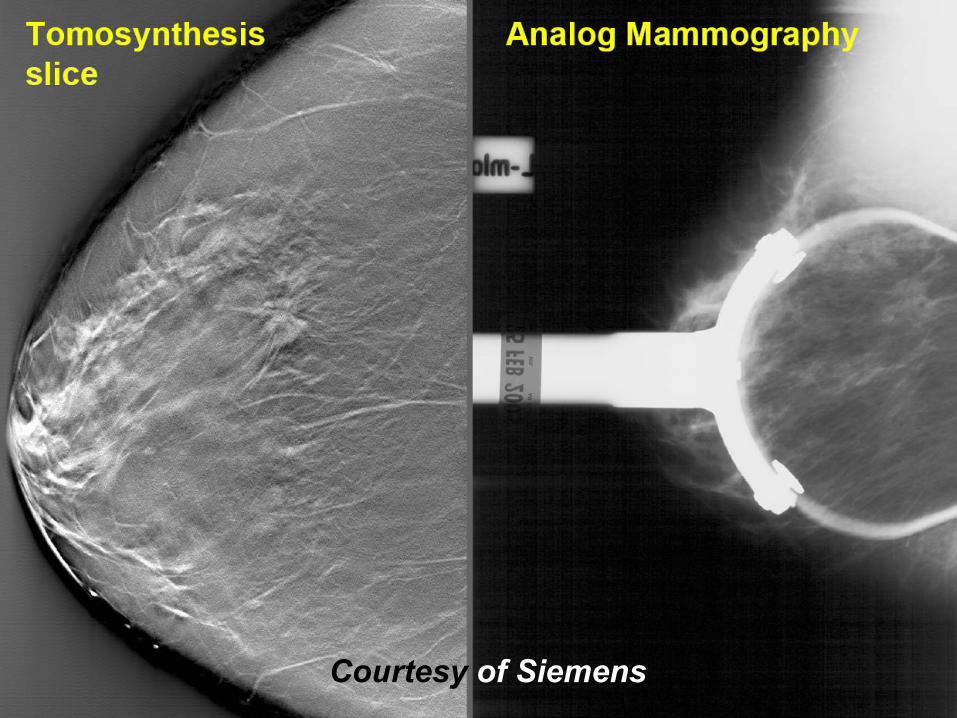
Breast TomosynthesisBreast Tomosynthesis
• Acquisition of multiple images during Tomo scan
• Displays the breast in slices
• Reduces tissue overlap• Reduction in compression
pressure• Dose? Similar to one view
mammogramCourtesy of Siemens
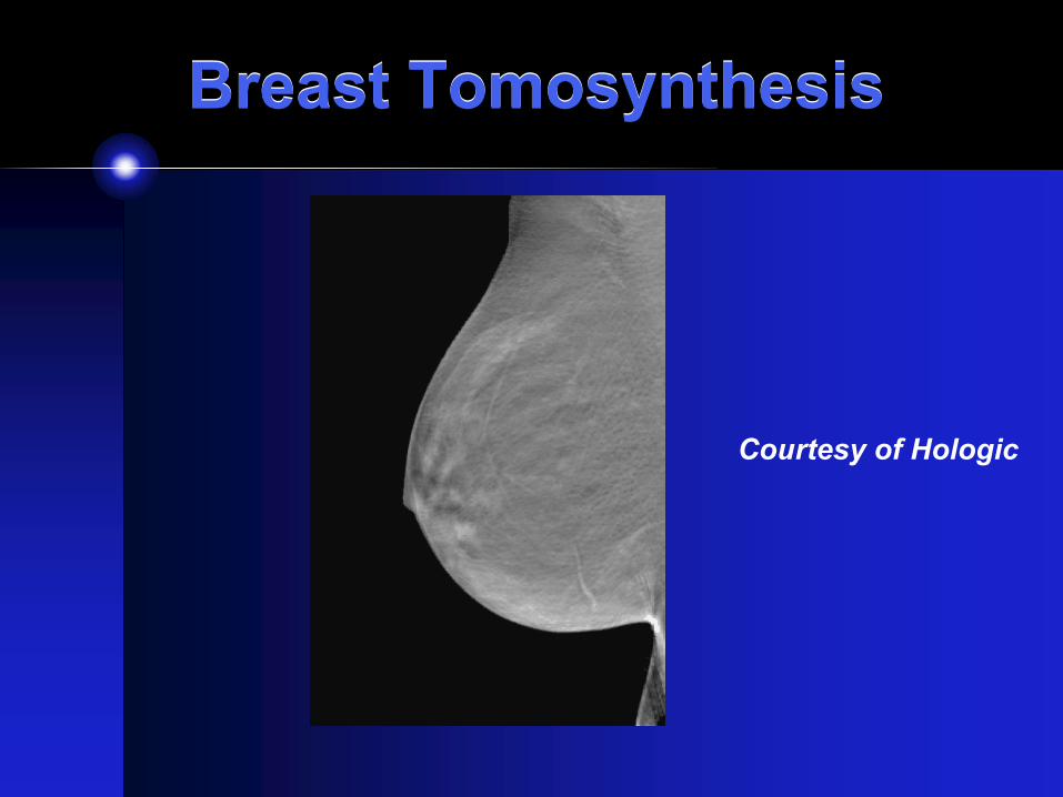
Breast TomosynthesisBreast Tomosynthesis
Courtesy of Hologic
Courtesy of Siemens

Courtesy of Siemens

Contrast-enhanced Mammography
Contrast-enhanced Mammography
• A pre-contrast mammogram is performed• An iodine-based contrast media is injected• Post-contrasted images are then acquired• These images (pre-contrast and contrasted)
are subtracted
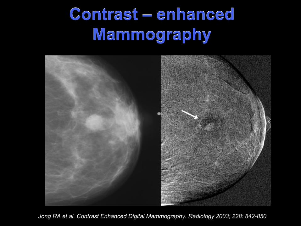
Contrast – enhanced Mammography
Contrast – enhanced Mammography
Jong RA et al. Contrast Enhanced Digital Mammography. Radiology 2003; 228: 842-850
Breast MRBreast MR• Has a high sensitivity in detecting
breast lesion• Has a high negative predictive value• No radiation

Breast MR: DrawbacksBreast MR: Drawbacks• Picks up benign lesion as well• Gives rise to higher number of
unneccesary biopsies• Cost

Breast MRBreast MR• Not used for general population
screening• Reserved for young women with high
risk of breast carcinoma• Performed also in women with
suspected multiple breast carcinomas

Breast MRBreast MR
Courtesy of GE

Breast MRBreast MR• Static T1WI, T2WI, STIR• Contrast-enhanced• Dynamic contrast-enhanced• Spectroscopy

Breast MRBreast MR
Courtesy of GE

Breast MRBreast MR
Courtesy of GE

Breast MRBreast MR
Courtesy of GE
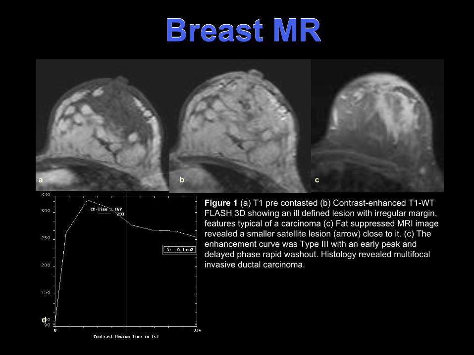
Breast MR Breast MR
Figure 1 (a) T1 pre contasted (b) Contrast-enhanced T1-WT FLASH 3D showing an ill defined lesion with irregular margin, features typical of a carcinoma (c) Fat suppressed MRI image revealed a smaller satellite lesion (arrow) close to it. (c) Theenhancement curve was Type III with an early peak and delayed phase rapid washout. Histology revealed multifocalinvasive ductal carcinoma.
ca
d
b

Breast MR - SpectroscopyBreast MR - Spectroscopy
Courtesy of GE

Breast MR - BiopsyBreast MR - Biopsy
Courtesy of GE

ConclusionConclusion
• Exiting developments in breast imaging mainly due to digital technology
• Multiple approach to image the breast• Do not forget that NOTHING can
compensate for an examination that is done badly
• Perform the best that you can every time!

THANK YOU