unit 8 - blood / lymphatic / cardiovascular … 8 - blood / lymphatic / cardiovascular systems...
TRANSCRIPT
Medical Anatomy and Physiology
Unit Eight – Blood / Lymphatic / Cardiovascular Page 1 Draft Copy
UNIT 8 - BLOOD / LYMPHATIC / CARDIOVASCULAR SYSTEMS LECTURE NOTES 8.01 COMPONENTS OF THE BLOOD AND THEIR FUNCTIONS Blood is the complex transport medium which provides vital transportation services for the body. It picks up food and oxygen from the digestive and respiratory systems and delivers them to the body cells while picking up their waste products. Blood also transports hormones, enzymes, antibodies, buffers, and other critical biochemical substances. In addition, blood helps regulate heat in the body by distributing it from the muscles. There are four blood components: erythrocytes, leukocytes, thrombocytes, and plasma. A. Erythrocytes
Erythrocytes are the red blood cells or RBCs. They are responsible for transporting both oxygen and carbon dioxide.
B. Leukocytes
Leukocytes are the white blood cells or WBCs. They are responsible for protecting the body against infection.
C. Thrombocytes Thrombocytes are the platelets or plts. They assist in hemostasis and blood clotting. D. Plasma
Plasma is the fluid portion of blood. It consists mostly of water and also contains dissolved substances such as electrolytes, hormones, gases, and organic compounds.
8.02 DESCRIBE ERYTHROCYTES AND THE STRUCTURE OF HEMOGLOBIN A. Erythrocytes 1. Erythrocytes are the most numerous of all formed elements. Men average
approximately 5.5 million per cubic millimeter of blood while women average 4.8 million per cubic millimeter of blood.
2. The mature erythrocyte is an anucleated biconcave disc approximately 7 mm in diameter. 3. RBCs are produced by the red bone marrow. 4. At maturity, the erythrocyte does not contain many organelles found in most body cells to make room for the principle pigment, hemoglobin. 5. The advantage of the biconcave disc shape is so the cell can move without injury through the narrow blood capillaries.
Medical Anatomy and Physiology
Unit Eight – Blood / Lymphatic / Cardiovascular Page 2 Draft Copy
B. Hemoglobin 1. Hemoglobin is the principle pigment of the erythrocyte. 2. Hemoglobin is composed of four protein chains called the globin. Each protein chain contains a red pigment called heme. The heme is composed of an iron atom. 3. The heme transports oxygen while the globin transports carbon dioxide. (Each heme can carry up to four molecules of oxygen). 4. It is estimated each erythrocyte contains 200 to 300 million molecules of hemoglobin. 8.03 LEUKOCYTES A. Description
The leukocytes are nucleated cells capable of dividing and living for years. They are larger than erythrocytes. They help protect the body from infection and provide immunity. There are 5,000 - 10,000 leukocytes per cubic millimeter of blood. They are produced in the red bone marrow. There are five types of leukocytes. The leukocytes are divided into two classifications, the granulocytes and the agranulocytes.
B. Types of Leukocytes 1. Granulocytes
These leukocytes have protein granules in their cytoplasm. There are three specific types.
a. Neutrophils Neutrophils have cytoplasmic granules which stain pink or light purple. They are the most numerous of all leukocytes, accounting for about 60% of the total count. The nuclei are divided into two, three, or four lobes. The neutrophils help to protect the body by performing phagocytosis.
b. Basophils Basophils have large cytoplasmic granules which stain dark blue or purple. These cells are very mobile and travel easily throughout the body. Their nuclei resemble the letter S. These cells produce histamine (used in the inflammation response) and heparin (an anticoagulant used to prevent blood clotting).
c. Eosinophils Eosinophils contain large cytoplasmic granules which stain reddish-orange. Their nuclei contain two lobes and resemble head phones. Eosinophils ingest inflammatory chemicals and proteins and help to protect against allergens.
2. Agranulocytes These leukocytes do not have large granules in the cytoplasm. There are two specific types.
Medical Anatomy and Physiology
Unit Eight – Blood / Lymphatic / Cardiovascular Page 3 Draft Copy
a. Monocytes Monocytes are the largest of all leukocytes. They have a dark kidney bean-shaped nucleus surrounded by large amounts of a distinctive light bluish-gray cytoplasm. These cells are very mobile and protect the body by performing phagocytosis.
b. Lymphocytes Lymphocytes are the smallest of all leukocytes. They have a large spherical nucleus surrounded by a limited amount of pale blue stained cytoplasm. The lymphocytes will specialize into the T-lymphocytes (T-cells) and the B-lymphocytes (B-cells). The T-cells are responsible for directly attacking antigens or infected cells while the B-cells produce antibodies.
8.04 HEMOSTASIS AND COAGULATION A. Description
Hemostasis refers to stopping blood flow which is extremely important when the blood vessels are damaged. Hemostasis is most effective in slowing down blood losses from smaller blood vessels. Damage to larger vessels will require additional interventions to slow down and stop the bleeding. There the three stages include vascular spasm, platelet plug formation, and coagulation.
B. Three Stages 1. Vascular Spasm
Vascular spasm occurs when an arteriole or venule is broken or has been cut. The smooth muscles in the blood vessel wall are stimulated to contract and the blood loss is decreased almost immediately. The reflex response may last only a few minutes, but the effect will last for around 30 minutes. Serotonin, a chemical released by the platelets, also stimulates the blood vessel walls to contract.
2. Platelet Plug Formation Platelets tend to stick to the exposed ends (collagen) of injured blood vessels. Actually, platelets stick to any rough surface which makes internal blood clotting possible. When the platelets come into contact with the collagen, their shapes actually change and many spiny processes begin to extend from their membranes. At the same time, the platelets stick to each other to form a platelet plug in the blood vessel break.
3. Coagulation a. Coagulation is the actual formation of a blood clot. It is the most effective of all hemostatic mechanisms. b. There are two mechanisms associated with blood clotting -- the intrinsic
mechanism and the extrinsic mechanism. The intrinsic mechanism is triggered by the release of chemical substances from the platelets while the extrinsic mechanism is triggered by the release of chemical substances released from the damaged blood vessels and tissues.
Medical Anatomy and Physiology
Unit Eight – Blood / Lymphatic / Cardiovascular Page 4 Draft Copy
c. The process of coagulation depends on the presence of clotting factors (blood proteins produced by the liver) as well as calcium ions in the blood. d. The basic event in blood coagulation is the conversion of the soluble
plasma protein fibrinogen to relatively insoluble threads of protein fibers. These form a strong netting over the blood vessel to hold the ends together while healing occurs.
8.05 THROMBUS AND EMBOLUS A. Thrombus (plural - thrombi) 1. A thrombus is a blood clot that has formed abnormally in a blood vessel. 2. A thrombus may be life-threatening because the clot may occlude a blood vessel and stop the blood supply to an organ or body part. 3. Predisposing factors for thrombi include heart and blood vessel disorders,
atherosclerosis, obesity, heredity, age, immobility, smoking, and the use of oral contraceptives.
4. Treatment of thrombi includes the use of anticoagulants and sometimes bedrest. B. Embolus (plural - emboli)
1. An embolus occurs when a thrombus has become dislodged or fragmented and is carried away from the original site by the flow of the blood.
2. The embolus will travel until it reaches a narrow blood vessel where it becomes lodged and obstructs blood flow distal to the blockage.
3. Emboli may be solid, liquid, or gas. They may consist of tissue, tumor cells, globules of fat, air bubbles, clumps of bacteria, and foreign bodies.
4. Emboli typically affect four organs -- the heart, lungs, brains, and the kidneys. 5. Treatment includes the use of anticoagulants and basket filters to try to trap
the emboli.
Medical Anatomy and Physiology
Unit Eight – Blood / Lymphatic / Cardiovascular Page 5 Draft Copy
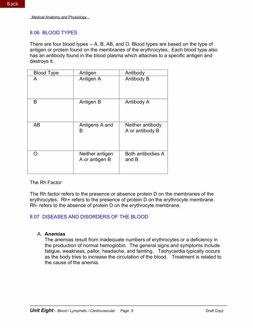
8.06 BLOOD TYPES There are four blood types -- A, B, AB, and O. Blood types are based on the type of antigen or protein found on the membranes of the erythrocytes. Each blood type also has an antibody found in the blood plasma which attaches to a specific antigen and destroys it.
Blood Type Antigen Antibody A
Antigen A Antibody B
B
Antigen B Antibody A
AB Antigens A and B
Neither antibody A or antibody B
O Neither antigen A or antigen B
Both antibodies A and B
The Rh Factor The Rh factor refers to the presence or absence protein D on the membranes of the erythrocytes. Rh+ refers to the presence of protein D on the erythrocyte membrane. Rh- refers to the absence of protein D on the erythrocyte membrane. 8.07 DISEASES AND DISORDERS OF THE BLOOD A. Anemias
The anemias result from inadequate numbers of erythrocytes or a deficiency in the production of normal hemoglobin. The general signs and symptoms include fatigue, weakness, pallor, headache, and fainting. Tachycardia typically occurs as the body tries to increase the circulation of the blood. Treatment is related to the cause of the anemia.
Medical Anatomy and Physiology
Unit Eight – Blood / Lymphatic / Cardiovascular Page 6 Draft Copy
1. Hemorrhagic Anemia Hemorrhagic anemia is caused by a decrease in the amount of circulating erythrocytes lost because of hemorrhage or bleeding. The blood loss may be acute such as with a trauma or surgery or chronic such as blood loss associated with ulcers. Blood transfusions and IV fluids are successful to replace lost blood. It is important to prevent shock which may lead to death.
2. Aplastic Anemia
Aplastic anemia is characterized by the inability of the red bone marrow to produce erythrocytes which have been destroyed due to toxic chemicals and anti-cancer drugs. Bone marrow transplants or stem cell transplants are effective treatments. Transfusions are helpful in relieving short-term relief from the anemia symptoms.
3. Iron Deficiency Anemia
Iron deficiency anemia occurs when the body is deficient of iron which impairs the body's ability to make normal hemoglobin. The erythrocytes made are smaller and paler than normal red cells. Treatment includes iron supplements taken with vitamin C to improve the absorption of iron in the intestines.
4. Pernicious Anemia
Pernicious anemia occurs when there is a dietary deficiency of vitamin B12 or the loss of the intrinsic factor from the lining of the stomach which prevents the absorption of vitamin B12. Not only is the number of erythrocytes reduced, but those cells that are produced are abnormally large and have fragile cell membranes. It is treated with injections of vitamin B12.
5. Hemolytic Anemia
Hemolytic anemia results from the abnormal destruction of erythrocytes. The cause of the hemolytic anemia must be determined before the appropriate treatment can be administered.
B. Hemolytic Disease of the Newborn
Hemolytic disease of the newborn is also known as erythroblastosis fetalis. This disease occurs in the fetus if the fetus is Rh+ while the mother is Rh-. During the first pregnancy, the mother will form antibodies against the Rh+ blood. Since the antibodies have only formed, they do not cross the placenta and harm the fetus’ blood. If a second Rh+ pregnancy occurs, the antibodies have already been made, now cross through the placenta and begin to destroy the Rh+ red blood cells. The result is a reduction in the number of circulating red blood cells in the fetus as well as jaundice due to the accumulation of bilirubin in the skin. The Rh- mother must receive an injection of Rhogam after every Rh+ pregnancy in order
Medical Anatomy and Physiology
Unit Eight – Blood / Lymphatic / Cardiovascular Page 7 Draft Copy
to destroy the antibodies that have been made against the Rh+ blood. The fetus may require transfusions and light therapy.
C. Hemophilia
Hemophilia is caused by a sex-linked genetic trait resulting in the inability to produce blood-clotting factor VIII. As a result, the intrinsic pathway cannot complete its set of chemical reactions and the enzyme thromboplastin is not made. As a result, fibrinogen cannot be converted to fibrin and the blood cannot clot. This disease appears almost exclusively in males, due to the fact that they only have one X-chromosome. The most common signs include excessive bruising, nosebleeds, and bleeding into the joints. The disease is treated by injecting the missing blood clotting factor, or factor VIII into the blood.
D. Leukemia
Leukemia is a cancer of the blood characterized by the overproduction of immature white blood cells, which are released prematurely into the circulation. In contrast to popular belief, more adults are afflicted with this disease than children. The causes are unknown. The large number of immature leukocytes reduce the production of erythrocytes and platelets The symptoms include increased susceptibility to infection because of the immature white blood cells; fatigue, pallor, and anemia due to a decrease in the number of circulating red blood cells; and increased bruising due to reduced numbers of platelets. Treatment typically includes transfusions, bone marrow transplants, and chemotherapy.
E. Mononucleosis
Mononucleosis, or infectious mononucleosis, is a noncancerous leukocyte disorder caused by a virus. This virus is typically transmitted in the saliva and is nicknamed the "kissing disease." Unlike its name seems to suggest, the affected leukocyte is actually a lymphocyte. Signs and symptoms include extreme fatigue, sore throat, rash, enlargement of cervical lymph nodes, and splenomegaly. It typically resolves itself with four to six weeks.
F. Polycythemia
Polycythemia is an excessive number of erythrocytes. This disease is caused by an overproduction of blood cells within the body. The result is viscous (thick) blood which flows slowly through the blood vessels. The person is at increased risk for heart attack and stroke due to the formation of blood clots within the blood. The cause is unknown. Treatment includes phlebotomy to remove excess blood as well as medications to try to reduce the excessive blood formation.
Medical Anatomy and Physiology
Unit Eight – Blood / Lymphatic / Cardiovascular Page 8 Draft Copy
8.08 THE COMPONENTS OF THE LYMPHATIC SYSTEM A. Tonsils
1. Description and Location The tonsils are large lymphoid nodules located in the wall of the pharynx, and
include the adenoids and palatine tonsils. 2. Function Since the mouth is a port of entry into the body for harmful organisms we
breathe, as well as those found in the food we eat, the tonsils help to destroy them.
B. Spleen 1. Description and Location
The spleen is the largest collection of lymphatic tissue in the body. It is located in the LUQ, lateral to the stomach. It has a deep red color because of all the blood it contains. It contains areas of white pulp which resemble lymphoid nodules and red pulp which contain large quantities of blood.
2. Functions The spleen is able to filter the blood and remove damaged or
infected cells. It also helps to initiate the immune response when antigens are detected in the blood. In addition, the spleen serves as a large blood reservoir.
C. Thymus 1. Description and Location
The thymus is posterior to the sternum and superior to the heart. It is a two lobed structure consisting of two layers -- a cortex and a medulla. It reaches its greatest size the first two years after birth and gradually shrinks with age.
2. Function The thymus gland produces a hormone called thymosin which helps to mature lymphocytes into T-lymphocytes (T-cells).
D. Lymph Nodes 1. Description and Location
The lymph nodes are small, oval, lymphatic organs which are surrounded by a fibrous capsule. They are located in clusters along the lymphatic vessels and are clustered in the cervical, axillary, inguinal regions and in the abdominal cavity. The lymph nodes contain large numbers of lymphocytes.
2. Function The lymph nodes filter and purify the lymph before it is returned to the blood. E. Red Bone Marrow 1. Description and Location The spongy bone found in the bones. In the adult, these bones include the flat bones such as the pelvis, ribs, and sternum.
Medical Anatomy and Physiology
Unit Eight – Blood / Lymphatic / Cardiovascular Page 9 Draft Copy
2. Function The red bone marrow produces all blood cell types, including the white blood cells used to protect the body from infection. F. Lymph Vessels 1. Description and Location Lymphatic vessels, or lymphatics, are similar to veins and contain valves. They are located throughout the body. Excess tissue fluid is absorbed into the tiny lymphatic capillaries where the fluid becomes lymph. The lymph travels through the lymphatic vessels, and into two large collecting ducts. 2. Lymphatic vessels transport lymph through the body back to the blood. 8.09 MOVEMENT OF LYMPH THROUGH THE BODY A. Movement of Lymph Through Lymphatic Vessels
Lymph is transported along a network of vessels beginning with the lymphatic capillaries. From there, lymph is transported into lymphatic vessels which empty into the thoracic duct and the right lymphatic duct. Eventually, these lymphatic ducts drain lymph into the subclavian veins and back into the blood.
B. Mechanisms Moving Lymph
The movement of lymph through the lymphatic vessels is similar to the processes which control the movement of blood through the veins.
1. Pressure gradients caused by the physical movement of breathing. 2. Skeletal muscle contractions which help to move lymph along the lymphatic vessels. 3. Valves in the lymphatic vessels which help to assist the one-way movement of lymph. 8.10 CONTRAST ANTIGENS AND ANTIBODIES A. Antigens
An antigen is a foreign protein capable of initiating the immune response and the production of antibodies.
B. Antibodies An antibody is a globular protein produced by the B-plasma cells which will bind to specific antigens to promote their destruction or removal from the body.
Medical Anatomy and Physiology
Unit Eight – Blood / Lymphatic / Cardiovascular Page 10 Draft Copy
8.11 ROLES OF T-CELLS AND B-CELLS IN THE IMMUNE RESPONSE A. T-CELLS
T-cells are white blood cells which were produced in the bone marrow and matured in the thymus gland under the influence of the hormone thymosin. They function in CMI or cell-mediated immunity. The T-cells are responsible for directly attacking antigens and antigen-infected cells.
1. Cytotoxic T-Cells These cells are known as the killer T-cells which help to track down bacteria, fungi, protozoa, or foreign tissues that contain antigens. They are responsible for performing phagocytosis of antigens and antigen-infected cells.
2. Helper T-Cells Helper T-cells release a variety of chemicals which help to coordinate specific and nonspecific defenses, stimulate cell-mediated immunity and antibody-mediated immunity by increasing the production of T cells and B cells.
3. Memory T-Cells Memory T-cells store a code of the antigen which may be used if the antigen appears a second time. These cells will immediately differentiate into the cytotoxic T-cells producing a more rapid and effective cellular response.
4. Suppressor T-Cells Suppressor T-cells stop the responses of the T-cells and the B-cells when the level of the antigen has decreased.
B. B-Cells
B-cells are white blood cells which are produced in the bone marrow and matured in an unknown location -- probably the bone marrow. They are responsible for AMI or antibody mediated immunity which is a chemical attack on the antigens.
1. Plasma Cells Plasma cells, sometimes referred to as B-plasma cells, make and secrete large numbers of antibodies that will fight against antigens. Antibodies function by the following ways: a. neutralization - preventing the antigen from attaching to a cell by binding
the toxin or viruses. b. agglutination - the process of clumping antigens together by making them
easier to find and be destroyed by phagocytic cells. c. activation of the complement - helps to destroy the antigen by attracting
more phagocytic cells to the area, destroying cell membranes, and promoting inflammation.
d. antigens covered with antibodies attract the phagocytic white blood cells including the neutrophils, macrophages, and eosinophils.
e. enhancement of phagocytosis f. the inflammation response to localize an infection by releasing histamine
and causing redness, swelling, heat, and pain.
Medical Anatomy and Physiology
Unit Eight – Blood / Lymphatic / Cardiovascular Page 11 Draft Copy
2. Memory B-Cells Memory B-cells help in the response to a second exposure to the same antigens. At that time, they respond and differentiate into antibody-secreting plasma cells providing a rapid response to the antigen.
8.12 ACTIVE AND PASSIVE IMMUNITY AND NATURAL VERSUS ARTIFICIAL
ACQUISITION OF IMMUNITY
A. Active Immunity Active immunity occurs when the person has been exposed to an antigen and
the body produces antibodies during the immune response. B. Passive Immunity Passive immunity occurs when the person has been given the antibodies to fight
a specific antigen. C. Natural Immunity Natural immunity begins at birth and is enhanced when the individual is exposed
to new antigens and the person makes antibodies to fight against them. Natural immunity may also refer to the passing of antibodies from the mother to the fetus and the mother to baby as she breastfeeds the infant.
D. Artificial Immunity Artificial immunity stimulates the production of antibodies under controlled
conditions so the individual will be able to overcome any natural exposure to the same type of antigen in the future. This includes the use of vaccinations to help the body stimulate the immune response. It may also refer to the injection of ready-made antibodies such as gamma globulins to help fight infections.
8.13 DISEASES AND DISORDERS ASSOCIATED WITH THE LYMPHATIC SYSTEM A. Acquired Immunodeficiency Syndrome (AIDS)
The major cause of AIDS is an infection by the human immunodeficiency virus (HIV) which infects the T-helper cells resulting in the progressive destruction of cell mediated immunity by the T cells and eventually humoral (antibody) immunity. The patient is susceptible to opportunistic infections such as Kaposi's sarcoma and pneumocystis carnii pneumonia. This syndrome was first described in 1981 by the Centers for Disease Control. HIV is transmitted by infected blood or body fluids, especially those which contain white blood cells, such as semen.
AIDS infection begins with infection by HIV which is only detectable by laboratory tests. At that time, the person is diagnosed with HIV infection. Eventually there will be an appearance of symptoms, a diagnosis of AIDS (when the T-helper cell [CD4 cell] drops below 200 cells per mm3. HIV infection is characterized by flu-like symptoms, weight loss, fatigue, night sweats, and fevers.
Medical Anatomy and Physiology
Unit Eight – Blood / Lymphatic / Cardiovascular Page 12 Draft Copy
There is no cure for AIDS. Antiviral therapy is available which does seem to control the virus for long periods. At this time, however, there is no known cure.
B. Measles
Measles, also known as rubeola, is a highly contagious viral infection that may be one of the most dangerous of all childhood infections. Measles is spread by direct contact or by contact with infected respiratory droplets. Its incubation period is from one to two weeks. Signs and symptoms include fever, photophobia, malaise, anorexia, conjunctivitis, coryza, hoarseness, cough, and Koplik's spots. Koplik's spots appear as bluish white specks surrounded by a red halo and are the definitive signs of measles. They appear in the mouth and may bleed. Severe infections may lead to pneumonia and encephalitis. Use of the measles vaccine has reduced the incidence of measles in children, but there are more adolescents being diagnosed with the disease. Measles is a major cause of death in children worldwide.
C. Mumps
Mumps is a viral infection affecting the parotid salivary glands. It is common in children. Recovery is good, although it may cause sterility or meningitis. The virus is transmitted by droplets or direct contact. Signs and symptoms include fatigue, headache, low-grade fever, difficulty chewing, and earache, which are followed by parotid gland swelling. The use of vaccines has reduced the incidence in the United States.
D. Rubella
Rubella, or German measles, is a mildly contagious viral infection which produces a three day rash and swelling of the lymph nodes. The rubella virus is transmitted contact with contaminated body fluids or articles of clothing. The signs and symptoms include headache, fever, fatigue, lymph node enlargement, and red maculopapular rash. The rash typically begins on the face and spreads rapidly over the body, but disappears after about three days. Rubella has devastating consequences on a growing fetus and may cause blindness, heart problems, and/or deafness. The spread of rubella in the United States has been controlled by the use of vaccines.
E. Tetanus
Tetanus, also known as lockjaw, is a bacterial infection. It is generally systemic and is fatal in over 50% of unimmunized people. Transmission of the bacteria generally begins when a person is walking through contaminated dirt and receives a puncture wound. The exotoxins produced by tetanus enter the body and cause local infection and tissue death. There are painful, involuntary muscle contractions of the face, neck, and back. Seizures can also occur. Treatment requires the use of the tetanus antitoxin and may need respiratory support until antibiotics can control the infection.
Medical Anatomy and Physiology
Unit Eight – Blood / Lymphatic / Cardiovascular Page 13 Draft Copy
8.14 GENERAL FUNCTIONS OF THE CARDIOVASCULAR SYSTEM
A. The cardiovascular system, sometimes known as a part of the circulatory system, is composed of the heart and a closed system of blood vessels through which blood is circulated.
B. The primary function is circulation. Critical transportation needs include the movement of oxygen and carbon dioxide, heat, nutrients, hormones, waste products, enzymes, electrolytes, and other substances on a continuing basis.
8.15 LAYERS OF THE HEART
There are three distinct layers which form the heart wall. They include the epicardium, the myocardium, and the endocardium.
A. Epicardium
The epicardium is the outermost layer. It is a serous membrane which is composed of epithelial tissue and some connective tissue. It provides a small amount of protection to the heart.
B. Myocardium
The myocardium is the middle, muscular wall of the heart. It is composed of cardiac muscle, blood vessels, and nerves. The muscular layer is responsible for pumping the blood through the heart and into the great vessels.
C. Endocardium
The endocardium is the most inner layer. It is composed of epithelial tissue and is very smooth. The blood passing through the heart is in contact with this layer.
8.16 CHAMBERS OF THE HEART A. General Description 1. The human heart is a four-chambered muscular organ, the size and shape of a person’s closed fist. 2. It lies in the mediastinum or the middle region of the thorax. 3. There are two upper chambers of the heart known as the atria (singular, atrium) and two lower chambers called the ventricles. 4. The left chambers are separated from the right chambers by an extension of the heart wall called the septum. B. The Atria (singular: Atrium) 1. The atria are the two superior chambers and are called the “receiving
chambers” because they receive blood from vessels called veins. The right atrium receives deoxygenated blood from the superior vena cava, the inferior vena cava, and the coronary sinus. The left atrium receives oxygenated blood from the pulmonary veins and the lungs.
Medical Anatomy and Physiology
Unit Eight – Blood / Lymphatic / Cardiovascular Page 14 Draft Copy
2. The atria are thin-muscular walled chambers which allow blood flow into the ventricles. C. The Ventricles 1. The ventricles are the two inferior chambers. They are often called the
“pumping chambers” because they pump blood out of the heart and into blood vessels known as arteries. The right ventricle pumps deoxygenated blood into pulmonary arteries which take blood to the lungs. The left ventricle pumps oxygenated blood into the aorta.
2. Since more force is needed to pump blood through the body, the myocardium of each ventricle is thicker than the myocardium of the atria. 3. The myocardium of the left ventricle is the thickest layer because the left
ventricle pumps blood into the whole body while the right ventricle pumps blood into the lungs.
8.17 THE GREAT BLOOD VESSELS OF THE HEART A. The Superior Vena Cava
The superior vena cava drains deoxygenated blood from veins in the head, neck, and arms into the right atrium.
B. The Inferior Vena Cava
The inferior vena cava drains deoxygenated blood from veins in the abdomen and legs into the right atrium.
C. Pulmonary Trunk
The pulmonary trunk is the first portion of the pulmonary artery. It arises directly from the right ventricle after the pulmonary semilunar valve. The pulmonary trunk branches to form the left and right pulmonary arteries.
D. Pulmonary Arteries
The pulmonary arteries branch from the pulmonary trunk to take deoxygenated blood to the lungs where carbon dioxide and oxygen gas exchange occurs.
E. Pulmonary Veins
The pulmonary veins take oxygenated blood from the lungs into the left atrium of the heart.
F. The Aorta 1. The aorta is the largest artery in the body extending from the left ventricle after the aortic semilunar valve. It arches and descends into the lower abdomen. 2. There are many arteries which branch from the aorta to deliver oxygen-rich
blood to the body tissues. The first branches of the aorta include the coronary arteries. The openings to the coronary arteries are located just above the aortic semilunar valve. As the aorta arches, the major branches
Medical Anatomy and Physiology
Unit Eight – Blood / Lymphatic / Cardiovascular Page 15 Draft Copy
include the brachiocephalic artery, the left common carotid artery, and the left subclavian artery.
a. As the aorta leaves the left ventricle, the first branches of the aorta include the coronary arteries, which supply the myocardium with oxygen-rich blood. b. The brachiocephalic artery transports blood into arteries supplying the right arm and the right side of the head. c. The left common carotid artery transports blood into arteries which will supply the left side of the head. d. The left subclavian artery transports blood into arteries of the left arm. 8.18 HEART VALVES A. Description of Heart Valves 1. The heart valves are flap-like structures permitting the flow of blood in one direction only. 2. Four valves are important to the normal functioning of the heart. 3. The valves are formed from the endocardium. 4. The names of the four valves are the tricuspid valve, pulmonary semilunar valve, bicuspid (mitral) valve, and the aortic semilunar valve. B. The Four Specific Heart Valves 1. Tricuspid Valve
The tricuspid valve is located between the right atrium and the right ventricle. It is composed of three flaps (cusps). The tricuspid valve is one of the atrioventricular (AV) valves. It controls the flow of blood through the right atrioventricular orifice (opening) located between the right atrium and the right ventricle.
2. Pulmonary Semilunar Valve The pulmonary semilunar valve is located between the right ventricle and the pulmonary trunk. It is composed of three half-moon shaped flaps.
3. Bicuspid (Mitral Valve) The bicuspid valve is located between the left atrium and the left ventricle. It is composed of two flaps (cusps). The bicuspid valve is the other atrioventricular (AV) valve. It controls the flow of blood through the left atrioventricular orifice (opening) located between the left atrium and the left ventricle.
4. Aortic Semilunar Valve The aortic semilunar valve is located between the left ventricle and the aorta. It is composed of three half-moon shaped flaps.
Medical Anatomy and Physiology
Unit Eight – Blood / Lymphatic / Cardiovascular Page 16 Draft Copy
8.19 THE FLOW OF BLOOD THROUGH THE HEART
A. The superior vena cava drains deoxygenated blood from the head, neck, and arms while the inferior vena cava drains deoxygenated blood from the abdomen and the legs into the right atrium. The coronary sinus drains deoxygenated blood from the myocardium into the right atrium.
B. From the right atrium, deoxygenated blood flows through the tricuspid valve into the right ventricle.
C. From the right ventricle, deoxygenated blood flows through the pulmonary semilunar valve into the pulmonary trunk
D. The pulmonary trunk branches to form the right and left pulmonary arteries, which take deoxygenated blood to the lungs for gas exchange. Carbon dioxide is released from the blood while oxygen is picked up by the blood.
E. Oxygenated blood returns from the heart through the right and left pulmonary veins into the left atrium.
F. From the left atrium, oxygenated blood flows through the bicuspid (mitral) valve into the left ventricle.
G. From the left ventricle, oxygenated blood flows through the aortic semilunar valve into the aorta.
H. From the aorta, oxygenated blood flows into arteries, arterioles, capillaries, venules, and veins, eventually reaching the superior and inferior vena cava once again.
I. The body’s entire blood supply is circulated every minute. 8.20 THE CARDIAC CONDUCTION SYSTEM A. Description
The cardiac conduction system, sometimes called the intrinsic conduction system, is the heart's own internal conducting system which allows it to control its own beat. This exhibits the property of automaticity.
B. The heart beat may be modified by nerve impulses sent from the brain. C. There are five parts of the cardiac conduction system include the following: SA Node, AV Node, AV Bundle, Bundle Branches, and Conduction (Purkinje) Fibers.
1. The normal cardiac impulse that initiates myocardial contraction begins in the SA (sinoatrial) node which is located in the upper right atrium. The SA node is known as the pacemaker. This means even without any stimulation by nerve impulses from the brain, the SA node initiates impulses at regular intervals. (Even if the heart is removed from the body, it will continue to beat on its own for a period to time). Each impulse generated from the SA node travels swiftly through the myocardium of both atria causing atrial contraction.
Medical Anatomy and Physiology
Unit Eight – Blood / Lymphatic / Cardiovascular Page 17 Draft Copy
2. The nerve impulse will enter the AV (atrioventricular) node located at the inferior part of the right atrium. The AV node will slow down the nerve impulse allowing for the complete contraction of both atria chambers.
3. The impulse is relayed through the AV bundle (Bundle of His) into the right and left bundle branches. These bundle branches will take impulses to the right and left ventricles.
4. The impulse will continue through the conduction (Purkinje) fibers which stimulates the myocardium of both ventricles to contract simultaneously.
8.21 THE CARDIAC CYCLE A. Description of the Cardiac Cycle 1. The term cardiac cycle refers to one complete heartbeat consisting of the contraction (systole) and relaxation (diastole) of the atria and the ventricles. 2. The atria will contract simultaneously and then, as the atria relax, the two ventricles contract and then relax. B. Steps of the Cardiac Cycle 1. When the atria are in diastole, the blood flows into them from the superior vena cava, the inferior vena cava as well as the coronary sinus.
2. As these chambers fill, the pressure within the atria gradually increases. 3. About 70% of the blood within the atria flows directly into the ventricles (which
are in ventricular diastole), through the AV orifice, past the AV valves before the atrial walls contract.
4. During atrial systole, the atrial pressure rises greatly which pushes the remaining 30% of the atrial blood into the ventricles.
5. This is followed by atrial diastole. 6. Blood will fill the ventricles which increases the pressure within them. 7. Eventually, ventricular systole occurs and blood is forced into the pulmonary
trunk and the aorta. 8. As ventricular systole occurs, the AV valves, (tricuspid and bicuspid valves)
guarding the atrioventricular orifices close passively and begin to bulge back into the atria which increases atrial pressure.
9. At the same time, the papillary muscles contract and by pulling on the chordae tendineae, they prevent the cusps of the AV valves from bulging too far into the atria. The first heart sound, lubb, is created when blood hits against the closed AV valves.
10. During ventricular systole, the AV valves remain closed. The atria are in diastole and the atrial pressure gradually increases as the atria fill with the blood.
11. At the same time, the pulmonary semilunar valve and the aortic semilunar valve open allowing the blood to flow into the pulmonary trunk and the aorta.
12. When the ventricles are nearly empty and the pressure begins to drop, the pulmonary semilunar valve and the aortic semilunar valve are closed by
Medical Anatomy and Physiology
Unit Eight – Blood / Lymphatic / Cardiovascular Page 18 Draft Copy
arterial blood flowing back toward the ventricles. This creates the second heart sound, the dubb.
13. During ventricular diastole the AV valves open passively and the blood flows from the atria into the ventricles.
8.22 STROKE VOLUME AND HEART RATE A. Stroke Volume
Stroke volume (SV) is the volume of blood pumped with each heartbeat. For the purpose of making calculations, one can assume a normal stroke volume of 70 ml (milliliters).
B. Heart Rate
Heart rate (HR) is the number of heart beats in one minute. Generally, normal heart beats per minute range from 60 to 100. Most people average between 72 and 80 beats per minute.
C. Cardiac Output
Cardiac output (CO) is determined by the volume of blood pumped out of the ventricles by each beat (stroke volume or SV) multiplied by heart rate (HR).
SV X HR = CO When calculating the cardiac output, we generally take an average stroke volume of 70 ccs. If we take an average heart rate of 80 beats per minutes, the cardiac output would be 5,600 milliliters or 5.6 liters per minute. D. Factors Affecting Cardiac Output
Anything that makes the heart beat faster or anything that makes the heart beat stronger (which will increase the stroke volume) will increase cardiac output. This can include exercise, stress, medications, the effects of nicotine, etc.
8.23 ARTERIES, VEINS, AND CAPILLARIES A. Arteries
1. An artery is a blood vessel which transports blood away from the heart. 2. After birth, all arteries except the pulmonary artery and its branches transport
oxygenated blood. 3. Small arteries are called arterioles. 4. The arteries are composed of three layers:
a. Tunica externa (adventitia): The tunica externa (the outer layer) is composed of fibrous connective tissue and provides flexible support that resists collapse or injury.
Medical Anatomy and Physiology
Unit Eight – Blood / Lymphatic / Cardiovascular Page 19 Draft Copy
b. Tunica media: The tunica media (the middle layer) is composed of smooth muscle and elastic connective tissue. It allows for constriction and dilation of the blood vessels. It is innervated by the autonomic nervous system (the sympathetic and parasympathetic divisions).
c. Tunica intima (endothelium): The tunica intima (the inner layer) is composed of epithelial tissue and provides a smooth inner lining.
B. Capillaries
1. A capillary is a small vessel which carries blood from the arterioles to the venules.
2. It is the site nutrients and wastes are exchanged between the blood and the body cells.
3. The capillary is composed only of a single layer of endothelium (tunica intima). The thinness permits ease of nutrient and waste transport across the blood vessel wall with the body cells.
C. Veins
1. A vein is a blood vessel which transports blood towards the heart. 2. All of the veins except the pulmonary veins transport deoxygenated blood. 3. Small veins are called venules. 4. The veins are composed of same three layers found in arteries.
a. Tunica externa (adventitia): The tunica externa (the outer layer) is composed of fibrous connective tissue and provides flexible support that resists collapse or injury.
b. Tunica media: The tunica media (the middle layer) is composed of smooth muscle and elastic connective tissue. It allows for constriction and dilation of the blood vessels. It is innervated by the autonomic nervous system (the sympathetic and parasympathetic divisions).
c. Tunica intima (endothelium): The tunica intima (inner layer) is composed of epithelial tissue and provides a smooth inner lining. It is also modified with valves to ensure the flow of blood in one direction.
5. The major differences between veins and arteries is that the layers of the veins are thinner and the tunica intima forms valves and the direction they carry blood. 8.24 PULSE AND PULSE POINTS A. Definition
1. A pulse is defined as the alternate expansion and recoil of an artery. 2. It is caused when the blood from the heart is pumped into the aorta
intermittently resulting in an alternating increase and decrease in the pressure of the aorta and then the arteries.
3. It is also the result of the elasticity of the artery which allows them to expand and recoil with the changing pressures.
Medical Anatomy and Physiology
Unit Eight – Blood / Lymphatic / Cardiovascular Page 20 Draft Copy
B. Pulse Points
1. Definition The pulse can be felt wherever an artery lies near the surface and over a
bone or other firm background. 2. Locations of Pulse Points a. Radial Artery: The radial artery is felt at the wrist on the thumb side.
It may be used to take a patient's pulse when assessing the vital signs. b. Temporal Artery: The temporal artery is felt in front of the ear or above
and to the outer side of the eye. c. Common Carotid Artery: The common carotid artery is felt along the
sides of the trachea in the neck. d. Facial Artery: The facial artery is felt at the lower margin of the lower
jawbone. e. Brachial Artery: The brachial artery is felt at the inner bend of the
elbow in the antecubital space. This artery is commonly used to measure the blood pressure.
f. Femoral Artery: The femoral artery is located in the groin. g. Popliteal Artery: The popliteal artery is felt in the area (pit) behind the
knee. h. Dorsalis Pedis Artery: The dorsalis pedis artery is felt on the upper
surface of the foot. 8.25 BLOOD PRESSURE A. Measurement of Blood Pressure
1. Blood pressure is measured with the aid of an apparatus known as a sphygmomanometer, which makes it possible to measure the amount of air pressure equal to the pressure within an artery.
2. The measurement is made in terms of how many millimeters high the pressure raises a column of mercury in a glass tube, although we do not use mercury blood pressure cuffs.
3. The sphygmomanometer usually consists of a cuff which is wrapped around the arm over the brachial artery.
4. Air is pumped into the cuff by means of the bulb. In this way, air pressure is exerted against the outside of the artery. Air is added until the air pressure exceeds the pressure within the artery which means the artery is compressed.
5. At this time, no pulse can be heard through a stethoscope placed over the brachial artery at the antecubital space.
6. By slowly releasing the air in the cuff, the air pressure is decreased until it approximately equals the blood pressure within the artery.
7. At this point, the vessel opens slightly and a small spurt of blood comes through producing sounds with a rather sharp, tap-like quality.
Medical Anatomy and Physiology
Unit Eight – Blood / Lymphatic / Cardiovascular Page 21 Draft Copy
8. This is followed by increasingly louder sounds that suddenly change becoming more muffled and disappearing altogether. These sounds are called Korotkoff sounds.
9. The first sound is read which indicates the systolic blood pressure. Systolic blood pressure is the force with which the blood is pushing against the artery walls when the ventricles are contracting.
10. The lowest point at which the sounds can be heard, just before they disappear, is approximately equal to the diastolic blood pressure or the force of the blood when the ventricles are relaxed.
B. Blood pressure is used to measure the health of the heart and blood vessels.
Normal blood pressure is between 100/60 and 120/80. A new, prehypertensive category has been created for blood pressure between 120/80 and 139/89. People who have these readings should have their blood pressure evaluated more frequently by their health care professional. Hypertension or high blood pressure includes any reading about 140/90. Hypertension does require treatment to prevent diseases such as a myocardial infarction or a cerebrovascular accident.
8.26 PULMONARY AND SYSTEMIC CIRCULATION ROUTES A. Pulmonary Circulation
1. Description Pulmonary Circulation involves the structures of the heart associated with
transporting deoxygenated blood from the body tissues to the lungs where gas exchange occurs.
2. The Pulmonary Pathway Deoxygenated blood enters the right atrium of the heart from the inferior vena
cava, the superior vena cava, and the coronary sinus. Deoxygenated blood moves through the tricuspid valve and into the right ventricle. From there, deoxygenated blood is pumped past the pulmonary semilunar valve, into the pulmonary trunk, the pulmonary arteries and into the lungs where gas exchange occurs.
B. Systemic Circulation 1. Description
Systemic circulation involves the structures of the heart which transport oxygenated blood from the lungs to the body tissues.
2. The Systemic Pathway Oxygenated blood is transported from the lungs through the pulmonary veins and into the left atrium of the heart. Oxygenated blood moves through the bicuspid valve and into the left ventricle. From there, oxygenated blood is pumped past the aortic semilunar valve, into the aorta, and into one of its many branches eventually reaching the body tissues by means of arteries, arterioles, and capillaries.
Medical Anatomy and Physiology
Unit Eight – Blood / Lymphatic / Cardiovascular Page 22 Draft Copy
08.27 IDENTIFY THE FOLLOWING DISEASES OR DISORDERS OF THE CARDIOVASCULAR SYSTEM.
A. Aneurysm
An aneurysm is an abnormal dilation found in an arterial wall. An aneurysm can be caused by atherosclerosis, arteriosclerosis, a history of trauma or infection. They can be located in any artery. Symptoms vary with where it is located, but generally there are known to cause pain. Treatment includes the surgical removal of the affected part with a graft replacement. The biggest complication of aneurysm is their potential for rupture which may lead quickly to death due to the massive blood loss. If an aneurysm ruptures in the cranial cavity, it will probably cause a stroke.
B. Arteriosclerosis
Arteriosclerosis is the hardening of an artery which impairs its ability to regulate blood pressure. It is estimated that one-half of the deaths in the United States are directly related to arteriosclerosis. Arteriosclerosis is the cause of coronary artery disease and the leading cause of strokes.
C. Atherosclerosis
Atherosclerosis is a form of arteriosclerosis which is characterized by the formation of fatty plaques in the arteries. When cholesterol remains in the blood for extended periods of time, circulating monocytes try to remove the cholesterol from the bloodstream. Eventually, the monocytes become filled with the cholesterol and attach to the walls of the blood vessels. These cells release chemicals which thicken the inner wall. Eventually the fatty plaque s formed and projects into the lumen of the blood vessel reducing the flow of blood.
D. Cerebrovascular Accident
This is a sudden impairment of the cerebral circulation in one or more of the blood vessels that supply the brain. The blood vessels may rupture or be blocked by fat or a blood clot. This disrupts the supply of oxygen to the brain and causes necrosis in the brain tissue. It is the third most common cause of death in the United States. Factors that increase the risk of this disorder are atherosclerosis, lack of exercise, diabetes mellitus, use of oral contraceptives, cigarette smoking, high triglyceride levels and a family history. Symptoms include the following:
• Sudden numbness or weakness of face, arm, or leg, especially on one side of the body.
• Sudden confusion or trouble speaking or understanding speech. • Sudden trouble seeing in one or both eyes. • Sudden trouble walking, dizziness, or loss of balance or coordination • Sudden severe headache with no known cause.
Medical Anatomy and Physiology
Unit Eight – Blood / Lymphatic / Cardiovascular Page 23 Draft Copy
Treatment includes improving circulation to the brain by the use of anticoagulants, maintaining an open airway, ensuring adequate nutrition, and rehabilitation. It is extremely important to call 911 and get medical attention immediately. The use of anticoagulants, like aspirin, are important to decreasing permanent damage to the neurons by restoring the flow of oxygen. Techniques have also been implemented in which the clot may be surgically removed from the blood vessel in the brain.
E. Coronary Artery Disease (CAD) Coronary Artery Disease is a form of atherosclerosis which occurs in the coronary arteries. The coronary arteries are responsible for taking highly oxygenated blood from the left ventricle to the myocardium. As the lumen of a coronary artery fills with fat, the tissue distal to the blockage has a reduced amount of oxygen and nutrients which causes damage and may provoke a heart attack. This disorder is near epidemic proportions in the United States and is attributed to a high fat diet, lack of exercise, smoking, oral contraceptives, high blood pressure, obesity, diabetes mellitus and stress. Symptoms of coronary artery disease include angina, or chest pain which is relieved when the person stops his/her activity and rests. It may be accompanied by nausea, vomiting, fainting, and sweating. Treatment may include medication to dilate the coronary arteries during the anginal attacks and angioplasty to remove the fatty plaques from the arteries.
F. Hypertension
High blood pressure is a blood pressure reading of 140/90 mmHg or higher. Both numbers are important. Nearly one in three American adults has high blood pressure. Once high blood pressure develops, it usually lasts a lifetime. The good news is that it can be treated and controlled. High blood pressure is called "the silent killer" because it usually has no symptoms. Some people may not find out they have it until they have trouble with their heart, brain, or kidneys. Causes of hypertension include family history, race, stress, obesity, increased dietary intake of fats and sodium, smoking, lack of physical exercise and aging. Complications of hypertension include heart failure, aneurysms, kidney failure, heart attacks, stroke, and blindness. A blood pressure reading below 120/80 is considered normal. In general, lower is better. However, very low blood pressures can sometimes be a cause for concern and should be checked out by a doctor. Doctors classify blood pressures under 140/90 as either "normal," or "prehypertension." "Normal" blood pressures are lower than 120/80. "Prehypertension" is blood pressure between 120 and 139 for the top number, or between 80 and 89 for the bottom number. For example, blood pressure readings of 138/82, 128/89, or 130/86 are all in the "prehypertension" range. If

Medical Anatomy and Physiology
Unit Eight – Blood / Lymphatic / Cardiovascular Page 24 Draft Copy
your blood pressure is in the prehypertension range, it is more likely that you will end up with high blood pressure unless you take action to prevent it.
Treatment includes the use of medication to lower blood pressure along with lifestyle changes such as smoking cessation, losing weight, changing how one eats, etc.
G. Murmur
A murmur occurs when there is a defect in the cusp of a heart valve resulting in the leakage of blood though the closed valve. The valve may have been scarred by infection, the use of medications, or be congenital. Murmurs are classified according to how much blood is leaking through. A severe murmur will require surgery with a valve replacement.
H. Myocardial Infarction (MI) (Heart Attack)
A heart attack occurs when the supply of blood and oxygen to an area of the myocardium is blocked causing the death of the myocardium. A heart attack is a life-threatening event. The warning signs of a heart attack include Chest discomfort. Most heart attacks involve discomfort in the center of the chest that lasts for more than a few minutes, or goes away and comes back. The discomfort can feel like uncomfortable pressure, squeezing, fullness, or pain. Heart attack pain can sometimes feel like indigestion or heartburn, Discomfort in other areas of the upper body, including pain, discomfort, or numbness in one or both arms, the back, neck, jaw, or stomach. Shortness of breath and other symptoms, including a cold sweat, nausea and vomiting, or feeling light-headed or dizzy.
The risk factors for a heart attack include the following: aging, a family history of heart disease, the presence of coronary artery disease, smoking, high blood pressure, high blood cholesterol, obesity, not exercising, and atherosclerosis.
Each year, more than a million persons in the U.S. have a heart attack and about half (515,000) of them die. About one-half of those who die do so within 1 hour of noticing the symptoms and before reaching the hospital. Emergency personnel can often stop arrhythmias with emergency CPR (cardiopulmonary resuscitation), defibrillation (electrical shock), and prompt advanced cardiac life support procedures. If care is sought soon enough, blood flow in the blocked artery can be restored in time to prevent permanent damage to the heart. Additional care remains support until the heart has time to heal. Medications may be prescribed and diet and lifestyle changes are recommended.