module: blood &lymphatic system anat.lect
TRANSCRIPT
Module: Blood &Lymphatic system
Anat.Lect.2Dr. Mohamed Fathi
Ass. Prof. Of Anatomy & human embryology
BMS department
By the end of the lecture you must know:
1- Leucocytes ( description, function and number)
2- Differential leukocytic count (granular and a granular leucocytes).
3- granular leucocytes:
A. Neutrophils ( Number, life span, description under LM&EM and function)
B. Eosinophils (Number, shape & size, description description under LM&EM and function)
C. Basophils (Number, shape & size, description description under LM&EM and function)
4- Agranular leucocytes:
A. Lymphocytes (types, description, Function).
B. Types of T. Lymocytes and function of each.
C. Monocytes (Number, shape & size, description description under LM&EM and function)
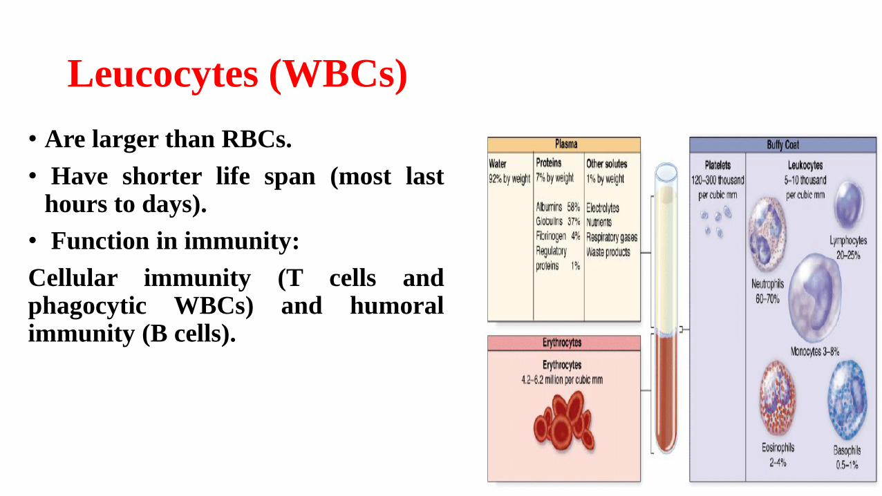
Leucocytes (WBCs)
• Are larger than RBCs.
• Have shorter life span (most lasthours to days).
• Function in immunity:
Cellular immunity (T cells andphagocytic WBCs) and humoralimmunity (B cells).
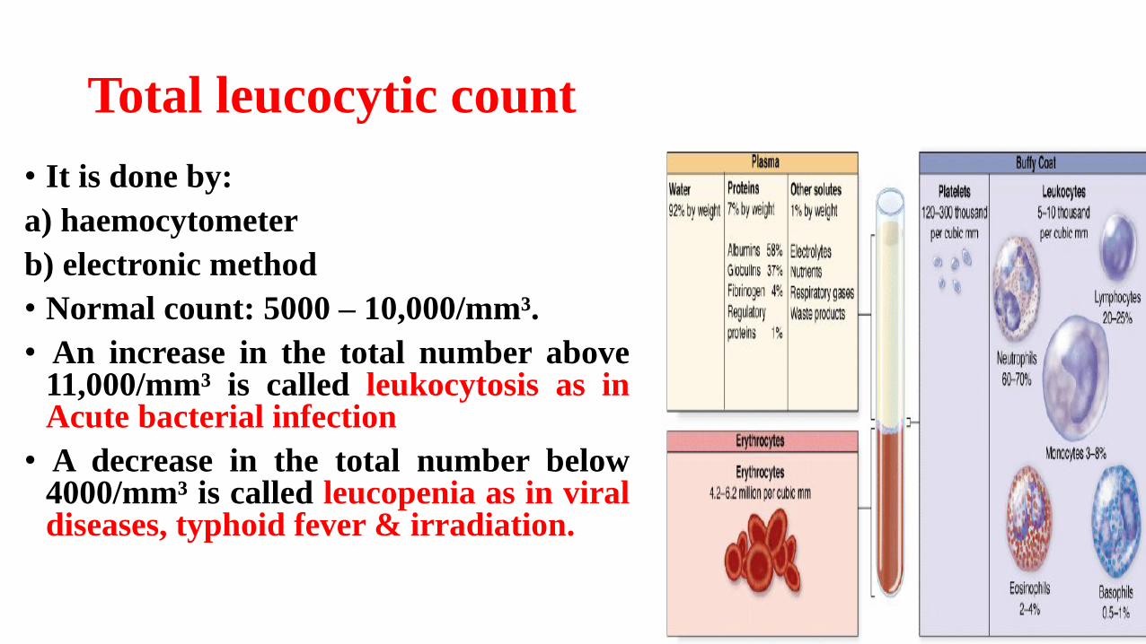
Total leucocytic count
• It is done by:
a) haemocytometer
b) electronic method
• Normal count: 5000 – 10,000/mm³.
• An increase in the total number above11,000/mm³ is called leukocytosis as inAcute bacterial infection
• A decrease in the total number below4000/mm³ is called leucopenia as in viraldiseases, typhoid fever & irradiation.
Differential leukocytic count
• Leukocytes are classified according to presence or absence of specific granules into:
1. Granular leukocytes: formed of: neutrophils, eosinophils and basophils.
2. Agranular (non-granular) leukocytes: formed of: lymphocytes and monocytes.
Granular Leukocytes
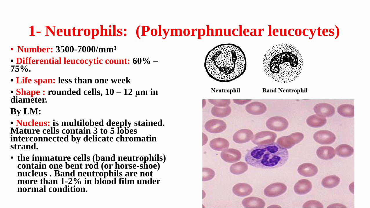
1- Neutrophils: (Polymorphnuclear leucocytes)• Number: 3500-7000/mm³
• Differential leucocytic count: 60% –75%.
• Life span: less than one week
• Shape : rounded cells, 10 – 12 μm in diameter.
By LM:
• Nucleus: is multilobed deeply stained. Mature cells contain 3 to 5 lobes interconnected by delicate chromatin strand.
• the immature cells (band neutrophils) contain one bent rod (or horse-shoe) nucleus . Band neutrophils are not more than 1-2% in blood film under normal condition.
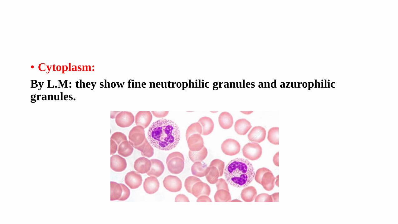
• Cytoplasm:
By L.M: they show fine neutrophilic granules and azurophilicgranules.
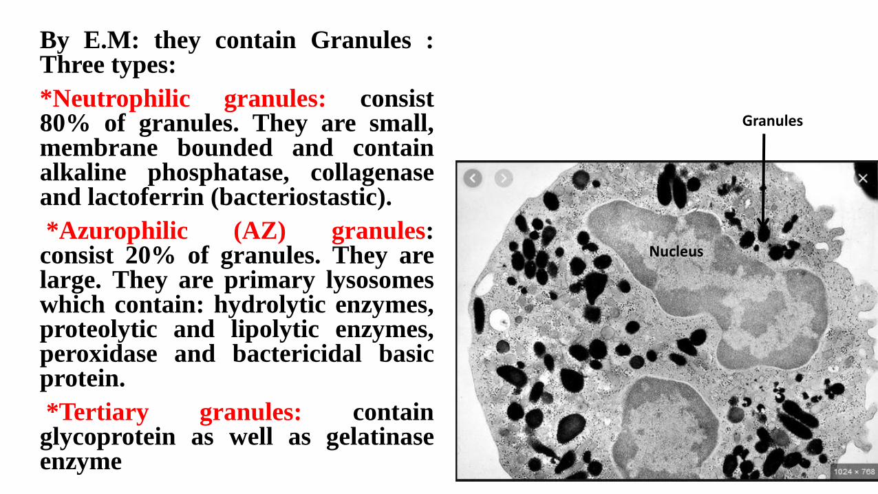
By E.M: they contain Granules :Three types:
*Neutrophilic granules: consist80% of granules. They are small,membrane bounded and containalkaline phosphatase, collagenaseand lactoferrin (bacteriostastic).
*Azurophilic (AZ) granules:consist 20% of granules. They arelarge. They are primary lysosomeswhich contain: hydrolytic enzymes,proteolytic and lipolytic enzymes,peroxidase and bactericidal basicprotein.
*Tertiary granules: containglycoprotein as well as gelatinaseenzyme
Nucleus
Granules
Functions of neutrophils
1. Defence against bacterial infection.
2. Phagocytosis of bacteria and destroys them through neutriphilicand azurophilic granules.
3. Dead neutrophils forms pus cells.
4. Raise the body temperature by producing pyrogens.
5. Stimulate the bone marrow to produce more neutrophils.
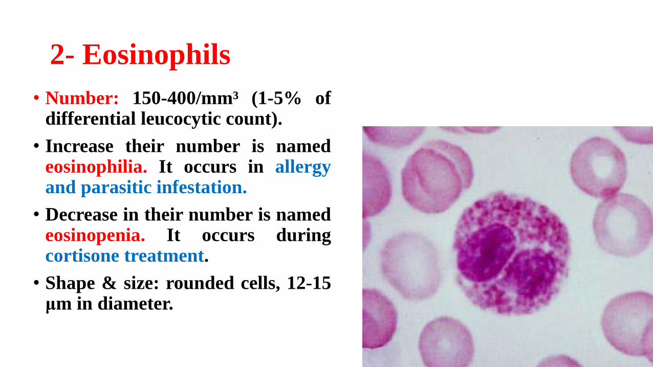
2- Eosinophils
• Number: 150-400/mm³ (1-5% ofdifferential leucocytic count).
• Increase their number is namedeosinophilia. It occurs in allergyand parasitic infestation.
• Decrease in their number is namedeosinopenia. It occurs duringcortisone treatment.
• Shape & size: rounded cells, 12-15μm in diameter.
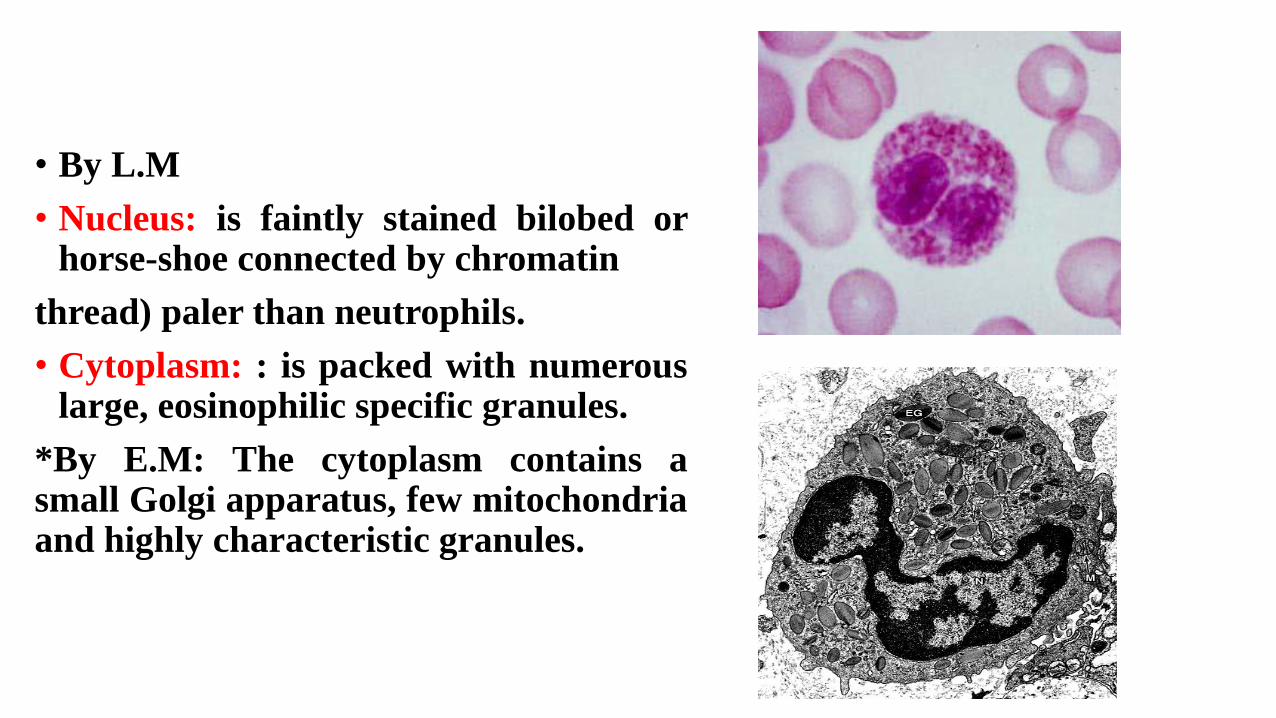
• By L.M
• Nucleus: is faintly stained bilobed orhorse-shoe connected by chromatin
thread) paler than neutrophils.
• Cytoplasm: : is packed with numerouslarge, eosinophilic specific granules.
*By E.M: The cytoplasm contains asmall Golgi apparatus, few mitochondriaand highly characteristic granules.
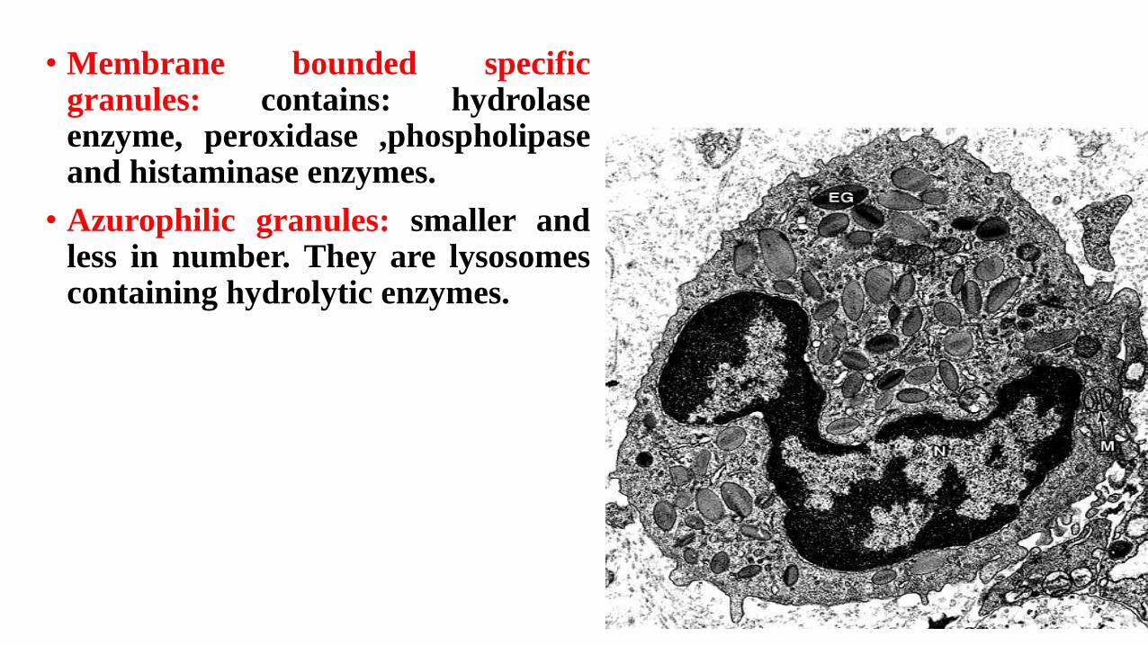
• Membrane bounded specificgranules: contains: hydrolaseenzyme, peroxidase ,phospholipaseand histaminase enzymes.
• Azurophilic granules: smaller andless in number. They are lysosomescontaining hydrolytic enzymes.
Functions:
Play an important role in controlling the local response inallergic reaction:
• They phagocytose antigen – antibody complex.
• They produce histaminases, which degrade histamineduring hypersensitive reaction.
• Play a role in the defense against certain types of parasites.
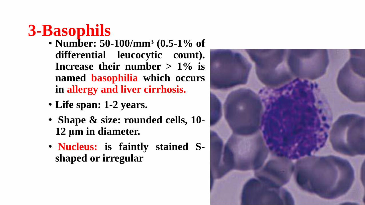
3-Basophils• Number: 50-100/mm³ (0.5-1% of
differential leucocytic count).Increase their number > 1% isnamed basophilia which occursin allergy and liver cirrhosis.
• Life span: 1-2 years.
• Shape & size: rounded cells, 10-12 μm in diameter.
• Nucleus: is faintly stained S-shaped or irregular
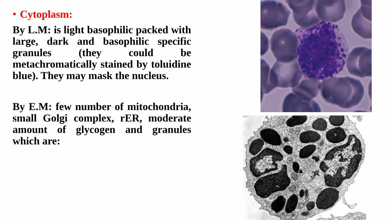
• Cytoplasm:
By L.M: is light basophilic packed withlarge, dark and basophilic specificgranules (they could bemetachromatically stained by toluidineblue). They may mask the nucleus.
By E.M: few number of mitochondria,small Golgi complex, rER, moderateamount of glycogen and granuleswhich are:
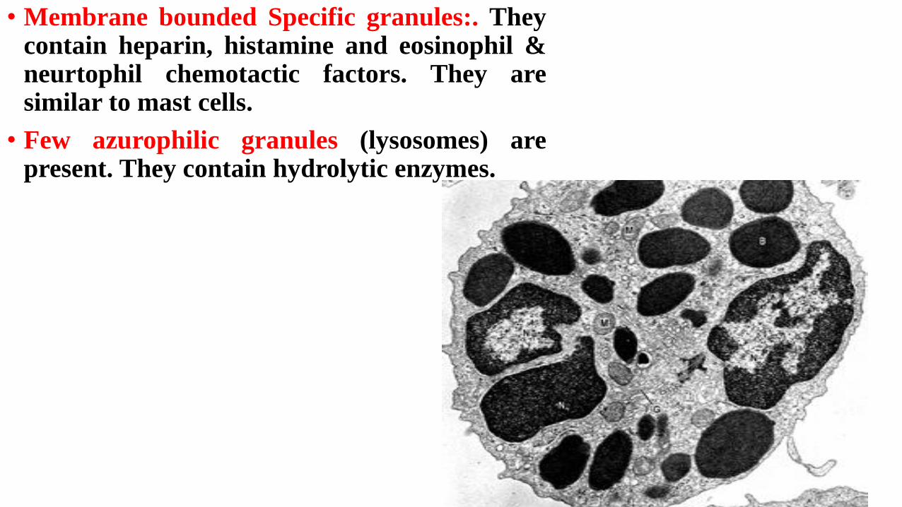
• Membrane bounded Specific granules:. Theycontain heparin, histamine and eosinophil &neurtophil chemotactic factors. They aresimilar to mast cells.
• Few azurophilic granules (lysosomes) arepresent. They contain hydrolytic enzymes.
• Functions:
1) Similar to mast cells, basophils have receptors for IgE. Whenthey bind to specific antigen, cells release histamine, heparin &eosinophil chemotactic factor promoting allergic reaction.Histamine causes vasodilatation and smooth muscle contraction.
2) Have very weak phagocytic activity.
Non-Granular Leucocytes

Lymphocytes
• Number: 1500-2500/mm³ (20 – 25% of differential leucocytic count).
• Increase their number above 40% is named lymphocytosis. It occurs in chronic diseases as T.B. and syphilis, viral infection as influenza, and in lymphoma or leukaemia.
• Decrease in their number below 20% is named lymphopenia. It occurs in AIDS and aplastic aneamia.
• Size:
• 1. Small lymphocytes: 6-8 μm in diameter representing majority of lymphocytes in circulating blood.
• 2. Medium-sized lymphocytes: 10-15 μm in diameter representing few blood lymphocytes.

• N.B. Large lymphocytes: 15-18 μm in diameter. They are presentmainly in lymphatic tissue. The majority of large lymphocytesseen in blood represent activated B lymphocytes. Some circulatinglarge lymphocytes are natural killer (NK) cells.
• Functionally: lymphocytes are divided in the peripheral bloodinto: B (15%), T (80%) lymphocytes & NK cells (5%).
• Null Lymphocytes: representing 5 – 10 % of lymphocytes in theblood. They are composed of two distinct populations:
1. Circulating stem cells: they give rise to all type of formedelements of blood.
2. Natural killer (NK) cells.
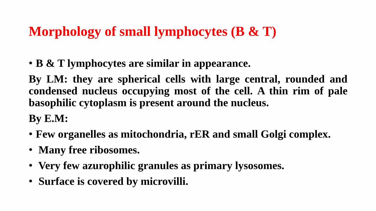
Morphology of small lymphocytes (B & T)
• B & T lymphocytes are similar in appearance.
By LM: they are spherical cells with large central, rounded andcondensed nucleus occupying most of the cell. A thin rim of palebasophilic cytoplasm is present around the nucleus.
By E.M:
• Few organelles as mitochondria, rER and small Golgi complex.
• Many free ribosomes.
• Very few azurophilic granules as primary lysosomes.
• Surface is covered by microvilli.
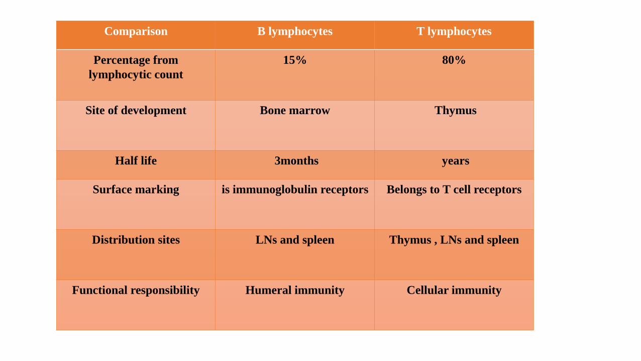
Comparison B lymphocytes T lymphocytes
Percentage from
lymphocytic count
15% 80%
Site of development Bone marrow Thymus
Half life 3months years
Surface marking is immunoglobulin receptors Belongs to T cell receptors
Distribution sites LNs and spleen Thymus , LNs and spleen
Functional responsibility Humeral immunity Cellular immunity
Functions of lymphocytes
• B lymphocytes: are responsible for the humoral immunity. Whencells are exposed to an antigen, they are activated to daughter cellscarrying same program as the mother cells. Some of these cellswill differentiate to plasma cells producing antibodies (1ryimmune response). Other cells will remain inactive as memorycells. When exposed to the same antigen again, these cells willrespond more rapid and more extensive (2ry immune response).
• T lymphocytes: are responsible for the cell mediated immunity.
Types of T lymphocytes
1. T-cytotoxic (killer) cells: destroy foreign cells by direct contact.
2. T-suppressor cells: interfere with other immuno-competent cellssuppressing their activity (may be to prevent autoimmune response).
3. T-helper cells: release of cytokines (lymphokines) as interferon &interleukin to enhance humoral immune response and help B cells.
4. T-memory cells: long lived cells to provide 2ry response to the sameantigen.
5. T-delayed hypersensitivity: important in delayed hypersensitivityreactions as in T.B.
6. T-amplifier: activate B– and T lymphocytes.
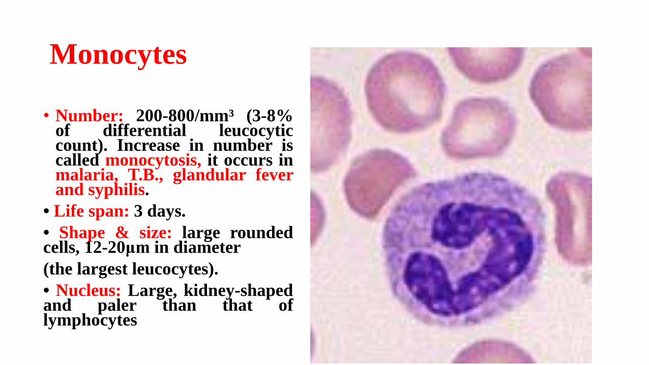
Monocytes
• Number: 200-800/mm³ (3-8%of differential leucocyticcount). Increase in number iscalled monocytosis, it occurs inmalaria, T.B., glandular feverand syphilis.
• Life span: 3 days.
• Shape & size: large roundedcells, 12-20μm in diameter
(the largest leucocytes).
• Nucleus: Large, kidney-shapedand paler than that oflymphocytes
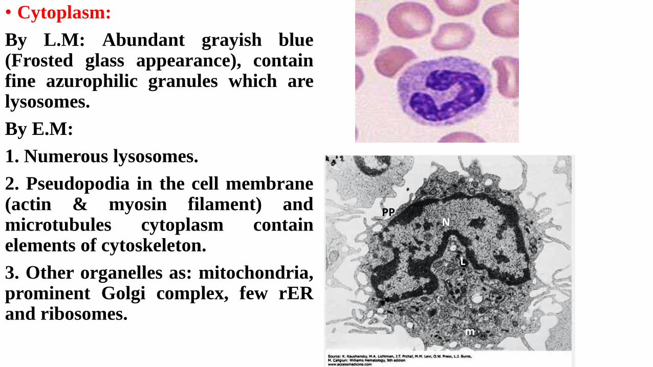
• Cytoplasm:
By L.M: Abundant grayish blue(Frosted glass appearance), containfine azurophilic granules which arelysosomes.
By E.M:
1. Numerous lysosomes.
2. Pseudopodia in the cell membrane(actin & myosin filament) andmicrotubules cytoplasm containelements of cytoskeleton.
3. Other organelles as: mitochondria,prominent Golgi complex, few rERand ribosomes.
NPP
L
m
• Functions:
1. Migrate to C.T. and change into macrophage.
2. Able to fuse together to form foreign body giant cell.
3. Circulating monocytes phagocytose: bacteria, viruses and foreignbodies.
Medical application