um ebv+ proliferations - ihot.lt /limfomos 2011/ebv+... · intro: difficulties talking and dealing...
TRANSCRIPT

EBV+ PROLIFERATIONS UM
LT SPECTRUM IN CONTEXT OF DATA OF 2010/2011

MOTO advices, but please be so kind and JUST WRITE WHAT
Surgeon B

Intro: Difficulties talking and dealing with EBV
Low level of clinical discrimination Low level of discrimination of histological features Not enough experience in pathology IH: EBV LMP1 (2008-‐05-‐26), EBER (2010-‐06-‐15) Transplantation history from 1974: kidney, heart, bone marrow, liver, heart+lung (singles). Pancreas? Intestine? Complex?

Lymphoproliferation vs lymphoma True reactive states/lymphadenopathies: polyclonal, smt monotypic (Ig), smt oligoclonal (EBV) Atypical/borderline lymphoproliferations:
pleomorphic LPDs; LyG I,II°; Castleman d. ): polyclonal/monoclonal; polytypic/monotypic; smt treated as lymphomas; Lymphoma like lesions, monomorphic (gyn tract; germinotropic LPD): polyclonal, monoclonal (?); treatment?
Lymphomas: monoclonal, monotypic (Ig)

Binary paradigm
Lymphoma ( true )
Reactive ( true )

NCP 2010 data
0,4% 0,4% 0,4% 0,4% 0,4% 0,8% 0,8% 0,8% 0,8% 1,2% 1,2% 1,2% 1,6% 1,6%
3,6% 3,6% 3,6% 4,8%
8,0% 9,2% 10,0%
12,8% 32,4%
0,0% 5,0% 10,0% 15,0% 20,0% 25,0% 30,0% 35,0%
Su enteropatija susijusi T limfoma
Burkitto limfoma
Mycosis fungoides
AGE: FOREVER YOUNG AND REACTIVE

Case 1: B11-36165 14 yrs old male;
2.3cm (lateral neck lymphadenopathy).

Case 1: B11-36165 Fol
Hyperplasia Marginal expansion

Case 1: B11-36165 Bcl6 CD30

Ki67 ~85%
Case 1: B11-36165 Ki67

Case 1: B11-36165
Pax-‐5
CD30
CD20 Paracortex

EBER
Case 1: B11-36165 EBV LMP1 Paracortex

Case 1: B11-36165 14 yrs old male; Histo:
FH with lysis; Paracortex expansion with atypical T >> B immunoblasts, CD30+ activated cells, plasmoid cells (immunoblastic lymphadenopathy); Focal marginal zone expansion / immature sinus histiocytosis.
Blood: leucopenia, reactive lymphocytes, high EBV titers, Anti EBV IgM; Active EBV infection (IM): lymphadenopathy.

Case 2: B10-15726 6 yrs old male;
: No clinical data; Tonsilectomy.

Case 2: B10-15726
Necrosis

Ki67
Case 2: B10-15726

CD20
Case 2: B10-15726
EBV LMP1
Ki67 ~85% Paracortex

Case 2: B10-15726 6 yrs old male; No clinical data: tonsilectomy; EBV+ tonsilitis: active EBV infection (IM?); Lost for follow up.

EBV LPL histological signature 1 Geographic necrosis; Imunoblastic expansion: T >> B; Follicular hyperplasia; Extranodal spread; Toxoplasma lymphadenitis ( ) like changes:
histiocytosis Clusters of epithelioid macrophages
Pleomorphic lymphoid infiltrates Angiocentricity HL like cells Plasmablastic/plasmacytoid histology Other

Case off: EBV Dx: B11-30174 32 yrs old female; Neck lymphadenopathy: 2 nodes up to 2,5 cm.
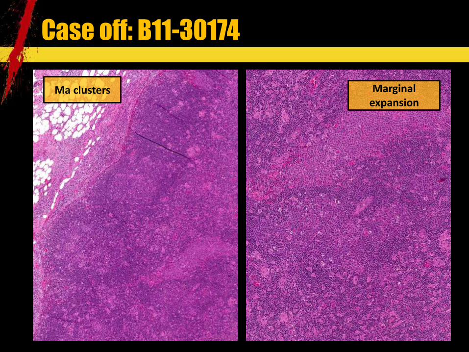
Case off: B11-30174
Marginal expansion
Ma clusters

Case off: B11-30174
Necrosis
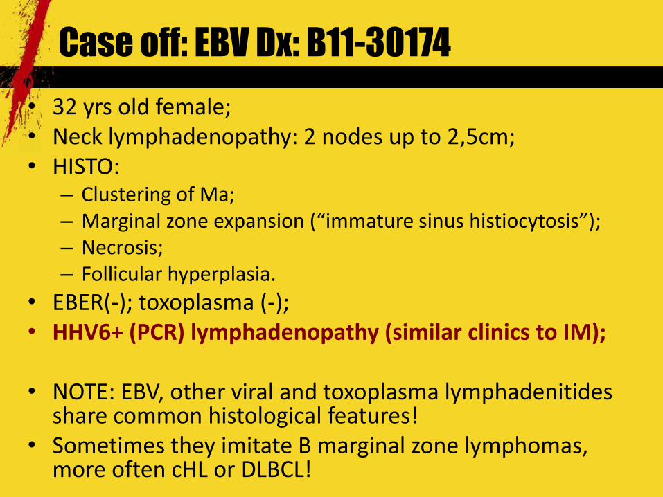
Case off: EBV Dx: B11-30174 32 yrs old female; Neck lymphadenopathy: 2 nodes up to 2,5cm; HISTO:
Clustering of Ma; ;
Necrosis; Follicular hyperplasia.
EBER(-‐); toxoplasma (-‐); HHV6+ (PCR) lymphadenopathy (similar clinics to IM); NOTE: EBV, other viral and toxoplasma lymphadenitides share common histological features! Sometimes they imitate B marginal zone lymphomas, more often cHL or DLBCL!

EBV + CONVENTIONAL LYMPHOMAS

Case 3: B10-33601 85 yrs old female; Generalized lymphadenopathy (neck, axillary), B symptoms (2 yrs); BM: reactive changes; LN biopsy (axillary).

Case 3: B10-33601
Clear infiltrate

Case 3: B10-33601
EBV LMP1
CD68

Case 3: B10-33601
EBV LMP1
CD30
CD15

Case 3: B10-33601 85 yrs old female; IH: CD30+100%; CD15+10%; LCA(-‐); EBV LMP1+100%; CD79a(-‐); Pax-‐5+70%; Bcl6(+) 30% (faint);
cellularity (rich with LAM); BM: negative; Treatment: 6 × AVD.

Case off: AILT with EBER+ CD20+CD30+CD15+ giant cells
CD21 FDC network

Case off: AILT: IgH clonal;; IGK polyclonal;; TCR / clonal
EBV EBER
CD30
CD15
CD79a

Case 4: B11-8589 58 yrs old male; 2011 Feb: acute peritonitis, B symptoms, multiple polyps/infiltrates in small intestine, paraaortic lymphadenopathy, slight splenomegaly, multiple infiltrates in the lungs (CT); Biopsy of the pancreatic tumor and resection of perforated small intestine; No history of malabsorbtion.

Case 4: B11-5728

Case 4: B11-5728

CD56
Case 4: B11-5728
CD3
Ki67

Case 4: B11-8589 58 yrs old male; Biopsy from nasopharynx: the same picture; BM negative; DIAGNOSIS: Extranodal NK/T lymphoma, nasal type: EBER+; CD3/CD7/CD56/CD45RO+, Granzym B+, CD2/CD30/CD5/CD4/CD8(-‐); CD43(-‐/+); Ki67 ~ 95%: spread in small intestine with ulceration/perforation and 1/3 node; 6× of intensive chemo: SMILE and R therapy to sinus area Perforation of intestines/peritonitis without signs of tumor; In remission now; NOTE: from 2010 history of nasal polyps treated conservatively!

TRANSPLANT RELATED

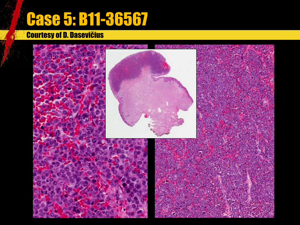
Case 5: B11-36567 64 yrs old male; Both kidneys were removed due to nephrolithiasis in 1998; Single kidney transplant in 2000 December; Under Cyclosporin; B11-‐36567: Tumor of the scrotum skin removed: brown 1,5 cm tumor close to margin.
Courtesy of D. Dasevi
Courtesy of D. Dasevi
Case 5: B11-36567

Case 5: B11-36567 Courtesy of D. Dasevi Ki67 CD138
Mum1 LCA

Case 5: B11-36567 IH: LCA+, CD43+, CD138+, MUM1+, CD4+, CD15+, CD8+(5%); Ig kappa/Ig lambda/IgM-‐, Ki67 100%, EBER(-‐); Ig kappa/ Ig lambda/IgM(-‐), EBV LMP1(-‐), TdT/CD34(-‐), CD10/bcl2/CD20/CD79a/PAX5/bcl6(-‐), CD3/CD5/CD2/CD7/ALK1/ D1a/MPO(-‐), CD56/CD30(-‐), PanCK(-‐); DGN: Posttransplant (~11 yrs after kidney Tx) lymphoproliferative disease (PTLD) (monomorphic) EBV(-‐): B plasmablastic lymphoma in the skin; PCR: IgH/IgK clonal; TCR / uninformative; BM: negative; visceral lymphadenopathy; Treatment: R-‐CHOP-‐14; agranulocytosis; febrilic neutropenia; B11-‐36567 (November): kidney transplant was removed (rejection); under hemodialysis now.
https://www.ipath-‐network.com/ltpatho/object/view/26338

Case 6: B08-24723 36 yrs old male; Renal transplantation in 1993; under immunosupression (mycophenolate mofetil; cyclosporin; steroids); 2008 Feb: chronic rejection (biopsy); Wight loss, B symptoms, rectal pain; CT: neck lymphadenopathy; 2008 Sep: B08-‐24723 biopsy from rectal ulcer; BM: negative.

Case 6: B08-24723
Ki67
CD20: heterogenous

Case 6: B08-24723
Mum1
EBER

Case 6: B08-24723 36 yrs old male; Renal transplantation in 1993; B08-‐24723 biopsy from rectal ulcer; IH: CD20+ (heterogenous), Ki-‐67 ~80%, MUM1+, Bcl2+ (faint), CD10/BCL6(-‐), CD30/CD15 not done; DGN.: DLBCL (PTLD, monomorphic, EBV+ DLBCL) (~15yrs); EBER+ (2011); Trumatic fracture (radius, femur) and delyrium; Treatment (2008): R-‐HiCHOP with endolumbal prophylaxis.

Posttransplant LPD (WHO 2008) Early lesions 99711 (polyclonal):
Plasmacytic hyperplasia; IM like; Other (mass forming FH, other);
Polymorphic 99713 (B clonal, T polyconal) Monomorphic (acording to lymphoma they resemble)(B/T clonal):
DLBCL, Burkitt, Myeloma, Plasmacytoma like, other; PTCL, NOS; Hepatosplenic T cell lymphoma; other.
cHL type PTLD NOTE1: indolent small B cell lymphomas are not included! NOTE2: Most EBV+, some EBV(-‐)! NOTE3: polymoprhic: aggressive, mass forming infiltrate.
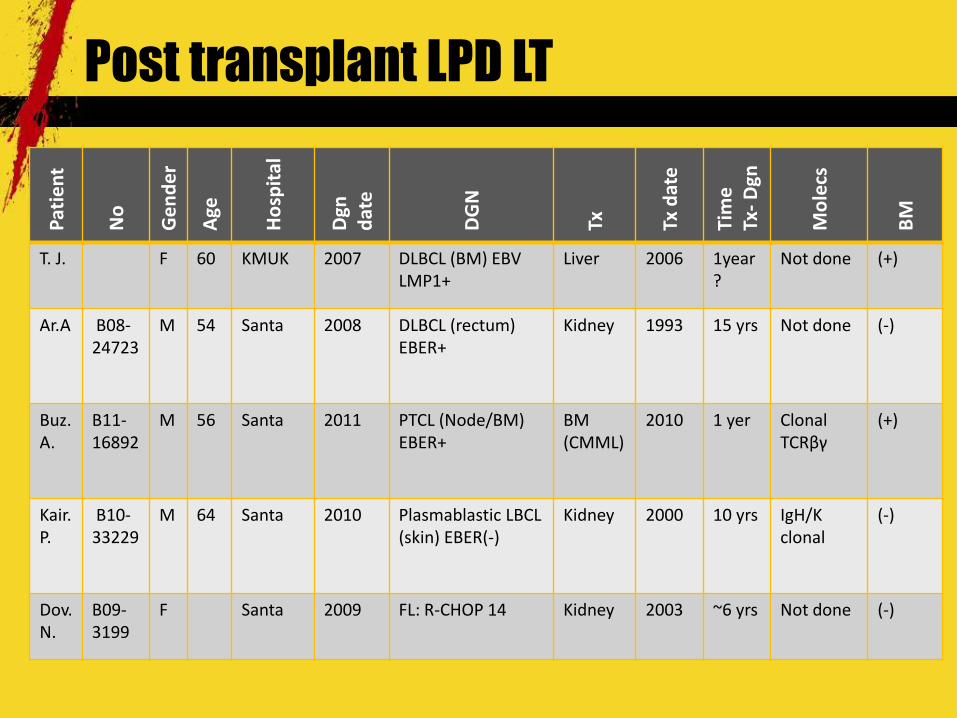
Post transplant LPD LT Patie
nt
No
Gend
er
Age
Hospita
l
Dgn
date
DGN
Tx
Tx date
Time
Tx-‐ D
gn
Molecs
BM
T. J. F 60 KMUK 2007 DLBCL (BM) EBV LMP1+
Liver 2006 1year?
Not done (+)
Ar.A B08-‐24723
M 54 Santa
2008 DLBCL (rectum) EBER+
Kidney 1993
15 yrs
Not done (-‐)
Buz.A.
B11-‐16892
M 56 Santa 2011 PTCL (Node/BM) EBER+
BM (CMML)
2010 1 yer Clonal TCR
(+)
Kair.P.
B10-‐33229
M 64 Santa 2010 Plasmablastic LBCL (skin) EBER(-‐)
Kidney 2000 10 yrs IgH/K clonal
(-‐)
Dov.N.
B09-‐3199
F Santa 2009 FL: R-‐CHOP 14 Kidney 2003 ~6 yrs Not done (-‐)

THERAPY RELATED
Case 7: B09-12583 37 yrs old male (2009); 2001: cytopenia and splenomegaly; suspected EBV (IM); Diagnosis of T LGL verified 2007/2009 (BM); Treatment (outside) unknown (Mtx? Oth.?); 2009: small intestine perforation: resection.

Case 7: B09-12583

Case 7: B09-12583
Ig kappa
Ig lambda

Case 7: B09-12583
Ki67
CD138

Case 7: B09-12583
CD8
CD57
CD3
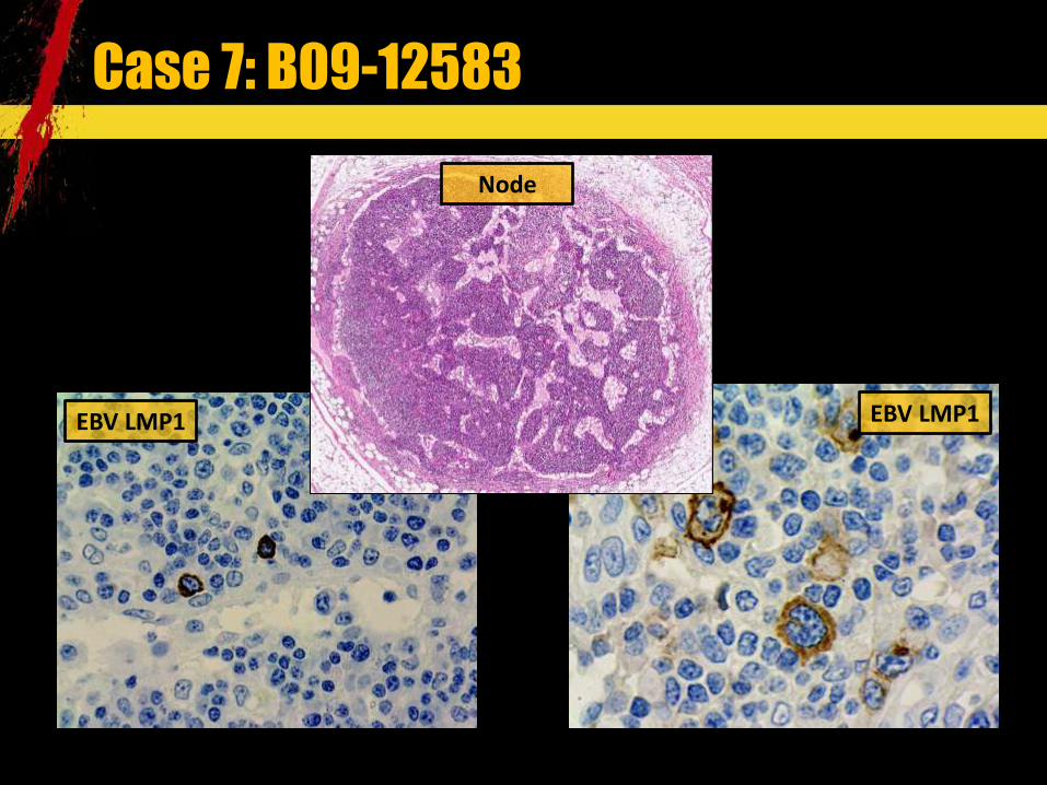
Case 7: B09-12583 Node
EBV LMP1 EBV LMP1

Case 7: B09-12583 37 yrs old male (2009); Probably T LGL from 2001; Small intestine perforation: resection; IH: INFILTRATE: Ki67 ~80% , CD138+ 100%, CD79a+ 20% , Ig kappa/Ig lambda+ (polytypic), Mum1+ 100%. Multiple CD30+ large cells: EBV LMP1/EBER+; Minute T population: CD3/CD2/CD7+ > CD5+, CD56(-‐), CD57+ (in part), CD4+ > CD8+/GranzymB; B09-‐12583: EBV+ LPD (plasmacytic hyperplasia/polymorphic), probably associated with immunosupression/treatment (Mtx?) Minimal T LGL infiltration (TCR clonal; IgH/IgK polyclonal) ???; 2009: DOD after multiple reresections.

EBV LPL histological signature 3 Geographic necrosis; Imunoblastic expansion: T >> B; Follicular hyperplasia; Extranodal spread; Toxoplasma lymphadenitis ( ) like changes:
histiocytosis Clusters of epithelioid macrophages
Pleomorphic lymphoid infiltrates Angiocentricity HL like cells Plasmablastic/plasmacytoid histology Other: immunosupressive therapy? Tx?

Yatrogenic LPD Similar scheme as in PTLD and age related LPL??? Early lesions: ??? Polymorphic: Mtx Monomorphic (acording to lymphoma they resemble): Mtx (DLBCL; PTCL, NOS) (years) TNF inhibitors (DLBCL, HSTCL, oth.) (weeks-‐months)
cHL: Mtx NOTE: indolent small B cell lymphomas are included!

AGE: NOT SO YOUNG AS BEFORE (>50YRS)

Case 8: B08-16708 67yrs old female; Weakness, B symptoms; visceral and peripheral lymphadenopathy; splenomegaly; pulmonary infiltration; 2004: cervix uteri: poorly differentiated squamous carcinoma (biopsy); B08-‐16708: node biopsy; B08-‐16369: BM biopsy.

Case 8: B08-16708
Ki67
CD30
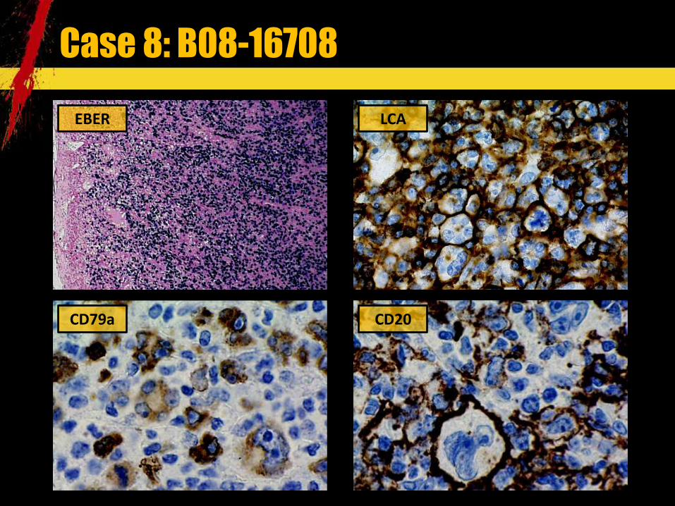
Case 8: B08-16708 EBER
CD79a
LCA
CD20

Case 8: B08-16708 67yrs old female; 2008 June: B08-‐16708 (node): Age related LBCL (polymorphic) ; IH (ABC): LCA+; CD20+; CD30+; Bcl2+ 100%; p53+ 40%; Mum1+; Bcl6/CD10-‐; Ki67 ~80%; B08-‐16369 (BM): Age related LBCL, 20%; PCR: IgK clonal; IgH polyclonal; Septic shock; Treatment: symptomic; DOD: 2008 June.
http://www.ipath-‐network.com/ltpatho/object/view/5832

Hess 2010 data
104 106 109
84
5 6 7 9 2 4
8 5 6 3 3 3 3 3 2 1 0
20
40
60
80
100
120
2007 2008 2009 2010
DLBCL, NOS (403)CNS DDBLL (19)
ALK+ LBCL (1)
Case 9: B11-12891 55 yrs old male; Suspition of IM vs PTCL NOS; Lymphadenopathy, splenomegaly, B symptoms, leucocytosis (neutrophylia), low performance; Nodes (2 biopsies) 2011 April-‐May; BM 2011 June/October; Good response to steroids.

Case 9: B11-12891 1st node

Case 9: B11-12891
1st node CD30

Case 9: B11-12891
1st node
CD79a
CD4 > CD8
Ki67
Ig kappa

Case 9: B11-12891
2nd node (after steroids)

Case 9: B11-12891 2nd node (after steroids) EBER
CD3
CD20

Case 9: B11-12891 55 yrs old male; EBV+ IM like lymphadenopathy: CAEBV (age?); Histo LN: T immunoblastic lymphadenopathy and macrophageal reaction ( like, like); BM: pleomorphic focal T CD4+ infiltration and plasmacytosis; IgH and TCR oligoclonal; (DNA degradation) EBER+ (in second biopsy; first RNA/DNA degradation?); Good response to Rituximab.

EBV LPL histological signature 4 Geographic necrosis; Imunoblastic expansion: T >> B; Follicular hyperplasia; Extranodal spread; Toxoplasma lymphadenitis ( ) like changes:
histiocytosis Clusters of epithelioid macrophages
Pleomorphic lymphoid infiltrates Angiocentricity HL like cells Plasmablastic/plasmacytoid histology Other

Age asociated LPD Similar cheme as in PTLD and yatrogenic LPL??? Early lesions: reactive lymphoid hyperplasia Polymorphic: mucocutaneous ulcer/extranodal, nodal
Monomorphic (according to lymphoma they resemble): Age related DLBCL; Other?

Age related DBCL Patie
nt
Gend
er
Age
Hospita
l
Dgn da
te
Other
DGN
Therap
y
Follo
w
up 2011
Dec
B.I F 67 Santa 2008 July 2004 squamous ca c. uteri; BM+
Age DLBCL
No IgK clon; IgH poly
DOD 2008 July
S.J. M 78 KJL
2011 March
BM(-‐) Age DLBCL
R-‐CHOP 14 ; MINE
Not done Alive, 2011 Oct relapse
P.P M 77 KJL Age DLBCL
R-‐CHOP
Not done
Alive
A.J. M Santa 2011 BM(-‐) Age DLBCL
R-‐CHOP 14 Not done Alive

RARITIES:

64 yrs old male; B10-‐25858 (biopsy): gastric adenocarcinoma; B10-‐27024 (node biopsy): ??? B10-‐28518: gastric resection with lymhonodectomy.
Case 10: B10-28518
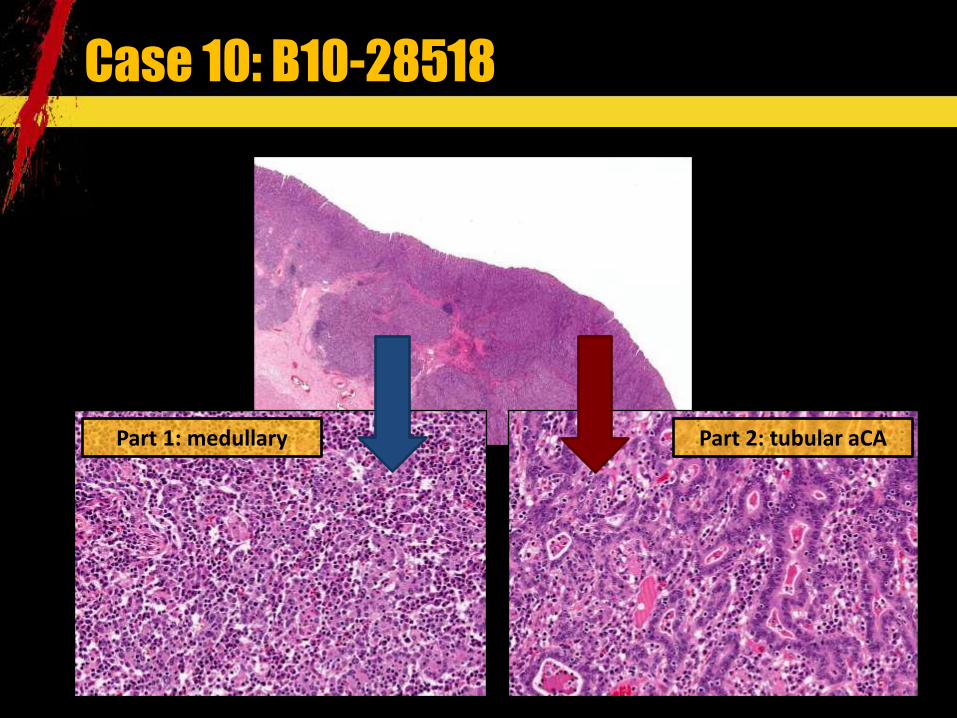
Case 10: B10-28518
Part 2: tubular aCA Part 1: medullary

Case 10: B10-28518
CD3 Ig lambda

Case 10: B10-28518
Gastric
Plasmablastic lymhoma
EBER
EBER

64 yrs old male; EBV+ HHV8+ plasmablastic lymphoma (germinotropic LPL) in perigastric 3/29 nodes; Gastric cardiac EBV+ lymphoepithelioma like carcinoma (LELC) : TNM 2010: pT1b (submucosa) N1 (summary: mts 1/29). Intestinal type, Lauren. Radical resection; Clonality (PGR): plasmablastic lymphoma: Ig H clonal; TCR polyclonal; TCR ; TCR clonal; HHV8: positive (PGR); HHV8 (LANA)+ IH (dr.A.Tzankov); HIV (-‐); Follow up: 2011: B11-‐35289 (BM): negative; Treatment: R-‐CHOP 14. http://www.ipath-‐network.com/ltpatho/object/view/25005
Case 10: B10-28518

EBV+ carcinomas
Gastric lymphoepithelioma like carcinoma (LELC), lymphoid stroma rich carcinoma, carcinoma Pancreatic carcinoma; Bile duct carcinoma; Salivary gland carcinoma; Small intestinal carcinoma? Breast carcinoma?

SUMMING UP

EBV+ tumors/proliferations Lymphoid-‐proliferations (LPL); EBV+ FDC sarcoma (IMFT-‐like; spleen/liver); Undifferentiated nasopharyngeal carcinoma EBV+ carcinomas (medullary or lymphoepithelioma like (LELC)): salivary gland, stomach, intestine, pancreas, bile duct, breast, thymus; EBV+ smooth muscle tumor (SMT: benign and malignant, immunodeficiency associated): tonsil, vulva, larynx, bronchi, liver, meninges, urinary bladder; Oral squamous carcinoma.

Nasopharyngeal undifferentiated carcinoma
PanCK
EBER

EBV+ lymphoproliferations EBV+ lymphadenitis, tonsilitis, infectious mononucleosis EBV dermatitis (Gianotti-‐ Crosti s.): papular acrodermatitis of childhood Hairy leucoplakia (mostly HIV) EBV+ gastritis EBV+ polymorphic nodal or extranodal (muco-‐cutaneous ulcer) LPL of elderly/ yatrogenic EBV+ LBCL of elderly
lymphoma (endemic >> sporadic) cHL LyG (I-‐II°) and DLBCL (LyG) EBV+ DLBCL DLBCL asociated with chronic inflammation Primary effusion LBCL Plasmablastic LBCL CLL/SLL with EBV+ giant cells (Fludara associated) CLL/SLL with Richter transformation (EBV+ cHL, clone unrelated) (Fludara associated) germinotropic lymphoproliferation plasmablastic LBCL

EBV+ lymphoproliferations Extranodal T/NK cell lymphoma, nasal type Aggressive NK-‐cell leukaemia Gama/delta hepatosplenic T cell lymphoma (HSTL) (some)?; Gama/delta non-‐hepatosplenic lymphoma; Enteropathy asociated T cell lymphoma (some)? Peripheral T cell lymphoma (some) Angioimmunoblastic T cell lymphoma (AILT) Systemic EBV+ T lymphoproliferative fulminant hemophagocytic Hydroa vacciniforme-‐like lymphoma (Asia, Central, South America nad Mexico) HIV associated (NHL entities: plasmablastic LBCL, cHL, oth.) Primary immunodeficiency associated LPL Treatment associated LPL Transplantation associated PTLD

Immunodeficiency related EBV lymphoproliferations
???
Primary immunodeficiency?
Age related (immunodeficiency?)
Therapy related immunodeficiency
Transplantation related
immunodeficiency
EBV
??? Plasmacytic hyperplasia
Polymorphic LPL (nodal/ extranodal)
Monomorphic nodal/extranodal
(lymphoma)
Mucocutaneous ulcer Traumatic oral granuloma?
Toxoplasma like lymphadenitis
AIDS
Autoimmune disease related
immunodeficiency

Summary 1: when to look for EBV? Every DLBCL after 50yrs: EBV (EBER)? Every LPL with giant cells (HL like), angiocentricity, geographic necrosis: EBV (EBER)?
Plasmablastic LBCL: EBER;
other: EBER; cHL: EBV LMP1? EBER? (prognosis?); Posttransplant LPL setting: EBER; Primary or aquired immunodeficiency asociated LPL: EBER; Undifferentiated carcinomas (head/neck; mts): EBER; Lymphoepithelial/medullary carcinomas: EBER;
like): EBV (EBER)?

Summary 2 Immunodeficiency means not only HIV infection or primary/inherited phenomenon; Reactive EBV related changes are heterogenous vary from classic immunoblastic lymphadenopaty to mucocutaneous ulcer, postransplant like LPL; Borders between LPL and lymphoma sometimes is blurred; EBV is needed for categorization of LPL and for prognostication; The detection of EBV in the tissue/pathology means immunodeficiency in various extent?
Summary 3 The links of HESS with transplantation registry and rheumatic disease registry is needed; Pathology forms must be filled more preciselly (transplantation? Autoimmune disease? Immunocompetence? Viral diseases? HIV? Rheumatic diseases?) EBV LMP1 in NCP: from 2008-‐05-‐26; EBER: from 2010-‐06-‐15; Really we need more experience on LPDs...

New paradigm
Reactive
picture, settings, clonality,
phenotype
Borderline / polymorphic
picture, settings,
clonality, phenotype
Lymphoma
picture, settings, clonality,
phenotype

Literature 1. Dojcinov SD, Venkataraman G, Pittaluga S et al. Age-‐related EBV-‐associated
lymphoproliferative disorders in the Western population: a spectrum of reactive lymphoid hyperplasia and lymphoma. Blood. 2011 May 5;117(18):4726-‐35. Epub 2011 Mar 8.
2. Di Napoli A, Giubettini M, Duranti E et al. Iatrogenic EBV-‐positive lymphoproliferative disorder with features of EBV+ mucocutaneous ulcer: evidence for concomitant TCR IGH rearrangements in the Hodgkin-‐like neoplastic cells. Virchows Arch. 2011 May;458(5):631-‐6. Epub 2011 Mar 12.
3. Dojcinov SD, Venkataraman G, Raffeld M et al. EBV positive mucocutaneous ulcer-‐-‐a study of 26 cases associated with various sources of immunosuppression. Am J Surg Pathol. 2010 Mar;34(3):405-‐17.
4. Age-‐related Epstein-‐Barr virus-‐associated B-‐cell lymphoproliferative disorders: special references to lymphomas surrounding this newly recognized clinicopathologic disease. Shimoyama Y, Yamamoto K, Asano N et al. Cancer Sci. 2008 Jun;99(6):1085-‐91.
5. Swerdlow S H, Campo E. WHO Classification of Tumours of Haematopoietic and Lymphoid Tissues, Fourth Edition. IARC Press: Lyon, 2008
6. Okano M, Gross TG. Acute or Chronic Life-‐Threatening Diseases Associated With Epstein-‐Barr Virus Infection. Am J Med Sci. 2011 Nov 17. [Epub ahead of print]
7. Hum Pathol. 2007 Sep;38(9):1293-‐304. Epstein-‐Barr virus-‐associated lymphoproliferative disorders. Rezk SA, Weiss LM.
8.

Acknowledgements
Alex and Basel Team The visit to LT and high level hematopatho INCTR iPath community Discusions on the Cases LT Laimonas and Santa Team Every Day Hemato Mindaugas and Klaip da Team Cases from the West LT Vestina, Inga and Kaunas Team Cases from Kaunas Rokas , Graphics and Data analysis Julius Every Day Help/Cases/Photos/Data analysis Gods and Saints Rest of All
Design

EBV+ PROLIFERATIONS UM
LT SPECTRUM IN CONTEXT OF DATA OF 2010/2011

NCP data 2010
344
58
1 0
50
100
150
200
250
300
350
400
NHL cHL NLP HL

NCP data 2010
88 81
58
32 25 23 20
12 9 9 9 6 4 4 3 3 3 2 2 2 2 1 1 1 1 1 1 0
10
20
30
40
50
60
70
80
90
100
Limfomos

NCP data 2010
54
8 7 4 2 2 1 1 1 1
0
10
20
30
40
50
60
DLBC
L NOS
DLBC
L ex M
ZL
DLBC
L Bu
rkit-‐like
DLBC
L ex FL
DLBC
L ex CLL
LYG III°
"Grey zone
" lim
foma su ta
rpiniais DL
BCL ir
LBCL ALK+
DLBC
L Leg type
Difuzin dideli B

NCP data 2010
22
17
6 5
4 4 3
2 2 2 2 2 2 1 1 1 1 1 1 1 1 1
9
0
5
10
15
20
25
Limfm
azgis
Skrand
is
Oda
Retrop
erito
ninis tarpas
Plautis
Kasa
Tarpup
lautis
Pasaita
s
Prieno
sinis antis
Kepe
nys
Galvos s
megen
ys
Kaklas
Bron
chas
Tarpup
lautis
Difuzin dideli B

NCP data 2010
1
11
5
3 3
2
1
0
2
4
6
8
10
12
14
Skrandis Limfmazgis Kepenys
limfoma
-‐ lokalizacijos

HESS preliminary data 2010
144
130
112 113
125
92
29 34
23
14
21 26
31
21
30
13
26 22
15
27
19 23
42
21
14 11
14 15 14 16
10,3
11,8
8,0 7,5
8,9
5,8
0
2
4
6
8
10
12
14
0
20
40
60
80
100
120
140
160
2005 2006 2007 2008 2009 2010
DLBCL SLL MCL MZL FCL DLBCL : FCL