uk healthcare investments - florida vascular societyfvs.org/pdf/2016/fridayapril29/aorto-enteric...
TRANSCRIPT

Aorto-enteric Fistula
Eric D. Endean, MD
Gordon L Hyde Professor of Surgery
Florida Vascular Society

Disclosures
- 2 -
No Disclosures

Objectives
- 3 -
1. Describe the types of aorto-enteric fistulas
2. Discuss the mode of presentation and diagnosis of aorto-enteric fistula
3. Discuss treatment options for aorto-enteric fistula

Patient Presentation
39-year-old woman transferred from OSH with GI bleed
Past History:
Underwent “aorto-bifemoral” bypass in 2009
Presented to OSH with GI bleed in Dec. 2012
Upper and lower endoscopy were negative
Bleeding ceased, hematocrit stabilized – patient discharged
Re-bleeding shortly after discharge
Transfused 4 units PRBCs at OSH
- 4 -

• Transferred to our institution
– Transfused 3 U PRBC in route
• In Emergency Department
– Blood pressure: 108/72
– HR: 120 – 160
– Tender abdomen
– Large amount of blood per rectum in bed
• Taken emergently to the OR
- 5 -

Operative Findings
• Midline laparotomy with control of the aorta at the level of the renal arteries
• Aortic graft exposed –– End-side anastomosis
– Graft was excised down to individual graft limbs
• Aorta over sewn (distal aortic occlusion)
• Duodenum with large enterotomy:– Kocher maneuver
– Stapled proximal and distally
– Jejunoduodenostomy
– Abdomen packed
- 6 -

Follow -up
• Returned to the OR the following day
– Packs removed
– Third portion of duodenum resected
– Omentum placed over the aortic repair
– Discharged two weeks later
– No revascularization of lower extremities
• Clinic follow up
– Half-mile claudication
– ABIs: Right 0.67; Left 0.72
- 7 -

Case Presentation
• 61 year old man presented to OSH with massive upper and lower GI hemorrhage
• History of EtOH abuse
• History of aorto-bifemoral graft 13 years previously for occlusive disease
• Outside CT scan suggestive of peri-graft inflammation
• 2 units PRBCs, 3 liters crystalloid
• Levophed and dopamine were initiated
UK-ED Evaluation
• Received 8 units blood, 8 liters crystalloid in ED
• Weaned off pressors
• GI medicine endoscopy in ED: Large amount of blood in stomach and duodenum
• Hypotensive and transferred emergently to OR
OR Intervention
• Rapid exposure of left limb of aorto-bifem graft
• Balloon tamponade of aorta
• A total of 5 aortic endo-cuffs placed from the graft to infrarenal aorta: converted end-side anastomosis to end-end
• Cessation of bleeding
• Patient transferred to ICU for ongoing care

Post-operative Care
• Patient was resuscitated and stabilized in ICU• Colonoscopy demonstrated ischemic changes
in the sigmoid with patchy mucosal necrosis• CT scan showed air and fluid around the graft

Treatment of Infected Graft
• Two weeks later, returned to OR for treatment of infected graft and revascularization
• Options considered:
– Graft excision and extra-anatomic bypass
– Graft excision and in-line replacement with autogenous tissue

Second Operation
• At laparotomy: infarcted descending and sigmoid colon– Colectomy, feeding tube
• Two duodenal perforations controlled with over sewing
• Excision of prosthetic graft• Aorto-right femoral and right to
left femoral-femoral bypass grafts with SFV
• Omentum interposed between graft and bowel

Post-operative Course
• Creation of transverse colostomy
• Abdomen left open
• Wound VAC dressing change
• Returned to OR 4 times for wash-out
• Two weeks after revascularization, developed hemorrhage from abdominal wound/NG tube

Operative Findings
• Graft bile-stained
• Erosion of vein graft with active hemorrhage
• Dehiscence of one of duodenum repair sites
• Vein graft repaired primarily
• Duodenum excluded
– Stapled distal to ampulla
– Large drain in duodenum through enterotomy
– Jejunostomy tube
– Gastrostomy tube

Outcome
• Returned to OR the following day for re-creation of the colostomy
• Re-hemorrhage 24 hours later
• Further intervention felt futile
• Patient allowed to expire without additional surgical intervention


Classification of Aorto-enteric Fistula
• Primary
– 0.02 – 0.07% in autopsy series
• Secondary
– Fistula
– Erosion
– <1% of patients following abdominal aortic reconstruction
• Manifestations of GI hemorrhage and/or sepsis

Primary Fistula
• Diseased native aorta – Aortic erosion into bowel – most common
• Usually degenerative AAA
• Aortitis or mycotic aneurysm
• Calcific plaque resulting in erosion into bowel
• Primary GI pathology – Peptic ulcer erosion
– Neoplasm
– Tuberculous mesenteric lymphadenitis/infection
– Pancreatic pseudocyst
– Foreign body
– Radiation

Secondary Fistula
• Aorto-enteric fistula – Communication between aortic lumen and bowel lumen at aorticanastomosis Hemorrhage
– Usually involves pseudoaneurysm at the aortic anastomosis
– Most often involves 3rd or 4th portion of duodenum
– Graft infection• Cause of the pseudoaneurysm
• Secondary to bowel contents in contact with the graft
– Duodenal/bowel defect
– Devitalization of retroperitoneal tissue

Secondary Erosion
• Graft-enteric erosion –Graft material in communication with bowel lumen Infectious symptoms
– Develops mid-graft -away from the anastomosis
– No direct communication with the aortic/graft lumen
- 21 -

Inept Surgeon Aorto-enteric Fistula

Pathogenesis
• Infection – breakdown of suture line/graft infection– Contamination at time of surgery
– Bacteremia
• Pulsatile pressure– Pressure of non-compliant prosthesis against bowel
• Technical – Bowel injury (retraction, cautery, during dissection)
– Desiccation of bowel during procedure• ? Advantage of retroperitoneal approach
• Fistula after endovascular repair– Endoleak with growth of residual sac
– Multiple coiling attempts to treat endoleak
– Erosion of stent graft
– Endotension
– Infection
- 23 -

Prevention
• Prevent graft infection: strict sterile technique, skin prep, draping, perioperative antibiotics
– Most bacterial isolates from aortic graft infection are skin flora
• Protection of bowel (duodenum) from retractor injury
• Closure of retroperitoneum - interpose tissue between graft and bowel (duodenum)
– Retroperitoneum/aneurysm wall
– Consider omentum

Diagnosis
• History of prior aortic graft + GI bleed = aorto-enteric fistula until proven otherwise
– Average 2 - 6 years after aortic graft placement
– Can be months to > 10 years
• “Sentinel” or “Herald” bleed (50% - 80%)
– Up to 50% will have a second episode of bleeding within 24 hours
– Massive GI bleed

Confirmatory Studies
• 50% are diagnosed preoperatively
– Endoscopy
– CT scan
– Angiography
– MRI
• Laparotomy

Endoscopy• Detection rate for AEF is ~25%
• Visualization of graft prosthesis confirms diagnosis (extrinsic mass, distal duodenal ulcer, bleeding)
• Clot in the duodenum should not be dislodged
• Must visualize entirety of the duodenum to be negative for aorto-duodenal fistula (may require pediatric colonoscope)
• 75% located at junction of 3rd and 4th portion of duodenum
• Endoscopy may identify/exclude other source of bleeding

CT Scan
• CT angiography gives anatomic information
• Signs of infection:– Fluid around graft
– Air in fluid
– Bowel wall thickening
• Visualize pseudoaneurysm
• Currently most reliable test – detection rate ~60%

Angiography
• Pseudoaneurysm at proximal anastomosis
• Rarely will demonstrate extravasation of contrast into bowel lumen
• May not document graft-enteric erosion
• Provides anatomic information (location of renal arteries, lower extremity runoff)

MRI scan
• T2-weighted images: increased signal intensity in presence of infection
• May better delineate extent of graft infection

My Initial Algorithm
• Check the call schedule: verify I am on call
• Check my calendar: see if I am scheduled to be out of town
• Put my head in the sand and hope it will go away
• Find a junior partner to do a “great case”
• Recheck the call schedule once more – just in case

Treatment Objectives
•Control hemorrhage
•Repair bowel
•Control infection– Remove all infected graft
– Debride infected tissue
•Revascularize ischemic tissue (legs)
Life Over Limb!

Hemorrhage
• Herald bleed
– 30% will re-bleed within 6 hours
– 50% will re-bleed within 24 hours
• Control of hemorrhage
– Balloon tamponade
– Endoprosthesis
– Aortic cross clamp• Infra-renal
• Supra-renal/supra-celiac
• Intrathoracic
- 33 -

Management of aorta
• Remove the graft
• Over sew the aorta with planned extra-anatomic bypass
– Concern for aortic stump blow-out
– Repair aorta for end-side anastomosis
• In-line repair of the aorta – Autogenous vein (Deep leg vein)
– Prosthetic graft (Rifampin soaked, silver impregnated graft)
– Homograft
– Endoluminal prosthesis

Extra-anatomic Revascularization
• Axillary-bifemoralbypass
– Usually after aortic control/ligation
• Suture ligation of aorta– Two layer closure
– Consider covering aortic stump with omentum/fascia
Shephard AD, Conrad MF Alternative, Open Revascularization for Aortoiliac Occlusive Disease. in Mastery of Vascular and Endovascular Surgery, eds Zelenock, Huber, Messina, Lumsden, Moneta. Lippincott Williams & Wilkins, p. 369, 2006.

In-line Revascularization
• Neo-aortoiliac system: Deep vein (femoral/popliteal)
• Prosthetic in- situgraft replacement
– Prosthetic graft (rifampin-soaked, silver coated)
– HomograftHuber, TS. Management of Infected Aortic Grafts. in Mastery of Vascular and Endovascular Surgery, eds Zelenock, Huber, Messina,
Lumsden, Moneta. Lippincott Williams & Wilkins, p. 407, 2006.

SFV Reconstruction Options
Clagett GP, Valentine J, Hagino RT. J Vasc Surg 1997;25:255-70

Endovascular Repair
- 38 -
Kakkos. Eur J Vasc Endovasc Surg 2011;41:625-34
25 patients: 8 endovascular; 17 open– Comparable preoperative characteristics
– Early outcomes:
– 2-year outcome:

Conclusions
• Endovascular repair has fewer complications and mortality in postoperative period
• Short-term advantage is lost by the second postoperative year
• Endovascular repair should be considered a bridge
- 39 -
Kakkos. Eur J Vasc Endovasc Surg 2011;41:625-34

Management of Bowel
• Primary repair
• Resection and re-anastomosis
• Bypass bowel segment (pyloric exclusion)
• Temporary bowel clamp/suture control of contamination
• Interpose omentum between vascular structure and bowel
- 40 -

Enteric Repair
• 791 cases between 1951-2010– Enteric procedure was described in 331
– Vascular procedure described in 515
- 41 -
dos Santos. Ann Vasc Surg 2014; 28:756-62

Outcome
- 42 -
• Most Frequent Cause of Death: Fistula recurrence/sepsis• Improved results:
• Omentum interposition• In situ graft
dos Santos. Ann Vasc Surg 2014; 28:756-62

Contemporary Series
37 patients– 6 University hospitals,
– 2000 – 2008
– Age: 72 ± 9 years
– Most frequent organism -Candida, E coli
– Presenting symptoms:• Sepsis - 41%
• GI bleed without shock – 17%
• GI bleed with shock – 10%
• Abdominal/back pain - 8%
• Graft limb thrombosis - 5%
• Groin fistula – 4%
• Peripheral abscess – 3%
• Femoral pseudoaneurysm -2%
• Peritonitis – 2%
Type of repair:
– In situ reconstruction – 25
• Silver-coated graft – 13
• Cryopreserved allograft – 8
• Rifampin-soaked graft – 3
• Autogenous vein - 1
– Extra-anatomic revascularization– 9
• Axillo-bifemoral – 7
• Bilateral Axillo-femoral - 2
– Endovascular repair – 3
- 43 -
Batt M. Eur J Vasc Endovasc Surg 2001;41:748-57
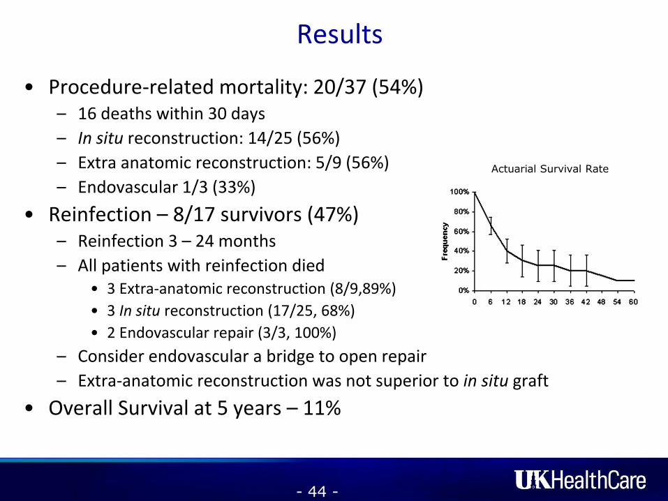
Results
• Procedure-related mortality: 20/37 (54%)– 16 deaths within 30 days
– In situ reconstruction: 14/25 (56%)
– Extra anatomic reconstruction: 5/9 (56%)
– Endovascular 1/3 (33%)
• Reinfection – 8/17 survivors (47%)– Reinfection 3 – 24 months
– All patients with reinfection died• 3 Extra-anatomic reconstruction (8/9,89%)
• 3 In situ reconstruction (17/25, 68%)
• 2 Endovascular repair (3/3, 100%)
– Consider endovascular a bridge to open repair
– Extra-anatomic reconstruction was not superior to in situ graft
• Overall Survival at 5 years – 11%
- 44 -
Actuarial Survival Rate

Late outcome following open surgical management of secondary aortoenteric fistula
Bíró. Arch Surg 2011; 396:1221-9
• 48 patients with secondary aorto-enteric fistula
• 1989 – 2009
• Indication for initial operation– Occlusive disease – 41
– Aneurysm – 7
• Graft location– Aortobifemoral – 40
– Aortic tube graft – 6
– Aortoiliac – 2
• Emergency procedure in 28 (58%)– Unstable hemodynamics
• Reconstruction– In situ – 35 (73%)
– Extra-anatomic – 11 (23%)
– No reconstruction (graft removal) – 2 (4%)
- 45 -

Results• Early outcome
– 30-day mortality: 23/48, 46%• Anastomotic bleeding• Duodenal leakage• Multisystem organ failure• Ischemic colon• Coronary event
• Late outcome– Three and five year survival: 35%
and 27%, respectively– Emergently operated group had
higher mortality than “urgent” group (59% vs. 38%, p<0.04)
• Conclusions:– Emergent (bleeding) aorto-
enteric fistula are associated with higher mortality
– In-situ replacement with graft may be feasible option
- 46 -
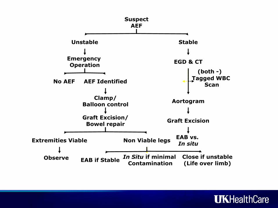
SuspectAEF
Unstable Stable
EGD & CT
Aortogram
Graft Excision
Emergency Operation
No AEF AEF Identified
Clamp/Balloon control
Graft Excision/Bowel repair
EAB vs. In situ
Extremities Viable Non Viable legs
Observe EAB if StableClose if unstable(Life over limb)
In Situ if minimal Contamination
Tagged WBC Scan
(both -)
(+ for GI source)

Conclusions
• Aorto-enteric fistulas must be considered in any patient with GI hemorrhage and prior aortic graft placement
• CT angio and upper endoscopy are diagnostic studies of choice in stable patients
• Treatment is determined by patient stability• Treatment must include control of hemorrhage,
repair of bowel, debridement of infected tissue and revascularization if stable
• My preference is in-situ replacement with autogenous tissue (limited by urgency of operation)
• Patients have a high mortality and morbidity

Thank you

Post operative CT scan
Aorta at renal
Endograft cuffs
End-side anastomosis
Aortic endo cuffs
L renal arteryNative aorta

Outcome
• Untreated: Uniformly fatal
• Mortality: 30% - 40% (Range 13% - 85%)
• Long-term survival: 50% at 3 years
• Amputation rate: 10%
• Significant post-operative morbidity– Long ICU stays
– Organ failure
