environmental enteric dysfunction and growth failure ... · pdf fileenvironmental enteric...
TRANSCRIPT
Environmental Enteric Dysfunction and Growth Failure/Stunting in Global Child HealthVictor Owino, PhD, a Tahmeed Ahmed, PhD, b Michael Freemark, MD, c Paul Kelly, MD, d, e Alexander Loy, PhD, f Mark Manary, MD, g Cornelia Loechl, PhDa
aInternational Atomic Energy Agency, Vienna, Austria; bInternational Centre for Diarrhoeal Research, Bangladesh,
Dhaka, Bangladesh; cDivision of Pediatric Endocrinology,
Duke University Medical Center, Durham, North Carolina; dUniversity of Zambia, Lusaka, Zambia; eBlizard Institute,
Queen Mary University of London, London, United Kingdom; fDepartment of Microbiology and Ecosystem Science,
Research Network “Chemistry meets Microbiology, ”
University of Vienna, Vienna, Austria; and gWashington
University, St Louis, Missouri
Dr Owino drafted the initial manuscript, wrote the
conclusion, formatted the manuscript to conform
to Pediatrics style, and reviewed and revised
the manuscript; Dr Ahmed wrote the section on
emerging approaches for prevention and treatment
of environmental enteric dysfunction (EED), and
reviewed and revised the manuscript; Dr Freemark
wrote the section on growth failure and stunting
in malnutrition and EED and edited the manuscript;
Dr Kelly wrote the section on pathobiology of
EED and contributed to Future Directions, and
reviewed and revised the manuscript; Dr Loy
wrote the section on the diagnostic potential of
stable isotope assays, and reviewed and revised
the manuscript; Dr Manary wrote the section on
application of –“-omic” technology in EED diagnosis,
contributed to Future Directions, and reviewed and
revised the manuscript; Dr Loechl conceptualized
and facilitated discussions for the perspective,
and reviewed and revised the manuscript; and all
authors approved the fi nal manuscript as submitted
and agree to be accountable to all aspects of the
work.
DOI: 10.1542/peds.2016-0641
Accepted for publication May 10, 2016
Address correspondence to Victor Owino, PhD,
Nutritional and Health-Related Environmental
Malnutrition in young children
increases the risks of death from
diarrhea, pneumonia, and other
infectious diseases and is associated
with growth failure, cognitive
delay, and loss of productivity. 1 – 4
Malnutrition manifests as “wasting, ”
with loss of tissue mass and marked
reductions (>2 SDs below the mean)
in weight-for-height z scores, and
“stunting, ” a chronic condition
associated with height-for-age
z scores less than –2. The pathogenesis
of stunting, which is more prevalent
than wasting, is poorly understood.
Prenatal and postnatal nutritional
deficits and enteric and systemic
infections clearly contribute, but
recent findings implicate a central role
for environmental enteric dysfunction
(EED), a generalized disturbance of
small intestinal structure and function
with blunting or atrophy of intestinal
villi, inflammatory cell infiltrates,
abstractApproximately 25% of the world’s children aged <5 years have stunted
growth, which is associated with increased mortality, cognitive dysfunction,
and loss of productivity. Reducing by 40% the number of stunted children is
a global target for 2030. The pathogenesis of stunting is poorly understood.
Prenatal and postnatal nutritional deficits and enteric and systemic
infections clearly contribute, but recent findings implicate a central role
for environmental enteric dysfunction (EED), a generalized disturbance
of small intestinal structure and function found at a high prevalence in
children living under unsanitary conditions. Mechanisms contributing
to growth failure in EED include intestinal leakiness and heightened
permeability, gut inflammation, dysbiosis and bacterial translocation,
systemic inflammation, and nutrient malabsorption. Because EED has
multiple causal pathways, approaches to manage it need to be multifaceted.
Potential interventions to tackle EED include: (1) reduction of exposure to
feces and contact with animals through programs such as improved water,
sanitation, and hygiene; (2) breastfeeding and enhanced dietary diversity;
(3) probiotics and prebiotics; (4) nutrient supplements, including zinc,
polyunsaturated fatty acids, and amino acids; (5) antiinflammatory agents
such as 5-aminosalicyclic acid; and (6) antibiotics in the context of acute
malnutrition and infection. Better understanding of the underlying causes
of EED and development of noninvasive, practical, simple, and affordable
point-of-care diagnostic tools remain key gaps. “Omics” technologies
(genomics, epigenomics, transcriptomics, proteomics, and metabolomics)
and stable isotope techniques (eg, 13C breath tests) targeted at children and
their intestinal microbiota will enhance our ability to successfully identify,
manage, and prevent this disorder.
STATE-OF-THE-ART REVIEW ARTICLEPEDIATRICS Volume 138 , number 6 , December 2016 :e 20160641
To cite: Owino V, Ahmed T, Freemark M, et al.
Environmental Enteric Dysfunction and Growth
Failure/Stunting in Global Child Health. Pediatrics.
2016;138(6):e20160641
by guest on December 15, 2016Downloaded from
OWINO et al
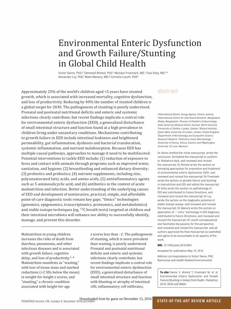
and hyperplasia of small intestinal
crypts (Fig 1). EED is found at a high
prevalence in stunted children living
under unsanitary conditions and is
pandemic in developing countries
with limited resources ( Table 1).
Major gaps in our understanding
of the pathogenesis of EED and its
relationship to stunting limit our
ability to diagnose and effectively
prevent and treat this condition.
The present state-of-the-art
consensus statement summarizes
a 3-day meeting organized by
the International Atomic Energy
Agency, which focused on EED and
the prospects for its reduction or
amelioration in children living in the
developing world.
PATHOBIOLOGY OF EED
EED may be defined as a global
disturbance of intestinal structure
and function that has its origin in
environmental factors. The condition
occurs with high frequency in
developing areas with poor sanitation
and limited public health resources,
in association with microbial and
parasitic contamination of food
and water. It is unlikely that any 1
pathogen explains the pathology
of EED and more likely that it
represents frequent, low-inoculum
exposure to a range of pathogens, 5, 6
which could be regarded as a form of
dysbiosis.
The identification that there is a
change in small intestinal structure
and function in the tropics originated
in the 1960s, 1 but it is only in the
last ∼2 decades that we have come
to understand that it may have
implications for nutrition and long-
term health of children living in
low-resource settings. 2 – 4 In early
reports, the focus was on structural
derangements (shortened, blunted
villi and increased crypt depth) and
disturbances of permeability and
absorption. More recently, additional
derangements have been identified,
including intestinal inflammation, 7
systemic inflammation, 8 and
changes in the microbiome. 9 The
complexity of the EED syndrome
is such that these derangements
cannot be assumed to operate
to the same degree in different
children. For example, 1 child may
have a very “leaky” gut with severe
microbial translocation (entry of
gastrointestinal organisms into the
systemic compartment) but not much
malabsorption, whereas another
child may have more significant
malabsorption but only mild
translocation. The meeting organized
by the International Atomic Energy
Agency identified several domains
that may need to be individually
measured to provide a full picture
of gut dysfunction and to assess the
impact of different interventions.
These domains describe axes
of measurement and aspects of
pathophysiology: (1) gut leakiness/
permeability 10; (2) microbial
translocation 10, 11; (3) gut
2
FIGURE 1Histologic sections from distal duodenal biopsy specimens from Zambian patients with EED. (A) Relatively normal mucosa has long, slender villi and short crypts, with only a slight increase in lamina propria lymphocytes; the villus height: crypt depth ratio approximates 3:1. (B) A biopsy specimen from a child with severe EED and moderate malnutrition showing villus shortening and reduction in villus height: crypt depth ratio to slightly more than 1:1. (C) Confocal laser endomicroscopy shows leakage of fl uorescein (arrows) around a villus after an intravenous injection into the intestinal lumen.
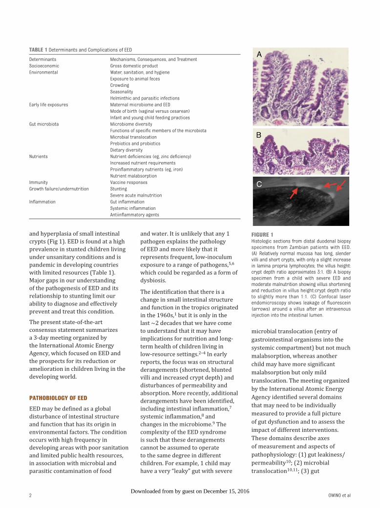
TABLE 1 Determinants and Complications of EED
Determinants Mechanisms, Consequences, and Treatment
Socioeconomic Gross domestic product
Environmental Water, sanitation, and hygiene
Exposure to animal feces
Crowding
Seasonality
Helminthic and parasitic infections
Early life exposures Maternal microbiome and EED
Mode of birth (vaginal versus cesarean)
Infant and young child feeding practices
Gut microbiota Microbiome diversity
Functions of specifi c members of the microbiota
Microbial translocation
Prebiotics and probiotics
Dietary diversity
Nutrients Nutrient defi ciencies (eg, zinc defi ciency)
Increased nutrient requirements
Proinfl ammatory nutrients (eg, iron)
Nutrient malabsorption
Immunity Vaccine responses
Growth failure/undernutrition Stunting
Severe acute malnutrition
Infl ammation Gut infl ammation
Systemic infl ammation
Antiinfl ammatory agents
by guest on December 15, 2016Downloaded from
PEDIATRICS Volume 138 , number 6 , December 2016
inflammation 7; (4) systemic
inflammation 8; (5) dysbiosis 9; and
(6) nutrient malabsorption. 11– 13
As opposed to focal defects
(as seen in Crohn’s disease),
EED predominantly affects the
proximal small intestine in a global
distribution. The condition is
seasonal, 5 reversible, 14 and generally
asymptomatic, as distinct from
diarrheal disease. The anatomic
and pathophysiologic basis of EED
is reflected in the aforementioned
domains. As with other
enteropathies, EED is characterized
by villus blunting, inflammation in
the epithelium and lamina propria,
and leakiness due to perturbation
of tight junction integrity and
microerosions ( Fig 1). Evidence is
also found of disturbances of mucus 15
and antimicrobial peptides, 16
which, together with tight junction
failure and microerosions, could
permit entry of microorganisms
and their component parts into the
systemic compartment from which,
in health, they would be excluded.
This translocation drives gut
inflammation, further exacerbating
gut dysfunction, and systemic
inflammation, 8 which can further
perturb immune function and lead
to anorexia. This positive feedback
underlies the vicious cycle of
malnutrition, infection, and immune
failure described >5 decades ago
in classic studies of malnutrition in
Central America. 4
Within the spectrum of EED, several
disorders are recognized that look
phenotypically similar and may
have similar effects on childhood
growth. Although these disorders
have a common origin in unsanitary
environments, 4, 6 they likely have
distinct etiopathogeneses and
therefore present different or
overlapping targets for intervention.
For example, the enteropathy
of severe acute malnutrition
comprises elements of acute and
chronic infection, inflammation, and
malabsorption. Seen in children with
weight-for-age z scores of –3 SDs or
less, it has high (10%–20%) mortality
when complicated by diarrhea,
pneumonia, sepsis, hypoglycemia,
and/or dehydration. 17, 18 EED may
also include components of HIV-
related enteropathy5 and aflatoxin-
mediated enteropathy, 19
conditions that we currently know
very little about. 1 Zinc deficiency
can cause enteropathy, but
extensive experience with zinc
supplementation suggests that it can
ameliorate the effects of diarrhea and
reduce gastrointestinal permeability
but does not improve nutrient
absorption. 20 Helminth infection,
established for hookworm but not
clearly shown for other helminths,
may also contribute to EED.
It is self-evident from these
observations that interventions for
the various elements of EED are
distinct and should be evaluated
separately. A recent review proposed
the inclusion of exposure to chemical
toxicants such as pesticides and
drugs as potential causes of EED. 21
Put simply, EED does not have a
single cause, and it is unlikely to be
resolved by a single intervention.
GROWTH FAILURE AND STUNTING IN MALNUTRITION AND EED
The term “stunted” is applied to
infants and children whose lengths
(or heights) are >2 SDs below the
median for age as determined by
using World Health Organization
growth standards. Stunting in the
developing world results most
commonly from chronic nutrient
deficiencies, recurrent infection(s),
and/or chronic inflammation
(including EED). Nevertheless, large
cohorts of “stunted” subjects may
also include infants and children
with hormonal or metabolic
disorders causing postnatal growth
failure; children with genetic or
familial forms of short stature;
and premature and/or small for
gestational age infants who fail to
achieve adequate catch-up growth.
Indeed, in 20% to 25% of infants
and children considered “stunted, ”
the growth failure begins in utero:
prematurity and intrauterine
growth restriction, particularly in
combination, increase the risk of
postnatal stunting 22 – 25 by twofold to
sevenfold. This explains, in part, the
high rates of stunting in developing
countries, where mean length-for-
age z scores at birth approximate
–0.5, and low birth weight (LBW) is
6 times more common than in the
developed world.23, 25
The health, maturity, and economic
and social status of the mother play
central roles in the pathogenesis
of LBW and the growth of the child
after birth. Factors predisposing to
LBW and childhood stunting include:
a history of moderate or severe
maternal malnutrition, stunting, or
early age at pregnancy; suboptimal
pregnancy weight gain; maternal
smoking; and inadequate infant
feeding practices. 22 – 25 The advent
of EED in infancy or early childhood
likely amplifies growth deficits
sustained during the intrauterine
and perinatal periods, resulting in
stunting. Length-for-age z scores of
stunted children typically decline
from birth to a nadir between 18
and 24 months of age, presumably
because rapidly growing infants and
toddlers are particularly vulnerable
to nutritional, infectious, and toxic
environmental insults. In concert
with nutritional deficits incurred
during fetal, perinatal, and early
postnatal life, the imposition of
EED may limit nutrient delivery
and utilization and thereby impair
the maturation and proliferation of
small intestinal epithelial cells, renal
nephrons, pancreatic β cells, and
skeletal myocytes and growth plate
chondrocytes.26, 27
The specific mechanisms by which
EED causes growth failure and
postnatal stunting are poorly
understood, although inadequate or
3by guest on December 15, 2016Downloaded from
OWINO et al
inconsistent energy intake, recurrent
infection, and local and systemic
inflammation likely play important
roles. Factors contributing to growth
failure may include immaturity of the
gut microbiome 28 and deficiencies of
certain gut microbes and/or breast
milk constituents such as sialylated
oligosaccharides that promote gut
barrier integrity, nutrient utilization,
and tissue anabolism. 29 – 31 Even in the
absence of diarrhea, the permeability
of the small bowel toward
carbohydrates and α1-antitrypsin
is increased, 7, 32 suggesting a role
for macronutrient malabsorption.
Many children with EED also have
deficiencies in micronutrients
absorbed by the small bowel such
as iron and zinc, which, if depleted,
can reduce appetite, villous surface
area, and gastrointestinal absorptive
capacity. 33, 34
Inflammation of the small intestine
in EED is associated with high
C-reactive protein levels and may
be accompanied by release of
cytokines that reduce appetite
and food intake 35 and impede
production and action of chondrocyte
growth factors. Recent studies 36, 37
found that stunted, malnourished
Ugandan infants and children (age
6 months–5 years) had high levels
of interleukin 6 (IL-6), which blocks
growth hormone induction of
insulin-like growth factor 1 (IGF-1)
production and inhibits IGF action
at the growth plate. 38, 39 Likewise,
IL-6 levels were elevated soon after
delivery in a subset of Zimbabwean
infants with LBW. 8 Interestingly,
IL-6 levels are high in children and
adults with inflammatory bowel
disease and correlate inversely with
childhood growth rates and IGF-1
levels. 40 Thus, the rise in IL-6 (and
other cytokines) in association with
small bowel inflammation may limit
food intake, IGF-1 production, and
linear growth in children with EED.
Inadequate intake and malabsorption
of zinc in EED may also reduce IGF-1
production and action 41 and thereby
attenuate linear growth.
Growth failure in EED may be
exacerbated by the development of
acute malnutrition, most commonly a
consequence of gastrointestinal and
pulmonary infections and sepsis. 42
Endocrinologic studies 36, 37 provide
insight into the pathogenesis of
growth failure in malnourished
children. Nutrient deprivation
provokes a striking increase in
growth hormone and fall in insulin,
which in concert promote lipolysis
and deplete white adipose fat
stores and thereby reduce levels
of the adipocyte hormone leptin.
Hypoleptinemia downregulates
the hypothalamic-pituitary-thyroid
axis and inhibits conversion of
T4 to its more active form, T3. 43
The fall in T3 impairs chondrocyte
maturation and growth. The
stress of acute malnutrition and
concurrent infection activates the
hypothalamic-pituitary-adrenal axis
and stimulates a rise in cortisol36, 37
and IGF binding protein 1, which in
combination inhibit IGF-1 action and
induce chondrocyte apoptosis. 44, 45
A reduction in hepatic growth
hormone receptor expression 46
and inhibition of growth hormone
signaling47 by fibroblast growth
factor 21 limit IGF-1 production and
thereby contribute to growth failure
in patients with EED.
Therapeutic measures in EED,
including nutritional supplements
and antibiotics, may fail to restore
growth in children stunted before
the age of 2 years. 24 In some cases,
this outcome may simply reflect
a genetic or familial tendency to
short stature. Alternatively, failure
of catch-up growth in children with
prenatal or early postnatal growth
failure might be explained by: (1)
inadequate reserve, or epigenetic
changes, 48, 49 in cells critical for
growth, including myocytes and
chondrocytes; (2) long-term defects
in small intestinal maturation and
growth 50; and/or (3) recurrent bouts
of nutrient deprivation, infection,
and cytokine excess associated with
small bowel inflammation. The
SHINE (Sanitation Hygiene Infant
Nutrition Efficacy) trial is currently
investigating the hypothesis that EED
has adverse consequences in addition
to postnatal growth failure, including
reduced oral vaccine efficacy,
anemia, impaired neurocognitive
development, and fetal growth
restriction and prematurity resulting
from maternal EED.51 EED has
recently been linked to reduced
efficacy of oral polio and rotavirus
vaccines in Bangladeshi infants. 52
EED BIOMARKERS AND DIAGNOSTIC TESTS
Difficulties in identifying the
multiple etiologies of EED and in
distinguishing EED from other types
of intestinal dysfunctions impose
considerable challenges for diagnosis
and hamper development of specific
diagnostic tests. Endoscopic and
histopathologic evaluation of small
intestinal biopsy specimens, with
continuous measures of mucosal
architecture (villus height in
micrometers rather than ordinal
scales of blunting), 5 allow for direct
observation of aberrant epithelial
structures and inflammation
status. However, noninvasive or
less-invasive diagnostic assays
are preferred for logistic reasons
(ie, use in nonclinical settings and
because they are better accepted by
patients). Depending on the aspect
of gut function or dysfunction of
interest, biomarkers of EED may fall
under 1 of 5 categories, 53 namely: (1)
intestinal absorption and mucosal
permeability; (2) enterocyte mass
and function; (3) inflammation; (4)
microbial translocation and immune
activation; and (5) intestinal injury
and repair.
The most commonly applied
noninvasive assay to assess EED
is a dual sugar test, the lactulose:
mannitol test, which is based
4by guest on December 15, 2016Downloaded from
PEDIATRICS Volume 138 , number 6 , December 2016
on oral dosing and subsequent
urinary measurement of lactulose
and mannitol to evaluate both
epithelial absorptive capacity and
permeability in the small intestine
(aforementioned domains 1 and
7). 54, 55 In addition, recent research
has identified various serum and
fecal biomarkers of intestinal
inflammation in the context of
EED. 3, 56 Serum biomarkers include
lipopolysaccharide, soluble CD
14, IGF-1, ferritin, IL-6, IL-1β,
C-reactive protein, zonulin, and
endogenous endotoxin-core
antibody (EndoCab, Hycult Biotech,
Uden, the Netherlands). Fecal
biomarkers include regenerating
islet-derived 1 beta, calprotectin,
myeloperoxidase, neopterin,
α1-antitrypsin, and lactoferrin.
Although these biomarkers permit
assessment of intestinal/systemic
inflammation and/or intestinal
epithelial barrier dysfunction, the
main limitation to their use is that
they are not specific for EED because
they correlate with prevalence,
activity, and/or severity of various
other gastrointestinal diseases. One
of the downstream consequences
of EED is vaccine failure, 28, 57 but it
has not been generally accepted as a
diagnostic measure of EED. Cutting
edge innovations such as -omics and
nuclear technologies (stable isotope
techniques) may provide a much-
needed capability to diagnose and
better understand EED.
APPLICATION OF -OMICS TECHNOLOGY
Much has been learned about the
biology of health and disease states
through application of -omics
technologies: genomics, epigenomics,
transcriptomics, proteomics, and
metabolomics. 58 The essence of
-omics is an agnostic survey across
the total spectrum of a given type
of molecule or analyte class. As
a pathologic condition, EED is an
excellent candidate for -omics
surveys because so little is known
about the mechanisms through which
it exerts its deleterious effects.
A large transcriptomic study of
rural African children using a novel
method to assess host transcripts
in feces found diverse activation of
many of the immunologic responses
seen in the gut epithelium. 59 Twelve
transcripts were associated with
the severity of EED, including
chemokines that stimulate T-cell
proliferation, Fc fragments of
multiple immunoglobulin families,
interferon-induced proteins,
activators of neutrophils and B
cells, and mediators that dampen
cellular responses to hormones. EED-
associated transcripts were mapped
to pathways related to cell adhesion
and responses to a broad spectrum
of viruses, bacteria, and parasites.
Several mucins, regulatory factors,
and protein kinases associated with
maintenance of the mucous layer
were expressed at lower levels in
children with EED than in normal
children. The pattern of expression
was compatible with an assault
by multiple microorganisms from
diverse phyla. In addition, antiviral
transcripts were detected. This rich
data set offers clues for those seeking
pharmacologic intervention against
EED as well as novel fecal biomarkers
for the condition.
Epigenomics and proteomics have
not been applied to EED as far as
we know. Metabolomic analysis has
been undertaken, but major findings
have not been released. 60 Although
the complex effects of prenatal and
postnatal environmental factors
clearly complicate the analysis of
children with EED, we anticipate
future research using genomic and
metabolomic approaches to dissect
the pathobiology of EED.
DIAGNOSTIC POTENTIAL OF STABLE ISOTOPE ASSAYS
Beyond the use of the lactulose:
mannitol test and various serum and
fecal biomarkers, 11, 56 nonradioactive,
stable isotope techniques are
emerging as promising noninvasive/
less invasive, safe tools for measuring
gastrointestinal function and
determining EED. The foundation of
these methods is oral administration
of an isotopically labeled compound
and subsequent monitoring of the
appearance of the compound or
its catabolic products in breath,
feces, urine, and/or blood. 61
Depending on the type of labeled
compound, stable isotope assays
can assess epithelial function in
several domains (ie, absorption,
permeability, metabolism) and
can be used to characterize a
particular microorganism or group
of microorganisms that catabolize
the ingested compound. This
latter feature is especially useful
for probing microbial activities in
the upper gastrointestinal tract in
EED; analysis of fecal microbiota 9
provides an inadequate proxy for
the composition and function of
microbiota in the stomach, the
duodenum, or the small bowel.
Characterizing microbial
activity, particularly in the upper
gastrointestinal tract, is important
for 2 reasons. First, EED is associated
with infections by pathogens (eg,
Helicobacter pylori) and microbial
overgrowth and general dysbiosis
in the small intestine in humans.
Second, exposure to a defined
mixture of commensal bacterial
isolates, including Escherichia coli and members of the Bacteroidales,
triggered a phenotype in moderately
malnourished mice that resembled
human EED. 62 This finding provides
strong evidence for the role played by
microorganisms in the pathogenesis
of EED.
Available noninvasive, stable isotope
breath tests for potential use in
EED include, but are not limited
to, a highly sensitive and specific 13C-urease assay for H pylori 63 and
application of various 13C-sugars (eg,
sucrose, xylose, glucose, lactose) 64, 65
or 13C-labeled glycosyl ureides 66, 67 to
5by guest on December 15, 2016Downloaded from
OWINO et al
measure epithelial barrier function,
absorptive capacity, and intestinal
transit time, and to identify small
intestinal bacterial overgrowth and
dysbiosis. In addition, intestinal
absorption and bioavailability of
specific micronutrients, in particular
iron and zinc, from diets can be
measured after oral ingestion of
isotopically labeled iron (54Fe, 57Fe,
and 58Fe) 68 and zinc (67Zn, 68Zn,
and 70Zn) 12, 69 compounds. Although
the use of different stable isotope
compounds in a single composite
assay for simultaneous assessment
of multiple intestine-associated
clinical end points has not yet been
fully exploited, the available and
established stable isotope assays
have great potential for application in
EED diagnostics and research.
EMERGING APPROACHES FOR THE PREVENTION AND TREATMENT OF EED
The treatment of EED is fraught with
difficulties. First, in the absence of
robust point-of-care biomarkers, the
identification of EED in the individual
child is problematic. Moreover, there
is no robust evidence from clinical
trials that specific interventions can
cure or ameliorate the signs and
symptoms of EED.
Because EED has its roots in the
environment, the mainstay of
preventing the condition is to
“clean” the environment. This
approach is challenging because
water scarcity still afflicts 40% of
the world’s population, and 13%
of the population still defecates
in the open. 70 Furthermore, one-
sixth of the world’s people lack
access to safe drinking water. The
provision of basic sanitation facilities,
potable water, and improved
hygiene practices cut the chain of
transmission of pathogenic bacteria
that can colonize the small intestine
and cause EED. Indeed, a study in
Bangladesh found that ensuring
a clean environment increased
population height-for-age 0.54 SD
compared with children living in
default conditions. 71 Moreover,
stunting prevalence was reduced
by 22%. More recently, a public
sanitation program in Mali showed
that enhanced access to toilets did
not reduce the prevalence of diarrhea
but increased childhood growth,
particularly in those <2 years of
age. 72 A possible explanation could
be reduced chronic exposure to
pathogenic bacteria, resulting in
reduced severity or prevalence of
EED.
Ensuring hygiene at critical times is
critical to preventing EED. A study
conducted in slums and villages in
Bangladesh revealed that 40% of
complementary foods prepared by
mothers were contaminated with
E coli; this contamination resulted
in higher rates of diarrhea and
malnutrition. 73 It is now believed
that zinc deficiency, rampant in
developing countries, co-exists with
EED and increases the severity of the
condition. 20, 33, 34 Given the inadequate
dietary intake of zinc in children
living in developing countries, the
importance of its supplementation, 74
either long term or at least as part of
treatment of diarrhea, cannot be over
estimated. The claim that exclusive
breastfeeding can reduce gut
inflammation was recently validated
in South African children. 75
In summary, emerging evidence
suggests that the following factors,
in combination, can reduce the
incidence, prevalence, and severity of
EED: (1) access to safe drinking water
and improved hygiene practices in
low-income countries; (2) provision
of sanitary toilet facilities and
changes in public behavior regarding
their use; (3) exclusive breastfeeding
for the first 6 months and continued
breastfeeding thereafter; and (4) zinc
supplementation.
Because stunting is a hallmark
of EED, a major goal should be to
prevent the condition as well as treat
its complications. In the context
of poor socioeconomic conditions,
preventive and therapeutic
measures include increasing access
of children to appropriate and
adequate diets containing animal-
source foods, supplementation with
zinc, and adequate treatment of
recurrent illnesses such as diarrhea
and pneumonia. Population-
wide prevention of EED will
require adequate nutrition and
health maintenance of all girls of
reproductive age and women prior
to, during, and after pregnancy.
Under conditions of extreme food
insecurity, supplementation with
nutritious ready-to-use food for
children with severe or moderate
acute malnutrition has been shown to
enhance clinical recovery. 76, 77
Agents that can offset or reduce
chronic inflammation at the
gut mucosal level are currently
being evaluated. These include
5-aminosalicyclic acid, which in a
recent study was not efficacious 78;
the nasal steroid budesonide; and an
immunomodulatory small molecule
called oglufanide disodium. 3
Only well-designed, randomized
controlled trials that assess efficacy
and adverse effects in settings of
high prevalence of stunting/EED can
lead the way to effective and safe
treatments.
SUMMARY AND FUTURE DIRECTIONS
Reducing by 40% the number of
children aged <5 years who are
stunted is a global target for 2030.
Systematic reviews have revealed
that optimal nutrition intervention
packages for high-risk children may
only partially reduce the prevalence
and severity of stunting. 79, 80 We do
not know if nutritional therapies
fail because stunted children have
altered intestinal microbiota,
insufficient nutrient intake, nutrient
malabsorption, or disordered
partition of nutrients. It is also
unclear if nutrient utilization/
wastage is too high to permit
adequate lean tissue accretion.
6by guest on December 15, 2016Downloaded from
PEDIATRICS Volume 138 , number 6 , December 2016
Finally, we don't know if insults
inflicted before or soon after birth are
fully reversible even with adequate
postnatal nutrient repletion.
The complexity of the EED syndrome
is such that gastrointestinal
derangements cannot be assumed
to operate to the same degree in
different children. There may be
considerable individual variation
in gut "leakiness", bacterial
translocation, malabsorption, and
nutrient requirements. Given the
burden of concurrent infection
and inflammation, EED-afflicted
children may require considerably
higher nutrient intakes than healthy
children in order to maintain normal
weight gain.
Our understanding of EED is severely
limited by its complex spectrum,
absence of robust biomarkers, and
noninvasive, simple point-of-care
diagnostic tools. Several domains
may need to be evaluated in each
child to provide a full picture of
gut dysfunction and to assess the
impact of different interventions.
These domains include: (1) gut
leakiness/permeability; (2)
microbial translocation; (3)
gut inflammation; (4) systemic
inflammation; (5) dysbiosis; and
(6) nutrient malabsorption. Further
investigation will be needed to
characterize fully the effects of
gut dysfunction on hormonal and
metabolic status, childhood growth,
and neurocognitive function.
Potential interventions to tackle
EED should include: (1) increased
access to clean water, and improved
sanitation and hygiene, including
reduction of exposure to feces
and contact with animals; (2)
promotion of dietary diversity
and breastfeeding; (3) adequate
supplementation with micro- and
macronutrients including zinc and
amino acids; (4) antiinflammatory
agents; and (5) antibiotics for
children with severe acute
malnutrition and infection. Some
of these interventions (especially
water, sanitation, and hygiene,
infant feeding practices, and
nutrient repletion) are being tested
individually or combined in large
randomized controlled studies in a
number of countries. Information
gained from these investigations
may guide the development of novel
therapies in the future.
The application of omics technologies
(eg, genomics, epigenomics,
transcriptomics, proteomics,
metabolomics) and use of stable
isotopes should allow us to better
define the nature and extent of
gastrointestinal damage and
dysfunction in EED and can be used
to characterize microbial activities
in the upper gastrointestinal tract
in affected children. Stable isotopes
can also be employed to assess body
composition as a proxy for dietary
quality and nutritional status and as
a determinant of childhood morbidity
and mortality. While validation of
the various diagnostic techniques is
obligatory, these new approaches
should ultimately enhance our ability
to prevent and treat environmental
enteropathy.
CONCLUSIONS
EED does not have a single cause or
even a single causal pathway, and it
is unlikely to be resolved by a single
intervention. The identification
of EED is fraught with difficulties
due to the absence of robust
point-of-care biomarkers. Better
understanding of the underlying
causes and pathogenesis of EED,
development of noninvasive,
practical, simple, and affordable
point-of-care diagnostic tools,
and longitudinal studies designed
to treat or ameliorate signs and
symptoms of EED remain key
gaps. Cutting-edge innovations
using the field of -omics and stable
isotope techniques may provide a
much-needed capability to better
understand, prevent, and treat EED.
ACKNOWLEDGMENTS
We sincerely thank all participants
who generated the information on
which this article is based during
the technical meeting of EED held
at the International Atomic Energy
Agency headquarters, Vienna,
Austria, October 28–30, 2015. We
are also grateful to Kirsten Glenn for
proofreading the manuscript.
7
ABBREVIATIONS
EED: environmental enteric
dysfunction
IGF: insulin-like growth factor
IL-6: interleukin 6
LBW: low birth weight
Studies Section, Division of Human Health, International Atomic Energy Agency, Vienna International Centre, PO Box 100, 1400 Vienna, Austria. E-mail: v.owino@
iaea.org
PEDIATRICS (ISSN Numbers: Print, 0031-4005; Online, 1098-4275).
Copyright © 2016 by the American Academy of Pediatrics
FINANCIAL DISCLOSURE: Drs Owino and Loechl are employees of the International Atomic Energy Agency. Drs Freemark, Kelly, Loy, and Manary received travel
reimbursement from the International Atomic Energy Agency to attend the technical meeting.
FUNDING: All aspects of the technical meeting on Environmental Enteric Dysfunction, Undernutrition and the Microbiome were funded by the International Atomic
Energy Agency. Dr Loy is supported by the Vienna Science and Technology Fund (WWTF, project LS12-001) and the Austrian Science Fund (FWF, project I 2320-B22).
Dr Freemark received support from the Duke Global Health Institute.
POTENTIAL CONFLICT OF INTEREST: The authors have indicated they have no potential confl icts of interest to disclose.
by guest on December 15, 2016Downloaded from
OWINO et al
REFERENCES
1. Prendergast A, Kelly P. Enteropathies in
the developing world: neglected effects
on global health. Am J Trop Med Hyg.
2012;86(5):756–763
2. Keusch GT, Denno DM, Black RE, et al.
Environmental enteric dysfunction:
pathogenesis, diagnosis, and clinical
consequences. Clin Infect Dis.
2014;59(suppl 4):S207–S212
3. Petri WA Jr, Naylor C, Haque R.
Environmental enteropathy and
malnutrition: do we know enough to
intervene? BMC Med. 2014;12:187
4. Guerrant RL, DeBoer MD, Moore SR,
Scharf RJ, Lima AA. The impoverished
gut—a triple burden of diarrhoea,
stunting and chronic disease.
Nat Rev Gastroenterol Hepatol.
2013;10(4):220–229
5. Kelly P, Menzies I, Crane R, et al.
Responses of small intestinal
architecture and function over time
to environmental factors in a tropical
population. Am J Trop Med Hyg.
2004;70(4):412–419
6. Humphrey JH. Child undernutrition,
tropical enteropathy, toilets,
and handwashing. Lancet.
2009;374(9694):1032–1035
7. Kosek M, Haque R, Lima A, et al; MAL-ED
Network. Fecal markers of intestinal
infl ammation and permeability
associated with the subsequent
acquisition of linear growth defi cits
in infants. Am J Trop Med Hyg.
2013;88(2):390–396
8. Prendergast AJ, Rukobo S, Chasekwa
B, et al. Stunting is characterized by
chronic infl ammation in Zimbabwean
infants. PLoS One. 2014;9(2):e86928
9. Smith MI, Yatsunenko T, Manary MJ,
et al. Gut microbiomes of Malawian
twin pairs discordant for kwashiorkor.
Science. 2013;339(6119):548–554
10. Kelly P, Besa E, Zyambo K, et al.
Endomicroscopic and transcriptomic
analysis of impaired barrier function
and malabsorption in environmental
enteropathy. PLoS Negl Trop Dis.
2016;10(4):e0004600
11. Korpe PS, Petri WA Jr. Environmental
enteropathy: critical implications of a
poorly understood condition. Trends
Mol Med. 2012;18(6):328–336
12. Krebs NF, Miller LV, Hambidge KM. Zinc
defi ciency in infants and children: a
review of its complex and synergistic
interactions. Paediatr Int Child Health.
2014;34(4):279–288
13. Denno DM, VanBuskirk K, Nelson ZC,
Musser CA, Hay Burgess DC, Tarr PI.
Use of the lactulose to mannitol ratio
to evaluate childhood environmental
enteric dysfunction: a systematic
review. Clin Infect Dis. 2014;59(suppl
4):S213–S219
14. Lindenbaum J, Harmon JW, Gerson
CD. Subclinical malabsorption in
developing countries. Am J Clin Nutr.
1972;25(10):1056–1061
15. Amadi B, Fagbemi AO, Kelly P, et al.
Reduced production of sulfated
glycosaminoglycans occurs in
Zambian children with kwashiorkor
but not marasmus. Am J Clin Nutr.
2009;89(2):592–600
16. Dhaliwal W, Bajaj-Elliott M, Kelly P.
Intestinal defensin gene expression
in human populations. Mol Immunol.
2003;40(7):469–475
17. Amadi B, Kelly P, Mwiya M, et al.
Intestinal and systemic infection, HIV,
and mortality in Zambian children with
persistent diarrhea and malnutrition.
J Pediatr Gastroenterol Nutr.
2001;32(5):550–554
18. Kerac M, Bunn J, Chagaluka G, et al.
Follow-up of post-discharge growth
and mortality after treatment for
severe acute malnutrition (FuSAM
study): a prospective cohort study.
PLoS One. 2014;9(6):e96030
19. Smith LE, Stoltzfus RJ, Prendergast
A. Food chain mycotoxin exposure,
gut health, and impaired growth:
a conceptual framework. Adv Nutr.
2012;3(4):526–531
20. Lindenmayer GW, Stoltzfus RJ,
Prendergast AJ. Interactions between
zinc defi ciency and environmental
enteropathy in developing countries.
Adv Nutr. 2014;5(1):1–6
21. Mapesa JO, Maxwell AL, Ryan
EP. An exposome perspective to
environmental enteric dysfunction.
Environ Health Perspect.
2016;124(8):1121–1126
22. Tiwari R, Ausman LM, Agho KE.
Determinants of stunting and severe
stunting among under-fi ves: evidence
from the 2011 Nepal Demographic
and Health Survey. BMC Pediatr.
2014;14:239
23. Prendergast AJ, Humphrey JH. The
stunting syndrome in developing
countries. Paediatr Int Child Health.
2014;34(4):250–265
24. Sania A, Spiegelman D, Rich-Edwards J,
et al. The contribution of preterm birth
and intrauterine growth restriction to
childhood undernutrition in Tanzania.
Matern Child Nutr. 2015;11(4):618–630
25. Christian P, Lee SE, Donahue Angel M,
et al. Risk of childhood undernutrition
related to small-for-gestational age
and preterm birth in low- and middle-
income countries. Int J Epidemiol.
2013;42(5):1340–1355
26. Thorn SR, Rozance PJ, Brown
LD, Hay WW Jr. The intrauterine
growth restriction phenotype: fetal
adaptations and potential implications
for later life insulin resistance
and diabetes. Semin Reprod Med.
2011;29(3):225–236
27. Zohdi V, Sutherland MR, Lim K, Gubhaju
L, Zimanyi MA, Black MJ. Low birth
weight due to intrauterine growth
restriction and/or preterm birth:
effects on nephron number and
long-term renal health. Int J Nephrol.
2012;2012:136942
28. Subramanian S, Huq S, Yatsunenko
T, et al. Persistent gut microbiota
immaturity in malnourished
Bangladeshi children. Nature.
2014;510(7505):417–421
29. Schwarzer M, Makki K, Storelli G, et
al. Lactobacillus plantarum strain
maintains growth of infant mice during
chronic undernutrition. Science.
2016;351(6275):854–857
30. Blanton LV, Charbonneau MR, Salih
T, et al. Gut bacteria that prevent
growth impairments transmitted
by microbiota from malnourished
children. Science. 2016;351(6275):19
31. Charbonneau MR, O’Donnell
D, Blanton LV, et al. Sialylated
milk oligosaccharides promote
microbiota-dependent growth in
8by guest on December 15, 2016Downloaded from
PEDIATRICS Volume 138 , number 6 , December 2016
models of infant undernutrition. Cell.
2016;164(5):859–871
32. Gilmartin AA, Petri WA Jr. Exploring the
role of environmental enteropathy in
malnutrition, infant development and
oral vaccine response. Philos Trans R
Soc Lond B Biol Sci. 2015;19;370(1671).
pii: 20140143
33. Southon S, Gee JM, Bayliss CE, Wyatt
GM, Horn N, Johnson IT. Intestinal
microfl ora, morphology and
enzyme activity in zinc-defi cient and
Zn-supplemented rats. Br J Nutr.
1986;55(3):603–611
34. Prasad AS. Clinical manifestations
of zinc defi ciency. Annu Rev Nutr.
1985;5:341–363
35. Braun TP, Marks DL. Pathophysiology
and treatment of infl ammatory
anorexia in chronic disease. J Cachexia
Sarcopenia Muscle. 2010;1(2):135–145
36. Bartz S, Mody A, Hornik C, et al.
Severe acute malnutrition in
childhood: hormonal and metabolic
status at presentation, response
to treatment, and predictors of
mortality. J Clin Endocrinol Metab.
2014;99(6):2128–2137
37. Mody A, Bartz S, Hornik CP, et al. Effects
of HIV infection on the metabolic and
hormonal status of children with
severe acute malnutrition. PLoS One.
2014;9(7):e102233
38. De Benedetti F, Alonzi T, Moretta A,
et al. Interleukin 6 causes growth
impairment in transgenic mice
through a decrease in insulin-
like growth factor-I. A model for
stunted growth in children with
chronic infl ammation. J Clin Invest.
1997;99(4):643–650
39. Sederquist B, Fernandez-Vojvodich P,
Zaman F, Sävendahl L. Recent research
on the growth plate: impact of
infl ammatory cytokines on longitudinal
bone growth. J Mol Endocrinol.
2014;53(1):T35–T44
40. Sanderson IR. Growth problems
in children with IBD. Nat
Rev Gastroenterol Hepatol.
2014;11(10):601–610
41. Cesur Y, Yordaman N, Doğan M.
Serum insulin-like growth factor-I
and insulin-like growth factor binding
protein-3 levels in children with
zinc defi ciency and the effect of zinc
supplementation on these parameters.
J Pediatr Endocrinol Metab.
2009;22(12):1137–1143
42. Jones KD, Thitiri J, Ngari M, Berkley
JA. Childhood malnutrition: toward
an understanding of infections,
infl ammation, and antimicrobials. Food
Nutr Bull. 2014;35(suppl 2):S64–S70
43. Araujo RL, Andrade BM, da Silva ML,
Ferreira AC, Carvalho DP. Tissue-
specifi c deiodinase regulation
during food restriction and low
replacement dose of leptin in rats.
Am J Physiol Endocrinol Metab.
2009;296(5):E1157–E1163
44. Sävendahl L. The effect of acute and
chronic stress on growth. Sci Signal.
2012;5(247):pt9
45. Lee PD, Giudice LC, Conover CA, Powell
DR. Insulin-like growth factor binding
protein-1: recent fi ndings and new
directions. Proc Soc Exp Biol Med.
1997;216(3):319–357
46. Maes M, Maiter D, Thissen JP,
Underwood LE, Ketelslegers JM.
Contributions of growth hormone
receptor and postreceptor defects
to growth hormone resistance in
malnutrition. Trends Endocrinol Metab.
1991;2(3):92–97
47. Fazeli PK, Klibanski A. Determinants
of GH resistance in malnutrition. J
Endocrinol. 2014;220(3):R57–R65
48. Gat-Yablonski G, Phillip M. Nutritionally-
induced catch-up growth. Nutrients.
2015;7(1):517–551
49. Quilter CR, Cooper WN, Cliffe KM, et
al. Impact on offspring methylation
patterns of maternal gestational
diabetes mellitus and intrauterine
growth restraint suggest common
genes and pathways linked to
subsequent type 2 diabetes risk. FASEB
J. 2014;28(11):4868–4879
50. Mickiewicz M, Zabielski R, Grenier
B, et al. Structural and functional
development of small intestine in
intrauterine growth retarded porcine
offspring born to gilts fed diets with
differing protein ratios throughout
pregnancy. J Physiol Pharmacol.
2012;63(3):225–239
51. Prendergast AJ, Humphrey JH, Mutasa
K, et al; Sanitation Hygiene Infant
Nutrition Effi cacy (SHINE) Trial Team.
Assessment of environmental enteric
dysfunction in the SHINE trial: methods
and challenges. Clin Infect Dis.
2015;61(suppl 7):S726–S732
52. Naylor C, Lu M, Haque R, et al;
PROVIDE Study Teams. Environmental
enteropathy, oral vaccine failure
and growth faltering in infants
in Bangladesh. EBioMedicine.
2015;2(11):1759–1766
53. Syed S, Ali A, Duggan C. Environmental
enteric dysfunction in children: a
review. J Pediatr Gastroenterol Nutr.
2016;63(1):6–14
54. Lee GO, Kosek P, Lima AA, et al.
Lactulose: mannitol diagnostic test
by HPLC and LC-MSMS platforms:
considerations for fi eld studies
of intestinal barrier function and
environmental enteropathy. J Pediatr
Gastroenterol Nutr. 2014;59(4):544–550
55. Menzies I. Absorption of intact
oligosaccharide in health and
disease. Biochem Soc Trans.
1974;2(5):1042–1047
56. Crane RJ, Jones KD, Berkley JA.
Environmental enteric dysfunction: an
overview. Food Nutr Bull. 2015;36(suppl
1):S76–S87
57. Levine MM. Immunogenicity and
effi cacy of oral vaccines in developing
countries: lessons from a live cholera
vaccine. BMC Biol. 2010;8:129
58. Chu Y, Corey DR. RNA sequencing:
platform selection, experimental
design, and data interpretation.
Nucleic Acid Ther. 2012;22(4):271–274
59. Yu J, Ordiz MI, Stauber J, et al
Environmental enteric dysfunction
includes a broad spectrum of
infl ammatory responses and
epithelial repair processes. Cell Mol
Gastroenterol Hepatol. 2015;2(2):158–
174.e1
60. Preidis GA, Hotez PJ. The newest
“omics”—metagenomics and
metabolomics—enter the battle
against the neglected tropical
diseases. PLoS Negl Trop Dis.
2015;9(2):e0003382
61. Romagnuolo J, Schiller D, Bailey
RJ. Using breath tests wisely in
a gastroenterology practice: an
evidence-based review of indications
and pitfalls in interpretation. Am J
Gastroenterol. 2002;97(5):1113–1126
9by guest on December 15, 2016Downloaded from
OWINO et al
62. Brown EM, Wlodarska M, Willing BP, et al.
Diet and specifi c microbial exposure
trigger features of environmental
enteropathy in a novel murine model.
Nat Commun. 2015;6:7806
63. Di Rienzo TA, D’Angelo G, Ojetti V, et al.
13C-urea breath test for the diagnosis
of Helicobacter pylori infection. Eur
Rev Med Pharmacol Sci. 2013;17(suppl
2):51–58
64. Dellert SF, Nowicki MJ, Farrell MK,
Delente J, Heubi JE. The 13C-xylose
breath test for the diagnosis of
small bowel bacterial overgrowth in
children. J Pediatr Gastroenterol Nutr.
1997;25(2):153–158
65. Ritchie BK, Brewster DR, Davidson GP,
et al. 13C-sucrose breath test: novel
use of a noninvasive biomarker of
environmental gut health. Pediatrics.
2009;124(2):620–626
66. Ruemmele FM, Heine WE, Keller KM,
Lentze MJ. Metabolism of glycosyl
ureides by human intestinal brush
border enzymes. Biochim Biophys Acta.
1997;1336(2):275–280
67. Berthold HK, Schober P, Scheurlen
C, et al. Use of the lactose-[13C]
ureide breath test for diagnosis of
small bowel bacterial overgrowth:
comparison to the glucose hydrogen
breath test. J Gastroenterol.
2009;44(9):944–951
68. Troesch B, Egli I, Zeder C, Hurrell RF,
Zimmermann MB. Fortifi cation iron as
ferrous sulfate plus ascorbic acid is
more rapidly absorbed than as sodium
iron EDTA but neither increases serum
nontransferrin-bound iron in women. J
Nutr. 2011;141(5):822–827
69. Sparacino G, Shames DM, Vicini P, King
JC, Cobelli C. Double isotope tracer
method for measuring fractional
zinc absorption: theoretical analysis.
Am J Physiol Endocrinol Metab.
2002;282(3):E679–E687
70. United Nations. The Millennium
Development Goals Report. New York,
NY: Department of Economic and
Social Affairs of the United Nations
Secretariat; 2015
71. Lin A, Arnold BF, Afreen S, et al.
Household environmental conditions
are associated with enteropathy and
impaired growth in rural Bangladesh.
Am J Trop Med Hyg. 2013;89(1):130–137
72. Pickering AJ, Djebbari H, Lopez C,
Coulibaly M, Alzua ML. Effect of a
community-led sanitation intervention
on child diarrhoea and child growth
in rural Mali: a cluster-randomised
controlled trial. Lancet Glob Health.
2015;3(11):e701–e711
73. Islam MA, Ahmed T, Faruque ASG,
et al. Microbiological quality
of complementary foods and
its association with diarrhoeal
morbidity and nutritional status of
Bangladeshi children. Eur J Clin Nutr.
2012;66(11):1242–1246
74. Chen P, Soares AM, Lima AA, et
al. Association of vitamin A and
zinc status with altered intestinal
permeability: analyses of cohort data
from northeastern Brazil. J Health
Popul Nutr. 2003;21(4):309–315
75. Moodley-Govender E, Mulol H, Stauber
J, Manary M, Coutsoudis A. Increased
exclusivity of breastfeeding associated
with reduced gut infl ammation
in infants. Breastfeed Med.
2015;10(10):488–492
76. Christian P, Shaikh S, Shamim AA, et
al. Effect of fortifi ed complementary
food supplementation on child
growth in rural Bangladesh: a cluster-
randomized trial. Int J Epidemiol.
2015;44(6):1862–1876
77. Brewster DR, Manary MJ, Menzies
IS, O’Loughlin EV, Henry RL. Intestinal
permeability in kwashiorkor. Arch Dis
Child. 1997;76(3):236–241
78. Jones KD, Hünten-Kirsch B, Laving
AM, et al. Mesalazine in the initial
management of severely acutely
malnourished children with
environmental enteric dysfunction: a
pilot randomized controlled trial. BMC
Med. 2014;12:133
79. Dewey KG, Adu-Afarwuah S. Systematic
review of the effi cacy and effectiveness
of complementary feeding
interventions in developing countries.
Matern Child Nutr. 2008;4(suppl
1):24–85
80. Bhutta ZA, Ahmed T, Black RE, et al;
Maternal and Child Undernutrition
Study Group. What works?
Interventions for maternal and child
undernutrition and survival. Lancet.
2008;371(9610):417–440
10by guest on December 15, 2016Downloaded from
DOI: 10.1542/peds.2016-0641; originally published online November 4, 2016; 2016;138;Pediatrics
Manary and Cornelia LoechlVictor Owino, Tahmeed Ahmed, Michael Freemark, Paul Kelly, Alexander Loy, Mark
Child HealthEnvironmental Enteric Dysfunction and Growth Failure/Stunting in Global
ServicesUpdated Information &
/content/138/6/e20160641.full.htmlincluding high resolution figures, can be found at:
References
/content/138/6/e20160641.full.html#ref-list-1at:This article cites 76 articles, 28 of which can be accessed free
Subspecialty Collections
/cgi/collection/nutrition_subNutrition
/cgi/collection/international_child_health_subInternational Child Healththe following collection(s):This article, along with others on similar topics, appears in
Permissions & Licensing
/site/misc/Permissions.xhtmltables) or in its entirety can be found online at: Information about reproducing this article in parts (figures,
Reprints /site/misc/reprints.xhtml
Information about ordering reprints can be found online:
rights reserved. Print ISSN: 0031-4005. Online ISSN: 1098-4275.Grove Village, Illinois, 60007. Copyright © 2016 by the American Academy of Pediatrics. All and trademarked by the American Academy of Pediatrics, 141 Northwest Point Boulevard, Elkpublication, it has been published continuously since 1948. PEDIATRICS is owned, published, PEDIATRICS is the official journal of the American Academy of Pediatrics. A monthly
by guest on December 15, 2016Downloaded from

DOI: 10.1542/peds.2016-0641; originally published online November 4, 2016; 2016;138;Pediatrics
Manary and Cornelia LoechlVictor Owino, Tahmeed Ahmed, Michael Freemark, Paul Kelly, Alexander Loy, Mark
Child HealthEnvironmental Enteric Dysfunction and Growth Failure/Stunting in Global
/content/138/6/e20160641.full.html
located on the World Wide Web at: The online version of this article, along with updated information and services, is
of Pediatrics. All rights reserved. Print ISSN: 0031-4005. Online ISSN: 1098-4275.Boulevard, Elk Grove Village, Illinois, 60007. Copyright © 2016 by the American Academy published, and trademarked by the American Academy of Pediatrics, 141 Northwest Pointpublication, it has been published continuously since 1948. PEDIATRICS is owned, PEDIATRICS is the official journal of the American Academy of Pediatrics. A monthly
by guest on December 15, 2016Downloaded from