transmission electron microscopy confirmation of orientia
TRANSCRIPT
Our biobank has a few limitations. First, we en-rolled a convenience sample of children depending on the availability of study staff. However, in this study, the proportion of children with Lyme disease did not differ between enrolled and unenrolled but eligible patients. Second, some children with early or early-disseminated Lyme disease might have had false negative serologic results. However, we con-ducted follow-up to identify children who had ini-tially negative 2-tier Lyme serologic results but tested positive within 30 days of enrollment. Finally, be-cause our network includes only 8 enrollment sites, we were unable to include all regions to which Lyme disease is endemic.
AcknowledgmentsWe would like to thank patients and families who agreed to participate in the study.
This study received funding from Boston Children’s Hospital and Global Lyme Alliance.
Interested collaborators should contact Pedi Lyme Net to discuss potential collaborations.
About the AuthorDr. Nigrovic is an associate professor of Pediatrics and Emergency Medicine at Harvard Medical School. Her research interests include the diagnosis of Lyme disease in children.
References 1. Hinckley AF, Connally NP, Meek JI, Johnson BJ,
Kemperman MM, Feldman KA, et al. Lyme disease testing by large commercial laboratories in the United States. Clin Infect Dis. 2014;59:676–81. https://doi.org/10.1093/cid/ciu397
2. Wormser G-PP, Dattwyler R-JJ, Shapiro E-DD, Halperin JJ, Steere AC, Klempner MS, et al. The clinical assessment, treatment, and prevention of Lyme disease, human granulocytic anaplasmosis, and babesiosis: clinical practice guidelines by the Infectious Diseases Society of America. Clin Infect Dis. 2006;43:1089–134. https://doi.org/10.1086/508667
3. Branda JA, Body BA, Boyle J, Branson BM, Dattwyler RJ, Fikrig E, et al. Advances in serodiagnostic testing for Lyme disease are at hand. Clin Infect Dis. 2018;66:1133–9. https://doi.org/10.1093/cid/cix943
4. Garro A, Bennett J, Balamuth F, Levas MN, Neville D, Branda JC, et al.; Pedi Lyme Net. Positive 2-tiered Lyme disease serology is uncommon in asymptomatic chil-dren living in endemic areas of the United States. Pediatr Infect Dis J. 2019;38:e105–7. https://doi.org/10.1097/INF.0000000000002157
5. Molins CR, Sexton C, Young JW, Ashton LV, Pappert R, Beard CB, et al. Collection and characterization of samples for establishment of a serum repository for Lyme disease diagnostic test development and evaluation. J Clin Microbiol. 2014;52:3755–3762. https://doi.org/10.1128/JCM.01409-14
6. Horn EJ, Dempsey G, Schotthoefer AM, Prisco UL, McArdle M, Gervasi SS, et al. The Lyme Disease Biobank: characterization of 550 patient and control samples from the East Coast and Upper Midwest of the United States. J Clin Microbiol. 2020;58:1–12.
7. Boston Children’s Hospital. Pedi Lyme Net. 2020 [cited 2020 Aug 3]. http://www.childrenshospital.org/research/centers-departmental-programs/Pedi-Lyme-Net
8. Centers for Disease Control and Prevention. Recommendations for test performance and interpretation from the Second National Conference on Serologic Diagnosis of Lyme Disease. MMWR Morb Mortal Wkly Rep. 1995;44:590–1.
9. Nowakowski J, Schwartz I, Liveris D, Wang G, Aguero-Rosenfeld ME, Girao G, et al.; Lyme Disease Study Group. Laboratory diagnostic techniques for patients with early Lyme disease associated with erythema migrans: a comparison of different techniques. Clin Infect Dis. 2001;33:2023–7. https://doi.org/10.1086/324490
10. Lantos PM, Lipsett SC, Nigrovic LE. False positive Lyme disease IgM immunoblots in children. J Pediatr. 2016;174:267–269.e1. https://doi.org/10.1016/j.jpeds.2016.04.004
Address for correspondence: Lise Nigrovic, Division of Emergency Medicine, Boston Children’s Hospital, 300 Longwood Avenue, Boston, MA 02115, USA; email: [email protected]
Transmission Electron Microscopy Confirmation of Orientia tsutsugamushi in Human Bile
Yujeong Lee,1 Seung Il Kim,1 Yoon-sun Yi, Hayoung Lee, Joo-Hee Hwang, Edmond Changkyun Park, Sangmi Jun,2 Chang-Seop Lee2
Author affiliations: Korea Basic Science Institute, Cheongju, South Korea (Y. Lee, S.I. Kim, Y.-s. Yi, H. Lee, E.C. Park, S. Jun); Korea Research Institute of Chemical Technology, Daejeon, South Korea (Y. Lee, S.I. Kim, E.C. Park, S. Jun); University of Science and Technology, Daejeon (S.I. Kim, H. Lee, E.C. Park); Jeonbuk National University, Jeonju, South Korea (J.-H. Hwang, C.-S. Lee); Jeonbuk National University Hospital, Jeonju (J.-H. Hwang, C.-S. Lee)
Emerging Infectious Diseases • www.cdc.gov/eid • Vol. 26, No. 12, December 2020 3101
RESEARCH LETTERS
1These authors contributed equally to this article.2These authors were co-principal investigators.
Orientia tsutsugamushi is a gram-negative obligately intracellular coccobacillus and a causative patho-
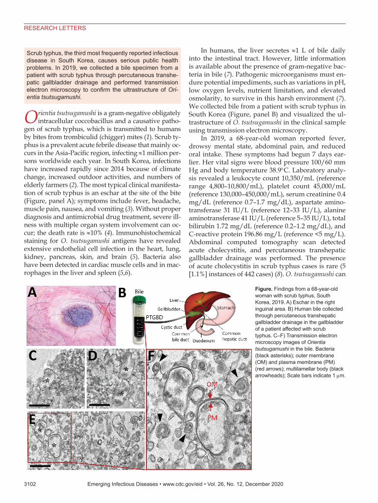
gen of scrub typhus, which is transmitted to humans by bites from trombiculid (chigger) mites (1). Scrub ty-phus is a prevalent acute febrile disease that mainly oc-curs in the Asia-Pacific region, infecting ≈1 million per-sons worldwide each year. In South Korea, infections have increased rapidly since 2014 because of climate change, increased outdoor activities, and numbers of elderly farmers (2). The most typical clinical manifesta-tion of scrub typhus is an eschar at the site of the bite (Figure, panel A); symptoms include fever, headache, muscle pain, nausea, and vomiting (3). Without proper diagnosis and antimicrobial drug treatment, severe ill-ness with multiple organ system involvement can oc-cur; the death rate is ≈10% (4). Immunohistochemical staining for O. tsutsugamushi antigens have revealed extensive endothelial cell infection in the heart, lung, kidney, pancreas, skin, and brain (5). Bacteria also have been detected in cardiac muscle cells and in mac-rophages in the liver and spleen (5,6).
In humans, the liver secretes ≈1 L of bile daily into the intestinal tract. However, little information is available about the presence of gram-negative bac-teria in bile (7). Pathogenic microorganisms must en-dure potential impediments, such as variations in pH, low oxygen levels, nutrient limitation, and elevated osmolarity, to survive in this harsh environment (7). We collected bile from a patient with scrub typhus in South Korea (Figure, panel B) and visualized the ul-trastructure of O. tsutsugamushi in the clinical sample using transmission electron microscopy.
In 2019, a 68-year-old woman reported fever, drowsy mental state, abdominal pain, and reduced oral intake. These symptoms had begun 7 days ear-lier. Her vital signs were blood pressure 100/60 mm Hg and body temperature 38.9°C. Laboratory analy-sis revealed a leukocyte count 10,350/mL (reference range 4,800–10,800/mL), platelet count 45,000/mL (reference 130,000–450,000/mL), serum creatinine 0.4 mg/dL (reference 0.7–1.7 mg/dL), aspartate amino-transferase 31 IU/L (reference 12–33 IU/L), alanine aminotransferase 41 IU/L (reference 5–35 IU/L), total bilirubin 1.72 mg/dL (reference 0.2–1.2 mg/dL), and C-reactive protein 196.86 mg/L (reference <5 mg/L). Abdominal computed tomography scan detected acute cholecystitis, and percutaneous transhepatic gallbladder drainage was performed. The presence of acute cholecystitis in scrub typhus cases is rare (5 [1.1%] instances of 442 cases) (8). O. tsutsugamushi can
3102 Emerging Infectious Diseases • www.cdc.gov/eid • Vol. 26, No. 12, December 2020
RESEARCH LETTERS
Scrub typhus, the third most frequently reported infectious disease in South Korea, causes serious public health problems. In 2019, we collected a bile specimen from a patient with scrub typhus through percutaneous transhe-patic gallbladder drainage and performed transmission electron microscopy to confirm the ultrastructure of Ori-entia tsutsugamushi.
Figure. Findings from a 68-year-old woman with scrub typhus, South Korea, 2019. A) Eschar in the right inguinal area. B) Human bile collected through percutaneous transhepatic gallbladder drainage in the gallbladder of a patient affected with scrub typhus. C–F) Transmission electron microscopy images of Orientia tsutsugamushi in the bile. Bacteria (black asterisks); outer membrane (OM) and plasma membrane (PM) (red arrows); multilamellar body (black arrowheads); Scale bars indicate 1 µm.
Emerging Infectious Diseases • www.cdc.gov/eid • Vol. 26, No. 12, December 2020 3103
RESEARCH LETTERS
also cause liver injury in some patients, but the patient reported here did not have any such signs (9). We confirmed scrub typhus using indirect immunofluo-rescence assay (IgG 5,120) and nested PCR selective for the 56-kDa gene of O. tsutsugamushi (Appendix, https://wwwnc.cdc.gov/EID/article/26/12/20-2188-App1.pdf). The O. tsutsugamushi identified belonged to the Boryong strain (the most common strain in South Korea). We also detected an eschar in the right inguinal area (Figure, panel A). The patient completely recovered after doxycycline treatment.
A drainage tube was placed in the patient’s gall-bladder, and the bile was directly discharged and col-lected through the tube (Figure, panel B). We tested the bile specimen for pathogens using nested quan-titative reverse transcription PCR and DNA sequenc-ing to detect a specific O. tsutsugamushi gene encod-ing a 56-kDa protein (Appendix) (10). After chemical fixation, the sample was embedded in 100% Epon 812 resin and ultrathin (≈80-nm thick) sections were stained with 2% uranyl acetate and 1% lead citrate (Appendix) (10). This sample preparation method might not preserve the ultrastructure of live bacteria, but structural features of the bacteria can be clearly observed. The ultrastructural details were acquired using transmission electron microscopy at 120 kV. Despite the presence of a wide variety of components, we detected O. tsutsugamushi in the bile (asterisks in Figure, panels C–E). The bacteria showed a coccoba-cillus shape and were 0.5–0.7-µm in diameter and 1.2–2.5-µm long, all typical features of O. tsutsugamushi (5,10). The bacterial cytoplasm was surrounded by an outer membrane, an internal plasma membrane, and a peptidoglycan layer (Figure, panel F). Moreover, the periplasmic space appeared as an electron-lucent gap between the 2 membranes. We also observed a thicker outer leaflet of the cell wall membrane, which is a typical and diagnostic sign of Orientia (5). We also detected multilamellar bodies, which are cholesterol-carrying particles, in the bile sample (black arrow-heads in Figure, panel F).
Previously, human scrub typhus disease was studied using a mouse model mimicking the disease and examining clinical samples postmortem (5,6). However, the host cell of O. tsutsugamushi in humans has not been completely defined. In this study, we confirmed detection of O. tsutsugamushi in human bile, an environment in which bacterial survival is challenging. This observation (i.e., the presence of O. tsutsugamushi in human bile) might be useful for di-agnosing scrub typhus in patients who do not show clear eschars or skin rash, broadening the potential routes for diagnosing the disease.
This work was supported by the Basic Science Research Programs (NRF-2015R1D1A1A01060251 and 2018R1D-1A3B07049557) of the National Research Foundation of Korea, which is funded by the Ministry of Education; the Korea Basic Science Institute grant (C030221), and National Research Council of Science & Technology grant by the Korea government (CRC-16-01-KRICT).
About the AuthorsMs. Y. Lee is a researcher at the Korea Basic Science Institute. Her primary interests are electron microscopy of infectious viral and bacterial diseases. Dr. Kim is a senior researcher at the Korea Basic Science Institute. His research interests are in the proteomics of infectious diseases.
References 1. Liu YX, Cao WC, Gao Y, Zhang JL, Yang ZQ, Zhao ZT, et al.
Orientia tsutsugamushi in eschars from scrub typhus patients. Emerg Infect Dis. 2006;12:1109–12. https://doi.org/10.3201/eid1207.050827
2. Kim DM. Clinical features and diagnosis of scrub typhus. Infect Chemother. 2009;41:315–22. https://doi.org/10.3947/ic.2009.41.6.315
3. Lee CS, Hwang JH. Images in clinical medicine. Scrub syphus. N Engl J Med. 2015;373:2455. https://doi.org/ 10.1056/NEJMicm1503639
4. VieBrock L, Evans SM, Beyer AR, Larson CL, Beare PA, Ge H, et al. Orientia tsutsugamushi ankyrin repeat-containing protein family members are type 1 secretion system substrates that traffic to the host cell endoplasmic reticulum. Front Cell Infect Microbiol. 2015;4:186. https://doi.org/10.3389/fcimb.2014.00186
5. Moron CG, Popov VL, Feng HM, Wear D, Walker DH. Identification of the target cells of Orientia tsutsugamushi in human cases of scrub typhus. Mod Pathol. 2001;14:752–9. https://doi.org/10.1038/modpathol.3880385
6. Shelite TR, Saito TB, Mendell NL, Gong B, Xu G, Soong L, et al. Hematogenously disseminated Orientia tsutsugamushi–infected murine model of scrub typhus [corrected]. Erratum in: PLoS Negl Trop Dis. 2014;8:e3175. PLoS Negl Trop Dis. 2014;8:e2966. https://doi.org/10.1371/journal.pntd.0002966
7. Begley M, Gahan CG, Hill C. The interaction between bacteria and bile. FEMS Microbiol Rev. 2005;29:625–51. https://doi.org/10.1016/j.femsre.2004.09.003
8. Lee H, Ji M, Hwang JH, Lee JY, Lee JH, Chung KM, et al. Acute cholecystitis in patients with scrub typhus. J Korean Med Sci. 2015;30:1698–700. https://doi.org/ 10.3346/jkms.2015.30.11.1698
9. Chung JH, Lim SC, Yun NR, Shin SH, Kim CM, Kim DM. Scrub typhus hepatitis c onfirmed by immunohistochemical staining. World J Gastroenterol. 2012;18:5138–41. https://doi.org/10.3748/wjg.v18.i36.5138
10. Ro HJ, Lee H, Park EC, Lee CS, Il Kim S, Jun S. Ultrastructural visualization of Orientia tsutsugamushi in biopsied eschars and monocytes from scrub typhus patients in South Korea. Sci Rep. 2018;8:17373. https://doi.org/10.1038/s41598-018-35775-9
Address for correspondence: Sangmi Jun, Center for Research Equipment, Korea Basic Science Institute, 162 Yeongudanji-ro, Ochang-eup, Cheongwon-gu, Cheongju-si, Chungcheongbuk-do, 28119, South Korea; email: [email protected]
Page 1 of 3
Article DOI: https://doi.org/10.3201/eid2612.202188
Transmission Electron Microscopy Confirmation of Orientia tsutsugamushi in
Human Bile Appendix
Genotyping by DNA Amplification and Sequencing
Peripheral blood mononuclear cells collected from acute-phase blood samples of
patients with scrub typhus were purified using a QIAamp DNA Blood Mini Kit (QIAGEN,
Hilden, Germany) according to the manufacturer's protocol. Nested PCR was performed.
Primers 34 (forward, 5′-TCA AGC TTA TTG CTA GTG CAA TGT CTGC-3′; the 56-kDa
gene based on the Gilliam strain) and 55 (5′-AGG GAT CCC TGC TGT GCT TGC TGCG-
3′) were used in the first PCR. Nested PCR primers 10 (5′-GAT CAA GCT TCC TCA GCC
TAC TAT AAT GCC-3′) and 11 (5′-CTA GGG ATC CCG ACA GAT GCA CTA TTA
GGC-3′) were used in the second PCR amplification to generate a 483-bp fragment. Nested
PCR was performed as described previously by Kim et al. (1). The amplified PCR products
were confirmed using 1.2% agarose gel electrophoresis, purified with a QIAquick gel
extraction kit (QIAGEN) and sent to COSMO Genetech (Seoul, South Korea) for sequencing.
Identification of 56-kDa Gene by DNA Amplification and Sequencing Using Bile-Derived DNA
Total genomic DNA was extracted from the bile using a QIAamp DNA Mini Kit
(QIAGEN), as previously described (2,3). The 56-kDa gene of O. tsutsugamushi was
amplified using nested PCR, as previously described (4). Briefly, initial rounds of
amplifications were performed using touchdown PCR, and products from the initial
amplification were used as the template in the second PCR reaction to generate an 83-bp
fragment. Nested PCR was performed using Solg 2X Multiplex PCR Smart mix (Solgent,
Daejeon, South Korea). Levels of the 56-kDa gene were normalized using the human Gapdh
(hGapdh) gene. Information on the primers used in this study is summarized in the Appendix
Table. The final PCR products were visualized with a chemiDoc XRS+ system (Bio-Rad,

Page 2 of 3
Hercules, CA, USA) (Appendix Figure 1) and sent to COSMO Genetech for sequencing. The
sequencing results were aligned with the original sequence of the 56-kDa gene using Clustal
Omega, which revealed the 56-kDa gene in bile-derived DNA (Appendix Figure 2).
Transmission Election Microscopy (TEM)
For ultrastructural analysis of ultrathin sections, collected human bile was pelleted via
centrifugation and resuspended in 0.1% glutaraldehyde solution and 4% paraformaldehyde,
3.5% sucrose in phosphate buffer (0.1 M, pH 7.4) for 2 h at 4°C. The samples were washed in
phosphate buffer, and then post-fixed in 1% osmium tetroxide (Electron Microscopy
Sciences, Hatfield, PA, USA) in phosphate buffer (0.1 M, pH 7.4) for 2 h at 4°C. Samples
were dehydrated in a graded ethanol series, followed by propylene oxide, and then
progressively infiltrated with a 2:1, 1:1, and 1:2 mixture of propylene oxide and Epon 812
resin (Electron Microscopy Sciences). Samples finally embedded in 100% Epon 812 resin
and polymerized at 70°C for 24 h. Ultrathin plastic sections (80-nm thick) were cut at room
temperature using a Leica EM UC6 ultramicrotome (Leica Microsystems GmbH, Wetzlar,
Germany) and collected on 200-mesh carbon-coated grids. The grids were post-stained with
2% aqueous uranyl acetate and 1% lead citrate at room temperature for 15 and 5 min,
respectively. A FEI Tecnai G2 Spirit Twin 120 KV TEM (FEI Company, Hillsboro, OR,
USA) was used for TEM analysis (4,5)
References
1. Lee YM, Kim DM, Lee SH, Jang MS, Neupane GP. Phylogenetic analysis of the 56 kDa protein
genes of Orientia tsutsugamushi in southwest area of Korea. Am J Trop Med Hyg.
2011;84:250–4. PubMed https://doi.org/10.4269/ajtmh.2011.09-0601
2. Neri V, Margiotta M, de Francesco V, Ambrosi A, Valle ND, Fersini A, et al. DNA sequences and
proteic antigens of H. pylori in cholecystic bile and tissue of patients with gallstones. Aliment
Pharmacol Ther. 2005;22:715–20. PubMed https://doi.org/10.1111/j.1365-2036.2005.02644.x
3. Lee JW, Lee DH, Lee JI, Jeong S, Kwon KS, Kim HG, et al. Identification of Helicobacter pylori
in gallstone, bile, and other hepatobiliary tissues of patients with cholecystitis. Gut Liver.
2010;4:60–7. PubMed https://doi.org/10.5009/gnl.2010.4.1.60
4. Ro HJ, Lee H, Park EC, Lee CS, Il Kim S, Jun S. Ultrastructural visualization of Orientia
tsutsugamushi in biopsied eschars and monocytes from scrub typhus patients in South Korea.
Sci Rep. 2018;8:17373. PubMed https://doi.org/10.1038/s41598-018-35775-9
Page 3 of 3
5. Goldsmith CS, Miller SE. Modern uses of electron microscopy for detection of viruses. Clin
Microbiol Rev. 2009;22:552–63. PubMed https://doi.org/10.1128/CMR.00027-09
Appendix Table. List of primers used in the study Primer name Sequence c56k F 5′-AGCTGATCGTGACTTTGGGATT-3′ c56k R 5′-AGCATTTGATAATGCAGCAAGACC-3′ rt56k F 5′-CCTAACATACCTCAGGCGCA-3′ rt56k R 5′-AACCAAGCGATCCTAGCTGC-3′ Human GAPDH F 5′-CGGGAAACTGTGGCGTGATG-3′ Human GAPDH R 5′-ATGACCTTGCCCACAGCCTT-3′ *F, forward; R, reverse.
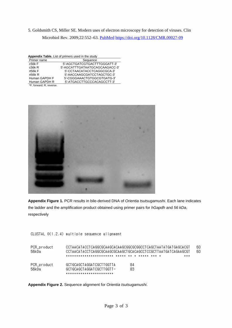
Appendix Figure 1. PCR results in bile-derived DNA of Orientia tsutsugamushi. Each lane indicates
the ladder and the amplification product obtained using primer pairs for hGapdh and 56 kDa,
respectively
Appendix Figure 2. Sequence alignment for Orientia tsutsugamushi.