topic 9: myology · 1/10/2013 1 topic 9: myology what are the different muscle types? morphology...
TRANSCRIPT

1/10/2013
1
Topic 9: Myology
� What are the different muscle types?
� Morphology & location
� Physiology
� How do muscles develop?
� What do muscles look like at the cellular level?
� How do muscles contract?
� How do whole muscles work?
� How are muscles functionally divided?
� Human facial expression
What are the different muscle types?
Liem et al. Fig. 10-1; www.cytochemistry.net
_______
Muscle
_______
Muscle
_______
Muscle
Skeletal muscle,
attached to bones
Hearth muscle
-Unitary: visceral
organs
-Multiunit: iris &
blood vessels

1/10/2013
2
Liem et al. Fig. 10-1
Striated Muscle Cardiac Muscle Smooth Muscle
Cell Size Small, elongate Small, elongate
# nuclei Many Single Single
Control Voluntary Involuntary Involuntary
Stimulus Unitary: myoMultiunit: neuro
Contraction Force
Contraction Rate Slow to fast Moderate Relatively slow
Fatigue Fatigues Does not Does not
What are the different muscle types?
How do muscles develop?
Wolpert, 1998
Mesoderm
Paraxial
Kidneys
Somatic
Lateral PlateIntermediate
SplanchnicMyotome
Visceral
Muscles
Body
Wall
Muscles
Limb
Muscles
Dermatome
Sclerotome
Heart
What type of muscle makes up each of these?
1/10/2013
1
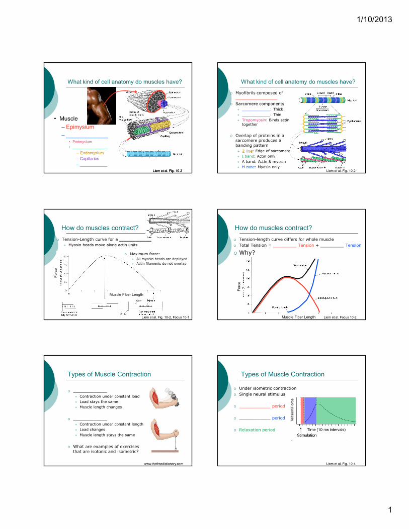
What kind of cell anatomy do muscles have?
Liem et al. Fig. 10-2
• Muscle
– Epimysium
– _____________• Perimysium
• _________________
– Endomysium
– Capillaries
– ____________
Liem et al. Fig. 10-2
� Myofibrils composed of
________________
� Sarcomere components
� ____________: Thick
� ____________: Thin
� Tropomyosin: Binds actintogether
� Overlap of proteins in a sarcomere produces a
banding pattern
� Z line: Edge of sarcomere
� I band: Actin only
� A band: Actin & myosin
� H zone: Myosin onlyLiem et al. Fig. 10-2
What kind of cell anatomy do muscles have?
How do muscles contract?
� Tension-Length curve for a ___________
� Myosin heads move along actin units
Liem et al. Fig. 10-2, Focus 10-1
Force
Muscle Fiber Length
� Maximum force:
� All myosin heads are deployed
� Actin filaments do not overlap
� Tension-length curve differs for whole muscle
� Total Tension = _________ Tension + _________ Tension
� Why?
Liem et al. Focus 10-2
Force
Muscle Fiber Length
How do muscles contract?
Types of Muscle Contraction
� _____________
� Contraction under constant load
� Load stays the same
� Muscle length changes
� _____________
� Contraction under constant length
� Load changes
� Muscle length stays the same
� What are examples of exercises that are isotonic and isometric?
www.thefreedictionary.com
Types of Muscle Contraction
� Under isometric contraction
� Single neural stimulus
� ____________ period
� ____________ period
� Relaxation period
Liem et al. Fig. 10-4
Tension/Force
1/10/2013
2
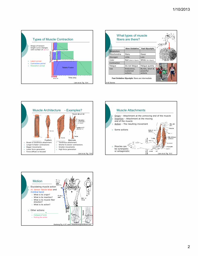
Types of Muscle Contraction
� Shape of tension-length curve changes with number of stimuli
� Latent period
� Contraction period
� Relaxation period
Liem et al. Fig. 10-4
Tension/Force
Time (ms)
Tetanic Fusion
What types of muscle
fibers are there?
© KE Bonine
Slow Oxidative Fast Glycolytic
Metabolism
Mitochondria Many Fewer
Glycogen Little Lots
Color Red (black in figure) White (& in figure)
Contraction
Fatigue Do not fatigue Fatigue quickly
Use Endurance,sustained
actions
Bursts, short & powerful
actions
Fast Oxidative Glycolytic fibers are intermediate
Muscle Architecture
� Broad vs tendinous attachment
� Longer & faster contractions
� Bigger movements
� Lower force generation
� Force diffuse vs focusedLiem et al. Fig. 10-6
_________ Fusiform _________ Bipinnate
� Tendinous attachment
� Shorter & slower contractions
� Smaller movements
� High force generation
- Examples? Muscle Attachments
� Origin – Attachment at the unmoving end of the muscle
� Insertion – Attachment at the moving
end of the muscle
� Action – The resulting movement
� Some actions
� Muscles can
be synergistic or antagonistic
Liem et al. Fig. 10-3
Motion
� Elucidating muscle action
� m. tensor fascia latae and
iliotibial band
� What is its origin?
� What is its insertion?
� What is its muscle fiber direction?
� What is its action?
� Other actions:� ___________ & ___________
� Collapse of arch
� Rolling the ankle
Kardong Fig. 4-37; www. thestretchinghandbook.com
Medial Medial

1/10/2013
1
How are muscles functionally divided?
� Trunk musculature
� ________ & _________
� ________ & _________
� Appendicular musculature
� Dorsal & Ventral
� Pairs of groups have different innervations
� What does each group develop from?
Liem et al. Fig. 10-8
� Trends in axial musculature evolution� ________________
� ________________
� ________________ of function (muscle fiber direction)
Liem et al. Fig. 10-16, 10-18
Fish
SalamanderLizard
How have the trunk
muscles evolved?
Liem et al. Fig. 10-20, 10-21
� Similar trends in limb musculature evolution� Duplication
� Differentiation
� Specialization of function (muscle fiber direction)
Fish
Lizard
Cat
How have limb
muscles evolved? Platysma and Facial Muscles
� Derived from hyoid muscles
� Form lips & cheeks
� Coupled with evolution of nipples
� What are actions of:
� Frontal muscle
� m. orbicularis oculi
� m. orbicularis oris
� m. quadratus labii superior
Liem et al. Fig. 10-14