the urinary system - angelfire · renal corpuscle - where fluid is filtered. 2. renal tubule and...
TRANSCRIPT
Chapter 26 1
BIOLOGY 2402 Anatomy and Physiology Lecture
Chapter 26
THE URINARY SYSTEM
Chapter 26 2
THE URINARY SYSTEM (Metabolism of nutrients results in the production of wastes by body cells, including carbon dioxide, excess water, and heat.) A human waste product is any substance that has no function in the body. (Protein catabolism produces toxic nitrogenous wastes such as ammonia and much less toxic urea.) (In addition, many of the essential ions such as sodium, chloride, sulfate, phosphate, and hydrogen tend to accumulate in excess of the body's need.) *(All the toxic materials and the excess essential materials must be eliminated.) Several Tissues, Organs, and Processes that contribute to the temporary confinement of wastes, transport of waste materials for disposal, recycling of material, and excretion of excess or toxic substances in the body are:
1. Body buffers. Bind excess hydrogen ions (H+), which prevent an increase in the acidity of body fluids.
2. Blood. The bloodstream provides pick-up and delivery
services for the transport of wastes, just like garbage trucks and sewer lines supply the same service for a community.
3. Liver. Is the primary site for metabolic recycling, for example,
conversion of amino acids into glucose or glucose into fatty acids. Also changes toxic substances into less toxic ones, such as ammonia into urea.
4. Lungs. With each exhalation, the lungs excrete CO2, heat, and
a little water vapor.
5. Sudoriferous (sweat) glands in the skin. Especially during exercise, sudoriferiou glands in the skin help eliminate excess
Chapter 26 3 heat, waste, water, and CO2 plus small quantities of salts and urea.
6. Gastrointestinal tract. Through defecation, the
gastrointestinall tract eliminates solid, undigested foods, wastes, some CO2, water, salts, and heat.
7. Kidneys. Eliminates excess water, ammonia, urea, bilirubin,
uric acid, some bacteria toxins, H+ and other ions, plus some heat and CO2.
FUCTIONS OF THE URINARY SYSTEM
1. Filtration of the blood 2. Regulation of blood volume 3. Regulation of the concentration of solutes in the blood 4. Regulation of the pH of the extracellular fluid 5. Regulation of red blood cell synthesis 6. Vitamin D synthesis
Make up of the urinary system
1) Two kidneys 2) Two ureters 3) One urinary bladder 4) a single urethra
(Does so by removing and restoring selected amounts of water and solutes that constitute urine). Nephrology is the specialized branch of medicine that deals with the structure, function, and diseases of the male and female urinary systems
Chapter 26 4 and the male reproductive system. Urology is the branch of medicine related to the male and female urinary systems and the male reproductive system.
KIDNEYS
Are two reddish organs that resemble kidney beans in shape. Found just above the waist between the parietal peritoneum and the posterior wall of the abdomen. Located between the levels of the last thoracic and third lumbar vertebrae. Partially protected by the eleventh and twelfth pairs of ribs. Their placement is described as retroperitoneal, since they are external to the peritoneal lining of the abdominal cavity. *The right kidney is slightly lowered then the left because of the large area occupied by the liver. Clinical Application Nephroptosis, is an inferior displacement of dropping of the kidney. External Anatomy
Three layers of tissue surround the kidneys:
a) Renal Capsule - the innermost layer. Serves as a barrier against trauma and the spread of infection.
b) Adipose Capsule (perirenal fat) - the second layer. Protects from trauma and hold kidney firmly in place within the abdominal cavity.
Chapter 26 5
c) Renal fascia - the outermost layer. Anchors the kidney to its surrounding structures and to the abdominal wall.
Internal Anatomy
Hilus - a notch through which the ureter leaves the kidney.
Renal Sinus - entrance to cavity in the kidney through hilus. A coronal (frontal) section through a kidney reveals: 1. Renal Cortex- an outer reddish area 2. Renal Medulla - inner reddish-brown area 3. Renal (Medullary) Pyramids - within the medulla, 8 to 18 striated
triangular structures. 4. Renal Papillae- the bases of the pyramids 5. Renal columns - cortical substance between the renal pyramids. 6. Parenchyma - (functional portion of the kidney)- together the cortex
and renal pyramids constitute the parenchyme.
-Structurally, the parenchyma of each kidney consist of approximately 1 million microscopic units called nephrons, the functional units of the kidney.
7. Renal pelvis - a large cavity in the renal sinus. 8. Major and Minor Calyces – Cap-like extensions at the edge of
pelvis. Urine Pathway Each minor calyx receives urine from callectin ducts of one pyramid and delivers urine to major calyx. From the major calyces, the urine drains into the renal pelvis and out through the ureter to the urinary bladder.
Chapter 26 6 BLOOD AND NERVE SUPPLY
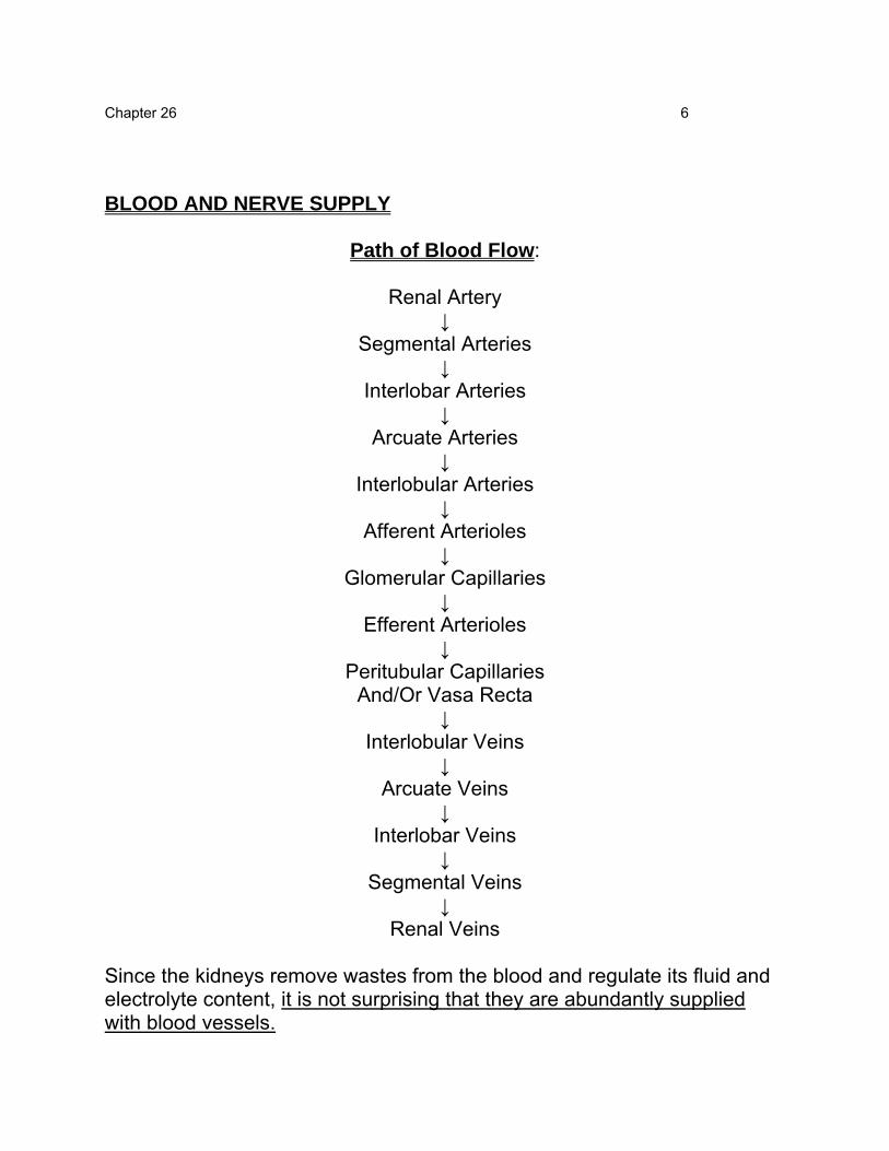
Path of Blood Flow:
Renal Artery ↓
Segmental Arteries ↓
Interlobar Arteries ↓
Arcuate Arteries ↓
Interlobular Arteries ↓
Afferent Arterioles ↓
Glomerular Capillaries ↓
Efferent Arterioles ↓
Peritubular Capillaries And/Or Vasa Recta
↓ Interlobular Veins
↓ Arcuate Veins
↓ Interlobar Veins
↓ Segmental Veins
↓ Renal Veins
Since the kidneys remove wastes from the blood and regulate its fluid and electrolyte content, it is not surprising that they are abundantly supplied with blood vessels.
Chapter 26 7 Kidneys make up just 1% of the total body mass, but receive 20-25% of the resting cardiac output via the right and left renal arteries. Amounts to about 1200 ml of blood per minute. Nerve supply of the kidneys is derived from the renal plexus of the autonomic nervous system. Nerve from this plexus accompany the renal arteris and their branches and are distributed to the blood vessels. Because these are vasomotor nerves, they regulate the flow of blood through the kidney by regulating the diameters of the arterioles. Nephron Is the functional unit of the kidney. Three basic functions: 1. Glomerular filtration - some substances are permitted to pass from
the blood into the nephrons, while other are kept out. 2. Tubular secretion- as the filtered liquid (filtrate) moves through the
nephrons, it gains some additional material (wastes and excess substances).
3. Tubular reabsorption- other substances (useful materials) are
returned to the blood. *As a result of these activities of nephrons, urine is formed.
Chapter 26 8 Parts of the Nephron Consists of two portions: 1. Renal corpuscle - where fluid is filtered. 2. Renal tubule and Collecting ducts - into which the filtered fluid
passes. Renal Corpuscle Has two components: a. Glomerulus - a tuft of capillary loops.
b. Glomerular (Bowman's) Capsule - a double-walled epithelial cup, surrounding the glomerulus.
*(Their arrangement is analogous to a fist (glomerulus) punched into a limp balloon (glomerular capsule) until fist is covered by two layers of balloon with space in between.) Afferent Arteriole - through which blood enters a glomerulus. Efferent Arteriole - through which blood exits a glomerulus. Capsular (Bowman's) Space - Separates the outer wall (Parietal layer) of the glomerular capsule from the inner wall (visceral layer). Parietal layer of the glomeruler capsule consists of simple squamous epithelium and forms the outer wall of the cup. Visceral layer consists of modified simple squamous epithelial cells called podocytes that form the inner wall of the cup. Endothelial-capsular (filtration) membrane is formed by the combination
Chapter 26 9 of endothelial cells and podocytes. Filtered Substances Pass Through The Three Layers Of This Membrane In The Following Order:
1. Endothelial fenestrations (pores) of the glomerulus: Prevent filtration of blood cells but allow all components of blood plasma to pass through.
2. Basement membrane of the glomerulus: Prevents filtration
of large proteins.
3. Slit membrane between pedicles: Prevents filtration of medium-sized proteins.
*(As blood flows through the glomerular capillaries, water and most kinds of solutes filter from blood plasma into the capsular space.) *(Large plasma proteins and the formed elements in blood do not normally pass through.) Renal Tubule and Ducts Filtered fluid passes into the renal tubule from the capsular space. Renal tubule has three main sections: (a). Proximal convoluted tubule (PCT) (b). Loop of Henle (Nephron loop) (c). Distal convoluted tubule (DCT) (Convoluted means the tubule is coiled rather than straight); (Proximal signifies the tubule portion attached to the glomerular capsule), (Distal the portion that is further away)
Chapter 26 10 Renal Corpuscle Proximal Convoluted tubule } Lie in the Cortex of the kidney Distal Convoluted tubule Loop of Henle: Extends into the Medulla of the kidney, makes a hairpin
turn, then returns to the cortex. Distal convoluted tubules of several nephrons empty into a single collecting duct. Collecting Ducts merge to form several hundred large Papillary Ducts, which drain into a Minor Calyx. (Each kidney has about 1 million Renal Corpuscles, PCTS, Loops of Henle, and DCTs but a much smaller number of collecting ducts and even fewer papillary (ducts). Cortical and Juxtamedullary Nephrons In a Nephron, the Loop of Henle connects the proximal and distal Convoluted tubules. First portion of the Loop of Henle dips into the renal medulla, where it is called the descending limb of the loop of Henle. Descending limb of the loop of Henle bends in a U-shape and returns to the renal cortex as the ascending limb of the loop of Henle.
Cortical Nephron usually has its glomerulus in the outer (superficial) portion of the renal cortex; its short loop of Henle penetrates only into the outer region of the renal medulla. About 80-85% of the nephrons have short loops of Henle. They receive their blood supply from peritubular capillaries that arise from efferent arterioles.
Chapter 26 11 Juxtamedullary Nephron usually has its glomerulus deep in the renal cortex close to the renal medulla, and its long loop of the Henle stretches through the renal medulla and almost reaches the renal papilla. About 15-20% are long loops of Henle. They receive their blood supply from peritubular capillaries and vasa recta that arise from efferent arterioles. Histology of The Nephron and Associated Structures A single layer of epithelial cells forms the wall of: - the renal corpuscle, - renal tubule - collecting duct - papillary duct 1. Parietal layer of the glomerular (Bowman's) capsule consists of
simple squamous epithelium;
Visceral layer of the glomerular (Bowman's) capsule consists of modified simple squamous epithelial cells called podocytes.
2. Proximal convoluted tubule (PCT), the cells are cuboidal and have
a prominent brush border of microvilli on the apical surface (surface facing the lumen).
Microvilli, like those of the small intestine, increase the surface area for absorption and secretion.
About 65% of the filtered water and up to 100% of some filtered solutes return to the bloodstream from the PCT.
3. Descending limb of the loop of Henle and the first part of the
ascending limb of Henle, the thin ascending limb, is simple squamous epithelium.

Chapter 26 12 (Cortical nephrons lack the thin ascending limb). 4. Thick ascending limb of the loop of Henle is composed of cuboidal
to low columnar epithelium. 5. In each nephron, the final portion of the ascending limb of loop of
Henle makes contact with the afferent arteriole serving its own renal corpuscle.
Macula Densa is the collective name for the cells of the renal tubule in this region; are tall and crowded together.
These cells monitor the Na+ and Cl- concentration of fluid in the tubule lumen.
Juxtaglomerular (JG) cells - are modified smooth muscle fibers (their nuclei are round instead of long) in the wall of the Afferent arteriole (and sometimes Efferent arteriole).
Macula densa and Juxtaglomerular (JC) cells together constitute the Juxtaglomerular Apparatus (JGA).
JGA helps regulate blood pressure and the rate of blood filtration by the kidney.
6. Cells of the distal convoluted tubule (DCT) and collecting ducts
are cuboidal and have a few microvilli. Beginning in the last portion of the distal convoluted tubule (DCT) and continuing into the collecting ducts, two different cells are present:
(1) Principal cells, which respond to antidiuretic hormone
(ADH) and aldosterone, two hormones that regulate kidney functions.
(2) Intercalated cells, which secrete H+ and thereby rid the
body of excess acids.

Chapter 26 13 7. Cells of the large papillary ducts are simple columnar epithelium.
RENAL PHYSIOLOGY
Physiology of Urine Formation
BLOOD STREAM � Renal Arteriole
GLOMERULUS C-Secretion
A- Filtration � Water, sugar, salt urea/other wastes BOWMAN'S CAPSULE
↓ B- Reabsorption RENAL TUBULE
� Urea/Waste � Salts
� Water }Urine � Acid
RENAL PELVIS ↓
URETER ↓
BLADDER ↓
URETHRA ↓
URINARY MEATUS ↓
Urine is expelled from the body
Nephrons and collecting ducts within the kidney do the major work of the urinary system. Other parts of the system are primarily passageways and storage area.

Chapter 26 14 Glomerular Filtration
Glomerular Filtration is the first step in renal regulation of blood composition and volume.
In filtration, pressure forces fluid and all solutes smaller than a certain size through a membrane.
(All obey Starling's Law of the Capillaries ("Filtration is almost equal to Reabsorption")
Filtration occurs in the renal corpuscles of the kidney across the endothelial-capsular membranes.
Blood pressure forces water and dissolved components through the: (i) endothelial fenestrations (pores) of the capillaries, (ii) basement membrane, and (iii) slit membranes between pendicels
Glomerular filtrate is the resulting fluid that enters the capsular space.
Filtration fraction is the fraction of plasma in the afferent arterioles of the kidneys that becomes glumerular filtrate.
Filtration fraction varies considerably in both health and disease.
(About 180 liters (48 gal) of filtrate enter the capsular space each day, represents 65 times the entire blood plasma volume).
(However, 178-179 liters return to the bloodstream by tubular reabsorption so only 1 to 2 liters (about 1-3 qt) are excreted as urine).
In a healthy person, glomerular filtration contains all the materials present in the blood except the formed elements and most plasma

Chapter 26 15 proteins, which are too large to pass through the endothelial-capsular membranes.
Several structural features of the Renal Corpuscles that enhance their Blood-Filtering Capacity: 1. The Endothelial-Capsular membrane is thin and porous.
-Allow passage of smaller molecules. 2. Glomerular Capillaries present a large surface area.
3. Capillary blood pressure is high.
Blood pressure is higher in the glomerular capillaries than in capillaries elsewhere in the body.
A higher pressure means more filtration.
(Efferent arteriole is smaller in diameter than the Afferent arteriole, so there is high resistance to the outflow of blood from the glomerulus.)
Net Filtration Pressure Blood filtering depends on three main pressures: 1. Glomerular Blood Hydrostatic Pressure (GBHP) - chief one 2. Capsular Hydrostatic Pressure (CHP) - opposing force
3. Blood Colloid Osmotic Pressure (BCOP) - opposing force 1. Glomerular Blood Hydrostatic Pressure (GBHP):
Promotes filtration - it pushes water and solutes in blood plasma
Chapter 26 16 through the glomerular filter.
Hydrostatic Pressure is the force that a fluid under pressure exerts against the walls of its container.
Glomerular blood hydrostatic pressure is the blood pressure in glomerular capillaries.
Is about 55 mm Hg
2. Capsular Hydrostatic Pressure (CHP):
Is a back pressure that opposes filtration (glomerular blood hydrostatic pressure).
As the filtrate is forced into the capsular space between the walls of the glomerular capsule, it meets two forms of resistance:
(a) the walls of the capsule; and (b) the fluid that has already filled the renal tubule Results to some filtrate being pushed back into the capillary.
The amount of "push" is the Capsular hydrostatic pressure is about 15 mm Hg
3. Blood Colloid Osmotic Pressure (BCOP):
Is the second force opposing filtration (glomerular blood hydrostatic pressure)
Is due to the presence of proteins (such as albumin, globulins, and fibrinogen) in blood plasma.
Osmotic Pressure is the pressure required to prevent the net movement of water into a solution containing solutes when the
Chapter 26 17 solutions are separated by a selectively permeable membrane.
The greater the solute concentration of a solution, the greater its osmotic pressure.
Because blood contains a much higher concentration of proteins than glomerular filtrate, some water moves from the filtrate back into the glomerular capillaries.
BCOP averages 30 mm Hg in glomerular capillaries. Net Filtration Pressure (NFP) is the total pressure that promotes filtration. Can be determined by subtracting the forces that oppose filtration from the glomerular blood hydrostatic pressure. NFP = GBHP - (CHP + BCOP) Promotes filtration Oppose filtration (By substituting the values just given, a normal NFP may be calculated as follow:) NFP = (55 mm Hg) - (15mm Hg + 30 mm Hg) = (55 mm Hg) - (45 mm Hg) = 10 mm Hg (This means that a pressure of only 10 mm Hg causes a normal amount of plasma (minus plasma protein) to filter from the glomerulus into the capsular space.) Glomerular Filtration Rate (GFR) Glomerular Filtration Rate is the amount of filtrate formed in al the renal corpuscles of both kidneys each minute.
Chapter 26 18 In a normal adult, GFR is about 125 ml/min (about 180 liters (48 gal) a day.
GFR is directly related to the pressure that determine NFP
(Any factor that alters NFP will affect GFR. For example, severe blood loss reduces systemic blood pressure, which also decreases the glomerular blood hydrostatic pressure (GBHP))
(If GBHP drops to 45 mm Hg, filtration stops because this is the magnitude of the opposing force).
Anuria is a condition resulting to a daily urine output of less than 50 ml.
May be caused by insufficient pressure for filtration or by inflammation of the glomeruli so that plasma cannot enter the capsule space.
Homeostasis of body fluid requires that the kidneys maintain a relatively constant Glomerular filtration rate (GFR)
If the GFR is too high, needed substances may pass so quickly through the renal tubules that they are not reasbsorbed and instead are lost in the urine.
If the GFR is too low, nearly all the filtrate may be reabsorbed and certain waste products may not be adequately excreted.
Regulation of GFR
Glomerular filtration rate (GFR) increases when blood flows into the glomerular capillaries more rapidly than normal.
Glomerular Blood Flow depends on the following:
(1) Systemic Blood pressure; and (2) The diameter of the Afferent and Efferent arterioles.
Chapter 26 19 Three Principal Mechanisms that Control Systemic Blood Pressure and Blood Vessel Diameter: (A) Renal Autoregulation of GFR
Renal Autoregulation is the ability of the kidney to maintain a constant blood pressure and GFR despite changes in systemic arterial pressure.
Renal autoregulation is intrinsic (it operates completely within the kidneys, even if they are removed from the body, for example, in a transplant operation.
Renal autoregulation operates by a negative feedback system that involves the juxtaglomerular apparatus of JGA
When Controlled Conditions: Net Filtration Pressure (NFP) and Glomerular Filtration Rate (GFR) are low due to low blood pressure;
(a) the proximal convoluted tubules, and
(b) loop of Henle reabsorb more than the normal fraction of Na+, Cl-, and water.
(c) at the end of the loop of Henle, macula densa cells in the JGA (receptors) detect decreased delivery of Na+, Cl-, and water.
(d) by an unknown mechanism (input), cells in the JGA (control center) are inhibited and decrease their release of a vasoconstrictor substance (output).
(e) with less vasoconstrictor substance present, the afferent arteriole (effector) dilates, allowing more blood to flow into the glomerulus.
(f) this increases NFP and GFR (responses) and brings about a return to homeostasis.
If blood pressure is elevated, on the other hand, NFP and GFR are higher than normal and the opposite sequence of events occurs.
Chapter 26 20 B) Hormonal Regulation of GFR
Two hormones that contribute to regulate GFR are: (a) Angiotensin II; and (b) Atrial Natriuretic Peptide (ANP)
Renin-angiotensin system
When blood pressure (BP) and therefore GFR (controlled condition) decrease, juxtaglomerular cells and macula densa cells of the JGA (receptor) detect decreased stretch and decreased delivery of Na+, Cl-, and water.
Juxtaglomerular cells then secrete an enzyme called Renin (input) in response to several kinds of stimuli.
Once released into the blood, renin acts on a plasma protein produced by the liver called Angiotensinogen.
Renin converts Angiotensinogen into Angiotensin I; Angiotensin I passes through the lungs;
An enzyme, Angiotensin Converting Enzyme (ACE) in the lunge converts Angiotensin I to Angiotensin II.
Angiotensin II is an active hormone. Actions of Angiotensin II
1. Vasoconstriction of arterioles - It constricts Efferent Arterioles to
elevate glomerular blood pressure and raise GFR back to normal. 2. Stimulation of Aldosterone secretion by the adrenal cortex. - it
stimulates the adrenal cortex to secrete aldosterone, which enhances reabsorption of Na+ by the principal cells in the collecting ducts.

Chapter 26 21 3. Stimulation of Thirst center in the hypathalamus. - it acts on the
thirst center in the hypothalamus to increase water intake. Increases blood volume a pressure GFR are restored to normal.
4. Stimulation of Antidiuretic Hormone (ADH) secretion from the
posterior pituitary gland. - It stimulates the release of ADH from the posterior pituitary gland, resulting in water retention by the kidney.-- Increases blood volume and pressure.
Angiotensin II helps restore normal systemic and renal blood pressure, which normalize GRR and bring a return to homeostasis.
Atrial Natriuretic Peptide (ANP), is a second hormone that influences glomerular filtration and other renal processes.
It is secreted by cells in the atria (superior chambers) of the heart.. Promotes both excretion of water (diuresis) and excretion of sodium (natriuresis).
Secretion of ANP is stimulated by increased stretching of the atria, as occurs when blood volume increases.
ANP increases GFR, perhaps by increasing the permeability of the filter or by dilating the afferent arterioles.
It also suppresses secretion of antidiuretic hormone (ADH), aldosterone, and renie.
C) Neural Regulation of GFR
Like most blood vessels of the body, those of the kidneys are supplied by vasoconstrictor fibers from the sympathetic division of the Autonomic Nervous System.
At rest, sympathetic stimulation is minimal and renal blood vessels are maximally dilated
Chapter 26 22
With greater sympathetic stimulations, however, as might occur during exercise, hemorfhage, or a fight-or-flight response, vasoconstriction of the afferent arterioles predominates.
Strong sympathetic stimulation also causes the adrenal medulla to secrete epinephrine, which also decreases GFR by bringing about vasoconstriction of the afferent arteriole.
Tubular Reabsorption *About 99% of the filtrate is reabsorbed as it passes through the Renal tubules. *Only about 1% of the filtrate leaves the body as urine (1.5 liters a day). Tubular Reabsorption is the movement of water and solute back into the blood of a pertubular capillary or vasa recta. Solutes that are reabsorbed by both active and passive processes: -Glucose -Amino acids -Urea -Ions (eg. Na+, K+, Ca2+, Cl-, HCO3-, and HPO42-) Water reabsorption occurs by the passive process of Osmosis. Small proteins and peptides are reabsorbed by Pinocytosis. REABSORPTION OF Na+ IN THE PCT *Reabsorption of Na+ is especially important because more of them pass
Chapter 26 23 the glomerular filter than any other substance except water. *Sodium ions (Na+) are reabsorbed in each portion of the Renal tubule and collecting duct by several transport system - Simple (passive) diffusion - Leakage channels
- Sodium pump (Na+/K+ ATP - ase) - Primary active transport
REABSORPTION OF NUTRIENTS IN THE PCT 100% of the filtered glucose, amino acids, lactic acid, and other possibly useful metabolities are reabsorbed in the PCT. *All these substances are reabsorbed by Na+ symporters, which work by secondary active transport. (see Key- fig. 26.14) Substances brought into PCT cells by syporters generally leave the cells by facilitated diffusion and then enter peritubular capillaries by simple diffusion. Reabsorption in The Loop of Henle 100% of the filtered 80-90% of the filtered HCO-3 65% of Na+ and H2O } Have been reabsorbed by the end of the 50% of Cl- and K+ of the Proximal convoluted Tubule (PCT) Loop of Henle reabsorbs:
30% of the filtered K+ 20% of the filtered Na+
35% of the filtered Cl-15% of the filtered water
Chapter 26 24 *For the first time, reabsorption of water by osmosis is not automatically coupled to reabsorption of filtered solutes. *This feature allows you to produce a large volume of very dilute urine or a small volume of very concentrated urine. Reabsorption in the DCT and Collecting Ducts Fluid enters the distal convoluted tubules (DCT) at a rate of about 25 ml/min because 80% of the filtered water has now been reabsorbed. As fluid flows along the DCT, Na+-Cl- symporters in the apical membranes reabsorb Na+ and Cl-. By the time fluid reaches the end of the DCT, about 90% of the filtered solutes and water have been returned to the bloodstream. Still, in one day almost 20 liters of fluid enters the collecting ducts whereas only 1-2 liters appear as urine. Two hormones: aldosterone and antidiuretic hormone, help maintain homeostasis of blood volume, composition, and pressure by fine-tuning reabsorption. They act on the principal cells, which are located in the final portion of the DCT and throughout the collecting duct. Aldosterone, secreted by the cortex of the adrenal gland, increases Na+ and water reabsorption by principal cells. Also stimulates secretion of K+ from principal cells into the tubular fluid. When the level aldosteron is low, the principal cells reabsorb little Na+. Antidiuretic hormone (ADH) is produced by the hypothalamus and released into the bloodstream by the posterior pituitary gland.
Chapter 26 25 In absence of ADH, the principal cells have a very low permeability to water. When ADH is absent, a condition known as diabetes insipidus, the kidneys may secrete up to 20 liters per day of very dilute urine. Water Reabsorption About 90% of the water reabsorption, a total of 160 liters per day, occurs by osmosis together with reabsorption of solutes such as Na+, Cl-, and glucose. Water absorption by osmosis is termed obligatory water reabsorption because the water is “obliged” to follow the solute. Occurs in the: - Proximal convoluted Tubule (PCT)
- Descending limb of the loop of Henle, and - Early distal convoluted tubule (DCT)
Reabsorption of the remaining 10% of the water in the tubular fluid, a total of 10-20 liters per day, is termed facultative water reabsorption - capable of adapting to a need.
Tubular Secretion Is the third process involved in Urine formation: Tubular Secretion removes materials from the blood and adds them into tubular fluid. (Whereas tubular reabsorption returns filtered substances to the blood, tubular secretion removes material from the blood). Secreted substances include:
Chapter 26 26 -Potassium ions (K+) -Hydrogen ions (H+) -(Ammonium ions (NH4+) - Creatinine - Drugs (penicillin and Para-aminohippuric acid)
*Tubular secretion has two principal effects;
(1) It rids the body of certain materials (2) It helps control blood pH.
Secretion Of K+ Most filtered potassium ions are reabsorbed in the PCT, Loop of Henle, and DCT. Secretion of K+ is controlled by: 1. Aldosterol - in the presence of more aldosterone, more K+ is
secreted. 2. K+ concentration in Plasma - when plasma K+ concentration is high,
K+ secretion increases. 3. Na+ Concentration in Distal Convoluted Tubules - High levels of Na+
increases the rate of Na+ absorption and K+ secretion. Secretion of H+ *Body maintains normal blood pH (7.35 to 7.45) despite continued production of far more acids than bases by metabolic reactions. Cells of the Renal Tubule can raise blood pH in Three ways 1) Secreting hydrogen ions (H+) into the filtrate - rids the blood if acid
and makes urine more acidic. 2) Reabsorbing filtered HCO-3 - the most important buffer of H+ in
extracellular fluids,
Chapter 26 27 3) Producing new HCO-3 - to augment buffering of H+ in the blood. Secretion of NH3 and NH4+ Ammonia (NH3) is a waste product derived from the deamination of Amino Acid by liver cells. Liver converts much of the Ammonia to Urea, a less toxic compound.
PRODUCTION OF DILUTE AND CONCENTRATED URINE
Despite the large intake of fluids at some times and little or no intake at others, the total volume of fluid in the body remains fairly stable. Homeostasis of body fluid volume depends in large part on the ability of the kidneys to regulate the rate at which water is lost in the urine. Antidiuretic hormone (ADH) is the hormone that regulates this homeostatic function in the kidneys. *ADH controls the water permeability of principal cells in the collecting ducts. *In the absence of ADH, the ducts are virtually impermeable to water, and urine contains a large volume of water. *In the presence of ADH, the collecting ducts become quite permeable to water.
- Much water is then reabsorbed back into blood, leaving less in the urine.

Chapter 26 28 MECHANISM OF URINE DILUTION
(For the kidneys to produce dilute urine, they must form urine that contains more water than do blood for the amount of solutes.) (Is accomplished when the Renal Tubules allow an increased amount of water to be eliminated.) How Kidneys produce dilute urine: (Mechanism of urine dilution) Glomerular filtrate has the same ratio of water and solute particles as blood. Its osmotic concentration is about 300 milliosmoles per liter (mOsm/liter) *(The normal osmotic concentration of glomerular filtrate as it enters the Proximal Convoluted Tubule (PCT) in the cortex of the kidney is about 300 milliosmoles (mOsm) per liter.) (A milliosmole represents the concentration of particles mainly NaCl in the filtrate); (It is a measure of the Osmotic properties of a solution.) Fluid leaving the proximal convoluted is still isotonic to plasma. In the loop of Henle,
The osmolarity of the fluid in the tubular lumen increases as fluid flows down the descending limb.
The osmolarity of the fluid in the tubular lumen decreases as fluid flows up the ascending limb.
Reason for the changes in osmolarity:
Is the reabsorption of Na+, K+, and Cl- from the tubular fluid by
Chapter 26 29 symporters in cells of the thick ascending limb
The ions pass into cells of the thick ascending limb, then into interstitial fluid (fluid between tubules, ducts, and capillaries) and finally into the vesa recta capillaries.
Since ions but not water molecules, are leaving the filtrate, the concentration of the filtrate drops to about 150 mOsm/liter.
By the time the dilute filtrate passes into the Papillary duct, it concentration can be as low as 65 to 70 mOsm/liter.
Urine may be four times more dilute than blood plasma and glomerular filtrate.
Such a dilute urine is said to be hypo-osmotic (hypotomicc) to blood plasma. Blood plasma swells to absorbance.
MECHANISMS OF URINE CONCENTRATION (During times of low water intake, the kidneys still must eliminate wastes and excess ions while conserving water.) Is accomplished by increasing the volume of water that is reabsorbed into blood and thus producing a more concentrated urine. It is primarily the long-loop juxtamedullary nephrons that establish the conditions for producing concentrated urine. Such a concentrated urine is said to be hypersmotic (hypertomic) to blood plasma. Urine can be four times more concentrated (up to 1200 mOsm/liter) than blood plasma and glomerular filtrate (300 mOsm/liter).
The ability of ADH to cause excretion of concentrated urine depends on the presence of a high concentration of solutes in the interstitial fluid of the renal medulla.
Chapter 26 30
(You can see that the solute concentration of the interstitial fluid in the kidney increases from about 300 mOsm/liter in the cortex to about 1200 mOsm/liter deep in the renal medulla.)
Na+, Cl-, and urea are major solutes that contribute to this high osmolarity.
Two Factors that Contribute to Building and Maintaining this Osmotic Gradient: 1. Differences in solute and water reabsorption in different sections of
the loop of Henle and collecting duct, 2. The counter current flow of fluid and blood that occurs due to the
anatomical arrangement of the loop of Henle and Vasa recta. A. Solute and Water Reabsorption Production of concentrated urine occurs as follows:
(I) Thick ascending limb cells of the loop of Henle establish the ionic osmotic gradient in the renal medulla.
In the thick ascending limb of the loop of Henle, Na+-K+-2Cl- symporters reabsorb these solutes from the tubular fluid
As the ions pass into the interstitial fluid of the outer renal medulla, they become concentrated in the fluid and are carried deep into the inner renal medulla by the blood flowing in the vasa recta
2. The collecting duct reabsorbs more water.
Whe ADH increases the water permeability of the principal cells, water quickly moves by osmosis from the collecting duct tubular fluid, through the principal cells, into the interstitial fluid of the inner renal medulla, and into the blood of the vasa recta.
Chapter 26 31
3. Urea recycling causes a buildup of urea in the renal medulla.
With the loss of water from the fluid in the collecting and papillary ducts, urea and other solutes left behind become more concentrated.
Because duct cells deep in the renal medulla are permeable to urea, it diffuses from the fluid in the duct into the interstitial fluid of the renal medulla
As urea builds up in the intestinal fluid, some diffuses into the tubular fluid in the descending and thin ascending limbs of the long loops of Henle.
As fluid flow along the collecting and papillary ducts, water continues to move out by osmosis because AHH is present.
This further increases the concentration of urea in the tubular fluid, more urea diffuse into the intestinal fluid of the inner renal medulla, and the cycle repeats itself.
B. Countercurrent Mechanism
Countercurrent mechanism is the second factor that maintains the osmotic gradient in the renal medulla.
It is based on the anatomical arrangement of the long loop of Henle of juxtamedullay nephrons and the vasa recta.
Descending limb of the loop of Henle carries tubular fluid from the renal cortex deep into the renal medulla.
Ascending limb carries it in the opposite direction. Thus fluid flowing in one tube runs counter (opposite) to fluid flowing in a nearby parallel
Chapter 26 32 tube, an arrangement called countercurrent flow.
The descending limb is relatively permeable to water and relatively impermeable to solutes except urea.
Because the interstitial fluid outside the descending limb is more concentrated than the tubular fluid within it, water moves out of the descending limb by osmosis.
This water movemrnt causss the concentration of the tubular fluid to increase.
At the hairpin turn of the loop, its concentration is as high as 120 mOsm/liter.
The ascending limb is relatively impermeable to water, but is symporters reabsorb Na+, K+, and Cl- from the tubular fluid into the interstitial fluid of the renal medulla.
As the fluid flows through the ascending limb and the ions move out, the concentration of tubular fluid in the ascending limb progressively decreases.
Near the renal cortex, the concentration has fallen to 100 mOsm/liter.
The overall effect of the countercurrent flow is that tubular fluid becomes progressively more concentrated as it flows down the descending limb and progressively more dilute as it moves up the ascending limb.
Vasa recta , consists of ascending and descending portions that are parallel to each other and to the loop of Henle.
Just as tubular fluid flows in opposite direction in the loop of Henle, blood flows in opposite directions in the ascending and descending portions of the vasa recta,
This countercurrent flow of blood helps maintain the osmotic gradient
Chapter 26 33 in the renal medulla while providing oxygen and nutrient to cells in the renal medulla.
Let’s see how it happens:
Blood entering the vasa recta has a solute concentration of about 300 mOsm/liter.
As it flows down the descending portion into the renal medulla, where the interstitial fluid becomes more and more concentrated, Na+, Cl-, and urea diffuse from interstitial fluid into the blood.
But after the blood becomes more concentrated, it flows into the ascending portion of the vasa recta.
Then the interstitial fluid becomes less and less concentrated.
As a result, ions and urea diffuse out of the blood into the interstitial fluid so that blood leaving the vasa recta has a solute concentration only slightly higher than blood entering it.
Thus blood flows through the renal medulla without disturbing the osmotic gradient because it carries away very few extra solutes.
EVALUATION OF KIDNEY FUNCTION
Screening Tests that can provide Information about kidney Functions: 1. Blood urea Nitrogen (BUN) - measures the nitrogen in blood that is
part of urea.
(When glomerular filtration rate decreases severely, as may occur with the renal disease or obstruction of the urinary tract, BUN rises steeply.)
*One strategy in treating such patients is to minimize their protein

Chapter 26 34 intake, thus reducing the rate of urea production.
2. Plasma Creatinine. Creatinine is the end product of catabolism of
creatine phosphate in skeletal muscle.
*Normally, its blood value is steady. *When the creatinine level rises above 1.5 mg/dl (about 135 mm/liter), this usually is an indication of poor renal function 135 mm/liter), this usually is an indication of poor renal function.
3. Renal Plasma Clearance - expresses how effectively the kidneys
remove a substance from blood plasma.
(High renal clearance indicates efficient removal of a substance from the blood into the urine; low renal clearance indicates inefficient excretion.)
*Clearance is usually expressed in milliliters per minute and is calculated from the following equation:
Renal Clearance = UV
P
U = Conc. of the substance in urine (mg/ml) P = Conc. of the same in Plasma V = Urine flow rate (ml/mm)
HEMODIALYSIS THERAPY
1. Filtering blood through an artificial device is called hemodialysis.
Dialysis means the separation of large nondiffusible particles from smaller diffusible ones through a selectively permeable membrane.
Chapter 26 35 2. The kidney machine filters the blood of wastes and adds nutrients; a
recent variation is called Continuous Ambulatory Peritoneal Dialysis (CAPD).
HOMEOSTASIS
1. The kidney assume the major role of excretion. 2. Besides the kidneys, the lungs, integument, and gastrointestinal tract
assume excretory functions.
URETERS
1. The ureters are retroperitoneal (behind the peritoneal) and consist of a mucosa, muscularis, and fibrous coat.
2. The ureters transport urine from the renal pelvis to the urinary bladder, primarily by peristalsis.
URINARY BLADDER
1. The urinary bladder is posterior to the symphysis pubis. Its function
is to store urine prior to micturition. 2. Histological, the urinary bladder consists of a mucosa (with rugae), a
muscularis (detrusor muscle), and a serous coat. 3. Incontinence is a lack of control over micturition.
Retention is failure to void urine completely or normally.
URETHRA
1. The urethra is a tube leading from the floor of the urinary bladder to the exterior.
Chapter 26 36 (males are blessed with external--ladies are more likely to have infection)
2. Its function is to discharge urine from the body.
URINALYSIS
1. Urine volume is influenced by blood pressure, blood concentration (blood osmotic pressure), temperature, diuretic, and emotions.
2. The physical characteristics of urine evaluated in a urinalysis (UA)
are color, odor, turbidity, pH, and specific gravity. 3. Chemically, normal urine contains about 95% water and 5% solutes.
The solutes include urea, creatinine, uric acid, hippuric acid, indican, ketone bodies, salts, and ions.
4. Abnormal constituents diagnosed through urinalysis include albumin,
urobilinogen, casts, renal calculi, and microbes. AGING AND THE URINARY SYSTEM 1. After age 40, kidney function decreases. 2. Common problems related to aging include incontinence, urinary tract
infections, prostate disorders, and renal calculi. DEVELOPMENTAL ANATOMY OF THE URINARY SYSTEM 1. The kidneys develop from intermediate mesoderm. 2. They develop in the following sequence: pronephros, mesonephros,
and metanephros.
Chapter 26 37 DISORDERS: HOMEOSTATIC IMBALANCES 1. Gout is a high level of uric acid in the blood. 2. Glomerulonephritis is an inflammation of the glomeruli of the kidney. 3. Pyelitis is an inflammation of the kidney pelvis and calyces;
pyelonephritis is an interstitial inflammation of one or both kidneys. 4. Cystitis is an inflammation of the urinary bladder. 5. Nephrotic Syndrome is characterized by protein in the urine due to
increased endothelial-capsular membrane permeability. 6. Polycystic disease is an inherited kidney disease in which nephrons
are deformed. 7. Renal failure is classified as acute and chronic. 8. The term urinary tract infection (UTI) refers to either an infection of a
part of the urinary system or the presence of large numbers of microbes in urine.