renal physiology - zmchdahod.com 1_physiology_29...microscopic structure nephron- 1.2 million/kidney...
TRANSCRIPT

DR.CHARUSHILA RUKADIKAR ASSISTANT PROFESSOR
PHYSIOLOGY
RENAL PHYSIOLOGY
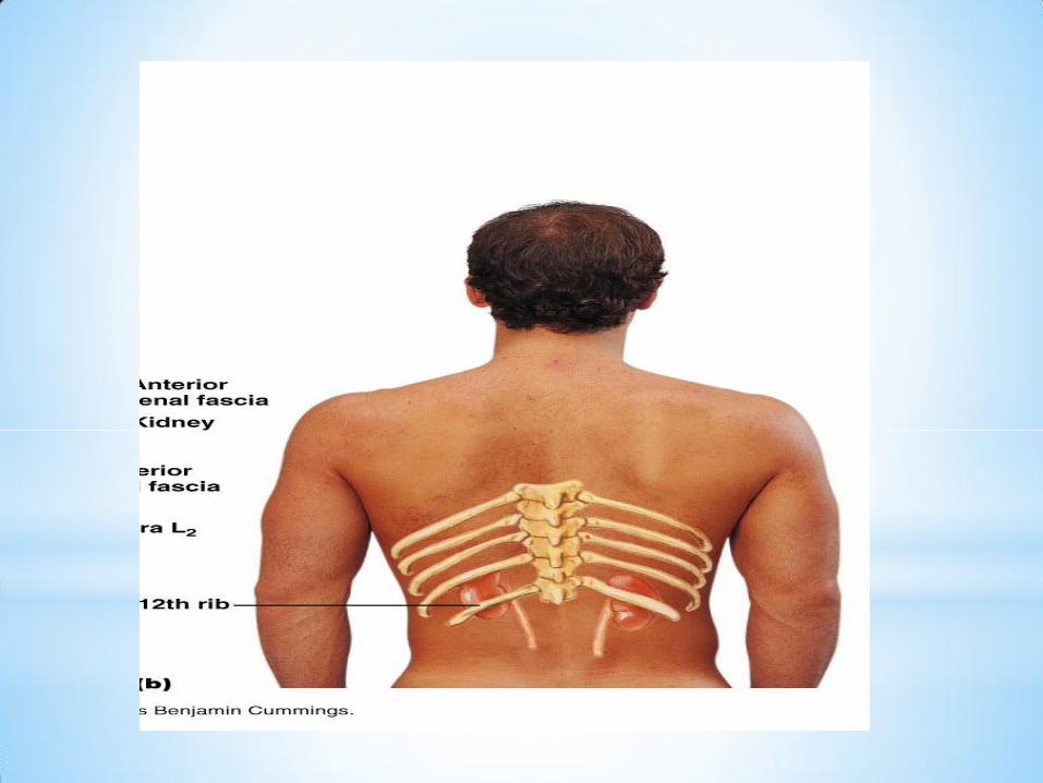
GROSS ANATOMY
Location
*Bean-shaped
*Retroperitoneal
*At level of T12–L1 vertebrae.
*The right kidney lies slightly inferior to left kidney.
Size, shape
*150 gm
*10x5x2.5 cm
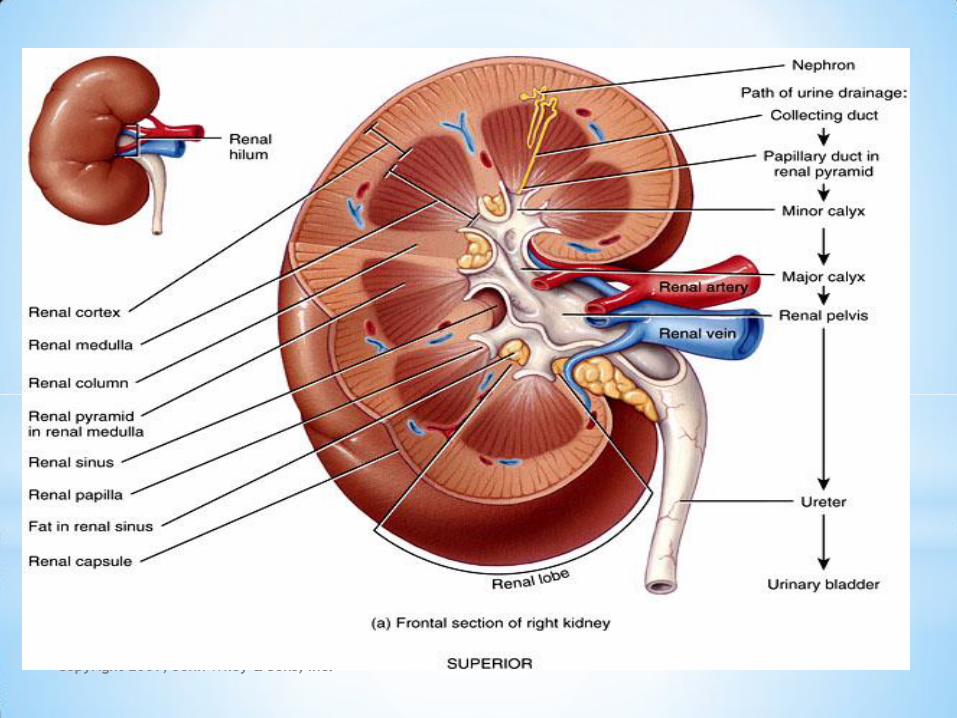
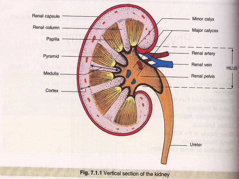
INTERNAL STRUCTURES
Medulla
Cortex
Copyright 2009, John Wiley & Sons, Inc.
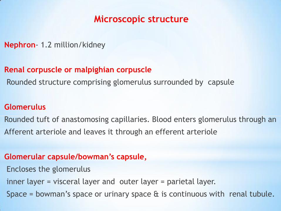
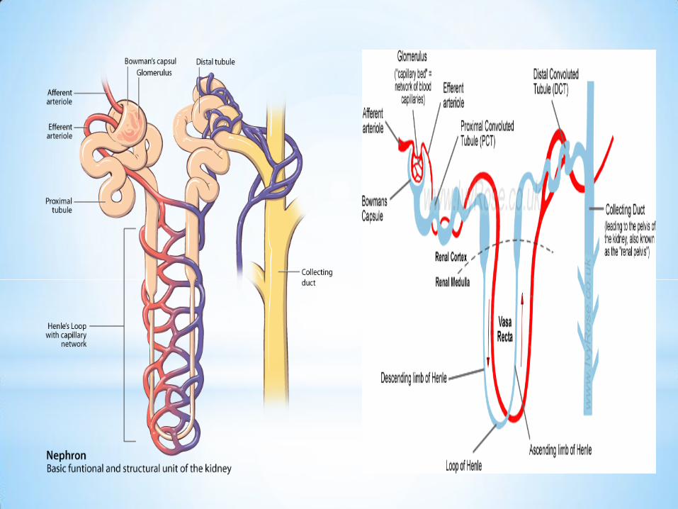
Microscopic structure
Nephron- 1.2 million/kidney
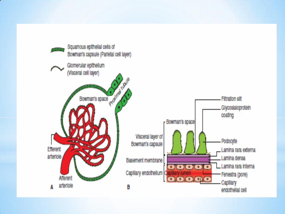
Renal corpuscle or malpighian corpuscle
Rounded structure comprising glomerulus surrounded by capsule
Glomerulus
Rounded tuft of anastomosing capillaries. Blood enters glomerulus through an
Afferent arteriole and leaves it through an efferent arteriole
Glomerular capsule/bowman’s capsule,
Encloses the glomerulus
inner layer = visceral layer and outer layer = parietal layer.
Space = bowman’s space or urinary space & is continuous with renal tubule.
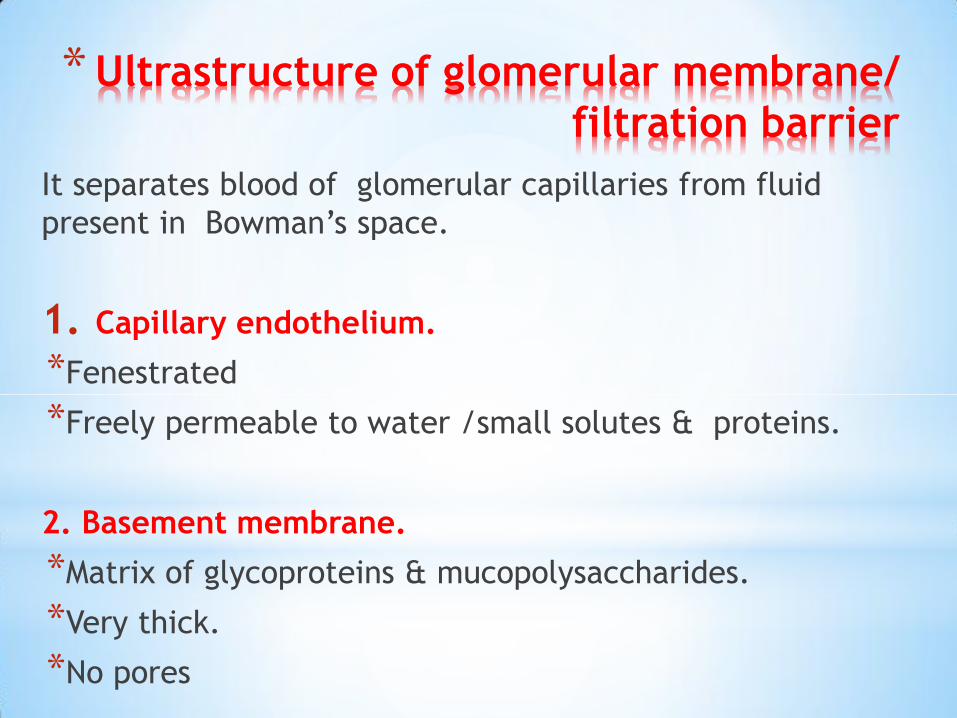
*Ultrastructure of glomerular membrane/
filtration barrier
It separates blood of glomerular capillaries from fluid
present in Bowman’s space.
1. Capillary endothelium.
*Fenestrated
*Freely permeable to water /small solutes & proteins.
2. Basement membrane.
*Matrix of glycoproteins & mucopolysaccharides.
*Very thick.
*No pores
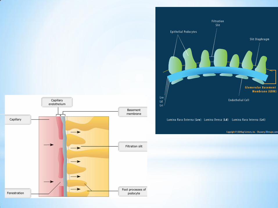
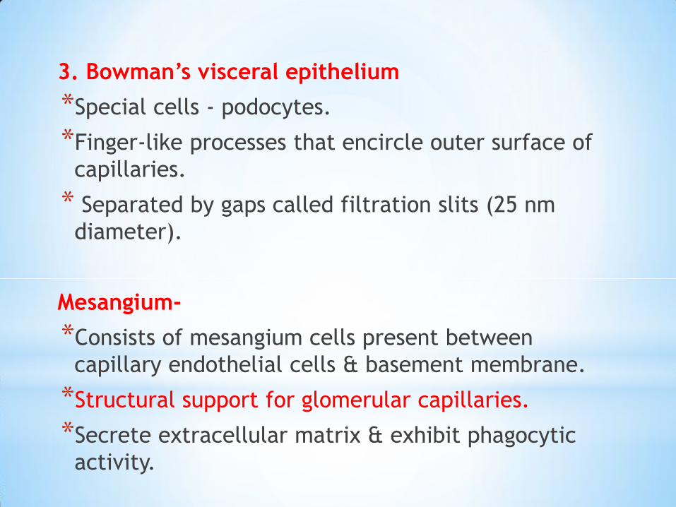
3. Bowman’s visceral epithelium
*Special cells - podocytes.
*Finger-like processes that encircle outer surface of
capillaries.
* Separated by gaps called filtration slits (25 nm
diameter).
Mesangium-
*Consists of mesangium cells present between
capillary endothelial cells & basement membrane.
*Structural support for glomerular capillaries.
*Secrete extracellular matrix & exhibit phagocytic
activity.
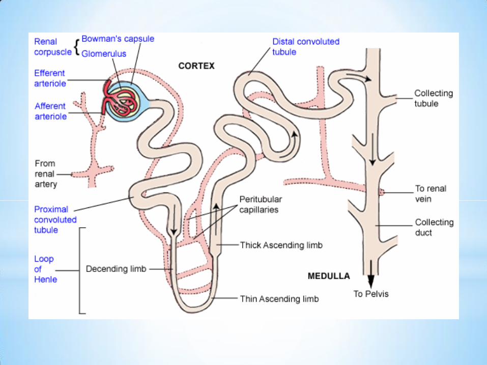
1. Proximal tubule.
Proximal convoluted tubule f/b proximal straight tubule /
pars recta
2. Intermediate tubule / loop of Henle
*Descending thin segment (DTS),
*Ascending thin segment (ATS),
*Thick ascending limb (TAL).
*In juxtamedullary nephrons - DTS joins ATS to form hair pin
band . ATS reaches up to junction of outer & inner medulla.
*In cortical nephrons- No ATS, DTS is continuous at bend of
loop with TAL. Near end of TAL, nephron passes between
its afferent & efferent arteriole. This short segment of the
TAL is called macula densa.
3. Distal convoluted tubule.
*It begins a short distance beyond macula densa and extends to a
point in cortex when connecting tubules of two or more
nephrons join to form cortical collecting ducts.
4. Collecting duct.
i. Cortical collecting duct ( portion in cortex)
ii. Outer medullary collecting duct (portion in outer medulla)
iii. Inner medullary collecting duct (IMCD)( portion
present in inner medulla)

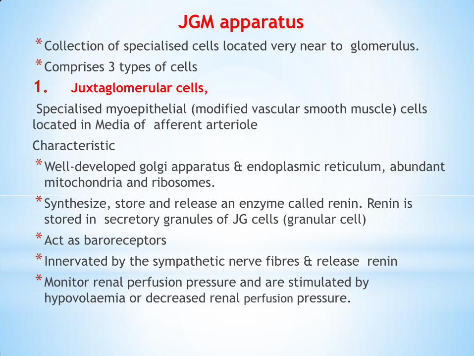
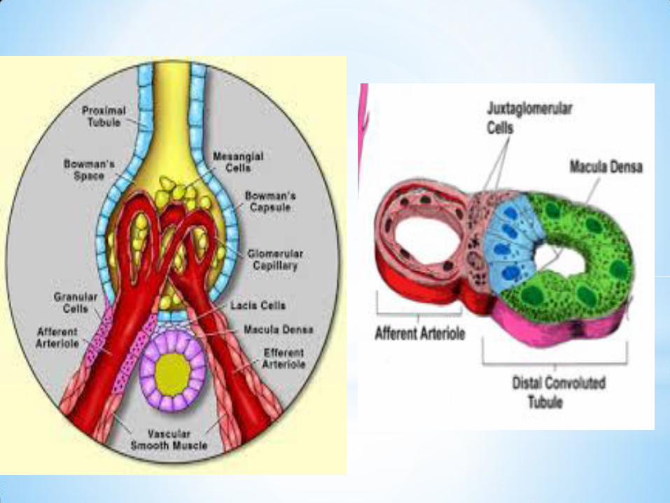
JGM apparatus
*Collection of specialised cells located very near to glomerulus.
*Comprises 3 types of cells
1. Juxtaglomerular cells,
Specialised myoepithelial (modified vascular smooth muscle) cells
located in Media of afferent arteriole
Characteristic
*Well-developed golgi apparatus & endoplasmic reticulum, abundant
mitochondria and ribosomes.
*Synthesize, store and release an enzyme called renin. Renin is
stored in secretory granules of JG cells (granular cell)
*Act as baroreceptors
* Innervated by the sympathetic nerve fibres & release renin
*Monitor renal perfusion pressure and are stimulated by
hypovolaemia or decreased renal perfusion pressure.


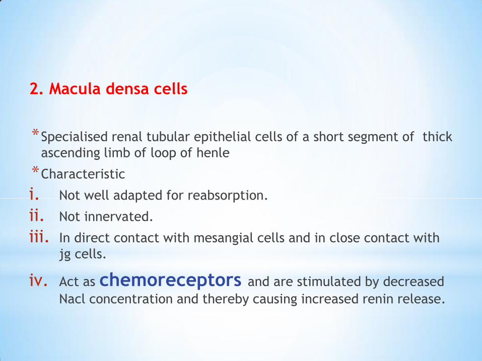
2. Macula densa cells
*Specialised renal tubular epithelial cells of a short segment of thick
ascending limb of loop of henle
*Characteristic
i. Not well adapted for reabsorption.
ii. Not innervated.
iii. In direct contact with mesangial cells and in close contact with
jg cells.
iv. Act as chemoreceptors and are stimulated by decreased
Nacl concentration and thereby causing increased renin release.
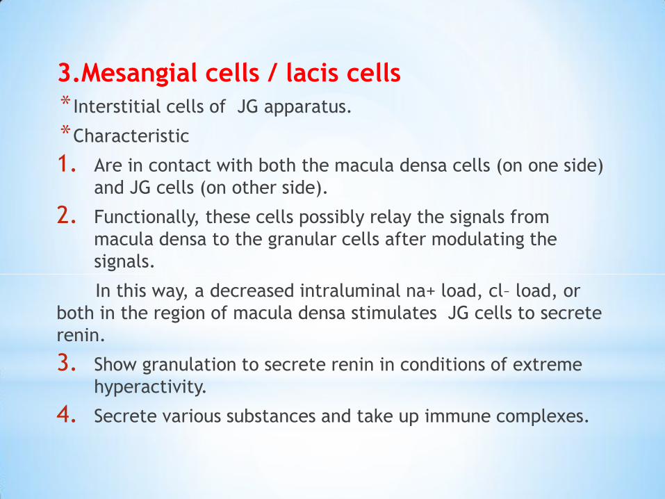
3.Mesangial cells / lacis cells
* Interstitial cells of JG apparatus.
*Characteristic
1. Are in contact with both the macula densa cells (on one side)
and JG cells (on other side).
2. Functionally, these cells possibly relay the signals from
macula densa to the granular cells after modulating the
signals.
In this way, a decreased intraluminal na+ load, cl– load, or
both in the region of macula densa stimulates JG cells to secrete
renin.
3. Show granulation to secrete renin in conditions of extreme
hyperactivity.
4. Secrete various substances and take up immune complexes.
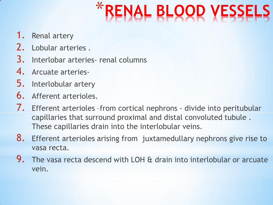
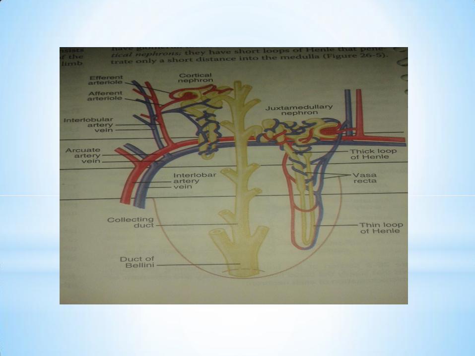
*RENAL BLOOD VESSELS
1. Renal artery
2. Lobular arteries .
3. Interlobar arteries- renal columns
4. Arcuate arteries-
5. Interlobular artery
6. Afferent arterioles.
7. Efferent arterioles –from cortical nephrons - divide into peritubular
capillaries that surround proximal and distal convoluted tubule .
These capillaries drain into the interlobular veins.
8. Efferent arterioles arising from juxtamedullary nephrons give rise to
vasa recta.
9. The vasa recta descend with LOH & drain into interlobular or arcuate
vein.
*
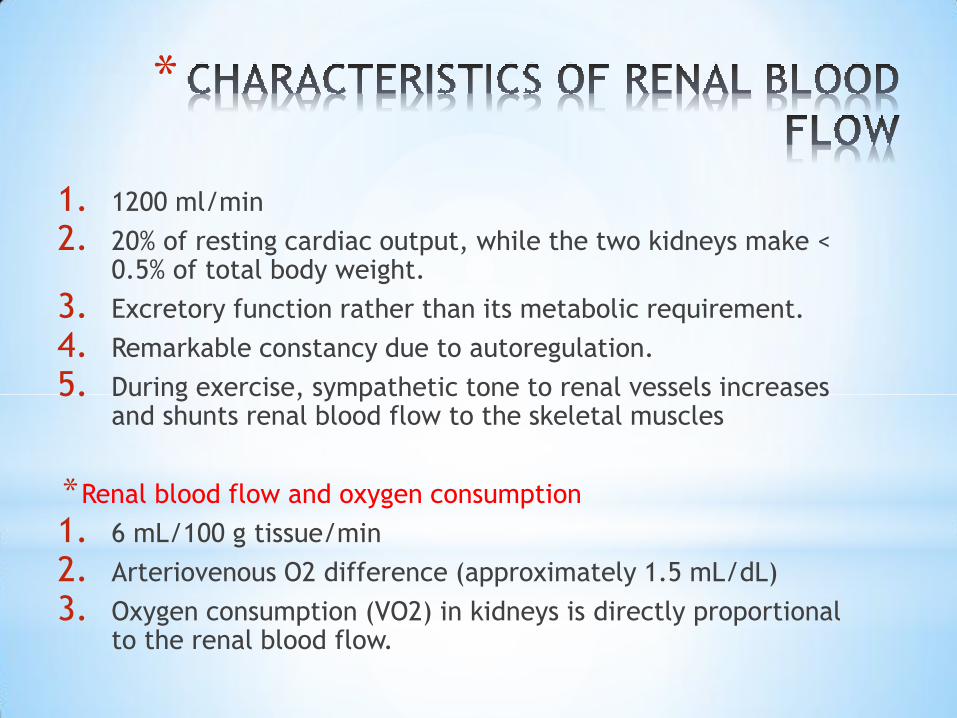
1. 1200 ml/min
2. 20% of resting cardiac output, while the two kidneys make < 0.5% of total body weight.
3. Excretory function rather than its metabolic requirement.
4. Remarkable constancy due to autoregulation.
5. During exercise, sympathetic tone to renal vessels increases and shunts renal blood flow to the skeletal muscles
*Renal blood flow and oxygen consumption
1. 6 mL/100 g tissue/min
2. Arteriovenous O2 difference (approximately 1.5 mL/dL)
3. Oxygen consumption (VO2) in kidneys is directly proportional to the renal blood flow.
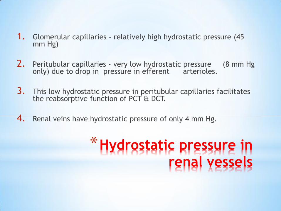
*Hydrostatic pressure in
renal vessels
1. Glomerular capillaries - relatively high hydrostatic pressure (45 mm Hg)
2. Peritubular capillaries - very low hydrostatic pressure (8 mm Hg only) due to drop in pressure in efferent arterioles.
3. This low hydrostatic pressure in peritubular capillaries facilitates the reabsorptive function of PCT & DCT.
4. Renal veins have hydrostatic pressure of only 4 mm Hg.
*
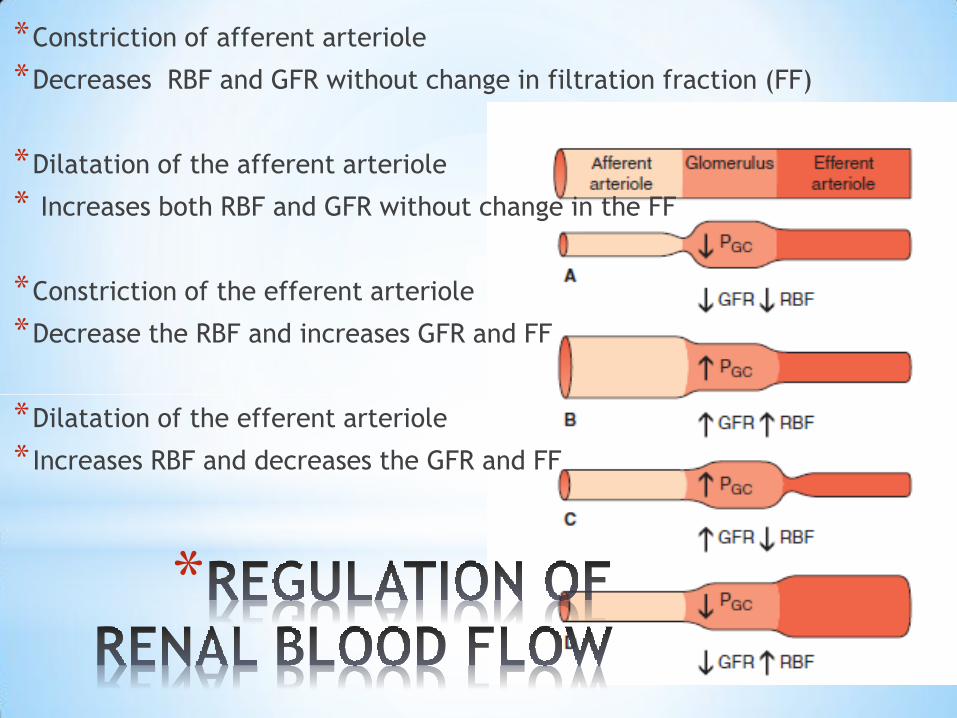
*Constriction of afferent arteriole
*Decreases RBF and GFR without change in filtration fraction (FF)
*Dilatation of the afferent arteriole
* Increases both RBF and GFR without change in the FF
*Constriction of the efferent arteriole
*Decrease the RBF and increases GFR and FF
*Dilatation of the efferent arteriole
* Increases RBF and decreases the GFR and FF
*REGULATION
OF RENAL
BLOOD FLOW

*
*The RBF and thus the GFR remain constant over a wide range of
renal arterial pressures (80–200 mm Hg)
Mechanisms of autoregulation
1. That responds to changes in arterial pressure (Myogenic
mechanism)
*When renal arterial pressure is raised,
*Afferent arterioles are stretched,
* It contract and increase the vascular resistance.
*The increased vascular resistance which offsets the effect of
increased arterial pressure
*Thereby maintains a constant
RBF and GFR
*
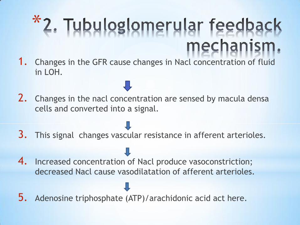
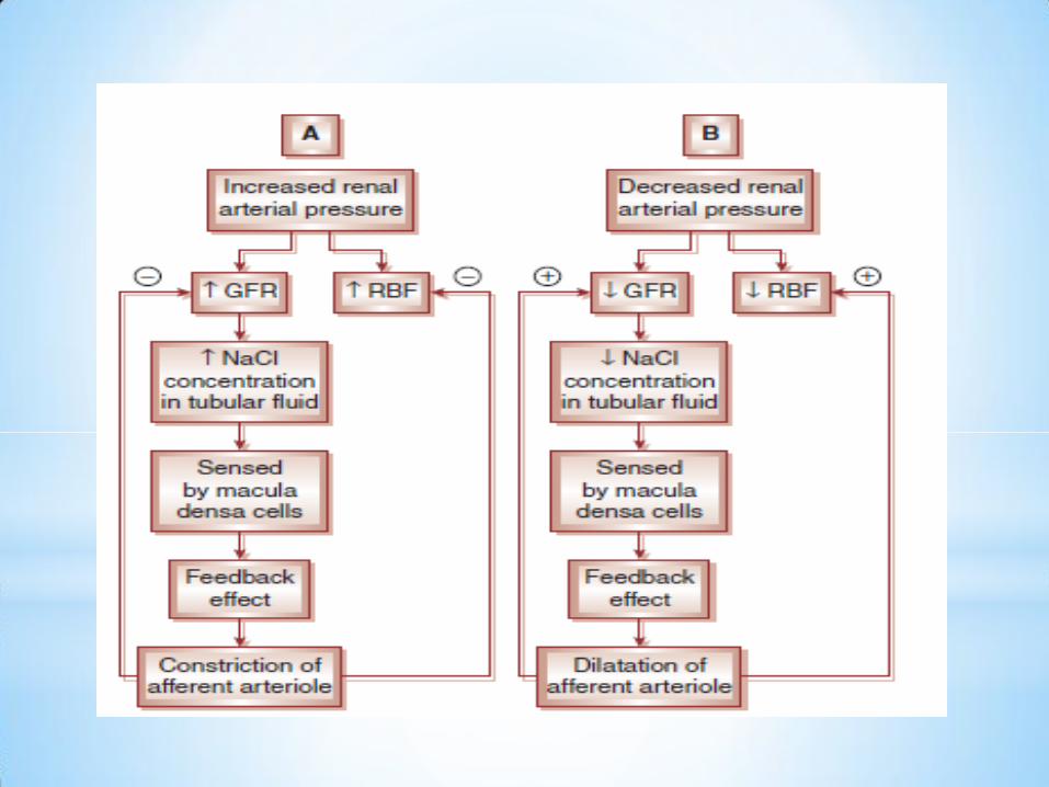
1. Changes in the GFR cause changes in Nacl concentration of fluid
in LOH.
2. Changes in the nacl concentration are sensed by macula densa
cells and converted into a signal.
3. This signal changes vascular resistance in afferent arterioles.
4. Increased concentration of Nacl produce vasoconstriction;
decreased Nacl cause vasodilatation of afferent arterioles.
5. Adenosine triphosphate (ATP)/arachidonic acid act here.

*
*A small change in GFR has great effect on urinary output and therefore on loss of solutes and water.
*Autoregulation of RBF and GFR is an effective mechanism for uncoupling renal function from fluctuations in the arterial pressure and maintain fluid and electrolyte balance.
*Autoregulation of RBF and GFR is virtually absent at mean arterial blood pressure below 80 mm Hg,
*Autoregulation is not a perfect mechanism
*Several hormones and other factors can change RBF and GFR, despite autoregulation mechanisms.
*
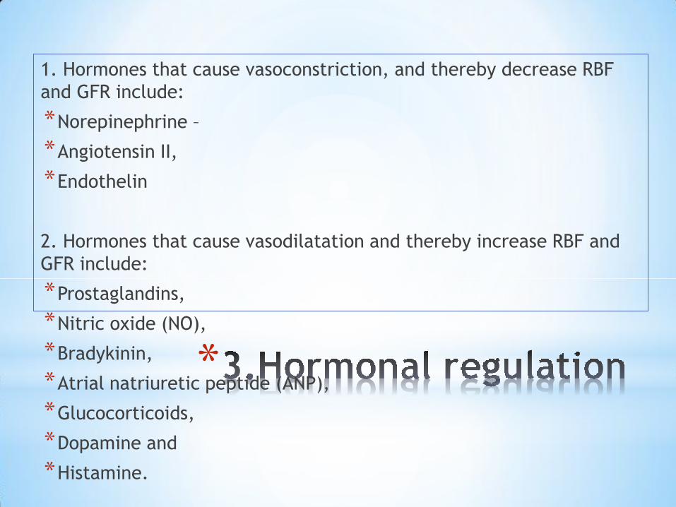
1. Hormones that cause vasoconstriction, and thereby decrease RBF
and GFR include:
*Norepinephrine –
*Angiotensin II,
*Endothelin
2. Hormones that cause vasodilatation and thereby increase RBF and
GFR include:
*Prostaglandins,
*Nitric oxide (NO),
*Bradykinin,
*Atrial natriuretic peptide (ANP),
*Glucocorticoids,
*Dopamine and
*Histamine.
*
*Under normal circulatory conditions, sympathetic tone is minimum.
*Mild-to-moderate stimulation of sympathetic nerves usually has mild effects on RBF because of autoregulation mechanism.
*Strong acute stimulation of sympathetic nerves may produce marked fall in RBF (even to 10−30% of normal) temporarily due to constriction of both afferent and efferent arterioles.
*Mainly by α1-adrenergic receptors and to a lesser extent by post-synaptic α2-adrenergic receptors.
*
*LONG QUESTION
1. Regulation of renal bood flow
*SHORT NOTE
1. Glomerulus & its membrane
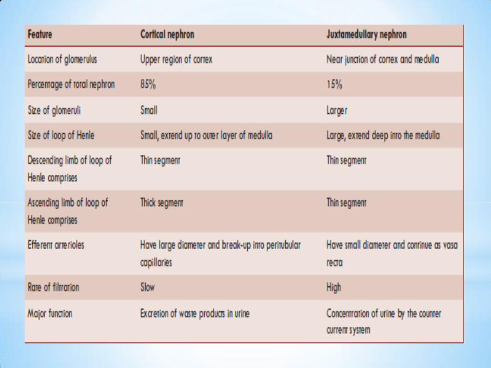
2. Difference between 2 types of nephron
3. Juxtra glomurular apparatus
4. Tubuloglomurular feedback mechanism
5. Regulation of GFR/RBF (LONG QUESTION)