renal histology laith srour - medicinebau.com · remember that the renal corpuscle both indicates...
TRANSCRIPT

RENAL HISTOLOGY
mohammad
Sticky Note
laith srour

me
Highlight
REnal capsule
me
Highlight
medulla
me
Highlight
cortex
me
Highlight
hilum
me
Highlight
ureter
me
Highlight
papila

me
Text Box
cortex
me
Text Box
outer medulla
me
Text Box
inner medulla

me
Text Box
Renal capsule

me
Sticky Note
Remember that the renal corpuscle both indicates the cortex as well as the cortical labyrinth (E) within the cortex. In addition, the cortical labyrinth contains both the proximal and distal convoluted tubules. The medullary rays (F), which appear as rays radiating from the medulla, contain the thick ascending and descending segments of the renal loop and collecting tubules
me
Highlight
cortical labrynth
me
Highlight
medullary rays

me
Highlight
renal corpuscle
me
Highlight
urinary space

me
Highlight
Thick descending
me
Highlight
Thick ascending

me
Highlight
Thick descending
me
Highlight
thick ascending
me
Highlight
Thin

me
Highlight
thin
me
Highlight
collecting tubule

me
Highlight
Papillary duct
me
Highlight
ureter
me
Highlight
Renal pelvis
me
Text Box
lined bu transitional wpithelium\Urothelium
me
Pencil

me
Text Box
A coronal view (left) shows the major blood vessels . An expanded diagram (right) includes the microvascular components extending into the cortex, and medulla from the interlobular vessels are shown on the right. Pink boxes indicate vessels with arterial blood and light blue indicate the venous return. The intervening lavender boxes and vessels are intermediate sites where most reabsorbed material reenters the blood.

Renal Corpuscle
me
Sticky Note
At the beginning of each nephron is a renal corpuscle, about 200 μm in diameter and containing a tuft of glomerular capillaries, surrounded by a double-walled epithelial capsule called

Lining of BC
Parietal – Simple
squamous Visceral-
Podocytes

me
Sticky Note
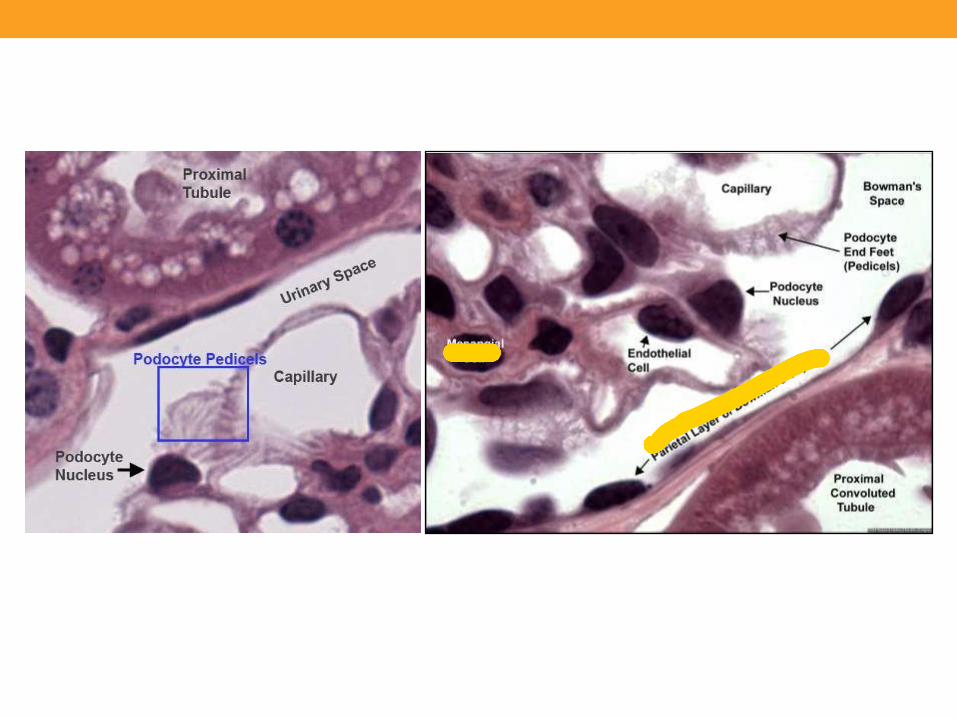
(a) The renal corpuscle is a small mass of capillaries called the glomerulus housed within a bulbous glomerular capsule. The internal lining of the capsule is composed of complex epithelial cells called podocytes, which cover each capillary, forming slit-like spaces between interdigitating processes called pedicels. Blood enters and leaves the glomerulus through the afferent and efferent arterioles, respectively. (c) Filtrate is produced in the corpuscle when blood plasma is forced under pressure through the capillary fenestrations, across the filtration membrane or GBM surrounding the capillary, and through the filtration slit diaphragms located between the podocyte pedicels. (d) The scanning electron microscopy (SEM) shows the distinctive appearance of podocytes and their pedicel processes that cover glomerular capillaries. X800
me
Sticky Note
b) The micrograph shows the major histologic features of a renal corpuscle. The glomerulus (G) of capillaries is surrounded by the capsular space (CS) covered by the simple squamous parietal layer (PL) of Bowman capsule. Near the corpuscle is that nephron’s macula densa (MD) and sections of proximal convoluted tubules (PCT) and distal convoluted tubules (DCT). H&E. X300.
me
Text Box
imp.
me
Highlight
Simple squamous parietal layer
me
Highlight
Proximal convolted tubule ,Look how its thicker than DCT
me
Highlight
Macula densa near Distal convoluted tubule (DCT)\\ The cells not the tube-like thing

me
Text Box
podocyte cell body
me
Text Box
Basement membrane of capillary
me
Text Box
endothelium
me
Text Box
pedicles

me
Highlight
me
Highlight

me
Sticky Note
The glomerular filtration barrier consists of three layered components: the fenestrated capillary endothelium, the glomerular basement membrane (GBM), and filtration slit diaphragms between pedicels. The major component of the filter is formed by fusion of the basal laminae of a podocyte and a capillary endothelial cell. (a) TEM shows cell bodies of two podocytes (PC) and the series of pedicels on the capillary (C) basement membrane separated by the filtration slit diaphragms. Around the capillaries and podocytes is the capsular space (CS) into which the filtrate enters. The enclosed area is shown in part b. X10,000. (b) At higher magnification, both the fenestrations (F) in the endothelium (E) of the capillary (C) and the filtration slits (FS) separating the pedicels (P) are clearly seen on the two sides of the thick, fused basement membrane (BM). Thin slit diaphragms (SD) bridge the slits between pedicels. X45,750.

Mesangial Cells
me
Highlight
Simple squamous
me
Highlight

Proximal Convoluted Tubule
me
Highlight
me
Highlight
me
Highlight
me
Highlight
me
Text Box
covered with simple cuboidal
me
Sticky Note
(a) The micrograph shows the continuity at a renal corpuscle’s tubular pole (TP) between the simple cuboidal epithelium of a proximal convoluted tubule (P) and the simple squamous epithelium of the capsule’s parietal layer. The urinary space (U) between the parietal layer and the glomerulus (G) drains into the lumen of the proximal tubule. The lumens of the proximal tubules appear filled, because of the long microvilli of the brush border and aggregates of small plasma proteins bound to this structure. By contrast, the lumens of distal convoluted tubules (D) appear empty, lacking a brush border and protein. (b) Here the abundant peritubular capillaries and draining venules (arrows) that surround the proximal (P) and distal (D) convoluted tubules are clearly seen. Both X400. H&E.
me
Text Box
So look for torturus simple cuboidal cells making a tube\\ Also it contains some stuff unlike distal which is clear\\ and they are abundant
me
Highlight
me
Pencil
me
Text Box
This is still parietal layer so it is simple squamous
me
Text Box
this is proximal tubule with simple cuboidal

Loop of Henle
me
Sticky Note
(d) A cross section through a medullary pyramid shows the simple squamous epithelium of the thin descending and ascending limbs of loops of Henle (T) and its thick ascending limbs (A), as well as the pale columnar cells of collecting ducts (CD). Note also the homogeneous interstitium with capillaries smaller than the thin limbs. X160. Mallory trichrome.
me
Pencil
me
Text Box
thick ascending limb\\simple cuboidal
me
Pencil
me
Text Box
Thin ascending & descending\\simple squamous
me
Pencil
me
Text Box
collecting ducts\\columnar
me
Pencil
me
Text Box
capillaries smaller than Thin limb\\one nucleus(be carefull because in thin limb nucleus is not always seen good
me
Text Box
cd
me
Text Box
a
me
Text Box
t


DCT
Distal Convoluted Tubule
me
Pencil
me
Text Box
PCT
me
Highlight
me
Highlight
me
Highlight
me
Pencil
me
Text Box
Peritubular capillaries and drainage venules filed with blood
me
Highlight
me
Pencil
me
Text Box
DCT


Juxtaglomerular Apparatus
me
Sticky Note
The JGA forms at the point of contact between a nephron’s distal tubule (D) and the vascular pole of its glomerulus (G). At that point cells of the distal tubule become columnar as a thickened region called the macula densa (MD). Smooth muscle cells of the afferent arteriole’s (AA) tunica media are converted from a contractile to a secretory morphology as juxtaglomerular granule cells (JG). Also present are lacis cells (L), which are extraglomerular mesangial cells adjacent to the macula densa, the afferent arteriole, and the efferent arteriole (EA). In this specimen the lumens of proximal tubules (P) appear filled and the urinary space (US) is somewhat swollen.
me
Pencil
me
Text Box
Macula densa\\columnar

Collecting Tubule

me
Highlight
Vasa recta
me
Sticky Note
Pale-staining columnar principal cells, in which ADH-regulated aquaporins of the cell membrane allow more water reabsorption, are clearly seen in these transversely sectioned collecting ducts (CD), surrounded by interstitium with vasa recta (VR)

Ureter
me
Sticky Note
(a) Diagram of a ureter in cross section shows a characteristic pattern of longitudinally folded mucosa, surrounded by a thick muscularis that moves urine by regular waves of peristalsis. The lamina propria is lined by a unique stratified epithelium called transitional epithelium or urothelium that is resistant to the potentially deleterious effects of contact with hypertonic urine. (b) Histologically the muscularis (Mu) is much thicker than the mucosa (M) and adventitia (A). X18. H&E.
me
Text Box
Mucosa lined with urothellium

Ureter
me
Text Box
mucosa
me
Text Box
muscularis
me
Text Box
adventetia

Urinary
Bladder
Empty Full
me
Sticky Note
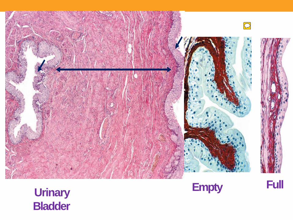
**When the bladder is empty, the mucosa is highly folded and the urothelium (U) has bulbous umbrella cells. ** When the bladder is full, the mucosa is pulled smooth, the urothelium (U) is thinner, and the umbrella cells are flatter.

me
Text Box
prostatic urethra Lined by urothelium
me
Highlight

me
Highlight
or spongy\\The spongy urethra, about 15 cm in length, is enclosed within erectile tissue of the penis (see Chapter 21) and is lined by stratified columnar and pseudostratified columnar epithelium (Figure 19–18), with stratified squamous epithelium distally
me
Highlight

Urethra
me
Sticky Note
The urethra is a fibromuscular tube that carries urine from the bladder to the exterior of the body. (a) A transverse section shows that the mucosa has large longitudinal folds around the lumen (L). X50. H&E. (b) A higher magnification of the enclosed area shows the unusual stratified columnar nature of the urethral epithelium (E). This thick epithelial lining varies between stratified columnar in some areas and pseudostratified columnar elsewhere, but it becomes stratified squamous at the distal end of the urethra

Thank you