the placenta, an afterthought no longer science news blog 20181210.docx · web viewa team of...
TRANSCRIPT

1 5/14/23 Name Student number
https://nyti.ms/2Qyynk3The Placenta, an Afterthought No Longer
An ephemeral organ, long dismissed merely as afterbirth, increasingly is viewed as critical to understanding the health and
course of pregnancy.By Apoorva Mandavilli
The placenta may be dismissed as “afterbirth,” deemed an afterthought in discussions about pregnancy and even relegated, literally, to the trash bin. But at long last it is beginning to get its due.In the past three weeks, scientists have published three significant studies of this ephemeral organ. One gave a detailed analysis of all the genes expressed, or converted into functioning proteins, in the placenta; another experimented with a way to silence that expression when it causes trouble. In the third, researchers created mini-placentas, three-dimensional clusters of cells, or organoids, that mimic the real thing in the lab, and can be used as models for studying it.
The placenta forms when cells from a fertilized egg secure a beachhead in the uterine lining. Ninety percent of the placenta is made up of cells not from
the mother but from the fetus. David M. Phillips/Science SourceIn addition, at a recent meeting in Bethesda, Md., of the Human Placenta Project, several teams of researchers showed off sophisticated new techniques that enable the placenta to be studied in real time. That work could help doctors diagnose dangerous complications in pregnancy — including pre-eclampsia (a form of high blood pressure), preterm birth and fetal growth restriction — early enough to intervene. It might also help to reveal why boys are
much more vulnerable than girls to disorders of brain development, including schizophrenia, A.D.H.D., autism, dyslexia and Tourette syndrome.“The missing link between complications during pregnancy and development of the fetal brain has been hiding in plain sight for a long time,” said Dr. Daniel R. Weinberger, director of the Lieber Institute for Brain Development in Baltimore, Md. “It’s the placenta.”An embryonic invasionDuring the course of human pregnancy, the placenta grows from a few cells into an organ weighing more than a pound. It often is compared to an aggressive cancer. But a more apt metaphor might be a military invasion, as 90 percent of the placenta is made up of cells not from the mother but from the fetus.Early in gestation, the fertilized egg implants itself in the mother’s uterine lining and sends out a few cells to breach it. These foot soldiers produce proteins that disarm the mother’s defenses, destroy the smooth muscles that line her blood vessels and dilate and redirect the vessels to feed the embryo. As the placental beachhead grows, its cells specialize to do the work of heart, lungs, liver and kidneys until the fetus can fend for itself. Groups of cells exchange oxygen for carbon dioxide; provide nutrients and hormones; protect the fetus from harmful stress, germs and chemicals; and remove waste.This incursion fails as often as 20 percent of the time, and when it does, it can cause severe complications for the fetus, at birth and afterward. It may also forecast trouble for the mother’s health later in life: pre-eclampsia can portend heart disease and stroke, and gestational diabetes can signal later obesity and metabolic disease.“There is nothing in medicine that can return so much on an investment as a healthy pregnancy and delivery, because that has years and years of impact later,” said Dr. George R. Saade, chief of

2 5/14/23 Name Student number
obstetrics at the University of Texas Medical Branch. “And placental health is critical to the health of a pregnancy.”
It’s not yet clear what makes female fetuses more resilient. But during the first trimester, 58 genes are expressed differently in male fetuses than in females, according to an analysis published in January, in the journal Biology of Sex Differences. Several of these genes are on the X chromosome. A female fetus has two X chromosomes and two copies of these genes, with one copy typically staying silent. But the analysis showed that many of these gene copies are activated regardless, and so they become a larger factor in female placentas than in males. (The more detailed analysis of gene expression published three weeks ago did not look at sex differences, but provides a framework for similar analyses.)In May, Dr. Weinberger’s team at the Lieber Institute looked specifically at genes implicated in schizophrenia. They found that many of these genes are abundantly expressed in the fetal placenta, and are activated at even higher levels when the pregnancy is under stress; the effect is more dramatic in male fetuses than in females.“We suggested that placentas of male fetuses seem to be more susceptible at a genetic level,” Dr. Weinberger said. “I’m very confident the same story is going to be there for autism, A.D.H.D. and other developmental behavioral problems.”Womb with a viewTechnological limitations have obscured the central role that the placenta plays in the health of both baby and mother. The placenta is a dynamic organ, but it usually has been studied by dissecting it after delivery.“That’s too late,” said Dr. Saade of the University of Texas. “It’s like studying cardiac disease or any other medical condition just by doing an autopsy.”
Problems with the placenta often begin in the spiral arteries of the mother — the arteries that the fetus commandeers to feed itself. If they are blocked or too narrow, the fetus may not get enough oxygen and nutrients, and the mother’s blood pressure may spike toward pre-eclampsia. This can begin as early as the first trimester, but few tools are available to diagnose it at that stage.“The tests that are available today are all designed for the third trimester, and that’s way too late,” said Dr. Alfred Z. Abuhamad, chair of the department of obstetrics and gynecology at Eastern Virginia Medical School in Norfolk, Va. In 2014, the child-health division of the National Institutes of Health set out to find noninvasive methods to identify complications earlier. An infusion of $80 million into placenta research prompted Dr. Abuhamad and others to adapt technologies used in other fields, and has already provided valuable insights into early pregnancy.Some scientists are betting on magnetic resonance imaging scans, or M.R.I.s, as the most sensitive detectors of placental problems. They are using a method that measures oxygen levels in the blood; it is quick and, so far, seems to catch problems as early as the second trimester. Several teams worldwide are evaluating the technique, each in hundreds of women.But M.R.I. is not widely in use in obstetricians’ offices, Dr. Abuhamad said, in contrast with ultrasound machines, which would be a more practical option. Traditional ultrasounds can show the structure and location of the placenta, not how well the organ is functioning. But advances over the past five years have sharpened the machine’s focus. One enables the device to detect tiny blood vessels; another, called elastography, was developed to examine the liver, and can help measure the density of placental tissue.Dr. Abuhamad’s team is using these advances in ultrasound to chart placental health in about 500 pregnant women, including 300 at

3 5/14/23 Name Student number
high risk of complications. They are collecting ultrasound data and blood samples from the women at eight time points during pregnancy to see which early features track with problems later on.Other teams are trying to identify particles the placenta may release into the bloodstream because that could lead to a simple blood test for diagnosing problems. And one group of researchers is developing an oximeter, a device that quantifies the light reflected back through layers of fat as a measure of blood oxygen.It will be at least five years before any of these tests makes their way to doctors’ offices. But when they are ready, they are likely to have a huge impact on obstetric practice, said Dr. Diana W. Bianchi, director of the National Institute of Child Health and Human Development.“The way that prenatal care is currently structured, you hardly see your obstetrician in the first trimester,” she said. “And by the time you get to the third trimester, you’re seeing the obstetrician weekly.” Instead, when placental screening identified a problem, women might be encouraged to see their doctors frequently through the first trimester. “Knowing that this starts early in the first trimester,” said Dr. Abuhamad, “could we then intervene in the first trimester — identify early, intervene early and prevent the complications?”
http://bit.ly/2QIpynAA sobering conclusion: Adult hearts contain no stem
cellsMost tissues of animals and humans contain stem cells
During a myocardial infarction, commonly known as a heart attack, the blood supply to part of the heart muscle is cut off. As a consequence, part of the heart muscle dies. Because the heart is a pump that maintains the blood circulation through our vessels, this is obviously a life-threatening situation. Most tissues of animals and humans contain stem cells that come to the rescue upon tissue
damage: they rapidly produce large numbers of 'daughter cells' in order to replace lost tissue cells.For two decades researchers and clinicians have searched for cardiac stem cells, stem cells that should reside in the heart muscle and that could repair the heart muscle after a myocardial infarction. Multiple research groups have claimed the definitive identification of cardiac stem cells, yet none of these claims have held up. See for instance the following recent press release: "US governments halts heart stem-cell study". The existence of cardiac stem cells and their significance for adult hearts remains therefore heavily debated.To solve this debate, researchers from the Hubrecht Institute in Utrecht, the Amsterdam University Medical Center, the École Normale Supérieure (ENS) de Lyon and the Francis Crick Institute London, led by Hans Clevers, focused on the broadest and most direct definition of stem cell function in the mouse heart: the ability of a cell to replace lost tissue by cell division. In the heart, this means that any cell that can produce new heart muscle cells after a heart attack would be termed a cardiac stem cell. The authors generated a 'cell-by-cell' map of all dividing cardiac cells before and after a myocardial infarction using advanced molecular and genetic technologies.The study establishes that many types of cells divide upon damage of the heart, but that none of these are capable of generating new heart muscle. In fact, many of the 'false leads' of past studies can now be explained: cells that were previously named cardiac stem cells now turn out to produce blood vessels or immune cells, but never heart muscle. Thus, the sobering conclusion is drawn that heart stem cells do not exist. In other words, heart muscle that is lost due to a heart attack cannot be replaced. This finding -while disappointing- settles a long-standing controversy.The authors make a second important observation. Connective tissue cells (also known as fibroblasts) that are intermingled with

4 5/14/23 Name Student number
heart muscle cells respond vigorously to a myocardial infarction by undergoing multiple cell divisions. In doing so, they produce scar tissue that replaces the lost cardiac muscle. While this scar tissue contains no muscle and thus does not contribute to the pump function of the heart, the fibrotic scar 'holds together' the infarcted area. Indeed, when the formation of the scar tissue is blocked, the mice succumb to acute cardiac rupture. Thus, while scar formation is generally seen as a negative outcome of myocardial infarction, the authors stress the importance of the formation of scar tissue for maintaining the integrity of the heart.Publication: Profiling proliferative cells and their progeny in damaged murine heartsKai Kretzschmar*, Yorick Post* (* = equal contribution), Marie Bannier-Hélaouët, Andrea Mattiotti, Jarno Drost, Onur Basak, Vivian S. W. Li, Maaike van den Born, Quinn D. Gunst, Danielle Versteeg, Lieneke Kooijman, Stefan van der Elst, Johan H. van Es, Eva van Rooij, Maurice J. B. van den Hoff, and Hans Clevers (2018).Proceedings of the National Academy of Sciences (PNAS)Prof. dr. Hans Clevers is group leader at the Hubrecht Institute, professor of Molecular Genetics at the University Medical Center Utrecht and Utrecht University and Oncode Investigator.
http://bit.ly/2zJQ5auEssential oils from garlic and other herbs kill 'persister'
Lyme disease bacteriaLaboratory study hints that plant compounds may be better than
current antibiotics at treating persistent Lyme bacteria and associated symptoms
Oils from garlic and several other common herbs and medicinal plants show strong activity against the bacterium that causes Lyme disease, according to a study by researchers at Johns Hopkins Bloomberg School of Public Health. These oils may be especially useful in alleviating Lyme symptoms that persist despite standard antibiotic treatment, the study also suggests.The study, published October 16 in the journal Antibiotics , included lab-dish tests of 35 essential oils--oils that are pressed from plants or their fruits and contain the plant's main fragrance, or "essence."
The Bloomberg School researchers found that 10 of these, including oils from garlic cloves, myrrh trees, thyme leaves, cinnamon bark, allspice berries and cumin seeds, showed strong killing activity against dormant and slow-growing "persister" forms of the Lyme disease bacterium."We found that these essential oils were even better at killing the 'persister' forms of Lyme bacteria than standard Lyme antibiotics," says study senior author Ying Zhang, MD, PhD, professor in the Department of Molecular Microbiology and Immunology at the Bloomberg School.There are an estimated 300,000 new cases of Lyme disease each year in the United States. Standard treatment with doxycycline or an alternative antibiotic for a few weeks usually clears the infection and resolves symptoms. However, about 10 to 20 percent of patients report persistent symptoms including fatigue and joint pain--often termed "persistent Lyme infection" or "post-treatment Lyme disease syndrome" (PTLDS) that in some cases can last for months or years.The cause of this lingering syndrome isn't known. But it is known that cultures of Lyme disease bacteria, Borrelia burgdorferi, can enter a so-called stationary phase in which many of the cells divide slowly or not at all. The slow-dividing or dormant cells are "persister" cells, which can form naturally under nutrient starvation or stress conditions, and are more resistant to antibiotics. Some researchers have sought other drugs or medicinal compounds that can kill persister Lyme bacteria in the hope that these compounds can be used to treat people with persistent Lyme symptoms.Zhang and his laboratory have been at the forefront of these efforts. In 2014, his lab screened FDA-approved drugs for activity against persister Lyme bacteria and found many candidates including daptomycin (used to treat MRSA) that had better activity than the current Lyme antibiotics. In 2015, they reported that a three-

5 5/14/23 Name Student number
antibiotic combination--doxycycline, cefoperazone and daptomycin--reliably killed Lyme persister bacteria in lab dish tests. In a 2017 study they found that essential oils from oregano, cinnamon bark, clove buds, citronella and wintergreen killed stationary phase Lyme bacteria even more potently than daptomycin, the champion among tested pharmaceuticals.In the new study Zhang and his team extended their lab-dish testing to include 35 other essential oils, and found 10 that show significant killing activity against stationary phase Lyme bacteria cultures at concentrations of just one part per thousand. At this concentration, five of these oils, derived respectively from garlic bulbs, allspice berries, myrrh trees, spiked ginger lily blossoms and may change fruit successfully killed all stationary phase Lyme bacteria in their culture dishes in seven days, so no bacteria grew back in 21 days.Oils from thyme leaves, cumin seeds and amyris wood also performed well, as did cinnamaldehyde, the fragrant main ingredient of cinnamon bark oil.Lab-dish tests such as these represent an early stage of research, but Zhang and colleagues hope in the near future to continue their investigations of essential oils with tests in live animals, including tests in mouse models of persistent Lyme infection. If those tests go well and the effective doses seem safe, Zhang expects to organize initial tests in humans."At this stage these essential oils look very promising as candidate treatments for persistent Lyme infection, but ultimately we need properly designed clinical trials," he says."Identification of Essential Oils with Strong Activity against Stationary Phase Borrelia burgdorferi" was written by Jie Feng, Wanliang Shi, Judith Miklossy, Genevieve M. Tauxe, Conor J. McMeniman, and Ying Zhang.Support for the research came from the Global Lyme Alliance, LivLyme Foundation, NatCapLyme, and the Einstein-Sim Family Charitable Fund.
http://bit.ly/2QBoFNS
Sculptor Unknowingly Poisons Herself with Her Own Art
When a sculptor in Toronto started feeling ill in 2013, she had no idea that her art was the reason why.
By Sara G. Miller, Health EditorThe sculptor, Gillian Genser, had been using blue mussel shells in her sculptures for the past 15 years, and, as a result, unknowingly poisoning herself.The culprit? Heavy metals, including arsenic and lead, found in the mussel shells. In a moving personal essay published Nov. 28 in Toronto Life, Genser described the onset of her symptoms — which began with agitation, headaches and vomiting, and later progressed to symptoms such as hearing loss in one ear and short-term memory problems. It took two-year- for doctors to nail down the diagnosis of heavy metal poisoning.
Credit: ShutterstockIndeed, Genser wrote, it wasn't until she visited the Royal Ontario Museum in Toronto and spoke with a curator of invertebrates that she put the pieces together. The curator told Genser that toxins can build up in the shells of invertebrates, leading her to research blue mussels. As Live Science has previously reported, chemicals accumulate in mussels as they filter feed, making them good barometers for water pollutants.Genser wrote that, in her case, the mussels she had been working with likely came from water contaminated with industrial waste. After 15 years of working with the mussel shells, she had built up high levels of arsenic and lead in her blood. She will "never fully recover" and continues to live with many symptoms, she wrote. However, she went on to complete her mussel-sculpture, a

6 5/14/23 Name Student number
depiction of the biblical Adam, in 2015. She calls him her "beautiful death."Heavy-metal poisoning occurs when too much of a heavy metal accumulates in a person's body, according to National Organization for Rare Disorders (NORD). At high levels, heavy metals can replace certain minerals in a variety of processes in the body, which can have deleterious effects.The symptoms of heavy-metal poisoning can vary, depending on the metals involved, according to NORD. Both arsenic and lead poisoning cause a wide range of symptoms, and can be life-threatening. Arsenic poisoning, for example, can cause symptoms such as headaches, drowsiness, confusion and seizures, as well as intestinal problems. Lead poisoning in adults can cause symptoms such as high blood pressure, muscle weakness and nerve pain.
http://bit.ly/2RGAZcVResearchers classify Alzheimer's patients in 6
subgroups'Alzheimer's, like breast cancer, is not one disease,' says lead author Shubhabrata Mukherjee at UW School of Medicine.
Researchers studying Alzheimer's disease have created an approach to classify patients with Alzheimer's disease, a finding that may open the door for personalized treatments. "Alzheimer's, like breast cancer, is not one disease," said lead author Shubhabrata Mukherjee, research assistant professor in general internal medicine at the University of Washington School of Medicine. "I think a good drug might fail in a clinical trial because not all the subjects have the same kind of Alzheimer's.
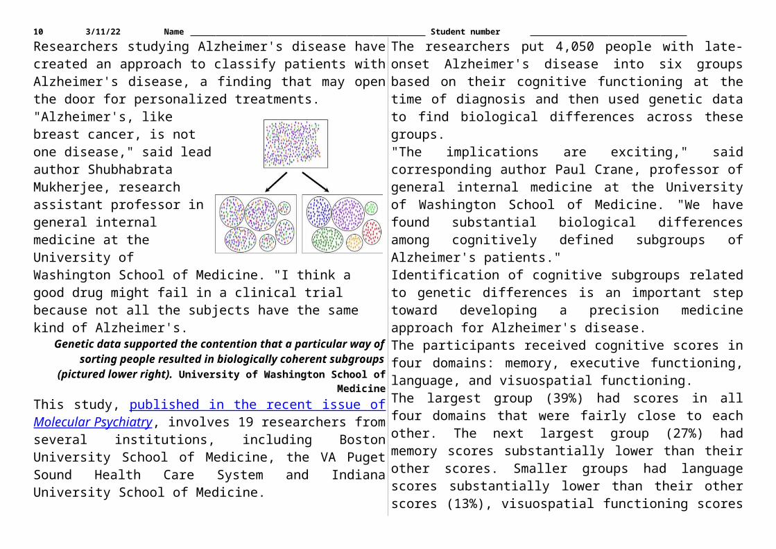
Genetic data supported the contention that a particular way of sorting people resulted in biologically coherent subgroups (pictured lower right). University
of Washington School of MedicineThis study, published in the recent issue of Molecular Psychiatry , involves 19 researchers from several institutions, including Boston University School of Medicine, the VA Puget Sound Health Care System and Indiana University School of Medicine.The researchers put 4,050 people with late-onset Alzheimer's disease into six groups based on their cognitive functioning at the time of diagnosis and then used genetic data to find biological differences across these groups."The implications are exciting," said corresponding author Paul Crane, professor of general internal medicine at the University of Washington School of Medicine. "We have found substantial biological differences among cognitively defined subgroups of Alzheimer's patients."Identification of cognitive subgroups related to genetic differences is an important step toward developing a precision medicine approach for Alzheimer's disease.The participants received cognitive scores in four domains: memory, executive functioning, language, and visuospatial functioning.The largest group (39%) had scores in all four domains that were fairly close to each other. The next largest group (27%) had memory scores substantially lower than their other scores. Smaller groups had language scores substantially lower than their other scores (13%), visuospatial functioning scores substantially lower than their other scores (12%), and executive functioning scores substantially lower than their other scores (3%). There were 6% who had two domains that were substantially lower than their other scores.

7 5/14/23 Name Student number
The participants came from five studies, and it took more than two years to standardize the neuropsychological test scores across all the studies in order to detect meaningful patterns. The mean age was 80, 92 percent self-reported white race, and 61 percent were female.The investigators used genome-wide genetic data to find out if the subgroups are biologically distinct.Investigators found 33 single nucleotide polymorphisms (SNPs) - specific locations throughout the genome - where the genetic association was very strong for one of the subgroups. These genetic relationships were stronger than the strongest effects found by an earlier and much larger international consortium study where Alzheimer's disease was treated as a single homogeneous condition.Several years ago, the International Genomics of Alzheimer's Project Consortium published the largest genome-wide association study of Alzheimer's disease and found about 20 SNPs associated with Alzheimer's disease risk. This study found 33 additional SNPs with even stronger relationships with a single subgroup.The study also found a particularly strong relationship between a particular variant of the APOE gene and risk for the memory subgroup. The APOE e4 allele is a very strong risk factor for developing Alzheimer's disease for people with European ancestry, and it also appears to influence which cognitive subtype of Alzheimer's a person is likely to develop.People can currently find out if they have an APOE e4 allele with direct-to-consumer testing; however, the researchers note that many people with an APOE e4 allele never develop Alzheimer's disease, and many who don't carry any known genetic risk factor nevertheless end up with the condition.While world leaders want to find a cure for Alzheimer's by 2025, so far no one has been able to develop an effective treatment let alone
a cure. But this study suggests that thinking of Alzheimer's disease as six distinct conditions may provide a way forward."This study is not the end, it's a start," said Mukherjee.Currently, 5.7 million people in the United States are living with the disease and that is expected to grow to 14 million by 2050, according to the Alzheimer's Association.The research team also included investigators from the Department of Biostatistics at University of Kentucky, Department of Epidemiology at John Hopkins Bloomberg School of Public Health, departments of neurology, human genetics and psychiatry at the University of Pittsburgh, Center for Translational & Computational Neuroimmunology at Columbia University Medical Center, Rush Alzheimer's Disease Center at Rush University Medical Center in Chicago, and Kaiser Permanente Washington Health Research Institute in Seattle.
http://bit.ly/2rpU0EXVolcanoes fed by 'mush' reservoirs rather than molten
magma chambersVolcanoes are not fed by molten magma formed in large
chambers finds a new study, overturning classic ideas about volcanic eruptions.
Instead, the study suggests that volcanoes are fed by so-called 'mush reservoirs' - areas of mostly solid crystals with magma in the small spaces between the crystals.Our understanding of volcanic processes, including those leading to the largest eruptions, has been based on magma being stored in liquid-filled 'magma' chambers - large, underground caves full of liquid magma. However, these have never been observed.The new study, by researchers at Imperial College London and the University of Bristol and published today in Nature, suggests the fundamental assumption of a magma chamber needs a re-think.Lead author Professor Matthew Jackson, from the Department of Earth Sciences and Engineering at Imperial, said: "We now need to look again at how and why eruptions occur from mush reservoirs. We can apply our findings to understanding volcanic eruptions with implications for public safety and also to understand the formation of metal ore deposits associated with volcanic systems."

8 5/14/23 Name Student number
In order to erupt, volcanoes need a source of magma - melted, liquid rock - containing relatively few solid crystals. Traditionally, this magma was thought to be formed and stored in a large underground cave, called a magma chamber.Recent studies of magma chemistry have challenged this view, leading to the suggestion of the mush reservoir model, where smaller pools of magma sit in the small gaps between solid crystals. However, the mush reservoir model could not explain how magmas containing relatively few crystals arise and are delivered to volcanoes in order for them to erupt at the surface.Now, with sophisticated modelling of mush reservoirs, the research team has come up with a solution. Within the mush reservoir scenario, the magma is less dense than the crystals, causing it to rise up through the spaces between them.As it rises, the magma reacts with the crystals, melting them and leading to local areas containing magma with relatively few crystals. It is these short-lived areas of increased magma that can lead to eruptions.Co-author Professor Stephen Sparks, from the University of Bristol's School of Earth Sciences, said: "A major mystery about volcanoes is that they were thought to be underlain by large chambers of molten rock. Such magma chambers, however, were very difficult to find."The new idea developed by geologists at Imperial and Bristol is that molten rock forms within largely crystalline hot rocks, spending most of its time in little pores within the rock rather than in large magma chambers. However, the rock melt is slowly squeezed out to form pools of melt, which can then erupt or form ephemeral magma chambers."As well as the initiation of eruptions, the new mush reservoir model can help explain other phenomena in volcanic systems, such as how
the magma chemical composition evolves and how much older crystals can be erupted within younger magmas.
http://bit.ly/2zMbkIXA universal DNA nano-signature for cancer
Researchers from the University of Queensland's Australian Institute for Bioengineering and Nanotechnology (AIBN) have discovered a unique nano-scaled DNA signature that appears to
be common to all cancers.Based on this discovery, the team has developed a novel technology that enables cancer to be quickly and easily detected from any tissue type, e.g. blood or biopsy.The study, which was supported by a grant from the National Breast Cancer Foundation and is published in the journal Nature Communications , reveals new insight about how epigenetic reprogramming in cancer regulates the physical and chemical properties of DNA and could lead to an entirely new approach to point-of-care diagnostics."Because cancer is an extremely complicated and variable disease, it has been difficult to find a simple signature common to all cancers, yet distinct from healthy cells," explains AIBN researcher Dr Abu Sina.To address this, Dr Sina and Dr Laura Carrascosa, who are working with Professor Matt Trau at AIBN, focussed on something called circulating free DNA.Like healthy cells, cancer cells are always in the process of dying and renewing. When they die, they essentially explode and release their cargo, including DNA, which then circulates."There's been a big hunt to find whether there is some distinct DNA signature that is just in the cancer and not in the rest of the body," says Dr Carrascosa.So they examined epigenetic patterns on the genomes of cancer cells and healthy cells. In other words, they looked for patterns of

9 5/14/23 Name Student number
molecules, called methyl groups, which decorate the DNA. These methyl groups are important to cell function because they serve as signals that control which genes are turned on and off at any given time.In healthy cells, these methyl groups are spread out across the genome. However, the AIBN team discovered that the genome of a cancer cell is essentially barren except for intense clusters of methyl groups at very specific locations.This unique signature -- which they dubbed the cancer "methylscape", for methylation landscape -- appeared in every type of breast cancer they examined and appeared in other forms of cancer, too, including prostate cancer, colorectal cancer and lymphoma."Virtually every piece of cancerous DNA we examined had this highly predictable pattern," says Professor Trau.He says that if you think of a cell as a hard-drive, then the new findings suggest that cancer needs certain genetic programmes or apps in order to run. "It seems to be a general feature for all cancer," he says. "It's a startling discovery."They also discovered that, when placed in solution, those intense clusters of methyl groups cause cancer DNA fragments to fold up into three-dimensional nanostructures that really like to stick to gold.Taking advantage of this, the researchers designed an assay which uses gold nanoparticles that instantly change colour depending on whether or not these 3D nanostructures of cancer DNA are present."This happens in one drop of fluid," says Trau. "You can detect it by eye, it's as simple as that."The technology has also been adapted for electrochemical systems, which allows inexpensive and portable detection that could eventually be performed using a mobile phone.
So far they've tested the new technology on 200 samples across different types of human cancers, and healthy cells. In some cases, the accuracy of cancer detection runs as high as 90%."It works for tissue derived genomic DNA and blood derived circulating free DNA," says Sina. "This new discovery could be a game-changer in the field of point of care cancer diagnostics." It's not perfect yet, but it's a promising start and will only get better with time, says the team."We certainly don't know yet whether it's the Holy Grail or not for all cancer diagnostics," says Trau, "but it looks really interesting as an incredibly simple universal marker of cancer, and as a very accessible and inexpensive technology that does not require complicated lab based equipment like DNA sequencing."
http://bit.ly/2RGYz9lInstitute of Human Virology researchers discover that a
bacterial protein promotes cancerResearch suggests suggests that bacterial infections may contribute to far more cancers than previously thought
Baltimore, MD, - The Institute of Human Virology (IHV) at the University of Maryland School of Medicine (UMSOM) announced today the discovery that DnaK, a protein of the bacterium mycoplasma, interferes with the mycoplasma-infected cell's ability to respond to and repair DNA damage, a known origin of cancer.Little or no mycoplasma DnaK DNA sequences were found associated with the tumor, which was fully developed, suggesting a hit-and-run or hide mechanism of transformation, indicating that the damage is done early, but the protein may not be needed once the cancer cells are formed.The study was published in the Proceedings of the National Academy of Sciences and suggests that bacterial infections may contribute to far more cancers than previously thought. The announcement was made by Robert Gallo, MD, The Homer &
10 5/14/23 Name Student number
Martha Gudelsky Distinguished Professor in Medicine and Co-Founder and Director, Institute of Human Virology, University of Maryland School of Medicine and Davide Zella, PhD, Assistant Professor of Biochemistry and Molecular Biology, Institute of Human Virology, University of Maryland School of Medicine. Drs. Gallo and Zella collaborated with Hervé Tettelin, PhD, Associate Professor of Microbiology and Immunology, Institute for Genome Sciences, University of Maryland School of Medicine."Currently, approximately 20% of cancers are thought to be caused by infection, most are known to be due to viruses," said Dr. Gallo. "Mycoplasmas are a family of bacteria that are associated with cancers, especially in people with HIV. Our work provides an explanation for how a bacterial infection can trigger a series of events that lead to cancer. Of particular importance, the infection did not need to persist and the protein did not need to be continuously present in all cancer cells. The study also provides a mechanism for how some bacterial infections can interfere with specific cancer drugs."Researchers utilized immune-compromised mice as a model for analyzing the effect of mycoplasma infection on the development of lymphoma. They compared how quickly non-infected immune-compromised mice developed lymphoma compared to mycoplasma-infected immune-compromised mice. The mice were infected with a strain of mycoplasma from an HIV patient. The researchers found that mycoplasma infection caused the mice to develop lymphoma earlier in life than non-infected immune-compromised mice and that some, but not all, of the cancer cells had bacterial DNA. Finding only a small amount of bacterial DNA in the cancer cells suggested that the infection did not have to persist to trigger cancer."We focused on a protein called DnaK, which is part of a family of proteins that function as a 'chaperone' for other proteins protecting
them from damage or helping them to fold," said Dr. Zella. "However, in this case, DnaK reduces the activity of important cellular proteins involved in DNA repair and anti-cancer-activities, such as p53. Thus, cells infected with mycoplasma would not be able to properly repair damaged DNA, thus, potentially increasing the risk for cancer development."The scientists noted that the bacteria can release DnaK and the DnaK enters nearby uninfected cells. The study also demonstrates that by reducing p53, DnaK can also reduce the efficacy of anti-cancer drugs. Thus, mycoplasma infection could not only trigger events leading to the accumulation of DNA damage and oncogenesis in infected cells, but also trigger cancer-causing events in nearby uninfected cells that took up DnaK released from infected neighboring cells."We analyzed the amino acid sequences of DnaK from many bacteria and found that the DnaK proteins from bacteria associated with cancer grouped together were different DnaK sequences from bacteria that are not associated with cancer," said Dr. Tettelin. "This raises the possibility that other bacteria have the same cancer-promoting ability."According to Dr. Gallo, "This hit-and-run, or hide, mechanism mediated by a protein common to many cancer-associated bacteria changes how we need to think about infection and at least some cancers. Furthermore, this provides a basis for understanding how infection can influence the effectiveness of some cancer treatments.""This is fascinating science with important implications," said UMSOM Dean E. Albert Reece, MD, PhD, MBA, who is also the Executive Vice President for Medical Affairs, University of Maryland, and the John Z. and Akiko K. Bowers Distinguished Professor. "We are pleased to see a cross-collaboration between two disciplines here at the University of Maryland School of Medicine.

11 5/14/23 Name Student number
Our Institute of Human Virology's basic science laboratory research was aided by the School's Institute of Genome Sciences' sequencing expertise, bringing the research to full fruition."This research was partially funded by the Maryland Cigarette Restitution Fund (CRF) Program. Morgan State University also participated in this study.
http://bit.ly/2E7PmTrA toxin that travels from stomach to brain may trigger
ParkinsonismCombination of paraquat and lectin travels from stomach to the
brain, causing ParkinsonismUniversity Park, Pa. - Combining low doses of a toxic herbicide with sugar-binding proteins called lectins may trigger Parkinsonism -- symptoms typical of Parkinson's disease like body tremors and slowing of body motions -- after the toxin travels from the stomach to the brain.In a study with rats, researchers at Penn State College of Medicine found that after ingesting paraquat, a once widely used herbicide that has been banned in the U.S. since 2007, along with lectins -- sugar-binding proteins found widely in nature -- the animals developed Parkinsonism.According to Thyagarajan Subramanian, professor of neurology and neural and behavioral sciences and co-author on the study, the findings -- recently published in the journal Parkinson's Disease -- offer clues to how and why Parkinson's disease develops, and offer a model to test new medications in the future."This study gives solid evidence that lectins, while in the presence of certain toxins, may be one potential culprit for the cause of Parkinsonism," Subramanian said. "Additionally, this animal model can be a tool in the future to continue developing new medications and treatments for Parkinson's disease."
The researchers were able to track the formation and spread of a misfolded protein called alpha-synuclein, which previous research has linked with Parkinson's."We were able to demonstrate that if you have oral paraquat exposure, even at very low levels, and you also consume lectins -- perhaps in the form of uncooked vegetables, dairy or eggs -- then it could potentially trigger the formation of this protein alpha-synuclein in the gut," Subramanian said. "Once it's formed, it can travel up the vagus nerve and to the part of the brain that triggers the onset of Parkinson's disease."R. Alberto Travagli, professor of neural and behavioral sciences and senior author of the study, said that while toxins like paraquat have been suspected of contributing to Parkinson's for decades, the scientific evidence was small. While paraquat was linked with Parkinsonism in previous studies, those experiments typically used high doses of paraquat that humans were not likely to encounter in real life.Additionally, lectins, which are used in medications to help deliver substances into the brain or stomach, also have been associated with certain rare forms of Parkinsonism. But the researchers weren't sure if it was the lectins themselves that were causing Parkinsonism, or if they were helping different substances get into the body that then triggered the symptoms."Experimenting with the lectins together with the toxin makes sense, because lectins are used in pharmacology to chaperone other substances into the body," Travagli said. "So it makes sense that the two can be combined and used to make the toxicity more potent, even though the amount of toxin is very low."Using a rat model, the researchers exposed the animals daily to small doses of paraquat and lectins for seven days. After stopping the treatment, the researchers waited two weeks. Then, the

12 5/14/23 Name Student number
researchers did a variety of tests to measure problems with motor function and other symptoms typical of Parkinsonism.The researchers observed a decrease in motor function that was consistent with Parkinsonism. But to confirm that the symptoms were related to Parkinsonism and not another cause, Travagli said he and the other researchers did several additional tests."After observing that these animals did indeed show symptoms of Parkinsonism, we wanted to double check and make sure we weren't looking at animals that had these symptoms for another reason," Travagli said. "We administered levodopa, which is a common medication for Parkinson's disease. We saw a return to almost normal types of motor responses, which was a clear indication that we were looking at some sort of Parkinsonism."Additionally, the researchers said when the vagus nerve was disconnected from the stomach prior to exposure to paraquat and lectins, the animals were protected from Parkinsonism, confirming the route of the alpha-synuclein from the gut to the brain.In the future, Travagli and Subramanian said they will explore whether interventions in the form of diet modifications or medications that interfere with the transport of alpha synuclein from the stomach via the vagus nerve could be used to help prevent or slow the development of Parkinsonism in this rat model. This includes a natural substance called squalamine which has been shown to remove alpha synuclein from the gut and is now in clinical trials for the certain symptoms associated with Parkinson's disease.Laura Anselmi, research associate; Cecilia Bove, doctoral student in neuroscience; F. Holly Coleman, lab manager; K. Le, research technologist; Megha Subramanian, research technologist; and Kala Venkiteswaran, assistant professor of neurology, also worked on this research.This research was supported by the National Institutes of Health, the Michael J. Fox Foundation for Parkinson's Disease and the Tobacco Settlement Fund.
https://wb.md/2QlMUjF
Acute Flaccid Myelitis Cases Have Peaked for the Year, CDC says
Case numbers of the rare polio-like illness acute flaccid myelitis (AFM) appear to have peaked, according to the US Centers for
Disease Control and Prevention (CDC).Marcia Frellick
The CDC on Monday said as of November 30, 134 cases of AFM, which causes muscle weakness and paralysis and affects mostly children, have been confirmed this year in 33 states out of the 299 cases reported. The number grew in this week's report by 18, but most of those cases happened in September and October.The pattern of cases in 2018 follows one the CDC has seen in the previous 4 years, with strong emergence every other year: 120 confirmed cases in 2014, 22 cases in 2015, 149 cases in 2016, and 33 cases in 2017.Most cases are reported between August and October and typically drop off in November. That was true for this year as well, with the number of people under investigation for AFM decreasing in recent weeks. Reporting is ongoing and the CDC will continue to investigate and confirm the cases.The CDC has been closely monitoring AFM since the first significant emergence in 2014. Since then, the agency has found that more than 90% of patients had a mild respiratory illness before developing AFM and that viral infections from enteroviruses are common, especially in children. "We don't know why a small number of people develop AFM, while most others recover," the CDC states on its website. "We are continuing to investigate this."CDC Director Robert R. Redfield, MD, announced last month the creation of an AFM task force to help define the cause of the illness and possible treatment options."I want to reaffirm to parents, patients, and our Nation CDC's commitment to this serious medical condition," Redfield said in a

13 5/14/23 Name Student number
statement. "This task force will ensure that the full capacity of the scientific community is engaged and working together to provide important answers and solutions to actively detect, more effectively treat, and ultimately prevent AFM and its consequences."The task force will make key recommendations to the CDC's Board of Scientific Counselors. It is scheduled to submit its first report December 6 in a public meeting in Atlanta, Georgia.
http://bit.ly/2QGXf9jBaby born via uterus transplant from deceased donor
Healthy girl arrives after world-first procedure.Nick Carne reports.
There have been 10 previous attempted uterus transplants but this is the first to result in a live birth. The first baby has been born following a uterus transplantation from a deceased donor, according to a case study from Brazil published in The Lancet. The transplant surgery took 10.5 hours and involved connecting the donor’s uterus and recipient's veins, arteries, ligaments and vaginal canals. Fertilised eggs were implanted seven months later. A healthy baby girl was born via caesarean section at 35 weeks and three days, weighing 2550 grams.The researchers from the University of São Paulo say the findings demonstrate the feasibility of the technique, but note that the outcomes and effects of organs sourced from live and deceased donors are yet to be compared.They say there have been 10 previous reported attempts to transplant a uterus from a deceased donor – in the US, Turkey and the Czech Republic – but theirs is the first to result in a live birth. There have been 39 transplants from living uterus donors, with 11 livebirths. The first was in Sweden in 2014."The first uterus transplants from live donors were a medical milestone, creating the possibility of childbirth for many infertile
women with access to suitable donors and the needed medical facilities,” says research leader Dani Ejzenberg. “However, the need for a live donor is a major limitation as donors are rare, typically being willing and eligible family members or close friends. The numbers of people willing and committed to donate organs upon their own deaths are far larger than those of live donors, offering a much wider potential donor population." The whole question of uterus transplants presents ethical challenges, of course, and earlier this year a group of British doctors warned that “the procedure remains experimental and success of the practice still requires further strictly controlled clinical trials”.The Brazilian recipient was a 32-year-old woman born without a uterus as a result of Mayer-Rokitansky-Küster-Hauser syndrome, a disorder that mainly affects the reproductive system. She had one in-vitro fertilisation cycle four months before transplant, resulting in eight fertilised eggs which were cryopreserved. The 45-year-old donor, who died of subarachnoid haemorrhage, had had three successful vaginal deliveries.The surgery took place in September, 2016. Five months after transplantation, the uterus showed no signs of rejection, ultrasound scans showed no anomalies and the recipient was having regular menstruation.The fertilised eggs were implanted after seven months – compared with about a year for previous uterus transplants – and the recipient was confirmed to be pregnant 10 days later. There were no reported issues during the pregnancy, other than a kidney infection at 32 weeks.The baby girl was born less than four weeks later, in December, 2017, and the transplanted uterus was removed during the caesarean section, showing no anomalies. Mother and baby were discharged

14 5/14/23 Name Student number
three days after birth, with what the researchers say was an uneventful early follow-up. They note that the transplant involved major surgery and recipients would need to be healthy to avoid complications during. In addition, the surgery used high doses of immunosuppression, which could be reduced in future, and involved moderate levels of blood loss, although these were considered manageable.The recipient and her partner received monthly psychological counselling from professionals specialised in transplants and fertility throughout before, during and after the transplant.
http://bit.ly/2zQZ1uWBringing balance to the universe: New theory could
explain missing 95 percent of the cosmosTheory that both dark matter and dark energy can be unified into
a fluid which possesses a type of 'negative gravity'Scientists at the University of Oxford may have solved one of the biggest questions in modern physics, with a new paper unifying dark matter and dark energy into a single phenomenon: a fluid which possesses 'negative mass'. If you were to push a negative mass, it would accelerate towards you. This astonishing new theory may also prove right a prediction that Einstein made 100 years ago. Our current, widely recognised model of the Universe, called LambdaCDM, tells us nothing about what dark matter and dark energy are like physically. We only know about them because of the gravitational effects they have on other, observable matter. This new model, published today in Astronomy and Astrophysics, by Dr Jamie Farnes from the Oxford e-Research Centre, Department of Engineering Science, offers a new explanation. Dr Farnes says: 'We now think that both dark matter and dark energy can be unified into a fluid which possesses a type of 'negative gravity', repelling all other material around them. Although this
matter is peculiar to us, it suggests that our cosmos is symmetrical in both positive and negative qualities.'The existence of negative matter had previously been ruled out as it was thought this material would become less dense as the Universe expands, which runs contrary to our observations that show dark energy does not thin out over time. However, Dr Farnes' research applies a 'creation tensor', which allows for negative masses to be continuously created. It demonstrates that when more and more negative masses are continually bursting into existence, this negative mass fluid does not dilute during the expansion of the cosmos. In fact, the fluid appears to be identical to dark energy.Dr Farnes's theory also provides the first correct predictions of the behaviour of dark matter halos. Most galaxies are rotating so rapidly they should be tearing themselves apart, which suggests that an invisible 'halo' of dark matter must be holding them together. The new research published today features a computer simulation of the properties of negative mass, which predicts the formation of dark matter halos just like the ones inferred by observations using modern radio telescopes.Albert Einstein provided the first hint of the dark universe exactly 100 years ago, when he discovered a parameter in his equations known as the 'cosmological constant', which we now know to be synonymous with dark energy. Einstein famously called the cosmological constant his 'biggest blunder', although modern astrophysical observations prove that it is a real phenomenon. In notes dating back to 1918, Einstein described his cosmological constant, writing that 'a modification of the theory is required such that "empty space" takes the role of gravitating negative masses which are distributed all over the interstellar space.' It is therefore possible that Einstein himself predicted a negative-mass-filled universe.

15 5/14/23 Name Student number
Dr Farnes says: 'Previous approaches to combining dark energy and dark matter have attempted to modify Einstein's theory of general relativity, which has turned out to be incredibly challenging. This new approach takes two old ideas that are known to be compatible with Einstein's theory - negative masses and matter creation - and combines them together. 'The outcome seems rather beautiful: dark energy and dark matter can be unified into a single substance, with both effects being simply explainable as positive mass matter surfing on a sea of negative masses.'Proof of Dr Farnes's theory will come from tests performed with a cutting-edge radio telescope known as the Square Kilometre Array (SKA), an international endeavour to build the world's largest telescope in which the University of Oxford is collaborating. Dr Farnes adds: 'There are still many theoretical issues and computational simulations to work through, and LambdaCDM has a nearly 30 year head start, but I'm looking forward to seeing whether this new extended version of LambdaCDM can accurately match other observational evidence of our cosmology. If real, it would suggest that the missing 95% of the cosmos had an aesthetic solution: we had forgotten to include a simple minus sign.'
http://bit.ly/2zSPaELThis strange marine creature has an immune system
remarkably similar to oursImmune system of golden star tunicate shows some unexpected
similarities to our ownBy Mitch Leslie
The golden star tunicate may look like a flower, but this marine invertebrate is a brawler, attacking its
tunicate neighbors in melees that feature ferocious cell-to-cell combat.
These golden star tunicates use their immune cells to fight each other. Christophe Courteau/Minden Pictures
Now, scientists have discovered that the immune system of this pugnacious animal shows some unexpected similarities to our own. The finding could help uncover new approaches for preventing rejection of transplanted organs or treating cancer.“It’s pretty exciting,” says comparative immunologist Larry Dishaw of the University of South Florida College of Medicine in St. Petersburg, who wasn’t connected to the research. “They’ve laid out a nice, convincing story here.”Tunicates are the closest living relatives of vertebrates—the group that includes humans, sharks, mice, and turtles—but the two evolutionary lines separated about 500 million years ago. The 3-millimeter-long, tube-shaped animals cluster in colonies on rocks and other hard underwater surfaces, fanning out like petals. When one growing colony contacts another, they have to decide “are they going to fight or are they going to fuse,” says study co-author Benyamin Rosental, a cellular immunologist now at Ben-Gurion University of the Negev in Beersheba, Israel.Unless both colonies carry the same version of a particular protein, they fight. Cells from the two colonies attack and destroy one another in battles akin to what happens when the human immune system rejects a transplanted organ.To probe how the golden star tunicate’s immune system works, a team led by Rosental and bioinformatician Mark Kowarsky of Stanford University in Palo Alto, California, isolated 34 types of cells from the animal. They found some cells switched on the same genes that are active in our hematopoietic stem cells, the blood-forming cells that spawn all the cells of our immune system. Like

16 5/14/23 Name Student number
vertebrate hematopoietic stem cells, the tunicate versions can divide and specialize into different cell types, the scientists determined.The researchers identified other parallels between the tunicate and vertebrate immune systems. Cells such as macrophages that devour invaders are a key part of vertebrate defenses. The animals harbored three kinds of these protectors. One type had never been detected before in the tunicates, and it shared a similar gene activity pattern with macrophages.Another way in which the tunicates’ immune system mirrors the vertebrate version involves cells that are specialized to kill other cells. In our bodies, these assassins include natural killer cells, which target tumor cells or cells infected by viruses. As the scientists report online today in Nature, tunicates also deploy such cell executioners. When the researchers staged fights in the lab dish between cells from different tunicates, they found that the bodies piled up. Analyzing the genes that are active in these killer cells may help researchers pin down the crucial genes that spur organ rejection, Rosental says, and could suggest new ways to eliminate cancer cells.The body of a tunicate seems simple, Dishaw says, but the new study shows “this simple system has incredible complexity” in its immune system. The overlap with humans indicates some features of the vertebrate immune system originated in our invertebrate ancestors. Rosental and colleagues are now studying other invertebrates such as sea urchins to determine how much further back in evolutionary history these features extend.
http://bit.ly/2QJXNLvMentors with wide-ranging research interests nurture
the most successful scientistsA study of academic mentoring involving thousands of
biomedical scientists suggests that researchers are more likely to
have a successful academic career if the mentors they work with have different areas of expertise.
By Emma StoyePrevious work has shown the positive effects of mentoring, with mentors who are successful themselves tending to train more successful mentees. But data scientist Jean Liénard at Oregon Health & Science University in the US, and colleagues, wanted to explore in more detail the way PhD students and postdocs are influenced by the academics they train under.The team looked at more than 18,000 ‘trios’ of biomedical researchers drawn from the Academic Family Tree open database. Each trio consisted of a mentee, their graduate mentor and their postdoctoral mentor. They used the number of researchers trained as a measure of academic success, as well as metrics such as the number of publications that could be drawn from open databases. They also analysed the abstracts of papers published by researchers so they could compare the similarity of research topics among mentors and mentees.Their results indicate that postdoctoral mentors are more instrumental to researchers’ success than graduate ones, and that researchers are likely to be more successful overall if their two mentors – graduate and postdoctoral – have expertise in different areas. They suggest this may support the idea that researchers who are able to integrate different ideas from dissimilar mentors are more likely to develop independent research careers and find success in academia.They note that the trends they identified have persisted for at least 40 years, despite changes to the overall numbers of PhD students, postdocs and research positions over time.ReferencesJ F et al, Nat. Commun., 2018, 9, 4840 (DOI: 10.1038/s41467-018-07034-y)
http://bit.ly/2PtMhPC

17 5/14/23 Name Student number
Remarkably preserved fossil sea reptile reveals skin that is still soft
The remains of an 180 million-year-old ichthyosaur (literally 'fish-lizard') have been analysed, and the fossil is so well-preserved that its soft-tissues retain some of their original
pliability.The study, published in Nature, contributes to our understanding on how convergent evolution works, and shows that ichthyosaurs adapted to marine conditions in a way that is remarkably similar to that of modern whales.The ichthyosaur lived in what is today southern Germany during the Jurassic Period some 180 million years ago. At that time, the approximately two-metre long reptile swam in a vast ocean that was then covering large parts of present-day Europe. Johan Lindgren of Lund University, Sweden, led the international collaboration that resulted in the most comprehensive and in-depth examination of a soft-tissue fossil ever undertaken. Among other things, the study reveals that the soft parts have fossilised so quickly that both the original cells and their internal contents are preserved."You can clearly see both the body outline and remains of internal organs. We can even distinguish the different cellular layers within the skin", explains Johan Lindgren.The researchers identified blubber underneath the skin. To date, such specialized fat-laden tissue has only been found in modern marine mammals and adult individuals of the leatherback sea turtle. The presence of blubber indicates that ichthyosaurs had metabolic rates that were higher than are those of typical reptiles living today. These data could help explain why ichthyosaurs had an almost global distribution, even in cold waters, and how they could dive to considerable depths, as well as grow as fast as they did.
The team also examined remains of the animal's liver, which included part of the original biochemistry (e.g., eumelanin pigment and haemoglobin residues). The molecular and imaging analyses were performed in laboratories in Sweden, Germany, Japan and the USA. "It's truly remarkable that the biomolecules we discovered so closely match the tissues that we could identify", says Johan Lindgren.In the study, the researchers also succeeded in showing that the fossil contains tissues that still retain some of their original pliability, even though 180 million years have passed since the material was fresh. The team used chemicals to remove the mineral phase of the specimen, i.e. the inorganics that once turned the animal carcass into a petrified fossil."We then discovered that the soft parts retain a certain degree of elasticity", says Johan Lindgren. The results also reveal the colouring of adult ichthyosaurs: the upper part of the body was dark, whereas the belly was light. This colouration acted either as camouflage or UV protection, or both. It may also have helped the animal to warm up faster in cold climates and/or after long and deep dives. Not only do the results provide insights into the biology, physiology and ecology of derived ichthyosaurs, they also show how little we know about the fossilisation process and what can actually be preserved in the fossil record. Moreover, they could add to our knowledge on convergent evolution, as ichthyosaurs display an interesting mix of characteristics otherwise found in toothed whales (such as dolphins and porpoises) and the leatherback sea turtle.
http://bit.ly/2zPEGWy500-Year-Old Body of Man Wearing Thigh-High Boots
Found in London Sewer Construction
18 5/14/23 Name Student number
The splayed position of the 500-year-old body found along the Thames River, and the fact that he was still wearing expensive
boots, suggest that the death was an accident.By Megan Gannon, Live Science Contributor
During the construction of London's massive "super sewer," archaeologists discovered something unusual in the mud: a 500-year-old skeleton of a man still wearing his thigh-high leather boots.The Museum of London Archaeology (MOLA) announced this week that the skeleton was unearthed on the shores of the Thames, near a bend in the river downstream from the Tower of London.
Copyright MOLA Headland Infrastructure"By studying the boots, we've been able to gain a fascinating glimpse into the daily life of a man who lived as many as 500 years ago," said Beth Richardson, a finds specialist who analyzes artifacts at MOLA Headland, a consortium of archaeologists. "They have helped us to better understand how he may have made his living in hazardous and difficult conditions, but also how he may have died. It has been a privilege to be able to study something so rare and so personal."The blackened boots had some custom features: They were reinforced with extra soles and stuffed with a mossy material, perhaps for warmth or a better fit. Based on the boot style, the researchers think this man died in the late 15th or early 16th century.The boots are also an indication that the man's burial wasn't an intentional one; leather boots at the time were quite expensive and likely would have been recycled, not buried with the dead. The skeleton was found facedown with his arms splayed over his head, another hint that his body was quickly covered with mud after death.
But with no obvious fatal injuries visible on the bones, the man's cause of death remains a mystery. MOLA researchers floated at least one possibility — that he fell into the mud, perhaps while climbing a wall upstream, and got trapped and drowned.The area where the man was found is a natural confluence where materials build up in the river, MOLA researchers said.The 500-year-old skeleton was still wearing
expensive leather boots when it was discovered along the banks of the Thames
River. copyright MOLA Headland InfrastructureArchaeologists were able to glean some ideas about the man's life from the evidence. Worn grooves on his teeth might have been caused by a repetitive action — maybe he was a sailor or a fisherman who had to pass ropes through his teeth, the researchers speculated. Those thigh-high leather boots would have been appropriate for a life on the water, as they would have kept a person's legs and feet dry while wading through the Thames' muck."Marks on his skeleton have allowed us to proffer ideas about the aches and pains he may have suffered from on a daily basis, the toll his job took on his body and even a little about what he might have looked like," Niamh Carty, an osteologist at MOLA Headland, said in a statement. The researchers also think the man, who may have been no older than 35, suffered from osteoarthritis, perhaps caused by repetitive work and stress on his bones.The Thames Tideway Tunnel is a 15-mile-long (25 kilometers) sewer designed to stop the overflow of waste in London's sewer

19 5/14/23 Name Student number
system from getting into into the Thames. ("Fatbergs" that clog Victorian-era pipes are a recurring problem.)The project is expected to be completed around 2024; the burial was discovered during the construction of a shaft where one of the tunnel's boring machines will be digging.The Thames is sometimes considered London's longest archaeological site, and the river muck has historically turned up a lot of surprising finds, from Neolithic wooden clubs to broken-up pieces of Napoleonic-era warships. For more than 10 years, the Thames Discovery Programme has been organizing groups of volunteers to look for artifacts and monitor archaeological remains that are exposed when the tide is low.
http://bit.ly/2G6deJKDrawing is better than writing for memory retention
Older adults who take up drawing could enhance their memory, according to a new study.
Researchers from the University of Waterloo found that even if people weren't good at it, drawing, as a method to help retain new information, was better than re-writing notes, visualization exercises or passively looking at images."We found that drawing enhanced memory in older adults more than other known study techniques," said Melissa Meade, PhD candidate in cognitive neuroscience at Waterloo."We're really encouraged by these results and are looking into ways that it can be used to help people with dementia, who experience rapid declines in memory and language function."As part of a series of studies, the researchers asked both young people and older adults to do a variety of memory-encoding techniques and then tested their recall.Meade conducted this study with Myra Fernandes a Psychology professor in cognitive neuroscience at Waterloo and recent UW PhD graduate Jeffrey Wammes.
The researchers believe that drawing led to better memory when compared with other study techniques because it incorporated multiple ways of representing the information--visual, spatial, verbal, semantic and motoric."Drawing improves memory across a variety of tasks and populations, and the simplicity of the strategy means that it can be used in many settings," said Myra Fernandes.As part of the studies, the researchers compared different types of memory techniques in aiding retention of a set of words, in a group of undergraduate students and a group of senior citizens.Participants would either encode each word by writing it out, by drawing it, or by listing physical attributes related to each item. Later on after performing each task, memory was assessed.Both groups showed better retention when they used drawing rather than writing to encode the new information, and this effect was especially large in older adults.Retention of new information typically declines as people age, due to deterioration of critical brain structures involved in memory such as the hippocampus and frontal lobes.In contrast, we know that visuospatial processing regions of the brain, involved in representing images and pictures, are mostly intact in normal aging, and in dementia."We think that drawing is particularly relevant for people with dementia because it makes better use of brain regions that are still preserved, and could help people experiencing cognitive impairment with memory function," said Meade."Our findings have exciting implications for therapeutic interventions to help dementia patients hold on to valuable episodic memories throughout the progression of their disease"The study appears in Experimental Aging and Research.

20 5/14/23 Name Student number
http://bit.ly/2L7bnn3An ancient strain of plague may have led to the decline
of Neolithic EuropeansA team of researchers from France, Sweden, and Denmark have identified a new strain of Yersinia pestis, the bacteria that causes plague, in DNA extracted from 5,000-year-old human remains.
Their analyses, publishing December 6 in the journal Cell, suggest that this strain is the closest ever identified to the genetic origin of plague. Their work also suggests that plague may have been spread among Neolithic European settlements by traders, contributing to the settlements' decline at the dawn of the Bronze Age."Plague is maybe one of the deadliest bacteria that has ever existed for humans. And if you think of the word 'plague,' it can mean this infection by Y. pestis, but because of the trauma plague has caused in our history, it's also come to refer more generally to any epidemic. The kind of analyses we do here let us go back through time and look at how this pathogen that's had such a huge effect on us evolved," says senior author Simon Rasmussen (@simonrasmu), a metagenomics researcher at the Technical University of Denmark and the University of Copenhagen.To better understand the evolutionary history of the plague, Rasmussen and his colleagues trawled through publicly available genetic data from ancient humans, screening for sequences similar to more modern plague strains. They found a strain they had never seen before in the genetic material of a 20-year-old woman who died approximately 5,000 years ago in Sweden. The strain had the same genes that make the pneumonic plague deadly today and traces of it were also found in another individual at the same grave site--suggesting that the young woman did likely die of the disease.This strain of the plague is the oldest that's ever been discovered. But what makes it particularly interesting is that, by comparing it to other strains, the researchers were able to determine that it's also the
most basal--meaning that it's the closest strain we have to the genetic origin of Y. pestis. It likely diverged from other strains around 5,700 years ago, while the plague that was common in the Bronze Age and the plague that is the ancestor of the strains in existence today diverged 5,300 and 5,100 years ago, respectively. This suggests that there were multiple strains of plague in existence at the end of the Neolithic period.Rasmussen also believes that this finding offers a new theory about how plague spreads. Massive human migrations from the Eurasian steppe down into Europe are known to have occurred around 5,000 years ago, but how these cultures were able to displace the Neolithic farming culture that was present in Europe at the time is still debated. Previous researchers have suggested that the invaders brought the plague with them, wiping out the large settlements of Stone Age farmers when they arrived. But if the strain of plague the researchers found in the Swedish woman diverged from the rest of Y. pestis 5,700 years ago, that means it likely evolved before these migrations began and around the time that the Neolithic European settlements were already starting to collapse.At the time, mega-settlements of 10,000-20,000 inhabitants were becoming common in Europe, which made job specialization, new technology, and trade possible. But they also may have been the breeding ground for plague. "These mega-settlements were the largest settlements in Europe at that time, ten times bigger than anything else. They had people, animals, and stored food close together, and, likely, very poor sanitation. That's the textbook example of what you need to evolve new pathogens," says Rasmussen."We think our data fit. If plague evolved in the mega-settlements, then when people started dying from it, the settlements would have been abandoned and destroyed. This is exactly what was observed in these settlements after 5,500 years ago. Plague would also have

21 5/14/23 Name Student number
started migrating along all the trade routes made possible by wheeled transport, which had rapidly expanded throughout Europe in this period," he says.Eventually, he suggests, the plague would have arrived through these trade interactions at the small settlement in Sweden where the woman his team studied lived. Rasmussen argues that the woman's own DNA also provides further evidence for this theory--she isn't genetically related to the people who invaded Europe from the Eurasian steppe, supporting the idea that this strain of plague arrived before the mass migrations did. The archaeology also supports this hypothesis, as there were still no signs of the invaders by the time she died.Of course, there are some limitations to what the data from this study can tell us. Most importantly, the researchers have not yet identified the plague in individuals from the mega-settlements where it may have evolved. "We haven't really found the smoking gun, but it's partly because we haven't looked yet. And we'd really like to do that, because if we could find plague in those settlements, that would be strong support for this theory," says Rasmussen.Regardless, he believes that this study is a step toward understanding how plague--and other pathogens--became deadly. "We often think that these superpathogens have always been around, but that's not the case," he says. "Plague evolved from an organism that was relatively harmless. More recently, the same thing happened with smallpox, malaria, Ebola, and Zika. This process is very dynamic--and it keeps happening. I think it's really interesting to try to understand how we go from something harmless to something extremely virulent."This research was supported by the Carlsberg Foundation, The Lundbeck Foundation, The Danish National Research Foundation, the IHU Méditerranée Infection, Marseille, France, the French Government under the «Investissements d'avenir» program, the Région Provence Alpes Côte d'Azur, the European funding FEDER PRIMI, and the A*MIDEX Foundation.
Cell, Rascoven et al.: "Emergence and spread of basal lineages of Y. pestis during the Neolithic decline" https://www.cell.com/cell/fulltext/S0092-8674(18)31464-8
http://bit.ly/2RN5ePtStatins overprescribed for primary prevention
Use of statins for primary prevention has been hotly debated among experts
Even healthy people who don't suffer from a cardiovascular disease are prescribed cholesterol-lowering drugs, known as statins, if they meet certain risk criteria. However, for years the use of statins for primary prevention has been hotly debated among experts. "Ultimately, this measure helps to prevent heart attacks or strokes in only a few cases. But all people who take statins are at risk of experiencing the side effects," says Milo Puhan, professor of epidemiology and public health at the University of Zurich.No systematic studies for guidelinesWhen deciding whether to prescribe statins to a patient, doctors use a number of risk factors such as cholesterol level, BMI and smoking to determine the likelihood of a person suffering a heart attack or stroke in the next 10 years. If this figure reaches or exceeds 10%, many medical guidelines recommend the use of statins; however, some guidelines put this number at 7.5%, whereas a Swiss association of general practitioners only suggests doing so from 20%. If these guidelines, most of which are drawn up by cardiology organizations, are to be believed, more than one third of all people between the age of 40 and 75 would have to take statins as a preventive measure - in other words, hundreds of millions of people around the world. According to Puhan, however, these guidelines were drawn up without properly taking into account the unwanted side effects, such as muscle pain, cataracts, liver defects or diabetes. "The thresholds set by experts aren't based on any systematic studies."

22 5/14/23 Name Student number
Weighing up benefits and harmful effectsStriking a good balance between the benefits and the harmful side effects is therefore one of the great challenges of developing improved guidelines for preventive statin use. This is why Prof. Milo Puhan and his team at the Epidemiology, Biostatistics and Prevention Institute of UZH have now for the first time carried out a comprehensive statistical modeling study.The researchers systematically compiled all data from specialist literature that documents the benefits and side effects of the preventive use of statins. To include the view of patients in their model, they also performed a survey among healthy people about the significance of heart attacks, strokes and certain side effects.Using this information, the scientists determined new thresholds for men and women across different age groups between 40 and 75. They also compared the benefits and unwanted side effects of four widely used statin preparations.Recommendation given to too many people"Our study shows that today statins are recommended far too often," says Puhan about the study's findings. According to his estimates, the newly set thresholds could cut the number of people who are given a recommendation to take statins by half.The benefits of cholesterol-lowering drugs have been greatly exaggerated particularly when it comes to senior citizens: For the 70 to 75 age group, the study's model put the threshold at approx. 21% - in other words, the benefits of statins outweigh the harm from potential side effects of statins only if there's a 21% risk or higher that a person will suffer a heart attack or a stroke in the next 10 years. For men and women aged 40 to 45, the threshold is slightly lower, at 14% and 17% respectively. The researchers also noted that two of the four examined statin preparations, atorvastatin and rosuvastatin, had a significantly better balance of benefits and harms than the other two (simvastatin und pravastatin).
Considering the study's findings, Puhan recommends that all people concerned should discuss their individual risk for cardiovascular disease as well as possible side effects with their doctors before deciding whether to take statins as a preventive measure.
http://bit.ly/2SC6ViMIncreasing statins dose and patient adherence could
save more livesThousands of heart attacks and deaths from cardiovascular disease could be prevented by patients taking higher doses of
statins and taking the drugs as advised by doctors.The findings come from a study led by researchers at Imperial College London and University of Leicester, which estimates that 12,000 cardiovascular events - such as a heart attack or stroke - among high risk patients in the UK could be averted by placing patients on high intensity cholesterol-lowering drugs and ensuring patients stick to the correct treatment regimen.The paper, published today in the journal JAMA Network Open, is the first to look at the combined effect of high intensity statin treatment and adherence in patients who have already had a cardiovascular event and who are at increased risk of future cardiovascular events compared to the general public.It reveals that that those taking the highest doses (intensity) of statins to reduce their LDL cholesterol levels (the 'bad' cholesterol which builds up in the arteries) and, crucially, who take the medication as advised by their doctor, see the biggest reductions in risk for future cardiovascular events.Patient adherence describes the degree to which a patient follows medical advice and takes medication correctly. It can significantly affect how successful a treatment is, as patients may not take a drug regularly, skip days, or stop the medication altogether - especially if they see or feel no immediate benefit from taking a drug. This is

23 5/14/23 Name Student number
particularly important as high cholesterol levels in the blood may not cause any noticeable symptoms compared to other conditions.Professor Kausik Ray, from the School of Public Health at Imperial and lead author of the study, said: "The basic message here is that long-term adherence achieves better long-term cholesterol reductions, and in turn, achieves better long-term outcomes for patients."In terms of risk reduction, we can see the people who do the best are those who are adhering to the recommended dosage and are on more potent drug regimens. But if someone is not going to take a treatment as recommended, they may actually be better off on higher doses of statins so that when they are taking the medication, they are achieving greater cholesterol reductions."In the latest study, the team analysed patient data drawn from Clinical Practice Research Datalink (CPRD) which includes over five million records from more than 450 GP practices. They focused on three groups of patients at high risk of cardiovascular events: those with established heart disease; those with diabetes but no history of heart disease; and those with chronic kidney disease but no prior heart attack and stroke.Almost 30,000 patients recently started on cholesterol lowering medications were scored using a measure which combined the intensity of treatment they received (statins alone or combined with another cholesterol-lowering drug called ezetimibe) and their adherence - with those who took their medication as prescribed 80% of the time over period classed as 'adherent'.When researchers measured the relative risk reduction compared to untreated patients an average of three years after treatment, they found those patients with the highest score (who were on the highest intensity treatment and had the highest adherence) had the greatest reduction in LDL cholesterol levels and cardiovascular risk, with a 40% reduction in their risk of cardiovascular events.
By comparison, those patients on the lowest intensity treatment and with poor adherence had a risk reduction of just 5% compared to those not on medication. According to the team, in patients with established heart disease approximately 72 cardiovascular events were observed per 1000 patients per year. But with optimal treatment - high dose medication and high adherence - this would be expected to be reduced to 48 per 1000 patients per year, a reduction of 12,000 cases based on the estimated 500,000 heart disease patients in the UK.To improve adherence, the researchers advise that perhaps doctors spend more time with patients to explain the benefits and risks of the drugs, so patients are able to make better decisions about their own health based on benefits and risks. They add that adherence could be overcome with new delivery methods, such as slow release drugs which patients are required to take less frequently.Professor Kamlesh Khunti, Professor of Primary Care Diabetes and Vascular Medicine at the University of Leicester, said: "Adherence to lipid lowering therapy is poor within first six months, with studies showing that 40-60% of people are not adherent to statin therapies. This novel concept of a measure of adherence and intensity is likely to be applicable to other medications such as antihypertensive and glucose lowering therapies as well."Professor Ray added: "There is a lot of confusion around dietary cholesterol and medications that lower cholesterol. While changing your diet is a good thing, it may not be enough for those patients who are already in the high-risk groups - such as those with heart disease. Here, statins provide additional benefits over and above lifestyle."It doesn't matter how patients get to this point - through obesity, smoking, genetic risk factors - what we know is that once you have one heart attack or other cardiovascular event, you are at much

24 5/14/23 Name Student number
higher risk of more events in future and that lowering your LDL cholesterol levels is key to improving outcomes. "For these patients, taking the right medication, at the right dose, at the right time - and sticking to this regimen - is critical in lowering their risk of future cardiovascular events."The research was funded by Amgen Europe, which manufactures the cholesterol-lowering PCSK9 inhibitor evolocumab (Repatha).
http://bit.ly/2G7oXIbInfant formula companies are behind the guidelines on
milk allergy, and their sales are soaringInfant formula manufacturers have been funding the
development of guidelines for the diagnosis and treatment of cows’ milk allergy in infants
Karleen Gribble * Nina J Berry **
There has been a six-fold increase in sales of infant formula prescribed for babies with cows’ milk protein allergy in the United Kingdom from 2006 to 2016. This is despite no evidence of a concurrent increase in the prevalence of infants with the allergy. An investigation published today in the BMJ found infant formula manufacturers have been funding the development of guidelines for the diagnosis and treatment of cows’ milk allergy as well as providing research and consultancy funds to those who wrote them.Rates of cow’s milk allergy appear to have been relatively stable – estimated at between 1-2% over the last decade. Research has found the perception of an allergic response to cow’s milk protein in children is ten times greater than what actual diagnosis would indicate. This means guidelines on allergy for doctors are really important.In some cases, doctors who spoke to the BMJ said the guidelines were so vague that virtually every single infant could potentially be diagnosed using these symptoms.
A diagnosis can only be made only by excluding cow’s milk protein from the maternal diet, observing symptoms, and then reintroducing it. But the BMJ paper notes that evidence for advising such exclusions to treat non-specific symptoms in breastfed infants is weak.The paper also found much of the education for health professionals and parents about cows’ milk allergy was provided by organisations also funded by the infant formula industry.Previous research has found that changes in diagnostic and treatment guidelines can have enormous effects on the revenue of pharmaceutical and nutritional products. Conflicts of interest due to industry funding have been found to affect doctors’ prescribing behaviour, research results and the quality of patient care.Parents are also vulnerable to marketing. They crave a happy, quiet, calm baby who sleeps, eats and poos in a predictable pattern. But babies wake often. They can have difficulty adjusting to life outside the womb and their stomachs are getting used to digesting food. They vomit. They cry for reasons that are hard to understand. Marketing takes this normal infant behaviour and turns it into a problem that can be solved by buying a product.When businesses are allowed to shape the guidelines health professionals use to diagnose and treat, this can lead to guidelines that find normal infant behaviour is treatable – with a product. Unfortunately there may similar pressures on doctors in Australia.A variety of infant formula products available in Australia claim to be antidotes to normal challenges new parents face such as crying, vomiting and constipation. The above advertisement is from the publication Australian Doctor (which is available through subscription to medical professionals and includes drug advertising). It is a powerful piece of persuasion. It invokes every parent’s desire for a good night’s sleep and a contented, healthy baby to drive purchasing behaviour. Notice the

25 5/14/23 Name Student number
presence of the mother in this picture. Although we can’t see her, we presume she is also sleeping somewhere. When parents are desperate for help, doctors want to provide it. Colic is a variation of normal infant behaviour. It has no known medical cause or cure and this can make doctors feel powerless. However, this advertisement offers them a way to help. It gives doctors a solution – they just need to suggest the infant formula. Parents are told to seek assistance from health professionals when they are concerned about their baby. However, if health professional guidelines and education is contaminated with marketing and influenced in other ways by infant formula manufacturers, the support they provide will be of poor quality. Health professionals need independent, non-commercial information on infant feeding and parents should be protected from predatory marketing through effective enforcement of regulations.*Adjunct Associate Professor, School of Nursing and Midwifery, Western Sydney University**Post-doctoral Research Associate, Sydney School of Public Health, University of SydneyDisclosure statementKarleen Gribble is affiliated with the Infant and Young Child Feeding in Emergencies Core Group, the World Breastfeeding Trends Initiative, and the Australian Breastfeeding Association. Nina J Berry's research is supported by funding from Australian Government Department of Health; the Australia Indonesia Centre and the University of Sydney. Dr Berry is a member of the International Union for Health Promotion and Education; the Public Health Association of Australia; the Asian Studies Association of Australia; the World Public Health Nutrition Association; Collaboration on Social Science in Immunisation; the Sydney Global Child Health Research Network; the Sydney South East Asia Centre; the Sydney Lifespan Research Network; and volunteers as a trainer, assessor, and counsellor for the National Breastfeeding Helpline.PartnersUniversity of Sydney and Western Sydney University provide funding as members of The Conversation AU.
http://bit.ly/2SBHIonNearly two-thirds of people at high risk of heart disease
and stroke have excess belly fat
Nearly two-thirds of people at high risk of heart disease and stroke have excess belly fat
Dubai, United Arab Emirates - Nearly two-thirds of people at high risk of heart disease and stroke have excess belly fat, according to results of the European Society of Cardiology (ESC) EUROASPIRE V survey presented today at the World Congress of Cardiology & Cardiovascular Health in Dubai, United Arab Emirates.1
Excess fat around the middle of the body (central obesity) is a marker of abnormal fat distribution. This belly fat is bad for the heart, even in people who are not otherwise overweight or obese.Cardiovascular diseases are the leading cause of death in Europe. Each year in Europe there are more than 11 million new cases of cardiovascular disease and 3.9 million deaths caused by cardiovascular disease.2 Elimination of risk behaviours would prevent at least 80% of cardiovascular diseases.3
The study also found that less than half (47%) of those on antihypertensive medication reached the blood pressure target of less than 140/90 mmHg (less than 140/85 mmHg in patients with self-reported diabetes). Among those taking lipid-lowering drugs, only 43% attained the LDL cholesterol target of less than 2.5 mmol/L. In addition, many participants not taking any antihypertensive and/or lipid-lowering therapy had elevated blood pressure and elevated LDL cholesterol. Among patients being treated for type 2 diabetes, 65% achieved the blood sugar target of glycated haemoglobin (HbA1c) less than <7.0%. Professor Kornelia Kotseva, chair of the EUROASPIRE Steering Committee from Imperial College London, UK, said: "The survey shows that large proportions of individuals at high risk of cardiovascular disease have unhealthy lifestyle habits and uncontrolled blood pressure, lipids and diabetes."

26 5/14/23 Name Student number
EUROASPIRE is a series of cross sectional surveys on the prevention of cardiovascular disease in ESC member countries.4
The results of the primary care arm of EUROASPIRE V are reported today. The study was conducted in 2017 to 2018 in 78 general practices in 16 primarily European countries.5
Each general practice enrolled consecutive individuals under the age of 80 years with no history of coronary artery disease or other atherosclerotic disease, but who were at high risk of developing cardiovascular disease. High risk was defined as having high blood pressure, high cholesterol, and/or diabetes; the study therefore recruited individuals who had been prescribed antihypertensive, lipid-lowering, and/or anti-diabetes treatments (diet and/or oral hypoglycaemics and/or insulin).Participants were retrospectively identified using medical records and invited to an interview and clinical examination. Questions were asked about smoking, diet, physical activity, blood pressure, lipids and diabetes. Measurements included height, weight, waist circumference, blood pressure, low-density lipoprotein (LDL) cholesterol levels, and blood sugar levels. The primary outcomes were the proportions of participants achieving targets for cardiovascular disease prevention in the 2016 European guidelines.3
A total of 2,759 participants were interviewed and examined using standardised methods and instruments. Nearly two-thirds (64%) were centrally obese (waist circumference 88 cm or greater for women and 102 cm or higher for men). Some 37% were overweight (body mass index [BMI] 25 to 29.9 kg/m2) and 44% were obese (BMI 30kg/m2 or above). Nearly one in five participants (18%) were smokers and just 36% achieved the recommended physical activity level of at least 30 minutes, five times per week.Professor Kotseva said: "GPs should proactively look for cardiovascular risk factors so that comprehensive treatment and advice can be given. She added: "GPs need to go beyond treating
the risk factors they know about, and always investigate smoking, obesity, unhealthy diet, physical inactivity, blood pressure, cholesterol, and diabetes. People are often unaware that they need treatment - for example they visit their GP for their diabetes but do not know they also have high blood pressure. In our study, many participants with high blood pressure and cholesterol were not being treated."She continued: "These data make it clear that more efforts must be made to improve cardiovascular prevention in people at high risk of cardiovascular disease. Our analysis highlights the need for health care systems to invest in prevention.""Public health initiatives will also help to prevent heart disease and stroke," Professor Kotseva highlighted. "This includes smoking bans, taxing foods high in sugar and saturated fat, and providing areas for exercise."Notes to editorSources of funding: The EUROASPIRE V survey was carried out under the auspices of the European Society of Cardiology EURObservational Research Programme. The survey was supported through unrestricted research grants to the European Society of Cardiology from Amgen, Elli Lilly, Ferrer, NovoNordisk, Pfizer and Sanofi. The sponsors of the EUROASPIRE surveys had no role in the design, data collection, data analysis, or data interpretation. Disclosures: Professor Kotseva had research grant support from the European Society of Cardiology for the EUROASPIRE V survey. References and notes1The EUROASPIRE V results will be presented on Friday 7 December, 15:00 to 16:00 Dubai time, during the session 'EORP Registries: EUROASPIRE V Study on CVD Prevention and Diabetes - principal results in patients at high CVD risk in primary care'. 2European cardiovascular disease statistics 2017. 3Piepoli MF, Hoes AW, Agewall S, et al. 2016 European Guidelines on cardiovascular disease prevention in clinical practice. European Heart Journal. 2016;37:2315-2381. 4About EUROASPIRE: EUROASPIRE (EUROpean Action on Secondary and Primary Prevention In order to Reduce Events) is a series of cross sectional surveys of the practice of preventive cardiology in patients with coronary heart disease and people at high risk of developing cardiovascular disease across Europe. Five EUROASPIRE surveys have been conducted under the auspices of the European Society of Cardiology's Euro Heart Survey and EURObservational Research Programme. 5The participating countries were: Bosnia and Herzegovina, Bulgaria, Croatia, Czech

27 5/14/23 Name Student number Republic, Egypt, Greece, Kazakhstan, Kyrgyzstan, Lithuania, Poland, Portugal, Romania, Russia, Sweden, Ukraine, and the UK.
https://wb.md/2UvBA2YOpen-Label Placebos: Why Are They Effective?
Open-Label Placebo EffectsTom G. Bartol, NP
The outcomes of placebo treatments have shown significant improvements in many double-blind, randomized trials when the patient does not know whether the treatment is placebo or real. A new study[1] suggests that placebos given "open label," with the recipient's full knowledge that it is a placebo, may still have a significant measurable clinical effect.In one small trial, patients were told that it was not known whether the placebo would help their condition, but were also told, "Let's see what happens." In a treatment trial for irritable bowel syndrome, patients who received the placebo had a 60% relative improvement versus 35% with usual care.[2] Other trials showed three times relative efficacy of placebo versus usual treatment for low back pain and cancer-related fatigue.[3,4] Kaptchuk and colleagues[1] found that two thirds of patients surveyed would be willing to take an open-label placebo if recommended. More research is encouraged to determine a clear role for open-label placebo.ViewpointThe placebo effect has been well studied; it's been determined that even after the administration of an inert substance, the effect is real. The effects of placebo are thought to be due to the "therapeutic encounter, with its rituals, symbols, and interactions."[5] Even active medications have a placebo effect beyond their chemical effects. With open-label placebo, the rituals and interactions do not build
expectations to the same degree as a double-blind trial. The participants were told that the placebo contained only fiber.Another study[6] looking at open-label placebo treatment of allergic rhinitis symptoms found that positive expectations did not contribute to the efficacy of placebo despite a significant placebo effect. Participants in these trials overwhelmingly deny having any initial positive expectation that the placebo will work but express hope, because nothing else has helped their condition to this point. The placebo effect in these open-label trials may be related to the desperation of people who have not experienced relief with conventional treatment.The concept of an open-label placebo is intriguing. Although somewhat counterintuitive, the role of open-label placebos should be clarified more with further research. Future studies should explore the "desperation effect," to determine whether it exists during the initial treatment of a condition in addition to treatment with open-label placebo after all other therapies have failed.References1. Kaptchuk TJ, Miller FG. Open label placebo: can honestly prescribed placebos evoke meaningful therapeutic benefits? BMJ. 2018;363:k8889. Abstract 2. Kaptchuk TJ, Friedlander E, Kelley JM, et al. Placebos without deception: a randomized controlled trial in irritable bowel syndrome. PLoS One. 2010;5:e15591. Source 3. Carvalho C, Caetano JM, Cunha L, Rebouta P, Kaptchuk TJ, Kirsch I. Open-label placebo treatment in chronic low back pain: a randomized controlled trial. Pain. 2016;157:2766-2772. Abstract 4. Hoenemeyer TW, Kaptchuk TJ, Mehta TS, Fontaine KR. Open-label placebo treatment for cancer-related fatigue: a randomized-controlled clinical trial. Sci Rep. 2018;8:2784. Source 5. Kaptchuk TJ, Miller FG. Placebo effects in medicine. N Engl J Med. 2015;373:8-9. Abstract 6. Schaefer M, Sahin T, Berstecher B. Why do open-label placebos work? A randomized controlled trial of open-label placebo induction with and without extended information about the placebo effects in allergic rhinitis. PLoS One. 2018;13:e0192759. Source
http://bit.ly/2G8NFYKScience history: The first de-bugger

28 5/14/23 Name Student number
Grace Hopper discovered the computer bug – which was, literally, a bug.
Jeff Glorfeld reports.In 1969 the US Data Processing Management Association instituted a new award, the computer science man of the year. Its first winner? A 63-year-old woman from New York named Grace Hopper.Among her achievements include being possibly the first person to attribute a malfunctioning computer to having a “bug” in its system.
A tribute to Grace Hopper, created by the Military Officers' Association of America. MOAA
Grace Murray Hopper, born December 9, 1906, tried to enlist in the United States Navy after the US had entered the Second World War, but at 34 was deemed too old. However, in in 1943 she was allowed to join the US Navy Reserve.It wasn’t that Hopper lacked qualifications: she’d graduated with high distinction from the prestigious Vassar College in 1928 with a bachelor's degree in mathematics and physics; and she earned a master's degree in 1930, then a PhD in mathematics from Yale in 1934.She went on to contribute to some of the most momentous developments in computer technology, and was often called the “first lady of software”.Beginning in 1937, Harvard University graduate student Howard Aitken was working on a machine that could solve advanced mathematical physics problems. Aiken envisioned using modified, commercially available technologies coordinated by a central control system.
Supported by Harvard and IBM, which was known for its calculating machines and punch-card systems, the Aitkin team developed the Mark I computer. With the US embroiled in World War II, Navy recruit Grace Hopper was assigned to the project. She became one of the four original “coders”, the first computer programmers.According to the History of Computers website, by August 1944 the Mark I was in full operation. The machine worked by means of holes punched into paper tape. One day, as it was running, all operations suddenly stopped. The story goes that Hopper found the first computer "bug": a moth had got into the machine and its smashed body was blocking holes in the paper tape. Hopper is credited with coining the word "debugging" to describe the work to eliminate program faults.In 1949 she went to work for the Eckert-Mauchly Computer Corporation, which was developing the next generations of computers, first the Electronic Numerical Integrator and Computer, or ENIAC, followed in the early 1950s by the Universal Automatic Computer, or UNIVAC. Her revolutionary contribution was the idea of automatic programming. In 1952, according to Hopper’s biography produced by Yale, she developed the first compiler, called A-0, which translated mathematical code into machine-readable code, an important step towards creating modern programming languages.In 1953 she proposed the idea of writing programs in words, rather than symbols, and by 1956 her team had produced FLOW-MATIC, “the first programming language to use word commands”. “Hopper’s project of creating word-based languages helped expand the community of computer users. By developing programs that used word commands rather than symbols, Hopper believed that more people would feel comfortable using computers.”

29 5/14/23 Name Student number
In 1959, Hopper took a leading part in the Conference on Data Systems Languages, aiming to develop a common business computer language that could be used across industries and sectors. The finished product was COBOL, short for “common business-oriented language”. By the 1970s, COBOL was the most extensively used computer language in the world.When she retired from the Navy, in 1986, at age 79, she held the rank of rear admiral and was the oldest serving officer in the service. Her colleagues called her “amazing Grace”.She died in Berkeley, California, on January 1, 1992, aged 85, and was buried with full military honours in Arlington National Cemetery.