the nmda receptor subunits nr2a and nr2b show histological and ultrastructural localization patterns...
TRANSCRIPT

7/28/2019 The NMDA Receptor Subunits NR2A and NR2B Show Histological and Ultrastructural Localization Patterns Similar to…
http://slidepdf.com/reader/full/the-nmda-receptor-subunits-nr2a-and-nr2b-show-histological-and-ultrastructural 1/19
The Journal of Neuroscience, Octob er 1994, 14(10): 61026120
The NMDA Receptor Subunits NR2A and NR2B Show Histologicaland Ultrastructural Localization Patterns Similar to Those of NRI
R. S. Petral ia, Y.-X. Wang, and R. J. Wenthold
Laboratory of Neurochemistry, NIDCD, NIH, Bethesda, Maryland 20892
Neuronal plastic ity associated with learning, memory and
development is controlled, in part, by NMDA receptors, which
are complexes consisting of the subunit NMDARl (NRl) and
one or more NMDARS subunits (NR2A-NR2D). We made a
polyclonal antibody to a C-terminus peptide of NRSA. In anal-
ysi s of transfected cell membranes, this antibody recognizes
NRSA and NRSB, and to a slight extent, NRSC and NRSD. In
Western blots o f rat brain, the antibody labeled a single bandthat comigrated with NRSA and NRSB. This antibody (NRSA/
B) did not cross-react with extracts from transfected cells
expressing other glutamate receptor subunits, nor did it label
non-neuronal tissues. lmmunostained sections of rat brain
showed significant staining throughout the nervous system,
including olfac tory bulb, cerebral cortex, hippocampus, cau-
date-putamen, and many brainstem nuclei, as well as in neu-
rons of spinal cord and sensory ganglia. This widespread
distribution of staining was similar to that found with an an-
tibody to NRl, supporting the presence of functional NRl/
NR2 complexes throughout the nervous system. In the cer-
ebellum, in contrast to staining with NRl antibgdy, Purkinje
cell staining with NRSA/B antibody was low, indicating that
these neurons may lack functional NMDA receptors. EM ex-
amination revealed dense staining in dendrites and post-
synaptic densities in cerebral cortex and hippocampus, sim-
ilar to those seen with antibody to NRl. Since functional
NMDA receptor complexes at synapses appear to require
both NRl and NR2 subunit proteins for full function, this
study provides structural evidence for functional NRl /NR2
receptors in vivo in the nervous system.
[Key words: excitatory amino acids, ultrastructure, im-
munocytochemistry, AMPA, hippocampus, cerebellum]
Glutamate receptors are the most widespreadneurotransmitterreceptors n the CNS, being involved to someextent in nearly
every central neural circuit, aswell as n many peripheral circuits(e.g., reviewed by Collingridge and Lester, 1989; Monaghan etal., 1989;Nakanishi, 1992). Glutamate receptorscanbe dividedinto four major classeseachcontaining severalsubunits),basedon both structural and pharmacological properties (reviewed byNakanishi, 1992; Seeburg, 1993): (1) metabotropic glutamatereceptors mGluRl-mGluR7; Nakanishi, 1992; Saugstad t al.,1993;Okamoto et al., 1994); (2 and 3) two classes f ionotropic,
Received Feb. 7, 1994; revised Mar. 22, 1994; accep ted A pr. 6, 1994.
We thank Drs. M. Schneider and M. Tachibana for reviewing the manuscript,
and Drs. S. Heinemann, S. Nakanishi, and P. Seeburg for supplying cDNA clones.Correspondence should be addressed to Ronald S. Petralia, Laboratory of Neu-
rochemistry, Building 36, Room 5D-08, NIDCD, NIH, Bethesda, MD 20892.Copyright 0 1994 Society for Neuroscience 0270-6474/94/146102-19$05.00/O
non-NMDA receptors ncluding AMPA (oc-amino-3-hydroxy-5-methyl-4-isoxazolepropionate) receptors (GluR l-GluR4 orGluRA-GluRD) and kainate receptors (GluRS-GluR7, KA 1,KA2); and (4) ionotropic, NMDA receptors.
NMDA receptors are perhaps the most widely studied glu-tamate receptorsbecause f their direct involvement in neuronaldevelopment (e.g., reviewed by Burgoyne et al., 1993)and syn-
aptic plasticity, especially nvolving long-term potentiation andlong-term depression Kirkwood et al., 1993; reviewed by Mal-enka and Nicoll, 1993), implicated in learning and memory.NMDA receptors are comprised of two classes f subunits:onesubunit of NRl (or NMDARl; Moriyoshi et al., 1991) thatexists n at least eight splicevariants (Anantharam et al., 1992;Nakanishi et al., 1992;Sugiharaet al., 1992; Durand et al, 1993;Hollmann et al., 1993; Kusiak and Norton, 1993; Standaert etal., 1993); and four subunits of NR2 (or NMDAR2), that is,NR2A, NR2B, NR2C, NR2D (NR2D- 1, NR2D-2) (Monyer etal., 1992; Nakanishi, 1992; Ishii et al., 1993). Similar NR sub-units have been described n mouse (Ikeda et al., 1992; Kut-suwadaet al., 1992; Meguro et al., 1992;Yamazaki et al., 1992),human (Karp et al., 1993;Planells-Cases t al., 1993), andDro-
sophilamelunoguster Ultsch et al., 1993).Sincewhen expressedalone, thesesubunits show ittle (NRl) or no (NR2) physiolog-ical response, t is likely that NMDA receptors exist as heter-omeric complexes,containing NRl combined with one or moreNR2 subunits (Monyer et al., 1992; Nakanishi, 1992; Waffordet al., 1993);pharmacologyand physiology of the complex wouldvary according to different combinations of NRl variants andNR2 subunits (Stem et al., 1992; Cik et al., 1993; Durand etal., 1993; Hollmann et al., 1993; Ishii et al., 1993; Mori et al.,1993; Raditsch et al., 1993; Wafford et al., 1993). In additionto the NRl/NR2 receptor complexes,which seem o correspondmost closely to NMDA receptors described n classicalphysi-ological studies, a number of other possibleNMDA receptors
have been reported (Kumar et al., 1991; Itano et al., 1992;Alford and Dubuc, 1993; Mattson et al., 1993; Smimova et al.,1993a,b).
Data suggesting hat NRl/NR2 complexes form the majorfunctional NMDA receptor class n the nervous system comefrom in situ hybridization (NRl: Moriyoshi et al., 1991; Shi-gemoto et al., 1992; Furuyama et al., 1993; Sato et al., 1993;Tiille et al., 1993; Watanabe et al., 1993; NR2: Monyer et al.,1992; Ishii et al., 1993;T611e t al., 1993; Watanabeet al., 1993)and immunocytochemical (NR 1: Brose et al., 1993; Petralia etal., 1994; Siegelet al., 1994) studies,which show that mRNA(NRl, NR2) and protein (NRl only) for these subunits arewidespread hroughout the CNS and PNS, consistentwith phys-
iological studies dentifying functional NMDA receptors n mostregionsof the nervous system reviewed by Petralia et al., 1994).

7/28/2019 The NMDA Receptor Subunits NR2A and NR2B Show Histological and Ultrastructural Localization Patterns Similar to…
http://slidepdf.com/reader/full/the-nmda-receptor-subunits-nr2a-and-nr2b-show-histological-and-ultrastructural 2/19
The Journal of Neuroscience, October 1994, 74(10) 6103
In this line, immunocytochemical studies have been most useful
since they allow confirmation of the presence of the subunit
protein within neuronal processes, especially with ultrastruc-
tural localization (Petralia et al., 1994), which can be used, for
example, to ident ify subunits present in specific synaptic pop-
ulations. In fac t, the best morphological evidence for the pres-
ence of functional NR l/NR2-type NMDA receptors in the ner-vous system is the immunocytochemical detection of NR2
because (1) localization of mRNA for an NMDA receptor sub-
unit in a neuron may not always coincide with expression of
the protein (Sucher et al., 1993), and (2) NR2 subunits are
required for ful ly functional NMDA receptors (Monyer et al.,
1992; Ishii et al., 1993); that is, NRl protein may be present in
neurons that neither contain NR2 nor exhibit funct ional NMDA
receptor channels (e.g., Purkinje cells, discussed in Petralia et
al., 1994). Thus, using an NR2 antibody, we show histological
and ultrastructural evidence that NRl and NR2A/B proteins
are colocalized in many neuronal populations, and are postsyn-
aptic, supporting their role in NMDA receptor function in viva.
Materials and Methods
Antibody production and characterization. A 20 amino acid peptide,corresponding to the C-terminus of NRZA (Fig. l), was conjugated tobovine serum albumin with glutaraldehyde and used to produce anti-bodies in rabbits, as previously described (Wenthold et al., 1992). Theantibodies were aff ini ty purified using the synthetic peptide attached toActivated CH-Sepharose 4B (Pharmacia LKB Biotechnologv, Piscata-way, NJ). The spec ifici ty of t‘he antibodies was determiner&ing im-munoblots of rat tissues and transfected cell membranes. The cDNAclones used in this study were kindly provided by the following: NRZA-NR2D, KA 1, and KA2, Dr. P. Seeburg (Univ. Heidelberg); NMDARl,Dr. S. Nakanishi (Kyoto Univ. ); GIuR l-GluR7, Dr. S. Heinemann (TheSalk Institute). cDNAs were transfected into human embryonic kidneycells (HEK-293) as described (Wenthold et al., 1994). Expression ofprotein in the transfected cells was verified using antibodies specif ic for
each subunit except for GluRS, KA 1, and NR2D. The amount of trans-fected cell membrane sample applied to the gel to characterize theNR2A/B antibody was the amount required to produce an intenselystained band using the respective subunit-specific antibody. Sampleswere prepared and analyzed by sodium dodecyl su lfate-polyacrylamidegel electrophoresis (SDS-PAGE) as described (Wenthold et al., 1994).Primary antibody was used at 0.5 pg/ml. Immunoreactive bands weredetected using the ECL detection system (Amersham, Arlington Heights,IL). Molecular weights were estimated using unstained standards, my-
osin (200 kDa), P-galactosidase (116 kDa), phosphorylase B (97 kDa),BSA (66 kDa), and ovalbumin (45 kDa), obtained from Bio-Rad (Rich-mond. CA). Prestained standards from GIBCO-Bethesda Research Labswere myosin, phosphorylase B, BSA, and ovalbumin and migrated atA4, = 210k, 103k, 7 1k, and 46k, respectively . For deglycosylation ofreceptor in transfected cell membranes, SDS-solubilized membraneswere treated as described (Wenthold et al., 1994).
Tissue preparation. Young male Sprague-Dawley rats (11 l-2 16 gm)were anesthetized with a 1:l mixture of ketamine and Rompun, andperfused transcardially, as described previously (Petralia and Wenthold,1992; Petralia et al., 1994), with 0.12 M phosphate buf fer (pH 7.2)followed by 4% paraformaldehyde (in same buffe r) with or without 0.1%glutaraldehyde for light/electron or light microscopy, respectively.
Immunocytochemistry. The most useful NR2A/B antibody concen-trations were OS-l.5 &ml (for both fir st and second bleeds), with themost commonly used concentration being 1 &ml. Antibody to NRlwas used at 34 &ml for the first bleed (as used in Petralia et al., 1994)and 0.5-l pg/ml for the second bleed. NRl antibodies were run on somesections in most experiments in order to compare results withthose obtained with NR2A/B antibody. Only new data on NRl anti-body, that is, not included in Petralia et al. (1994), are mentioned inthe results. Also, some sections were immunolabeled with a monoclonalantibody to NRI (provided by Drs. Nils Brose and Stephen F. Heine-mann, The Salk Institute; Sucher et al., 1993; Siegel et al., 1994). We
used a preembedding immunocytochemical procedure as described indetail previous ly (Petralia and Wenthold, 1992; Petralia et al., 1994).
DLGTmGSAHFS
Figure 1. Amino acid sequence of the C-terminus of NRZA used to
produce the antibody, compared to C-termini of NR2B, NR2C, andNR2D-2. Identical amino acids in NR2B, NRZC, and NR2D-2 areshown in white letters on black.
Brie fly, 50 pm sections were blocked in 10% normal goat serum/PBS,incubated overnight in primary antibody and visualized using an avi-
din-biotin-peroxidase system (Vectastain kit, Vector Laboratories. Bur-’lington, CA) and 3’,3-diaminobenzidine tetiahydrochloride (DAB: 10
mg/20 ml PBS + 5 ~1 30% hydrogen peroxide). Sections for electronmicroscopy were fixed in 1% osmium tetroxide and embedded in Poly/
BED 8 12 resin (Polysciences, Inc., Warrington, PA). Yellow sections(averaging 75 nm) were taken from the edge (i.e., perpendicular to the
plane of the section) of the 50 urn sections on an LKB Ultratome IV&ramicrotome, anh examined’ unstained in a JEOL JEM-100CX II
transmission electron microscope.Controls. Sections in which PBS was substituted for the primary an-
tibody (PBS controls) were run in every experiment for both light andelectron microscopy of all structures studied. In addition, three pread-sorption control tests were run in which sections were incubated withantibody that was preincubated with specific peptide conjugated to BSA(50 &ml), along with appropriate accompanying sections treated withprimary antibody (without peptide) and sections with primary antibody
and glutaraldehvde-treated BSA (50 Ml/ml. without DeDtidekAnatomical &vey. Basic pro&d&es w&e as de&&ed in previous
studies (Petralia and Wenthold, 1992; Petralia et al., 1994). Sagittalsections (17 sides of 13 rats) were taken from up to six levels (PW79-85; i.e. , corresponding to Figures 79-85 in Paxinos and Watson, 1986).Coronal sections (four rats) were taken from up to 13 different levels,corresponding to figures in PW. Transverse sections from cervical spinalcord (14 rats), cervical dorsal root ganglia (14 rats) , trigeminal ganglia(2 rats), pituitary glands (15 rats), and pineal glands (12 rats) wereexamined. In addition, sections were examined from one female rat(134 gm) and included sagittal sections (one side), cervical spinal cord,
pineal gland, and cerv ical dorsal root ganglia. Thin sections for electronmicroscopy (two rats) of cerebral cor tex, hippocampus and cerebellumwere taken from sagittal vibratome sections corresponding to PW82-85 as described for the NRl study (Petralia et al., 1994). All layers ofcerebral cortex and cerebellar cortex and CAl/CA2 and CA3 regionsof hippocampus were examined with electron microscopy. However,layers of the cerebral cortex were not identified in thin sections.
As described previously (Petralia and Wenthold, 1992; Petralia et al.,1994), choice of optimum antibody concentrations was based on the
presence ofdense staining in some structures as compared to PBS controlsections. Level ofsta ining in Table 1 represents the average ofall animalsexamined, and was based on an arbitrary relat ive scale where the denseststained structures were assigned a value of 4, and structures in whichstaining was not higher than the PBS control were assigned a value of0. Designations of staining intensity in the text of “light, moderate,moderately dense, and dense” correspond roughly to table values of 1,2, 3, and 4, respect ively. Densi ty of staining was compared amongstructures stained with each antibody (NR 1, NR2A/B) but not betweenthe two antibodies. Typically, staining penetrated severa l micrometerson both sides of the section; lack of staining in the middle of the widthof the section was considered in all assessments of light and electronmicroscope localization of staining. Staining is described as “neuropi-lar” and “neuronal.” as defined in Petralia and Wenthold (1992: seealso Petralia et al., i994); that is, neuronal staining refers to staining ofthe cell body, excluding nucleus, and the major dendrites that could betraced from the cell body, while neuropilar staining includes processesnot traced to specific cell bodies and the unresolvable matrix betweencells. Our descriptions of immunostaining in large structures o f the brain(e.g., amygdala, hypothalamus, septum, thalamus, etc.) can be consid-ered valid for the portion examined only, since such structures extendbeyond the sections examined as described above, and as noted pre-viously (Petralia and Wenthold, 1992). Identification of structures withelectron microscopy was based on defined criteria (e.g., Peters et al.,
1991), as reviewed prev iously (Petralia and Wenthold, 1992; Petraliaet al., 1994).

7/28/2019 The NMDA Receptor Subunits NR2A and NR2B Show Histological and Ultrastructural Localization Patterns Similar to…
http://slidepdf.com/reader/full/the-nmda-receptor-subunits-nr2a-and-nr2b-show-histological-and-ultrastructural 3/19
6104 Petralia et al. * NMDA Receptor Distribution in Rat Brain
Table 1. Localization of NRZA/B antibody in the rat brain
Forebrain
Isocortex
Frontal/temporal (area l)/parietal (area 2)
Forelimb/hindlimb area/occipital cortex (area 1)
Parietal cortex (area 1)I-IV
V-VI
White matter
Allocortex
Cingulate/perirhinal/retrosplenial agranular cortex
Retrosplenial granular cortex
Claustrum
Olfactory regions
Anterior olfactory n.
Olfactory tubercle
Piriform cortex
Layer Ia
Layer Ib
Layer IIAccessory olfactory bulb
External p lexiform layer (EPL)
Neuron cell bodies in EPL
Granule cells
Main olfactory bulb
Glomeruli
Processes surrounding glomeruli
Periglomerular cells
External plexiform layer (EPL)
Neuron cell bodies in EPL
Mitral cells
Granule cells
Hippocampal formation (cortex)
Entorhinal cortex
Subiculum/presubiculum
CA1
Molecular layer
Pyramidal layer
Stratum oriens
CA2, CA3
Molecular layer, distal portion
Pyramidal layer
Stratum oriens
Dentate gyrus
Molecular layer
Outer %
Inner %
Granular layer
Polymorph layer (hilus)
Induseum griseum
Amygdala
Posteromedial cortical n.
Basolateral n.
Basomedial n./posterodorsal medial n./lateral n.
Central n.
Dorsal endopiriform n.
Lateral septal n., pars dorsalis
Basal ganglia
Caudate-putametinucleus accumbens
Globus pallidus/subthalamic n./basal n. Meynert
2-2.5
2-2.5
2-2.5
2.5
0.5-l
2-2.5
1.5-2
2
2-2.5
3-3.5
2.5-3
2.5-3
l-l.5
2.5-3.5
2.5-3
1.5-2
2-2.5
1.5-2
2.5-3
1.5
2.5-3.5
1.5
1-2
2
2.5-3.5
2-2.5
1.5-2
2-3
2
1.5-2
2.5-3.5
2
1.5-2
l-l.52-3
2
2.5-3.5
2.5-3
2-3
2-2.5
2.5
2
2-2.5
2-3
2

7/28/2019 The NMDA Receptor Subunits NR2A and NR2B Show Histological and Ultrastructural Localization Patterns Similar to…
http://slidepdf.com/reader/full/the-nmda-receptor-subunits-nr2a-and-nr2b-show-histological-and-ultrastructural 4/19
The Journal of Neuroscience, October 1994 14(10) 6195
Table 1. Continued
Horizontal limb of the diagonal band
Vertical limb of the diagonal band
N. fields of Forel/substantia nigra
Epithalamus
Medial habenula
Lateral habenula
Pineal gland
Thalamus
Ventral nuclear group
Lateral nuclear group (laterodorsal/lat. post. n.)
Anterior nuclear group
Anteroventral n./anteromedial n.
Anterodorsal n.
Interanteromedial n.
Posterior n. group
Intralaminar nuclear group
Central medial n.
Parafascicular n.
Midline groupRhomboidal n./reuniens n.
Anterior and posterior paraventricular n.
Reticulothalamic n.
Neurons
Zona incerta
Lateral geniculate n.
DorsolateraUventrolateral (magnocellular) n.
Ventrolateral (parvocellular) n.
Medial geniculate (dorsal, medial, ventral) n.
Suprageniculate n.
Hypothalamus
Supraoptic n./arcuate n.
Retrochiasmatic portion of the supraoptic n.
Paraventricular n., magnocellular portion
Periventricular n./posterior n./medial mammillary n.
Suprachiasmatic n./lat. hypothalamic area
Jcn.-median eminence/infundibulum
Pituitary gland
Anterior lobe
2-2.5
2
2-2.5
2.5-3.5
2
3-4
2-2.5
2
2
2.5
1.5-2
1.5-2
1.5-2
2
2
2-2.5
2.5-3
3-4
2
2-2.5
1.5-2
2-2.5
2
2.5-3.5
3-3.5
2.5-3
2
1.5-2
3.54
3-3.5
Intermediate lobe
Posterior lobe
White matter
Corpus callosum
Internal capsule/optic chiasm/mammillothalamic tract
Supraoptic decussation
Anterior commissure
AnteriorPosterior
Medial lemniscus
Fomix
Stria terminalis
Brainstem
Sensory
Visual system
2
2-3
1
0.5
1.5
0.5-ll-2
o-o.5
1.5-2
2
Superior colliculus
Superficial gray/optic nv. layer
Intermediate gray
Intermediate white/deep gray
Large multipolar neurons, lat. lay. 3
1.5-2
2
1.5-2
2.5-3.5

7/28/2019 The NMDA Receptor Subunits NR2A and NR2B Show Histological and Ultrastructural Localization Patterns Similar to…
http://slidepdf.com/reader/full/the-nmda-receptor-subunits-nr2a-and-nr2b-show-histological-and-ultrastructural 5/19
6106 Petralia et al. l NMDA Receptor Distribution in Rat Brain
Table 1. Continued
Vth cranial nerve
Mesencephalic n. V neurons
Spinal n. V
Oral part
Interpolar part
Caudal part
Marginal zone
Gelatinous, magnocell. layers
Principal n. V
Auditory nuclei
Cochlear nuclei
Anteroventral
Posteroventral/dorsal
N. trapezoid body
Superior olive
Lateral
Medial
Periolivary nuclei
Superior/lateroventralDorsal
Medioventral
N. lateral lemniscus (dors./interm./vent.)
Inferior colliculus
External (rostra1 part)/central
Dorsal
Vestibular nuclei
Medial n./prepositus hypoglossal n.
Lateral n.
Neuropil
Neurons
Superior n./nucleus X
Spinal n.
External cuneate n.
Motor
Oculomotor n. (III)/abducens n. (VI)
Edinger-Westphal n./dors. motor n. vagus (X)
Motor n. V
Neuropil
Neurons
Facial n. (VII)
Neuropil
Neurons
Ambiguus n.
Hypoglossal n. (XII)
Large neurons o f supraspinal n.
Reticular core, incl. central grayPontine nuclei
Interpeduncular n.
Red n.
Neuropil
Neurons
Inferior olive
Reticular formation
Gigantocellular n./paragigantocellular n.
Large neurons
Lateral n.
Pontine n., caudal part
2-3
2-2.5
2.5
2.5-3
2-2.5
2
2
2.5-3
2.5
2.5
2.5-3
1.5-22
2.5-3
2-2.5
1.5-2
2-2.5
2.5
1.5-2
2.5-3.5
2-2.5
2
2.5-3
2-2.5
1.5-2
2
2.5-3.5
2-2.5
3-3.5
2.5
2.5-3
2.5-4
2
3
1.5
2.5-3
1.5-2
1.5-2
3-4
2.5-3
1.5-2
Oral part, large neurons 34

7/28/2019 The NMDA Receptor Subunits NR2A and NR2B Show Histological and Ultrastructural Localization Patterns Similar to…
http://slidepdf.com/reader/full/the-nmda-receptor-subunits-nr2a-and-nr2b-show-histological-and-ultrastructural 6/19
The Journal of Neuroscience, Octobe r 1994, 14(10) 6107
Table 1. Continued
Raphe n.
Magnus 2
Pallidus 1.5-2
Mesencephalic central gray 2
Locus coeruleus 2.5-3
White matter (tracts n. V, VII) 0.5-l
Cerebellum
Deep nuclei
Interpositus/medial 1.5-2
Lateral 2-2.5
Cortex
Molecular and granular layers 2
Bergmann glia o-O.5
Purkinje cell bodies o-1.5
Purkinje cell dendrites 0.5-2
Molecular layer cells -
Golgi cells -
White matter 0.5
Glia O-O.5
Cervical spinal cord
A. Laminae I-II I
B. Laminae VII-VIII3-3.5
C. Motoneurons of lamina IX1.5-2
D. Lamina X3-4
2-2.5
Ganglia
A. Vestibular ganglion cells 2-3
B. Trigeminal ganglion cells 2-3.5
C. Cervical dorsal root ganglion cells 2.54
Localization is scaled from 0 to 4, where 0 represents the level seen in corresponding control sections and 4 is the denseststaining. - , staining not evident.
Results
Immunoblot analysis of membranesof HEK-293 cells trans-fected with NR2A showeda single mmunoreactive band mi-grating at IV, estimated to be 172,000 (Fig. 2~). NR2B showedtwo immunoreactive bands, one comigrating with NR2A andthe secondestimated to migrate at an M, of 165,000. Immu-nolabeling of both NR2A and NR2B wasblocked by previoustreatment of the antibody with the synthetic peptide used toproduce the antibody (Fig. 2b). The two immunoreactive bandsin the NR2B sampleappeared o represent he glycosylated anddeglycosylated forms of the protein, since enzymatic deglyco-
sylation with N-glycosidaseF resulted in labeling only at thelower molecular weight (Fig. 2~). This treatment also reducedthe size of NR2A to an M, of 165,000. In addition to the robustrecognition of NR2A and NR2B, we saw a minor recognitionof NR2C and NR2D-2 (Fig. 2~). The identification of NR2Band NR2C wasverified usingantibodies selective or NR2B andNR2C, respectively (R. Wenthold, unpublished observation),while the position of NR2D-2 was estimated basedon its mo-lecular weight. No interaction was seenwith NRl (Fig. 2~) orsubunits of non-NMDA receptors (Fig. 2d). Analysis of im-munoblots of rat tissuesshowed abeling of a singleband mi-grating with a IV, estimated to be 172,000 Fig. 3). Labelingwasmost ntense n cortex and hippocampusand light in hindbrain
and cerebellum.Staining corresponding o the molecular weightof the NR2C subunit was not seen n cerebellum, even with
heavily developed mmunoblots. Staining was not seen n non-neuronal samples.
Light microscope istribution
Immunostaining with antibody to NR2A/B was widespreadthroughout the nervous system Figs. 4-9), and the overall dis-tribution in neuronal populations resembled hat described orantibody to NRl (Figs. 4, 6, 8) (Petralia et al., 1994). Manyregions contained substantial staining with antibody to NR2A/B
in the neuropil, with moderately densestaining n dendritesandpuncta, and lessstaining in neuron cell bodies (Figs. 6, 8). Incontrast, staining of the samestructures with antibody to NR 1
often wassubstantial n neuron somas nd only light to moderatein neuropil (Figs. 6c, 8d).
The best examples of prominent neuropilar staining withNR2AIB antibody were the external plexiform layer of the ol-factory bulb (Figs. 4b, 6~) and the cerebral cortex (Fig. 6b,d).In the former, most mitral cell bodies and neuron cell bodiesof the external plexiform layer werestained ightly with antibodyto NR2A/B, while dendritic processes f all sizeswere stainedmoderately to densely. In the cerebral cortex, pyramidal cellbodies layers 3 and 5) were stained ightly to moderately whiletheir major apical dendriteswere stainedmoderately to densely.Staining in other neuron somasand dendrites of the cerebralcortex was variable and generally moderate. In comparison,
denseststaining seenwith antibody to NRl was in the mitral(Fig. 4~) and pyramidal (Fig. 6c) cell bodies of olfactory bulb

7/28/2019 The NMDA Receptor Subunits NR2A and NR2B Show Histological and Ultrastructural Localization Patterns Similar to…
http://slidepdf.com/reader/full/the-nmda-receptor-subunits-nr2a-and-nr2b-show-histological-and-ultrastructural 7/19

7/28/2019 The NMDA Receptor Subunits NR2A and NR2B Show Histological and Ultrastructural Localization Patterns Similar to…
http://slidepdf.com/reader/full/the-nmda-receptor-subunits-nr2a-and-nr2b-show-histological-and-ultrastructural 8/19
The Journal of Neuroscience, October 1994. 14(10) 6109
Figure 4. Low magnifications of sag-ittal sections (a, b), corresponding ap-nroximately to Figure 80 of Paxinos andWatson (1986; i.e., PWIO), and coronalsections (about PW29-30) (c. d). im-munolabeled with antibody toNR2lVB(b, d) and antibody to NRl (a, c).Transverse sections of cervical spinalcord are seen in the upper right of a andb. Small arrows, Purkinje cell layer ofcerebellum. AO, Anterior olfac tory n.;AOB, accessory olfac tory bulb; Ar, ar-cuate hypothalamic n.; BI, basolateralamygdaloid n.; cc, corpus callosum; CP,
caudate-putamen; DC, dorsal cochlearn.; DG, dentate gyrus; DH, outer dorsalhorn; EC, external cuneate n.; EP, ex-ternal plexiform layer of olfac tory bulb;Fr, frontal cortex; Ha, habenula; HI,hindlimb area of cortex; IC, inferiorcolliculus; IO, inferior olive; In, inter-posedcerehellar .; La, lateralamyg-daloid n.; LD, lateral septal n., dorsalpart; Me, median eminence; Mi, mitralcell layer of olfactory bulb, Mn, mo-toneurons of ventral horn; ox, opticchiasm; i, pirifonn cortex;Pn, pon-tine n.;PI, parietalcortex, area1;Rt,reticulothalamic n.; SC, superior col-liculus; SC, suprachiasmatic n.; So, su-praoptic n.; SK, spinal trigeminal n.,caudal part; Tu, olfac tory tubercle; VC,ventral cochlear n.; 7, facial n.; 8, au-ditory nerve; 12, hypoglossal n .; III,third ventricle: IV. fourth ventricle. In
all light microscope figures (Q-9) dorsalis to the top and rostra1 is to the left(sagittal sections) unless otherwise stat-ed. Magnification: a and b, 3.6 x ; c andd, 7.1 x.
paraventricular nuclei as described for antibody to NRI (Pe-tralia et al., 1994), although staining reached only moderatelydense evels with NR2A/B antibody compared to dense evels
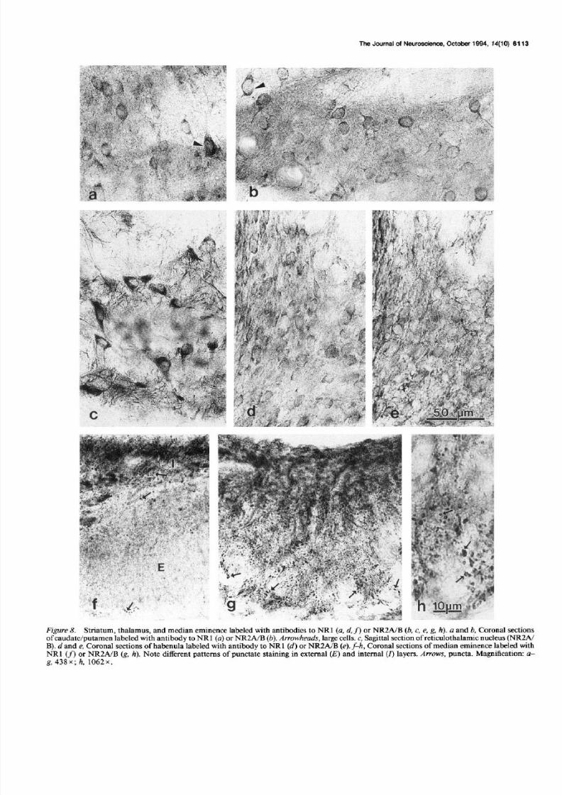
with NRl antibody. Both neurons and neuropil of the supra-chiasmatic nucleus (Fig. 5~) were stained ightly to sometimesmoderately with antibody to NR2A/B, while they were stainedmoderately with antibody to NRl . In the arcuate nucleus/me-dian eminence region (Figs.4d, 8g,h), sections mmunolabeledwith antibody to NR2A/B bore numerous dense puncta in the
external layer of the median eminence. Smaller puncta were
dot-like while larger onesoften were shaped ike irregular rings.These puncta often were arranged in long trains, presumably
reflecting their regular placement along an unstained process,which wassometimesvisible. Suchpuncta were uncommon andlessdefinitive in the internal layer of the median eminenceandarcuate nucleus. In contrast, with antibody to NRl, similarpuncta were common in the internal layer of the median emi-nence and arcuate nucleus, and were rare in the external layer
of the median eminence origin of the pituitary portal system;for example, Armstrong, 1985) (Fig. 8~). Staining was promi-nent in all lobes of the pitui tary with both antibodies, although
it was densestn the anterior and posterior lobeswith antibodyto NR2A/B and in the intermediate lobe with antibody to NR 1(Petralia et al., 1994).
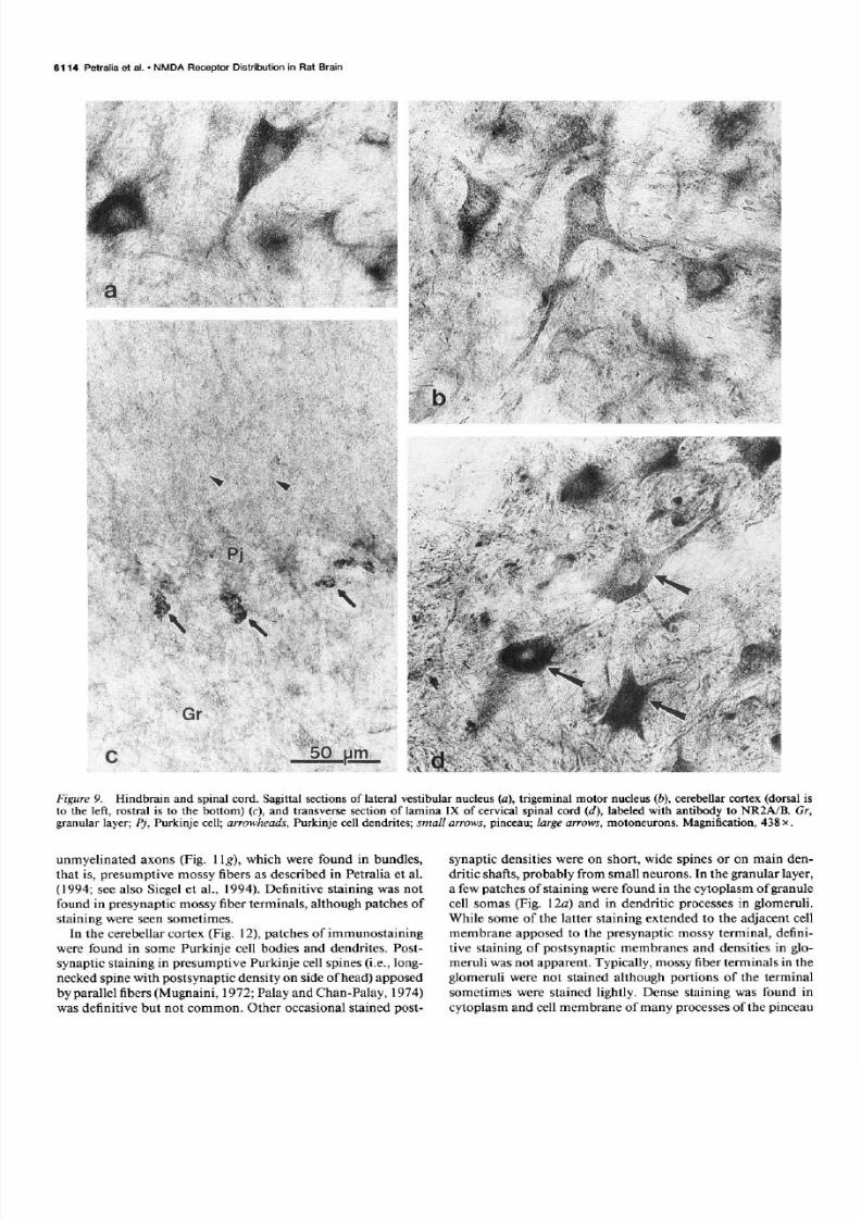
Immunostaining wasprominent and similar overall with bothantibodies throughout many nuclei in the midbrain and hind-brain (Table 1, Fig. 9a,b), including the dorsal cochlear (Fig.5b), ateral vestibular (Fig. 9a), trigeminal motor (Fig. 9b), facial,supraspinalnuclei, large multipolar neurons of the deep ayersof the superior colliculus, a few large multipolar neurons n theexternal cortex of the inferior colliculus, and large neurons ofsome medial reticular nuclei such as the gigantocellular and
paragigantocellular nuclei. Staining of neurons in some nuclei,
such as he dorsal motor nucleusof the vagus, hypoglossalnu-cleus, nferior olive, and locuscoeruleus, ypically was ight withNR2A/B antibody compared o a moderateor moderately dense

7/28/2019 The NMDA Receptor Subunits NR2A and NR2B Show Histological and Ultrastructural Localization Patterns Similar to…
http://slidepdf.com/reader/full/the-nmda-receptor-subunits-nr2a-and-nr2b-show-histological-and-ultrastructural 9/19
6110 Petralia et al. l NMDA Receptor Distribution in Rat Brain
Figure 5. Low magnifications of coronal sections immunolabeled with antibody to NRZA/B, arranged from rostra1 forebrain (a) to cervical spinalcord (e). Abbreviations are as defined for Figure 4. Magnification: 7.1 x .
staining with NRl antibody (Petralia et al., 1994); althoughneuropilar staining wasmoderate with both antibodies. Stainingwith antibody to NR2A/B of the cortex of the cerebellum wasmoderate overall, in both granular and molecular layers (Figs.4b, 5b, SC).Purkinje cell stainingwith NR2A/B antibody variedfrom unstained to lightly stained (Figs. 4b, 5b, SC), n contrastto staining with antibody to NRl in which the Purkinje cellbodies were stained densely (Fig. 4a) (Petralia et al., 1994).Staining with antibody to NR2A/B was prominent in the pin-ceau (Fig. SC), hat is, a complex structure consistingmainly ofa branched, convoluted basket cell axon surrounding the prox-imal portion of the Purkinje cell axon (Palay and Chan-Palay,1974). Staining of this structure was not evident with antibodyto NR 1. Staining in the cervical spinalcord wasmostprominentin lamina IX and dorsal horn. In lamina IX, many large mo-toneurons were stained moderately dense o densely, and themoderately stained neuropil contained numerous, arge, densepuncta (Fig. 9d). Neurons in most other laminae (V-VIII) werestained ightly or were unstained. In contrast, many neurons nthese amina stained moderately with antibody to NRl (Petralia
et al., 1994). In cranial (vestibular, trigeminal) and spinal gan-glia, neurons of all sizes stained moderately to densely withantibodies to NRl and NR2A/B.
No definitive differences n stainingpattern wereseenbetweensections rom male rats and a limited number of sections roma female rat, although t is ikely that somedifferences n NMDAreceptorsexist betweensexes Akinci and Johnston, 1993;Han-ack and Loscher, 1993).
Electron microscopedistribution
Immunostaining with antibody to NR2A/B was dense n manydendrites hroughout the cerebral cortex, hippocampus,and cer-ebellar cortex. Staining was lessdense n cell bodies and otherdendrites and consisted of patches of staining. These patcheswereassociatedwith microtubules, vesicles, ough endoplasmic
reticulum, Golgi, and the surfacesof mitochondria and the nu-clear envelope, asdescribed or immunolabeling with antibodyto NR 1 (Petralia et al., 1994). mmunostaining of synapses su-ally was imited to the postsynaptic density and membraneandassociated ytoplasm, with no staining n the synaptic cleft andlittle or none n the presynaptic terminal, which contained roundor pleomorphic (mostly round) vesicles.Absence of staining inthe cleft supportsan intracellular location of the C-terminus ofNR2, as suggestedor NRl (Tingley et al., 1993; Petralia et al.,1994). Although staining was usually only postsynaptic, a lightstaining was found rarely in presynaptic terminals but was notdefinitive. In spine synapses, staining typically extendedthroughout the entire spine head and into the neck, and some-times was very dense.There was no clear evidence of stainingin glia although some of the small, stained, unidentified pro-cesses ould be parts of glial cells. Other stained structures n-cluded the inner surfaceof blood vessels nd, occasionally, my-elinatedaxons (asnoted for CA 1 hippocampalmyelinated axonswith antibodies to AMPA receptors; Martin et al., 1993).
In the cerebralcortex (Fig. lo), staineddendritesand synapses
were found throughout the gray matter with antibody to NR2A/B. Among postsynaptic densitieswith definitive staining, densestaining (Fig. lOb,c) was seenmore commonly with NR2A/Bantibody than with NRl antibody (Petralia et al., 1994). Oneexample of dense mmunolabelingof a presynaptic terminal wasseen,but structural details were obscuredby the staining.
In the hippocampus (Fig. 1 ), densely stained postsynapticdensities and spineswere found commonly in stratum oriens(Fig. 1 a,b) and molecular ayer (Fig. 1 c,d) of both the CAY2and CA3 regions. n the stratum lucidum, staining sometimeswasseen n postsynaptic densitiesof specializedspines f mossyfiber synapsesFig. 11e). While this staining was not common,it varied from light to dense,compared o the light stainingseenuncommonly in similar synapses ith antibody to NRl (Petraliaet al., 1994). In addition, patches of staining were found in

7/28/2019 The NMDA Receptor Subunits NR2A and NR2B Show Histological and Ultrastructural Localization Patterns Similar to…
http://slidepdf.com/reader/full/the-nmda-receptor-subunits-nr2a-and-nr2b-show-histological-and-ultrastructural 10/19
Figure 6. Olfactory bulb and cerebral cortex labeled with antibody to NR2A/B (a, b, d) or NR 1 (c). a, Sagittal section of external plexiform layerof olfac tory bulb. Mi, mitral cell; arrowheads, dendrites. b, Coronal section of hindlimb area of cerebral cortex. IV-U, layers IV-VI. c and d,Coronal sections of layer V of hindlimb/parietal area of cerebral cortex (caudal to area in b), labeled with antibody to NRl (c) and NRZA/B (d).Arrowheads, apical dendrites of pyramidal cell. Magnification: a, c, and d, 438 x ; b, 114 x .

7/28/2019 The NMDA Receptor Subunits NR2A and NR2B Show Histological and Ultrastructural Localization Patterns Similar to…
http://slidepdf.com/reader/full/the-nmda-receptor-subunits-nr2a-and-nr2b-show-histological-and-ultrastructural 11/19
6112 Petralia et al. l NMDA Rece ptor Distribution in Rat Brain
Figure 7. Sagittal sections of hippocampus labeled with antibody to NRZA/B. a, Low magnification of rostra1 two-thirds of hippocampus. Areaslabeled b-d are shown at higher magnifications in panels b-d. b, CA1 region. c, CA3 region. d, Dentate gyrus. Cl and C3, CA1 and CA3 regions;DC, dentate gyrus; Gr, granule cells of dentate gyrus; sl, stratum lucidum; arrows, hilar ce lls; arrowheads, apical dendrite of pyramidal cells; asterisk,lighter-stained proximal third of molecular layer of dentate gyrus. Magnification: a, 56 x ; &d, 438 x .
7/28/2019 The NMDA Receptor Subunits NR2A and NR2B Show Histological and Ultrastructural Localization Patterns Similar to…
http://slidepdf.com/reader/full/the-nmda-receptor-subunits-nr2a-and-nr2b-show-histological-and-ultrastructural 12/19
The Journal of Neuroscience, October 1994, 14(10) 6113
Figure 8. Striatum, thalamus, and median eminence labeled with antibodies to NRl (a, d, f) or NR2A/B (!I , c, e, g, h). a and b, Coronal sectionsof caudatejputamen labeled with antibody to NR 1 (a) or NR2AIB (b). Arrowheads, large cells. c, Sag&al section of reticulothalamic nucleus (NRZAIB). d and e, Coronal sections of habenula labeled with antibody to NRl (d) or NRZA/B (e). f-h , Coronal sections of median eminence labeled with
NRl (fl or NRZA/B (g, h). Note different patterns o f punctate staining in external (E) and internal (r) layers. Arrows, puncta. Magnification: a-g, 438x; h, 1062x.
7/28/2019 The NMDA Receptor Subunits NR2A and NR2B Show Histological and Ultrastructural Localization Patterns Similar to…
http://slidepdf.com/reader/full/the-nmda-receptor-subunits-nr2a-and-nr2b-show-histological-and-ultrastructural 13/19
6114 Petralia et al. - NMDA Receptor Distribution in Rat Brain
Figure 9. Hindbrain and spinal cord. Sag&al sections of lateral vestibular nucleus (a), trigeminal motor nucleus (b), cerebellar cortex (dorsal isto the lef t, rostra1 is to the bottom) (c), and transverse section of lamina IX o f cervical spinal cord (d), labeled with antibody to NRZA/B. Gr,granular layer; Pj, Purkinje cell; arrowheads, Purkinje cell dendrites; small arrows, pinceau; large arrows, motoneurons. Magnification, 438 x .
unmyelinated axons (Fig. 1 lg), which were found in bundles,that is, presumptive mossy fibers as described in Petralia et al.(1994; seealso Siegelet al., 1994). Definitive staining was notfound in presynaptic mossy iber terminals, although patchesofstaining were seensometimes.
In the cerebellar cortex (Fig. 12) patchesof immunostainingwere found in some Purkinje cell bodies and dendrites. Post-synaptic staining in presumptive Purkinje cell spines i.e., long-neckedspinewith postsynaptic density on sideof head)apposed
by parallel fibers Mugnaini, 1972;Palay and Chart-Palay, 1974)was defini tive but not common. Other occasional stained post-
synaptic densitieswere on short, wide spinesor on main den-dritic shafts,probably from small neurons. n the granular ayer,a few patchesof stainingwere found in the cytoplasm of granulecell somas Fig. 12~) and in dendritic processesn glomeruli.While some of the latter staining extended to the adjacent cellmembrane apposed o the presynaptic mossy terminal, defini-tive staining of postsynaptic membranesand densities n glo-meruli wasnot apparent. Typically, mossy iber terminals n theglomeruli were not stained although portions of the terminalsometimeswere stained lightly. Dense staining was found incytoplasm and cell membraneof many processes f the pinceau

7/28/2019 The NMDA Receptor Subunits NR2A and NR2B Show Histological and Ultrastructural Localization Patterns Similar to…
http://slidepdf.com/reader/full/the-nmda-receptor-subunits-nr2a-and-nr2b-show-histological-and-ultrastructural 14/19
The Journal of Neuroscience, Octob er 1994, 14(10) 6115
Figure 10. Electronmicrographsf cerebral ortex. mmunostaining ith antibody o NR2A/B is shownn b andc. a, Controlsectionno primaryantibody)shows omplete bsencef immunostainingn synapseun; hisexamplewas neof thedensestound;mostwere ighter).Notemoderatestainingn myelin asterisks). b andc, Dense tainingwas ound n dendritesd) and n postsynaptic ensitiesarrows) andadjacent ytoplasm fdendriticspines. n, unstained ynapse. agnification,50,000 .
(Fig. 12~). In addition, staining was found in patches n un-myelinated fibers found in bundles n the molecular layer (Fig.12b), that is, presumedparallel fibers, as described or stainingwith antibody to NRl (Petralia et al., 1994).
Controls
Staining was absent from PBS control sections.Preadsorptioncontrol sectionswere unstained except for a very light stainingin some cells and processes,notably the trigeminal sensory,lateral reticular, pontine, supraoptic, and dorsalcochlear nuclei,and sometimesPurkinje cells. n addition, staining n the pinealgland, posterior pituitary lobe and glia of the peripheral white
matter of the spinal cord was only slightly reduced by pread-sorption. Correspondingexperimental sections i.e., no peptide)were stained normally. Staining was absent from PBS controlthin sectionsof cerebral cortex, hippocampus, and cerebellarcortex examined with electron microscopy (Figs. lOa, 1 fl, ex-cept for myelin sheathsof axons (Fig. lOa) and large, multi-vesicular body-like structures, as described for NRl (Petraliaet al., 1994). As in the NRl study, synapses ere never stainedexcept for a few of the small spinesynapses,which bore lightlystained postsynaptic densities, n the stratum oriens and mo-lecular layer of the hippocampus. None of the latter possesseddensestaining as seenwith NR2A/B antibody in postsynapticdensitiesand cytoplasm of smallspinesynapses f these egions.

7/28/2019 The NMDA Receptor Subunits NR2A and NR2B Show Histological and Ultrastructural Localization Patterns Similar to…
http://slidepdf.com/reader/full/the-nmda-receptor-subunits-nr2a-and-nr2b-show-histological-and-ultrastructural 15/19
61 16 Petralia et al. - NMDA Receptor Distribution in Rat Brain
Figure II. Electron micrographs of hippocampus. Immunostaining with antibody to NR2A/B shown in micrographs u-e and g. u-d, Dense
staining in postsynaptic densities (arrows) in dendritic spines in the stratum oriens (a, b) and molecular layer (c, d) of CA2 (a, d) and CA3 (b, c)regions. e, Presumed apical dendrite of pyramidal cell of CA3 region with dense patches of immunostaining. Note staining (not common) in
postsynapt ic densities (arrows) of specialized spines apposed to mossy fiber terminal (asterisk). J Corresponding control section (no primaryantibody) shows complete absence of immunostaining in dendrite (left half of micrograph) and specialized spine synapses (un) of CA3 region. g,
Patches of staining (arrowheads) in unmyelinated axons of CA3 region. Magnification: e-g, 20,000; u-d, 50,000 x .

7/28/2019 The NMDA Receptor Subunits NR2A and NR2B Show Histological and Ultrastructural Localization Patterns Similar to…
http://slidepdf.com/reader/full/the-nmda-receptor-subunits-nr2a-and-nr2b-show-histological-and-ultrastructural 16/19
The Journal of Neuroscience, Octobe r 1994. 74(10) 6117
Figure 12. Electron micrographs of cerebellar cortex, immunostained with antibody to NRZA/B. a, Granular layer showing dense staining insome processes of a presumptive pinceau (smaN arrows) and light staining (large arrow) in cytoplasm of a granule cell (Gr). b, Molecular layershowing patches of staining (arrowheads) in presumed parallel fibers . un, unstained parallel fiber synapse. Magnification, 20,000 x .
Discussion
In this study we developed an antibody to the C-terminus ofNR2A that reacts intensely with both NR2A and NR2B onimmunoblots of transfected cell membranesand labelsa singleband that comigrateswith the NR2A and NR2B subunits n ratbrain tissues.Since four and six of the 20 amino acid residuesof the sequence sed for producing the antibody are identicalin NR2C and NR2D-2, respectively, we performed immunoblotanalysis of membranesof cell transfected with these subunits,and found a weak reaction with both NR2C and NR2D-2. Cor-responding labeling of bands in rat tissues,particularly cere-bellum where NR2C is most abundant and in hindbrain whereNR2D is most abundant, was not seen.This lack of labeling islikely due to the low affinity of the antibody for the NR2C and
NR2D-2 subunits or may indicate a relatively low expressionof these subunits in brain. Basedon these results we interpretour immunocytochemical labeling to represent primarily theNR2A and NR2B subunits. This is supported by the fact thatthe antibody gives rather weak abeling n the cerebellum,whereNR2C is most abundantly expressed.However, we cannot ruleout the possibility that in somepopulations of neurons, NR2Cand NR2D-2 are abundantly expressed, nd our labeling reflectsprimarily thesesubunits.
The similarity of this distribution to that of antibody to NRl(Petralia et al., 1994) indicates that NRl and NR2 colocalizein most neurons n the CNS, as ndicated in in situ hybridizationstudies Monyer et al., 1992;Nakanishi, 1992; Ishii et al., 1993).
This is consistent with physiological studies that support thepresenceof functional NMDA receptors n most structures n
the CNS (discussedn Stone and Burton, 1988; Collingridge and
Lester, 1989; Petralia et al., 1994).Ultrastructural distributionof NR2 was also similar to that of NRl, with staining mostcommonly found in dendritesand postsynapticmembranesanddensities.Any differences n stainingdensity in individual struc-tures examined with light or electron microscopy may reflectdifferences n distribution and/or different ratios of NR 1 NR2subunits n various portions of individual neurons. The wide-spreaddistribution of NRl and NR2A/B antibody staining inneuronal populations implies colocalization with other gluta-mate receptor types throughout the nervous system (see dis-cussion n Petralia et al., 1994); this supportsa model in whichpostsynaptic membranes ontain non-NMDA receptors or fastexcitatory transmissionand NMDA receptors that modify theresponse Bekkers and Stevens, 1989; Jones and Baughman,
1991; Riquelme et al., 1993).
Comparison to in situ hybridization and ligand binding
studies
The pattern of immunostaining with antibody to NR2A/B cor-responds o the combined patterns of mRNA distribution ofNR2A and NR2B, with a possiblecontribution of NR2C andNR2D (Monyer et al., 1992; Nakanishi, 1992; Watanabe et al.,1992, 1993; Ishii et al., 1993).For example, substantialstainingin some neuron populations of midbrain and hindbrain corre-
sponds o significant mRNA levels of NR2A, such as in thepontine nuclei, inferior colliculus, and inferior olive (Ishii et al.,1993). Staining in some structures such as hypothalamus and
spinal cord, where ittle or no mRNA for NR2A and NR2B hasbeen detected, could reflect the presenceof NR2C or NR2D

7/28/2019 The NMDA Receptor Subunits NR2A and NR2B Show Histological and Ultrastructural Localization Patterns Similar to…
http://slidepdf.com/reader/full/the-nmda-receptor-subunits-nr2a-and-nr2b-show-histological-and-ultrastructural 17/19
6116 Petralia et al. - NMDA Receptor Distribution in Rat Brain
(Monyer et al., 1992; Ishii et al., 1993; Tij lle et al., 1993; Wa-
tanabe et al., 1993). The lower staining with NR2A/B antibody
than with NR 1 antibody in many neuron types throughout many
regions of the brain may correspond to the apparent high ex-
pression of mRNA for NR 1 compared overall to NR2A-NR2D,
as illustrated for many forebrain structures (Monyer et al., 1992;
Nakanishi, 1992) possibly indicat ing (1) the expression of
NMDA receptor complexes with high NRl:NR2 ratios, (2) ad-
dit ional expression of monomeric NRl receptor complexes, and/
or (3) a higher cytoplasmic pool of NR 1. However, lower stain-
ing seen in some structures that contain a predominance of
NR2D mRNA (i.e., throughout the brain stem; Ishii et al., 1993)
may be due to low detection of NR2D protein by our antibody,
as indicated on immunoblots, or to a predominance of mRNA
for NR2D-1, which is not detected by our antibody. The pres-
ence of dense staining with NR 1 antibody in cerebellar Purkinje
cells, compared to lit tle or no staining with NR2A/B antibody,
is supported by in situ hybrid ization studies indicat ing signifi-
cant mRNA for NRl in Purkinje cells (Moriyoshi et al., 1991)
compared to apparently lit tle or no mRNA for NR2 subunits
in Purkinje cells. Also, it is consistent with physiological studiesindicating that Purkinje cells in adult animals do not have func-
tional NMDA receptors (i .e., since monomeric NRl receptors
show l itt le functional response; Moriyoshi et al., 199 1; discussed
in Petralia et al., 1994). The low levels of NR2 protein some-
times seen in Purkinje cells in our study may be nonspecific
since similar levels sometimes were seen in preadsorption con-
trols. However, cel l level studies of NR2 mRNA distribution
in the cerebellum have not been published, and we cannot rule
out that one or more NR2 subunits are present in low amounts
in some Purkinje cells.As with the antibody to NR 1, the widespread distribution of
NR2A/B antibody is consistent overal l with data from NMDA
ligand binding studies, and has been discussed in detai l for NR 1
antibody (Petralia et al., 1994). As also noted in that article (seealso Monaghan et al., 1993) and demonstrated by pharmaco-
logical /binding studies of subunit expression in cel l cultures andoocytes (Buller et al., 1993; Marti et al., 1993; Raditsch et al.,
1993; Wafford et al., 1993; Yamakura et al., 1993) presence of
NMDA receptor complexes made up of NRl in combination
with one or more NR2 subunits may account for many of thedifferences in ligand binding seen in different regions of the
brain. Interestingly, higher dendr itic staining with NR2A/B an-
tibody, compared to higher cel l body staining with our NRl
antibody, corresponds with higher binding of NMDA-sensitive
@H-glutamate in dendr itic zones compared to white matter
regions and cel l body layers (Monaghan and Cotman, 1985)
that is, suggesting a direct relationship between NR2 subunitand ligand-b inding site distribution.
Cellular distribution
The dense staining seen with our NRl and NR2A/B antibodies
in many neuron cel l bodies could indicate maintenance of a high
cytoplasmic pool, as suggested for AMPA receptors (e.g., Pe-tralia and Wenthold, 1992). Immunostaining with antibodies
to NRl and NR2A/B, in many dendrites, dendritic spines, and
postsynaptic densities implies that complexes of NR l/NR2 sub-
units are present postsynaptic to glutamatergic input. In the
cerebral cortex, densely stained postsynaptic densities were less
common with NRl antibody (Petralia et al., 1994) than withNR2A/B antibody, perhaps reflecting a typically lower number
of NR 1 subunit molecules in the postsynaptic region. This could
be due to variations in NR 1 NR2A/B ratios in receptor com-
plexes. Alternatively, moderate staining with NR 1 antibody could
reflect its detect ion of only four of the NRl variants, although
these four are the major ones expressed in rat brain (Sugihara
et al., 1992). In the hippocampus, the intense postsynaptic la-
bel ing of many small spine synapses with NR2A/B antibody
complements the widespread distribution of staining for NRl
in similar synapses, thus supporting numerous studies showing
NMDA receptor-mediated long term potentiation (LTP) in both
CA 1 and CA3 regions (discussed in Zalutsky and Nicoll, 1990;
Malenka and Nicoll, 1993; Petralia et al., 1994). In postsynaptic
densities apposed by mossy fiber terminals in the CA3 region
of the hippocampus, immunostaining was typica lly low with
either NR 1 or NR2A/B antibodies, although dense staining with
antibody to NR2A/B was seen in a few act ive zones. This is
genera lly consistent with the presence of only NMDA-indepen-dent LTP in this synaptic population (e.g., Zalutsky and Nicoll,
1990) although the data suggest the presence of limited popu-
lations of NR 1 and NR2 subunits. Differential staining of outer
and inner portions of the dentate gyrus molecu lar layer implies
that different levels ofNR2 subunits are associated with di fferentinputs onto granule cel l dendrites (see discussion for NRl; Pe-
tralia et al., 1994). The pattern of staining with NR2 antibody,
that is, denser staining in the outer portion, differs from that
described in some studies with NR 1 antibodies (polyc lonal NR 1:
Petralia et al., 1994; monoclonal NRl: Siegel et al., 1994; R. S.
Petral ia, Y.-X. Wang, and R. J. Wenthold, unpubl ished obser-
vations), but resembles that described in another (polyclonal
NRI: Brose et al., 1993). Differences between the staining with
our NR2 antibody and that of NR 1 antibodies can be attributed
to the presence of different ratios of NRl:NR2 associated with
different inputs. The similarity of our NR2 antibody staining
to that of one of the polyclonal NRl antibodies (Brose et al.,
1993) may be due to preferential recognition of different subunit
variants by different antibodies. In sections of cerebellar cortex
immunostained with antibody to NR2A/B, the infrequency of
stained, postsynaptic densities in presumptive Purkinje cel l spines
apposed to paralle l fibers is consistent with the typically low
staining of Purkinje cells. The few examples of stained densities
may represent a minor subpopulation of Purkinje cel l spines
conta ining NR2 subunits. The significant staining for NR 1 and
NR2A/B of presumptive paralle l fibers in the molecular layer
may account for at least part of the high levels of NR 1 and NR2
mRNA found in the granular layer (Moriyoshi et al., 1991;
Monyer et al., 1992; Ishii et al., 1993; Watanabe et al., 1993),
that is, NR2 protein expressed in the axons of the granule cells.
Nevertheless, there is considerable evidence for NMDA recep-
tors at granule cell-mossy fiber synapses (e.g., Silver et al., 1992),although staining of these synapses with NRl and NR2A/B
antibodies was not definitive. This low staining may be due, at
least partly, to the low detection of NR2C by NR2A/B antibody,
since NR2C mRNA is high in the granular layer (Ishii et al.,
1993). Staining with NR2A/B antibody of the pinceau may be
due to cross-reaction with another antigen, that is, a protein
that is highly localized to the pinceau (Tigyi et al., 1990), or
indicate that molecular layer basket cells contain NR2. Presence
of NR2 mRNA in molecu lar layer cells has not been described,
substantial mRNA has been described only in the granular layer
(Ishii et al., 1993; Watanabe et al., 1993). A preliminary report
describes immunosta ining of molecular layer cells with an NR2Cantibody (Mulac-Jericevic et al., 1993) but these results have
not been corroborated.

7/28/2019 The NMDA Receptor Subunits NR2A and NR2B Show Histological and Ultrastructural Localization Patterns Similar to…
http://slidepdf.com/reader/full/the-nmda-receptor-subunits-nr2a-and-nr2b-show-histological-and-ultrastructural 18/19
The Journal of Neuroscience, Octobe r 1994, 14(10) 6119
References
Akinci MK, Johnston GAR (1993) Sex differences in acute swimstress-induced changes in the binding of MK-80 1 to the NMDA sub-class of glutamate receptors in mouse forebrain. J Neurochem 61:2290-2293.
Alford S, Dubuc R (1993) Glutamate metabotropic receptor mediateddepression of synaptic inputs to lamprey reticulospinal neurones. Brain
Res 605: 175-I 79.Anantharam V, Panchal RG, Wilson A, Kolchine VV, Treistman SN,
Bayley H (1992) Combinatorial RNA splicing alters the surfacecharge on the NMDA receptor. FEBS Lett 30527-30.
Armstrong WE (1985) Hypothalamic supraoptic and paraventricularnuclei. In: The rat nervous system, Vol 1 (Paxinos G, ed), pp 119-128. New York: Academic.
Bekkers JM, Stevens CF (1989) NMDA and non-NMDA receptorsare co-localized at individual excitatory synapses in cultured rat hip-pocampus. Nature 341:230-233.
Brose N. Gasic GP. Vetter DE. Sullivan JM. Heinemann SF (1993)Protein chemical’characterization and immunocytochemical‘local:ization of the NMDA receptor subunit NMDA Rl. J Biol Chem 268:22663-2267 1.
Buller AL, Monisett RA, Monaghan DT (1993) The NR2 subunitcontributes to the pharmacological diversity o f native NMDA recep-tors. Sot Neurosci Abstr 19: 1356.
Burgoyne RD, Graham ME, Cambray-Deakin M (1993) Neurotrophiceffects of NMDA receptor activation on developing cerebellar granulecells. J Neurocytol 22:689-695.
Cik M. Chazot PL. Steohenson FA (1993) Optimal exnression of.
cloned NMDAR 1 NMDAR2A heteromeric glutamate receptors: abiochemical characterization. Biochem J 296:877-883.
Collingridge GL, Lester RAJ (1989) Excitatory amino acid receptorsin the vertebrate central nervous system. Pharmacol Rev 4 1:143-2 10.
Durand GM, Bennett MVL, Zukin RS (1993) Splice variants of theN-methyl-D-aspartate receptor NR 1 identify domains involved inregulation by polyamines and protein kinase C. Proc Nat1 Acad SciUSA 90:6731-6735.
Furuyama T, Kiyama H, Sato K, Park HT, Maeno H, Takagi H, To-hvama M (1993) Region-svecific exvression of subunits of iono-tropic glutamate recept&s (AMPA-type, KA-type and NMDA recep-tors) in the rat spinal cord with special reference to nociception. Mol
Brain Res 18:141-151.Hollmann M, Boulter J, Maron C, Beasley L, Sullivan J, Pecht G,
Heinemann S (1993) Zinc potentiates agonist-induced currents atcertain splice variants of the NMDA receptor. Neuron 10:943-954.
H&tack D, Liischer W (1993) Sex differences in NMDA receptormediated responses in rats. Brain Res 620: 167-l 70.
Ikeda K, Nagasawa M, Mori H, Araki K, Sakimura K, Watanabe M,Inoue Y, Mishina M ( 1992) Cloning and expression of the ~4 subunit
of the NMDA receptor channel. FEBS Lett 3 13:34-38.Ishii T, Moriyoshi K, Sugihara H, Sakurada K, Kadotani H, Yokoi M,
Akazawa C, Shigemoto R, Mizuno N, Masu M, Nakanishi S (1993)Molecular characterization of the fam ily of the N-methyl-D-aspartate
receptor subunits. J Biol Chem 268:2836-2843.Itano Y, Murayama T, Kitamura Y, Nomura Y (1992) Glutamate
inhibits adenylate cyclase act ivi ty in dispersed rat hippocampal cellsdirectly via an N-methyl-D-aspartate-like metabotropic receptor. J
Neurochem 59:822-828.Jones KA, Baughman RW (199 1) Both NMDA and non-NMDA sub-
types of glutamate receptors are concentrated at synapses on cerebralcortical neurons in culture. Neuron 7593-603.
Karp SJ, Masu M, Eki T, Ozawa K, Nakanishi S (1993) Molecularcloning and chromosomal localization ofthe key subunit of the humanN-methyl-D-aspartate receptor. J Biol Chem 268:3728-3733.
Kirkwood A, Dudek SM, Gold JT, Aizenman CD, Bear MF (1993)
Common forms of synaptic plasticity in the hippocampus and neo-cortex in vitro. Science 260: 15 18-l 52 1.
Kumar KN, Tilakaratne N, Johnson PS, Allen AE, Michaelis EK (199 1)Cloning of cDNA for the glutamate-binding subunit o f an NMDA
receptor complex. Nature 354:70-73.Kusiak JW, Norton DD (1993) A splice variant of the N-methyl-D-
aspartate (NMDARl) receptor. Mel Brain Res 20:64-70.Kutsuwada T, Kashiwabuchi N, Mori H, Sakimura K, Kushiya E, Araki
K, Meguro H, Masaki H, Kumanishi T, Arakawa M, Mishina M
(1992) Molecular diversity of the NMDA receptor channel. Nature358:3641.
Malenka RC, Nicoll RA (1993) NMDA-receptor-dependent synapticplast icity: multiple forms and mechanisms. Trends Neurosci 16:521-527.
Marti T, Benke D, Mertens S, Heckendom R, Pozza M, Allgeier H,Angst C, Laurie D, Seeburg P, Mohler H (1993) Molecular distinc-tion of three N-methyl-D-aspartate-receptor subtypes in situ and de-velopmental receptor maturation demonstrated with the photoaffin ityligand ‘251-labeled CGP 55802A. Proc Nat1 Acad Sci 90:8434-8438.
Martin LJ, Blackstone CD, Levey AI, Huganir RL, Price DL (1993)AMPA glutamate receptor subunits are differentially distributed inrat brain. Neuroscience 53:327-358.
Mattson MP, Kumar KN, Wang H, Cheng B, Michaelis EK (1993)Basic FGF regulates the expression of a functional 7 1 kDa NMDAreceptor protein that mediates calcium influx and neurotoxicity inhippocampal neurons. J Neurosci 13:45754588.
Meguro H, Mori H, Araki K, Kushiya E, Kutsuwada T, Yamazaki M,Kumanishi T, Arakawa M, Sakimura K, Mishina M (1992) Func-tional characterization of a heteromeric NMDA receptor channel ex-pressed from cloned cDNAs. Nature 357:70-74.
Monaghan DT, Cotman CW (1985) Distribution of N-methyl-D-as-partate-sensitive r.+H]glutamate-binding sites in rat brain. J Neu-rosci 5:2909-29 19.
Monaghan DT, Bridges RJ, Cotman CW (1989) The exci tatory aminoacid receptors: their classes, pharmacology, and distinct properties in
the function of the central nervous system. Annu Rev PharmacolToxic01 291365-102.Monaghan DT, Clark HC, Schneider BE (1993) Distributions of NMDA
receptor subtypes correspond to specif ic receptor subunits. Sot Neu-rosci Abstr 19: 1356.
Monyer H, Sprengel R, Schoepfer R, Herb A, Higuchi M, Lomeli H,Bumashev N, Sakmann B, Seeburg PH (1992) Heteromeric NMDAreceptors: molecular and functional distinction of subtypes. Science256:1217-1221.
Mori H, Yamakura T, Masaki H, Mishina M (1993) Involvement ofthe carboxyl-terminal region in modulation by TPA of the NMDAreceptor channel. Neuroreport 4:5 19-522.
Moriyoshi K, Masu M, Ishii T, Shigemoto R, Mizuno N, Nakanishi S(1991) Molecular cloning and characterization of the rat NMDAreceptor. Nature 354:31-37.
Mugnaini E (1972) The histology and cyto logy of the cerebellar cortex.In: The comparative anatomy and histology of the cerebellum: the
human cerebellum, cerebellar connections, and cerebellar cortex (Lar-sell 0, Jansen J, eds), pp 201-25 1. Minneapolis: Unive rsity of Min-nesota.
Mulac-Jericevic B, Benke TA, Peterson NL, Angelides KJ (1993) Dis-tribution of NMDA receptor subunits on rat hippocampal, cerebellarand cortical neurons in culture and in brain sl ices using subunit spe-cifi c antibodies. Sot Neurosci Abstr 19: 1355.
Nakanishi N, Axe1 R, Shenider NA (1992) Alternative splicing gen-
erates functionally distinct N-methyl-D-aspartate receptors. Proc Nat1Acad Sci 89:8552-8556.
Nakanishi S (1992) Molecular diversity of glutamate receptors andimplications for brain function. Science 258:597-603.
Okamoto N, Hori S, Akazawa C, Hayashi Y, Shigemoto R, Mizuno N,
Nakanishi S (1994) Molecular characterization of a new metabo-tropic glutamate receptor mGluR7 coupled to inhibitory cyc lic AMP
signal transduction. J Biol Chem 269: 123 1-1236.
Palay SL, Chan-Palay V (1974) Cerebellar cortex: cytology and or-ganization. New York: Springer.
Paxinos G, Watson C (1986) The rat brain in stereotaxic coordinates,2d ed. New York: Academic.
Peters A, Palay SL, Webster HF (1991) The fine structure o f thenervous system, 3d ed. New York: Oxford UP.
Petralia RS, Wenthold RJ (1992) Light and electron immunocyto-chemical localization of AMPA-selective glutamate receptors in the
rat brain. J Comp Neural 3 18:329-354.
Petralia RS, Yokotani N, Wenthold RJ (1994) Light and electronmicroscope distribution of the NMDA receptor subunit NMDARlin the rat nervous system using a selective anti-peptide antibody. JNeurosci 14~667-696.
Planells-Cases R, Sun W, Ferrer-Montiel AV, Montal M (1993) Mo-lecular cloning, functional expression, and pharmacological charac-terization of an N-methyl-D-aspartate receptor subunit from human
brain. Proc Nat1 Acad Sci USA 90:5057-5061.Raditsch M, Ruppersberg JP, Kuner T, Gunther W, Schoepfer R, See-

7/28/2019 The NMDA Receptor Subunits NR2A and NR2B Show Histological and Ultrastructural Localization Patterns Similar to…
http://slidepdf.com/reader/full/the-nmda-receptor-subunits-nr2a-and-nr2b-show-histological-and-ultrastructural 19/19
6120 Petralia et al. * NMDA Receptor Distribution in Rat Brain
burg PH, Jahn W, Witzemann V (1993) Subunit-specific block ofcloned NMDA receptors by argiotoxin,,,. FEBS Lett 324:63-66.
Riquelme G, Wyneken U, Villanueva S, Orrego F (1993) Recordingsofglutamate receptor channels in isolated postsynaptic densities. Neu-roreport 4: 1163-l 166.
Sato K, Kiyama H, Park HT, Tohyama M (1993) AMPA, KA andNMDA receptors are expressed in the rat DRG neurones. Neuroreport4:1263-1265.
Saugstad JA, Kinzie JM, Segerson TP, Westbrook GL (1993) Char-acterization of a new metabotropic glutamate receptor (MGLUR7)
homologous to the AP4 receptor (MGLUR4). Sot Neurosci Abstr 19:68.
Seeburg PH ( 1993) The molecular biology of mammalian glutamatereceptor channels. Trends Neurosci 16:%9-365. -
Shieemoto R. Ohishi H. Nakanishi S. Mizuno N (1992) ExoressionoTthe mRNA for the rat NMDA receptor (NMDARl) in thesensoryand autonomic ganglion neurons. Neurosci Lett 144:229-232.
Siegel, SJ, Brose N, Janssen WG, Gasic GP, Jahn R, Heinemann SF(1994) Regional, cellular, and ultrastructural distribution of N-me-thyl-D-aspartate receptor subunit 1 in monkey hippocampus. ProcNat1 Acad Sci 91:564-568.
Silver RA, Traynelis SF, Cull-Candy SG (1992) Rapid-time-courseminiature and evoked excitatory currents at cerebellar synapses insitu. Nature 355:163-166.
Smirnova T, Stinnakre J, Mallet J (1993a) Characterization of a pre-synaptic glutamate receptor. Science 262:430-433.Smimova T, Laroche S, Errington ML, Hicks AA, Bliss TVP, Mallet J
(1993b) Transsynaptic expression o f a presynaptic glutamate recep-
tor during hippocampal long-term potentiation. Science 262:433-436
Standaert DG, Testa CM, Penney JB Jr, Young AB (1993) Altema-tivelv spliced isoforms of the NMDARl dutamate receptor subunit:
differentia l expression in the basal gang& of the rat. Neurosci Lett152:161-164.
Stem P, Behe P, Schoepfer R, Colquhoun D (1992) Single-channelconductances of NMDA receptors expressed from cloned cDNAs:
comparison with nat ive receptors. Proc R Sot Lond [Biol] 250:271-277.
Stone TW, Burton NR (1988) NMDA receptors and ligands in thevertebrate CNS. Prog Neurobiol 30:333-368.
Sucher NJ. Brose N. Deitcher DL. Awobuluvi M. Gasic GP. Badine H.
Cepko CL, Greenberg ME, Jahn’R, Heinemann’SF, Lipton’SA (1593)Expression of endogenous NMDARl transcripts without receptorprotein suggests post-transcriptional control in PC12 cells. J BiolChem 268:22299-22304.
Sugihara H, Moriyoshi K, Ishii T, Masu M, Nakanishi S (1992) Struc-tures and properties of seven isoforms of the NMDA receptor gen-
erated by alternative splicing. Biochem Biophys Res Commun 185:826-832.
T&vi G, Matute C, Miledi R (1990) Monoclonal antibodies to cere-bellar’ pinceau terminals obtained after immunization with brain
mRNA-iniected Xenonus oocvtes. Proc Nat1 Acad Sci USA 87:528-532. -
Tingley WG, Roche KW, Thompson AK, Huganir RL (1993) Reg-ulation of NMDA receptor phosphorylation by alternative splicingof the C-terminal domain. Nature 364:70-73.
Tiille TR, Berthele A, Zieglgansberger W, Seeburg PH, Wisden W (1993)The differential expression o f 16 NMDA and non-NMDA receptorsubunits in the rat spinal cord and in periaqueductal gray. J Neurosci13:5009-5028.
Ultsch A, Schuster CM, Laube B, Betz H, Schmitt B (1993) Glutamatereceptors of Drosophila melunoguster primary structure of a putativeNMDA receptor protein expressed in the head of the adult fl y. FEBSLett 324:17i-177. -
Waffo rd KA. Bain CJ. Bourdelles BL. Whitina PJ. Kemp JA (1993)Preferential co-assembly of recombinant NMDA receptors composedof three different subunits. Neuroreport 4: 1347-l 349.
Watanabe M, Inoue Y, Sakimura K, Mishina M (1992) Develop-mental changes in distribution of NMDA receptor channel subunit
mRNAs. Neuroreport 3: 1138-l 140.Watanabe M, Inoue Y, Sakimura K, Mishina M (1993) Dist inct dis-tributions of five N-methyl-D-aspartate receptor channel subunitmRNAs in the forebrain. J Comp Neurol 338:377-390.
Wenthold RJ, Yokotani N, Doi K, Wada K (1992) Immunochemicalcharacterization of the non-NMDA glutamate receptor using subunit-specific antibodies: evidence for a hetero-oligomeric structure in ratbrain. J Biol Chem 267:501-507.
Wenthold RJ. Trumov VA. Zhu WS. Petralia RS (1994) Biochemicaland assembly properties’of GluR6 and KA2, two members o f thekainate receptor fam ily, determined with subunit-specific antibodies.J Biol Chem 269:1332-1339.
Yamakura T, Mori H, Masaki H , Shimoji K, Mishina M (1993) Dif-ferent sensitivities of NMDA receptor channel subtypes to non-com-petitive antagonists. Neuroreport 4:687-690.
Yamazaki M, Mori H, Araki K, Mori KJ, Mishina M (1992) Cloning,expression and modulation of a mouse NMDA receptor subunit .
FEBS Lett 300:39-45.Zalutsky RA, Nicoll RA (1990) Comparison of two forms of long-
term potentiation in single hippocampal neurons. Science 248: 16 19-
1624.