nmda -receptor activation induces calpain-mediated
TRANSCRIPT

Neuron
Article
NMDA-Receptor Activation InducesCalpain-Mediated b-Catenin Cleavagesfor Triggering Gene ExpressionKentaro Abe1 and Masatoshi Takeichi1,*1Graduate School of Biostudies, Kyoto University, Kitashirakawa, Sakyo-ku, Kyoto 606-8502,
and RIKEN Center for Developmental Biology, 2-2-3 Minatojima-Minamimachi, Chuo-ku, Kobe 650-0047, Japan
*Correspondence: [email protected] 10.1016/j.neuron.2007.01.016
SUMMARY
The canonical Wnt-b-catenin signaling pathwayis important for a variety of developmental phe-nomena as well as for carcinogenesis. Here, weshow that, in hippocampal neurons, NMDA-receptor-dependent activation of calpain in-duced the cleavage of b-catenin at the N termi-nus, generating stable, truncated forms. Theseb-catenin fragments accumulated in the nucleusand induced Tcf/Lef-dependent gene transcrip-tion. We identified Fosl1, one of the immediate-early genes, as a target of this signaling path-way. In addition, exploratory behavior by miceresulted in a similar cleavage of b-catenin, aswell as activation of the Tcf signaling pathway,in hippocampal neurons. Both b-catenin cleav-age and Tcf-dependent gene transcription weresuppressed by calpain inhibitors. These find-ings reveal another pathway for b-catenin-dependent signaling, in addition to the canoni-cal Wnt-b-catenin pathway, and suggest thatthis other pathway could play an importantrole in activity-dependent gene expression.
INTRODUCTION
Activity-dependent gene expression in neurons is impor-
tant for their development and survival as well as for synap-
tic plasticity (Deisseroth et al., 2003; Kandel, 2001; West
et al., 2002). Several transcription mechanisms have
been shown to be involved in this process (West et al.,
2002). However, how neural activity is transduced to the
gene-transcription machinery remains largely unclear.
One of the well-known signaling systems for regulating
gene expression is the canonical Wnt-b-catenin pathway
(Moon et al., 2004). In this signaling pathway, regulation
of the cytosolic b-catenin level is a key event. In the
absence of Wnt signals, a constitutively active kinase,
GSK-3b, phosphorylates the N-terminal region of cytosolic
b-catenin, leading to the subsequent ubiquitination and
proteasome-mediated degradation of b-catenin (Aberle
N
et al., 1997; Liu et al., 2002; Rubinfeld et al., 1996). Once
Wnt signals are activated, on the other hand, these proteo-
lytic processes are suppressed due to the inhibition of
GSK-3b activity. Then, the stabilized b-catenin is translo-
cated into the nuclei, where it associates with the tran-
scription factor Tcf/Lef and thereby activates gene tran-
scription. The b-catenin-Tcf system is essential for a wide
variety of developmental phenomena (Logan and Nusse,
2004) and is involved in carcinogenesis (Polakis, 2000),
but its physiological role in mature neurons is not well un-
derstood. Nevertheless, malactivation of b-catenin-Tcf-
mediated gene regulation has been reported for various
neurological diseases, including Alzheimer’s disease
(Chong et al., 2005; De Ferrari and Inestrosa, 2000) and bi-
polar disorder (Gould and Manji, 2002), implying that Tcf/
Lef-mediated gene transcription plays a role in brain func-
tions. The Wnt signaling system has alternative pathways,
collectively called the noncanonical pathway (Montcou-
quiol et al., 2006), that are also important for various biolog-
ical phenomena, such as cell polarity regulation; however,
this pathway does not utilize b-catenin as a signaling
mediator.
b-catenin works not only in the above signaling cascade
but also in cadherin-mediated cell-cell adhesions. The
cadherin-catenin complex is localized in synaptic junc-
tions and is involved in synapse formation and stabiliza-
tion (Salinas and Price, 2005; Takeichi and Abe, 2005).
Synaptic localization of cadherins or catenins changes
with synaptic activity (Abe et al., 2004; Murase et al.,
2002; Okamura et al., 2004), and such changes may be
important for the structural and functional plasticity of syn-
apses (Huntley et al., 2002; Murase and Schuman, 1999;
Okamura et al., 2004; Takeichi and Abe, 2005). However,
how synaptic activity regulates the function of these adhe-
sion proteins still remains an unresolved question.
The NMDA-R is an ionotropic glutamate receptor that
plays an important role in synaptic plasticity (Malenka
and Nicoll, 1993), a cellular mechanism for learning and
memory (Nakazawa et al., 2004). Activation of the
NMDA-R results in calcium influx. This NMDA-R-mediated
calcium influx activates a variety of enzymes, including cal-
pain, a calcium-dependent protease. In synapses, spec-
trin (Lynch and Baudry, 1987), NMDA-R2A and -R2B (Gutt-
mann et al., 2001), GluR1 (Bi et al., 1996), PSD-95 (Lu et al.,
euron 53, 387–397, February 1, 2007 ª2007 Elsevier Inc. 387

Neuron
Activity-Dependent b-Catenin Cleavage
2000), and p35 (Kerokoski et al., 2004) have been demon-
strated to be substrates of calpain-mediated cleavage.
Moreover, inhibition of calpain activity was shown to sup-
press the formation of LTP (Staubli et al., 1988). These
observations suggest that calpain plays an important role
in both synaptic plasticity and neuronal degeneration
(Chan and Mattson, 1999; Lynch and Baudry, 1984, 1987;
Vanderklish et al., 1995, 2000).
In the present study, we examined whether neural
activity had any effect on cadherin or catenins, focusing
on NMDA-R-activity-dependent processes. When we ac-
tivated the NMDA-R with glutamate in cultured hippocam-
pal neurons, it resulted in N terminus truncations of b-
catenin. This process was calpain dependent and caused
b-catenin to become resistant to GSK-3b-mediated prote-
olysis. The stabilized b-catenin fragments accumulated in
the nuclei, where they activated Tcf-mediated gene tran-
scription. We further provide evidence that b-catenin-
induced gene transcription also occurs in vivo. Thus, we
found that two signaling pathways, the NMDA-R-depen-
dent calpain activation and b-catenin-mediated gene
regulation, merged together in neurons, forming a novel
mechanism (to our knowledge) for activity-dependent
gene expression.
RESULTS
Activity-Dependent Cleavages of b-Catenin
by Calpain
We prepared hippocampal cultures and treated them with
10 mM glutamate for 30 min. Western blot analysis of cell
lysates showed that this treatment caused the generation
of two fragments of b-catenin (Figure 1A). These b-catenin
fragments were recognized as 85 and 75 kDa bands by
antibodies specific for the b-catenin C terminus but not
by those raised against the N terminus, thus suggesting
that the N terminus of b-catenin had been truncated. In
contrast, glutamate treatment had no obvious effects on
N-cadherin or aN-catenin. The 85 and 75 kDa b-catenin
fragments represented �5% of the total b-catenin in the
cell lysate, as estimated by comparing the band intensities
after serial dilutions of the samples.
The above cleavage of b-catenin was NMDA-R depen-
dent, because pretreatment of neurons with the NMDA-R
antagonist D-2-amino-5-phosphonopentanoate (APV,
250 mM) inhibited the b-catenin truncation, whereas neither
the a-amino-3-hydroxy-5-methyl-4-isoxazolepropionic
acid (AMPA) receptor antagonist 6-cyano-7-nitroquinoxa-
line-2,3-dione (CNQX, 20 mM) nor the mGluR antagonist
(+)-a-methyl-4-carbocyphenylglycine (MCPG, 50 mM or 1
mM) showed any effects (Figure 1B). On the other hand,
treatment of neurons with the NMDA-R agonist NMDA
(25 mM) caused the same cleavage of b-catenin, whereas
the AMPA-R agonist AMPA (2 mM) or the group I mGluR
agonist 3,5-dihydroxyphenylglycine (DHPG, 10 mM) was
not effective (Figure 1B), confirming that the above phe-
nomenon was NMDA-R specific. Electrical stimulation of
neurons also produced similar b-catenin fragments (see
388 Neuron 53, 387–397, February 1, 2007 ª2007 Elsevier Inc
Figure S1A in the Supplemental Data available online),
although the efficacy was lower than that in the glutamate
treatment, probably because we could stimulate only
local portions of the culture, as contrasted with the case
of glutamate experiments, in which all neurons could be
exposed to glutamate.
Searching for proteases responsible for the cleavage of
b-catenin, we identified calpain, a calcium-dependent
protease, which is activated via NMDA-R activation (Van-
derklish et al., 2000). Preincubation of neurons with the
calpain inhibitor MDL28170 (20 mM) blocked b-catenin
cleavage (Figure 2A). To test whether calpain directly
cleaved b-catenin, we immunoprecipitated b-catenin
and incubated the precipitate with recombinant m-calpain.
After incubation, we detected two b-catenin fragments
with the same sizes as found in the glutamate-treated neu-
rons (Figure 2B). The concentration of these fragments
increased during prolonged incubation with m-calpain,
Figure 1. b-Catenin Cleavage by Glutamate Treatment(A) Western blot detection of b-catenin, N-cadherin, and aN-catenin
from the lysates of control or glutamate-treated neurons. Hippocampal
cultures were treated with 10 mM glutamate for 30 min. Two different
anti-b-catenin antibodies, which recognize the C terminus and N termi-
nus, respectively, were used. Black arrowheads indicate full-length b-
catenin, and open arrowheads indicate its 85 and 75 kDa fragments.
Glu, glutamate (throughout the figures).
(B) Effects of glutamate receptor subtype-specific antagonists or ago-
nists on b-catenin cleavage. Hippocampal cultures were pretreated
with the indicated antagonists for 20 min and then stimulated with glu-
tamate (10 mM) for 30 min. For agonist stimulation, the cultures were
incubated with the reagents for 30 min. Bar graph, densitometric anal-
ysis of the relative band intensities of the 85 kDa fragment. Data are
presented as the mean ± SEM. n = 4 independent experiments. Aster-
isks indicate a statistical difference against the control (p < 0.01, Tukey
test).
.

Neuron
Activity-Dependent b-Catenin Cleavage
supporting the idea that calpain directly cleaves b-cate-
nin. We also collected N-cadherin-catenin complexes
by immunoprecipitation with anti-N-cadherin antibodies.
Similar b-catenin cleavage fragments were observed
when we incubated these complexes with the recombi-
nant m-calpain in vitro (data not shown), indicating that
the cadherin-bound population of b-catenin is accessible
to calpain. Interestingly, when the N-cadherin-catenin
complexes were collected from the lysates of glutamate-
treated neurons, they did not coprecipitate with the
cleaved b-catenin fragments (Figure 2C). This observation
suggests that these b-catenin fragments cannot stably as-
sociate with N-cadherin. While it remains possible that
cadherin-associated b-catenin is resistant to calpain-me-
diated cleavage in vivo, the ability of m-calpain to cleave
N-cadherin-bound b-catenin in vitro supports the former
possibility.
Figure 2. Calpain-Dependent Cleavage of b-Catenin
(A) Hippocampal cultures were treated with glutamate (10 mM) for 30
min in the absence or presence of the calpain inhibitor MDL28170
(20 mM). The inhibitor was added 20 min before the glutamate treat-
ment. Bar graph, densitometric analysis of the relative band intensities
of the 85 kDa fragment. Data are presented as the mean ± SEM. Aster-
isks indicate a statistical difference against the control (p < 0.01, Tukey
test).
(B) b-catenin was immunoprecipitated with anti-b-catenin antibodies
and incubated with m-calpain during the indicated periods. The result-
ing fragments were detected with anti-b-catenin C terminus antibody.
(C) Hippocampal neurons were stimulated with glutamate (10 mM) for
30 min, and subsequently, N-cadherin was immunoprecipitated with
anti-N-cadherin antibodies from their lysates. The resultant precipi-
tates were analyzed with anti-b-catenin C terminus antibody.
(D) Cleavage sites (arrows) in the b-catenin N terminus identified by
peptide sequencing. Sites phosphorylated by GSK-3b and CK1 are in-
dicated by asterisks, and the amino acids that are mutated in some
cancers are indicated in halftone (Polakis, 2000).
N terminus peptide sequencing of the calpain-cleaved
b-catenin fragments revealed that the 85 kDa fragment
was produced by the cleavage at one of the three contig-
uous sites corresponding to amino acids (aa) 28, 29, and
30 and that for the 75 kDa fragment the cleavage occurred
at aa 95 (Figure 2D). Hereafter, the 85 kDa fragment is
referred to as DN28, DN29, or DN30 b-catenin, and the
75 kDa fragment as DN95 b-catenin. The cleavage sites
for generation of the larger three fragments reside next
to sites of GSK-3b-mediated phosphorylation, and the
DN95 b-catenin lost these sites, suggesting that their sen-
sitivity to the GSK-3b-dependent proteolysis machinery
might have been altered (Barth et al., 1997). We therefore
tested the stability of these fragments by expressing them
in L cells, in which the endogenous b-catenin is degraded
due to the absence of any cadherin (Shibamoto et al.,
1998). The results showed that GSK-3b-mediated phos-
phorylation (Liu et al., 2002) and subsequent ubiquitination
(Aberle et al., 1997), which are generally observed in full-
length b-catenin, were significantly reduced not only in
DN95 but also in DN28 and DN30 b-catenins (Figure S2),
suggesting that all these fragments had acquired a resis-
tance to the GSK-3b-dependent degradation machinery.
Nuclear Translocation of Cleaved b-Catenin
Fragments
b-catenin, stabilized by the canonical Wnt signaling, is
known to move into the nuclei to participate in the tran-
scriptional control of specific genes (Logan and Nusse,
2004; Moon et al., 2004). To test whether the NMDA-R-
dependent stabilization of b-catenin would have a similar
effect, we immunostained hippocampal neurons for b-cat-
enin before or after glutamate treatment. In nontreated
neurons, we did not detect any nuclear localization of b-
catenin. However, shortly after the treatment, b-catenin
became localized in the nuclei in more than 30% of pyra-
midal neurons in the culture (Figures 3A and 3B). This phe-
nomenon was inhibited by preincubation of neurons with
the NMDA-R antagonist, whereas neither AMPA-R nor
mGluR antagonist was inhibitory (Figure 3B). The nuclear
accumulation was also inhibited by preincubation of cells
with the calpain inhibitor (Figure 2C). In addition, overex-
pression of an intrinsic calpain inhibitor, calpastatin (Goll
et al., 2003), resulted in the complete suppression of nu-
clear b-catenin accumulation (data not shown). All these
results demonstrate that NMDA-R and calpain were in-
volved in the nuclear accumulation of b-catenin. More-
over, the nuclear b-catenin could not be recognized by
the antibodies against its N terminus (Figure 3D), indicat-
ing that only the N terminus-truncated b-catenin had ac-
cumulated in the nuclei. Interestingly, the nuclear accumu-
lation or the cleavage of b-catenin occurred even in the
presence of a soluble Wnt antagonist, Dkk-1 (Glinka
et al., 1998) (Figure S3). In addition, fragmentation of b-
catenin could not be observed when neurons had been
treated with Wnt3a, a Wnt expressed by the hippocampus
(data not shown). These observations suggest that Wnt
Neuron 53, 387–397, February 1, 2007 ª2007 Elsevier Inc. 389
Neuron
Activity-Dependent b-Catenin Cleavage
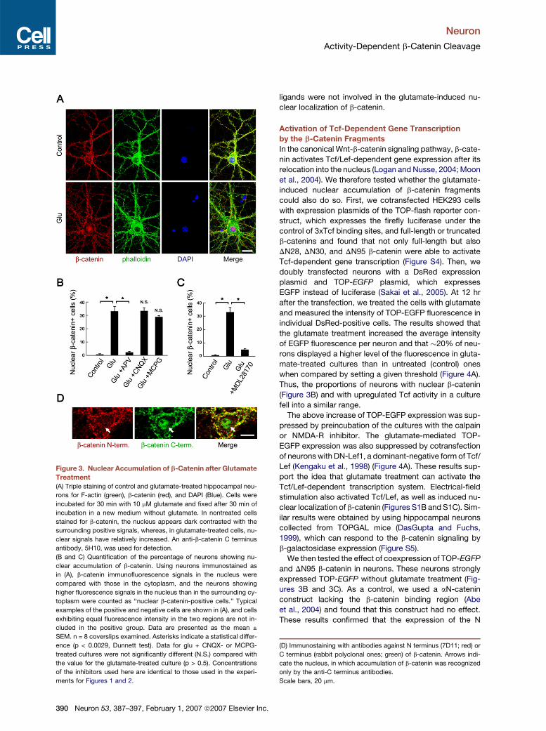
Figure 3. Nuclear Accumulation of b-Catenin after Glutamate
Treatment
(A) Triple staining of control and glutamate-treated hippocampal neu-
rons for F-actin (green), b-catenin (red), and DAPI (Blue). Cells were
incubated for 30 min with 10 mM glutamate and fixed after 30 min of
incubation in a new medium without glutamate. In nontreated cells
stained for b-catenin, the nucleus appears dark contrasted with the
surrounding positive signals, whereas, in glutamate-treated cells, nu-
clear signals have relatively increased. An anti-b-catenin C terminus
antibody, 5H10, was used for detection.
(B and C) Quantification of the percentage of neurons showing nu-
clear accumulation of b-catenin. Using neurons immunostained as
in (A), b-catenin immunofluorescence signals in the nucleus were
compared with those in the cytoplasm, and the neurons showing
higher fluorescence signals in the nucleus than in the surrounding cy-
toplasm were counted as ‘‘nuclear b-catenin-positive cells.’’ Typical
examples of the positive and negative cells are shown in (A), and cells
exhibiting equal fluorescence intensity in the two regions are not in-
cluded in the positive group. Data are presented as the mean ±
SEM. n = 8 coverslips examined. Asterisks indicate a statistical differ-
ence (p < 0.0029, Dunnett test). Data for glu + CNQX- or MCPG-
treated cultures were not significantly different (N.S.) compared with
the value for the glutamate-treated culture (p > 0.5). Concentrations
of the inhibitors used here are identical to those used in the experi-
ments for Figures 1 and 2.
390 Neuron 53, 387–397, February 1, 2007 ª2007 Elsevier Inc.
ligands were not involved in the glutamate-induced nu-
clear localization of b-catenin.
Activation of Tcf-Dependent Gene Transcription
by the b-Catenin Fragments
In the canonical Wnt-b-catenin signaling pathway, b-cate-
nin activates Tcf/Lef-dependent gene expression after its
relocation into the nucleus (Logan and Nusse, 2004; Moon
et al., 2004). We therefore tested whether the glutamate-
induced nuclear accumulation of b-catenin fragments
could also do so. First, we cotransfected HEK293 cells
with expression plasmids of the TOP-flash reporter con-
struct, which expresses the firefly luciferase under the
control of 3xTcf binding sites, and full-length or truncated
b-catenins and found that not only full-length but also
DN28, DN30, and DN95 b-catenin were able to activate
Tcf-dependent gene transcription (Figure S4). Then, we
doubly transfected neurons with a DsRed expression
plasmid and TOP-EGFP plasmid, which expresses
EGFP instead of luciferase (Sakai et al., 2005). At 12 hr
after the transfection, we treated the cells with glutamate
and measured the intensity of TOP-EGFP fluorescence in
individual DsRed-positive cells. The results showed that
the glutamate treatment increased the average intensity
of EGFP fluorescence per neuron and that �20% of neu-
rons displayed a higher level of the fluorescence in gluta-
mate-treated cultures than in untreated (control) ones
when compared by setting a given threshold (Figure 4A).
Thus, the proportions of neurons with nuclear b-catenin
(Figure 3B) and with upregulated Tcf activity in a culture
fell into a similar range.
The above increase of TOP-EGFP expression was sup-
pressed by preincubation of the cultures with the calpain
or NMDA-R inhibitor. The glutamate-mediated TOP-
EGFP expression was also suppressed by cotransfection
of neurons with DN-Lef1, a dominant-negative form of Tcf/
Lef (Kengaku et al., 1998) (Figure 4A). These results sup-
port the idea that glutamate treatment can activate the
Tcf/Lef-dependent transcription system. Electrical-field
stimulation also activated Tcf/Lef, as well as induced nu-
clear localization of b-catenin (Figures S1B and S1C). Sim-
ilar results were obtained by using hippocampal neurons
collected from TOPGAL mice (DasGupta and Fuchs,
1999), which can respond to the b-catenin signaling by
b-galactosidase expression (Figure S5).
We then tested the effect of coexpression of TOP-EGFP
and DN95 b-catenin in neurons. These neurons strongly
expressed TOP-EGFP without glutamate treatment (Fig-
ures 3B and 3C). As a control, we used a aN-catenin
construct lacking the b-catenin binding region (Abe
et al., 2004) and found that this construct had no effect.
These results confirmed that the expression of the N
(D) Immunostaining with antibodies against N terminus (7D11; red) or
C terminus (rabbit polyclonal ones; green) of b-catenin. Arrows indi-
cate the nucleus, in which accumulation of b-catenin was recognized
only by the anti-C terminus antibodies.
Scale bars, 20 mm.

Neuron
Activity-Dependent b-Catenin Cleavage
Figure 4. Activation of Tcf/Lef-Depen-
dent Transcription by Glutamate
(A and B) Neurons were doubly transfected
with DsRed expression plasmid and TOP-
EGFP plasmid in a 2:1 ratio or were triply trans-
fected with these two plasmids and DN-Lef1 or
DN95 b-catenin expression plasmid in a 1:1:1
ratio, in which the amount of TOP-EGFP plas-
mid was equal in all the conditions. An aN-cat-
enin construct lacking the b-catenin binding re-
gion (aN-277-954) (Abe et al., 2004) was used
as a negative control. In (A), transfected cul-
tures were treated with glutamate for 30 min
and fixed after further incubation in a fresh me-
dium without glutamate for 30 min. In (B), no
glutamate treatment was performed. EGFP re-
porter was immunostained, and the fluores-
cence intensity of the EGFP in individual cells
was measured. The average fluorescence in-
tensity in each experimental condition and the
cumulative percentage plot are shown. Data
are presented as the mean ± SEM. n values
from 4 independent experiments are indicated
for each bar. Asterisks indicate a statistical dif-
ference against the control (p < 0.01, Tukey test
in [A]; Steel test in [B]).
(C) Examples of neurons transfected with DN95
b-catenin or the control aN-277-954, which
exhibit EGFP fluorescence near the average
intensity, are shown. Scale bar, 20 mm.
terminus-cleaved b-catenin was sufficient to activate
Tcf-dependent transcription in neurons.
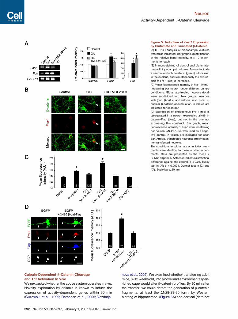
To identify genes actually regulated by b-catenin in nu-
clei, we examined whether any of the genes known to be
regulated by Wnt signaling (Nusse, 2006) responded to
glutamate treatment. RT-PCR analysis of hippocampal
cultures revealed that Fosl1, the gene encoding Fra-1,
was upregulated by glutamate treatment and that this up-
regulation could be suppressed by pretreatment with the
calpain inhibitor (Figure 5A). Furthermore, immunostaining
for Fra-1 in hippocampal cultures showed that, after the
glutamate treatment, some neurons exhibited increased
levels of Fra-1 (Figure 5B). We measured immunofluores-
cence signals in individual neurons and found that the
average intensity of fluorescence per neuron significantly
increased in the glutamate-treated cultures (Figure 5C).
Next, we separately quantified immunofluorescence sig-
nals in neurons with and without nuclear b-catenin, identi-
fied by double immunostaining for Fra-1 and b-catenin, in
the glutamate-treated cultures and found that the former
population of cells displayed significantly higher fluores-
N
cence intensities (Figure 5C), showing a correlation be-
tween Fra-1 upregulation and the nuclear relocation of
b-catenin. Furthermore, preincubation with the calpain
or NMDA-R inhibitors suppressed the upregulation of
Fra-1 (Figures 5B and 5C), supporting the notion that cal-
pain and NMDA-R-dependent nuclear translocation of
b-catenin was involved in the observed upregulation of
Fra-1.
In addition, we tested the effects of exogenous expres-
sion of DN95 b-catenin and found that it could increase the
expression of endogenous Fra-1, even without glutamate
stimulation (Figure 5D). Taken together, these results indi-
cate that Fosl1 may be one of the downstream genes reg-
ulated by calpain and b-catenin-dependent transcription
in hippocampal neurons. Another early-responsive gene,
Fos, the gene encoding c-fos, was also upregulated by
glutamate treatment. However, this upregulation was not
suppressed by the calpain inhibitor (Figure 5A), indicating
that the upregulation of Fosl1, but not that of Fos, was
specifically regulated by glutamate-dependent activation
of calpain.
euron 53, 387–397, February 1, 2007 ª2007 Elsevier Inc. 391
Neuron
Activity-Dependent b-Catenin Cleavage
Figure 5. Induction of Fosl1 Expression
by Glutamate and Truncated b-Catenin
(A) RT-PCR analysis of hippocampal cultures
treated as indicated. Bar graphs, quantification
of the relative band intensity. n = 10 experi-
ments for each.
(B) Immunostaining of control and glutamate-
treated hippocampal cultures. Arrows indicate
a neuron in which b-catenin (green) is localized
in the nucleus, and simultaneously the expres-
sion of Fra-1 (red) is increased.
(C) Mean fluorescence intensity of Fra-1 immu-
nostaining per neuron under different culture
conditions. Glutamate-treated neurons (total)
were subdivided into two groups, neurons
with (nuc. b-cat +) and without (nuc. b-cat �)
nuclear b-catenin accumulation. n values are
indicated for each bar.
(D) Expression of endogenous Fra-1 (red) is
upregulated in a neuron expressing DN95 b-
catenin-Flag (blue), but not in the one not
expressing this construct. Bar graph, mean
fluorescence intensity of Fra-1 immunostaining
per neuron. aN-277-954 was used as a nega-
tive control. n values are indicated for each
bar. Arrows, transfected neurons; arrowheads,
nontransfected neurons.
The conditions for glutamate or inhibitor treat-
ments were identical to those in other experi-
ments. Data are presented as the mean ±
SEM in all panels. Asterisks indicate a statistical
difference against the control (p < 0.01, Tukey
test in [A]; p < 0.0001, Dunnet test in [C] and
[D]). Scale bars, 20 mm.
Calpain-Dependent b-Catenin Cleavage
and Tcf Activation In Vivo
We next asked whether the above system operates in vivo.
Novelty exploration by animals is known to induce the
expression of activity-dependent genes within 30 min
(Guzowski et al., 1999; Ramanan et al., 2005; Vazdarja-
392 Neuron 53, 387–397, February 1, 2007 ª2007 Elsevier Inc
nova et al., 2002). We examined whether transferring adult
mice, 8–12 weeks old, into a novel and environmentally en-
riched cage would alter b-catenin profiles. By 30 min after
the transfer, we could detect the generation of b-catenin
fragments, at least the DN28-29-30 form, by Western
blotting of hippocampal (Figure 6A) and cortical (data not
.

Neuron
Activity-Dependent b-Catenin Cleavage
Figure 6. Neural Activity-Dependent Activation of Tcf-Mediated Transcription In Vivo
(A) Western blot analysis of hippocampal lysates collected from home-caged or novelty-exploring mice, injected or not with the calpain inhibitor
MDL28170 2 hr before the novelty exploration test. a-tubulin was blotted as a loading control. For each condition, 16 animals in total were examined
through eight independent experiments. The truncation of b-catenin was observed in at least four out of the 16 animals, only in the novelty-exploring
group without the calpain inhibitor. Black arrowhead indicates full-length b-catenin, and open arrowhead indicates the 85 kDa b-catenin fragment.
The latter band increased only after novelty exploration in the absence of MDL28170. Bar graph, densitometric analysis of the relative band intensities
of the 85 kDa fragment. Data are presented as the mean ± SEM. n = 16 animals. Asterisk indicates a statistical difference against the control
(p < 0.019, Steel test).
(B) X-gal staining of brain sections obtained from home-caged or novelty-exploring TOPGAL mice.
(C) Immunostaining for b-gal (green) and nuclei (DAPI, red) in the CA1 pyramidal layer of the hippocampus of TOPGAL mice. Arrows point to some of
the b-gal-positive glial cells, which are present equally in both home-cage and novelty-exploration conditions.
(D) Quantification of the percentage of b-gal-positive neurons in the CA1 region. Data are presented as the mean ± SEM. n = 12 animals for each.
Asterisks indicate a statistical difference between experimental groups (p < 0.00025, Dunnett test).
Scale bars, 200 mm in (B), 20 mm in (C).
shown) lysates. When mice had been intravenously in-
jected with the calpain inhibitor MDL28170 2 hr prior
to the novelty exploration test, these fragments did not
appear. We then assessed whether the expression of a b-
galactosidase reporter could be induced in TOPGAL mice
in the novel environment. Adult TOPGAL mice were trans-
ferred to a novel and enriched cage or kept in their home
cage as a control and sacrificed after 30 min. In both
groups we could detect the expression of b-galactosidase
in several regions of the brain, including the cortex and the
hippocampus (Figure 6B). However, in the CA1 region of
the hippocampus, the number of b-galactosidase-positive
pyramidal cells had significantly increased after the
exploratory behavior (Figure 6C). This induction was
suppressed by injection of the calpain inhibitor 2 hr before
the test (Figure 6C), although the injection of calpain
inhibitor had no obvious effects on the locomotor activity
of the mice (Figure S6). Taken together, these data indicate
that activity-dependent stimulation of Tcf-dependent gene
transcription also occurred in vivo, at least in CA1 pyrami-
dal neurons.
DISCUSSION
Our results revealed a novel (to our knowledge) signal-
ing mechanism controlling activity-dependent gene
Neuron 53, 387–397, February 1, 2007 ª2007 Elsevier Inc. 393

Neuron
Activity-Dependent b-Catenin Cleavage
expression. In cultured hippocampal neurons, NMDA-R-
dependent activation of calpain resulted in the N terminus
cleavage of b-catenin. Activation of calpain triggered by
calcium influx is known to degrade a number of proteins,
including pre- and postsynaptic components, and this
process has been implicated not only in neurodegenera-
tive processes but also in modulating synaptic plasticity
(Chan and Mattson, 1999). We showed that b-catenin
can be added to the list of calpain substrates localized
in neurons. The resultant b-catenin fragments became re-
sistant to the GSK-3b-dependent degradation machinery
and accumulated in the nuclei. This process, in turn, acti-
vated Tcf-dependent gene transcription.
In the canonical Wnt signaling pathway, Wnt and GSK-
3b act upstream of the b-catenin-Tcf-dependent gene reg-
ulation machinery, and this pathway is widely used for the
regulation of gene expression required for developmental
and carcinogenetic processes (Logan and Nusse, 2004;
Moon et al., 2004; Polakis, 2000). The signaling mecha-
nism described in the present study appears to have re-
sulted from the substitution of the Wnt-GSK-3b signaling
mechanisms by the NMDA-R-calpain system, leading to
downstream activation of the b-catenin-Tcf-dependent
pathway. This substitution allows neurons to utilize b-
catenin as a mediator of activity-dependent gene expres-
sion, since the NMDA-R is a critical sensor of physiological
stimuli that can induce plastic changes in neurons. On the
other hand, we do not see any relations between this newly
recognized b-catenin-dependent cascade and the nonca-
nonical Wnt pathway because the latter system does not
require b-catenin (Montcouquiol et al., 2006).
We identified Fosl1 as a target gene of the NMDA-
R-mediated b-catenin signaling system. This gene is a known
target of the canonical Wnt signaling pathway (Mann et al.,
1999), but our results indicate that Fosl1 activation can
also occur through the NMDA-R dependent b-catenin
pathway. Notably, Fosl1 was shown to be upregulated in
the rodent brain following learning (Faure et al., 2006),
supporting the idea that the NMDA-R-b-catenin signaling
system is physiologically relevant. Other genes are likely
activated by this system, and identifying such genes re-
mains an important goal to aid our understanding of which
neural processes are controlled by this signaling pathway.
Interestingly, a recent study suggested that Wnt secretion
might also be involved in the NMDA-R-dependent neural
activity (Chen et al., 2006). Thus, the two b-catenin-de-
pendent signaling systems, the canonical Wnt-dependent
and NMDA-R-dependent ones, might represent two par-
allel mechanisms utilized in a larger signaling network, al-
though additional studies are necessary to clarify how
these different systems are coordinated to regulate neuro-
nal functions. Of note, the 75 kDa b-catenin fragment was
initially identified in some cancers (Rios-Doria et al., 2004),
and thus, calpain-dependent activation of the b-catenin-
Tcf signaling pathway may also operate in other cellular
systems.
We provided evidence that the calpain-mediated b-cat-
enin-Tcf signaling pathway also operates in vivo. Novelty
394 Neuron 53, 387–397, February 1, 2007 ª2007 Elsevier Inc
exploration by mice resulted in calpain-dependent cleav-
age of b-catenin, as well as activation of the Tcf signaling
pathway in hippocampal neurons. The overall amount of
cleaved b-catenin detected after the novelty exploration
appears small; however, the tissue lysates used for this
analysis should have contained a large excess of cells
that were nonresponsive to physiological stimuli and re-
sulted in a high proportion of noncleaved b-catenin in
these other cells. Although our preliminary analyses did
not detect any behavioral changes in calpain-inhibitor-
injected mice, it would be intriguing to perform more de-
tailed analyses of brain functions in these mice, as it was
reported that inhibition of calpain (Staubli et al., 1988) or
b-catenin signaling (Chen et al., 2006) impairs long-term
potentiation. In addition, although the details of the mech-
anisms involved have not yet been well elucidated, drugs
that affect b-catenin stability, such as lithium and valproic
acid, have been prescribed as mood stabilizers (Gould
and Manji, 2002). Further, calpain malactivation (Chong
et al., 2005; Zatz and Starling, 2005) and aberrant Wnt
signaling (Chong et al., 2005; De Ferrari and Inestrosa,
2000; Gould and Manji, 2002, 2005; Moon et al., 2004)
have been implicated in various neurological disorders,
implying that dysfunction of b-catenin-dependent gene
expression may be involved in neurological pathologies.
In addition to its critical role in gene regulation, b-catenin
is also a component of the cadherin-catenin complex that
is essential for the stability of synaptic junctions (Takeichi
and Abe, 2005). We found that b-catenin in the cadherin-
catenin complex could be cleaved by calpain, but b-cate-
nin fragments were not detectable in the immunoprecipi-
tated cadherin-catenin complexes. This suggests that
the cleaved b-catenin is unable to stably associate with
cadherin, resulting in the observed translocation into the
nuclei. If calpain-mediated release of b-catenin from cad-
herin occurs excessively, it might reduce the amount of
the functional cadherin-catenin complexes that are re-
quired to maintain normal synaptic junctions (Takeichi
and Abe, 2005). Thus, the NMDA-R-dependent b-catenin
cleavage could result in bidirectional effects, i.e., stimula-
tion of Tcf-dependent gene transcription and structural
modulation of synaptic junctions. Recent studies showed
that ADAM10 and presenilins could cleave the cytoplasmic
domain of cadherin and release it from the cell membrane
in an activity-dependent manner (Marambaud et al., 2003;
Reiss et al., 2005; Uemura et al., 2006). Such cadherin deg-
radation may also enhance the translocation of b-catenin
into the nuclei. Determining how these various activity-
dependent cleavages of cadherin and catenins ultimately
regulate neuronal function is an important subject for
future study.
EXPERIMENTAL PROCEDURES
Cell Culture
Hippocampal cultures were prepared from E17 wild-type or TOPGAL
mice (DasGupta and Fuchs, 1999) (Jackson Laboratory) as described
(Abe et al., 2004) and analyzed at 20–24 DIV. For reporter assays and
.

Neuron
Activity-Dependent b-Catenin Cleavage
endogenous Fosl1-induction experiments, neurons were transfected
at 10 DIV using Effectene (Qiagen) as described (Abe et al., 2004)
and analyzed after 12 hr. For glutamate treatment, 10 mM glutamate
was applied to the medium and incubated for 30 min. After the incuba-
tion, the whole medium was replaced with fresh medium lacking gluta-
mate, and the cultures were further incubated prior to analyses. For
calpain inhibition, MDL28170 (20 mM, Sigma) was added to the me-
dium 20 min before glutamate treatment. L cells were maintained in
DMEM/F-12 medium (Iwaki) supplemented with 10% fetal-calf serum
and 2.5 mM glutamine.
Immunoprecipitation and In Vitro Calpain Assay
P0 brains were lysed with an immunoprecipitation buffer (50 mM Tris-
HCl [pH 7.5], 10% glycerol, 150 mM NaCl, 1% NP-40, and protease in-
hibitor cocktail [Roche]), the supernatants were subjected to immuno-
precipitation with anti-b-catenin antibody, and the immunoprecipitates
were collected with Protein G Sepharose (GE Healthcare). Immuno-
precipitation of N-cadherin from cultured hippocampal cell lysates
was done using the same solutions and reagents as above. For in vitro
cleavage by calpain, the immunoprecipitates were incubated at room
temperature with purified m-calpain (Calbiochem) in the immunopre-
cipitation buffer supplemented with 5 mM DTT and 1 mM CaCl2. For
determination of the cleavage sites, bacterially expressed recombi-
nant GST-b-catenin was constructed and incubated with m-calpain
under the same conditions. The resultant products were separated
by SDS-PAGE, and the bands were cut out and then subjected to N
terminus peptide sequencing (Aproscience). Immunoprecipitation
from L cell lysates was carried out by using RIPA buffer (50 mM Tris-
HCl [pH 7.5], 150 mM NaCl, 1% NP-40, 1% sodium deoxycholate,
0.1% SDS, protease inhibitor cocktail, and phosphatase inhibitor
cocktail [Sigma]).
Immunostaining
Immunostaining of cells was performed as described earlier (Abe et al.,
2004). For the immunostaining of brain slices, mice were sacrificed,
and their brains were then immediately fixed with ice-cold 2% PFA
for 90 min. Coronal sections (60 mm thick) were made with a vibratome
and postfixed for 60 min at 4�C. Free-floating sections were blocked
in TBS with 5% BSA and 0.3% Triton X-100 at 4�C overnight and
incubated sequentially with first antibodies at 4�C for 48 hr and with
secondary Alexa-conjugated antibodies (Invitrogen) at 4�C overnight.
The sections were mounted with Fluosave (Calbiochem) supple-
mented with 40,60-diamidino-2-phenylindole (DAPI). Images were ac-
quired with the LSM510 META multiphoton confocal system (Zeiss),
and fluorescence intensity was analyzed by using LSM510 software.
For X-gal staining, coronal sections (180 mm thick) were fixed with
an X-gal fixative (13 PBS, 1% PFA, 0.2% glutaraldehyde, 2 mM MgCl2,
5 mM EGTA, 0.05% NP-40) for 20 min and washed three times with
a washing buffer (13 PBS, 2 mM MgCl2, 0.1% NP-40). The sections
were then stained with an X-gal staining solution [13 PBS, 5 mM
K3Fe(CN)6, 5 mM K4Fe(CN)6, 2 mM MgCl2, 1 mg ml�1 X-gal (5-
bromo-4-chloro-3-indolyl-b-D-galactoside)] for 24 hr at 37�C. Images
were acquired with a Leica M420 microscope, equipped with a Leica
DC500 CCD.
Antibodies and Reagents
We used mouse monoclonal antibodies against b-catenin N terminus
region (7D11; Calbiochem), b-catenin C terminus (5H10; a gift from
M.J. Wheelock, University of Nebraska, Omaha, NE), a-tubulin
(DM1A; Sigma), b-galactosidase (Promega), Flag-tag (M2; Sigma),
and ubiquitin (6C1.17; BD Bioscience); and rabbit polyclonal anti-
bodies against b-catenin C terminus (SIGMA), Fra-1 (Santa Cruz),
GFP (Chemicon), and phospho-Ser33/37/Thr41-b-catenin (Cell Sig-
naling). F-actin was visualized with Alexa-488-conjugated phalloidin
(Invitrogen), and nuclei were stained with DAPI. APV, NMDA, AMPA,
DHPG, MDL28170, and MG132 were purchased from Sigma; CNQX
and MCPG were from Calbiochem; and recombinant Wnt3a and
Dkk-1 were from R&D systems. TOP-flash and FOP-flash reporter
plasmids were from Upstate.
Plasmid Construction
For the construction of the expression plasmid for DN28 b-catenin,
amino acids 29–781 of the b-catenin coding region were amplified by
PCR with a primer set of 50- gcggccgccaccatgtcttacttggattctggaat-30
and 50-gtcgaccaggtcagtatcaaaccag-30 (bcat-C primer), and subcloned
into pCA-Sal-flag, as described (Abe et al., 2004). Expression plasmids
for DN30 and DN95 b-catenin were constructed in the same way with
primer sets of 50-gcggccgccaccatgttggattctggaatccatt-30 and the
bcat-C primer for DN30 b-catenin; and 50-gcggccgccaccatggctgcc
atgttccctgagac-30 and the bcat-C primer for DN95 b-catenin. All the
constructs were checked by sequencing.
RT-PCR
From hippocampal cultures (at 18–21 DIV) treated with glutamate for
60 min, total RNA was extracted by using RNAeasy (Qiagen). cDNAs
were synthesized by using a SuperScript III kit (Invitrogen). One nano-
gram of the resultant first-strand cDNAs were subsequently used for
PCR analysis with the following primers: Gapdh, 50-agaaggtggtgaagc
aggca-30 and 50-cgaaggtggaagagtgggag-30; Fosl1, 50-cacctagacagaa
ggtgcccttt-30 and 50-ctcctgcgttgtgccatt-30; Fos, 50-acctcccgctctgtgcc
agatgtg-30 and 50-ttgctgctgctgccctttcggtgg-30. PCR was carried out
under 25 cycles at 94�C, 60�C, and 72�C, each for 30 s. The number
of the cycles was determined so that the resultant products were am-
plified within the linear range. The intensity of PCR and Western blot
bands was analyzed by using Image-J.
Novelty Exploration Assay
Adult TOPGAL mice, 8–12 weeks old, were habituated to the labora-
tory overnight and transferred into new, enriched cages or kept in their
home cages as a control. The mice were sacrificed 30 min after the
transfer (Ramanan et al., 2005). For injection of the calpain inhibitor,
MDL28170 was dissolved in PEG300/EtOH (9:1) and diluted 1:1 in sa-
line. Mice were anesthetized and subsequently injected via a tail vein
with 30 mg kg�1 of the solution (Neumar et al., 1998). For behavioral
analysis, at 2 hr after calpain-inhibitor injection, mice were transferred
to novel cages (45 cm 3 25 cm), and their locomotion was recorded by
using a video camera for 30 min. The moving distances of the mice
were analyzed automatically with a Scion Image macro.
Statistical Analysis
Statistical analyses used in this study are indicated in each figure
legend.
Supplemental Data
The Supplemental Data for this article can be found online at http://
www.neuron.org/cgi/content/full/53/3/387/DC1/.
ACKNOWLEDGMENTS
We thank I. Matsuo for TOPGAL mice, the Laboratory for Animal Re-
sources and Genetic Engineering in CDB for mouse breeding, H. Ishi-
gami and C. Yoshii for maintenance of mice, Y. Wakamatsu for TOP-
EGFP plasmid, S. Nakagawa for pCA-DN-Lef1 plasmid, and T. Tanoue
for critical reading of the manuscript. This work was supported by
a grant from the program Grants-in-Aid for Specially Promoted Re-
search of the Ministry of Education, Science, Sports, and Culture of
Japan to M.T.; and by a grant-in-aid for scientific research from the
Japan Society for the Promotion of Science for Junior Scientists to K.A.
Received: June 26, 2006
Revised: October 3, 2006
Accepted: January 17, 2007
Published: January 31, 2007
Neuron 53, 387–397, February 1, 2007 ª2007 Elsevier Inc. 395

Neuron
Activity-Dependent b-Catenin Cleavage
REFERENCES
Abe, K., Chisaka, O., Van Roy, F., and Takeichi, M. (2004). Stability of
dendritic spines and synaptic contacts is controlled by alpha N-cate-
nin. Nat. Neurosci. 7, 357–363.
Aberle, H., Bauer, A., Stappert, J., Kispert, A., and Kemler, R. (1997).
beta-catenin is a target for the ubiquitin-proteasome pathway.
EMBO J. 16, 3797–3804.
Barth, A.I., Pollack, A.L., Altschuler, Y., Mostov, K.E., and Nelson, W.J.
(1997). NH2-terminal deletion of beta-catenin results in stable colocal-
ization of mutant beta-catenin with adenomatous polyposis coli pro-
tein and altered MDCK cell adhesion. J. Cell Biol. 136, 693–706.
Bi, X., Chang, V., Molnar, E., McIlhinney, R.A., and Baudry, M. (1996).
The C-terminal domain of glutamate receptor subunit 1 is a target for
calpain-mediated proteolysis. Neuroscience 73, 903–906.
Chan, S.L., and Mattson, M.P. (1999). Caspase and calpain sub-
strates: roles in synaptic plasticity and cell death. J. Neurosci. Res.
58, 167–190.
Chen, J., Park, C.S., and Tang, S.J. (2006). Activity-dependent synap-
tic WNT release regulates hippocampal long-term potentiation. J. Biol.
Chem. 281, 11910–11916.
Chong, Z.Z., Li, F., and Maiese, K. (2005). Stress in the brain: novel cel-
lular mechanisms of injury linked to Alzheimer’s disease. Brain Res.
Brain Res. Rev. 49, 1–21.
DasGupta, R., and Fuchs, E. (1999). Multiple roles for activated LEF/
TCF transcription complexes during hair follicle development and
differentiation. Development 126, 4557–4568.
De Ferrari, G.V., and Inestrosa, N.C. (2000). Wnt signaling function in
Alzheimer’s disease. Brain Res. Brain Res. Rev. 33, 1–12.
Deisseroth, K., Mermelstein, P.G., Xia, H., and Tsien, R.W. (2003). Sig-
naling from synapse to nucleus: the logic behind the mechanisms.
Curr. Opin. Neurobiol. 13, 354–365.
Faure, A., Conde, F., Cheruel, F., and el Massioui, N. (2006). Learning-
dependent activation of Fra-1: involvement of ventral hippocampus
and SNc/VTA complex in learning and habit formation. Brain Res.
Bull. 68, 233–248.
Glinka, A., Wu, W., Delius, H., Monaghan, A.P., Blumenstock, C., and
Niehrs, C. (1998). Dickkopf-1 is a member of a new family of secreted
proteins and functions in head induction. Nature 391, 357–362.
Goll, D.E., Thompson, V.F., Li, H., Wei, W., and Cong, J. (2003). The
calpain system. Physiol. Rev. 83, 731–801.
Gould, T.D., and Manji, H.K. (2002). The Wnt signaling pathway in bi-
polar disorder. Neuroscientist 8, 497–511.
Gould, T.D., and Manji, H.K. (2005). Glycogen synthase kinase-3: a pu-
tative molecular target for lithium mimetic drugs. Neuropsychophar-
macology 30, 1223–1237.
Guttmann, R.P., Baker, D.L., Seifert, K.M., Cohen, A.S., Coulter, D.A.,
and Lynch, D.R. (2001). Specific proteolysis of the NR2 subunit at
multiple sites by calpain. J. Neurochem. 78, 1083–1093.
Guzowski, J.F., McNaughton, B.L., Barnes, C.A., and Worley, P.F.
(1999). Environment-specific expression of the immediate-early gene
Arc in hippocampal neuronal ensembles. Nat. Neurosci. 2, 1120–1124.
Huntley, G.W., Gil, O., and Bozdagi, O. (2002). The cadherin family of
cell adhesion molecules: multiple roles in synaptic plasticity. Neurosci-
entist 8, 221–233.
Kandel, E.R. (2001). The molecular biology of memory storage: a dia-
logue between genes and synapses. Science 294, 1030–1038.
Kengaku, M., Capdevila, J., Rodriguez-Esteban, C., De La Pena, J.,
Johnson, R.L., Belmonte, J.C., and Tabin, C.J. (1998). Distinct WNT
pathways regulating AER formation and dorsoventral polarity in the
chick limb bud. Science 280, 1274–1277.
396 Neuron 53, 387–397, February 1, 2007 ª2007 Elsevier Inc
Kerokoski, P., Suuronen, T., Salminen, A., Soininen, H., and Pirttila, T.
(2004). Both N-methyl-D-aspartate (NMDA) and non-NMDA receptors
mediate glutamate-induced cleavage of the cyclin-dependent kinase 5
(cdk5) activator p35 in cultured rat hippocampal neurons. Neurosci.
Lett. 368, 181–185.
Liu, C., Li, Y., Semenov, M., Han, C., Baeg, G.H., Tan, Y., Zhang, Z.,
Lin, X., and He, X. (2002). Control of beta-catenin phosphorylation/
degradation by a dual-kinase mechanism. Cell 108, 837–847.
Logan, C.Y., and Nusse, R. (2004). The Wnt signaling pathway in devel-
opment and disease. Annu. Rev. Cell Dev. Biol. 20, 781–810.
Lu, X., Rong, Y., and Baudry, M. (2000). Calpain-mediated degradation
of PSD-95 in developing and adult rat brain. Neurosci. Lett. 286, 149–
153.
Lynch, G., and Baudry, M. (1984). The biochemistry of memory: a new
and specific hypothesis. Science 224, 1057–1063.
Lynch, G., and Baudry, M. (1987). Brain spectrin, calpain and long-
term changes in synaptic efficacy. Brain Res. Bull. 18, 809–815.
Malenka, R.C., and Nicoll, R.A. (1993). NMDA-receptor-dependent
synaptic plasticity: multiple forms and mechanisms. Trends Neurosci.
16, 521–527.
Mann, B., Gelos, M., Siedow, A., Hanski, M.L., Gratchev, A., Ilyas, M.,
Bodmer, W.F., Moyer, M.P., Riecken, E.O., Buhr, H.J., and Hanski, C.
(1999). Target genes of beta-catenin-T cell-factor/lymphoid-
enhancer-factor signaling in human colorectal carcinomas. Proc.
Natl. Acad. Sci. USA 96, 1603–1608.
Marambaud, P., Wen, P.H., Dutt, A., Shioi, J., Takashima, A., Siman,
R., and Robakis, N.K. (2003). A CBP binding transcriptional repressor
produced by the PS1/epsilon-cleavage of N-cadherin is inhibited by
PS1 FAD mutations. Cell 114, 635–645.
Montcouquiol, M., Crenshaw, E.B., 3rd, and Kelley, M.W. (2006). Non-
canonical Wnt signaling and neural polarity. Annu. Rev. Neurosci. 29,
363–386.
Moon, R.T., Kohn, A.D., De Ferrari, G.V., and Kaykas, A. (2004). WNT
and beta-catenin signalling: diseases and therapies. Nat. Rev. Genet.
5, 691–701.
Murase, S., and Schuman, E.M. (1999). The role of cell adhesion mol-
ecules in synaptic plasticity and memory. Curr. Opin. Cell Biol. 11,
549–553.
Murase, S., Mosser, E., and Schuman, E.M. (2002). Depolarization
drives beta-Catenin into neuronal spines promoting changes in synap-
tic structure and function. Neuron 35, 91–105.
Nakazawa, K., McHugh, T.J., Wilson, M.A., and Tonegawa, S. (2004).
NMDA receptors, place cells and hippocampal spatial memory. Nat.
Rev. Neurosci. 5, 361–372.
Neumar, R.W., DeGracia, D.J., Konkoly, L.L., Khoury, J.I., White, B.C.,
and Krause, G.S. (1998). Calpain mediates eukaryotic initiation factor
4G degradation during global brain ischemia. J. Cereb. Blood Flow
Metab. 18, 876–881.
Nusse, R. (2006). The Wnt homepage (http://www.stanford.edu/
�rnusse/wntwindow.html).
Okamura, K., Tanaka, H., Yagita, Y., Saeki, Y., Taguchi, A., Hiraoka, Y.,
Zeng, L.H., Colman, D.R., and Miki, N. (2004). Cadherin activity is
required for activity-induced spine remodeling. J. Cell Biol. 167, 961–
972.
Polakis, P. (2000). Wnt signaling and cancer. Genes Dev. 14, 1837–
1851.
Ramanan, N., Shen, Y., Sarsfield, S., Lemberger, T., Schutz, G., Lin-
den, D.J., and Ginty, D.D. (2005). SRF mediates activity-induced
gene expression and synaptic plasticity but not neuronal viability.
Nat. Neurosci. 8, 759–767.
Reiss, K., Maretzky, T., Ludwig, A., Tousseyn, T., de Strooper, B., Hart-
mann, D., and Saftig, P. (2005). ADAM10 cleavage of N-cadherin and
.

Neuron
Activity-Dependent b-Catenin Cleavage
regulation of cell-cell adhesion and beta-catenin nuclear signalling.
EMBO J. 24, 742–752.
Rios-Doria, J., Kuefer, R., Ethier, S.P., and Day, M.L. (2004). Cleavage
of beta-catenin by calpain in prostate and mammary tumor cells. Can-
cer Res. 64, 7237–7240.
Rubinfeld, B., Albert, I., Porfiri, E., Fiol, C., Munemitsu, S., and Polakis,
P. (1996). Binding of GSK3beta to the APC-beta-catenin complex and
regulation of complex assembly. Science 272, 1023–1026.
Sakai, D., Tanaka, Y., Endo, Y., Osumi, N., Okamoto, H., and Waka-
matsu, Y. (2005). Regulation of Slug transcription in embryonic ecto-
derm by beta-catenin-Lef/Tcf and BMP-Smad signaling. Dev. Growth
Differ. 47, 471–482.
Salinas, P.C., and Price, S.R. (2005). Cadherins and catenins in syn-
apse development. Curr. Opin. Neurobiol. 15, 73–80.
Shibamoto, S., Higano, K., Takada, R., Ito, F., Takeichi, M., and Ta-
kada, S. (1998). Cytoskeletal reorganization by soluble Wnt-3a protein
signalling. Genes Cells 3, 659–670.
Staubli, U., Larson, J., Thibault, O., Baudry, M., and Lynch, G. (1988).
Chronic administration of a thiol-proteinase inhibitor blocks long-term
potentiation of synaptic responses. Brain Res. 444, 153–158.
Takeichi, M., and Abe, K. (2005). Synaptic contact dynamics controlled
by cadherin and catenins. Trends Cell Biol. 15, 216–221.
N
Uemura, K., Kihara, T., Kuzuya, A., Okawa, K., Nishimoto, T., Nino-
miya, H., Sugimoto, H., Kinoshita, A., and Shimohama, S. (2006). Char-
acterization of sequential N-cadherin cleavage by ADAM10 and PS1.
Neurosci. Lett. 402, 278–283.
Vanderklish, P., Saido, T.C., Gall, C., Arai, A., and Lynch, G. (1995).
Proteolysis of spectrin by calpain accompanies theta-burst stimula-
tion in cultured hippocampal slices. Brain Res. Mol. Brain Res. 32,
25–35.
Vanderklish, P.W., Krushel, L.A., Holst, B.H., Gally, J.A., Crossin, K.L.,
and Edelman, G.M. (2000). Marking synaptic activity in dendritic
spines with a calpain substrate exhibiting fluorescence resonance
energy transfer. Proc. Natl. Acad. Sci. USA 97, 2253–2258.
Vazdarjanova, A., McNaughton, B.L., Barnes, C.A., Worley, P.F., and
Guzowski, J.F. (2002). Experience-dependent coincident expression
of the effector immediate-early genes arc and Homer 1a in hippo-
campal and neocortical neuronal networks. J. Neurosci. 22, 10067–
10071.
West, A.E., Griffith, E.C., and Greenberg, M.E. (2002). Regulation of
transcription factors by neuronal activity. Nat. Rev. Neurosci. 3, 921–
931.
Zatz, M., and Starling, A. (2005). Calpains and disease. N. Engl. J. Med.
352, 2413–2423.
euron 53, 387–397, February 1, 2007 ª2007 Elsevier Inc. 397