the identification of a fluorescent reducing … · j. clin. path. (1960), 13, 140. the...
TRANSCRIPT

J. clin. Path. (1960), 13, 140.
THE IDENTIFICATION OF A FLUORESCENT REDUCINGSUBSTANCE IN THE URINE OF PATIENTS WITH
RHEUMATOID ARTHRITISTHE EXCRETION OF 3-HYDROXYANTHRANILIC ACID IN THIS
AND OTHER CONDITIONS
BY
MARY McMILLAN*From the Department of Clinical Chemistry, Edinburgh Royal Infirmary
(RECEIVED FOR PUBLICATION SEPTEMBER 7, 1959)
The report that 2,5-dihydroxyphenylpyruvic acid occurs in the urine of patients with collagendisease has not been confirmed, and no evidence was found for regarding collagen disease asbeing due to an inherent error in tyrosine metabolism as suggested by Japanese workers.A strongly reducing substance was conspicuous in the urine of patients with rheumatoid arthritis
but not in that of normal persons. This substance was identified as 3-hydroxyanthranilicacid (3-HAA).A method is described for the measurement of 3-HAA in urine, employing ether extraction,
paper chromatography in a formate solvent, and visual assessment by fluorescence in ultra-violetlight and colour with Ehrlich's reagent.Normal persons excreted from less than 125 to 375 ,ug./day (average rather lower than 200 ,ug./
day); those suffering from rheumatoid arthritis excreted from less than 125 to 2,250 ,sg./day(average 820 utg./day".The excessive excretion of 3-HAA in persons with rheumatoid arthritis was investigated in
terms of other aspects of the activity of the disease. Some relationship was suggested betweenexcretion levels, the erythrocyte sedimentation rate, and the sensitized sheep cell test.
Cases of osteoarthritis, other orthopaedic conditions, and miscellaneous diseases were examined.A few of the latter, including diseases of the haemopoietic system, were associated with increasedexcretion of 3-HAA.
It has recently been claimed that in the urineof patients suffering from one or other of thecollagen diseases there may be detected a postu-lated but hitherto unisolated intermediate oftyrosine metabolism, namely, 2,5-dihydroxyphenyl-pyruvic acid (Nishimura, Shibata, Yasui, andOkamoto, 1956). This substance was recognizedby its purple fluorescence in ultra-violet light afterpaper chromatography. It was characterized byreduction of ammoniacal silver nitrate and Brigg'sreagents and by reactions with ferric chloride,Fenton's and hydrazine reagents. Its chromato-graphic behaviour in two solvent systems and itsultra-violet absorption spectrum were found to bethe same as those of a synthetic specimen of2,5-dihydroxyphenylpyruvic acid. In the system
* Present address: Department of Bacteriology, EdinburghRoyal Infirmary.
n-butanol: acetic acid: water (4: 1: 1) two spotswere obtained, depending on whether the urinewas acid or neutral during its concentration; oneof the spots was thought to be the quinonederivative of 2,5-dihydroxyphenylpyruvic acid andwas seen also in chromatograms of the syntheticsubstance.
In 38 cases of collagen disease the urinecontained the substance described. It was absentfrom the urine of normal persons, those sufferingfrom a variety of other diseases, or unaffectedrelatives of patients with collagen disease. Theauthors concluded that a fundamental biochemicalclue to the aetiology of collagen diseases had beenfound, and that these diseases must be groupedwith phenylketonuria, alcaptonuria, tyrosinosis,and albinism in that they are caused by aninherent error in the metabolism of tyrosine.
copyright. on F
ebruary 18, 2020 by guest. Protected by
http://jcp.bmj.com
/J C
lin Pathol: first published as 10.1136/jcp.13.2.140 on 1 M
arch 1960. Dow
nloaded from

IDENTIFICATION OF FLUORESCENT REDUCING SUBSTANCE IN URINE 141
Examination of Urine for 2,5-Dihydroxy-phenylpyruvic Acid
The Japanese workers' description of the prepara-tion of urine extracts for chromatography was limitedto the following paragraph:
"Either as it was or acidified with 10% H2SO4,the 24-hr. urine of diffuse collagen disease patientswas concentrated till about 20 ml. of it remained,and after another further treatment of it with 60%alcohol, and the resultant elimination of inorganicsalts, the urine thus obtained was subjected toexperiment. Then one-dimensional paper chromato-graphy was performed."In an attempt to follow the above procedure 250 ml.
of urine (freshly collected in acid to give a final pHof 3) from a case of acute rheumatoid arthritis wasdistilled at reduced pressure under nitrogen, theexternal temperature being kept below 40' C. until afew millilitres of viscous residue remained. This wastaken up in alcohol to a concentration of 600, andcentrifuged. The supernatant was subjected tochromatography overnight on Whatman No. I paperby the ascending technique using the solvent systemspecified by the Japanese workers (n-butanol:aceticacid: water 4:1:1). When viewed under ultra-violetlight (" hanovia " medium pressure quartz arc withWood's glass filter or " hanovia" chromatolite Hgresonance lamp 2537A) a large number of fluorescentareas was detectable and subsequent spraying withammoniacal silver nitrate solution (0.1 N AgNO325 ml., ammonia S.G. 0.88 8 ml., water to 50 ml.)revealed a number of reducing substances. However,no fluorescent reducing substance was seen whichhad the RF value stipulated by Nishimura and hiscolleagues. Furthermore, and perhaps of especialimportance in view of the number of closely relatedcompounds into which the quinol sought might betransformed, no difference was detectable in urinefrom normal individuals and rheumatoid patientswhen chromatography was carried out in the butanolsystem or in phenol: water: H2S03, a solvent moresuitable than the former for the preservation of ahighly oxidizable substance (McMillan, 1957).An attempt was made to purify the urine before
chromatography by adsorption upon alumina(McMillan, 1957). A readily available quinolderivative fairly closely related to that being sought,namely, 2,5-dihydroxybenzoic acid (gentisic acid), wasadded to whole urine in amounts of 1 ,ug./ml. Thequinol remained in the filtrate during alumina treat-ment whereas under identical conditions the compound3,4-dihydroxyphenylacetic acid was adsorbed and nonewas detected in the filtrate. In neither the eluate northe filtrate of urine from a case of active rheumatoidarthritis could a fluorescent reducing substance befound to correspond with the Japanese description.When fresh urine, collected in acid, was extracted
with ether at pH 3 and the ether-soluble portionchromatographed, a fluorescent reducing substancewas conspicuous in the rheumatoid specimens butnot in those from normal persons. The ether extractswere distilled to dryness at reduced pressure at a
temperature not exceeding 40' C., and the residue,which was derived from 250 ml. urine, was taken upin 1 ml. ethanol. Upon chromatography, 0.02 ml. ofthis solution gave an easily visible purple spot whenviewed under ultra-violet light and had an RF Of0.84 in the butanol system and 0.61 in the phenolsystem. The former RF is higher than either ofthe values quoted by the Japanese workers for2,5-dihydroxyphenylpyruvic acid or its quinone.
Identification of the Fluorescent ReducingSubstance in Urine from Rheumatoid Patients as
3-Hydroxyanthranilc AcidThe extract described above was chromato-
graphed in the butanol system and the paper wassprayed with a variety of colour-producingreagents with the following results at the site offluorescence:Anunniacal Silver Nitrate. - Dark grey;
reduction occurred in the cold and preceded thatof any other substance present elsewhere on thepaper.Gibbs' Reagent (2,6-Dichlorquinone Chloro-
imide 0.2% w/v in Ethanol).-(1) Reagent alone,a purple colour exclusive to the fluorescent spotdeveloped slowly. (2) The reagent diluted withan equal volume of 0.66 M phosphate buffer atpH 7.0 gave an immediate purple colour withvarious colours appearing later in neighbouringareas. (3) The reagent followed by saturatedsodium bicarbonate solution or borate solutiongave an immediate blue colour at the fluorescentspot, quickly followed, especially in the case ofborate, by an array of colours obscuring theoriginal single spot.
Ferric Chloride (0.2% w/v in Water).-A faintyellow colour appeared.Potassium Ferricyanide (6.6% Aqueous Solution
in Phosphate Buffer at pH 5.9).-This gave a pinkcolour.
Potassium Iodate (1% w/v in Water).-Thisgave a grey colour.
Fenton's Reagent (Nishimura, Yasui, Okamoto,Kanazawa, Kotake, and Shibata, 1958).-Thisgave a brown colour.
Ehrlich's Reagent.-This is made up ofp-dimethylaminobenzaldehyde, 10% w/v, in con-centrated HCI solution, 1 vol., acetone, 4 vol.,mixed immediately before use, and gave a slowlydeveloping distinct orange-yellow colour.The following reagents either gave no colours
at all in the appropriate area or else gave colourswhich did not properly coincide with the fluores-cent spot: Diazotized sulphanilic acid, p-nitraniline
copyright. on F
ebruary 18, 2020 by guest. Protected by
http://jcp.bmj.com
/J C
lin Pathol: first published as 10.1136/jcp.13.2.140 on 1 M
arch 1960. Dow
nloaded from

MARY McMILLAN
and p-anisidine respectively, ninhydrin, nitroso-naphthol reagent, xanthydrol reagent, Altman'sreagent, 2,4-dinitrophenylhydrazine (for all thepreceding reagents see Smith, 1958), Millon'sreagent, Brigg's reagent (Nishimura et al., 1958),preceding reagents (see Smith, 1958), Millon'sRoux reagent (Roux, 1951) and bromothymolblue-acidified potassium iodide-starch ammonia(as used for homogentisic acid by Consden,Forbes, Glynn, and Stanier, 1952). The fluores-cent substance was seen to develop a browncolour when the papers were exposed to theatmosphere for an hour or two; it was unstablein alkaline solution, a pH of 9 causing appreciableloss.By consideration of the above properties and by
comparing the observed RF in various solventsystems with those published for phenolic acidsknown to occur in urine (for references see Smith,1958), it appeared probable that the fluorescentsubstance was 3-hydroxyanthranilic acid. Aspecimen of this substance (Roche) proved tohave the same fluorescence under both ultra-violetlight sources and to give the same colour reactionsas the unknown substance. When added to theunknown substance in a urine concentrate it hadan identical RF in four solvent systems, namely,0.61 in phenol: water: H2SO3; 0.84 in n-butanol:acetic acid: water (4: 1: 1), 0.52 in benzene: aceticacid: water (125 :72: 3), and 0.63 in sodiumformate: formic acid: water (10 g.: 1 ml.: 200nil.). Both substances were largely destroyed inthe system iso-propanol:0.88 ammonia: water(200:10:20).
Semiquantitative Measurement of the Amount of3-Hydroxyanthranilic Acid in Urine
The fluorescence of 3-hydroxyanthranilic acid(3-HAA) in ultra-violet light could be detectedeasily on the chromatogram paper in amounts aslow as 0.25 jug., whereas none of the colourreactions was as sensitive as this. Chromatographyin the butanol system was unsuited to quantitativework because very many phenolic acids have asimilar high RF value; urine extracts prepared bysimple ether extraction gave neighbouring spotswhich were highly fluorescent under ultra-violetlight, and, though they could be distinguishedfrom 3-hydroxyanthranilic acid by their bluecolour, they partially, or sometimes completely,obscured the purple colour of 3-HAA. Thebenzene system was unsuitable because the spotsbecame too spread out. Both the phenol andformate systems gave sharply defined spots andsome urine extracts could be satisfactorilyassessed in terms of standard amounts of 3-HAA
run on the same sheets of paper. Many, however,showed one or more fluorescent substance whichinterfered more or less seriously with such acomparison. Two-dimensional chromatography,employing the phenol system followed by theformate, failed to eradicate the interference.After the use of any of the above solvent systemscolour development on the paper with Ehrlich'sreagent or buffered potassium ferricyanidesolution proved insufficiently sensitive andammoniacal silver nitrate or Gibbs reagent atvarious pH values insufficiently specific.Owing to the presence of a basic group, 3-HAA
is not extracted by ether from highly acidaqueous solution. When urine was acidified withone-tenth of its volume of concentrated HCI andextracted with ether, the concentrated etherextracts on chromatography usually containedmuch material fluorescent in ultra-violet light,whereas subsequent extraction of the aqueousresidue after adjustment of the pH to 3 gave apreparation which frequently showed only onefluorescent area, that due to 3-HAA. These find-ings were applicable to chromatography in thephenol, formate, and butanol systems. At eachpH ether was extracted with four equal portionswhose total volume equalled that of the aqueousphase. Experiments showed that, when added tothe urine in amounts as high as 1 jug. /ml., 3-HAAcould be satisfactorily accounted for during theprocesses of extraction, concentration, andchromatography; the four combined extractsmade at pH 3 contained as far as could beassessed by visual comparison the whole of theadded 3-HAA, whereas the four ether extracts ofthe highly acid urine and two additional extractsmade at pHI 3 subsequent to the four normallyprepared showed no trace of 3-HAA.
After such a procedure of double ether extrac-tion some urine extracts continued to giveadditional fluorescent spots. Commonly therewas found a blue spot immediately above thepurple 3-HAA in both phenol and formatesystems; the fluorescence of this substance wasmuch greater under the "hanovia" 2537 A lampthan under the Woods glass whereas that of3-HAA was similar under both, so that therewas no danger of confusion. In order to guardagainst the possibility of interference at the actualsite of 3-HAA, though this appeared unlikely,after assessment under ultra-violet light thechromatogram was always sprayed with Ehrlich'sreagent and some hours later the intensities andsizes of the yellow spots were compared with thoseof the standards. Amounts less than 0.5 ug.usually did not show up, but for amounts greater
142
copyright. on F
ebruary 18, 2020 by guest. Protected by
http://jcp.bmj.com
/J C
lin Pathol: first published as 10.1136/jcp.13.2.140 on 1 M
arch 1960. Dow
nloaded from

IDENTIFICATION OF FLUORESCENT REDUCING SUBSTANCE IN URINE 143
than this there was never any great discrepancybetween the values obtained by the two methodsof comparison. Chromatograms developed in thephenol system were easily assessed by fluores-cence in ultra-violet light and Ehrlich colour(pink), the paper being freed of phenol by benzen2extraction followed by hanging in a current ofwarm air for two to three hours. In fact, in80% of the assays to be reported duplicatechromatograms were thus run; there was alwaysclose agreement with the standard method usingformate. After double ether extraction somechromatograms run in the butanol system anddried in warm air were sprayed with Gibbs'reagent followed by saturated sodium dihydrogenphosphate solution. This method of assessmentwas quite unsatisfactory as the initial spots of3-HAA were rapidly obscured by several otherclosely situated substances (cf. Tompsett, 1959).
It was found that urine acidified to pH 3 orthereabouts maintained its content of 3-HAA formany days when stored in the refrigerator. Theether extracts were unstable and were alwaysassayed as soon as possible, usually on the day ofpreparation. Similarly the developed chromato-grams were always assessed as soon as the paperswere dry.The possibility of completing the assay of
3-HAA by spectrofluorometric measurement wasnot investigated in this study.
MethodPreparation of Extract.-Urine was collected for 24
hours, 15 ml. of 10% H2S04 being used as preservative.To a filtered aliquot measuring 10% of the wholespecimen was added one-tenth of its volume ofconcentrated HCI. The mixture was then shaken fortwo-minute periods with an equal volume of peroxide-free ether divided into four equal portions. Centri-fugation was usually necessary to effect separation.After each extraction the ether layer was pipettedoff and discarded. The pH of the aqueous phase wasthen adjusted with a volume of 10 N NaOH equiva!entto that of the concentrated HCI previously used,followed by more dilute alkali until a pH of 3.5was reached (B.D.H. 2540 indicator paper). Etherextraction was repeated with the same amounts asbefore. The four resulting ether fractions werecombined and distilled to dryness at reduced pressureunder nitrogen at a bath temperature not exceeding400 C. The residue was taken up in four smallsuccessive amounts of ethanol and the solution wasevaporated to a volume of 0.5 ml. This pale, straw-coloured solution was used. for chromatography.The general chromatographic technique was that
described by McMillan (1957). The paper was markedout into four strips (each 2 cm. wide) for the urineextract (usually 0.01, 0.005, 0.0025, and 0.00125 ml.)
and four for the standard 3-hydroxyanthranilic acidsolution (2.0, 1.0, 0.5, and 0.25 /Lg.). Before use thestandard was diluted 1 in 5 in ethanol from a stockrefrigerated solution containing 20 mg./20 ml. ethanolto which 0.05 ml. of concentrated HCI had beenadded. The solvent (sodium formate 10 g., water200 ml., formic acid I ml.) was renewed on eachoccasion. Two or three sheets of paper were hungin the same tank for two to three hours until thesolvent front reached about 25 cm. A sheet of spongerubber and a wrapper of cellophane paper wereapplied to the underside of the lid of the tank whichwas then quickly sealed by pressure from above(F. Albert-Recht, personal communication). Thesheets were dried in the air at room temperature usinga hair dryer. The amount of 3-hydroxyanthranilicacid in each of the four urine extract chromatogramswas assessed visually by transmitted light from boththe ultra-violet lamps previously described. Thepaper was then sprayed with Ehrlich's reagent andfurther comparisons made after about 18 hours.
ResultsThe excretion of 3-hydroxyanthranilic acid by
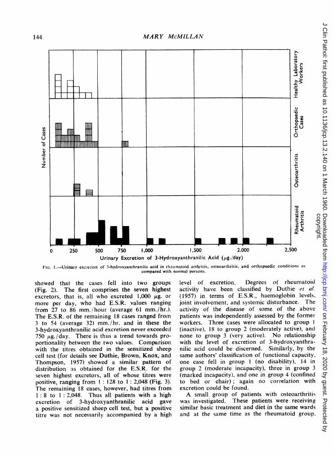
83 persons was determined. Twenty-five patientssuffering from rheumatoid arthritis (all of whomwere in-patients of the Rheumatic Unit, NorthernGeneral Hospital, Edinburgh) excreted quantitiesranging from less than 125 ,ug. (the minimalamount detectable by the method) to 2,250 ,ug./day, the average figure being 820 ,ug./day. In afew instances excretion was measured on morethan one occasion, when values of the same orderwere found. The figures bore no relationship tothe age or sex of the patient. The patients hadreceived no steroid therapy or other drugs for atleast three days before the urine was collected.Twelve healthy laboratory workers excreted
from less than 125 to 375 ,ug./day, the averagebeing rather lower than 200 ,ug. / day. Fig. 1shows that whereas only two of these normalpersons excreted more than 250 jig./day, allrheumatoid subjects save three excreted 375 ,ug. ormore/day. There would therefore appear to besome considerable difference between these twogroups. One normal person who received heavydoses of either aspirin or panadol for three daysshowed the same low excretion three days later.
Analysis of the group of rheumatoid patientsfailed to show any correlation between the levelof excretion of 3-hydroxyanthranilic acid andsome of the criteria which might be used to assessthe severity of the disease, namely, the durationof symptoms, the extent of tissue damage as'shown radiologically, or the degree of anaemia.Comparison of the erythrocyte sedimentationrate (E.S.R.) measured by the Westergren methodwith excretion of 3-hydroxyanthranilic acid
copyright. on F
ebruary 18, 2020 by guest. Protected by
http://jcp.bmj.com
/J C
lin Pathol: first published as 10.1136/jcp.13.2.140 on 1 M
arch 1960. Dow
nloaded from
MARY McMILLAN
. ... ...
o0 I llll EUmmma)-oE
z
iliiiiiiiiiiiiiii iilli lili il *1 Pil" i
I~~~~~~~~~~~~~~~~~~~~~~~~~~~~~~~~~~~~~~~~~~~~~~(0Ia)
0
3 4J
a)L-
0 250 500 750 1,000 1,500 2,000Urinary Excretion of 3-Hydroxyanthranilic Acid (Itg./day)
FIG. I.-Urinary excretion of 3-hydroxyanthranilic acid in rhunumatoid arthritis, osteoarthritis, and orthopaedic conditions as
compared with normal persons.
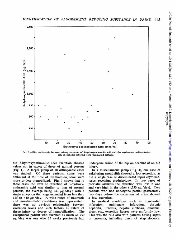
showed that the cases fell into two groups
(Fig. 2). The first comprises the seven highestexcretors, that is, all who excreted 1,000 ,ug. or
more per day, who had E.S.R. values rangingfrom 27 to 86 mm. /hour (average 61 mm./hr.).The E.S.R. of the remaining 18 cases ranged froin3 to 54 (average 32) mm. /hr. and in these the3-hydroxyanthranilic acid excretion never exceeded750 Mg./ day. There is thus a trend towards pro-
portionality between the two values. Comparisonwith the titres obtained in the sensitized sheepcell test (for details see Duthie, Brown, Knox, andThompson, 1957) showed a similar pattern ofdistribution as obtained for the E.S.R. for theseven highest excretors, all of whose titres were
positive, ranging from 1: 128 to 1: 2,048 (Fig. 3).The remaining 18 cases, however, had titres from1: 8 to 1: 2,048. Thus all patients with a highexcretion of 3-hydroxyanthranilic acid gave
a positive sensitized sheep cell test, but a positivetitre was not necessarily accompanied by a high
level of excretion. Degrees of rheumatoidactivity have been classified by Duthie et al.(1957) in terms of E.S.R., haemoglobin levels,joint involvement, and systemic disturbance. Theactivity of the disease of some of the abovepatients was independently assessed by the formerworkers. Three cases were allocated to group I
(inactive), 18 to group 2 (moderately active), anclnone to group 3 (very active). No relationshipwith the level of excretion of 3-hydroxyanthra-nilic acid could be discerned. Similarly, by thesame authors' classification of functional capacity,one case fell in group 1 (no disability), 14 ingroup 2 (moderate incapacity), three in group 3(marked incapacity), and one in group 4 (confinedto bed or chair); again no correlation withexcretion could be found.A small group of patients with osteoarthritis
was investigated. These patients were receivingsimilar basic treatment and diet in the same wardsand at the same time as the rheumatoid group,
0
0L-om
vJoSC
a)_I
U
.'
X taO c= Ut0
-1
2,500
I 1-1 Ii i
I
r-
r- v I v r T I 0I.
144
I
a
copyright. on F
ebruary 18, 2020 by guest. Protected by
http://jcp.bmj.com
/J C
lin Pathol: first published as 10.1136/jcp.13.2.140 on 1 M
arch 1960. Dow
nloaded from
IDENTIFICATION OF FLUORESCENT REDUCING SUBSTANCE IN URINE 145
2,500
2,000
.--
-L 1,500.-o
51000
2L-
5%1000
250
10 20 30 40 50 60 70Erythrocyte Sedimentation Rate (mm./hr.)
80 90 100
FIG. 2.-The relationship between urinary excretion of 3-hydroxyanthranilic acid and the erythrocyte sedimentationrate in patients suffering from rheumatoid arthritis.
but 3-hydroxyanthranilic acid excretion showedvalues not in excess of those of normal persons
(Fig. 1). A larger group of 18 orthopaedic caseswas studied. Of these patients, some were
ambulant at the time of examination, some weremore or less immobilized. Fig. 1 shows that inthese cases the level of excretion of 3-hydroxy-anthranilic acid was similar to that of normalpersons, the average being 240 ,ug./day; with a
single exception the range extended from less than125 to 440 Mig. /day. A wide range of traumaticand non-traumatic conditions was represented;there was no obvious relationship betweenexcretion levels and such factors as extent oftissue injury or degree of immobilization. Theexceptional patient who excreted as much as 750yg./day was one who 15 weeks previously had
undergone fusion of the hip on account of an oldinjury.
In a miscellaneous group (Fig. 4), one case ofankylosing spondylitis showed a low excretion, asdid a single case of disseminated lupus erythema-tosus receiving prednisolone. In two cases ofpsoriatic arthritis the excretion was low in one
and very high in the other (1,750 ,g./day). Twopatients who had undergone partial gastrectomytwo days before the collection of urine showeda low excretion.
In medical conditions such as myocardialinfarction, pulmonary infarction, chronicnephritis, uraemia, hepatic cirrhosis, duodenalulcer, etc., excretion figures were uniformly low.This was the rule also with patients having sepsisor anaemia, including cases of staphylococcal
.~~~~~~~~~~~~~~~~
.~~~~ ~ ~~~~~~~~~~~~
.~~~~~~~~ 00
0 0 0 0~~~~
I I
copyright. on F
ebruary 18, 2020 by guest. Protected by
http://jcp.bmj.com
/J C
lin Pathol: first published as 10.1136/jcp.13.2.140 on 1 M
arch 1960. Dow
nloaded from

MARY McMILLAN
2,000
.2ES-o-
X 1,000*0
5'1000
250.
1:8 1: 16 1:32 1:64 1: 128 1:256 1:512 1: 1,024 1:2,048Titre in Sensitized Sheep Cell Test
1o. 3.-The relationship between the titre obtained in the sensitized sheep cell test and the urinary excretion of3-hydroxyanthranilic acid.
.*0
0
ESOu otoy
0 i
0.-
1 .lI
1%1-~~~ ~ ~ ~ ~_ __
250 500 750 I,000 1,500 2,000 2,500Urinary Excretion of 3-Hydroxyanthranilic Acid (,ug./day)
Fro. 4.-Urinaryexcretion of 3-hydroxyanthranilic acid in diseases of the haemopoietic system and in other miscellaneous conditions.
146
a)
U
0
D0
E
z
rk%
copyright. on F
ebruary 18, 2020 by guest. Protected by
http://jcp.bmj.com
/J C
lin Pathol: first published as 10.1136/jcp.13.2.140 on 1 M
arch 1960. Dow
nloaded from

IDENTIFICATION OF FLUORESCENT REDUCING SUBSTANCE IN URINE 147
pneumonia and active pulmonary tuberculosis.Fever alone caused no rise. In view of thepossible relationship between the E.S.R. and theexcretion of 3-hydroxyanthranilic acid in rheu-matoid arthritis special attention was paid topatients having a very high E.S.R. from othercauses. All gave low figures with the exceptionof one patient with myelosclerosis with aplasticanaemia who had an E.S.R. of 125 mm./hourand an excretion of 3-hydroxyanthranilic acidas high as 1,000 tug./day. The other five patientshaving blood diseases included in this survey alsoshowed a tendency to raised outputs. Thus threepatients with chronic myeloid leukaemia, whosewhite cell counts were 209,000, 182,000, and60,800/c.mm. respectively, excreted 250, 750, and1,000 ,ug. /day; the second of these had undergoneno treatment, the other two were receivingirradiation therapy. Two other patients sufferingfrom megakaryocytic leukaemia and myelo-matosis, whose white cell counts were not raised,excreted 190 and 440 jig./day respectively.
DiscussionThis study fails to substantiate the claim of
Nishimura et al. that 2,5-dihydroxyphenylpyruvicacid is excreted in the urine by patients withrheumatoid arthritis. The data published bythe Japanese workers were incomplete, but an
approximation to their method of identificationfailed to demonstrate any such compound. In theabsence of excretion of 2,5-dihydroxyphenyl-pyruvic acid there is no evidence for the hypo-thesis that the collagen diseases are due to an
inherent error in the metabolism of tyrosine.Chromatographic examination of the urine in
cases of rheumatoid arthritis for a stronglyreducing substance has shown, however, that free3-hydroxyanthranilic acid is excreted in increasedamount as compared with normal subjects. Thecompound is known to be a normal metabolite oftryptophan; it is the immediate degradationproduct of 3-hydroxykynurenine and a precursorof nicotinic acid. It has so far been of limitedmedical interest, mainly as a possible carcinogen.Since the suggestion that o-aminophenols werecarcinogenic (Bonser, Clayson, Jull, and Pyrah,1954), attention has been drawn to the twonaturally occurring compounds, namely, 3-hydroxykynurenine and 3-hydroxyanthranilicacid, which have this structure. In fact, 3-hydroxyanthranilic acid was shown to producecancer of the bladder in mice (Watson, 1956) andto be excreted in the unconjugated state inincreased amount in human cancer of the bladder(Boyland and Williams, 1956). The latter
authors, who used a method involving adsorptionon charcoal and gradient-elution chromatography,give the normal excretion of free 3-hydroxy-anthranilic acid as 8 to 32 (average 20) mg./10 1.urine; this is considerably higher than the valuesreported in the present paper.
Dalgliesh and Tekman (1954) reported anincreased urinary output of kynurenine and 3-hydroxykynurenine in half of a series of febrilepatients, suggesting it was due to increased tissuebreakdown; loss of weight without fever andthyrotoxicosis did not have this effect. Musajo,Benassi, and Parpajola (1956) reported thatrecession of fever did not arrest the excessiveexcretion of kynurenines. In a large series, thesame authors (1955), using paper chromatography,found "very small amounts" of 3-HAA to beexcreted by normal human subjects. In 11 out of13 subjects with pulmonary tuberculosis excretionwas definitely higher. Also in 59 out of 76 casesof leukaemia of various types, myelomatosis,Hodgkin's disease, and lymphosarcoma, consider-able quantities of kynurenine and 3-hydroxy-kynurenine were found, but none was detectablein normal persons; no details were given inregard to 3-HAA, apart from a statement thatexcretion was raised in about one-third of thecases. The authors emphasized that the detectionof tryptophan metabolites in human urine in theabsence of special diets or administration oftryptophan was rare, and such excretion waslinked with pathological conditions.An increased excretion of 3-HAA in any of the
collagen diseases has not been reported previously.In the present series of cases the magnitude of theincrease would appear to bear no relation to anyof the major features of the disease except perhapsthe E.S.R., where there was some proportionality,and the sensitized sheep cell test, which was alwayspositive in association with the highest excretionrates. A high E.S.R. per se does not lead toincreased excretion since many non-rheumatoidpatients had a very high E.S.R. without such anincrease. The rheumatoid patients had receivedno steroid or other drugs for at least three daysbefore 3-HAA excretion was measured. A normalperson who received heavy doses of either aspirinor panadol for three days showed no increase in3-HAA excretion three days later. The effect ofdrugs on excretion by the rheumatoid patientswas not investigated.
In agreement with Musajo et al. (1956), no directrelationship was found between 3-HAA excretionand fever; but contrary to the above report noincrease was found in active pulmonary tuber-culosis.
copyright. on F
ebruary 18, 2020 by guest. Protected by
http://jcp.bmj.com
/J C
lin Pathol: first published as 10.1136/jcp.13.2.140 on 1 M
arch 1960. Dow
nloaded from

MARY McMILLAN
The raised excretion in some patients withhaematological disorders is of interest and meritsa further survey and investigation. Cases oferythrogenesis imperfecta have been reported toexcrete abnormal amounts of anthranilic acid(Altman and Miller, 1953). In diseases of theblood-forming organs, then, it would appear thatmore than one tryptophan metabolite may beexcreted in excess. Whether metabolites otherthan 3-HAA are involved in rheumatoid arthritishas not been determined and there is no evidenceto suggest that the findings in rheumatoid arthritisare in any way related to those in blood disease.An increase in urinary 3-HAA could be due to
increased tryptophan catabolism. However, manypatients in whom tissue was probably wastingfailed to show this effect and in rheumatoidarthritis itself there was no correlation with suchwasting. Possibly 3-hydroxyanthranilic acidcould accumulate as a result of enzymicimbalance such as can be produced in vivo in thecase of certain other tryptophan metabolites byvitamin deficiency. Further insight into theabnormality in rheumatoid arthritis might begained by studies along these lines.
This research was supported by a grant from theScottish Hospitals Endowment Research Trust to Dr.
C. P. Stewart and this assistance is gratefully acknow-ledged. I am grateful to Dr. C. P. Stewart for hiscritical interest; to Dr. J. J. R. Duthie for muchhelpful co-operation in connexion with the patientsunder his care at the Rheumatic Unit, NorthernGeneral Hospital, Edinburgh, and also for criticism ofthis paper; also to the staff of the Orthopaedic,Medical, and Radiotherapy Departments of theEdinburgh Royal Infirmary for facilities in connexionwith the collection of specimens.
REFERENCESAltman, K. I., and Miller, G. (1953). Nature (Lond.), 172, 868.Bonser, G. M., Clayson, D. B., Jull, J. W., and Pyrah, L. N. (1954).
Brit. J. Urol., 26, 49.Boyland, E., and Williams, D. C. (1956). Biochem. J., 64, 578.Consden, R., Forbes, H. A. W., Glynn, L. E., and Stanier, W. M.
(1952). Ibid., 50, 274.Daigliesh, C. E., and Tekman, S. (1954). Ibid., 56, 458.Duthie, J. J. R., Brown, P. E., Knox, J. D. E., and Thompson, M.
(1957). Ann. rheum. Dis., 16, 411.McMillan, Mary (1957). Lancet, 1, 715.Musajo, L., Benassi, C. A., and Parpajola, A. (1955). Nature (Lond.),
175, 855.-- (1956). Clin. Chim. Acta, 1, 229.Nishimura, N., Shibata, Y., Yasui, M., and Okamoto, H. (1956).
Proc. Japan Acad., 32, 697.- Yasui, M., Okamoto, H., Kanazawa, M., Kotake, Y., and
Shibata, Y. (1958). A.M.A. Arch. Derm., 77, 255.Roux, D. G. (1951). Nature (Lond.), 168, 1041.Smith, I. (1958). Chromatographic Techniques. Heinemann,
London.Tompsett, S. L. (1959). Clin. Chim. Acta, 4, 411.Watson, J. G. (1956). Surg. Forum, 7, 679.
148
copyright. on F
ebruary 18, 2020 by guest. Protected by
http://jcp.bmj.com
/J C
lin Pathol: first published as 10.1136/jcp.13.2.140 on 1 M
arch 1960. Dow
nloaded from