the efficacy of riboflavin for collagen cross-linking and
TRANSCRIPT
Avicenna J Dent Res. 2017 September; 9(3):e13254.
Published online 2017 May 30.
doi: 10.5812/ajdr.13254.
Research Article
The Efficacy of Riboflavin for Collagen Cross-Linking and Optimizing
the Bond Strength of an Etch and Rinse Adhesive System to Dentin
Shahin Kasraei,1 Mona Malek,2,* Zahra Khamverdi,3 and Maryam Mojtahedi4
1DDS, MS, Professor, Department of Restorative Dentistry, Dental School, Shahid Beheshti University of Medical Sciences, Tehran, Iran2DDS, Post Graduate Student, Department of Restorative Dentistry, Dental School, Hamedan University of Medical Sciences, Hamedan, IR Iran3DDS, MS, Professor, Dental Research Center, Department of Restorative Dentistry, Dental School, Hamedan University of Medical Sciences, Hamedan, IR Iran4DDS, MS, Assistant Professor, Dental Research Center, Department of Restorative Dentistry, Dental School, Birjand University of Medical Sciences, Birjand, IR Iran
*Corresponding author: Mona Malek, DDS, Post Graduate Student, Departmentof Restorative Dentistry, Dental School, Hamedan University of Medical Sciences, Hamedan, IRIran. Fax: +98-38138381085, E-mail: [email protected]
Received 2016 October 06; Revised 2017 January 23; Accepted 2017 April 03.
Abstract
Background and Objectives: Previous studies have shown that increasing collagen resistance to degradation stabilizes the resin-dentin interface and collagen cross-linkers can prevent the degradation of collagen fibrils as such. This study sought to assess theefficacy of light-activated riboflavin for collagen cross-linking and optimizing the micro tensile bond strength (µTBS) of an etch andrinse adhesive system to dentin.Methods: Occlusal surfaces of 12 sound premolars were ground in order to expose dentin. The teeth were randomly divided intotwo groups. Adper single bond was applied on etched dentin and light cured in the control group and Composite was then applied.In the test group, all the steps were done in a similar manner to the control group with the difference that after acid etching, 0.1%riboflavin solution was applied on dentin surface and was activated by blue light irradiation for 2 minutes. After thermocycling,the teeth were sectioned in 2 directions in such a way that 14 resin-dentin sticks with 1 mm2 cross-section were prepared in eachgroup (n = 14). The µTBS of samples was then measured and analyzed with independent t-test. The statistical analyses’ significancelevel was set at α = 0.05. Mode of failure was determined under a stereomicroscope at × 40 magnification. Moreover, the cross-linking effect of light-activated riboflavin solution on type I collagen was assessed using sodium dodecyl sulfate-polyacrylamide gelelectrophoresis (SDS-PAGE) and resin-dentin interface was photographed under a scanning electron microscope (SEM).Results: In gel electrophoresis, bands were formed in wells in the test group (and not in the negative control group). T-test foundno significant difference in the mean µTBS of the test and the control groups (P = 0.9).Conclusions: Based on the results, light-activated riboflavin was capable of collagen cross-linking, but its application as a collagencross-linker on etched dentin had no significant effect on µTBS of Single Bond dentin bonding agent to dentin.
Keywords: Photo-Activation, Riboflavin, Crosslinking, Dentin, Biodegradation, Collagen
1. Background
The durability of hybrid layer depends on the stabilityof its components such as collagen fibrils and polymericchains. Collagen fibrils exposed by acid etching whichhave not been infiltrated by resin cannot resist denatura-tion (1). These collagen fibrils are more susceptible to creepand degradation due to cyclic fatigue after long-term func-tion (2). On the other hand, matrix metalloproteinases(MMPs) are exposed and activated by acidic agents duringthe bonding process and since they play a role in the degra-dation of type I collagen, which is the organic componentof the hybrid layer, they can gradually break down collagenfibrils at the resin-dentin interface (3-7).
It appears that increasing the collagen resistance tobiodegradation can further stabilize the resin-dentin in-terface (8-10). Collagen cross-linkers can protect collagenfibrils from degradation and enhance their mechanical
and chemical properties (11-13). This is the main objectivebehind the use of collagen cross-linkers along with adhe-sives in the bonding process. The advantages offered by col-lagen cross-linkers when used in conjunction with bond-ing agents are mainly due to their ability to inhibit MMPs.Some studies have provided evidence regarding the lim-ited activity of MMPs following the use of collagen crosslinkers (9, 14, 15).
The currently available approaches to increase colla-gen cross-linking are divided into two groups of chemicaland physical or photo-oxidative methods (9). The currentlyused chemical cross-linkers have drawbacks such as toxi-city, poor control over the degree of cross-linking and in-stability (16). Although several cross-linkers such as glu-taraldehyde and proanthocyanidin have shown efficienttherapeutic effects on dentin collagens, they need to beused for long periods of time, which limits their applica-tion in the clinical setting (17-20). Cross-linkers must be al-
Copyright © 2017, Avicenna Journal of Dental Research. This is an open-access article distributed under the terms of the Creative Commons Attribution-NonCommercial 4.0International License (http://creativecommons.org/licenses/by-nc/4.0/) which permits copy and redistribute the material just in noncommercial usages, provided theoriginal work is properly cited.

Kasraei S et al.
lowed adequate time clinically to prevent collagen degra-dation. Moreover, they must have minimal cytotoxicity toprotect the resin-dentin interface (2).
It has been reported that the presence of oxygenfree radicals is necessary for collagen cross-linking inthe photo-oxidative method. Riboflavin (vitamin B2) isamong the most potent generators of oxygen free radicalswhen activated with light (21, 22). UVA-activated riboflavin(RF/UVA) was recently used as a cross-linker in ophthalmol-ogy. It has been documented that it increases the cross-linking of type I collagen (23) and stops keratoconus (22).Maximum absorbance peaks of riboflavin as a cross-linkerare at 270, 366, and 445 nm wavelengths (24, 25).
Light-activated riboflavin can break weak bonds withincollagen fibrils and create oxygen free radicals. Also, newcovalent bonds are formed between hydroxyl functionalgroups of riboflavin and proline or lysine in collagen (26,27). Collagen cross-linking with riboflavin enhances its me-chanical properties and delays its enzymatic degradation(28).
Due to the biocompatibility and cross linking of dentincollagen matrix, this technique may be used as a method ofdentin surface preparation or as an optimizer of adhesivesystems (29). This study aimed to assess the efficacy of ri-boflavin activated with light (by a light curing unit) for col-lagen cross linking and optimizing the bond strength of anetch and rinse adhesive system to dentin. The first null hy-pothesis was that light-activated riboflavin would have noeffect on collagen cross linking. The second null hypoth-esis was that activated riboflavin would have no effect onbond strength of etch and rinse adhesives to dentin.
2. Methods
This in-vitro experimental study was conducted on 12sound human premolars without caries, occlusal wear orrestorations which had been extracted for orthodonticpurposes within the past 3 months. The teeth were im-mersed in 0.2% thymol solution.
Riboflavin solution was added to 1.5mg/mL bovine col-lagen (Sigma Aldrich, St. Louis, MO, USA) (30). The crosslinking effect of riboflavin solution on type I collagen wasassessed using SDS-PAGE; 0.1% riboflavin solution was pre-pared by dissolving riboflavin-5-phosphate (Sigma Aldrich,St. Louis, MO,USA) in distilled water. In the negative con-trol group, distilled water was added to collagen. In thepositive control group, 0.1% riboflavin solution was addedto collagen and activated by UVA with 365 nm wavelength(Jenway 6105 U.V/Vis Spectrophotometer, Jenway LTD, Dun-mow, Essex, UK).
To prepare samples for SDS-PAGE, the samples were di-luted with buffer and boiled at 100°C for five minutes.
Next, 40 µgr of each sample was placed in wells of 8% SDS-polyacrylamide gel. Electrophoresis was performed at 150v voltage and gel was stained with Coomassie blue. Af-ter two hours of gentle vibration of gel on a shaker, thedye solution was removed and discoloring solution wasadded. The standard molecular weight markers (8 - 220KDa; Sigma-Aldrich, St. Louis, MO, USA) were run in parallelwith the samples in gel. The prepared riboflavin solutionswere stored in light-resistant test tubes and applied on thedentin surfaces (15µL) at room temperature (24°C). An LEDlight curing unit (DemiTM Plus; Demetron Kerr, USA) withalight guide diameter of 8 mm, 450 to 470 nm wavelengthand light intensity of 700 mW/cm2 was used.
2.1. Specimen Preparation
The occlusal enamel of the teeth was trimmed usingan orthodontic trimmer (Pars Medical, Tehran, Iran) underwater coolant to 1mm below the dentinoenamel junctionand continued to expose 5 mm diameter of dentin. To cre-ate a standard smear layer, the surface of the teeth was pol-ished with 600-grit moist silicon carbide paper. The teethwere then randomly divided into two groups of six. In thecontrol group, the surface of samples was etched with 35%phosphoric acid for 10 seconds and was then rinsed withdistilled water. Two layers of etch and rinse adhesive (Ad-per Single Bond) was applied on dentin surface for 15 sec-onds and was gently air sprayed for 5 seconds followed by10 seconds of light curing with an LED light curing unit.
In light-activated riboflavin group, all steps were per-formed in a similar manner to the control group with thedifference that after acid etching, 0.1% riboflavin solutionwas applied on dentin surface, gently air dried and acti-vated by light using an LED light curing unit for two min-utes. The next steps of adhesive application were similar tothose in the control group.
2.2. Micro Tensile Bond Strength Tests
To measure the µTBS, after applying the dentin bond-ing agent, 2 mm thick increments of composite (FiltekZ250, 3M ESPE, St Paul, MN, USA) were applied on the sur-face and light cured to do a 4 mm thick composite buildup.The teeth were immersed in distilled water at 37°C for24 hours to complete polymerization and were then sub-jected to 5,000 thermal cycles between 5 - 55°C with 15 sec-onds of dwell time (Nemo Co, Mashhad, IRAN). Using a di-amond blade at low speed (Nemo Co, Mashhad, IRAN) un-der water coolant, the samples were sectioned into resin-dentin bars measuring 1 × 1 mm and were stored in wa-ter at 37°C for three months (31). A total of 14 resin-dentinsticks were prepared in each group (n = 14). The sticks weremounted on a custom-made metal jig of a universal test-ing machine (Santam T20, Tehran, Iran) with 50 N load-cell
2 Avicenna J Dent Res. 2017; 9(3):e13254.
Kasraei S et al.
using cyanoacrylate glue. Load was applied at a crossheadspeed of 1 mm/minute until failure. Tensile stress at frac-ture was recorded as the µTBS. The data were analyzed us-ing SPSS version 16 and independent t-test. P < 0.05 wasconsidered statistically significant.
2.3. Evaluation of the Mode of Failure and SEM Analysis
To determine the mode of failure, each sample was ob-served under a stereomicroscope (SZ40, Olympus, Tokyo,Japan) at×40 magnification twice. Mode of failure was de-termined as adhesive, cohesive in dentin, cohesive in com-posite resin or mixed. The bonding interface of sampleswere also inspected under a SEM (Leo 1450VP, Zeiss, Wetzlar,Germany). Some specific areas at the resin-dentin interfacewere photographed at×2500 and×5000 magnifications.
3. Results
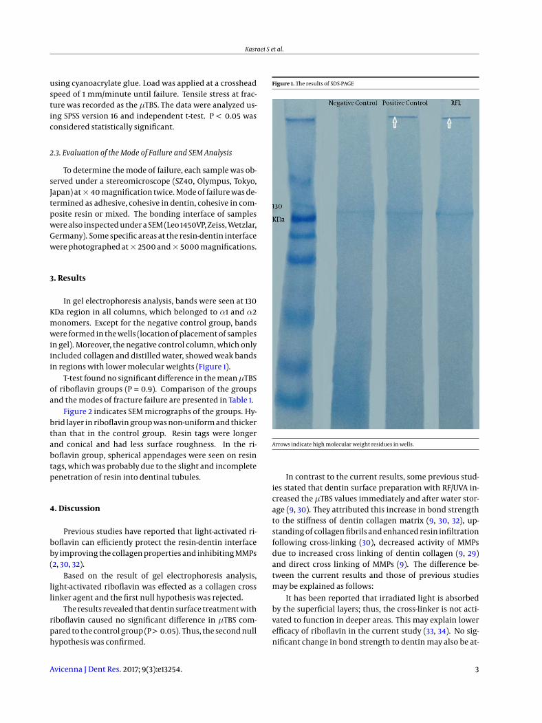
In gel electrophoresis analysis, bands were seen at 130KDa region in all columns, which belonged to α1 and α2monomers. Except for the negative control group, bandswere formed in the wells (location of placement of samplesin gel). Moreover, the negative control column, which onlyincluded collagen and distilled water, showed weak bandsin regions with lower molecular weights (Figure 1).
T-test found no significant difference in the meanµTBSof riboflavin groups (P = 0.9). Comparison of the groupsand the modes of fracture failure are presented in Table 1.
Figure 2 indicates SEM micrographs of the groups. Hy-brid layer in riboflavin group was non-uniform and thickerthan that in the control group. Resin tags were longerand conical and had less surface roughness. In the ri-boflavin group, spherical appendages were seen on resintags, which was probably due to the slight and incompletepenetration of resin into dentinal tubules.
4. Discussion
Previous studies have reported that light-activated ri-boflavin can efficiently protect the resin-dentin interfaceby improving the collagen properties and inhibiting MMPs(2, 30, 32).
Based on the result of gel electrophoresis analysis,light-activated riboflavin was effected as a collagen crosslinker agent and the first null hypothesis was rejected.
The results revealed that dentin surface treatment withriboflavin caused no significant difference in µTBS com-pared to the control group (P > 0.05). Thus, the second nullhypothesis was confirmed.
Figure 1. The results of SDS-PAGE
Arrows indicate high molecular weight residues in wells.
In contrast to the current results, some previous stud-ies stated that dentin surface preparation with RF/UVA in-creased the µTBS values immediately and after water stor-age (9, 30). They attributed this increase in bond strengthto the stiffness of dentin collagen matrix (9, 30, 32), up-standing of collagen fibrils and enhanced resin infiltrationfollowing cross-linking (30), decreased activity of MMPsdue to increased cross linking of dentin collagen (9, 29)and direct cross linking of MMPs (9). The difference be-tween the current results and those of previous studiesmay be explained as follows:
It has been reported that irradiated light is absorbedby the superficial layers; thus, the cross-linker is not acti-vated to function in deeper areas. This may explain lowerefficacy of riboflavin in the current study (33, 34). No sig-nificant change in bond strength to dentin may also be at-
Avicenna J Dent Res. 2017; 9(3):e13254. 3

Kasraei S et al.
Table 1. Means Value (SD) of Micro Tensile Dentin Bond Strength (MPa) and Failure Modes of Study Groups
Group Surface Pretreatment Micro Tensile BondStrength
95% Wald Confidence Interval P Valuea Failure Mode A/M/CR/CD
Lower Upper
Control No pretratment 12.64 (2.35) -2.53 2.230.90
3/11/0/0
RF/BL Application andphotoactivation of RF% 0.1
(PH = 3)
12.79 (3.64) -2.55 2.25 8/6/0/0
Abbreviations: A, Adhesive; CD, Cohesive in Dentin; CR, Cohesive in Resin composite; M, Mixed; RF, Riboflavin; SD, Standard Deviation.a Independent T-test, n = 14.
Figure 2. SEM Micrographs Showing the Resin-Dentin Interface Following the Application of Etch and Rinse Adhesive and Light Curing with an LED Light Curing Unit with andWithout the Application of Riboflavin; E, Control Group at× 2500 Magnification; F, Control Group at× 5000 Magnification; G, Riboflavin Group at× 2500 Magnification; H,Riboflavin Group at × 5000 Magnification
tributed to the reinforcing effect of hydration by RF/UVA(27, 35), which can adversely affect the formation of an op-timized and stable hybrid layer (31).
Extending the application time of Riboflavin and theexposure time of the light activated may generate a bet-ter result, but is less practicable for dental use. However,these limited findings may serve as a foundation for fu-ture studies of Riboflavin as an adjunctive dentin bonding
treatment (30).
Riboflavin can impair the function of adhesive systemby water sorption from dentin and the dilution of theprimer. Water sorption after polymerization and the re-lease of un-reacted hydrophilic monomers from the hy-brid layer can decrease the physical properties of the ad-hesive layer after 24 hours (36). Moreover, chemical-crosslinking does not change the internal stiffness of collagen
4 Avicenna J Dent Res. 2017; 9(3):e13254.
Kasraei S et al.
molecules (37). Thus, loose collagen fibrils are susceptibleto creep and subsequent fracture due to fatigue after long-term function (2).
On the other hand, the use of cross-linkers such as lightactivated riboflavin can reinforce the collagen smear re-maining on the surface (38). Collagen smear on the sur-face of etched dentin decreases the infiltration of adhe-sive into the remaining demineralized collagen network.It also forms a layer rich in organic materials with verysmall resin content on the surface of hybrid layer. Eventu-ally, both of these factors decrease the strength and dura-bility of the hybrid layer as well as the bond to dentin sub-strate. This finding was in agreement with the results ofHass et al. who reported that riboflavin increased micro-hardness, indicating that collagen cross-linkers can serveas bio-modifiers and increase the mechanical strength andstability and decrease degradation compared to normaltissue. They added that cross-linkers create a barrier whichinhibits further penetration into the tissue. It affects thefixation depth and limits the efficacy for increasing thestrength of dental substrate (31).
Further studies are required on the efficacy of cross-linkers for the creation of a stable and optimized hybridlayer and their effects on the strength and durability ofbond to dentin.
4.1. Conclusions
Within the limitations of this study, the results showedthat light activated riboflavin was capable of collagen crosslinking, but its application on etched dentin as a colla-gen cross linker had no significant effect onµTBS of SingleBond dentin adhesive agent to dentin.
References
1. Tjaderhane L, Nascimento FD, Breschi L, Mazzoni A, Tersariol IL, Ger-aldeli S, et al. Optimizing dentin bond durability: control of colla-gen degradation by matrix metalloproteinases and cysteine cathep-sins. Dent Mater. 2013;29(1):116–35. doi: 10.1016/j.dental.2012.08.004.[PubMed: 22901826].
2. Hass V, Luque-Martinez IV, Gutierrez MF, Moreira CG, Gotti VB, FeitosaVP, et al. Collagen cross-linkers on dentin bonding: Stability of theadhesive interfaces, degree of conversion of the adhesive, cytotoxi-city and in situ MMP inhibition. Dent Mater. 2016;32(6):732–41. doi:10.1016/j.dental.2016.03.008. [PubMed: 27087688].
3. Breschi L, Martin P, Mazzoni A, Nato F, Carrilho M, Tjaderhane L, et al.Use of a specific MMP-inhibitor (galardin) for preservation of hybridlayer. Dent Mater. 2010;26(6):571–8. doi: 10.1016/j.dental.2010.02.007.[PubMed: 20299089].
4. De Munck J, Van den Steen PE, Mine A, Van Landuyt KL, PoitevinA, Opdenakker G, et al. Inhibition of enzymatic degradation ofadhesive-dentin interfaces. J Dent Res. 2009;88(12):1101–6. doi:10.1177/0022034509346952. [PubMed: 19861692].
5. Zheng P, Zaruba M, Attin T, Wiegand A. Effect of different ma-trix metalloproteinase inhibitors on microtensile bond strength ofan etch-and-rinse and a self-etching adhesive to dentin. Oper Dent.2015;40(1):80–6. doi: 10.2341/13-162-L. [PubMed: 24815915].
6. Li H, Li T, Li X, Zhang Z, Li P, Li Z. Morphological effects of MMPs in-hibitors on the dentin bonding. Int J Clin ExpMed. 2015;8(7):10793–803.[PubMed: 26379873].
7. Carvalho C, Fernandes FP, Freitas Vda P, Franca FM, Basting RT, TurssiCP, et al. Effect of green tea extract on bonding durability of an etch-and-rinse adhesive system to caries-affected dentin. J Appl Oral Sci.2016;24(3):211–7. doi: 10.1590/1678-775720150518. [PubMed: 27383701].
8. Bedran-Russo AK, Vidal CM, Dos Santos PH, Castellan CS. Long-term effect of carbodiimide on dentin matrix and resin-dentinbonds. J Biomed Mater Res B Appl Biomater. 2010;94(1):250–5. doi:10.1002/jbm.b.31649. [PubMed: 20524201].
9. Cova A, Breschi L, Nato F, Ruggeri AJ, Carrilho M, Tjaderhane L,et al. Effect of UVA-activated riboflavin on dentin bonding. J DentRes. 2011;90(12):1439–45. doi: 10.1177/0022034511423397. [PubMed:21940521].
10. Green B, Yao X, Ganguly A, Xu C, Dusevich V, Walker MP, et al. Grapeseed proanthocyanidins increase collagen biodegradation resistancein the dentin/adhesive interface when included in an adhesive. JDent. 2010;38(11):908–15. doi: 10.1016/j.jdent.2010.08.004. [PubMed:20709136].
11. Castellan CS, Pereira PN, Grande RH, Bedran-Russo AK. Mechani-cal characterization of proanthocyanidin-dentin matrix interaction.Dent Mater. 2010;26(10):968–73. doi: 10.1016/j.dental.2010.06.001.[PubMed: 20650510].
12. Liu Y, Chen M, Yao X, Xu C, Zhang Y, Wang Y. Enhancement in dentincollagen’s biological stability after proanthocyanidins treatment inclinically relevant time periods. Dent Mater. 2013;29(4):485–92. doi:10.1016/j.dental.2013.01.013. [PubMed: 23434233].
13. Xu C, Wang Y. Cross-linked demineralized dentin maintains its me-chanical stability when challenged by bacterial collagenase. J BiomedMater Res B Appl Biomater. 2011;96(2):242–8. doi: 10.1002/jbm.b.31759.[PubMed: 21210503].
14. Mazzoni A, Angeloni V, Apolonio FM, Scotti N, Tjaderhane L, Tezvergil-Mutluay A, et al. Effect of carbodiimide (EDC) on the bond stability ofetch-and-rinse adhesive systems. Dent Mater. 2013;29(10):1040–7. doi:10.1016/j.dental.2013.07.010. [PubMed: 23916318].
15. Scheffel DL, Hebling J, Scheffel RH, Agee KA, Cadenaro M, Turco G, etal. Stabilization of dentin matrix after cross-linking treatments, invitro. Dent Mater. 2014;30(2):227–33. doi: 10.1016/j.dental.2013.11.007.[PubMed: 24332989].
16. Han B, Jaurequi J, Tang BW, Nimni ME. Proanthocyanidin: a naturalcrosslinking reagent for stabilizing collagen matrices. J BiomedMaterRes A. 2003;65(1):118–24. doi: 10.1002/jbm.a.10460. [PubMed: 12635161].
17. Al-Ammar A, Drummond JL, Bedran-Russo AK. The use of collagencross-linking agents to enhance dentin bond strength. J BiomedMater Res B Appl Biomater. 2009;91(1):419–24. doi: 10.1002/jbm.b.31417.[PubMed: 19507140].
18. Bedran-Russo AK, Pashley DH, Agee K, Drummond JL, Miescke KJ.Changes in stiffness of demineralized dentin following applica-tion of collagen crosslinkers. J Biomed Mater Res B Appl Biomater.2008;86(2):330–4. doi: 10.1002/jbm.b.31022. [PubMed: 18161815].
19. Castellan CS, Bedran-Russo AK, Karol S, Pereira PN. Long-term stabil-ity of dentin matrix following treatment with various natural colla-gen cross-linkers. J Mech Behav Biomed Mater. 2011;4(7):1343–50. doi:10.1016/j.jmbbm.2011.05.003. [PubMed: 21783144].
20. Macedo GV, Yamauchi M, Bedran-Russo AK. Effects of chemi-cal cross-linkers on caries-affected dentin bonding. J Dent Res.2009;88(12):1096–100. doi: 10.1177/0022034509351001. [PubMed:19892915].
21. Snibson GR. Collagen cross-linking: a new treatment paradigm incorneal disease - a review. Clin Exp Ophthalmol. 2010;38(2):141–53. doi:10.1111/j.1442-9071.2010.02228.x. [PubMed: 20398104].
22. Sorkin N, Varssano D. Corneal collagen crosslinking: a systematicreview. Ophthalmologica. 2014;232(1):10–27. doi: 10.1159/000357979.[PubMed: 24751584].
Avicenna J Dent Res. 2017; 9(3):e13254. 5
Kasraei S et al.
23. Arbelaez MC, Sekito MB, Vidal C, Choudhury SR. Collagen cross-linking with riboflavin and ultraviolet-A light in keratoconus: One-year results. Oman J Ophthalmol. 2009;2(1):33–8. doi: 10.4103/0974-620X.48420. [PubMed: 21234222].
24. Spoerl E, Mrochen M, Sliney D, Trokel S, Seiler T. Safety of UVA-riboflavin cross-linking of the cornea. Cornea. 2007;26(4):385–9. doi:10.1097/ICO.0b013e3180334f78. [PubMed: 17457183].
25. Kohlhaas M, Spoerl E, Schilde T, Unger G, Wittig C, Pillunat LE.Biomechanical evidence of the distribution of cross-links in corneastreated with riboflavin and ultraviolet A light. J Cataract RefractSurg. 2006;32(2):279–83. doi: 10.1016/j.jcrs.2005.12.092. [PubMed:16565005].
26. Sionkowska A. Flash photolysis and pulse radiolysis studies oncollagen Type I in acetic acid solution. J Photochem Photobiol B.2006;84(1):38–45. doi: 10.1016/j.jphotobiol.2006.01.007. [PubMed:16504532].
27. Wollensak G, Aurich H, Pham DT, Wirbelauer C. Hydration behav-ior of porcine cornea crosslinked with riboflavin and ultraviolet A. JCataract Refract Surg. 2007;33(3):516–21. doi: 10.1016/j.jcrs.2006.11.015.[PubMed: 17321404].
28. Heo J, Koh RH, Shim W, Kim HD, Yim HG, Hwang NS. Riboflavin-induced photo-crosslinking of collagen hydrogel and its applicationin meniscus tissue engineering. Drug Deliv Transl Res. 2016;6(2):148–58. doi: 10.1007/s13346-015-0224-4. [PubMed: 25809935].
29. Fawzy A, Nitisusanta L, Iqbal K, Daood U, Beng LT, Neo J. Char-acterization of riboflavin-modified dentin collagen matrix. J DentRes. 2012;91(11):1049–54. doi: 10.1177/0022034512459053. [PubMed:22914538].
30. Chiang YS, Chen YL, Chuang SF, Wu CM, Wei PJ, Han CF, et al.Riboflavin-ultraviolet-A-induced collagen cross-linking treatmentsin improving dentin bonding. Dent Mater. 2013;29(6):682–92. doi:
10.1016/j.dental.2013.03.015. [PubMed: 23582694].31. Almahdy A, Koller G, Sauro S, Bartsch JW, Sherriff M, Watson TF, et
al. Effects of MMP inhibitors incorporated within dental adhesives. JDent Res. 2012;91(6):605–11. doi: 10.1177/0022034512446339. [PubMed:22518030].
32. Hass V, de Paula AM, Parreiras S, Gutierrez MF, Luque-Martinez I, deParis Matos T, et al. Degradation of dentin-bonded interfaces treatedwith collagen cross-linking agents in a cariogenic oral environment:An in situ study. J Dent. 2016;49:60–7. doi: 10.1016/j.jdent.2016.02.009.[PubMed: 27106766].
33. Spoerl E, Hoyer A, Pillunat LE, Raiskup F. Corneal cross-linking and safety issues. Open Ophthalmol J. 2011;5:14–6. doi:10.2174/1874364101105010014. [PubMed: 21399770].
34. Thorsrud A, Nicolaissen B, Drolsum L. Corneal collagen crosslink-ing in vitro: inhibited regeneration of human limbal epithelialcells after riboflavin-ultraviolet-A exposure. J Cataract Refract Surg.2012;38(6):1072–6. doi: 10.1016/j.jcrs.2011.12.038. [PubMed: 22624908].
35. Hayes S, Boote C, Kamma-Lorger CS, Rajan MS, Harris J, Dooley E,et al. Riboflavin/UVA collagen cross-linking-induced changes in nor-mal and keratoconus corneal stroma. PLoS One. 2011;6(8):e22405. doi:10.1371/journal.pone.0022405. [PubMed: 21850225].
36. Paul SJ, Leach M, Rueggeberg FA, Pashley DH. Effect of water contenton the physical properties of model dentine primer and bondingresins. J Dent. 1999;27(3):209–14. [PubMed: 10079627].
37. Liao J, Yang L, Grashow J, Sacks MS. Molecular orientation of colla-gen in intact planar connective tissues under biaxial stretch. ActaBiomater. 2005;1(1):45–54. doi: 10.1016/j.actbio.2004.09.007. [PubMed:16701779].
38. Eliades G, Palaghias G, Vougiouklakis G. Effect of acidic conditionerson dentin morphology, molecular composition and collagen confor-mation in situ. Dent Mater. 1997;13(1):24–33. [PubMed: 9467320].
6 Avicenna J Dent Res. 2017; 9(3):e13254.