introduction to corneal cross-linking (cxl) · peschke® d - standard riboflavin solution with...
TRANSCRIPT

Introduction to Corneal
Cross-Linking (CXL)

Keratokonus
• Incidence approx. 1 / 2000
• > 150’000 affected in US
• Commonest cause for PK in Western countries
• ~ 20% of PK’s in US (EBAA 2008)
• ~ 50 million spent on KC healthcare in US
• Begins in puberty and can progress or arrest at any
time, usually arrests at ~ age 40
• Progressive corneal thinning resulting in mixed
myopic and irregular astigmatism
• Usually bilateral; though asymmetric
• May be unilateral or asymmetric at initial presentation
2

1. History
2. Mechanism
3. Practical Application
4. Clinical Experience
Agenda
3

1. History
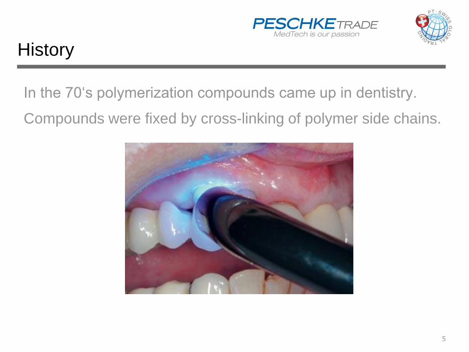
In the 70‘s polymerization compounds came up in dentistry.
Compounds were fixed by cross-linking of polymer side chains.
History
5

1st Publication - German
6

1st Publication - English
7

26 8
4
18
33 30
146
0
40
80
120
160
1998 1999 2000 2001 2002 2003 2004 2005
Number of CXL in Dresden since 1998
8

Cross-Linking and the Eye?
History
Transforming soft tissue
into a more rigid tissue
Transforming
progressive keratoconus into
forme fruste keratoconus
9
History
10
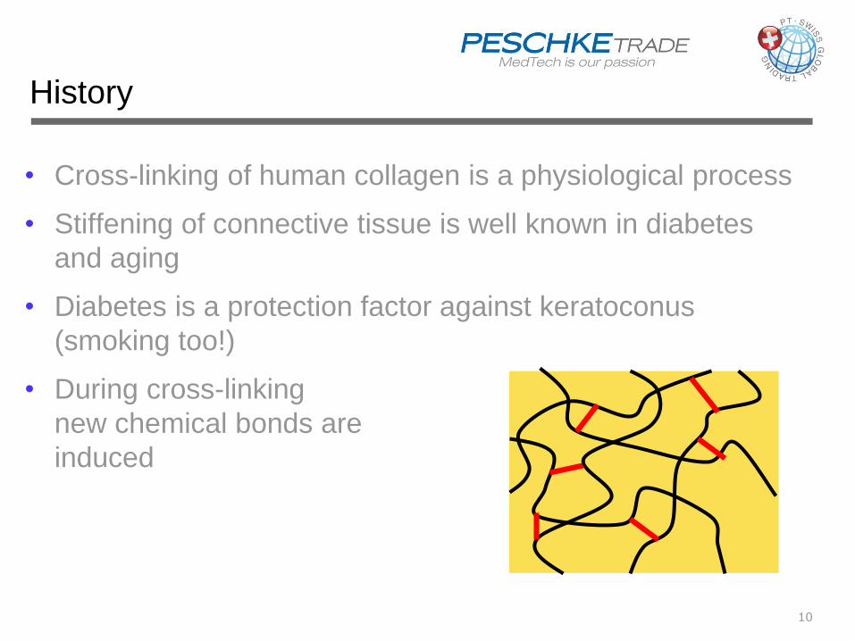
• Cross-linking of human collagen is a physiological process
• Stiffening of connective tissue is well known in diabetes
and aging
• Diabetes is a protection factor against keratoconus
(smoking too!)
• During cross-linking
new chemical bonds are
induced

History
1. Combined application of UVA and riboflavin
2. Creation of oxygen radicals O2
3. Induction of collagen cross-links
-CH2-CH2-CH2-CH2 = NH-CH2-CH2-CH2-CH2-
Riboflavin (Vitamin B2)Ultraviolet irradiation
collagen fibril collagen fibril
Speck 2006
11

2. Mechanism
12
Speck 2006
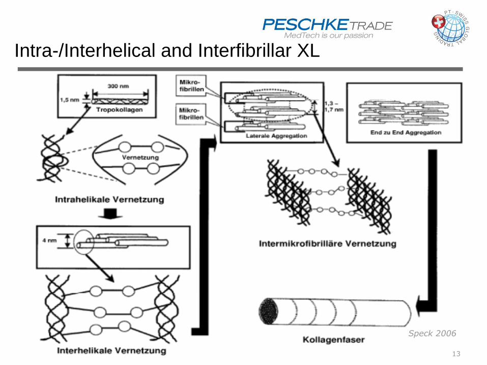
Intra-/Interhelical and Interfibrillar XL
13

Photochemically cross-linked collagen scaffolds gave fine
microstructures with interconnected nano-sized fibers.
Photochemical cross-linking improves the physicochemical
properties of collagen scaffolds. Chan BP, Sp KF: J Biomed Mater Res A. 75(2005)689-701
Background of Corneal Cross-Linking
14

after
Cross-linked vs.
non-cross-linked Cornea
before
15

3. Practical Application
16

• Progressive keratoconus
• Iatrogenic keratoconus
• Pellucid marginal degeneration
• Bullous keratopathy
• Prevention of keratectasia
• Infectious keratitis
Indications
17
Contra-Indications
• Corneal thickness < 400 µ
• Pregnancy
• Prior herpetic infection
• Ruptured Descemet`s membrane
• Autoimmune disorders
18

Necessary Equipment for CXL
• CCL-corneal cross-linking system
• PESCHKE® riboflavin solution TE, M, D, L
• PESCHKE® H hypotonic solution in case of cornea < 400 µ
• BSS – balanced salt solution
• Topical anesthetic (i.e. oxybuprocaine, tetracaine)
• Ultrasound pachymeter
• Speculum (with blades)
• Hockey knife/Amoils brush for epi-off
• LASIK sponge for limbal protection
• Sponges, pouch
• Drapes
• Bandage contact lens
• Post-op medication: pain killer, antibiotics, lubricants
19

`
20

CCL Vario
21
Technical Specifications:Wavelength range: 365 nm
Illumination intensity: 3 – 9 – 18 mW/cm2
Working distance: 50 mm + 5 mm
Light emission: Continuous wave
Spot sizes (continuously adjustable): 7.0 – 11.0 mm
Timer: 30 – 10 – 5 min
Electric power: 100 – 240 V
Dimensions Hard case (w,l,h in cm) 37 x 46 x 14
Weight (total) 7,5 kg

Peschke CXL
Technical Specifications:Wavelength: 365 nm
Illumination intensity (continuously adjustable): 3 – 30 mW/cm2
Working distance: 50 mm + 5 mm
Fixation light with adjustable brightness
Light emission: Continuous, Interval or Pulsed
Spot sizes (adjustable): 1.0 – 11.0 mm
Timer: Automatic time adjustment / audible alarm
Auxiliary timer for Riboflavin Installation
Electric power: 100 – 240 V, 50/60 Hz
Dimensions: 28x 12.5 x 10 cm
Weight: 1.8 kg

Peschke® TE - Our new transepithelial Solution for epi-on procedure• No removal of the corneal epithelium necessary• Significant reduction of pain and danger of postoperative infections• Recommended instillation time: 20 minutes (1 drop every 2 minutes = 10 drops)• Pre-loaded glass syringe containing 2.0 ml liquid• Ingredients: 0.25 % Riboflavin (Vitamin B2), 1.2 % HPMC, 0.01 % Benzalkoniumchloride
Peschke® M - Standard Riboflavin Solution without Dextran for epi-off procedure• Does not reduce corneal thickness• Recommended instillation time: 20 minutes (1 drop every 2 minutes = 10 drops)• Pre-loaded glass syringe containing 3.0 ml liquid• Ingredients: 0.1 % Riboflavin (Vitamin B2), 1.1 % HPMC
Peschke® D - Standard Riboflavin Solution with Dextran for epi-off procedure• The Dresden Original: Time proven Riboflavin solution with dextran• Recommended instillation time: 20 minutes (1 drop every 2 minutes = 10 drops)• Pre-loaded glass syringe containing 3.0 ml liquid• Ingredients: 0.1 % Riboflavin (Vitamin B2), 20 % dextran 500
Peschke® H - Hypotonic Riboflavin Solution for corneal swelling• To swell thin corneas (< 400 μ) by means of osmotic effect• Recommended instillation time: 1 drop every 5 seconds until corneal thickness has reached 400 μ• Pre-loaded glass syringe containing 1.5 ml liquid• Ingredients: 0.1 % Riboflavin (Vitamin B2)
Peschke® L - Riboflavin Solution for use with LASIK procedures• For use in connection with LASIK procedures on thin corneas• Recommended usage: after flap preparation and excimer treatment: put 3 – 5 drops on stroma, put flap back, wait 3 – 4 minutes, open flap and rinse off Riboflavin, put flap back and radiate with 1/2 of the recommended energy (1/2 of the time)
• Pre-loaded glass syringe containing 1.95 ml liquid• Ingredients: > 0.23 % Riboflavin (Vitamin B2)
PESCHKE Riboflavin Solutions
23

Peschke D
Riboflavin20 %Dextran
(Peschke)
Ricrolin TEETDA
15 %dextran
Riboflavin
BAC, 0.44% NaCl
no dextran
perfect
transepithelial
riboflavin
Peschke-TE
iontophoresis
Ricrolin15% dextran
(SOOFT)VibeX(Avedro)
Peschke Mno dextranno NaCl
ring application
BAC, no dextranno NaCl,
methylcellulose
ParaCel
24

Cross-Linking Options
25
CXL
epi-off
3 mW
30 min
epi-off
18 mW
5 min
epi-on
3 mW
30 min
epi-on
18 mW
5 min
Peschke® D
‚Dresden‘ Method
(+ Peschke® H)
Peschke® M
(+ Peschke® H)
Peschke® D
(+ Peschke® H)
Peschke® M
(+ Peschke® H)
Peschke® TE
(+ Peschke® H)
Peschke® TE
(+ Peschke® H)

• Corneal thickness at least 400 µm.
• Oxybuprocaine or tetracaine (as needed).
• If not transepithelial ⇨ abrasio corneae Ø 8 – 9 mm.
• Peschke® riboflavin solution,1 drop every 2 minutes
for 20 minutes (AC must be yellow under blue light)
• If cornea thinner than 400 µ ⇨ swell with hypotonic PESCHKE®
H solution.
• Radiate only clear cornea
(protect limbal stem cells!);
1 drop BSS every 5 seconds
Parameters used
26

Parameters and Time
3 mW ⇨ 30 min3 mW x 1800 sec = 5400 mJ
9 mW ⇨ 10 min9 mW x 600 sec = 5400 mJ
18 mW ⇨ 5 min18 mW x 300 sec = 5400 mJ
30 mW ⇨ 3 min30 mW x 180 sec = 5400 mJ
27

• Antibiotic ointment and bandage contact lens
• Antibiotic drops 3 times a day until epithelium is
closed
• Pain killer (as needed)
• Lubricants as needed
• Remove contact lens when epithelium is closed
(day 3)
• Post-op visits: day 1, 3, 5, 1 m, 3 m, 6 m, yearly
Post-operative Treatment
28
Steroids slow down epithelial healing!
NSAIDS can cause corneal melting!

Post-operative Observations
6 weeks:
• Slight clouding of the cornea
• Movement of cone to center ⇨ eye gets more myopic!
3 months:
• All eyes back to at least original K-values.
6 months:
• Average flattening ~ 2 diopters
29

Safety Aspects - Riboflavin Shielding
3.00 mW/cm²
1.49 mW/cm²
0.74 mW/cm²
0.36 mW/cm²
0.18 mW/cm²
0.09 mW/cm²
0.06 mW/cm²
0μm
100μm
200μm
300μm
400μm
500μm
600μm
100%
50%
25%
12%
6%
3%
2%
damage threshold endothelium 0.3mW/cm2
3 mW/cm²
Speck, 2006
30

300 µ
Safety Aspects - Keratocites
31

Safety Aspects - Corneal demarcation line
32

Pre op
6 m postop
3 m postop1 m postop
• Cross-linking kills keratocytes
300 μm deep
• Re-population takes 6 months
A. Caporossi, MD
Safety Aspects - Confocal Microscopy
33

4. Clinical Experience
34

1. Biomechanical Stability
2. Biochemical Stability
Clinical Experience
35

Kohlhaas/Spoerl: J Cataract Refract Surg. 32 (2006)279-283
treated untreated
Degree of Cross-Linking in
Relation to Corneal Thickness
36

• Increase of tangential solidity
• Restructuration and reshaping of the cornea
• Move of corneal apex to center
• Regularization of corneal surface
• Increased contact lens tolerance
• Reduction of corneal sensitivity
Biomechanics after CXL
37

Biochemical Effects
38

First report 2004
39

Mortensen/Makdoumi, 2008
• 84 year old woman
• Symptoms since
one week
• Large ulcer, hypopyon
• BCVA Hand Movements
Patient no. 2
Biochemical Stability
40

Mortensen/Makdoumi, 2008
• Treatment with CXL day
of diagnosis
• Greatly reduced
symptoms day after
treatment
• Disappearance of
hypopyon
• Slow epithelialization
• Picture after one week
41
Patient no. 2
Biochemical Stability

Mortensen/Makdoumi, 2008
• Follow-up after
six months
• BCVA 20/200
• No epithelial defect
• Large macula nasally
• Complete epithelialization
42
Patient no. 2
Biochemical Stability

Price 2012
Mechanism of Action
43

• Gram positive + gram negative strains
• Healing one week to several months
• Quick improvement: pain, light sensitivity,
inflammation, hypopyon
• Reduced corneal sensation
• Effect on Langerhans and dentritic cells
• Inhibitory effect on corneal immune system (?)
• Increased resistance against collagenasis
• Induction of apoptosis of corneal keratocites modify
environment for pathogens
Effects and Reasons
44

Other Applications
45

Results for Keratoconus
46

5-069-053-059-043-049-03
58
cross-linking
56
54
52
Results in Keratoconic Eyes
47

X-Linking
Results in Keratoconic Eyes
48

• No adverse reactions except haze
• According to corneal topography, progression halted
in every case
• Corneal thickness decreases after surgery
Results in Keratoconic Eyes
1m 3m 6m 12m0 μm
50 μm
49

max. K-readings decrease!
Prof. Dr. E. Spoerl, 2006
Results in Keratoconic Eyes
Cross-Linking
-10 0 10 20 30 40 50
-5
-4
-3
-2
-1
0
Months
Ch
an
ge in
max.
K-r
ead
ing
/ D
50

BSCVA increases!
Prof. Dr. E. Spoerl, 2006
Results in Keratoconic Eyes
Cross-Linking
-10 0 10 20 30 40 50
0
1
2
3
4
5
Months
Ch
an
ge in
BS
CV
A /
lin
es
51

• BSCVA increases
• Corneal surface regularizes, keratoconus indices decrease > 60 % of all patients!
Prof. Dr. E. Spoerl, 2006
Results in Keratoconic Eyes
1m 3m 6m 12m
1.2
1.1
1.0
1.3
KI
52

Safety:
Lines lost / gained – 1 year
Reiter, 201053

• No permanent side effects or adverse reactions (haze, myopia)
• According to corneal topography, progression halted in every case
• Maximal K-readings were significantly reduced in
65% of the cases
• Duration of treatment 30 – 60 min
Clinical Observations
54

Iatrogenic Keratectasia
55

• LASIK for -4 D
• Pre-op pachymetry 500 μm, no signs of FFKC
• Bilateral iatrogenic keratectasia starting 12 months after surgery
• Cross-linking right eye
untreated
after
Results in Iatrogenic Ectasia
before
56

12 Months CXL follow-up
Cross-Linking, Kmax: 57,27
5 months po, Kmax: 54,55
3.5 months po, Kmax: 54,87
12 months po, Kmax: 53,96
57

Corneal thickness should be at least
400 μm.
What shall we do with thin corneas (<400 μm)?
Special cases
58
• The human cornea can swell up to 2 - 3-fold in
thickness
• Let the cornea swell by means of hypotonic
riboflavin solution:
PESCHKE® H hypotonic solution
Important: 1 drop every 5 seconds!!!

Instillation of PESCHKE® hypotonic solution
postpre difference
Special cases
59

Pachymetry < 400μm
• 22 yr, ♂
• BCVA: 0.8
• ORA: CH 6.9mmHg
• Pachymetry: 360μm
• w/o epithelium: 280μm
• After application of hypotonic riboflavin: 495μm
60

• CXL in bullous keratopathy and cornea edema
• Kanellopoulos, CXL Congress 2007
• Ehlers N. et al, CXL Congress 2008
• CXL for the treatment of infectious keratitis
• CXL in combination with intracorneal ring-segments
• Fabiani L., Las Palmas 2007, SEO
• Coscunseven E. et al, CXL Congress 2008
• CXL and PRK / Lasik
• Kanellopoulos, CXL Congress 2008
• Scleral cross-linking – Stabilisation of Myopia
• Iseli H. P. et al, CXL Congress 2008
Future
61

• Corneal cross-linking is a minimally invasive procedure to
improve biomechanical and biochemical stability of the
cornea.
• Progression of keratoconus and corneal melting can be
stopped with corneal cross-linking.
• Treatment is inexpensive and easy to handle.
• Safety issues: 400 µm corneal thickness required to
prevent endothelial damage. In case of thinner corneas
use hypotonic riboflavin solution to swell cornea.
Conclusion
62

Thank you!
63