genipin cross-linking of type ii collagen-chondroitin …journal of medical and biological...
TRANSCRIPT

Journal of Medical and Biological Engineering, 27(1): 7-14
7
Genipin Cross-linking of Type II Collagen-chondroitin
Sulfate-hyaluronan Scaffold for Articular Cartilage Therapy
Chih-Shen Ko Chun-Hsien Wu Hsin-Ho Huang1 I-Ming Chu*
Department of Chemical Engineering, National Tsing Hua University, Hsinchu Taiwan 300, R O C 1Bioengineering Research Center, National Tsing Hua University, Hsinchu Taiwan 300, R O C
Received 20 Dec 2006; Accepted 20 Mar 2007
Abstract
Articular cartilage extracellular matrixes are composed of type II collagen, chondroitin sulfate (CS) and
hyaluronan (HA). Type II collagen is the major structural protein in hyaline cartilage. Modification of the collagen
scaffold which mimics articular cartilage biochemically by glycosaminoglycans (GAGs) hyaluronan (HA) and
chondroitin sulfate (CS) may enhance chondrocyte differentiation into more functional forms or promote preservation
of the differentiated state of the cells. In the investigation, we prepared, characterized, and evaluated type II collagen
scaffold with and without HA and CS. In order to fabricate porous scaffold containing collagen, HA and CS the mixture
was freeze dried at –20 °C, -80 °C and -196 °C. The porous scaffold was cross-linked by genipin to enhance mechanical
stability. Scanning electron microscope (SEM) observation of the scaffold demonstrated that the scaffold had
interconnected pores after cross-linking process with mean diameters of 80~300 µm and porosity of 90-95% depending
on freezing temperature. In cytotoxicity test using human articular primary cells, the cross-linked scaffold showed no
significant cytotoxicity. The data of GAG and DNA content of cultivated cartilage on scaffolds demonstrated that high
molecular mass HA and long chain CS have potential to up-regulate biochemical synthesis rate of chondrocytes.
Keywords: Type II collagen, Genipin, Chondroitin sulfate, Hyaluronan, Hyaline cartilage
Introduction
An ideal tissue-engineered cartilage should be able to
produce extracellular matrix (ECM) that mimics the natural
ECM in composition and properties. Articular cartilage
extracellular matrixes are composed of type II collagen (COL
II), chondroitin sulfate (CS) and hyaluronan (HA). CS and HA
may offer the binding and modulation growth factors and
cytokines and involvement in adhesion, proliferation [1-3],
differentiation and migration of cells. HA assists in the
development and integration of cartilage tissue and is in higher
quantities during embryonic cartilage development [4-6]. COL
II is the major structural protein in hyaline cartilage and
contains the Arg–Gly–Asp sequence, which facilitates cell
attachment [6]. Various biodegradable and biocompatible
materials have been employed in scaffolds for engineering
cartilage, and these biomaterials include COL I or II and CS [4,
5, 7-9] polylactic and polyglycolic acid [10, 11],
polysaccharides, chitosan /collagen/GAGs, beta-chitin sponge
[12-15], COL II and hyaluronic acid [16], gelatin / CS / HA [6],
alginate, demineralized bone and hydroxyapatite composites.
As mentioned above, the physico-chemical characteristics of
the biomaterials can affect greatly the phenotype of the
* Corresponding author: I-Ming Chu
Tel: +886-3-5713704; Fax: +886-3-5715408
E-mail: [email protected]
chondrocyte. Therefore scaffolds that are similar to natural
ECM may be able to create a microenvironment that can
induce chondrocytes into appropriate differentiation and
functional state under in vitro conditions. Consequently,
modification of the collagen scaffolds by CS and HA may
enhance chondrocyte differentiation into better cartilage and
provide the necessary molecules for cell attachment. In this
study, a novel cross-linking agent, genipin, was used to
construct biodegradable porous composite COL II scaffolds
with different amounts of CS and HA. Genipin derived from
plant substances affords versatile cross-linking reactivity
between amino groups, while having very low toxicity toward
cells (17).
We prepared, characterized, and evaluated COL II
scaffold with and without HA and CS in the investigation. The
performances of the scaffolds for cartilage growth from
chondrocytes were assessed by morphology, H&E stain, alcian
blue stain, and ECM production and gene expression.
We expected that porous scaffold which mimicked
articular cartilage biochemically might induce appropriate
differentiation of cells and synthesis of extracellular matrix.
Preliminary results showed that the porous scaffold could be
used as a support matrix for cartilage reconstruction.
Materials and Methods

J. Med. Biol. Eng., Vol. 27. No. 1 2007
8
Collagen type II isolation and scaffold fabrication
Collagen type II was treated with pepsin to produce
atelocollagen, which could be purified, and we modified the
final washing procedure in order to increase purity. Briefly,
collagen II was purified from bovine trachea by the following
steps (7): Bovine tracheae removed of all adhering tissues and
fat were cut into parts of 0.5 cm sizes and immersed in D.I.
water for 2 h. They were then washed by (i) 8 times of volume
of 50 mM Tris-HCl (pH 7.2) buffer containing 25 mM
EDTA-Na2 and 2 mM N-ethylmaleimide (NEM) for 16 h, (ii) 4
M guanidine-HCl for 24 h (4 times), (iii) 0.5 M acetic acid for
96 h (8 vol, 4 times × 24 times) and (iv) 0.5 M acetic acid plus
8×10⁶ units pepsin / L (Sigma) for 16 h. After centrifugation at
12,000 rpm for 20 min, the collagen was precipitated by 0.86
M NaCl and resuspended in 0.5 M acetic acid at 0.5 % w/v.
This precipitation-resuspension step was repeated 3 times. The
final collagen solution was dialyzed against 0.02 M phosphate
buffer (pH 7.4). After centrifugation (12000 rpm, 20 min),
collagen was then washed twice and lyophilized. Afterwards,
COL II matrix was made by lyophilization under different
conditions to render it porous, namely, 100 mg lyophilized
collagen were homogenized in acetic acid at 4 °C at 1.2 % w/v.
The solution was poured into molds and then was frozen and
lyophilized. The pore size of the scaffold was controlled by the
freezing temperatures and the acetic acid concentration present
in the solution.
The pure COL II matrices were left in 70% alcohol
overnight before cross-linking. The COL II matrices were
cross-linked using 0.25% w/v genipin to enhance mechanical
stability of the composite matrix and reduce antigenicity and
prevent rapid digestion in vivo. For cross-linking condition,
the porous matrices with or without CS (bovine trachea
chondroitin sulphate, Sigma C-9819) and HA (Fluka) were
immersed in genipin solution in 70% ethanol (pH 7.4) at 37 °C
for 48 h. Furthermore, collagen I matrices were cross-linked
by genipin and 1-ethyl-3- (3- dimethyl-amino- propyl)
carbodiimide hydrochloride (EDC) (18) (Sigma Chemical Co.,
St. Louis, MO) compared with cross-linked COL II biologic
effect after cultivating human articular chondrocytes.
Matrix porosity was analyzed using multivolume
pycnometer 1305, and purity of atelocollagen was evaluated
using SDS-PAGE, immunostaining, hematoxylin and eosin
(H&E) and alcian blue stains. Matrix morphology was
observed using scanning electron microscopy (SEM).
The Td of matrices was measured using differential
scanning calorimetry (DSC). The degree of matrix
cross-linking was analyzed spectrophotometrically after
reaction with ninhydrin. The CS and HA amounts in matrices
were determined by using p-dimethylaminobenzaldehyde (19).
Cross-linked porous matrix degradation: Matrices were
degraded by 200 U/ml of collagenase in PBS for 7 weeks (7).
Water-binding capacity (swelling test): Matrices were
incubated in 2 ml PBS (pH 7.4) at room temperature for 2
h.Water-binding capacity = (wet weight – dry weight) / (dry
weight) × 100% (5).
Cultivation of chondrocytes
Normal human articular chondrocytes (N-HACs) were
obtained from Cambrex Bio Science Walkersville
(Walkersville, MD). Frozen-preserved N-HACs were thawed,
suspended with N-HAC growth medium (CGM, Cambrex Bio
Science Walkersville), and plated in a 75-cm2 culture flask for
culturing 10 days. After trypsinization, suitable cells were
seeded onto cross-linked matrices as follows.
Cross-linked matrices (collagen type II+CS+HA) were
immersed in 70% v/v ethanol (4 times × 30 min), followed by
washing with PBS (pH 7.4, 5 times × 20 min). The
cross-linked matrices were placed in 24-wells plates, followed
by seeding chondrocyte suspension into porous matrices via
syringe injection containing 1.5 × 10⁶ cells. Chondrocytes
were incubated at 37 °C, in 5% CO2 for extended period of
time. After one day, cell-seeded matrices were transferred to
new 24-well plates to eliminate cells attached to the culture
plates. Culture medium was changed at three-day intervals.
Biochemical analyses
Total DNA and glycosaminoglycan analysis. Porous
matrix was digested by the papain in 1 ml 0.1 M NaAc, 0.01 M
L-cysteine-HCl, 0.05M EDTA-Na2 (pH 6.0), 0.2 M NaCl
including 64 U/ml papain (Sigma p-3125, USA) for 16 h at 65
°C. The DNA content, namely cells number of chondrocytes,
of cell-seeded matrix was determined by 1.5 ml Hoechst
solution (0.2 µg Hoechst 33528 /ml) (Fluka 14530, USA)
containing 100 µl papain digestion solution and fluorometric
quantification (20). The matrices’ DNA contents were obtained
from a standard curve of calf thymus DNA (ICN Biomedicals,
Inc., 195129, USA).
Total GAG content of cultivated matrices was assayed
employing 2.5 ml 1, 9-dimethyl methylene blue solution and
aliquots of 100 µl papain digest solution. The optical density
was determined at 525 nm (21). Standard curve was
established by CS of serial concentrations.
Histology and immuno-histochemistry. After cultivation
cross-linked COL II constructs were placed in 4% neutral
formaldehyde buffer, embedded in paraffin, and sectioned at 5
µm. For histology, we used H&E and alcian blue histochemical
reaction to estimate morphology and cellular
glycosaminoglycan deposition, respectively. In regard to
collagen immuno-histochemistry, after deparaffinizing with
xylene, sections were dehydrated in alcohol (a gradient alcohol:
100%, 90%, 70%, 50%, and PBS). Primary antibody, mouse
anti-human COL II, and a biotinylated secondary antibody
(goat anti-rabbit/mouse, DAKO) were applied to slides as
described previously (22). Calf skin and articular cartilage
tissue for antibody were included in the process as negative
and positive controls, respectively.
Mechanical testing
Non-cross-linked and cross-linked collagen matrices were
mechanically tested with an Instron Mini 44 (Canton,
Massachusetts) and Merlin software. Testing was completed
with the tensile grip compression rate at 4.00 mm/min, and
matrices’ compressional thickness was 5 mm. The matrices’
thickness was measured by an electronic digital caliper.
RNA extraction and RT-PCR
The matrices containing N-HACs were washed twice
with PBS. The matrices were cut into fragments, and total
Type II Collagen Scaffold for Articular Cartilage
9
Table 1. RT-PCR primers sequence used
Gene Primer 5’→ 3’ PCR Product (bp)
GAPDH F:AGCCTCAAGATCATCAGCAATG
R:TTTTCTAGACGGCAGGTCAGG 321
Collagen I
F:GAGACTTCTACAGGGCTGA
R:AGTTCTTGGCTGGGATGTTTT 306
Collagen II F:ACTTGCGTCTACCCCAATCC
R:ACAGTCTTGCCCACTTACC 383
Aggrecan F:TGAGGAGGGCTGGAACAAGTACC
R:GGAGGTGGTAATTGCAGGGAACA 350
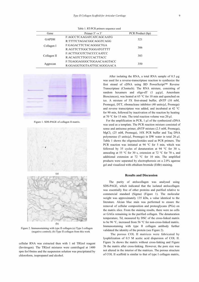
Figure 1. SDS-PAGE of collagen II matrix.
(a)
(b)
Figure 2. Immunostaining with type II collagen.(a) Type I collagen
(negative control); (b) Type II collagen from this work
cellular RNA was extracted then with 1 ml TRIzol reagent
(Invitrogen). The TRIzol mixtures were centrifuged at 1400
rpm for10mins and the suspension solution was precipitated by
chloroform, isopropanol and alcohol.
After isolating the RNA, a total RNA sample of 0.5 µg
was used for a reverse-transcriptase reaction to synthesize the
first strand of cDNA using BD PowerScript™ Reverse
Transcriptase (Clontech). The RNA mixture, consisting of
random hexamers and oligo-dT (1 µg/µl, Amersham
Biosciences), was heated at 65 °C for 10 min and quenched on
ice. A mixture of 5X first-strand buffer, dNTP (10 mM,
Promega), DTT, ribonuclease inhibitor (40 units/µl, Promega)
and reverse transcriptase was added, and incubated at 42 °C
for 90 min, followed by inactivation of the reaction by heating
at 70 °C for 15 min. The total reaction volume was 20 µl.
For the amplification in PCR, 1 µl of the synthesized cDNA
was used as a template. The PCR reaction mixture consisted of
sense and antisense primer, dNTP mixture (2.5 mM, Promega),
MgCl2 (25 mM, Promega), 10X PCR buffer and Taq DNA
polymerase (5 units/µl, Promega) in DW water in total 20 µl.
Table 1 shows the oligonucleotides used as PCR primers. The
PCR reaction was initiated at 94 °C for 5 min, which was
followed by 35 cycles of denaturation at 94 °C for 30 s,
annealing at 55 °C for 30 s, extension at 72 °C for 70 s, and
additional extension at 72 °C for 10 min. The amplified
products were separated by electrophoresis on a 2.0% agarose
gel and visualized with ethidium bromide (EtBr) staining.
Results and Discussion
The purity of atelocollagen was analysed using
SDS-PAGE, which indicated that the isolated atelocollagen
was essentially free of other proteins and purified relative to
commercial standard (Sigma) (Figure 1). The molecular
weight was approximately 135 kDa, a value identical to the
literature. Alcian blue stain was performed to ensure the
removal of cellular composition and proteoglycans (PGs) on
the matrix slice. From the staining results, there were no cells
or GAGs remaining in the purified collagen. The denaturation
temperature, Td, measured by DSC of the cross-linked matrix
to be 96 °C, increased from 56 °C for non-cross-linked matrix.
Immunostaining with type II collagen antibody further
validated the identity of the protein (see Figure 2).
The porous COL II matrices were fabricated by
lyophilization of 0.5 M acetic acid dispersion of COL II.
Figure 3a shows the matrix without cross-linking and Figure
3b the matrix after cross-linking. However, the pore size was
not altered in the interior of the matrices. The porous structure
of COL II scaffold is similar to that of type I collagen matrix,

J. Med. Biol. Eng., Vol. 27. No. 1 2007
10
(a) (b)
Figure 3. COL II matrix before cross-linking (a) and after being cross-linked by genipin (b).
(a) (b)
Figure 4. Internal structures of type I (a) and type II (b) collagen matrices.
(a) (b) (c)
Figure 5. Porous structures of COL II matrices fabricated at various freezing temperatures (a) –20 °C, (b) –80 °C, (c) –196 °C
as shown in Figure 4. Highly porous and inter-connected
structure was observed by SEM, formed by lamellae structures
of proteins. The freeze-dried COL II matrices had the same
porous structures, since we used acetic acid as porogen in both
cases; also the sizes of the pores were also influenced by the
temperature of freezing. Figure 5 shows the different types of
sectional morphology of porous matrices that were prepared by
freeze-drying at various freezing temperatures. The COL II
matrices fabricated at –20 °C, –80 °C, –196 °C had pore sizes
in the interior, ranging from 80-300, 50-250 and 20-150 µm,
respectively, and porosity measurement showed around 96%
before cross-linking, but was about 90-95% after cross-linking.
Matrices prepared at –20 °C, which gave the largest pores of
50-100 µm at the surface and 80-300 µm in the interior that
were more suitable for cells to migrate.
Mechanical evaluation of matrices non-cross-linked and
cross-linked by genipin (GP COL I or II) or EDC (EDC COL I)
revealed the typical nonlinear stress-strain behavior of
biological tissues. The GP COL II matrices had a maximum
compression loading of 180 mN that was determined at a 0.9
compression strain ratio, similar to GP COL I, and the
non-cross-linked COL II matrices had a maximum
compression loading of only 60 mN at the same compression
strain ratio (Figure 6). As a result of mechanical testing, the
non-cross-linked COL II matrices were broken, but the
cross-linked COL matrices (GP COL I or II) returned to
original shape like a sponge. As to the EDC COL I, it had
broken at a 0.8 compression ratio similar to EDC COL II (data
not shown). Hence, the GP COL II possessed better
mechanical strength, suitable for bioreactor culture. We
selected three different types of matrices, GP COL I, II and
EDC COL I, for further study by individually seeding normal
human chondrocytes.
Cellular proliferation was determined based on DNA
assay. In the GP COL II matrix culture, total cell number
increased 26.3-fold during 2 weeks’ culture period, whereas
the cell number decreased after 5 weeks. However, total cell
number increased 27-fold during 2 weeks’ cultivation onto the

Type II Collagen Scaffold for Articular Cartilage
11
Compression loading (mN)
Compression loading (mN)
Compression strain ratio
(a)
Compression strain ratio
(b)
Figure 6. Mechanical test of non-cross-linked (a) and cross-linked
COL II by GP (b). Testing was completed using an Instron
Mini 44 machine with the tensile grip compression rate
at 4.00 mm/min, and matrices’ compressional thickness
was 5 mm.
Figure7. Cell number of seeded collagen I and II matrices for
cultivation time. EDC COL I: COL I matrices cross-linked
by EDC agent; GP COL II: COL II matrices cross-linked
by genipin (GP) solution; GP COL I: COL I matrices
cross-linked by GP. Error bars represent the standard error
of the mean, and n=3.
EDC COL I matrices, but also decreased after 5 weeks. The
EDC COL II matrices had less increase in total cell number
than others (Figure 7).
The level of GAG synthesis in GP COL II culture was
two-fold higher than that in EDC COL I during 14 days of
Figure 8. GAG content of seeded collagen I and II matrices for
cultivation time. EDC COL I: COL I matrices
cross-linked by EDC agent; GP COL II: COL II
matrices cross-linked by GP; GP COL I: COL I matrices
cross-linked by GP. Error bars represent the standard
error of the mean, and n=3.
1 2 3
GAPDH
AGN
COL I
COL II
Figure 9. Expression of N-HACs specific genes in 14 days EDC
COL I (lane 1), GP COL II (lane 2) and I (lane 3) matrices
(n=3) cultures analyzed by RT-PCR. Total RNA analyzed
for collagen type I (COL I), collagen type II (COL II),
aggrecan (AGN) and with the housekeeping gene
glyceraldehyde-3- phosphate dehydrogenase (GAPDH) as
internal control.
culturing and was also 1.5-fold higher than GP COL I matrices
(Figure 8). However, There was not much difference in GAG
levels between different matrices after 5 weeks of culturing.
Regarding gene expression, we analyzed the mRNA
expression of cartilage-specific genes from chondrocytes
cultured in EDC COL I, GP COL II and GP COL I matrices at
the end of 14th day using RT-PCR. Collagen I was expressed
in all cultures, but its expression was up-regulated in GP COL
I, showing a mostly prechondrocyte pattern. Cells produced
aggrecan mRNA in all cultures, but its expression was
down-regulated in EDC COL I as was COL II mRNA. In the
GP COL II matrices, the mRNA expressions of
cartilage-specific genes (i.e.: COL II, aggrecan) were markedly
enhanced at day 14 (Figure 9). For this reason, GP COL II
matrices seems to be the best one for chondrocytes to maintain
right phenotype during the early culturing stage, and
cross-linking CS and HA onto COL II matrix may provide
cartilage cells with the correct extracellular signals.

J. Med. Biol. Eng., Vol. 27. No. 1 2007
12
Table 2. Characteristics of the matrices after cross-linking
Matrix Relative water absorption (%) Porosity (%)
COL II 100.0 96
*COL II 150.5 95
COL II + HA + CS 116.2 90
The porous matrices with or without CS were immersed in genipin
solution(0.25 %) in 70% ethanol (pH 7.4) at 37 °C for 48 h. COL II:
non-cross-linked COL II matrix, *COL II: cross-linked COL II matrix.
Table 3. Chemical properties of the matrices after cross-linking
Matrix Degree
of cross-linking
CS
(µg/matrix )
HA
(µg/matrix)
COL II
*COL II 65%
COL II+ HA + CS 56% 18.6 18.55
The porous matrices with or without CS were immersed in genipin
solution(0.25 %) in 70% ethanol (pH 7.4) at 37 °C for 48 h. COL II:
non-crosslinked COL II matrix, *COL II: cross-linked COL II matrix.
The porosity of the matrix was affected by cross-linking
reaction conditions and the secondary formation of large
crystals of ice, which influenced the structure of cross-linking
matrix in the freeze-drying process. After cross-linking with
genipin, the porous structure remained mostly intact, except at
the surface where pores collapsed to a certain extent. This
damage to the structure can be prevented by cross-linking at
the presence of 70% v/v ethanol. Porosity decreased after COL
II was cross-linking with HA and CS (see Table 2).
The swelling capacity generally decreased as the degree
of cross-linking increased. In our investigation, the
water-binding capacity of cross-linking matrices changed
significantly after cross-linking, as shown in Table 2. The high
water-binding capacity property of sponge-like matrices
appeared to be dependent on the highly porous interconnecting
network of the matrices, which had a good absorbent feature
with hydrophilic materials in the interior of matrices.
Comparing to the cross-linked matrices, untreated matrices
showed lower water uptake ability. Untreated matrices
collapsed and lost their porous structure partially when
immersed in PBS. COL II matrices cross-linking by genipin, in
the presence of CS and HA, not only resulted in the formation
of bio-mimicking ECM, but also forms covalent binding of CS
and HA. The COL / HA / CS matrices contained CS and HA
amounts of approximately 18.60 and 18.55 mg per matrix,
respectively. Figure 10 shows a COL II / HA / CS matrix
stained for CS, after treatment with alcian blue. Incorporated
CS were seen distributed throughout the matrix.
Higher degree of cross-linking gave higher mechanical
strength (Table 3). Susceptibility of these matrices to
degradation by proteolytic enzyme was decreased. To measure
the biodegradability of the matrix, 200 U/ml collagenase was
added to the buffer solution in which the matrices were
incubated. After 45 days, COL II cross-linked matrices
retained 65% of their original weight. The enzymatic
degradation rate of the genipin cross-linked matrices was
slower than those by
Figure 10. Alcian blue stain of COL II/HA/CS matrix
(a)
(b)
Figure 11. The cultivation of chondrocyte in COL II/HA/CS matrix
for 2 weeks: (a) H&E stain and (b) alcian blue stain for
PGs.
cross-linked by EDC. As for the stability of matrices during
the in vitro cultures, the cross-linked matrices maintained their
structural integrity during culture, but non-cross-inked sponge
showed slight contraction due to the cells proliferation.
Chondrocytes grown on the matrices were analyzed for
their morphology and biochemical properties using
histochemistry. After 14 days of culturing, cells on the COL II
/HA/CS matrices showed a round morphology by H&E
staining. The spherically shaped appearance was indicative of
the chondrocytic phenotype (Figure 11 a). The result indicated
the matrices could maintain the differentiation state of the
chondrocytes or induce re-differentiation of the chondrocytes,
but some elongated cells were found in the periphery of the
matrices. This represented dedifferentiation of chondrocytes
into fibrocartilagenous-like cells. Pericellular metachromatical
staining was observed in cell-dense areas of the matrices using

Type II Collagen Scaffold for Articular Cartilage
13
Figure 12. Cells number of cross-linked COL II matrices seeded
with chondrocytes as a function of different culturing
time. Pure: COL II matrices; BTCS: COL II / CS/HA.
Values are mean ± SD (n=3).
Figure 13. GAG content of cross-linked COL II matrices seeded
with chondrocytes as a function of different culturing times. Pure: COL II matrices; BTCS: COL II / CS/HA.
Values are mean ± SD (n=3).
alcian blue, indicating large quantity of PGs was being
synthesized and secreted to the ECM (Figure 11b). Matrix
secretion and proliferation of chondroctes were evaluated by
determination of the DNA and GAG content. All cross-linked
matrices showed a great deal of increase in the DNA content
after 6 weeks of culture, indicating proliferation of
chondrocytes The DNA content showed an increase of 5.5- and
4-fold for pure and CS matrices, respectively, from day 1 to six
weeks (Figure 12). Total GAG content also showed an increase,
ranging from 20 ~ 60 µg/matrix, for all matrices. The COL II /
HA / CS matrices produced the strongest simulative effect on
chondrocytes biosynthesis (Figure 13). This could be due to
the synergism of HA and CS that may have switched on the
CD 44 receptor, which was essential for cartilage homeostasis
[3, 23]. The extracellular matrix not only regulates cellular
behavior, but also plays a role in binding, release, storage and
presentation of soluble mediators within articular the cartilage
tissues. Some studies have evaluated the effects of growth
factors supplemented into serum-free medium [13, 24] in
addition to scaffold engineering. Further improvement of
cartilage regeneration can be explored in supplying appropriate
growth factors and physical stimulation [25, 26] to the cells
grown on the matrices.
Conclusions
Genipin cross-linking and a freeze-drying technology
successfully fabricated the cross-linked matrices retaining
interconnecting porous structure. The cross-linking matrices
had porosity of 90-95% and pore size of 80-300 µm. These
cross-linked matrices were non-cytotoxic, flexible and
mimicked ECM of natural hyaline cartilage better than other
biomaterials seen in the literature. Moreover, the COL II / HA /
CS matrices could provide better dynamic microenvironment
for chondrocytes. The results indicated that the matrices could
promote appropriate chondrocytic phenotype in vitro. Further
study on the mechanism of HA/CS effects by glycosidase
digestion and their possible therapeutic effects on osteoarthritis
will be pursued in the future.
Acknowledgements
This research was funded by the Technology Development
Program for Academia Grant 91-EC-17-A-17-S1-0009,
Ministry of Economic Affairs, Taiwan.
References
[1] K. Kawasaki, Y. Uchio, N. Adachi and M. Matsusaki,
“Hyaluronic acid enhances proliferation and chondroitin sulfate
synthesis in cultured chondrocytes embedded in collagen gels,”
J. Cell. Physiol., 179: 142-148, 1999.
[2] S. Maniwa, M. Ochi, T. Motomura, T. Nishikori, J. Chen and H.
Naora, “Effect of hyaluronic acid and basic fibroblast growth
factor on motility of chondrocytes and synovial cells in culture,”
Acta Orthop. Scand. 72: 299-303, 2001.
[3] S. Ohno, H. J. Im, C. B. Knudson and W. Knudson, “Hyaluronan
oligosaccharide -induced activation of transcription factors in
bovine articular chondrocytes,” Arthrit. Rheum., 52: 800-809,
2005.
[4] J. S. Pieper, A. Oosterhof, P. J. Dijkstra, J. H. Veerkamp and T. H.
van Kuppevelt, “Preparation and characterization of porous
cross-linked collagenous matrices containing bioavailable
chondroitin sulphate,” Biomaterials, 20: 847-858, 1999.
[5] S. N. Park, J. C. Park, H. O. Kim, M. J. Song and H. Suh,
“Characterization of porous collagen /hyaluronic acid scaffold
modified by 1-ethyl-3- (3-dimethyl- aminopropyl) carbodiimide
cross-linking,” Biomaterials, 23: 1205-1212, 2002.
[6] C. H. Chang, H. C. Liu, C. C. Lin, C. H. Chou and F. H. Lin,
“Gelatin-chondroitin- hyaluronan tri-copolymer scaffold for
cartilage tissue engineering,” Biomaterials, 24: 4853-4858,
2003.
[7] N. F. Pieper, P. M. van der Kraan. T. Hafmans, J. Kamp, P. Buma,
J. L. C. van Susante, W. B. van den Berg, J. H. Veerkamp and T.
H. van Kuppevelt, “Crosslinked type II collagen matrices:
preparation, characterization, and potential for cartilage
engineering,” Biomaterials, 23: 3183-3192, 2002.
[8] J. L. C. van Susante, J. Pieper, P. Buma, T. H. van Kuppevelt, H.
van Beuningen,; van der Kraan, J. H. Veerkamp, W. B van den
Berg, R. P. H., Veth, “Linkage of chondroitin-sulfate to type I
collagen scaffolds stimulated the bioactivity of seeded
chondrocytes in vitro,” Biomaterials, 22: 2359-2369, 2001.
[9] K. J. Shields, M. J. Beckman, G. L.Bowlin, J. S., Wayne,
“Mechanical properties and cellular proliferation of electrospun
collagen type II,” Tissue Eng., 10: 1510-1517, 2004.
[10] Z Ma, C. Gao, Y. Gong and J. Shen, “Cartilage tissue engineering
PLLA scaffold with surface immobilized collagen and basic

J. Med. Biol. Eng., Vol. 27. No. 1 2007
14
fibroblast growth factor,” Biomaterials, 26: 1253-1259, 2005.
[11] L. E. Freed, A. P. Hollander, I. Martin, J. R.Barry, R. Langer and
G. Vunjak- Novakovic, “Chondrogenesis in a
Cell-Polymer-Bioreactor System,” Exp. Cell Res., 240: 58-65,
1998.
[12] Q. Li, C. G. Williams, D. Sun. J. Wang, K. Leong and J. H.
Elisseeff, “Photo-crosslinkable polysaccharides based on
chondroitin sulfate,” J. Biomed. Mater. Res., 68A: 28-33, 2004.
[13] J. E. Lee, K. E. Kim, I. C. Kwon, H. J. Ahn, S. H. Lee, H. C.
Cho, H. J. Kim, S. C. Seong and M. C. Lee, ”Effect of the
controlled-released TGF-beta 1 from chitosan microspheres on
chondrocytes cultured in a collagen/chitosan
/glycosaminoglycan scaffold,” Biomaterials, 25: 4163-4173,
2004.
[14] M. Abe, M. Takahashi, S. Tokura, H. Tamura and A.
Nagano, ”Cartilage-scaffold composites produced by
bioresorbable beta-chitin sponge with cultured rabbit
chondrocytes,” Tissue Eng., 10: 585-594, 2004.
[15] Y. Dausse, L. Grossin, G. Miralles, S. Pelletier, D. Mainard, P.
Gillet, E. Dellacherie, P. Netter and E. Payan, “Cartilage repair
using new polysaccharidic biomaterials: macroscopic,
histological and biochemical approaches in a rat model of
cartilage defect,” Osteoarthrit. Cartilage, 11: 16-28, 2003.
[16] T. Taguchi, T. Ikoma and J. Tanaka, “An improved method to
prepare hyaluronic acid and type II collagen composite
matrices,” J. Biomed. Mater. Res., 61: 330-336, 2002.
[17] H. W. Sung, C. N. Chen, R. N. Huang, J. C. Hsu and W. H.
Chang, “In vitro surface characterization of a biological patch
fixed with a naturally occurring cross-linking agent,”
Biomaterials, 21: 1353-1362, 2000.
[18] J. S. Pieper, T. Hafmans, J. H. Veerkamp and T. H. van
Kuppevelt, “ Development of tailor-made collagen-
glycosaminglycan matrices: EDC/NHS crosslinking, and
ultra-structural aspects,” Biomaterials, 21: 581-593, 2000.
[19] I. V. Yannas, P. L. Gordon, J. F. Burke, C. Huang and R. H.
Rubenstein, “Design of an artificial skin. II. Control of chemical
composition,” J. Biomed. Mater. Res., 14: 107-131, 1980.
[20] Y. J. Kim, R. L. Y. Sah, J. Y. H. Doong and A. J. Grodzinsky,
“Fluorometric assay of DNA in cartilage explants using Hoechst
33258,” Anal. Biochem., 174: 168-176, 1988.
[21] R. W. Farndale, D. J. Buttle and A. J. Barrett, ”Improved
quantitation and discrimination of sulphated
glycosaminoglycans by use dimethylmethylene blue,” Biochim.
Biophy. Acta., 883: 173-177, 1986.
[22] H. C. Chen, H. P. Lee, M. L. Sung, C. J. Liao and Y. C. Hu, “A
novel rotating-shaft bioreactor for two phase cultivation of tissue
engineered cartilage,” Biotechnol. Prog., 20: 1802-1809, 2004.
[23] P. M. Van der Kraan, P. Buma, T. van Kuppevelt and W. B. van
den Berg, “Interaction of chondrocytes, extracellular matrix and
growth factors: relevance for articular cartilage tissue
engineering,” Osteoarthrit., Cartilage 10: 631-637, 2002.
[24] N. Veilleux and M. Spector, “Effects of FGF-2 and IGF-1 on
adult canine articular chondrocytes in type II
collagen-glycosaminoglycan scaffolds in vitro,” Osteoarthrit.
Cartilage, 13: 278-286, 2005.
[25] S. Mizuno, F. Allemann and J. Glowacki, “Effects of medium
perfusion on matrix production by bovine chondrocytes in
three-dimensional collagen sponges,” J. Biomed. Mater. Res., 56:
368-375, 2001.
[26] T. Ikenoue, M. C. Trindade, M. S. Lee, E. Y. Lin, D. J.
Schurman, S. B. Goodman and R. L. Smith,
“Mechanoregulation of human articular chondrocyte aggrecan
and type II collagen expression by intermittent hydrostatic
pressure in vitro,” J. Orthop. Res., 21: 110-116, 2003.