the blood chapter 13. introduction specialized connective tissue –plasma: fluid part (55%)...
TRANSCRIPT
THE BLOOD
Chapter 13
Introduction
• Specialized connective tissue– Plasma: fluid part (55%)– Formed blood cells (45%)
• Erythrocytes• Leukocytes• Thrombocytes
2

FUNCTIONS OF THE BLOOD

Functions of the Blood (cont’d.)
• Transports: O2, CO2, nutrients, waste, hormones
• Regulates: body pH, body temperature
• Clotting mechanism
• Protection against foreign microbes and toxins
• Osmosis

THE CLASSIFICATION OF BLOOD CELLS AND THE
COMPOSITION OF PLASMA
The Classification of Blood Cells
• Erythrocytes (RBCs)– 95% of the volume of blood cells
• Leukocytes (WBCs)– Granular: neutrophils, eosinophils, basophils– Agranular: monocytes, lymphocytes
• Thrombocytes: platelets
The Composition of Plasma
• Fluid portion of blood is 91% water
• Plasma proteins: 7%– Albumin, globulin, fibrinogen
• Plasma solutes: 2%– Ions, nutrients, waste products, gases,
enzymes, hormones

FORMATION OF BLOOD CELLS: HEMATOPOIESIS
Formation of Blood Cells: Hematopoiesis (cont’d.)
• Produced in red bone marrow
• Lymphocytes and monocytes produced by– Lymph nodes, spleen, tonsils
• Stem cells: undifferentiated mesenchymal cells

BLOOD CELL ANATOMY AND FUNCTIONS
Blood Cell Anatomy and Functions (cont’d.)
• Erythrocytes– Biconcave disks– No nucleus– Contain hemoglobin
• Heme: binds O2
• Globin: binds CO2
Blood Cell Anatomy and Functions (cont’d.)
• Granular leukocytes– Neutrophils
• Phagocytize foreign substances
– Eosinophils • Produce antihistamines
– Basophils • Produce heparin, histamine, serotonin
Blood Cell Anatomy and Functions (cont’d.)
• Agranular leukocytes– Monocytes
• Phagocytize bacteria and cellular debris• Macrophages: in tissues
– Lymphocytes• T lymphocytes• B lymphocytes
Blood Cell Anatomy and Functions (cont’d.)
• Thrombocytes or platelets– Disk-shaped cellular fragments with a nucleus– Prevent fluid loss when blood vessels
damaged– Produced from large megakaryocytes
THE CLOTTING MECHANISM
The Clotting Mechanism (cont’d.)
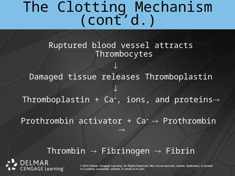
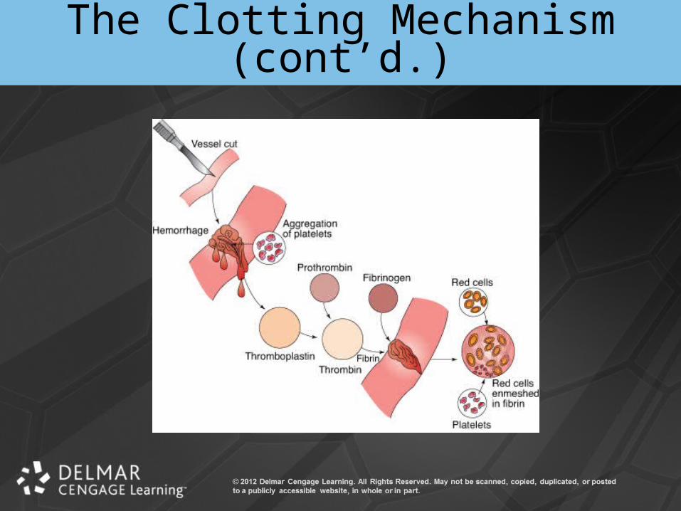
Ruptured blood vessel attracts Thrombocytes
Damaged tissue releases Thromboplastin
Thromboplastin + Ca+, ions, and proteins
Prothrombin activator + Ca+ Prothrombin
Thrombin Fibrinogen Fibrin
The Clotting Mechanism (cont’d.)
• Clot– Fibrin forms long threads acting like a net– Platelets get enmeshed
• Syneresis: clot retraction
• Fibrinolysis: dissolution of blood clot
The Clotting Mechanism (cont’d.)
• Thrombosis: unwanted clotting
• Embolus: circulating blood clot
• Infarction – Tissues killed as a result of loss of blood
supply
The Clotting Mechanism (cont’d.)

Animation – Blood
Click Here to Play Blood Animation

THE BLOOD GROUPS
Introduction
• Human blood is of different types– Only certain combinations are compatible
• Agglutination: clumping of RBCs– Occurs when blood groups mismatched– Transfusion reaction
The ABO Blood Group
• Type A– Anti-B antibodies
• Type B– Anti-A antibodies
• Type AB– No antibodies
The ABO Blood Group (cont’d.)
• Type O– Anti-A and anti-B antibodies
The Rh Blood Group
• Eight Rh antigens
• Antigen D: most important
• Anti-Rh antibodies develop after exposure– Rh-negative mother carrying Rh-positive baby
• Erythroblastosis fetalis• RhoGAM - protects Rh-positive fetus
Summary
• Described the functions of blood
• Classified blood cells into different groups based on anatomy and function
• Discussed how and where blood cells are formed
• Explained the clotting mechanism
• Named the different blood groups