stem cells: the holy grail of regenerative medicine
TRANSCRIPT
Chapter 2Stem Cells: The Holy Grailof Regenerative Medicine
Ram K. Singh, Snehal M. Gaikwad, Subhoshree Chatterjeeand Pritha Ray
2.1 Introduction
Stem cells occupy a special position in cellular hierarchy during differentiation anddevelopment of any organism. These undifferentiated cells have the potential toform any other cell type with specialized function and are characterized by theirability to self-renew and to differentiate. Both these cellular properties are primerequisites for the success of current regenerative medicine.
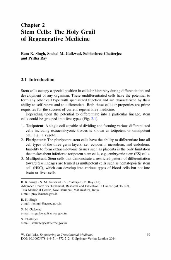
Depending upon the potential to differentiate into a particular lineage, stemcells could be grouped into five types (Fig. 2.1).
1. Totipotent: A single cell capable of dividing and forming various differentiatedcells including extraembryonic tissues is known as totipotent or omnipotentcell, e.g., a zygote.
2. Pluripotent: The pluripotent stem cells have the ability to differentiate into allcell types of the three germ layers, i.e., ectoderm, mesoderm, and endoderm.Inability to form extraembryonic tissues such as placenta is the only limitationthat makes them inferior to totipotent stem cells, e.g., embryonic stem (ES) cells.
3. Multipotent: Stem cells that demonstrate a restricted pattern of differentiationtoward few lineages are termed as multipotent cells such as hematopoietic stemcell (HSC), which can develop into various types of blood cells but not intobrain or liver cells.
R. K. Singh � S. M. Gaikwad � S. Chatterjee � P. Ray (&)Advanced Centre for Treatment, Research and Education in Cancer (ACTREC),Tata Memorial Centre, Navi Mumbai, Maharashtra, Indiae-mail: [email protected]
R. K. Singhe-mail: [email protected]
S. M. Gaikwade-mail: [email protected]
S. Chatterjeee-mail: [email protected]
W. Cai (ed.), Engineering in Translational Medicine,DOI: 10.1007/978-1-4471-4372-7_2, � Springer-Verlag London 2014
19
4. Oligopotent: Oligopotent stem cells are able to differentiate into few cell typesof specific lineages such as lymphoid stem cells that can be differentiated onlyinto basophil, neutrophil, eosinophil, monocyte, and thrombocytes.
5. Unipotent: Unipotent stem cells can only differentiate into one particular celltype such as hepatoblasts forming hepatocytes.
Besides normal developmental functions in multicellular organisms, thesemother cells are believed to be the holy grail of medical therapy with high promisefor regenerative medicine.
2.2 Engineering of Stem Cells
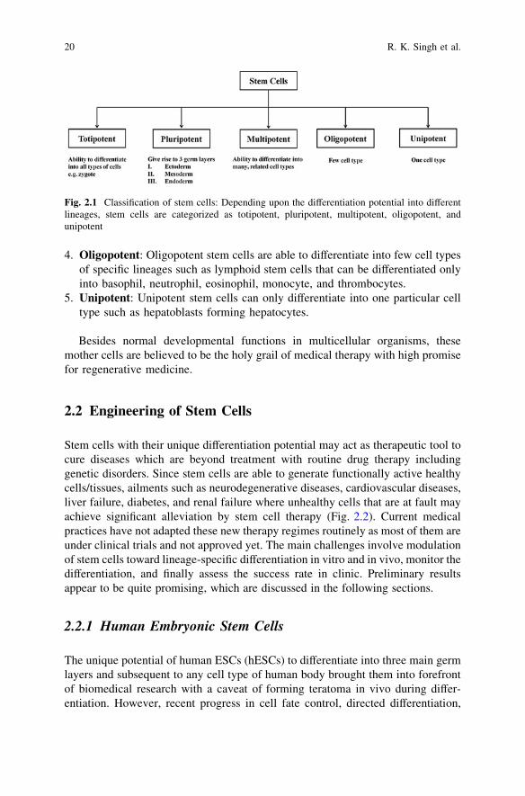
Stem cells with their unique differentiation potential may act as therapeutic tool tocure diseases which are beyond treatment with routine drug therapy includinggenetic disorders. Since stem cells are able to generate functionally active healthycells/tissues, ailments such as neurodegenerative diseases, cardiovascular diseases,liver failure, diabetes, and renal failure where unhealthy cells that are at fault mayachieve significant alleviation by stem cell therapy (Fig. 2.2). Current medicalpractices have not adapted these new therapy regimes routinely as most of them areunder clinical trials and not approved yet. The main challenges involve modulationof stem cells toward lineage-specific differentiation in vitro and in vivo, monitor thedifferentiation, and finally assess the success rate in clinic. Preliminary resultsappear to be quite promising, which are discussed in the following sections.
2.2.1 Human Embryonic Stem Cells
The unique potential of human ESCs (hESCs) to differentiate into three main germlayers and subsequent to any cell type of human body brought them into forefrontof biomedical research with a caveat of forming teratoma in vivo during differ-entiation. However, recent progress in cell fate control, directed differentiation,
Fig. 2.1 Classification of stem cells: Depending upon the differentiation potential into differentlineages, stem cells are categorized as totipotent, pluripotent, multipotent, oligopotent, andunipotent
20 R. K. Singh et al.
and tissue engineering crossed the boundaries of laboratory and extended to thearena of regenerative medicine. hESC lines are conventionally derived from theinner cell mass (ICM) of preimplantation-stage blastocysts, morula-stage embryos,or late-stage blastocysts and express pluripotency markers (transcription factors—Oct4, Sox2, Nanog; surface antigens—SSEA-4, SSEA-3; proteoglycans—TRA-1-60, TRA-1-81) [1]. To maintain the undifferentiated state, these cells are co-cultured with a support or feeder layer derived from mouse embryonic fibroblast(MEF) that provides all the essential growth factors [1]. The reported success ratefor hESC derivation is highly variable, possibly due to variation in embryo qualityand culture conditions. Major progress had happened in derivation, propagation,cryopreservation, and efficient passaging of hESCs. Since clinical application ofhESCs critically depends on well-characterized growth and differentiation of stemcells, much effort was put in developing conditioned media that will enable feeder-free growth of ESC cells and eliminate animal products [2, 3]. The original culturesystem for the maintenance of hESC using MEF feeder cell layer support pos-sesses risk of zoonosis transmitted by animal pathogens, potential activation ofanimal retroviruses, and possibility of immune rejection due to the presence ofnonhuman sialic acid. Several approaches such as use of extracellular matrix(ECM) derived from MEF than living feeder cells, hESC-derived fibroblasts,
Fig. 2.2 Schematic representation of differentiation process of embryonic stem cell (ESC), adultstem cell (ASC), and induced pluripotent stem cells (iPSCs): While ESCs can generate threegerm layers via directed differentiation, it may give rise to teratomas through uncontrolleddifferentiation in vivo. The ASCs have restricted potential to generate myogenic, osteogenic,adipogenic, and neurogenic lineages under specified conditions. In contrary to ESC and ASC thatonly differentiate toward specific lineage, iPSCs follow a dedifferentiation (from somatic cell)and then differentiation to germ-layer-specific cellular lineages
2 Stem Cells: The Holy Grail of Regenerative Medicine 21
defined culture medium containing components solely derived from purifiedhuman material, MEF-conditioned Matrigel layer to establish and maintain clin-ical-grade hESCs cell lines are in progress [4]. However, these xeno-free andfeeder-independent culture systems are costly and laborious and may lead toabnormal karyotype during long-term culture. Recently, Akopian et al. in con-junction with the Internal Stem Cell Initiative Consortium (ISCIC) comparedseveral commercially available ESC culture media with Knockout Serum Repla-cer, FGF-2, and MEF cell layers for propagation of several hESC cell linesestablished in five different laboratories and showed that only mTeSR1 andSTEMPRO were able to support most cell lines up to 10 passages [2].
The next major challenge for translational application of hESC is to direct theirdifferentiation toward a specific cell lineage. The pioneer study by Itskovtz-eldoret al. [5] showed that hESC cells were capable of forming ‘‘embryoid bodies’’ (EB)comprised of three embryonic germ layers. In this study, hESCs were grown insuspension to induce their differentiation into EBs. Formation of in vitro EBrequired special cocktail of supplements and growth factors such as glutamine,beta-mercaptoethanol, nonessential amino acids, leukemia inhibitory factor (LIF),and basic fibroblast growth factor (bFGF). Under these conditions, majority of thecells remained in an undifferentiated state. For the formation of EBs, ES cells weretransferred using either collagenase or trypsin/EDTA to plastic petri plates to allowaggregation and prevent adherence. About one million ES cells were plated in eachof the 50-mm petri plates, and the hEBs were grown in the same culture mediumwithout LIF and bFGF. The differentiation status of the human ES cells and EBswas determined by the expression pattern of several lineage-specific markers suchas gamma-globin (hematopoietic cells), alpha-cardiac actin (myocardial cells),neurofilament (neuronal cells), and alpha-fetoprotein (endodermal cells).
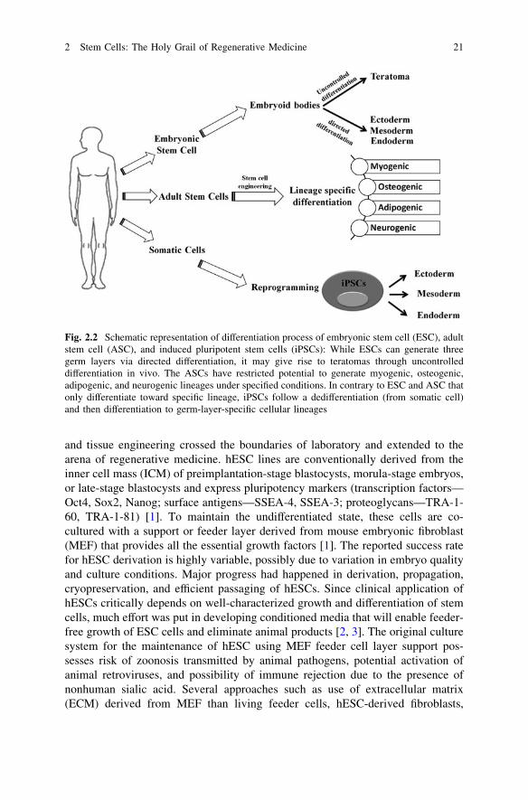
Recently, Chen et al. performed a stepwise differentiation of ESCs to insulin-secreting functional beta-cells where ESCs first formed a definitive endoderm inthe presence of indolactam-V and FGF, then Pdx1-expressing pancreatic pro-genitors, followed by the formation of endocrine progenitors and eventuallyinsulin-producing beta-cells (Fig. 2.3) [6, 7].
Fig. 2.3 In vitro differentiation of ESCs into insulin-producing beta-cells: Indolactam-V guidesthe differentiation of ESC into insulin-producing beta-cells via definitive endoderm, pancreaticprogenitor, endocrine progenitor, and beta-cells
22 R. K. Singh et al.
Thus, strategic development of tissue-specific adult cells with fully functionalpotential from undifferentiated ES cells is possible and holds great promise fortranslational application.
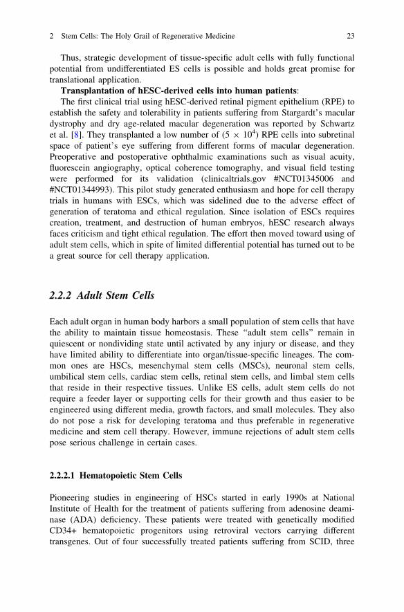
Transplantation of hESC-derived cells into human patients:The first clinical trial using hESC-derived retinal pigment epithelium (RPE) to
establish the safety and tolerability in patients suffering from Stargardt’s maculardystrophy and dry age-related macular degeneration was reported by Schwartzet al. [8]. They transplanted a low number of (5 9 104) RPE cells into subretinalspace of patient’s eye suffering from different forms of macular degeneration.Preoperative and postoperative ophthalmic examinations such as visual acuity,fluorescein angiography, optical coherence tomography, and visual field testingwere performed for its validation (clinicaltrials.gov #NCT01345006 and#NCT01344993). This pilot study generated enthusiasm and hope for cell therapytrials in humans with ESCs, which was sidelined due to the adverse effect ofgeneration of teratoma and ethical regulation. Since isolation of ESCs requirescreation, treatment, and destruction of human embryos, hESC research alwaysfaces criticism and tight ethical regulation. The effort then moved toward using ofadult stem cells, which in spite of limited differential potential has turned out to bea great source for cell therapy application.
2.2.2 Adult Stem Cells
Each adult organ in human body harbors a small population of stem cells that havethe ability to maintain tissue homeostasis. These ‘‘adult stem cells’’ remain inquiescent or nondividing state until activated by any injury or disease, and theyhave limited ability to differentiate into organ/tissue-specific lineages. The com-mon ones are HSCs, mesenchymal stem cells (MSCs), neuronal stem cells,umbilical stem cells, cardiac stem cells, retinal stem cells, and limbal stem cellsthat reside in their respective tissues. Unlike ES cells, adult stem cells do notrequire a feeder layer or supporting cells for their growth and thus easier to beengineered using different media, growth factors, and small molecules. They alsodo not pose a risk for developing teratoma and thus preferable in regenerativemedicine and stem cell therapy. However, immune rejections of adult stem cellspose serious challenge in certain cases.
2.2.2.1 Hematopoietic Stem Cells
Pioneering studies in engineering of HSCs started in early 1990s at NationalInstitute of Health for the treatment of patients suffering from adenosine deami-nase (ADA) deficiency. These patients were treated with genetically modifiedCD34+ hematopoietic progenitors using retroviral vectors carrying differenttransgenes. Out of four successfully treated patients suffering from SCID, three
2 Stem Cells: The Holy Grail of Regenerative Medicine 23
continued doing well up to 3.6 years after gene therapy, whereas one patientsuffered serious adverse effect. During a routine checkup after 30 months of genetherapy, lymphocytosis consisting of a monoclonal population of Vc9/Vd1, c/d Tcells of mature phenotype was detected. One pro-viral integration site was foundon chromosome 11 within the LMO-2 locus. This insertion leads to an aberrantexpression of the LMO-2 transcript in the monoclonal T-cell population (char-acteristics of acute lymphoblastic leukemia) [9].
These adverse events have resulted in discontinuation of the use of such longterminal repeat (LTR)-driven gamma-retroviral vectors for the genetic manipu-lations of HSCs, but at the same time, it provided a major thrust for developingnovel approaches. New and modified types of retroviral vector such as a ‘‘self-inactivating’’ (SIN) vector [10], lentiviral vectors [11], and lineage-restrictedvectors [12] are now entering the clinic. These vectors might reduce the risk oftransactivation of proto oncogenes after semi random integrations.
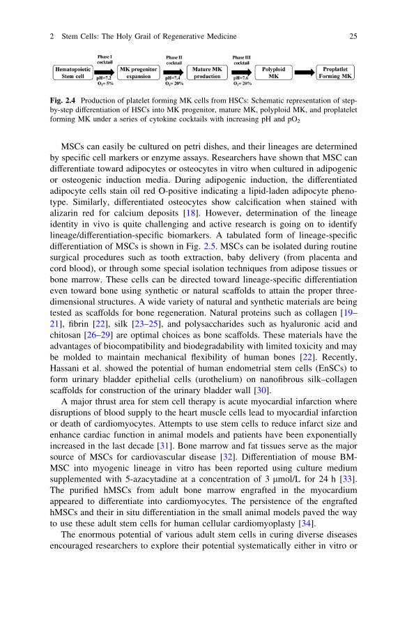
Thrombocytopenia, a deficiency in blood platelets, is a major consequence ofseveral hematological malignancies and chemotherapy [13]. In vitro plateletproduction from hematopoietic stem and progenitor cells (HSPCs)-derivedmegakaryocytes (Mks) could augment the supply and elude problems associatedwith bacterial and viral contamination, as well as immune rejection. Panugantiet al. [14] using HSPCs developed a three-stage strategy for ex vivo expansion ofhigh-ploidy megakaryocytic cells for large-scale platelet production (Fig. 2.4).The CD34+ HSPCs culture was started in a cytokine cocktail at 5 % O2 (pH 7.2).At day 5, cells were shifted to 20 % O2 (pH 7.4) and maintained in 1 of the 17cytokine cocktails (identified using a 24 factorial design of experiment method toevaluate the effects of interleukin (IL)-3, IL-6, IL-9, and high- or low-dose stemcell factor (SCF) in conjunction with thrombopoietin (Tpo) and IL-11) forexpansion of mature Mks from progenitors. The combination of Tpo, high-doseSCF, IL-3, IL-9, and IL-11 produced maximum Mk expansion. These Mks whencultured in IMDM ? 20 % BIT 9,500 gave rise to platelets with functionalactivity similar to that of fresh platelets from normal donors, as validated by basaltubulin distribution and the expression of surface markers.
Later Eric Lagasse showed in vivo differentiation of purified HSCs intohepatocytes in a mouse model of a lethal hereditary liver disease. As few as 50adult HSCs injected intravenously had the capacity to reconstitute hematopoiesisand produce hepatocytes [15].
2.2.2.2 Mesenchymal Stem Cells
MSCs comprised of the major portion of adult stem cells were first identified byFriedenstein from adult bone marrow [16]. These MSCs were shown to differen-tiate into osteoblasts, chondrocytes, adipocytes, and hematopoietic supportingstroma when a single colony-forming unit-fibroblast (CFU-F) was transplantedin vivo [17].
24 R. K. Singh et al.
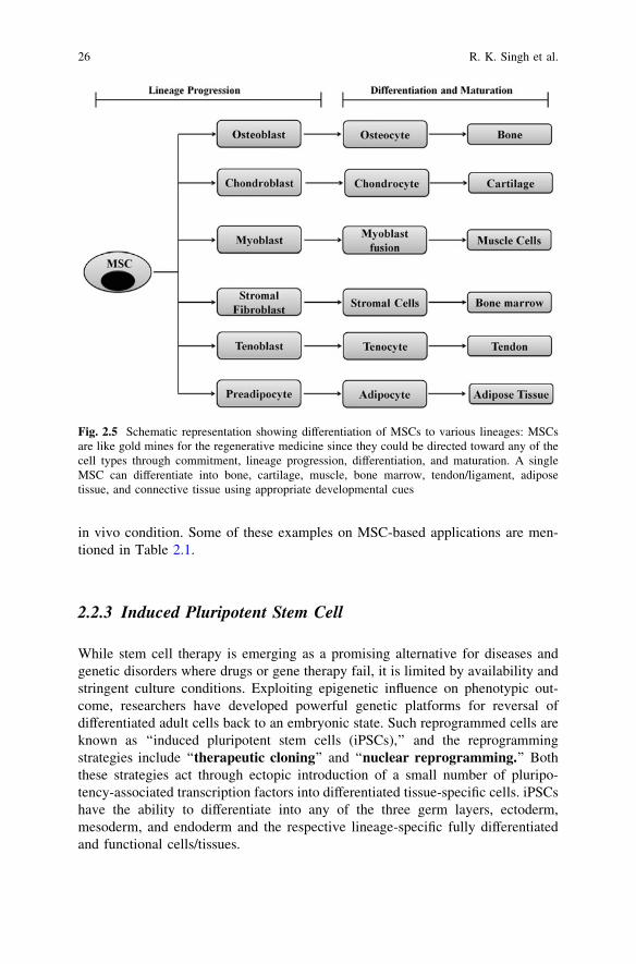
MSCs can easily be cultured on petri dishes, and their lineages are determinedby specific cell markers or enzyme assays. Researchers have shown that MSC candifferentiate toward adipocytes or osteocytes in vitro when cultured in adipogenicor osteogenic induction media. During adipogenic induction, the differentiatedadipocyte cells stain oil red O-positive indicating a lipid-laden adipocyte pheno-type. Similarly, differentiated osteocytes show calcification when stained withalizarin red for calcium deposits [18]. However, determination of the lineageidentity in vivo is quite challenging and active research is going on to identifylineage/differentiation-specific biomarkers. A tabulated form of lineage-specificdifferentiation of MSCs is shown in Fig. 2.5. MSCs can be isolated during routinesurgical procedures such as tooth extraction, baby delivery (from placenta andcord blood), or through some special isolation techniques from adipose tissues orbone marrow. These cells can be directed toward lineage-specific differentiationeven toward bone using synthetic or natural scaffolds to attain the proper three-dimensional structures. A wide variety of natural and synthetic materials are beingtested as scaffolds for bone regeneration. Natural proteins such as collagen [19–21], fibrin [22], silk [23–25], and polysaccharides such as hyaluronic acid andchitosan [26–29] are optimal choices as bone scaffolds. These materials have theadvantages of biocompatibility and biodegradability with limited toxicity and maybe molded to maintain mechanical flexibility of human bones [22]. Recently,Hassani et al. showed the potential of human endometrial stem cells (EnSCs) toform urinary bladder epithelial cells (urothelium) on nanofibrous silk–collagenscaffolds for construction of the urinary bladder wall [30].
A major thrust area for stem cell therapy is acute myocardial infarction wheredisruptions of blood supply to the heart muscle cells lead to myocardial infarctionor death of cardiomyocytes. Attempts to use stem cells to reduce infarct size andenhance cardiac function in animal models and patients have been exponentiallyincreased in the last decade [31]. Bone marrow and fat tissues serve as the majorsource of MSCs for cardiovascular disease [32]. Differentiation of mouse BM-MSC into myogenic lineage in vitro has been reported using culture mediumsupplemented with 5-azacytadine at a concentration of 3 lmol/L for 24 h [33].The purified hMSCs from adult bone marrow engrafted in the myocardiumappeared to differentiate into cardiomyocytes. The persistence of the engraftedhMSCs and their in situ differentiation in the small animal models paved the wayto use these adult stem cells for human cellular cardiomyoplasty [34].
The enormous potential of various adult stem cells in curing diverse diseasesencouraged researchers to explore their potential systematically either in vitro or
Fig. 2.4 Production of platelet forming MK cells from HSCs: Schematic representation of step-by-step differentiation of HSCs into MK progenitor, mature MK, polyploid MK, and proplateletforming MK under a series of cytokine cocktails with increasing pH and pO2
2 Stem Cells: The Holy Grail of Regenerative Medicine 25
in vivo condition. Some of these examples on MSC-based applications are men-tioned in Table 2.1.
2.2.3 Induced Pluripotent Stem Cell
While stem cell therapy is emerging as a promising alternative for diseases andgenetic disorders where drugs or gene therapy fail, it is limited by availability andstringent culture conditions. Exploiting epigenetic influence on phenotypic out-come, researchers have developed powerful genetic platforms for reversal ofdifferentiated adult cells back to an embryonic state. Such reprogrammed cells areknown as ‘‘induced pluripotent stem cells (iPSCs),’’ and the reprogrammingstrategies include ‘‘therapeutic cloning’’ and ‘‘nuclear reprogramming.’’ Boththese strategies act through ectopic introduction of a small number of pluripo-tency-associated transcription factors into differentiated tissue-specific cells. iPSCshave the ability to differentiate into any of the three germ layers, ectoderm,mesoderm, and endoderm and the respective lineage-specific fully differentiatedand functional cells/tissues.
Fig. 2.5 Schematic representation showing differentiation of MSCs to various lineages: MSCsare like gold mines for the regenerative medicine since they could be directed toward any of thecell types through commitment, lineage progression, differentiation, and maturation. A singleMSC can differentiate into bone, cartilage, muscle, bone marrow, tendon/ligament, adiposetissue, and connective tissue using appropriate developmental cues
26 R. K. Singh et al.
The concept of iPSCs demonstrated long back by Sir John Gurdon when hesuccessfully cloned a frog using intact nuclei from intestinal epithelium cells of[42]. Later, he showed that even nuclei from terminally differentiated adult cells(e.g., blood cells, skeletal muscle, and kidney cells) could generate Xenopus larvaewith nuclear transfer [42]. Decades later, in 2006, Takahashi et al. demonstratedthe ability of adult mouse fibroblasts to reprogram themselves into pluripotentstem cells by introduction of four key transcription factors (Oct3/4, Sox2, c-Myc,and Klf4) [43]. In November 2007, two independent studies were publishedsimultaneously on successful transformation of differentiated human cells intopluripotent stem cells. While Takahashi et al. used retroviral delivery Oct3/4,Sox2, Klf4, and c-Myc combinations to induce pluripotency in human fibroblasts,Yu et al. delivered Oct4, Nanog, Sox2, and LIN28 by lentiviral transduction inhESC-derived mesenchymal cells to induce pluripotency [44, 45]. Thesegroundbreaking experiments by Sir Gurdon and Yamanaka and his group wereacknowledged by Nobel Prize award in 2010.
The most exciting and oversimplified part of iPSC generation is that a com-bination of only four transcription factors is able to reverse the differentiationprocess. To identify this main core of pluripotent factors, Yamanaka et al. (2006)evaluated twenty-four candidate genes and Thomson et al. (2007) screened sixteentranscription factors in an assay system in which the induction of the pluripotentstate could be detected through the development of resistance to neomycin gene.
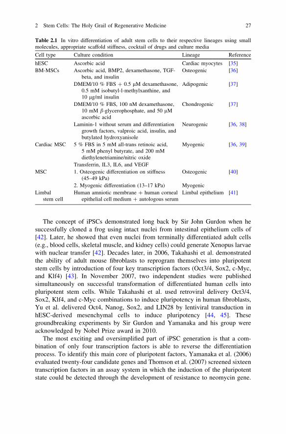
Table 2.1 In vitro differentiation of adult stem cells to their respective lineages using smallmolecules, appropriate scaffold stiffness, cocktail of drugs and culture media
Cell type Culture condition Lineage Reference
hESC Ascorbic acid Cardiac myocytes [35]BM-MSCs Ascorbic acid, BMP2, dexamethasone, TGF-
beta, and insulinOsteogenic [36]
DMEM/10 % FBS ? 0.5 lM dexamethasone,0.5 mM isobutyl-l-methylxanthine, and10 lg/ml insulin
Adipogenic [37]
DMEM/10 % FBS, 100 nM dexamethasone,10 mM b-glycerophosphate, and 50 lMascorbic acid
Chondrogenic [37]
Laminin-1 without serum and differentiationgrowth factors, valproic acid, insulin, andbutylated hydroxyanisole
Neurogenic [36, 38]
Cardiac MSC 5 % FBS in 5 mM all-trans retinoic acid,5 mM phenyl butyrate, and 200 mMdiethylenetriamine/nitric oxide
Myogenic [36, 39]
Transferrin, IL3, IL6, and VEGFMSC 1. Osteogenic differentiation on stiffness
(45–49 kPa)Osteogenic [40]
2. Myogenic differentiation (13–17 kPa) MyogenicLimbal
stem cellHuman amniotic membrane ? human corneal
epithelial cell medium ? autologous serumLimbal epithelium [41]
2 Stem Cells: The Holy Grail of Regenerative Medicine 27
Both the groups identified Oct4, Sox2, and Nanog as the major pluripotencydeterminants. Though promising, the current iPSC reprogramming method expe-riences certain drawbacks such as
1. Requires host cell to be genetically engineered to express a drug resistance genedriven by a marker of pluripotency.
2. Requires viral-mediated integration of transgenes into the genome.3. Reactivation of c-Myc in differentiated progeny of the induced ES-like cells is
common and may result in tumor formation [46].
Thus, alternative approaches such as use of purified transcription factors,replacement of c-Myc, and strategy to avoid drug resistance selection method arebeing explored [47].
Their et al. [48] reported generation of TAT-modified cell permeate versions ofrecombinant Oct4 and Sox2 proteins (Oct4 TAT and Sox2 TAT), and later, Zhouet al. generated protein-piPSCs from murine embryonic fibroblasts. The deleteri-ous effects of c-Myc could be circumvented by using n-myc, and host cell need notbe drug resistant if using serum-free condition for iPSCs generation [49]. Recently,miRNA particularly the miR302/367 cluster was used to generate iPSCs frommouse and human somatic cells without adding the exogenous transcription fac-tors. This miRNA-based reprogramming was found to be more efficient (twofold)than the standard Oct4/Sox2/Klf4/Myc-mediated reprogramming and ultimatelyovercome the deleterious effect of c-Myc reactivation [50].
Using the above-mentioned methods, one can now generate individual-specificiPS cell lines to derive patient-specific progenitor cells and eliminate immunerejection crisis. Moreover, iPSC-based technology will facilitate the production ofcell line panels that closely reflect the genetic diversity of a population enablingthe discovery, development, and validation of therapies tailored for each indi-vidual. Till today, iPSCs has been generated from ten different species mouse,human, rhesus monkey, rat, dog, rabbit, horse, and bird [43, 44, 51–58] and intovarious lineages as listed in Table 2.2.
However, it still would be a long way for iPSCs to reach the clinic, whichrequires stringent and systematic validation of lineage-specific differentiation.
2.3 Monitoring of Stem Cells
Success of regenerative medicine and stem cell therapy depends on efficientin vivo differentiation of stem cells into specific lineages. Monitoring of engi-neered stem cells in cell cultures and in vivo before and after transplantation is aprerequisite for any stem cell application. It is also necessary to perform suchstudies directly in living subjects in a longitudinal, reliable, and accurate manner.Various microscopic techniques are extensively used for detail visualization ofgrowth, differentiation, and functional validation of stem cells and iPSCs in cul-tures. Some of these techniques are also utilized for monitoring of the stem cells in
28 R. K. Singh et al.
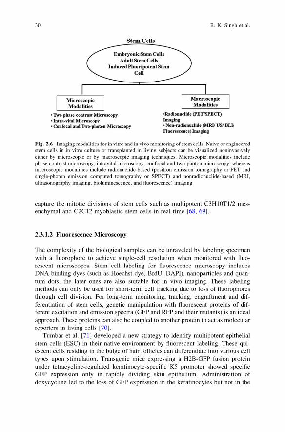
living subjects with high resolution. In parallel, macroscopic or noninvasivein vivo imaging modalities turn out to be indispensible for longitudinal monitoringof translational applications. Commonly used noninvasive imaging modalities forstem cell therapy are radioisotopic imaging (PET or positron emission tomographyand SPECT or single-photon emission computed tomography), CT, ultrasound,magnetic resonance imaging (MRI), and optical imaging (bioluminescence andfluorescence). The next two sections will elaborate microscopic and macroscopicimaging of stem cells, an essential requirement for clinical application (Fig. 2.6).
2.3.1 Microscopic Techniques
For centuries, microscopy is an indispensable tool for visualizing dynamics ofbiomolecules in live cells. Optical microscopic techniques including conventionallight (phase contrast) microscopy, fluorescence microscopy, confocal and multi-photon microscopy, intravital microscopy (IVM) have emerged as powerful toolsfor noninvasive monitoring and characterization of engineered stem cells andtissues [67].
2.3.1.1 Phase Contrast Microscopy
The age-old phase contrast microscopy is routinely required to monitor the cultureconditions and kinetics of HSCs during their expansion for therapeutic use.Recently, these microscopes were improved with automated time-lapse system to
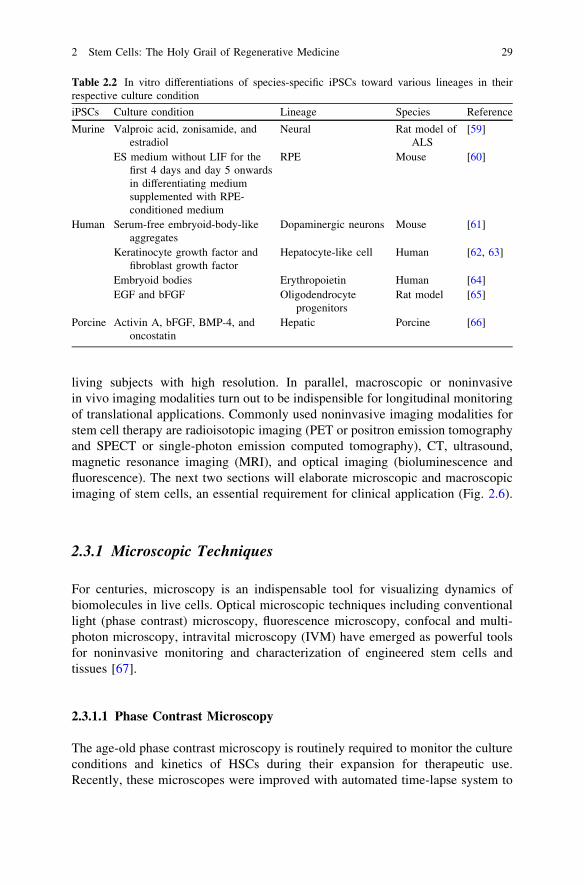
Table 2.2 In vitro differentiations of species-specific iPSCs toward various lineages in theirrespective culture condition
iPSCs Culture condition Lineage Species Reference
Murine Valproic acid, zonisamide, andestradiol
Neural Rat model ofALS
[59]
ES medium without LIF for thefirst 4 days and day 5 onwardsin differentiating mediumsupplemented with RPE-conditioned medium
RPE Mouse [60]
Human Serum-free embryoid-body-likeaggregates
Dopaminergic neurons Mouse [61]
Keratinocyte growth factor andfibroblast growth factor
Hepatocyte-like cell Human [62, 63]
Embryoid bodies Erythropoietin Human [64]EGF and bFGF Oligodendrocyte
progenitorsRat model [65]
Porcine Activin A, bFGF, BMP-4, andoncostatin
Hepatic Porcine [66]
2 Stem Cells: The Holy Grail of Regenerative Medicine 29
capture the mitotic divisions of stem cells such as multipotent C3H10T1/2 mes-enchymal and C2C12 myoblastic stem cells in real time [68, 69].
2.3.1.2 Fluorescence Microscopy
The complexity of the biological samples can be unraveled by labeling specimenwith a fluorophore to achieve single-cell resolution when monitored with fluo-rescent microscopes. Stem cell labeling for fluorescence microscopy includesDNA binding dyes (such as Hoechst dye, BrdU, DAPI), nanoparticles and quan-tum dots, the later ones are also suitable for in vivo imaging. These labelingmethods can only be used for short-term cell tracking due to loss of fluorophoresthrough cell division. For long-term monitoring, tracking, engraftment and dif-ferentiation of stem cells, genetic manipulation with fluorescent proteins of dif-ferent excitation and emission spectra (GFP and RFP and their mutants) is an idealapproach. These proteins can also be coupled to another protein to act as molecularreporters in living cells [70].
Tumbar et al. [71] developed a new strategy to identify multipotent epithelialstem cells (ESC) in their native environment by fluorescent labeling. These qui-escent cells residing in the bulge of hair follicles can differentiate into various celltypes upon stimulation. Transgenic mice expressing a H2B-GFP fusion proteinunder tetracycline-regulated keratinocyte-specific K5 promoter showed specificGFP expression only in rapidly dividing skin epithelium. Administration ofdoxycycline led to the loss of GFP expression in the keratinocytes but not in the
Fig. 2.6 Imaging modalities for in vitro and in vivo monitoring of stem cells: Naive or engineeredstem cells in in vitro culture or transplanted in living subjects can be visualized noninvasivelyeither by microscopic or by macroscopic imaging techniques. Microscopic modalities includephase contrast microscopy, intravital microscopy, confocal and two-photon microscopy, whereasmacroscopic modalities include radionuclide-based (positron emission tomography or PET andsingle-photon emission computed tomography or SPECT) and nonradionuclide-based (MRI,ultrasonography imaging, bioluminescence, and fluorescence) imaging
30 R. K. Singh et al.
SCs due to their slow cycling and quiescent nature. The fluorescence microscopyof the sections from the keratinocytes clearly demonstrated the label-retainingcapability of SC niche. This method was later used by many research groups toisolate and purify the label-retaining cells (LRCs) or SCs for further character-ization [70, 71].
Fluorescent probes with emission wavelengths in the near-infrared (NIR)spectra (*700–800 nm) enhance the feasibility of tracking cells in vitro and insmall animal models due to high depth penetration and lower absorption andscattering [72]. Several dyes including NIR fluorochrome DiD, a lipophilic dyethat binds to the cell membrane, have proven effective for both in vitro labeling ofMSCs and in vivo cell tracking with optical imaging [73, 74].
As described in Sect. 2.2.3, direct reprogramming of somatic cells into iPSCscan be achieved by overexpression of four reprogramming factors (RFs). Adynamic fluorescence microscopy study of iPSCs expressing Nanog-GFP suggeststhat the number of cell divisions is a key parameter of driving epigenetic repro-gramming to pluripotency [75]. Similarly, fluorescence microscopy combined withlong-term time-lapse imaging and single-cell tracking revealed the ‘‘birth’’ ofpluripotent cells and early iPSC clusters from murine embryonic fibroblaststransduced with multicistronic lentiviral vectors carrying RFs (Klf4, Sox2, Oct4,Myc) tagged with different fluorescence proteins (GFP, RFP, YFP) [76].
2.3.1.3 Confocal Microscopy and Multiphoton Microscopy
A major drawback of fluorescence microscopy is that irrespective of the verticalfocusing of the specimen, illumination causes the entire specimen to fluoresce andis unable to produce tomographic images. Confocal microscopy, multiphotonmicroscopy, and intravital microscopy are competent of generating tomographicimaging essential for localizing fluorescent targets in three-dimensional space[77–79].
In a recent study, transgenic ES cells co-expressing myristoylated RFP (labelsplasma membrane) and histone H2B-GFP (labels active chromatin) fusions wereintroduced into a nontransgenic embryo and then dissected out of the maternaluterus at mid-gestation period and cultured ex utero on the stage of a confocalmicroscope. These labeled ES cells produced information on dynamic changes inmorphology and chromatin distribution that occurred during mitotic progression[80]. The powerful confocal/two-photon hybrid microscopy can also track theclonal history of HSPCs expressing various fluorescence proteins noninvasively inintact tissues, including bone marrow with long-term monitoring after transplan-tation [81].
2 Stem Cells: The Holy Grail of Regenerative Medicine 31
2.3.1.4 Intravital Microscopy
Intravital microscope (IVM) can be referred as ‘‘microscope for living subjects,’’which enables single-cell imaging in thin sections of live tissues [82]. In aremarkable study, Rompolas et al. [83] monitored the regeneration of hair folliclestemporarily in a transgenic mouse expressing H2B-GFP driven by keratin 14promoter. They demonstrated that stem cells are quiescent during initial stages ofhair regeneration and their progeny is actively dividing within follicular organi-zation [83].
In another elegant study, Takayama et al. [84, 85] imaged the functioning ofhuman iPSC-derived platelets during thrombus formation by intravital microscopyin live mice. The study demonstrated that transient expression of c-Myc wascritical for efficient platelet generation from human iPSCs, which were capable ofmediating hemostasis and thrombosis in a laser-induced vessel wall injury.
2.3.2 Macroscopic Imaging Modalities
Though microscopic imaging techniques can generate critical information, theyare restricted at cellular level and do not depict the kinetics, distribution, andlocation of in vivo differentiation of stem cells in a living subject. Even IVMrequires an artificially created ‘‘window chamber’’ or ‘‘tissue flap’’ at the bodysurface and is not applicable for deep tissue imaging. Recent developments inmacroscopic or in vivo imaging modalities enable the ‘‘visualization, character-ization, and measurement of biological processes at the molecular and cellularlevels in humans and other living systems.’’ Along with other applications, thesemodalities are now widely used for imaging stem cell delivery, migration andlocalization, cell viability, and therapeutic effects in various diseases [86–88].
The Six commonly used modalities for imaging stem cell therapy are ultra-sound, CT, SPECT, PET, MRI, and optical. For many instances, multimodalityapproaches that can collectively assess different parameters specific for eachmodality are gaining popularity. For imaging the stem cells in vivo, it is requiredto label the cells with an appropriate probe. The two important methods forlabeling cells are as follows:
(a) Direct labeling such as with magnetic resonance contrast agents, radionuc-lides, fluorophores, and nanoparticles.
(b) Indirect labeling with reporter genes.
Direct labeling of cells poses limitation to long-term monitoring since the signalgets diminished by dilution or loss of labeling agents via cell division or differ-entiation. This limitation can be overcome with ‘‘indirect-labeling strategy’’ wherecells are exogenously labeled with reporter genes (bioluminescence, fluorescence,PET, SPECT, or MR reporters) and then implanted for imaging. Indirect labelingrequires genetic manipulation of the cells that are often not possible to follow in
32 R. K. Singh et al.
clinics [86–90]. However, both the strategies have own advantages and disad-vantages and should be implemented by experimental need.
2.3.2.1 Radionuclide Imaging
Majority of the radionuclide imaging techniques follow direct-labeling strategiesand are extensively used in human studies. Both radionuclide imaging techniques,i.e., PET and SPECT, have picomolar sensitivity and high tissue penetration withleast attenuation and are tomographic in nature.
Positron Emission Tomography (PET)
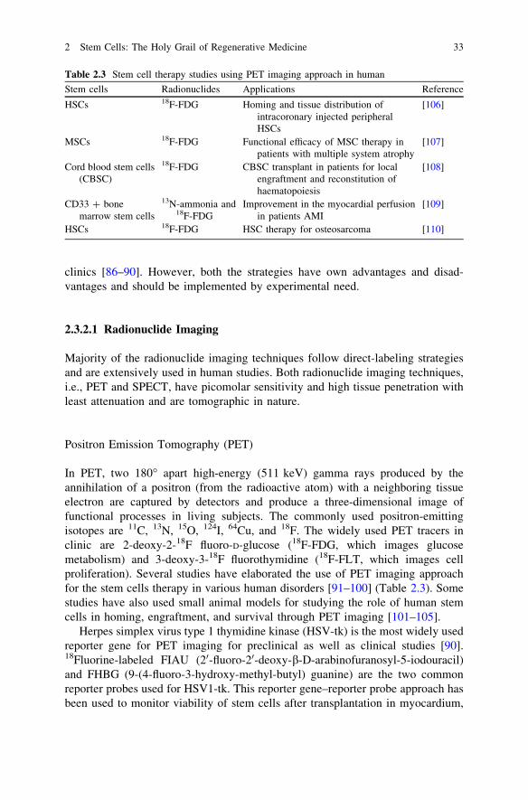
In PET, two 180� apart high-energy (511 keV) gamma rays produced by theannihilation of a positron (from the radioactive atom) with a neighboring tissueelectron are captured by detectors and produce a three-dimensional image offunctional processes in living subjects. The commonly used positron-emittingisotopes are 11C, 13N, 15O, 124I, 64Cu, and 18F. The widely used PET tracers inclinic are 2-deoxy-2-18F fluoro-D-glucose (18F-FDG, which images glucosemetabolism) and 3-deoxy-3-18F fluorothymidine (18F-FLT, which images cellproliferation). Several studies have elaborated the use of PET imaging approachfor the stem cells therapy in various human disorders [91–100] (Table 2.3). Somestudies have also used small animal models for studying the role of human stemcells in homing, engraftment, and survival through PET imaging [101–105].
Herpes simplex virus type 1 thymidine kinase (HSV-tk) is the most widely usedreporter gene for PET imaging for preclinical as well as clinical studies [90].18Fluorine-labeled FIAU (20-fluoro-20-deoxy-b-D-arabinofuranosyl-5-iodouracil)and FHBG (9-(4-fluoro-3-hydroxy-methyl-butyl) guanine) are the two commonreporter probes used for HSV1-tk. This reporter gene–reporter probe approach hasbeen used to monitor viability of stem cells after transplantation in myocardium,
Table 2.3 Stem cell therapy studies using PET imaging approach in human
Stem cells Radionuclides Applications Reference
HSCs 18F-FDG Homing and tissue distribution ofintracoronary injected peripheralHSCs
[106]
MSCs 18F-FDG Functional efficacy of MSC therapy inpatients with multiple system atrophy
[107]
Cord blood stem cells(CBSC)
18F-FDG CBSC transplant in patients for localengraftment and reconstitution ofhaematopoiesis
[108]
CD33 ? bonemarrow stem cells
13N-ammonia and18F-FDG
Improvement in the myocardial perfusionin patients AMI
[109]
HSCs 18F-FDG HSC therapy for osteosarcoma [110]
2 Stem Cells: The Holy Grail of Regenerative Medicine 33

tracking, and survival of autologous MSCs in pig myocardium and tumor stroma[111, 112].
Recently, human sodium iodide symporter (hNIS) gene has emerged as animportant PET reporter gene that could be used for SPECT imaging as well.Uptake of 124Iodine was seen in areas deficit of myocardial perfusion in ratmyocardium injected with MSCs expressing hNIS gene by PET imaging [113].NIS-mediated 124I PET imaging was also used to monitor the delivery and survivalof endothelial progenitor cells (EPCs) after transplantation into the rat heart [114].Some other promising PET reporter genes for stem cell therapy are dopamine 2-like receptor (D2R), human somatostatin receptor subtype 2 (hSSTr2), humannorepinephrine transporter (hNET), neurotensin receptors, and cytosine deaminase[115].
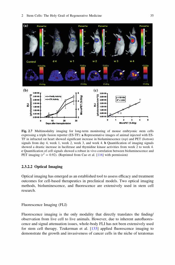
Stem cell therapy is often benefited from multimodality imaging approaches. Inan elegant study, Cao et al. [116] monitored the survival, proliferation, andmigration of ESCs expressing a triple fusion (TF) reporter comprised of a fluo-rescence (mrfp), a bioluminescence (fluc), and a PET reporter (ttk) gene aftertransplantation into rat myocardium for 40 days (Fig. 2.7). Some studies have usedbifusion reporters such as tk-GFP to monitor neuronal stem cell, therapy intreatment of malignant glioma [117].
Single-Photon Emission Computed Tomography (SPECT)
In contrast to PET imaging, SPECT imaging is a log-order less sensitive techniquedue to the presence of collimators (to restrict detection of nonspecific randomgamma rays) between the detectors. The widely used SPECT radionuclides are99mTc, 111In, and 123I. SPECT imaging probes are extensively applied to imagein vivo trafficking and biodistribution of MSCs in myocardial injuries in largeanimal models [118–120]. Some human clinical studies have also used 111In-oxineto track bone marrow stem cells or pro-angiogenic progenitor cells in acute andchronic myocardial injuries. Goussetis et al. [121] performed SPECT imaging with99mTc hexamethylpropyleneamine-oxime-labeled autologous CD133-CD34+ bonemarrow progenitor cells transplanted in patients with ischemic cardiomyopathy.Higher uptake of radioactivity was observed in the infracted area of the heart,suggesting preferential migration and retention of stem cells in the chronicischemic myocardium.
Both HSV1-tk and hNIS genes can serve as SPECT reporter genes in combi-nation with probes labeled with SPECT isotopes (123I/125I-labeled probes FIAU or99mTc) [113, 122]. Table 2.4 summarizes various studies involved in stem cellvisualization by SPECT in human. Studies related to small animal models can befound in other articles [123–125].
34 R. K. Singh et al.
2.3.2.2 Optical Imaging
Optical imaging has emerged as an established tool to assess efficacy and treatmentoutcomes for cell-based therapeutics in preclinical models. Two optical imagingmethods, bioluminescence, and fluorescence are extensively used in stem cellresearch.
Fluorescence Imaging (FLI)
Fluorescence imaging is the only modality that directly translates the finding/observation from live cell to live animals. However, due to inherent autofluores-cence and signal attenuation issues, whole-body FLI has not been extensively usedfor stem cell therapy. Tzukerman et al. [133] applied fluorescence imaging todemonstrate the growth and invasiveness of cancer cells in the niche of teratomas
Fig. 2.7 Multimodality imaging for long-term monitoring of mouse embryonic stem cellsexpressing a triple fusion reporter (ES-TF): a Representative images of animal injected with ES-TF in infracted rat heart showed significant increase in bioluminescence (top) and PET (bottom)signals from day 4, week 1, week 2, week 3, and week 4. b Quantification of imaging signalsshowed a drastic increase in luciferase and thymidine kinase activities from week 2 to week 4.c Quantification of cell signals showed a robust in vivo correlation between bioluminescence andPET imaging (r2 = 0.92). (Reprinted from Cao et al. [116] with permission)
2 Stem Cells: The Holy Grail of Regenerative Medicine 35
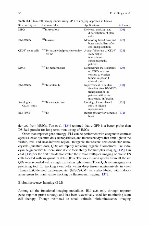
derived from hESCs. Tao et al. [134] reported that e-GFP is a better probe thanDS-Red protein for long-term monitoring of HSCs.
Other than reporter gene strategy, FLI can be performed with exogenous contrastagents such as quantum dots, nanoparticles, and fluorescent dyes that emit light in thevisible, red, and near-infrared region. Inorganic fluorescent semiconductor nano-crystals (quantum dots, QDs) are rapidly replacing organic fluorophores like indo-cyanine green with NIR emission due to their ability for multiplex imaging [135]. Linet al. [136] for the first time demonstrated the in vivo multiplex imaging of mouse EScells labeled with six quantum dots (QDs). The six emission spectra from all the sixQDs were recorded with a single excitation light source. These QDs are emerging as apromising tool for tracking stem cells within deep tissues noninvasively in vivo.Human ESC-derived cardiomyocytes (hESCs-CM) were also labeled with indocy-anine green for noninvasive tracking by fluorescent imaging [137].
Bioluminescence Imaging (BLI)
Among all the functional imaging modalities, BLI acts only through reportergene–reporter probe strategy and has been extensively used for monitoring stemcell therapy. Though restricted to small animals, bioluminescence imaging
Table 2.4 Stem cell therapy studies using SPECT imaging approach in human
Stem cell types Radionuclides Applications Reference
MSCs 111In-tropolone Delivery, tracking, anddifferentiation of stemcells
[126]
BM-MSCs 111In-oxide Monitoring blood flow andbone metabolism aftercell transplantation
[127]
CD34+ stem cells 99mTc hexamethylpropyleneamineoxime
5-year follow-up of CD34+
stem cell innonischemiccardiomyopathypatients
[128]
MSCs 99mTc-pertechnetate Demonstrate the feasibilityof MSCs as viruscarriers to ovariantumors in phase Iclinical trails
[129]
BM-MSCs 99mTc-sestamibi Improvement in cardiacfunction after BMMSCstransplantation inpatients with acutemyocardial infarction
[130]
AutologousCD34+ cells
99mTc-exametazime Homing of transplantedcells to injuredmyocardium
[131]
BM-HSCs 99mTc Repair efficacy for ischemicheart
[132]
36 R. K. Singh et al.
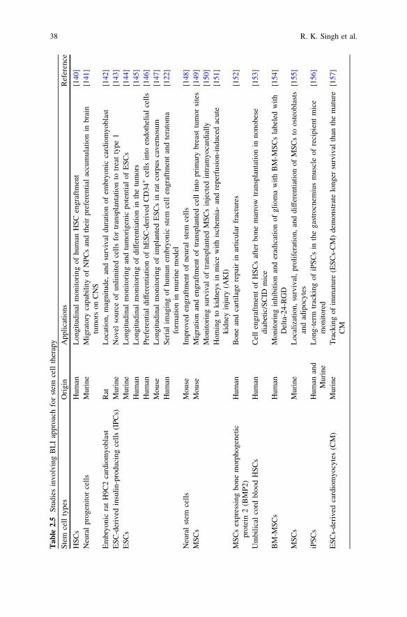
generates essential clues on differentiation, behavior, and viability of stem cellsafter transplanted in small animals, assisting to predict the behavior of stem celltherapy in humans. A glimpse of the large number of BLI-based studies is sum-marized in Table 2.5, and a few important studies are discussed.
Tsuji et al. [138], using iPSC-derived neurospheres in a mouse model of spinalcord injury, demonstrated that iPSCs can ‘‘safely’’ promote locomotor functionrecovery in injured mouse models with BLI. They observed that these cells caneven differentiate into trilineage neural cells in the injured spinal cord. In anotherstudy, Daadi et al. [139] investigated the efficacy of human neural stem cells(hNSCs) derived from human ES cells to repair brain injury. In this study, rats withneonatal HI (hypoxic-ischemic) brain injury were implanted with hNSCsexpressing luciferase and their survival was monitored using BLI. The studysuggests that hNSCs transplants are able to enhance brain injury repair in responseto HI brain injury and that the location and survival can be monitored noninva-sively. Further, the deleterious effect of ESC differentiation on teratomas wasspatially and temporally monitored by Cao et al. [116] with high sensitivity, whichwas not possible with other imaging modalities (Fig. 2.7).
2.3.2.3 Magnetic Resonance Imaging
MRI is the sole imaging modality that generates both functional information andanatomical information. Among all the other noninvasive imaging techniques,MRI has the highest spatial resolution (*100 lm) and thus is the most preferredstrategy for stem cell imaging. Since endogenous molecules (such as H2 atoms) donot generate enough contrast to achieve that high resolution, supra-paramagneticiron oxide (SPIOs) and paramagnetic nanoparticles are often used to label the cellsto enhance image contrast. However, SPIO-based MRI is not well suited for long-term monitoring since SPIOs get diluted with cell proliferation and are oftenengulfed by macrophages [86]. Chelated gadolinium (Gd3+), manganese (Mn2+),and iron (Fe3+) could also act as contrast agents. Recently, certain metal-ion-basedenzymes namely metalloproteinase, transferrin, ferritin, tyrosinase are beingevaluated as reporter genes in MR imaging [87].
In contrast to optical imaging where smaller animals such as mouse and rat arepreferred as model systems, MR-based stem cell imaging is tested both in smallerand larger animals and in humans. The first autologous transplantation of ironoxide-labeled iPSCs reprogrammed from canine adipose stromal cells and fibro-blasts showed repair of infracted myocardium and hindlimb ischemia by MRimaging in adult mongrel dogs [158]. Similarly, an enhanced effect of combininghuman cardiac stem cells and bone marrow MSCs to reduce infarct size and restorecardiac function after myocardial infarction was followed by MR imaging in aYorkshire swine model [159].
To overcome the shortcoming of the contrast agents, reporter-gene-basedstrategies are also being employed in stem cell imaging by MR. Liu et al. [160]showed that engraftment of transgenic mouse ESCs expressing human ferritin
2 Stem Cells: The Holy Grail of Regenerative Medicine 37
Tab
le2.
5S
tudi
esin
volv
ing
BL
Iap
proa
chfo
rst
emce
llth
erap
y
Ste
mce
llty
pes
Ori
gin
App
lica
tion
sR
efer
ence
HS
Cs
Hum
anL
ongi
tudi
nal
mon
itor
ing
ofhu
man
HS
Cen
graf
tmen
t[1
40]
Neu
ral
prog
enit
orce
lls
Mur
ine
Mig
rato
ryca
pabi
lity
ofN
PC
san
dth
eir
pref
eren
tial
accu
mul
atio
nin
brai
ntu
mor
son
CN
S[1
41]
Em
bryo
nic
rat
H9C
2ca
rdio
myo
blas
tR
atL
ocat
ion,
mag
nitu
de,
and
surv
ival
dura
tion
ofem
bryo
nic
card
iom
yobl
ast
[142
]E
SC
-der
ived
insu
lin-
prod
ucin
gce
lls
(IP
Cs)
Mur
ine
Nov
elso
urce
ofun
lim
ited
cell
sfo
rtr
ansp
lant
atio
nto
trea
tty
pe1
[143
]E
SC
sM
urin
eL
ongi
tudi
nal
mon
itor
ing
and
tum
orig
enic
pote
ntia
lof
ES
Cs
[144
]H
uman
Lon
gitu
dina
lm
onit
orin
gof
diff
eren
tiat
ion
inth
etu
mor
s[1
45]
Hum
anP
refe
rent
ial
diff
eren
tiat
ion
ofhE
SC
-der
ived
CD
34+
cell
sin
toen
doth
elia
lce
lls
[146
]M
ouse
Lon
gitu
dina
lm
onit
orin
gof
impl
ante
dE
SC
sin
rat
corp
usca
vern
osum
[147
]H
uman
Ser
ial
imag
ing
ofhu
man
embr
yoni
cst
emce
llen
graf
tmen
tan
dte
rato
ma
form
atio
nin
mur
ine
mod
el[1
22]
Neu
ral
stem
cell
sM
ouse
Impr
oved
engr
aftm
ent
ofne
ural
stem
cell
s[1
48]
MS
Cs
Mou
seM
igra
tion
and
engr
aftm
ent
oftr
ansp
lant
edce
llin
topr
imar
ybr
east
tum
orsi
tes
[149
]M
onit
orin
gsu
rviv
alof
tran
spla
nted
MS
Cs
inje
cted
intr
amyo
card
iall
y[1
50]
Hom
ing
toki
dney
sin
mic
ew
ith
isch
emia
-an
dre
perf
usio
n-in
duce
dac
ute
kidn
eyin
jury
(AK
I)[1
51]
MS
Cs
expr
essi
ngbo
nem
orph
ogen
etic
prot
ein
2(B
MP
2)H
uman
Bon
ean
dca
rtil
age
repa
irin
arti
cula
rfr
actu
res
[152
]
Um
bili
cal
cord
bloo
dH
SC
sH
uman
Cel
len
graf
tmen
tof
HS
Cs
afte
rbo
nem
arro
wtr
ansp
lant
atio
nin
nono
bese
diab
etic
/SC
IDm
ice
[153
]
BM
-MS
Cs
Hum
anM
onit
orin
gin
hibi
tion
and
erad
icat
ion
ofgl
iom
aw
ith
BM
-MS
Cs
labe
led
wit
hD
elta
-24-
RG
D[1
54]
MS
Cs
Mur
ine
Loc
aliz
atio
n,su
rviv
al,
prol
ifer
atio
n,an
ddi
ffer
enti
atio
nof
MS
Cs
toos
teob
last
san
dad
ipoc
ytes
[155
]
iPS
Cs
Hum
anan
dM
urin
eL
ong-
term
trac
king
ofiP
SC
sin
the
gast
rocn
emiu
sm
uscl
eof
reci
pien
tm
ice
mon
itor
ed[1
56]
ES
Cs-
deri
ved
card
iom
yocy
tes
(CM
)M
urin
eT
rack
ing
ofim
mat
ure
(ES
Cs-
CM
)de
mon
stra
telo
nger
surv
ival
than
the
mat
ure
CM
[157
]
38 R. K. Singh et al.
heavy chain (FTH) resulted in increased cellular iron uptake and MRI contrast anddid not interfere with stem cell pluripotency, neural differentiation, and teratomaformation.
Stem cell therapy has immense potential to treat neurodegenerative diseases,traumatic injury, and stroke. However, risk is associated with intracranial surgeryused to deliver the cells to the brain. In some studies, MRI was combined withultrasound modality to obtain higher sensitivity and resolution. For targeteddelivery of neural stem cells to brain, Burgess et al. [161] employed MRI-guidedfocused ultrasound (MRIgFUS) imaging to monitor noninvasive delivery of stemcells from the blood to the brain by opening the blood–brain barrier at specificregions (striatum and hippocampus) in rat brain. Entry of cells crossing the BBB tobrain was verified by MRI. The study also demonstrated that these stem cellsstarted expressing double cortin, a marker of immature neurons, indicatingoccurrence of in vivo differentiation. An excellent review by Qiu et al. [162]described many such MRI-based studies for stem cell therapy such as migrationand homing of hematopoietic stem–progenitor cells to injured arteries and ath-erosclerosis, stem–progenitor-cell-mediated vascular gene therapy, and severalnovel techniques for magnetic labeling of stem or progenitor cells. Table 2.6describes some of the MRI-based stem cell tracking studies in living subjects.
2.3.2.4 Ultrasound Imaging
Ultrasound imaging (US) utilizes the interaction of sound waves with living tissueto produce an anatomical image. Since US imaging is the only modality thatgenerates real-time images during scanning, several investigators are using thistechnology for longitudinal monitoring of stem-cell-mediated tissue repair andvessel formation. One such application was shown by Watts et al. [173] whereequine fetal-derived embryonic-like stem cells (fdESCs) expressing Oct 4, Nanog,SSEA-4, TRA-1-60, TRA-1-81 stem cell markers and telomerase were implantedin equine flexor tendonitis model through intralesional injection. Thoroughbredhorses (n = 8) were induced with tendon injury in the mid-metacarpal region ofthe superficial digital flexor tendon and injected with fdESCs, and serial ultrasoundexaminations were performed. After 8 weeks, significant improvement in tissuearchitecture, tendon size, tendon lesion size, and tendon linear fiber pattern wasfound by US imaging, which was further corroborated by tissue histology.
To enhance the contrast of signal intensity of US imaging, microbubbles (MBs)tagged with nanoparticles, antibodies, or other signatures are being developed.Such an approach was demonstrated by Leng et al. [174] where biocompatiblepolymer MBs were internalized by human bone-marrow-derived MSCs (MB-MSCs) and used for US imaging. Nude mice injected with MSCs and MB-MSCsin the hindlimb region were temporally imaged by ultrasound, which showed thatthe MB-MSCs are acoustically active in vivo and can be imaged for at least 4 hfrom the time of injection [174].
2 Stem Cells: The Holy Grail of Regenerative Medicine 39
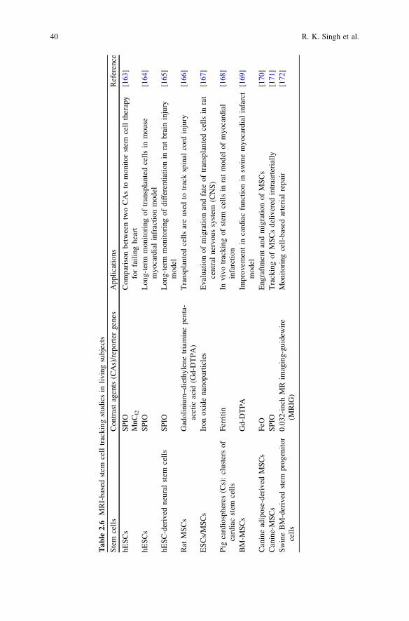
Tab
le2.
6M
RI-
base
dst
emce
lltr
acki
ngst
udie
sin
livi
ngsu
bjec
ts
Ste
mce
lls
Con
tras
tag
ents
(CA
s)/r
epor
ter
gene
sA
ppli
cati
ons
Ref
eren
ce
hES
Cs
SP
IOM
nCl2
Com
pari
son
betw
een
two
CA
sto
mon
itor
stem
cell
ther
apy
for
fail
ing
hear
t[1
63]
hES
Cs
SP
IOL
ong-
term
mon
itor
ing
oftr
ansp
lant
edce
lls
inm
ouse
myo
card
ial
infr
acti
onm
odel
[164
]
hES
C-d
eriv
edne
ural
stem
cell
sS
PIO
Lon
g-te
rmm
onit
orin
gof
diff
eren
tiat
ion
inra
tbr
ain
inju
rym
odel
[165
]
Rat
MS
Cs
Gad
olin
ium
–die
thyl
ene
tria
min
epe
nta-
acet
icac
id(G
d-D
TP
A)
Tra
nspl
ante
dce
lls
are
used
totr
ack
spin
alco
rdin
jury
[166
]
ES
Cs/
MS
Cs
Iron
oxid
ena
nopa
rtic
les
Eva
luat
ion
ofm
igra
tion
and
fate
oftr
ansp
lant
edce
lls
inra
tce
ntra
lne
rvou
ssy
stem
(CN
S)
[167
]
Pig
card
iosp
here
s(C
s):
clus
ters
ofca
rdia
cst
emce
lls
Fer
riti
nIn
vivo
trac
king
ofst
emce
lls
inra
tm
odel
ofm
yoca
rdia
lin
farc
tion
[168
]
BM
-MS
Cs
Gd-
DT
PA
Impr
ovem
ent
inca
rdia
cfu
ncti
onin
swin
em
yoca
rdia
lin
farc
tm
odel
[169
]
Can
ine
adip
ose-
deri
ved
MS
Cs
FeO
Eng
raft
men
tan
dm
igra
tion
ofM
SC
s[1
70]
Can
ine-
MS
Cs
SP
IOT
rack
ing
ofM
SC
sde
live
red
intr
aart
eria
lly
[171
]S
win
eB
M-d
eriv
edst
empr
ogen
itor
cell
s0.
032-
inch
MR
imag
ing-
guid
ewir
e(M
RIG
)M
onit
orin
gce
ll-b
ased
arte
rial
repa
ir[1
72]
40 R. K. Singh et al.
2.3.2.5 Computed Tomography
X-ray CT is a purely anatomical imaging modality, which generates high-reso-lution three-dimensional anatomical images. CT imaging, however, can be appliedto monitor degree of differentiation of embryonic or adult stem cells. Arporn-maeklong et al. [175] demonstrated that undifferentiated hESCs can be cultivatedin osteogenic medium to increase the quantity of osteoblast-like cells (hESCs-OS).These hESCs-OS when transplanted in mice with calvarial defects showed limitedmineralization of tissue in central region, margin of defect, and calvarial boneadjacent to the defect site by micro-CT imaging. Image analysis revealed that bonemineral density of the new bone in the cranial defect generated by the transplantedcells at passage 5 was significantly higher than that in the controls (without cellimplantation). However, CT being purely anatomical imaging modality has limiteduse in measurement and monitoring of stem cell differentiation. Integrated PET-CT and SPECT-CT are better approaches for such evaluations.
2.4 Applications of Stem Cells
The previous two sections describe the advances in stem cell research and mon-itoring the outcome in live cells as well as in live animals. The most challengingphase is to get ‘‘biological solutions to biological diseases’’ with an aim to achievesuccess in curing patients. To estimate the extent of success, a large number ofclinical trials are ongoing with stem cell transplantation in various pathologicalconditions.
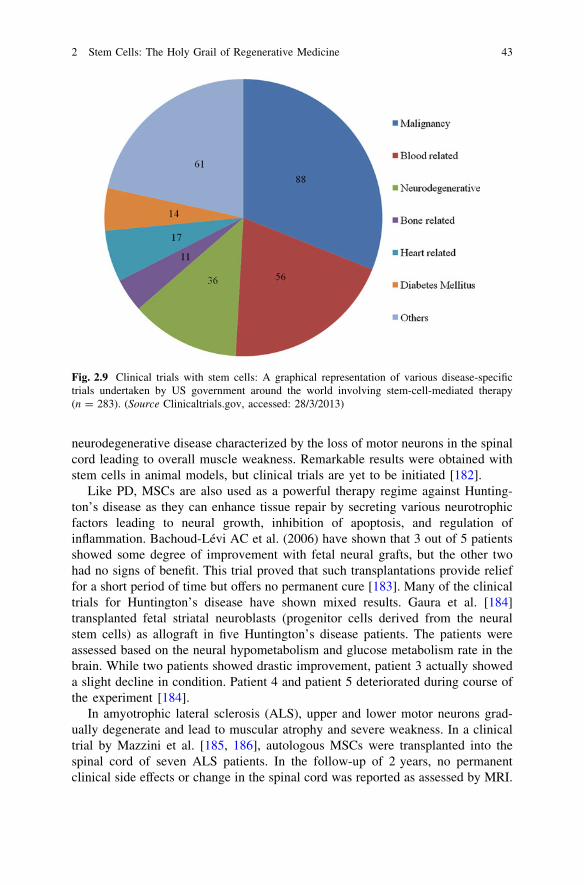
In this section, we will provide an overview of these studies categorized by thedisease types based on the information obtained from trials mostly initiated by theNational Institute of Health (Figs. 2.8 and 2.9). Due to overwhelming number ofanimal studies, we have limited this section only to human trials. The animalstudies can be found in other excellent reviews [176, 177].
2.4.1 Stem Cells and Neurodegenerative Diseases
Two percent of worldwide death is contributed by neurodegenerative diseases suchas Alzheimer’s and Parkinson’s, and medical science is still unable to cure thesediseases. Recent progress in stem cell science showed that functional neuronalreplacement is possible, raising the hope for ultimate cure for these dreadfuldiseases. The idea of stem-cell-based therapy for neurodegenerative diseases is notnew. The first neuronal transplantation was reported in 1890 by Thompson whomade a very bold attempt to transplant cortical tissue from a cat into the brain of adog [178]. This encouraged scientists to consider cell transplantation to treatdeadly neurodegenerative diseases such as Parkinson’s and Alzheimer’s.
2 Stem Cells: The Holy Grail of Regenerative Medicine 41

Earlier, the Parkinson’s disease (PD) patients received a transplant of theadrenal medullary tissue, which did not result in significant improvement. Later,transplantation with fetal tissue and human embryonic neural tissue for cell-basedtherapy was also explored. However, serious ethical issues with these tissuereplacements led to search for the alternate cell source. One such cell source wasfound to be the neural stem cells [179]. Clinical trials with transplantation ofhuman fetal mesencephalic tissues rich in neuronal stem cells have producedsatisfactory results. In a trial by Freed et al. [180], 40 patients between the age of35–75 years were divided into transplantation group (mesencephalic tissue wasimplanted bilaterally into the putamen) and sham surgery group (tissue implantedby drilling a hole into the brain but not disturbing the dura mater) and receivedmesencephalic tissue transplantation. At the end of the surgery, younger patients inthe transplantation group showed better signs of improvement as compared to thesham surgery, while significant improvement was seen in the group of the olderpatients. A small trial by Venketaramana et al. [181] generated a lot of hope for thePD patients. In this pilot study, seven advanced-stage PD patients between the ageof 22 and 65 years were subjected to bone-marrow-derived MSC transplantationextracted from the iliac crest. Patients were reported to show improvement and areduction in the levels of L-DOPA. Spinal muscular dystrophy is a dreadful
Fig. 2.8 Diseases treated with stem cells: Stem cells are emerging as an alternate or sole optionfor treating a wide spectrum of diseases such as neurodegenerative diseases, immune disorders,diabetes type 1, cardiac aliments, kidney disease, malignancies, liver disease, bone and retinaldisorders. At certain times, health damages caused by disasters could also be managed with stemcell therapy
42 R. K. Singh et al.
neurodegenerative disease characterized by the loss of motor neurons in the spinalcord leading to overall muscle weakness. Remarkable results were obtained withstem cells in animal models, but clinical trials are yet to be initiated [182].
Like PD, MSCs are also used as a powerful therapy regime against Hunting-ton’s disease as they can enhance tissue repair by secreting various neurotrophicfactors leading to neural growth, inhibition of apoptosis, and regulation ofinflammation. Bachoud-Lévi AC et al. (2006) have shown that 3 out of 5 patientsshowed some degree of improvement with fetal neural grafts, but the other twohad no signs of benefit. This trial proved that such transplantations provide relieffor a short period of time but offers no permanent cure [183]. Many of the clinicaltrials for Huntington’s disease have shown mixed results. Gaura et al. [184]transplanted fetal striatal neuroblasts (progenitor cells derived from the neuralstem cells) as allograft in five Huntington’s disease patients. The patients wereassessed based on the neural hypometabolism and glucose metabolism rate in thebrain. While two patients showed drastic improvement, patient 3 actually showeda slight decline in condition. Patient 4 and patient 5 deteriorated during course ofthe experiment [184].
In amyotrophic lateral sclerosis (ALS), upper and lower motor neurons grad-ually degenerate and lead to muscular atrophy and severe weakness. In a clinicaltrial by Mazzini et al. [185, 186], autologous MSCs were transplanted into thespinal cord of seven ALS patients. In the follow-up of 2 years, no permanentclinical side effects or change in the spinal cord was reported as assessed by MRI.
Fig. 2.9 Clinical trials with stem cells: A graphical representation of various disease-specifictrials undertaken by US government around the world involving stem-cell-mediated therapy(n = 283). (Source Clinicaltrials.gov, accessed: 28/3/2013)
2 Stem Cells: The Holy Grail of Regenerative Medicine 43
A relatively slow linear decline of the forced vital capacity was seen in four out ofseven patients, indicating that MSCs might have the ability to repair tissue damageand prolong the survival of the patients. A similar exercise was performed byMazzini et al. [187] with 10 patients. The results obtained indicated that stem celltherapy could be extremely beneficial for ALS patients. Thus, stem cell therapyupon proper guidance and validation is finally bringing hope for patients withneurodegenerative diseases such as ALS, chronic spinal injuries, advanced PD[188].
2.4.2 Stem Cells and Kidney Diseases
Both human ES cells and allogenic/autologous HSC transplantations are used tocorrect kidney disorders in clinics. Trivedi et al. [189, 190] was the first group toreport their clinical experience with 24 patients. In their experiment, they intro-duced unfractionated HSCs into the thymus and bone marrow before surgery andinfused them peripherally after transplantation. The aim of the study was to designstrategy to enhance tolerance to cadaver renal transplantation and thus preventgraft rejection. Patients were divided into two groups of which group-A receivedinfusion of the concentrated marrow before and after surgery and group-Bunderwent direct transplantation. Since this procedure did not yield any graftrejection, the study continued, and by 2011, more than 1,000 transplantations wereperformed with further modifications.
2.4.3 Stem Cells in Immunodeficiency and Thalassemia
Life-threatening immunodeficiency can be induced either by drugs or by mal-function of immune system for which cell-based gene transfer is a good treatmentoption. Unfortunately, several such trials are reported with little benefit. Thrasheret al. [191, 192] reported a trial in which engineered CD34+ bone marrow cellsfailed to produce any effect in the subjects. Gaspar et al. [192] treated 10 patientswith autologous CD34+HSPCs transduced with (gamma c) cc retroviral vectors.Follow-up of 80 months showed all patients to be alive with functional T cells,though mild pulmonary infections in most patients and development of acute T-cell lymphoblastic leukemia in one patient were seen. Another strikingly similarattempt was made by Hacein-Bey-Abina et al. [193] where CD34+ cells isolatedfrom five X-SCID patients were transduced with a retroviral vector expressinggamma c(cc) transgene and then transplanted back in patients. The follow-up datafor four out of five patients were quite promising. Their T cells and the naturalkiller cells carrying the transduced gene appeared normal in number, in phenotype,and in proliferative response for at least 2 years after therapy.
44 R. K. Singh et al.

Stem-cell-mediated gene therapy was attempted to tackle thalassemia, a blood-related disorder that affects 7 % of population worldwide. The first trial involvingtransfer of b-globin gene into the CD34+ bone marrow cells using lentiviralvectors was performed by Cavazzana-Calvo et al. Among three patients, the firstone failed to engraft. Fortunately, the second patient accepted the graft and wascontinuously followed up for the next 5 years. The third patient was engraftedafter a few months without complications. In a detailed report, Cavazzana-Calvoet al. [176, 194] have discussed the health of the second patient who had becometransfusion independent for a period of 21 months after transplantation. Theprocess of the trial is diagrammatically represented in Fig. 2.10.
In the last few years, a few trials were done on diseases such as ADA, gaucher,X-SCID using engineered HSCs with limited success [195].
2.4.4 Stem Cells and Type 1 Diabetes
Type 1 diabetes is an autoimmune disease associated with T-cell-mediateddestruction of insulin-producing cells, which results in a lifelong dependence oninsulin. Several attempts such as pancreas transplantation and islet transplantationhave been attempted by scientists and clinicians to improve the quality of life ofthe patients [196].
Fig. 2.10 Schematic representation of the trial conducted by Cavazzana-Calvo et al.: HSCs areisolated from patient by bone marrow aspiration (a) followed by patients treated withchemotherapy (b) cells mobilized in culture subjected to retroviral transduction with appropriategene (c), and finally, these engineered CD34+ HSC cells are infused into the body of the patient (d)
2 Stem Cells: The Holy Grail of Regenerative Medicine 45
Autologous and allogenic transplantations of hematopoietic cells were found torescue patients from their diabetic symptoms. Recently, Gu et al. [197] published aclinical trial in which 28 patients who were having type 1 diabetes, antiglutaminedecarboxylase antibody, and devoid of conditions such as cardiorespiratoryinsufficiency, renal or kidney failure or chronic and acute infection enrolled forautologous HSC transplantation between the years 2007 and 2010. After theadministration of cyclophosphamide and rabbit antithymocyte globulin, autolo-gous HSCs were infused. The daily requirement of insulin started decreasing inthese patients within a month, and the decrease was found to be significant duringthe course of next 3 months and remained stable for the next 24 months. A similarobservation was made in a group of patients with diabetes ketoacidosis (DKA) inwhich 20 out of 23 patients showed a remarkable insulin-free state. Twelve out ofthe twenty patients maintained the state of complete remission (CR) for31 months, and the rest came back with the disease [197].
The umbilical cord blood rich in T regulatory cells and stem cells preserved atthe time of birth also proves to be beneficial to combat such disease conditions. Ina clinical study, Haller et al. [198] reported the results of infusion (about 100 mlcord blood/year) of own stored cord blood in 23 patients with diabetes type 1. Nosignificant adverse effects were observed in 15 of these patients who were a part ofthe follow-up regime. In fact, the study illustrated the process of cord bloodinfusion in young to be feasible. However, the infusion failed to preserve theC-peptide levels in children.
Human placenta being a rich source of pluripotent and multipotent stem cellshas become an attractive resource to the translational researchers. In one such trialperformed, Hou et al. [199, 200] have shown that human amnion epithelial cellscan be differentiated into insulin-producing cells of the pancreas and can reversethe state of hyperglycemia in C57 diabetic mouse. Unfortunately, no such trial hasbeen initiated in patients.
2.4.5 Stem Cells and Malignancies
Stem cell transplantation has become a standard of care for the hematologicalcancers but yet to reach clinic for all types of solid tumors. HSC transplantation isa process in which HSCs are injected into the patients receiving bone-marrow-toxic drugs with or without whole-body radiation therapy. HSC transplantationcould be of two types: allogenic transplantation when the stem cells come fromanother person with matched immune profile and autologous transplantation wherethe stem cells come from own body.
Allogenic transplantation is used to treat acute myelogenous leukemia (AML),chronic myelogenous leukemia (CML), chronic lymphocytic leukemia (CLL), andnon-Hodgkin’s lymphoma. CLL accounts for 25 % of all leukemia in which
46 R. K. Singh et al.

allogenic stem cell transplantation is a good option of treatment. However, due tothe lack of sufficient matching donors, this transplantation method is rarely fol-lowed. A trial by Michallet et al. (2011) enrolled 223 patients between the age of31–65 years and in a stage of CR who were divided into two groups, 112 fortransplantation arm or ACST arm and 111 for observation arm. Eighty of the 112patients were subjected to transplantation of autologous stem cells, and the restwere spared due to collection failure, refusal, or secondary malignancies.According to the results published in 2011, no significant difference was foundbetween ASCT and observation arm with respect to mortality without relapse evenafter 43.7 months of follow-up. However, a statistically significantly improvedevent-free survival was obtained in the ASCT arm. Only occurrence of myelo-dysplastic syndrome was reported. Thus, the transplantation study proved to bebeneficial for CLL patients in CR. In contrary to the above-mentioned trial, a studyby Sutton et al. [203] did not show beneficial effects of ASCT over combinedchemotherapy of fludarabine and cyclophosphamide in patients aged between 18and 65 years with Binet stage B or C CLL who were not treated before [201–203].
A few more randomized clinical trials were performed with allogenic HSCtransplantation. The main focus of these trials was to overcome the chances ofdeveloping chronic graft versus host disease (GVHD), which is a very commoncomplication encountered by clinicians after allogenic stem cell transplantation.Socie et al. [201, 204] performed a randomized trial that assessed prophylactictreatment regime with ATGs. Administration of corticosteroids or antithymocyteglobulin along with drugs such as methotrexate and tacrolimus was shown toreduce the chances of GVHD.
Apart from trials on hematological malignancies, few very interesting clinicaltrials have been performed on solid tumors such as neuroblastoma, Wilms’ tumor,retinoblastoma. In an attempt by George et al. 205, high-risk neuroblastomapatients enrolled between the years 1999 and 2002 were initially subjected to fivecycles of standard chemotherapy and peripheral blood stem cell transplantationwas performed after recovery from the second or the third cycle of chemotherapy.Out of the selected 97 patients, 51 died, and in the remaining 46, patient’s pro-gression-free survival was estimated to be around 47 and 45 % at the end of 5 and7 years, respectively. The overall survival rate was found to be around 60 and53 % at the end of 5 and 7 years, respectively. Thus, a combination of high-dosetherapy with autologous stem cell rescue proved to be beneficial for high-riskneuroblastoma patients. An identical study was performed by Berthold et al. [206]with increased number of high-risk neuroblastoma patients. Patients subjected tohigh-dose chemotherapy with autologous stem cell transplantation were shown tohave an extended 3-year event-free survival as compared to the batch of subjectsgiven a maintenance therapy.
2 Stem Cells: The Holy Grail of Regenerative Medicine 47
2.4.6 Stem Cells and Heart Ailments
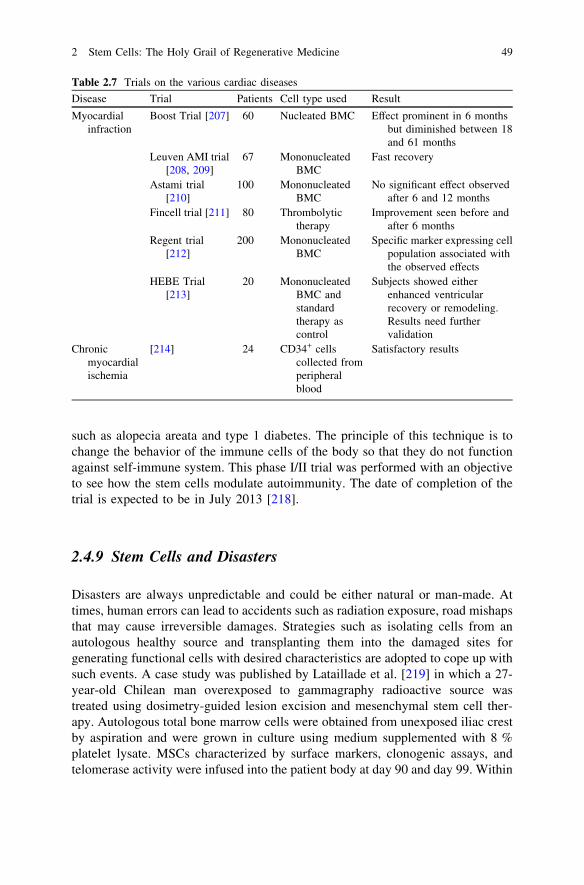
Heart ailments such as myocardial infarction and ischemic heart failure lead todrastic loss of cardiomyocytes, interstitial cells, and vascular cells, which makerecovery slow and difficult. A number of clinical trials are being conducted withmononuclear bone marrow cells rich in MSCs transplantation and are summarizedin Table 2.7 [207].
In a recent report, Vrtovec and his colleagues (2013) discussed a clinical trialregistered under NIH (NCT01350310) performed on 110 dilated cardiomyopathypatients. In phase I, the patients received doses of granulocyte-stimulating factor,and in phase II, 55 out of 110 patients were given an infusion of bone-marrow-derived autologous CD34+ cells. During the 5-year follow-up, patients wereassessed by echocardiography and walking test. However, the most significantfeature of the trial was the use of 99 m Tc hexamethylpropyleneamine oximetracer accumulation method, which was used to determine the homing of theinfused CD34+ cells. The stem cell transplantation was hence seen to be wellassociated with longtime survival of the patients [128].
2.4.7 Stem Cells and Retinal Diseases
Age-related macular degeneration (AMD), glaucoma, and diabetic retinopathy arethe three most common causes of retinal degeneration, visual impairment, andcomplete blindness. In the recent years, a number of studies have been performedon animal models that have explored the potential of mouse or human adult bone-marrow-derived stem cells containing endothelial precursor to stabilize and rescueretinal blood vessels in experimental retinal dystrophies. Fewer numbers of clin-ical studies were done. Jonas et al. [177, 216, 217] produced case reports of trialsperformed in three patients with end-stage macular degeneration and glaucoma.Bone marrow was harvested, and mononuclear cells were separated by Ficolldensity gradient sedimentation. The cells were then injected into the intraocularspace. After the procedure was completed, a regular follow-up was done till12 months. In spite of the trial being technically feasible, no significantimprovement was obtained.
2.4.8 Stem Cells and Alopecia Areata
Alopecia areata is a common T-cell-mediated autoimmune disease leading tochronic and recurrent hair loss. The incidence of the disease is 0.1–0.2 %worldwide. Cell therapy is extremely limited and discouraging for alopecia areata.A clinical trial was initiated in August 2012 using the technique of ‘‘stem celleducator.’’ This technique is extremely useful in treating autoimmune diseases
48 R. K. Singh et al.
such as alopecia areata and type 1 diabetes. The principle of this technique is tochange the behavior of the immune cells of the body so that they do not functionagainst self-immune system. This phase I/II trial was performed with an objectiveto see how the stem cells modulate autoimmunity. The date of completion of thetrial is expected to be in July 2013 [218].
2.4.9 Stem Cells and Disasters
Disasters are always unpredictable and could be either natural or man-made. Attimes, human errors can lead to accidents such as radiation exposure, road mishapsthat may cause irreversible damages. Strategies such as isolating cells from anautologous healthy source and transplanting them into the damaged sites forgenerating functional cells with desired characteristics are adopted to cope up withsuch events. A case study was published by Lataillade et al. [219] in which a 27-year-old Chilean man overexposed to gammagraphy radioactive source wastreated using dosimetry-guided lesion excision and mesenchymal stem cell ther-apy. Autologous total bone marrow cells were obtained from unexposed iliac crestby aspiration and were grown in culture using medium supplemented with 8 %platelet lysate. MSCs characterized by surface markers, clonogenic assays, andtelomerase activity were infused into the patient body at day 90 and day 99. Within
Table 2.7 Trials on the various cardiac diseases
Disease Trial Patients Cell type used Result
Myocardialinfraction
Boost Trial [207] 60 Nucleated BMC Effect prominent in 6 monthsbut diminished between 18and 61 months
Leuven AMI trial[208, 209]
67 MononucleatedBMC
Fast recovery
Astami trial[210]
100 MononucleatedBMC
No significant effect observedafter 6 and 12 months
Fincell trial [211] 80 Thrombolytictherapy
Improvement seen before andafter 6 months
Regent trial[212]
200 MononucleatedBMC
Specific marker expressing cellpopulation associated withthe observed effects
HEBE Trial[213]
20 MononucleatedBMC andstandardtherapy ascontrol
Subjects showed eitherenhanced ventricularrecovery or remodeling.Results need furthervalidation
Chronicmyocardialischemia
[214] 24 CD34+ cellscollected fromperipheralblood
Satisfactory results
2 Stem Cells: The Holy Grail of Regenerative Medicine 49
five and a half months postradiation, the patient showed signs of improvementwith complete healing. After a series of successful animal experiments, a phase I/IIclinical trial was initiated in the year 2011 (NCT01298830). The trial was designedbased on the use of encapsulated and engineered human bone-marrow-derivedMSCs to cure traumatic brain injury. In spite of obtaining some positive resultsfrom 11 patients, the trial was terminated in 2013 [220].
2.4.10 Stem Cells and Bone Disorders
Bone loss has always been a challenge for the clinicians. Marcacci et al. [221]published a trial report that they conducted on four subjects of whom two patientshad an affected ulna and the other two had defective tibia and humerus. Asmentioned in Sect. 2.2.3, a porous bioceramics was used as the scaffold in thisstudy. Bone marrow stem cells isolated from the patients were cultured with minormodifications and seeded on the scaffold within 36 h from harvest. During thefollow-up course, no complications in terms of pain, swelling, or infections wereobserved. Even after 6–7 years of postsurgery, a good integration of implants wasobserved in patients 1 and 3. Angiographic evaluation performed even after6.5 years postsurgery indicated vascularization of the grafted zone. Pre/postop-erative radiography and 2D and 3D CT scans were used to monitor bonedevelopment.
Apart from the above-mentioned bone disease, osteogenesis imperfecta is avery painful genetic disorder caused by a defect in the collagen type I encodinggene. The disease is characterized by bony deformities, fragility, fractures, andshort stature. Two identical clinical trials were performed by Horwitz et al. (1999and 2001). Analysis was conducted using techniques such as dual-energy X-rayabsorptiometry and bone histologic examination from bone biopsy samples takenbefore and after transplantation using the technique of microscopy as explainedpreviously. In the first trial, three patients were infused with unmanipulated bonemarrow from HLA-matched identical siblings. In the second trial, a similarapproach was adopted for the three patients, while the two patients in the controlarm were not given any specific therapy. Improvement in bone histology and asignificant increase in the number of osteoblasts were observed. Unlike the secondtrial, the first one also accounted for a significant increase in bone mineral content.However, complications such as sepsis, pulmonary insufficiency, and bifrontalhygroma were commonly seen in both trials. In spite of the aftereffects as men-tioned above, these trials indeed generated a lot of hope in patients suffering fromthis tragic genetic defect [222, 223].
50 R. K. Singh et al.
2.4.11 Stem Cells and Liver Diseases
Liver diseases such as liver cirrhosis and hepatitis B are considered to be extre-mely life threatening. No effective treatment regime has been designed for almosta decade now. Hence, intensive research is carried out using HSCs due to theirpromising results in other fatal diseases. A phase I trial was designed and pub-lished by Gordon et al. and Levicar et al. [224, 225] in which CD34+ HSCs ingranulocyte-stimulating-factor-mobilized blood were infused into the portal veinof 5 patients with chronic liver diseases. Further, the data collected during thefollow-up period of 12–18 months were discussed in the report published in 2008.During the follow-up period of 12–18 months, patients were assessed on the basisof CT and levels of bilirubin and alpha-fetoproteins in serum and they showed noside effects. The study implicated that the treatment with stem cells lasts for about12 months. Terai et al. [226] in 2006 conducted autologous bone marrow trans-plantation in patients with liver cirrhosis. One of the successful trials reported byGasbarrini et al. [227–229] showed a reversal of drug-induced liver failure bytransplanting CD34+ BMSCs. Zang et al. [230] discussed a trial on infusion ofumbilical cord MSCs in 30 patients with 15 controls (only saline). Reduction in theamount of ascites, improvement in liver function with increased serum albumin,and decrease in serum bilirubin levels were achieved. Another remarkable clinicaltrial on end-stage liver disease was reported by Kharaziha [231] in which autol-ogous MSCs were used. The cells were isolated from the iliac crest and grownin vitro to get a count of 30 to 50 millions. Posttransplantation follow-ups wereregularly done in the 1st, 2nd, 4th, 8th, and 24th weeks. The transplantation waswell tolerated as seen in the improved liver function test results that were reflectedin the levels of serum creatinine, serum albumin, and serum bilirubin.
2.4.12 Stem Cells and Pharmaceutical Companies
Cell therapy by using stem cells has been a subject of interest for clinicians andscientists for almost a decade. The recent years have seen many pharmaceuticalcompanies participating in stem cell research, therefore making regenerativemedicine a vibrant industry. The first FDA-approved trial for patients with spinalcord injury with ESC-derived oligodendrocyte progenitor cells (GRNOPC1)(#NCT01217008) was conducted by Geron, which is currently on hold. Recently,Genzyme reported a quality control program conducted on 303 patients subjectedto autologous transplantation of cultured chondrocytes to repair knee damages.The follow-up of these patients produced positive results [232]. Many companiesare also involved in iPSC research for future like Progenitor Labs involved in stemcell engineering [233, 234]. Some company-sponsored trials are listed inTable 2.8.
2 Stem Cells: The Holy Grail of Regenerative Medicine 51
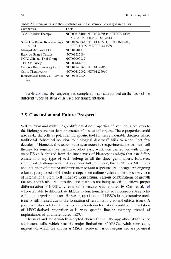
Table 2.9 describes ongoing and completed trials categorized on the basis of thedifferent types of stem cells used for transplantation.
2.5 Conclusion and Future Prospect
Self-renewal and multilineage differentiation properties of stem cells are keys tothe lifelong homeostatic maintenance of tissues and organs. These properties couldalso make the cells as potential therapeutic tool for many incurable diseases wheretraditional ‘‘chemical solution to biological diseases’’ fails to work. Last fewdecades of biomedical research have seen extensive experimentation on stem celltherapy for regenerative medicine. Most early work was carried out with plurip-otent ES cells derived from the inner mass of blastocyst embryo that can differ-entiate into any type of cells belong to all the three germ layers. However,significant challenge was met in successfully culturing the hESCs on MEF cellsand induction of directed differentiation toward a specific cell lineage. An ongoingeffort is going to establish feeder-independent culture system under the supervisionof International Stem Cell Initiative Consortium. Various combinations of growthfactors, chemicals, cell densities, and matrices are being tested to achieve properdifferentiation of hESCs. A remarkable success was reported by Chen et al. [6]who were able to differentiate hESCs to functionally active insulin-secreting beta-cells in a stepwise manner. However, application of hESCs in regenerative med-icine is still limited due to the formation of teratoma in vivo and ethical issues. Apotential future solution for overcoming teratoma formation would be implantationof hESC-derived progenitor cells with specific lineage memory instead ofimplantation of undifferentiated hESC.
The next and most widely accepted choice for cell therapy after hESC is theadult stem cells, which beat the major limitations of hESCs. Adult stem cells,majority of which are known as MSCs, reside in various organs and are potential
Table 2.8 Companies and their contribution in the stem-cell-therapy-based trials
Companies Trials
TCA Cellular Therapy NCT00518401; NCT00643981; NCT00721006;NCT00790764; NCT00548613
Shenzhen Beike BiotechnologyCo. Ltd
NCT01360164; NCT01343511; NCT01610440;NCT01742533; NCT01443689
Manipal Acunova Ltd NCT01501773Banc de Sang i Teixits NCT01227694NCIC Clinical Trial Group NCT00003032TECAM Group NCT00984178Cellonis Biotechnology Co. Ltd NCT01143168; NCT01142050Osiris Therapeutics NCT00482092; NCT01233960International Stem Cell Service
LtdNCT01152125
52 R. K. Singh et al.
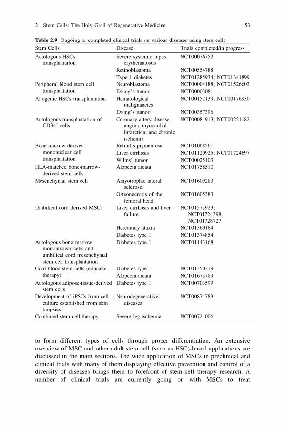
to form different types of cells through proper differentiation. An extensiveoverview of MSC and other adult stem cell (such as HSC)-based applications arediscussed in the main sections. The wide application of MSCs in preclinical andclinical trials with many of them displaying effective prevention and control of adiversity of diseases brings them to forefront of stem cell therapy research. Anumber of clinical trials are currently going on with MSCs to treat
Table 2.9 Ongoing or completed clinical trials on various diseases using stem cells
Stem Cells Disease Trials completed/in progress
Autologous HSCstransplantation
Severe systemic lupuserythematosus
NCT00076752
Retinoblastoma NCT00554788Type 1 diabetes NCT01285934; NCT01341899
Peripheral blood stem celltransplantation
Neuroblastoma NCT00004188; NCT01526603Ewing’s tumor NCT00003081
Allogenic HSCs transplantation Hematologicalmalignancies
NCT00152139; NCT00176930
Ewing’s tumor NCT00357396Autologous transplantation of
CD34+ cellsCoronary artery disease,
angina, myocardialinfarction, and chronicischemia
NCT00081913; NCT00221182
Bone-marrow-derivedmononuclear celltransplantation
Retinitis pigmentosa NCT01068561Liver cirrhosis NCT01120925; NCT01724697Wilms’ tumor NCT00025103
HLA-matched bone-marrow-derived stem cells
Alopecia areata NCT01758510
Mesenchymal stem cell Amyotrophic lateralsclerosis
NCT01609283
Osteonecrosis of thefemoral head
NCT01605383
Umbilical cord-derived MSCs Liver cirrhosis and liverfailure
NCT01573923;NCT01724398;NCT01728727
Hereditary ataxia NCT01360164Diabetes type 1 NCT01374854
Autologous bone marrowmononuclear cells andumbilical cord mesenchymalstem cell transplantation
Diabetes type 1 NCT01143168
Cord blood stem cells (educatortherapy)
Diabetes type 1 NCT01350219Alopecia areata NCT01673789
Autologous adipose-tissue-derivedstem cells
Diabetes type 1 NCT00703599
Development of iPSCs from cellculture established from skinbiopsies
Neurodegenerativediseases
NCT00874783
Combined stem cell therapy Severe leg ischemia NCT00721006
2 Stem Cells: The Holy Grail of Regenerative Medicine 53
neurodegenerative diseases, thalassemia, diabetes, cardiovascular diseases, andmalignancies and even in disaster-related injuries. However, success rate is stilllow and more number of trials would be required to assess the real potential ofstem cell therapy.
The low abundance and need of large-scale culture of adult stem cells forclinical application can overcome by iPSCs, which shows great promise forregenerative medicine. However, application of iPSCs is still limited in preclinicalstages and expected to reach clinic soon.
Finally, monitoring the efficacy of stem cell therapy is an extremely importantcomponent and a large number of microscopic and macroscopic techniques arebeing implemented to check for in vitro differentiation, in vivo differentiation, andfunctional output. Each of these cell based and preclinical imaging techniques hasown merits and demerits and is often used in combination to achieve a multimodalapproach. Since stem cell imaging requires high resolution, MRI despite havinglower sensitivity is probably the most suitable technology. A large number ofpreclinical applications are using bioluminescence imaging as well. These imagingtechniques are also important in understanding the interactions of local microen-vironment with stem cells. Overall, the excitement and hope for effective treatmentfor many incurable diseases by stem cells are quite promising but require athorough and long-term monitoring at preclinical level, followed by practicalprotocols. It is highly expected that detailed investigations of stem cell biology andclinical applicability will result in revolutions in medical technologies.
References
1. Thomson JA, Itskovitz-Eldor J, Shapiro SS, Waknitz MA, Swiergiel JJ, Marshall VS, JonesJM (1998) Embryonic stem cell lines derived from human blastocysts. Science282(5391):1145–1147
2. Akopian V, Andrews PW, Beil S, Benvenisty N, Brehm J, Christie M, Ford A, Fox V,Gokhale PJ, Healy L, Holm F, Hovatta O, Knowles BB, Ludwig TE, McKay RD, MiyazakiT, Nakatsuji N, Oh SK, Pera MF, Rossant J, Stacey GN, Suemori H (2010) Comparison ofdefined culture systems for feeder cell free propagation of human embryonic stem cells.Vitro Cell Dev Biol Anim 46(3–4):247–258. doi:10.1007/s11626-010-9297-z
3. Xu C, Inokuma MS, Denham J, Golds K, Kundu P, Gold JD, Carpenter MK (2001) Feeder-free growth of undifferentiated human embryonic stem cells. Nat Biotechnol19(10):971–974. doi:10.1038/nbt1001-971
4. Lei T, Jacob S, Ajil-Zaraa I, Dubuisson JB, Irion O, Jaconi M, Feki A (2007) Xeno-freederivation and culture of human embryonic stem cells: current status, problems andchallenges. Cell Res 17(8):682–688. doi:10.1038/cr.2007.61
5. Itskovitz-Eldor J, Schuldiner M, Karsenti D, Eden A, Yanuka O, Amit M, Soreq H,Benvenisty N (2000) Differentiation of human embryonic stem cells into embryoid bodiescompromising the three embryonic germ layers. Mol Med 6(2):88–95
6. Chen S, Borowiak M, Fox JL, Maehr R, Osafune K, Davidow L, Lam K, Peng LF, SchreiberSL, Rubin LL, Melton D (2009) A small molecule that directs differentiation of humanESCs into the pancreatic lineage. Nat Chem Biol 5(4):258–265. doi:10.1038/nchembio.154
54 R. K. Singh et al.
7. Kroon E, Martinson LA, Kadoya K, Bang AG, Kelly OG, Eliazer S, Young H, RichardsonM, Smart NG, Cunningham J, Agulnick AD, D’Amour KA, Carpenter MK, Baetge EE(2008) Pancreatic endoderm derived from human embryonic stem cells generates glucose-responsive insulin-secreting cells in vivo. Nat Biotechnol 26(4):443–452. doi:10.1038/nbt1393
8. Schwartz SD, Hubschman JP, Heilwell G, Franco-Cardenas V, Pan CK, Ostrick RM,Mickunas E, Gay R, Klimanskaya I, Lanza R (2012) Embryonic stem cell trials for maculardegeneration: a preliminary report. Lancet 379(9817):713–720. doi:10.1016/S0140-6736(12)60028-2
9. Hacein-Bey-Abina S, von Kalle C, Schmidt M, Le Deist F, Wulffraat N, McIntyre E,Radford I, Villeval JL, Fraser CC, Cavazzana-Calvo M, Fischer A (2003) A serious adverseevent after successful gene therapy for X-linked severe combined immunodeficiency.N Engl J Med 348(3):255–256. doi:10.1056/NEJM200301163480314
10. Yu SF, von Ruden T, Kantoff PW, Garber C, Seiberg M, Ruther U, Anderson WF, WagnerEF, Gilboa E (1986) Self-inactivating retroviral vectors designed for transfer of whole genesinto mammalian cells. Proc Natl Acad of Sci USA. 83(10):3194–3198
11. Naldini L, Blomer U, Gallay P, Ory D, Mulligan R, Gage FH, Verma IM, Trono D (1996) Invivo gene delivery and stable transduction of nondividing cells by a lentiviral vector.Science 272(5259):263–267
12. May C, Rivella S, Callegari J, Heller G, Gaensler KM, Luzzatto L, Sadelain M (2000)Therapeutic haemoglobin synthesis in beta-thalassaemic mice expressing lentivirus-encodedhuman beta-globin. Nature 406(6791):82–86. doi:10.1038/35017565
13. Vadhan-Raj S, Patel S, Bueso-Ramos C, Folloder J, Papadopolous N, Burgess A,Broemeling LD, Broxmeyer HE, Benjamin RS (2003) Importance of predosing ofrecombinant human thrombopoietin to reduce chemotherapy-induced earlythrombocytopenia. J Clinc Oncol 21(16):3158–3167. doi:10.1200/JCO.2003.08.003
14. Panuganti S, Schlinker AC, Lindholm PF, Papoutsakis ET, Miller WM (2013) Three-stageex vivo expansion of high-ploidy megakaryocytic cells: toward large-scale plateletproduction. Tissue Eng Part A 19(7–8):998–1014. doi:10.1089/ten.TEA 2011.0111
15. Lagasse E, Connors H, Al-Dhalimy M, Reitsma M, Dohse M, Osborne L, Wang X,Finegold M, Weissman IL, Grompe M (2000) Purified hematopoietic stem cells candifferentiate into hepatocytes in vivo. Nat Med 6(11):1229–1234. doi:10.1038/81326
16. Friedenstein AJ, Deriglasova UF, Kulagina NN, Panasuk AF, Rudakowa SF, Luria EA,Ruadkow IA (1974) Precursors for fibroblasts in different populations of hematopoietic cellsas detected by the in vitro colony assay method. Exp Hematol 2(2):83–92
17. Friedenstein AJ, Chailakhyan RK, Latsinik NV, Panasyuk AF, Keiliss-Borok IV (1974)Stromal cells responsible for transferring the microenvironment of the hemopoietic tissues.Cloning in vitro and retransplantation in vivo. Transplantation 17(4):331–340
18. Polisetty N, Fatima A, Madhira SL, Sangwan VS, Vemuganti GK (2008) Mesenchymalcells from limbal stroma of human eye. Mol Vis 14:431–442
19. Chan BP, Hui TY, Wong MY, Yip KH, Chan GC (2010) Mesenchymal stem cell-encapsulated collagen microspheres for bone tissue engineering. Tissue Eng Part C Methods16(2):225–235. doi:10.1089/ten.tec.2008.0709
20. Sumanasinghe RD, Bernacki SH, Loboa EG (2006) Osteogenic differentiation of humanmesenchymal stem cells in collagen matrices: effect of uniaxial cyclic tensile strain on bonemorphogenetic protein (BMP-2) mRNA expression. Tissue Eng 12(12):3459–3465.doi:10.1089/ten.2006.12.3459
21. Sumanasinghe RD, Osborne JA, Loboa EG (2009) Mesenchymal stem cell-seeded collagenmatrices for bone repair: effects of cyclic tensile strain, cell density, and media conditionson matrix contraction in vitro. J Biomed Mater Res A 88(3):778–786. doi:10.1002/jbm.a.31913
22. Ahmed TA, Dare EV, Hincke M (2008) Fibrin: a versatile scaffold for tissue engineeringapplications. Tissue Eng Part B Rev 14(2):199–215. doi:10.1089/ten.teb2007.0435
2 Stem Cells: The Holy Grail of Regenerative Medicine 55
23. Kim HJ, Kim UJ, Kim HS, Li C, Wada M, Leisk GG, Kaplan DL (2008) Bone tissueengineering with premineralized silk scaffolds. Bone 42(6):1226–1234. doi:10.1016/j.bone.2008.02.007
24. Li C, Vepari C, Jin HJ, Kim HJ, Kaplan DL (2006) Electrospun silk-BMP-2 scaffolds forbone tissue engineering. Biomaterials 27(16):3115–3124. doi:10.1016/j.biomaterials.2006.01.022
25. Mieszawska AJ, Fourligas N, Georgakoudi I, Ouhib NM, Belton DJ, Perry CC, Kaplan DL(2010) Osteoinductive silk-silica composite biomaterials for bone regeneration.Biomaterials 31(34):8902–8910. doi:10.1016/j.biomaterials.2010.07.109
26. Patterson J, Siew R, Herring SW, Lin AS, Guldberg R, Stayton PS (2010) Hyaluronic acidhydrogels with controlled degradation properties for oriented bone regeneration.Biomaterials 31(26):6772–6781. doi:10.1016/j.biomaterials.2010.05.047
27. Kim J, Kim IS, Cho TH, Kim HC, Yoon SJ, Choi J, Park Y, Sun K, Hwang SJ (2010) Invivo evaluation of MMP sensitive high-molecular weight HA-based hydrogels for bonetissue engineering. J Biomed Mater Res A 95(3):673–681. doi:10.1002/jbm.a.32884
28. Kim J, Kim IS, Cho TH, Lee KB, Hwang SJ, Tae G, Noh I, Lee SH, Park Y, Sun K (2007)Bone regeneration using hyaluronic acid-based hydrogel with bone morphogenic protein-2and human mesenchymal stem cells. Biomaterials 28(10):1830–1837. doi:10.1016/j.biomaterials.2006.11.050
29. Venkatesan J, Kim SK (2010) Chitosan composites for bone tissue engineering–anoverview. Mar Drugs 8(8):2252–2266. doi:10.3390/md8082252
30. Shoae-Hassani A, Mortazavi-Tabatabaei SA, Sharif S, Seifalian AM, Azimi A,Samadikuchaksaraei A, Verdi J (2013) Differentiation of human endometrial stem cellsinto urothelial cells on a three-dimensional nanofibrous silk-collagen scaffold: anautologous cell resource for reconstruction of the urinary bladder wall. J Tissue EngRegen Med. doi:10.1002/term.1632
31. Mummery CL, Davis RP, Krieger JE (2010) Challenges in using stem cells for cardiacrepair. Sci Transl Med 2(27):27ps17. doi:10.1126/scitranslmed.3000558
32. Lee RH, Kim B, Choi I, Kim H, Choi HS, Suh K, Bae YC, Jung JS (2004) Characterizationand expression analysis of mesenchymal stem cells from human bone marrow and adiposetissue. Cell Physiol Biochem 14(4–6):311–324. doi:10.1159/000080341
33. Makino S, Fukuda K, Miyoshi S, Konishi F, Kodama H, Pan J, Sano M, Takahashi T, HoriS, Abe H, Hata J, Umezawa A, Ogawa S (1999) Cardiomyocytes can be generated frommarrow stromal cells in vitro. J Clin Invest 103(5):697–705. doi:10.1172/JCI5298
34. Toma C, Pittenger MF, Cahill KS, Byrne BJ, Kessler PD (2002) Human mesenchymal stemcells differentiate to a cardiomyocyte phenotype in the adult murine heart. Circulation105(1):93–98
35. Takahashi T, Lord B, Schulze PC, Fryer RM, Sarang SS, Gullans SR, Lee RT (2003)Ascorbic acid enhances differentiation of embryonic stem cells into cardiac myocytes.Circulation 107(14):1912–1916. doi:10.1161/01.CIR.0000064899.53876.A3
36. Dominici M, Le Blanc K, Mueller I, Slaper-Cortenbach I, Marini F, Krause D, Deans R,Keating A, Prockop D, Horwitz E (2006) Minimal criteria for defining multipotentmesenchymal stromal cells. The International Society for Cellular Therapy positionstatement. Cytotherapy 8(4):315–317. doi:10.1080/14653240600855905
37. Pittenger MF, Mackay AM, Beck SC, Jaiswal RK, Douglas R, Mosca JD, Moorman MA,Simonetti DW, Craig S, Marshak DR (1999) Multilineage potential of adult humanmesenchymal stem cells. Science 284(5411):143–147
38. Mruthyunjaya S, Manchanda R, Godbole R, Pujari R, Shiras A, Shastry P (2010) Laminin-1induces neurite outgrowth in human mesenchymal stem cells in serum/differentiationfactors-free conditions through activation of FAK-MEK/ERK signaling pathways. BiochemBiophys Res Commun 391(1):43–48. doi:10.1016/j.bbrc.2009.10.158
39. Vecellio M, Meraviglia V, Nanni S, Barbuti A, Scavone A, DiFrancesco D, Farsetti A,Pompilio G, Colombo GI, Capogrossi MC, Gaetano C, Rossini A (2012) In vitro epigenetic
56 R. K. Singh et al.
reprogramming of human cardiac mesenchymal stromal cells into functionally competentcardiovascular precursors. PLoS ONE 7(12):e51694. doi:10.1371/journal.pone.0051694
40. Zouani OF, Kalisky J, Ibarboure E, Durrieu MC (2013) Effect of BMP-2 from matrices ofdifferent stiffnesses for the modulation of stem cell fate. Biomaterials 34(9):2157–2166.doi:10.1016/j.biomaterials.2012.12.007
41. Fatima A, Sangwan VS, Iftekhar G, Reddy P, Matalia H, Balasubramanian D, VemugantiGK (2006) Technique of cultivating limbal derived corneal epithelium on human amnioticmembrane for clinical transplantation. J Postgrad Med 52(4):257–261
42. Gurdon JB (1962) The developmental capacity of nuclei taken from intestinal epitheliumcells of feeding tadpoles. J Embryol Exp Morphol 10:622–640
43. Takahashi K, Yamanaka S (2006) Induction of pluripotent stem cells from mouseembryonic and adult fibroblast cultures by defined factors. Cell 126(4):663–676.doi:10.1016/j.cell.2006.07.024
44. Takahashi K, Tanabe K, Ohnuki M, Narita M, Ichisaka T, Tomoda K, Yamanaka S (2007)Induction of pluripotent stem cells from adult human fibroblasts by defined factors. Cell131(5):861–872. doi:10.1016/j.cell.2007.11.019
45. Yu J, Vodyanik MA, Smuga-Otto K, Antosiewicz-Bourget J, Frane JL, Tian S, Nie J,Jonsdottir GA, Ruotti V, Stewart R, Slukvin II, Thomson JA (2007) Induced pluripotentstem cell lines derived from human somatic cells. Science 318(5858):1917–1920.doi:10.1126/science.1151526
46. Okita K, Ichisaka T, Yamanaka S (2007) Generation of germline-competent inducedpluripotent stem cells. Nature 448(7151):313–317. doi:10.1038/nature05934
47. Kawai H, Yamashita T, Ohta Y, Deguchi K, Nagotani S, Zhang X, Ikeda Y, Matsuura T,Abe K (2010) Tridermal tumorigenesis of induced pluripotent stem cells transplanted inischemic brain. J Cereb Blood Flow Metab 30(8):1487–1493. doi:10.1038/jcbfm.2010.32
48. Zhou H, Wu S, Joo JY, Zhu S, Han DW, Lin T, Trauger S, Bien G, Yao S, Zhu Y, SiuzdakG, Scholer HR, Duan L, Ding S (2009) Generation of induced pluripotent stem cells usingrecombinant proteins. Cell Stem Cell 4(5):381–384. doi:10.1016/j.stem.2009.04.005
49. Blelloch R, Venere M, Yen J, Ramalho-Santos M (2007) Generation of induced pluripotentstem cells in the absence of drug selection. Cell Stem Cell 1(3):245–247. doi:10.1016/j.stem.2007.08.008
50. Anokye-Danso F, Trivedi CM, Juhr D, Gupta M, Cui Z, Tian Y, Zhang Y, Yang W, GruberPJ, Epstein JA, Morrisey EE (2011) Highly efficient miRNA-mediated reprogramming ofmouse and human somatic cells to pluripotency. Cell Stem Cell 8(4):376–388. doi:10.1016/j.stem.2011.03.001
51. Liu H, Zhu F, Yong J, Zhang P, Hou P, Li H, Jiang W, Cai J, Liu M, Cui K, Qu X, Xiang T,Lu D, Chi X, Gao G, Ji W, Ding M, Deng H (2008) Generation of induced pluripotent stemcells from adult rhesus monkey fibroblasts. Cell Stem Cell 3(6):587–590. doi:10.1016/j.stem.2008.10.014
52. Liao J, Cui C, Chen S, Ren J, Chen J, Gao Y, Li H, Jia N, Cheng L, Xiao H, Xiao L (2009)Generation of induced pluripotent stem cell lines from adult rat cells. Cell Stem Cell4(1):11–15. doi:10.1016/j.stem.2008.11.013
53. Ezashi T, Telugu BP, Alexenko AP, Sachdev S, Sinha S, Roberts RM (2009) Derivation ofinduced pluripotent stem cells from pig somatic cells. Proc Natl Acad of Sci USA106(27):10993–10998. doi:10.1073/pnas.0905284106
54. Shimada H, Nakada A, Hashimoto Y, Shigeno K, Shionoya Y, Nakamura T (2010)Generation of canine induced pluripotent stem cells by retroviral transduction and chemicalinhibitors. Mol Reprod Dev 77(1):2. doi:10.1002/mrd.21117
55. Wu Y, Zhang Y, Mishra A, Tardif SD, Hornsby PJ (2010) Generation of induced pluripotentstem cells from newborn marmoset skin fibroblasts. Stem Cell Res 4(3):180–188.doi:10.1016/j.scr.2010.02.003
56. Honda A, Hirose M, Hatori M, Matoba S, Miyoshi H, Inoue K, Ogura A (2010) Generationof induced pluripotent stem cells in rabbits: potential experimental models for humangenerative medicine. J Biol Chem 285(41):31362–31369. doi:10.1074/jbc.M110.150540
2 Stem Cells: The Holy Grail of Regenerative Medicine 57
57. Nagy K, Sung HK, Zhang P, Laflamme S, Vincent P, Agha-Mohammadi S, Woltjen K,Monetti C, Michael IP, Smith LC, Nagy A (2011) Induced pluripotent stem cell linesderived from equine fibroblasts. Stem Cell Rev 7(3):693–702. doi:10.1007/s12015-011-9239-5
58. Lu Y, West FD, Jordan BJ, Mumaw JL, Jordan ET, Gallegos-Cardenas A, Beckstead RB,Stice SL (2012) Avian-induced pluripotent stem cells derived using human reprogrammingfactors. Stem Cells Dev 21(3):394–403. doi:10.1089/scd2011.0499
59. Yoshikawa T, Samata B, Ogura A, Miyamoto S, Takahashi J (2013) Systemicadministration of valproic acid and zonisamide promotes differentiation of inducedpluripotent stem cell-derived dopaminergic neurons. Front Cell Neurosci 7:11. doi:10.3389/fncel.2013.00011
60. Mekala SR, Vauhini V, Nagarajan U, Maddileti S, Gaddipati S, Mariappan I (2013)Derivation, characterization and retinal differentiation of induced pluripotent stem cells.J Biosci 38(1):123–134
61. Nishimura K, Takahashi J (2013) Therapeutic application of stem cell technology towardthe treatment of Parkinson’s disease. Biol Pharma Bull. 36(2):171–175
62. Song Z, Cai J, Liu Y, Zhao D, Yong J, Duo S, Song X, Guo Y, Zhao Y, Qin H, Yin X, WuC, Che J, Lu S, Ding M, Deng H (2009) Efficient generation of hepatocyte-like cells fromhuman induced pluripotent stem cells. Cell Res 19(11):1233–1242. doi:10.1038/cr.2009.107
63. Sullivan GJ, Hay DC, Park IH, Fletcher J, Hannoun Z, Payne CM, Dalgetty D, Black JR,Ross JA, Samuel K, Wang G, Daley GQ, Lee JH, Church GM, Forbes SJ, Iredale JP,Wilmut I (2010) Generation of functional human hepatic endoderm from human inducedpluripotent stem cells. Hepatology 51(1):329–335. doi:10.1002/hep.23335
64. Kobari L, Yates F, Oudrhiri N, Francina A, Kiger L, Mazurier C, Rouzbeh S, El-Nemer W,Hebert N, Giarratana MC, Francois S, Chapel A, Lapillonne H, Luton D, Bennaceur-Griscelli A, Douay L (2012) Human induced pluripotent stem cells can reach completeterminal maturation: in vivo and in vitro evidence in the erythropoietic differentiationmodel. Haematologica 97(12):1795–1803. doi:10.3324/haematol.2011.055566
65. Pouya A, Satarian L, Kiani S, Javan M, Baharvand H (2011) Human induced pluripotentstem cells differentiation into oligodendrocyte progenitors and transplantation in a rat modelof optic chiasm demyelination. PLoS ONE 6(11):e27925. doi:10.1371/journal.pone.0027925
66. Aravalli RN, Cressman EN, Steer CJ (2012) Hepatic differentiation of porcine inducedpluripotent stem cells in vitro. Vet J 194(3):369–374. doi:10.1016/j.tvjl.2012.05.013
67. Georgakoudi I, Rice WL, Hronik-Tupaj M, Kaplan DL (2008) Optical spectroscopy andimaging for the noninvasive evaluation of engineered tissues. Tissue Eng Part B Rev14(4):321–340. doi:10.1089/ten.teb2008.0248
68. Huh S, Ker DF, Bise R, Chen M, Kanade T (2011) Automated mitosis detection of stem cellpopulations in phase-contrast microscopy images. IEEE Trans Med Imaging 30(3):586–596.doi:10.1109/TMI.2010.2089384
69. S-iH Sungeun Eom, Ker Dai Fei Elmer, Bise Ryoma, Kanade Takeo (2007) Tracking ofhematopoietic stem cells in microscopy images for lineage determination. J Latex ClassFiles 6(01–09):9
70. Jang YY, Ye Z, Cheng L (2011) Molecular imaging and stem cell research. Mol Imagings10(2):111–122
71. Tumbar T, Guasch G, Greco V, Blanpain C, Lowry WE, Rendl M, Fuchs E (2004) Definingthe epithelial stem cell niche in skin. Science 303(5656):359–363. doi:10.1126/science.1092436
72. Giepmans BN, Adams SR, Ellisman MH, Tsien RY (2006) The fluorescent toolbox forassessing protein location and function. Science 312(5771):217–224. doi:312/5771/217
73. Sutton EJ, Henning TD, Pichler BJ, Bremer C, Daldrup-Link HE (2008) Cell tracking withoptical imaging. Eur Radiol 18(10):2021–2032. doi:10.1007/s00330-008-0984-z
58 R. K. Singh et al.
74. Sutton EJ, Boddington SE, Nedopil AJ, Henning TD, Demos SG, Baehner R, Sennino B, LuY, Daldrup-Link HE (2009) An optical imaging method to monitor stem cell migration in amodel of immune-mediated arthritis. Opt Express 17(26):24403–24413. doi:194171
75. Hanna J, Saha K, Pando B, van Zon J, Lengner CJ, Creyghton MP, van Oudenaarden A,Jaenisch R (2009) Direct cell reprogramming is a stochastic process amenable toacceleration. Nature 462(7273):595–601. doi:10.1038/nature08592
76. Warlich E, Kuehle J, Cantz T, Brugman MH, Maetzig T, Galla M, Filipczyk AA, Halle S,Klump H, Scholer HR, Baum C, Schroeder T, Schambach A (2011) Lentiviral vector designand imaging approaches to visualize the early stages of cellular reprogramming. Mol Ther19(4):782–789. doi:10.1038/mt.2010.314
77. Conchello JA, Lichtman JW (2005) Optical sectioning microscopy. Nat Methods2(12):920–931. doi:10.1038/nmeth815
78. Helmchen F, Denk W (2005) Deep tissue two-photon microscopy. Nat Methods2(12):932–940. doi:10.1038/nmeth818
79. Rice WL, Kaplan DL, Georgakoudi I (2010) Two-photon microscopy for non-invasive,quantitative monitoring of stem cell differentiation. PLoS ONE 5(4):e10075. doi:10.1371/journal.pone.0010075
80. Jérôme Artus A-KH (2007) Live imaging genetically-encoded fluorescent proteins inembryonic stem cells using confocal microscopy. In: Méndez-Vilas A, Díaz J (eds) Modernresearch and educational topics in microscopy. Sloan-Kettering Institute, New York
81. Malide D, Metais JY, Dunbar CE (2012) Dynamic clonal analysis of murine hematopoieticstem and progenitor cells marked by 5 fluorescent proteins using confocal and multiphotonmicroscopy. Blood 120(26):e105–e116. doi:blood-2012-06-440636
82. Ntziachristos V (2010) Going deeper than microscopy: the optical imaging frontier inbiology. Nat Methods 7(8):603–614. doi:10.1038/nmeth.1483
83. Rompolas P, Deschene ER, Zito G, Gonzalez DG, Saotome I, Haberman AM, Greco V(2012) Live imaging of stem cell and progeny behaviour in physiological hair-follicleregeneration. Nature 487(7408):496–499. doi:10.1038/nature11218
84. Takayama N, Eto K (2012) Pluripotent stem cells reveal the developmental biology ofhuman megakaryocytes and provide a source of platelets for clinical application. Cell MolLife Sci 69(20):3419–3428. doi:10.1007/s00018-012-0995-4
85. Takayama N, Nishimura S, Nakamura S, Shimizu T, Ohnishi R, Endo H, Yamaguchi T,Otsu M, Nishimura K, Nakanishi M, Sawaguchi A, Nagai R, Takahashi K, Yamanaka S,Nakauchi H, Eto K (2010) Transient activation of c-MYC expression is critical for efficientplatelet generation from human induced pluripotent stem cells. J Exp Med 207(13):2817–2830. doi:10.1084/jem.20100844
86. Rodriguez-Porcel M, Wu JC, Gambhir SS (2008) Molecular imaging of stem cells. StemBook, Cambridge
87. Shah K, Jacobs A, Breakefield XO, Weissleder R (2004) Molecular imaging of gene therapyfor cancer. Gene Ther 11(15):1175–1187. doi:10.1038/sj.gt.3302278
88. Nguyen PK, Lan F, Wang Y, Wu JC (2011) Imaging: guiding the clinical translation ofcardiac stem cell therapy. Circ Res 109(8):962–979. doi:10.1161/CIRCRESAHA.111.242909
89. Beeres SL, Bengel FM, Bartunek J, Atsma DE, Hill JM, Vanderheyden M, Penicka M,Schalij MJ, Wijns W, Bax JJ (2007) Role of imaging in cardiac stem cell therapy. J Am ColCardiol 49(11):1137–1148. doi:10.1016/j.jacc.2006.10.072
90. Rodriguez-Porcel M (2010) In vivo imaging and monitoring of transplanted stem cells:clinical applications. Curr Cardiol Rep 12(1):51–58. doi:10.1007/s11886-009-0073-1
91. Chan AT, Abraham MR (2012) SPECT and PET to optimize cardiac stem cell therapy.J Nucl Cardiol 19(1):118–125. doi:10.1007/s12350-011-9485-6
92. Dimayuga VM, Rodriguez-Porcel M (2011) Molecular imaging of cell therapy forgastroenterologic applications. Pancreatology 11(4):414–427. doi:10.1159/000327395
93. Kircher MF, Gambhir SS, Grimm J (2011) Noninvasive cell-tracking methods. Nat RevClin Oncol 8(11):677–688. doi:10.1038/nrclinonc.2011.141
2 Stem Cells: The Holy Grail of Regenerative Medicine 59
94. Manley NC, Steinberg GK (2012) Tracking stem cells for cellular therapy in stroke. CurrPharm Des 18(25):3685–3693
95. Sandu N, Schaller B (2010) Stem cell transplantation in brain tumors: a new field formolecular imaging? Mol Med 16(9–10):433–437. doi:10.2119/molmed.2010.00035
96. Shah P, Choi BG, Mazhari R (2011) Positron emission tomography for the evaluation andtreatment of cardiomyopathy. Ann New York Acad Sci 1228:137–149. doi:10.1111/j.1749-6632.2011.06017.x
97. Wang J, Tian M, Zhang H (2011) PET molecular imaging in stem cell therapy forneurological diseases. Eur J Nucl Med Mol Imaging 38(10):1926–1938. doi:10.1007/s00259-011-1860-7
98. Wu C, Ma G, Li J, Zheng K, Dang Y, Shi X, Sun Y, Li F, Zhu Z (2013) In vivo cell trackingvia (1)(8)F-fluorodeoxyglucose labeling: a review of the preclinical and clinical applicationsin cell-based diagnosis and therapy. Clin Imaging 37(1):28–36. doi:10.1016/j.clinimag.2012.02.023
99. Zhou R, Acton PD, Ferrari VA (2006) Imaging stem cells implanted in infarctedmyocardium. J Am Col Cardiol 48(10):2094–2106. doi:10.1016/j.jacc.2006.08.026
100. Bengel FM (2011) Noninvasive stem cell tracking. J Nucl Cardiol 18(5):966–973.doi:10.1007/s12350-011-9436-2
101. Cao F, Li Z, Lee A, Liu Z, Chen K, Wang H, Cai W, Chen X, Wu JC (2009) Noninvasive denovo imaging of human embryonic stem cell-derived teratoma formation. Cancer Res69(7):2709–2713. doi:10.1158/0008-5472.CAN-08-4122
102. Martinez-Quintanilla J, Bhere D, Heidari P, He D, Mahmood U, Shah K (2013) In vivoImaging of the therapeutic efficacy and fate of bimodal engineered stem cells in malignantbrain tumors. Stem Cells. doi:10.1002/stem.1355
103. Rueger MA, Backes H, Walberer M, Neumaier B, Ullrich R, Simard ML, Emig B, Fink GR,Hoehn M, Graf R, Schroeter M (2010) Noninvasive imaging of endogenous neural stem cellmobilization in vivo using positron emission tomography. J Neurosci 18:6454–6460.doi:10.1523/JNEUROSCI.6092-09.2010
104. Shields AF, Grierson JR, Dohmen BM, Machulla HJ, Stayanoff JC, Lawhorn-Crews JM,Obradovich JE, Muzik O, Mangner TJ (1998) Imaging proliferation in vivo with [F-18]FLTand positron emission tomography. Nat Med 4(11):1334–1336. doi:10.1038/3337
105. Tarantal AF, Lee CC, Batchelder CA, Christensen JE, Prater D, Cherry SR (2012)Radiolabeling and in vivo imaging of transplanted renal lineages differentiated from humanembryonic stem cells in fetal rhesus monkeys. Mol Imaging Biol 14(2):197–204.doi:10.1007/s11307-011-0487-1
106. Kang WJ, Kang HJ, Kim HS, Chung JK, Lee MC, Lee DS (2006) Tissue distribution of18F-FDG-labeled peripheral hematopoietic stem cells after intracoronary administration inpatients with myocardial infarction. J Nucl Med 47(8):1295–1301
107. Lee PH, Kim JW, Bang OY, Ahn YH, Joo IS, Huh K (2008) Autologous mesenchymal stemcell therapy delays the progression of neurological deficits in patients with multiple systematrophy. Clin Pharmcol Ther 83(5):723–730. doi:10.1038/sj.clpt.6100386
108. Marini C, Podesta M, Massollo M, Capitanio S, Fiz F, Morbelli S, Brignone M, BacigalupoA, Piana M, Frassoni F, Sambuceti G (2012) Intrabone transplant of cord blood stem cellsestablishes a local engraftment store: a functional PET/FDG study. J Biomed Biotechnol2012:767369. doi:10.1155/2012/767369
109. Castellani M, Colombo A, Giordano R, Pusineri E, Canzi C, Longari V, Piccaluga E,Palatresi S, Dellavedova L, Soligo D, Rebulla P, Gerundini P (2010) The role of PET with13 N-ammonia and 18F-FDG in the assessment of myocardial perfusion and metabolism inpatients with recent AMI and intracoronary stem cell injection. J Nucl Med51(12):1908–1916. doi:10.2967/jnumed.110.078469
110. Loeb DM, Hobbs RF, Okoli A, Chen AR, Cho S, Srinivasan S, Sgouros G, Shokek O,Wharam MD Jr, Scott T, Schwartz CL (2010) Tandem dosing of samarium-153ethylenediamine tetramethylene phosphoric acid with stem cell support for patients withhigh-risk osteosarcoma. Cancer 116(23):5470–5478. doi:10.1002/cncr.25518
60 R. K. Singh et al.
111. Li Z, Wu JC, Sheikh AY, Kraft D, Cao F, Xie X, Patel M, Gambhir SS, Robbins RC, CookeJP (2007) Differentiation, survival, and function of embryonic stem cell derived endothelialcells for ischemic heart disease. Circulation 116(11 Suppl):I46–I54. doi:10.1161/CIRCULATIONAHA.106.680561
112. Hung SC, Deng WP, Yang WK, Liu RS, Lee CC, Su TC, Lin RJ, Yang DM, Chang CW,Chen WH, Wei HJ, Gelovani JG (2005) Mesenchymal stem cell targeting of microscopictumors and tumor stroma development monitored by noninvasive in vivo positron emissiontomography imaging. Clin Cancer Res 11(21):7749–7756. doi:10.1158/1078-0432.CCR-05-0876
113. Terrovitis J, Kwok KF, Lautamaki R, Engles JM, Barth AS, Kizana E, Miake J, Leppo MK,Fox J, Seidel J, Pomper M, Wahl RL, Tsui B, Bengel F, Marban E, Abraham MR (2008)Ectopic expression of the sodium-iodide symporter enables imaging of transplanted cardiacstem cells in vivo by single-photon emission computed tomography or positron emissiontomography. J Am Col Cardiol. 52(20):1652–1660. doi:10.1016/j.jacc.2008.06.051
114. Higuchi T, Anton M, Saraste A, Dumler K, Pelisek J, Nekolla SG, Bengel FM, SchwaigerM (2009) Reporter gene PET for monitoring survival of transplanted endothelial progenitorcells in the rat heart after pretreatment with VEGF and atorvastatin. J Nucl Med50(11):1881–1886. doi:10.2967/jnumed.109.067801
115. Yaghoubi SS, Campbell DO, Radu CG, Czernin J (2012) Positron emission tomographyreporter genes and reporter probes: gene and cell therapy applications. Theranostics2(4):374–391. doi:10.7150/thno.3677
116. Cao F, Lin S, Xie X, Ray P, Patel M, Zhang X, Drukker M, Dylla SJ, Connolly AJ, Chen X,Weissman IL, Gambhir SS, Wu JC (2006) In vivo visualization of embryonic stem cellsurvival, proliferation, and migration after cardiac delivery. Circulation 113(7):1005–1014.doi:10.1161/CIRCULATIONAHA.105.588954
117. Rath P, Shi H, Maruniak JA, Litofsky NS, Maria BL, Kirk MD (2009) Stem cells as vectorsto deliver HSV/tk gene therapy for malignant gliomas. Curr Stem Cell Res Ther 4(1):44–49
118. Kraitchman DL, Tatsumi M, Gilson WD, Ishimori T, Kedziorek D, Walczak P, Segars WP,Chen HH, Fritzges D, Izbudak I, Young RG, Marcelino M, Pittenger MF, Solaiyappan M,Boston RC, Tsui BM, Wahl RL, Bulte JW (2005) Dynamic imaging of allogeneicmesenchymal stem cells trafficking to myocardial infarction. Circulation112(10):1451–1461. doi:10.1161/CIRCULATIONAHA.105.537480
119. Blackwood KJ, Lewden B, Wells RG, Sykes J, Stodilka RZ, Wisenberg G, Prato FS (2009)In vivo SPECT quantification of transplanted cell survival after engraftment using (111)In-tropolone in infarcted canine myocardium. J Nucl Med 50(6):927–935. doi:10.2967/jnumed.108.058966
120. Gu E, Chen WY, Gu J, Burridge P, Wu JC (2012) Molecular imaging of stem cells: trackingsurvival, biodistribution, tumorigenicity, and immunogenicity. Theranostics 2(4):335–345.doi:10.7150/thno.3666
121. Goussetis E, Manginas A, Koutelou M, Peristeri I, Theodosaki M, Kollaros N, Leontiadis E,Theodorakos A, Paterakis G, Karatasakis G, Cokkinos DV, Graphakos S (2006)Intracoronary infusion of CD133+ and CD133-CD34+ selected autologous bone marrowprogenitor cells in patients with chronic ischemic cardiomyopathy: cell isolation, adherenceto the infarcted area, and body distribution. Stem Cells 24(10):2279–2283. doi:10.1634/stemcells.2005-0589
122. Pomper MG, Hammond H, Yu X, Ye Z, Foss CA, Lin DD, Fox JJ, Cheng L (2009) Serialimaging of human embryonic stem-cell engraftment and teratoma formation in live mousemodels. Cell Res 19(3):370–379. doi:10.1038/cr.2008.329
123. Zhou R, Thomas DH, Qiao H, Bal HS, Choi SR, Alavi A, Ferrari VA, Kung HF, Acton PD(2005) In vivo detection of stem cells grafted in infarcted rat myocardium. J Nucl Med46(5):816–822
124. Zhou Y, Sun M, Li H, Yan M, He Z, Wang W, Lu S (2013) Recovery of behavioralsymptoms in hemi-parkinsonian rhesus monkeys through combined gene and stem celltherapy. Cytotherapy 15(4):467–480. doi:10.1016/j.jcyt.2013.01.007
2 Stem Cells: The Holy Grail of Regenerative Medicine 61
125. Mitkari B, Kerkela E, Nystedt J, Korhonen M, Mikkonen V, Huhtala T, Jolkkonen J (2013)Intra-arterial infusion of human bone marrow-derived mesenchymal stem cells results intransient localization in the brain after cerebral ischemia in rats. Exp Neurol 239:158–162.doi:10.1016/j.expneurol.2012.09.018
126. Bindslev L, Haack-Sorensen M, Bisgaard K, Kragh L, Mortensen S, Hesse B, Kjaer A,Kastrup J (2006) Labelling of human mesenchymal stem cells with indium-111 for SPECTimaging: effect on cell proliferation and differentiation. Eur J Nucl Med Mol Imaging33(10):1171–1177. doi:10.1007/s00259-006-0093-7
127. Phulpin B, Dolivet G, Marie PY, Poussier S, Huger S, Bravetti P, Graff P, Merlin JL, Tran N(2011) Feasibility of treating irradiated bone with intramedullary delivered autologousmesenchymal stem cells. J Biomed Biotechnol 2011:560257. doi:10.1155/2011/560257
128. Vrtovec B, Poglajen G, Lezaic L, Sever M, Domanovic D, Cernelc P, Socan A, Schrepfer S,Torre-Amione G, Haddad F, Wu JC (2013) Effects of intracoronary CD34 ? stem celltransplantation in nonischemic dilated cardiomyopathy patients: 5-year follow-up. Circ Res112(1):165–173. doi:CIRCRESAHA.112.276519
129. Mader EK, Butler G, Dowdy SC, Mariani A, Knutson KL, Federspiel MJ, Russell SJ,Galanis E, Dietz AB, Peng KW (2013) Optimizing patient derived mesenchymal stem cellsas virus carriers for a phase I clinical trial in ovarian cancer. J Transl Med 11:20.doi:10.1186/1479-5876-11-20
130. Chen SL, Fang WW, Qian J, Ye F, Liu YH, Shan SJ, Zhang JJ, Lin S, Liao LM, Zhao RC(2004) Improvement of cardiac function after transplantation of autologous bone marrowmesenchymal stem cells in patients with acute myocardial infarction. Chin Med J (Engl)117(10):1443–1448
131. Musialek P, Tekieli L, Kostkiewicz M, Majka M, Szot W, Walter Z, Zebzda A, Pieniazek P,Kadzielski A, Banys RP, Olszowska M, Pasowicz M, Zmudka K, Tracz W (2011)Randomized transcoronary delivery of CD34(+) cells with perfusion versus stop-flowmethod in patients with recent myocardial infarction: Early cardiac retention of (9)(9)(m)Tc-labeled cells activity. J Nucl Cardiol 18(1):104–116. doi:10.1007/s12350-010-9326-z
132. Perin EC, Silva GV, Zheng Y, Gahremanpour A, Canales J, Patel D, Fernandes MR, KellerLH, Quan X, Coulter SA, Moore WH, Herlihy JP, Willerson JT (2012) Randomized,double-blind pilot study of transendocardial injection of autologous aldehydedehydrogenase-bright stem cells in patients with ischemic heart failure. Am Heart J163(3):415–421, 421 e411. doi:10.1016/j.ahj.2011.11.020
133. Tzukerman M, Rosenberg T, Ravel Y, Reiter I, Coleman R, Skorecki K (2003) Anexperimental platform for studying growth and invasiveness of tumor cells within teratomasderived from human embryonic stem cells. Proc Natl Acad Sci USA 100(23):13507–13512.doi:10.1073/pnas.2235551100
134. Tao W, Evans BG, Yao J, Cooper S, Cornetta K, Ballas CB, Hangoc G, Broxmeyer HE(2007) Enhanced green fluorescent protein is a nearly ideal long-term expression tracer forhematopoietic stem cells, whereas DsRed-express fluorescent protein is not. Stem Cells25(3):670–678. doi:10.1634/stemcells.2006-0553
135. Frangioni JV (2003) In vivo near-infrared fluorescence imaging. Curr Opin Chem Biol7(5):626–634
136. Lin S, Xie X, Patel MR, Yang YH, Li Z, Cao F, Gheysens O, Zhang Y, Gambhir SS, RaoJH, Wu JC (2007) Quantum dot imaging for embryonic stem cells. BMC Biotechnol 7:67.doi:10.1186/1472-6750-7-67
137. Boddington SE, Henning TD, Jha P, Schlieve CR, Mandrussow L, DeNardo D, BernsteinHS, Ritner C, Golovko D, Lu Y, Zhao S, Daldrup-Link HE (2010) Labeling humanembryonic stem cell-derived cardiomyocytes with indocyanine green for noninvasivetracking with optical imaging: an FDA-compatible alternative to firefly luciferase. CellTransplant 19(1):55–65. doi:10.3727/096368909X478579
138. Tsuji O, Miura K, Okada Y, Fujiyoshi K, Mukaino M, Nagoshi N, Kitamura K, Kumagai G,Nishino M, Tomisato S, Higashi H, Nagai T, Katoh H, Kohda K, Matsuzaki Y, Yuzaki M,Ikeda E, Toyama Y, Nakamura M, Yamanaka S, Okano H (2010) Therapeutic potential of
62 R. K. Singh et al.
appropriately evaluated safe-induced pluripotent stem cells for spinal cord injury. Proc NatlAcad Sci USA 107(28):12704–12709. doi:10.1073/pnas.0910106107
139. Daadi MM, Davis AS, Arac A, Li Z, Maag AL, Bhatnagar R, Jiang K, Sun G, Wu JC,Steinberg GK (2010) Human neural stem cell grafts modify microglial response andenhance axonal sprouting in neonatal hypoxic-ischemic brain injury. Stroke 41(3):516–523.doi:10.1161/STROKEAHA.109.573691
140. Wang X, Rosol M, Ge S, Peterson D, McNamara G, Pollack H, Kohn DB, Nelson MD,Crooks GM (2003) Dynamic tracking of human hematopoietic stem cell engraftment usingin vivo bioluminescence imaging. Blood 102(10):3478–3482. doi:10.1182/blood-2003-05-1432
141. Tang Y, Shah K, Messerli SM, Snyder E, Breakefield X, Weissleder R (2003) In vivoracking of neural progenitor cell migration to glioblastomas. Hum Gene Ther14(13):1247–1254. doi:10.1089/104303403767740786
142. Wu JC, Chen IY, Sundaresan G, Min JJ, De A, Qiao JH, Fishbein MC, Gambhir SS (2003)Molecular imaging of cardiac cell transplantation in living animals using opticalbioluminescence and positron emission tomography. Circulation 108(11):1302–1305.doi:10.1161/01.CIR.0000091252.20010.6E
143. Raikwar SP, Zavazava N (2012) PDX1-engineered embryonic stem cell-derived insulinproducing cells regulate hyperglycemia in diabetic mice. Transplant Res 1(1):19.doi:10.1186/2047-1440-1-19
144. Cao F, van der Bogt KE, Sadrzadeh A, Xie X, Sheikh AY, Wang H, Connolly AJ, RobbinsRC, Wu JC (2007) Spatial and temporal kinetics of teratoma formation from murineembryonic stem cell transplantation. Stem Cells Dev 16(6):883–891. doi:10.1089/scd2007.0160
145. Priddle H, Grabowska A, Morris T, Clarke PA, McKenzie AJ, Sottile V, Denning C, YoungL, Watson S (2009) Bioluminescence imaging of human embryonic stem cells transplantedin vivo in murine and chick models. Cloning Stem Cells 11(2):259–267. doi:10.1089/clo2008.0056
146. Tian X, Hexum MK, Penchev VR, Taylor RJ, Shultz LD, Kaufman DS (2009)Bioluminescent imaging demonstrates that transplanted human embryonic stem cell-derived CD34(+) cells preferentially develop into endothelial cells. Stem Cells27(11):2675–2685. doi:10.1002/stem.204
147. Jung-Joon Min UCNL, Han Ho Jae, Lee Hyun-Suk, Moon Sung-Min, Heo Jung-Sun, LeeYun Jung, Ahn Kyuyoun, Park Kwangsung (2008) Visualization of embryonic stem cellsurvival in vivo by adenoviral-driven bioluminescence reporter in rat corpus cavernosum.Tissue Eng Regen Med 5(4):743–749
148. Sher F, van Dam G, Boddeke E, Copray S (2009) Bioluminescence imaging of Olig2-neuralstem cells reveals improved engraftment in a demyelination mouse model. Stem Cells27(7):1582–1591. doi:10.1002/stem.76
149. Ling X, Marini F, Konopleva M, Schober W, Shi Y, Burks J, Clise-Dwyer K, Wang RY,Zhang W, Yuan X, Lu H, Caldwell L, Andreeff M (2010) Mesenchymal stem cellsoverexpressing IFN-beta inhibit breast cancer growth and metastases through Stat3signaling in a syngeneic tumor model. Cancer Microenviron 3(1):83–95. doi:10.1007/s12307-010-0041-8
150. Deuse T, Peter C, Fedak PW, Doyle T, Reichenspurner H, Zimmermann WH, EschenhagenT, Stein W, Wu JC, Robbins RC, Schrepfer S (2009) Hepatocyte growth factor or vascularendothelial growth factor gene transfer maximizes mesenchymal stem cell-basedmyocardial salvage after acute myocardial infarction. Circulation 120(11 Suppl):S247–S254. doi:10.1161/CIRCULATIONAHA.108.843680
151. Togel F, Yang Y, Zhang P, Hu Z, Westenfelder C (2008) Bioluminescence imaging tomonitor the in vivo distribution of administered mesenchymal stem cells in acute kidneyinjury. Am J Physiol Renal Physiol 295(1):F315–F321. doi:10.1152/ajprenal.0 0098.2008
2 Stem Cells: The Holy Grail of Regenerative Medicine 63
152. Zachos T, Diggs A, Weisbrode S, Bartlett J, Bertone A (2007) Mesenchymal stem cell-mediated gene delivery of bone morphogenetic protein-2 in an articular fracture model. MolTher 15(8):1543–1550. doi:10.1038/sj.mt.6300192
153. Gao J, Li Y, Lu S, Wang M, Yang Z, Yan X, Zheng Y (2009) Enhanced in vivo motility ofhuman umbilical cord blood hematopoietic stem/progenitor cells introduced via intra-bonemarrow injection into xenotransplanted NOD/SCID mouse. Exp Hematol 37(8):990–997.doi:10.1016/j.exphem.2009.05.006
154. Yong RL, Shinojima N, Fueyo J, Gumin J, Vecil GG, Marini FC, Bogler O, Andreeff M,Lang FF (2009) Human bone marrow-derived mesenchymal stem cells for intravasculardelivery of oncolytic adenovirus Delta24-RGD to human gliomas. Cancer Res69(23):8932–8940. doi:10.1158/0008-5472.CAN-08-3873
155. Wang H, Cao F, De A, Cao Y, Contag C, Gambhir SS, Wu JC, Chen X (2009) Traffickingmesenchymal stem cell engraftment and differentiation in tumor-bearing mice bybioluminescence imaging. Stem Cells 27(7):1548–1558. doi:10.1002/stem.81
156. Pearl JI, Lee AS, Leveson-Gower DB, Sun N, Ghosh Z, Lan F, Ransohoff J, Negrin RS,Davis MM, Wu JC (2011) Short-term immunosuppression promotes engraftment ofembryonic and induced pluripotent stem cells. Cell Stem Cell 8(3):309–317. doi:10.1016/j.stem.2011.01.012
157. Boheler KR, Joodi RN, Qiao H, Juhasz O, Urick AL, Chuppa SL, Gundry RL, Wersto RP,Zhou R (2011) Embryonic stem cell-derived cardiomyocyte heterogeneity and the isolationof immature and committed cells for cardiac remodeling and regeneration. Stem Cells Int2011:214203. doi:10.4061/2011/214203
158. Lee AS, Xu D, Plews JR, Nguyen PK, Nag D, Lyons JK, Han L, Hu S, Lan F, Liu J, HuangM, Narsinh KH, Long CT, de Almeida PE, Levi B, Kooreman N, Bangs C, Pacharinsak C,Ikeno F, Yeung AC, Gambhir SS, Robbins RC, Longaker MT, Wu JC (2011) Preclinicalderivation and imaging of autologously transplanted canine induced pluripotent stem cells.J Biol Chem 286(37):32697–32704. doi:10.1074/jbc.M111.235739
159. Williams AR, Hatzistergos KE, Addicott B, McCall F, Carvalho D, Suncion V, MoralesAR, Da Silva J, Sussman MA, Heldman AW, Hare JM (2013) Enhanced effect of combininghuman cardiac stem cells and bone marrow mesenchymal stem cells to reduce infarct sizeand to restore cardiac function after myocardial infarction. Circulation 127(2):213–223.doi:10.1161/CIRCULATIONAHA.112.131110
160. Liu J, Cheng EC, Long RC, Yang SH, Wang L, Cheng PH, Yang J, Wu D, Mao H, ChanAW (2009) Noninvasive monitoring of embryonic stem cells in vivo with MRI transgenereporter. Tissue Eng Part C Methods 15(4):739–747. doi:10.1089/ten.TEC2008.0678
161. Burgess A, Ayala-Grosso CA, Ganguly M, Jordao JF, Aubert I, Hynynen K (2011) Targeteddelivery of neural stem cells to the brain using MRI-guided focused ultrasound to disrupt theblood-brain barrier. PLoS ONE 6(11):e27877. doi:10.1371/journal.pone.0027877
162. Qiu B, Yang X (2008) Molecular MRI of hematopoietic stem-progenitor cells: in vivomonitoring of gene therapy and atherosclerosis. Nat Clin Pract Cardiovas Med5(7):396–404. doi:10.1038/ncpcardio1217
163. Chung J, Yamada M, Yang PC (2009) Magnetic resonance imaging of human embryonicstem cells. Curr Protoc Stem Cell Biol. Chapter 5 (Unit 5A):3. doi:10.1002/9780470151808.sc05a03s10
164. Himes N, Min JY, Lee R, Brown C, Shea J, Huang X, Xiao YF, Morgan JP, Burstein D,Oettgen P (2004) In vivo MRI of embryonic stem cells in a mouse model of myocardialinfarction. Magn Res Med 52(5):1214–1219. doi:10.1002/mrm.20220
165. Daadi MM, Li Z, Arac A, Grueter BA, Sofilos M, Malenka RC, Wu JC, Steinberg GK(2009) Molecular and magnetic resonance imaging of human embryonic stem cell-derivedneural stem cell grafts in ischemic rat brain. Mol Ther 17(7):1282–1291. doi:10.1038/mt.2009.104
166. Shen J, Zhong XM, Duan XH, Cheng LN, Hong GB, Bi XB, Liu Y (2009) Magneticresonance imaging of mesenchymal stem cells labeled with dual (MR and fluorescence)
64 R. K. Singh et al.
agents in rat spinal cord injury. Acad Radiol 16(9):1142–1154. doi:10.1016/j.acra.2009.03.016
167. Sykova E, Jendelova P (2006) Magnetic resonance tracking of transplanted stem cells in ratbrain and spinal cord. Neurodegener Dis 3(1–2):62–67. doi:10.1159/000092095
168. Campan M, Lionetti V, Aquaro GD, Forini F, Matteucci M, Vannucci L, Chiuppesi F, DiCristofano C, Faggioni M, Maioli M, Barile L, Messina E, Lombardi M, Pucci A, PistelloM, Recchia FA (2011) Ferritin as a reporter gene for in vivo tracking of stem cells by 1.5-Tcardiac MRI in a rat model of myocardial infarction. Am J Physiol Heart Circ Physiol300(6):H2238–H2250. doi:10.1152/ajpheart.0 0935.2010
169. Amado LC, Saliaris AP, Schuleri KH, St John M, Xie JS, Cattaneo S, Durand DJ, Fitton T,Kuang JQ, Stewart G, Lehrke S, Baumgartner WW, Martin BJ, Heldman AW, Hare JM(2005) Cardiac repair with intramyocardial injection of allogeneic mesenchymal stem cellsafter myocardial infarction. Proc Natl Acad Sci USA 102(32):11474–11479. doi:10.1073/pnas.0504388102
170. Wood JA, Chung DJ, Park SA, Zwingenberger AL, Reilly CM, Ly I, Walker NJ, Vernau W,Hayashi K, Wisner ER, Cannon MS, Kass PH, Cherry SR, Borjesson DL, Russell P,Murphy CJ (2012) Periocular and intra-articular injection of canine adipose-derivedmesenchymal stem cells: an in vivo imaging and migration study. J Ocul Pharmacol Ther28(3):307–317. doi:10.1089/jop2011.0166
171. Lu SS, Liu S, Zu QQ, Xu XQ, Yu J, Wang JW, Zhang Y, Shi HB (2013) In vivo MRimaging of intraarterially delivered magnetically labeled mesenchymal stem cells in acanine stroke model. PLoS ONE 8(2):e54963. doi:10.1371/journal.pone.0054963
172. Meng Y, Zhang F, Blair T, Gu H, Feng H, Wang J, Yuan C, Zhang Z, Qiu B, Yang X (2012)MRI of auto-transplantation of bone marrow-derived stem-progenitor cells for potentialrepair of injured arteries. PLoS ONE 7(2):e31137. doi:10.1371/journal.pone.0031137
173. Watts AE, Yeager AE, Kopyov OV, Nixon AJ (2011) Fetal derived embryonic-like stemcells improve healing in a large animal flexor tendonitis model. Stem Cell Res Ther 2(1):4.doi:10.1186/scrt45
174. Leng X, Wang J, Fu H, Fisher A, Chen X, Villanueva FS (2009) In-vivo stem cell trackingusing ultrasound imaging. Circulation 120(Suppl 18):S326
175. Arpornmaeklong P, Wang Z, Pressler MJ, Brown SE, Krebsbach PH (2010) Expansion andcharacterization of human embryonic stem cell-derived osteoblast-like cells. CellReprogram 12(4):377–389. doi:10.1089/cell.2009.0079
176. Nienhuis AW, Persons DA (2012) Development of gene therapy for thalassemia. ColdSpring Harb Perspect Med 2(11). doi:2/11/a011833
177. Siqueira RC (2011) Stem-cell therapy for retinal diseases In: Kallos PMS (ed) Embryonicstem cells—differentiation and pluripotent alternatives. InTech, p 506
178. Dunnett SB, Bjorklund A (1987) Mechanisms of function of neural grafts in the adultmammalian brain. J Exp Biol 132:265–289
179. Drucker-Colin R, Verdugo-Diaz L (2004) Cell transplantation for Parkinson’s disease:present status. Cell Mol Neurobiol 24(3):301–316
180. Freed CR, Greene PE, Breeze RE, Tsai WY, DuMouchel W, Kao R, Dillon S, Winfield H,Culver S, Trojanowski JQ, Eidelberg D, Fahn S (2001) Transplantation of embryonicdopamine neurons for severe Parkinson’s disease. New Engl J Med 344(10):710–719.doi:10.1056/NEJM200103083441002
181. Venkataramana NK, Kumar SK, Balaraju S, Radhakrishnan RC, Bansal A, Dixit A, RaoDK, Das M, Jan M, Gupta PK, Totey SM (2010) Open-labeled study of unilateralautologous bone-marrow-derived mesenchymal stem cell transplantation in Parkinson’sdisease. Transl Res 155(2):62–70. doi:S1931-5244(09)00220-5
182. Davies T, Amy Greenwood P, Opitz S, Girard L (2008) Stem cell science: overviews ofselected disease areas stem cells and neurodegenerative disease
183. Bachoud-Levi AC, Gaura V, Brugieres P, Lefaucheur JP, Boisse MF, Maison P, Baudic S,Ribeiro MJ, Bourdet C, Remy P, Cesaro P, Hantraye P, Peschanski M (2006) Effect of fetalneural transplants in patients with Huntington’s disease 6 years after surgery: a long-term
2 Stem Cells: The Holy Grail of Regenerative Medicine 65
follow-up study. Lancet Neurol. 5 (4):303–309. doi:S1474-4422(06)70381-7[pii] 10.1016/S1474-4422(06)70381-7
184. Gaura V, Bachoud-Levi AC, Ribeiro MJ, Nguyen JP, Frouin V, Baudic S, Brugieres P,Mangin JF, Boisse MF, Palfi S, Cesaro P, Samson Y, Hantraye P, Peschanski M, Remy P(2004) Striatal neural grafting improves cortical metabolism in Huntington’s diseasepatients. Brain. 127(Pt 1):65–72. doi:10.1093/brain/awh003
185. Mazzini L, Fagioli F, Boccaletti R (2004) Stem-cell therapy in amyotrophic lateral sclerosis.Lancet 364(9449):1936–1937. doi:S0140673604174709
186. Mazzini L, Fagioli F, Boccaletti R, Mareschi K, Oliveri G, Olivieri C, Pastore I, Marasso R,Madon E (2003) Stem cell therapy in amyotrophic lateral sclerosis: a methodologicalapproach in humans. Amyotroph Lateral Scler Other Motor Neuron Disord 4(3):158–161.doi:WEYT8N4CMFY5MVM6
187. Mazzini L, Ferrero I, Luparello V, Rustichelli D, Gunetti M, Mareschi K, Testa L, Stecco A,Tarletti R, Miglioretti M, Fava E, Nasuelli N, Cisari C, Massara M, Vercelli R, Oggioni GD,Carriero A, Cantello R, Monaco F, Fagioli F (2010) Mesenchymal stem cell transplantationin amyotrophic lateral sclerosis: a phase I clinical trial. Exp Neurol 223(1):229–237.doi:S0014-4886(09)00317-3
188. Trounson A, Thakar RG, Lomax G, Gibbons D (2011) Clinical trials for stem cell therapies.BMC Med. 9:52. doi:1741-7015-9-52
189. Acharya VN (2012) Stem cell therapy for kidney disease—present and future. J AssocPhysicians India 60:32–33
190. Trivedi HL, Vanikar AV, Vakil JM, Shah VR, Modi PR, Trivedi VB (2004) A strategy toachieve donor-specific hyporesponsiveness in cadaver renal allograft recipients by donorhaematopoietic stem cell transplantation into the thymus and periphery. Nephrol DialTransplant 19(9):2374–2377. doi:10.1093/ndt/gfh27419/9/2374
191. Thrasher AJ, Hacein-Bey-Abina S, Gaspar HB, Blanche S, Davies EG, Parsley K, GilmourK, King D, Howe S, Sinclair J, Hue C, Carlier F, von Kalle C, de Saint Basile G, le Deist F,Fischer A, Cavazzana-Calvo M (2005) Failure of SCID-X1 gene therapy in older patients.Blood 105(11):4255–4257. doi:2004-12-4837
192. Gaspar HB, Cooray S, Gilmour KC, Parsley KL, Adams S, Howe SJ, Al Ghonaium A,Bayford J, Brown L, Davies EG, Kinnon C, Thrasher AJ (2011) Long-term persistence of apolyclonal T cell repertoire after gene therapy for X-linked severe combinedimmunodeficiency. Sci Transl Med 3(97):97ra79. doi:3/97/97ra79
193. Hacein-Bey-Abina S, Le Deist F, Carlier F, Bouneaud C, Hue C, De Villartay JP, ThrasherAJ, Wulffraat N, Sorensen R, Dupuis-Girod S, Fischer A, Davies EG, Kuis W, Leiva L,Cavazzana-Calvo M (2002) Sustained correction of X-linked severe combinedimmunodeficiency by ex vivo gene therapy. New Engl J Med 346(16):1185–1193.doi:10.1056/NEJMoa012616346/16/1185
194. Cavazzana-Calvo M, Payen E, Negre O, Wang G, Hehir K, Fusil F, Down J, Denaro M,Brady T, Westerman K, Cavallesco R, Gillet-Legrand B, Caccavelli L, Sgarra R, Maouche-Chretien L, Bernaudin F, Girot R, Dorazio R, Mulder GJ, Polack A, Bank A, Soulier J,Larghero J, Kabbara N, Dalle B, Gourmel B, Socie G, Chretien S, Cartier N, Aubourg P,Fischer A, Cornetta K, Galacteros F, Beuzard Y, Gluckman E, Bushman F, Hacein-Bey-Abina S, Leboulch P (2010) Transfusion independence and HMGA2 activation after genetherapy of human beta-thalassaemia. Nature 467(7313):318–322. doi:nature09328
195. Riviere I, Dunbar CE, Sadelain M (2012) Hematopoietic stem cell engineering at acrossroads. Blood 119(5):1107–1116. doi:blood-2011-09-349993
196. Lehmann R, Pavlicek V, Spinas GA, Weber M (2005) Islet transplantation in type I diabetesmellitus. Ther Umsch 62(7):481–486
197. Gu W, Hu J, Wang W, Li L, Tang W, Sun S, Cui W, Ye L, Zhang Y, Hong J, Zhu D, Ning G(2012) Diabetic ketoacidosis at diagnosis influences complete remission after treatment withhematopoietic stem cell transplantation in adolescents with type 1 diabetes. Diabetes Care35(7):1413–1419. doi:dc11-2161
66 R. K. Singh et al.
198. Haller MJ, Wasserfall CH, McGrail KM, Cintron M, Brusko TM, Wingard JR, Kelly SS,Shuster JJ, Atkinson MA, Schatz DA (2009) Autologous umbilical cord blood transfusion invery young children with type 1 diabetes. Diabetes Care 32(11):2041–2046. doi:dc09-0967
199. Miki T (2011) Amnion-derived stem cells: in quest of clinical applications. Stem Cell ResTher 2(3):25. doi:scrt66
200. Hou Y, Huang Q, Liu T, Guo L (2008) Human amnion epithelial cells can be induced todifferentiate into functional insulin-producing cells. Acta Biochim Biophys Sin (Shanghai)40(9):830–839
201. Rancea M, Skoetz N, Monsef I, Hubel K, Engert A, Bauer K (2012) Fourteenth biannualreport of the Cochrane haematological malignancies group–focus on autologous stem celltransplantation in hematological malignancies. J Natl Cancer Inst 104(14):NP. doi:djs278
202. Michallet M, Dreger P, Sutton L, Brand R, Richards S, van Os M, Sobh M, Choquet S,Corront B, Dearden C, Gratwohl A, Herr W, Catovsky D, Hallek M, de Witte T,Niederwieser D, Leporrier M, Milligan D (2011) Autologous hematopoietic stem celltransplantation in chronic lymphocytic leukemia: results of European intergrouprandomized trial comparing autografting versus observation. Blood 117(5):1516–1521.doi:blood-2010-09-308775
203. Sutton L, Chevret S, Tournilhac O, Divine M, Leblond V, Corront B, Lepretre S, Eghbali H,Van Den Neste E, Michallet M, Maloisel F, Bouabdallah K, Decaudin D, Berthou C, BriceP, Gonzalez H, Chapiro E, Radford-Weiss I, Leporrier N, Maloum K, Nguyen-Khac F, DaviF, Lejeune J, Merle-Beral H, Leporrier M (2011) Autologous stem cell transplantation as afirst-line treatment strategy for chronic lymphocytic leukemia: a multicenter, randomized,controlled trial from the SFGM-TC and GFLLC. Blood 117(23):6109–6119. doi:blood-2010-11-317073
204. Socie G, Schmoor C, Bethge WA, Ottinger HD, Stelljes M, Zander AR, Volin L, Ruutu T,Heim DA, Schwerdtfeger R, Kolbe K, Mayer J, Maertens JA, Linkesch W, Holler E, KozaV, Bornhauser M, Einsele H, Kolb HJ, Bertz H, Egger M, Grishina O, Finke J (2011)Chronic graft-versus-host disease: long-term results from a randomized trial on graft-versus-host disease prophylaxis with or without anti-T-cell globulin ATG-Fresenius. Blood117(23):6375–6382. doi:blood-2011-01-329821
205. George RE, Li S, Medeiros-Nancarrow C, Neuberg D, Marcus K, Shamberger RC,Pulsipher M, Grupp SA, Diller L (2006) High-risk neuroblastoma treated with tandemautologous peripheral-blood stem cell-supported transplantation: long-term survival update.J Clin Oncol 24(18):2891–2896. doi:24/18/2891
206. Berthold F, Boos J, Burdach S, Erttmann R, Henze G, Hermann J, Klingebiel T, Kremens B,Schilling FH, Schrappe M, Simon T, Hero B (2005) Myeloablative megatherapy withautologous stem-cell rescue versus oral maintenance chemotherapy as consolidationtreatment in patients with high-risk neuroblastoma: a randomised controlled trial. LancetOncol 6(9):649–658. doi:S1470-2045(05)70291-6
207. Wollert KC, Drexler H (2010) Cell therapy for the treatment of coronary heart disease: acritical appraisal. Nat Rev Cardiol. 7(4):204–215. doi:nrcardio.2010.1
208. Wollert KC, Meyer GP, Lotz J, Ringes-Lichtenberg S, Lippolt P, Breidenbach C, FichtnerS, Korte T, Hornig B, Messinger D, Arseniev L, Hertenstein B, Ganser A, Drexler H (2004)Intracoronary autologous bone-marrow cell transfer after myocardial infarction: the BOOSTrandomised controlled clinical trial. Lancet 364(9429):141–148. doi:10.1016/S0140-6736(04)16626-9S0140673604166269
209. Janssens S, Dubois C, Bogaert J, Theunissen K, Deroose C, Desmet W, Kalantzi M, HerbotsL, Sinnaeve P, Dens J, Maertens J, Rademakers F, Dymarkowski S, Gheysens O, VanCleemput J, Bormans G, Nuyts J, Belmans A, Mortelmans L, Boogaerts M, Van de Werf F(2006) Autologous bone marrow-derived stem-cell transfer in patients with ST-segmentelevation myocardial infarction: double-blind, randomised controlled trial. Lancet367(9505):113–121. doi:S0140-6736(05)67861-0
210. Schachinger V, Erbs S, Elsasser A, Haberbosch W, Hambrecht R, Holschermann H, Yu J,Corti R, Mathey DG, Hamm CW, Suselbeck T, Assmus B, Tonn T, Dimmeler S, Zeiher AM
2 Stem Cells: The Holy Grail of Regenerative Medicine 67
(2006) Intracoronary bone marrow-derived progenitor cells in acute myocardial infarction.New Engl J Med 355(12):1210–1221. doi:355/12/1210
211. Lunde K, Solheim S, Aakhus S, Arnesen H, Abdelnoor M, Egeland T, Endresen K, IlebekkA, Mangschau A, Fjeld JG, Smith HJ, Taraldsrud E, Grogaard HK, Bjornerheim R, BrekkeM, Muller C, Hopp E, Ragnarsson A, Brinchmann JE, Forfang K (2006) Intracoronaryinjection of mononuclear bone marrow cells in acute myocardial infarction. New Engl JMed 355(12):1199–1209. doi:355/12/1199
212. Huikuri HV, Kervinen K, Niemela M, Ylitalo K, Saily M, Koistinen P, Savolainen ER,Ukkonen H, Pietila M, Airaksinen JK, Knuuti J, Makikallio TH (2008) Effects ofintracoronary injection of mononuclear bone marrow cells on left ventricular function,arrhythmia risk profile, and restenosis after thrombolytic therapy of acute myocardialinfarction. Eur Heart J 29(22):2723–2732. doi:ehn436
213. Tendera M, Wojakowski W, Ruzyllo W, Chojnowska L, Kepka C, Tracz W, Musialek P,Piwowarska W, Nessler J, Buszman P, Grajek S, Breborowicz P, Majka M, Ratajczak MZ(2009) Intracoronary infusion of bone marrow-derived selected CD34+ CXCR4+ cells andnon-selected mononuclear cells in patients with acute STEMI and reduced left ventricularejection fraction: results of randomized, multicentre myocardial regeneration byintracoronary infusion of selected population of stem cells in acute myocardial infarction(REGENT) trial. Eur Heart J 30(11):1313–1321. doi:ehp073
214. van der Laan A, Hirsch A, Nijveldt R, van der Vleuten PA, van der Giessen WJ,Doevendans PA, Waltenberger J, Ten Berg JM, Aengevaeren WR, Zwaginga JJ, BiemondBJ, van Rossum AC, Tijssen JG, Zijlstra F, Piek JJ (2008) Bone marrow cell therapy afteracute myocardial infarction: the HEBE trial in perspective, first results. Neth Heart J16(12):436–439
215. Losordo DW, Schatz RA, White CJ, Udelson JE, Veereshwarayya V, Durgin M, Poh KK,Weinstein R, Kearney M, Chaudhry M, Burg A, Eaton L, Heyd L, Thorne T, Shturman L,Hoffmeister P, Story K, Zak V, Dowling D, Traverse JH, Olson RE, Flanagan J, Sodano D,Murayama T, Kawamoto A, Kusano KF, Wollins J, Welt F, Shah P, Soukas P, Asahara T,Henry TD (2007) Intramyocardial transplantation of autologous CD34+ stem cells forintractable angina: a phase I/IIa double-blind, randomized controlled trial. Circulation115(25):3165–3172. doi:CIRCULATIONAHA.106.687376
216. Jonas JB, Witzens-Harig M, Arseniev L, Ho AD (2010) Intravitreal autologous bone-marrow-derived mononuclear cell transplantation. Acta Ophthalmol 88(4):e131–e132.doi:AOS1564
217. Jonas JB, Witzens-Harig M, Arseniev L, Ho AD (2008) Intravitreal autologous bonemarrow-derived mononuclear cell transplantation: a feasibility report. Acta Ophthalmol86(2):225–226. doi:AOS987
218. Yong Zhao M, PhD phase 1/phase 2 study of stem cell educator therapy in alopecia areata219. Lataillade JJ, Doucet C, Bey E, Carsin H, Huet C, Clairand I, Bottollier-Depois JF, Chapel
A, Ernou I, Gourven M, Boutin L, Hayden A, Carcamo C, Buglova E, Joussemet M, deRevel T, Gourmelon P (2007) New approach to radiation burn treatment by dosimetry-guided surgery combined with autologous mesenchymal stem cell therapy. Regen Med2(5):785–794. doi:10.2217/17460751.2.5.785
220. Heile A, Brinker T (2011) Clinical translation of stem cell therapy in traumatic brain injury:the potential of encapsulated mesenchymal cell biodelivery of glucagon-like peptide-1.Dialogues Clin Neurosci 13(3):279–286
221. Marcacci M, Kon E, Moukhachev V, Lavroukov A, Kutepov S, Quarto R, MastrogiacomoM, Cancedda R (2007) Stem cells associated with macroporous bioceramics for long bonerepair: 6- to 7-year outcome of a pilot clinical study. Tissue Eng 13(5):947–955.doi:10.1089/ten 2006.0271
222. Horwitz EM, Prockop DJ, Fitzpatrick LA, Koo WW, Gordon PL, Neel M, Sussman M,Orchard P, Marx JC, Pyeritz RE, Brenner MK (1999) Transplantability and therapeutic
68 R. K. Singh et al.
effects of bone marrow-derived mesenchymal cells in children with osteogenesisimperfecta. Nat Med 5(3):309–313. doi:10.1038/6529
223. Horwitz EM, Prockop DJ, Gordon PL, Koo WW, Fitzpatrick LA, Neel MD, McCarvilleME, Orchard PJ, Pyeritz RE, Brenner MK (2001) Clinical responses to bone marrowtransplantation in children with severe osteogenesis imperfecta. Blood 97(5):1227–1231
224. Gordon MY, Levicar N, Pai M, Bachellier P, Dimarakis I, Al-Allaf F, M’Hamdi H, Thalji T,Welsh JP, Marley SB, Davies J, Dazzi F, Marelli-Berg F, Tait P, Playford R, Jiao L, JensenS, Nicholls JP, Ayav A, Nohandani M, Farzaneh F, Gaken J, Dodge R, Alison M, ApperleyJF, Lechler R, Habib NA (2006) Characterization and clinical application of human CD34+stem/progenitor cell populations mobilized into the blood by granulocyte colony-stimulating factor. Stem Cells 24(7):1822–1830. doi:2005-0629
225. Levicar N, Pai M, Habib NA, Tait P, Jiao LR, Marley SB, Davis J, Dazzi F, Smadja C,Jensen SL, Nicholls JP, Apperley JF, Gordon MY (2008) Long-term clinical results ofautologous infusion of mobilized adult bone marrow derived CD34+ cells in patients withchronic liver disease. Cell Prolif 41(Suppl 1):115–125. doi:CPR491
226. Terai S, Ishikawa T, Omori K, Aoyama K, Marumoto Y, Urata Y, Yokoyama Y, Uchida K,Yamasaki T, Fujii Y, Okita K, Sakaida I (2006) Improved liver function in patients withliver cirrhosis after autologous bone marrow cell infusion therapy. Stem Cells24(10):2292–2298. doi:2005-0542
227. Lyra AC, Soares MB, da Silva LF, Fortes MF, Silva AG, Mota AC, Oliveira SA, Braga EL,de Carvalho WA, Genser B, dos Santos RR, Lyra LG (2007) Feasibility and safety ofautologous bone marrow mononuclear cell transplantation in patients with advanced chronicliver disease. World J Gastroenterol 13(7):1067–1073
228. Gasbarrini A, Rapaccini GL, Rutella S, Zocco MA, Tittoto P, Leone G, Pola P, GasbarriniG, Di Campli C (2007) Rescue therapy by portal infusion of autologous stem cells in a caseof drug-induced hepatitis. Dig Liver Dis 39(9):878–882. doi:S1590-8658(06)00312-4
229. Pai M, Spalding D, Xi F, Habib N (2012) Autologous bone marrow stem cells in thetreatment of chronic liver disease. Int J Hepatol 2012:307165. doi:10.1155/2012/307165
230. Zhang Z, Lin H, Shi M, Xu R, Fu J, Lv J, Chen L, Lv S, Li Y, Yu S, Geng H, Jin L, Lau GK,Wang FS (2012) Human umbilical cord mesenchymal stem cells improve liver function andascites in decompensated liver cirrhosis patients. J Gastroenterol Hepatol 27(Suppl2):112–120. doi:10.1111/j.1440-1746.2011.07024.x
231. Kharaziha P, Hellstrom PM, Noorinayer B, Farzaneh F, Aghajani K, Jafari F, Telkabadi M,Atashi A, Honardoost M, Zali MR, Soleimani M (2009) Improvement of liver function inliver cirrhosis patients after autologous mesenchymal stem cell injection: a phase I-IIclinical trial. Eur J Gastroenterol Hepatol 21(10):1199–1205. doi:10.1097/MEG.0b013e32832a1f6c
232. Persidis A (1999) Tissue engineering. Nat Biotechnol 17(5):508–510. doi:10.1038/8700233. McKernan R, McNeish J, Smith D (2010) Pharma’s developing interest in stem cells. Cell
Stem Cell 6(6):517–520. doi:S1934-5909(10)00222-5234. Kim HJ, Jin CY (2012) Stem cells in drug screening for neurodegenerative disease. Korean
J Physiol Pharmacol 16(1):1–9. doi:10.4196/kjpp.2012.16.1.1
2 Stem Cells: The Holy Grail of Regenerative Medicine 69

http://www.springer.com/978-1-4471-4371-0