application of nanomaterials in stem cell regenerative...
TRANSCRIPT
Review ArticleApplication of Nanomaterials in Stem Cell RegenerativeMedicine of Orthopedic Surgery
Su Pan,1,2 Hongmei Yu,3 Xiaoyu Yang,1 Xiaohong Yang,2 YanWang,3
Qinyi Liu,1 Liliang Jin,4 and Yudan Yang3,5
1Department of Orthopedics, The Second Hospital of Jilin University, Changchun, Jilin, China2Jilin University School of Pharmaceutical Sciences, Changchun, Jilin, China3China-Japan Union Hospital of Jilin University, Changchun, Jilin, China4Department of Pathobiological Sciences, Louisiana State University, Baton Rouge, LA, USA5State Key Laboratory on Integrated Optoelectronics, College of Electronic Science and Engineering, Jilin University,Changchun, Jilin, China
Correspondence should be addressed to Yudan Yang; [email protected]
Received 27 January 2017; Accepted 8 March 2017; Published 6 July 2017
Academic Editor: Jian Zhong
Copyright © 2017 Su Pan et al. This is an open access article distributed under the Creative Commons Attribution License, whichpermits unrestricted use, distribution, and reproduction in any medium, provided the original work is properly cited.
Regenerative medicine aims to achieve functional rehabilitation of tissue or cells injured through wound, disease, or aging.Recent findings suggest that nanotechnology provides advanced biomaterials with specified morphologies which can create ananoscale extracellular environment capable of promoting the adhesion and proliferation of stem cells and accelerating stem celldifferentiation in a controlledmanner in tissue engineering.This review summarizes the biological effects of nanomaterials and theirregenerativemedicine applications in orthopedic surgery research, including bone, cartilage, tendons, and nerve tissue engineering.
1. Introduction
Stem cell is a special class of cells which can self-renewand differentiate into specific types of functional cell (suchas cardiac cells, pancreatic islet cells, and nerve cells), andthen these cells are transplanted into the patient to replacedamaged tissue, and it is the basic principle of regenerativemedicine. Stem cells and regenerative medicine is a newbiomedical field in recent years with significantly clinicalvalue, which promotes wound healing of body and can beused in treatment of disease through stem cell transplan-tation, differentiation, and tissue regeneration. With moreand more trauma and increasing aging population, an incre-mental number of orthopedic procedures are performedwith orthopedic implants annually. However, there are manycomplications for traditional orthopedic implants, such asinfection, poor host tissue conformity, and implant looseningresulting in treatment failure.
Nowadays, nanotechnology and nanomaterials are heav-ily employed in various biomedical fields [1–11]. Among
them, regeneration therapy using stem cells and nanometertechnology both belong to the latest conduits of biotechno-logical research. One of the main objectives for a successfulimplementation of regenerative medicine treatment is tocontrol the preservation and proliferation of stem cells andto accelerate their differentiation in a managed manner.The development of nanometer technology has provided anaccess for unambiguous comprehension of stem cell ther-apy in vivo by mimicking the environments of extracel-lular matrix in the culture, and nanotechnology seems toadapt a great possibility in providing new outlook for stemcell research. Nanotechnology carries in its innovation ofdynamic three-dimensional nanoenvironments or nanoscaf-folds with patterned nanomorphologies and different bioac-tive molecular substrates for preservation, proliferation, anddifferentiation of stem cells required for advancement oftissue engineering. Novel regenerative medicine methodscombine different scaffold biomaterials with stem cells toprovide biological implant or substitutes that can repair andeventually improve tissue functions.
HindawiJournal of NanomaterialsVolume 2017, Article ID 1985942, 12 pageshttps://doi.org/10.1155/2017/1985942
2 Journal of Nanomaterials
Bone tissue engineering
nHA Stem cells behaviors
Skeletal defects repair
Internal fixation
Spinal fusion
nHAP/PLLA
nHA/BCP
ET/nHAP
PCL/nHA
PLAGA/nHA
Scaffolds
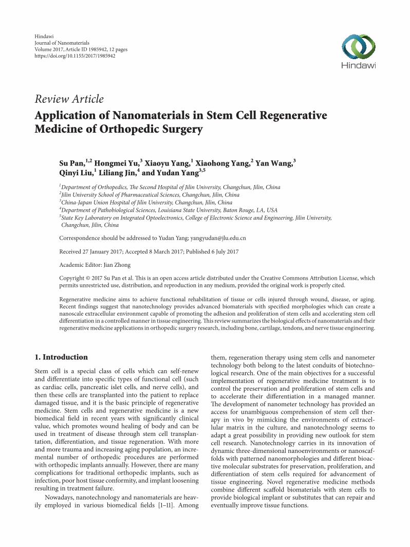
Figure 1: The application of nHA scaffolds in bone tissue engineering.
This review will discuss the biological effects of nanoma-terials and their regenerative medicine applications in ortho-pedic surgery research, including bone, cartilage, tendonstissue, and nerve tissue engineering.
2. Bone Tissue Engineering
Currently, bone compositions fabricated according to thefundamental of tissue engineering are being regarded as anideal selection for the functional restoration of segmentaldeossification. Biomaterials used for bone tissue engineeringinclude nanohydroxyapatite, titanium, calcium phosphate,graphene oxide, carbon nanotubes, or the compound of them[12–14]. The surfaces or three-dimensional nanostructures ofimplant which better mimic the naturalistic environments ofbone extracellular matrix in the culture, naturally nanocom-pound tissue, can promote the osteogenic differentiation ofstem cell which is important for bone tissue engineering.
2.1. Nanohydroxyapatite. Hydroxyapatite is the principalinorganic mineral component of animal and human teethand bones which is difficult to be dissolved in a solutionand to be utilized in various applications. If nanometer sizehydroxyapatite (nHA) particles can be made, it will exhibit arange of unique properties (Figure 1).
2.1.1. Effect on Biological Behavior of Stem Cells. Three dif-ferent shapes of nHAP/PLLA scaffolds were prepared, suchas needle structure, spherical structure, and rod structure,on which passaged-3 rat MSCs were cultured in three-dimensional scaffolds, and it was demonstrated that needlenHAP/PLLA scaffolds appeared to provide the most pro-portionate environment for bone regeneration using MSCs.It was demonstrated that a novelly fabricated eri-tasar silkfibroin nanofibrous scaffold (ET) scaffold hybrided with nHAdeposition by a surface precipitation method stimulatedosteogenic differentiation in the absence of any growth fac-tors due to the improved properties of it in physical chemistryand biology. It may offer effectual and profitable biomaterialsfor bone tissue engineering in orthopedic surgery by promot-ing stem cell differentiation on ET/nHAP scaffold withoutany growth factor additive in stem cell culture [15]. A fibrousstructure and synergistic action of collagen and nHA with
highmolecular weight poly-L-lactide (PLLA) played a crucialrole in stimulating human mesenchymal stem cells (hMSCs)differentiation towards osteogenic lineages, indicating itspossibility for bone defect repair based on stem cell therapies[16].
Conventional biphasic calcium phosphate (BCP) ceram-ics with porous structure, displaying superior bone conduc-tion and biocompatibility without intrinsically osteoinduc-tive, were coated with nHA due to supernal osteoinductivepotentiality of it. The new nHA coating BCP scaffolds weredemonstrated to be more suitable for the adhesion andproliferation of stem cell andMSCs osteogenic differentiationas compared with the conventional BCP scaffolds, indicatingthe property of nHA conducive for osteoinduction makingthis scaffolds more adaptive for applications in bone regener-ation [17]. The proliferation, osteogenic phenotypic markersexpression, and mineral deposition were elevated whenhMSCswere seeded on a new three-dimensional, porous poly(D,L-lactide-co-glycolide) (PLAGA)/nHA scaffold suitablefor high-aspect ratio vessel (HARV) bioreactor applications,and the degradation pattern of the scaffold was not altered bynHA incorporation and maintained its mechanical integrityfor 6 weeks in the dynamic culture environment, which canbe used as a potential tissue engineering matrix allowing forthe generation of bone tissue [18]. Upon culturing hMSCson a bioactive nanofiber scaffold made of poly-caprolactone(PCL) and nHA, it was demonstrated that the mineral phaseexistence increased alkaline phosphatase (ALP) activity andmRNAexpression levels of genes related to osteoblast withoutany osteogenous composition, and it was suggested that thearchitecture of this novel nanofibrous scaffolds and its chem-ical constituent could regulate hMSCs osteogenic differen-tiation [19]. In conclusion, nanofibrous scaffolds composedof nHA manufactured to different shape or hybrided withvarious nanomaterials will have favourable effect on bio-logical behavior of stem cells and then enhance osteoin-ductive potential of it by some molecular biology mecha-nism.
2.1.2. Skeletal Defects Repair. Successful clinical repair ofnonhealing bone defects relies on artificial bone with robustefficiency of osteoinduction and outstanding biomechanicalstability. Allogenic BMSCs seeded on the nHA/polyamide
Journal of Nanomaterials 3
6 (nHA/PA6) composite scaffolds used as porous matriceswere implanted in the 8mm diameter calvarial defect ofrats, and this showed good biocompatibility and osteoin-duction which enhanced bone inductivity at the initialstage after implantation [20]. A composite scaffold com-posed of nHA-type I collagen beads, BMSCs, and plateletrich plasma (PRP) enhanced significantly the new boneformation which could generate transplants with effectivelymeliorative bone tissue reorganization promising skeletaldefects restoration [21]. It was used to repair success-fully the nodulated skeletal defectiveness by eight weeksafter operation of novel nHA/collagen/PLLA/chitin fibers(nHACP/CF) scaffolds with goat bone mesenchymal stemcells (BMSCs) cultured and autograft bone. Furthermore, theresults revealed that the level of ALP andDNA in goat BMSCscultured on it was markedly increased. It was demonstratedthat this novel scaffold could be a proper graft for bonedefects repair [22]. In conclusion, in cases of large bonedefects resulting from trauma, infection, tumors, osteomyeli-tis surgical debridement, and a variety of congenital dis-eases, tissue-engineered structures may provide alternativesto traditional bone substitute by culturing autogeneic stemcells of patients with nHA composite scaffolds in culture sys-tem.
2.1.3. Internal Fixation. Bone plate set and screws for bonefractures therapy by internal fixation methods showed excel-lent biocompatibility and biomechanical properties. ThenHA/polyamide 66/glass fiber compoundwith good biocom-patibility and biomechanical properties was demonstrated toenhance the attachment and proliferation of MSCs withoutany negative effect on the mineralization of matrix andMSCs osteogenesis. This new compound might be used foroperations by internal fixation [23].
2.1.4. Spinal Fusion. Spinal fusion surgery performed withan autologous osseous graft obtained from the crest of iliumis a conventional treatment in therapy spondylolisthesis inchildren or lumbago derived from intervertebral discs degen-eration. However, the procedure is foregone to have severalshortcomings, such as nearthrosis, ache, or the adversereactions of blood transfusion. It has been demonstratedthat a novel graft material comprised of a new matrix ofmineralized collagen and nHA integrated with autogeneicadipose-derived mesenchymal stem cells (ADMSCs) hadeffective impact on rabbit posterolateral spinal fusion [24].
In conclusion, nanohydroxyapatite scaffold has bothnanomaterials properties and good biocompatibility whichrender it a very broad application prospect in bone regener-ative medicine, including the regulation of stem cell biolog-ical behavior (such as proliferation and differentiation), theimprovement of bone tissue regeneration promising skeletaldefects repair, internal fixation surgery, and spinal fusion inorthopedic surgery.
2.2. Ti. Osteogenic cells belong to anchorage-dependentcells in which the increase of adhesion and cytoskeletonchanges in early stage is imperative to stimulate osteogenesisand to accomplish a more expedient osseointegration of
implant materials surfaces in vivo. Metallic implant materialsincluding titanium and its alloys are conventionally usedin orthopedic applications due to their superior mechanicalfunctions and biological unreactiveness.
2.2.1. Effect of Topography and Measurement. It was pre-sumed that the original progenitor cells morphology on thenanoscale surface topographymight have effect on the succe-dent stem cell functions [25].
Stemcellswere cultured onto titaniumdiscswith differentstructures, such as sandblasted, sandblasted and large-gritacid etched, and full contact coverage (FCC). The resultsshowed that the FCC titanium surface on which osteoblast-like cells matured most rapidly might have a crucial effecton the primeval phase of bone union course [26]. Differentmicro- and nanostructure of a new nanosized oxidized tita-nium graft enhanced the adhesion and osteogenic differentia-tion of BMSC plated onto the oxidative surfaces which exhib-ited a greater tanglesome micro- and nanosized structuresand had increased levels of coarseness parameters contrastingto the convention alone [27]. Titanium surface features werecontrolled as subnano-, nano-, and submicron scales whichcould activate integrin-ligand proteins interactions, and thenthe results showed that osteoblast differentiation of primarymouse BMSC was accelerated significantly only on thesurface of titanium with nano- and submicron hybrid after2 weeks. Furthermore rapid cytoskeletal reorganization ofBMSC on the transparent surface of titanium with nanoscalewas demonstrated, which definitively induced increasedgenes expression level of osteoblast dominant form after threeweeks [28]. It was observed that oxidized titanium nanotubeswith a diameter of 70 nm was the optimal dimension forthe osteogenic differentiation in adipose-derived stem cellsof human (hASCs). Furthermore the results revealed that thenanotopography of oxidized titanium nanotubes directs stemdestiny by upregulating methylation level of histone H3 atlysine 4 in the promoter regions of osteogenic genes Runx2and osteocalcin, by inhibiting demethylase retinoblastomabinding protein [15, 29].
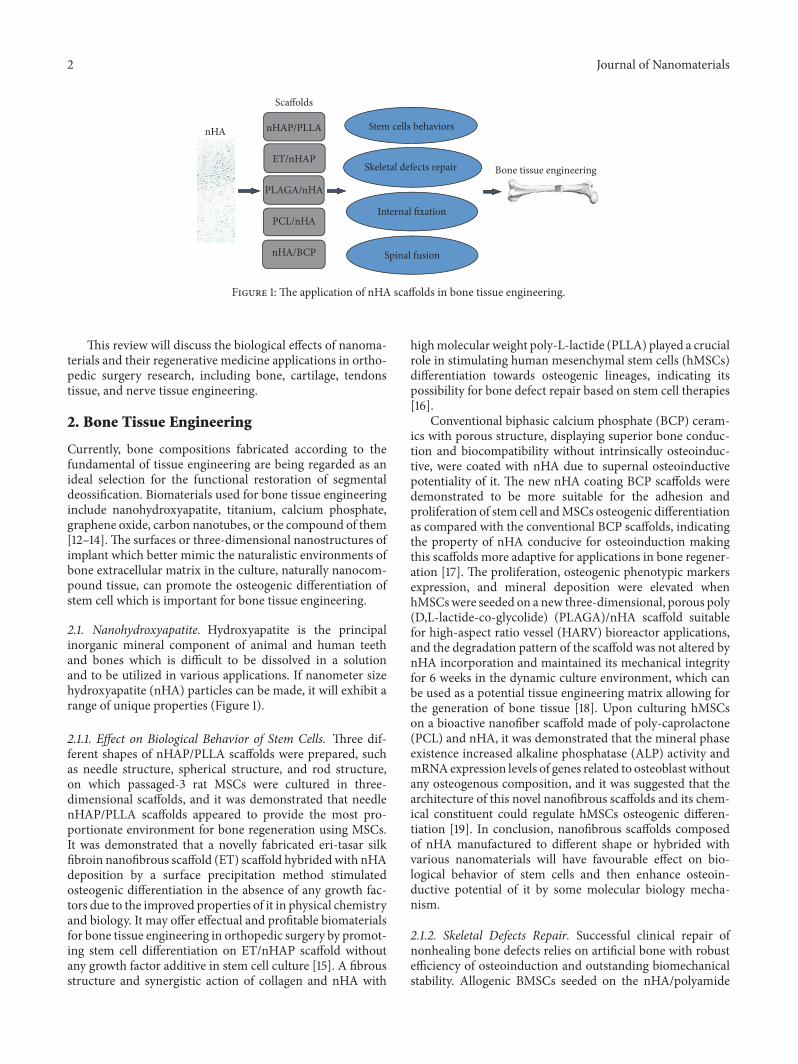
2.2.2. Titanium Surfaces Modification. Human bone ECM iscomprised of collagen nanofibers and nanostructured hy-droxyapatite particles in nanometer dimension improvingcells growth in its nanofibrous porous structure, so thebiomimetic and bioactive titanium surface with nanostruc-tured coating might control stem cell adhesion and osseoin-tegration (Figure 2). It was demonstrated that the adhesionand proliferation of osteoblast and MSCs were enhancedon titanium treated with nHA/carbon nanotubes [30]. Thestem cells cultured on titanium nanostructure coated withECM components exhibited faster osteoblastic differentia-tion and more efficient deposition of mineralized matrixas compared with uncoated structure. Heatedly oxidativeTi-6Al-4V coating with biomimetic chitosan/alginate filmcontaining nanoscale silver particles enhanced significantlythe morphology, viability, and proliferation of BMSC. Fur-thermore, the composite film restrained the multiplicationof Escherichia coli and Staphylococcus aureus and upregulatedsignificantly the expression of ALP [31].
4 Journal of Nanomaterials
Stem cell
nHA/carbon nanotubes
Chitosan/alginate film +nanoscale silver particles
Ti surfaces modification
ECM components
Osteoblastic differentiation
Osteoblastand MSCs
Mineralized matrix deposition
Adhesionproliferation
BMSC
AntibiosisALPMorphologyviabilityproliferation
Figure 2: The effect of titanium surfaces modification on stem cell.
Overall, these studies present a towardly stratagem toimprove biocompatibility and potential of these novel nanos-tructured materials for orthopedic applications.
2.3. Calcium Phosphate. Nanostructured calcium phosphate(CaP) biomaterials/scaffolds are of particular interest tomeet the increasing need for osteoanagenesis, because theyshare chemical/crystallographic analogies to inorganic com-ponents of bone. It was demonstrated that the MSCs prolif-eration was enhanced when cultured on the nanosized threemethods-biomimetic discs coated with CaP as comparedwith the uncoated or galvano-chemically coated structures[32]. Novel porous Cap-Ti6Al4V (Cap-Ti) hybrids withconspicuous physical and chemical properties regulated thepropagation, osteogenous differentiation, and matrix miner-alization of human periosteumderived cells (hPDCs), such asregulating osteoclast formation by CaP coating absorption ofhPDCs, decreasing gene expression level of osteoprotegerin,and promotingmultinucleated giant cells aggregation near tothe Cap-Ti hybrids surface. Furthermore, Cap-Ti compoundinduced heterotopic bone formation during hypodermicimplantation by a manner depending on cell density [33].
Calcium phosphate cement (CPC) possesses superiorosteoconductivity and it is easy to be assimilated or replacedby regenerative bone tissue, so CPC is suitable to be injectedin bone defect position to form powered supports. It wasdemonstrated that umbilical cord mesenchymal stem cells ofhuman (hUCMSCs) attaching to high-strength CPC showedsuperior propagation or osteogenous differentiation. Theresults revealed that hUCMSCs transported by CPCmight bea probable alternative in orthopedic treatment [34]. However,CPC is limited to non-stress-bearing repairs due to the lowstrength of it that can be reinforced by chitosan incorpora-tion. It was reported that MSCs differentiated to osteoblastscell lines and expressed ALP in high level which is a bonemarker on the CPC and chitosan scaffolds with increasedstrength as compared to CPC [35]. Magnesium phosphate
cement (MPC) was combined with CPC to develop novelcalcium-magnesium phosphate cement (CMPC) which wasimplanted into bone defects in rabbits. It was demonstratedthat CMPC has a shorter setting time and markedly bettermechanical properties than either CPC or MPC. Further-more, the histological evaluation showed that the introduc-tion of MPC into CPC enhanced the efficiency of new boneformation, and theCMPCalso exhibited good biocompatibil-ity, biodegradability, and osteoconductivity with host bone invivo [35].
2.4. Graphene. Polymeric material composed of graphene isdeveloped for biomedical applications, such as biosensing forthe increased electrical conductivity of the graphene compos-ite material. Furthermore, soft biomaterials can be reinforcedby graphene incorporated to polymer. Graphene oxide (GO)is a broadly investigated form of grapheme. GO is rich in oxy-gen, including functional groups, such as epoxide carboxyland hydroxyl groups. It is important for bone regenerationtherapy to control the propagation and differentiation of stemcells in a controlled manner. It was proved that osteogenicdifferentiation of stem cell is increased when cultured onmechanically stiff substrates. Graphene could provide atowardly biocompatible scaffold to accelerate the specificdifferentiation of hMSCs into bone cells without hamperingtheir proliferation [36]. GO flakes conjugated to collagensponges which were clinically approved scaffolds to providesoft microenvironment for bone regeneration increased thescaffold stiffness 3-fold without cytotoxicity and enhancedsignificantly osteogenic differentiation of hMSCs, so thisnovel 3D GO-collagen scaffolds could offer a novel platformfor stem cell study and bone regeneration [37]. PCL com-pound of GO, reducedGO (RGO), and amine-functionalizedGO (AGO) were prepared at various filling material compo-nents. The results demonstrated that AGO and GO particlessignificantly enhanced hMSCs proliferation, and AGO wasmost effectual inmodulating the osteogenic differentiation ofstem cell leading to mineralization [38].
Journal of Nanomaterials 5
15 kV ×1,000 10 �휇m 10 40 SEI
(a)
15 kV ×1,000 10 �휇m 10 40 SEI
(b)
15 kV ×1,000 10 �휇m SEI11 36
(c)
15 kV ×1,000 10 �휇m SEI11 36
(d)
15 kV ×1,000 10 �휇m SEI11 36
(e)
15 kV ×1,000 10 �휇m SEI11 36
(f)
15 kV ×1,000 10 �휇m SEI11 36
(g)
15 kV ×2,500 10 �휇m 10 SEI36
(h)
15 kV ×1,000 10 �휇m 10 SEI36
(i)
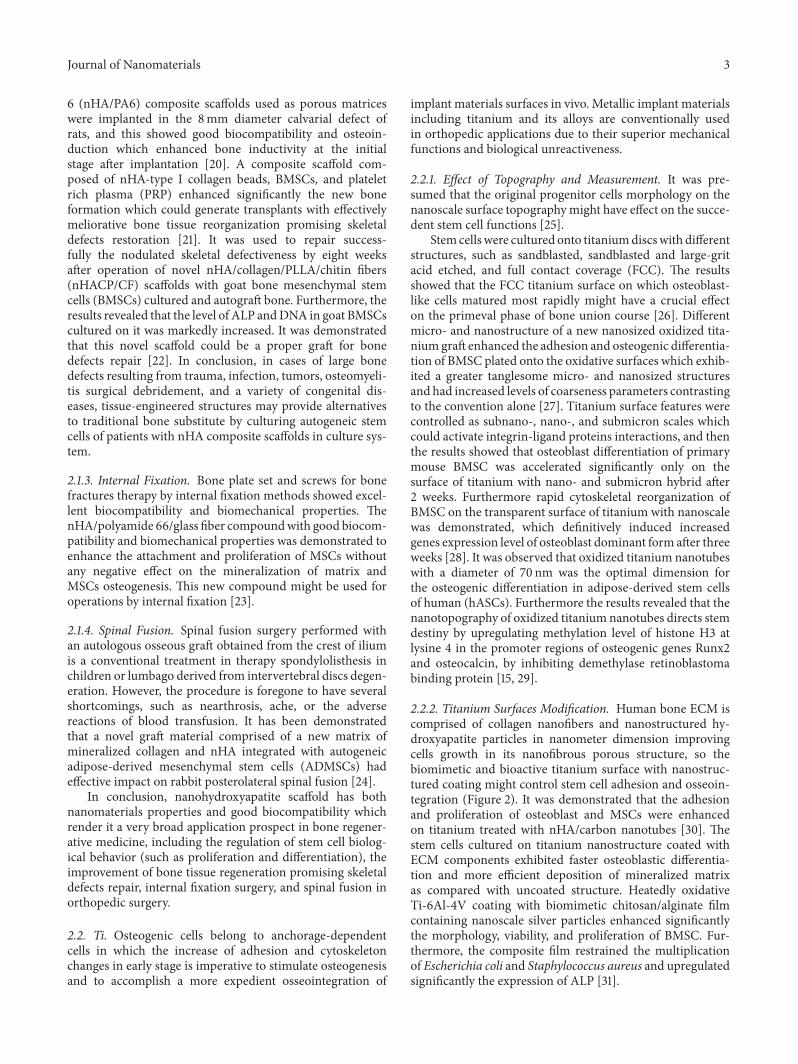
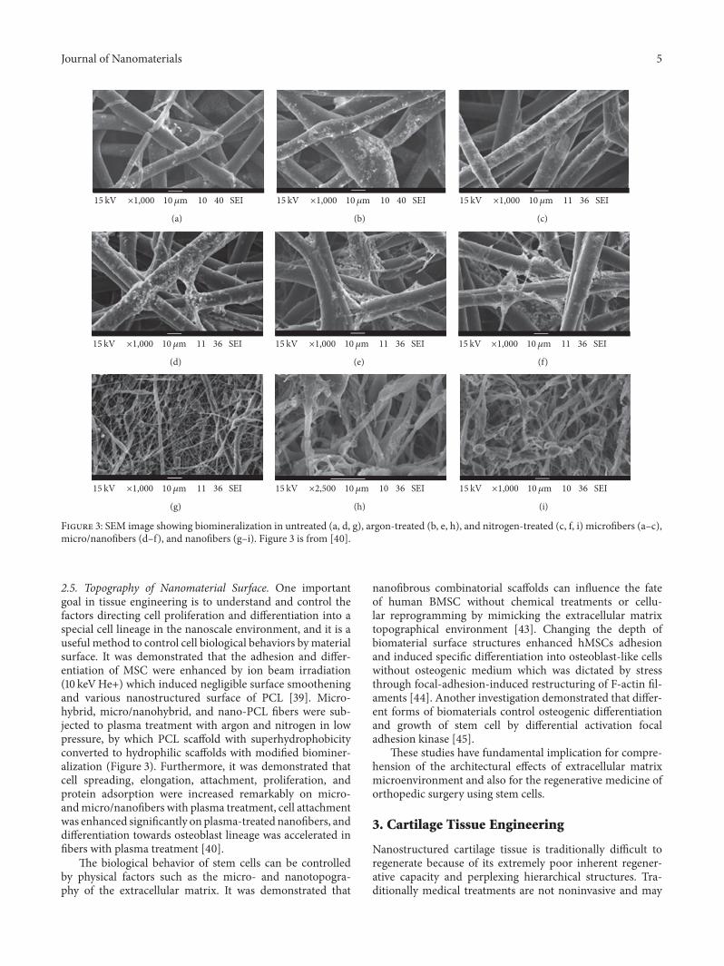
Figure 3: SEM image showing biomineralization in untreated (a, d, g), argon-treated (b, e, h), and nitrogen-treated (c, f, i) microfibers (a–c),micro/nanofibers (d–f), and nanofibers (g–i). Figure 3 is from [40].
2.5. Topography of Nanomaterial Surface. One importantgoal in tissue engineering is to understand and control thefactors directing cell proliferation and differentiation into aspecial cell lineage in the nanoscale environment, and it is auseful method to control cell biological behaviors bymaterialsurface. It was demonstrated that the adhesion and differ-entiation of MSC were enhanced by ion beam irradiation(10 keVHe+) which induced negligible surface smootheningand various nanostructured surface of PCL [39]. Micro-hybrid, micro/nanohybrid, and nano-PCL fibers were sub-jected to plasma treatment with argon and nitrogen in lowpressure, by which PCL scaffold with superhydrophobicityconverted to hydrophilic scaffolds with modified biominer-alization (Figure 3). Furthermore, it was demonstrated thatcell spreading, elongation, attachment, proliferation, andprotein adsorption were increased remarkably on micro-andmicro/nanofibers with plasma treatment, cell attachmentwas enhanced significantly on plasma-treated nanofibers, anddifferentiation towards osteoblast lineage was accelerated infibers with plasma treatment [40].
The biological behavior of stem cells can be controlledby physical factors such as the micro- and nanotopogra-phy of the extracellular matrix. It was demonstrated that
nanofibrous combinatorial scaffolds can influence the fateof human BMSC without chemical treatments or cellu-lar reprogramming by mimicking the extracellular matrixtopographical environment [43]. Changing the depth ofbiomaterial surface structures enhanced hMSCs adhesionand induced specific differentiation into osteoblast-like cellswithout osteogenic medium which was dictated by stressthrough focal-adhesion-induced restructuring of F-actin fil-aments [44]. Another investigation demonstrated that differ-ent forms of biomaterials control osteogenic differentiationand growth of stem cell by differential activation focaladhesion kinase [45].
These studies have fundamental implication for compre-hension of the architectural effects of extracellular matrixmicroenvironment and also for the regenerative medicine oforthopedic surgery using stem cells.
3. Cartilage Tissue Engineering
Nanostructured cartilage tissue is traditionally difficult toregenerate because of its extremely poor inherent regener-ative capacity and perplexing hierarchical structures. Tra-ditionally medical treatments are not noninvasive and may

6 Journal of Nanomaterials
cause many complications. Nanomaterials fabrication com-mitted to producing useful biologically scaffolds of tissue-engineered cartilage used to promote the chondrification ofstem cells. Several novel 3D biomimetic nanostructured scaf-folds were studied which was fabricated by PLLA polymersand multiwalled carbon nanotubes treated with hydrogen.The scaffolds embedded by multiwalled carbon nanotubesexhibited a vigorous enhancement in shatter strength with acompressed Young’s modulus accordant to human cartilage.It was demonstrated that MSCs was preferred to attach onsmaller fiber diameter structure and increasing chondrogenicdifferentiations were induced by combination of plasma-treated multiwalled carbon nanotubes and PLLA [46].
Another investigation demonstrated that the attachment,distribution, viability, and propagation of stem cells culturedin vitro in the 3D nHA coated poly(lactide-co-glycolide)scaffolds were increased as compared with poly(lactide-co-glycolide) one. Furthermore, it was demonstrated thatthe osteochondral defects of rats knees treated by three-dimensional poly(lactide-co-glycolide)/nHA scaffold com-bining with MSCs were stuffed with crystalline cartilage.Furthermore, there was affluent mucopolysaccharide andcollagen type II sedimentation after surgery. The results sug-gested that the poly(lactide-co-glycolide)/nHA-MSCs com-pound might have probable utilization in tissue-engineeredcartilage [47]. Traditionally, chondrogenic differentiation isaccomplished by stem cells culturing with chondrogenic fac-tors in the culture medium, such as the protein transforminggrowth factor-beta 3. However, cell-to-cell interaction anddiffusional limitation of transforming growth factor-beta 3mainly provided by pellets may restrain stem cells differenti-ation to restrain. It was demonstrated that a novel GO couldbe used as substrates for adhesion of stem cells and deliveredgrowth factor during the stem cell differentiation to cartilage.GO used to adsorb fibronectin and transforming growthfactor-beta 3 was incorporated in pellets of hASCs to obtain ahybrid pellets of hASC-GO sheets (size = 0.5–1mum) whichpromoted the hASCs differentiation to cartilage throughincreasing the interaction of cell with fibronectin and provid-ing transforming growth factor-beta 3 efficiently. The novelhASC-GO sheets may offer novel methods for hASC cultureto obtain tissue-engineered cartilage [48].
4. Tendons Tissue Engineering
The reconstructive articulation usually tends to fail at thetendon-to-bone insertion site, so an orthobiologic material isformidably required to provide a stable transition from hardbone to soft ligament tissue.
A nanostructured material using PLLA mineralized withhydroxyapatite and magnesium nanoparticles was developedto regenerate the tendon-to-bone insertion site which wasfashioned in the shape of an o-ring to recover functionality toinjured entheses, the graded transition of mineralized fibrouscartilage connecting ligament to bone. Tendon stem cells areuseful in effectual restoration or regeneration of woundedtendons.The destiny of tendon stem cells is not unambiguousduring the implantation. It was demonstrated that taggingtendon stem cells with super-paramagnetic iron oxide (SPIO)
nanomaterials was a practicable method for tracing ten-don stem cells used to determine the feasibility of it. Thisnovel SPIO labeled tendon stem cells provide a noninvasiveapproach to wounded tendons observe restoration [49].
5. Nerve Tissue Engineering
Spinal cord injury is defined as the direct or indirect factorsthat cause spinal cord damage, in the corresponding segmentof the injury, associated with motor, sensory and sphincterdysfunction, muscle tension abnormalities and pathologicalchanges, and so on. The extent and clinical manifestationsof spinal cord injury depend on the location and nature ofthe primary injury. Spinal cord injury is the most seriouscomplication of spinal injury, which often leads to severedysfunction of the injured segment following limb. Infection,tumor, spinal degenerative disease, scoliosis, myeloschisis,spondylolisthesis, and various types of spinal fractures anddislocations caused by indirect external forces can lead tospinal cord injury. The nerve repair of spinal cord injury isa difficult and important problem in regenerative medicine,andmore andmore research is used to repair the nerve alongwith the depth of people’s understanding in the nerve biologyand material science.
5.1. Effect of Surface Topography. Conclusive extracellularenvironment has a crucial impact in cell life by regulationof cellmorphology, proliferation, survival, and differentiationinto a specific cell line. A main restraint in the clinical appli-cation of stem cell technique is the lack of available regulateof its biological behavior, such as attachment, multiplication,and differentiation. Current in vitro studies have indicatedthat biological materials with nanosurfaces morphology canaffect cell behavior. Thus, confirmation of biological mate-rials supporting adequate ES cell adhesion, proliferation,and differentiation into special cell linage is a fascinatingmeasure to investigate. Current medicinal cell therapy, usingthe special ability of stem cells in differentiating to nervecorpuscles which is loaded by polymer materials, focuses onthe restoration of the central nervous system damage.
A thin film of gold with different roughness of surfacemorphology was fabricated by the conjugation of microfab-rication technology. Then the biomimetic possibility of thethin film was investigated, compared with glass coverslipsand plastic tissue culture materials, for regulating the dif-ferentiation of neural precursor derived from ES cell. It wasdemonstrated that the neural precursors derived from EScells which were cultured for five days had the best adhesionon the gold films and were carried out with the maximumdifferentiation on gold films with surface roughness ofstandard deviation 21 nm without any conventional diffluentneurotrophic factor. In addition, the influence of the gratingaxis on the axons growth direction is observed when theneural precursors derived from ES cell were seeded in aconjunction of microscale trenches and nanoscale surfaceroughness. These data suggest that biological material designcan maximize the differentiation and organization of neuralprecursors derived from ES cells, and it can be found to beuseful as a synergistic supplement of diffluent neurotrophic
Journal of Nanomaterials 7
2D 3D
ES cell
Aligned nanofibrousscaffolds
Gold films with surface roughness
AdhesionProliferationDifferentiation
Fibrin gelscaffold
Porous polymer film with honeycomb structure
Matrigel nanostructure
SurvivalProliferationNeuron outgrowth
Nerve tissue engineering
Spinal cord injury
Tumor
Myeloschisis
Spinal degenerative disease
Scoliosis
Spondylolisthesis
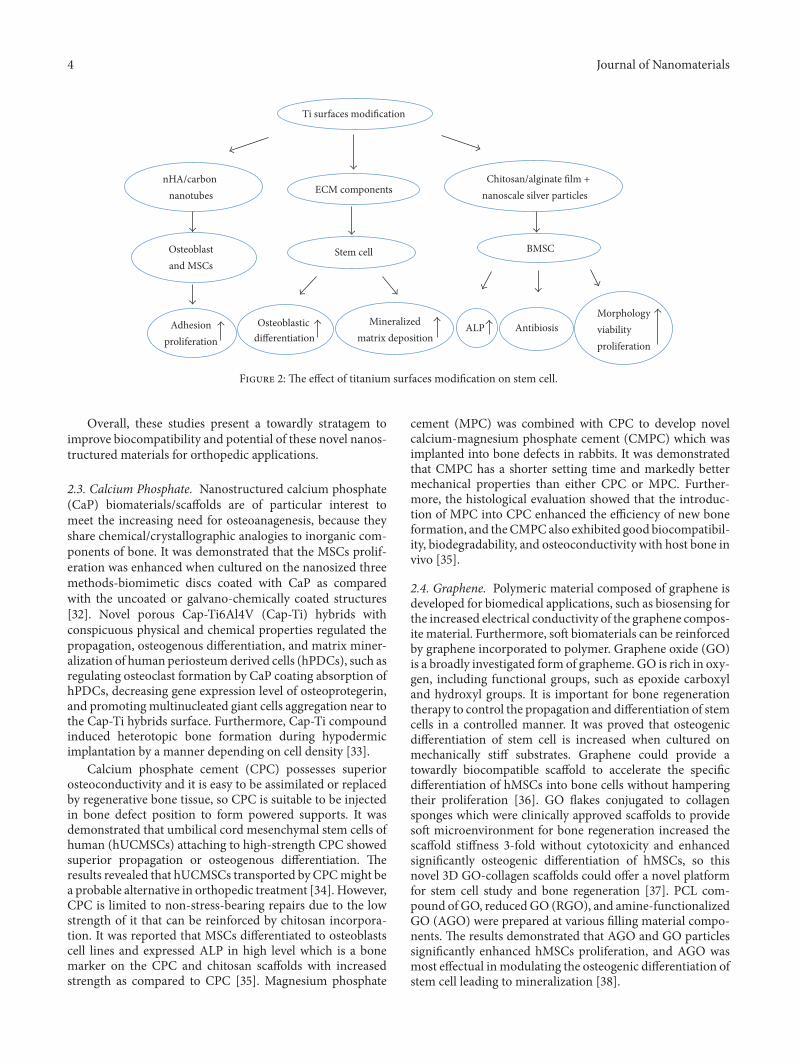
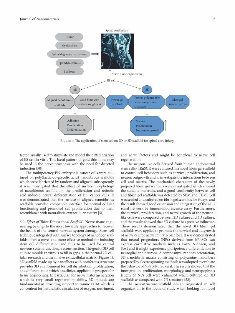
Figure 4: The application of stem cell on 2D or 3D scaffold for spinal cord injury.
factor usually used to stimulate andmodel the differentiationof ES cell in vitro. This basal pattern of gold thin films maybe used in the nerve prosthesis with the need for directedinduction [50].
The multipotency P19 embryonic cancer cells were cul-tured on poly(lactic-co-glycolic acid) nanofibrous scaffoldswhich were fabricated by random and aligned; subsequentlyit was investigated that the effect of surface morphologyof nanofibrous scaffold on the proliferation and retinoicacid induced neural differentiation of P19 cancer cells. Itwas demonstrated that the surface of aligned nanofibrousscaffolds provided compatible interface for normal cellularfunctioning and promoted cell proliferation due to theirresemblance with naturalistic extracellular matrix [51].
5.2. Effect of Three-Dimensional Scaffold. Nerve tissue engi-neering belongs to the most towardly approaches to recoverthe health of the central nervous system damage. Stem celltechnique integrated with surface topology of nanofiber scaf-folds offers a novel and more effective method for inducingstem cell differentiation and thus to be used for centralnervous system functional reconstruction.The goal of 3D cellculture moulds in vitro is to fill in gaps in the normal 2D cel-lular research and the in vivo extracellular matrix (Figure 4).3D scaffold made up by nanofibers with poriferous structureprovides 3D environment for cell distribution, proliferation,and differentiationwhich has clinical application prospect fortissue engineering. In particular, for nerve historegenerationwhich is very small regeneration ability, 3D moulds arefundamental in providing support to mimic ECM which isconvenient for naturalistic circulation of oxygen, nutriment,
and nerve factors and might be beneficial to nerve cellregeneration.
The neuron-like cells derived from human endometrialstem cells (hEnSCs)were cultured in a novel fibrin gel scaffoldto control cell behaviors such as survival, proliferation, andneuron outgrowth and to investigate the interactions betweencell and matrix. The mechanical characters of the newlyprepared fibrin gel scaffolds were investigated which showedthe suitable materials, and a good conformity between celland fibrin gel scaffolds was detected by SEM and TEM. Cellwas seeded and cultured on fibrin gel scaffolds for 6 days, andthe result showed good expansion and integration of the neu-ronal network by immunofluorescence assay. Furthermore,the survival, proliferation, and nerve growth of the neuron-like cells were compared between 2D culture and 3D culture,and the results showed that 3D culture has positive influence.These results demonstrated that the novel 3D fibrin gelscaffolds were applied to promote the survival and outgrowthof nerve cell for nerve injury repair [52]. It was demonstratedthat neural progenitors (NPs) derived from hEnSCs canexpress correlative markers such as Pax6, Nidogen, andSox1 and it might experience pluripotency differentiation toneuroglial and neurons. A compositive, random orientation,3D nanofibrils matrix consisting of polyamine nanofibersprepared by electrospinningmethodswas adopted to evaluatethe behavior ofNPs cultured on it.The results showed that theimmigration, proliferation, morphology, and neurapophysislength of NPs cell were enhanced when cultured on 3Dscaffolds as compared with 2D structure [53].
The nanostructure scaffold design originated in selforganization is the focus of study when looking for novel
8 Journal of Nanomaterials
RADA 16-BMHP1 RADA 16-BMHP2 RADA 16-RGD RADA 16 Control
100 �휇m
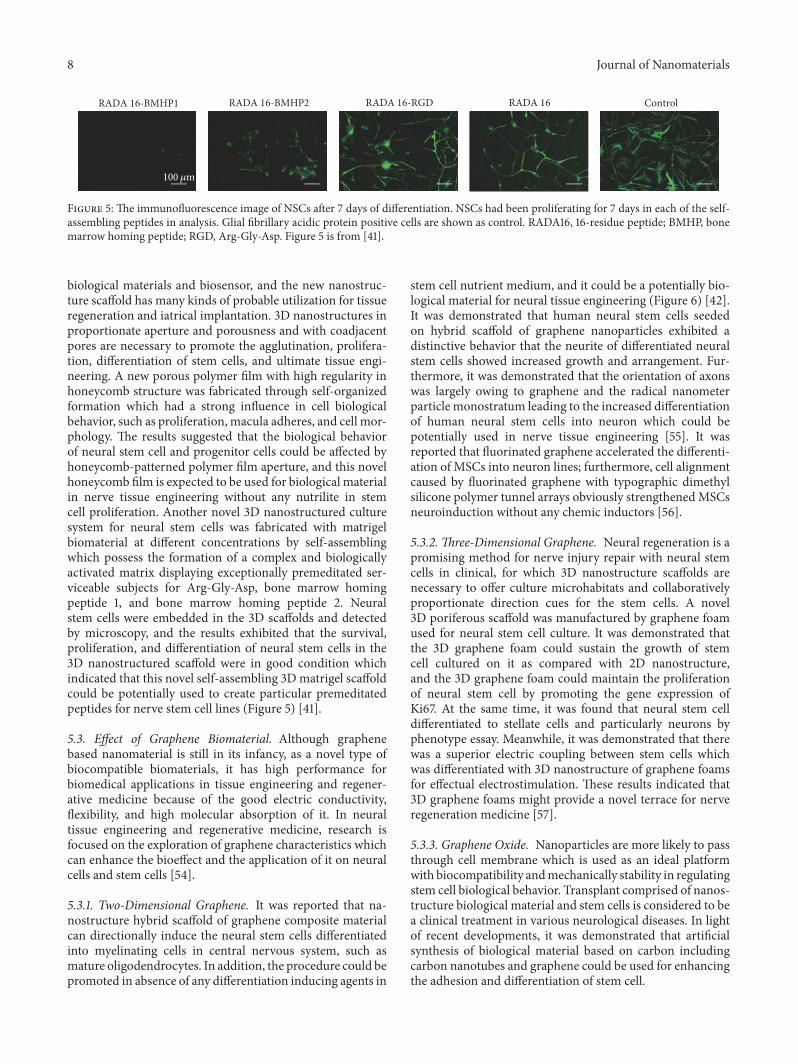
Figure 5: The immunofluorescence image of NSCs after 7 days of differentiation. NSCs had been proliferating for 7 days in each of the self-assembling peptides in analysis. Glial fibrillary acidic protein positive cells are shown as control. RADA16, 16-residue peptide; BMHP, bonemarrow homing peptide; RGD, Arg-Gly-Asp. Figure 5 is from [41].
biological materials and biosensor, and the new nanostruc-ture scaffold has many kinds of probable utilization for tissueregeneration and iatrical implantation. 3D nanostructures inproportionate aperture and porousness and with coadjacentpores are necessary to promote the agglutination, prolifera-tion, differentiation of stem cells, and ultimate tissue engi-neering. A new porous polymer film with high regularity inhoneycomb structure was fabricated through self-organizedformation which had a strong influence in cell biologicalbehavior, such as proliferation, macula adheres, and cell mor-phology. The results suggested that the biological behaviorof neural stem cell and progenitor cells could be affected byhoneycomb-patterned polymer film aperture, and this novelhoneycomb film is expected to be used for biological materialin nerve tissue engineering without any nutrilite in stemcell proliferation. Another novel 3D nanostructured culturesystem for neural stem cells was fabricated with matrigelbiomaterial at different concentrations by self-assemblingwhich possess the formation of a complex and biologicallyactivated matrix displaying exceptionally premeditated ser-viceable subjects for Arg-Gly-Asp, bone marrow homingpeptide 1, and bone marrow homing peptide 2. Neuralstem cells were embedded in the 3D scaffolds and detectedby microscopy, and the results exhibited that the survival,proliferation, and differentiation of neural stem cells in the3D nanostructured scaffold were in good condition whichindicated that this novel self-assembling 3Dmatrigel scaffoldcould be potentially used to create particular premeditatedpeptides for nerve stem cell lines (Figure 5) [41].
5.3. Effect of Graphene Biomaterial. Although graphenebased nanomaterial is still in its infancy, as a novel type ofbiocompatible biomaterials, it has high performance forbiomedical applications in tissue engineering and regener-ative medicine because of the good electric conductivity,flexibility, and high molecular absorption of it. In neuraltissue engineering and regenerative medicine, research isfocused on the exploration of graphene characteristics whichcan enhance the bioeffect and the application of it on neuralcells and stem cells [54].
5.3.1. Two-Dimensional Graphene. It was reported that na-nostructure hybrid scaffold of graphene composite materialcan directionally induce the neural stem cells differentiatedinto myelinating cells in central nervous system, such asmature oligodendrocytes. In addition, the procedure could bepromoted in absence of any differentiation inducing agents in
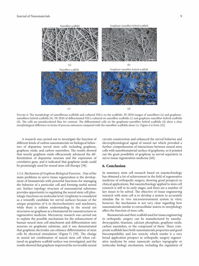
stem cell nutrient medium, and it could be a potentially bio-logical material for neural tissue engineering (Figure 6) [42].It was demonstrated that human neural stem cells seededon hybrid scaffold of graphene nanoparticles exhibited adistinctive behavior that the neurite of differentiated neuralstem cells showed increased growth and arrangement. Fur-thermore, it was demonstrated that the orientation of axonswas largely owing to graphene and the radical nanometerparticle monostratum leading to the increased differentiationof human neural stem cells into neuron which could bepotentially used in nerve tissue engineering [55]. It wasreported that fluorinated graphene accelerated the differenti-ation of MSCs into neuron lines; furthermore, cell alignmentcaused by fluorinated graphene with typographic dimethylsilicone polymer tunnel arrays obviously strengthenedMSCsneuroinduction without any chemic inductors [56].
5.3.2. Three-Dimensional Graphene. Neural regeneration is apromising method for nerve injury repair with neural stemcells in clinical, for which 3D nanostructure scaffolds arenecessary to offer culture microhabitats and collaborativelyproportionate direction cues for the stem cells. A novel3D poriferous scaffold was manufactured by graphene foamused for neural stem cell culture. It was demonstrated thatthe 3D graphene foam could sustain the growth of stemcell cultured on it as compared with 2D nanostructure,and the 3D graphene foam could maintain the proliferationof neural stem cell by promoting the gene expression ofKi67. At the same time, it was found that neural stem celldifferentiated to stellate cells and particularly neurons byphenotype essay. Meanwhile, it was demonstrated that therewas a superior electric coupling between stem cells whichwas differentiated with 3D nanostructure of graphene foamsfor effectual electrostimulation. These results indicated that3D graphene foams might provide a novel terrace for nerveregeneration medicine [57].
5.3.3. Graphene Oxide. Nanoparticles are more likely to passthrough cell membrane which is used as an ideal platformwith biocompatibility andmechanically stability in regulatingstem cell biological behavior. Transplant comprised of nanos-tructure biological material and stem cells is considered to bea clinical treatment in various neurological diseases. In lightof recent developments, it was demonstrated that artificialsynthesis of biological material based on carbon includingcarbon nanotubes and graphene could be used for enhancingthe adhesion and differentiation of stem cell.
Journal of Nanomaterials 9
Nanofiber scaffold
(a)
Graphene-nanofiber hybrid scaffold
(b)
Nanofiber scaffold
(c)
Graphene-nanofiber hybrid scaffold
(d)
Figure 6: The morphology of nanofibrous scaffolds and cultured NSCs on the scaffolds. FE-SEM images of nanofibers (a) and graphene-nanofibers hybrid scaffolds (b). FE-SEM of differentiated NSCs cultured on nanofiber scaffolds (c) and graphene-nanofiber hybrid scaffolds(d). The cells are pseudocolored blue for contrast. The differentiated cells on the grapheme-nanofiber hybrid scaffolds (d) show a clearmorphological difference in terms of process extension compared with the nanofiber scaffolds alone (c). Figure 6 is from [42].
A research was carried out to investigate the function ofdifferent kinds of carbon nanomaterials on biological behav-iors of dopamine nerval stem cells including graphene,graphene oxide, and carbon nanotubes. The results showedthat merely graphene oxide efficaciously advanced the dif-ferentiation of dopamine neurons and the expression ofcorrelative gene, and it indicated that graphene oxide couldbe promisingly used for neural stem cell therapy [58].
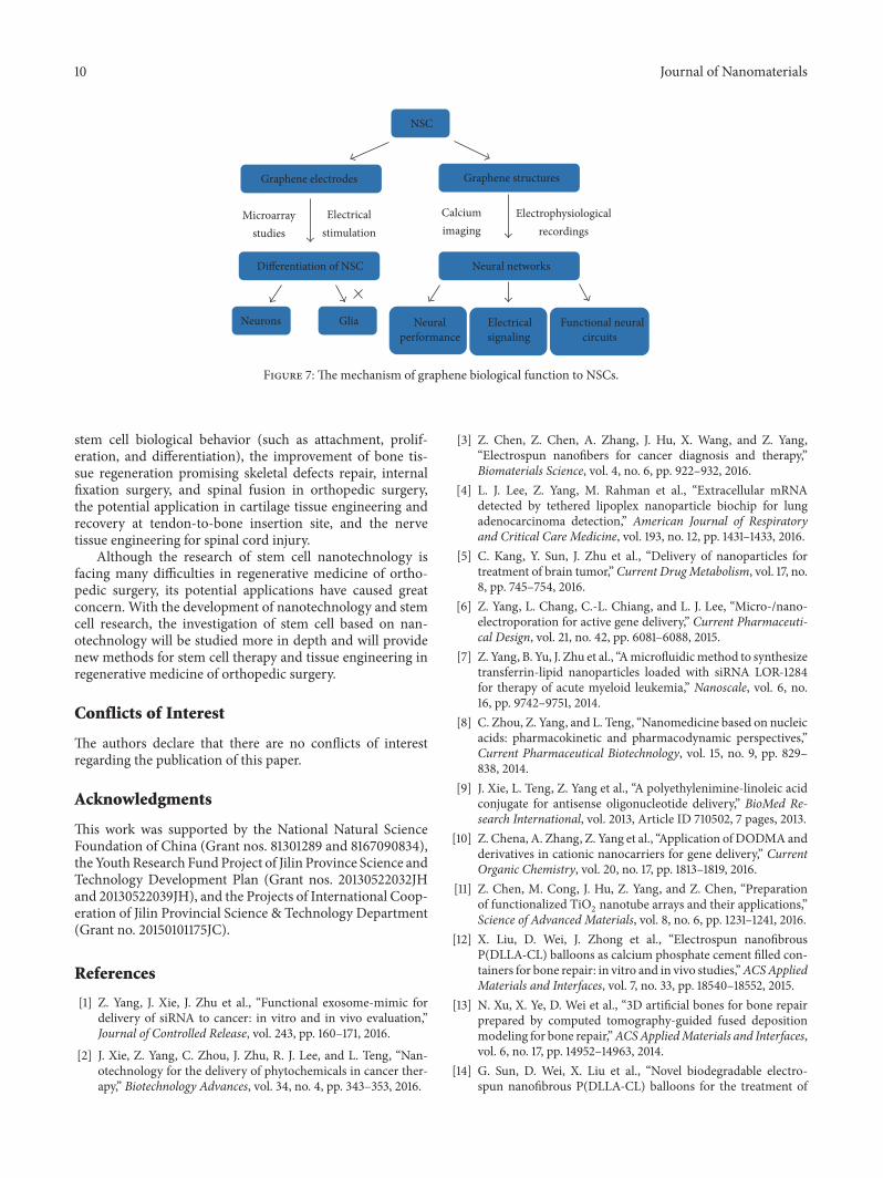
5.3.4.Mechanism of Graphene Biological Function. One of themain problems in nerve tissue regeneration is the develop-ment of biomaterials with powerful functions for managingthe behavior of a particular cell and forming useful neuralnet. Surface topology structure of nanomaterial substratesprovides opportunity to regulating the neural stem cell phys-iologic functions onmolecular level. Grapheme is consideredas a towardly candidate for nerval surfaces because of theunique properties of it in electrochemistry and machinery,while there is seldom understanding in the neural netstructures on graphene as a biomaterial for nanostructures inregenerative medicine. Microarray research was carried outto explain the possible mechanism for the enhancement ofhuman neural stem cell attachment and differentiation intoneurons on grapheme substrate, and it was demonstratedthat graphene electrodes can enhance differentiation of stemcells by electrical stimulation (Figure 7) [59]. The changein neural net combination of neural stem cell when cul-tured on graphene scaffold surface was investigated, and theresults showed that graphene improved the serviceable neural
circuits construction and enhanced the nerval behavior andelectrophysiological signal of neural net which provided afurther comprehension of interactions between neural stemcells with nanobiomaterial surface of grapheme, so it pointedout the great possibility of graphene as nerval separatrix innerve tissue regeneration medicine [60].
6. Conclusion
In summary, stem cell research based on nanotechnologyhas obtained a lot of achievement in the field of regenerativemedicine of orthopedic surgery, showing good prospects inclinical applications. But nanotechnology applied to stem cellresearch is still in its early stages, and there are a number ofkey issues to be solved. The objective of tissue engineeringresearch with stem cell is to develop a system to accuratelysimulate the in vivo microenvironment system in vitro;however, the mechanism is not very clear regarding hownanomaterials similar to extracellular matrix in morphologyaffect the function of stem cells.
Biomaterials and their scaffold used for tissue engineeringin orthopedic surgery can be manufactured by nanohy-droxyapatite, titanium, calcium phosphate, graphene oxide,carbon nanotubes, or the compound of them. These com-posite scaffolds have both nanomaterials properties and goodbiocompatibility and low toxicity which render it a verybroad application prospect in orthopedic surgery regener-ative medicine by some nanoscale surface topography ormolecular biology mechanism, including the regulation of
10 Journal of Nanomaterials
NSC
Graphene electrodes Graphene structures
Microarraystudies
Electricalstimulation
Differentiation of NSC
Calciumimaging
Electrophysiologicalrecordings
GliaNeurons
Neural networks
Neural performance
Electricalsignaling
Functional neuralcircuits
Figure 7: The mechanism of graphene biological function to NSCs.
stem cell biological behavior (such as attachment, prolif-eration, and differentiation), the improvement of bone tis-sue regeneration promising skeletal defects repair, internalfixation surgery, and spinal fusion in orthopedic surgery,the potential application in cartilage tissue engineering andrecovery at tendon-to-bone insertion site, and the nervetissue engineering for spinal cord injury.
Although the research of stem cell nanotechnology isfacing many difficulties in regenerative medicine of ortho-pedic surgery, its potential applications have caused greatconcern. With the development of nanotechnology and stemcell research, the investigation of stem cell based on nan-otechnology will be studied more in depth and will providenew methods for stem cell therapy and tissue engineering inregenerative medicine of orthopedic surgery.
Conflicts of Interest
The authors declare that there are no conflicts of interestregarding the publication of this paper.
Acknowledgments
This work was supported by the National Natural ScienceFoundation of China (Grant nos. 81301289 and 8167090834),the YouthResearch FundProject of Jilin Province Science andTechnology Development Plan (Grant nos. 20130522032JHand 20130522039JH), and the Projects of International Coop-eration of Jilin Provincial Science & Technology Department(Grant no. 20150101175JC).
References
[1] Z. Yang, J. Xie, J. Zhu et al., “Functional exosome-mimic fordelivery of siRNA to cancer: in vitro and in vivo evaluation,”Journal of Controlled Release, vol. 243, pp. 160–171, 2016.
[2] J. Xie, Z. Yang, C. Zhou, J. Zhu, R. J. Lee, and L. Teng, “Nan-otechnology for the delivery of phytochemicals in cancer ther-apy,” Biotechnology Advances, vol. 34, no. 4, pp. 343–353, 2016.
[3] Z. Chen, Z. Chen, A. Zhang, J. Hu, X. Wang, and Z. Yang,“Electrospun nanofibers for cancer diagnosis and therapy,”Biomaterials Science, vol. 4, no. 6, pp. 922–932, 2016.
[4] L. J. Lee, Z. Yang, M. Rahman et al., “Extracellular mRNAdetected by tethered lipoplex nanoparticle biochip for lungadenocarcinoma detection,” American Journal of Respiratoryand Critical Care Medicine, vol. 193, no. 12, pp. 1431–1433, 2016.
[5] C. Kang, Y. Sun, J. Zhu et al., “Delivery of nanoparticles fortreatment of brain tumor,” Current DrugMetabolism, vol. 17, no.8, pp. 745–754, 2016.
[6] Z. Yang, L. Chang, C.-L. Chiang, and L. J. Lee, “Micro-/nano-electroporation for active gene delivery,” Current Pharmaceuti-cal Design, vol. 21, no. 42, pp. 6081–6088, 2015.
[7] Z. Yang, B. Yu, J. Zhu et al., “Amicrofluidicmethod to synthesizetransferrin-lipid nanoparticles loaded with siRNA LOR-1284for therapy of acute myeloid leukemia,” Nanoscale, vol. 6, no.16, pp. 9742–9751, 2014.
[8] C. Zhou, Z. Yang, and L. Teng, “Nanomedicine based on nucleicacids: pharmacokinetic and pharmacodynamic perspectives,”Current Pharmaceutical Biotechnology, vol. 15, no. 9, pp. 829–838, 2014.
[9] J. Xie, L. Teng, Z. Yang et al., “A polyethylenimine-linoleic acidconjugate for antisense oligonucleotide delivery,” BioMed Re-search International, vol. 2013, Article ID 710502, 7 pages, 2013.
[10] Z. Chena, A. Zhang, Z. Yang et al., “Application of DODMAandderivatives in cationic nanocarriers for gene delivery,” CurrentOrganic Chemistry, vol. 20, no. 17, pp. 1813–1819, 2016.
[11] Z. Chen, M. Cong, J. Hu, Z. Yang, and Z. Chen, “Preparationof functionalized TiO
2nanotube arrays and their applications,”
Science of Advanced Materials, vol. 8, no. 6, pp. 1231–1241, 2016.[12] X. Liu, D. Wei, J. Zhong et al., “Electrospun nanofibrous
P(DLLA-CL) balloons as calcium phosphate cement filled con-tainers for bone repair: in vitro and in vivo studies,”ACSAppliedMaterials and Interfaces, vol. 7, no. 33, pp. 18540–18552, 2015.
[13] N. Xu, X. Ye, D. Wei et al., “3D artificial bones for bone repairprepared by computed tomography-guided fused depositionmodeling for bone repair,”ACSAppliedMaterials and Interfaces,vol. 6, no. 17, pp. 14952–14963, 2014.
[14] G. Sun, D. Wei, X. Liu et al., “Novel biodegradable electro-spun nanofibrous P(DLLA-CL) balloons for the treatment of
Journal of Nanomaterials 11
vertebral compression fractures,” Nanomedicine: Nanotechnol-ogy, Biology, and Medicine, vol. 9, no. 6, pp. 829–838, 2013.
[15] N. Panda, A. Bissoyi, K. Pramanik, and A. Biswas, “Directingosteogenesis of stem cells with hydroxyapatite precipitatedelectrospun eri-tasar silk fibroin nanofibrous scaffold,” Journalof Biomaterials Science, Polymer Edition, vol. 25, no. 13, pp.1440–1457, 2014.
[16] H. R. A. Balaji Raghavendran, S. Puvaneswary, S. Talebian et al.,“A comparative study on in vitro osteogenic priming poten-tial of electron spun scaffold PLLA/HA/Col, PLLA/HA, andPLLA/Col for tissue engineering application,” PLoS ONE, vol.9, no. 8, Article ID e104389, 2014.
[17] J. Hu, Y. Zhou, L. Huang, J. Liu, and H. Lu, “Effect of nano-hydroxyapatite coating on the osteoinductivity of porous bipha-sic calcium phosphate ceramics,” BMC Musculoskeletal Disor-ders, vol. 15, no. 1, article 114, 2014.
[18] Q. Lv, M. Deng, B. D. Ulery, L. S. Nair, and C. T. Laurencin,“Nano-ceramic composite scaffolds for bioreactor-based boneengineering basic research,” Clinical Orthopaedics and RelatedResearch, vol. 471, no. 8, pp. 2422–2433, 2013.
[19] A. Polini, D. Pisignano, M. Parodi, R. Quarto, and S. Scaglione,“Osteoinduction of humanmesenchymal stem cells by bioactivecomposite scaffolds without supplemental osteogenic growthfactors,” PLoS ONE, vol. 6, no. 10, Article ID e26211, 2011.
[20] A. Khadka, J. Li, Y. Li, Y. Gao, Y. Zuo, and Y. Ma, “Evaluationof hybrid porous biomimetic nano-hydroxyapatite/polyamide6 and bone marrow-derived stem cell construct in repair ofcalvarial critical size defect,” Journal of Craniofacial Surgery, vol.22, no. 5, pp. 1852–1858, 2011.
[21] B.-N. Lin, S. W. Whu, C.-H. Chen et al., “Bone marrow mes-enchymal stem cells, platelet-rich plasma and nanohydroxyapa-tite-type I collagen beadswere integral parts of biomimetic bonesubstitutes for bone regeneration,” Journal of Tissue Engineeringand Regenerative Medicine, vol. 7, no. 11, pp. 841–854, 2013.
[22] X. Liu, X. Li, Y. Fan et al., “Repairing goat tibia segmentalbone defect using scaffold cultured with mesenchymal stemcells,” Journal of Biomedical Materials Research Part B: AppliedBiomaterials, vol. 94, no. 1, pp. 44–52, 2010.
[23] B. Qiao, J. Li, Q. Zhu et al., “Bone plate composed of aternary nano-hydroxyapatite/polyamide 66/glass fiber compos-ite: biomechanical properties and biocompatibility,” Interna-tional Journal of Nanomedicine, vol. 9, no. 1, pp. 1423–1432, 2014.
[24] Z.-B. Tang, J.-K. Cao, N. Wen et al., “Posterolateral spinalfusion with nano-hydroxyapatite-collagen/PLA composite andautologous adipose-derived mesenchymal stem cells in a rab-bit model,” Journal of Tissue Engineering and RegenerativeMedicine, vol. 6, no. 4, pp. 325–336, 2012.
[25] P. J. ter Brugge, R. Torensma, J. E. De Ruijter, C. G. Figdor, and J.A. Jansen, “Modulation of integrin expression on rat bone mar-row cells by substrates with different surface characteristics,”Tissue Engineering, vol. 8, no. 4, pp. 615–626, 2002.
[26] S. Tete, F. Mastrangelo, R. Quaresima et al., “Influence of novelnano-titanium implant surface on human osteoblast behaviorand growth,” Implant Dentistry, vol. 19, no. 6, pp. 520–531, 2010.
[27] M. Annunziata, A. Oliva, A. Buosciolo, M. Giordano, A. Guida,and L. Guida, “Bone marrow mesenchymal stem cell responseto nano-structured oxidized and turned titanium surfaces,”Clinical Oral Implants Research, vol. 23, no. 6, pp. 733–740, 2012.
[28] D. Khang, J. Choi, Y.-M. Im et al., “Role of subnano-, nano-and submicron-surface features on osteoblast differentiation ofbonemarrowmesenchymal stem cells,”Biomaterials, vol. 33, no.26, pp. 5997–6007, 2012.
[29] L. Lv, Y. Liu, P. Zhang et al., “The nanoscale geometry of TiO2nanotubes influences the osteogenic differentiation of humanadipose-derived stem cells by modulating H3K4 trimethyla-tion,” Biomaterials, vol. 39, pp. 193–205, 2015.
[30] M.Wang,N. J. Castro, J. Li,M.Keidar, and L.G. Zhang, “Greaterosteoblast and mesenchymal stem cell adhesion and prolifera-tion on titanium with hydrothermally treated nanocrystallinehydroxyapatite/magnetically treated carbon nanotubes,” Jour-nal of Nanoscience andNanotechnology, vol. 12, no. 10, pp. 7692–7702, 2012.
[31] M. B. Eslaminejad, M. Zandi, E. Nejati, and E. Zomorodian,“Study of mesenchymal stem cell proliferation and bone differ-entiation on composite scaffolds of PLLA and nano hydroxya-patite with different morphologies,” Cell Journal, vol. 12, no. 4,pp. 469–476, 2010.
[32] E. Garcıa-Gareta, J. Hua, J. C. Knowles, and G. W. Blunn,“Comparison of mesenchymal stem cell proliferation and dif-ferentiation between biomimetic and electrochemical coatingson different topographic surfaces,” Journal of Materials Science:Materials in Medicine, vol. 24, no. 1, pp. 199–210, 2013.
[33] Y. C. Chai, G. Kerckhofs, S. J. Roberts et al., “Ectopic boneformation by 3D porous calcium phosphate-Ti6Al4V hybridsproduced by perfusion electrodeposition,” Biomaterials, vol. 33,no. 16, pp. 4044–4058, 2012.
[34] H.H. K. Xu, L. Zhao,M. S. Detamore, S. Takagi, and L. C. Chow,“Umbilical cord stem cell seeding on fast-resorbable calciumphosphate bone cement,” Tissue Engineering—Part A, vol. 16,no. 9, pp. 2743–2753, 2010.
[35] J. L. Moreau and H. H. K. Xu, “Mesenchymal stem cellproliferation and differentiation on an injectable calcium phos-phate—chitosan composite scaffold,” Biomaterials, vol. 30, no.14, pp. 2675–2682, 2009.
[36] T. R. Nayak, H. Andersen, V. S. Makam et al., “Graphene forcontrolled and accelerated osteogenic differentiation of humanmesenchymal stem cells,” ACS Nano, vol. 5, no. 6, pp. 4670–4678, 2011.
[37] S. Kang, J. B. Park, T.-J. Lee et al., “Covalent conjugation ofmechanically stiff graphene oxide flakes to three-dimensionalcollagen scaffolds for osteogenic differentiation of human mes-enchymal stem cells,” Carbon, vol. 83, pp. 162–172, 2015.
[38] S. Kumar, S. Raj, E. Kolanthai, A. K. Sood, S. Sampath, andK. Chatterjee, “Chemical functionalization of graphene to aug-ment stem cell osteogenesis and inhibit biofilm formation onpolymer composites for orthopedic applications,” ACS AppliedMaterials and Interfaces, vol. 7, no. 5, pp. 3237–3252, 2015.
[39] G. Marletta, G. Ciapetti, C. Satriano, F. Perut, M. Salerno,and N. Baldini, “Improved osteogenic differentiation of humanmarrow stromal cells cultured on ion-induced chemically struc-tured poly-𝜀-caprolactone,”Biomaterials, vol. 28, no. 6, pp. 1132–1140, 2007.
[40] D. Sankar, K. T. Shalumon, K. P. Chennazhi, D. Menon, andR. Jayakumar, “Surface plasma treatment of poly(caprolactone)micro, nano, and multiscale fibrous scaffolds for enhancedosteoconductivity,” Tissue Engineering—Part A, vol. 20, no. 11-12, pp. 1689–1702, 2014.
[41] C. Cunha, S. Panseri, O. Villa, D. Silva, and F. Gelain, “3Dculture of adult mouse neural stem cells within functional-ized self-assembling peptide scaffolds,” International Journal ofNanomedicine, vol. 6, pp. 943–955, 2011.
[42] S. Shah, P. T. Yin, T.M.Uehara, S.-T.D. Chueng, L. Yang, andK.-B. Lee, “Guiding stem cell differentiation into oligodendrocytes

12 Journal of Nanomaterials
using graphene-nanofiber hybrid scaffolds,” Advanced Materi-als, vol. 26, no. 22, pp. 3673–3680, 2014.
[43] A. Polini, S. Scaglione, R.Quarto, andD. Pisignano, “Compositeelectrospun nanofibers for influencing stem cell fate,” in StemCell Nanotechnology: Methods and Protocols, K. Turksen, Ed.,pp. 25–40, Humana Press, Totowa, NJ, USA, 2013.
[44] O. F. Zouani, C. Chanseau, B. Brouillaud et al., “Alterednanofeature size dictates stem cell differentiation,” Journal ofCell Science, vol. 125, no. 5, pp. 1217–1224, 2012.
[45] M. Manfrini, C. Di Bona, A. Canella et al., “Mesenchymal stemcells from patients to assay bone graft substitutes,” Journal ofCellular Physiology, vol. 228, no. 6, pp. 1229–1237, 2013.
[46] B. Holmes, N. J. Castro, J. Li, M. Keidar, and L. G. Zhang,“Enhanced human bone marrow mesenchymal stem cell func-tions in novel 3D cartilage scaffolds with hydrogen treatedmulti-walled carbon nanotubes,” Nanotechnology, vol. 24, no.36, Article ID 365102, 2013.
[47] D. Xue, Q. Zheng, C. Zong et al., “Osteochondral repair usingporous poly(lactide-co-glycolide)/ nano-hydroxyapatite hybridscaffolds with undifferentiated mesenchymal stem cells in a ratmodel,” Journal of Biomedical Materials Research—Part A, vol.94, no. 1, pp. 259–270, 2010.
[48] H. H. Yoon, S. H. Bhang, T. Kim, T. Yu, T. Hyeon, and B.-S. Kim,“Dual roles of graphene oxide in chondrogenic differentiationof adult stem cells: cell-adhesion substrate and growth factor-delivery carrier,” Advanced Functional Materials, vol. 24, no. 41,pp. 6455–6464, 2014.
[49] Y. Yang, J. Zhang, Y. Qian et al., “Superparamagnetic iron oxideis suitable to label tendon stem cells and track them in vivo withMR imaging,” Annals of Biomedical Engineering, vol. 41, no. 10,pp. 2109–2119, 2013.
[50] G. J. Bakeine, J. Ban, G. Grenci et al., “Design, fabricationand evaluation of nanoscale surface topography as a tool indirecting differentiation and organisation of embryonic stem-cell-derived neural precursors,” Microelectronic Engineering,vol. 86, no. 4–6, pp. 1435–1438, 2009.
[51] S. Irani, M. Zandi, N. Salamian, S. M. Saeed, M. Daliri Joupari,and S.M.Atyabi, “The study of P19 stem cell behavior on alignedoriented electrospun poly(lactic-co-glycolic acid) nano-fibersfor neural tissue engineering,” Polymers for Advanced Technolo-gies, vol. 25, no. 5, pp. 562–567, 2014.
[52] M. Navaei-Nigjeh, G. Amoabedini, A. Noroozi et al., “Enhanc-ing neuronal growth from human endometrial stem cellsderived neuron-like cells in three-dimensional fibrin gel fornerve tissue engineering,” Journal of Biomedical MaterialsResearch. Part A, vol. 102, no. 8, pp. 2533–2543, 2014.
[53] A. Rahjouei, S. Kiani, A. Zahabi, N. Z. Mehrjardi, M. Hashemi,and H. Baharvand, “Interactions of human embryonic stemcell-derived neural progenitors with an electrospun nanofib-rillar surface in vitro,” The International Journal of ArtificialOrgans, vol. 34, no. 7, pp. 559–570, 2011.
[54] S. Ryu and B.-S. Kim, “Culture of neural cells and stem cells ongraphene,” Tissue Engineering and Regenerative Medicine, vol.10, no. 2, pp. 39–46, 2013.
[55] A. Solanki, S.-T. D. Chueng, P. T. Yin, R. Kappera, M. Chhow-alla, and K.-B. Lee, “Axonal alignment and enhanced neuronaldifferentiation of neural stem cells on graphene-nanoparticlehybrid structures,”AdvancedMaterials, vol. 25, no. 38, pp. 5477–5482, 2013.
[56] Y. Wang, W. C. Lee, K. K. Manga et al., “Fluorinated graphenefor promoting neuro-induction of stem cells,” Advanced Mate-rials, vol. 24, no. 31, pp. 4285–4290, 2012.
[57] H. Yoon, S. H. Ahn, and G. H. Kim, “Three-dimensional poly-caprolactone hierarchical scaffolds supplemented with naturalbiomaterials to enhance mesenchymal stem cell proliferation,”Macromolecular Rapid Communications, vol. 30, no. 19, pp.1632–1637, 2009.
[58] D. Yang, T. Li, M. Xu et al., “Graphene oxide promotes thedifferentiation of mouse embryonic stem cells to dopamineneurons,” Nanomedicine, vol. 9, no. 16, pp. 2445–2455, 2014.
[59] S. Y. Park, J. Park, S. H. Sim et al., “Enhanced differentiation ofhuman neural stem cells into neurons on graphene,” AdvancedMaterials, vol. 23, no. 36, pp. H263–H267, 2011.
[60] M. Tang, Q. Song, N. Li, Z. Jiang, R. Huang, and G. Cheng,“Enhancement of electrical signaling in neural networks ongraphene films,” Biomaterials, vol. 34, no. 27, pp. 6402–6411,2013.

Submit your manuscripts athttps://www.hindawi.com
ScientificaHindawi Publishing Corporationhttp://www.hindawi.com Volume 2014
CorrosionInternational Journal of
Hindawi Publishing Corporationhttp://www.hindawi.com Volume 2014
Polymer ScienceInternational Journal of
Hindawi Publishing Corporationhttp://www.hindawi.com Volume 2014
Hindawi Publishing Corporationhttp://www.hindawi.com Volume 2014
CeramicsJournal of
Hindawi Publishing Corporationhttp://www.hindawi.com Volume 2014
CompositesJournal of
NanoparticlesJournal of
Hindawi Publishing Corporationhttp://www.hindawi.com Volume 2014
Hindawi Publishing Corporationhttp://www.hindawi.com Volume 2014
International Journal of
Biomaterials
Hindawi Publishing Corporationhttp://www.hindawi.com Volume 2014
NanoscienceJournal of
TextilesHindawi Publishing Corporation http://www.hindawi.com Volume 2014
Journal of
NanotechnologyHindawi Publishing Corporationhttp://www.hindawi.com Volume 2014
Journal of
CrystallographyJournal of
Hindawi Publishing Corporationhttp://www.hindawi.com Volume 2014
The Scientific World JournalHindawi Publishing Corporation http://www.hindawi.com Volume 2014
Hindawi Publishing Corporationhttp://www.hindawi.com Volume 2014
CoatingsJournal of
Advances in
Materials Science and EngineeringHindawi Publishing Corporationhttp://www.hindawi.com Volume 2014
Smart Materials Research
Hindawi Publishing Corporationhttp://www.hindawi.com Volume 2014
Hindawi Publishing Corporationhttp://www.hindawi.com Volume 2014
MetallurgyJournal of
Hindawi Publishing Corporationhttp://www.hindawi.com Volume 2014
BioMed Research International
MaterialsJournal of
Hindawi Publishing Corporationhttp://www.hindawi.com Volume 2014