institute for regenerative medicine hematopoietic stem
TRANSCRIPT
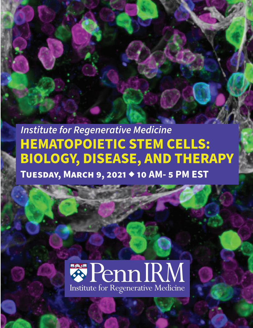
HEMATOPOIETIC STEM CELLS: BIOLOGY, DISEASE, AND THERAPYTUESDAY, MARCH 9, 2021 ◆ 10 AM- 5 PM EST
Institute for Regenerative Medicine
About the IRM
The Institute for Regenerative Medicine (IRM) at the University of Pennsylvania is a community of experts working to create a future where diseases are cured with cellular solutions. Our mission is to understand how cells and tissues are formed and to apply that knowledge to next generation diagnostics and therapies. We are proud to feature faculty from 5 different Penn Schools, the Children’s Hospital of Philadelphia (CHOP), and the Wistar Institute.
IRM Symposia
The IRM typically hosts 2 major symposia each year, one in the spring and one in the fall. These events are a chance to bring together IRM researchers and experts from around the world to discuss the latest topics in regenerative biology and medicine. Recent symposia topics have included the science and ethics of models of early human development, cell & tissue organization engineering, and cellular plasticity. For information on future IRM events, please visit our website (https://irm.med.upenn.edu/).
Symposium Organizers
• Ivan Maillard, MD, PhD, is a Professor of Medicine at the University of Pennsylvania and Co-Director of the IRM’s Hematopoietic Stem Cell Program.
• Wei Tong, PhD, is an Investigator at the CHOP Research Institute and a Professor of Pediatrics at the University of Pennsylvania. Wei serves as Co-Director of the IRM’s Hematopoietic Stem Cell Program.
Contents
• Symposium schedule• Poster abstracts• Sponsors
Cover Photo Credit
Daniel Lucas, Cincinnati Children’s Hospital
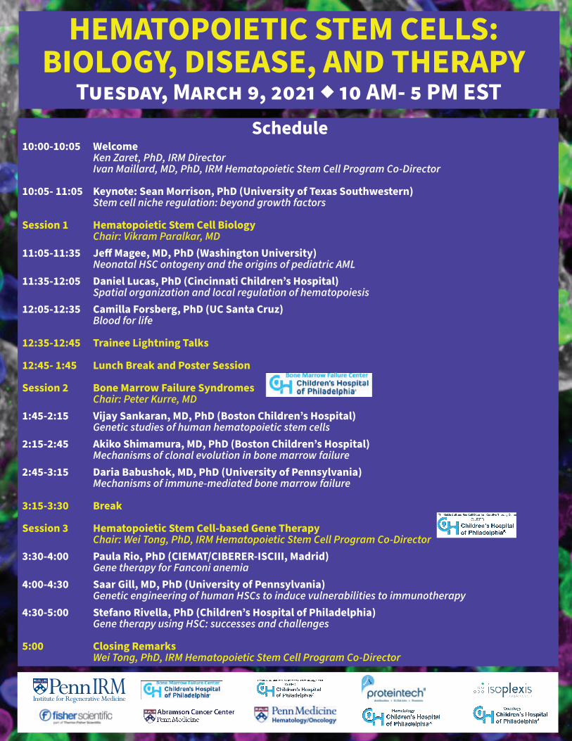
HEMATOPOIETIC STEM CELLS: BIOLOGY, DISEASE, AND THERAPY
TUESDAY, MARCH 9, 2021 ◆ 10 AM- 5 PM ESTSchedule
10:00-10:05 WelcomeKen Zaret, PhD, IRM DirectorIvan Maillard, MD, PhD, IRM Hematopoietic Stem Cell Program Co-Director
10:05- 11:05 Keynote: Sean Morrison, PhD (University of Texas Southwestern)Stem cell niche regulation: beyond growth factors
Session 1 Hematopoietic Stem Cell BiologyChair: Vikram Paralkar, MD
11:05-11:35 Je� Magee, MD, PhD (Washington University)Neonatal HSC ontogeny and the origins of pediatric AML
11:35-12:05 Daniel Lucas, PhD (Cincinnati Children’s Hospital)Spatial organization and local regulation of hematopoiesis
12:05-12:35 Camilla Forsberg, PhD (UC Santa Cruz) Blood for life
12:35-12:45 Trainee Lightning Talks
12:45- 1:45 Lunch Break and Poster Session
Session 2 Bone Marrow Failure SyndromesChair: Peter Kurre, MD
1:45-2:15 Vijay Sankaran, MD, PhD (Boston Children’s Hospital)Genetic studies of human hematopoietic stem cells
2:15-2:45 Akiko Shimamura, MD, PhD (Boston Children’s Hospital)Mechanisms of clonal evolution in bone marrow failure
2:45-3:15 Daria Babushok, MD, PhD (University of Pennsylvania)Mechanisms of immune-mediated bone marrow failure
3:15-3:30 Break
Session 3 Hematopoietic Stem Cell-based Gene TherapyChair: Wei Tong, PhD, IRM Hematopoietic Stem Cell Program Co-Director
3:30-4:00 Paula Rio, PhD (CIEMAT/CIBERER-ISCIII, Madrid)Gene therapy for Fanconi anemia
4:00-4:30 Saar Gill, MD, PhD (University of Pennsylvania)Genetic engineering of human HSCs to induce vulnerabilities to immunotherapy
4:30-5:00 Stefano Rivella, PhD (Children’s Hospital of Philadelphia)Gene therapy using HSC: successes and challenges
5:00 Closing RemarksWei Tong, PhD, IRM Hematopoietic Stem Cell Program Co-Director

Daniel P. Brown 1,2, Saar I. Gill 2,3 Affiliations 1 Pharmacology Graduate Group, University of Pennsylvania, Philadelphia, PA, USA 2 Center for Cellular Immunotherapies, University of Pennsylvania, Philadelphia, PA, USA 3 Division of Hematology-Oncology, Department of Medicine, University of Pennsylvania, Perelman School of Medicine, Philadelphia, PA, USA
Therapeutic Reversion of Pathogenic Point Mutations with Adenine Base Editing Acute myeloid leukemia (AML) is a poor-prognosis hematological malignancy characterized by
dysregulated cell proliferation and differentiation. Approximately 20% of AML cases are linked to isocitrate dehydrogenase 1 and 2 (IDH1 and IDH2) missense mutations that elicit the production of the oncometabolite 2-hydroxyglutarate (2-HG). 2-HG contributes to AML pathogenesis by directly inhibiting TET family DNA demethylases and subsequently silencing tumor suppressor genes. IDH1 was recently implicated as disease-initiating in some patients with AML. Therapies that target mutant IDH1 are of great interest, and the FDA recently approved ivosidenib for relapsed AML. Nonetheless, the drug as monotherapy is not curative.
The most common IDH1 mutation is a guanosine for adenine substitution at codon 132. We sought to reverse this pathogenic mutation using adenine base editors (ABEs), which are synthetic Cas9- fusion proteins that enact locus-specific adenine-to-guanine base substitutions without inducing double strand breaks, thereby reducing genotoxicity and unintended genomic alterations. We reasoned that this approach would selectively target mutant IDH1 while sparing the endogenous gene, yielding permanent reversion of the mutation with minimal off-target effects. We tested 8 guides and delivered the base editor ABEmax using electroporation to Molm14 cells. We found 75% reversion of R132H, and we are now quantifying 2HG using high-performance liquid chromatography. In future work we will develop delivery system involving lipid nanoparticle-encapsulated ABE machinery for therapeutic reversion of pathogenic point mutations in vivo.

Understanding Epigenome of Hematopoietic Stem cells
Author: JIGISHA CHANDRA - Bachelors of Pharmacy Honours, Birla Institute of Technology and Sciences,Pilani (BITS Pilani), Pilani,India
AbstractHematopoietic stem cells have potential to differentiate into mature cells of both myeloid andlymphoid lineages because of mechanisms of self renewal . Hematopoietic stem cells and cellsof myeloid and lymphoid lineage all have the same genome but they have different structureand function.What is the reason behind this? This is because of the difference in theirexpression of epigenome of cells of various lineages .Their gene expression is governed by alot of epigenetic markers like DNA Methylation, post-translational histone modifications,nucleararchitecture,noncoding RNAs etc. Various epigenetic markers are responsible for expression orrepression of an individual or sets of genes. This helps the Hematopoietic stem cells todifferentiate into cells of various lineages. This article will focus on how with the help ofunderstanding epigenetic machinery we can understand the development and differentiation ofhematopoietic stem cells into various other cells. Also it will help us understand how change inepigenome can lead to change in the architecture of cells.Having an understanding of the abovementioned points, will help us to understand clinical applications - such as diagnosis ,prognosis and therapy for disorders related to Hematopoietic stem cells.
Keywords-Hematopoietic stem cells,self renewal, DNA Methylation, post-translational histonemodifications,nuclear architecture,noncoding RNAs,as diagnosis , prognosis and therapy fordisorders related toHematopoietic stem cells.
A bioengineered bone marrow ECM niche model to support long-term HSCs in vitro Hannah Donnelly1*, Ewan Ross1, Rio Hermantara2, Christopher West3, Bruno Peault3, Adam West2, Manuel Salmeron-Sanchez1, Matthew J. Dalby1. 1Centre for the Cellular Microenvironment, University of Glasgow, Glasgow, G12 8QQ, UK. 2Institute of Cancer Sciences, Wolfson Wohl Cancer Research Centre, University of Glasgow,
Glasgow, G61 1QH, UK. 3MRC Centre for Regenerative Medicine, University of Edinburgh, Edinburgh, EH16 4UU, UK.
Hematopoietic stem cells (HSCs) have the ability to regenerate the entire blood and immune
system1, as such, they hold enormous clinical potential. In vivo they are supported by
mesenchymal stromal cells (MSCs) in a specialised microenvironment that provides physical
and functional regulatory cues, termed the bone marrow (BM) niche. Once HSCs are removed
from the niche their ability to self-renew is lost quickly, cells rapidly proliferate and
spontaneously differentiate away from the long-term reconstituting (LT-HSC) phenotype in a
matter of days, rendering them clinically ineffective 2. Here, we have developed a system to
recapitulate properties of the BM niche microenvironment to support LT-HSCs in vitro. Using
poly (ethyl acylate) (PEA) to promote material-driven unfolding of the extracellular matrix
protein fibronectin (FN), we tether BM niche associated growth factors (GF) (e.g. BMP-2) to the
exposed GF binding domain on FN3. Then by incorporating a collagen type I hydrogel in the
stiffness range of the bone marrow (~0.1 kPa)4, we were able to demonstrate maintenance of
a niche-like phenotype in MSCs. This included expression of nestin, a key niche stromal marker5,
and production of HSC maintenance cytokines CXCL12 and SCF. Upon co-culture with HSCs, we
found that this niche-like system is able to support LT-HSCs in culture. This material-based
system offers a platform for investigation into fundamental mechanisms of stem cell regulation,
such as stem cell metabolism, as well as a more humanised model for genotoxicity screening of
drugs, small molecules and cellular therapies.
Acknowledgements
We gratefully acknowledge support of EPSRC grant EP/P001114/1 and the technical
assistance provided by Carol-Anne Smith.
References:
1. Jagannathan-Bogdan, M. & Zon, L. I. Development 140, 2013.
2. Boitano, A. E. et al. Science. 329, 2010.
3. Cheng, Z. A. et al. Advanced Science. 1800361, 2018.
4. Jansen, L. A., et al. Journal of the Mechanical Behavior of Biomedical Materials. 50, 2015.
5. Méndez-Ferrer, S. et al. Nature 466, 2010.
Different patterns of chromatin opening by hematopoietic pioneer factors
Megan A Frederick1,2, Meilin Fernandez Garcia2, Gregory Donahue2, Edgar Luzete Monteiro2, Kenneth S Zaret2 Institute for Immunology, Perelman School of Medicine, University of Pennsylvania, Philadelphia, PA1,,Institute for Regenerative Medicine, Department of Cell and Developmental Biology, Perelman School of Medicine, University of Pennsylvania, Philadelphia, PA2 Pioneer factors can elicit local exposure of a nucleosome within chromatin and ultimately recruit co-regulators and remodelers to yield open chromatin sites seen in vivo. Yet how different pioneer transcription factors initially expose a targeted nucleosome in compacted chromatin structures is unclear. We used nucleosome arrays in vitro with a central nucleosome that can be targeted by the hematopoietic Ets factor PU.1 and the bZIP factors C/EBPα, and C/EBPβ. Each class of factor can elicit targeted nucleosome exposure on linker histone-compacted arrays, but with different hypersensitivity patterns, as discerned from long-read sequencing. The DNA binding domains (DBDs) of PU.1 and C/EBPα are sufficient for mononucleosome binding but are much less efficient than the full-length proteins, which can function cooperatively, in opening compacted chromatin. Thus, pioneer factors use their DBD to bind nucleosomes and other domains to elicit distinct patterns of target nucleosome exposure within compacted chromatin in vitro. Finally, nucleosome exposure by PU.1 enables further chromatin remodeling by a SWI/SNF complex. Together our data give a mechanistic view of how transcription factors cooperate to disrupt chromatin structures to initiate DNA accessibility to additional regulatory factors.
Title: A novel role of let-7b/ LIN28B/IMP-1/IGF-II axis as a molecular regulator of megakaryocyte
development during aging
Authors & affiliations: Ravi Kumar Gutti1
1 Department of Biochemistry, University of Hyderabad, INDIA
E-mail: [email protected]
Abstract:
Background: microRNAs (miRNAs) have been shown to play crucial role in the regulation of stem-cell differentiation in normal as well as malignant haematopoiesis. Neonatal cord blood (CB) cells are promising alternatives to adult bone marrow (PB) in cellular therapies. CB cells have potential to develop into specialized cells, such as erythrocytes or megakaryocytes (MK). MKs are responsible for production of platelets and thrombocytopenia (low platelet count) is common among sick infants. We hypothesize that developmental differences between CB and PB-MKs contribute to the vulnerability of neonates to develop severe thrombocytopenia and miRNA role in megakaryocytopoiesis is unknown. Methods: We cultured human CB and PB-derived CD34+ cells in the presence of thrombopoietin for 14 days and collected cultures expressing >90% CD41+ by flow cytometry. miRNA was prepared using the miRNeasy mini kit (Qiagen) and expression analysis of 88 miRNAs was performed using an array (Qiagen). Web-based computational approaches were used for putative target prediction and the protein levels were detected using western blot analysis. Results: Out of 88 miRNAs involved in stem cell development, let-7b was the only miRNA down regulated (~10-fold) in neonates compared to adult megakaryocyte (MK), and the levels of let-7b was differentially expressed in all stages of MK development (progenitors to maturation). We show that the low levels of let-7b in fetal and neonatal MK progenitors cause their relatively high proliferative rate by allowing an up-regulation of insulin-like growth factor II mRNA-binding protein 1 (IMP-1) and insulin-like growth factor II (IGF-II), and that these factors contribute to the marked hyperproliferative response of fetal, but not adult MK progenitors. Conclusions: The present study shows functional significance of LIN28B/IMP-1/IGF-II axis during megakaryocyte development. Therefore, it could be a potential target in neonatal thrombocytopenia and other platelet disorders.
LNK(SH2B3) Inhibition Enhances Human Fanconi Anemia Hematopoietic Stem and
Progenitor Cell Function
Nicholas Holdreith1,2, Grace Lee1,2, Peter Nicholas1,3, Timothy S. Olson1,3 and Wei Tong1,2,*
1 Division of Hematology, Children's Hospital of Philadelphia, Philadelphia, PA 19104
2 Department of Pediatrics, Perelman School of Medicine at the University of Pennsylvania,
Philadelphia, PA 19004
3 Cellular Therapy and Transplant Section, Division of Oncology, Children's Hospital of
Philadelphia, Philadelphia, PA, 19104
Hematopoietic stem cell transplantation (HSCT) remains the only curative treatment for a variety
of hematological diseases. Allogenic HSCT requires hematopoietic stem cells (HSCs) from
matched donors and comes with cytotoxicity and mortality. Recent advances in genome
modification of HSCs have demonstrated the possibility of using autologous HSCT-based gene
therapy to cure monogenic diseases, such as the inherited bone marrow failure syndrome Fanconi
Anemia (FA). However, insufficient HSC numbers and our inability to reliably expand HSCs,
results in delayed hematopoietic recovery and increased risk of graft failure. We and others
previously identified the adaptor protein Lnk(Sh2b3) as a critical negative regulator of murine
HSC homeostasis. However, whether LNK(SH2B3) controls human HSCs has not been studied.
Here, we demonstrate that depletion of LNK via lentiviral expression of miR30-based short hairpin
RNAs (shRNAs) resulted in robust expansion of transplantable human HSCs that provided
balanced multilineage reconstitution in primary and secondary mouse recipients. Importantly,
LNK depletion enhanced cytokine mediated JAK/STAT activation in CD34+ hematopoietic stem
and progenitor cells (HSPCs). Moreover, we demonstrate that LNK depletion ameliorated
functional defects in primary HSPCs associated with FA. In xenotransplant, engraftment defects
of FANCD2-depleted FA-like HSCs were markedly improved by LNK inhibition. Finally,
targeting LNK in primary bone marrow HSPCs from FA patients enhanced their colony forming
potential in vitro. Together, these results demonstrate the potential of targeting LNK to expand
HSCs to improve HSCT and HSCT-based gene therapy.
Title: Constrained chromatin accessibility in PU.1-mutated agammaglobulinemia patients.
Carole Le Coz1, David N. Nguyen2, Chun Su1, Michael V. Gonzalez1, Andrew D. Wells1,
Alex Marson2, Gregory M.K. Poon3, and Neil Romberg1
1Children’s Hospital of Philadelphia, Philadelphia, PA, 19104, USA. 2University of California San Francisco, San Francisco, CA, 94143, USA. 3Georgia State University, Atlanta, GA, 30303, USA.
Background: The pioneer transcription factor (TF) PU.1 controls hematopoietic cell fate
by decompacting stem cell heterochromatin and allowing non-pioneer TFs to enter
otherwise inaccessible genomic sites. PU.1 deficiency fatally arrests lymphopoiesis and
myelopoiesis in mice but human congenital PU.1 disorders have not previously been
described. We studied six unrelated, agammaglobulinemic patients each harboring a
heterozygous mutation (four de novo, two unphased) of SPI1, the gene encoding PU.1.
Rationale: To determine if and how SPI1 mutations cause PU.1-mutated
agammaglobulinemia (PU.MA).
Methods: PU.MA subject DNA, peripheral blood and bone marrow samples were
analyzed with whole exome sequencing, CITE-Seq, flow cytometry and
immunohistochemistry. The transcriptional capacity, nuclear localization and DNA
binding of PU.1 mutants were assessed with reporter lines, confocal microscopy,
electrophoretic mobility shift assays. Hematopoiesis and hematopoietic chromatin
accessibility were modelled in vitro using genetically edited RS4;11 pro-B cell lines and
human hematopoietic stem cells (HSCs).
Results: Affected patients lacked circulating B-cells and possessed few conventional
dendritic cells. Introducing disease-similar SPI1 mutations into human hematopoietic
stem and progenitor cells impaired early in vitro B-cell and myeloid-cell differentiation.
Patient SPI1 mutations encoded destabilized PU.1 proteins unable to nuclear localize or
bind target DNA. In PU.1-haploinsufficient pro-B cell lines, euchromatin was less
accessible to non-pioneer TFs critical for B-cell development and gene expression
patterns associated with the pro to pre-B-cell transition were undermined.
Conclusions: Our findings molecularly describe a novel form of agammaglobulinemia
and underscore PU.1's critical, dose-dependent role as a hematopoietic euchromatin
gatekeeper
HectD1 controls hematopoietic stem cell regeneration by coordinating ribosome assembly
and protein synthesis
Kaosheng Lv, 1,2 Chujie Gong, 1,2 Charles Antony, 3 Xu Han, 1,2 Jian-Gang Ren, 1,2 Ryan
Donaghy, 1,2 Ying Cheng, 1,2,4 Simone Pellegrino, 5,6,7 Alan J. Warren, 5,6,7 Vikram R. Paralkar, 3
and Wei Tong 1,2,*
1 Division of Hematology, Children's Hospital of Philadelphia, Philadelphia, PA 19104
2 Division of Pediatrics, Perelman School of Medicine at the University of Pennsylvania,
Philadelphia, PA 19104
3 Division of Hematology-Oncology, Department of Medicine, Perelman School of Medicine
at the University of Pennsylvania, Philadelphia, PA 19104
4 Present address: Center for Mitochondrial Biology and Medicine, The Key Laboratory of
Biomedical Information Engineering of Ministry of Education, School of Life Science and
Technology, Xi'an Jiaotong University, Xi'an, China. 710049
5 Cambridge Institute for Medical Research, Cambridge, United Kingdom
6 Department of Haematology, University of Cambridge, Cambridge, United Kingdom
7 Wellcome Trust–Medical Research Council Stem Cell Institute, University of Cambridge,
Cambridge, United Kingdom
* Correspondence author: [email protected]
Abstract
Impaired ribosome function is the underlying etiology in a group of bone marrow failure
syndromes called ribosomopathies. However, how ribosomes are regulated remains poorly
understood, as are approaches to restore hematopoietic stem cell (HSC) function attributable to
defective ribosome biogenesis. Here we uncover a previously-unappreciated role for the E3
ubiquitin ligase HectD1 in regulating HSC function via ribosome assembly and protein translation.
Hectd1-deficient HSCs exhibit a striking defect in transplantation ability and ex vivo maintenance,
concomitant with a reduced protein synthesis and growth rate under stress conditions.
Mechanistically, HectD1 ubiquitinates and degrades ZNF622, an assembly factor for the ribosomal
60S subunit. HectD1 loss led to an accumulation of ZNF622 and the anti-association factor eIF6
on the 60S, resulting in 60S/40S joining defects. Importantly, Znf622 depletion in Hectd1-deficient
HSCs restored ribosomal subunit joining, protein synthesis, and HSC reconstitution capacity.
These findings highlight the importance of ubiquitin-coordinated ribosome assembly in HSC
regeneration.
Abstract for HSC symposium 9th March – Chanelle McGuinness (lifETIME CDT PhD candidate) Title: Improving in vitro bone marrow niches for stem cell maintenance Authors and Affiliations: Chanelle A. S. McGuinness*, Yinbo Xiao*, Steve Swioklo§, Manuel-Salmeron-Sanchez*, Hannah Donnelly*, Matthew J Dalby* *Centre for the Cellular Microenvironment, University of Glasgow. §Atelerix Ltd. Abstract: The therapeutic potential of hematopoietic stem cells (HSCs) is well known. However, harnessing their full potential is limited since they quickly lose their stem cell properties in vitro. In addition, mesenchymal stromal cells (MSCs) fail to adequately support HSCs ex vivo. These limitations in ex vivo culture of stem cells are believed to be due to lack of regulatory signals that are normally supplied to HSCs and MSCs within the bone marrow niche. Biomaterials have been of great interest to mimic aspects of the 3D environment of tissues. In our laboratories, recent work demonstrated how MSCs can be maintained and supported using low stiffness collagen hydrogels on PEA coated surfaces functionalised with fibronectin and growth factor BMP-2 in vitro to mimic aspects of the bone marrow niche. This 3D niche model could support MSC niche characteristics including Nestin, SCF and CXCL12 expression and promote maintenance of long-term HSCs. However, the use of collagen hydrogels brings limitations to this model since they are animal derived, difficult to modify and demonstrate batch-to-batch variability. Alternative hydrogels are therefore of interest to advance this model. This new project will investigate the use of alginate gels in our PEA system for the improvement of HSC maintenance and better reliability. Alginate offers a highly tunable and modifiable alternative to collagen that can provide additional benefits to the niche system, offering flexibility in adjusting its properties and has the potential to preserve cells in a more quiescent state. Acknowledgements: lifETIME ESPRC-SFI CDT in engineered tissues (EP/S02347X/1). Carol-Anne Smith for technical help.

Active biomaterials for the ex-vivo expansion of Hematopoietic Stem Cells: from
biointerfaces to functionalised hydrogels.
Michaela Petaroudi1, Aleixandre Rodrigo-Navarro1, Oana Dobre1, Matthew Dalby1, Manuel Salmeron-Sanchez1
1Centre for the Cellular Microenvironment, University of Glasgow, Glasgow, UK
Abstract
Inspired by the huge clinical significance of Hematopoietic Stem Cell (HSC) transplants, we aim to develop a
synthetic platform for their expansion, ex-vivo. Our system is based on a biointerface between human HSCs,
the non-pathogenic bacterial species Lactococcus lactis (L. lactis), and synthetic polyethylene glycol (PEG)
hydrogels, that mimics the chemical and mechanical properties of the bone marrow (BM). The bacteria have
been genetically engineered to produce human CXCL12, TPO, VCAM1 and FN, key cytokines for HSC
expansion, while the hydrogels can be designed to resemble the BM stiffness and architecture. We can
further add a layer of Mesenchymal Stem Cells (MSCs) to more closely mimic the BM microenvironment.
Results suggest that our L. lactis-CD34+ cell co-culture system supports HSC phenotype maintenance and can
achieve up to 19-fold HSC expansion in 2D experiments. HSC and MSC viability in 3D cultures is not affected
by the presence of the hydrogels or the biofilm, while our biointerface can maintain a stem-like phenotype
in cultured MSCs for up to 14-days. Finally, we show that the hydrogels can maintain the HSC population in
3D, while their biophysical properties can be specifically tuned to encourage HSC proliferation.
Our data suggests that this novel system has a promising potential to mimic the native conditions that
support HSC maintenance and proliferation and induce ex-vivo HSC expansion. To our knowledge, this is the
first attempt to combine a living biointerface, recombinant signal production, and mechanical support
provided by hydrogels for BM and HSC niche engineering.
Graphical abstract
Figure 1. Overview of the system and the various platforms we can create for different applications.

Transgene-free generation of long-term hematopoietic stem cells Giorgia Scapin, Ph.D.1,2,3 and Dhvanit I. Shah, Ph.D. 1,2,3,4
1Harvard Medical School, Boston, Massachusetts 02115, USA 2 Brigham and Women’s Hospital, Boston, Massachusetts 02115, USA 3 Harvard Stem Cell Institute, Cambridge, Massachusetts 02115, USA 4 Garuda Therapeutics, Cambridge, MA 02139 The development of engraftable human long-term hematopoietic stem cells (LT-HSC) from pluripotent stem cells (hPSC) has been a long-sought goal. Definitive HSCs are born from the hemogenic endothelial cells (HEC) residing in the ventral wall of the dorsal aorta (DA) during embryonic development. Blood flow-mediated shear-stress stimulates the endothelial-to-HSC transition (EHT). However, it remains unknown why the ventral wall, and not the dorsal wall, of the DA is the restricted site of the EHT when blood flows through the entire DA, and attempts to mimic blood-flow mediated biomechanical cues have not produced engraftable human HSCs. Since HSCs can form despite the absence of active blood circulation, we analyzed the heartbeat-mediated pulsatile displacement experienced by the zebrafish embryo DA walls. Spinning disc confocal microscopy and single-particle tracking analyses of pulsating blood vessels showed that pulsation-mediated circumferential stretch is restricted to the ventral DA part and responsible for the magnitude, the site, and timing of the HSC formation. We next developed a bioreactor to establish the functional link between blood vessel pulsation and HSC formation. Our serial transplant and limiting dilution assays demonstrated that pulsation-mediated circumferential stretching of HECs activates the mechanoreceptor Piezo1 leading to 3-fold production of mouse LT-HSCs; which reconstitute to normal multi-lineage adult blood. To translate our findings in human setting, we treated human PSC-derived HECs with the Piezo1 activator Yoda1 obtaining the first transgene-free human LT-HSCs that can self-renew, engraft, and reconstitute multi-lineage blood upon serial transplantations, which is critical for generating off-the-shelf transgene-free HSC-based cellular therapies.
Translational control in hematopoietic stem cell maintenance and expansion
Hanna Shin*, Dheeraj Bhavanasi, and Peter S. Klein
Hematology/Oncology department, Perelman School of Mecidine at the University of Pennsylvania, Philadelphia, PA, USA
Hematopoietic stem cell transplantation (HSCT) is clinically used for multiple hematopoietic diseases. Umbilical cord-blood (UCB) HSCs require less stringent HLA matching and have reduced risk of graft versus host disease, but most UCB units have insufficient numbers of HSCs for transplant in adults. Ex vivo HSC expansion would greatly improve the utility of UCB for HSCT. However, HSCs lose their capacity for long-term self-renewal in standard culture conditions. Our lab established a culture system that maintains long-term HSCs ex vivo. Using this system, we performed a high-throughput screen (HTS) for hematopoietic stem and progenitor cell (HSPC) expansion and identified multiple compounds that target the translation initiation factor eIF4E and inhibit upstream of eIF4E. As the low rate of translation in HSCs is essential for their function, we selected an eIF4E targeting drug, 4E1RCat, for further analysis. 4E1RCat allowed 10-fold expansion of human HSCs within 4 days as measured by limiting dilution analysis. Therefore, targeting translation initiation may facilitate HSC expansion ex vivo. We will optimize ex vivo HSC expansion using 4E1RCat and test whether 4E1RCat reduces translation in HSCs. And, we will study how eIF4E signaling regulates translation in HSC homeostasis. Our HTS identified multiple compounds that inhibit ERK and p38 MAP kinase pathways, which converge on MNK1 to activate translation through phosphorylation of eIF4E. Thus, we will test whether eIF4E phosphorylation by MNK1 regulates global translation in HSCs and whether inhibition of these pathways facilitates ex vivo expansion of HSCs.
Title: Improved Gene Therapy for metachromatic leukodystrophy Authors: Lucas Tricoli, Adeline Vanderver, Laura Adang, Max Chappell, Laura Breda, Naoto Tanaka, Michael Triebwasser, Amaliris Gonzalez, Lars Schlotawa, Peter Kurre, Stephanie Hurwitz, Stefano Rivella Abstract: MLD is an autosomal recessive lysosomal storage disease (LSD) characterized by a mutation of the enzyme Arylsulfatatse A (ARSA) most often affecting children in the late infantile stage (LI) (less than 30 months of age at onset), which is the most rapidly progressive form of the disease. Utilizing ex-vivo hematopoietic stem cell (HSC) gene therapy, a functional copy of the ARSA gene replacing mutated ARSA causing MLD, has the potential to restoring proper enzyme activity and has preliminary demonstrated successful treatment in clinical trials. Current approaches for MLD rely on the use of AAV or lentiviral vectors (LVs), which for AAV may not sustain high level of gene expression in neurological disorders or for LVs requires multiple integrations to prevent the development of the disease. In addition for the currently existing clinical LV these levels are considered insufficient to help symptomatic patients. For all these reasons, we propose that a vector expressing higher levels of ARSA at single integration will reduce the chances of genotoxicity, may be curative in symptomatic patients and, in the future, may also reduce the requirement for full myeloablation. For our top performing vectors, ARSA activity, normalized to VCN, demonstrated that TCO-EAAWP-UTR+, TCO-EAFWP-UTR- and TCO-AEAFWP-UTR- showed more ARSA activity (respectively 2X, 10X and 4X) compared to the clinical vector with protein analysis by western blot showing a similar trend. We have also demonstrated with our vectors the ability to secrete more functional ARSA enzyme into the surrounding media in cell culture, which is a critical modality for transfer of functional ARSA from microglia to oligodendrocytes. Current experiments in vector transduced mouse HSC have demonstrated high levels of transduction with superior levels of ARSA activity. Transplanted WT mice are currently living vibrantly with a high level of transduction from our top performing vector.
Fibroblastic stromal cells expressing Delta-like Notch ligands control extrathymic T cell development in mesenteric lymph nodes
Ashley Vanderbeck, Samantha Kelly, Leolene Carrington, Frederick Allen, Joshua Brandstadter, Eric Perkey, Gloria Jih, Anneka Allman, Daniela Gomez-Atria, and Ivan Maillard
Early T cell development is supported by signals mediated by the Notch1 receptor in T lineage progenitors and Delta-like4 (Dll4) ligand in thymic epithelial cells. Although T cell development is restricted to the thymus, extrathymic T cell development has been reported in athymic Foxn1nu/nu nude mice as well as during times of thymopoietic stress such as post-bone marrow transplantation (BMT). A population of CD4+CD8+ double positive T cell progenitors can be found in the mesenteric lymph nodes (MLN) in both athymic mice and early post-BMT, suggesting the presence of an extrathymic niche conducive to T cell development. However, whether Notch signaling is required, as well as the cellular source(s) of Notch ligand throughout this process in the MLN, remains unknown. We hypothesize that MLNs harbor a unique environment in which Notch ligands are available to circulating progenitors and critical to sustain extrathymic T cell development in these contexts. To test this, we utilized systemic neutralizing antibodies as well as loss-of-function genetic models to assess whether the Notch ligands Dll4 or Dll1 are important for extrathymic T cell development. We found that, like in homeostatic thymocyte development, Dll4 appears to play an essential role in generation of early T cell progenitors found in the MLN. Furthermore, the source of Dll4 appears to reside within subsets of non-hematopoietic fibroblastic stromal cells lineage traced by a Ccl19-Cre transgene. In sum, these findings shed new light on the cellular and molecular cues regulating T cell development outside of the thymus.
Engineering the pan-leukocyte antigen CD45 to facilitate CAR-T cell therapy against hematologic malignancies Nils Wellhausen1,2, Christopher K. Garcia3,4,5, Saar I. Gill1,6,*, Carl H. June1,2,7,* 1Center for Cellular Immunotherapies, University of Pennsylvania Perelman School of Medicine, Philadelphia, PA, USA 2Parker Institute for Cancer Immunotherapy at University of Pennsylvania, Philadelphia, PA, USA 3Department of Molecular and Cellular Physiology, Stanford University School of Medicine, Stanford, CA, USA 4Department of Structural Biology, Stanford University School of Medicine, Stanford, CA, USA 5Howard Hughes Medical Institute, Stanford University School of Medicine, Stanford, CA, USA 6Division of Hematology-Oncology, Department of Medicine, University of Pennsylvania Perelman School of Medicine, PA, USA 7Department of Pathology and Laboratory Medicine, University of Pennsylvania Perelman School of Medicine, PA, USA *Co-corresponding authors Abstract Currently, chimeric antigen receptor (CAR)-T cells must be individually designed for each disease, targeting lineage-associated antigens such as CD19 for B-cell malignancies or BCMA for myeloma. This approach is inefficient and limited to antigens for which on-target/off-tumor toxicities from the CAR-T cells is clinically tolerated. By targeting the pan-hematologic antigen CD45, a single “drug” could be used for most hematologic malignancies, thereby accelerating clinical research and expediting the treatment of more patients. A unique hurdle in developing CD45-directed CAR-T cells is that healthy leukocytes (including CAR-T cells) express CD45 themselves. We hypothesize that engineering the CD45 molecule on hematopoietic cells (including CAR-T cells) to lack the target epitope required for recognition by CAR-45 can generate functional, fratricide-resistant CAR-T cells and a hematopoietic system that is resistant to CD45-targeted CAR-T cell therapy. We show that CD45 CAR-T cells are susceptible to fratricide, as expected. By deleting CD45 in primary human T-cells we prevented fratricide without affecting CAR-T cell function in vitro. Using this approach, we identified three CD45-directed CAR constructs that demonstrate target-specific cytotoxicity against multiple hematologic cancer cell lines (T-ALL, AML and CML). Using truncated versions of the CD45 extracellular domain, we mapped the binding domain that is recognized by the CAR constructs, information that will be used to edit this region using CRISPR/Cas base editing in human hematopoietic stem cells to avoid long-term toxicities.
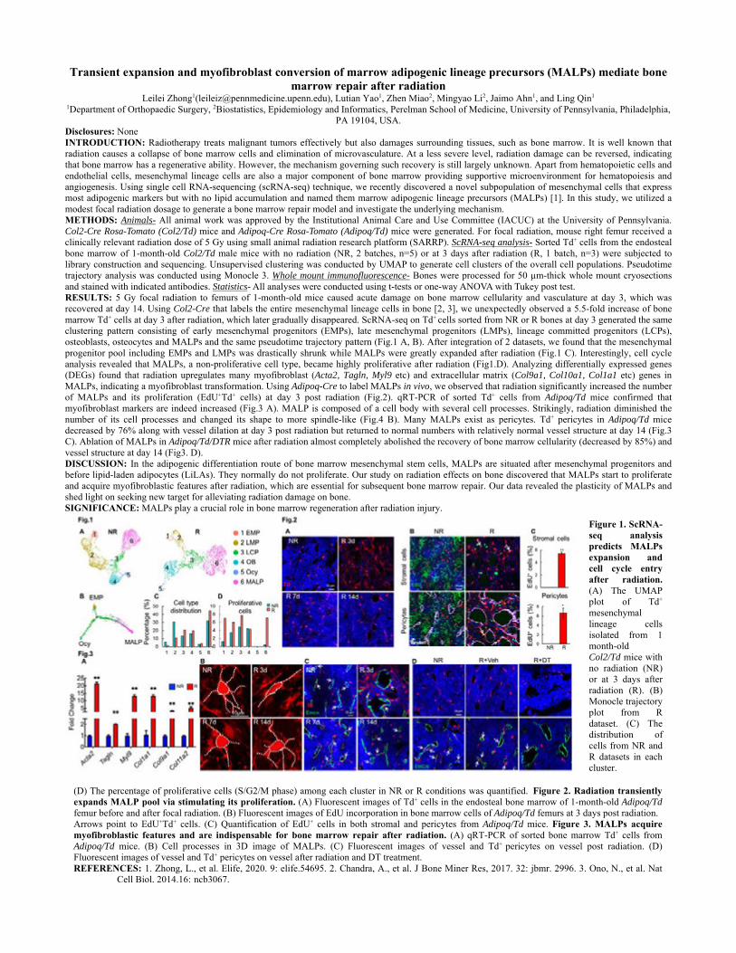
Transient expansion and myofibroblast conversion of marrow adipogenic lineage precursors (MALPs) mediate bone marrow repair after radiation
Leilei Zhong1([email protected]), Lutian Yao1, Zhen Miao2, Mingyao Li2, Jaimo Ahn1, and Ling Qin1 1Department of Orthopaedic Surgery, 2Biostatistics, Epidemiology and Informatics, Perelman School of Medicine, University of Pennsylvania, Philadelphia,
PA 19104, USA. Disclosures: None INTRODUCTION: Radiotherapy treats malignant tumors effectively but also damages surrounding tissues, such as bone marrow. It is well known that radiation causes a collapse of bone marrow cells and elimination of microvasculature. At a less severe level, radiation damage can be reversed, indicating that bone marrow has a regenerative ability. However, the mechanism governing such recovery is still largely unknown. Apart from hematopoietic cells and endothelial cells, mesenchymal lineage cells are also a major component of bone marrow providing supportive microenvironment for hematopoiesis and angiogenesis. Using single cell RNA-sequencing (scRNA-seq) technique, we recently discovered a novel subpopulation of mesenchymal cells that express most adipogenic markers but with no lipid accumulation and named them marrow adipogenic lineage precursors (MALPs) [1]. In this study, we utilized a modest focal radiation dosage to generate a bone marrow repair model and investigate the underlying mechanism. METHODS: Animals- All animal work was approved by the Institutional Animal Care and Use Committee (IACUC) at the University of Pennsylvania. Col2-Cre Rosa-Tomato (Col2/Td) mice and Adipoq-Cre Rosa-Tomato (Adipoq/Td) mice were generated. For focal radiation, mouse right femur received a clinically relevant radiation dose of 5 Gy using small animal radiation research platform (SARRP). ScRNA-seq analysis- Sorted Td+ cells from the endosteal bone marrow of 1-month-old Col2/Td male mice with no radiation (NR, 2 batches, n=5) or at 3 days after radiation (R, 1 batch, n=3) were subjected to library construction and sequencing. Unsupervised clustering was conducted by UMAP to generate cell clusters of the overall cell populations. Pseudotime trajectory analysis was conducted using Monocle 3. Whole mount immunofluorescence- Bones were processed for 50 µm-thick whole mount cryosections and stained with indicated antibodies. Statistics- All analyses were conducted using t-tests or one-way ANOVA with Tukey post test. RESULTS: 5 Gy focal radiation to femurs of 1-month-old mice caused acute damage on bone marrow cellularity and vasculature at day 3, which was recovered at day 14. Using Col2-Cre that labels the entire mesenchymal lineage cells in bone [2, 3], we unexpectedly observed a 5.5-fold increase of bone marrow Td+ cells at day 3 after radiation, which later gradually disappeared. ScRNA-seq on Td+ cells sorted from NR or R bones at day 3 generated the same clustering pattern consisting of early mesenchymal progenitors (EMPs), late mesenchymal progenitors (LMPs), lineage committed progenitors (LCPs), osteoblasts, osteocytes and MALPs and the same pseudotime trajectory pattern (Fig.1 A, B). After integration of 2 datasets, we found that the mesenchymal progenitor pool including EMPs and LMPs was drastically shrunk while MALPs were greatly expanded after radiation (Fig.1 C). Interestingly, cell cycle analysis revealed that MALPs, a non-proliferative cell type, became highly proliferative after radiation (Fig1.D). Analyzing differentially expressed genes (DEGs) found that radiation upregulates many myofibroblast (Acta2, Tagln, Myl9 etc) and extracellular matrix (Col9a1, Col10a1, Col1a1 etc) genes in MALPs, indicating a myofibroblast transformation. Using Adipoq-Cre to label MALPs in vivo, we observed that radiation significantly increased the number of MALPs and its proliferation (EdU+Td+ cells) at day 3 post radiation (Fig.2). qRT-PCR of sorted Td+ cells from Adipoq/Td mice confirmed that myofibroblast markers are indeed increased (Fig.3 A). MALP is composed of a cell body with several cell processes. Strikingly, radiation diminished the number of its cell processes and changed its shape to more spindle-like (Fig.4 B). Many MALPs exist as pericytes. Td+ pericytes in Adipoq/Td mice decreased by 76% along with vessel dilation at day 3 post radiation but returned to normal numbers with relatively normal vessel structure at day 14 (Fig.3 C). Ablation of MALPs in Adipoq/Td/DTR mice after radiation almost completely abolished the recovery of bone marrow cellularity (decreased by 85%) and vessel structure at day 14 (Fig3. D). DISCUSSION: In the adipogenic differentiation route of bone marrow mesenchymal stem cells, MALPs are situated after mesenchymal progenitors and before lipid-laden adipocytes (LiLAs). They normally do not proliferate. Our study on radiation effects on bone discovered that MALPs start to proliferate and acquire myofibroblastic features after radiation, which are essential for subsequent bone marrow repair. Our data revealed the plasticity of MALPs and shed light on seeking new target for alleviating radiation damage on bone. SIGNIFICANCE: MALPs play a crucial role in bone marrow regeneration after radiation injury.
Figure 1. ScRNA-seq analysis predicts MALPs expansion and cell cycle entry after radiation. (A) The UMAP plot of Td+ mesenchymal lineage cells isolated from 1 month-old Col2/Td mice with no radiation (NR) or at 3 days after radiation (R). (B) Monocle trajectory plot from R dataset. (C) The distribution of cells from NR and R datasets in each cluster.
(D) The percentage of proliferative cells (S/G2/M phase) among each cluster in NR or R conditions was quantified. Figure 2. Radiation transiently expands MALP pool via stimulating its proliferation. (A) Fluorescent images of Td+ cells in the endosteal bone marrow of 1-month-old Adipoq/Td femur before and after focal radiation. (B) Fluorescent images of EdU incorporation in bone marrow cells of Adipoq/Td femurs at 3 days post radiation. Arrows point to EdU+Td+ cells. (C) Quantification of EdU+ cells in both stromal and pericytes from Adipoq/Td mice. Figure 3. MALPs acquire myofibroblastic features and are indispensable for bone marrow repair after radiation. (A) qRT-PCR of sorted bone marrow Td+ cells from Adipoq/Td mice. (B) Cell processes in 3D image of MALPs. (C) Fluorescent images of vessel and Td+ pericytes on vessel post radiation. (D) Fluorescent images of vessel and Td+ pericytes on vessel after radiation and DT treatment. REFERENCES: 1. Zhong, L., et al. Elife, 2020. 9: elife.54695. 2. Chandra, A., et al. J Bone Miner Res, 2017. 32: jbmr. 2996. 3. Ono, N., et al. Nat
Cell Biol, 2014.16: ncb3067.

Thank You to Our Generous Sponsors!
Founded by scientists, Proteintech Group recognises the tireless efforts and sacrifices made by researchers to advance scientific discovery. Proteintech aims to match its’ customers’ dedication to research with high quality, reliable reagents that contribute to reproducible results. Learn about their human cell-expressed, recombinant proteins.
The future of advanced medicines relies upon deeper access to in vivo biology to create durable, curative impacts on disease. IsoPlexis’ systems, which drive convergence of dynamic proteomics and single cell biology for the first time, are creating this deeper connection to accelerate curative medicines.
The Pediatric Comprehensive Bone Marrow Failure Center (CBMFC) at Children’s Hospital of Philadelphia (CHOP) was established in 2010 as a comprehensive, multidisciplinary center dedicated to providing world-class care for children and adults with inherited and acquired forms of bone marrow failure (BMF). The CBMFC at CHOP collaborates with partners at the University of Pennsylvania (Penn) for both clinical and research efforts.
Penn's Hematology/Oncology Division is a comprehensive program dedicated to the clinical care and research, providing the most advanced treatment for all types of cancers and blood-related disorders. Our faculty members, more than 130, are involved in research to develop new and more effective methods for diagnosing and treating these diseases and for educating the physicians and researchers of the future.
The Sickle Cell and Red Cell Disorders Curative Therapy Center (CuRED) offers integrated and coordinated care from multiple pediatric specialists for children with sickle cell disease, thalassemia, and other red cell disorders. The CuRED team provides patients with a comprehensive evaluation and individualized cutting-edge treatments for management of their chronic disease, while exploring the potential for a curative therapy. CuRED is also driving towards the development of their own gene therapy for sickle cell disease and thalassemia leveraging CHOP’s world-class translational research expertise.
The Division of Hematology at Children's Hospital of Philadelphia provides inpatient and outpatient services for children and adolescents with all hematologic disorders other than malignancies.
Research in the Division of Oncology falls under Children's Hospital of Philadelphia Center for Childhood Cancer Research, which was established more than a decade ago as a state-of-the art program that integrates a wide array of basic, translational, and clinical research efforts. The Center brings together the diverse talents of investigators in Children’s Hospital renowned multidisciplinary program in pediatric cancer research, patient care, and genomics.
The Hematologic Malignancies Program at the Abramson Cancer Center drives basic scientific discoveries and translates them into novel therapeutics for patients with myeloid and lymphoid neoplasms. Formed in 1994, the Program has two scientific aims: 1) Develop a mechanisms-based understanding of the genetic, cellular, and biochemical processes regulating malignant hematopoiesis; and 2) Translate basic scientific discoveries into more effective and less toxic therapies for hematologic malignancies.
As the premier scientific marketplace, Fisher Scientific has defined unparalleled choice and convenience for over a century. Focused first and foremost on delivering innovative yet eminently practical solutions, we have developed a comprehensive portfolio of laboratory equipment and supplies, chemicals, safety products, and services to help our customers increase productivity and drive innovation in research, healthcare, education and industry — all with greater ease and efficiency than ever before.