state of the art of additive manufacturing for …
TRANSCRIPT
1
STATE OF THE ART OF ADDITIVE MANUFACTURING FOR POLYMERIC MEDICAL IMPLANTS
NSENGIMANA Joseph1 & VAN DER WALT Jacobus G2.
1Central University of Technology, Free State, Centre for Rapid Prototyping and Manufacturing (CRPM), Department of Mechanical & Mechatronics Engineering, Faculty of Engineering & Information Technology, Bloemfontein, South Africa.
2Central University of Technology, Free State, Centre for Rapid Prototyping and Manufacturing (CRPM), Department of Mechanical & Mechatronics Engineering, Faculty of Engineering & Information Technology, Bloemfontein, South Africa.
ABSTRACT
Additive Manufacturing (AM) commonly known as 3D printing has found many applications in the automotive, aerospace and medical industries. The flexibility to fabricate 3D objects of any complexity displayed by AM technologies such as Selective Laser Sintering (SLS), Stereolithography (SLA), Fused Deposition Modeling (FDM), PolyJet printing and electrospinning, has been used to improve the lives of many patients through the provision of polymeric implants, scaffolds and devices for drug delivery. The common limitation of such applications is the biocompatibility of the AM material with the human body and systems. An ideal non degradable implant would not invoke an inflammatory or toxic response whereas for a degradable implant, the degradants must also be metabolized in the body after fulfilling its purpose, thus leaving no trace. Furthermore, inertness, weight similar to human bone or even lighter, capability to generate no artifacts on Computer Tomography (CT) and Magnetic Resonance Imaging (MRI) scans, sufficient strength to resist functional stresses for load bearing implants, low and no thermal conductivity, easy sterilization and low cost of manufacturing are the desired characteristics for the acceptance of the use of an implant in a human body. Metallic and ceramic implants have been extensively used for medical implants. However the possible need for a second surgery to remove metallic implants, the stress shielding effect, the radio-opacity of the metal and long-term presence of metallic ions in vivo are major disadvantages of metallic implants which can be overcome by the use of their counterparts manufactured from polymeric materials. Building on the already established AM powder based technologies; a transition from micro to nanosized powder particles to improve the mechanical properties of SLS polymeric implants is a new trend of development. The optimum ratio of Hydroxyapatite (HA) to polymer composites and the establishment of measuring standards to meet the requirement of a medical implant are the actual challenges of AM for polymeric medical implants.
Keywords: Additive manufacturing, polymers, medical implants

2
1. INTRODUCTION
The replacement of a human limb is dated as far back as at least 3500 B.C. when an Indian poem mentioned an iron leg made for an amputee warrior [58]. A wood and leather prosthesis dated between 1069 and 664 B.C. was found in 2000 in a tomb near the ancient city of Thebes in Egypt [11]. The rapid development of machinery during the industrial revolution of the 19th century often caused accidents resulting in the loss of limbs which increased the need for artificial devices which mimic the functionality of the limbs they replace as far as possible.
Traditional time-consuming and inaccurate manual prototyping was followed by a second phase of prototyping around the mid-1970s when a 3D model in a virtual environment could be simulated and tested with exact material and properties but with limited ability to assess the functional performance of the final product. Rapid Prototyping (RP) culminated in the 1980s with the rapid growth of Computer Aided Design (CAD) software. The existence of modern software package such as SolidWorks, Pro/ENGINEER, CATIA, etc. can be used to create digital 3D models that can be processed through Additive Manufacturing (AM) technologies to manufacture physical models. Software from Materialise such as Magics and 3-Matic can also be used with AM to produce anatomical models, to assist with preoperative evaluation and intraoperation procedures [12], or to produce immediate use medical implants and devices. AM is defined as “the process of joining materials to make objects from 3D models data, usually layer upon layer, as opposed to subtractive manufacturing” [33]. Depending on the used AM technology, metallic, ceramic or polymeric materials in powder, liquid or solid form can be used to build a medical device or implant of any shape complexity, thus overcoming the limitations of traditional subtractive fabrication methods. Due to the advantages of the use of polymers, AM technologies such as Selective Laser Sintering (SLS), Stereolithography (SLA), Fused Deposition Modeling (FDM), 3D Printing (3DP), PolyJet and electrospinning have been used to improve the lives of many patients through the provision of polymeric implants, scaffolds and devices for drug delivery.
This paper constitutes a review of the recent developments in polymeric medical implantation and device fabrication using the potential of the six mentioned AM technologies.
2. CLASSIFICATION OF POLYMERIC MATERIALS
Polymers are materials made up of long chain molecules formed primarily by carbon-to-carbon bonds (Figure 1). Due to the wide-range of use of polymeric biomaterials, a single ideal polymer or polymeric family does not exist. Instead, a library of materials is available to researchers that can be synthesized and engineered to best match the specifications of the material desired for biomedical functions [9].
Figure 1: Generic chemical structure of a polymer: Polyethylene molecule
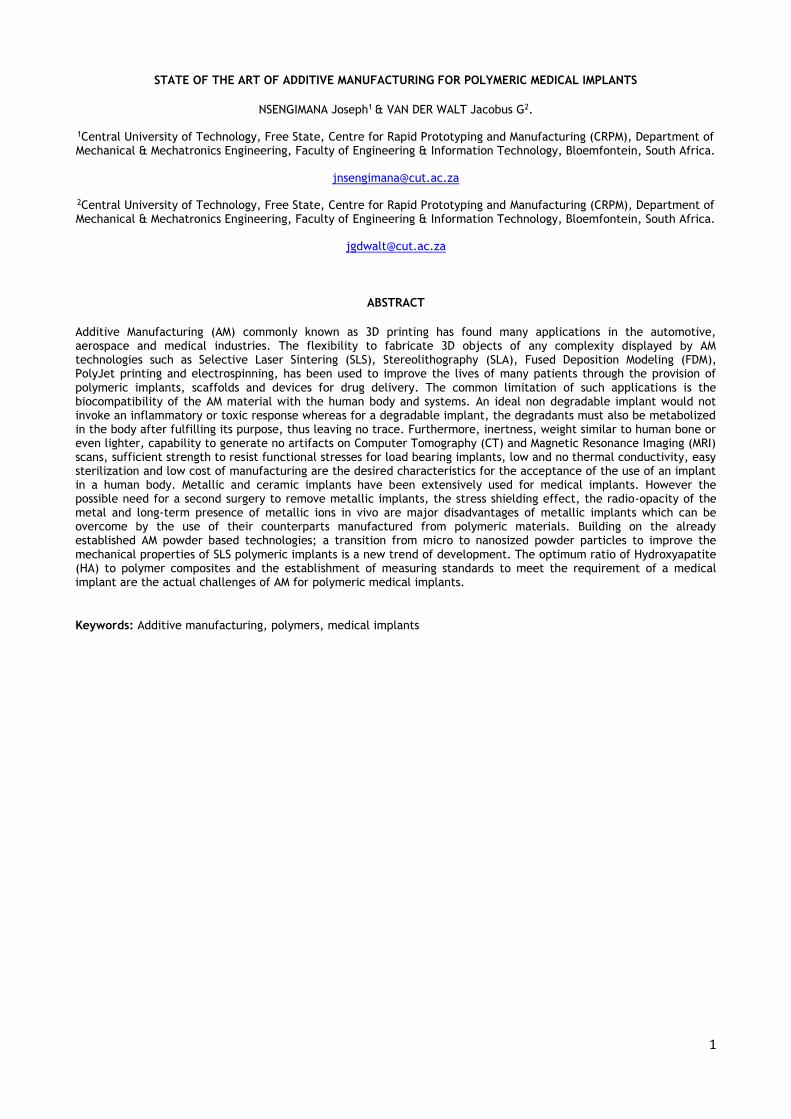
As far as the AM of medical devices and implants is concerned, the classification of polymers may be based on their thermal properties, application areas, and the level of degradability in the human body. All AM technologies use thermal energy to cure, to melt or to fuse the material. The thermal interaction between the heat and a particular polymeric material must be thoroughly controlled for the building of a dimensionally accurate medical implant and devices with satisfactory surface finish as well as acceptable mechanical properties for load bearing applications. Based on their thermal behavior, polymeric materials can be classified as shown by Figure 2.
C
H
H
C
H
H
C
H
H
C
H
H
C
H
H
C
H
H
C
H
H
C
H
H
CH2-CH2 repeating unit (monomer)
3
Figure 2: Thermal classification of polymers
A thermoplastic is a polymeric material that becomes soft and deformable when heated and rigid when cooled. This process is reversible for a number of times without chemically altering the material. On the other hand, a thermoset is a polymeric material that undergoes irreversible chemical changes when it is cured through heat, catalysts or ultraviolet light. Elastomers also known as rubbers are thermosetting polymeric materials characterized by low elastic modulus or with a high elongation at break.
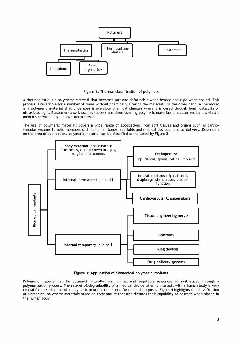
The use of polymeric materials covers a wide range of applications from soft tissues and organs such as cardio-vascular systems to solid members such as human bones, scaffolds and medical devices for drug delivery. Depending on the area of application, polymeric material can be classified as indicated by Figure 3.
Figure 3: Application of biomedical polymeric implants
Polymeric material can be obtained naturally from animal and vegetable resources or synthetized through a polymerization process. The rate of biodegradability of a medical device when it interacts with a human body is very crucial for the selection of a polymeric material to be used for medical purposes. Figure 4 highlights the classification of biomedical polymeric materials based on their nature that also dictates their capability to degrade when placed in the human body.
Polymers
Thermoplastics
AmorphousSemi-
crystalline
Thermosetting plastics
Elastomers
Bio
medic
al
impla
nts
Body external (non-clinical): Prostheses, dental crown bridges,
surgical instruments
Internal permanent (clinical)
Orthopedics:
Hip, dental, spinal, retinal implants
Neural implants : Spinal cord, diaphragm stimulation, bladder
function
Cardiovascular & pacemakers
Internal temporary (clinical)
Tissue engineering nerve
Scaffolds
Fixing devices
Drug delivery systems

4
Figure 4: Natural and synthetic polymeric materials
It is imperative that AM technology selected for the manufacturing of a customized medical implant or device, the combination of the above characteristics of polymers be taken into consideration in order to meet the requirements of any specific medical application.
3. CHARACTERISTICS OF A GOOD MEDICAL IMPLANT
Figure 4 shows that the replacement of a natural human feature with an artificial feature for healing, increasing of biological function or for repairing of abnormalities can be applied at all levels of a human body. The introduced implant or medical device to cohabitate with human body must fulfill the following main characteristics:
4.1 Biocompatibility and bio-inertness
The implant must interact positively with the host environment. Based on application and specific requirements, an ideal implant would be favorable for cell adhesion, non-toxic, non-irritant, non-allergic, non-carcinogenic, and resistant to corrosion, oxidation and hydrolysis. The degradants of a biodegradable implant must be metabolized in the body after fulfilling its purpose thus leaving no trace.
4.2 Bone-similar weight or even lighter
For load bearing implant such as bone plates, total hip or knee replacements, spine cages or intervertebral discs, it is desirable to have enough strength to fulfill the intended purpose for its placement in a human body. However it is more advantageous that the mechanical properties of the implant be more or less close to the properties of the natural bone in order to avoid the stress shielding effect. A much larger hardness and stiffness of bio-metallic implants compared to the bone renders difficult the future regrowth of the bone [5]. As a result of the reduction in bone density (osteopenia) leading to a removal of normal stress from the bone by an implant with greater modulus of elasticity. According to Wolff's law, bone in a healthy person or animal will remodel in response to the loads it is placed under. Therefore, if the bone load decreases, bone will become less dense and weaker because there is no stimulus for continued remodeling that is required to maintain bone mass. An implant should transfer bearing loads to the bone in order to avoid bone resorption. During the design process, this can be achieved by using implant materials with stiffness or Young modulus similar to the bone in cohabitation with the implant.
4.3 Capability to generate no artefacts on Computer Tomography and Magnetic Resonance Imaging (MRI)
scans
Computer Tomography (CT) and Magnetic Resonance Imaging (MRI) are scanning techniques used to produce medical images with high resolution and precision whereby sets of two dimensional (2D) cross sections of the body are stacked to create three dimensional (3D) digital models. Implants and external prostheses can then be sculpted in the virtual
Bio
medic
al
poly
meri
c m
ate
rials
Natural
Proteins (silk, collagen, gelatin, fibrinogen, elastin, keratin, cutin, myosin)
Polysaccharides (Cellulose, amylose, dextran, chitin, glucoaminoglican)
Polynucleotides (DNA & RNA)Synthetic biodegradable (scaffolds, drug
delivery, temporal fixing devices,
temporal barrier)
Synthetic non-biodegradable
(load bearing prosthetic implants, pacemakers)
5
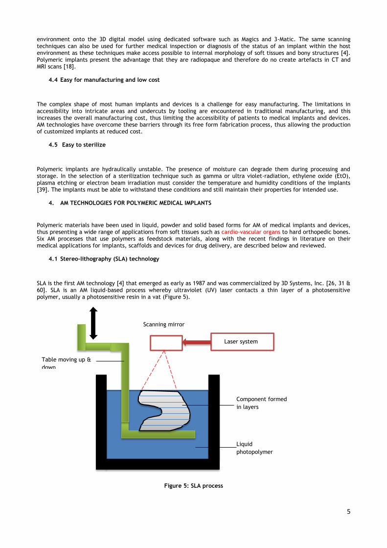
environment onto the 3D digital model using dedicated software such as Magics and 3-Matic. The same scanning techniques can also be used for further medical inspection or diagnosis of the status of an implant within the host environment as these techniques make access possible to internal morphology of soft tissues and bony structures [4]. Polymeric implants present the advantage that they are radiopaque and therefore do no create artefacts in CT and MRI scans [18].
4.4 Easy for manufacturing and low cost
The complex shape of most human implants and devices is a challenge for easy manufacturing. The limitations in accessibility into intricate areas and undercuts by tooling are encountered in traditional manufacturing, and this increases the overall manufacturing cost, thus limiting the accessibility of patients to medical implants and devices. AM technologies have overcome these barriers through its free form fabrication process, thus allowing the production of customized implants at reduced cost.
4.5 Easy to sterilize
Polymeric implants are hydraulically unstable. The presence of moisture can degrade them during processing and storage. In the selection of a sterilization technique such as gamma or ultra violet-radiation, ethylene oxide (EtO), plasma etching or electron beam irradiation must consider the temperature and humidity conditions of the implants [39]. The implants must be able to withstand these conditions and still maintain their properties for intended use.
4. AM TECHNOLOGIES FOR POLYMERIC MEDICAL IMPLANTS
Polymeric materials have been used in liquid, powder and solid based forms for AM of medical implants and devices, thus presenting a wide range of applications from soft tissues such as cardio-vascular organs to hard orthopedic bones. Six AM processes that use polymers as feedstock materials, along with the recent findings in literature on their medical applications for implants, scaffolds and devices for drug delivery, are described below and reviewed.
4.1 Stereo-lithography (SLA) technology
SLA is the first AM technology [4] that emerged as early as 1987 and was commercialized by 3D Systems, Inc. [26, 31 & 60]. SLA is an AM liquid-based process whereby ultraviolet (UV) laser contacts a thin layer of a photosensitive polymer, usually a photosensitive resin in a vat (Figure 5).
Figure 5: SLA process
Laser system
Scanning mirror
Table moving up &
down
Liquid
photopolymer
Component formed
in layers

6
This results in curing or solidification of a specific location of the layer. When the curing of the layer is finished, the platform is lowered down into the vat, a new layer of resin is added on the top of the first layer and the process continues until the entire part is built. Comparatively to the resolution of other AM process, the fabrication of items measuring several cubic centimeters with a superior resolution of 20 µm can be achieved in the SLA process [37]. This constitutes the major advantage of the process when there is a requirement to prepare well-designed architectures especially for manufacturing of biodegradable scaffolds for tissue engineering where various pore architectures can be applied for osteocell growth and integration.
The restriction to use one resin and the difficulties encountered in removing uncured unreacted resin which can be a substantial source of contamination are the major challenges and limitations in SLA technology [37]. Melchels, Feijen & Grijpma [37] reported the work done by Matsuda & Mizutani [34] where it was shown that degradable crosslinked structures prepared by SLA using poly(trimethylene carbonate-co-ε-caprolactone) resins caused no adverse effects after a one month implantation period under the dorsal skin of rats. Using the SLA process, Popov et al. [43] overcame the intrinsic toxicity and relatively poor mechanical properties of acrylic polymers by manufacturing composite scaffolds for guided bone regeneration and cranio-maxillofacial implants using polyfunctional methacrylic oligomers and a mixture of these with a bioactive HA. After the SLA process, the samples were treated with supercritical carbon dioxide (Sc CO2) to enhance their biocompatibility by removal of toxic residues and to create microporosity needed for osteointegration and osteoinduction. The test in vivo into white rats showed good results leading the researchers to conclude that SLA technology can be used to produce any part of human skeleton for implantation. Using a point to-point Coordinate Measurement Device (CMD), Taft, Kondor & Grant [53] assessed the accuracy of RP models by validating the accuracy of SLA skull models fabricated by SLA of a liquid monomer Accura 60 SLA epoxy resin. They found that there exist a significant difference between the control and each of the SLA models (with a significance level p<.001) in the Z axis additive build. Significant dimensional differences were not consistently detected in the X and Y axes. Dimensional deviations fell within the size of the Multi Detector CT scans voxel dimensions. They concluded that the absolute magnitude of the error was small within the generally accepted tolerance for patient treatment. Poly(propylene fumarate), (meth) acrylated poly (trimethylene carbonate co caprolactone, poly (lactide), polycaprolactone and poly(ethylene glycol) are polymeric materials that have been used in SLA process for scaffolds. Hydrogels which are hydrophilic while preserving 3D structure have been extensively used for fabrication of tissue engineering and organs [36]. Olszewski & Reychler [42] have extensively reviewed the clinical applications, accuracy and artifacts of SLA models in cranio-maxillofacial (CMF) and mandibular surgery.
4.2 Selective Laser Sintering (SLS) technology
SLS is a process in which powdered material is fused by the application of CO2 or Nd: YAG laser energy to produce parts. A CAD model is first tessellated and sliced into layers of 50-300 µm [45]. The laser beam in continuous or pulse mode generates the heat for scanning and joining powder in a predetermined sizes and shapes of layers. Fine polymeric powder with particle sizes of between 20 to 100 µm of diameter is spread on the substrate using a recoating system. Once laser scanning cures a slice, the bed is lowered and so that a covering of powder can be spread evenly over the build area by the recoater. The process is repeated until the part is complete. In this process support structures are not required as the un-sintered powder supports the part (Figure 6). The un-sintered powder is cleaned away and can be recycled once the model is complete. From the point of view of medical application of AM technologies, the SLS process is highly recommended as this technology does not need rigid supports which after removal, may impact negatively on the accuracy of the fabricated parts. In addition to that the powder based technology enhances the fabrication of porous implants and scaffolds needed for the bone regeneration and ingrowth of blood vessels to enhance cell infiltration and mass transport of nutrients and metabolic waste throughout the scaffolds [44]. A wide range of polymeric materials such as nylon PA12, PA6, PA66, Polyetheretherketone (PEEK), Polycaprolactone (PCL) Polyethylene (PE), Polycarbonate (PC), Polymethyl methacrylate (PMMA), Polystyrene (PS) and High-Density Polyethylene (HDPE) can be processed through SLS for production of medical implants and devices.
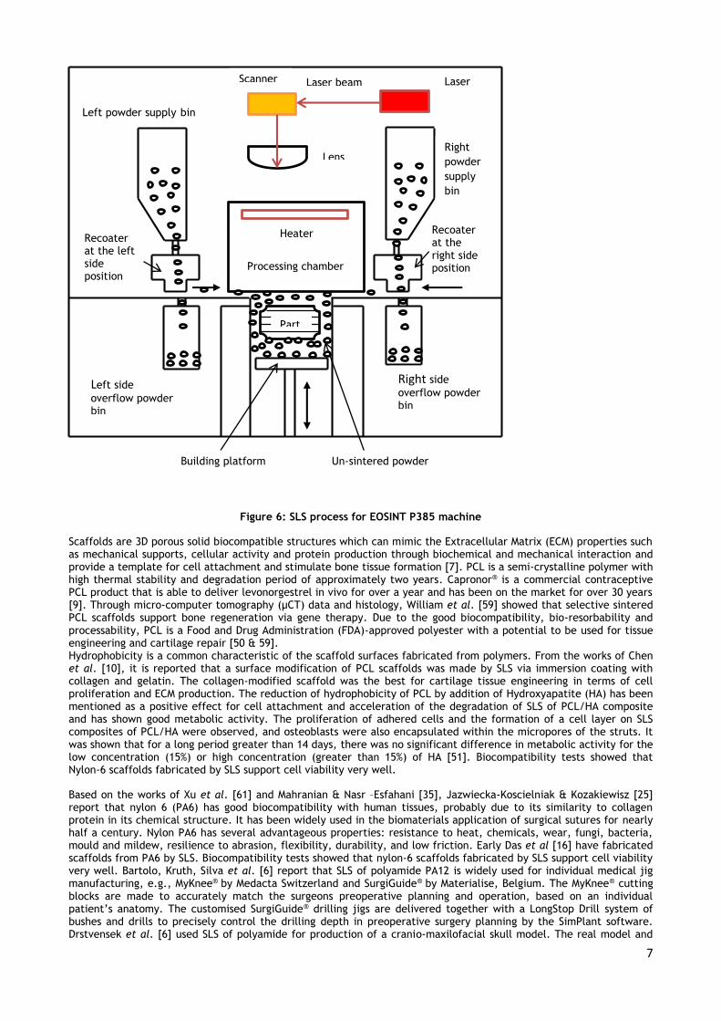
7
Figure 6: SLS process for EOSINT P385 machine
Scaffolds are 3D porous solid biocompatible structures which can mimic the Extracellular Matrix (ECM) properties such as mechanical supports, cellular activity and protein production through biochemical and mechanical interaction and provide a template for cell attachment and stimulate bone tissue formation [7]. PCL is a semi-crystalline polymer with high thermal stability and degradation period of approximately two years. Capronor® is a commercial contraceptive PCL product that is able to deliver levonorgestrel in vivo for over a year and has been on the market for over 30 years [9]. Through micro-computer tomography (µCT) data and histology, William et al. [59] showed that selective sintered PCL scaffolds support bone regeneration via gene therapy. Due to the good biocompatibility, bio-resorbability and processability, PCL is a Food and Drug Administration (FDA)-approved polyester with a potential to be used for tissue engineering and cartilage repair [50 & 59]. Hydrophobicity is a common characteristic of the scaffold surfaces fabricated from polymers. From the works of Chen et al. [10], it is reported that a surface modification of PCL scaffolds was made by SLS via immersion coating with collagen and gelatin. The collagen-modified scaffold was the best for cartilage tissue engineering in terms of cell proliferation and ECM production. The reduction of hydrophobicity of PCL by addition of Hydroxyapatite (HA) has been mentioned as a positive effect for cell attachment and acceleration of the degradation of SLS of PCL/HA composite and has shown good metabolic activity. The proliferation of adhered cells and the formation of a cell layer on SLS composites of PCL/HA were observed, and osteoblasts were also encapsulated within the micropores of the struts. It was shown that for a long period greater than 14 days, there was no significant difference in metabolic activity for the low concentration (15%) or high concentration (greater than 15%) of HA [51]. Biocompatibility tests showed that Nylon-6 scaffolds fabricated by SLS support cell viability very well.
Based on the works of Xu et al. [61] and Mahranian & Nasr –Esfahani [35], Jazwiecka-Koscielniak & Kozakiewisz [25] report that nylon 6 (PA6) has good biocompatibility with human tissues, probably due to its similarity to collagen protein in its chemical structure. It has been widely used in the biomaterials application of surgical sutures for nearly half a century. Nylon PA6 has several advantageous properties: resistance to heat, chemicals, wear, fungi, bacteria, mould and mildew, resilience to abrasion, flexibility, durability, and low friction. Early Das et al [16] have fabricated scaffolds from PA6 by SLS. Biocompatibility tests showed that nylon-6 scaffolds fabricated by SLS support cell viability very well. Bartolo, Kruth, Silva et al. [6] report that SLS of polyamide PA12 is widely used for individual medical jig manufacturing, e.g., MyKnee® by Medacta Switzerland and SurgiGuide® by Materialise, Belgium. The MyKnee® cutting blocks are made to accurately match the surgeons preoperative planning and operation, based on an individual patient’s anatomy. The customised SurgiGuide® drilling jigs are delivered together with a LongStop Drill system of bushes and drills to precisely control the drilling depth in preoperative surgery planning by the SimPlant software. Drstvensek et al. [6] used SLS of polyamide for production of a cranio-maxilofacial skull model. The real model and
Left powder supply bin
Scanner Laser beam Laser
Lens
Heater
Processing chamber
Right
powder
supply
bin
Left side
overflow powder bin
Right side
overflow powder bin
Part
Building platform Un-sintered powder
Recoater at the right side position
Recoater at the left side position
8
the implant were used for testing dimensional accuracy and as a communication tool between the engineer and the medical doctor during the phase of surgery operation planning. Zhang et al. [64] used SLS process to manufacture hydroxyapatite-reinforced polyethylene and polyamide composites as potential customized maxillofacial implants. In vitro tests were carried out to assess cellular responses, in terms of cell attachment, morphology, proliferation, differentiation, and mineralized nodule formation, using primary human osteoblast cells. This study showed that the SLS processed composite implant was biocompatible, with no adverse effects observed on cell viability and metabolic activity, supporting a normal metabolism and growth pattern for osteoblasts. Polyetheretherketone (PEEK) is a semi-crystalline thermoplastic polymer. PEEK combines a very good strength and stiffness with an excellent thermal and chemical resistance. Its mechanical properties remain stable up to temperatures of about 240°C for prolonged periods of time [49]. PEEK is used as a biomaterial for orthopedic implants for toes and fingers, total knee or hip, spinal system, elbow and dental replacements. A further advantage is the reduced implant stiffness that diminishes the stress concentration in the surrounding bone, thus preventing the stress shielding phenomenon and stimulates a quick healing of the patient. Table 1 shows the mechanical properties of the most used materials for fabrication of medical implants, compared to mechanical properties of cortical bone.
Table 1: Mechanical properties of implant materials
Material Elastic modulus (GPa) Yield strength (MPa) Tensile strength (MPa)
Cortical bone 15-30 (tensile) 7-40 (compressive)
30-40 70-15 (tensile) 100-230 (compressive)
PEEK 3.6-8.3 90-170
PCL 23
Titanium 110 485 760
Ti6Al4V 116 896-1034 760-1103
Stainless steels 190 221-1213 586-1351
Co-Cr alloys 210-253 448-1606 655-1896
From Table 1, it can be observed that among the listed materials, mechanical properties of PEEK are close the ones of cortical bone, thus making PEEK a suitable material for orthopedic implants. Schmidt, Pohle & Rechtenwald [48] investigated the influence of the SLS process parameters on mechanical properties of PEEK parts. They concluded that the process of laser sintering PEEK is feasible not only for zero load bearing parts which are applicable for tissue engineering or thin and small scaled parts but it is also feasible for functional and individual shaped parts which fulfil the requirements of nonresorbable implants. Although PEEK is biocompatible, chemically stable, and radiolucent and has an elastic modulus similar to that of normal human bone (Table 1), it is biologically inert, preventing good integration with adjacent bone tissues upon implantation. Based on the work of Rui & Tingting [46], Almasi et al. [2] reviewed the preparation methods for improving PEEK’s bioactivity for orthopedic and dental application. Physical treatment through surface modification, chemical treatment, surface coating (deposition of HA, titanium, gold, titanium dioxide (TiO2), diamond-like carbon (DLC) or tert-butoxides), and making PEEK bioactive composites are the current strategies for improvement of bioactivity of PEEK. The optimal ratio of HA in the composite of PEEK/HA for scaffolds fabrication has been studied by many researchers. Tan et al. [54] found that the viability for the use of PEEK for tissue engineering scaffolds was reiterated by incorporating different amount of HA into PEEK, especially for bones as apatite is one of the natural occurring components in human bones. They concluded that in order to produce a structure with good integrity, it was proposed that the composition of HA in the mixture PEEK/HA powder for SLS process should be kept at 40 wt% HA. The use of nano sized powder particle is the new trend for improvement of the bioactivity and mechanical properties of PEEK/HA composite. Li, Yeung C.Y, Yeung K.W.K & Tjong [30] by SLS process fabricated four ratios namely 15.1, 21.6, 29.2, and 38.2 vol% nano HA (nHA) PEEK/nHA nanocomposites. Their mechanical, thermal and in vitro cell properties were investigated. The tensile strength and fracture strain of PEEK- nanocomposites filled with 21.6 and 29.2 vol% nHA were found to match closely with those of human cortical bones. Sintered 15.1 and 29.2 vol% nHA/PEEK nanocomposites exhibited excellent biocompatibility. The 29.2 vol% nHA/PEEK nanocomposite promoted adhesion and proliferation of mouse and human osteoblasts effectively. Although different bioactive additives such as HA, Ti, TiO2, β-tricalcium phosphate (β-TCP), and bioactive glass improve the bioactivity of PEEK’s composite, it is reported that the low mechanical properties of PEEK’s composite are still the most important weakness. The trend of research in the bioactivity of PEEK shows very encouraging results with a potential to overcome the existing problems in the current techniques for production of bioactive PEEK implants [2].
4.3 Fuse Deposition Modelling (FDM)
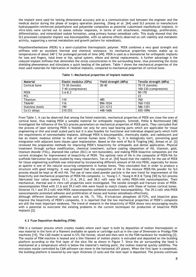
FDM is a nonlaser process which creates models where each layer is built by deposition of molten thermoplastic or wax material in the form of a filament available on spools or cartridge such as in the case of Dimension or Prodigy FDM machines [15]. The CAD model is saved in STL file format, sliced and then sent to the FDM hardware for modelling [3]. The nozzle of FDM machine is heated and a thin filament of molten plastic is extruded and deposited on the building platform according to the first layer of the slice file as shown in Figure 7. Since the air surrounding the head is maintained at a temperature which is below the material’s melting point, the molten material quickly solidifies. The extrusion nozzle controlled by CAM software can move in the horizontal (XY plane). When the first layer is completed, the building platform is lowered by one layer thickness (Z-axis) and the next layer is deposited. The process continues
9
until the entire part is built. Support structures which may be water based are built during the process to support overhanging structures of parts and can be removed manually.
Figure 7: FDM process
For more complicated geometries, it is suitable to remove the water based supports by simply dissolving them in water with detergent in a heated ultrasonic bath [14 & 21]. After the removal of the supports, the FDM part can be viewed as a laminate composite structure with vertically stacked layers of bonded fibers or rasters [65]. The use of FDM in terms of implants has been limited to the fabrication of anatomic models that can serve as templates for the fabrication of custom implants [6]. Gronet, Waskewicz & Richardson [21] used an anatomical model fabricated through FDM process of wax (NeoWax; Dentsply International, York, Pa) material. The model was used for fabrication of acrylic cranial implants for two patients of 35 and 49 years old. FDM for medical application has evolved around PCL. Zein et al. [63] produced porous scaffolds with honeycomb structure having channel size of 160-770 µm. Sharma [49] reports that Endres et al [19] produced PCL and PCL/HA scaffolds with gyroid architecture using the FDM process. A trabecular scaffold was produced through FDM using polybutylene terephthalate (PBT). Compressive trabecular scaffolds matched bone samples in porosity [55]. Other works performed by using extrusion based technologies such as Precision Extruding Deposition (PED) and Low-temperature Deposition Manufacturing (LDM) for fabrication of scaffolds in poly(ethylene glycol)-terephthalatepoly( butylenes terephthalate) (PEGT/PBT) and a blend of poly(butylmethacrylate-methylmethacrylate) (P(BMA/MMA)), poly(ethylene oxideterephtalate)-co-poly(butylene terephtalate) (PEOT/PBT), poly(L-lactide) (PLLA) and TCP composite are reported in Bartolo et al. [6]. Perez et al. [42] conducted a study on sterilization of Acrylonitrile Butadienne Stylene (ABS) derivatives namely ABSi, ABS-M30, ABS-M30i, ABS-ESD7, and a polycarbonate-ABS blend (PC-ABS) parts, manufactured through FDM process. For ABS-M30i which is biocompatible material, as per ISO 10993 USP Class VI, thus making it a good candidate for use in the medical, pharmaceutical, and food packaging industries, it was found that the autoclave sterilization does not give good results as bending and indentation were very apparent on all ABS derivative parts. However EtO, hydrogen peroxide gas and gamma radiation presented good sterilization results for ABS-M30i.
Support material spool Building material spool
Thermoplastic filaments
Drive wheels
x,y-
travels
Part
Build platform moving in Z-
direction
Support
Extrusion nozzle
x
y
z
10
4.4 3D Printing (3DP)
3D Printing (3DP) also known as Binder Jet printing system uses a print head to selectively disperse a binder onto a thin layer of powder that is spread over a tray using a roller. The print head scans the powder tray and delivers a continuous jet of a solution that binds the powder particles as it touches them. There is no need for support structures in 3DP as the surrounding powder, supports the unconnected parts. In the finishing process, the surfaces of the built model are infiltrated with a cyanoacrylate-based material to harden the structure. Compared with other 3D technologies, 3DP is quicker, easier and more cost effective. The extensive use of 3DP for fabrication of medical models for preoperational surgery of mandibular and maxillofacial reconstruction has been published by Choi & Kim [12] and Olszewski & Reyshler [41].
4.5 Polyjet
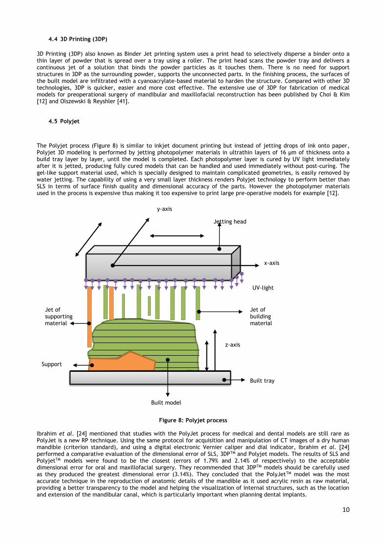
The Polyjet process (Figure 8) is similar to inkjet document printing but instead of jetting drops of ink onto paper, Polyjet 3D modeling is performed by jetting photopolymer materials in ultrathin layers of 16 μm of thickness onto a build tray layer by layer, until the model is completed. Each photopolymer layer is cured by UV light immediately after it is jetted, producing fully cured models that can be handled and used immediately without post-curing. The gel-like support material used, which is specially designed to maintain complicated geometries, is easily removed by water jetting. The capability of using a very small layer thickness renders Polyjet technology to perform better than SLS in terms of surface finish quality and dimensional accuracy of the parts. However the photopolymer materials used in the process is expensive thus making it too expensive to print large pre-operative models for example [12].
Figure 8: Polyjet process
Ibrahim et al. [24] mentioned that studies with the PolyJet process for medical and dental models are still rare as PolyJet is a new RP technique. Using the same protocol for acquisition and manipulation of CT images of a dry human mandible (criterion standard), and using a digital electronic Vernier caliper and dial indicator, Ibrahim et al. [24] performed a comparative evaluation of the dimensional error of SLS, 3DPTM and Polyjet models. The results of SLS and PolyjetTM models were found to be the closest (errors of 1.79% and 2.14% of respectively) to the acceptable dimensional error for oral and maxillofacial surgery. They recommended that 3DPTM models should be carefully used as they produced the greatest dimensional error (3.14%). They concluded that the PolyJetTM model was the most accurate technique in the reproduction of anatomic details of the mandible as it used acrylic resin as raw material, providing a better transparency to the model and helping the visualization of internal structures, such as the location and extension of the mandibular canal, which is particularly important when planning dental implants.
z-axis
Built tray
Jet of supporting material
Built model
Support
Jet of building material
y-axis
x-axis
Jetting head
UV-light

11
Salmi et al [47] used a Coordinate Measuring Machine (CMM) ZEISS C 700 model with RENISHAW PH 1 measuring tip and carried out a comparative study on the accuracy of SLS, Polyjet and 3DP skull models with a particular focus on the repeatability of the results. With multiple measurements, the distance between the measuring balls and the exact location of the measuring head could be used to determine the centre points of the measuring balls in the same coordinates as the CMM. By using the measuring balls and determining their centres, they were able to achieve excellent repeatability of the measurements for the 3DP (0.08%) and for the PolyJet (0.12%) when using the novel method of measuring the centre point of balls. The PolyJet technique resulted in the least errors (0.18 ±0.12% & 0.18±0.13%) compared with SLS (0.79±0.26% & 0.80±0.32%) or 3DP (0.67±0.43% & 0.69, 0.44%, 0.38±0.22% & 0.55±0.37%). Recently, Drstvensek et al. [18]. used Polyjet technology for manufacturing of skull implant model to be later used as a pattern for silicone rubber molding.
4.6 Electrospinning and nano fabrication technology
Although the term ‘‘electrospinning’’, derived from ‘‘electrostatic spinning’’, was used around 1994, from 1934 to 1944, Formhals published a series of patents describing an experimental setup for the production of polymer filaments using an electrostatic force [23]. Electrospinning (Figure 9) is an AM technology that combines electrospraying and spinning processes for the production of nanofibers from a wide range of polymeric materials. It is simple and versatile process by which nanofibers with diameters ranging from a few nanometres to several micrometres can be produced using an electrostatically driven jet of polymer solution (solution electrospinning) or polymer melt (melt electrospinning). The list of polymeric materials which can be electrospun is provided by Huang et.al. [23], Bartolo et al.[6], Agarwal, Wendorff & Greiner [1].
Figure 9: Electrospinning process
A typical electrospinning setup only requires a high voltage power supply, a syringe, a flat tip needle and a conducting collector. By creation of a potential voltage difference between the polymer solution and the collection plate, electrostatic forces overcome the solution surface tension to pull a jet of charged fluid that splits into nanofibers that fall towards the collection plate and solidify. The polymer jet splits into multiple nanofibers that are deposited at the collector. The solvent evaporates as the jet is electrospun and leaving dry nanofibers on the collector [62]. The important advantages of electrospinning technique are the production of very thin fibers to the order of few nanometers with very large surface area to volume ratio, flexibility in surface functionality for various purposes, superior mechanical properties and ease of process as suggested by many experts in this field. The possibility of large scale productions combined with the simplicity of the process makes this technique attractive for many different applications [1].
High voltage power supply
Polymeric jet
Needle
Polymer solution Syringe driver
Nanofiber formation
Conducting plate
(collector)

12
Electrospinning technology provides a wide range of medical applications including tissue engineering, scaffold fabrication, implants, drug delivery and biotransformation to wound healing. From a biological point of view, almost all of human tissues and organs including bone, dentin, collagen, cartilage and skin, are deposited in nanofibrous forms or structures. Electrospun biocompatible polymers nanofibers can be used as a thin porous film to be deposited onto a hard tissue prosthetic implant. Nisbet et al. [40] investigated the neurite infiltration and cellular response to electrospun PCL scaffolds implanted into the brain. The study showed that neurite extension within the implanted PCL scaffolds has occurred indicating that the electrospun PCL has integrated with the brain circuitry and was not being encapsulated by fibrous tissue. Hu et al. [22] reviewed the application of electrospinning for polymeric nanofibers in drug delivery, including carrier materials, loaded drugs and their release kinetics, and illustrated their application for local chemotherapy. However a repeatable massive uniform nanofiber fabrication, at industrial scale, with desired morphology, mechanical and chemical properties, remained a challenge. In addition to that, there was still a need of conducting intensive in vivo to support a good number of in vitro studies.
5. CONCLUSION
Recent findings in literature on application of the six most used AM technologies for polymeric medical implants, scaffolds and medical devices for drug delivery are reviewed in this paper. Due to the specific characteristics of each AM technology and the biocompatibility of polymers, the limitation of suitable polymeric materials for medical application is still a challenge for future research. The copolymerization and the use of the composite polymer / hydroxyapatite have been the ways to improve mechanical properties and biocompatibility of homopolymers. For the SLS process, studies have shown that the development of nanosized powder for PEEK bioactive composites will be a promising solution to obtain both mechanical and biological benefits in the field of medical implantation. Electrospinning which uses a wide range of polymers could also be another alternative to introduce and to promote nanofiber technology in the medical industry of emerging and developing countries. The establishment of standards for assessment of the dimensional accuracy of additive manufactured medical models will be a new achievement for medical industry.
6. ACKNOWLEDGMENT
This work is based on the research supported by the South African Research Chairs Initiative of the Department of
Science and Technology and National Research Foundation of South Africa (Grant №97994) and the Collaborative
Program in Additive Manufacturing (Contract №CSIR-NLC-CPAM-15-MOA-CUT-01).
REFERENCES
[1] Agarwal, S., Wendorff, J.H., & Greiner, A. 2008. Use of electrospinning technique for biomedical applications, Polymer 49, pp 5603-5621.
[2] Almasi, D., Iqbal, N., Sadeghi, M., Sudin, I., Kadir, M.R.A., & Kamarul, T. 2016. Preparation methods for improving PEEK’s bioactivity for orthopedic and dental application: a review, International Journal of Biomaterials 2016, pp 1-12.
[3] Anoop, K.S., Vedansh, C., Saurav, D. & Siba, S.M. 2011. Optimization of process parameters in Fused Deposition Modeling using weighted principal component analysis, Journal of Advanced Manufacturing Systems 10(2), pp 241-259.
[4] Azari, A. & Nikzad, S. 2009. The evolution of rapid prototyping in dentistry: a review, Rapid Prototyping, Journal 15 (3), pp 216–225.
[5] Balazic, M & Kopac, J. 2007. Improvements of medical implants based on modern materials and new technologies, Journal of Achievements in Materials and Manufacturing 25(2), pp 31-34.
[6] Bartolo, P., Kruth, J.P., Silva, J. et al. 2012. Biomedical production of implants by additive electro-chemical and physical processes, CIRP Annals - Manufacturing Technology 61, pp 635-655.
[7] Bose, S., Vahabzadeh, S. & Bandyopadyyay, A. 2013. Bone tissue engineering using 3D printing, Material Today 16(12), pp 496-504.
[8] Brendel, C. M. 2009. Biocompatibility of polymer implants for medical applications, Master’s thesis, The Graduate Faculty of The University of Akron.
[9] Bret, D. Ulery, B.D., Nair, L.S., & Laurencin, C.T. 2011, Biomedical applications of biodegradable polymers, J Polym Sci B Polym Phys. 49(12), pp 832–864.
[10] Chen, C.H., Lee M.Y., Shyu, V.B.H., Chen, Y.C., Chen, C.T. & Chen, J.P. 2014. Surface modification of polycaprolactone scaffolds fabricated via selective laser sintering for cartilage tissue engineering, Mater. Sci. Eng. C 40, pp 389–397.
[11] Choi, C.Q. 2007. World’s first prosthetic: Egyptian mummy’s Fake Toe, life Science, 27 July 2007 (www.lifescience.com/4555-world prosthetic-Egyptian-mummy-fake toe.html) retrieved on 11 June 2016.

13
[12] Choi,J.W. & Kim, N. 2015. Clinical Application of Three-Dimensional Printing Technology in Craniofacial Plastic Surgery, Archives of Plastic Surgery 42(3), pp 267-277.
[13] Clay, G. &Taylor, D.P.M. [nd.]. Absorbable fixation devices, [s.n]. [14] Daneshmand, S. & Aghanajafi, C. 2012. Description and modelling of additive manufacturing technology for
aerodynamic coefficient measurement, Journal of mechanical Engineering 58(2), pp 125-133. [15] Daneshmand, S., Aghanajafi, C. & Shahverdi, H. 2013. Investigation of rapid manufacturing technology effect
on aerodynamics properties, Tehnicki Vjesnik 20(3), pp 425-433. [16] Das, S., Hollister, S.J, Flanagan, C., Adewunmi, A., Bark, K., Chen, C., Ramaswamy, K., Rose, D. & Widjaja,
E. 2003. Freeform fabrication of Nylon-6 tissue engineering scaffolds, Rapid Prototyping Journal. 9, pp 43-49. [17] Dhandayuthapani, B.,Yoshida, Y., Maekawa,T. & Kumar, D.S. 2011. Polymeric scaffolds in tissue engineering
application: A review, International Journal of Polymer Science, pp 1-19. [18] Drstvensek, I., Hren, N.I., Strojnik, T. et al. 2015. Applications of Rapid Prototyping in cranio-maxillofacial
surgery procedures, International Journal of Biology and Biomedical Engineering 1(2), pp 29-38. [19] Endres, M., Hutmacher, D.W., Salgado, A.J., Kaps, C., Ringe, J., Reis, R.L., Sittinger, M, Brandwood, A. &
Schantz, J.T. 2004. Osteogenic Induction of human bone marrow –derived mesenchymal projenitor cells in novel synthetic polymer hydrogel-matrices, Tissue Engineering 9(4), pp 689-702.
[20] Green, S. & Schlegel, J., [n.d.]. A Polyaryletherketone biomaterial for use in medical Implant applications, [s.n.].
[21] Gronet, P.M., Waskewicz, G.A. & Richardson, C. 2003. Performed acrylic cranial implants using fused deposition modelling: a clinical report, The Journal of Prosthetic dentistry 90(5), pp 429-433.
[22] Hu, X., Liu, S., Zhou, G., Huang, Y., Xie, Z.,and Jing, X. 2014. Electrospinning of polymeric nanofibers for drug delivery applications, Journal of Controlled Release 185, pp 12-21.
[23] Huang, Z.M., Zang, Y.Z., Kotaki, M. & Ramakrishna, S. 2003. A review on polymer nanofibers by electrospinning and their applications in nanocomposites, Composites Science & Technology 3, pp 2223-2253.
[24] Ibrahim, D. et al. 2009. Dimensional error of selective laser sintering, Three –dimensional printing and PolyjetTM model in production of mandibular anatomy, Journal of Craniomaxillofacial Surgery 37, pp 167-173.
[25] Jazwiecka-Koscielniak, E. & Kozakiewicz, M. 2014. A new modification of individually designed polymer implant visible in X-ray for orbital reconstruction, Journal of Craniomaxillofacial Surgery 42, pp 1520-1529.
[26] Kaufui, V.W. & Aldo, F. 2012. A review of Additive Manufacturing. ISRB Mechanical Engineering 2012, pp 1-10. [27] Khan, W., Muntimadugu, E., Jaffe, M. & Domb, A.J. 2014. Implantable medical devices, Focal Controlled Drug
Delivery, Advances in Delivery Science and Technology. [28] Kolan, K.C.R., Leu, M.C., Hilmas, G.E. & Comte, T. [n.d]. Effect of Architecture and Porosity on Mechanical
Properties of Borate Glass Scaffolds made by Selective Laser Sintering, [s.n], pp 816-826. [29] Kruth, J.P., G. Levy, G., F. Klocke, F. & Childs, T.H.C. 2007. Consolidation phenomena in laser and powder-
bed based layered manufacturing, Annals of the CIRP 56 (2), pp 730-759. [30] Li, K., Yeung, C.Y., Yeung, K.W.K., & Tjong, S.C. 2012. Sintered Hydroxyapatite/Polyetheretherketone
nanocomposites: mechanical and biocompatibility, Advanced Engineering materials, 14(4), pp B155-B165. [31] Liron, N. 2005. An investigation of performance of low cost rapid prototyping machines, MSc. thesis, Dept. of
Engineering & Technology, University of Montfort. [32] Ma, R. & Tang, T. 2014. Current strategies to improve the bioactivity of PEEK, International Journal of
Molecular Sciences 15, pp 5426-5445. [33] Manfredi, D., Calignano, F., Krishnan, M., Canali, R., Ambrosio, E.P., Biamino, S., Urgues, D., Pavese, M. &
Fino, P. 2014. Additive manufacturing of Al alloys and Aluminium Matrix Composites (AMCs), INTECH open science/open minds.
[34] Matsuda, T. & Mizutani, M. 2002. Liquid acrylate-endcapped biodegradable poly(e-caprolactone-co-trimethylene carbonate). II. Computer-aided stereolitho-graphic microarchitectural surface photoconstructs, J Biomed Mater Res 62(3): pp 395-403.
[35] Mehrabanian, M, Nasr-Esfahani, M. 2011. HA/nylon 6,6 porous scaffolds fabricated by saltleaching/solvent casting technique: effect of nano-sized filler content on scaffold properties. Int J Nanomed 6, pp 1651-1659.
[36] Melchels, F.P.W., Domingosc, M.A.N., Travis J. Kleina, T.J. et al. 2012. Additive manufacturing of tissues and organs, Progress in Polymer Science 37, pp 1079-1104.
[37] Melchels, F.P.W., Feijen, J. & W.Grijpma, D.W. 2010. A review on stereolithography and its applications in medical engineering, Biomaterials 31, pp 6121-6130.
[38] Middleton, J.C. & Tipton, A.J. 2000. Synthetic biodegradable polymers as orthopedic devices, Biomaterials 21, pp 2335-2346.
[39] Nag, S. & Banerjee, R. 2012. Fundamentals of medical implant materials, Materials for Medical Devices 23, pp 6-17.
[40] Nisbet, D.R., Rodda, E.E., Horne, M.K., Forsynthe, J.S. & Finkelstein, D.I. 2009. Neurite infiltration and cellular response to electrospun polycaprolactone scaffolds implanted into the brain, Biomaterials 30, pp 4573-4580.
[41] Olszewski, R. & Reychler, H. 2011. Clinical applications of rapid prototyping models in cranio-maxillofacial surgery, Advanced Applications of Rapid Prototyping Technology in Modern Engineering, pp 173-206.
[42] Perez, M., Block, M., Espalin, D., Winker, R., Hoppe, T., Medina, K., Wicker, R. 2012. Sterilization of FDM-manufactured parts, [s.l.], pp 285-296.
[43] Popov, V.K., Evseev, A.V., Ivanov, A.L. et al. 2004. Laser stereolithography and supercritical fluid processing for custom-designed implant fabrication, Journal of Materials Science: Materials in Medicine 15, pp 123-128.
[44] Prashant, K.J., Senthilkumaran, K., Pandey, P.M. & Rao, P.V.M. 2006. Advances in materials for powder based rapid prototyping, International Conference on Recent Advances in Materials and Processing, December 15-16, 2006, PSG-tech. Coimbatore, INDIA.

14
[45] Raghunath, N. and Pandey, M.P. 2007. Improving accuracy through shrinkage modelling by using Tagushi method in Selective laser Sintering, International Journal of Machine Tools & Manufacture 47, pp 985-995.
[46] Rui, M. & Tingting, T. 2014. Current strategies to improve bioactivity of PEEK, International Journal of Molecular Sciences. pp 5426-5445.
[47] Salmi, M. 2013. Accuracy of medical models made by additive manufacturing (rapid prototyping), Journal of Cranio-Maxillo-Facial Surgery 41, pp 603-609.
[48] Schmidt, M., Pohle, D., & Rechtenwald, T. 2007. Selective laser sintering of PEEK, Annals of CIRP 56, pp 205-208.
[49] Sharma, C.P. 2010. Biointegrating medical implant materials, Woodhead Publishing Limited. [50] Shirazi, S.F.S., Gharehkhani, S., Mehrali, M. et al. 2015. A review on powder-based additive manufacturing for
tissue engineering: Selective sinter sintering and Inkjet 3D printing, Science and Technology of Advanced Materials 16, pp 1-20.
[51] Singh, G.B. & Kumar, P. 2014. Methods to improve surface finish of parts produced by Fused Deposition Modelling, Manufacturing Science and Technology 2(3), pp 51-55.
[52] Susan Hurrell, S., Milroy, G.E. & Cameron, R.E. 2003. The Degradation of Polyglycolide in Water and Deuterium Oxide. Part I: The Effect of Reaction Rate, Polymer 44, pp 1421–1424.
[53] Taft, R.M., Kondor, S. & Grant, G. 2011. Accuracy of rapid prototype models for head and neck reconstruction, The Journal of Prosthetic Dentistry 106(6), pp 399-408.
[54] Tan, K.H., Chua, C.K.., Leong, K.F. et al. 2003. Scaffolds development using selective laser sintering of polyetheretherketone-hydroxyapatite biocomposite blends, Biomaterials 24(18), pp 3115-3123.
[55] Tellis, B.C., Szivek, J.A., Bliss, C.L, Margolis, D.S., Vaidyanathan, R.K., Calvert, P. 2008. Trabecular Scaffold Created Using Micro CT Guided Fused Deposition Modelling, Materials Science Engineering C 28, pp 171–178.
[56] Vojislav, P., Haro, J.V., Blasco, J.R. & Portolés, L. 2012. Additive Manufacturing Solutions for Improved Medical Implants, Biomedicine, pp 147-180.
[57] Whipps, H. & Britt, R.R. 2009. Humans 2.0: Replacing the mind and the body, Life Science, 2 August 2009 retrieved on 11 June 2016 from www.lifescience.com/9699-humans-2-0-replacing-mind-body.html.
[58] Williams, J.M., Adewunmi, A., Schek, R.M., Flanagan, C.L., Krebsbach, P.H., Feinberg, S.E., Hollister, S.J. & Das, S. 2005. Bone tissue engineering using polycaprolactone scaffolds fabricated via selective laser sintering, Biomaterials 26(23), pp 4817-27.
[59] Wiria, F.E., Leong, K.F., Chua, C.K., & Liu, Y. 2007. Poly-ε-Caprolactone/Hydroxyapetite for tissue engineering scaffold fabrication via selective laser sintering, Acta Biomatirialia 3, pp 1-12.
[60] Wohlers, T. & Gornet, T. 2011. History of additive manufacturing, Wohlers Report 2011, State of the Industry, WOHLERS ASSOCIATES, INC.
[61] Xu, Q., Lu, H., Zhang, J., Lu, G., Deng, Z. & Mo, A. 2010. Tissue engineering scaffold material of porous nano-hydroxyapatite/polyamide 66, Int J Nanomed 13, pp 331-335.
[62] Zafar, M., Najeeb, S., Kurshid, Z.et al. 2016. Potential of electrospun nanofibers for biomedical and dental applications, Materials 9(73), pp 1-21.
[63] Zein, I., Hutmacher, D.T., Tan, K.C., & Teoh, S.H. 2002. Fused deposition modelling of novel scaffold architectures for tissue engineering applications, Biomaterials 23(4), pp 1169-1185.
[64] Zhang, Y.; Hao, L.; Savalani, M.M.; Harris, R.A.; di Silvio, L.; Tanner, K.E. 2009. In vitro biocompatibility of hydroxyapatite-reinforced polymeric composites manufactured by selective laser sintering, Journal of Biomedical. Material.
[65] Ziemian, C., Sharma, M. & Ziemian, S. 2012. Anisotropic Mechanical properties of ABS parts fabricated by Fused Deposit Modelling, Mechanical Engineering, Dr. Murat Gokcek (Ed.), In Tech, pp 159-180.