additive manufacturing of biomaterials, tissues, and organs · additive manufacturing of...
TRANSCRIPT

Editorial
Additive Manufacturing of Biomaterials, Tissues, and Organs
()
Abstract—The introduction of additive manufacturing(AM), often referred to as three-dimensional (3D) printing,has initiated what some believe to be a manufacturingrevolution, and has expedited the development of the fieldof biofabrication. Moreover, recent advances in AM havefacilitated further development of patient-specific health-care solutions. Customization of many healthcare productsand services, such as implants, drug delivery devices,medical instruments, prosthetics, and in vitro models, wouldhave been extremely challenging—if not impossible—with-out AM technologies. The current special issue of theAnnals of Biomedical Engineering presents the latest trendsin application of AM techniques to healthcare-related areasof research. As a prelude to this special issue, we review herethe most important areas of biomedical research and clinicalpractice that have benefited from recent developments inadditive manufacturing techniques. This editorial, there-fore, aims to sketch the research landscape within which theother contributions of the special issue can be betterunderstood and positioned. In what follows, we brieflyreview the application of additive manufacturing techniquesin studies addressing biomaterials, (re)generation of tissuesand organs, disease models, drug delivery systems, implants,medical instruments, prosthetics, orthotics, and AM objectsused for medical visualization and communication.
Keywords—Bioprinting, Biofabrication, Biomaterials, Drug
delivery, Medical devices, Tissue regeneration.
INTRODUCTION
Additive manufacturing (AM), also known as 3Dprinting, has emerged during recent years as a flexibleand powerful technique for advanced manufacturing inhealthcare. Even though the underlying technology hasbeen in development for more than two decades, thelevel of maturity and perfection required for real-worldapplications has been achieved only recently. Mostimportantly, a wide range of biomedical materials cannow be processed using additive manufacturing tech-niques with increasing accuracy. Moreover, a number ofAM processes and the resulting products have alreadybeen approved by regulatory bodies for (routine) clini-
cal use, and a draft ver-sion of FDA guidance foradditively manufactureddevices has already beenpublished.1 At the sametime, AM technology hasbeen applied for (re)gen-eration of living tissuestructures that could beapplied as regenerativeimplants and diseasemodels. This field of‘‘biofabrication’’28 isdeveloping exponentially,underscoring the poten-tial of applying AM inhealthcare. Some otherareas, such as pharma-cology, oncology, sur-gery, and rehabilitationhave also provided inter-esting clinical and researchapplications for additivemanufacturing.
The current special is-sue aims to review andshowcase some of themost promising trends inapplication of AM tohealthcare. This reviewand the research articlespresented here cover a wide range of applications, rang-ing from cardiovascular19 to orthopedic,9,10,58 craniofa-cial,50 and drug screening.71 As a prelude to this specialissue, we decided to write an extended editorial andbriefly review the most important trends in application ofAM to healthcare, thereby setting the stage for whatappears in the rest of the issue. In that sense, this editorialmight be seen as a ‘‘review of reviews,’’ where we do nottry to engage in the details of various areas of researchbut rather to sketch the bigger picture through clearexamples, reference to the papers appearing in this special
Amir A. Zadpoor
Jos Malda
1Food and Drug Administration (FDA), Technical Considerations
for Additive Manufactured Devices—Draft Guidance for Industry
and Food and Drug Administration Staff, Issued on May 10, 2016.
Annals of Biomedical Engineering, Vol. 45, No. 1, January 2017 (� 2016) pp. 1–11
DOI: 10.1007/s10439-016-1719-y
0090-6964/17/0100-0001/0 � 2016 Biomedical Engineering Society
1
issue, as well as to the other essential literature. In par-ticular, we have not tried to review the details of AMtechniques (Fig. 1), as these can be found in some of theexcellent review papers appearing in this issue; see, e.g.,Refs. 32, 48, 53, 79. Instead, the areas where AM couldimprove the quality of healthcare are reviewed in thefollowing sections of this editorial, which are organized inorder of perceived impact.
ADDITIVE MANUFACTURING
OF BIOMATERIALS
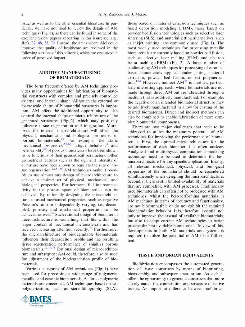
The form freedom offered by AM techniques pro-vides many opportunities for fabrication of biomate-rial constructs with complex and precisely controlledexternal and internal shape. Although the external ormacroscale shape of biomaterial structures is impor-tant, AM offers the additional opportunity to alsocontrol the internal shape or microarchitecture of thegenerated structures (Fig. 2), which may positivelyinfluence tissue regeneration and integration. More-over, the internal microarchitecture will affect thephysical, mechanical, and biological properties ofporous biomaterials;75 For example, the staticmechanical properties,2,8,60 fatigue behavior,4 andpermeability69 of porous biomaterials have been shownto be functions of their geometrical parameters. Othergeometrical features such as the sign and intensity ofcurvature have been shown to regulate the rate of tis-sue regeneration.22,57,75 AM techniques make it possi-ble to use almost any design of microarchitecture toachieve a desired set of physical, mechanical, andbiological properties. Furthermore, full interconnec-tivity in the porous space of biomaterials can beachieved. By rationally designing the microarchitec-ture, unusual mechanical properties, such as negativePoisson’s ratio or independently varying, i.e., decou-pled, porosity and mechanical properties, can beachieved as well.76 Such rational design of biomaterialmicroarchitecture is something that fits within thelarger context of mechanical metamaterials and hasreceived increasing attention recently.73 Furthermore,the microarchitecture of biodegradable biomaterialsinfluences their degradation profile and the resultingtissue regeneration performance of (highly) porousbiomaterials.13,14,78 Rational design of microarchitec-ture and subsequent AM could, therefore, also be usedfor adjustment of the biodegradation profile of bio-materials.
Various categories of AM techniques (Fig. 1) havebeen used for processing a wide range of polymeric,metallic, and ceramic biomaterials. As far as polymericmaterials are concerned, AM techniques based on vatpolymerization, such as stereolithography (SLA),
those based on material extrusion techniques such asfused deposition modeling (FDM), those based onpowder bed fusion technologies such as selective lasersintering (SLS), and material jetting alternatives, suchas inkjet printing, are commonly used (Fig. 1). Themost widely used techniques for processing metallicbiomaterials are currently based on powder bed fusion,such as selective laser melting (SLM) and electronbeam melting (EBM) (Fig. 2). A large number ofstudies using AM techniques for processing of ceramic-based biomaterials applied binder jetting, materialextrusion, powder bed fusion, or vat polymeriza-tion.1,64 However, indirect AM32 is another, particu-larly interesting approach, where biomaterials are notmade through direct AM but are fabricated through amedium that is additively manufactured; For example,the negative of an intended biomaterial structure maybe additively manufactured to allow for casting of thedesired biomaterial. Direct and indirect methods canalso be combined to enable fabrication of more com-plex biomaterial components.
There are two major challenges that need to beaddressed to utilize the maximum potential of AMtechniques for improving the performance of bioma-terials. First, the optimal microarchitecture for theperformance of each biomaterial is often unclear.Analytical and multiphysics computational modelingtechniques need to be used to determine the bestmicroarchitecture for any specific application. Ideally,all relevant mechanical, physical, and biologicalproperties of the biomaterial should be consideredsimultaneously when designing the microarchitecture.Secondly, there is still limited availability of materialsthat are compatible with AM processes. Traditionallyused biomaterials can often not be processed with AMtechniques, whilst the best-performing materials inAM machines, in terms of accuracy and functionality,are not biocompatible or do not exhibit the requiredbiodegradation behavior. It is, therefore, essential notonly to improve the arsenal of available biomaterials,but also to adapt current AM technologies to betterprocess the best available biomaterials. In view of this,developments in both AM materials and systems isrequired to utilize the potential of AM to its full ex-tent.
TISSUE AND ORGAN EQUIVALENTS
Biofabrication encompasses the automated genera-tion of tissue constructs by means of bioprinting,bioassembly, and subsequent maturation. As such, itoffers the opportunity to generate constructs that moreclosely match the composition and structure of nativetissues. An important difference between biofabrica-
A. A. ZADPOOR AND J. MALDA2
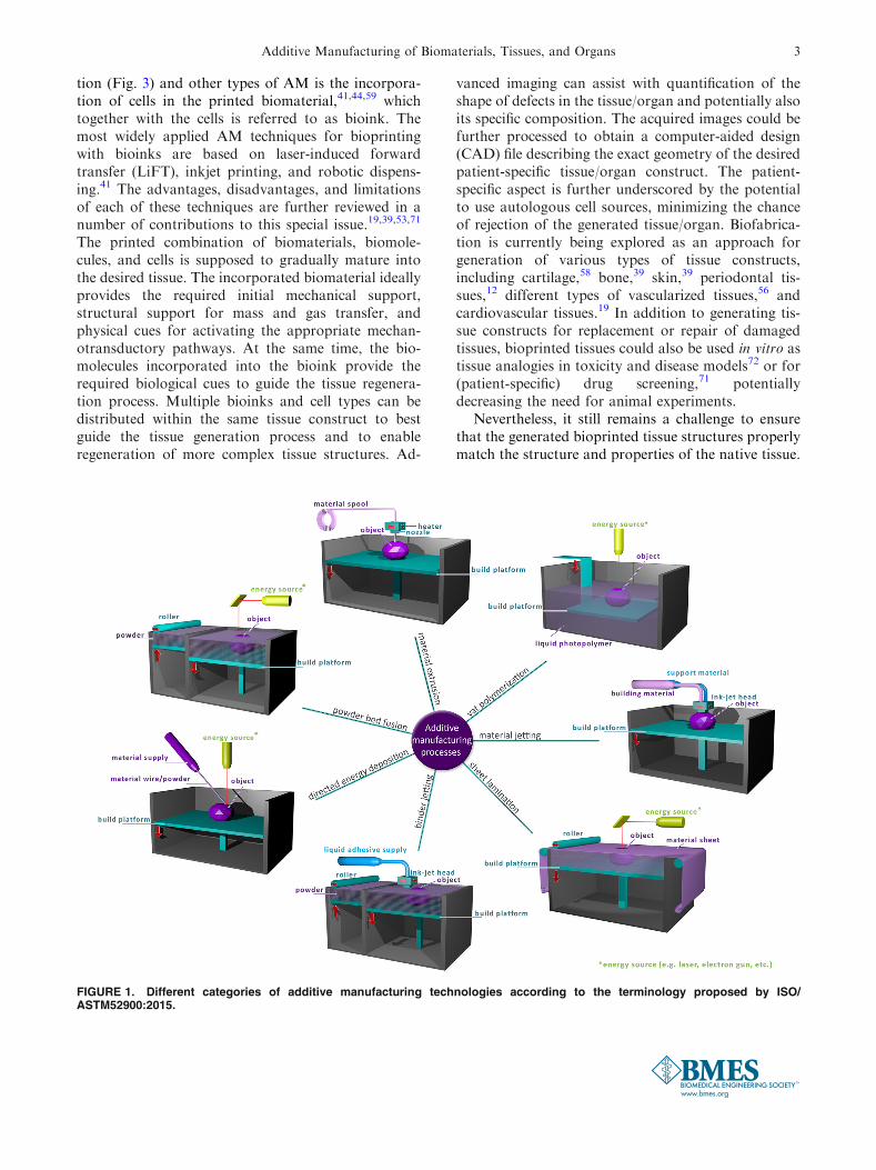
tion (Fig. 3) and other types of AM is the incorpora-tion of cells in the printed biomaterial,41,44,59 whichtogether with the cells is referred to as bioink. Themost widely applied AM techniques for bioprintingwith bioinks are based on laser-induced forwardtransfer (LiFT), inkjet printing, and robotic dispens-ing.41 The advantages, disadvantages, and limitationsof each of these techniques are further reviewed in anumber of contributions to this special issue.19,39,53,71
The printed combination of biomaterials, biomole-cules, and cells is supposed to gradually mature intothe desired tissue. The incorporated biomaterial ideallyprovides the required initial mechanical support,structural support for mass and gas transfer, andphysical cues for activating the appropriate mechan-otransductory pathways. At the same time, the bio-molecules incorporated into the bioink provide therequired biological cues to guide the tissue regenera-tion process. Multiple bioinks and cell types can bedistributed within the same tissue construct to bestguide the tissue generation process and to enableregeneration of more complex tissue structures. Ad-
vanced imaging can assist with quantification of theshape of defects in the tissue/organ and potentially alsoits specific composition. The acquired images could befurther processed to obtain a computer-aided design(CAD) file describing the exact geometry of the desiredpatient-specific tissue/organ construct. The patient-specific aspect is further underscored by the potentialto use autologous cell sources, minimizing the chanceof rejection of the generated tissue/organ. Biofabrica-tion is currently being explored as an approach forgeneration of various types of tissue constructs,including cartilage,58 bone,39 skin,39 periodontal tis-sues,12 different types of vascularized tissues,56 andcardiovascular tissues.19 In addition to generating tis-sue constructs for replacement or repair of damagedtissues, bioprinted tissues could also be used in vitro astissue analogies in toxicity and disease models72 or for(patient-specific) drug screening,71 potentiallydecreasing the need for animal experiments.
Nevertheless, it still remains a challenge to ensurethat the generated bioprinted tissue structures properlymatch the structure and properties of the native tissue.
FIGURE 1. Different categories of additive manufacturing technologies according to the terminology proposed by ISO/ASTM52900:2015.
Additive Manufacturing of Biomaterials, Tissues, and Organs 3
A current limitation is the limited availability ofbioinks that possess appropriate physical propertiesfor the printing process and simultaneously provide asuitable niche for the cells to differentiate towards thedesired lineage. Different classes of hydrogels havebeen employed as parts of bioink systems used in tis-sue/organ bioprinting.41,43,45,55,62
A promising approach to simultaneously complywith the numerous requirements that AM techniquesand bioinks must satisfy to guarantee optimal tissuequality and maximum tissue complexity is to combinevarious AM technologies and bioinks to benefit fromthe best aspects of different approaches. Recentapplication of this pragmatic approach has producedsome promising results.36
DRUGS AND DRUG DELIVERY
Various techniques for drug administration anddelivery devices including solid dosage forms,26
implantable drug delivery vehicles,23,33 and topicaldrug delivery systems25 could benefit from what AMhas to offer. The recent approval of an AM drugproduct by the FDA in August 201549 marked the
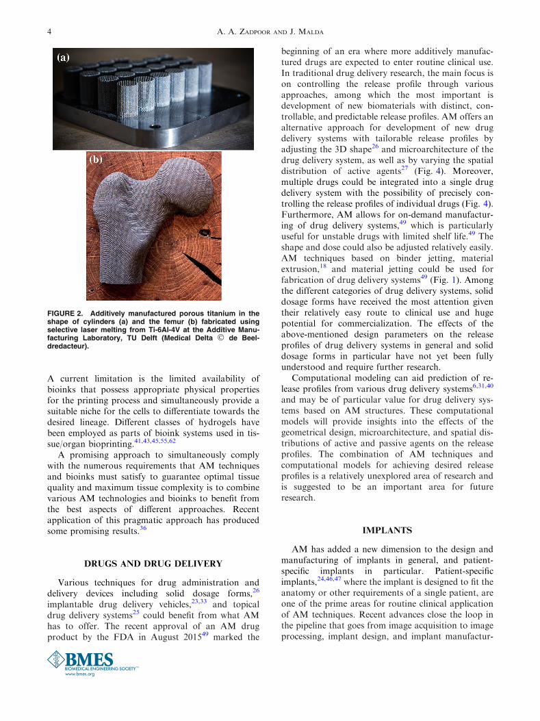
beginning of an era where more additively manufac-tured drugs are expected to enter routine clinical use.In traditional drug delivery research, the main focus ison controlling the release profile through variousapproaches, among which the most important isdevelopment of new biomaterials with distinct, con-trollable, and predictable release profiles. AM offers analternative approach for development of new drugdelivery systems with tailorable release profiles byadjusting the 3D shape26 and microarchitecture of thedrug delivery system, as well as by varying the spatialdistribution of active agents27 (Fig. 4). Moreover,multiple drugs could be integrated into a single drugdelivery system with the possibility of precisely con-trolling the release profiles of individual drugs (Fig. 4).Furthermore, AM allows for on-demand manufactur-ing of drug delivery systems,49 which is particularlyuseful for unstable drugs with limited shelf life.49 Theshape and dose could also be adjusted relatively easily.AM techniques based on binder jetting, materialextrusion,18 and material jetting could be used forfabrication of drug delivery systems49 (Fig. 1). Amongthe different categories of drug delivery systems, soliddosage forms have received the most attention giventheir relatively easy route to clinical use and hugepotential for commercialization. The effects of theabove-mentioned design parameters on the releaseprofiles of drug delivery systems in general and soliddosage forms in particular have not yet been fullyunderstood and require further research.
Computational modeling can aid prediction of re-lease profiles from various drug delivery systems6,31,40
and may be of particular value for drug delivery sys-tems based on AM structures. These computationalmodels will provide insights into the effects of thegeometrical design, microarchitecture, and spatial dis-tributions of active and passive agents on the releaseprofiles. The combination of AM techniques andcomputational models for achieving desired releaseprofiles is a relatively unexplored area of research andis suggested to be an important area for futureresearch.
IMPLANTS
AM has added a new dimension to the design andmanufacturing of implants in general, and patient-specific implants in particular. Patient-specificimplants,24,46,47 where the implant is designed to fit theanatomy or other requirements of a single patient, areone of the prime areas for routine clinical applicationof AM techniques. Recent advances close the loop inthe pipeline that goes from image acquisition to imageprocessing, implant design, and implant manufactur-
FIGURE 2. Additively manufactured porous titanium in theshape of cylinders (a) and the femur (b) fabricated usingselective laser melting from Ti-6Al-4V at the Additive Manu-facturing Laboratory, TU Delft (Medical Delta � de Beel-dredacteur).
A. A. ZADPOOR AND J. MALDA4
ing, as the entire process can now be streamlinedthrough CAD systems that integrate some or all of therequired steps. The free-form nature of AM processesenables implants with anatomically complex geome-tries to be manufactured quickly, reliably, and cost-effectively. Companies that integrate the variousaspects required for patient-specific AM are alreadyactive in the market, and their implants are alreadyused in the clinic.
In addition to enabling production of patient-specific implants, AM allows for incorporation ofcomplex geometrical features not only in patient-specific implants but also in generic implants; Forexample, additively manufactured implants couldincorporate rationally designed lattice structures intotheir design (Fig. 5) to adjust the mechanical proper-ties of the implants, thereby preventing the stress-shielding phenomenon. Moreover, the large pore
spaces provided by these lattice structures facilitatetissue ingrowth and osseointegration. These structuresalso provide pore spaces, which could be used for drugdelivery purposes, e.g., to facilitate tissue regenerationor combat infection.70 Finally, lattice structures havehuge adjustable surface areas that could be biofunc-tionalized68 to achieve improved tissue regenerationperformance3,17 and antibacterial behavior.5,67
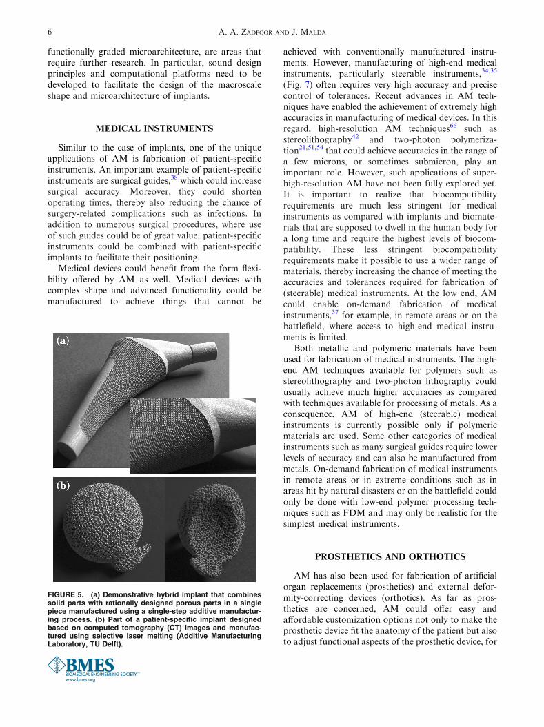
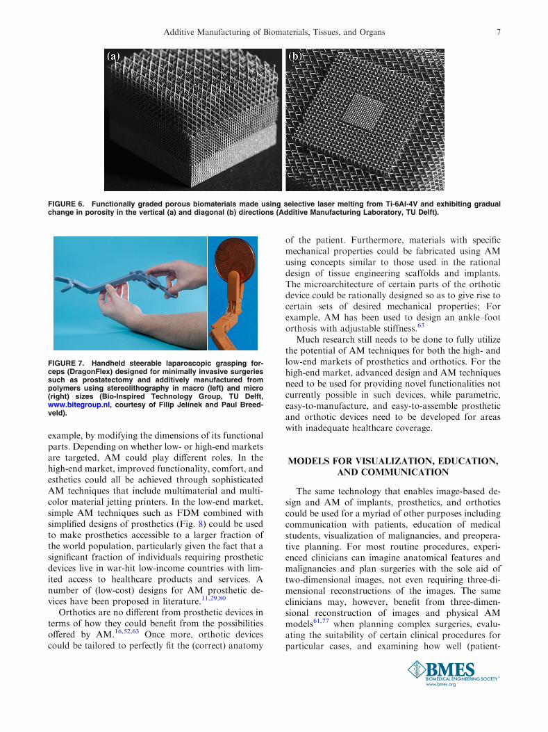
Ultimately, the design of hybrid implants couldintegrate solid volumes with various types of latticestructure (Fig. 5). This allows for optimal distributionof mechanical properties within the implant, providingsufficient mechanical support in areas where mechan-ical stress is greatest but allowing for incorporation ofporous structures in areas where stresses are lower,tissue unloading should be avoided, or bone ingrowthis essential, such as the surface of the anchoring partsof the implant. Functionally graded geometries (Fig. 6)could also be realized using AM techniques such that,for example, the porosity of the lattice structuregradually decreases from the implant surface, which isin contact with tissue and could benefit from tissueingrowth, to the center of the implant, which may needto be stronger to carry mechanical loads.
Metals are the materials most commonly used forAM of functional and load-bearing implants. Powderbed fusion processes including selective laser melting(SLM) and electron beam melting (EBM) are oftenused for this purpose.
Streamlined design and digital manufacturing ofpatient-specific implants and incorporation of complexgeometrical features into the design of generic im-plants, as well as evaluation of the actual clinical per-formance of patient-specific implants and implantsincorporating features such as hybrid design and
FIGURE 3. Biofabricated auricular implant: (a) macroscopic appearance of a fiber-reinforced biofabricated auricular constructbased on gelatin methacryloyl hydrogel and polycaprolactone fibers, (b) magnified view of reinforcing fibers (white) in thehydrogel (red), (c) Safranin O staining (stains proteoglycans red) of a histological section of a gelatin methacryloyl hydrogelconstruct after 6 weeks in vivo (subcutaneous mouse model) (Utrecht Biofabrication Facility, courtesy of Iris Otto, UniversityMedical Center Utrecht).
FIGURE 4. Additive manufacturing techniques could beused to (1) achieve complex distribution of several compo-nents in solid dosage forms, (2) develop drug products forspecific patient groups, e.g., children, and (3) adjust the do-sage of drug products.
Additive Manufacturing of Biomaterials, Tissues, and Organs 5
functionally graded microarchitecture, are areas thatrequire further research. In particular, sound designprinciples and computational platforms need to bedeveloped to facilitate the design of the macroscaleshape and microarchitecture of implants.
MEDICAL INSTRUMENTS
Similar to the case of implants, one of the uniqueapplications of AM is fabrication of patient-specificinstruments. An important example of patient-specificinstruments are surgical guides,38 which could increasesurgical accuracy. Moreover, they could shortenoperating times, thereby also reducing the chance ofsurgery-related complications such as infections. Inaddition to numerous surgical procedures, where useof such guides could be of great value, patient-specificinstruments could be combined with patient-specificimplants to facilitate their positioning.
Medical devices could benefit from the form flexi-bility offered by AM as well. Medical devices withcomplex shape and advanced functionality could bemanufactured to achieve things that cannot be
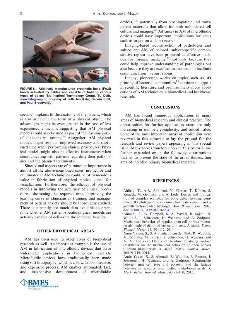
achieved with conventionally manufactured instru-ments. However, manufacturing of high-end medicalinstruments, particularly steerable instruments,34,35
(Fig. 7) often requires very high accuracy and precisecontrol of tolerances. Recent advances in AM tech-niques have enabled the achievement of extremely highaccuracies in manufacturing of medical devices. In thisregard, high-resolution AM techniques66 such asstereolithography42 and two-photon polymeriza-tion21,51,54 that could achieve accuracies in the range ofa few microns, or sometimes submicron, play animportant role. However, such applications of super-high-resolution AM have not been fully explored yet.It is important to realize that biocompatibilityrequirements are much less stringent for medicalinstruments as compared with implants and biomate-rials that are supposed to dwell in the human body fora long time and require the highest levels of biocom-patibility. These less stringent biocompatibilityrequirements make it possible to use a wider range ofmaterials, thereby increasing the chance of meeting theaccuracies and tolerances required for fabrication of(steerable) medical instruments. At the low end, AMcould enable on-demand fabrication of medicalinstruments,37 for example, in remote areas or on thebattlefield, where access to high-end medical instru-ments is limited.
Both metallic and polymeric materials have beenused for fabrication of medical instruments. The high-end AM techniques available for polymers such asstereolithography and two-photon lithography couldusually achieve much higher accuracies as comparedwith techniques available for processing of metals. As aconsequence, AM of high-end (steerable) medicalinstruments is currently possible only if polymericmaterials are used. Some other categories of medicalinstruments such as many surgical guides require lowerlevels of accuracy and can also be manufactured frommetals. On-demand fabrication of medical instrumentsin remote areas or in extreme conditions such as inareas hit by natural disasters or on the battlefield couldonly be done with low-end polymer processing tech-niques such as FDM and may only be realistic for thesimplest medical instruments.
PROSTHETICS AND ORTHOTICS
AM has also been used for fabrication of artificialorgan replacements (prosthetics) and external defor-mity-correcting devices (orthotics). As far as pros-thetics are concerned, AM could offer easy andaffordable customization options not only to make theprosthetic device fit the anatomy of the patient but alsoto adjust functional aspects of the prosthetic device, for
FIGURE 5. (a) Demonstrative hybrid implant that combinessolid parts with rationally designed porous parts in a singlepiece manufactured using a single-step additive manufactur-ing process. (b) Part of a patient-specific implant designedbased on computed tomography (CT) images and manufac-tured using selective laser melting (Additive ManufacturingLaboratory, TU Delft).
A. A. ZADPOOR AND J. MALDA6
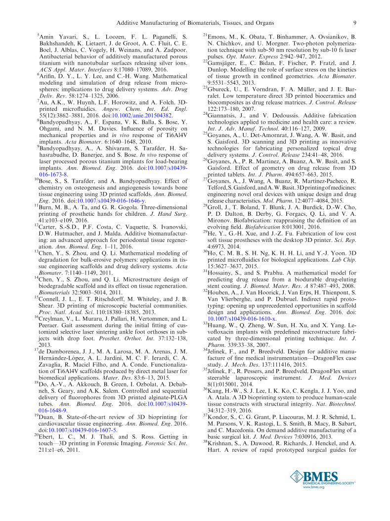
example, by modifying the dimensions of its functionalparts. Depending on whether low- or high-end marketsare targeted, AM could play different roles. In thehigh-end market, improved functionality, comfort, andesthetics could all be achieved through sophisticatedAM techniques that include multimaterial and multi-color material jetting printers. In the low-end market,simple AM techniques such as FDM combined withsimplified designs of prosthetics (Fig. 8) could be usedto make prosthetics accessible to a larger fraction ofthe world population, particularly given the fact that asignificant fraction of individuals requiring prostheticdevices live in war-hit low-income countries with lim-ited access to healthcare products and services. Anumber of (low-cost) designs for AM prosthetic de-vices have been proposed in literature.11,29,80
Orthotics are no different from prosthetic devices interms of how they could benefit from the possibilitiesoffered by AM.16,52,63 Once more, orthotic devicescould be tailored to perfectly fit the (correct) anatomy
of the patient. Furthermore, materials with specificmechanical properties could be fabricated using AMusing concepts similar to those used in the rationaldesign of tissue engineering scaffolds and implants.The microarchitecture of certain parts of the orthoticdevice could be rationally designed so as to give rise tocertain sets of desired mechanical properties; Forexample, AM has been used to design an ankle–footorthosis with adjustable stiffness.63
Much research still needs to be done to fully utilizethe potential of AM techniques for both the high- andlow-end markets of prosthetics and orthotics. For thehigh-end market, advanced design and AM techniquesneed to be used for providing novel functionalities notcurrently possible in such devices, while parametric,easy-to-manufacture, and easy-to-assemble prostheticand orthotic devices need to be developed for areaswith inadequate healthcare coverage.
MODELS FOR VISUALIZATION, EDUCATION,
AND COMMUNICATION
The same technology that enables image-based de-sign and AM of implants, prosthetics, and orthoticscould be used for a myriad of other purposes includingcommunication with patients, education of medicalstudents, visualization of malignancies, and preopera-tive planning. For most routine procedures, experi-enced clinicians can imagine anatomical features andmalignancies and plan surgeries with the sole aid oftwo-dimensional images, not even requiring three-di-mensional reconstructions of the images. The sameclinicians may, however, benefit from three-dimen-sional reconstruction of images and physical AMmodels61,77 when planning complex surgeries, evalu-ating the suitability of certain clinical procedures forparticular cases, and examining how well (patient-
FIGURE 6. Functionally graded porous biomaterials made using selective laser melting from Ti-6Al-4V and exhibiting gradualchange in porosity in the vertical (a) and diagonal (b) directions (Additive Manufacturing Laboratory, TU Delft).
FIGURE 7. Handheld steerable laparoscopic grasping for-ceps (DragonFlex) designed for minimally invasive surgeriessuch as prostatectomy and additively manufactured frompolymers using stereolithography in macro (left) and micro(right) sizes (Bio-Inspired Technology Group, TU Delft,www.bitegroup.nl, courtesy of Filip Jelınek and Paul Breed-veld).
Additive Manufacturing of Biomaterials, Tissues, and Organs 7
specific) implants fit the anatomy of the patient, whichis also printed in the form of a physical object. Theadvantages might be even greater in the case of lessexperienced clinicians, suggesting that AM physicalmodels could also be used as part of the learning curveof clinicians in training.74 Altogether, AM physicalmodels might result in improved accuracy and short-ened time when performing clinical procedures. Phys-ical models might also be effective instruments whencommunicating with patients regarding their patholo-gies and the planned treatments.
Since visual aspects are of paramount importance inalmost all the above-mentioned cases, multicolor andmultimaterial AM techniques could be of tremendousvalue in fabrication of physical models aimed forvisualization. Furthermore, the efficacy of physicalmodels in improving the accuracy of clinical proce-dures, shortening the required time, improving thelearning curve of clinicians in training, and manage-ment of patient anxiety should be thoroughly studied.There is currently not much data available to deter-mine whether AM patient-specific physical models areactually capable of delivering the intended benefits.
OTHER BIOMEDICAL AREAS
AM has been used in other areas of biomedicalresearch as well. An important example is the use ofAM in fabrication of microfluidic devices that havewidespread applications in biomedical research.Microfluidic devices have traditionally been madeusing soft lithography, which is a slow, labor-intensive,and expensive process. AM enables automated, fast,and inexpensive development of microfluidic
devices,7,30 potentially from biocompatible and trans-parent materials that allow for both unhindered cellculture and imaging.65 Advances in AM of microfluidicdevices could have important implications for areassuch as organ-on-a-chip research.
Imaging-based reconstruction of pathologies andsubsequent AM of colored, subject-specific demon-strative replica have been proposed as effective meth-ods for forensic medicine,20 not only because theycould help improve understanding of pathologies butalso because they are excellent instruments to facilitatecommunication in court rooms.
Finally, pioneering works on topics such as 3Dprinting of bacterial communities15 continue to appearin scientific literature and promise many more appli-cations of AM techniques in biomedical and healthcareresearch.
CONCLUSIONS
AM has found numerous applications in manyareas of biomedical research and clinical practice. Theopportunities for further application areas are onlyincreasing in number, complexity, and added value.Some of the most important areas of application werereviewed in this editorial to lay the ground for theresearch and review papers appearing in this specialissue. Many topics touched upon in this editorial arefurther expanded on in the following contributionsthat try to present the state of the art in this excitingarea of interdisciplinary biomedical research.
REFERENCES
1Ahlfeld, T., A.R. Akkineni, Y. Forster, T. Kohler, S.Knaack, M. Gelinsky, and A. Lode. Design and fabrica-tion of complex scaffolds for bone defect healing: com-bined 3D plotting of a calcium phosphate cement and agrowth factor-loaded hydrogel. Ann. Biomed. Eng. 2016.doi:10.1007/s10439-016-1685-4.2Ahmadi, S., G. Campoli, S. A. Yavari, B. Sajadi, R.Wauthle, J. Schrooten, H. Weinans, and A. Zadpoor.Mechanical behavior of regular open-cell porous bioma-terials made of diamond lattice unit cells. J. Mech. Behav.Biomed. Mater. 34:106–115, 2014.3Amin Yavari, S., S. Ahmadi, J. van der Stok, R. Wauthle,A. Riemslag, M. Janssen, J. Schrooten, H. Weinans, andA. A. Zadpoor. Effects of bio-functionalizing surfacetreatments on the mechanical behavior of open poroustitanium biomaterials. J. Mech. Behav. Biomed. Mater.36:109–119, 2014.4Amin Yavari, S., S. Ahmadi, R. Wauthle, B. Pouran, J.Schrooten, H. Weinans, and A. Zadpoor. Relationshipbetween unit cell type and porosity and the fatiguebehavior of selective laser melted meta-biomaterials. J.Mech. Behav. Biomed. Mater. 43:91–100, 2015.
FIGURE 8. Additively manufactured prosthetic hand (FA3Dhand) activated by cables and capable of holding varioustypes of object (Bio-Inspired Technology Group, TU Delft,www.bitegroup.nl, courtesy of Jelle ten Kate, Gerwin Smit,and Paul Breedveld).
A. A. ZADPOOR AND J. MALDA8
5Amin Yavari, S., L. Loozen, F. L. Paganelli, S.Bakhshandeh, K. Lietaert, J. de Groot, A. C. Fluit, C. E.Boel, J. Alblas, C. Vogely, H. Weinans, and A. Zadpoor.Antibacterial behavior of additively manufactured poroustitanium with nanotubular surfaces releasing silver ions.ACS Appl. Mater. Interfaces 8:17080–17089, 2016.6Arifin, D. Y., L. Y. Lee, and C.-H. Wang. Mathematicalmodeling and simulation of drug release from micro-spheres: implications to drug delivery systems. Adv. DrugDeliv. Rev. 58:1274–1325, 2006.7Au, A.K., W. Huynh, L.F. Horowitz, and A. Folch. 3D-printed microfluidics. Angew. Chem. Int. Ed. Engl.55(12):3862–3881, 2016. doi:10.1002/anie.201504382.8Bandyopadhyay, A., F. Espana, V. K. Balla, S. Bose, Y.Ohgami, and N. M. Davies. Influence of porosity onmechanical properties and in vivo response of Ti6Al4Vimplants. Acta Biomater. 6:1640–1648, 2010.9Bandyopadhyay, A., A. Shivaram, S. Tarafder, H. Sa-hasrabudhe, D. Banerjee, and S. Bose. In vivo response oflaser processed porous titanium implants for load-bearingimplants. Ann. Biomed. Eng. 2016. doi:10.1007/s10439-016-1673-8.
10Bose, S., S. Tarafder, and A. Bandyopadhyay. Effect ofchemistry on osteogenesis and angiogenesis towards bonetissue engineering using 3D printed scaffolds. Ann. Biomed.Eng. 2016. doi:10.1007/s10439-016-1646-y.
11Burn, M. B., A. Ta, and G. R. Gogola. Three-dimensionalprinting of prosthetic hands for children. J. Hand Surg.41:e103–e109, 2016.
12Carter, S.-S.D., P.F. Costa, C. Vaquette, S. Ivanovski,D.W. Hutmacher, and J. Malda. Additive biomanufactur-ing: an advanced approach for periodontal tissue regener-ation. Ann. Biomed. Eng. 1–11, 2016.
13Chen, Y., S. Zhou, and Q. Li. Mathematical modeling ofdegradation for bulk-erosive polymers: applications in tis-sue engineering scaffolds and drug delivery systems. ActaBiomater. 7:1140–1149, 2011.
14Chen, Y., S. Zhou, and Q. Li. Microstructure design ofbiodegradable scaffold and its effect on tissue regeneration.Biomaterials 32:5003–5014, 2011.
15Connell, J. L., E. T. Ritschdorff, M. Whiteley, and J. B.Shear. 3D printing of microscopic bacterial communities.Proc. Natl. Acad. Sci. 110:18380–18385, 2013.
16Creylman, V., L. Muraru, J. Pallari, H. Vertommen, and L.Peeraer. Gait assessment during the initial fitting of cus-tomized selective laser sintering ankle foot orthoses in sub-jects with drop foot. Prosthet. Orthot. Int. 37:132–138,2013.
17de Damborenea, J. J., M. A. Larosa, M. A. Arenas, J. M.Hernandez-Lopez, A. L. Jardini, M. C. F. Ierardi, C. A.Zavaglia, R. Maciel Filho, and A. Conde. Functionaliza-tion of Ti6Al4V scaffolds produced by direct metal laser forbiomedical applications. Mater. Des. 83:6–13, 2015.
18Do, A.-V., A. Akkouch, B. Green, I. Ozbolat, A. Debab-neh, S. Geary, and A.K. Salem. Controlled and sequentialdelivery of fluorophores from 3D printed alginate-PLGAtubes. Ann. Biomed. Eng. 2016. doi:10.1007/s10439-016-1648-9.
19Duan, B. State-of-the-art review of 3D bioprinting forcardiovascular tissue engineering. Ann. Biomed. Eng. 2016.doi:10.1007/s10439-016-1607-5.
20Ebert, L. C., M. J. Thali, and S. Ross. Getting intouch—3D printing in Forensic Imaging. Forensic Sci. Int.211:e1–e6, 2011.
21Emons, M., K. Obata, T. Binhammer, A. Ovsianikov, B.N. Chichkov, and U. Morgner. Two-photon polymeriza-tion technique with sub-50 nm resolution by sub-10 fs laserpulses. Opt. Mater. Express 2:942–947, 2012.
22Gamsjager, E., C. Bidan, F. Fischer, P. Fratzl, and J.Dunlop. Modelling the role of surface stress on the kineticsof tissue growth in confined geometries. Acta Biomater.9:5531–5543, 2013.
23Gbureck, U., E. Vorndran, F. A. Muller, and J. E. Bar-ralet. Low temperature direct 3D printed bioceramics andbiocomposites as drug release matrices. J. Control. Release122:173–180, 2007.
24Giannatsis, J., and V. Dedoussis. Additive fabricationtechnologies applied to medicine and health care: a review.Int. J. Adv. Manuf. Technol. 40:116–127, 2009.
25Goyanes, A., U. Det-Amornrat, J. Wang, A. W. Basit, andS. Gaisford. 3D scanning and 3D printing as innovativetechnologies for fabricating personalized topical drugdelivery systems. J. Control. Release 234:41–48, 2016.
26Goyanes, A., P. R. Martinez, A. Buanz, A. W. Basit, and S.Gaisford. Effect of geometry on drug release from 3Dprinted tablets. Int. J. Pharm. 494:657–663, 2015.
27Goyanes, A., J. Wang, A. Buanz, R. Martınez-Pacheco, R.Telford,S.Gaisford,andA.W.Basit.3Dprintingofmedicines:engineering novel oral devices with unique design and drugrelease characteristics.Mol. Pharm. 12:4077–4084, 2015.
28Groll, J., T. Boland, T. Blunk, J. A. Burdick, D.-W. Cho,P. D. Dalton, B. Derby, G. Forgacs, Q. Li, and V. A.Mironov. Biofabrication: reappraising the definition of anevolving field. Biofabrication 8:013001, 2016.
29He, Y., G.-H. Xue, and J.-Z. Fu. Fabrication of low costsoft tissue prostheses with the desktop 3D printer. Sci. Rep.4:6973, 2014.
30Ho, C. M. B., S. H. Ng, K. H. H. Li, and Y.-J. Yoon. 3Dprinted microfluidics for biological applications. Lab Chip.15:3627–3637, 2015.
31Hossainy, S., and S. Prabhu. A mathematical model forpredicting drug release from a biodurable drug-elutingstent coating. J. Biomed. Mater. Res. A 87:487–493, 2008.
32Houben, A., J. Van Hoorick, J. Van Erps, H. Thienpont, S.Van Vlierberghe, and P. Dubruel. Indirect rapid proto-typing: opening up unprecedented opportunities in scaffolddesign and applications. Ann. Biomed. Eng. 2016. doi:10.1007/s10439-016-1610-x.
33Huang, W., Q. Zheng, W. Sun, H. Xu, and X. Yang. Le-vofloxacin implants with predefined microstructure fabri-cated by three-dimensional printing technique. Int. J.Pharm. 339:33–38, 2007.
34Jelınek, F., and P. Breedveld. Design for additive manu-facture of fine medical instrumentation—DragonFlex casestudy. J. Mech. Des. 137:111416, 2015.
35Jelınek, F., R. Pessers, and P. Breedveld. DragonFlex smartsteerable laparoscopic instrument. J. Med. Devices8(1):015001, 2014.
36Kang, H.-W., S. J. Lee, I. K. Ko, C. Kengla, J. J. Yoo, andA. Atala. A 3D bioprinting system to produce human-scaletissue constructs with structural integrity. Nat. Biotechnol.34:312–319, 2016.
37Kondor, S., C. G. Grant, P. Liacouras, M. J. R. Schmid, L.M. Parsons, V. K. Rastogi, L. S. Smith, B. Macy, B. Sabart,and C. Macedonia. On demand additive manufacturing of abasic surgical kit. J. Med. Devices 7:030916, 2013.
38Krishnan, S., A. Dawood, R. Richards, J. Henckel, and A.Hart. A review of rapid prototyped surgical guides for
Additive Manufacturing of Biomaterials, Tissues, and Organs 9

patient-specific total knee replacement. J. Bone Joint Surg.Br. 94:1457–1461, 2012.
39Lee, V.K., and G. Dai. Printing of three-dimensional tissueanalogs for regenerative medicine. Ann. Biomed. Eng. 2016.doi:10.1007/s10439-016-1613-7.
40Lin, C.-C., and A. T. Metters. Hydrogels in controlled re-lease formulations: network design and mathematicalmodeling. Adv. Drug Deliv. Rev. 58:1379–1408, 2006.
41Malda, J., J. Visser, F. P. Melchels, T. Jungst, W. E.Hennink, W. J. Dhert, J. Groll, and D. W. Hutmacher.25th anniversary article: engineering hydrogels for biofab-rication. Adv. Mater. 25:5011–5028, 2013.
42Melchels, F. P., J. Feijen, and D. W. Grijpma. A review onstereolithography and its applications in biomedical engi-neering. Biomaterials 31:6121–6130, 2010.
43Morris, V.B., S. Nimbalkar, M. Younesi, P. McClellan,and O. Akkus. Mechanical properties, cytocompatibilityand manufacturability of chitosan:PEGDA hybrid-gelscaffolds by stereolithography. Ann. Biomed. Eng. 1–11,2016.
44Muller, M., E. Ozturk, Ø. Arlov, P. Gatenholm, and M.Zenobi-Wong. Alginate sulfate–nanocellulose bioinks forcartilage bioprinting applications. Ann. Biomed. Eng. 1–14,2016.
45Murphy, S. V., A. Skardal, and A. Atala. Evaluation ofhydrogels for bio-printing applications. J. Biomed. Mater.Res. A 101:272–284, 2013.
46Murr, L., S. Gaytan, F. Medina, H. Lopez, E. Martinez, B.Machado, D. Hernandez, L. Martinez, M. Lopez, and R.Wicker. Next-generation biomedical implants using addi-tive manufacturing of complex, cellular and functionalmesh arrays. Philos. Trans. A Math. Phys. Eng. Sci.368:1999–2032, 2010.
47Murr, L.E., S.M. Gaytan, E. Martinez, F. Medina, andR.B. Wicker. Next generation orthopaedic implants byadditive manufacturing using electron beam melting. Int. J.Biomater. 2012:245727, 2012. doi:10.1155/2012/245727.
48Nguyen, A.K., and R.J. Narayan. Liquid-phase laser in-duced forward transfer for complex organic inks and tissueengineering. Ann. Biomed. Eng. 2016. doi:10.1007/s10439-016-1617-3.
49Norman, J., R.D. Madurawe, C.M. Moore, M.A. Khan,and A. Khairuzzaman. A new chapter in pharmaceuticalmanufacturing: 3D-printed drug products. Adv. Drug De-liv. Rev. 2016. doi:10.1016/j.addr.2016.03.001.
50Nyberg, E.L., A.L. Farris, B.P. Hung, M. Dias, J.R.Garcia, A.H. Dorafshar, and W.L. Grayson. 3D-printingtechnologies for craniofacial rehabilitation, reconstruction,and regeneration. Ann. Biomed. Eng. 1–13, 2016.
51Ovsianikov, A., and B. N. Chichkov. Three-dimensionalmicrofabrication by two-photon polymerization technique.Comput. Aided Tissue Eng. 868:311–325, 2012.
52Pallari, J. H., K. W. Dalgarno, and J. Woodburn. Masscustomization of foot orthoses for rheumatoid arthritisusing selective laser sintering. IEEE Trans. Biomed. Eng.57:1750–1756, 2010.
53Park, J.H., J. Jang, J.-S. Lee, and D.-W. Cho. Three-di-mensional printing of tissue/organ analogues containingliving cells. Ann. Biomed. Eng. 2016. doi:10.1007/s10439-016-1611-9.
54Park, S. H., D. Y. Yang, and K. S. Lee. Two-photonstereolithography for realizing ultraprecise three-dimen-sional nano/microdevices. Laser Photon. Rev. 3:1–11, 2009.
55Placone, J.K., J. Navarro, G.W. Laslo, M.J. Lerman, A.R.Gabard, G.J. Herendeen, E.E. Falco, S. Tomblyn, L.
Burnett, and J.P. Fisher. Development and characteriza-tion of a 3D printed, keratin-based hydrogel. Ann. Biomed.Eng. 2016. doi:10.1007/s10439-016-1621-7.
56Richards, D., J. Jia, M. Yost, R. Markwald, and Y. Mei.3D bioprinting for vascularized tissue fabrication. Ann.Biomed. Eng. 2016. doi:10.1007/s10439-016-1653-z.
57Rumpler, M., A. Woesz, J. W. Dunlop, J. T. van Dongen,and P. Fratzl. The effect of geometry on three-dimensionaltissue growth. J. R. Soc. Interface 5:1173–1180, 2008.
58Schon, B. S., G. J. Hooper, and T. B. Woodfield. Modulartissue assembly strategies for biofabrication of engineeredcartilage. Ann. Biomed. Eng. 2016. doi:10.1007/s10439-016-1609-3.
59Skardal, A., and A. Atala. Biomaterials for integrationwith 3-D bioprinting. Ann. Biomed. Eng. 43:730–746, 2015.
60Sobral, J. M., S. G. Caridade, R. A. Sousa, J. F. Mano, andR. L. Reis. Three-dimensional plotted scaffolds with con-trolled pore size gradients: effect of scaffold geometry onmechanical performance and cell seeding efficiency. ActaBiomater. 7:1009–1018, 2011.
61Spottiswoode, B., D. Van den Heever, Y. Chang, S.Engelhardt, S. Du Plessis, F. Nicolls, H. Hartzenberg, andA. Gretschel. Preoperative three-dimensional model cre-ation of magnetic resonance brain images as a tool to assistneurosurgical planning. Stereotact. Funct. Neurosurg.91:162–169, 2013.
62Stichler, S., T. Jungst, M. Schamel, I. Zilkowski, M.Kuhlmann, T. Bock, T. Blunk, J. Teßmar, and J. Groll.Thiol-ene clickable poly(glycidol) hydrogels for biofabri-cation. Ann. Biomed. Eng. 1–13, 2016.
63Telfer, S., J. Pallari, J. Munguia, K. Dalgarno, M.McGeough, and J. Woodburn. Embracing additive manu-facture: implications for foot and ankle orthosis design.BMC Musculoskelet. Disord. 13:1, 2012.
64Trombetta, R., J.A. Inzana, E.M. Schwarz, S.L. Kates, andH.A. Awad. 3D printing of calcium phosphate ceramics forbone tissue engineering and drug delivery. Ann. Biomed.Eng. 2016. doi:10.1007/s10439-016-1678-3.
65Urrios, A., C. Parra-Cabrera, N. Bhattacharjee, A.M.Gonzalez-Suarez, L.G. Rigat-Brugarolas, U. Nallapatti, J.Samitier, C.A. DeForest, F. Posas, J.L. Garcia-Cordero,and A. Folch. 3D-printing of transparent bio-microfluidicdevices in PEG-DA. Lab Chip 2016. doi:10.1039/C6LC00153J.
66Vaezi, M., H. Seitz, and S. Yang. A review on 3D micro-additive manufacturing technologies. Int. J. Adv. Manuf.Technol. 67:1721–1754, 2013.
67Vaithilingam, J., S. Kilsby, R. D. Goodridge, S. D. Christie,S. Edmondson, and R. J. Hague. Immobilisation of anantibacterial drug to Ti6Al4V components fabricated usingselective laser melting. Appl. Surf. Sci. 314:642–654, 2014.
68Vaithilingam, J., S. Kilsby, R. D. Goodridge, S. D.Christie, S. Edmondson, and R. J. Hague. Functionalisa-tion of Ti6Al4V components fabricated using selective lasermelting with a bioactive compound. Mater. Sci. Eng. C46:52–61, 2015.
69Van Bael, S., Y. C. Chai, S. Truscello, M. Moesen, G.Kerckhofs, H. Van Oosterwyck, J.-P. Kruth, and J. Sch-rooten. The effect of pore geometry on the in vitro bio-logical behavior of human periosteum-derived cells seededon selective laser-melted Ti6Al4V bone scaffolds. ActaBiomater. 8:2824–2834, 2012.
70van der Stok, J., M. Koolen, M. de Maat, S. AMin Yavari,J. Alblas, P. Patka, J. Verhaar, E. van Lieshout, A. A.Zadpoor, and H. Weinans. Full regeneration of segmental
A. A. ZADPOOR AND J. MALDA10
bone defects using porous titanium implants loaded withBMP-2 containing fibrin gels. Eur. Cells Mater. 2015:141–154, 2015.
71Vanderburgh, J., J.A. Sterling, and S.A. Guelcher. 3Dprinting of tissue engineered constructs for in vitro model-ing of disease progression and drug screening. Ann. Biomed.Eng. 2016. doi:10.1007/s10439-016-1640-4.
72Wang, C., Z. Tang, Y. Zhao, R. Yao, L. Li, and W. Sun.Three-dimensional in vitro cancer models: a short review.Biofabrication 6:022001, 2014.
73Wang, X., S. Xu, S. Zhou, W. Xu, M. Leary, P. Choong,M. Qian, M. Brandt, and Y. M. Xie. Topological designand additive manufacturing of porous metals for bonescaffolds and orthopaedic implants: a review. Biomaterials83:127–141, 2016.
74Waran, V., V. Narayanan, R. Karuppiah, S. L. Owen, andT. Aziz. Utility of multimaterial 3D printers in creatingmodels with pathological entities to enhance the trainingexperience of neurosurgeons: technical note. J. Neurosurg.120:489–492, 2014.
75Zadpoor, A. A. Bone tissue regeneration: the role of scaf-fold geometry. Biomater. Sci. 3:231–245, 2015.
76Zadpoor, A.A. Mechanical meta-materials. Mater. Horiz.3:371–381, 2016. doi:10.1039/C6MH00065G.
77Zein, N. N., I. A. Hanouneh, P. D. Bishop, M. Samaan, B.Eghtesad, C. Quintini, C. Miller, L. Yerian, and R. Klatte.Three-dimensional print of a liver for preoperative plan-ning in living donor liver transplantation. Liver Transplant.19:1304–1310, 2013.
78Zhang, Q., Y. Jiang, Y. Zhang, Z. Ye, W. Tan, and M.Lang. Effect of porosity on long-term degradation of poly
(e-caprolactone) scaffolds and their cellular response.Polym. Degrad. Stab. 98:209–218, 2013.
79Zhang, Y.S., K. Yue, J. Aleman, K. Mollazadeh-Moghaddam, S.M. Bakht, J. Yang, W. Jia, V. Dell’Erba, P.Assawes, and S.R. Shin. 3D bioprinting for tissue and or-gan fabrication. Ann. Biomed. Eng. 1–16, 2016.
80Zuniga, J., D. Katsavelis, J. Peck, J. Stollberg, M. Petry-kowski, A. Carson, and C. Fernandez. Cyborg beast: alow-cost 3d-printed prosthetic hand for children with up-per-limb differences. BMC Res. Notes 8:1, 2015.
Amir A. Zadpoor
Additive Manufacturing Laboratory, Departmentof Biomechanical EngineeringDelft University of Technology (TU Delft),Mekelweg 2, Delft 2628 CD, The NetherlandsElectronic mail: [email protected]
Jos Malda
Department of Orthopaedics, University MedicalCenter Utrecht, PO Box 85500, 3508 GA Utrecht,The NetherlandsDepartment of Equine Sciences, Faculty ofVeterinary Medicine, Utrecht University,Yalelaan 112, 3584 CM Utrecht, The Netherlands
Additive Manufacturing of Biomaterials, Tissues, and Organs 11