stabilizing snai2 in human trabecular meshwork apoptosis
TRANSCRIPT

Page 1/17
SNHG3 cooperates with ELAVL2 to modulate cellapoptosis and extracellular matrix accumulation bystabilizing SNAI2 in human trabecular meshworkcells under oxidative stressSizhen Li
Nanjing Tongren HospitalQingsong Yang ( [email protected] )
Nanjing Tongren Hospital https://orcid.org/0000-0003-2904-1503Zixiu Zhou
Nanjing Tongren HospitalMin Fu
Nanjing Tongren HospitalXiaodong Yang
Nanjing Tongren HospitalKuanxiao Hao
Nanjing Tongren HospitalYating Liu
Nanjing Tongren Hospital
Research article
Keywords: SNHG3, ELAVL2, SNAI2, trabecular meshwork cells, extracellular matrix accumulation
Posted Date: December 22nd, 2020
DOI: https://doi.org/10.21203/rs.3.rs-130712/v1
License: This work is licensed under a Creative Commons Attribution 4.0 International License. Read Full License

Page 2/17
AbstractBackground: Glaucoma is the main reason for irreversible blindness, and pathological increasedintraocular pressure is the leading risk factor for glaucoma. It is reported that trabecular meshwork cellinjury is closely associated with the elevated intraocular pressure. The current study aimed to investigatethe role of SNHG3 in human trabecular meshwork (HTM) cells under oxidative stress.
Methods: A series of experiments including real-time quantitative polymerase chain reaction (RT-qPCR),subcellular fractionation assay, western blot analysis, cell counting kit-8 (CCK-8) assay, RNA pull down,�ow cytometry analysis, and RIP assay were employed to explore the biological function and regulatorymechanism of SNHG3 in HTM cells under oxidative stress.
Results: First, we observed that H2O2 induced SNHG3 upregulation in HTM cells. Then, we found thatSNHG3 silencing alleviated H2O2-induced oxidative damage in HTM cells. Moreover, SNAI2 knockdownalleviated the oxidative damage induced by H2O2 in HTM cells. Mechanistically, SNHG3 bound withELAVL2 to stabilize SNAI2. Finally, SNAI2 overexpression counteracted the effect of SNHG3 silencing onH2O2-induced HTM cells.
Conclusion: Our results demonstrated that SNHG3 cooperated with ELAVL2 to modulate cell apoptosisand extracellular matrix (ECM) accumulation by stabilizing SNAI2 in HTM cells under oxidative stress.
IntroductionGlaucoma is characterized by atrophy and depression of optic papilla, visual �eld defect and visualacuity decline 1,2. Pathological elevation of intraocular pressure and inadequate blood supply of opticnerve are the main reasons for glaucoma 3,4. Previous research has showed that trabecular meshwork(TM) cells exert crucial effect on aqueous humour circulation, and TM cell injury is closely related to theelevated intraocular pressure 5,6. Hence, �nding novel biomarkers that modulate human trabecularmeshwork (HTM) cell injury in glaucomatous patients is of vital signi�cance.
In primary open angle glaucoma, increased intraocular pressure results in deformation at the optic nervehead; especially, intraocular pressure elevation leads to the deformation at the lamina cribrosa regionwhere extracellular matrix (ECM) molecules such as �bronectin and collagen tend to accumulate 7-9.Changes in the levels of regulators of ECM homeostasis such as matrix metalloproteinases (MMPs)occur in the primary open angle glaucoma lamina cribrosa 10. MMPs are zinc-dependent endopeptidases,which degrade ECM components such as �bronectin and collagen 11,12. MMPs are crucial regulators ofaqueous humor out�ow for they can remodel TM ECM and keep a stable out�ow resistance and ensuingintraocular pressure 13-15. Therefore, MMPs are promising therapeutic targets for glaucoma treatment dueto their capability to regulate aqueous humor out�ow 13,14.

Page 3/17
Long noncoding RNAs (lncRNAs) are a category of noncoding RNAs (ncRNAs), with over 200 nucleotidesin size 16,17. LncRNAs are related to multiple cellular functions, and most of the cellular functions involvethe binding of lncRNAs with one or more RNA-binding proteins (RBPs) to increase the stability of targetmessenger RNAs (mRNAs) 18-21. LncRNAs are active regulators in a variety of diseases, includingglaucoma 22-24. For example, lncRNA GAS5 knockdown relieves glaucoma in rat models via decreasingthe apoptosis of retinal ganglion cells 25. LncRNA MALAT1 regulates the apoptosis of retinal ganglioncells via the PI3K/Akt pathway in glaucomatous rats 26. LncRNA MEG3 participates in glaucomaprogression by facilitating the autophagy of retinal ganglion cells 27. LncRNA SNHG3 (small nucleolarRNA host gene 3) has been found to participate in regulating the development of several diseases 28-30.Importantly, SNHG3 has been found to be related to neurodegeneration 31, and it has been reported to beupregulated in H2O2-induced HTM cells 32
. Accordingly, we predicted that SNHG3 may exert certain effecton glaucoma.
DNA damage is related to various neurodegenerative diseases, including glaucoma 32,33. In comparisonwith healthy controls, oxidative DNA damage is distinctly increased in the TM of patients with glaucoma32. In this study, a H2O2-induced oxidative damage HTM cell model was established and the role ofSNHG3 in H2O2-induced HTM cells was explored. Our investigation may provide a novel direction for thetreatment of glaucoma.
Materials And MethodsCell lines and cell culture
HTM cells were acquired from cadaver eyes with the approval of the Ethics Committee of NanjingTongren Hospital (Jiangsu, China). HTM cells were incubated in Dulbecco’s modi�ed Eagle’s medium(DMEM; USA) containing 10% fetal bovine serum (FBS; Invitrogen, USA), 100 U/ml penicillin (Sigma-Aldrich, USA) and 100 μg/ml streptomycin (Sigma-Aldrich, USA), and then stored in the humidi�edatmosphere with 5% CO2 at 37℃.
Cell treatment
The cultured HTM cells were treated with disparate concentrations of H2O2 (0 – 300 μM) to establish anin vitro oxidative cell damage model. Non-disposed cells acted as a control.
Cell transfection
For overexpression of SNAI2, the SNAI2 sequence was synthesized and inserted into the pcDNA3.1(Invitrogen, U.S.A.) plasmid to produce the pcDNA3.1/SNAI2. For knockdown of SNHG3, SNAI2 orELAVL2, short hairpin RNA (shRNA) speci�cally targeting SNHG3, SNAI2 or ELAVL2 were devised andsynthesized by GenePharma (Shanghai, China). Lipofectamine 2000 (Invitrogen, U.S.A.) was applied forcell transfection in line with manufacturer’s recommendations.

Page 4/17
CCK-8 assay
The viability of HTM cells were detected by using Cell Counting Kit-8 (CCK-8). The transfected cells with1×104 cells per well were planted in 96-well plates. After incubation for 48 h, CCK-8 solution (10 μl) wassupplemented into each well. After that, the incubation continued for 4 h. Cell viability was visualizedusing a microplate reader (EL340; BioTek Instruments, Hopkinton, MA) at a wavelength of 450 nm.
Flow cytometry analysis
The �ow cytometry was conducted to assess HTM cell apoptosis with Annexin V Kit (Beyotime, China)with reference to protocols stipulated by the manufacturer. Cells transfected with indicated plasmid ornegative control were collected 48 h later and washed twice with PBS. Subsequently, cells (1 × 10⁶
cells/mL) were double stained by propidium iodide (PI) and Annexin V-FITC. The CELLQUEST program(Becton Dickinson, USA) was used to record cells.
Western blot
Transfected cells were lysed applying RIPA buffer (Thermo, USA), and cultured for 15 min at 4°C. Then,the lysate was centrifuged, and the concentrations of the proteins were measured using an BCA ProteinAssay Kit (Thermo, USA). The proteins were isolated on 10% sodium dodecyl sulfate polyacrylamide gelelectrophoresis (SDS-PAGE) and moved onto PVDF membranes (Thermo, USA). Then, 5% skim milk wasused to block the membranes for 1 h. Next, the membranes were incubated with primary antibodiesovernight at 4°C. The primary antibodies were obtained from Abcam company (Shanghai, China): SNAI2(ab51772), ELAVL2 (ab72603), collagen (ab34710), �bronectin (ab2413), collagen III (ab7778), MMP3(ab52915), MMP9 (ab76003) and GAPDH (ab8245). Subsequently, secondary antibody was used to beincubated with the membranes for 2 h at room temperature. Finally, an enhanced chemiluminescence kit(GE Healthcare, Chicago, IL) was adopted to observe the signals.
Real-time quantitative polymerase chain reaction (RT-qPCR)
TRIzol reagent (ThermoFisher Scienti�c Invitrogen, USA) was employed to extract total RNA. Then, totalRNA was reverse transcribed to cDNA using the High Capacity cDNA Reverse Transcription Kits (AppliedBiosystems, Foster City, CA, USA). RT-qPCR analysis was performed using a SYBR Premix Ex Taq II kit(Cat. #RR820A, TaKaRa) on the 7500 Real-Time PCR Systems (Applied Biosystems). U6 served as anendogenous control for miRNAs. The detection results for mRNAs were normalized to GAPDH. Expressionfold changes were calculated adopting the 2-ΔΔCt method.
Subcellular fractionation assay
Total RNA was extracted applying a DNA/RNA Isolation Kit (DP422, Tiangen Biotech, Beijing, China) inline with the instruction. The nuclear and cytoplasmic fractions were separated applying NE-PER Nuclearand Cytoplasmic Extraction Reagents (Thermo Scienti�c). Total RNA was isolated from the nuclear andcytoplasmic fractions using Trizol (Invitrogen).
Page 5/17
RNA pull down assay
The biotinylated RNAs were cultured with cell lysates, and then cultured with streptavidin beads(Invitrogen). Subsequently, the RNAs in complexes captured by streptavidin beads were washed, puri�edand assessed by western blot analysis.
RNA immunoprecipitation (RIP) assay
The RIP assay was carried out with a Millipore EZ-Magna RIP RNA-Binding Protein Immunoprecipitationkit (Millipore, Bedford, MA, USA). The precipitated RNAs were evaluated using RT-qPCR. Antibodiesagainst ELAVL2 and IgG were used. IgG served as the negative control.
Data statistics
Data analysis was conducted using SPSS 23.0 (IBM SPSS, Chicago, IL, USA). All data are exhibited as themeans ± SD. Statistical signi�cance among more than 2 groups was calculated using one-way ANOVAand Tukey’s post-hoc test. Difference between 2 groups was evaluated using Student’s t-test. P < 0.05was regarded to be statistically signi�cant.
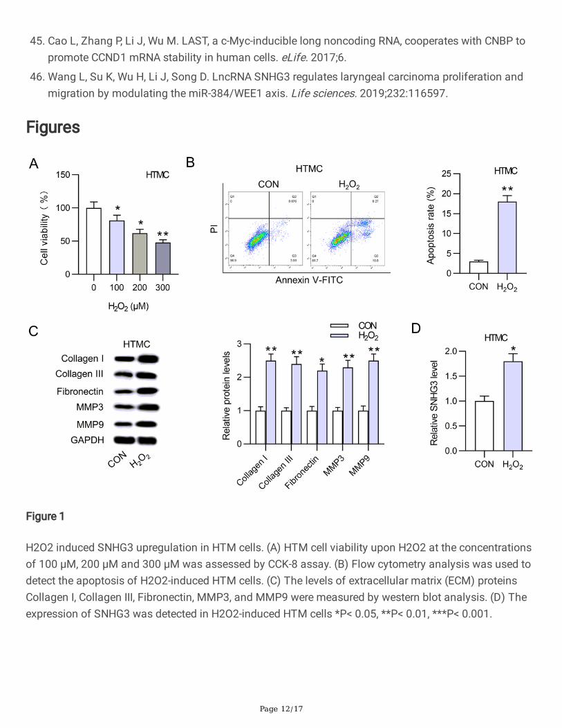
ResultsH2O2 induced SNHG3 upregulation in HTM cells
We �rst established a H2O2-induced oxidative damage model in HTM cells. Then, HTM cells were treatedwith different concentrations of H2O2 (0, 100, 200, and 300 µM). Next, the viability and apoptosis of H2O2-induced HTM cells were detected. CCK-8 assay revealed that HTM cell viability was inhibited by H2O2 atthe concentrations of 100 µM, 200 µM and 300 µM (Fig. 1A). Since the IC50 of viability was exhibited atthe concentration of 200 µM, we chose 200 µM H2O2 to dispose HTM cells in the subsequent assays.Flow cytometry analysis showed H2O2 induced the apoptosis of HTM cells compared with the controlgroup (Fig. 1B). Additionally, the levels of extracellular matrix (ECM) proteins Collagen I, Collagen III,Fibronectin, MMP3, and MMP9 were obviously increased by H2O2 in HTM cells (Fig. 1C). Moreover,SNHG3 expression was upregulated in HTM cells (Fig. 1D). In summary, theses data showed that theH2O2-induced oxidative damage model in HTM cells was established successfully.SNHG3 silencing alleviated H2O2-induced oxidative damage in HTM cells
To detect the biological function of SNHG3 in H2O2-induced HTM cells, SNHG3 was knockdown bytransfection of sh-SNHG3#1 or sh-SNHG3#2 (Fig. 2A). Then, CCK-8 assay demonstrated SNHG3knockdown promoted the viability of H2O2-induced HTM cells (Fig. 2B). Flow cytometry analysis showedthat SNHG3 silencing suppressed the apoptosis of H2O2-induced HTM cells (Fig. 2C). Moreover, the levelsof ECM proteins Collagen III, Collagen I, Fibronectin, MMP3, and MMP9 were reduced by SNHG3 silencing
Page 6/17
in H2O2-induced HTM cells (Fig. 2D). Overall, SNHG3 silencing alleviated H2O2-induced oxidative damagein HTM cells.
SNAI2 knockdown alleviated H2O2-induced oxidative damage in HTM cells
SNAI2 has been widely reported to regulate ECM proteins 34,35, so we predicted that it regulated ECMproteins in HTM cells. To evaluate the biological function of SNAI2 in H2O2-induced HTM cells, SNAI2was knockdown by transfection of sh-SNAI2#1 or sh-SNAI2#1. Western blot analysis demonstrated thatSNAI2 protein level was increased by H2O2, but it was then decreased by transfection of sh-SNAI2#1 orsh-SNAI2#2 (Fig. 3A). Then, CCK-8 assay showed SNAI2 knockdown promoted the viability of H2O2-induced HTM cells (Fig. 4B). Flow cytometry analysis revealed that SNAI2 knockdown suppressed theapoptosis of H2O2-induced HTM cells (Fig. 3C). Moreover, the levels of ECM proteins Collagen III, CollagenI, Fibronectin, MMP3, and MMP9 were reduced by SNAI2 knockdown in H2O2-induced HTM cells (Fig. 3D).In summary, SNAI2 silencing alleviated H2O2-induced oxidative damage in HTM cells.
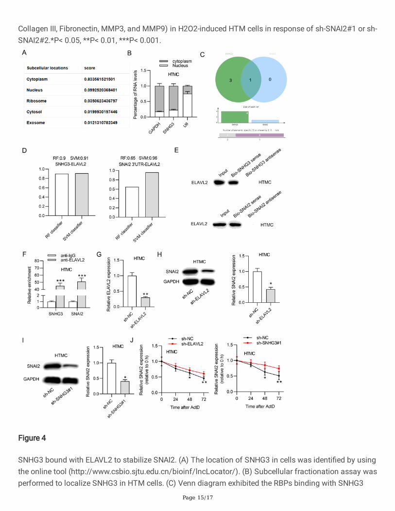
SNHG3 bound with ELAVL2 to stabilize SNAI2
Subsequently, we explored the molecular mechanism of SNHG3 in HTM cells. First, we found that SNHG3mainly existed in the cytoplasm of cells by using online tool(http://www.csbio.sjtu.edu.cn/bioinf/lncLocator/) (Fig. 4A). Then, we further found that SNHG3 mainlyexisted in the cytoplasm of HTM cells (Fig. 4B). Next, as shown in Fig. 4C, by searching the websitehttp://rbpdb.ccbr.utoronto.ca, one RBP, ELAVL2, was identi�ed to bind with SNHG3 and SNAI2 (condition:binding score > 10). Based on the website http://pridb.gdcb.iastate.edu//RPISeq/, the accuracies ofrandom forest (RF) and support vector machine (SVM) classi�ers for SNHG3-ELAVL2 binding were 0.9and 0.91 respectively, and the accuracies of RF and SVM classi�ers for SNAI2 3'UTR-ELAVL2 bindingwere 0.65 and 0.96 respectively, suggesting ELAVL2 bound with SNHG3 and SNAI2 (Fig. 4D). RNA pulldown assay demonstrated that ELAVL2 was enriched in the Bio-SNHG3 sense group and the Bio-SNAI2sense group but not the Bio-SNHG3 antisense group and Bio-SNAI2 antisense group, con�rming thatELAVL2 bound with SNHG3 and SNAI2 (Fig. 4E). Similarly, RIP assay suggested that both SNHG3 andSNAI2 were enriched in the anti-ELAVL2 group instead of the anti-IgG group (Fig. 4F). Subsequently, HTMcells were transfected with sh-ELAVL2, showing the decreased ELAVL2 expression (Fig. 4G). Then,western lot analysis revelated that ELAVL2 or SNHG3 silencing reduced the protein level of SNAI2 in HTMcells (Fig. 4H-I). Moreover, ELAVL2 silencing or SNHG3 silencing decreased SNAI2 mRNA half-life(Fig. 4J). Overall, SNHG3 bound with ELAVL2 to stabilize SNAI2.
SNAI2 overexpression counteracted the effect of SNHG3 silencing on H 2 O 2 -induced HTM cells
To detect whether SNAI2 overexpression counteracted the effect of SNHG3 silencing on HTM cells, rescueassays were conducted. First, SNAI2 protein level was elevated by the transfection of pcDNA3.1/SNAI2 inH2O2-induced HTM cells (Fig. 5A). Then, we found that SNAI2 overexpression counteracted the promotiveeffect of SNHG3 silencing on H2O2-induced HTM cell viability (Fig. 5B). SNAI2 overexpression offset the
Page 7/17
inhibitive effect of SNHG3 silencing on H2O2-induced HTM cell apoptosis (Fig. 5C). Additionally, thesuppressive in�uence of SNHG3 knockdown on ECM proteins (Collagen I, Collagen III, Fibronectin, MMP3,and MMP9) was abolished by SNAI2 overexpression (Fig. 5D). In conclusion, SNAI2 overexpressionabrogated the effect of SNHG3 silencing on H2O2-induced HTM cells.
DiscussionIn this research, we established a H2O2-induced oxidative damage model in HTM cells and explored therole of SNHG3 in H2O2-induced HTM cells. Our data suggested that SNHG3 cooperated with ELAVL2 tomodulate cell apoptosis and ECM accumulation by stabilizing SNAI2 in H2O2-induced HTM cells.
Previous research demonstrated that lncRNAs have been implicated in neurodegenerative diseases,including Alzheimer's disease 36,37 and glaucoma 23,24. SNHG3 has been reported to regulate a variety ofdiseases, such as hepatocellular carcinoma 29 and osteosarcoma 30. Notably, SNHG3 has been found tobe related to neurodegeneration 31, and it has been reported to be upregulated in H2O2-induced HTM cells32. In the present study, our data revealed that SNHG3 was upregulated in H2O2-induced HTM cells andSNHG3 silencing alleviated H2O2-induced oxidative damage in HTM cells by promoting cell viability,inhibiting cell apoptosis and decreasing ECM protein level.
SNAI2 (snail family transcriptional repressor 2) is also known as SLUG, SLUGH, SLUGH1, SNAIL2, orWS2D; it has been reported to regulate a wide range of human diseases 38–40. Importantly, previousresearch has proved that SNAI2 is involved in the regulation of neurodegenerative diseases 41, and it mayparticipate in the modulation of glaucoma 42. However, the speci�c role and regulatory mechanism ofSNAI2 in glaucoma is not clear.
LncRNAs can cooperate with speci�c proteins to increase mRNA stability or induce mRNA decay incytoplasm 43. For example, LncRNA LINC00707 interacts with mRNA stabilizing protein HuR to increasethe stability of VAV3/F11R in gastric cancer 44. LncRNA LAST interacts with CNBP to promote CCND1mRNA stability in human cells 45. Previously, the regulatory mechanism of SNHG3 has been explored insome human diseases 29,30,46. However, the molecular regulatory mechanism of SNHG3 in glaucomaremains to be explored. In this study, we observed that SNHG3 bound with ELAVL2 to stabilize SNAI2 inH2O2-induced HTM cells. SNAI2 overexpression counteracted the effect of SNHG3 silencing on H2O2-induced HTM cells. In addition, we found that SNAI2 knockdown relieved H2O2-induced oxidative damagein HTM cells by promoting cell viability, inhibiting cell apoptosis and decreasing ECM protein level.
ConclusionsThe main purpose of the present research was to investigate the biological role and regulatorymechanism of SNHG3 in H2O2-induced HTM cells. Our results illustrated that SNHG3 interacted withELAVL2 to regulate cell proliferation, apoptosis and ECM accumulation by increasing the stability of
Page 8/17
SNAI2 in H2O2-induced HTM cells. This �nding may shed some light on the regulatory mechanism ofSNHG3 in human diseases and advance our understanding on the pathogenesis of glaucoma. Futurestudies should be conducted on the biological functions of SNHG3 in other cell types and the regulatorymechanism of H2O2-induced dysregulated SNHG3 expression. In addition, in vivo experiments could beadded to further validate the role of SNHG3 in glaucoma.
AbbreviationsHTM: human trabecular meshwork; RT-qPCR: real-time quantitative polymerase chain reaction; CCK-8: cellcounting kit-8; ECM: extracellular matrix; TM: trabecular meshwork; MMPs: matrix metalloproteinases;lncRNAs: long noncoding RNAs; ncRNAs: noncoding RNAs; RBPs: RNA-binding proteins; mRNAs:messenger RNAs; RIP: RNA immunoprecipitation;
DeclarationsEthical Approval and Consent to participate
Not applicable
Consent for publication
Not applicable
Availability of supporting data
The data underlying this article will be shared on reasonable request to the corresponding author.
Acknowledgement
Not applicable.
Funding
None
Con�icts of interest
The authors declare that there are no competing interests in this study.
Authors' contributions
Sizhen Li conceived and designed the experiments. Sizhen Li, Qingsong Yang, Zixiu Zhou, Min Fu,Xiaodong Yang, Kuanxiao Hao, and Yating Liu carried out the experiments. Sizhen Li and Qingsong Yanganalyzed the data. Sizhen Li and Qingsong Yang drafted the manuscript. All authors agreed to beaccountable for all aspects of the work. All authors have read and approved the �nal manuscript.
Page 9/17
References1. He S, Stankowska DL, Ellis DZ, Krishnamoorthy RR, Yorio T. Targets of Neuroprotection in Glaucoma.
Journal of ocular pharmacology and therapeutics : the o�cial journal of the Association for OcularPharmacology and Therapeutics. 2018;34(1-2):85-106.
2. Weinreb RN, Aung T, Medeiros FA. The pathophysiology and treatment of glaucoma: a review. Jama.2014;311(18):1901-1911.
3. Jonas JB, Aung T, Bourne RR, Bron AM, Ritch R, Panda-Jonas S. Glaucoma. Lancet (London,England). 2017;390(10108):2183-2193.
4. Patel N, McAllister F, Pardon L, Harwerth R. The effects of graded intraocular pressure challenge onthe optic nerve head. Experimental eye research. 2018;169:79-90.
5. Agarwal P, Agarwal R. Trabecular meshwork ECM remodeling in glaucoma: could RAS be a target?Expert opinion on therapeutic targets. 2018;22(7):629-638.
�. Saccà SC, Gandol� S, Bagnis A, et al. From DNA damage to functional changes of the trabecularmeshwork in aging and glaucoma. Ageing research reviews. 2016;29:26-41.
7. Nikhalashree S, George R, Shantha B, et al. Detection of Proteins Associated with Extracellular MatrixRegulation in the Aqueous Humour of Patients with Primary Glaucoma. Current eye research.2019;44(9):1018-1025.
�. NikhalaShree S, Karthikkeyan G, George R, et al. Lowered Decorin With Aberrant Extracellular MatrixRemodeling in Aqueous Humor and Tenon's Tissue From Primary Glaucoma Patients. Investigativeophthalmology & visual science. 2019;60(14):4661-4669.
9. Tamm ER, Fuchshofer R. What increases out�ow resistance in primary open-angle glaucoma? Surveyof ophthalmology. 2007;52 Suppl 2:S101-104.
10. Rönkkö S, Rekonen P, Kaarniranta K, Puustjärvi T, Teräsvirta M, Uusitalo H. Matrix metalloproteinasesand their inhibitors in the chamber angle of normal eyes and patients with primary open-angleglaucoma and exfoliation glaucoma. Graefe's archive for clinical and experimental ophthalmology =Albrecht von Graefes Archiv fur klinische und experimentelle Ophthalmologie. 2007;245(5):697-704.
11. Hrabec E, Naduk J, Strek M, Hrabec Z. [Type IV collagenases (MMP-2 and MMP-9) and theirsubstrates--intracellular proteins, hormones, cytokines, chemokines and their receptors]. Postepybiochemii. 2007;53(1):37-45.
12. Polette M, Birembaut P. Membrane-type metalloproteinases in tumor invasion. The internationaljournal of biochemistry & cell biology. 1998;30(11):1195-1202.
13. Wallace DM, Murphy-Ullrich JE, Downs JC, O'Brien CJ. The role of matricellular proteins in glaucoma.Matrix biology : journal of the International Society for Matrix Biology. 2014;37:174-182.
14. Wallace DM, Pokrovskaya O, O'Brien CJ. The Function of Matricellular Proteins in the LaminaCribrosa and Trabecular Meshwork in Glaucoma. Journal of ocular pharmacology and therapeutics :the o�cial journal of the Association for Ocular Pharmacology and Therapeutics. 2015;31(7):386-395.
Page 10/17
15. Chatterjee A, Villarreal G, Jr., Rhee DJ. Matricellular proteins in the trabecular meshwork: review andupdate. Journal of ocular pharmacology and therapeutics : the o�cial journal of the Association forOcular Pharmacology and Therapeutics. 2014;30(6):447-463.
1�. DiStefano JK. The Emerging Role of Long Noncoding RNAs in Human Disease. Methods inmolecular biology (Clifton, NJ). 2018;1706:91-110.
17. Lee H, Zhang Z, Krause HM. Long Noncoding RNAs and Repetitive Elements: Junk or IntimateEvolutionary Partners? Trends in genetics : TIG. 2019;35(12):892-902.
1�. Ferrè F, Colantoni A, Helmer-Citterich M. Revealing protein-lncRNA interaction. Brie�ngs inbioinformatics. 2016;17(1):106-116.
19. Fasolo F, Patrucco L, Volpe M, et al. The RNA-binding protein ILF3 binds to transposable elementsequences in SINEUP lncRNAs. FASEB journal : o�cial publication of the Federation of AmericanSocieties for Experimental Biology. 2019;33(12):13572-13589.
20. Lee S, Kopp F, Chang TC, et al. Noncoding RNA NORAD Regulates Genomic Stability by SequesteringPUMILIO Proteins. Cell. 2016;164(1-2):69-80.
21. Ramanathan M, Porter DF, Khavari PA. Methods to study RNA-protein interactions. Nature methods.2019;16(3):225-234.
22. Cissé Y, Bai L, Chen MT. LncRNAs in ocular neovascularizations. International journal ofophthalmology. 2019;12(12):1959-1965.
23. Cissé Y, Bai L, Meng T. LncRNAs in genetic basis of glaucoma. BMJ open ophthalmology.2018;3(1):e000131.
24. Wan P, Su W, Zhuo Y. The Role of Long Noncoding RNAs in Neurodegenerative Diseases. Molecularneurobiology. 2017;54(3):2012-2021.
25. Zhou RR, Li HB, You QS, et al. Silencing of GAS5 Alleviates Glaucoma in Rat Models by ReducingRetinal Ganglion Cell Apoptosis. Human gene therapy. 2019;30(12):1505-1519.
2�. Li HB, You QS, Xu LX, et al. Long Non-Coding RNA-MALAT1 Mediates Retinal Ganglion Cell ApoptosisThrough the PI3K/Akt Signaling Pathway in Rats with Glaucoma. Cellular physiology andbiochemistry : international journal of experimental cellular physiology, biochemistry, andpharmacology. 2017;43(5):2117-2132.
27. Sun W, Li YN, Ye JF, Guan YQ, Li SJ. MEG3 is involved in the development of glaucoma throughpromoting the autophagy of retinal ganglion cells. European review for medical and pharmacologicalsciences. 2018;22(9):2534-2540.
2�. Liu Z, Tao H. Small nucleolar RNA host gene 3 facilitates cell proliferation and migration in oralsquamous cell carcinoma via targeting nuclear transcription factor Y subunit gamma. Journal ofcellular biochemistry. 2020;121(3):2150-2158.
29. Zhang PF, Wang F, Wu J, et al. LncRNA SNHG3 induces EMT and sorafenib resistance by modulatingthe miR-128/CD151 pathway in hepatocellular carcinoma. Journal of cellular physiology.2019;234(3):2788-2794.

Page 11/17
30. Zheng S, Jiang F, Ge D, et al. LncRNA SNHG3/miRNA-151a-3p/RAB22A axis regulates invasion andmigration of osteosarcoma. Biomedicine & pharmacotherapy = Biomedecine & pharmacotherapie.2019;112:108695.
31. Arisi I, D'Onofrio M, Brandi R, et al. Gene expression biomarkers in the brain of a mouse model forAlzheimer's disease: mining of microarray data by logic classi�cation and feature selection. Journalof Alzheimer's disease : JAD. 2011;24(4):721-738.
32. Yao K, Yu Y, Li F, Jin P, Deng C, Zhang H. Integrative analysis of an lncRNA‐associated competingendogenous RNA network in human trabecular meshwork cells under oxidative stress. Molecularmedicine reports. 2020;21(3):1606-1614.
33. Sáez GT. DNA Damage and Repair in Degenerative Diseases 2016. International journal of molecularsciences. 2017;18(1).
34. Cheon DJ, Tong Y, Sim MS, et al. A collagen-remodeling gene signature regulated by TGF-β signalingis associated with metastasis and poor survival in serous ovarian cancer. Clinical cancer research :an o�cial journal of the American Association for Cancer Research. 2014;20(3):711-723.
35. Liu T, Zhang H, Sun L, et al. FSIP1 binds HER2 directly to regulate breast cancer growth andinvasiveness. Proceedings of the National Academy of Sciences of the United States of America.2017;114(29):7683-7688.
3�. Luo Q, Chen Y. Long noncoding RNAs and Alzheimer's disease. Clinical interventions in aging.2016;11:867-872.
37. Riva P, Ratti A, Venturin M. The Long Non-Coding RNAs in Neurodegenerative Diseases: NovelMechanisms of Pathogenesis. Current Alzheimer research. 2016;13(11):1219-1231.
3�. Pingault V, Ente D, Dastot-Le Moal F, Goossens M, Marlin S, Bondurand N. Review and update ofmutations causing Waardenburg syndrome. Human mutation. 2010;31(4):391-406.
39. Nakayama Y, Tsuruya Y, Noda K, et al. Negative feedback by SNAI2 regulates TGFβ1-inducedamelotin gene transcription in epithelial-mesenchymal transition. Journal of cellular physiology.2019;234(7):11474-11489.
40. Zhou W, Gross KM, Kuperwasser C. Molecular regulation of Snai2 in development and disease.Journal of cell science. 2019;132(23).
41. Santana MM, Chung KF, Vukicevic V, et al. Isolation, characterization, and differentiation ofprogenitor cells from human adult adrenal medulla. Stem cells translational medicine.2012;1(11):783-791.
42. Pattabiraman PP, Maddala R, Rao PV. Regulation of plasticity and �brogenic activity of trabecularmeshwork cells by Rho GTPase signaling. Journal of cellular physiology. 2014;229(7):927-942.
43. Gong C, Maquat LE. lncRNAs transactivate STAU1-mediated mRNA decay by duplexing with 3' UTRsvia Alu elements. Nature. 2011;470(7333):284-288.
44. Xie M, Ma T, Xue J, et al. The long intergenic non-protein coding RNA 707 promotes proliferation andmetastasis of gastric cancer by interacting with mRNA stabilizing protein HuR. Cancer letters.2019;443:67-79.
Page 12/17
45. Cao L, Zhang P, Li J, Wu M. LAST, a c-Myc-inducible long noncoding RNA, cooperates with CNBP topromote CCND1 mRNA stability in human cells. eLife. 2017;6.
4�. Wang L, Su K, Wu H, Li J, Song D. LncRNA SNHG3 regulates laryngeal carcinoma proliferation andmigration by modulating the miR-384/WEE1 axis. Life sciences. 2019;232:116597.
Figures
Figure 1
H2O2 induced SNHG3 upregulation in HTM cells. (A) HTM cell viability upon H2O2 at the concentrationsof 100 μM, 200 μM and 300 μM was assessed by CCK-8 assay. (B) Flow cytometry analysis was used todetect the apoptosis of H2O2-induced HTM cells. (C) The levels of extracellular matrix (ECM) proteinsCollagen I, Collagen III, Fibronectin, MMP3, and MMP9 were measured by western blot analysis. (D) Theexpression of SNHG3 was detected in H2O2-induced HTM cells *P< 0.05, **P< 0.01, ***P< 0.001.

Page 13/17
Figure 2
SNHG3 silencing alleviated H2O2-induced oxidative damage in HTM cells. (A) The knockdown e�ciencyof SNHG3 was assessed. (B) CCK-8 assay was used to evaluate the effect of SNHG3 silencing on theviability of H2O2-induced HTM cells. (C) Flow cytometry was performed to detect the apoptosis of H2O2-induced HTM cells after transfection of sh-SNHG3#1 or sh-SNHG3#2. (D) The Collagen I, Collagen III,Fibronectin, MMP3, and MMP9 proteins in H2O2-induced HTM cells after transfection of sh-SNHG3#1 orsh-SNHG3#2 were detected using western blot analysis. *P< 0.05, **P< 0.01, ***P< 0.001.

Page 14/17
Figure 3
SNAI2 knockdown alleviated H2O2-induced oxidative damage in HTM cells. (A) The knockdowne�ciency of SNAI2 was assessed. (B) CCK-8 assay was used to evaluate the effect of SNAI2 silencing onthe viability of H2O2-induced HTM cells. (C) Flow cytometry was carried out to detect the apoptosis ofH2O2-induced HTM cells upon sh-SNAI2#1 or sh-SNAI2#2. (D) The levels of ECM proteins (Collagen I,
Page 15/17
Collagen III, Fibronectin, MMP3, and MMP9) in H2O2-induced HTM cells in response of sh-SNAI2#1 or sh-SNAI2#2.*P< 0.05, **P< 0.01, ***P< 0.001.
Figure 4
SNHG3 bound with ELAVL2 to stabilize SNAI2. (A) The location of SNHG3 in cells was identi�ed by usingthe online tool (http://www.csbio.sjtu.edu.cn/bioinf/lncLocator/). (B) Subcellular fractionation assay wasperformed to localize SNHG3 in HTM cells. (C) Venn diagram exhibited the RBPs binding with SNHG3

Page 16/17
and SNAI2. (D) Online database (http://pridb.gdcb.iastate.edu//RPISeq/) was used to analyze the bindingability of ELAVL2 with SNHG3 or SNAI2. (E) RNA pull down assay was conducted to assess the bindingof ELAVL2 with SNHG3 or SNAI2. (F) RIP assay was used to evaluate the binding of ELAVL2 with SNHG3or SNAI2. (G) The knockdown e�ciency of ELAVL2 was detected. (H) The protein level of ELAVL2 in HTMcells after transfection of sh-ELAVL2. (I) The protein level of ELAVL2 in HTM cells after transfection of sh-SNHG3#1. (J) The expression of ELAVL2 in HTM cells after transfection of sh-ELAVL2 or sh-SNHG3#1.*P< 0.05, **P< 0.01, ***P< 0.001.

Page 17/17
Figure 5
SNAI2 overexpression counteracted the effect of SNHG3 silencing on H2O2-induced HTM cells. (A) Theoverexpression e�ciency of SNAI2 was assessed by western blot analysis. (B) The viability of H2O2-induced HTM cells after transfection of indicated plasmids was detected. (C) The apoptosis of H2O2-induced HTM cells after transfection of appointed plasmids. (D) The levels of ECM proteins (Collagen I,Collagen III, Fibronectin, MMP3, and MMP9) in H2O2-induced HTM cells in the different groups. *P< 0.05,**P< 0.01, ***P< 0.001.