establishing canine trabecular meshwork cell … · establishing canine trabecular meshwork cell...
TRANSCRIPT

i
ESTABLISHING CANINE TRABECULAR MESHWORK CELL CULTURE
By
Hsiang-Rong Tsai
A THESIS
Submitted to
Michigan State University
in partial fulfillment of the requirements
for the degree of
Physiology - Master of Science
2016

ii
ABSTRACT
ESTABLISHING CANINE TRABECULAR MESHWORK CELL CULTURE
By
Hsiang-Rong Tsai
Glaucoma is a leading cause of incurable blindness. The most common form of glaucoma is
primary open-angle glaucoma (POAG) and is associated with pathological alterations of aqueous
outflow facilities which lead to increased resistance and elevated intraocular pressure (IOP).
Trabecular meshwork (TM) cells residing within the iridocorneal angle are considered key
regulators of aqueous humor outflow; their malfunction is considered one of major factors in the
pathogenesis of POAG. Glaucoma not only affects humans, but also other species such as dogs.
Dogs are more accessible than other species to study the disease. Canine and human POAG share
many features such as plaque formation within the TM with resulting slow progressive elevated
IOP, and cupping of the optic nerve head. Because of the similarities between human and canine
disease, we posit that the study of canine TM cells will be beneficial for the understanding of
disease mechanisms in both dog and human. The purpose of this study was to establish primary
canine TM cell cultures. Canine TM explants were carefully isolated and transferred to the cell
culture dish. TM cells were identified by their expression of collagen type IV, alpha smooth
muscle actin (α-SMA) and laminin, but not desmin and keratin. Another key feature of TM cells
is their phagocytosis activity. Finally, the cultured TM cells form typical cross-linked actin
networks (CLANs). To the best of our knowledge, this is the first report of primary canine TM
(CTM) cultures.

iii
ACKNOWLEDGEMENTS
I would like thank all individuals who supported my research and thesis writing, especially
my colleagues, staff and professors from both the Department of Physiology and the College of
Veterinary Medicine at Michigan State University.
First of all, I would like to appreciate my mentor Dr. András M. Komáromy. Like a
lighthouse he provided me guidance for my research and professional life. His dedication for
both research and clinical work provide me with an example about the life of a clinical scientist.
Under his influence and encouragement, I have chosen to pursuit a degree in veterinary medicine
at Michigan State University following my graduate work. I intend to dedicate my life to both
research and clinical work with a hope to have a positive impact in this world. And I would like
to thank my committee members Dr. Susanne Mohr and Dr. Arthur Weber who have given me
constructive suggestion and guidance for my research. Special thanks to Dr. Susanne Mohr as
director of the Physiology Graduate Program who guided both my graduate work and my career
decision to pursuit a veterinary degree. I feel fortunate to have joined her program.
I would like to thank Mrs. Christine Herman, research associate, and Ms. Kristin Koehl,
veterinary technician, for their full support of my research work. Without their kind support, I
would not have been able to complete my research. I would also like to thank the following past
members of the Komáromy Lab for their help and company: Forrest Nussdorfer, Josh Laske, and
Gabriel Stewart.
Finally, I would like to thank my family members for their support, especially my parents
and brother. Without their support, my pursuit of research would have been much harder. And I

iv
want to thank the support from Taiwan through their generous scholarship and a grant from
Midwest Eye-Banks to assist my study.

v
TABLE OF CONTENTS
LIST OF TABLES vii
LIST OF FIGURES viii
KEY TO ABBREVIATIONS ix
CHAPTER 1 – INTRODUCTION 1
Definition of glaucoma 1
Primary open-angle glaucoma (POAG) 1
Aqueous humor (AH) dynamics 4
Aqueous humor formation 4
Aqueous humor outflow 5
Physiology and pathophysiology of the TM 6
Current treatment options for POAG 8
Animal models of POAG 11
In vitro model systems of POAG 12
Rational for studying canine glaucoma and TM 13
CHAPTER 2 – METHODS 15
Isolation of the canine TM (CTM) 15
Primary TM cell culture 17
Fibroblast primary culture 19
Characterization of TM cells – ICC 19
Characterization of TM cells - Phagocytosis assay 22
Dexamethasone (DEX) challenge 22
CHAPTER 3 – RESULTS 25
Histological evaluation of isolated CTM explant 25
Characterization of cultured TM cells: Immunocytochemistry 30
Characterization of cultured TM cells: Phagocytosis assay 31
Characterization of cultured TM cells: DEX challenge - CLAN formation 33
Characterization of cultured TM cells: DEX challenge – gene expression 35
CHAPTER 4 – DISCUSSION 38
Most TM tissue was excised for cell culture as confirmed by histological examination 38
Multiple TM-specific protein markers are observed at our TM cell strains 38
CLAN formation observed in our non-DEX treated CTM cells 39
Phagocytosis activity of the CTM cells 39
CTM cells didn’t have significant DEX response 40
The concern of inadvertently isolating non-TM cells 41
Senescence observed in the CTM cell strains 42

vi
CHAPTER 5 - SUMMARY AND FUTURE DIRECTIONS 43
Summary 43
Future direction 43
REFERENCES 47

vii
LIST OF TABLES
Table 1-1: Genes associated with POAG 3
Table 1-2: Current therapies for POAG: Pharmacological therapies 9
Table 1-3: Current therapies for POAG: Surgical therapies 10
Table 1-4: Animal models of POAG & ocular hypertension 11
Table 2-1: Characterization methods previously used for TM cell identification 21
Table 2-2: The list of antibodies used in the experiments 21
Table 3-1: Established primary CTM cell strains 26
Table 3-2: Characterization of canine cell strains 30
Table 3-3: Phagocytosis activity of canine cell strains 33
Table 3-4: Completed q-RTPCR of CTM cell strains and other type of cells 37
viii
LIST OF FIGURES
Figure 1-1 AH dynamics in the canine eye 6
Figure 1-2 ECM accumulation in the TM of POAG-affected dogs 13
Figure 2-1 Dissection procedure 16
Figure 2-2 Experimental design 18
Figure 3-1 Cell migration and morphology of canine TM cells and fibroblasts 29
Figure 3-2 The ICC result of CTM 31
Figure 3-3 Phagocytosis activity of cultured CTM cells 32
Figure 3-4 CLAN formation in canine TM cells 33
Figure 3-5 Relative number of CLAN-positive cells in different canine cell strains 35
Figure 3-6 Relative gene expression of MYOC following 3 days of DEX treatment in
different canine cell strains 36

ix
KEY TO ABBREVIATIONS
AAP angular aqueous plexus
AH aqueous humor
α-SMA alpha-smooth muscle actin
CE ciliary epithelium
CLAN cross-linked actin network
CSTM corneoscleral trabecular meshwork
CTM canine trabecular meshwork
DAPI 4', 6-diamidino-2-phenylindole: DEX: Dexamethasone
ECM extracellular matrix
FBS fetal bovine serum
ICA iridocorneal angle
ICC immunocytochemistry
IOP intraocular pressure
iPSCs induced pluripotent stem cells
ISVP intrascleral venous plexus
NPE non-pigment epithelium
PACG primary angle-closure glaucoma
PBS phosphate-buffered saline
PE pigmented epithelium
POAG primary open-angle glaucoma
q-RTPCR quantitative reverse transcription polymerase chain reaction

x
rAAV recombinant adeno-associated virus
TGF-2 transforming growth factor beta 2
TM trabecular meshwork
UTM uveal trabecular meshwork
wt wild type

1
CHAPTER 1 – INTRODUCTION
Definition of glaucoma
Glaucoma is a leading cause of incurable blindness. It is defined as a progressive optic
neuropathy characterized by the loss of retinal ganglion cells and clinically distinguishable
cupping of the optic nerve head 1. There are different forms of glaucoma, both primary and
secondary, which all share the same risk factor of elevated intraocular pressure (IOP). This
research is focused on primary glaucoma with no clinically observable underlying cause such as
inflammation 2, neoplasia
3, or pigmentary dispersion
4. The two most common forms of primary
glaucoma are primary open-angle glaucoma (POAG) and primary angle-closure glaucoma
(PACG). In both of these diseases, alterations along the aqueous humor (AH) outflow pathway
result in increased resistance and IOP elevation (Fig. 1-1). While these changes are associated
with closure of the iridocorneal angle (ICA) in PACG, the angle appears clinically normal and
open in POAG 1. In POAG, it is believed that the pathogenesis of disease is associated with
molecular and cellular pathological changes in the trabecular meshwork (TM), a key regulator of
AH outflow, resulting in impaired AH drainage from the eye. Those defects within the TM
remain largely unknown.
Primary open-angle glaucoma (POAG)
POAG is considered a major type of glaucoma worldwide 5. Elevated IOP is considered a
major risk factor of POAG, but it can also develop without obvious IOP change, such as in 50%
percent of European- and African- descent patients 6 and 92% for Japanese-descent patients
7,8.

2
In additional to IOP, several other POAG risk factors have been identified 9,10
, such as aging,
family history, and ethnicity.
Intraocular pressure (IOP): The level of IOP is the only risk factor which can be intervened
by medical and surgical treatment 11
. In healthy individuals, the range of habitual IOP is 16.3 -
21.3 mmHg. In contrast, the IOPs of hypertensive POAG patients are usually over 22 mmHg 12
.
In addition to being elevated, IOPs also fluctuates more in glaucomatous vs. normal eyes 12
.
Aging: Both the incidence and prevalence of POAG strongly correlate with aging,
especially after 40 years of age, due to gradual increase in AH outflow resistance 13
. For example,
people of European-descent have a 25% incidence to develop POAG by the age of 64 years and
up to 75% by 81 years 13
. A similar increasing age-related POAG incidence has also been
observed in people of African-descent. Senescence of TM cells may contribute to this aging-
related increase in AH outflow resistance 14
. Furthermore, aging seems to have a negative impact
on the biomechanical properties of the eye: Elevated scleral stiffness associated with aging can
result in increased stress and exacerbated IOP-related damage of the optic nerve head 15
. Finally,
aging-related decrease of cerebrospinal fluid (CSF) pressure 16
may contribute to optic nerve
head damage as well because of greater translaminar gradient 17
.
Family history and genetics: Up to 50% of POAG patients have affected family members 18
.
In addition, first degree relatives of an affected individual have a 22% risk to develop POAG vs.
2.3% risk in other family members 19
. This suggests that genetic predisposition is a critical risk
factor of POAG development. The study of POAG-affected families not only identified high risk
populations but also POAG candidate genes 20
. At least 7 genes have been associated with
POAG: MYOC 21,22
, CYP1B1 23
, WDR36 24
, OPTN 25,26
, LMX1B 27
, NTF4 28
and ASB10 29,30

3
(Table 1-1). Other genes have been associated with POAG risk factors such as optic nerve head
morphology, IOP and refractive error 31,32
.
Table 1-1: Genes associated with POAG
Gene Name Location/Locus Function Reference
MYOC Myocilin, TIGR 1q23/ GLC1A Cell migration,
mitochondria, ECM
turnover and cytoskeleton.
21,22
CYP1B1 Cytochrome
P450 family 1
2p21/GLC3A Metabolism of drug and
lipid synthesis
23,33
WDR36 WD repeat
domain 36
5q22/GLC1G Ribosomal processes and cell
growth
24,34,35
OPTN Optineurin 10p14-
p15/GLC1E
Rab-binding protein, TNF-
alpha signaling NFkB
pathway, immune response,
apoptosis, vesicular
transport, mitosis, cellular
morphogenesis, oxidative
stress protection.
25,36
LMX1B LIM homeobox
transcription
factor 1-beta
9q34.1/NPS Normal patterning of the
dorsoventral axis,
establishment of mid brain-
hindbrain boundary,
development of cerebellum
and dopaminergic and
serotonergic neurons.
27,37-39
NTF4 Neurotrophin-4 19q13.33/GLC1O Survival and differentiation
of mammal neurons,
neurotrophin signaling
pathway
28,40,41
ASB10 Ankyrin Repeat
And SOCS Box
Containing 10
7q35-q36/GLC1F IOP regulation 29,30
Ethnicity: The ethnicity was considered another important risk factor for glaucoma. For
example, the study on African-derived population in the world-wide glaucoma epidemiology has
consistently shown high prevalence of POAG compared with other populations 9,42-45
.

4
Aqueous humor (AH) dynamics
The AH is a transparent medium that fills the anterior chamber located between iris and
cornea. It does not only provide IOP to support the shape of the eye but also delivers nutrients
and removes waste from the eyes 46
. In the normal eye, IOP is tightly regulated by well-
coordinated production and outflow of AH 47
. In POAG-affected eyes, IOP can become elevated
due to a dysregulation of the normal AH dynamics, especially an increase of outflow resistance
through the TM – this is the main focus of our research. In the following paragraphs we will
briefly discuss AH dynamics in the normal eye, before entering the detail of the POAG disease
pathogenesis 48
.
Aqueous humor formation
The ciliary body produces the AH. It can be subdivided in the pars plicata and pars plana
which represent the anterior and posterior portion respectively. The ciliary epithelium (CE) of
the pars plicata is the main location of AH production 49
. The CE covers the ciliary processes
and is composed of two layers, the superficial non-pigment epithelium (NPE) and deep
pigmented epithelium (PE). The basolateral surfaces of NPE cells face the posterior chamber
while its apical surface forms gap junction to the counter part of the PE cells (arrow, Figure 1-1)
50. The other side of the PE is in contact with the stroma of the ciliary process where solutes
accumulate. In order to avoid blood protein and other inflammatory components to leak
uncontrollably into the eye, the NPE cells form tight junction with neighbor cells; this is the
blood-aqueous barrier 50
.
5
In order to bring the nutrition from the blood to the eye, lipid-soluble and water soluble
substances move via diffusion and ultrafiltration from the fenestrated capillaries across the
ciliary stroma to the CE.
In the final stage of AH production, the solutes/substances are transported across the CE
through diffusion, ultrafiltration and active transport. Among of them, active transport
contributes about 80% to 90% of total AH formation 46,51
. Active transportation relies on the ion
transporters 49
. The energy for active transportation is provided by hydrolysis of adenosine
triphosphate (ATP) and mediated by Na+-K
+-ATPase, which is mostly located in the NPE
50.
Besides Na+-K
+-ATPase, NPE also requires carbonic anhydrase to secrete bicarbonate to
regulate pH and maintain osmotic gradient for continuous AH secretion 52
. Carbonic anhydrase is
an important target for glaucoma treatment: Inhibitor of carbonic anhydrase reduces AH
production and thereby lowers IOP 53
.
Aqueous humor outflow
IOP is regulated and maintained mainly by the conventional outflow 54
. Once the AH is
formed, it flows from posterior chamber via pupil to the anterior chamber then drains through the
ICA. AH outflow consists of conventional/trabecular and unconventional/uveosclera outflow.
Unconventional outflow occurs via ciliary muscle and sclera, but is only responsible for <15% of
AH outflow in the normal human eye 55
. In contrast, trabecular outflow provides the major
drainage of AH and is regulated by the TM, a loose porous structure within the ICA (Figure 1-1.).
The TM is composed by three components through which the AH has to drain: uveal,
corneosclera and juxtacanalicular/ cribriform meshworks (Figure 1-1.) 56
. The pore size of the
TM and AH flow rate are controlled by ciliary muscle tendons that are connected to an elastin

6
net within the juxtacanalicular TM 57
. Once it has passed through the juxtacanalicular meshwork,
the AH enters the Schlemm’s canal and through collector channels the episcleral veins and the
systemic blood circulation. The circular Schlemm’s canal is a unique structure that can only be
found in human, primate and mouse 58-60
, while other species such as dog 61
, cat 62
, pig 63
, horse
64, rabbit
65 and zebrafish
66 have an angular aqueous plexus (AAP) instead to drain the AH.
Figure 1-1 AH dynamics in the canine eye. The AH is formed and secreted by the CE covering
the ciliary body (arrow). It drains through the TM and angular aqueous plexus within the ICA.
Physiology and pathophysiology of the TM
TM in normal eyes: The TM plays a critical role in regulating AH outflow and maintains
the IOP in a normal range. It is derived from neural crest and cranial paraxial mesoderm 67
.
There is increasing evidence that even once early differentiation is completed, a small population
of progenitor cells reside within the TM and serve to replenish the loss of TM cells in the adult
eye 68
. There is considerable evidence that the major AH outflow resistance is located in the

7
juxtacanalicular TM and the inner wall of Schlemm’s canal 69
. This resistance is regulated by
cellular and extracellular factors/components. The extra cellular matrix (ECM) production within
the TM plays a major role in the resistance regulation 57
. Uveal and corneosclera TM form the
beams of connective tissue or so called trabecular lamella which contain the core of elastic fibers
and collagen surrounded by the sheaths 69
. These sheaths are formed by the TM cells, which
highly express collagen type IV and laminin, important markers to characterize TM cells, while
the core of the beams consist of collagen I and III 70,71
. The beams connect to each other in
different layers of the TM to form porous filter-like structures. The thickness of beams increases
with age, contributing to the increased POAG risk with age 69
. In contrast, the juxtacanalicular
TM does not form beams of connective tissue but fibril elements of ECM instead.
Juxtacanalicular TM cells and ECM fibrils together form an irregular network, the cribriform
plexus which is lined against the inner wall endothelium of Schlemm’s canal; this
juxtacanalicular TM plays a major role in regulating the resistance of AH outflow 72
. Besides
ECM turnover, the TM cells also have phagocytosis activity as a self-cleaning mechanism and to
reduce/regulate the resistance of AH 73
.
TM in POAG eyes: Both cellular and extracellular factors contribute to increased AH
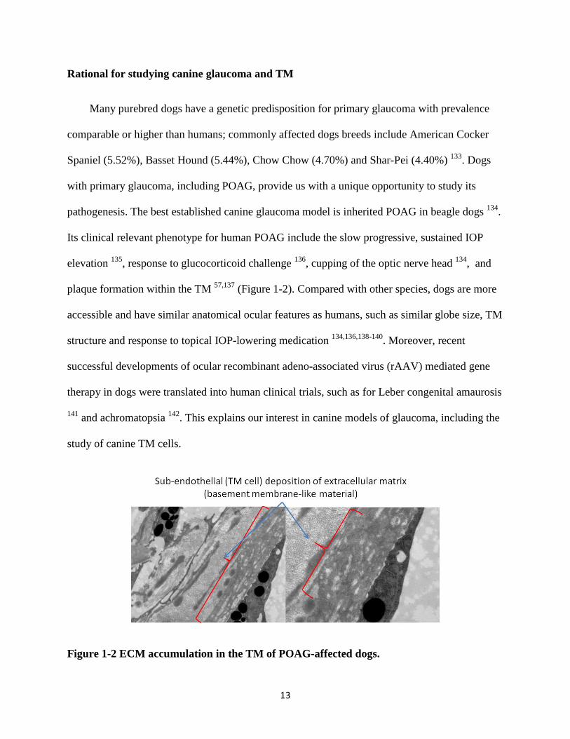
outflow resistance in POAG-affected eyes. ECM plaque formation and fibrosis have been found
within the juxtacanalicular TM of POAG-affected eyes (Figure 1-2.) 57,74,75
. Furthermore, the TM
cell structure appears to be altered in these eyes with increased formation of cross-linked actin
networks (CLANs) 75
. The increased number of CLAN-positive TM cells results in increased
cellular stiffness and contractility and higher AH outflow resistance 76
. Much effort has been put
toward the investigation of the molecular pathways and growth factors involved in the cellular
and extracellular changes within the TM 77
. Increased activation of transforming growth factor

8
beta 2 (TGF-2) likely plays a major role since elevated concentrations can be found within the
AH of POAG patients 78
. TGF-2 also plays a major role in wound healing supporting its
relevance in tissue fibrosis 79
. By increasing TGF-2 in perfused human anterior chambers,
outflow resistance increased and related ECM gene expression, such as fibronectin and
plasminogen activator inhibitor-1, were also noticeably upregulated 80
. In addition, increased
MYOC level in human POAG AH is considered another culprit for disease development within
the TM 81
.
Genetic predisposition and resulting altered gene expression is recognized as a critical
factor within the glaucomatous TM (see also Family history and genetics above). Mutations in
genes such as Pax6 82
, LTBP2 83
, CYP1B1 84
, MYOC 85
affect the TM development and can lead
to congenital glaucoma. The MYOC gene is considered a major POAG candidate gene 86
. Since
MYOC is highly expressed in the TM, the hypothesis was raised that mutant MYOC
accumulation within the TM results in TM malfunction and glaucoma 87
. Even though MYOC
mutations are contributing to the pathogenesis of some forms of POAG and MYOC is highly
expressed in glaucomatous TM cells, its major function remains unknown 88
.
Current treatment options for POAG
Pharmacological approach: Current pharmacological therapies for POAG aim to either
reduce AH formation or increase unconventional outflow (Table 1-2). For example, carbonic
anhydrase inhibitors are used to inhibit the production of AH at the CE 89
. Prostaglandin analogs
increase unconventional outflow 90
. Recently, a new class of glaucoma drugs has been developed,
Rho-kinase inhibitors, which relax the TM and increase AH outflow 91
.

9
Surgical approach: Surgical treatments focus on either reducing AH production or
increasing AH outflow (Table 1-3). These include the use of lasers to partially destroy the ciliary
processes in order to reduce AH production (cyclophotocoagulation) 92
. Trabeculoplasty or
trabeculectomy directly target the TM to reduce the outflow resistance. Various types of shunts
have been developed to facilitate AH outflow from the anterior chamber.
Table 1-2 and Table 1-3 summarize currently available therapies for POAG.
Table 1-2: Current therapies for POAG: Pharmacological therapies
Class Mechanism Available Drugs Reference
Prostaglandin
analogs
Increase
unconventional
outflow via
relaxation of
the ciliary
muscle and
ECM
remodeling in
ciliary body
Latanoprost, Bimatoprost,
Travoprost, Tafluprost,
Unoprostone
90,93,94
Beta-blockers Reduce AH
production
Levobunolol, Carteolol,
Metipranolol, Betaxolol,
Timolol
94-96
Alpha-agonists Constrict
afferent ciliary
process
vasculature to
reduce AH
production;
increase
unconventional
outflow
Brimonidine Tartrate,
Dipivefrin hydrochloride,
Apraclonidine
hydrochloride
90,96-99
Carbonic anhydrase
inhibitors
Reduce AH
production
Brinzolamide, Dorzolamide
Miotics Contracting
ciliary muscle
and sclera spur
to increase
conventional
outflow
Pilocarpine Hydrochloride,
Carbachol, Echothiophate
89,90

10
Table 1-2 (cont’d)
Rho kinase inhibitor Inhibit the
contraction of
ciliary muscle
and TM
Y-27632 100,101
Hyperosmotics Reduce the
volume of
aqueous fluid
Glycerin, Mannitol 91,102,103
Fix-combination Reduce AH
production and
elevate
draining of AH
outflow
Timolol maleate and
Dorzolamide HCl,
Brimonidine Tartrate and
Timolol maleate,
Brinzolamide and
Brimonidine, Latanoprost
and timolol, Bimatoprost
and timolol, Travoprost
and timolol.
104,105
Table 1-3: Current therapies for POAG: Surgery therapies
Type of surgery Mechanism Duration Reference
Laser
cyclophotocoagulation
Destroy ciliary
processes to
reduce AH
production
IOP remains normal
after 2 years from
surgery
92,111,112
Argon laser
trabeculoplasty
Targeting
pigmented and
non-pigmented
TM cells but
preserve
architecture of
TM to improve
drainage
44% of eyes maintain
normal IOP after 2 years
from surgery
92,113
Trabeculectomy Excise TM tissue
to increase AH
outflow
46.9% failure after 5
years from surgery
114,115
Tube shunt Insert the tube
through TM to
Schlemm’s canal
or anterior
chamber to
increase outflow
29.8% failure after 5
years from surgery
115

11
Animal models of POAG
Glaucoma is not only a human disease but also can be observed in other species. Animals
with primary glaucoma provide us with a chance to study disease mechanisms in more detail and
may present potential platforms to evaluate novel therapies development. In addition to these
spontaneous diseases, experimental and transgenic animal models have also been developed to
facilitate POAG study (Table 1-4). Induced ocular hypertension via episcleral vein in
experimental animal model was applied to imitate elevated IOP environment of POAG and
induce similar clinical phenotypes as POAG 116
. However, as previous paragraph, it should be
noticed that ocular hypertension is not always associated with pathogenesis of POAG and is not
used to determine POAG in clinical diagnosis but considered as major risk factor of POAG 1.
The approach of establishing experimental model by using induced ocular hypertension is to
overcome the scarcity of spontaneous model and the sample of patients. It is similar but not as
equal as the samples from spontaneous model and patients.
Table 1-4 lists some of the established animal models for POAG and ocular hypertension.
Table 1-4: Animal models of POAG & ocular hypertension
Species Type of
animal
model
Mechanism Clinical
phenotype
Reference
Monkey Spontaneous Inherited POAG 117
Induced Laser
photocoagulation of
TM
118
Induced Intracameral
injection of latex
microspheres
119
Dog Spontaneous Inherited,
ADAMTS10
mutation
POAG 120,121
Cat Spontaneous Inherited, LTBP2
mutation
POAG 122,123

12
Table 1-4 (cont’d)
Sheep Induced Glucocorticoids
induction
Ocular
hypertension
124
Cow Induced Glucocorticoids
induction
Ocular
hypertension
125
Rabbit Induced Glucocorticoids
induction
Ocular
hypertension
126
Rat Induced Episcleral vein
injection with
hypertonic saline
POAG 116
Mouse Transgenic MYOC mutation POAG 127
Mouse Transgenic OPTN mutation POAG 128
Mouse Transgenic α1 subunit of
collagen Type 1
mutation
Ocular
hypertension
129
Zebrafish Transgenic Lrp2 mutation POAG 130
In vitro model systems of POAG
Although animal models provide us unique opportunities to observe and study disease
mechanisms, there are downsides, including the need to sacrifice animals and the high housing
expenses. Moreover, species differences may prevent direct translation of findings from animals
to humans. In vitro systems that have been developed to study AH outflow pathways include TM
cell cultures (Table 2-1) and anterior ocular segment perfusion culture which has been
successfully developed on human 131
and bovine 132
. These systems allow the use of human
surgical TM tissue samples or whole donor eyes.
13
Rational for studying canine glaucoma and TM
Many purebred dogs have a genetic predisposition for primary glaucoma with prevalence
comparable or higher than humans; commonly affected dogs breeds include American Cocker
Spaniel (5.52%), Basset Hound (5.44%), Chow Chow (4.70%) and Shar-Pei (4.40%) 133
. Dogs
with primary glaucoma, including POAG, provide us with a unique opportunity to study its
pathogenesis. The best established canine glaucoma model is inherited POAG in beagle dogs 134
.
Its clinical relevant phenotype for human POAG include the slow progressive, sustained IOP
elevation 135
, response to glucocorticoid challenge 136
, cupping of the optic nerve head 134
, and
plaque formation within the TM 57,137
(Figure 1-2). Compared with other species, dogs are more
accessible and have similar anatomical ocular features as humans, such as similar globe size, TM
structure and response to topical IOP-lowering medication 134,136,138-140
. Moreover, recent
successful developments of ocular recombinant adeno-associated virus (rAAV) mediated gene
therapy in dogs were translated into human clinical trials, such as for Leber congenital amaurosis
141 and achromatopsia
142. This explains our interest in canine models of glaucoma, including the
study of canine TM cells.
Figure 1-2 ECM accumulation in the TM of POAG-affected dogs.

14
In addition, glaucomatous dogs represent a source of tissue for the study of disease
mechanisms. Although primary human TM cell cultures are well established, the size of the
trabeculectomy specimens is usually small 74
. This increases the difficulty to isolate and culture
sufficient TM cells, especially when considering the limited numbers of passages 74
. Whole eyes
from human donors with well-established history and clinical data are rare and difficult to obtain.
To the best of our knowledge, there are no published reports on the development of primary
canine TM cell cultures. Thus, there is a need to develop and characterize primary canine TM
cell cultures; this was the purpose of this study.

15
CHAPTER 2 – METHODS
Isolation of the canine TM (CTM)
17 canine eyes from 9 adult dogs were collected within 5 minutes of euthanasia. These were
either dogs from a local animal shelter or research dogs euthanized for reasons unrelated to this
study. The globes were stored on ice until further processing. The 5 eyes from 3 dogs obtained
from the shelter were used to refine the steps of tissue dissection and histologic identification of
tissue types. The 12 eyes obtained in the laboratory were used to harvest and culture TM cells
within 20 hours of enucleation (Table 3-1). The procedures were approved by the Michigan State
University IACUC, and were done in accordance with the ARVO Statement for the Use of
Animals in Ophthalmic and Vision Research.
The surfaces of the globes were aseptically prepared with 10% povidone iodine and 70%
ethanol, then rinsed with 1X working concentration phosphate-buffered saline, pH 7.4 (PBS).
The posterior segment was removed from the anterior segment by cutting the globe along the
pars plana of the ciliary body with a No. 11 scalpel blade and Westcott tenotomy scissors (Figure
2-1). The lens was then removed from the anterior segment by cutting the lens zonules with
Westcott tenotomy scissors (Figure 2-1). The ciliary body and iris were bluntly dissected from
the sclera in a posterior to anterior direction (Figure 2-1). The TM tissue could then be
recognized as a white band on the ciliary body, delimited anteriorly by the pectinate ligament
(Figure 2-1). The identification of the TM was confirmed histologically by processing the
dissected tissue routinely for paraffin embedding, sectioning, and staining with hematoxylin and
eosin (H&E) (Figure 2-1). Sections were electronically scanned and analyzed by using the
Aperior ScanScope slide scanner and software (Leica Microsystems Inc., Buffalo Grove, IL,
USA). Only a small trace of juxtacanalicular TM tissue remained connected to the sclera along a

16
pigmented line (Figure 2-1). The TM could be isolated and separated from uveal tract by sharp
dissection of the white band (Figure 2-1).
Figure 2-1 Dissection procedure. The globe was first dissected to separate the anterior from the
posterior segment (A & B). The ciliary body and iris were separated from the sclera (C). The
clear white TM band could be identified on the surface of the ciliary body (C, arrowhead). The
white band was carefully dissected from the ciliary body and cut into small pieces for primary
culture (D, arrowhead). Histologic evaluation of the dissected tissue confirmed the successful
isolation of the TM: Only traces of the corneoscleral TM (CSTM) and uveal TM (UTM)
remained connected to the sclera and ciliary body respectively (E,F, G, narrow arrowhead). And
AAP
CSTM AAP
CSTM
ISVP
Posterior Chamber
UTM
TM
H
E F G
D B A C

17
angular aqueous plexus (AAP) can be seen clearly besides remnant CSTM and was connected to
the intrascleral venous plexus (ISVP). The histologic appearance of the isolated explants was
consisted with TM (H, narrow arrowhead).
Primary TM cell culture
Once the TM was isolated, we followed previously described methods for other species to
establish primary cultures 143
. TM tissue was carefully cut in approximate 3x3mm square pieces
using Westcott tenotomy scissors. Each piece was placed separately in a 15.6-mm diameter well
(24-well Costar™ Cell Culture Plates, Corning, Tewksbury, MA) containing 500 l of low
glucose Dulbecco's Modified Eagle's Medium (DMEM) (Sigma-Aldrich, St. Louis, MO) with
1% penicillin/streptomycin (Sigma-Aldrich), 10% fetal bovine serum (FBS) (Atlas, Fort Collins,
CO) and 1% L-glutamine (Thermo Fisher Scientific, Waltham, MA ) 143
.
Within 2 weeks the cells migrated from the tissue onto the bottom surface of the well. The
shape of TM cells may vary slightly between species but can generally be described as oval to
elongated (see Results) in contrast to spindle-shaped fibroblasts 144-147
. Furthermore, TM cells
have a unique overlapping-pattern 144-147
.
The medium was replaced every 7 days in order to avoid a more frequent interruption of the
cell migration process. Once the cells in the well reached confluency within 2-3 weeks, they
were detached from the bottom of the well together with the TM tissue remnant with a1x
working concentration of trypsin (Thermo Fisher Scientific), re-suspended, and transferred in 5
mL of medium to a T-25 tissue culture flasks (Corning) for passage as previously described 144
.
The culture medium was then changed every 48 hours until the cells reached confluency, then

18
they were passaged again into three T-75 tissue culture flasks (Corning). After reaching
confluency within 7-10 days, the cells were counted and used for either analyses of cell
properties (immunocytochemistry (ICC), CLAN quantification, phagocytosis assay and DEX
challenge) or long-term storage (Figure 2-2). Long-term storage was done by cryopreservation:
Cells were suspended in cryopreservation medium which contained 10% cell culture quality
dimethyl sulfoxide (Sigma-Aldrich) and 90% DMEM (Sigma-Aldrich).The cell numbers were
determined with a hemocytometer (Thermo Fisher ScientificA). Then the cell suspension was
transferred to 1ml cryovials (USA Scientific, Inc., Ocala, FL) with a concentration of 106 cells
/ml. Cryovials were then transferred into a cell freezing container with 100% isopropyl alcohol
(Thermo Fisher Scientific) and kept in a -80℃ freezer (Thermo Fisher Scientific) overnight.
Then the cryovials were moved into a liquid nitrogen tank (Thermo Fisher Scientific) for long-
term storage.
Figure 2-2 Experimental design.
Isolate canine TM explant Primary culture Passage and maintain the
CTM strains
1. ICC
2. CLAN
3. Phagocytosis assay
4. DEX challenge
to characterize TM cell properties.
Establish and identify canine TM cells

19
Fibroblast primary culture
Primary fibroblast cell cultures were obtained from wild type (wt) canine skin samples
concurrently with eye collection and use as negative controls to compare with primary CTM
cultures. In the selected area, the hair was removed with clipper and the skin was aseptically
prepared with 70% ethanol and 4% chlorhexidine. Skin samples were then collected with a 4-
mm biopsy punch (Acuderm inc., Fort Lauderdale, FL). The procedure of fibroblast isolation
was previous described 148,149
. Briefly, the skin tissue was aseptically prepared with 70% ethanol
and then washed with PBS. Then the tissue was cut into small pieces and each piece was placed
separately into the wells of a 6-well receiver plate with poly-D-lysine coating (Millipore,
Billerica, MA). DMEM-high glucose medium with 10% FBS and 1% Antibiotic-Antimycotic
containing penicillin, streptomycin, and amphotericin B (Thermo Fisher Scientific) was prepared
for culture and replaced every 7 days. Once the cells reached confluency, they were treated with
trypsin for passage and expansion in T-25 tissue culture flasks (Corning) and subsequent
cryopreserved for future experiments. When needed for different experimental purposes, the
cells were passaged to T-75 tissue culture flasks (Corning) after reaching confluency in the T-25
tissue culture flasks.
Characterization of TM cells – ICC
Characterizing TM cells was challenging due to lack of a single specific TM cell marker.
Rather we had to rely on a combination of different cell markers to identify TM cells; these have
been well-established in other species 144-147
. TM cells highly express collagen type IV, laminin
and alpha-smooth muscle actin (-SMA) 144-147
(Table 2-1). Other markers such as desmin and
keratin were also introduced to exclude other types of cells such as corneal epithelium and ciliary

20
smooth muscle cells. Increased expression of myocilin and CLAN formation with DEX
stimulation are other TM-specific features.
For ICC, the cells were cultured in the Nunc Lab-Tek II Chamber Slide System (Thermo
Fisher Scientific, MA) with seeding density 0.05 x 10⁶ cell number per well until day reached
confluency. After approximate 7 days, routine immunostaining and DEX challenge were
performed. The cells were fixed in 4% paraformaldehyde for 30 minutes and then washed with
0.25% Triton-X100/1X working concentration PBS, pH 7.4. The cells were then incubated
overnight at 4℃ with primary antibodies (Table 2-2) 144
. Secondary antibodies were applied for 1
hour at room temperature (Table 2-2). Finally, Phalloidin/ F-actin AF488 1:100 (Thermo Fisher
Scientific) was added and incubated at room temperature for 1 hour to stain the F-actin network
in the cells. ProLong Gold Antifade Mountant with 4',6-diamidino-2-phenylindole (DAPI) (Life
Technologies, Carlsbad, CA) was added to the slides before covered with coverslips. The slides
were imaged and evaluated with epi-fluorescent microscopy (Eclipse 80i Fluorescent
Microscope, Nikon Instruments Inc., NY) and confocal microscopy (FV1000 Laser Scanning
Confocal Microscope, Olympus America Inc., PA).

21
Table 2-1: Characterization methods previously used for TM cell identification.
Species Morphology description Characterization
methods
Reference
Human Monolayer formation, cobblestone pattern, fast
grow after P8
ICC
DEX challenge
150
Human Endothelium-like monolayer ICC,
DEX challenge,
phagocytosis assay,
CLAN formation.
145,151-156
Primate Basement membrane, intercellular junction,
pinocytotic vesicles, microvillous projections,
and branched cell extension and monolayer
growth pattern.
Radiolabeling,
DEX challenge,
phagocytosis assay
157-159
Porcine Monolayer ICC
Southern
hybridization
Western blot
160
Mouse Similar morphology as HTM cells ICC
DEX challenge
CLAN formation
Phagocytosis assay
147,161
Bovine Morphology similar as human TM cells ICC
DEX challenge
CLAN formation
143,144
Table 2-2: The list of antibodies used in the experiments
Antibody Type Host Species Concentration Manufacturer
-SMA Monoclonal Mouse 1:500 BioGenex,
Fremont, CA Laminin Polyclonal Rabbit 1:250 Sigma-Aldrich, St.
Louis, MO Collagen type IV Polyclonal Rabbit 1:50 LS Bioscience,
Seattle, WA Cytokeratin Monoclonal Mouse 1:50 Invitrogen,
Carlsbad, CA Desmin Monoclonal Mouse 1:100 Cell Marque,
Rocklin, CA Phalloidin/ F-actin
AF488 Dye None 1:100 Thermo Fisher
Scientific,
Waltham, MA Anti-rabbit IgG
conjugated with
Texas Red
Polyclonal Goat 1:1000 Thermo Fisher
Scientific,
Waltham, MA Anti-mouse IgG
conjugated with
Texas Red
Polyclonal Goat 1:5000 Thermo Fisher
Scientific,
Waltham, MA

22
Characterization of TM cells - Phagocytosis assay
The TM cells were first cultured in Nunc Lab-Tek II Chamber Slide System (Thermo Fisher
Scientific). Then, E-coli conjugated pHrodo green particles (0.5 mg/well, Invitrogen) were added
and incubated for 1 hour at 37 ℃. Those particles remain non-fluorescent outside the cells at
neutral pH but emit green fluorescence once engulfed in phagosomes with an acidic pH.
Therefore, by observing green fluorescent particles within the cultured cells, we could confirm
phagocytosis activity 162
. The cells were fixed in 4% paraformaldehyde and co-stained with F-
actin to verify the cell structure and location. The slides were coverslipped with ProLong Gold
Antifade Mountant with DAPI. Epi-fluorescence microscopy (Eclipse 80i Fluorescent
Microscope, Nikon Instruments Inc) was used to observe and co-localize the cells and engulfed
particles to verify phagocytosis activity.
Dexamethasone (DEX) challenge
In order to verify that our isolated cell strains consist of TM cells, they were treated with
DEX as previous described and briefly outlined below 163
. Unique characteristics of TM cells are
the up-regulation of myocilin mRNA and protein as well as the increased formation of cross-
linked actin networks (CLANs) following treatment with DEX 164
. CLANs are defined as
geodesic structures composed of at least 3 triangles with 5 hubs 143,163
. This arrangement is
considered to increase the stiffness of TM cells, resulting in elevated AH outflow resistance in
vivo following treatment of POAG-affected eyes with DEX 75
.
Induction of myocilin mRNA: Confluent TM cells were cultured in 6-well plates and
treated for 3 days with either 100 nM DEX (Sigma-Aldrich, MO) or 0.1% ethanol as vehicle

23
control in DMEM-high glucose medium with 1% penicillin/streptomycin, 10% FBS, and 1% L-
glutamine. DEX was first dissolved in ethanol and prepared as 10-3
M stock solution before
treatment and therefore ethanol was selected as vehicle control. Approximately 106~10
7 cells
were used for both treatment groups to extract total RNA with RNeasy Mini Kit (Qiagen, Venlo,
Netherland) following manufactures instructions. Briefly, cells were harvested as a cell pellet
and mixed with 350 l of lysis buffer. Next, 70% ethanol was added to the lysate and filtered
through a RNeasy Mini spin column. Then the column was washed with washing buffer. Finally,
RNA was eluted with 30 l RNase-free water and collected for quantification with a Nanodrop
ND-1000 Spectrophotometer (Thermo Fisher Scientific). Only the samples with 260nm/280nm
ratio of ~ 2.0 or above were considered as pure RNA and accepted for future experiments. The
extracted RNA was then treated with DNase (Roche, Basel, Switzerland) to eliminate potential
host genome contamination. Reverse transcription of mRNA to cDNA was performed with
Superscript II (Invitrogen) with the addition of oligo dT (Life Technologies), 10 mM dNTP
(Invitrogen, CA), 0.1M DTT (Invitrogen), and RNase OUT (Invitrogen). The TaqMan probes
and primers for canine myocilin (Cat# Cf02627377_m1, Applied Biosystems, Waltham, MA)
and the housekeeping gene 18s (Cat# Hs99999901_s1) were used for quantitative-RTPCR (q-
RTPCR). mRNA extracted from canine TM tissue was used as positive control for canine
myocilin expression 165
.
q-RTPCR was performed on a 7500 Fast Real-Time PCR system (Applied Biosystems)
using 50 ng cDNA for each sample. cDNA was mixed with 1 l primer /probes and 10 l
TaqMan Fast Universal Master Mix 2X (Thermo Fisher Scientific) then added up with DNase-
free water to 20 l total volume. Relative gene expression for each gene was compared with
house keeper gene based on the equation 1/[2^(Cttarget gene – Cthousekeeper)] and △△Ct method was

24
applied for group comparison. Briefly, the Ct value of MYOC mRNA was normalized with 18s
to give △Ct value for each group. To calculate the difference △△Ct, the △Ct value of ethanol
treated group was subtracted from the △Ct value of the DEX treated group. △△Ct was used to
calculate relative fold changes of gene expression (2-ΔΔCt
) 166
.
CLAN quantification: Confluent TM cells cultured in Nunc Lab-Tek II Chamber Slide
System (Thermo Fisher Scientific) for ICC were treated for 10 days with either 100 nM DEX
(Sigma-Aldrich) or 0.1% ethanol as vehicle control in DMEM-high glucose medium with 1%
penicillin/streptomycin, 10% FBS, and 1% L-glutamine. The medium was replaced every other
day. At post-treatment day 10, the cells were collected and fixed with 4% paraformaldehyde.
Phalloidin/ F-actin AF488 (Thermo Fisher Scientific) was added and incubated at room
temperature for 1 hour to stain the cellular F-actin network. Finally, the slides were coverslipped
with ProLong Gold Antifade Mountant with DAPI (Life Technologies). All slides for CLAN
formation were evaluated with confocal microscopy (FV1000 Laser Scanning Confocal
Microscope, Olympus America Inc., PA) and images were taken at 10x, 20x, 40x, and 60x
magnification. For CLAN quantification, five regions in each well of the chamber slides with
approximately 50-230 cells per region were imaged with confocal microscopy (FV1000 Laser
Scanning Confocal Microscope, Olympus America Inc.) at 20x. The CLAN formation rate was
estimated by the ratio of CLAN-positive cells and total number of cells with DAPI staining. The
cells were counted in double-blind fashion as previously describe 144
. Then the CLANs formation
rate was compared between DEX and ethanol treated cells with student’s t-test Only P<0.05 was
considered significant.

25
CHAPTER 3 – RESULTS
Histological evaluation of isolated CTM explant
Although human and canine ocular anatomy are similar, there are some unique structures
within the canine ICA, such as the pectinate ligament and AAP. These slight anatomic
differences resulted in the need to develop the methods for TM isolation specifically for the dog.
As described under Methods, we performed several dissections of canine cadaver eyes with
histologic evaluations to verify isolation of the canine TM (Figure 2-1).
In summary, the CTM was effectively dissected from the uveal tract, and we confirmed
histologically that our procedure successfully isolated TM tissue from the sclera and ciliary body.
In Table 3-1, we provide the list of established CTM cell strains.

26
Table 3-1: Established primary CTM cell strains
Canine Participant Eye Gender Age(months) Cell strain ICC Phagocytosis DEX-
CLAN
DEX-MYOC Passage number
BER01 OS F 10.4 BERTM2B Y Y ND ND 2
BER01 OD F 10.4 BERTM7A Y Y Y ND 5
BER01 OS F 10.4 BERTM7B Y Y ND ND 6
BER01 OS F 10.4 BERTM8B Y Y Y Y 6
BER01 OS F 10.4 BERTM5B Y ND ND ND 2
BER01 OD F 10.4 BERTM1A Y ND ND ND 2
BER01 OD F 10.4 BERTM3A Y ND ND ND 2
BOST01 OS F 1.1 BOSTM1B Y Y Y ND 5
BOST01 OD F 1.1 BOSTM2A ND Y ND ND 1
BOST01 OS F 1.1 BOSTM2B ND Y ND ND 2
BOST01 OD F 1.1 BOSTM3A ND Y ND ND 2
BOST01 OS F 1.1 BOSTM3B ND Y ND ND 3
BOST01 OS F 1.1 BOSTM4B ND Y ND ND 2
R01 OD M 2.8 ROYTM3A ND ND Y ND 6
R01 OS M 2.8 ROYTM3B ND ND ND ND 4
R01 OD M 2.8 ROYTM4A ND ND ND ND 4
R01 OS M 2.8 ROYTM4B ND ND ND ND 4
R01 OS M 2.8 ROYSCTM ND ND ND ND 6
R01 OS M 2.8 ROYCRTM ND ND ND ND 4
TEL01 OD M 2.8 TELTM8A ND ND ND ND 4
TEL01 OS M 2.8 TELTM8B ND ND ND ND 4
CUR01 OD F 9.4 CURTM3A ND ND ND ND 11
CUR01 OS F 9.4 CURTM3B ND ND ND ND 4
CUR01 OD F 9.4 CURTM4A ND ND ND ND 4
CUR01 OS F 9.4 CURTM4B ND ND ND ND 4
HOU01 OD M 2.7 HOUTM1A ND ND ND ND 6
HOU01 OS M 2.7 HOUTM1B ND ND ND ND 4
HOU01 OD M 2.7 HOUTM2A ND ND ND ND 4

27
Table 3-1 (cont’d)
HOU01 OS M 2.7 HOUTM2B ND ND ND ND 4
OS: oculus sinister; OD: oculus dexter; F: Female; ND: not done; Y: done

28
The migration of TM cells onto the culture plate was noticed within 2 weeks after culturing
the explanted tissues (Figure 3-1). These cells continued to expand until they reach confluency
within additional 7 to 10 days. The growth pattern of CTM cells was similar as described in other
species with features of overlapping cell growth and cobble-stone like monolayer appearance
(Figure 3-1) 164,167
. The morphology of canine TM cells appeared flat with enlarged intracellular
spaces; this has also been observed in the cultured human TM cells 168
.
The TM cells that were isolated from adult canine eyes could be successfully passaged at
least up to 6 times, although the cells failed to reach confluency and became senescence after
passage 5. This phenomenon is consistent with other published results 150,169
and possibly
affected by the age of donors. Human TM cells from adult donors have fewer passage numbers
and longer population doubling times compared to human fetal TM cells 150,169
.

29
Figure 3-1 Cell migration and morphology of canine TM cells and fibroblasts. Cell
migration from the TM tissue onto the culture plate was noticed within 2 weeks (1A, arrow head).
The newly migrated cells had overlapped pattern and cobble stone-like monolayer (1B, arrow).
The small vacuoles around the nucleus suggest phagocytoses activity (1C, arrow). Canine
fibroblasts are shown for comparison (1D); compared to TM cells fibroblasts have less
intercellular space, and they have a more elongated shape. (calibration bars = 50 m)

30
Characterization of cultured TM cells: Immunocytochemistry
Since there is no specific marker for TM cells, we used combined markers as previously
described to identify the cultured canine TM cell strains 167,170
. In here, we present the results
from 8 different established CTM cell strains and 1 canine fibroblast cell strain which is
BERFB01 as negative control. The confluent canine TM cells express -SMA which can be
clearly seen in the cytoplasm with filamentous pattern. This is consistent with the previous
results of an IHC study of canine ICA 171
. Although the age of the canine can affect -SMA
expression in the TM tissue 171
, our CTM cell strains strongly express -SMA. Similarly,
laminin and collagen type IV stain intracellular vesicles but not the extracellular space of
cultured cells (Figure 3-2). No keratin or desmin was observed in the CTM cell strains which
indicated there were no cornea epithelium cells and ciliary smooth muscle cells, respectively
(Figure 3-2). Table 3-2 summarizes the result of our canine cell strain characterization.
Table 3-2: Characterization of canine cell strains
Cell strain Collagen
Type IV
Laminin α-SMA Desmin Keratin
BERTM7B + + + (+) - BERTM8B + + + (+) - BERTM5B + + + - - BERTM2B + + + - - BERTM7A + + + - - BERTM3A + + + - - BERTM1A + + + - - ROYTM3A + + + - - BOSTM1B + + + - - BERFB01 - - - - -
+: positive; -: negative; (+): slightly desmin-positive cells observed; BERFB01 is fibroblast

31
Figure 3-2 The ICC result of CTM. ICC staining shows positive expression of collagen IV (A),
laminin (C) and -SMA (B) in red, but no expression of desmin (D) and keratin (E). This
staining pattern is consistent with TM cells. (Calibration bar = 50 m)
Characterization of cultured TM cells: Phagocytosis assay
In addition to light microscopic observations and ICC, we also performed phagocytosis
assays to verify the TM characteristics of our cultured cells. Phagocytosis is considered an
important function of TM cells to clear debris within the TM in order to maintain aqueous humor
outflow 172-174
. Our first observed indication of phagocytosis activity was localization of pigment
particles in some of the cells (Figure 3-3). For confirmation, we incubated the cells for 1 hour
with acid-sensitive E-coli conjugated pHrodo green particles. When the green particles were
engulfed into phagosomes, they become green fluorescent and can be detected by
epifluorescence microscopy. We demonstrated that our cultured cells had phagocytosis activity
further confirming them to be TM cells (Figure 3-3).
A B C
D E

32
Figure 3-3 Phagocytosis activity of cultured CTM cells. After 1 hour incubation with green
fluorescent particles, phagocytosis activity of CTM was observed with particles engulfed clearly
in the intracellular space of cells overlapped with F-actin (3A, arrow shows one of many
engulfed particles). In comparison, canine fibroblasts did not show any phagocytosis activity
(3B). Indications for phagocytosis activity were also observed in unstained, untreated primary
TM cell cultures by pigment engulfed in cells. (Arrow, 3C&3D) (calibration bar = 50 m)

33
Table 3-3: Phagocytosis activity of canine cell strains
Cell strain Pigment engulfed Green particle engulfed
BERTM7A - +
BERTM7B - +
BERTM8B - +
BOSTM4B + ND
BOSTM3B + ND
BOSTM2B + ND
BOSTM1B + +
BOSTM3A + ND
BOSTM2A + ND
BERFB01 - -
-: not observed, +: observed; ND: not done; BERFB01: canine fibroblast
Characterization of cultured TM cells: DEX challenge - CLAN formation
The promotion of CLAN formation after DEX challenge is considered one of critical
features of many TM cells 143,145
. Such induction was rarely observed in other cell types 175
.
Therefore, we were interested whether cultured wt CTM cells will increase CLAN formation
after DEX treatment 143,147,164
.
Figure 3-4 CLAN formation in canine TM cells. CLANs could be observed in some cultured
CTM cells with and without DEX treatment (4A, arrow) but not in fibroblasts. (4B, arrow) The

34
structure is similar as previously reported with hub and triangle structure. (calibration bar = 10
m)
In order to determine DEX-induced upregulation of CLAN formation, we counted CLAN-
positive cells of CTM cells following 10 days of DEX treatment and compared them to cells
treated with ethanol vehicle control. Ethanol was used as solvent for DEX. Canine fibroblasts
were included as negative controls since there are no reports about CLAN formation in this cell
type 175
. The slides were evaluated with confocal microscopy and 5 cell strains including 3 CTM
cell strains (BERTM7A, BERTM8B, BOSTM1B) and 1canine fibroblast strain (BERFB01) were
used in this experiment for CLAN evaluation (Figure 3-5). Five regions in each well of the
chamber slides with approximately 50-230 cells per region were imaged and counted. However,
after 10 days of DEX treatment, we didn’t observe an increase in the number of CLAN positive
cells. Additionally, we didn’t find CLANs in the CTM cells under normal culture condition.
Student’s t test was performed to evaluate the result of CLANs number after treatment. There
was no significant difference between DEX-treated and ethanol-treated groups (P > 0.05, Figure
3-5). We counted 3±1% CLANs-positive cells (mean ± SD) in BER7ATM, 1±1% in BER8BTM
and 1±1% in BOS1BTM following DEX treatment while cells treated with ethanol have 4±3%
CLANs-positive cells in BER7ATM, 1±1% in BER8BTM and 1±1% in BOS1BTM. Consistent
with previous reports, this suggests that F-actin was altered in our cultured CTM cells 75,143,147
,
but the effect of DEX challenge was not obvious to be reflected on the number of CLANs.

35
Figure 3-5 Relative number of CLAN-positive cells in different canine cell strains. (mean ±
one SD) DEX: DEX treatment, EtOH: Ethanol treatment, FB: fibroblast. P value for BER7ATM
is 0.36, for BER8BTM is 0.72, and for BOS1BTM is 0.91. The result was shown there is no
significant difference between DEX-treated group and ethanol vehicle control group.
Characterization of cultured TM cells: DEX challenge – gene expression
In order to verify gene expression, we performed q-RTPCR of CTM cells cultured with
ethanol vehicle control or DEX treatment (Figure 3-6 & Table 3-6). One of the characteristics of
many TM cells strains is the upregulation of MYOC expression with DEX treatment 144
.
Our result showed a decreased rather than increased myocilin expression following the
DEX treatment. However, the sample size is small and additional experiments needed to be
3%
1% 1%
0%
4%
1% 1%
0% 0.00%
1.00%
2.00%
3.00%
4.00%
5.00%
6.00%
7.00%
8.00%
BER7ATM BER8BTM BOS1BTM FB
% o
f C
LAN
-po
stiv
e c
ells
in d
iffe
ren
t ce
ll st
rain
CLAN formation of CTM
DEX EtOH

36
performed. In BER8BTM, the fold expression of MYOC was 0.0002 while in BER7ATM it was
0.76 following DEX treatment (Figure 3-6).
Figure 3-6 Relative gene expression of MYOC following 3 days of DEX treatment in
different canine cell strains. The fold changes were variable across CTM cell strains but
indicated a decreased MYOC expression. There was 0.0002 fold expression in BER8BTM while
BER7ATM had a 0.76 fold change.
0.000246
0.760489
0.160428
0
0.2
0.4
0.6
0.8
1
BER8BTM BER7ATM BERFB01
Re
lati
ve F
old
Ch
ange
MYOC

37
F: Female; M: MYOC; Y: has amplification
Table 3-4: Completed q-RTPCR of CTM cell strains and other type of cells
Canine
participant
Gender Age
(months)
Cell strain Treatment Target genes Amplification △△Ct Housekeeping
genes
BER01 F 10.4 7ATM DEX/EtOH MYOC Y 0.76 18S
BER01 F 10.4 8BTM DEX/EtOH MYOC Y 0.0002 18S
BER01 F 10.4 FB01 DEX/EtOH MYOC Y 0.16 18S

38
CHAPTER 4 – DISCUSSION
To the best of our knowledge this is the first report of the successful isolation and culture of
CTM cells. We have identified several well-established TM-cell properties in our primary CTM
cell strains, such as phagocytosis activity, expression of -SMA, laminin and collagen type IV,
and CLAN formation.
Most TM tissue was excised for cell culture as confirmed by histologic examination
Performing resection of small TM tissue pieces has always been considered a challenge
whether for surgery 176,177
or primary culture 74,147,150
. Isolating TM tissue without excising other
type of tissue is critical to establishing CTM cell strains. Only traces of TM tissue remained
attached to the sclera and ciliary body after isolation, according to histologic examination. This
suggested the success of TM isolation and reduced the concern of mixing cell types in the cell
culture. A few pigmented cells, presumed uveal melanocytes, were observed in the culture dish
together with the TM cells. However, from published reports of culturing canine uveal
melanocytes, the cell culture medium and supplements for TM cells are not suitable to maintain
uveal melanocytes 178
. Those pigmented cells lacking the proper culture conditions would not
survive after replacing the medium several times. As such, we didn’t observe those cells after 1 -
2 passages.
Multiple TM-specific protein markers are observed at our TM cell strains
Our CTM cells did express the TM specific proteins mentioned in previously published
reports in other animal species 143,147
. Because there is no specific marker for TM cells, the

39
expression of a combination of multiple proteins had to be characterized in order to confirm that
our cultured cells are indeed TM cells. Following the well-established protocol developed by
others, we used the following five antibodies to identify our TM cells by ICC: laminin, -SMA,
collagen IV, keratin and desmin 144
. TM cells express laminin, -SMA and collagen IV but not
keratin and desmin. Based on this staining pattern, we are confident that we successfully cultured
CTM cells.
CLAN formation observed in our non-DEX treated CTM cells
CLAN formation is an important characteristic of TM cells 145
. Other ocular cells that have
been reported to form CLAN include lamina cribrosa cells 179
, human and bovine retinal pigment
epithelium, rabbit lens epithelium, bovine corneal endothelium and bovine iris pigment
epithelium 175
. But based on the anatomic location and our dissection technique, it is unlikely
that we cultured any of these other cell types. Corneal endothelial cells could have been cultured,
but we excluded this possibility based on cell morphology and positive α-SMA stain. This
suggests that the CLAN formation we observed occurred in CTM cells.
A low percentage of CLAN-positive cells could be observed under DEX untreated
conditions, which has been reported before 143
. DEX promotes CLAN formation but CLAN-
positive cells also exist under non-DEX treated conditions. Our result has shown ~1 -4% CLAN-
positive cells without DEX treatment which is consistent with previous publications 143,163
.
Phagocytosis activity of the CTM cells
One of the important phenomena observed during our primary culture of CTM cells was
phagocytosis activity. TM cells perform phagocytosis in order to remove debris and reduce the

40
resistance of trabecular AH outflow. Presumed phagocytic vesicles were observed in our
cultured cells once they migrated from the tissue explants into the culture plate; this was
consistent with previously published reports 180
. Moreover, functional phagocytosis activity was
confirmed by incubating the cultured cells with bio-particles; these particles could be clearly
seen inside the cells, again consistent with previously published reports 162,173
.
CTM cells didn’t have significant DEX response
We did observe clear CLAN structures after DEX treatment in our CTM cells. However,
this could also be seen in the EtOH-treated control group, and no significant difference in the
number of CLANs was observed. Approximately 1-4% of cultured CTM cells were CLAN-
positive even after 10 days of DEX treatment. This is in contrast to other species, such as human
145, bovine
143 and mouse
147 where an increase in CLAN positive cells can be observed
following DEX treatment. The result of human TM cells has shown that around 18- 55% of
CLAN-positive cells appear after 10 days DEX treatment 145
, while in bovine an estimated 40%
of DEX-treated TM cells were observed with CLAN structure 143
. Approximately 30% of
cultured murine TM cells were CLAN-positive with DEX treatment 147
. We suspect that the
induction of CLAN-formation by DEX in cultured TM cells correlates with a particular species’
IOP-responsiveness to DEX (see explanation below).
Similarly, we did not observe an increase in MYOC mRNA expression following DEX
treatment. Our q-RTPCR results indicated a slight difference but not large fold changes as
previously published 181-184
. However, when evaluating published results more closely, it became
clear that not all TM cells respond to DEX treatment, especially cells harvested from normal
human eyes 146
. Only 38.1% to 52.1% of normal human TM cells responded to DEX treatment

41
with an increased expression of the MYOC protein 146
. Moreover, we noticed that MYOC mRNA
expression is hard to detect even following DEX treatment. It is possible that our PCR probe will
have to be redesigned. The current probe is used effectively to measure MYOC RNA levels in
tissue. However, we assume that the quantity of harvested cells for q-RTPCR is much less than
in the tissue samples. This is the first time we have applied canine MYOC probes in cell culture.
We suspect we will need more sensitive probe or increase the amount of cells used in the
experiment. In the future, we will also quantify MYOC protein expression by Western blot as
complement to our q-RTPCR experiments.
It is possible that species differences should be considered in order to interpret our data
properly. DEX-induced ocular hypertension can been seen in normal cattle 125
, sheep 124
and
mouse 185
. However, based on our clinical experience and previous publications, the majority of
wt dogs are non-responders 186
. Considering that wt dogs do not respond to topical DEX
treatment with an increase in IOP, it is rational that we did not observe a change in CLAN
formation and MYOC expression in our wt canine TM cells. It remains to be shown if TM cells
from glaucomatous ADAMTS10-mutant dogs respond to DEX since IOP increases in these
animals with topical DEX treatment 136
.
The concern of inadvertently isolating non-TM cells
We did our best to exclude other, non-TM tissues during the dissection, and we
histologically confirmed our isolated TM explants. However, we cannot rule out the possibility
that other cell types were isolated along with the TM tissue, such as fibroblasts 187,188
, sclera spur
cells 189
, and ciliary smooth muscle cells 190
. In the future we may use additional antibodies to
verify in more detail the presence of other cell types by ICC.

42
Senescence observed in the CTM cell strains
We observed that the time of reaching cell confluency was gradually increased after several
passages. Limited passage number of TM cells ranging from 2 to 6 passages has been noticed
during cell culture. The phenomenon of TM cell senescence has been reported in several species.
Human TM cells have been reported to stop proliferation after 8 to 12 passages 170
; and porcine
TM cells have been shown to become flat and senescence at passage number 8 169
. Primary
bovine TM cells reached senescence after about 10 passages 144
. Similar results have also been
recorded for murine TM cells, which can proliferate until passage number 25 before reaching
senescence 147
. Compared with other species, with the exception of mice, our results on CTM
cell passage number do not appear different. Primary TM cells can reach senescence in early
passages due to the quality of the cells or the age of the donor.
It has been suggested that senescence of TM cells in vivo may contribute to increase in
trabecular outflow resistance in the POAG pathogenesis 191
. It remains to be shown if cultured
TM cells from canine glaucomatous eyes develop senescence at earlier passage numbers than wt
CTM cells. A possible solution to prevent senescence of cultured TM cells is their transfection
with mutant defective SV40 virus 170
, which can be included in our future investigations.

43
CHAPTER 5 - SUMMARY AND FUTURE DIRECTIONS
Summary
This study documents the first successful isolation and culture of CTM cells. These cells
were characterized based on their ability to perform phagocytosis and their expression of -SMA,
laminin and collagen type IV, but not keratin and desmin 75,143,144,147
. Moreover, we observed
CLAN formation, an alteration of F-actin that is somewhat specific for TM cells, since CLANs
are rarely seen in other cells types 175
. In summary, all our presented data is consistent with the
published results on cultured TM cells from other species.
Our primary cultured wt CTM cell cultures did not respond to DEX treatment. This is
consistent with our clinical experience: wt dogs, in contrast to some humans 192
and bovines 125
,
do not respond to DEX treatment with an increase in IOP. In contrast, ADAMTS10-mutant
beagles with POAG have shown such an IOP response to topical DEX treatment 136
. Nonetheless,
DEX-induced upregulation of MYOC expression 87
and increased CLANs formation 143,145,147
are
considered hallmarks of many TM cell strains.
Future directions
The tools developed here can be applied in the detailed study of disease mechanisms
involved in canine primary glaucoma, which is a leading cause of incurable blindness. Primary
CTM cultures from glaucomatous eyes will provide us with a unique opportunity to study
disease mechanisms without the secondary effects of elevated IOP and therapy. We consider this
a valuable alternative option to study human TM cells due to the scarcity of glaucomatous
human donor eyes. The investigation of CTM from wt and glaucoma-affected eyes will likely
assist in providing a better understanding of both canine and human disease.

44
Whether glaucomatous CTM cells can be successful established remains to be shown, one
of the initial challenges to isolate CTM from eyes with advanced stages of glaucoma is the
scarcity of TM tissue, due to the presumed loss of TM cells affected by chronically elevated IOP
193. This scarcity of glaucomatous TM tissue may increase the difficulty in isolating and
maintaining enough number of TM cells for experiments. We have launched initial attempts to
isolate TM cells from ADAMTS10-mutant dogs with advanced POAG with promising results.
We performed some additional preliminary gene expression analyses by evaluating
ADAMTS10, a gene that has previously been shown to be highly expressed in the TM 120
.
However, we didn’t find conclusive result on our established wt CTM cell strains 120
.
ADAMTS10 is a metalloproteinase responsible for ECM turnover. It may play a major role in
the regulation of ECM production within the TM. Mutation of ADAMTS10 can decrease ECM
turnover and thus contribute to the elevated resistance of AH outflow as seen in ADAMTS10-
mutant humans 194
and dogs with POAG 120
. However, the role of ADAMTS10 in the
pathogenesis of POAG remains largely unknown, and therefore we would like to further
investigate its role in our glaucomatous CTM cells in the future.
The comparison of cultured CTM cells from wt and glaucoma affected dogs will include the
evaluation of gene and protein expression patterns in order to determine up- or downregulation
of particular pathways, such as profibrotic factors (e.g., TGFβ2) and collagens, and possible
epigenetic effects. Moreover, we are interested how the difference in gene expression in the
glaucomatous CTM cells affects their biomechanical properties 195
.
From our previous research, the alteration of sclera collagen microstructure in the posterior
chamber led to the weakness of sclera was noticed in ADAMTS10-mutant dogs with advanced
POAG 196
. It appears that age-associated weakness of the sclera can increase the severity of the

45
damage to the optic nerve leading to an increase in the progression of the disease. Similar
biomechanical changes in the TM tissue has been noticed with DEX treatment 195
. This appears
that biomechanical alteration in POAG eyes may affect several locations to increase
susceptibility to the damage of elevated IOP. However, the mechanisms of biomechanical
changes of the tissue under chronically elevated IOP are not clear, nor are the properties of TM
cells. Such investigations are currently underway with our collaborators.
Cultured CTM cells provide a useful platform for large scale in vitro compound screening
to discover potential therapies for glaucoma and to understand their effects on molecular
pathways. For example, a novel Rho kinase inhibitor, K-115, was found to disrupt actin bundles
within the TM cells, thereby reducing their stiffness and resulting in decreased trabecular
resistance 197
. Although most compounds are focused on reducing outflow resistance, measuring
outflow resistance can be an issue in vitro. 3D-scaffod with seeded TM cells to simulate TM
tissue in vivo are under development developing to overcome this difficulty 198
. This system
could be applied to our CTM cells to screen potential drugs and study therapeutic effects. We
also intent to perform other compound screenings in collaboration with the MSU Assay
Development and Drug Repurposing Core.
Furthermore, CTM cells can be a used for in vitro screening of novel gene therapies.
Modification of gene expression in the TM cells by gene therapy is one of the major approaches
in the development of future, long-term IOP control 199,200
. For example, self-complementary
adeno-associated virus (AAV) vectors containing the MMP1 (matrix metalloproteinase 1) cDNA
have been successful delivered to the TM of the in vivo sheep model 201
. Non-viral vectors like
small interfering RNAs to suppress MYOC expression in TM cells in vitro have been
investigated 202
. Easy administration and sustained therapeutic effects makes it a potential target
46
for more effective treatment of glaucoma 203
. Moreover, several successful ocular AAV-
mediated gene therapies have been or are in the process of being translated from dogs into
clinical trials, such as for Leber congenital amaurosis 141
and achromatopsia 142
. This has
emphasized the important role of canine disease models in gene therapy development. Therefore,
we are interested in whether these CTM cells can be transfected by virus vectors, such as AAV,
to select the optimal virus vectors for gene therapy in future.
Finally, CTM cells are not only suitable for compound screening and gene therapy
development but can also be applied to gene editing with CRISPR-associated protein-9 nuclease
(Cas9) 204
. Gene-modified TM cells can be transferred back to the TM tissue to replace the
malfunctioning TM cells as part of cell-based therapy. Moreover, the possibility of developing
cell therapy based on induced pluripotent stem cells (iPSCs) as potential treatment of POAG has
been investigated 162
. iPSCs-derived TM cells have successfully rescued POAG phenotypes in
the MYOC mutant mouse 205
. This has been a thriving area in recent years and may be a new
approach for treating glaucoma in the future. However, whether such genome editing techniques
or iPSCs- based cell therapy can be applied on our CTM cells and dog model remains unknown.
We would like to further investigate its therapeutic potential on our CTM cell strains in the
future.

47
REFERENCES

48
REFERENCES
1. Kwon YH, Fingert JH, Kuehn MH, Alward WL. Primary open-angle glaucoma. N Engl J
Med. 2009;360(11):1113-1124.
2. Moorthy RS, Mermoud A, Baerveldt G, Minckler DS, Lee PP, Rao NA. Glaucoma
associated with uveitis. Surv Ophthalmol. 1997;41(5):361-394.
3. Radcliffe NM, Finger PT. Eye cancer related glaucoma: current concepts. Surv
Ophthalmol. 2009;54(1):47-73.
4. Sivaraman KR, Patel CG, Vajaranant TS, Aref AA. Secondary pigmentary glaucoma in
patients with underlying primary pigment dispersion syndrome. Clin Ophthalmol.
2013;7:561-566.
5. Weinreb RN, Khaw PT. Primary open-angle glaucoma. Lancet. 2004;363(9422):1711-
1720.
6. Sommer A, Tielsch JM, Katz J, et al. Relationship between intraocular pressure and
primary open angle glaucoma among white and black Americans. The Baltimore Eye
Survey. Arch Ophthalmol. 1991;109(8):1090-1095.
7. Mi XS, Yuan TF, So KF. The current research status of normal tension glaucoma. Clin
Interv Aging. 2014;9:1563-1571.
8. Sawada A, Rivera JA, Takagi D, Nishida T, Yamamoto T. Progression to Legal
Blindness in Patients With Normal Tension Glaucoma: Hospital-Based Study. Invest
Ophthalmol Vis Sci. 2015;56(6):3635-3641.
9. Quigley HA, Broman AT. The number of people with glaucoma worldwide in 2010 and
2020. The British journal of ophthalmology. 2006;90(3):262-267.
10. Boland MV, Quigley HA. Risk factors and open-angle glaucoma: classification and
application. J Glaucoma. 2007;16(4):406-418.
11. Caprioli J, Varma R. Intraocular pressure: modulation as treatment for glaucoma. Am J
Ophthalmol. 2011;152(3):340-344.e342.
12. Grippo TM, Liu JH, Zebardast N, Arnold TB, Moore GH, Weinreb RN. Twenty-four-
hour pattern of intraocular pressure in untreated patients with ocular hypertension. Invest
Ophthalmol Vis Sci. 2013;54(1):512-517.
13. Quigley HA, Vitale S. Models of open-angle glaucoma prevalence and incidence in the
United States. Invest Ophthalmol Vis Sci. 1997;38(1):83-91.

49
14. Liton PB, Challa P, Stinnett S, Luna C, Epstein DL, Gonzalez P. Cellular senescence in
the glaucomatous outflow pathway. Exp Gerontol. 2005;40(8-9):745-748.
15. Burgoyne CF. A biomechanical paradigm for axonal insult within the optic nerve head in
aging and glaucoma. Exp Eye Res. 2011;93(2):120-132.
16. Fleischman D, Berdahl JP, Zaydlarova J, Stinnett S, Fautsch MP, Allingham RR.
Cerebrospinal fluid pressure decreases with older age. PLoS One. 2012;7(12):e52664.
17. Ren R, Jonas JB, Tian G, et al. Cerebrospinal fluid pressure in glaucoma: a prospective
study. Ophthalmology. 2010;117(2):259-266.
18. Tielsch JM, Katz J, Sommer A, Quigley HA, Javitt JC. Family history and risk of
primary open angle glaucoma. The Baltimore Eye Survey. Arch Ophthalmol.
1994;112(1):69-73.
19. Wolfs RC, Klaver CC, Ramrattan RS, van Duijn CM, Hofman A, de Jong PT. Genetic
risk of primary open-angle glaucoma. Population-based familial aggregation study. Arch
Ophthalmol. 1998;116(12):1640-1645.
20. Fan BJ, Wang DY, Lam DS, Pang CP. Gene mapping for primary open angle glaucoma.
Clin Biochem. 2006;39(3):249-258.
21. Stone EM, Fingert JH, Alward WL, et al. Identification of a gene that causes primary
open angle glaucoma. Science. 1997;275(5300):668-670.
22. Sheffield VC, Stone EM, Alward WL, et al. Genetic linkage of familial open angle
glaucoma to chromosome 1q21-q31. Nat Genet. 1993;4(1):47-50.
23. Stoilov I, Akarsu AN, Sarfarazi M. Identification of three different truncating mutations
in cytochrome P4501B1 (CYP1B1) as the principal cause of primary congenital
glaucoma (Buphthalmos) in families linked to the GLC3A locus on chromosome 2p21.
Human molecular genetics. 1997;6(4):641-647.
24. Monemi S, Spaeth G, DaSilva A, et al. Identification of a novel adult-onset primary
open-angle glaucoma (POAG) gene on 5q22.1. Human molecular genetics.
2005;14(6):725-733.
25. Sahlender DA, Roberts RC, Arden SD, et al. Optineurin links myosin VI to the Golgi
complex and is involved in Golgi organization and exocytosis. J Cell Biol.
2005;169(2):285-295.
26. Rezaie T, Child A, Hitchings R, et al. Adult-onset primary open-angle glaucoma caused
by mutations in optineurin. Science. 2002;295(5557):1077-1079.

50
27. Vollrath D, Jaramillo-Babb VL, Clough MV, et al. Loss-of-function mutations in the
LIM-homeodomain gene, LMX1B, in nail-patella syndrome. Hum Mol Genet.
1998;7(7):1091-1098.
28. Lambert W, Agarwal R, Howe W, Clark AF, Wordinger RJ. Neurotrophin and
neurotrophin receptor expression by cells of the human lamina cribrosa. Invest
Ophthalmol Vis Sci. 2001;42(10):2315-2323.
29. Wirtz MK, Samples JR, Rust K, et al. GLC1F, a new primary open-angle glaucoma locus,
maps to 7q35-q36. Arch Ophthalmol. 1999;117(2):237-241.
30. Pasutto F, Keller KE, Weisschuh N, et al. Variants in ASB10 are associated with open-
angle glaucoma. Hum Mol Genet. 2012;21(6):1336-1349.
31. Klein BE, Klein R, Lee KE. Heritability of risk factors for primary open-angle glaucoma:
the Beaver Dam Eye Study. Invest Ophthalmol Vis Sci. 2004;45(1):59-62.
32. Young TL, Metlapally R, Shay AE. Complex trait genetics of refractive error. Arch
Ophthalmol. 2007;125(1):38-48.
33. Vincent AL, Billingsley G, Buys Y, et al. Digenic inheritance of early-onset glaucoma:
CYP1B1, a potential modifier gene. Am J Hum Genet. 2002;70(2):448-460.
34. Footz T, Dubois S, Sarfarazi M, Raymond V, Walter MA. Co-variation of STI1 and
WDR36/UTP21 alters cell proliferation in a glaucoma model. Mol Vis. 2011;17:1957-
1969.
35. Footz TK, Johnson JL, Dubois S, Boivin N, Raymond V, Walter MA. Glaucoma-
associated WDR36 variants encode functional defects in a yeast model system. Hum Mol
Genet. 2009;18(7):1276-1287.
36. De Marco N, Buono M, Troise F, Diez-Roux G. Optineurin increases cell survival and
translocates to the nucleus in a Rab8-dependent manner upon an apoptotic stimulus. J
Biol Chem. 2006;281(23):16147-16156.
37. Morello R, Zhou G, Dreyer SD, et al. Regulation of glomerular basement membrane
collagen expression by LMX1B contributes to renal disease in nail patella syndrome. Nat
Genet. 2001;27(2):205-208.
38. Riddle RD, Ensini M, Nelson C, Tsuchida T, Jessell TM, Tabin C. Induction of the LIM
homeobox gene Lmx1 by WNT7a establishes dorsoventral pattern in the vertebrate limb.
Cell. 1995;83(4):631-640.
39. Vogel A, Rodriguez C, Warnken W, Izpisúa Belmonte JC. Dorsal cell fate specified by
chick Lmx1 during vertebrate limb development. Nature. 1995;378(6558):716-720.

51
40. Pasutto F, Matsumoto T, Mardin CY, et al. Heterozygous NTF4 mutations impairing
neurotrophin-4 signaling in patients with primary open-angle glaucoma. American
journal of human genetics. 2009;85(4):447-456.
41. Pease ME, McKinnon SJ, Quigley HA, Kerrigan-Baumrind LA, Zack DJ. Obstructed
axonal transport of BDNF and its receptor TrkB in experimental glaucoma. Invest
Ophthalmol Vis Sci. 2000;41(3):764-774.
42. Racette L, Wilson MR, Zangwill LM, Weinreb RN, Sample PA. Primary open-angle
glaucoma in blacks: a review. Surv Ophthalmol. 2003;48(3):295-313.
43. Ashaye A, Ashaolu O, Komolafe O, et al. Prevalence and types of glaucoma among an
indigenous African population in southwestern Nigeria. Invest Ophthalmol Vis Sci.
2013;54(12):7410-7416.
44. Cook C. Glaucoma in Africa: size of the problem and possible solutions. J Glaucoma.
2009;18(2):124-128.
45. Rotchford AP, Johnson GJ. Glaucoma in Zulus: a population-based cross-sectional
survey in a rural district in South Africa. Arch Ophthalmol. 2002;120(4):471-478.
46. To CH, Kong CW, Chan CY, Shahidullah M, Do CW. The mechanism of aqueous
humour formation. Clin Exp Optom. 2002;85(6):335-349.
47. Goel M, Picciani RG, Lee RK, Bhattacharya SK. Aqueous humor dynamics: a review.
Open Ophthalmol J. 2010;4:52-59.
48. Gabelt BT, Kaufman PL. Changes in aqueous humor dynamics with age and glaucoma.
Prog Retin Eye Res. 2005;24(5):612-637.
49. Dunn JJ, Lytle C, Crook RB. Immunolocalization of the Na-K-Cl cotransporter in bovine
ciliary epithelium. Invest Ophthalmol Vis Sci. 2001;42(2):343-353.
50. Delamere NA. Ciliary Body and Ciliary Epithelium. Adv Organ Biol. 2005;10:127-148.
51. Mark HH. Aqueous humor dynamics in historical perspective. Surv Ophthalmol.
2010;55(1):89-100.
52. Pizzirani S, Gong H. Functional Anatomy of the Outflow Facilities. Vet Clin North Am
Small Anim Pract. 2015;45(6):1101-1126, v.
53. To CH, Do CW, Zamudio AC, Candia OA. Model of ionic transport for bovine ciliary
epithelium: effects of acetazolamide and HCO. Am J Physiol Cell Physiol.
2001;280(6):C1521-1530.
52
54. Stamer WD, Acott TS. Current understanding of conventional outflow dysfunction in
glaucoma. Current opinion in ophthalmology. 2012;23(2):135-143.
55. Nilsson SF. The uveoscleral outflow routes. Eye (Lond). 1997;11 ( Pt 2):149-154.
56. FLOCKS M. The anatomy of the trabecular meshwork as seen in tangential section. AMA
Arch Ophthalmol. 1956;56(5):708-718.
57. Tektas OY, Lutjen-Drecoll E. Structural changes of the trabecular meshwork in different
kinds of glaucoma. Experimental eye research. 2009;88(4):769-775.
58. Gong H, Tripathi RC, Tripathi BJ. Morphology of the aqueous outflow pathway. Microsc
Res Tech. 1996;33(4):336-367.
59. Lei Y, Overby DR, Boussommier-Calleja A, Stamer WD, Ethier CR. Outflow physiology
of the mouse eye: pressure dependence and washout. Invest Ophthalmol Vis Sci.
2011;52(3):1865-1871.
60. Smit BA, Johnstone MA. Effects of viscoelastic injection into Schlemm's canal in
primate and human eyes: potential relevance to viscocanalostomy. Ophthalmology.
2002;109(4):786-792.
61. Samuelson, DA, Gelatt, KN. Aqueous outflow in the beagle. II. Postnatal morphologic
development of the iridocorneal angle: corneoscleral trabecular meshwork and angular
aqueous plexus. Curr Eye Res.1984;3(6):795-807.
62. McLellan GJ, Miller PE. Feline glaucoma--a comprehensive review. Vet Ophthalmol.
2011;14 Suppl 1:15-29.
63. McMenamin PG, Steptoe RJ. Normal anatomy of the aqueous humour outflow system in
the domestic pig eye. J Anat. 1991;178:65-77.
64. Samuelson D, Smith P, Brooks D. Morphologic features of the aqueous humor drainage
pathways in horses. Am J Vet Res. 1989;50(5):720-727.
65. Edward DP, Bouhenni R. Anterior segment alterations and comparative aqueous humor
proteomics in the buphthalmic rabbit (an American Ophthalmological Society thesis).
Trans Am Ophthalmol Soc. 2011;109:66-114.
66. Gray MP, Smith RS, Soules KA, John SW, Link BA. The aqueous humor outflow
pathway of zebrafish. Invest Ophthalmol Vis Sci. 2009;50(4):1515-1521.
67. Gould DB, Smith RS, John SW. Anterior segment development relevant to glaucoma. Int
J Dev Biol. 2004;48(8-9):1015-1029.

53
68. Kelley MJ, Rose AY, Keller KE, Hessle H, Samples JR, Acott TS. Stem cells in the
trabecular meshwork: present and future promises. Exp Eye Res. 2009;88(4):747-751.
69. Tamm ER. The trabecular meshwork outflow pathways: structural and functional aspects.
Experimental eye research. 2009;88(4):648-655.
70. Lütjen-Drecoll E. Functional morphology of the trabecular meshwork in primate eyes.
Prog Retin Eye Res. 1999;18(1):91-119.
71. Marshall GE, Konstas AG, Lee WR. Immunogold localization of type IV collagen and
laminin in the aging human outflow system. Exp Eye Res. 1990;51(6):691-699.
72. Johnson M. 'What controls aqueous humour outflow resistance?'. Experimental eye
research. 2006;82(4):545-557.
73. Hann CR, Fautsch MP. Preferential fluid flow in the human trabecular meshwork near
collector channels. Invest Ophthalmol Vis Sci. 2009;50(4):1692-1697.
74. Stamer DW, Roberts BC, Epstein DL, Allingham RR. Isolation of primary open-angle
glaucomatous trabecular meshwork cells from whole eye tissue. Curr Eye Res.
2000;20(5):347-350.
75. Hoare MJ, Grierson I, Brotchie D, Pollock N, Cracknell K, Clark AF. Cross-linked actin
networks (CLANs) in the trabecular meshwork of the normal and glaucomatous human
eye in situ. Investigative ophthalmology & visual science. 2009;50(3):1255-1263.
76. Kuchtey J, Kuchtey RW. The microfibril hypothesis of glaucoma: implications for
treatment of elevated intraocular pressure. Journal of ocular pharmacology and
therapeutics : the official journal of the Association for Ocular Pharmacology and
Therapeutics. 2014;30(2-3):170-180.
77. Vranka JA, Kelley MJ, Acott TS, Keller KE. Extracellular matrix in the trabecular
meshwork: intraocular pressure regulation and dysregulation in glaucoma. Exp Eye Res.
2015;133:112-125.
78. Tripathi RC, Li J, Chan WF, Tripathi BJ. Aqueous humor in glaucomatous eyes contains
an increased level of TGF-beta 2. Experimental eye research. 1994;59(6):723-727.
79. Penn JW, Grobbelaar AO, Rolfe KJ. The role of the TGF-β family in wound healing,
burns and scarring: a review. Int J Burns Trauma. 2012;2(1):18-28.
80. Fleenor DL, Shepard AR, Hellberg PE, Jacobson N, Pang IH, Clark AF. TGFbeta2-
induced changes in human trabecular meshwork: implications for intraocular pressure.
Investigative ophthalmology & visual science. 2006;47(1):226-234.

54
81. Howell KG, Vrabel AM, Chowdhury UR, Stamer WD, Fautsch MP. Myocilin levels in
primary open-angle glaucoma and pseudoexfoliation glaucoma human aqueous humor.
Journal of glaucoma. 2010;19(9):569-575.
82. Kroeber M, Davis N, Holzmann S, et al. Reduced expression of Pax6 in lens and cornea
of mutant mice leads to failure of chamber angle development and juvenile glaucoma.
Hum Mol Genet. 2010;19(17):3332-3342.
83. Ali M, McKibbin M, Booth A, et al. Null mutations in LTBP2 cause primary congenital
glaucoma. American journal of human genetics. 2009;84(5):664-671.
84. Lim SH, Tran-Viet KN, Yanovitch TL, et al. CYP1B1, MYOC, and LTBP2 mutations in
primary congenital glaucoma patients in the United States. American journal of
ophthalmology. 2013;155(3):508-517 e505.
85. Chen Y, Jiang D, Yu L, et al. CYP1B1 and MYOC mutations in 116 Chinese patients
with primary congenital glaucoma. Arch Ophthalmol. 2008;126(10):1443-1447.
86. Huang W, Jaroszewski J, Ortego J, Escribano J, Coca-Prados M. Expression of the TIGR
gene in the iris, ciliary body, and trabecular meshwork of the human eye. Ophthalmic
Genet. 2000;21(3):155-169.
87. Tamm ER. Myocilin and glaucoma: facts and ideas. Prog Retin Eye Res. 2002;21(4):395-
428.
88. Jacobson N, Andrews M, Shepard AR, et al. Non-secretion of mutant proteins of the
glaucoma gene myocilin in cultured trabecular meshwork cells and in aqueous humor.
Hum Mol Genet. 2001;10(2):117-125.
89. Sall K. The efficacy and safety of brinzolamide 1% ophthalmic suspension (Azopt) as a
primary therapy in patients with open-angle glaucoma or ocular hypertension.
Brinzolamide Primary Therapy Study Group. Surv Ophthalmol. 2000;44 Suppl 2:S155-
162.
90. van der Valk R, Webers CA, Schouten JS, Zeegers MP, Hendrikse F, Prins MH.
Intraocular pressure-lowering effects of all commonly used glaucoma drugs: a meta-
analysis of randomized clinical trials. Ophthalmology. 2005;112(7):1177-1185.
91. Challa P, Arnold JJ. Rho-kinase inhibitors offer a new approach in the treatment of
glaucoma. Expert Opin Investig Drugs. 2014;23(1):81-95.
92. Kaplowitz K, Kuei A, Klenofsky B, Abazari A, Honkanen R. The use of endoscopic
cyclophotocoagulation for moderate to advanced glaucoma. Acta Ophthalmol.
2015;93(5):395-401.
93. Nordmann JP, Mertz B, Yannoulis NC, et al. A double-masked randomized comparison
of the efficacy and safety of unoprostone with timolol and betaxolol in patients with

55
primary open-angle glaucoma including pseudoexfoliation glaucoma or ocular
hypertension. 6 month data. Am J Ophthalmol. 2002;133(1):1-10.
94. Orzalesi N, Rossetti L, Bottoli A, Fumagalli E, Fogagnolo P. The effect of latanoprost,
brimonidine, and a fixed combination of timolol and dorzolamide on circadian intraocular
pressure in patients with glaucoma or ocular hypertension. Arch Ophthalmol.
2003;121(4):453-457.
95. Stewart RH, Kimbrough RL, Ward RL. Betaxolol vs timolol. A six-month double-blind
comparison. Arch Ophthalmol. 1986;104(1):46-48.
96. Stewart WC, Laibovitz R, Horwitz B, Stewart RH, Ritch R, Kottler M. A 90-day study of
the efficacy and side effects of 0.25% and 0.5% apraclonidine vs 0.5% timolol.
Apraclonidine Primary Therapy Study Group. Arch Ophthalmol. 1996;114(8):938-942.
97. Katz LJ. Brimonidine tartrate 0.2% twice daily vs timolol 0.5% twice daily: 1-year results
in glaucoma patients. Brimonidine Study Group. Am J Ophthalmol. 1999;127(1):20-26.
98. Liu JH, Medeiros FA, Slight JR, Weinreb RN. Diurnal and nocturnal effects of
brimonidine monotherapy on intraocular pressure. Ophthalmology. 2010;117(11):2075-
2079.
99. Toris CB, Camras CB, Yablonski ME. Acute versus chronic effects of brimonidine on
aqueous humor dynamics in ocular hypertensive patients. Am J Ophthalmol.
1999;128(1):8-14.
100. Brinchmann-Hansen O, Albrektsen T, Anmarkrud N. Pilocarpine drops do not reduce
intraocular pressure sufficiently in pseudoexfoliation glaucoma. Eye (Lond). 1993;7 ( Pt
4):511-516.
101. Reichert RW, Shields MB. Intraocular pressure response to the replacement of
pilocarpine or carbachol with echothiophate. Graefes Arch Clin Exp Ophthalmol.
1991;229(3):252-253.
102. Tanihara H, Inoue T, Yamamoto T, et al. Phase 1 clinical trials of a selective Rho kinase
inhibitor, K-115. JAMA Ophthalmol. 2013;131(10):1288-1295.
103. Waki M, Yoshida Y, Oka T, Azuma M. Reduction of intraocular pressure by topical
administration of an inhibitor of the Rho-associated protein kinase. Curr Eye Res.
2001;22(6):470-474.
104. Awasthi P, Srivastava SN. Role of oral glycerol in glaucoma. Br J Ophthalmol.
1965;49(12):660-666.
105. Volopich S, Mosing M, Auer U, Nell B. Comparison of the effect of hypertonic
hydroxyethyl starch and mannitol on the intraocular pressure in healthy normotensive

56
dogs and the effect of hypertonic hydroxyethyl starch on the intraocular pressure in dogs
with primary glaucoma. Vet Ophthalmol. 2006;9(4):239-244.
106. Martínez A, Sánchez M. Effects of dorzolamide 2% added to timolol maleate 0.5% on
intraocular pressure, retrobulbar blood flow, and the progression of visual field damage in
patients with primary open-angle glaucoma: a single-center, 4-year, open-label study.
Clin Ther. 2008;30(6):1120-1134.
107. Oztürk F, Ermiş SS, Inan UU, Aşagidag A, Yaman S. Comparison of the efficacy and
safety of dorzolamide 2% when added to brimonidine 0.2% or timolol maleate 0.5% in
patients with primary open-angle glaucoma. J Ocul Pharmacol Ther. 2005;21(1):68-74.
108. Hamacher T, Schinzel M, Schölzel-Klatt A, et al. Short term efficacy and safety in
glaucoma patients changed to the latanoprost 0.005%/timolol maleate 0.5% fixed
combination from monotherapies and adjunctive therapies. Br J Ophthalmol.
2004;88(10):1295-1298.
109. Greig SL, Deeks ED. Brinzolamide/brimonidine: a review of its use in patients with
open-angle glaucoma or ocular hypertension. Drugs Aging. 2015;32(3):251-260.
110. Schnober D, Hubatsch DA, Scherzer ML. Efficacy and safety of fixed-combination
travoprost 0.004%/timolol 0.5% in patients transitioning from bimatoprost 0.03%/timolol
0.5% combination therapy. Clin Ophthalmol. 2015;9:825-832.
111. Murthy GJ, Murthy PR, Murthy KR, Kulkarni VV. A study of the efficacy of endoscopic
cyclophotocoagulation for the treatment of refractory glaucomas. Indian J Ophthalmol.
2009;57(2):127-132.
112. Lindfield D, Ritchie RW, Griffiths MF. 'Phaco-ECP': combined endoscopic
cyclophotocoagulation and cataract surgery to augment medical control of glaucoma.
BMJ Open. 2012;2(3).
113. The Glaucoma Laser Trial (GLT). 2. Results of argon laser trabeculoplasty versus topical
medicines. The Glaucoma Laser Trial Research Group. Ophthalmology.
1990;97(11):1403-1413.
114. Rafuse PE. The optimal trabeculectomy: patient and procedure. Can J Ophthalmol.
2014;49(6):523-527.
115. Gedde SJ, Schiffman JC, Feuer WJ, et al. Treatment outcomes in the Tube Versus
Trabeculectomy (TVT) study after five years of follow-up. Am J Ophthalmol.
2012;153(5):789-803.e782.
116. Morrison JC, Moore CG, Deppmeier LM, Gold BG, Meshul CK, Johnson EC. A rat
model of chronic pressure-induced optic nerve damage. Exp Eye Res. 1997;64(1):85-96.

57
117. Dawson WW, Brooks DE, Hope GM, et al. Primary open angle glaucomas in the rhesus
monkey. Br J Ophthalmol. 1993;77(5):302-310.
118. Gaasterland D, Kupfer C. Experimental glaucoma in the rhesus monkey. Invest
Ophthalmol. 1974;13(6):455-457.
119. Weber AJ, Zelenak D. Experimental glaucoma in the primate induced by latex
microspheres. J Neurosci Methods. 2001;111(1):39-48.
120. Kuchtey J, Olson LM, Rinkoski T, et al. Mapping of the disease locus and identification
of ADAMTS10 as a candidate gene in a canine model of primary open angle glaucoma.
PLoS Genet. 2011;7(2):e1001306.
121. Kuchtey J, Kunkel J, Esson D, et al. Screening ADAMTS10 in dog populations supports
Gly661Arg as the glaucoma-causing variant in beagles. Investigative ophthalmology &
visual science. 2013;54(3):1881-1886.
122. Teixeira LB, Buhr KA, Bowie O, et al. Quantifying optic nerve axons in a cat glaucoma
model by a semi-automated targeted counting method. Molecular vision. 2014;20:376-
385.
123. Kuehn MH, Lipsett KA, Menotti-Raymond M, et al. A Mutation in LTBP2 Causes
Congenital Glaucoma in Domestic Cats (Felis catus). PLoS One. 2016;11(5):e0154412.
124. Gerometta R, Podos SM, Danias J, Candia OA. Steroid-induced ocular hypertension in
normal sheep. Investigative ophthalmology & visual science. 2009;50(2):669-673.
125. Gerometta R, Podos SM, Candia OA, et al. Steroid-induced ocular hypertension in
normal cattle. Arch Ophthalmol. 2004;122(10):1492-1497.
126. Ticho U, Lahav M, Berkowitz S, Yoffe P. Ocular changes in rabbits with corticosteroid-
induced ocular hypertension. Br J Ophthalmol. 1979;63(9):646-650.
127. Zhou Y, Grinchuk O, Tomarev SI. Transgenic mice expressing the Tyr437His mutant of
human myocilin protein develop glaucoma. Invest Ophthalmol Vis Sci. 2008;49(5):1932-
1939.
128. Chi ZL, Akahori M, Obazawa M, et al. Overexpression of optineurin E50K disrupts Rab8
interaction and leads to a progressive retinal degeneration in mice. Hum Mol Genet.
2010;19(13):2606-2615.
129. Dai Y, Lindsey JD, Duong-Polk X, Nguyen D, Hofer A, Weinreb RN. Outflow facility in
mice with a targeted type I collagen mutation. Invest Ophthalmol Vis Sci.
2009;50(12):5749-5753.

58
130. Veth KN, Willer JR, Collery RF, et al. Mutations in zebrafish lrp2 result in adult-onset
ocular pathogenesis that models myopia and other risk factors for glaucoma. PLoS Genet.
2011;7(2):e1001310.
131. Hann CR, Vercnocke AJ, Bentley MD, Jorgensen SM, Fautsch MP. Anatomic changes in
Schlemm's canal and collector channels in normal and primary open-angle glaucoma eyes
using low and high perfusion pressures. Invest Ophthalmol Vis Sci. 2014;55(9):5834-
5841.
132. Mao W, Tovar-Vidales T, Yorio T, Wordinger RJ, Clark AF. Perfusion-cultured bovine
anterior segments as an ex vivo model for studying glucocorticoid-induced ocular
hypertension and glaucoma. Investigative ophthalmology & visual science.
2011;52(11):8068-8075.
133. Gelatt KN, MacKay EO. Prevalence of the breed-related glaucomas in pure-bred dogs in
North America. Veterinary ophthalmology. 2004;7(2):97-111.
134. Gelatt KN, Peiffer RL, Gwin RM, Gum GG, Williams LW. Clinical manifestations of
inherited glaucoma in the beagle. Invest Ophthalmol Vis Sci. 1977;16(12):1135-1142.
135. Brooks DE, Strubbe DT, Kubilis PS, MacKay EO, Samuelson DA, Gelatt KN.
Histomorphometry of the optic nerves of normal dogs and dogs with hereditary glaucoma.
Experimental eye research. 1995;60(1):71-89.
136. Gelatt KN, Mackay EO. The ocular hypertensive effects of topical 0.1% dexamethasone
in beagles with inherited glaucoma. J Ocul Pharmacol Ther. 1998;14(1):57-66.
137. Samuelson DA, Gum GG, Gelatt KN. Ultrastructural changes in the aqueous outflow
apparatus of beagles with inherited glaucoma. Investigative ophthalmology & visual
science. 1989;30(3):550-561.
138. Mackay EO, McLaughlin M, Plummer CE, Ben-Shlomo A, Gelatt KN. Dose response for
travoprost® in the glaucomatous beagle. Vet Ophthalmol. 2012;15 Suppl 1:31-35.
139. Gelatt KN, Gum GG, Brooks DE, Wolf ED, Bromberg NM. Dose response of topical
pilocarpine-epinephrine combinations in normotensive and glaucomatous Beagles. Am J
Vet Res. 1983;44(11):2018-2027.
140. Gelatt KN, MacKay EO. Effect of different dose schedules of latanoprost on intraocular
pressure and pupil size in the glaucomatous Beagle. Veterinary ophthalmology.
2001;4(4):283-288.
141. Cideciyan AV, Jacobson SG, Beltran WA, et al. Human retinal gene therapy for Leber
congenital amaurosis shows advancing retinal degeneration despite enduring visual
improvement. Proceedings of the National Academy of Sciences of the United States of
America. 2013;110(6):E517-525.

59
142. Komaromy AM, Rowlan JS, Corr AT, et al. Transient photoreceptor deconstruction by
CNTF enhances rAAV-mediated cone functional rescue in late stage CNGB3-
achromatopsia. Molecular therapy : the journal of the American Society of Gene Therapy.
2013;21(6):1131-1141.
143. Wade NC, Grierson I, O'Reilly S, et al. Cross-linked actin networks (CLANs) in bovine
trabecular meshwork cells. Experimental eye research. 2009;89(5):648-659.
144. Mao W, Liu Y, Mody A, Montecchi-Palmer M, Wordinger RJ, Clark AF.
Characterization of a spontaneously immortalized bovine trabecular meshwork cell line.
Experimental eye research. 2012;105:53-59.
145. Clark AF, Wilson K, McCartney MD, Miggans ST, Kunkle M, Howe W. Glucocorticoid-
induced formation of cross-linked actin networks in cultured human trabecular meshwork
cells. Investigative ophthalmology & visual science. 1994;35(1):281-294.
146. Clark AF, Steely HT, Dickerson JE, et al. Glucocorticoid induction of the glaucoma gene
MYOC in human and monkey trabecular meshwork cells and tissues. Invest Ophthalmol
Vis Sci. 2001;42(8):1769-1780.
147. Mao W, Liu Y, Wordinger RJ, Clark AF. A magnetic bead-based method for mouse
trabecular meshwork cell isolation. Invest Ophthalmol Vis Sci. 2013;54(5):3600-3606.
148. Kim GA, Oh HJ, Kim MJ, et al. Effect of primary culture medium type for culture of
canine fibroblasts on production of cloned dogs. Theriogenology. 2015;84(4): 524-30.
149. Vangipuram M, Ting D, Kim S, Diaz R, Schüle B. Skin punch biopsy explant culture for
derivation of primary human fibroblasts. J Vis Exp. 2013(77):e3779.
150. Lin S, Lee OT, Minasi P, Wong J. Isolation, culture, and characterization of human fetal
trabecular meshwork cells. Curr Eye Res. 2007;32(1):43-50.
151. Morgan JT, Wood JA, Walker NJ, et al. Human trabecular meshwork cells exhibit
several characteristics of, but are distinct from, adipose-derived mesenchymal stem cells.
Journal of ocular pharmacology and therapeutics : the official journal of the Association
for Ocular Pharmacology and Therapeutics. 2014;30(2-3):254-266.
152. Polansky JR, Fauss DJ, Zimmerman CC. Regulation of TIGR/MYOC gene expression in
human trabecular meshwork cells. Eye (Lond). 2000;14 ( Pt 3B):503-514.
153. Porter KM, Epstein DL, Liton PB. Up-regulated expression of extracellular matrix
remodeling genes in phagocytically challenged trabecular meshwork cells. PLoS One.
2012;7(4):e34792.

60
154. Comes N, Buie LK, Borras T. Evidence for a role of angiopoietin-like 7 (ANGPTL7) in
extracellular matrix formation of the human trabecular meshwork: implications for
glaucoma. Genes Cells. 2011;16(2):243-259.
155. Liton PB, Luna C, Challa P, Epstein DL, Gonzalez P. Genome-wide expression profile of
human trabecular meshwork cultured cells, nonglaucomatous and primary open angle
glaucoma tissue. Mol Vis. 2006;12:774-790.
156. Stamer WD, Seftor RE, Snyder RW, Regan JW. Cultured human trabecular meshwork
cells express aquaporin-1 water channels. Curr Eye Res. 1995;14(12):1095-1100.
157. Yue BY, Kurosawa A, Elvart JL, Elner VM, Tso MO. Monkey trabecular meshwork cells
in culture: growth, morphologic, and biochemical characteristics. Graefes Arch Clin Exp
Ophthalmol. 1988;226(3):262-268.
158. Chang IL, Elner G, Yue YJ, Cornicelli A, Kawa JE, Elner VM. Expression of modified
low-density lipoprotein receptors by trabecular meshwork cells. Curr Eye Res.
1991;10(12):1101-1112.
159. Pfeffer BA, DeWitt CA, Salvador-Silva M, Cavet ME, López FJ, Ward KW. Reduced
myocilin expression in cultured monkey trabecular meshwork cells induced by a selective
glucocorticoid receptor agonist: comparison with steroids. Invest Ophthalmol Vis Sci.
2010;51(1):437-446.
160. Li J, Tripathi BJ, Tripathi RC. Modulation of pre-mRNA splicing and protein production
of fibronectin by TGF-beta2 in porcine trabecular cells. Invest Ophthalmol Vis Sci.
2000;41(11):3437-3443.
161. Begley CG, Yue BY, Hendricks RL. Murine trabecular meshwork cells in tissue culture.
Curr Eye Res. 1991;10(11):1015-1030.
162. Ding QJ, Zhu W, Cook AC, Anfinson KR, Tucker BA, Kuehn MH. Induction of
trabecular meshwork cells from induced pluripotent stem cells. Investigative
ophthalmology & visual science. 2014;55(11):7065-7072.
163. O'Reilly S, Pollock N, Currie L, Paraoan L, Clark AF, Grierson I. Inducers of cross-
linked actin networks in trabecular meshwork cells. Investigative ophthalmology & visual
science. 2011;52(10):7316-7324.
164. Clark AF, Brotchie D, Read AT, et al. Dexamethasone alters F-actin architecture and
promotes cross-linked actin network formation in human trabecular meshwork tissue.
Cell Motil Cytoskeleton. 2005;60(2):83-95.
165. Hart H, Samuelson DA, Tajwar H, et al. Immunolocalization of myocilin protein in the
anterior eye of normal and primary open-angle glaucomatous dogs. Vet Ophthalmol.
2007;10 Suppl 1:28-37.

61
166. Livak KJ, Schmittgen TD. Analysis of relative gene expression data using real-time
quantitative PCR and the 2(-Delta Delta C(T)) Method. Methods. 2001;25(4):402-408.
167. Tamm ER, Russell P, Piatigorsky J. Development of characterization of a immortal and
differentiated murine trabecular meshwork cell line. Investigative ophthalmology &
visual science. 1999;40(7):1392-1403.
168. Fautsch MP, Howell KG, Vrabel AM, Charlesworth MC, Muddiman DC, Johnson DH.
Primary trabecular meshwork cells incubated in human aqueous humor differ from cells
incubated in serum supplements. Invest Ophthalmol Vis Sci. 2005;46(8):2848-2856.
169. Yamazaki Y, Matsunaga H, Nishikawa M, et al. Senescence in cultured trabecular
meshwork cells. Br J Ophthalmol. 2007;91(6):808-811.
170. Pang IH, Shade DL, Clark AF, Steely HT, DeSantis L. Preliminary characterization of a
transformed cell strain derived from human trabecular meshwork. Current eye research.
1994;13(1):51-63.
171. Hassel B, Samuelson DA, Lewis PA, Gelatt KN. Immunocytochemical localization of
smooth muscle actin-containing cells in the trabecular meshwork of glaucomatous and
nonglaucomatous dogs. Veterinary ophthalmology. 2007;10 Suppl 1:38-45.
172. Buller C, Johnson DH, Tschumper RC. Human trabecular meshwork phagocytosis.
Observations in an organ culture system. Invest Ophthalmol Vis Sci. 1990;31(10):2156-
2163.
173. Zhang X, Ognibene CM, Clark AF, Yorio T. Dexamethasone inhibition of trabecular
meshwork cell phagocytosis and its modulation by glucocorticoid receptor beta. Exp Eye
Res. 2007;84(2):275-284.
174. Johnson DH, Richardson TM, Epstein DL. Trabecular meshwork recovery after
phagocytic challenge. Curr Eye Res. 1989;8(11):1121-1130.
175. Currie LM, Job R, Paraoan L, Grierson I. Do Cross-linked Actin Networks Form In Cells
Other Than The Trabecular Meshwork? 2011;52(14):4665-4665.
176. McIntire DJ. Conjunctival incision and trabeculectomy. Ophthalmic Surg. 1977;8(2):139.
177. Phillips CI. Trabeculectomy without conjunctival incision. Am J Ophthalmol.
1992;114(1):108-109.
178. Dawson-Baglien EM, Winkler PA, Bruewer AR, Petersen-Jones SM, Bartoe JT. Isolation
and cultivation of canine uveal melanocytes. Veterinary ophthalmology. 2015;18(4):285-
290.

62
179. Job R, Raja V, Grierson I, et al. Cross-linked actin networks (CLANs) are present in
lamina cribrosa cells. Br J Ophthalmol. 2010;94(10):1388-1392.
180. Wu Y, Chen W, Guo M, He Q, Hu Y. Effects of transforming growth factor-β2 on
myocilin expression and secretion in human primary cultured trabecular meshwork cells.
Int J Clin Exp Pathol. 2014;7(8):4827-4836.
181. Lo WR, Rowlette LL, Caballero M, Yang P, Hernandez MR, Borrás T. Tissue
differential microarray analysis of dexamethasone induction reveals potential
mechanisms of steroid glaucoma. Invest Ophthalmol Vis Sci. 2003;44(2):473-485.
182. Ishibashi T, Takagi Y, Mori K, et al. cDNA microarray analysis of gene expression
changes induced by dexamethasone in cultured human trabecular meshwork cells. Invest
Ophthalmol Vis Sci. 2002;43(12):3691-3697.
183. Leung YF, Tam PO, Lee WS, et al. The dual role of dexamethasone on anti-inflammation
and outflow resistance demonstrated in cultured human trabecular meshwork cells. Mol
Vis. 2003;9:425-439.
184. Rozsa FW, Reed DM, Scott KM, et al. Gene expression profile of human trabecular
meshwork cells in response to long-term dexamethasone exposure. Mol Vis.
2006;12:125-141.
185. Overby DR, Bertrand J, Tektas OY, et al. Ultrastructural changes associated with
dexamethasone-induced ocular hypertension in mice. Invest Ophthalmol Vis Sci.
2014;55(8):4922-4933.
186. Driscoll A, Blizzard C. Toxicity and Pharmacokinetics of Sustained-Release
Dexamethasone in Beagle Dogs. Adv Ther. 2016;33(1):58-67.
187. Karamichos D, Lakshman N, Petroll WM. Regulation of corneal fibroblast morphology
and collagen reorganization by extracellular matrix mechanical properties. Invest
Ophthalmol Vis Sci. 2007;48(11):5030-5037.
188. Harrison SA, Mondino BJ, Mayer FJ. Scleral fibroblasts. Human leukocyte antigen
expression and complement production. Invest Ophthalmol Vis Sci. 1990;31(11):2412-
2419.
189. Welge-Lüssen U, Eichhorn M, Bloemendal H, Lütjen-Drecoll E. Classification of human
scleral spur cells in monolayer culture. Eur J Cell Biol. 1998;75(1):78-84.
190. Tamm E, Flügel C, Baur A, Lütjen-Drecoll E. Cell cultures of human ciliary muscle:
growth, ultrastructural and immunocytochemical characteristics. Exp Eye Res.
1991;53(3):375-387.

63
191. Caprioli J. Glaucoma: a disease of early cellular senescence. Invest Ophthalmol Vis Sci.
2013;54(14):ORSF60-67.
192. Pleyer U, Ursell PG, Rama P. Intraocular pressure effects of common topical steroids for
post-cataract inflammation: are they all the same? Ophthalmol Ther. 2013;2(2):55-72.
193. Tektas OY, Lütjen-Drecoll E. Structural changes of the trabecular meshwork in different
kinds of glaucoma. Exp Eye Res. 2009;88(4):769-775.
194. Morales J, Al-Sharif L, Khalil DS, et al. Homozygous mutations in ADAMTS10 and
ADAMTS17 cause lenticular myopia, ectopia lentis, glaucoma, spherophakia, and short
stature. Am J Hum Genet. 2009;85(5):558-568.
195. Raghunathan VK, Morgan JT, Park SA, et al. Dexamethasone Stiffens Trabecular
Meshwork, Trabecular Meshwork Cells, and Matrix. Invest Ophthalmol Vis Sci.
2015;56(8):4447-4459.
196. Boote C, Palko JR, Sorensen T, et al. Changes in posterior scleral collagen microstructure
in canine eyes with an ADAMTS10 mutation. Molecular vision. 2016;22:503-517.
197. Kaneko Y, Ohta M, Inoue T, et al. Effects of K-115 (Ripasudil), a novel ROCK inhibitor,
on trabecular meshwork and Schlemm's canal endothelial cells. Sci Rep. 2016;6:19640.
198. Bernier S, Krebs M, Pantcheva MB. 3D Natural Biopolymer Scaffold for In Vitro
Modeling of the Trabecular Meshwork. Investigative Ophthalmology & Visual Science.
2015;56(7):1673-1673.
199. Barraza RA, McLaren JW, Poeschla EM. Prostaglandin pathway gene therapy for
sustained reduction of intraocular pressure. Molecular therapy : the journal of the
American Society of Gene Therapy. 2010;18(3):491-501.
200. Khare PD, Loewen N, Teo W, et al. Durable, safe, multi-gene lentiviral vector expression
in feline trabecular meshwork. Molecular therapy : the journal of the American Society of
Gene Therapy. 2008;16(1):97-106.
201. Borrás T, Buie LK, Spiga MG. Inducible scAAV2.GRE.MMP1 lowers IOP long-term in
a large animal model for steroid-induced glaucoma gene therapy. Gene Ther.
2016;23(5):438-449.
202. Huang AJ. Suppression of keratoepithelin and myocilin by small interfering RNA (an
American Ophthalmological Society thesis). Trans Am Ophthalmol Soc. 2007;105:365-
378.
203. Demetriades AM. Gene therapy for glaucoma. Curr Opin Ophthalmol. 2011;22(2):73-77.

64
204. Jinek M, Chylinski K, Fonfara I, Hauer M, Doudna JA, Charpentier E. A programmable
dual-RNA-guided DNA endonuclease in adaptive bacterial immunity. Science.
2012;337(6096):816-821.
205. Zhu W, Gramlich OW, Laboissonniere L, et al. Transplantation of iPSC-derived TM cells
rescues glaucoma phenotypes in vivo. Proc Natl Acad Sci U S A. 2016;113(25):E3492-
3500.