regulation of tensile homeostasis in the trabecular meshwork
TRANSCRIPT
This work was supported by The Glaucoma Research Foundation, NIH grants R01EY027920, P30EY014800, the University of Utah Neuroscience Initiative, USTAR Technology Acceleration Grant and unrestricted funds from RPB to the Department of Ophthalmology and Visual Sciences at the University of Utah.
•Cell culture and mechanical stress stimuli: human TM cells (hTM) andprimary human TM cells (pTM) of juxtacanalicular or corneoscleral originisolated from healthy or OAG donors were incubated in the dedicatedmedium (ScienCell Laboratories) at 37 oC and 5% CO2.
•Electrophysiology. Whole-cell transmembrane currents in hTM cells wereelicited by 1s RAMP pulses from -100 mV to 100 mV. The data were sampledat 10 kHz and filtered at 5 kHz. All experiments were conducted at a roomtemperature of 20–220C. Pressure steps were delivered by high-speedpressure clamp (see image).
•Calcium imaging. Intracellular calcium concentration [Ca2+]i was measuredin cells loaded with the ratiometric indicator Fura-2. Cells were stimulatedwith pharmacological agonists/antagonists of mechanosensitive channels.
•Immunohistochemistry. Cells were fixed using 4% FPA and immunostainedwith Phalloidin-conjugated Alexa Fluor 488, TRPV4, TREK1, TRPM4, zyxin,FAK, talin-1 and vinculin antibodies.
•Tensile stretch. Cells were stimulated with biaxial strain (0.5 Hz) in thepresence/absence of Y27632 (1, 5, 10 uM, Life Sciences) for indicateddurations (1, 3, 5 or 7 hours). Control cells were cultured under the sameconditions without stretch.
Quantitative Real-Time PCR, Western blots, Immunocytochemistry: standardmethods were used.
The permeability of TM monolayers was measured with ECIS (electric cell-substrate impedance sensing).Statistical analysis: Statistical comparisons were performed using one-wayANOVA test followed by post-hoc Tukey’s multiple comparison of means(Origin 8.0).
The hydraulic conductivity of the conventional outflowpathway is defined in part by the pressure-dependenceof the trabecular meshwork (TM). The objective of thisproject was to characterize the roles ofmechanosensing TREK-1, TRPV4 and TRPM4 channelsin setting the tensile homeostasis of TM cells, TMresistance and IOP dynamics. We investigated howthese channels transduce the effects of pressure andtensile stretch, and how their activation translates intochanges in intracellular calcium levels, cytoskeletalreorganization and TM-ECM interactions.
Regulation of tensile homeostasis in the trabecular meshworkOleg Yarishkin, Monika Lakk and David Krizaj
Department of Ophthalmology & Visual Sciences, University of Utah School of Medicine, Salt Lake City, UT
RESULTS
DESIGN & METHODS
INTRODUCTION TENSILE HOMEOSTASIS CONCLUSIONS
NEXT STEPS
ACKNOWLEDGEMENTS
TREK-1
The hypothesis rests on the idea that tensile homeostasis and membranepotential (MP) of TM cells are maintained by concomitant activation of opposingchannels that depolarize (TRPV4 + TRPM4) and hyperpolarize (TREK-1) thecells in response to pressure. TRPM4, a Ca-activated Na channel modulatesthe periodicity of TRPV4-mediated Ca oscillations.
TRPV4TRPM4
TREK
1TR
EK2
TASK
1TA
SK2
TASK
3TW
IK1
TWIK
2
TRAA
KTR
PV4
α-tub
ulin
THIK
1TH
IK2
100 bp200 bp300 bp400 bp
hTM
Mechanosensitive channel mRNA and protein are expressed in TM
Pressure steps elicit mechanosensitive currents
Mechanosensitive currents have TRPV4 and TREK-1 components
The TREK-1 antagonist HC067047 reduces the pressure-activated current, leaving a large outwardconductance.
The pressure-activated outward current is mimicked by TREK1 agonists (ML-402) and inhibited by TREK1 shRNAs.
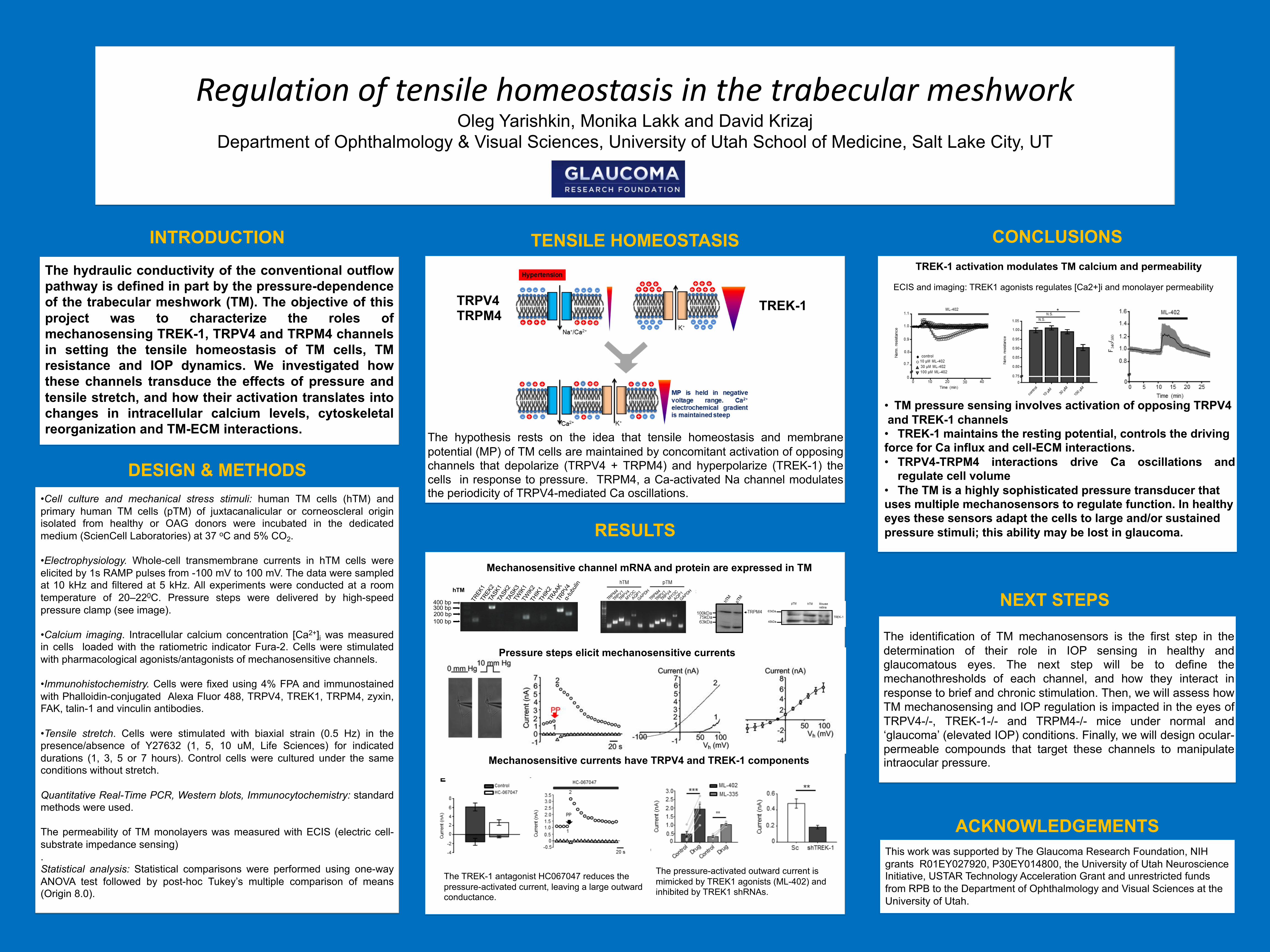
TREK-1 activation modulates TM calcium and permeability
• TM pressure sensing involves activation of opposing TRPV4and TREK-1 channels• TREK-1 maintains the resting potential, controls the drivingforce for Ca influx and cell-ECM interactions.• TRPV4-TRPM4 interactions drive Ca oscillations and
regulate cell volume• The TM is a highly sophisticated pressure transducer thatuses multiple mechanosensors to regulate function. In healthyeyes these sensors adapt the cells to large and/or sustainedpressure stimuli; this ability may be lost in glaucoma.
ECIS and imaging: TREK1 agonists regulates [Ca2+]i and monolayer permeability
The identification of TM mechanosensors is the first step in thedetermination of their role in IOP sensing in healthy andglaucomatous eyes. The next step will be to define themechanothresholds of each channel, and how they interact inresponse to brief and chronic stimulation. Then, we will assess howTM mechanosensing and IOP regulation is impacted in the eyes ofTRPV4-/-, TREK-1-/- and TRPM4-/- mice under normal and‘glaucoma’ (elevated IOP) conditions. Finally, we will design ocular-permeable compounds that target these channels to manipulateintraocular pressure.