soluble lutein in combination with brilliant blue as a new dye for chromovitrectomy
TRANSCRIPT

RETINAL DISORDERS
Soluble lutein in combination with brilliant blue as a new dyefor chromovitrectomy
Emmerson Badaro & Bruno Furlani & Juliana Prazeres & Mauricio Maia &
Acácio Alves Souza Lima & Diogo Souza-Martins & Cristina Muccioli &Luiz Filipe Adami Lucatto & Rubens Belfort Jr
Received: 11 July 2013 /Revised: 7 October 2013 /Accepted: 20 November 2013# Springer-Verlag Berlin Heidelberg 2014
AbstractSummary A new dye for vitreoretinal surgery comprised ofsoluble lutein/zeaxanthin 1 % and brilliant blue 0.025 % isadvantageous compared with other dyes currently used forchromovitrectomy, and showed no signs of toxicity at 1 monthof follow-up.Purpose To evaluate the feasibility and safety of a dye [solu-ble lutein/zeaxanthin (LZ) 1 % and brilliant blue (BB)0.025 %] for improving removal of vitreous, epiretinal mem-branes (ERM), and internal limiting membranes (ILM) inhumans.Methods We prospectively evaluated 18 eyes treated surgical-ly for a macular hole or ERM. Eighteen surgeons performedchromovitrectomy using the dye, and completed a question-naire to evaluate the efficacy and safety of the dye. . Exami-nations included best-corrected visual acuity and intraocularpressure measurements and optical coherence tomography,fluorescein angiography, and autofluorescence performed atbaseline and days 1, 7, and 30 postoperatively.Results The green dye was deposited on the posterior pole;vigorous dye flushing into the vitreous cavity was unneces-sary. All surgeons reported that the ILM stained greenish-blue;94.4 % reported ILM peeling adequate; the ERM stainedpoorly. No evidence of toxicity was observed.
Conclusion The new dye deposited on the posterior pole dueto its higher density. The ability to stain the ILMwas similar toBB. The new dye has ability to stain the vitreous, hyaloid, andespecially the ILM satisfactorily. The new dye may be usefulduring chromovitrectomy.
Keywords Chromovitrectomy . Internal limitingmembrane .
Lutein .Macular hole . Vitreoretinal surgery . Zeaxanthin
Introduction
Vitreoretinal surgery involves complex and delicatevitreoretinal techniques to manage many vitreoretinal dis-eases, macular hole (MH), epiretinal membrane (ERM), andvitreomacular traction syndrome. Chromovitrectomy [1] in-cludes the use of vital dyes or crystals to improve visualizationof the intraocular tissues during vitrectomy.
The ideal vital dye should be safe for intraocular use,selectively stain the intraocular membranes, and be eliminatedrapidly from the eye. Many dyes are available forchromovitrectomy [1–7], i.e., trypan blue (TB), brilliant blue(BB), indocyanine green (ICG), and infracyanine green. ICGis currently used to stain the internal limitingmembrane (ILM)to aid peeling, even though there are concerns about retinaltoxicity [8]. TB is thought to be the ideal dye for ERMidentification, ICG and BB are ideal for ILM peeling, andtriamcinolone acetonide (TA) is useful for vitreous identifica-tion [1–7]. Despite this, the United States Food and DrugAdministration has not approved any dyes for use duringchromovitrectomy. Currently, BB is not approved for humanuse in the US, and other agents such as ICG, TB, and TA areconsidered off-label drugs to be used during vitreoretinalprocedures. However, BB is approved for chromovitrectomyin Europe.
Clinical trial registration number: NCT01627977Local ethics committee number: 00553212.8.0000.5505
E. Badaro :B. Furlani : J. Prazeres :M. Maia :A. A. S. Lima :D. Souza-Martins :C. Muccioli : L. F. A. Lucatto : R. Belfort JrDepartment of Ophthalmology, Federal University of São Paulo, SãoPaulo, Brazil
M. Maia (*)Ophthalmology Department, Vitreoretinal Surgery Unit,Universidade Federal de São Paulo, Rua Botucatu 822, SãoPaulo 06023-062, Brazile-mail: [email protected]
Graefes Arch Clin Exp OphthalmolDOI 10.1007/s00417-013-2539-5

Lutein and zeaxanthin are lipophilic pigments belonging tothe group of carotenoids traditionally found in fruits andvegetables [9, 10]. These carotenoids are also physiologicallypresent in the macula lutea, an integral retinal structure inhumans [11]. The carotenoids are structural isomers that havea hydroxyl group at the terminal part of the molecule, whichpartly explains their different polarities and tropism for certainbiologic structures such as the macula [12–15]. Both areconsidered to be dyes, and are associated with prevention ofage-related maculopathies due to their antioxidant effects andexclusive distribution in the macula [11, 16, 17].
Lutein- and zeaxanthin-based dyes alone or combined withBB also might be used effectively as intraocular dyes inhuman cadaveric eyes to stain different intraocular microstruc-tures, including the ILM [18].
The current study evaluated the feasibility, advantages, andsafety of a novel dye (soluble lutein/zeaxanthin [LZ] 1 % withBB 0.025 %) for improving identification and removal of thevitreous, hyaloid, ERM, and ILM performed by differentsurgeons.
Patients and methods
Weprospectively studied 18 eyes of 18 consecutive patients (16women, two men; mean age, 67.2±7.7 years; range, 50–85 years); 50 % of patients were Caucasian, 44 % wereAfrican-American, and 5.6 % were Hispanic. Patients had adiagnosis of macular hole or ERM, and underwent pars planavitrectomy (PPV) using the combined soluble formulation ofLZ 1 % with BB 0.025 %. Inclusion criteria allowed inclusionof eyeswith chronicmacular hole and/or chronic ERM.Tables 1and 2 show the patient preoperative and postoperative data. TheEthics Committee of the Federal University of São Pauloapproved the study, which was conducted according to theresearch guidelines of the Association of Research in Visionand Ophthalmology and the tenets of the Declaration of Hel-sinki. The protocol was registered at www.clinicaltrials.gov(number NCT01627977). All patients provided informedconsent regarding the benefits and risks of the surgicalprocedure and the new dye tested.
Dye preparation
The preparatory technique described below is valid for luteinand zeaxanthin regardless of the chemical form, purity, crys-tallization, or degree of esterification. The primary steps in thecurrent study were development of a water-soluble solution,characterization of the absorbance of the LZ solution with theAquamate device™ (Thermo Spectronic, Cambridge, UK),and identification of the resultant color of the chromophoregroups. The physicochemical parameters were tested for theoptimum pH, osmolarity/osmolality, and concentration. We
performed solubility tests in water and polyvinyl alcohol, withultraviolet spectrophotometry, and analyzed blends comprisedof different colors within the visible spectrum.
Preparation
For the final formulation, the active component BB G-B0770™ (Sigma-Aldrich, St. Gallen, Switzerland) and luteinand zeaxanthin were weighed using the H33AR™ balance(Mettler Toledo, Barueri, Brazil), and when stirred with theQ298-1™ device (Quimis, Diadema, Brazil), dissolved in abuffer solution (pH, 7.0-7.4); the pH was checked using theQ600M device™ (Quimis), and balanced saline solution(BSS) served as a control for the osmolarities. The syringeswere filled in a sterile area with one 0.5-ml dose of the dyesolution, and each syringe was connected to a rubber stopperand then crimped. The final solution was sterilized byautoclaving for 20 min at 121°C in the Controller Odt-74version 2.0™ (Wilson Autoclaves, São Paulo, Brazil).
The final dye solution contained LZ 1 % and BB 0.025 %(osmolarity, 280 mOsm; density, 1.1; pH, 7.00). The new 0.5-ml syringes were bottled and stored at 4–8 °C, and used within30 days of production.
Inclusion and exclusion criteria
Patients of both genders were included if they were older than18 years, not pregnant, and required an elective vitrectomy totreat a macular hole of any size and any BCVA level or anERM with a BCVA below 20/25. Patients were excluded ifthey had any ocular condition that could limit or affect thepostoperative results.
Physical evaluation and ocular examinations
All patients underwent a physical evaluation preoperativelythat included measurement of the systemic blood pressure andheart rate. An ocular examination was performed preopera-tively and 1, 7, and 30 days postoperatively, which includedspectral-domain optical coherence tomography (SD-OCT)and fluorescein angiography (FA) using the Spectralis device(Heidelberg Engineering, Heidelberg, Germany), unless me-dia opacity was present.
The BCVA was measured using the Early Treatment Dia-betic Retinopathy Study (ETDRS) chart, and the intraocularpressure (IOP) was measured using a Goldmann tonometer; aslit-lamp evaluation also was performed.
Surgical procedures
Eighteen surgeons experienced in current chromovitrectomyperformed the standardized surgical procedures. Phakic pa-tients underwent standard combined phacoemulsification with
Graefes Arch Clin Exp Ophthalmol

IOL and then 3-port pars plana vitrectomy using 23-gaugeinstruments. After this, 0.3 ml of the new dye was delivered
gently through the posterior pole using a 23-gauge soft-tipcannula (Synergetics, O’Fallon, MO, USA). Flushing of the
Table 1 Patient demographic data
Patient Preop BCVA Postop BCVA Preop IOP Postop IOP Preop biomicroscopyfinding
Postopbiomicrosopy
Preop funduscopyfinding
Postop fundoscopyfinding
1 03 23 10 10 Cataract Pseudophakic MH None
2 20 20 12 18 Cataract Pseudophakic MH None
3 35 35 10 14 Cataract Pseudophakic ERM None
4 15 15 12 13 Cataract Pseudophakic MH None
5 37 38 14 14 Cataract Pseudophakic MH None
6 12 13 10 11 Cataract Pseudophakic MH MH
7 12 20 18 16 RK scars,Cataract
RK scars,Pseudophakic
MH None
8 1 2 13 16 Cataract Pseudophakic MH None
9 2 3 10 15 Cataract Pseudophakic MH MH
10 3 13 20 12 Cataract Pseudophakic MH MH
11 3 8 18 15 Cataract Pseudophakic MH None
12 2 9 19 17 Cataract Pseudophakic MH None
13 8 8 12 14 Cataract Pseudophakic MH MH
14 5 5 13 14 Cataract Pseudophakic MH MH
15 1 6 15 15 Cataract Pseudophakic MH None
16 13 15 13 10 Cataract Pseudophakic MH MH
17 23 31 18 12 Cataract Pseudophakic MH None
18 2 3 12 12 Pseudophakic Pseudophakic ERM None
Preop preoperatively, postop postoperatively, RK radial keratotomy, MH macular hole
Table 2 Preoperative and postoperative OCT and AF findings
Patient OCT findings preoperatively OCT findings postoperatively Preop AF findings Postop AF findings
Base diameter Minimal diameter Height Base diameter Minimal diameter Height
1 MH 473 273 407 None Window defect Normal
2 MH 1,218 958 452 None Window defect Normal
3 ERM None Normal Normal
4 MH 830 625 344 None Window defect Normal
5 MH 836 619 392 None Window defect Normal
6 MH 1,135 791 351 MH 801 526 410 Window defect Window defect
7 MH 633 467 399 None Window defect Normal
8 MH 996 558 392 None Window defect Normal
9 MH 2,071 726 516 MH 533 647 228 Window defect Window defect
10 MH 1,239 689 367 MH 718 520 357 Window defect Window defect
11 MH 588 471 387 None Window defect Normal
12 MH 1,172 749 450 None Window defect Normal
13 MH 912 629 342 MH 632 425 298 Window defect Window defect
14 MH 852 563 379 MH 458 419 268 Window defect Window defect
15 MH 821 534 347 None Window defect Normal
16 MH 1,032 618 366 MH 613 481 257 Window defect Window defect
17 MH 461 214 398 None Window defect Normal
18 ERM None Normal
Preop preoperatively, postop postoperatively, AF autofluorescence, MH macular hole
Graefes Arch Clin Exp Ophthalmol
dye was not needed because it was heavier than BSS (density,1.1). After the vitreous was removed, a second flush of 0.3 mlof the combination dye was delivered through the posteriorpole using the same technique as described previously.Octafluoropropane 15 % was injected into the vitreous cavity.The patients were instructed to remain prone for 3 dayspostoperatively.
Questionnaire
After each procedure, the surgeons completed a questionnairethat compared the ability of the combination dye to stain theintraocular structures. The findings were compared with thesurgeons’ experience with the dyes currently used inchromovitrectomy (Table 3).
Follow-up
The patients underwent a complete ophthalmologic examina-tion on days 1, 7, and 30 postoperatively that included mea-surement of the BCVA, IOP measurement, and a slit-lampevaluation. SD-OCTof the macular area was performed whenthe media was transparent preoperatively, and on days 1, 7,and 30 postoperatively. On postoperative day 30, fundusimaging, FA, and autofluorescence were performed. A para-metric tailed Student's t -test was used for statistical analysesof the BCVA improvement and IOP variations. P <0.05 wasconsidered statistically significant.
Results
The final solution of the combination dye contained solubleLZ 1% and BB 0.025% (osmolarity, 280 mOsm; density, 1.1;pH, 7.00) and resulted in a green solution.
Clinical and intraoperative findings
Each of the 18 surgeons performed surgery on only onepatient in this series, and no systemic effects were notedduring the follow-up period. With regard to the causes leadingto surgery, MH was diagnosed in 16 patients and ERM wasobserved in two eyes. The mean baseline number of ETDRSletters was 10.6±11.6 (range, 0–37) and the mean number ofETDRS letters on day 30 postoperatively was 12.4±10.6(range, 0–35). All eyes were followed for 30 days.
Intraoperative findings
Phaco-vitrectomy was performed in 17 eyes, and vitrectomyalone in one pseudophakic eye. All procedures were per-formed uneventfully. Five intraoperative characteristics ofthe new dye were observed: first, the solution consisted of
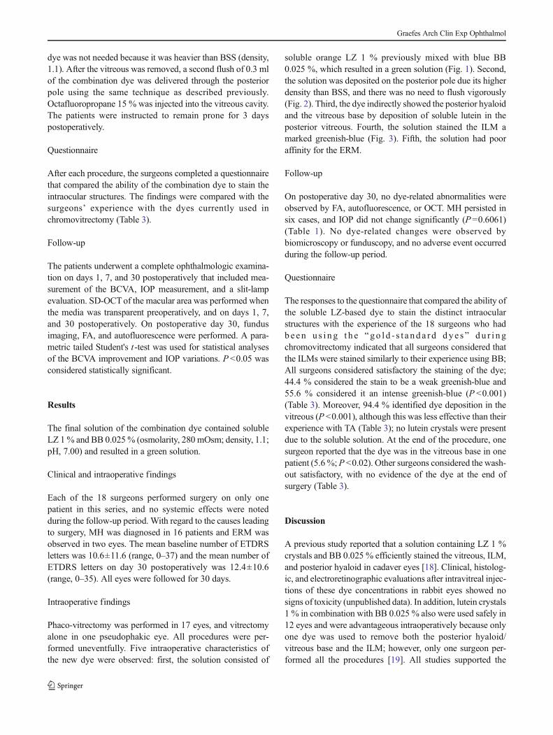
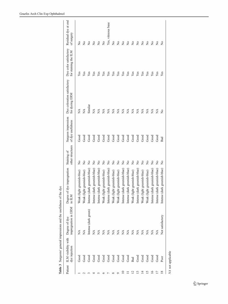
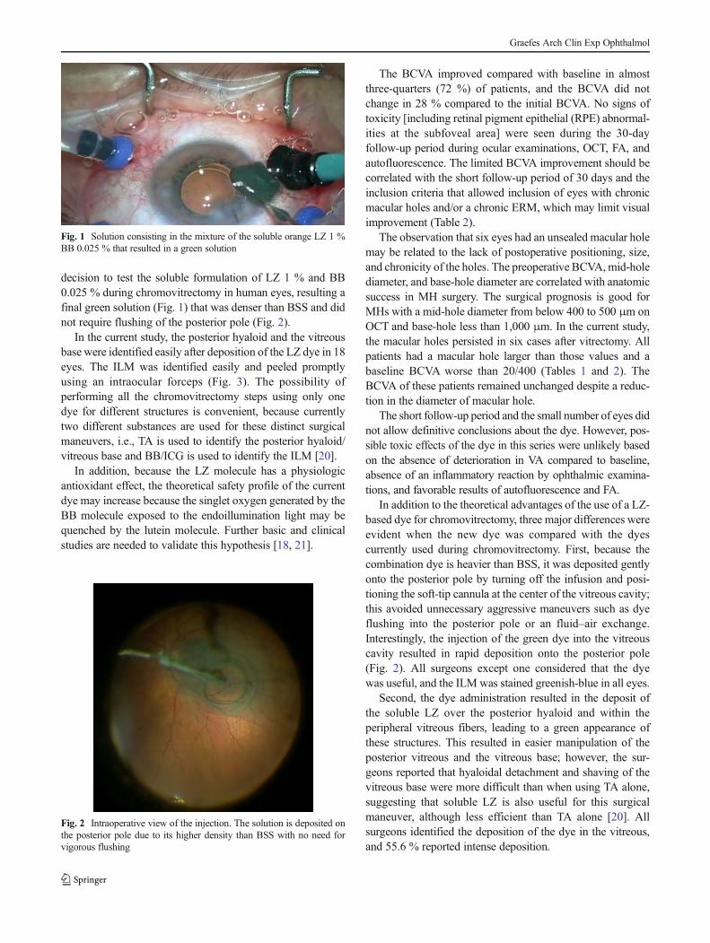
soluble orange LZ 1 % previously mixed with blue BB0.025 %, which resulted in a green solution (Fig. 1). Second,the solution was deposited on the posterior pole due its higherdensity than BSS, and there was no need to flush vigorously(Fig. 2). Third, the dye indirectly showed the posterior hyaloidand the vitreous base by deposition of soluble lutein in theposterior vitreous. Fourth, the solution stained the ILM amarked greenish-blue (Fig. 3). Fifth, the solution had pooraffinity for the ERM.
Follow-up
On postoperative day 30, no dye-related abnormalities wereobserved by FA, autofluorescence, or OCT. MH persisted insix cases, and IOP did not change significantly (P=0.6061)(Table 1). No dye-related changes were observed bybiomicroscopy or funduscopy, and no adverse event occurredduring the follow-up period.
Questionnaire
The responses to the questionnaire that compared the ability ofthe soluble LZ-based dye to stain the distinct intraocularstructures with the experience of the 18 surgeons who hadb e e n u s i n g t h e “go l d - s t a nd a r d dye s ” du r i n gchromovitrectomy indicated that all surgeons considered thatthe ILMs were stained similarly to their experience using BB;All surgeons considered satisfactory the staining of the dye;44.4 % considered the stain to be a weak greenish-blue and55.6 % considered it an intense greenish-blue (P <0.001)(Table 3). Moreover, 94.4 % identified dye deposition in thevitreous (P <0.001), although this was less effective than theirexperience with TA (Table 3); no lutein crystals were presentdue to the soluble solution. At the end of the procedure, onesurgeon reported that the dye was in the vitreous base in onepatient (5.6%; P <0.02). Other surgeons considered the wash-out satisfactory, with no evidence of the dye at the end ofsurgery (Table 3).
Discussion
A previous study reported that a solution containing LZ 1 %crystals and BB 0.025 % efficiently stained the vitreous, ILM,and posterior hyaloid in cadaver eyes [18]. Clinical, histolog-ic, and electroretinographic evaluations after intravitreal injec-tions of these dye concentrations in rabbit eyes showed nosigns of toxicity (unpublished data). In addition, lutein crystals1 % in combination with BB 0.025 % also were used safely in12 eyes and were advantageous intraoperatively because onlyone dye was used to remove both the posterior hyaloid/vitreous base and the ILM; however, only one surgeon per-formed all the procedures [19]. All studies supported the
Graefes Arch Clin Exp Ophthalmol
Tab
le3
Surgeons'generalim
pressionsandtheusefulness
ofthedye
Patient
ILM
visibilitywith
dyeinjection
Degreeof
dye
impregnatio
nin
ERM
Degreeof
dyeim
pregnatio
nin
ILM
Staining
ofotherstructures
Surgeon
impression
ofdyeusefulness
Dye
coloratio
nsatisfactory
fordyeing
ERM
Dye
colorsatisfactory
forstaining
theILM
Residuald
yeatend
ofsurgery
1Good
NA
Weak(light
greenish-blue)
No
Good
NA
Yes
No
2Good
NA
Weak(light
greenish-blue)
No
Good
NA
Yes
No
3Good
Intense(darkgreen)
Intense(darkgreenish-blue)
No
Good
Similar
Yes
No
4Good
NA
Intense(darkgreenish-blue)
No
Good
NA
Yes
No
5Good
NA
Intense(darkgreenish-blue)
No
Good
NA
Yes
No
6Good
NA
Weak(light
greenish-blue)
No
Good
NA
Yes
No
7Good
NA
Intense(darkgreenish-blue)
No
Good
NA
Yes
Yes,vitreous
base
8Good
NA
Weak(light
greenish-blue)
No
Good
NA
Yes
No
9Good
NA
Weak(light
greenish-blue)
No
Good
NA
Yes
No
10Good
NA
Intense(darkgreenish-blue)
No
Good
NA
Yes
No
11Good
NA
Intense(darkgreenish-blue)
No
Good
NA
Yes
No
12Bad
NA
Weak(light
greenish-blue)
No
Good
NA
Yes
No
13Good
NA
Intense(darkgreenish-blue)
No
Good
NA
Yes
No
14Good
NA
Weak(light
greenish-blue)
No
Good
NA
Yes
No
15Good
NA
Weak(light
greenish-blue)
No
Good
NA
Yes
No
16Good
NA
Intense(darkgreenish-blue)
No
Good
NA
Yes
No
17Good
NA
Intense(darkgreenish-blue)
No
Good
NA
Yes
No
18Po
orNot
satisfactory
Intense(darkgreenish-blue)
No
Bad
No
Yes
No
NAnotapplicable
Graefes Arch Clin Exp Ophthalmol
decision to test the soluble formulation of LZ 1 % and BB0.025 % during chromovitrectomy in human eyes, resulting afinal green solution (Fig. 1) that was denser than BSS and didnot require flushing of the posterior pole (Fig. 2).
In the current study, the posterior hyaloid and the vitreousbase were identified easily after deposition of the LZ dye in 18eyes. The ILM was identified easily and peeled promptlyusing an intraocular forceps (Fig. 3). The possibility ofperforming all the chromovitrectomy steps using only onedye for different structures is convenient, because currentlytwo different substances are used for these distinct surgicalmaneuvers, i.e., TA is used to identify the posterior hyaloid/vitreous base and BB/ICG is used to identify the ILM [20].
In addition, because the LZ molecule has a physiologicantioxidant effect, the theoretical safety profile of the currentdye may increase because the singlet oxygen generated by theBB molecule exposed to the endoillumination light may bequenched by the lutein molecule. Further basic and clinicalstudies are needed to validate this hypothesis [18, 21].
The BCVA improved compared with baseline in almostthree-quarters (72 %) of patients, and the BCVA did notchange in 28 % compared to the initial BCVA. No signs oftoxicity [including retinal pigment epithelial (RPE) abnormal-ities at the subfoveal area] were seen during the 30-dayfollow-up period during ocular examinations, OCT, FA, andautofluorescence. The limited BCVA improvement should becorrelated with the short follow-up period of 30 days and theinclusion criteria that allowed inclusion of eyes with chronicmacular holes and/or a chronic ERM, which may limit visualimprovement (Table 2).
The observation that six eyes had an unsealed macular holemay be related to the lack of postoperative positioning, size,and chronicity of the holes. The preoperative BCVA,mid-holediameter, and base-hole diameter are correlated with anatomicsuccess in MH surgery. The surgical prognosis is good forMHs with a mid-hole diameter from below 400 to 500 μm onOCT and base-hole less than 1,000 μm. In the current study,the macular holes persisted in six cases after vitrectomy. Allpatients had a macular hole larger than those values and abaseline BCVA worse than 20/400 (Tables 1 and 2). TheBCVA of these patients remained unchanged despite a reduc-tion in the diameter of macular hole.
The short follow-up period and the small number of eyes didnot allow definitive conclusions about the dye. However, pos-sible toxic effects of the dye in this series were unlikely basedon the absence of deterioration in VA compared to baseline,absence of an inflammatory reaction by ophthalmic examina-tions, and favorable results of autofluorescence and FA.
In addition to the theoretical advantages of the use of a LZ-based dye for chromovitrectomy, three major differences wereevident when the new dye was compared with the dyescurrently used during chromovitrectomy. First, because thecombination dye is heavier than BSS, it was deposited gentlyonto the posterior pole by turning off the infusion and posi-tioning the soft-tip cannula at the center of the vitreous cavity;this avoided unnecessary aggressive maneuvers such as dyeflushing into the posterior pole or an fluid–air exchange.Interestingly, the injection of the green dye into the vitreouscavity resulted in rapid deposition onto the posterior pole(Fig. 2). All surgeons except one considered that the dyewas useful, and the ILM was stained greenish-blue in all eyes.
Second, the dye administration resulted in the deposit ofthe soluble LZ over the posterior hyaloid and within theperipheral vitreous fibers, leading to a green appearance ofthese structures. This resulted in easier manipulation of theposterior vitreous and the vitreous base; however, the sur-geons reported that hyaloidal detachment and shaving of thevitreous base were more difficult than when using TA alone,suggesting that soluble LZ is also useful for this surgicalmaneuver, although less efficient than TA alone [20]. Allsurgeons identified the deposition of the dye in the vitreous,and 55.6 % reported intense deposition.
Fig. 1 Solution consisting in the mixture of the soluble orange LZ 1 %BB 0.025 % that resulted in a green solution
Fig. 2 Intraoperative view of the injection. The solution is deposited onthe posterior pole due to its higher density than BSS with no need forvigorous flushing
Graefes Arch Clin Exp Ophthalmol

Third, the ability of the dye to stain the ILM resulted ineasier ILM peeling in eyes with macular holes (Fig. 3). Therewas no evidence that intraocular lutein and zeaxanthin affect-ed the IOP or induced an inflammatory reaction.
The lutein and zeaxanthin molecules absorb light in thevisible spectrum (around 446 nm), which coincides with theblue region of the electromagnetic spectrum. For this reason,lutein and zeaxanthin may be natural absorbers of blue light,which may decrease harmful effects on the lens and retina[22]. However, this possible protective effect may be betterelucidated by additional basic and clinical studies. ILM peel-ing guided by ICG staining currently is performed worldwideto treat macular holes, especially in the United States [20];however, RPE atrophymay occur after ILM peeling guided byICG staining during macular hole surgery, because of the highosmolarity of the ICG solution, decomposition of ICG, iodineeffect, carbolinic complex formation, and the oxidative effectdue to singlet-oxygen release from ICG after light exposurefrom the light pipe during PPV [23–25]. We hypothesized thatthe combination of lutein, zeaxanthin, and BB in one dyesolution may have a good safety profile, and provides antiox-idant and light-scavenging properties to protect the retina andRPE.
The study limitations were the small number of eyes, theabsence of a control group, and the fact that multifocal elec-troretinography and microperimetry were not performed.
A more extensive evaluation of potential toxicity wouldprobably have limited value, since alterations secondary toILM peeling (macular manipulation) could lead to inconclu-sive results. In addition, the main objective of this study was toevaluate peeling of the ILM and ERM in humans duringchromovitrectomy performed by different surgeons; because18 surgeons performed the procedures, the potential for erro-neous personal impressions about the staining capacity of thedyes was minimized.
In summary, the current study showed that a new dyecomprised of soluble LZ 1 % and BB 0.025 % is usefulfor chromovitrectomy. The dye improved the intraoperativeidentification of the ILM similar to BB and the posteriorhyaloid/vitreous base identification. Although less effectivethan TA, the combination solution facilitates injection ofonly one heavy solution into the posterior pole, and has atheoretically good safety profile. The different surgeonsfound that the dye might be useful for vitreoretinal sur-gery, with improved ILM peeling in 100 % of eyes andimproved removal of the posterior hyaloid/vitreous base in
Fig. 3 Dye deposition at the ILMin an eye with a macular hole. aAn intraoperative image showsthe initial removal of the ILMstained greenish-blue. b , c Theintermediate phase of ILMpeeling shows the tip of theforceps grasping the greenish-blue-stained ILM. c The finalstage of ILM peeling
Graefes Arch Clin Exp Ophthalmol

94.4 % of eyes. Additional multicenter studies are needed toconfirm these findings.
Acknowledgments Many thanks to the study coordinator Luci Silvafor the efforts to conclude this study.
Proprietary interest The use of the combination of lutein/zeaxanthinand brilliant blue in human eyes is protected by international patent law(international patent number 61/468,838). The study was sponsored byKemin Inc., Des Moines, IA, USA.
References
1. Burk SE, Da Mata AP, Snyder ME, Rosa RH Jr, Foster RE (2000)Indocyanine green-assisted peeling of the retinal internal limitingmembrane. Ophthalmology 107(11):2010–2014
2. Rodrigues EB, Meyer CH, Farah ME, Kroll P (2005) Intravitrealstaining of the internal limitingmembrane using indocyanine green inthe treatment of macular holes. Ophthalmol J Int Ophtalmol Int JOphthalmol Z Augenheilkd 219(5):251–262
3. Teba FA, Mohr A, Eckardt C,Wong D, Kusaka S, Joondeph BC et al(2003) Trypan blue staining in vitreoretinal surgery. Ophthalmology110(12):2409–2412
4. Kumar A, Prakash G (2004) Differential staining with indocyaninegreen and trypan blue dye. Indian J Ophthalmol 52(4):339
5. Perrier M, Sebag M (2003) Epiretinal membrane surgery assisted bytrypan blue. Am J Ophthalmol 135(6):909–911
6. Feron EJ, Veckeneer M, Parys-Van Ginderdeuren R, Van Lommel A,Melles GR, Stalmans P (2002) Trypan blue staining of epiretinalmembranes in proliferative vitreoretinopathy. Arch Ophthalmol120(2):141–144
7. Peyman GA, Cheema R, ConwayMD, Fang T (2000) Triamcinoloneacetonide as an aid to visualization of the vitreous and the posteriorhyaloid during pars plana vitrectomy. Retina 20(5):554–555
8. Engelbrecht NE, Freeman J, Sternberg P Jr, Aaberg TM Sr, AabergTM Jr, Martin DF et al (2002) Retinal pigment epithelial changesafter macular hole surgery with indocyanine green-assisted internallimiting membrane peeling. Am J Ophthalmol 133(1):89–94
9. Yemelyanov AY, Katz NB, Bernstein PS (2001) Ligand-bindingcharacterization of xanthophyll carotenoids to solubilized membraneproteins derived from human retina. Exp Eye Res 72(4):381–392
10. Ahmed SS, Lott MN, Marcus DM (2005) The macular xanthophylls.Surv Ophthalmol 50(2):183–193
11. Nolan JSJ, Mellerio J, Godhinio M, Neelam K, Beatty S (2006)Monthly consistency of macular pigment optical density and serumconcentrations of lutein and zeaxanthin. Curr Eye Res 31:199–213
12. Gale CHN, Phillips D, Martyn C (2001) Plasma antioxidant vitaminsand carotenoids and age-related cataract. Ophthalmology 108:1992–1998
13. Snodderly DHG, Adler AJ (1991) Distribution of individual macularpigment carotenoids in central retina of macaque and squirrel mon-keys. Invest Ophthalmol Vis Sci 32:268–279
14. Sommerburg OKJ, Bird A, van Kuijk F (1998) Fruits and vegetablesthat are sources for lutein and zeaxanthin: the macular pigment inhuman eyes. Br J Ophthalmol 82:907–910
15. Mares-Perlman JBW, Klein R et al (1995) Serum antioxidants andage-related macular degeneration in a population-based case–controlstudy. Arch Ophthalmol 113:1518–1523
16. Falsini BPM, Iarossi G, Fadda A, Merendino E, Valentini P (2003)Influence of short-term antioxidant supplementation on macularfunction in age-related maculopathy. Ophthalmology 110:51–61
17. Bone RALJ, Mayne ST, Gomez CM, Tibor SE, Twaroska EE (2001)Macular pigment in donor eyes with and without AMD: a case-control study. Invest Ophthalmol Vis Sci 42:235–240
18. Sousa-Martins D, Maia M, Moraes M, Lima-Filho AA, RodriguesEB, Chen J et al (2012) Use of lutein and zeaxanthin alone orcombined with Brilliant Blue to identify intraocular structures intra-operatively. Retina 32(7):1328–1336
19. Maia M, Furlani BA, Souza-Lima AA, Martins DS, Navarro RM,Belfort R Jr (2013) LUTEIN: a new dye for chromovitrectomy.Retina.
20. Farah ME, Maia M, Rodrigues EB (2009) Dyes in ocular surgery:principles for use in chromovitrectomy. Am J Ophthalmol 148(3):332–340
21. Junghans A, Sies H, Stahl W (2001) Macular pigments lutein andzeaxanthin as blue light filters studied in liposomes. Arch BiochemBiophys 391(2):160–164
22. Kijlstra A, Tian Y, Kelly ER, Berendschot TT (2012) Lutein: morethan just a filter for blue light. Prog Retin Eye Res 31(4):303–315
23. Maia M, Margalit E, Lakhanpal R, Tso MO, Grebe R, Torres G et al(2004) Effects of intravitreal indocyanine green injection in rabbits.Retina 24(1):69–79
24. MaiaM, Haller JA, Pieramici DJ,Margalit E, De Juan E Jr, FarahMEet al (2004) Retinal pigment epithelial abnormalities after internallimiting membrane peeling guided by indocyanine green staining.Retina 24(1):157–160
25. MaiaM, Kellner L, De Juan E Jr, Smith R, FarahME,Margalit E et al(2004) Effects of indocyanine green injection on the retinal surfaceand into the subretinal space in rabbits. Retina 24(1):80–91
Graefes Arch Clin Exp Ophthalmol