structure of the lutein-binding domain of human stard3 at
TRANSCRIPT
research communications
Acta Cryst. (2016). F72, 609–618 http://dx.doi.org/10.1107/S2053230X16010694 609
Received 25 May 2016
Accepted 1 July 2016
Edited by W. N. Hunter, University of Dundee,
Scotland
Keywords: carotenoid-binding protein; START
domain; StARD3; lutein; protein tunnels and
cavities.
PDB reference: cholesterol and lutein-binding
domain of human STARD3, 5i9j
Supporting information: this article has
supporting information at journals.iucr.org/f
Structure of the lutein-binding domain of humanStARD3 at 1.74 A resolution and model of acomplex with lutein
Martin P. Horvath,a* Evan W. George,a Quang T. Tran,a Kody Baumgardner,a
Gabe Zharov,a Sarah Lee,a Hassan Sharifzadeh,b Saeed Shihab,b Ty Mattinson,b
Binxing Lib and Paul S. Bernsteinb*
aDepartment of Biology, University of Utah, 257 S 1400 E, Salt Lake City, UT 84112, USA, and bDepartment of
Ophthalmology and Visual Sciences, Moran Eye Center, University of Utah School of Medicine, Salt Lake City,
UT 84132, USA. *Correspondence e-mail: [email protected], [email protected]
A crystal structure of the lutein-binding domain of human StARD3 (StAR-
related lipid-transfer protein 3; also known as MLN64) has been refined to
1.74 A resolution. A previous structure of the same protein determined to 2.2 A
resolution highlighted homology with StARD1 and shared cholesterol-binding
character. StARD3 has since been recognized as a carotenoid-binding protein
in the primate retina, where its biochemical function of binding lutein with
specificity appears to be well suited to recruit this photoprotective molecule. The
current and previous structures correspond closely to each other (r.m.s.d. of
0.25 A), especially in terms of the helix-grip fold constructed around a solvent-
filled cavity. Regions of interest were defined with alternate conformations in
the current higher-resolution structure, including Arg351 found within the cavity
and �1, a loop of four residues found just outside the cavity entrance. Models of
the complex with lutein generated by rigid-body docking indicate that one of the
ionone rings must protrude outside the cavity, and this insight has implications
for molecular interactions with transport proteins and enzymes that act on
lutein. Interestingly, models with the "-ionone ring characteristic of lutein
pointing towards the bottom of the cavity were associated with fewer steric
clashes, suggesting that steric complementarity and ligand asymmetry may play a
role in discriminating lutein from the other ocular carotenoids zeaxanthin and
meso-zeaxanthin, which only have �-ionone rings.
1. Introduction
The macula lutea (yellow spot) at the center of the primate
retina is enriched in the xanthophyll carotenoids lutein,
zeaxanthin and meso-zeaxanthin. These carotenoids are
thought to protect the human eye from photo-oxidative stress
(Bernstein et al., 2016; Li et al., 2010; Beatty et al., 1999).
Indeed, epidemiological studies and prospective clinical trials
have shown that dietary intake and supplementation with
lutein and zeaxanthin increase the likelihood of avoiding
age-related macular degeneration (AMD), a leading cause of
blindness (Seddon et al., 1994; Age-Related Eye Disease
Study 2 Research Group, 2014; Wu et al., 2015; Bernstein et al.,
2016). The majority of these carotenoid molecules are speci-
fically localized in the outer plexiform layers (also known as
the Henle fiber layer) of the human fovea. GSTP1 and
StARD3 (also known as MLN64) have been identified as the
zeaxanthin-binding and lutein-binding protein in the human
macula, respectively, and these are thought to be responsible
for the specific tissue distribution and stability of the ocular
carotenoids (Bhosale et al., 2004; Li et al., 2011). StARD3 was
initially identified as a lutein-binding protein because of its
ISSN 2053-230X
high degree of homology to the carotenoid-binding protein
found in silkworm (Li et al., 2011). Its retina-related role was
confirmed through the examination of tissue-specific expres-
sion patterns, and binding studies monitored by surface
plasmon resonance (SPR) demonstrated that StARD3 binds
lutein with affinity and specificity (Li et al., 2011). The lutein-
binding function of StARD3 resides within the C-terminal
START domain comprising residues 216–444, hereafter
referred to as StARD3LBD (Li et al., 2011).
Interest in StARD3 predates the discovery of its lutein-
binding function and retina-protective role. Its X-ray crystal
structure was determined by the Hurley laboratory as a
surrogate for StARD1 (Tsujishita & Hurley, 2000). StARD1
mobilizes cholesterol as the first committed step in steroid
biogenesis, and homology between StARD3 and StARD1
suggested that StARD3 also binds cholesterol (Watari et al.,
1997; Tsujishita & Hurley, 2000). The most remarkable
structural feature characteristic of StARD3 and StARD1 is a
large (�1000 A3) solvent-filled cavity (Tsujishita & Hurley,
2000; Thorsell et al., 2011).
The structural basis by which StARD3 and GSTP1 recog-
nize, bind and protect lutein, zeaxanthin and meso-zeaxanthin
is not known. With the overarching goal of providing a
molecular picture for carotenoid recruitment to the human
retina, we have targeted carotenoid complexes of StARD3LBD
and GSTP1 for structure determination. Structures of these
proteins in their carotenoid-complexed forms have been
elusive because conditions which solubilize the hydrophobic
lutein and zeaxanthin molecules require either detergent or
organic solvents, and these agents have been determined to
prevent crystal growth and to damage preformed crystals.
While these challenges are being resolved, we have
obtained high-resolution X-ray diffraction data for the
unliganded proteins and report here the structure of
StARD3LBD refined to 1.74 A resolution. This structure
corresponds closely to the original 2.2 A resolution structure
(Tsujishita & Hurley, 2000). The new structure includes
additional solvent molecules and alternate conformations for
several residues, including Arg351, which is located within the
cavity, and residues Gly336–Gly339, which are part of the
omega loop found just outside the cavity entrance. Model-
building experiments indicate that at least one of the ionone
rings of lutein must protrude through a cavity entrance.
The current structure and its lutein-complexed model are
discussed in relation to mechanisms for carotenoid binding
specificity and the molecular interactions necessary for the
transport, protection and metabolism of carotenoids in the
human eye.
2. Methods
2.1. Cloning of an expression system for tagless StARD3LBD
The tagless StARD3LBD-pET-22b(+) expression system was
obtained by ligation-independent cloning (Aslanidis & de
Jong, 1990; Li & Elledge, 2007). Briefly, DNA encoding resi-
dues 216–444 of human StARD3 was amplified by PCR using
a His6-tagged StARD3 expression plasmid as the template.
Primers for this PCR reaction were designed to avoid the His6
tag and to include �20 bp of DNA flanking the NdeI/EcoRI
insertion site present in pET-22b(+). This StARD3-encoding
DNA was assembled with the pET-22b(+) vector by mixing
with two DNA fragments obtained by amplifying the following
regions from pET-22b(+): 287–3268 (contains an NdeI site and
the lacI gene) and 3245–197 (contains an EcoRI site and the
bla gene). Each of the three PCR products was purified by
electrophoresis in a 0.85% agarose gel prior to mixing and
transformation directly into heat-shock competent DH5�Escherichia coli cells. Ampicillin-resistant colonies were
screened by restriction-enzyme digestion and DNA sequen-
cing of miniprep DNA. The resulting T7 promoter-driven
expression vector encodes residues 216–444 of StARD3, as
confirmed by mass spectrometry.
2.2. Purification of tagless StARD3LBD
The lutein-binding domain of StARD3 was expressed in
E. coli Origami B (DE3) cells (Novagen) and purified by
ammonium sulfate fractionation followed by ion-exchange
and size-exclusion chromatography. The concentration of
StARD3LBD was determined from the absorbance at 280 nm
using an extinction coefficient of 1.1 ml mg�1 cm�1
(0.028 mM�1 cm�1) calculated by the method of Gill & von
Hippel (1989).
2.2.1. Growth of bacterial cultures. Bacterial cultures were
cultivated with shaking in 2�YT medium supplemented
with 30 mM potassium phosphate pH 7.5, 5 mM glucose,
10 mg ml�1 kanamycin, 12.5 mg ml�1 tetracycline and
500 mg ml�1 ampicillin. A 750 ml culture was expanded at
310 K. At an OD600 nm of 0.7–0.9, the culture was chilled on ice
before adding IPTG to a final concentration of 100 mM.
Induction proceeded at 290 K with shaking for 24–30 h prior
to harvesting the cells by centrifugation at 4000g. The cells
were washed in 10 mM Tris, 1 mM EDTA pH 8.2, 0.5 mM
phenylmethylsulfonyl fluoride (PMSF) and collected by
centrifugation prior to freezing at 253 K.
2.2.2. Ammonium sulfate precipitation. Frozen cells were
submerged in 100 g lysis buffer consisting of 0.1 M sodium
chloride, 0.05 M Tris pH 8, 1 mM EDTA. Upon thawing, cells
were lysed by sonication on ice. The lysate was centrifuged at
20 000g, and the supernatant was collected in an ice-cold glass
beaker. Solid ammonium sulfate was added slowly with stir-
ring on ice to achieve 30% saturation (17.5 g ammonium
sulfate per 100 g supernatant). After 30 min, the sample was
centrifuged at 15 000g for 20 min. Additional ammonium
sulfate was added slowly with stirring on ice to achieve 65%
saturation (an additional 22.5 g ammonium sulfate per 100 g
supernatant). After 40 min, the second ammonium sulfate
fraction was collected by centrifugation at 15 000g for 15 min.
The pellet was dissolved by adding 20 ml of freshly prepared,
ice-cold dialysis buffer consisting of 50 mM sodium chloride,
25 mM HEPES pH 6.5, 0.25 mM EDTA, 1 mM dithiothreitol
(DTT). The sample was dialyzed against 1.5 l dialysis buffer at
277 K without stirring overnight.
research communications
610 Horvath et al. � Lutein-binding domain of human StARD3 Acta Cryst. (2016). F72, 609–618
2.2.3. Ion-exchange and size-exclusion chromatography.
The dialysate was centrifuged at 20 000g in an ice-cold rotor
for 20 min, filtered through a low-protein-binding 0.45 mm
filter and loaded by gravity onto two 2 ml SP Sepharose
columns in parallel, each equilibrated in dialysis buffer. The
average flow rate was approximately 1 ml min�1. Columns
were washed with 10 ml dialysis buffer and eluted by
increasing the concentration of sodium chloride stepwise with
5 ml volume per step. Fractions enriched in StARD3LBD were
identified by SDS–PAGE stained with Coomassie, pooled and
concentrated to a volume of 0.3 ml before filtering using a
0.45 mm Nanosep spin filter (Pall Life Sciences) and injecting
onto a size-exclusion column equilibrated with SEC buffer
consisting of 20 mM Tris pH 7.5, 150 mM sodium chloride,
2 mM DTT. Fractions enriched in StARD3LBD protein were
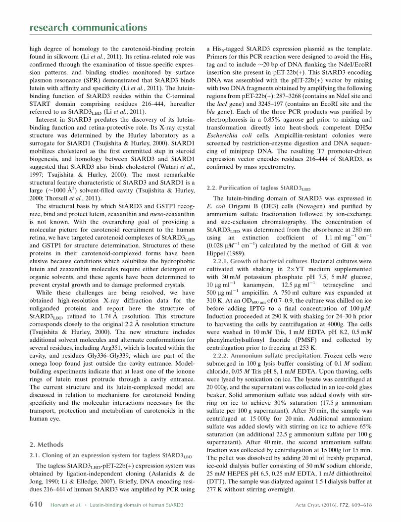
pooled and concentrated to 8 mg ml�1 (Fig. 1a).
2.3. Crystallography
2.3.1. Crystallization. Crystals of StARD3LBD were
obtained by the hanging-drop vapor-diffusion method as
described previously (Tsujishita & Hurley, 2000). Well solu-
tions consisting of 0.1 M CHES pH 8.6–9.4, 0.2 M lithium
sulfate, 0.8–0.96 M sodium/potassium tartrate, 0.01 M DTT
were combined with equal volumes (1.5–3 ml) of protein
solution. Rod-shaped crystals appeared within 2 d and grew
to a maximum size within one week at room temperature
(Fig. 1b). Crystals were moved to matched solutions addi-
tionally containing 15–20% ethylene glycol prior to cooling in
liquid propane and storage under liquid nitrogen.
2.3.2. Data collection. Crystals were measured in oscilla-
tion mode on the SIBYLS beamline 12.3.1 at the Advanced
Light Source (Classen et al., 2013) using synchrotron radiation
tuned to 1.116 A. The rod morphology made it possible to
reposition the crystal periodically during the course of data
collection so as to obtain complete and redundant data
without overexposing any one portion. Indexing and strategy
development was accomplished with HKL-2000 (Otwinowski
& Minor, 1997). Complete data sets were processed with XDS
and XSCALE (Kabsch, 2010a,b).
2.3.3. Refinement. Phases were obtained by placing PDB
entry 1em2 (Tsujishita & Hurley, 2000) into the nearly
isomorphous unit cell by rigid-body refinement. Refinement
continued with several iterative cycles of positional and
temperature-factor adjustment with PHENIX and model
adjustment with Coot (Adams et al., 2010; Emsley et al., 2010).
Early refinement rounds additionally applied torsion-angle
simulated annealing with a temperature protocol descending
from 1500 K to a final temperature of 50 K.
2.4. Model construction of the complex with lutein
Structural models of protein-complexed lutein were
obtained from the light-harvesting complexes of spinach and
pea with the following PDB entries: 1rwt (Liu et al., 2004), 3pl9
(Pan et al., 2011), 4xk8 (Qin et al., 2015), 4y28 (Mazor et al.,
2015) and 3jcu (Wei et al., 2016). Selected lutein molecules
were manually positioned within the StARD3 structure to
generate a number of docked templates. An ensemble of
27 324 structures was constructed by superposition of the 40
experimentally determined lutein structures from PDB entries
1rwt, 3pl9, 4xk8 and 3jcu onto these docked template mole-
cules with variations of least-squares alignments either
directly or with inverting molecule orientation. Ensemble
members were scored by determining the number of clashes
research communications
Acta Cryst. (2016). F72, 609–618 Horvath et al. � Lutein-binding domain of human StARD3 611
Figure 1Purity and crystals of StARD3LBD. (a) Purified StARD3LBD (15 mg) wasanalyzed by SDS–PAGE and stained with Coomassie Blue. The mostprominent (�90%) protein co-elutes with a 25 kDa molecular-weightmarker (expected mass 29 953 Da). (b) Rod-shaped crystals grew in 1–2 d. The scale bar indicates 100 mm. Crystals of StARD3LBD are colorless(top, middle) and acquire a golden color when stored in solutions alsocontaining powdered lutein (bottom).
Table 1Data collection and processing.
Values in parentheses are for the outer shell.
Diffraction source Beamline 12.3.1, ALSWavelength (A) 1.11583Temperature (K) 100Detector ADSC Quantum 315rRotation range per image (�) 0.5Exposure time per image (s) 0.2 and 1.5Space group P3121a, b, c (A) 83.39, 83.39, 82.19�, �, � (�) 90, 90, 120Mosaicity (�) 0.14–0.21Resolution range (A) 41.7–1.74 (1.79–1.74)Total No. of reflections 301752 (16789)No. of unique reflections 34160 (2466)Completeness (%) 99.5 (98.6)Multiplicity 8.8 (6.8)hI/�(I)i 25.8 (2.4)Rr.i.m. 0.048 (0.879)Overall B factor from Wilson plot (A2) 32.6
and the number of potential hydrogen bonds with use of a C
language program written by MPH. Frequency analysis to
detect the orientation preference was limited to the 8469
ensemble members that belonged to the ‘one portal’ set and
that scored at least one potential hydrogen bond.
3. Results
3.1. Protein expression and crystallization
Initial attempts to prepare crystals of StARD3LBD, purified
as described previously (Tsujishita & Hurley, 2000), yielded
small crystals with poor diffraction quality. We therefore
cloned a T7 promoter-driven expression system that produces
StARD3LBD without a tag, with the idea that the challenge in
producing crystals may have related to the removal of the His6
tag by protease treatment. Purification by means of ammo-
nium sulfate fractionation and two chromatography steps (and
no proteolysis treatment) yielded 5 mg protein from 0.75 l
bacterial culture (Fig. 1a). The yield and purity were lower
than that obtained with the tagged version; however, the
resulting protein readily produced large (50 � 50 � 500 mm)
rod-shaped crystals (Fig. 1b) which diffracted synchrotron
radiation to 1.7 A resolution (Table 1).
3.2. Structure of StARD3LBD
3.2.1. Structure determination. The structure of
StARD3LBD was determined by refinement against the newly
measured 1.74 A resolution data, starting with rigid-body
placement of a previously determined structure (Tsujishita &
Hurley, 2000). The starting model (PDB entry 1em2) is 99%
identical to the wild-type StARD3LBD determined in the
current work, and both proteins crystallized in the same P3121
space group with highly comparable unit-cell parameters. The
higher resolution limit reported here probably reflects differ-
ences in crystal size (the present crystals are twice as large in
each dimension), differences in synchrotron source flux (ALS
beamline 12.3.1 for the present study versus NSLS beamline
X4A for the previous study) and improvements in detector
capabilities (ADSC Q315r versus ADSC Q4). The final
structural model includes residues 231–444 of StARD3 (the
coordinates for residues 216–230 were omitted because no
research communications
612 Horvath et al. � Lutein-binding domain of human StARD3 Acta Cryst. (2016). F72, 609–618
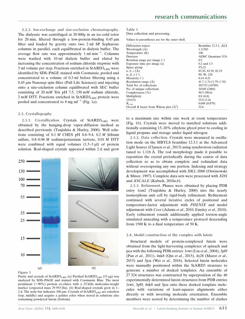
Figure 2Structure overview and representative electron density for StARD3LBD. (a) The C� trace of StARD3 is shown along with surfaces for key structuralfeatures: the omega loop (�1, lemon), portal 1 (P1, lime), portal 2 (P2, cyan) and solvent molecules located inside the cavity (red spheres inside graymolecular surfaces). (b) A stereoview of electron density and protein structure at portal 1 is shown. Electron density was calculated as a simulated-annealing composite OMIT map with 2|Fo| � |Fc| coefficients and is contoured at 1.1�. Map color indicates residues belonging to the omega loop(lemon), portal 1 (lime) and tunnel solvent (blue). Both conformations of the omega loop are shown (�1-a and �1-b). Residues lining portal 1 arelabeled (Thr313, Ala335, Gly336, Val342, Ser422 and Thr426).
electron density was observed), 224 water molecules, three
molecules of ethylene glycol, one sulfate ion and one molecule
of l-(+)-tartaric acid. The Rwork and Rfree values are 0.169 and
0.192, respectively. Table 2 reports statistical measures for
model validation.
3.2.2. Structure overview. Fig. 2 shows an overview of
the protein as well as representative electron density. As
described previously (Tsujishita & Hurley, 2000), StARD3LBD
adopts the helix-grip fold with a nine-stranded curved �-sheet
and three �-helices coalescing around a large solvent-filled
cavity. In helix-grip proteins the cavity entrance is guarded by
an omega loop (�1) that connects two �-strands (�5 and �6 in
StARD3LBD). The current structure is highly correlated with
the 2.2 A resolution structure (Tsujishita & Hurley, 2000).
Superposition yields a root-mean-square deviation (r.m.s.d.)
of 0.25 A for 210 C� atoms. The main differences in the
structure involve the restoration of the wild-type residues
Met307, Phe388 and Met427 (these residues were substituted
with selenomethionine in PDB entry 1em2) and the modeling
of 17 residues with alternate conformations, including Arg351,
which is inside the cavity, and residues Gly336–Gly339, which
are part of �1 near the cavity entrance.
3.2.3. Tunnel-like cavity. A striking feature of StARD3LBD
is the prominent tunnel-like cavity lined by hydrophobic and
polar residues with sufficient volume to accommodate a
molecule of cholesterol, as demonstrated by modeling
(Tsujishita & Hurley, 2000). The tunnel-like cavity measures
�20 A long from end to end, curves slightly and accom-
modates 18 solvent molecules, one of which is an ethylene
glycol in the current structure (Fig. 2). Electron-density maps
did not reveal any larger molecules located inside the cavity.
The cavity communicates with bulk solvent through two
openings, which we will call portal 1 and portal 2 (Fig. 2).
Portal 1 is wider in diameter compared with portal 2 (4 � 8 A
for portal 1 versus 2� 4 A for portal 2; distances are measured
between solvent-accessible surfaces). Indeed, structural
descriptions of helix-grip proteins generally speak of only one
cavity entrance.
research communications
Acta Cryst. (2016). F72, 609–618 Horvath et al. � Lutein-binding domain of human StARD3 613
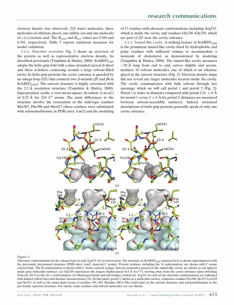
Figure 3Alternate conformations for the omega loop (a) and Arg351 (b) as stereoviews. The structure of StARD3LBD reported here is shown superimposed withthe previously determined structure (PDB entry 1em2; charcoal C atoms). Protein residues, including the A conformation, are shown with C atomscolored lime. The B conformation is shown with C atoms colored orange. Solvent molecules located in the tunnel-like cavity are shown as red spheresinside gray molecular surfaces. (a) Ala338 experiences the largest displacement (6.6 A for C�), moving away from the cavity entrance upon switchingfrom the �1-b to the �1-a conformation. (b) Hydrogen bonds and salt bridges evident for Arg351 in each of two alternate conformations are indicatedwith dashed yellow lines and distance measurements (A). In this panel, portal 2, shown as a molecular surface, comprises residues Pro304, Ile319, Leu328and Ile353, as well as the main-chain atoms of residues 301–303. Residue 388 is Phe (wild type) in the current structure and selenomethionine in thepreviously reported structure. For clarity, some residues and solvent molecules are not shown.
3.2.4. Alternate conformations. Structural flexibility is
evident for residues located near the cavity portals. A segment
of four residues (Gly336, Ala337, Ala338 and Gly339) was
associated with weaker-than-average electron density along
two alternative chain paths, suggesting that the �1 loop is
capable of movement just outside portal 1. These residues
were modeled in two alternative conformations (Fig. 3a), one
of which was seen in the original structure (�1-b; occupancy =
0.44). The other conformation (�1-a; occupancy = 0.56) differs
principally by a flip in the peptide bond connecting Ala338
and Gly339, which results in a maximal 6.6 A displacement
away from the tunnel opening experienced by C� of residue
Ala338 (Figs. 2b and 3a). Smaller displacements are observed
for the neighboring residue Ala337, which rotates by
approximately 90�. In addition to the alternate conformations
observed outside portal 1, the side chain of Arg351 was
resolved as two alternative conformations inside the tunnel
close to portal 2 (Fig. 3b). One of these reaches across the
cavity to form a salt bridge with Asp332 and is most similar
to the conformation of this residue reported previously
(Arg351-b; occupancy = 0.43). The second conformation
(Arg351-a; occupancy = 0.57) adopts a different rotamer and
forms hydrogen bonds to solvent molecules and the carbonyl
and hydroxyl O atoms of Ser362. Structural flexibility in these
regions may be necessary for the binding of bulky ligands such
as lutein. Alternate conformations are also evident for resi-
dues 276–280, which are located on the surface of StARD3LBD
and do not directly impact tunnel accessibility.
3.3. Building a model of the complex with lutein
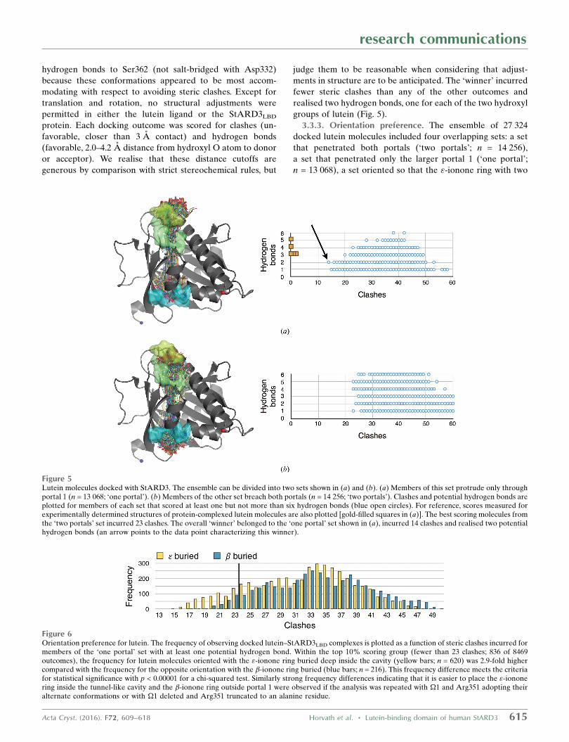
3.3.1. Lutein structure. Lutein (C40H56O2) belongs to the
xanthophyll carotenoids synthesized by plants and bacteria.
Two ionone rings, each bearing a hydroxyl group, are
connected by a long polyene backbone. The molecular struc-
ture includes three defined stereocenters (Fig. 4). We exam-
ined the structures of protein-complexed lutein molecules
found as part of the light-harvesting complexes from spinach
and pea (Liu et al., 2004; Pan et al., 2011; Qin et al., 2015; Wei et
al., 2016). All examples show curvature in the polyene back-
bone, steric complementarity with surfaces of the proteins,
especially at ionone ring-contacting pockets, and two or three
hydrogen bonds formed between each of the hydroxyl groups
of lutein and acceptor and donor groups presented by the
chlorophyll-binding protein subunits. The average length
measured between hydroxyl O atoms is 30.5 � 0.2 A. The size
and shape of lutein are thus closely matched to the size and
shape of the cavity found in StARD3LBD, but it would be
impossible to completely fit the entire molecule into the cavity.
Interestingly, crevices and protrusions of the protein found
just outside each portal appear to be articulated to specifically
accommodate some type of ligand. We propose that structural
features close to and outside one or both of the portals make
contact with an ionone ring, similar to the ionone ring-binding
pockets observed in light-harvesting complex structures (Liu
et al., 2004; Pan et al., 2011; Qin et al., 2015; Mazor et al., 2015;
Wei et al., 2016).
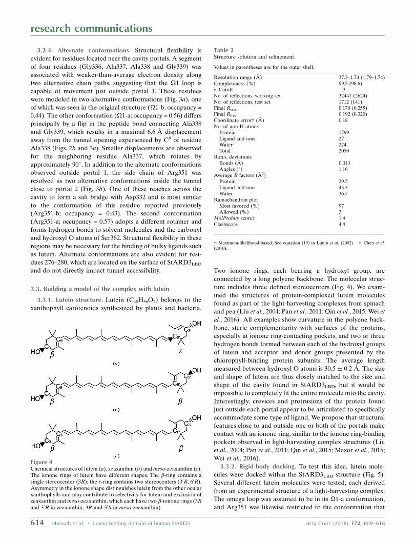
3.3.2. Rigid-body docking. To test this idea, lutein mole-
cules were docked within the StARD3LBD structure (Fig. 5).
Several different lutein molecules were tested, each derived
from an experimental structure of a light-harvesting complex.
The omega loop was assumed to be in its �1-a conformation,
and Arg351 was likewise restricted to the conformation that
research communications
614 Horvath et al. � Lutein-binding domain of human StARD3 Acta Cryst. (2016). F72, 609–618
Figure 4Chemical structures of lutein (a), zeaxanthin (b) and meso-zeaxanthin (c).The ionone rings of lutein have different shapes. The �-ring contains asingle stereocenter (3R); the "-ring contains two stereocenters (30R, 60R).Asymmetry in the ionone shape distinguishes lutein from the other ocularxanthophylls and may contribute to selectivity for lutein and exclusion ofzeaxanthin and meso-zeaxanthin, which each have two �-ionone rings (3Rand 30R in zeaxanthin; 3R and 30S in meso-zeaxanthin).
Table 2Structure solution and refinement.
Values in parentheses are for the outer shell.
Resolution range (A) 37.2–1.74 (1.79–1.74)Completeness (%) 99.5 (98.6)� Cutoff �3No. of reflections, working set 32447 (2624)No. of reflections, test set 1712 (141)Final Rcryst 0.170 (0.255)Final Rfree 0.192 (0.320)Coordinate error† (A) 0.18No. of non-H atoms
Protein 1799Ligand and ions 27Water 224Total 2050
R.m.s. deviationsBonds (A) 0.013Angles (�) 1.16
Average B factors (A2)Protein 29.5Ligand and ions 43.3Water 36.7
Ramachandran plotMost favored (%) 97Allowed (%) 3
MolProbity score‡ 1.4Clashscore 4.4
† Maximum-likelihood-based. See equation (19) in Lunin et al. (2002). ‡ Chen et al.(2010).
hydrogen bonds to Ser362 (not salt-bridged with Asp332)
because these conformations appeared to be most accom-
modating with respect to avoiding steric clashes. Except for
translation and rotation, no structural adjustments were
permitted in either the lutein ligand or the StARD3LBD
protein. Each docking outcome was scored for clashes (un-
favorable, closer than 3 A contact) and hydrogen bonds
(favorable, 2.0–4.2 A distance from hydroxyl O atom to donor
or acceptor). We realise that these distance cutoffs are
generous by comparison with strict stereochemical rules, but
judge them to be reasonable when considering that adjust-
ments in structure are to be anticipated. The ‘winner’ incurred
fewer steric clashes than any of the other outcomes and
realised two hydrogen bonds, one for each of the two hydroxyl
groups of lutein (Fig. 5).
3.3.3. Orientation preference. The ensemble of 27 324
docked lutein molecules included four overlapping sets: a set
that penetrated both portals (‘two portals’; n = 14 256),
a set that penetrated only the larger portal 1 (‘one portal’;
n = 13 068), a set oriented so that the "-ionone ring with two
research communications
Acta Cryst. (2016). F72, 609–618 Horvath et al. � Lutein-binding domain of human StARD3 615
Figure 6Orientation preference for lutein. The frequency of observing docked lutein–StARD3LBD complexes is plotted as a function of steric clashes incurred formembers of the ‘one portal’ set with at least one potential hydrogen bond. Within the top 10% scoring group (fewer than 23 clashes; 836 of 8469outcomes), the frequency for lutein molecules oriented with the "-ionone ring buried deep inside the cavity (yellow bars; n = 620) was 2.9-fold highercompared with the frequency for the opposite orientation with the �-ionone ring buried (blue bars; n = 216). This frequency difference meets the criteriafor statistical significance with p < 0.00001 for a chi-squared test. Similarly strong frequency differences indicating that it is easier to place the "-iononering inside the tunnel-like cavity and the �-ionone ring outside portal 1 were observed if the analysis was repeated with �1 and Arg351 adopting theiralternate conformations or with �1 deleted and Arg351 truncated to an alanine residue.
Figure 5Lutein molecules docked with StARD3. The ensemble can be divided into two sets shown in (a) and (b). (a) Members of this set protrude only throughportal 1 (n = 13 068; ‘one portal’). (b) Members of the other set breach both portals (n = 14 256; ‘two portals’). Clashes and potential hydrogen bonds areplotted for members of each set that scored at least one but not more than six hydrogen bonds (blue open circles). For reference, scores measured forexperimentally determined structures of protein-complexed lutein molecules are also plotted [gold-filled squares in (a)]. The best scoring molecules fromthe ‘two portals’ set incurred 23 clashes. The overall ‘winner’ belonged to the ‘one portal’ set shown in (a), incurred 14 clashes and realised two potentialhydrogen bonds (an arrow points to the data point characterizing this winner).
chiral centers was closest to portal 1 (n = 13 662) and a set
oriented in the opposite direction with this " ring close to
portal 2 (n = 13 662). The distribution of clashscores indicates
a strong tendency for high-scoring models to belong to the
‘one portal’ set (Fig. 5) and oriented so that the " ring is buried
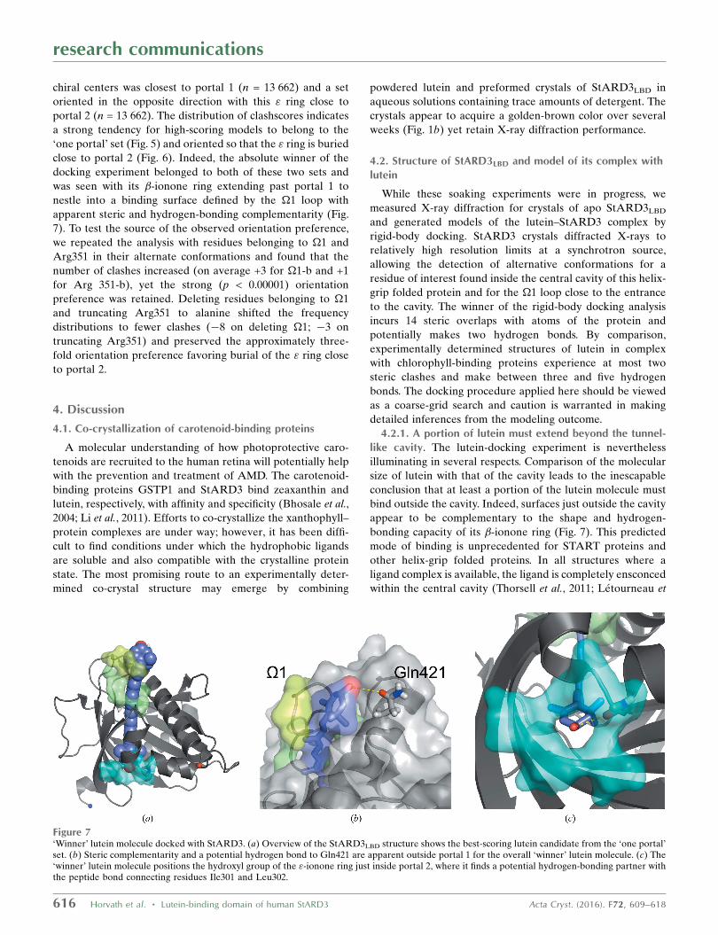
close to portal 2 (Fig. 6). Indeed, the absolute winner of the
docking experiment belonged to both of these two sets and
was seen with its �-ionone ring extending past portal 1 to
nestle into a binding surface defined by the �1 loop with
apparent steric and hydrogen-bonding complementarity (Fig.
7). To test the source of the observed orientation preference,
we repeated the analysis with residues belonging to �1 and
Arg351 in their alternate conformations and found that the
number of clashes increased (on average +3 for �1-b and +1
for Arg 351-b), yet the strong (p < 0.00001) orientation
preference was retained. Deleting residues belonging to �1
and truncating Arg351 to alanine shifted the frequency
distributions to fewer clashes (�8 on deleting �1; �3 on
truncating Arg351) and preserved the approximately three-
fold orientation preference favoring burial of the " ring close
to portal 2.
4. Discussion
4.1. Co-crystallization of carotenoid-binding proteins
A molecular understanding of how photoprotective caro-
tenoids are recruited to the human retina will potentially help
with the prevention and treatment of AMD. The carotenoid-
binding proteins GSTP1 and StARD3 bind zeaxanthin and
lutein, respectively, with affinity and specificity (Bhosale et al.,
2004; Li et al., 2011). Efforts to co-crystallize the xanthophyll–
protein complexes are under way; however, it has been diffi-
cult to find conditions under which the hydrophobic ligands
are soluble and also compatible with the crystalline protein
state. The most promising route to an experimentally deter-
mined co-crystal structure may emerge by combining
powdered lutein and preformed crystals of StARD3LBD in
aqueous solutions containing trace amounts of detergent. The
crystals appear to acquire a golden-brown color over several
weeks (Fig. 1b) yet retain X-ray diffraction performance.
4.2. Structure of StARD3LBD and model of its complex withlutein
While these soaking experiments were in progress, we
measured X-ray diffraction for crystals of apo StARD3LBD
and generated models of the lutein–StARD3 complex by
rigid-body docking. StARD3 crystals diffracted X-rays to
relatively high resolution limits at a synchrotron source,
allowing the detection of alternative conformations for a
residue of interest found inside the central cavity of this helix-
grip folded protein and for the �1 loop close to the entrance
to the cavity. The winner of the rigid-body docking analysis
incurs 14 steric overlaps with atoms of the protein and
potentially makes two hydrogen bonds. By comparison,
experimentally determined structures of lutein in complex
with chlorophyll-binding proteins experience at most two
steric clashes and make between three and five hydrogen
bonds. The docking procedure applied here should be viewed
as a coarse-grid search and caution is warranted in making
detailed inferences from the modeling outcome.
4.2.1. A portion of lutein must extend beyond the tunnel-like cavity. The lutein-docking experiment is nevertheless
illuminating in several respects. Comparison of the molecular
size of lutein with that of the cavity leads to the inescapable
conclusion that at least a portion of the lutein molecule must
bind outside the cavity. Indeed, surfaces just outside the cavity
appear to be complementary to the shape and hydrogen-
bonding capacity of its �-ionone ring (Fig. 7). This predicted
mode of binding is unprecedented for START proteins and
other helix-grip folded proteins. In all structures where a
ligand complex is available, the ligand is completely ensconced
within the central cavity (Thorsell et al., 2011; Letourneau et
research communications
616 Horvath et al. � Lutein-binding domain of human StARD3 Acta Cryst. (2016). F72, 609–618
Figure 7‘Winner’ lutein molecule docked with StARD3. (a) Overview of the StARD3LBD structure shows the best-scoring lutein candidate from the ‘one portal’set. (b) Steric complementarity and a potential hydrogen bond to Gln421 are apparent outside portal 1 for the overall ‘winner’ lutein molecule. (c) The‘winner’ lutein molecule positions the hydroxyl group of the "-ionone ring just inside portal 2, where it finds a potential hydrogen-bonding partner withthe peptide bond connecting residues Ile301 and Leu302.
al., 2015). Leaving a portion of the molecule on the outside of
the protein as proposed here has important implications for
the molecular interactions necessary for the transport and
metabolism of lutein.
4.2.2. Orientation preference and implications for ligandrecognition. An unexpected outcome of the docking experi-
ment was that the "-ionone ring, with two chiral centers,
appears to fit better inside the cavity, with fewer clashes and
additional potential hydrogen bonds associated with this
orientation, on average, in the ensemble of docking trials (see
Fig. 6). This pattern suggests an interesting model for ligand
selectivity: the binding-pocket asymmetry apparent for the
lutein–StARD3 model may positively identify lutein, which is
inherently asymmetrical with �-ionone and "-ionone rings,
and exclude the closely related xanthophylls zeaxanthin and
meso-zeaxanthin, which exhibit symmetry and have two
�-ionone rings. Consistent with this idea that symmetry plays a
role in xanthophyll selectivity, GSTP1, which is homodimeric,
is the zeaxanthin- and meso-zeaxanthin-binding protein in the
human retina (Bhosale et al., 2004). The winner of the lutein–
StARD3 docking experiment makes several close contacts
with the �1 loop (Fig. 7) and the side chain of Arg351,
suggesting that these contribute to the molecular-recognition
properties of StARD3. A strong orientation preference was
retained in the docking experiment with �1 and the side chain
of Arg351 adopting their alternate conformations or even
when they were removed. These results lead us to believe that
molecular interactions involving surfaces throughout and
outside the tunnel-like cavity, including �1 and Arg351,
probably act cooperatively to select a particular binding
orientation and positively identify lutein.
4.2.3. Second portal of StARD3. Although two portals
potentially allow solvent to access the cavity at opposite ends
of StARD3, one of these is much larger. The minimum
number of clashes encountered for lutein molecules docked so
as to penetrate both entrances was 23, which is nine more than
that observed for the best-scoring model docked so as to
penetrate just the larger portal. We therefore predict that
lutein binds deep within the cavity, that one of the ionone rings
protrudes through the larger opening which is guarded by the
�1 loop, and that there is a close approach to but no breach of
the smaller opening. It will be interesting to compare these
predictions with an experimental structure of the co-complex
and/or more refined modeling experiments that apply energy-
relaxation methods (Das & Baker, 2008; Leaver-Fay et al.,
2011).
4.2.4. Allosteric trigger point. StARD3LBD belongs to the
helix-grip protein family, and protein members with structural
homology include START proteins and allergen proteins,
as well as several biosynthetic enzymes. Inspection of these
structures reveals that StARD3 and StARD1 differ from the
others in sharing identical or highly conserved residues at
three positions that appear to be important for ligand binding:
Arg at position 351 (numbered as in StARD3; 188 in
StARD1), an acidic residue at position 332 (Asp in StARD3;
Glu169 in StARD1) and Gln at position 421 (position 258 in
StARD1). In other START-family members these three resi-
dues are variable, and all three are not found together. The
lutein-docking results suggest that Gln421 may be positioned
so as to make a hydrogen bond to a hydroxyl group of lutein
outside portal 1 (Fig. 7). Electron-density maps, now calcu-
lated to 1.74 A resolution, show that Arg351 adopts two
alternative conformations (Fig. 3b), one of which forms a salt
bridge to Asp332. Interestingly, this salt-bridged conformation
is probably incompatible with lutein occupancy owing to steric
overlap. Based on these observations, we suggest that the salt
bridge found in cavities of StARD3 and StARD1 may act as
an allosteric trigger point to communicate ligand binding to
other components of the steroid-generating apparatus in the
case of StARD1, and to retinal proteins and enzymes involved
with xanthophyll transport and metabolism in the case of
StARD3.
5. Conclusion
The structure of the lutein-binding domain of StARD3 has
been determined to 1.74 A resolution, revealing alternative
conformations for elements of protein structure that appear to
be critical for ligand binding. Modeling experiments indicate
that the biologically relevant ligand must protrude from the
cavity entrance because it is not possible to completely fit a
30 A long molecule of lutein into a cavity that measures only
20 A. Asymmetry in the tunnel-like cavity may play a role in
selecting lutein with its �- and "-ionone rings and discrimi-
nating against the other ocular carotenoids, zeaxanthin and
meso-zeaxanthin, which each have two �-ionone rings.
Acknowledgements
We thank Dr Scott Classen and Jane Tanamachi for assistance
with data collection at the SIBYLS beamline. Dr James Hurley
generously supplied the original expression system for
StARD3LBD. This work was supported by the National Insti-
tutes of Health (NIH; EY11600 to PSB and EY14600 to the
Department of Ophthalmology). EWG was supported by a
UROP fellowship through the Office of Undergraduate
Research at the University of Utah. SL was supported by a
Bioscience Summer Research Program with the Department
of Biology, University of Utah. The SIBYLS beamline at
the Advanced Light Source, Lawrence Berkeley National
Laboratory is supported in part by the US Department of
Energy (DOE) program Integrated Diffraction Analysis
Technologies (IDAT) and the DOE program Molecular
Assemblies Genes and Genomics Integrated Efficiently
(MAGGIE) under Contract No. DE-AC02- 05CH11231. The
Moran Eye Center has received an unrestricted departmental
grant from Research to Prevent Blindness.
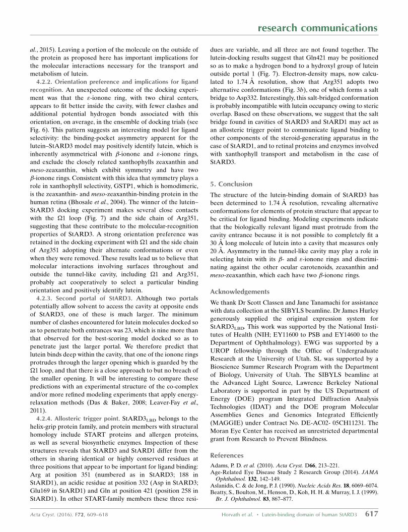
References
Adams, P. D. et al. (2010). Acta Cryst. D66, 213–221.Age-Related Eye Disease Study 2 Research Group (2014). JAMA
Ophthalmol. 132, 142–149.Aslanidis, C. & de Jong, P. J. (1990). Nucleic Acids Res. 18, 6069–6074.Beatty, S., Boulton, M., Henson, D., Koh, H. H. & Murray, I. J. (1999).
Br. J. Ophthalmol. 83, 867–877.
research communications
Acta Cryst. (2016). F72, 609–618 Horvath et al. � Lutein-binding domain of human StARD3 617
Bernstein, P. S., Li, B., Vachali, P. P., Gorusupudi, A., Shyam, R.,Henriksen, B. S. & Nolan, J. M. (2016). Prog. Retin. Eye Res. 50,34–66.
Bhosale, P., Larson, A. J., Frederick, J. M., Southwick, K., Thulin,C. D. & Bernstein, P. S. (2004). J. Biol. Chem. 279, 49447–49454.
Chen, V. B., Arendall, W. B., Headd, J. J., Keedy, D. A., Immormino,R. M., Kapral, G. J., Murray, L. W., Richardson, J. S. & Richardson,D. C. (2010). Acta Cryst. D66, 12–21.
Classen, S., Hura, G. L., Holton, J. M., Rambo, R. P., Rodic, I.,McGuire, P. J., Dyer, K., Hammel, M., Meigs, G., Frankel, K. A. &Tainer, J. A. (2013). J. Appl. Cryst. 46, 1–13.
Das, R. & Baker, D. (2008). Annu. Rev. Biochem. 77, 363–382.Emsley, P., Lohkamp, B., Scott, W. G. & Cowtan, K. (2010). Acta
Cryst. D66, 486–501.Gill, S. C. & von Hippel, P. H. (1989). Anal. Biochem. 182, 319–326.Kabsch, W. (2010a). Acta Cryst. D66, 125–132.Kabsch, W. (2010b). Acta Cryst. D66, 133–144.Leaver-Fay, A. et al. (2011). Methods Enzymol. 487, 545–574.Letourneau, D., Lefebvre, A., Lavigne, P. & LeHoux, J.-G. (2015).
Mol. Cell. Endocrinol. 408, 53–61.Li, B., Ahmed, F. & Bernstein, P. S. (2010). Arch. Biochem. Biophys.
504, 56–60.Li, M. Z. & Elledge, S. J. (2007). Nature Methods, 4, 251–256.Li, B., Vachali, P., Frederick, J. M. & Bernstein, P. S. (2011).
Biochemistry, 50, 2541–2549.
Liu, Z., Yan, H., Wang, K., Kuang, T., Zhang, J., Gui, L., An, X. &Chang, W. (2004). Nature (London), 428, 287–292.
Lunin, V. Y., Afonine, P. V. & Urzhumtsev, A. G. (2002). Acta Cryst.A58, 270–282.
Mazor, Y., Borovikova, A. & Nelson, N. (2015). Elife, 4, e07433.Otwinowski, Z. & Minor, W. (1997). Methods Enzymol. 276, 307–326.Pan, X., Li, M., Wan, T., Wang, L., Jia, C., Hou, Z., Zhao, X., Zhang, J.
& Chang, W. (2011). Nature Struct. Mol. Biol. 18, 309–315.Qin, X., Suga, M., Kuang, T. & Shen, J. R. (2015). Science, 348,
989–995.Seddon, J. M., Ajani, U. A., Sperduto, R. D., Hiller, R., Blair, N.,
Burton, T. C., Farber, M. D., Gragoudas, E. S., Haller, J., Miller,D. T., Yannuzzi, L. A. & Willett, W. (1994). JAMA, 272, 1413–1420.
Thorsell, A.-G., Lee, W. H., Persson, C., Siponen, M. I., Nilsson, M.,Busam, R. D., Kotenyova, T., Schuler, H. & Lehtio, L. (2011). PLoSOne, 6, e19521.
Tsujishita, Y. & Hurley, J. H. (2000). Nature Struct. Biol. 7, 408–414.
Watari, H., Arakane, F., Moog-Lutz, C., Kallen, C. B., Tomasetto, C.,Gerton, G. L., Rio, M. C., Baker, M. E. & Strauss, J. F. (1997). Proc.Natl Acad. Sci. USA, 94, 8462–8467.
Wei, X., Su, X., Cao, P., Liu, X., Chang, W., Li, M., Zhang, X. & Liu, Z.(2016). Nature (London), 534, 69–74.
Wu, J., Cho, E., Willett, W. C., Sastry, S. M. & Schaumberg, D. A.(2015). JAMA Ophthalmol. 133, 1415–1424.
research communications
618 Horvath et al. � Lutein-binding domain of human StARD3 Acta Cryst. (2016). F72, 609–618