skeletal system - mrs. moretz's science site · •movementdue to attached skeletal muscles...
TRANSCRIPT
SKELETAL SYSTEM
Moretz, 2017/2018
Medical Anatomy & Physiology
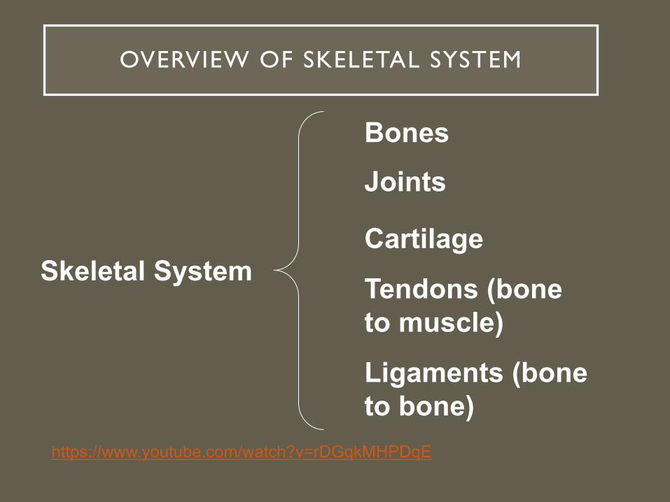
OVERVIEW OF SKELETAL SYSTEM
Skeletal System
BonesJoints
Cartilage
Ligaments (bone to bone)
Tendons (bone to muscle)
https://www.youtube.com/watch?v=rDGqkMHPDqE

FUNCTION OF THE SKELETAL SYSTEM
• Support of the body• Protection of soft organs• Movement due to attached skeletal
muscles• Storage of minerals and fats• Blood cell formation

TYPES OF BONE TISSUE
• Compact Bone: • Hard outer layer of bone• Protection & support & resistance to stress
• Spongy bone: • Less dense, small needle-like pieces of bone with many
open spaces• Support & protect bone marrow
• Bone marrow: Soft tissue inside bone that produces blood cells & stores fat

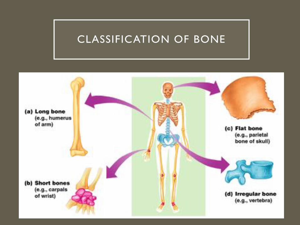
CLASSIFICATION OF BONE

• Typically longer than wide
• Have a shaft with heads at both ends
• Contain mostly compact & spongy tissue.
•Ex.: Femur, humerus
LONG BONES

SHORT BONES
• Cube-shape• Contain mostly spongy bone
• Examples: Carpals, tarsals

• Thin and flattened• Usually curved• Thin layers of compact bone around a layer of spongy bone
• Examples: Skull, ribs, sternum
FLAT BONES

IRREGULAR BONES
• Irregular shape• Variable amount of spongy & compact tissue
• Example: Vertebrae and hip bones

SESAMOID BONES
• Present in tendons where there is a lot of friction & tension.
• Protect tendons
• Not always ossified
• Usually small
• Ex. patellae

SUTURAL BONES
• Small bones located in joints between cranial bones
• Variable numbers

BONE STRUCTURE
• Diaphysis• Shaft• Composed of compact bone• Location of yellow marrow
(fat)

BONE STRUCTURE
• Epiphysis• Ends of the bone• Composed mostly of
spongy bone• Location of red marrow
(blood formation)
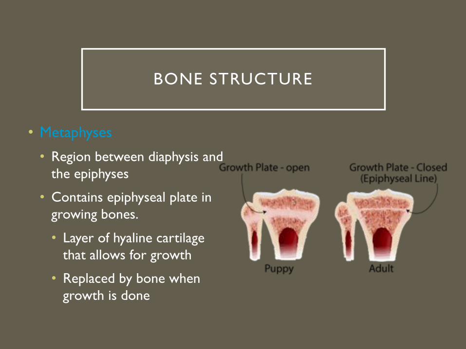
BONE STRUCTURE
• Metaphyses
• Region between diaphysis and the epiphyses
• Contains epiphyseal plate in growing bones.
• Layer of hyaline cartilage that allows for growth
• Replaced by bone when growth is done
BONE STRUCTURE
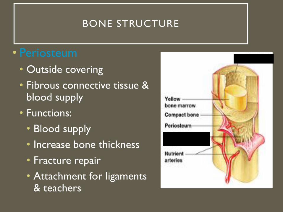
• Periosteum• Outside covering• Fibrous connective tissue &
blood supply• Functions:
• Blood supply• Increase bone thickness• Fracture repair• Attachment for ligaments
& teachers

• Articular cartilage• Hyaline cartilage
• Covers the external surface of the epiphyses
• Decreases friction at joint surfaces
BONE STRUCTURE
BONE STRUCTURE
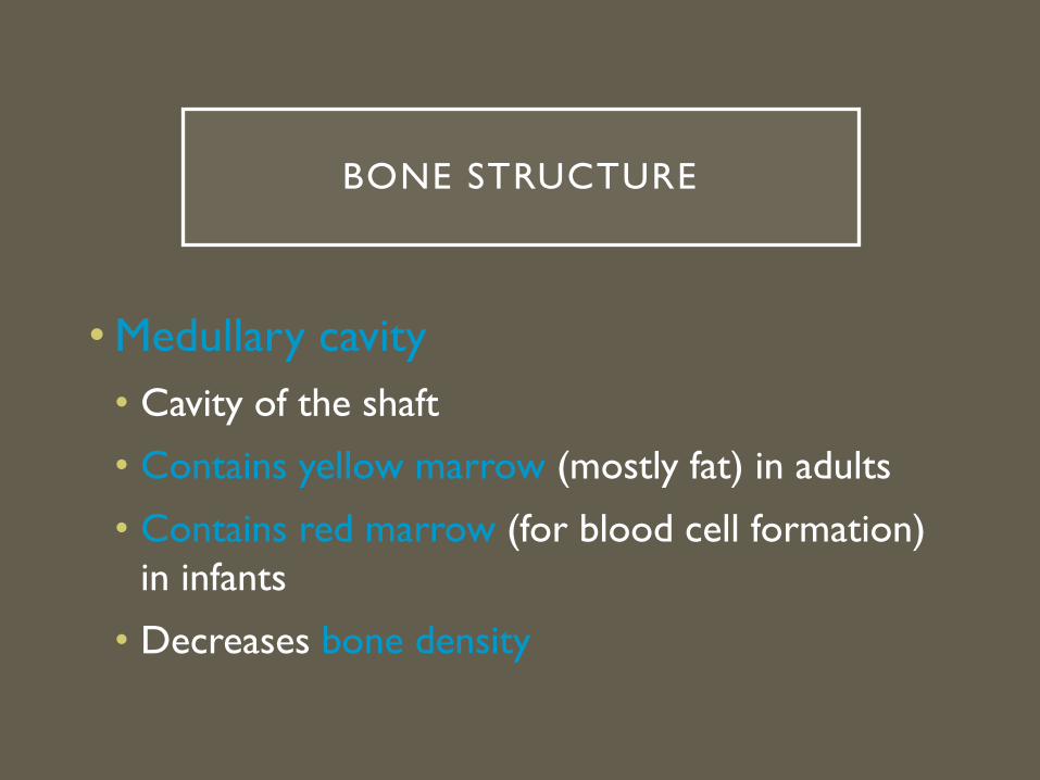
• Medullary cavity• Cavity of the shaft
• Contains yellow marrow (mostly fat) in adults
• Contains red marrow (for blood cell formation) in infants
• Decreases bone density


CALCIFICATION
Calcium phosphate + calcium hydroxide
Hydroxyapetite (crystals)
+
Calcium carbonate, Mg+, F+, K, sulfate
Deposited in the framework of collagen, crystalize, and harden

TYPES OF BONE CELLS
• Osteoprogenitor cells• Unspecialized bone stem cells
• Osteoblasts• Bone-forming cells for bone growth
• Initiate calcification
• Osteocytes• Mature bone cells
• Help exchange nutrients and waste with blood.
• Osteoclasts• Bone-destroying cells
• Break down bone matrix for remodeling and release of calcium
• Bone remodeling is a process done by both osteoblasts and osteoclasts


TISSUES OF THE COMPACT BONE
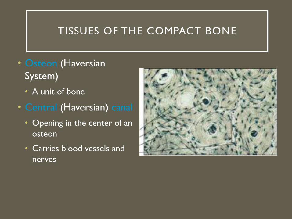
• Osteon (Haversian System)• A unit of bone
• Central (Haversian) canal• Opening in the center of an
osteon
• Carries blood vessels and nerves

• Lacunae• Cavities containing bone cells (osteocytes)
• Arranged in concentric rings
• Lamellae• Rings around the central canal• Sites of lacunae
• Canaliculi• Tiny canals• Radiate from the central canal to lacunae• Form a transport system

TISSUES OF THE SPONGY BONE
• Trabeculae• Lamellae arranged in irregular
columns
• Lined along lines of stress
• Contains spaces filled with red and yellow bone marrow
• Has blood vessels to nourish osteocytes.

BLOOD & NERVE SUPPLY OF THE BONE
• Periosteal artery & vein
• Nutrient artery & vein
• Supply inner part of compact bone of diaphysis and spongy bone
• Supply red marrow up to epiphyseal plates.
• Metaphyseal artery & vein
• Blood supply of red marrow
• Supplies spongy tissue of epiphyses
• Epiphyseal artery & vein
• Supplies spongy tissue of epiphyses

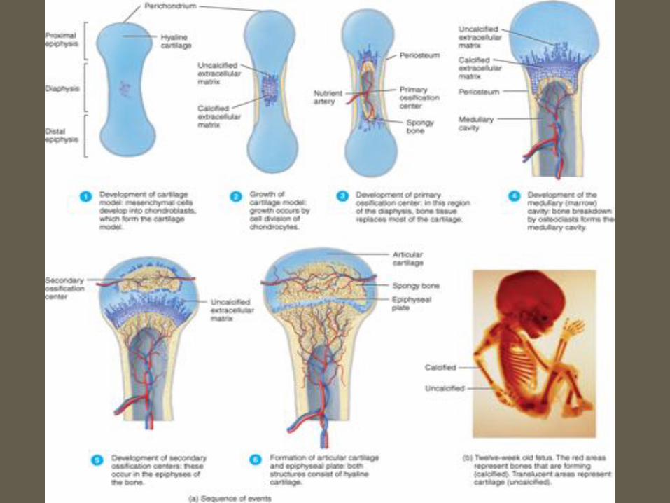
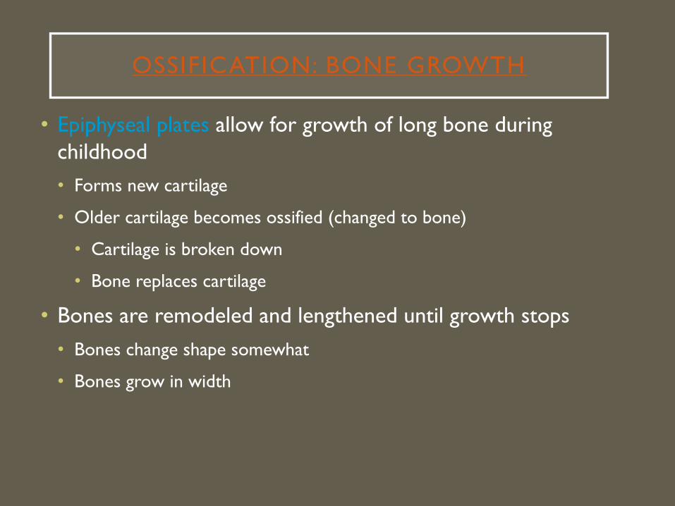
OSSIFICATION: BONE GROWTH
• 4 phases:• Initial bone formation - fetus
• Growth of bones - childhood
• Bone remodeling – throughout life
• Repair of fractures – throughout life

BONE FORMATION
• https://www.johnwiley.net.au/highered/interactions/media/Support/content/Support/skel2a/frameset.htm
• https://www.youtube.com/watch?v=xXgZap0AvL0• Two Types:
• Intramembranous ossification• Forms bones of skull, facial bones, mandible, and fetal “soft spots”• https://www.youtube.com/watch?v=C1PP83eou_8
• Endochondral ossification• Forms most bones• https://www.youtube.com/watch?v=Z-vRCYuH4uI

INTRAMEMBRANOUS OSSIFICATION
OSSIFICATION: BONE GROWTH
• Epiphyseal plates allow for growth of long bone during childhood• Forms new cartilage
• Older cartilage becomes ossified (changed to bone)
• Cartilage is broken down
• Bone replaces cartilage
• Bones are remodeled and lengthened until growth stops• Bones change shape somewhat
• Bones grow in width


FACTORS AFFECTING BONE GROWTH
• Minerals – Ca, P, Mg, F, Mn• Vitamins
• A – stimulates osteoblasts• C – synthesizes collegan• D – absorbs Ca• K and B12 – synthesize bone protein
• Hormones • Insulin-like growth factor – osteoblast production, stimulate mitosis, &
bone protein production• Sex hormones – increase osteoblast production; shut down growth at
epiphyseal plate

BONE REMODELING
• Bone remodeling – ongoing replacement of old bone tissue by new bone tissue.
• Bone resorption – removal of minerals
• Bone deposition – addition of minerals

MEDICAL CONCERNS
MEDICAL CONCERNS

MEDICAL CONCERNS
MEDICAL CONCERNS

HORMONAL CONTROL OF GROWTH

HORMONAL CONTROL OF GROWTH


COMMON TYPES OF FRACTURES


REPAIR OF BONE FRACTURES
• Hematoma (blood-filled swelling) is formed
• Break is splinted (immobilized) by fibrocartilage to form a callus
• Fibrocartilage callus is replaced by a bony callus
• Bony callus is remodeled to form a permanent patch
FRACTURE TREATMENT
1. Realignment of bone fragments
a. Reduction brings bones into alignment
2. Immobilization
3. Restoration of function

FRACTURE ACTIVITY
Sample (letter) Fracture Type Rationale

HUMAN SKELETON

SKELETAL SYSTEM ROTATION
• Skeletal system virtual lab
• Skeletal system half-body identification
• Skeletal system large skeletal identification
• Skeletal system coloring/labeling
• Skeletal system case study
• Skeletal system PSA

HUMAN SKELETON
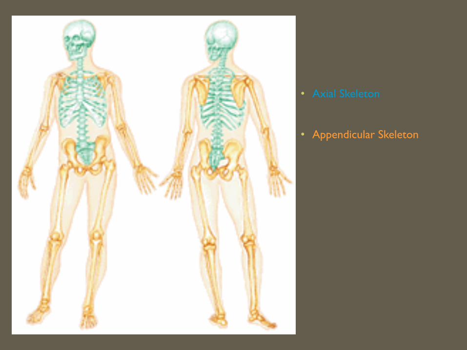
• 206 bones in the adult human body• Divided into 2 parts:
• Axial Skeleton
• Appendicular Skeleton
Skull
Vertebral column
Rib cage
Bones of arms and legs
Bones of shoulder
Pelvis
• Axial Skeleton
• Appendicular Skeleton

THE AXIAL SKELETON
• Divided into three parts
• Skull
• Vertebral column
• Rib Cage (bony thorax)

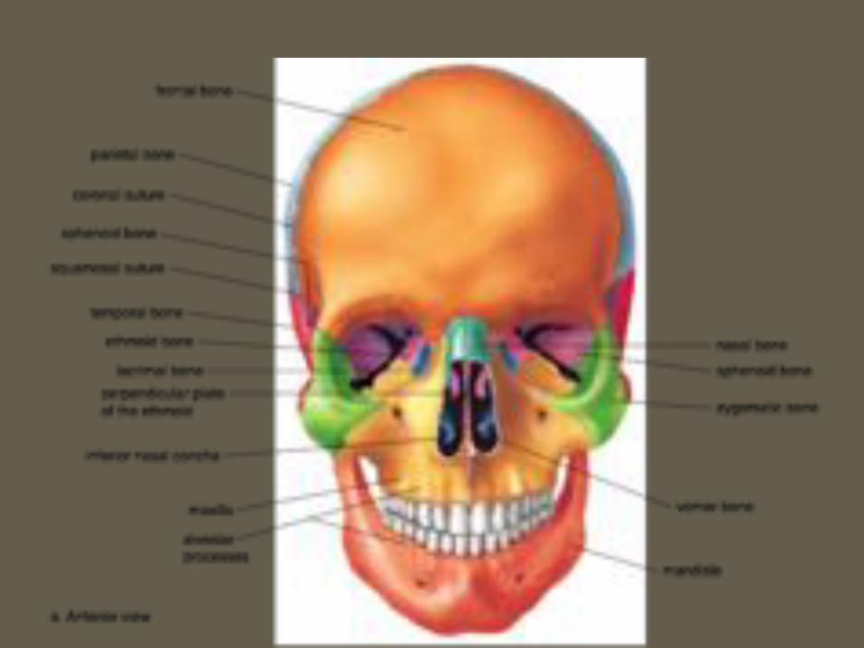
THE SKULL
• Two sets of bones• Cranium• Facial bones
• Skull bones are joined by sutures• Only the mandible is attached by
a freely movable joint
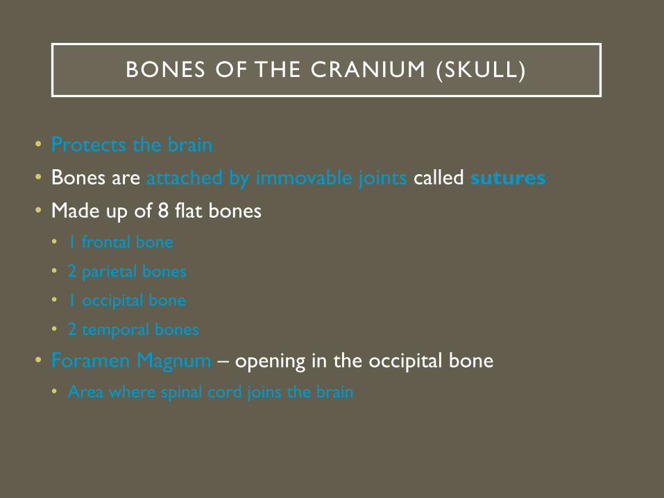
BONES OF THE CRANIUM (SKULL)
• Protects the brain• Bones are attached by immovable joints called sutures• Made up of 8 flat bones
• 1 frontal bone
• 2 parietal bones
• 1 occipital bone
• 2 temporal bones
• Foramen Magnum – opening in the occipital bone• Area where spinal cord joins the brain

• Lateral View of Skull

• Superior View of Skull

• Inferior View of Skull

THE FETAL SKULL
• Fontanelles – fibrous membranes connecting the cranial bones• Allow the brain
to grow
• Convert to bone within 24 months after birth

FACIAL BONES• Maxilla
• 2 bones that form the upper jaw
• Mandible
• Lower jaw
• Only movable bone of the skull
• Contains tooth sockets for 16 teeth
• Nasal bone
• Forms bridge of nose
• Zygomatic bone
• 2 cheek bones

THE VERTEBRAL COLUMN• Vertebrae separated by intervertebral discs (pads of fibrocartilage)
• The spine has 4 normal curvatures
• Each vertebrae is given a name according to its location
• Function: protect spinal cord, supports rib cage, attaches to pelvic

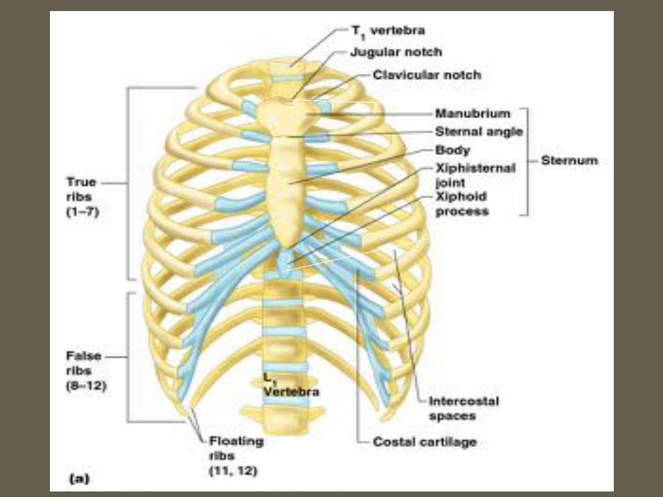
THE RIB CAGE (BONY THORAX)
• Forms a cage to protect major organs (heart, lungs)
• Made-up of three parts:
1) Ribs
• 12 ribs connected to the vertebrae
• True ribs:
• Ribs that connect directly to the sternum• False ribs:
• Ribs that attach to the sternum by a common cartilage• Floating ribs:
• Ribs that do not attach to the sternum

2) Sternum
• Flat, blade-shaped bone
• Composed of 3 bones: manubrium, body, xiphoid process
• Xiphoid process
• Inferior and smallest portion of sternum
• Attachment site for diaphragm
3) Thoracic vertebrae
• 12 thoracic vertebrae

THE APPENDICULAR SKELETON
• Pectoral girdle
• Limbs (appendages)
• Pelvic girdle
Composed of:

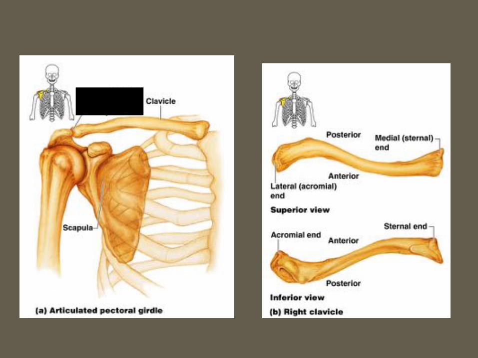
THE PECTORAL (SHOULDER) GIRDLE
• These bones allow the upper limbs to have exceptionally free movement
• Composed of 4 bones
- 2 Clavicles – collarbone• Slender and s-shaped
• Stabilizes shoulder but structurally weak (breaks easily) L
- 2 Scapulas – shoulder blade• Triangular shape

BONES OF THE UPPER LIMB
• Humerus (upper arm)
• Radius and ulna (forearm)
• Carpals, metacarpals, phalanges (hand)
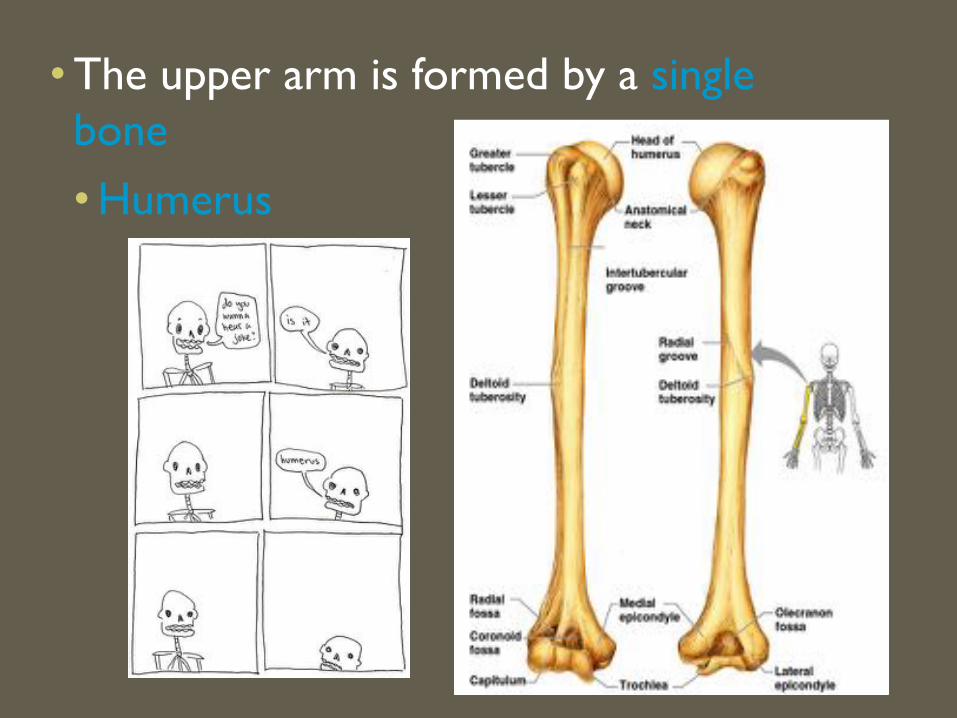
• The upper arm is formed by a single bone• Humerus
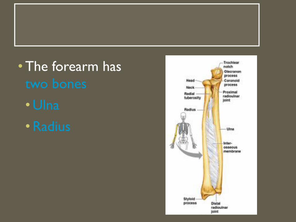
• The forearm has two bones• Ulna• Radius

• The hand• Carpals – wrist
• Metacarpals – palm
• Phalanges – fingers

BONES OF THE PELVIC GIRDLE
• Hip bones• Composed of:
• 2 coxal bones (hipbones)
• Sacrum
• Coccyx
• The total weight of the upper body rests on the pelvis
• Protects several organs• Reproductive organs
• Urinary bladder
• Part of the large intestine
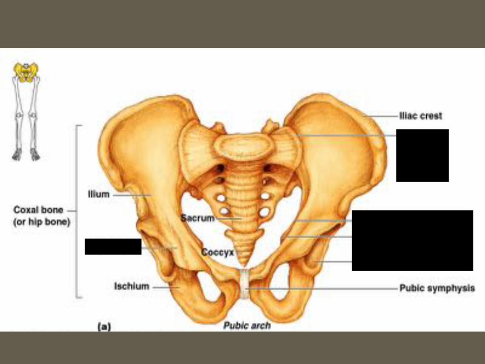
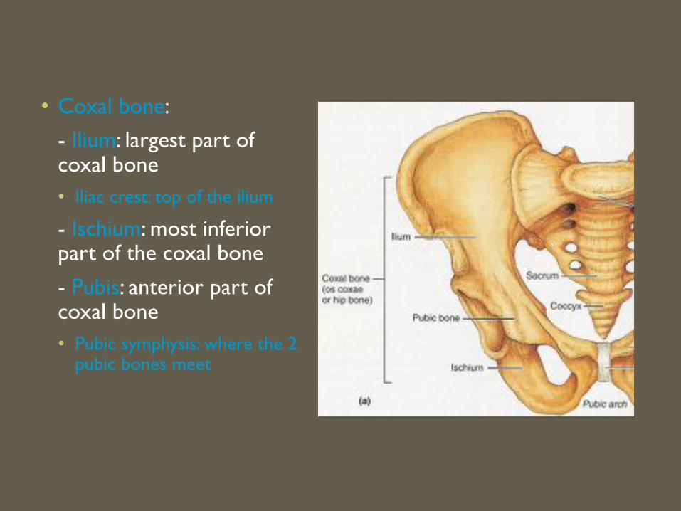
• Coxal bone:- Ilium: largest part of coxal bone• Iliac crest: top of the ilium
- Ischium: most inferior part of the coxal bone- Pubis: anterior part of coxal bone• Pubic symphysis: where the 2
pubic bones meet
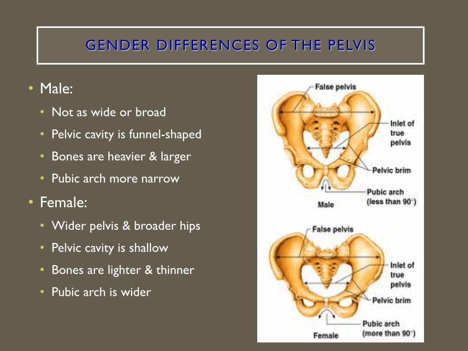
GENDER DIFFERENCES OF THE PELVIS
• Male:• Not as wide or broad
• Pelvic cavity is funnel-shaped
• Bones are heavier & larger
• Pubic arch more narrow
• Female:• Wider pelvis & broader hips
• Pelvic cavity is shallow
• Bones are lighter & thinner
• Pubic arch is wider

BONES OF THE LOWER LIMB
• Femur (thigh)
• Patella (kneecap)
• Tibia & Fibula (leg)
• Tarsals, metatarsals, phalanges (foot)

• Femur• Thigh bone• Strongest and
longest bone in the body
• Patella• Triangular bone
that protects the knee joint

• Tibia• Shinbone• Medial (towards the
middle) to the fibula• Thicker – bears weight
of femur
• Fibula• Lateral (away from
midline) to the tibia • Stabilize ankle
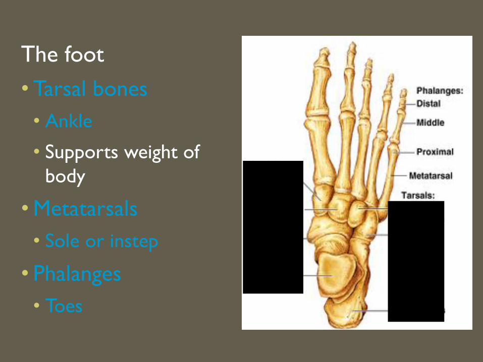
The foot• Tarsal bones
• Ankle
• Supports weight of body
• Metatarsals• Sole or instep
• Phalanges • Toes
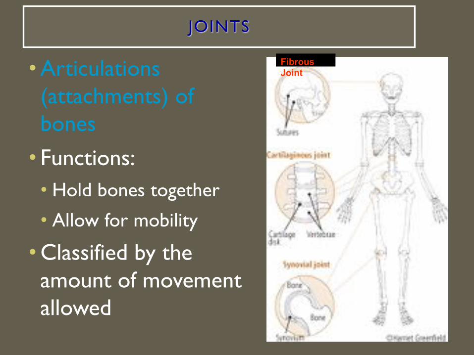
JOINTS
• Articulations (attachments) of bones
• Functions:• Hold bones together
• Allow for mobility
• Classified by the amount of movement allowed
Fibrous Joint

STRUCTURAL CLASSIFICATION OF JOINTS
• Fibrous joints• Generally immovable
• Fibrous connective tissue join bone to bone
• Example: cranium bones
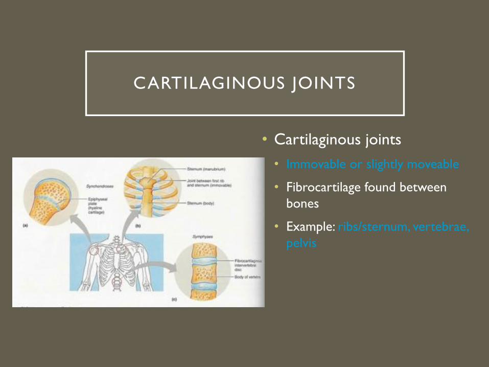
CARTILAGINOUS JOINTS
• Cartilaginous joints• Immovable or slightly moveable
• Fibrocartilage found between bones
• Example: ribs/sternum, vertebrae, pelvis
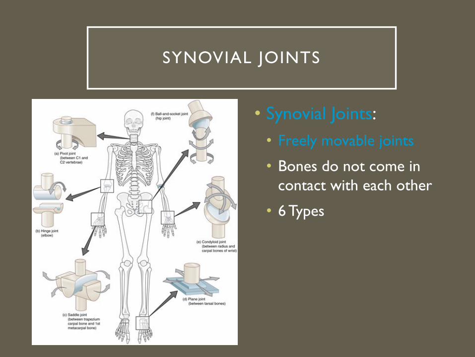
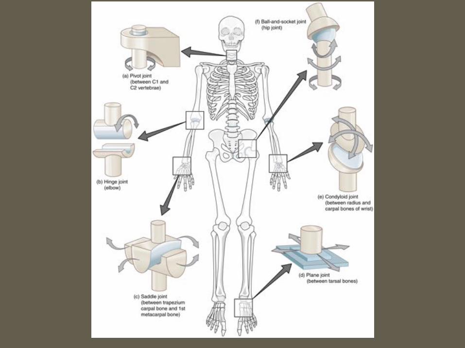
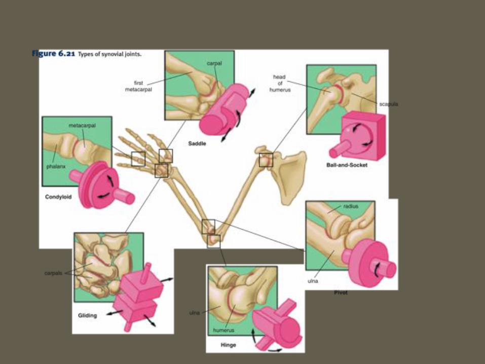
• Synovial Joints:• Freely movable joints
• Bones do not come in contact with each other
• 6 Types
SYNOVIAL JOINTS


JOINT VOCABULARY
• Ligaments
• Connective tissue that connects bone to bone
• Tendons
• Connect bone to muscles to further stabilize joint
• Synovial fluid
• Lubricating fluid found between bones to reduce friction
• Bursae
• Fluid-filled sacs that cushion joint (ex. knee)
• Meniscus
• Cartilaginous pads of tissue between the tibia and femur in knee joint

KNEE INJURIES
1. Torn Meniscus
§ The menisci absorb shock by compressing and spreading the weight evenly within the knee.
§ The menisci are attached to the tibia and joint and ligaments, allowing the menisci to pivot freely.
§ One of the most common knee injuries.
§ Grow weaker with age, and tear as a result of minor injuries or movements.
§ The most common injury occurs when the knee joint is bent and the knee is then twisted.
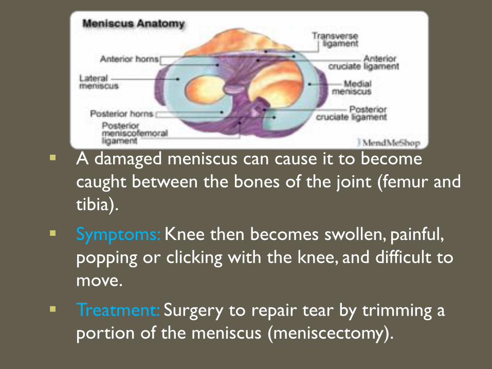
§ A damaged meniscus can cause it to become caught between the bones of the joint (femur and tibia).
§ Symptoms: Knee then becomes swollen, painful, popping or clicking with the knee, and difficult to move.
§ Treatment: Surgery to repair tear by trimming a portion of the meniscus (meniscectomy).

KNEE INJURIES CONT.
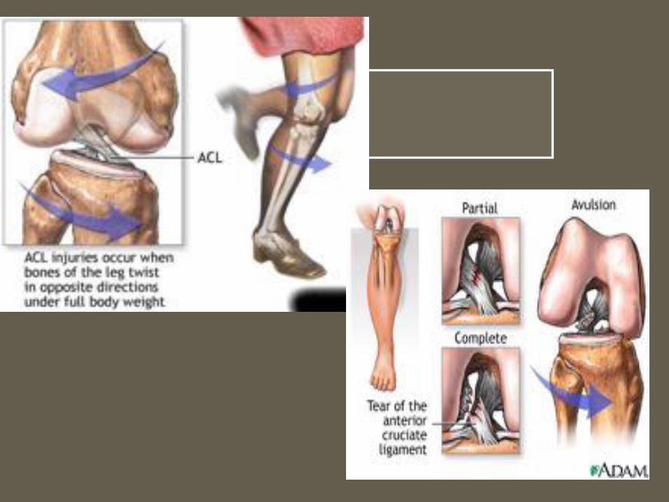
2. Torn ACL (Anterior Cruciate Ligament)• ACL provides stability to the joint• Common injury in athletes in contact sports• Occurs when the knee is locked with the foot
planted and the knee is twisted quickly. • The bones are more likely to rub against each
other (chronic ACL deficiency). • Can also damage the cartilage that covers the
ends of the bones and can trap and tear the menisci.
• Left untreated it can lead to osteoarthritis.

DISORDERS OF THE SKELETAL SYSTEM
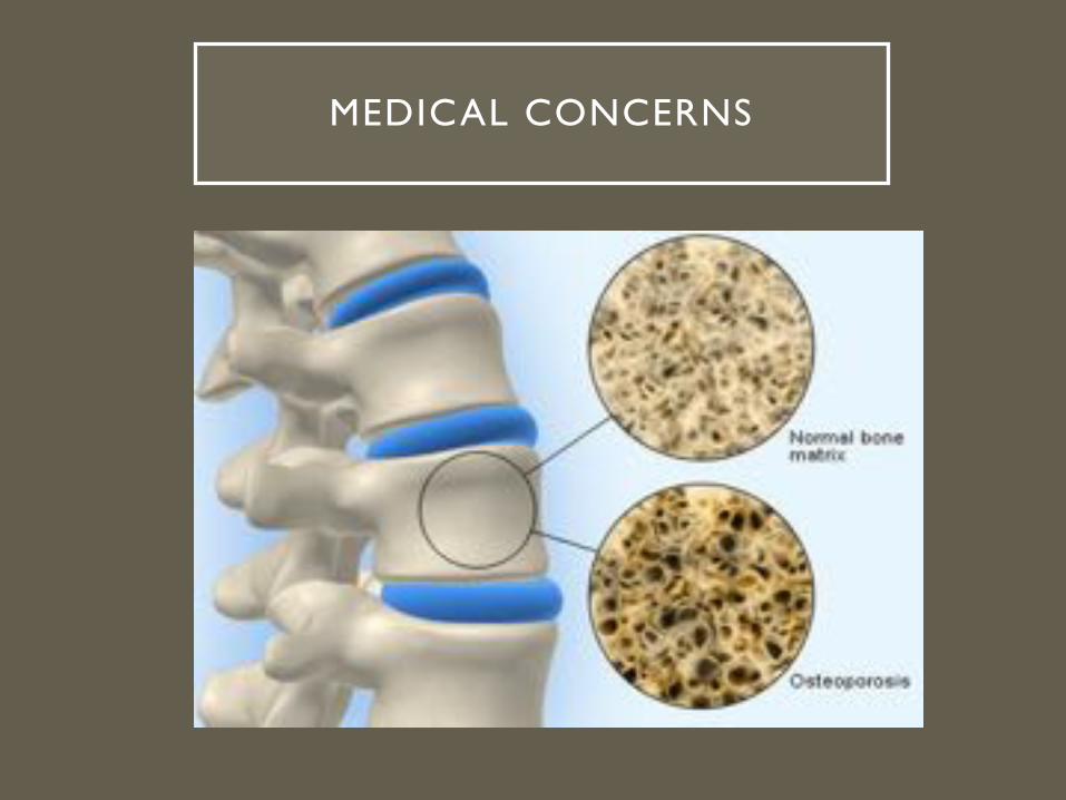
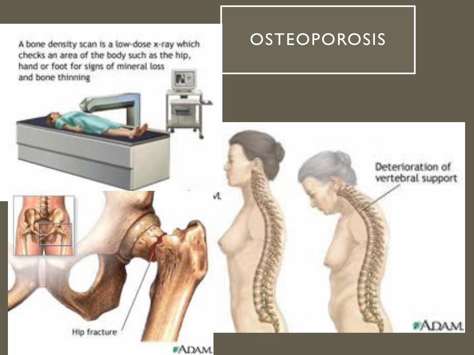
• Osteoporosis
• Most common bone disease
• 1 in 5 women in USA over 50
have osteoporosis
• Men over 70 are at risk
• During menopause, estrogen levels drop
• Body stops making new bone due to lack of calcium, resulting in brittle bones and fractures
OSTEOPOROSIS

ARTHRITIS• Inflammation of one or more joints
• Breakdown of cartilage causes bones to rub together, leading to pain, inflammation, and stiffness
• There are many different types of arthritis
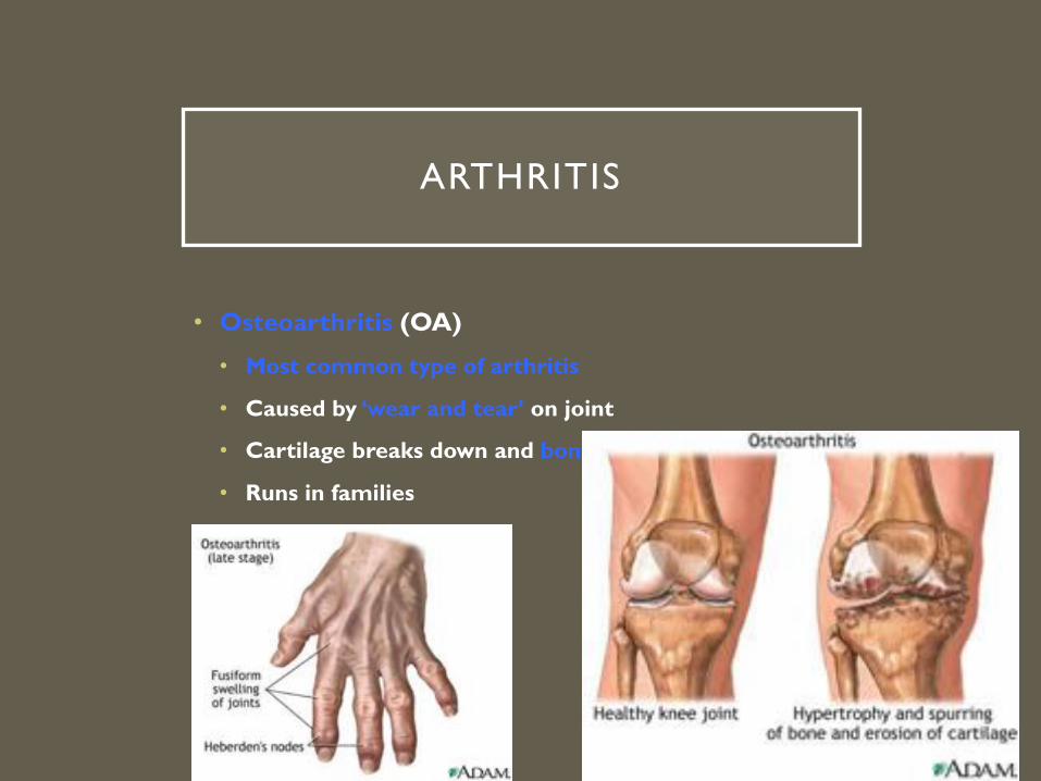
ARTHRITIS
• Osteoarthritis (OA)
• Most common type of arthritis
• Caused by ‘wear and tear’ on joint
• Cartilage breaks down and bony spurs may develop
• Runs in families
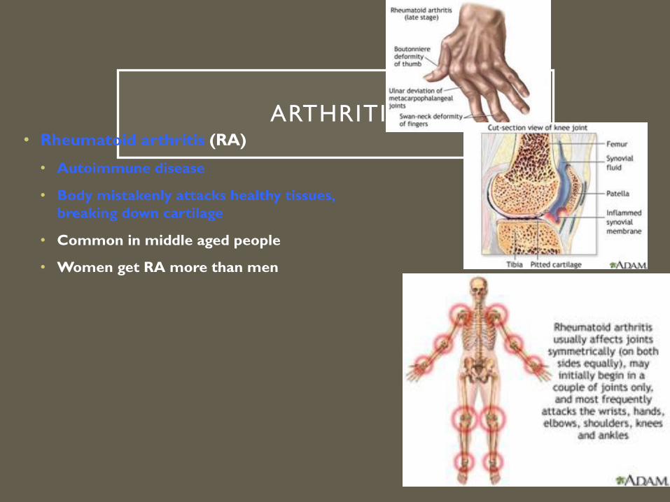
ARTHRITIS• Rheumatoid arthritis (RA)
• Autoimmune disease
• Body mistakenly attacks healthy tissues, breaking down cartilage
• Common in middle aged people
• Women get RA more than men
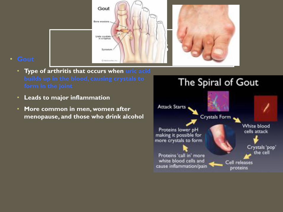
ARTHRITIS• Gout
• Type of arthritis that occurs when uric acid builds up in the blood, causing crystals to form in the joint
• Leads to major inflammation
• More common in men, women after menopause, and those who drink alcohol

RICKETS
• Uncommon
• Caused by a lack of Vitamin D, calcium, or phosphate, leading to a weakening and deformation of bones
• Vitamin D deficiency caused by lack of sunlight or rare genetic X-linked dominant trait