the skeletal system - wlwv.k12.or.us · the skeletal system • parts of the skeletal system •...
TRANSCRIPT

Chapter 5 The Skeletal System

The Skeletal System
• Parts of the skeletal system
• Bones (skeleton)
• Joints
• Cartilages
• Ligaments (bone to bone)(tendon=bone to muscle)
• Divided into two divisions
• Axial skeleton: bones of the skull, vertebral column, and rib cage
• Appendicular skeleton: bones of the upper and lower limbs, shoulder and hip
Functions of Bones
• Support of the body
• Protection of soft organs
• Movement due to attached skeletal muscles
• Storage of minerals and fats
• Blood cell formation
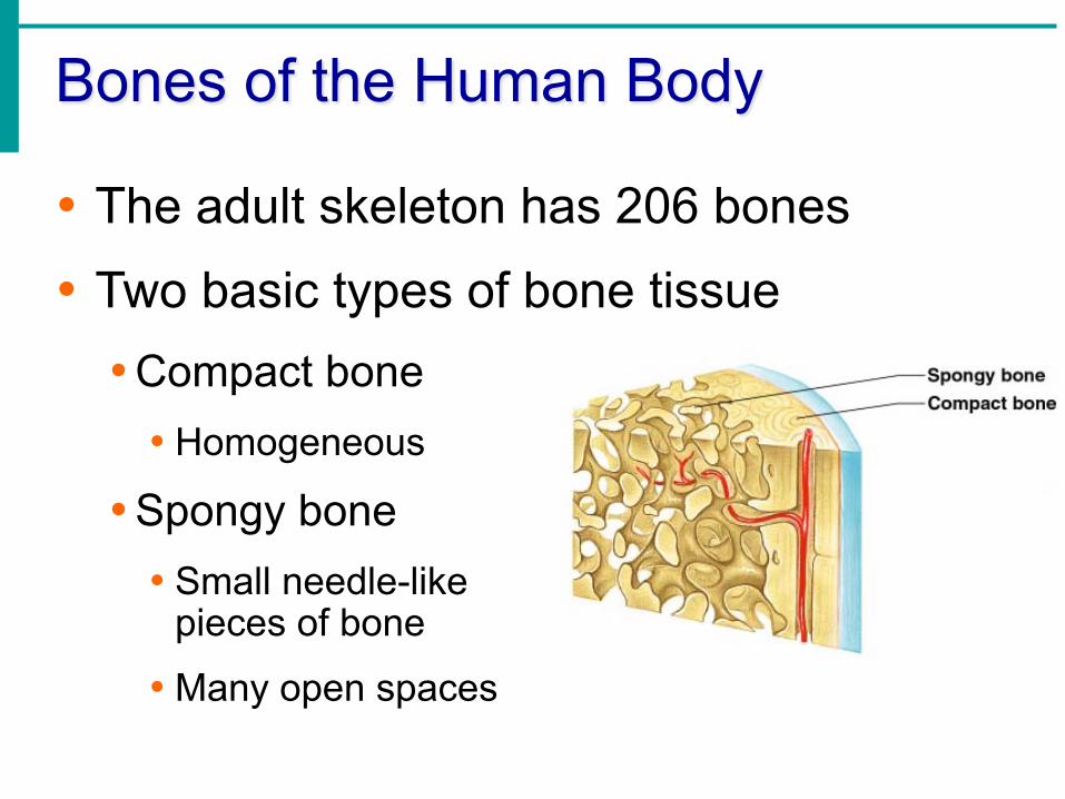
Bones of the Human Body
• The adult skeleton has 206 bones
• Two basic types of bone tissue
• Compact bone • Homogeneous
• Spongy bone • Small needle-like
pieces of bone
• Many open spaces

Classification of Bones on the Basis of Shape
Figure 5.1

Classification of Bones
• 1. Long bones
• Typically longer than wide
• Have a shaft with heads at both ends
• Contain mostly compact bone
• Examples: Femur, humerus

Classification of Bones
• 2. Short bones
• Generally cube-shape
• Contain mostly spongy bone
• Examples: Carpals, tarsals
Classification of Bones
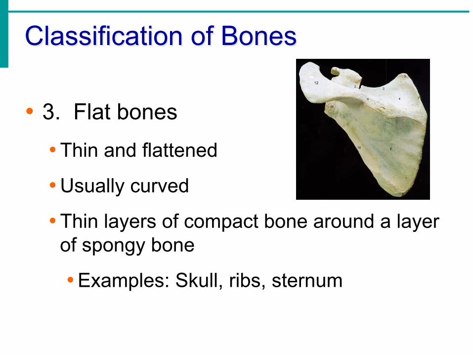
• 3. Flat bones
• Thin and flattened
• Usually curved
• Thin layers of compact bone around a layer of spongy bone
• Examples: Skull, ribs, sternum
Classification of Bones
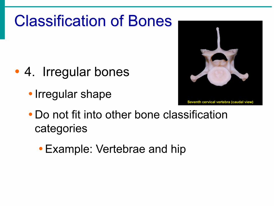
• 4. Irregular bones
• Irregular shape
• Do not fit into other bone classification categories
• Example: Vertebrae and hip
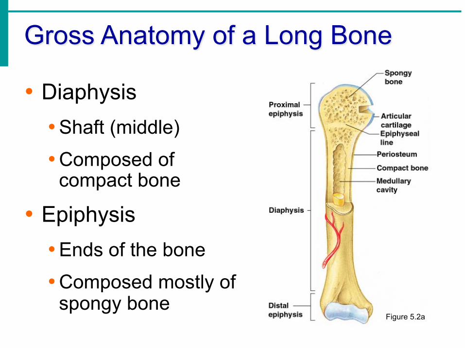
Gross Anatomy of a Long Bone
• Diaphysis
• Shaft (middle)
• Composed of compact bone
• Epiphysis
• Ends of the bone
• Composed mostly of spongy bone
Figure 5.2a
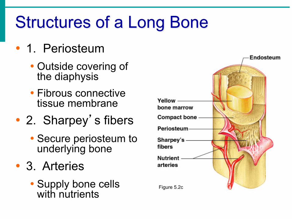
Structures of a Long Bone • 1. Periosteum
• Outside covering of the diaphysis
• Fibrous connective tissue membrane
• 2. Sharpey’s fibers • Secure periosteum to
underlying bone
• 3. Arteries • Supply bone cells
with nutrients Figure 5.2c
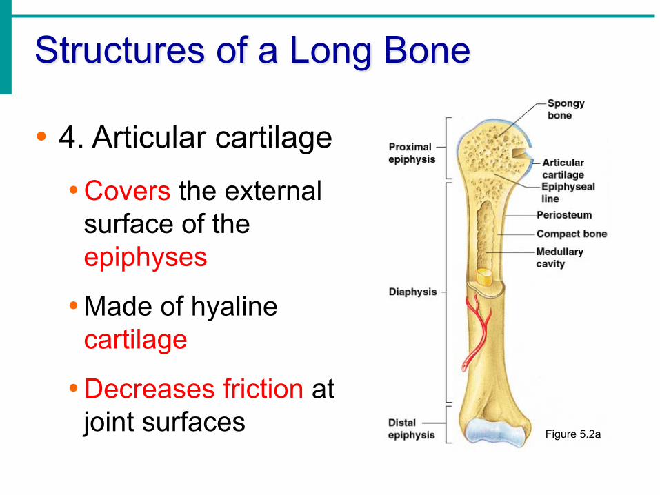
Structures of a Long Bone
• 4. Articular cartilage
• Covers the external surface of the epiphyses
• Made of hyaline cartilage
• Decreases friction at joint surfaces Figure 5.2a

Structures of a Long Bone
• 5. Medullary cavity
• Cavity of the shaft
• Contains yellow marrow (mostly fat) in adults
• Contains red marrow (for blood cell formation) in infants Figure 5.2a
Bone Markings
• Surface features of bones
• Sites of attachments for muscles, tendons, and ligaments
• Passages for nerves and blood vessels
• Categories of bone markings • Projections and processes – grow out from the
bone surface
• Depressions or cavities – indentations

Changes in the Human Skeleton
• In embryos, the skeleton is primarily hyaline cartilage
• During development, much of this cartilage is replaced by bone
• Cartilage remains in isolated areas • Bridge of the nose
• Parts of ribs
• Joints

Bone Growth
• Epiphyseal plates allow for growth of long bone during childhood
• New cartilage is continuously formed
• Older cartilage becomes ossified
• Cartilage is broken down
• Bone replaces cartilage

Bone Growth
• Bones are remodeled and lengthened until growth stops
• Bones change shape somewhat
• Bones grow in width

Long Bone Formation and Growth
Figure 5.4a

Types of Bone Cells • Osteocytes
• Mature bone cells
• Osteoblasts • Bone-forming cells
• Osteoclasts • Bone-destroying cells • Break down bone matrix for remodeling and release
of calcium

Bone Fractures
• A break in a bone • Types of bone fractures
• Closed (simple) fracture – break that does not penetrate the skin
• Open (compound) fracture – broken bone penetrates through the skin
• Bone fractures are treated by reduction and immobilization • Realignment of the bone

Common Types of Fractures
Table 5.2

Repair of Bone Fractures
• Hematoma (blood-filled swelling) is formed
• Break is splinted by fibrocartilage to form a callus
• Fibrocartilage callus is replaced by a bony callus
• Bony callus is remodeled to form a permanent patch
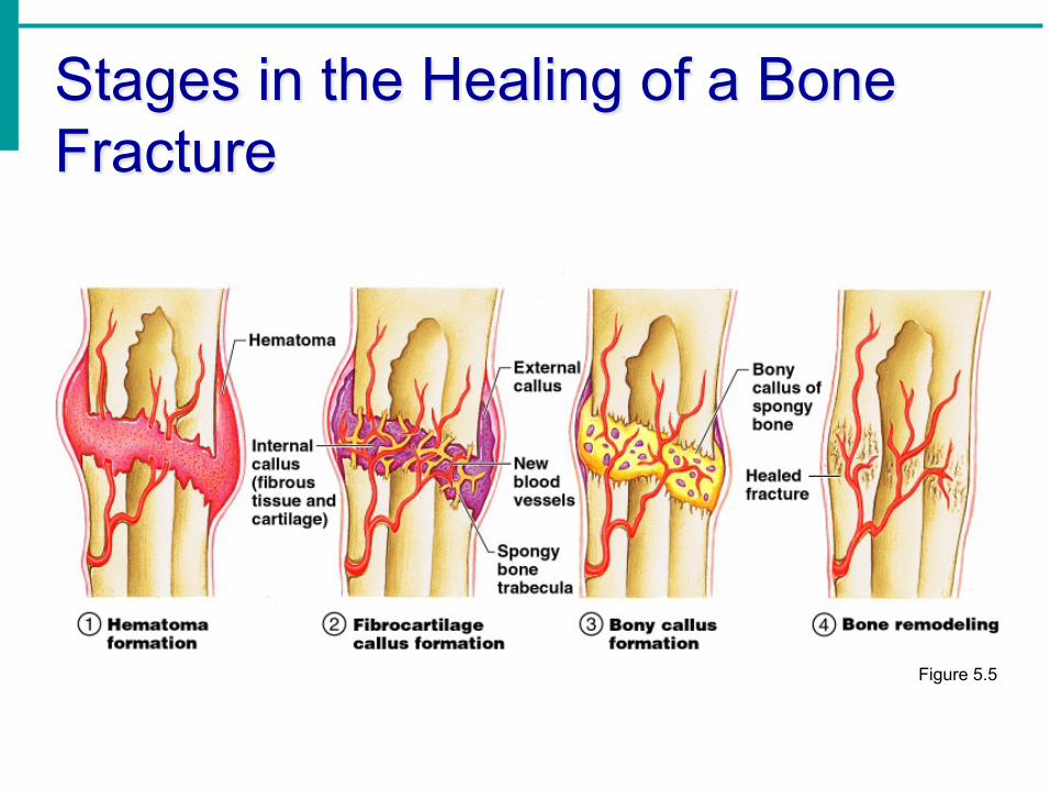
Stages in the Healing of a Bone Fracture
Figure 5.5

The Axial Skeleton • Divided into
three parts • Skull
• Vertebral column
• Bony thorax
The Skull
• Two sets of bones
• Cranium
• Facial bones
• Bones are joined by sutures
• Only the mandible is attached by a freely movable joint

The Skull
Figure 5.7

Bones of the Skull
Figure 5.11

Human Skull, Superior View
Figure 5.8
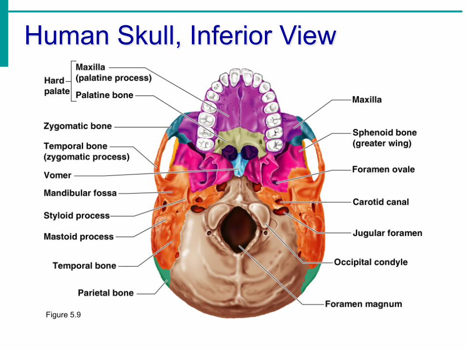
Human Skull, Inferior View
Figure 5.9

Paranasal Sinuses
• Hollow portions of bones surrounding the nasal cavity
Figure 5.10

Paranasal Sinuses
• Functions of paranasal sinuses • Lighten the skull • Give resonance and amplification to voice
Figure 5.10
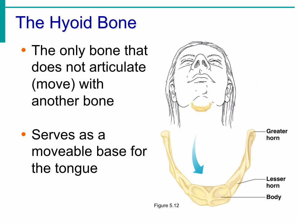
The Hyoid Bone • The only bone that
does not articulate (move) with another bone
• Serves as a moveable base for the tongue
Figure 5.12

The Fetal Skull
• The fetal skull is large compared to the infants total body length
Figure 5.13

The Fetal Skull
• Fontanelles – fibrous membranes connecting the cranial bones • Allow the brain
to grow
• Convert to bone within 24 months after birth
Figure 5.13


The Vertebral Column
• Vertebrae separated by intervertebral discs
• The spine has a normal curvature
• Each vertebrae is given a name according to its location Figure 5.14

The Bony Thorax
• Forms a cage to protect major organs
Figure 5.19a

The Bony Thorax
• Made-up of three parts
• Sternum
• Ribs
• Thoracic vertebrae
Figure 5.19a

The Appendicular Skeleton
• Limbs (appendages)
• Pectoral girdle
• Pelvic girdle

The Appendicular Skeleton
Figure 5.6c

The Pectoral (Shoulder) Girdle
• Composed of two bones
• Clavicle – collarbone
• Scapula – shoulder blade
• These bones allow the upper limb to have exceptionally free movement
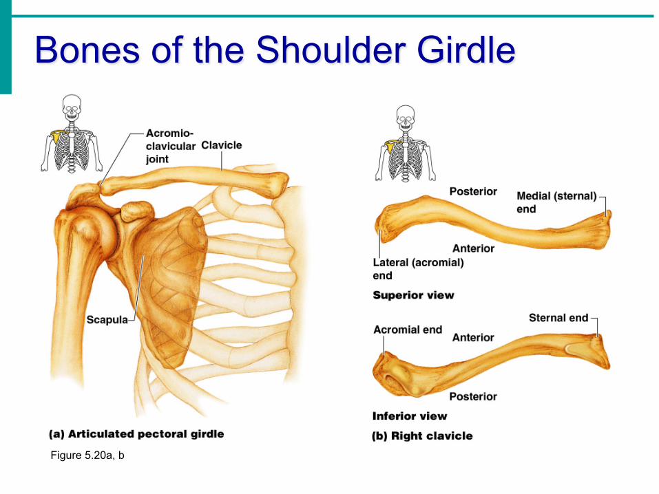
Bones of the Shoulder Girdle
Figure 5.20a, b

Bones of the Upper Limb
• The arm is formed by a single bone
• Humerus
Figure 5.21a, b

Bones of the Upper Limb
• The forearm has two bones
• Ulna
• Radius
Figure 5.21c

Bones of the Upper Limb
• The hand
• Carpals – wrist
• Metacarpals – palm
• Phalanges – fingers
Figure 5.22
Bones of the Pelvic Girdle • Hip bones • Composed of three pair of fused bones
• Ilium • Ischium • Pubic bone
• The total weight of the upper body rests on the pelvis
• Protects several organs • Reproductive organs • Urinary bladder • Part of the large intestine

The Pelvis
Figure 5.23a

Gender Differences of the Pelvis
Figure 5.23c


Bones of the Lower Limbs
• The thigh has one bone
• Femur – thigh bone
Figure 5.35a, b

Bones of the Lower Limbs
• The leg has two bones
• Tibia
• Fibula
Figure 5.35c

Bones of the Lower Limbs
• The foot
• Talus – ankle
• Metatarsals –
• Phalanges – toes
Figure 5.25

Joints
• Articulations of bones
• Functions of joints
• Hold bones together
• Allow for mobility
• Ways joints are classified
• Functionally
• Structurally

Functional Classification of Joints • Synarthroses –
immovable joints
• Amphiarthroses – slightly moveable joints
• Diarthroses – freely moveable joints

Structural Classification of Joints
• Fibrous joints
• Generally immovable
• Cartilaginous joints
• Immovable or slightly moveable
• Synovial joints
• Freely moveable

Fibrous Joints • Bones united by fibrous tissue –
synarthrosis or largely immovable.
Figure 5.27d, e

Cartilaginous Joints – mostly amphiarthrosis
• Bones connected by cartilage
• Examples
• Pubic symphysis
• Intervertebral joints
Figure 5.27b, c
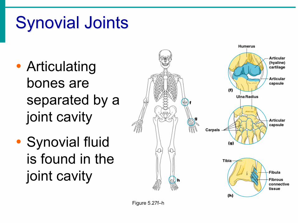
Synovial Joints
• Articulating bones are separated by a joint cavity
• Synovial fluid is found in the joint cavity
Figure 5.27f–h

Features of Synovial Joints- Diarthroses
• Articular cartilage (hyaline cartilage) covers the ends of bones
• Joint surfaces are enclosed by a fibrous articular capsule
• Have a joint cavity filled with synovial fluid
• Ligaments reinforce the joint
Structures Associated with the Synovial Joint • Bursae – flattened fibrous sacs
• Lined with synovial membranes
• Filled with synovial fluid
• Not actually part of the joint
• Tendon sheath
• Elongated bursa that wraps around a tendon

The Synovial Joint
Figure 5.28
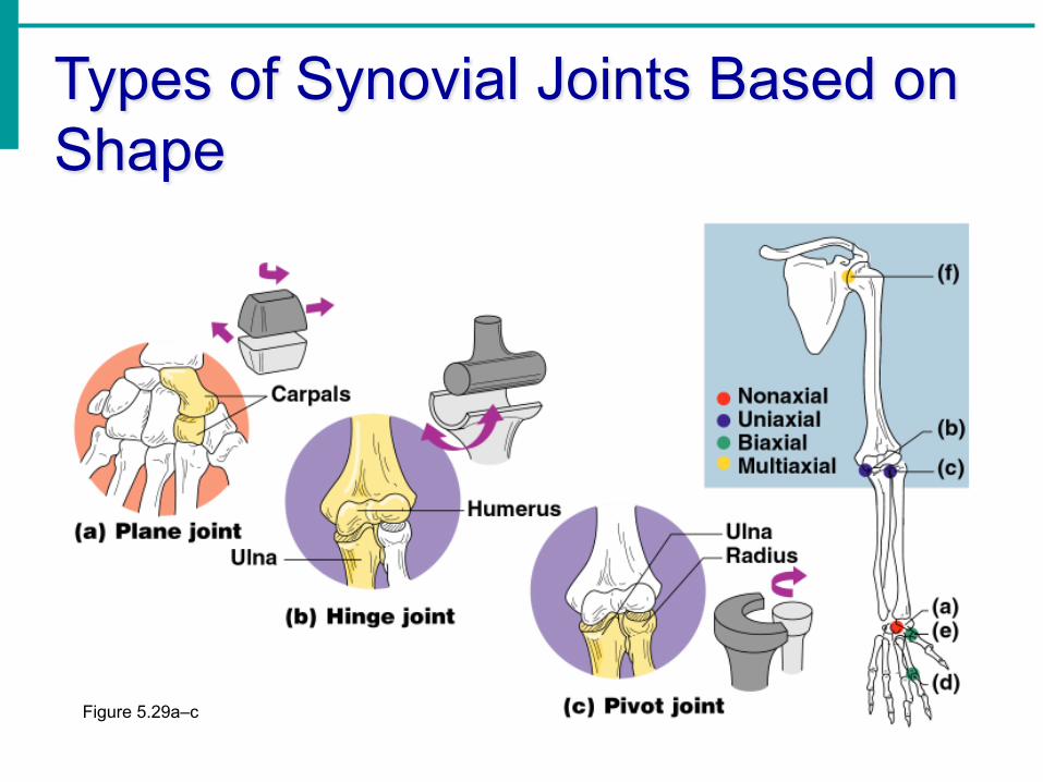
Types of Synovial Joints Based on Shape
Figure 5.29a–c

Types of Synovial Joints Based on Shape
Figure 5.29d–f

Inflammatory Conditions Associated with Joints • Bursitis – inflammation of a bursa usually
caused by a blow or friction
• Tendonitis – inflammation of tendon sheaths
• Arthritis – inflammatory or degenerative diseases of joints • Over 100 different types
• The most widespread crippling disease in the United States
Clinical Forms of Arthritis
• Osteoarthritis • Most common chronic arthritis
• Probably related to normal aging processes
• Rheumatoid arthritis • An autoimmune disease – the immune system
attacks the joints
• Symptoms begin with bilateral inflammation of certain joints
• Often leads to deformities
