skeletal system - divergent niche -...
TRANSCRIPT
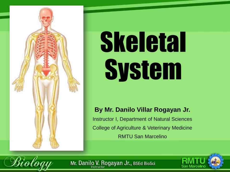
Skeletal System
By Mr. Danilo Villar Rogayan Jr.
Instructor I, Department of Natural Sciences
College of Agriculture & Veterinary Medicine
RMTU San Marcelino
Learning Outcomes
• Describe the parts of a long bone.
• List the substances that make up bone tissue.
• List the functions of bones.
• Identify bones by their classifications.
• Describe how long bones grow.
Learning Outcomes (cont.)
• List and describe the skeletal structures and one location of each structure.
• List the bones of the skull, spinal column, rib cage, shoulders, arms, hands, hips, legs, and feet. Describe the location of each bone.
• Define fontanels and explain their importance.
Learning Outcomes (cont.)
• Describe the three major types of joints and give examples of each.
• Describe the structure of a synovial joint.
• Describe the causes, signs and symptoms, and treatments of various diseases and disorders of the skeletal system.
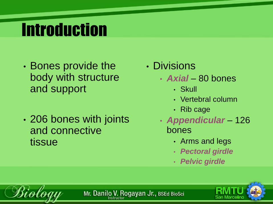
Introduction
• Bones provide the body with structure and support
• 206 bones with joints and connective tissue
• Divisions• Axial – 80 bones
• Skull
• Vertebral column
• Rib cage
• Appendicular – 126 bones
• Arms and legs
• Pectoral girdle
• Pelvic girdle
The Skeletal System
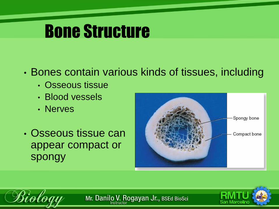
Bone Structure
• Bones contain various kinds of tissues, including• Osseous tissue
• Blood vessels
• Nerves
• Osseous tissue can appear compact or spongy
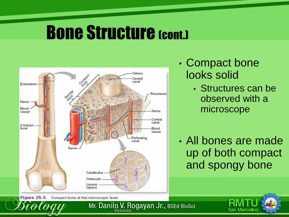
Bone Structure (cont.)
• Compact bone looks solid
• Structures can be observed with a microscope
• All bones are made up of both compact and spongy bone
Bone Structure (cont.)
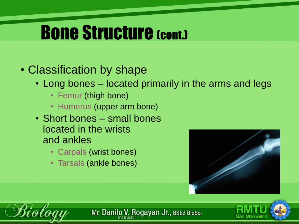
• Classification by shape• Long bones – located primarily in the arms and legs
• Femur (thigh bone)
• Humerus (upper arm bone)
• Short bones – small bones located in the wrists and ankles
• Carpals (wrist bones)
• Tarsals (ankle bones)
Bone Structure (cont.)
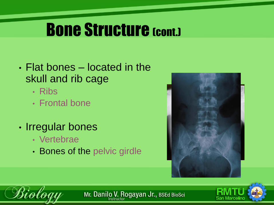
• Flat bones – located in the skull and rib cage
• Ribs
• Frontal bone
• Irregular bones• Vertebrae
• Bones of the pelvic girdle
Bone Structure: Gender Differences
• Male Skull• Larger and heavier
• Forehead shorter
• Face less round
• Jaw larger
• Mastoid processes more prominent
• Male pelvic bones• Heavier and thicker
• Obturator foramina and acetabula are larger and closer together
• Male pelvic cavity• Narrower and longer
• Less roomy and more funnel shaped
• Male sacrum• Narrower
• Sacral promontory projects forward
• Sacral curvature is less sharp posteriorly
• Male coccyx• Less movable
Bone Structure: Gender Differences (cont.)
Functions of Bones
• Give shape to body parts
• Support and protect soft structures
• Examples – brain, lungs, heart
• Allow body movement, because skeletal muscles attach to them
• Allow for voluntary movement
Functions of Bones (cont.)
• Red bone marrow of
bone produces new
blood cells –
hematopoiesis
• Store calcium
Checkpoint
ANSWER: Every cell in the body needs calcium, so the body must have a large supply readily available.
Why is it important for the bones to store calcium?
Bone Growth
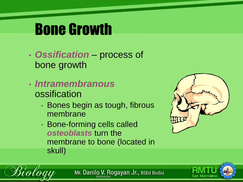
• Ossification – process of bone growth
• Intramembranousossification
• Bones begin as tough, fibrous membrane
• Bone-forming cells called osteoblasts turn the membrane to bone (located in skull)
Bone Growth (cont.)
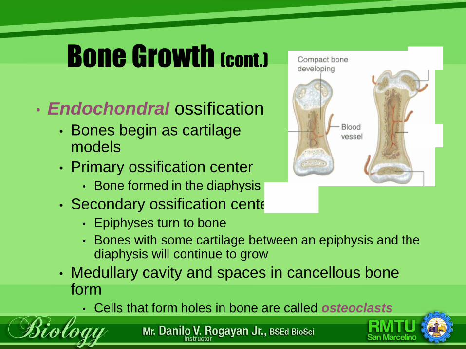
• Endochondral ossification • Bones begin as cartilage
models
• Primary ossification center• Bone formed in the diaphysis
• Secondary ossification center• Epiphyses turn to bone
• Bones with some cartilage between an epiphysis and the diaphysis will continue to grow
• Medullary cavity and spaces in cancellous bone form
• Cells that form holes in bone are called osteoclasts
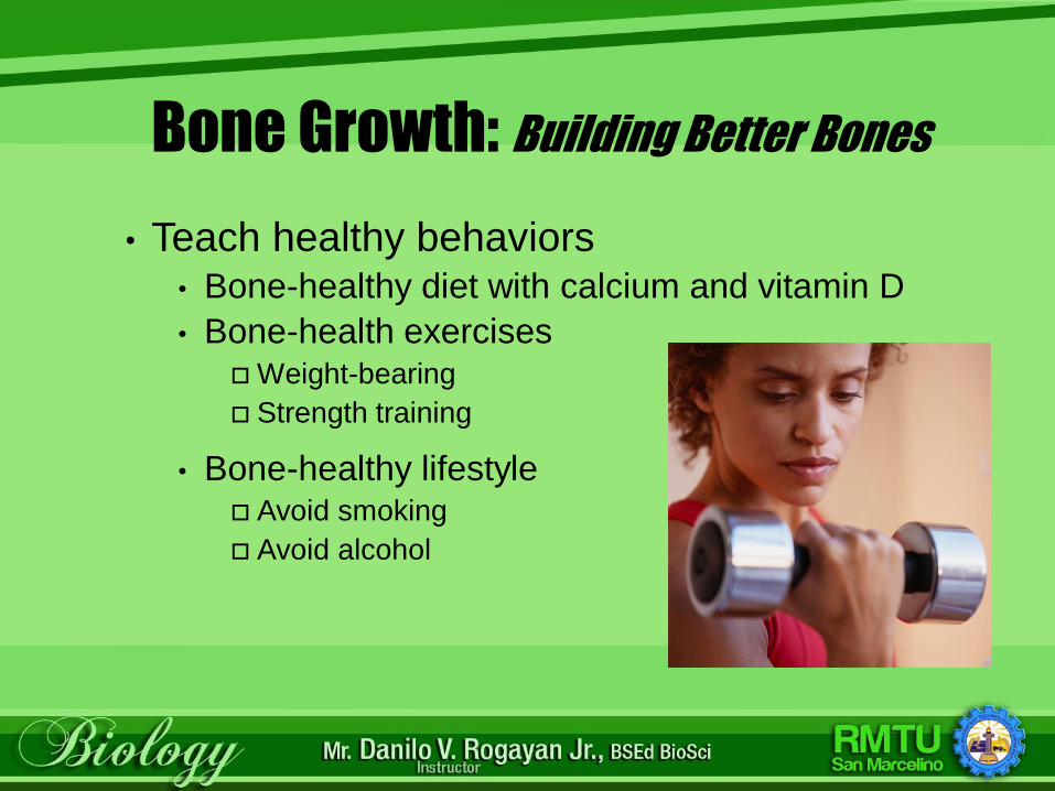
Bone Growth: Building Better Bones
• Teach healthy behaviors• Bone-healthy diet with calcium and vitamin D
• Bone-health exercises Weight-bearing
Strength training
• Bone-healthy lifestyle Avoid smoking
Avoid alcohol
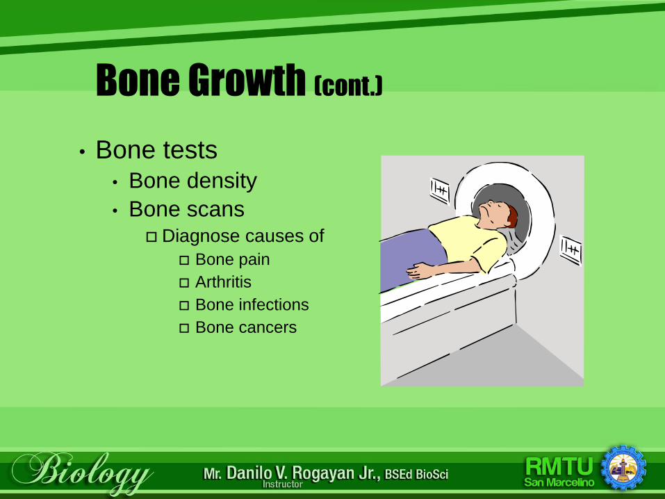
Bone Growth (cont.)
• Bone tests• Bone density
• Bone scans Diagnose causes of
Bone pain
Arthritis
Bone infections
Bone cancers
Checkpoint
What are the two types of bone growth?
ANSWER: Intramembranous ossification, in which
bones begin as tough membrane and are turned to
bone by osteoblasts, and endochondral ossification,
in which primary ossification occurs in the diaphysis
of the of the bone and secondary ossification occurs
in the epiphysis.

Bony Structures
• Rigid foundation
• Projections and processes for muscle and ligament attachment
• Depressions and hollows for articulations –the connection of bones at joints
• Openings for blood vessels and nerves
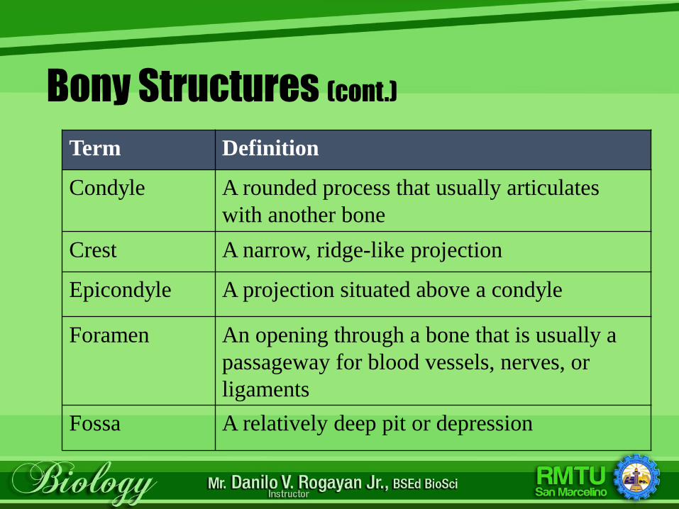
Bony Structures (cont.)
Term Definition
Condyle A rounded process that usually articulates
with another bone
Crest A narrow, ridge-like projection
Epicondyle A projection situated above a condyle
Foramen An opening through a bone that is usually a
passageway for blood vessels, nerves, or
ligaments
Fossa A relatively deep pit or depression
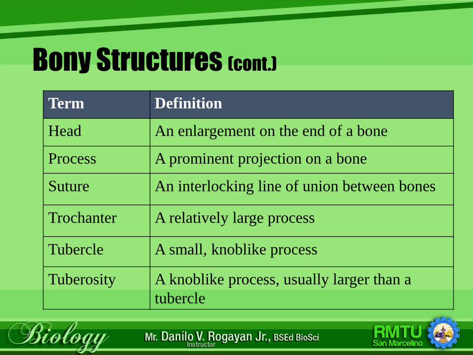
Bony Structures (cont.)
Term Definition
Head An enlargement on the end of a bone
Process A prominent projection on a bone
Suture An interlocking line of union between bones
Trochanter A relatively large process
Tubercle A small, knoblike process
Tuberosity A knoblike process, usually larger than a
tubercle
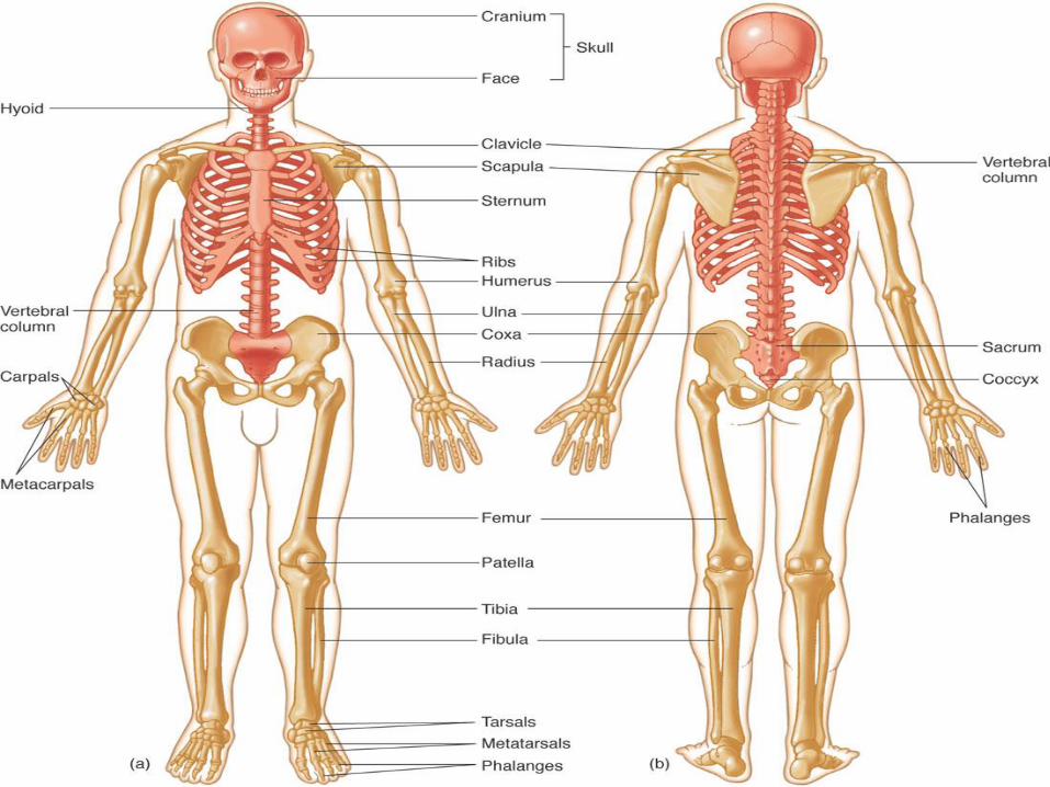
The Skull
• Two bone types: • Cranial – form the top, sides, and back of the skull
• Facial – form the face
“Soft spots” felt on an infant's skull are
actually fontanels
Tough membranes that connect the
incompletely developed bones
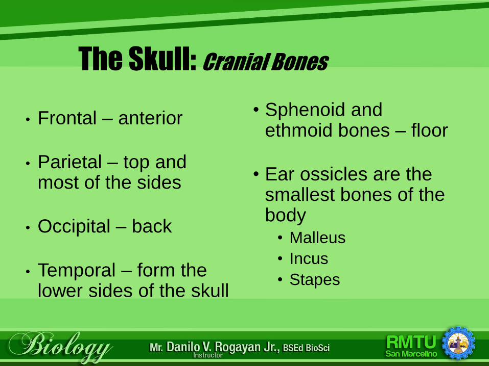
The Skull: Cranial Bones
• Frontal – anterior
• Parietal – top and most of the sides
• Occipital – back
• Temporal – form the lower sides of the skull
• Sphenoid and ethmoid bones – floor
• Ear ossicles are the smallest bones of the body
• Malleus
• Incus
• Stapes
The Skull (cont.)
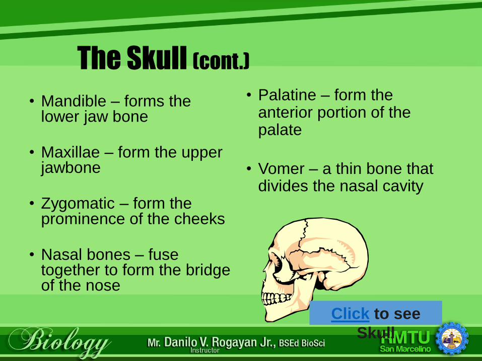
• Mandible – forms the lower jaw bone
• Maxillae – form the upper jawbone
• Zygomatic – form the prominence of the cheeks
• Nasal bones – fuse together to form the bridge of the nose
Click to see
Skull
• Palatine – form the anterior portion of the palate
• Vomer – a thin bone that divides the nasal cavity
The Spinal Column
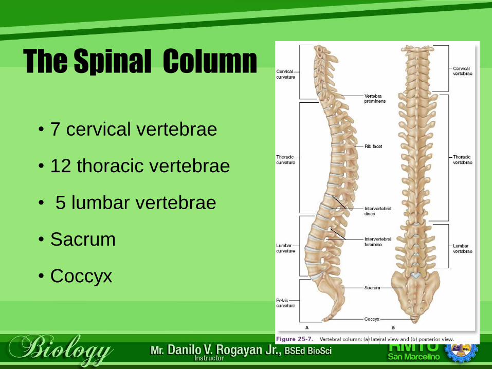
• 7 cervical vertebrae
• 12 thoracic vertebrae
• 5 lumbar vertebrae
• Sacrum
• Coccyx
The Spinal Column (cont.)
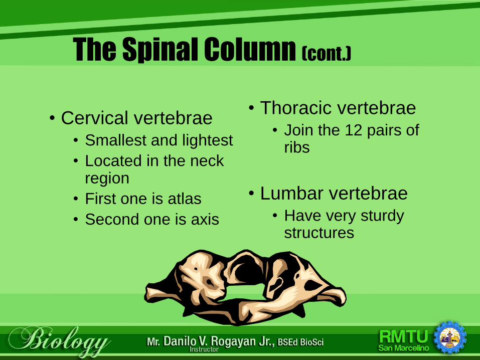
• Cervical vertebrae • Smallest and lightest
• Located in the neck region
• First one is atlas
• Second one is axis
• Thoracic vertebrae • Join the 12 pairs of
ribs
• Lumbar vertebrae • Have very sturdy
structures
The Spinal Column (cont.)
• Sacrum • A triangular-shaped bone that consists of five fused
vertebrae
• Coccyx • A small, triangular-shaped bone made up of 3 to 5
fused vertebrae
• Considered unnecessary
• More commonly called the tailbone
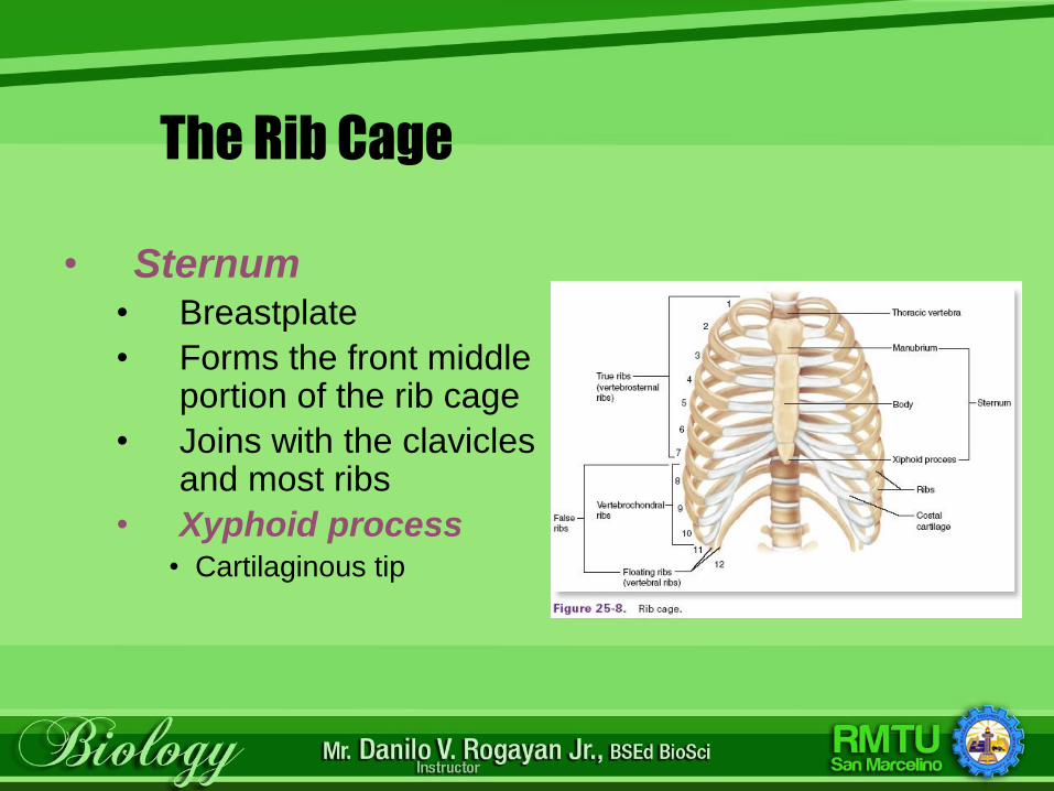
The Rib Cage
• Sternum• Breastplate
• Forms the front middle portion of the rib cage
• Joins with the clavicles and most ribs
• Xyphoid process• Cartilaginous tip
The Rib Cage (cont.)
• 12 pairs of ribs• All are attached
posteriorly to thoracic vertebrae
• True• First seven pairs of ribs
• Attach to sternum by costal cartilage
• False• Rib pairs 8, 9, and 10
• Attach to the costal cartilage of rib pair 7
• Floating• Rib pairs 11 and 12
• Do not attach anteriorly to any structure
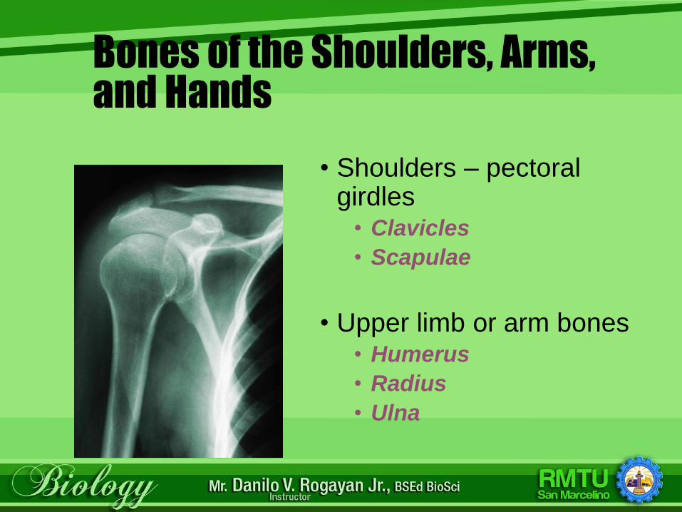
Bones of the Shoulders, Arms, and Hands
• Shoulders – pectoral girdles
• Clavicles
• Scapulae
• Upper limb or arm bones • Humerus
• Radius
• Ulna
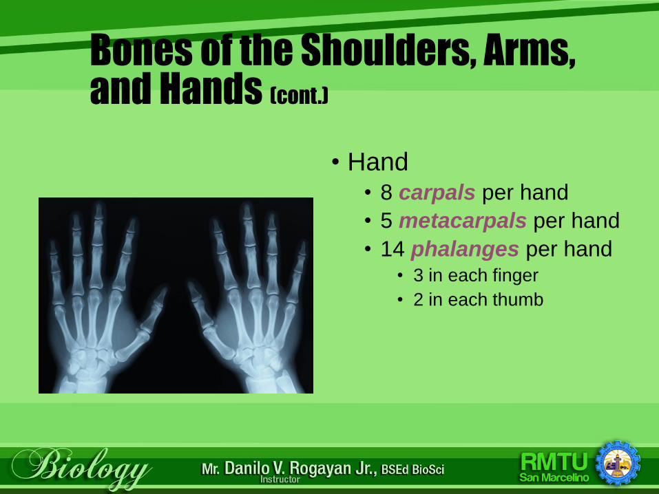
Bones of the Shoulders, Arms, and Hands (cont.)
• Hand • 8 carpals per hand
• 5 metacarpals per hand
• 14 phalanges per hand• 3 in each finger
• 2 in each thumb
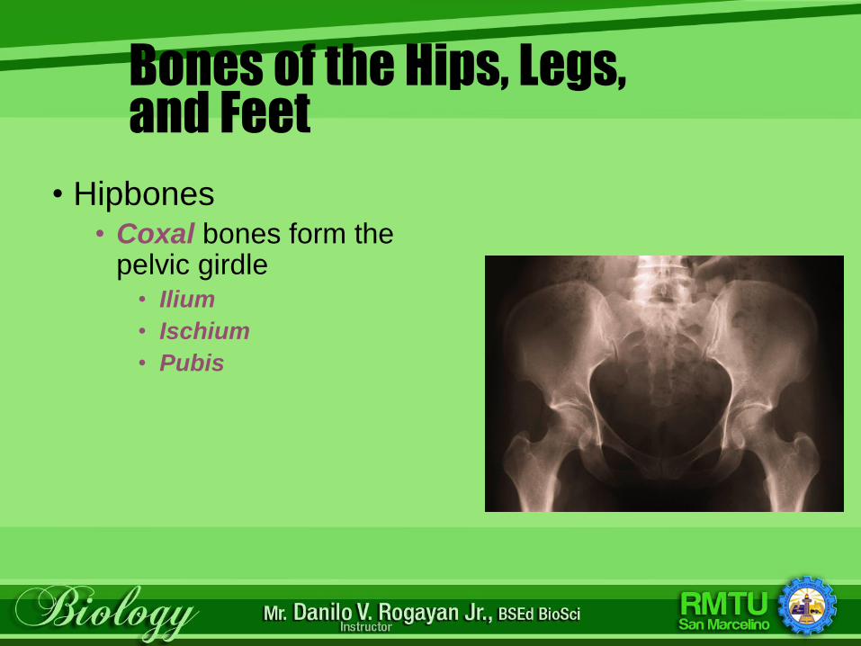
Bones of the Hips, Legs, and Feet
• Hipbones • Coxal bones form the
pelvic girdle• Ilium
• Ischium
• Pubis
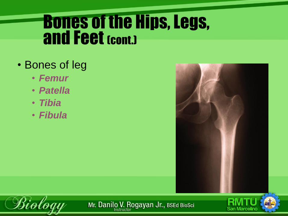
Bones of the Hips, Legs, and Feet (cont.)
• Bones of leg • Femur
• Patella
• Tibia
• Fibula
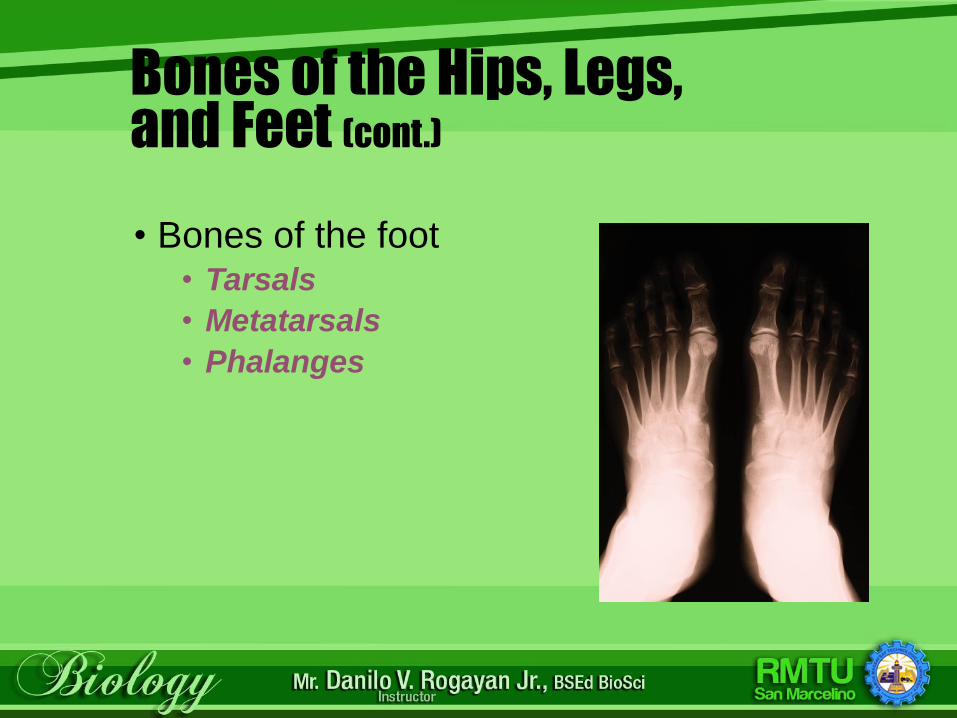
Bones of the Hips, Legs, and Feet (cont.)
• Bones of the foot • Tarsals
• Metatarsals
• Phalanges
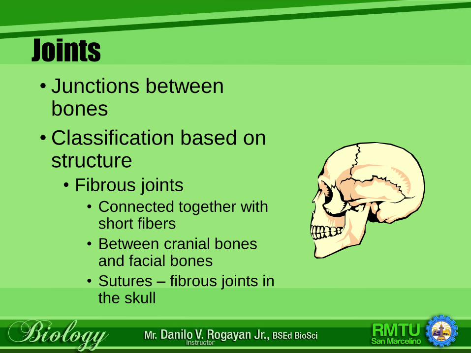
Joints• Junctions between
bones
• Classification based on structure
• Fibrous joints • Connected together with
short fibers
• Between cranial bones and facial bones
• Sutures – fibrous joints in the skull
Joints
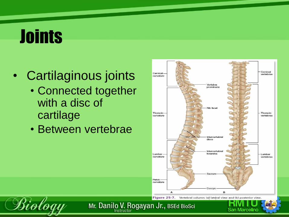
• Cartilaginous joints• Connected together
with a disc of cartilage
• Between vertebrae
Joints
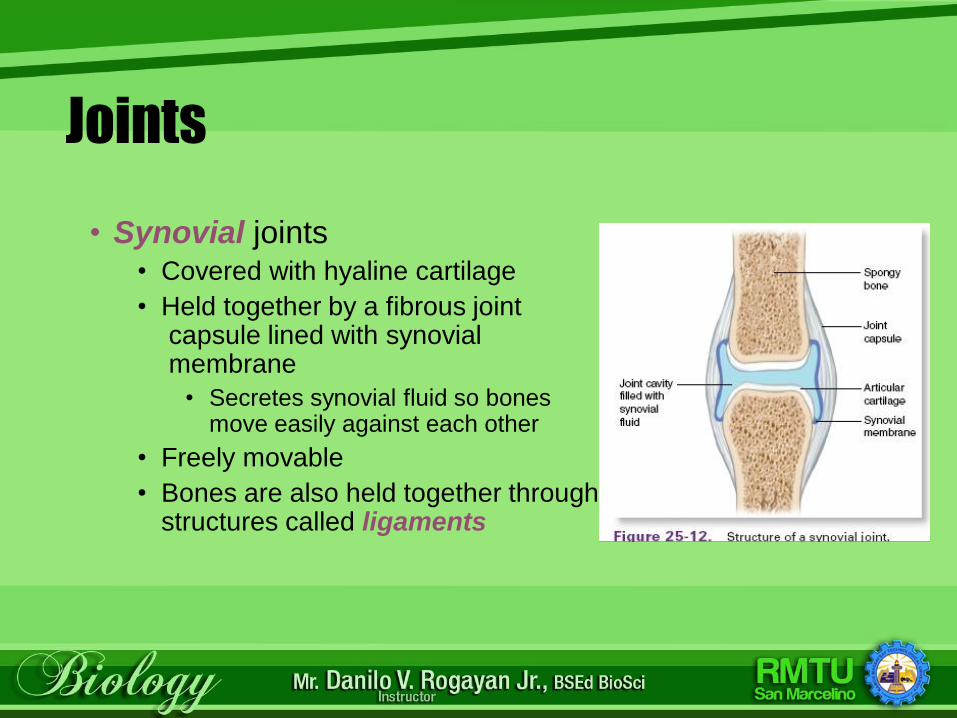
• Synovial joints • Covered with hyaline cartilage
• Held together by a fibrous jointcapsule lined with synovialmembrane
• Secretes synovial fluid so bones move easily against each other
• Freely movable
• Bones are also held together through tough, cord-like structures called ligaments
Common Diseases and Disorders
• Arthritis – general term meaning joint inflammation• Osteoarthritis – degenerative joint
disease, primarily of weight-bearing joints
• Rheumatoid Arthritis – chronic systemic inflammatory disease of smaller joints and surrounding tissues
Common Diseases and Disorders (cont.)
• Bursitis – inflammation of a bursa (fluid-filled sac that cushions tendons)
• Carpal Tunnel Syndrome – overuse of wrist; the median nerve in the wrist becomes compressed
• Ewing’s Family of Tumors (EFT) – a group of tumors that affect different tissue types; primarily bone
• Gout – a type of arthritis; deposits of uric acid crystals in the joints
Common Diseases and Disorders (cont.)
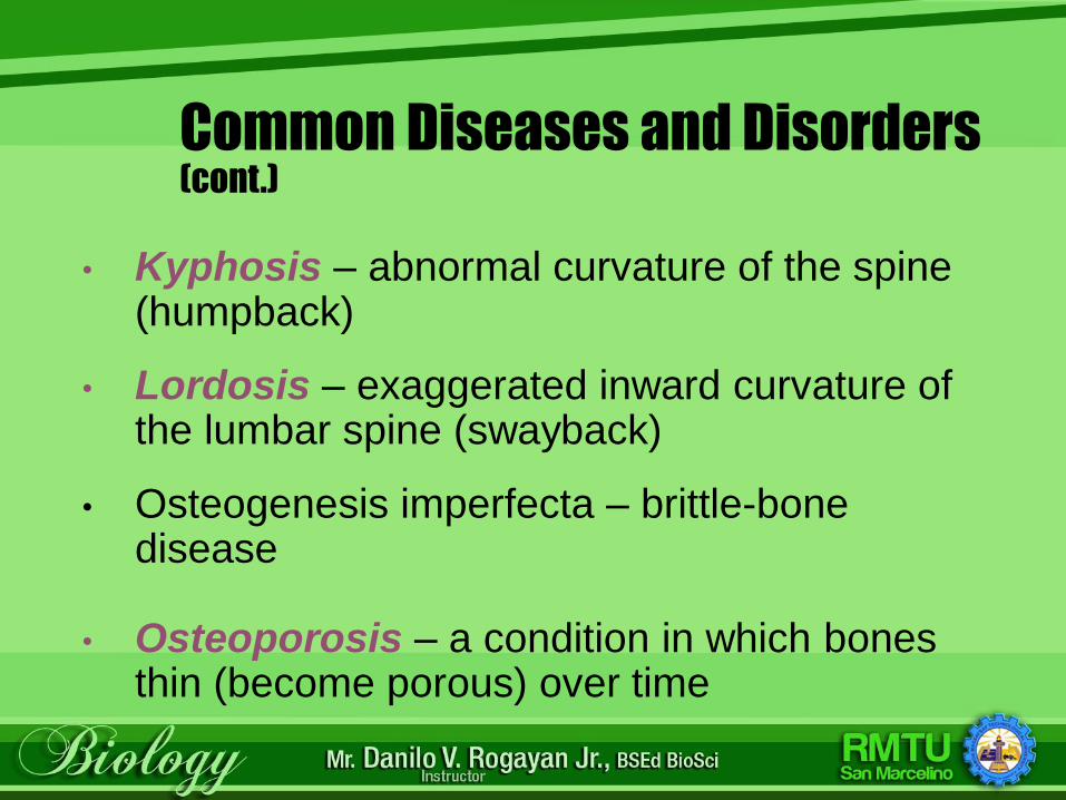
• Kyphosis – abnormal curvature of the spine (humpback)
• Lordosis – exaggerated inward curvature of the lumbar spine (swayback)
• Osteogenesis imperfecta – brittle-bone disease
• Osteoporosis – a condition in which bones thin (become porous) over time

Disorders
Kyphosis Lordosis
Common Diseases and Disorders (cont.)
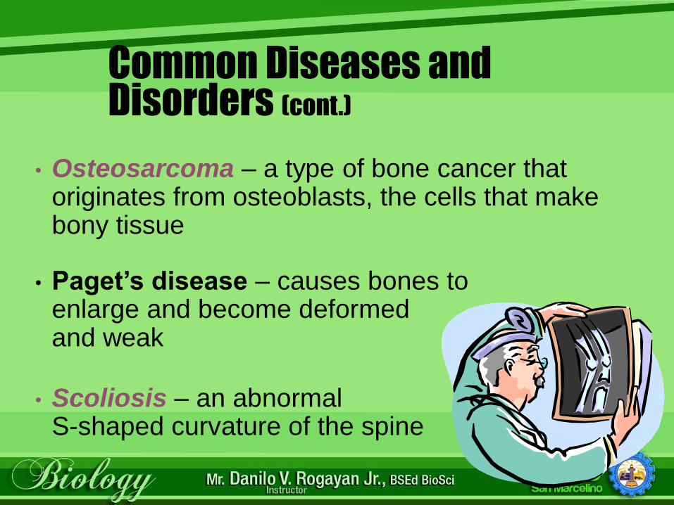
• Osteosarcoma – a type of bone cancer that originates from osteoblasts, the cells that make bony tissue
• Paget’s disease – causes bones to enlarge and become deformed and weak
• Scoliosis – an abnormal S-shaped curvature of the spine
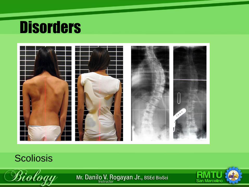
Disorders
Scoliosis
Checkpoint
Osteosarcoma is a type of bone cancer that originates from osteoblasts, the cells that make bony tissue.
The doctor has told your patient that he has an osteosarcoma. What do you know about this
disorder?
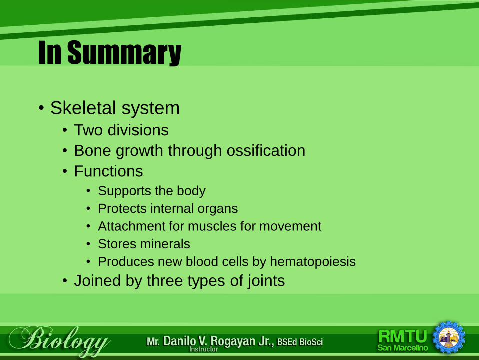
In Summary
• Skeletal system• Two divisions
• Bone growth through ossification
• Functions• Supports the body
• Protects internal organs
• Attachment for muscles for movement
• Stores minerals
• Produces new blood cells by hematopoiesis
• Joined by three types of joints
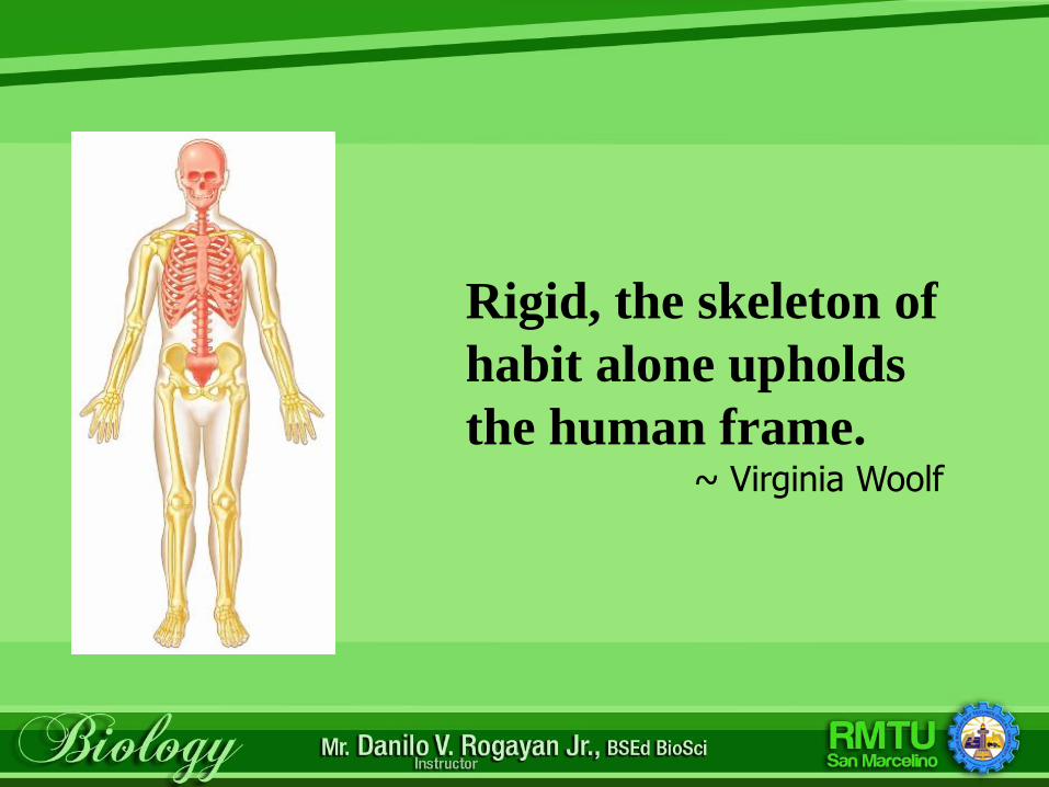
Rigid, the skeleton of
habit alone upholds
the human frame.~ Virginia Woolf
Reference:
Ramutkowski, Booth, Pugh. Thompson and
Whicker. (2015). Medical Assisting.
McGrawHill Companies, Inc.

Lakô hã salamát!Maraming salamat!