rig-i & cancer immunotherapy - invivogen · rig-i & cancer immunotherapy. may 2019. summary...
TRANSCRIPT
The development of immune checkpoint inhibitors (ICIs) has revolutionized cancer immunotherapy, although complete remission remains limited to a small panel of cancers and patients. ICIs act by relieving checkpoint restraints on antitumor T cell responses. They work best against immunogenic, T-cell inflamed or « hot » tumors. In contrast, ICIs are poorly efficient in «cold» tumor microenvironments (TMEs) that are largely devoid of T cells and infiltrated by immunosuppressive cells. In «hot» TMEs, increased expression of type I interferons (IFN-I) and IFN-stimulated genes (ISGs), such as apoptosis-inducing molecules and T-cell attracting chemokines, contribute to potent antitumor responses. Many therapeutic strategies are actively being explored to transform «cold» TMEs into «hot» ones. One emerging strategy exploits the adjuvancity of pattern recognition receptor (PRR) agonists. Indeed, combinations of ICIs with agonists of Toll-like receptor 9 (TLR9) or stimulator of interferon genes (STING) have reached clinical evaluation but have so far yielded disappointing preliminary results1.
Retinoic acid-inducible gene I (RIG-I), a viral RNA sensor, is a promising alternative to enhance ICI efficacy2,3. RIG-I is the best-known member of the RIG-I-like helicase receptor (RLR) family. Unlike TLR9 and STING, RIG-I is expressed in virtually all cell types, including tumor cells. Preclinical studies have shown that systemic delivery of a synthetic RIG-I agonist inhibits tumor growth through mechanisms similar to those triggered for elimination of virally-infected cells3. RIG-I engagement leads to preferential tumor cell death (via intrinsic or extrinsic apoptosis, and inflammasome-induced pyroptosis), and to IFN-I-mediated activation of the innate and adaptive immune systems4. RGT100, a specific RIG-I agonist, is currently in phase I/II clinical trials for treatment of advanced solid tumors and lymphomas (NCT03065023)4.
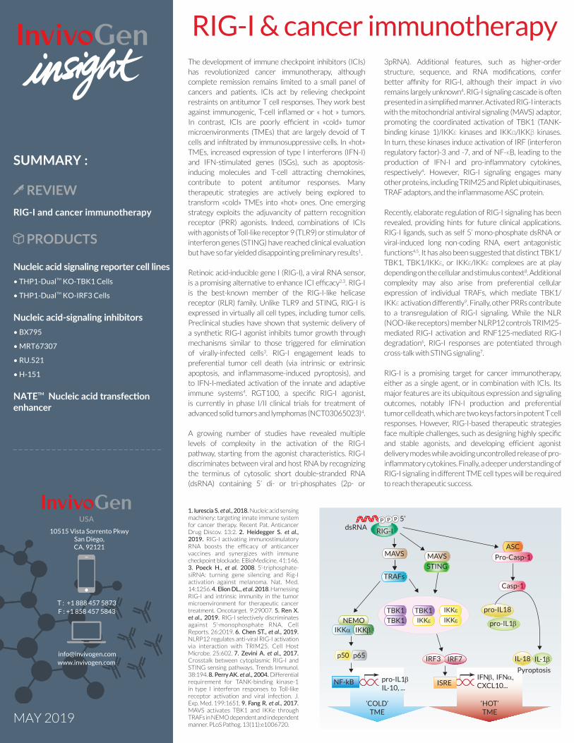
A growing number of studies have revealed multiple levels of complexity in the activation of the RIG-I pathway, starting from the agonist characteristics. RIG-I discriminates between viral and host RNA by recognizing the terminus of cytosolic short double-stranded RNA (dsRNA) containing 5’ di- or tri-phosphates (2p- or
3pRNA). Additional features, such as higher-order structure, sequence, and RNA modifications, confer better affinity for RIG-I, although their impact in vivo remains largely unknown4. RIG-I signaling cascade is often presented in a simplified manner. Activated RIG-I interacts with the mitochondrial antiviral signaling (MAVS) adaptor, promoting the coordinated activation of TBK1 (TANK-binding kinase 1)/IKKε kinases and IKKα/IKKβ kinases. In turn, these kinases induce activation of IRF (interferon regulatory factor)-3 and -7, and of NF-κB, leading to the production of IFN-I and pro-inflammatory cytokines, respectively4. However, RIG-I signaling engages many other proteins, including TRIM25 and Riplet ubiquitinases, TRAF adaptors, and the inflammasome ASC protein..
Recently, elaborate regulation of RIG-I signaling has been revealed, providing hints for future clinical applications. RIG-I ligands, such as self 5’ mono-phosphate dsRNA or viral-induced long non-coding RNA, exert antagonistic functions4,5. It has also been suggested that distinct TBK1/TBK1, TBK1/IKKε, or IKKε/IKKε complexes are at play depending on the cellular and stimulus context8. Additional complexity may also arise from preferential cellular expression of individual TRAFs, which mediate TBK1/IKKε activation differently9. Finally, other PRRs contribute to a transregulation of RIG-I signaling. While the NLR (NOD-like receptors) member NLRP12 controls TRIM25-mediated RIG-I activation and RNF125-mediated RIG-I degradation6, RIG-I responses are potentiated through cross-talk with STING signaling7.
RIG-I is a promising target for cancer immunotherapy, either as a single agent, or in combination with ICIs. Its major features are its ubiquitous expression and signaling outcomes, notably IFN-I production and preferential tumor cell death, which are two keys factors in potent T cell responses. However, RIG-I-based therapeutic strategies face multiple challenges, such as designing highly specific and stable agonists, and developing efficient agonist delivery modes while avoiding uncontrolled release of pro-inflammatory cytokines. Finally, a deeper understanding of RIG-I signaling in different TME cell types will be required to reach therapeutic success.
RIG-I & cancer immunotherapy
MAY 2019
SUMMARY :
REVIEWRIG-I and cancer immunotherapy
PRODUCTS
Nucleic acid signaling reporter cell lines• THP1-DualTM KO-TBK1 Cells
• THP1-DualTM KO-IRF3 Cells
Nucleic acid-signaling inhibitors• BX795
• MRT67307
• RU.521
• H-151
NATETM Nucleic acid transfection enhancer
1. Iurescia S. et al., 2018. Nucleic acid sensingmachinery: targeting innate immune system for cancer therapy. Recent Pat. AnticancerDrug Discov. 13:2. 2. Heidegger S. et al., 2019. RIG-I activating immunostimulatoryRNA boosts the efficacy of anticancervaccines and synergizes with immunecheckpoint blockade. EBioMedicine. 41:146. 3. Poeck H., et al. 2008. 5’-triphosphate-siRNA: turning gene silencing and Rig-Iactivation against melanoma. Nat. Med.14:1256. 4. Elion DL., et al. 2018. HarnessingRIG-I and intrinsic immunity in the tumormicroenvironment for therapeutic cancertreatment. Oncotarget. 9:29007. 5. Ren X.et al., 2019. RIG-I selectively discriminatesagainst 5’-monophosphate RNA. CellReports. 26:2019. 6. Chen ST., et al., 2019. NLRP12 regulates anti-viral RIG-I activation via interaction with TRIM25. Cell HostMicrobe. 25:602. 7. Zevini A. et al., 2017.Crosstalk between cytoplasmic RIG-I andSTING sensing pathways. Trends Immunol.38:194. 8. Perry AK. et al., 2004. Differentialrequirement for TANK-binding kinase-1in type I interferon responses to Toll-likereceptor activation and viral infection. J.Exp. Med. 199:1651. 9. Fang R. et al., 2017. MAVS activates TBK1 and IKKe throughTRAFs in NEMO dependent and independentmanner. PLoS Pathog. 13(11):e1006720.
IFNβ, IFNα,CXCL10...
IRF7
RIG-I
NF-kB ISRE
P P P
dsRNA
STING
NEMO
pro-IL1βIL-10, ...
IL-18 IL-1β
pro-IL1β
Pyroptosis
‘COLD’TME
‘HOT’TME
MAVSASC
Pro-Casp-1
Casp-1
pro-IL18
MAVS
TRAFs
IKKα IKKβ
p50 p65 IRF3
TBK1
TBK1 IKKεIKKε
IKKεTBK1
5’USA10515 Vista Sorrento Pkwy
San Diego, CA, 92121
T : +1 888 457 5873F : +1 858 457 5843
[email protected] www.invivogen.com
M
Nucleic acid signaling reporter cell lines
• THP-1 DualTM KO-TBK1 Cells NEW
• THP-1 DualTM KO-IRF3 Cells NEW
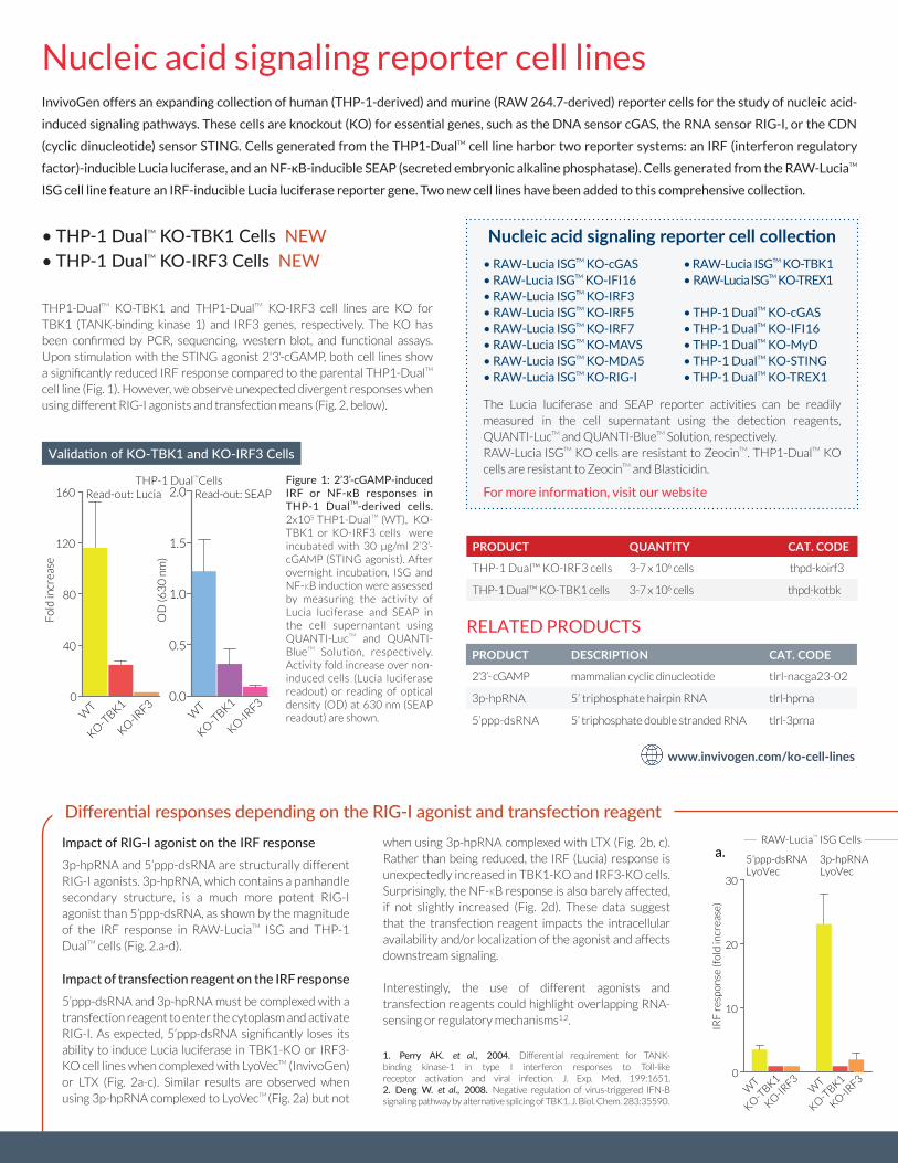
Impact of RIG-I agonist on the IRF response3p-hpRNA and 5’ppp-dsRNA are structurally different RIG-I agonists. 3p-hpRNA, which contains a panhandle secondary structure, is a much more potent RIG-I agonist than 5’ppp-dsRNA, as shown by the magnitude of the IRF response in RAW-LuciaTM ISG and THP-1 DualTM cells (Fig. 2.a-d).
Impact of transfection reagent on the IRF response5’ppp-dsRNA and 3p-hpRNA must be complexed with a transfection reagent to enter the cytoplasm and activate RIG-I. As expected, 5’ppp-dsRNA significantly loses its ability to induce Lucia luciferase in TBK1-KO or IRF3-KO cell lines when complexed with LyoVecTM (InvivoGen) or LTX (Fig. 2a-c). Similar results are observed when using 3p-hpRNA complexed to LyoVecTM (Fig. 2a) but not
when using 3p-hpRNA complexed with LTX (Fig. 2b, c). Rather than being reduced, the IRF (Lucia) response is unexpectedly increased in TBK1-KO and IRF3-KO cells. Surprisingly, the NF-κB response is also barely affected, if not slightly increased (Fig. 2d). These data suggest that the transfection reagent impacts the intracellular availability and/or localization of the agonist and affects downstream signaling.
Interestingly, the use of different agonists and transfection reagents could highlight overlapping RNA-sensing or regulatory mechanisms1,2.
1. Perry AK. et al., 2004. Differential requirement for TANK-binding kinase-1 in type I interferon responses to Toll-likereceptor activation and viral infection. J. Exp. Med. 199:1651.2. Deng W. et al., 2008. Negative regulation of virus-triggered IFN-Bsignaling pathway by alternative splicing of TBK1. J. Biol. Chem. 283:35590.
Differential responses depending on the RIG-I agonist and transfection reagent
www.invivogen.com/ko-cell-lines
PRODUCT QUANTITY CAT. CODE
THP-1 Dual™ KO-IRF3 cells 3-7 x 10⁶ cells thpd-koirf3
THP-1 Dual™ KO-TBK1 cells 3-7 x 10⁶ cells thpd-kotbk
RELATED PRODUCTS
PRODUCT DESCRIPTION CAT. CODE
2’3’- cGAMP mammalian cyclic dinucleotide tlrl-nacga23-02
3p-hpRNA 5’ triphosphate hairpin RNA tlrl-hprna
5’ppp-dsRNA 5’ triphosphate double stranded RNA tlrl-3prna
InvivoGen offers an expanding collection of human (THP-1-derived) and murine (RAW 264.7-derived) reporter cells for the study of nucleic acid-
induced signaling pathways. These cells are knockout (KO) for essential genes, such as the DNA sensor cGAS, the RNA sensor RIG-I, or the CDN
(cyclic dinucleotide) sensor STING. Cells generated from the THP1-DualTM cell line harbor two reporter systems: an IRF (interferon regulatory
factor)-inducible Lucia luciferase, and an NF-κB-inducible SEAP (secreted embryonic alkaline phosphatase). Cells generated from the RAW-LuciaTM
ISG cell line feature an IRF-inducible Lucia luciferase reporter gene. Two new cell lines have been added to this comprehensive collection.
Fol
din
crea
se
Read-out: Lucia
0
40
80
120
160
0.0
0.5
1.0
1.5
2.0
0
5
10100
200
300
400
Fol
d in
crea
se
OD
(63
0 n
m)
LuciaTM IS
G
KO-T
BK1
KO-IR
F3W
T
KO-T
BK1
KO-IR
F3
Read-out: Lucia Read-out: SEAP
KO-T
BK1
KO-IR
F3
RAW Cells THP-1 DualTM
Cells
WT
Validation of KO-TBK1 and KO-IRF3 Cells
Figure 1: 2’3’-cGAMP-induced IRF or NF-κB responses in THP-1 DualTM-derived cells. 2x10⁵ THP1-DualTM (WT), KO-TBK1 or KO-IRF3 cells were incubated with 30 µg/ml 2’3’-cGAMP (STING agonist). After overnight incubation, ISG and NF-κB induction were assessed by measuring the activity of Lucia luciferase and SEAP in the cell supernantant using QUANTI-LucTM and QUANTI-BlueTM Solution, respectively. Activity fold increase over non-induced cells (Lucia luciferase readout) or reading of optical density (OD) at 630 nm (SEAP readout) are shown.
5’ppp-dsRNA
WT
KO-T
BK1
KO-IR
F3
LTX3p-hpRNALTX
0
20
40
100
200
300
400
WT
KO-T
BK1
KO-IR
F3W
T
KO-T
BK1
KO-IR
F3
5’ppp-dsRNALTX
3p-hpRNALTX
0.0
0.5
1.0
1.5
NF
-κB
res
pon
se (O
D6
30
nm)
WT
KO-T
BK1
KO-IR
F3
5’ppp-dsRNALTX
3p-hpRNALTX
THP-1 DualTM
Cells
0
10
20
30
IRF
res
pon
se (f
old
incr
ease
)
5’ppp-dsRNALyoVec
3p-hpRNALyoVec
a. b. c. d.
KO-T
BK1
KO-IR
F3
KO-T
BK1
KO-IR
F3W
T0
250
500
750
1000
1500
2000
2500
KO-T
BK1
KO-IR
F3
KO-T
BK1
KO-IR
F3
RAW-LuciaTM
ISG Cells
WT
WT
WT
IRF
res
pon
se (f
old
incr
ease
)
THP1-DualTM KO-TBK1 and THP1-DualTM KO-IRF3 cell lines are KO for TBK1 (TANK-binding kinase 1) and IRF3 genes, respectively. The KO has been confirmed by PCR, sequencing, western blot, and functional assays. Upon stimulation with the STING agonist 2'3'-cGAMP, both cell lines show a significantly reduced IRF response compared to the parental THP1-DualTM
cell line (Fig. 1). However, we observe unexpected divergent responses when using different RIG-I agonists and transfection means (Fig. 2, below). The Lucia luciferase and SEAP reporter activities can be readily
measured in the cell supernatant using the detection reagents, QUANTI-LucTM and QUANTI-BlueTM Solution, respectively.RAW-Lucia ISGTM KO cells are resistant to ZeocinTM. THP1-DualTM KO cells are resistant to ZeocinTM and Blasticidin.
For more information, visit our website
• RAW-Lucia ISGTM KO-cGAS• RAW-Lucia ISGTM KO-IFI16• RAW-Lucia ISGTM KO-IRF3• RAW-Lucia ISGTM KO-IRF5• RAW-Lucia ISGTM KO-IRF7• RAW-Lucia ISGTM KO-MAVS• RAW-Lucia ISGTM KO-MDA5• RAW-Lucia ISGTM KO-RIG-I
Nucleic acid signaling reporter cell collection• RAW-Lucia ISGTM KO-TBK1• RAW-Lucia ISGTM KO-TREX1
• THP-1 DualTM KO-cGAS• THP-1 DualTM KO-IFI16• THP-1 DualTM KO-MyD• THP-1 DualTM KO-STING• THP-1 DualTM KO-TREX1
M
PRODUCT TARGET QUANTITY CAT. CODE
BX795 TBK1/IKKε 5 mg tlrl-bx7
H-151 STING 10 mg inh-h151
MRT67307 TBK1/IKKε 10 mg inh-mrt
RU.521 cGAS 2 mg inh-ru521
Nucleic acid signaling inhibitors
www.invivogen.com/prr-signaling-inhibitors
InvivoGen offers an extensive collection of high quality synthetic
inhibitors, including molecules targeting the nucleic acid-induced
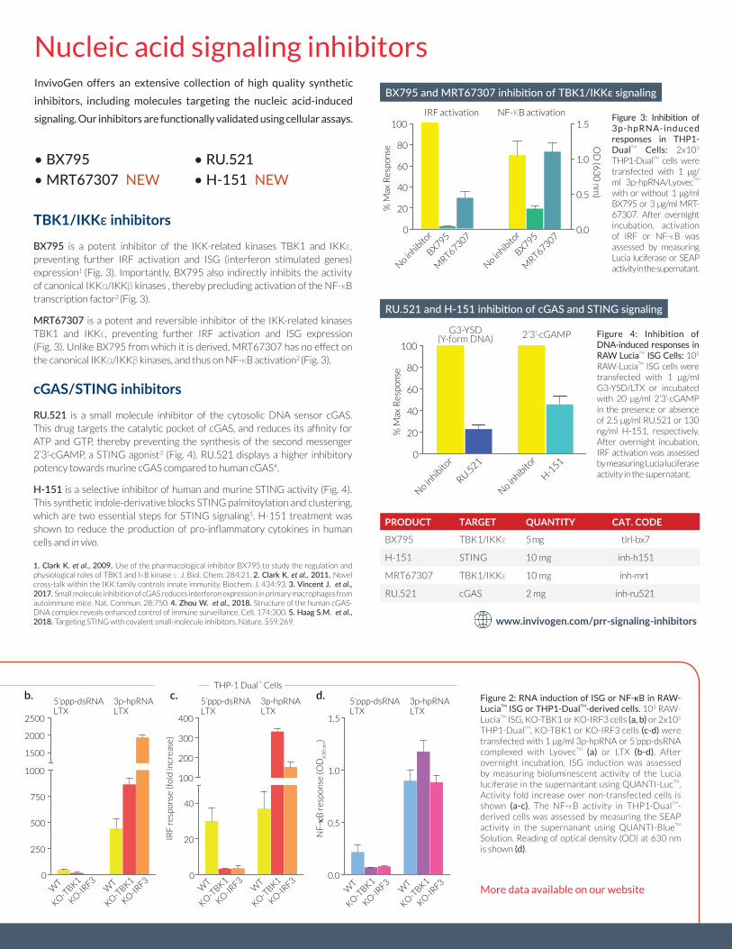
signaling. Our inhibitors are functionally validated using cellular assays. Figure 3: Inhibition of 3p-hpRNA-induced responses in THP1- DualTM Cells: 2x10⁵ THP1-DualTM cells were transfected with 1 µg/ml 3p-hpRNA/LyovecTM with or without 1 µg/ml BX795 or 3 µg/ml MRT-67307. After overnight incubation, activation of IRF or NF-κB was assessed by measuring Lucia luciferase or SEAP activity in the supernatant.
Figure 2: RNA induction of ISG or NF-κB in RAW-LuciaTM ISG or THP1-DualTM-derived cells. 105 RAW-LuciaTM ISG, KO-TBK1 or KO-IRF3 cells (a, b) or 2x10⁵ THP1-DualTM, KO-TBK1 or KO-IRF3 cells (c-d) were transfected with 1 µg/ml 3p-hpRNA or 5’ppp-dsRNA complexed with LyovecTM (a) or LTX (b-d). After overnight incubation, ISG induction was assessed by measuring bioluminescent activity of the Lucia luciferase in the supernantant using QUANTI-LucTM. Activity fold increase over non-transfected cells is shown (a-c). The NF-κB activity in THP1-DualTM-derived cells was assessed by measuring the SEAP activity in the supernanant using QUANTI-BlueTM Solution. Reading of optical density (OD) at 630 nm is shown (d).
• BX795• MRT67307 NEW
• RU.521• H-151 NEW
TBK1/IKKε inhibitors BX795 is a potent inhibitor of the IKK-related kinases TBK1 and IKKε, preventing further IRF activation and ISG (interferon stimulated genes) expression1 (Fig. 3). Importantly, BX795 also indirectly inhibits the activity of canonical IKKα/IKKβ kinases , thereby precluding activation of the NF-κB transcription factor2 (Fig. 3).
MRT67307 is a potent and reversible inhibitor of the IKK-related kinasesTBK1 and IKKε, preventing further IRF activation and ISG expression (Fig. 3). Unlike BX795 from which it is derived, MRT67307 has no effect on the canonical IKKα/IKKβ kinases, and thus on NF-κB activation2 (Fig. 3).
cGAS/STING inhibitorsRU.521 is a small molecule inhibitor of the cytosolic DNA sensor cGAS.This drug targets the catalytic pocket of cGAS, and reduces its affinity for ATP and GTP, thereby preventing the synthesis of the second messenger 2’3’-cGAMP, a STING agonist3 (Fig. 4). RU.521 displays a higher inhibitory potency towards murine cGAS compared to human cGAS4.
H-151 is a selective inhibitor of human and murine STING activity (Fig. 4).This synthetic indole-derivative blocks STING palmitoylation and clustering, which are two essential steps for STING signaling5. H-151 treatment wasshown to reduce the production of pro-inflammatory cytokines in human cells and in vivo.
1. Clark K. et al., 2009. Use of the pharmacological inhibitor BX795 to study the regulation andphysiological roles of TBK1 and IκB kinase ε. J. Biol. Chem. 284:21. 2. Clark K. et al., 2011. Novelcross-talk within the IKK family controls innate immunity. Biochem. J. 434:93. 3. Vincent J. et al., 2017. Small molecule inhibition of cGAS reduces interferon expression in primary macrophages fromautoimmune mice. Nat. Commun. 28:750. 4. Zhou W. et al., 2018. Structure of the human cGAS-DNA complex reveals enhanced control of immune surveillance. Cell. 174:300. 5. Haag S.M. et al., 2018. Targeting STING with covalent small-molecule inhibitors. Nature. 559:269.
5’ppp-dsRNA
WT
KO-T
BK1
KO-IR
F3
LTX3p-hpRNALTX
0
20
40
100
200
300
400
WT
KO-T
BK1
KO-IR
F3W
T
KO-T
BK1
KO-IR
F3
5’ppp-dsRNALTX
3p-hpRNALTX
0.0
0.5
1.0
1.5
NF
-κB
res
pon
se (O
D6
30
nm
)
WT
KO-T
BK1
KO-IR
F3
5’ppp-dsRNALTX
3p-hpRNALTX
THP-1 DualTM
Cells
0
10
20
30
IRF
res
pon
se (f
old
incr
ease
)
5’ppp-dsRNALyoVec
3p-hpRNALyoVec
a. b. c. d.
KO-T
BK1
KO-IR
F3
KO-T
BK1
KO-IR
F3W
T0
250
500
750
1000
1500
2000
2500
KO-T
BK1
KO-IR
F3
KO-T
BK1
KO-IR
F3
RAW-LuciaTM
ISG Cells
WT
WT
WT
IRF
res
pon
se (f
old
incr
ease
)
0
20
40
60
80
100
0.0
0.5
1.0
1.5
% M
ax R
espo
nse O
D (6
30
nm)
No inhib
itor
BX795
MRT67307
IRF activation NF-ΚB activation
No inhib
itor
BX795
MRT67307
BX795 and MRT67307 inhibition of TBK1/IKKε signaling
Figure 4: Inhibition of DNA-induced responses in RAW LuciaTM ISG Cells: 105 RAW-LuciaTM ISG cells were transfected with 1 µg/ml G3-YSD/LTX or incubated with 20 µg/ml 2’3’-cGAMP in the presence or absence of 2.5 µg/ml RU.521 or 130 ng/ml H-151, respectively. After overnight incubation, IRF activation was assessed by measuring Lucia luciferase activity in the supernatant.
% M
ax R
espo
nse
No inhib
itor
RU.521
0
20
40
60
80
100
G3-YSD(Y-form DNA) 2’3’-cGAMP
No inhib
itor
H-151
RU.521 and H-151 inhibition of cGAS and STING signaling
More data available on our website
Europe Tel: +33 562 71 69 39 Fax: +33 562 71 69 30 [email protected] Tel: +1 888 457 5873 Fax: +1 858 457 5843 [email protected] Tel: +852 3622 3480 Fax: +852 3622 3483 [email protected]
Europe Tel: +33 562 71 69 39 Fax: +33 562 71 69 30 [email protected] Tel: +1 888 457 5873 Fax: +1 858 457 5843 [email protected] Tel: +852 3622 3480 Fax: +852 3622 3483 [email protected]
PRODUCT QUANTITY CAT. CODE
NATE™ 1 ml (~100 reactions) lyec-nate
www.invivogen.com/nate
NATE™
Flexible: Can be used with all common transfection methods
Easy to use: Simply add to the cells prior to transfection
Efficient: Increases transfection yield in hard-to-transfect cells
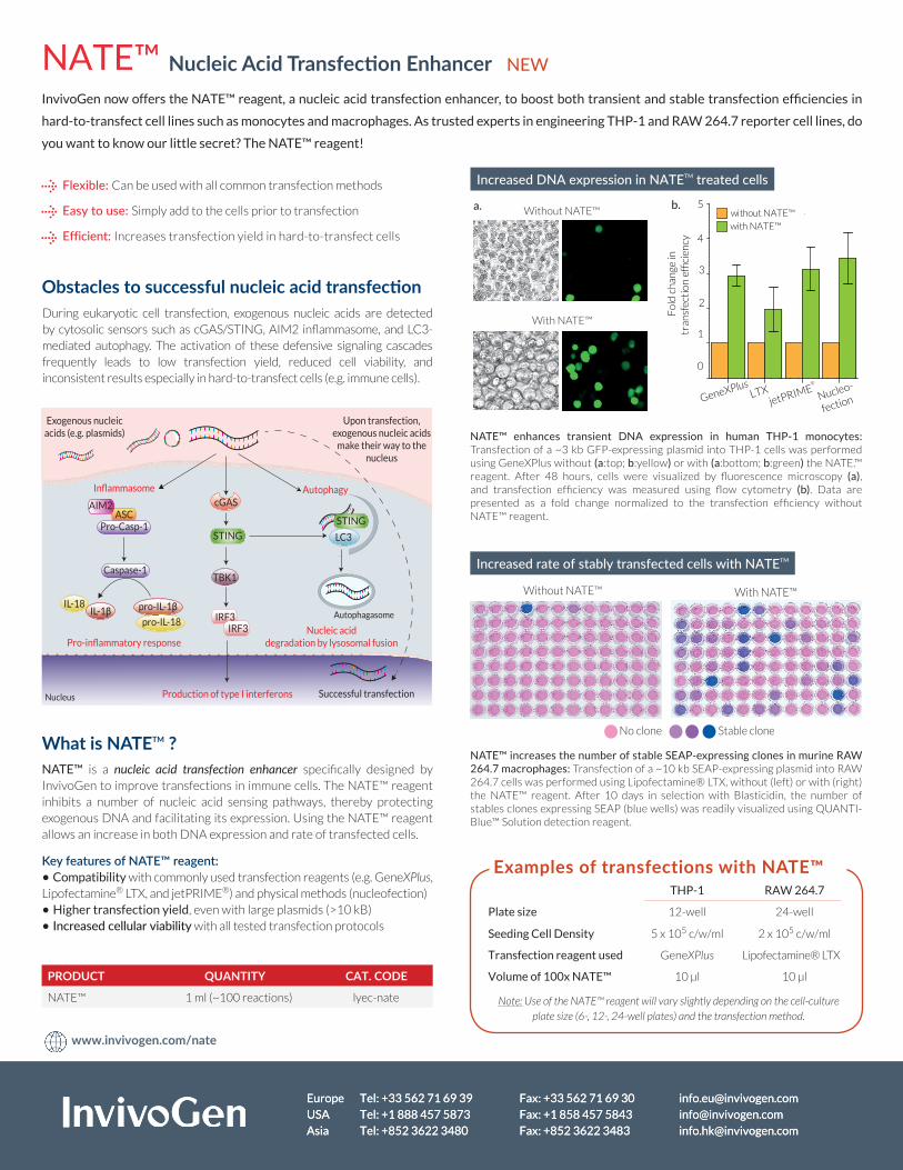
During eukaryotic cell transfection, exogenous nucleic acids are detected by cytosolic sensors such as cGAS/STING, AIM2 inflammasome, and LC3-mediated autophagy. The activation of these defensive signaling cascades frequently leads to low transfection yield, reduced cell viability, and inconsistent results especially in hard-to-transfect cells (e.g. immune cells).
InvivoGen now offers the NATE™ reagent, a nucleic acid transfection enhancer, to boost both transient and stable transfection efficiencies in
hard-to-transfect cell lines such as monocytes and macrophages. As trusted experts in engineering THP-1 and RAW 264.7 reporter cell lines, do
you want to know our little secret? The NATE™ reagent!
NATE™ enhances transient DNA expression in human THP-1 monocytes: Transfection of a ~3 kb GFP-expressing plasmid into THP-1 cells was performed using GeneXPlus without (a:top; b:yellow) or with (a:bottom; b:green) the NATE.™ reagent. After 48 hours, cells were visualized by fluorescence microscopy (a), and transfection efficiency was measured using flow cytometry (b). Data are presented as a fold change normalized to the transfection efficiency without NATE™ reagent.
Without NATE™ With NATE™
No clone Stable clone
NATE™ increases the number of stable SEAP-expressing clones in murine RAW 264.7 macrophages: Transfection of a ~10 kb SEAP-expressing plasmid into RAW 264.7 cells was performed using Lipofectamine® LTX, without (left) or with (right) the NATE™ reagent. After 10 days in selection with Blasticidin, the number of stables clones expressing SEAP (blue wells) was readily visualized using QUANTI-Blue™ Solution detection reagent.
IRF3
cGAS
TBK1
ASCPro-Casp-1
Caspase-1
IL-1β pro-IL-1β
LC3
Production of type I interferons
Pro-inflammatory response
Autophagasome
Nucleic acid degradation by lysosomal fusion
Exogenous nucleic acids (e.g. plasmids)
AutophagyInflammasome
Upon transfection, exogenous nucleic acids
make their way to the nucleus
Successful transfectionNucleus
STING
IRF3
AIM2
IL-18
pro-IL-18
STING
Nucleic Acid Transfection Enhancer NEW
Increased DNA expression in NATETM treated cells
Increased rate of stably transfected cells with NATETM
Without NATE™
With NATE™
0
1
2
3
4
5
Fol
d ch
ange
intr
ansf
ecti
on e
ffici
ency
GeneXPlusLTX
jetPRIME®
Nucleo-
fection
with NATE™without NATE™
a. b.
Examples of transfections with NATE™THP-1 RAW 264.7
Plate size 12-well 24-well
Seeding Cell Density 5 x 10⁵ c/w/ml 2 x 10⁵ c/w/ml
Transfection reagent used GeneXPlus Lipofectamine® LTX
Volume of 100x NATE™ 10 µl 10 µl
Note: Use of the NATE™ reagent will vary slightly depending on the cell‑culture plate size (6‑, 12‑, 24‑well plates) and the transfection method.
Obstacles to successful nucleic acid transfection
NATE™ is a nucleic acid transfection enhancer specifically designed by InvivoGen to improve transfections in immune cells. The NATE™ reagent inhibits a number of nucleic acid sensing pathways, thereby protecting exogenous DNA and facilitating its expression. Using the NATE™ reagent allows an increase in both DNA expression and rate of transfected cells.
Key features of NATE™ reagent:• Compatibility with commonly used transfection reagents (e.g. GeneXPlus, Lipofectamine® LTX, and jetPRIME®) and physical methods (nucleofection)• Higher transfection yield, even with large plasmids (>10 kB)• Increased cellular viability with all tested transfection protocols
What is NATETM ?