review article cytokines and chemokines as regulators of
TRANSCRIPT

Hindawi Publishing CorporationMediators of InflammationVolume 2013, Article ID 540370, 10 pageshttp://dx.doi.org/10.1155/2013/540370
Review ArticleCytokines and Chemokines as Regulators ofSkeletal Muscle Inflammation: Presenting the Case ofDuchenne Muscular Dystrophy
Boel De Paepe and Jan L. De Bleecker
Laboratory for Myopathology, Department of Neurology and Neuromuscular Reference Center,Ghent University Hospital, De Pintelaan 185, 9000 Ghent, Belgium
Correspondence should be addressed to Boel De Paepe; [email protected]
Received 12 July 2013; Accepted 9 September 2013
Academic Editor: Charles J. Malemud
Copyright © 2013 B. De Paepe and J. L. De Bleecker. This is an open access article distributed under the Creative CommonsAttribution License, which permits unrestricted use, distribution, and reproduction in any medium, provided the original work isproperly cited.
Duchenne muscular dystrophy is a severe inherited muscle disease that affects 1 in 3500 boys worldwide. Infiltration of skeletalmuscle by inflammatory cells is an important facet of disease pathophysiology and is strongly associated with disease severity inthe individual patient. In the chronic inflammation that characterizes Duchenne muscle, cytokines and chemokines are consideredessential activators and recruiters of inflammatory cells. In addition, they provide potential beneficiary effects on muscle fiberdamage control and tissue regeneration. In this review, current knowledge of cytokine and chemokine expression in Duchennemuscular dystrophy and its relevant animal disease models is listed, and implications for future therapeutic avenues are discussed.
1. Introduction
Duchennemuscular dystrophy (DMD) is an X-linkedmuscledisease, with a prevalence of 1 in 3500 boys worldwide.Patients develop progressive weakness of skeletal and respi-ratory muscles and dilated cardiomyopathy. Clinical onsetis usually between 2 and 5 years of age. Most patients looseindependent ambulation in their teens, after which scoliosisdevelops. Death usually occurs before forty years of age andis most often the result of respiratory or cardiac failure.The biochemical cause of DMD is a severe deficiency ofdystrophin, an essential component of the sarcolemmal dys-trophin-associated glycoprotein complex. When complexassembly is disturbed, the linkage between the muscle cell’scytoskeleton and the extracellular matrix is compromised,leading to sarcolemmal instability and increased vulnerabilityto mechanical stress [1]. Fibers undergo necrosis by excessiveCa2+ influx [2] and are progressively replaced by connectiveand adipose tissue.
The immune system plays a pivotal role in the pathogen-esis of DMD. Contraction of dystrophin deficient myofibersproduces severe damage and generates cycles of musclefiber necrosis and regeneration. Necrotizing myofibers areattacked by macrophages; inflammatory cells are presentthroughout the endomysial, perimysial, and perivascularareas. Macrophages are the most abundant immune cellsobserved in DMD muscle and both proinflammatory M1phenotype macrophages and regeneration-focussed M2 phe-notype macrophages are present. Within the inflammatoryareas, few T cells, B cells, and dendritic cells are also present.Infiltrating T cells are predominantly CD4+, and smallernumbers of CD8+T cells can be found [3].TheT cell receptorrepertoire of CD4+ and CD8+ T cells does not displaydominant V𝛼 or V𝛽 rearrangements, which points toward anonspecific cell recruitment to sites of muscle fiber destruc-tion [4]. In addition to their involvement in muscle damage,T cells also play an important role in the fibrotic processespresent in dystrophic muscle. T cell deficiency significantly

2 Mediators of Inflammation
reduces collagen matrix accumulation in the murine diseasemodel [5]. The underlying mechanisms are complex and relyon the interplay of immune cells and cytokines [6].
The build up of the inflammatory response is complexlyregulated through interactions between adhesion molecules,receptors, and soluble factors, recruiting immune cells fromthe blood stream to the muscle tissue [7].
2. Animal Models of DMD
In the last decade, improved genetic testing has made diag-nosticmuscle biopsies redundant inmost cases, whichmeansthat nowadays DMD muscle samples only rarely becomeavailable for pathological research. It is therefore even moreimperative to investigate animal models to gain insight intohuman disease.This is a feasible approach, as the dystrophin-associated protein complex is evolutionary ancient and highlyconserved among species. By far the most studied modelfor DMD is the murine mdx model. Mdx mice have apremature stop codon in the dystrophin gene, which leads tothe loss of functional protein. One should however remaincautious when extrapolating data obtained in the mdx modelto human disease. The clinical phenotype of mdx mice isless severe and follows a different time course than humandisease. Also, of importance in the context of this review,there are notable differences in the cytokine system of mousecompared to man. Dystrophin-deficient dogs seem to morecloselymimic human disease, for example, the severemyopa-thy in golden retriever muscular dystrophy (GRMD) [8].Dystrophin-deficient hypertrophic feline muscular dystro-phy (HFMD) is characterized by early disease onset andcontinuous muscle fiber regeneration in the absence ofsignificant inflammatory infiltration or proliferation of con-nective or adipose tissue. Some HFMD-affected cats developcardiomyopathy [9]. Recently, zebrafish with mutations inthe sapje locus containing the dystrophin gene have becomeavailable. Zebrafish embryos represent a convenient model tostudy disease [10] and are extremely suited to first-line drugscreening [11]. It is to be expected that studies inDMDdiseasemodels, addressing the underlying disease mechanisms aswell as therapeutic efficiencies, will continue to proliferate inthe near future.
3. Cytokines
Initially, no distinct pattern of cytokine expression could beshown for DMD [12], but since then several inflammatoryfactors have been reported to preferentially associate with thedisease [13].
3.1. TNF Family of Cytokines. Theproinflammatorymembersof the tumor necrosis factor (TNF) family are importantregulators of chronic inflammation. TNF-𝛼 (TNFSF2), theprototypic catabolic cytokine and most studied member ofthe TNF-family, is associated with helper T cell type-1-(Th1-)mediated cellular immunity. TNF-𝛼 is upregulated in DMDsera [14] with levels increased 1000 times in comparisonto levels in healthy subjects [15]. TNF-𝛼 mRNA expression
is significantly higher in circulating lymphocytes of DMDpatients compared to controls [16]. In DMD skeletal muscletissues, a proportion of muscle fibers are TNF-𝛼 immunore-active [17] most of which are regenerating muscle fibers [18].However, the primary source of TNF-𝛼 in DMD muscle isthe inflammatory cells (Figure 1(a)) that, by doing so, furtherperpetuate the inflammatory response. Diaphragm of mdxmice contains significantly higher TNF-𝛼mRNA levels thancontrols [19], and TNF-𝛼 protein strongly colocalizes withtissue infiltrating macrophages [20]. In contrast to what wasexpected, TNF-𝛼 knockout mdx mice do not exhibit anamelioration of muscle pathology [21], adding nuance tothe considered destructive role of TNF-𝛼 in muscle dys-trophy. Lymphotoxin-𝛽 (LT-𝛽; TNFSF3) is a key factor inlymphoneogenesis and, through the expression of adhesionmolecules, cytokines and chemokines, it regulates innate andadaptive immune responses. LT-𝛽 protein levels are signif-icantly upregulated in muscular dystrophies, compared tonormal skeletal muscle. Blood vessels and the sarcolemmaof DMD fibers are LT-𝛽 positive, and staining is furtherenhanced in the regenerating fibers, sometimes accompaniedwith sarcoplasmic staining [22]. LT-𝛽 expressed by musclefibers could serve as an anchor point to attract inflammatorycells to the tissue sites.
Muscle fiber necrosis, an accidental form of cell deathtriggered by physical tissue damage, is an abundant phe-nomenon in DMD. However, regulated forms of cell deathcould alternatively be involved in muscle damage develop-ment. Recently, a regulated form of necrosis, which canbe initiated by TNF-𝛼-induced receptor-interacting proteinkinase activity, has been recognized [23]. In addition, thewell-characterized process of apoptosis follows a series ofprogrammed events, relying upon regulated expression ofspecific proteins that signal cells to their death. DNA frag-mentation and changes in cell structure characteristic toapoptotic processes can be observed in soleus muscle frommdx mice [24]. Also, the percentage of apoptotic nuclei ishigher in DMD muscle than in controls [25]. Several TNFcytokine family members are actively involved in apoptosis.FasL (TNFSF6) has been shown to inducemuscle cell apopto-sis in vitro [26]. FasLmRNA expression is significantly higherin peripheral blood lymphocytes of DMD patients com-pared to controls [16]. A small proportion of DMD musclefibers express the corresponding receptor Fas [27]. Inductionof both ligand and receptor could, unlike in idiopathicinflammatory myopathies [28], indicate an involvement ofFas/FasL—mediated apoptosis in DMD muscle atrophy anddegeneration. TNF-like weak inducer of apoptosis (TWEAK;TNFSF12) is a major inducer of muscle wasting [29] andpreventer of muscle regeneration [30]. To our knowledge, nodata is available at this moment regarding TWEAK expres-sion in DMD.
3.2. Interleukins. Interleukins (IL), of which 36 differentforms have been identified so far, play a major role in theimmune system. Most important data available at this mo-ment will be discussed under this heading, except for IL-8which will be discussed in the chemokine section.

Mediators of Inflammation 3
(a) (b)
(c) (d)
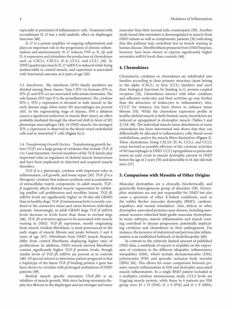
Figure 1: Immunofluorescent detection of TNF-𝛼 and IFN-𝛾 in Duchenne muscular dystrophy. (a)-(b) Muscle biopsy taken from an 8-year-old patient with Duchenne muscular dystrophy caused by duplication of dystrophin exon 2, resulting in severe muscle damage, few groups ofrevertant fibers, and strong utrophin staining. TNF-𝛼 (red in (a)) is detected in a small cluster of inflammatory cells and colocalizes with CD3(green in (b)). The asterisk is an indicative that helps to identify an individual TNF-𝛼+ T cell. (c)-(d) Muscle biopsy taken from a 2-year-oldpatient with Duchennemuscular dystrophy caused by c.5299-5302dupATTT in dystrophin exon 37.Myopathological evaluation of the biopsydescribed definite muscle damage, few groups of revertant fibers, and strong utrophin staining. IFN-𝛾 (red in (c)) is strongly expressed onthe blood vessel endothelium (arrow) and on perivascular CD3+ T cells (green in (d)). Highlighted are an IFN-𝛾+ CD3+ T cell attached tothe luminal side of the blood vessel (asterisk) and an interstitial IFN-𝛾+ CD3+ T cell (circle).
The involvement of proinflammatory IL-1 in musculardystrophy remains a topic of debate. Neither IL-1𝛼 nor IL-1𝛽 immunoreactivity could be shown in a study investigat-ing 8 DMD muscle samples [17], and the IL-1 family hasbeen reported downregulated in DMD serum [31]. However,another study describes IL-1𝛽 as being increased in DMDmuscle [32]. Also, diaphragm of mdx mice contains signifi-cantly higher IL-1𝛽mRNA levels than control mice [19], andIL-1𝛽 protein colocalizes with the infiltrating macrophages[20].
IL-6 is a cytokine with both proinflammatory and anti-inflammatory properties. It is a helper T cell type 2 (Th2)cytokine, meaning that it promotes IgE and eosinophilicresponses in atropy and counteracts Th1-driven proinflam-matory responses. On the other hand, IL-6 exhibits proin-flammatory activity through activation of the transcriptionfactor nuclear factor 𝜅B. IL-6 concentrations are significantlyhigher in serum of DMD patients (3.77 ± 2.71 pg/mL) com-pared to healthy age-matched controls (1.93 ± 1.38 pg/mL)[31] and follow the disease time-course [32]. In DMDmuscle,IL-6 mRNA levels display a significant increase compared tocontrols. The level increases with age: from 26-fold at age 4
years to 148-fold between 5 and 9 years [33]. Blocking IL-6 through injection with a monoclonal antibody causes anincrease of muscle inflammation in the mdx mouse model,further suggesting an anti-inflammatory effect, possibly bymediating muscle repair [34].
IL-10 functions as a suppressor of inflammation throughits differential effect on the different macrophage subtypes:deactivating M1 macrophages and activating the M2 phe-notype. M1 macrophages function within the Th1 responseand produce copious amounts of proinflammatory cytokines,while M2 macrophages promote angiogenesis and tissuerepair and remodeling [35], a phenomenon also present inmuscle [36]. IL-10 prevents the production of Th1-associatedcytokines such as TNF-𝛼 and IFN-𝛾 [37]. Its expression is 8to 15-fold increased in mdx quadriceps compared with wildtype muscle, possibly as a protective reflex of the tissue. IL-10null mutation causes severe reduction of muscle strength dueto an imbalance between M1 and M2 macrophages [38].
IL-15 has proinflammatory characteristics as a stimulatorof T cell proliferation and NK-activity but can also be ofbenefit to tissue recovery by increasing myogenic differen-tiation [39]. Mdx diaphragm contains some IL-15 reactivity
4 Mediators of Inflammation
especially in proximity of inflammatory cells. Treatment withrecombinant IL-15 has a mild anabolic effect on diaphragmfunction [40].
IL-17 is a potent amplifier of ongoing inflammation andplays an important role in the progression of chronic inflam-mation and autoimmunity. IL-17 induces TNF-𝛼, IL-1𝛽, andIL-6 expression and stimulates the production of chemokinessuch as CXCL1, CXCL5, IL-8, CCL2, and CCL7 [41]. InDMDquadricepsmuscle IL-17mRNA is induced while beingundetectable in control muscle, and expression is associatedwith functional outcome at 6 years of age [42].
3.3. Interferons. The interferon (IFN) family members aredivided among three classes. Type I IFN (in humans IFN-𝛼,IFN-𝛽, and IFN-𝜔) are associated with innate immunity.Thesole human IFN type II is the proinflammatoryTh1 cytokineIFN-𝛾. IFN-𝛾 expression is elevated in mdx muscle in theearly disease stage when many M1 macrophages are present[43]. In the regenerating stage of disease, IFN-𝛾 ablationcauses a significant reduction in muscle fiber injury, an effectprobably mediated through the observed shift in favor of M2phenotype macrophages [44]. In DMDmuscle, the strongestIFN-𝛾 expression is observed in the blood vessel endothelialcells and in interstitial T cells (Figure 1(c)).
3.4. Transforming Growth Factors. Transforming growth fac-tors (TGF) are a large group of cytokines that include TGF-𝛽1 to 3 andmyostatin. Several members of the TGF family playimportant roles as regulators of skeletal muscle homeostasisand have been implicated in inherited and acquired muscledisorders.
TGF-𝛽 is a pleiotropic cytokine with important roles ininflammation, cell growth, and tissue repair [45]. TGF-𝛽 is afibrogenic cytokine that induces synthesis and accumulationof extracellular matrix components. In adult muscle, TGF-𝛽 negatively affects skeletal muscle regeneration by inhibit-ing satellite cell proliferation and myofiber fusion. TGF-𝛽1mRNA levels are significantly higher in 30-day-old GRMDthan in healthy dogs. TGF-𝛽 immunoreactivity ismostly con-fined to the connective tissue and varies between individualanimals. Interestingly, in adult GRMD dogs TGF-𝛽 mRNAlevels decrease to levels lower than those in normal dogs[46]. TGF-𝛽1 activation appears to be associated with musclewasting in DMD. TGF-𝛽1 expression, mostly originatingfrom muscle resident fibroblasts, is most pronounced in theearly stages of muscle fibrosis and peaks between 2 and 6years of age [47]. Fibroblasts from DMD muscle biopsiesdiffer from control fibroblasts, displaying higher rates ofproliferation. In addition, DMD muscle-derived fibroblastscontain significantly higher TGF-𝛽 protein levels, thoughsimilar levels of TGF-𝛽1 mRNA are present as in controls[48]. Of special interest to determine patient prognosis is thata haplotype of the latent TGF-𝛽 binding protein 4 gene hasbeen shown to correlate with prolonged ambulation of DMDpatients [49].
Skeletal muscle specific myostatin (TGF-𝛽8) is aninhibitor of muscle growth. Mdx mice lacking myostatin dis-play less fibrosis in the diaphragm and are stronger and more
muscular than their normal mdx counterparts [50]. Anotherstudy found that myostatin is downregulated in muscle fromDMD infants as well as symptomatic patients [51] indicatingthat this pathway may contribute less to muscle wasting inhumandisease.Myofibroblasts prepared fromDMDbiopsies,however, have been shown to express significantly highermyostatin mRNA levels than controls [48].
4. Chemokines
Chemotactic cytokines or chemokines are subdivided intofamilies according to their primary structure (most belongto the alpha (CXCL) or beta (CCL) families) and exerttheir biological functions by binding to G protein-coupledreceptors [52]. Chemokines interact with other cytokinesand adhesion molecules and their activities go way furtherthan the attraction of leukocytes to inflammatory sites.CCL17, for instance, has been shown to enhance tissuefibrosis [53]. While the chemokine expression profile inhealthy skeletal muscle is fairly limited, many chemokines areinduced or upregulated in dystrophic muscle (Tables 1 and2) [54–58]. The individual muscle tissue distribution of somechemokines has been determined and shows that they candifferentially be allocated to inflammatory cells, blood vesselendothelium, and/or the muscle fibers themselves (Figure 2).Three chemokines, being CXCL8 (IL-8), CCL2, and CCL5,come forward as possible effectors of the cytotoxic activitiesofM1macrophages inDMD.CCL2upregulation in particularseems an early event in muscle dystrophy, present in DMDbefore the age of 2 years [55] and detectable in 14-day oldmdxmice [57].
5. Comparison with Myositis of Other Origins
Muscular dystrophies are a clinically, biochemically, andgenetically heterogeneous group of disorders [59]. Dystro-phin mutations are not just responsible for DMD but alsocause a spectrum of other X-linked conditions, such asthe milder Becker muscular dystrophy (BMD), cardiom-yopathies, and mental retardation. Also, defects in otherdystrophin-associated proteins cause disease, including auto-somal recessive inherited limb-girdle muscular dystrophies.In many subtypes, muscle inflammation and muscle wast-ing contribute to disease progression, potentially implicat-ing cytokines and chemokines in their pathogenesis. Forinstance, the presence of endomysial and perivascular inflam-mation is an established hallmark of dysferlinopathy [60].
In contrast to the relatively limited amount of publishedDMD data, a multitude of reports is available on the expres-sion of cytokines in the different idiopathic inflammatorymyopathies (IIM), which include dermatomyositis (DM),polymyositis (PM) and sporadic inclusion body myositis(IBM) [61]. This allows for some comparison between pri-mary muscle inflammation in IIM and dystrophy-associatedmuscle inflammation. In a single BMD patient included ina multiplex cytokine immunoassay study, CCL2 levels are11 pg/mg muscle protein, while those in 6 patients per IIMgroup were 45 ± 51 (DM), 15 ± 9 (PM) and 13 ± 9 (IBM),

Mediators of Inflammation 5
Table 1: Alpha-chemokine expression in Duchenne muscular dystrophy and its mouse model.
Systematic name Common name Tissue mRNA quantity Protein quantity Protein localization ReferenceCXCL1 GRO-alpha DMD quadriceps muscles BV, MF, M𝜑, T, DC [54]CXCL2 GRO-beta DMD quadriceps muscles BV, MF, M𝜑 [54]CXCL3 GRO-gamma DMD quadriceps muscles BV, MF, M𝜑, DC [54]CXCL8 IL-8 DMD quadriceps muscles BV, MF, M𝜑 [54]CXCL10 IP-10 DMD quadriceps muscles BV, (MF), M𝜑, T [54]CXCL11 ITAC DMD quadriceps muscles BV, (MF), M𝜑 [54]CXCL12 SDF-1 DMD quadriceps muscles Increased 2.3x [55]
DMD quadriceps muscles BV, MF [54]DMD serum Increased 1.2x [56]
CXCL14 BRAK mdx hindlimb muscles Increased 1.7x [57]Breast and kidney derived (BRAK); blood vessel (BV), alpha-chemokine (CXCL), dendritic cell (DC), Duchennemousemodel (mdx), growth related oncogene(GRO), interleukin 8 (IL-8), interferon-inducible protein of 10 kd (IP-10), interferon-inducible T cell alpha chemo-attractant (ITAC), muscle fiber (MF),macrophage (M𝜑), stromal cell-derived factor (SDF), T cell (T). Rare observations are indicated between brackets.
Table 2: Beta-chemokine expression in Duchenne muscular dystrophy and its mouse model.
Systematic name Common name Tissue mRNA quantity Protein quantity Protein localization ReferenceCCL2 MCP-1 mdx hindlimb muscles Increased 62.7x Increased 4.1x MF, M𝜑 [57]
DMD quadriceps muscles Increased 1.4x [55]BV, M𝜑 [54]
CCL3 MIP-1 alpha mdx diaphragm Increased [58]CCL5 RANTES mdx hindlimb muscles Increased 2.3x [57]
mdx diaphragm Increased Increased [58]DMD quadriceps muscles M𝜑 [54]
CCL7 MCP-3 mdx hindlimb muscles Increased 14.7x [57]DMD quadriceps muscles M𝜑 [54]
CCL8 MCP-2 mdx hindlimb muscles Increased 28.9x [57]CCL9 MIP-1 gamma mdx hindlimb muscles Increased 7.9x Increased 2.4x [57]CCL11 eotaxin mdx hindlimb muscles Increased 2.0x [57]CCL17 TARC DMD quadriceps muscles (M𝜑) [54]Blood vessel (BV), beta-chemokine (CCL), Duchennemuscular dystrophy (DMD),monocyte chemoattractant protein (MCP), Duchennemousemodel (mdx),muscle fiber (MF), macrophage (M𝜑), macrophage inflammatory protein (MIP), regulated upon activation, normal T cell expressed and secreted (RANTES),thymus and activation-regulated chemokine (TARC). Rare observations are indicated between brackets.
respectively [62]. In DMD quadriceps muscle, TNF-𝛼, IL-6, and CCL2 mRNA levels are lower than in juvenile DM[42]. The observed more moderate expression levels couldbe indicative to the secondary nature of muscle inflam-mation as opposed to the primary inflammatory origin ofthe IIM. Although there unmistakably are universal inflam-matory processes at hand, data also point to specific rolesfor cytokines and chemokines in DMD. The expression pro-files of M1 macrophages are peculiar when DMD and IIMare compared. Also, in IIM strong expression of CXCR3 isobserved on the muscle infiltrating T cells, indicating theirinvolvement inTh1 immune responses. Such polarization of Tcells is less obvious in DMDmuscle. In fact, the muscle infil-trating T cells in DMD express a strikingly limited repertoireof chemokines in comparison to their IIM counterparts [54].
6. Relevance to DMD Disease Management
The medical community still awaits the coming of age ofmolecular dystrophin salvaging therapies [63]. In this respect,exon skipping [64] and suppression of stop codons [65]are considered strategies of increasing functional dystrophinexpression. However, surfacing results of clinical trials, moreparticular those using AAV-mediated delivery of mini-dystrophin, are suggestive of important acquisition of T cellimmunity targeting the dystrophin protein [66]. Earlier, ithad been postulated that such priming was unlikely, due tothe presence of revertant fibers inmany patients which wouldtheoretically safeguard dystrophin replacement from theimmune system. Nonetheless, it is becoming more and moreobvious that monitoring of cellular immune responses will

6 Mediators of Inflammation
(a) (b)
(c) (d)
Figure 2: Chemokine staining in Duchenne muscular dystrophy. Nonconsecutive sections showing the same microscopic field containing anecrotic muscle fiber invaded by macrophages (asterisk). The muscle biopsy was taken from an 8-year-old patient with Duchenne musculardystrophy caused by duplication of dystrophin exon 2. Upon diagnostic myopathological evaluation, the biopsy displayed severe muscledamage, few groups of revertant fibers, and strong utrophin staining. Chemokines were immunostained and visualized with a secondaryantibody using the streptavidin-biotin labeling system and 3-amino-9-ethylcarbazole chromogen (Dako, Glostrup, Denmark). Cell nucleiwere counterstained with hematoxylin (blue). The sarcoplasm of a necrotic fiber is strongly positive for CXCL8 (red in (a)) and CXCL11 (redin (b)). The cytoplasm of the necrotic fiber and its invading inflammatory cells are moderately positive for CCL5 (red in (c)) and faintlypositive for CCL17 (red in (d)). Small regenerating fibers stain for all four chemokines with varying intensities.
be a priority in all ongoing and future experimental therapiesaimed at increasing the number of dystrophin positivemusclefibers. A recent study demonstrated that circulating dys-trophin primed T cells are frequently encountered in DMD,increased with age, and reduced by glucocorticoid therapy[67].
Immunosuppression, administering glucocorticoids inparticular, remains standard treatment for DMD today.Although anti-inflammatory therapy may add years to DMDpatient ambulation, steroids are associated with importantadverse effects [68]. The characterization of the factors thatdrive inflammation and guide specific subsets of leuko-cytes to the tissues raises hopes of attempting more selec-tive immunomodulatory intervention. Strategies aimed atneutralizing individual cytokines or chemokines could be anamenable approach to reduce side effects.
6.1. Targeting the Culprits While Sparing the Protectors. Spe-cifically targeting cytokines and chemokines with predom-inant proinflammatory activities, such as TNF-𝛼, is underexploration. The TNF-𝛼 neutralizing antibody infliximabdelays and reduces muscle damage in mdxmice [69]. SolubleTNF-receptor etanercept, a dimeric fusion protein composed
of an extracellular ligand-binding portion of the human p75TNF-receptor linked to the Fc portion of human IgG, reducesmuscle fibrosis [70] and necrosis [71]. The disruption ofchemokine-mediated signaling also seems, at first glance, anattractive therapeutic possibility. An approach could be toselectively block a chemokine receptor with a key catabolicrole by either a small-molecule antagonist, antibody, bindingprotein, or protein agonist [72]. Several chemokine-receptorantibodies are entering the clinic, including an anti-CCR2monoclonal antibody namedMLN1202 (Millenium Pharma-ceuticals, Cambridge, MA, USA) currently being tried forvarious inflammatory diseases. However, strategies targetingthe chemokine system present with certain inherent difficul-ties. Firstly, several chemokines are up-regulated in DMD.The redundancy of function of part of themmakes it difficultto design effective therapeutic interventions. Secondly, therecould be considerable inter-patient variability, as well asdifferences between the stages of the disease. More researchis necessary to address these issues. Thirdly, chemokines canhave benefits for tissue recovery, by activating muscle fiberregeneration and recruiting non-cytotoxic macrophage sub-populations that stimulate muscle tissue rebuilding [73]. Forinstance, when considering the anti-CCR2 avenue, its ligand

Mediators of Inflammation 7
CCL2 has the potential to drive forward chronic inflamma-tion, but the importance of CCL2 in muscle regeneration hasalso been recognized [74, 75].
In addition, strategies aimed at neutralizing fibrogeniccytokines or cytokines associated with muscle wasting areunder exploration for treating DMD. For instance, the TGF-𝛽1 antagonist pirfenidone improves cardiac function in mdxmice [76]. The TGF-𝛽 blocker suramin decreases fibrosisand offers benefit in grip strength in mdx mice [77]. ATGF-𝛽 neutralizing antibody decreases fibrosis and improvesregeneration in mdx mice [78]. Inhibition of myostatin witha neutralizing antibody [79], soluble decoy receptor [80], ormyostatin binding propeptide [81] has also been put forward.An in vitro model, using nodules of DMD muscle-derivedfibroblasts grown onto a solid substrate, has been developedwhich allows convenient screening of potential antifibroticagents [82].
6.2. Reprogramming the Immune Response. WhileM1macro-phages have a destructive cytokine repertoire, the M2 phe-notype promotes angiogenesis, tissue repair, and remodeling.In mdx muscle, M1 macrophages predominate during theearly, acute stage. The balance tips over to the M2 phenotypein the regenerative and progressive phase of the disease. Inother words, the M1/M2 balance evolves beneficially withM1 macrophages undergoing deactivation as the disease pro-gresses from the acute necrotic to the regenerative phase. M1density significantly reduces with age in mdx soleus (4 versus12 weeks) [43]. This could account for the milder diseasephenotype of mdx mice compared to human disease, as incontrast percentages of M1 and M2 phenotype macrophagesseem strikingly constant in DMD muscle taken at differentdisease stages [54]. Therapeutic agents regulating the M1/M2balance in favor of the M2 phenotype, such as cannabinoidCB2 receptor agonists, could be of benefit to patients [83].Interestingly, glucocorticoids as such have also been shownto favor a shift of macrophage phenotype, reducing thenumbers of M1 macrophages by half in patients treated withprednisone (0.75mg/kg/day) during 6 months [84].
7. Conclusions
In dystrophic skeletal muscle, part of the accumulating mus-cle damage is caused by ongoing activation of inflammatorycells rather than by direct mechanical damage. Currentknowledge, of which a large part is summarized in this review,supports an important and diversified role for cytokines andchemokines in the DMD-associated muscle inflammation.The fact that a number of chemokines are expressed directlyby the muscle fibers suggests that the tissue itself contributesto the chemotaxic process, actively perpetuating the chronicinflammation.
Abbreviations
BMD: Becker muscular dystrophyCCL: Beta-chemokineCXCL: Alpha-chemokineDMD: Duchenne muscular dystrophy
GRMD: Golden retriever muscular dystrophyHFMD: Hypertrophic feline muscular dystrophyIIM: Idiopathic inflammatory myopathiesIFN: InterferonIL: InterleukinTGF: Transforming growth factorTNF: Tumor necrosis factor.
Acknowledgments
The authors thank Professor Dr. Jean-Jacques Martin of theDepartment of Ultrastructural Neuropathology, Born-BungeInstitute, University of Antwerp and Antwerp UniversityHospital, Belgium, for providing patient biopsies and expertopinion.
References
[1] N. Deconinck and B. Dan, “Pathophysiology of Duchennemuscular dystrophy: current hypotheses,” Pediatric Neurology,vol. 36, no. 1, pp. 1–7, 2007.
[2] B. J. Petrof, “Molecular pathophysiology of myofiber injury indeficiencies of the dystrophin-glycoprotein complex,”AmericanJournal of Physical Medicine and Rehabilitation, vol. 81, no. 11,pp. S162–S174, 2002.
[3] R. M. McDouall, M. J. Dunn, and V. Dubowitz, “Nature of themononuclear infiltrate and themechanism ofmuscle damage injuvenile dermatomyositis and Duchenne muscular dystrophy,”Journal of the Neurological Sciences, vol. 99, no. 2-3, pp. 199–217,1990.
[4] R. Mantegazza, F. Andreetta, P. Bernasconi et al., “Analysis ofT cell receptor repertoire of muscle-infiltrating T lymphocytesin polymyositis. Restricted V𝛼/𝛽 rearrangements may indicateantigen-driven selection,” Journal of Clinical Investigation, vol.91, no. 6, pp. 2880–2886, 1993.
[5] J. Morrison, Q. L. Lu, C. Pastoret, T. Partridge, and G. Bou-Gharios, “T-cell-dependent fibrosis in the mdx dystrophicmouse,” Laboratory Investigation, vol. 80, no. 6, pp. 881–891,2000.
[6] J. Morrison, D. B. Palmer, S. Cobbold, T. Partridge, and G. Bou-Gharios, “Effects of T-lymphocyte depletion on muscle fibrosisin the mdx mouse,” American Journal of Pathology, vol. 166, no.6, pp. 1701–1710, 2005.
[7] J. Middleton, A. M. Patterson, L. Gardner, C. Schmutz, and B.A. Ashton, “Leukocyte extravasation: chemokine transport andpresentation by the endothelium,” Blood, vol. 100, no. 12, pp.3853–3860, 2002.
[8] N. J. H. Sharp, J. N. Kornegay, S. D. Van Camp et al., “An errorin dystrophin mRNA processing in golden retriever musculardystrophy, an animal homologue of Duchenne muscular dys-trophy,” Genomics, vol. 13, no. 1, pp. 115–121, 1992.
[9] F. Gaschen and J.-M. Burgunder, “Changes of skeletal muscle inyoung dystrophin-deficient cats: a morphological and morpho-metric study,”ActaNeuropathologica, vol. 101, no. 6, pp. 591–600,2001.
[10] D. I. Bassett and P. D. Currie, “The zebrafish as amodel formus-cular dystrophy and congenital myopathy,” Human MolecularGenetics, vol. 12, no. 2, pp. R265–R270, 2003.
[11] G. Kawahara, J. A. Karpf, J. A. Myers, M. S. Alexander, J. R.Guyone, and L. M. Kunkel, “Drug screening in a zebrafish

8 Mediators of Inflammation
model of Duchenne muscular dystrophy,” Proceedings of theNational Academy of Sciences of the United States of America,vol. 108, no. 13, pp. 5331–5336, 2011.
[12] I. Lundberg, J. M. Brengman, and A. G. Engel, “Analysis ofcytokine expression in muscle in inflammatory myopathies,Duchenne dystrophy, and non-weak controls,” Journal of Neu-roimmunology, vol. 63, no. 1, pp. 9–16, 1995.
[13] J. G. Tidball and M. Wehling-Henricks, “Damage and inflam-mation in muscular dystrophy: potential implications andrelationships with autoimmune myositis,” Current Opinion inRheumatology, vol. 17, no. 6, pp. 707–713, 2005.
[14] E. Porreca, M. D. Guglielmi, A. Uncini et al., “Haemostaticabnormalities, cardiac involvement and serum tumor necrosisfactor levels in X-linked dystrophic patients,” Thrombosis andHaemostasis, vol. 81, no. 4, pp. 543–546, 1999.
[15] K. Saito, D. Kobayashi, M. Komatsu et al., “A sensitive assayof tumor necrosis factor 𝛼 in sera from Duchenne musculardystrophy patients,”Clinical Chemistry, vol. 46, no. 10, pp. 1703–1704, 2000.
[16] E. Abdel-Salam, I. Abdel-Meguid, and S. S. Korraa, “Markers ofdegeneration and regeneration in Duchenne muscular dystro-phy,” Acta Myologica, vol. 28, no. 3, pp. 94–100, 2009.
[17] D. S. Tews and H. H. Goebel, “Cytokine expression profile inidiopathic inflammatory myopathies,” Journal of Neuropathol-ogy and Experimental Neurology, vol. 55, no. 3, pp. 342–347,1996.
[18] S. Kuru, A. Inukai, T. Kato, Y. Liang, S. Kimura, and G. Sobue,“Expression of tumor necrosis factor-𝛼 in regenerating musclefibers in inflammatory and non-inflammatory myopathies,”Acta Neuropathologica, vol. 105, no. 3, pp. 217–224, 2003.
[19] A. Kumar and A. M. Boriek, “Mechanical stress activates thenuclear factor-kappaB pathway in skeletal muscle fibers: apossible role in Duchennemuscular dystrophy,” FASEB Journal,vol. 17, no. 3, pp. 386–396, 2003.
[20] K. Hnia, J. Gayraud, G. Hugon et al., “L-arginine decreasesinflammation andmodulates the nuclear factor-𝜅B/matrixmet-alloproteinase cascade in mdx muscle fibers,” American Journalof Pathology, vol. 172, no. 6, pp. 1509–1519, 2008.
[21] M. J. Spencer, M. W. Marino, and W. M. Winckler, “Alteredpathological progression of diaphragm and quadriceps musclein TNF-deficient, dystrophin-deficient mice,” NeuromuscularDisorders, vol. 10, no. 8, pp. 612–619, 2000.
[22] K. K. Creus, B. De Paepe, J. Weis, and J. L. De Bleecker, “Themultifaceted character of lymphotoxin 𝛽 in inflammatorymyopathies and muscular dystrophies,” Neuromuscular Disor-ders, vol. 22, no. 8, pp. 712–719, 2012.
[23] P. Vandenabeele, L. Galluzzi, T. Vanden Berghe, and G. Kroe-mer, “Molecularmechanisms of necroptosis: an ordered cellularexplosion,”Nature ReviewsMolecular Cell Biology, vol. 11, no. 10,pp. 700–714, 2010.
[24] J. G. Tidball, D. E. Albrecht, B. E. Lokensgard, andM. J. Spencer,“Apoptosis precedes necrosis of dystrophin-deficient muscle,”Journal of Cell Science, vol. 108, no. 6, pp. 2197–2204, 1995.
[25] A. Serdaroglu, K. Gucuyener, S. Erdem, G. Kose, E. Tan, and C.Okuyaz, “Role of apoptosis in Duchenne’s muscular dystrophy,”Journal of Child Neurology, vol. 17, no. 1, pp. 66–68, 2002.
[26] M. Kondo, Y. Murakawa, N. Harashima, S. Kobayashi, S. Yam-aguchi, and M. Harada, “Roles of proinflammatory cytokinesand the Fas/Fas ligand interaction in the pathogenesis of inflam-matorymyopathies,” Immunology, vol. 128, no. 1, pp. e589–e599,2009.
[27] L. Behrens, A. Bender, M. A. Johnson, and R. Hohlfeld, “Cyto-toxic mechanisms in inflammatory myopathies. Co-expressionof Fas and protective Bcl-2 in muscle fibres and inflammatorycells,” Brain, vol. 120, no. 6, pp. 929–938, 1997.
[28] J. L. De Bleecker, V. I. Meire, I. E. Van Walleghem, I. M.Groessens, and J. M. Schroder, “Immunolocalization of Fas andFas ligand in inflammatory myopathies,” Acta Neuropatholog-ica, vol. 101, no. 6, pp. 572–578, 2001.
[29] C. Dogra, H. Changotra, S. Mohan, and A. Kumar, “Tumornecrosis factor-like weak inducer of apoptosis inhibits skeletalmyogenesis through sustained activation of nuclear factor-𝜅B and degradation of MyoD protein,” Journal of BiologicalChemistry, vol. 281, no. 15, pp. 10327–10336, 2006.
[30] A. Mittal, S. Bhatnagar, A. Kumar, P. K. Paul, S. Kuang, and A.Kumar, “Genetic ablation of TWEAK augments regenerationand post-injury growth of skeletal muscle in mice,” AmericanJournal of Pathology, vol. 177, no. 4, pp. 1732–1742, 2010.
[31] A. Rufo, A. Del Fattore,M. Capulli et al., “Mechanisms inducinglow bone density in Duchenne muscular dystrophy in mice andhumans,” Journal of Bone and Mineral Research, vol. 26, no. 8,pp. 1891–1903, 2011.
[32] N. P. Evans, S. A. Misyak, J. L. Robertson, J. Bassaganya-Riera,and R. W. Grange, “Immune-mediated mechanisms potentiallyregulate the disease time-course of Duchenne muscular dystro-phy and provide targets for therapeutic intervention,” PhysicalMedicine and Rehabilitation, vol. 1, no. 8, pp. 755–768, 2009.
[33] S. Messina, G. L. Vita, M. Aguennouz et al., “Activation of NF-𝜅B pathway in Duchenne muscular dystrophy: relation to age,”Acta Myologica, vol. 30, pp. 16–23, 2011.
[34] M. C. Kostek, K. Nagaraju, E. Pistilli et al., “IL-6 signalingblockade increases inflammation but does not affect musclefunction in the mdx mouse,” BMC Musculoskeletal Disorders,vol. 13, p. 106, 2012.
[35] A.Mantovani, A. Sica, S. Sozzani, P. Allavena, A. Vecchi, andM.Locati, “The chemokine system in diverse forms of macrophageactivation and polarization,” Trends in Immunology, vol. 25, no.12, pp. 677–686, 2004.
[36] L. Arnold, A. Henry, F. Poron et al., “Inflammatory monocytesrecruited after skeletal muscle injury switch into antiinflamma-tory macrophages to support myogenesis,” Journal of Experi-mental Medicine, vol. 204, no. 5, pp. 1057–1069, 2007.
[37] D. F. Fiorentino, A. Zlotnik, T. R. Mosmann, M. Howard, andA. O’Garra, “IL-10 inhibits cytokine production by activatedmacrophages,” Journal of Immunology, vol. 147, no. 11, pp. 3815–3822, 1991.
[38] S. A. Villalta, C. Rinaldi, B. Deng, G. Liu, B. Fedor, and J. G.Tidball, “Interleukin-10 reduces the pathology ofmdxmusculardystrophy by deactivating M1 macrophages and modulatingmacrophage phenotype,” Human Molecular Genetics, vol. 20,no. 4, pp. 790–805, 2011.
[39] P. S. Furmanczyk and L. S. Quinn, “Interleukin-15 increasesmyosin accretion in human skeletal myogenic cultures,” CellBiology International, vol. 27, no. 10, pp. 845–851, 2003.
[40] L. J. Harcourt, A. G. Holmes, P. Gregorevic, J. D. Schertzer,N. Stupka, and G. S. Lynch, “Interleukin-15 administrationimproves diaphragm muscle pathology and function in dys-trophic mdx mice,” American Journal of Pathology, vol. 166, no.4, pp. 1131–1141, 2005.
[41] J. C. Waite and D. Skokos, “Th17 response and inflammatoryautoimmune diseases,” International Journal of Inflammation,vol. 2012, Article ID 819467, 10 pages, 2012.

Mediators of Inflammation 9
[42] L. De Pasquale, A. D’Amico, M. Verardo, S. Petrini, E. Bertini,and F. De Benedetti, “Increased muscle expression of interleu-kin-17 inDuchennemuscular dystrophy,”Neurology, vol. 78, no.17, pp. 1309–1314, 2012.
[43] S. A. Villalta, H. X. Nguyen, B. Deng, T. Gotoh, and J. G. Tidbal,“Shifts in macrophage phenotypes and macrophage compe-tition for arginine metabolism affect the severity of musclepathology in muscular dystrophy,” Human Molecular Genetics,vol. 18, no. 3, pp. 482–496, 2009.
[44] S. A. Villalta, B. Deng, C. Rinaldi, M. Wehling-Henricks, and J.G. Tidball, “IFN-𝛾 promotes muscle damage in the mdx mousemodel of Duchenne muscular dystrophy by suppressing M2macrophage activation and inhibitingmuscle cell proliferation,”Journal of Immunology, vol. 187, no. 10, pp. 5419–5428, 2011.
[45] G. C. Blobe, W. P. Schiemann, and H. F. Lodish, “Role of trans-forming growth factor 𝛽 in human disease,” The New EnglandJournal of Medicine, vol. 342, no. 18, pp. 1350–1358, 2000.
[46] L. Passerini, P. Bernasconi, F. Baggi et al., “Fibrogenic cytokinesand extent of fibrosis in muscle of dogs with X-linked goldenretrievermuscular dystrophy,”NeuromuscularDisorders, vol. 12,no. 9, pp. 828–835, 2002.
[47] P. Bernasconi, E. Torchiana, P. Confalonieri et al., “Expressionof transforming growth factor-𝛽1 in dystrophic patient mus-cles correlates with fibrosis. Pathogenetic role of a fibrogeniccytokine,” Journal of Clinical Investigation, vol. 96, no. 2, pp.1137–1144, 1995.
[48] S. Zanotti, S. Gibertini, and M. Mora, “Altered production ofextra-cellularmatrix components bymuscle-derivedDuchennemuscular dystrophy fibroblasts before and after TGF-𝛽1 treat-ment,”Cell andTissue Research, vol. 339, no. 2, pp. 397–410, 2010.
[49] K. M. Flanigan, E. Ceco, K. M. Lamar et al., “LTBP4 genotypepredicts age of ambulatory loss in Duchenne muscular dystro-phy,” Annals of Neurology, vol. 73, no. 4, pp. 481–488, 2013.
[50] K. R. Wagner, A. C. McPherron, N. Winik, and S.-J. Lee, “Lossof myostatin attenuates severity of muscular dystrophy in mdxmice,” Annals of Neurology, vol. 52, no. 6, pp. 832–836, 2002.
[51] Y.-W. Chen, K. Nagaraju, M. Bakay et al., “Early onset of inflam-mation and later involvement of TGF𝛽 in Duchenne musculardystrophy,” Neurology, vol. 65, no. 6, pp. 826–834, 2005.
[52] M. Locati and P. M. Murphy, “Chemokines and chemokinereceptors: biology and clinical relevance in inflammation andAIDS,” Annual Review of Medicine, vol. 50, pp. 425–440, 1999.
[53] Y. Yogo, S. Fujishima, T. Inoue et al., “Macrophage derivedchemokine (CCL22), thymus and activation-regulated chemok-ine (CCL17), and CCR4 in idiopathic pulmonary fibrosis,”Respiratory Research, vol. 10, article no. 80, 2009.
[54] B. De Paepe, K. K. Creus, J. J. Martin, and J. L. De Bleecker,“Upregulation of chemokines and their receptors in Duchennemuscular dystrophy: potential for attenuation of myofibernecrosis,”Muscle & Nerve, vol. 46, no. 6, pp. 917–925, 2012.
[55] M. Pescatori, A. Broccolini, C. Minetti et al., “Gene expressionprofiling in the early phases of DMD: a constant molecularsignature characterizes DMD muscle from early postnatal lifethroughout disease progression,” FASEB Journal, vol. 21, no. 4,pp. 1210–1226, 2007.
[56] E. Abdel-Salam, I. Ehsan Abdel-Meguid, R. Shatla, and S. S.Korraa, “Stromal cell-derived factors in Duchenne musculardystrophy,” Acta Myologica, vol. 29, no. 3, pp. 398–403, 2010.
[57] J. D. Porter, W. Guo, A. P. Merriam et al., “Persistent over-expression of specific CC class chemokines correlates withmacrophage and T-cell recruitment in mdx skeletal muscle,”Neuromuscular Disorders, vol. 13, no. 3, pp. 223–235, 2003.
[58] A. Demoule, M. Divangahi, G. Danialou et al., “Expression andregulation of CC class chemokines in the dystrophic (mdx)diaphragm,”American Journal of Respiratory Cell andMolecularBiology, vol. 33, no. 2, pp. 178–185, 2005.
[59] E. Mercuri and F. Muntoni, “Muscular dystrophies,”The Lancet,vol. 381, no. 9869, pp. 845–860, 2013.
[60] E. Gallardo, R. Rojas-Garcıa, N. De Luna, A. Pou, R. H. BrownJr., and I. Illa, “Inflammation in dysferlin myopathy: immuno-histochemical characterization of 13 patients,”Neurology, vol. 57,no. 11, pp. 2136–2138, 2001.
[61] B. De Paepe, K. K. Creus, and J. L. De Bleecker, “Chemokinesin idiopathic inflammatorymyopathies,” Frontiers in Bioscience,vol. 13, no. 7, pp. 2548–2577, 2008.
[62] G. S. Baird and T. J. Montine, “Multiplex immunoassay analysisof cytokines in idiopathic inflammatory myopathy,” Archives ofPathology & Laboratory Medicine, vol. 132, no. 2, pp. 232–238,2008.
[63] M. Van Putten and A. Aartsma-Rus, “Opportunities and chal-lenges for the development of antisense treatment in neuromus-cular disorders,” Expert Opinion on Biological Therapy, vol. 11,no. 8, pp. 1025–1037, 2011.
[64] J. C. Van Deutekom, A. A. Janson, I. B. Ginjaar et al., “Localdystrophin restoration with antisense oligonucleotide PRO051,”TheNew England Journal of Medicine, vol. 357, no. 26, pp. 2677–2686, 2007.
[65] F. Muntoni and D. Wells, “Genetic treatments in musculardystrophies,” Current Opinion in Neurology, vol. 20, no. 5, pp.590–594, 2007.
[66] J. R.Mendell, K. Campbell, L. Rodino-Klapac et al., “Dystrophinimmunity in Duchenne’s muscular dystrophy,” The New Eng-land Journal of Medicine, vol. 363, no. 15, pp. 1429–1437, 2010.
[67] K. M. Flanigan, K. Campbell, L. Viollet et al., “Anti-dystrophinT-cell responses in Duchenne muscular dystrophy: prevalenceand a glucocorticoid treatment effect,” Human Gene TherapyMethods, 2013.
[68] B. L. Y.Wong andC.Christopher, “Corticosteroids inDuchennemuscular dystrophy: a reappraisal,” Journal of Child Neurology,vol. 17, no. 3, pp. 183–190, 2002.
[69] M. D. Grounds and J. Torrisi, “Anti-TNF𝛼 (Remicade) therapyprotects dystrophic skeletal muscle from necrosis,” FASEBJournal, vol. 18, no. 6, pp. 676–682, 2004.
[70] L. E. Gosselin and D. A. Martinez, “Impact of TNF-𝛼 blockadeon TGF-𝛽1 and type I collagen mRNA expression in dystrophicmuscle,”Muscle and Nerve, vol. 30, no. 2, pp. 244–246, 2004.
[71] S. Hodgetts, H. Radley, M. Davies, and M. D. Grounds,“Reduced necrosis of dystrophic muscle by depletion of hostneutrophils, or blockingTNF𝛼 functionwith Etanercept inmdxmice,” Neuromuscular Disorders, vol. 16, no. 9-10, pp. 591–602,2006.
[72] T. N. C. Wells, C. A. Power, J. P. Shaw, and A. E. I. Proudfoot,“Chemokine blockers—therapeutics in the making?” Trends inPharmacological Sciences, vol. 27, no. 1, pp. 41–47, 2006.
[73] J. G. Tidball andM.Wehling-Henricks, “Macrophages promotemuscle membrane repair and muscle fibre growth and regener-ation during modified muscle loading in mice in vivo,” Journalof Physiology, vol. 578, no. 1, pp. 327–336, 2007.
[74] L. Yahiaoui, D. Gvozdic, G. Danialou, M.Mack, and B. J. Petrof,“CC family chemokines directly regulate myoblast responses toskeletal muscle injury,” Journal of Physiology, vol. 586, no. 16, pp.3991–4004, 2008.

10 Mediators of Inflammation
[75] G. L. Warren, T. Hulderman, D. Mishra et al., “Chemokinereceptor CCR2 involvement in skeletal muscle regeneration,”FASEB Journal, vol. 19, no. 3, pp. 413–415, 2005.
[76] C. Van Erp, N. G. Irwin, and A. J. Hoey, “Long-term adminis-tration of pirfenidone improves cardiac function in mdx mice,”Muscle & Nerve, vol. 34, no. 3, pp. 327–334, 2006.
[77] A. P. T. Taniguti, A. Pertille, C. Y. Matsumura, H. S. Neto, andM. J. Marques, “Prevention of muscle fibrosis and myonecrosisin mdx mice by suramin, a TGF-𝛽1 blocker,” Muscle & Nerve,vol. 43, no. 1, pp. 82–87, 2011.
[78] F. Andreetta, P. Bernasconi, F. Baggi et al., “Immunomodulationof TGF-beta1 inmdxmouse inhibits connective tissue prolifera-tion in diaphragm but increases inflammatory response: impli-cations for antifibrotic therapy,” Journal of Neuroimmunology,vol. 175, no. 1-2, pp. 77–86, 2006.
[79] S. Bogdanovich, T. O. B. Krag, E. R. Barton et al., “Functionalimprovement of dystrophic muscle by myostatin blockade,”Nature, vol. 420, no. 6914, pp. 418–421, 2002.
[80] K. J. Morine, L. T. Bish, J. T. Selsby et al., “Activin IIB receptorblockade attenuates dystrophic pathology in a mouse model ofDuchenne muscular dystrophy,”Muscle & Nerve, vol. 42, no. 5,pp. 722–730, 2010.
[81] C. Qiao, J. Li, J. Jiang et al., “Myostatin propeptide gene deliveryby adeno-associated virus serotype 8 vectors enhances musclegrowth and ameliorates dystrophic phenotypes in mdx mice,”Human Gene Therapy, vol. 19, no. 3, pp. 241–253, 2008.
[82] S. Zanotti, S. Gilbertini, P. Salvadori, R. Mantegazza, and M.Mora, “Duchenne muscular dystrophy fibroblast nodules: acell-based assay for screening anti-fibrotic agents,” Cell andTissue Research, vol. 353, no. 3, pp. 659–670, 2013.
[83] A. Louvet, F. Teixeira-Clerc,M.-N. Chobert et al., “CannabinoidCB2 receptors protect against alcoholic liver disease by regulat-ing Kupffer cell polarization in mice,”Hepatology, vol. 54, no. 4,pp. 1217–1226, 2011.
[84] M. R. Hussein, S. A. Hamed, M. G. Mostafa, E. E. Abu-Dief,N. F. Kamel, and M. R. Kandil, “The effects of glucocorticoidtherapy on the inflammatory and dendritic cells in musculardystrophies,” International Journal of Experimental Pathology,vol. 87, no. 6, pp. 451–461, 2006.

Submit your manuscripts athttp://www.hindawi.com
Stem CellsInternational
Hindawi Publishing Corporationhttp://www.hindawi.com Volume 2014
Hindawi Publishing Corporationhttp://www.hindawi.com Volume 2014
MEDIATORSINFLAMMATION
of
Hindawi Publishing Corporationhttp://www.hindawi.com Volume 2014
Behavioural Neurology
EndocrinologyInternational Journal of
Hindawi Publishing Corporationhttp://www.hindawi.com Volume 2014
Hindawi Publishing Corporationhttp://www.hindawi.com Volume 2014
Disease Markers
Hindawi Publishing Corporationhttp://www.hindawi.com Volume 2014
BioMed Research International
OncologyJournal of
Hindawi Publishing Corporationhttp://www.hindawi.com Volume 2014
Hindawi Publishing Corporationhttp://www.hindawi.com Volume 2014
Oxidative Medicine and Cellular Longevity
Hindawi Publishing Corporationhttp://www.hindawi.com Volume 2014
PPAR Research
The Scientific World JournalHindawi Publishing Corporation http://www.hindawi.com Volume 2014
Immunology ResearchHindawi Publishing Corporationhttp://www.hindawi.com Volume 2014
Journal of
ObesityJournal of
Hindawi Publishing Corporationhttp://www.hindawi.com Volume 2014
Hindawi Publishing Corporationhttp://www.hindawi.com Volume 2014
Computational and Mathematical Methods in Medicine
OphthalmologyJournal of
Hindawi Publishing Corporationhttp://www.hindawi.com Volume 2014
Diabetes ResearchJournal of
Hindawi Publishing Corporationhttp://www.hindawi.com Volume 2014
Hindawi Publishing Corporationhttp://www.hindawi.com Volume 2014
Research and TreatmentAIDS
Hindawi Publishing Corporationhttp://www.hindawi.com Volume 2014
Gastroenterology Research and Practice
Hindawi Publishing Corporationhttp://www.hindawi.com Volume 2014
Parkinson’s Disease
Evidence-Based Complementary and Alternative Medicine
Volume 2014Hindawi Publishing Corporationhttp://www.hindawi.com